Note: Descriptions are shown in the official language in which they were submitted.
WO 2020/051236
PCT/US2019/049583
- 1 -
SYSTEMS AND METHODS FOR IDENTIFYING AND LOCATING REFLECTORS
USING ORTHOGONAL SEQUENCES OF REFLECTOR SWITCHING
FIELD OF THE INVENTION
[0001] The present invention relates to implantable reflectors, tags, or
markers and to
systems and methods for identifying and/or locating multiple markers within a
patient's
body, e.g., during surgical procedures or other procedures, such as during
lumpectomy
procedures.
BACKGROUND
[0002] Before a biopsy or surgical procedure to remove a lesion within a
breast, e.g.,
during a lumpectomy procedure, the location of the lesion must be identified.
For example,
mammography or ultrasound imaging may be used to identify and/or confirm the
location of
the lesion before the procedure. The resulting images may be used by a surgeon
during the
procedure to identify the location of the lesion and guide the surgeon, e.g.,
during dissection
to access and/or remove the lesion. However, such images are generally two
dimensional
and therefore provide only limited guidance for localization of the lesion
since the breast
and any lesion to be removed are three-dimensional structures. Further, such
images may
provide only limited guidance in determining a proper margin around the
lesion, i.e.,
defining a desired specimen volume to be removed.
[0003] To facilitate localization, immediately before a procedure, a wire may
be inserted
into the breast, e.g., via a needle, such that a tip of the wire is positioned
at the location of
the lesion_ Once the wire is positioned, it may be secured in place, e.g.,
using a bandage or
tape applied to the patient's skin where the wire emerges from the breast.
With the wire
placed and secured in position, the patient may proceed to surgery, e.g., to
have a biopsy or
lumpectomy performed.
[0004] One problem with using a wire for localization is that the wire may
move between
the time of placement and the surgical procedure. For example, if the wire is
not secured
sufficiently, the wire may move relative to the tract used to access the
lesion and
consequently the tip may misrepresent the location of the lesion. If this
occurs, when the
location is accessed and tissue removed, the lesion may not be fully removed
and/or healthy
tissue may be unnecessarily removed. In addition, during the procedure, the
surgeon may
merely estimate the location of the wire tip and lesion, e.g., based on
mammograms or other
images obtained during wire placement, and may proceed with dissection without
any
CA 03149936 2022-3-2
WO 2020/051236
PCT/US2019/049583
- 2 -
further guidance. Again, since such images are two dimensional, they may
provide limited
guidance to localize the lesion being treated or removed.
[0005] Alternatively, it has been suggested to place a radioactive seed to
provide
localization during a procedure. For example, a needle may be introduced
through a breast
into a lesion, and then a seed may be deployed from the needle. The needle may
be
withdrawn, and the position of the seed may be confirmed using mammography.
During a
subsequent surgical procedure, a hand-held gamma probe may be placed over the
breast to
identify a location overlying the seed. An incision may be made and the probe
may be used
to guide excision of the seed and lesion.
[0006] Because the seed is delivered through a needle that is immediately
removed, there
is risk that the seed may migrate within the patient's body between the time
of placement
and the surgical procedure. Thus, similar to using a localization wire, the
seed may not
accurately identify the location of the lesion, particularly, since there is
no external way to
stabilize the seed once placed. Further, such gamma probes may not provide
desired
precision in identifying the location of the seed, e.g., in three dimensions,
and therefore may
only provide limited guidance in localizing a lesion.
[0007] Accordingly, apparatus and methods for localization of lesions or other
tissue
structures in advance of and/or during surgical, diagnostic, or other medical
procedures
would be useful.
SUM-MARY
[0008] The present invention is directed to implantable reflectors, tags, or
markers, and to
systems and methods for identifying and/or locating multiple markers within a
patient's
body, e.g., during surgical procedures or other procedures, such as during
lumpectomy
procedures
100091 In accordance with one embodiment, a system is provided for
localization of a
target tissue region within a patient's body that includes a probe comprising
one or more
antennas for transmitting electromagnetic signals into a patient's body and
receiving
reflected signals from the patient's body, the probe further comprising a
light source for
delivering light pulses into a patient's body synchronized with the
electromagnetic signals,
and a plurality of markers sized for implantation within a patient's body.
Each marker may
include an energy convener configured to transform the light pulses from the
energy source
into electrical energy; a clock circuit coupled to the energy converter to
identify frames from
CA 03149936 2022-3-2
WO 2020/051236
PCT/U52019/049583
- 3 -
the light pulses; one or more elongate members coupled to a switch to provide
one or more
antennas; and a sequence generator coupled to the clock circuit to generate a
code sequence
based, at least in part, on the frames identified by the clock circuit, the
sequence generator
coupled to the switch to open and close the switch to modulate electromagnetic
signals from
the probe reflected by the marker based on the code sequence. The code
sequences
generated by the sequence generators of the plurality of markers may be
orthogonal to one
another and/or balanced, the probe comprising a processor configured to
analyze the
reflected signals to identify and locate each of the plurality of markers.
[0010] In accordance with another embodiment, a probe is provided for
identifying and
locating a plurality of markers implanted within a patient's body that
includes one or more
antennas for transmitting electromagnetic signals into a patient's body and
receiving
reflected signals from the patient's body; a light source for delivering light
pulses into a
patient's body synchronized with the electromagnetic signals, the light pulses
transmitted in
spaced-apart frames including a plurality of predetermined N pulses for
providing clock
signals to the markers such that the markers modulate their reflective
properties using
orthogonal and/or balanced code sequences triggered by the clock signals; and
a processor
for processing the reflected signals to separate the modulated signals from
the plurality of
markers based at least in part on the code sequences to identify and locate
each of the
plurality of markers substantially simultaneously.
[0011] In accordance with still another embodiment, a plurality of markers are
provided
for introduction into a target tissue region within a patient's body, each
marker including an
energy converter configured to transform light pulses from a light source into
electrical
energy; a clock circuit coupled to the energy converter to identify frames
from the light
pulses; one or more elongate members coupled to a switch to provide one or
more antennas;
and a sequence generator coupled to the clock circuit to generate a code
sequence based, at
least in part, on the frames identified by the clock circuit, the sequence
generator coupled to
the switch to open and close the switch to modulate electromagnetic signals
reflected by the
marker based on the code sequence. The code sequence generated by each of the
sequence
generators of the plurality of markers may be orthogonal to one another and/or
balanced to
facilitate identifying and/or locating the markers simultaneously.
[0012] In accordance with yet another embodiment, a method is provided for
identifying
and locating a plurality of markers implanted within a target tissue region
within a patient's
body that includes placing a tip of a probe adjacent the patient's body
oriented towards the
CA 03149936 2022-3-2
WO 2020/051236
PCT/U52019/049583
- 4 -
target tissue region; activating the probe to transmit electromagnetic signals
into the
patient's body, receive reflected signals from the patient's body, and in
synchronization with
transmitting the electromagnetic signals, deliver light pulses into the
patient's body,
whereupon the plurality of markers modulate reflected signals from the
respective reflector
tags based on orthogonal code sequences opening and closing respective
switches of the
markers; and processing the reflected signals, by the probe, to separate the
reflected signals
based at least in part on the code sequences to identify and locate each of
the plurality of
markers substantially simultaneously.
[0013] In accordance with still another embodiment, a method is provided for
localization
of a target tissue region within a patient's body. A plurality of markers may
be implanted
within the target tissue region within the patient's body. A tip of a probe
may be placed
adjacent the patient's body, e.g., positioned on the skin, oriented towards
the target tissue
region. The probe may be activated to transmit electromagnetic signals into
the patient's
body, receive reflected signals from the patient's body, and in
synchronization with
transmitting the electromagnetic signals, deliver light pulses into the
patient's body,
whereupon the plurality of markers modulate reflected signals from the
respective reflector
tags based on orthogonal code sequences opening and closing respective
switches of the
markers, and the probe may process the return signals to separate the
reflected signals based
at least in part on the code sequences to identify and locate each of the
plurality of markers
substantially simultaneously.
[0014] Other aspects and features of the present invention will become
apparent from
consideration of the following description taken in conjunction with the
accompanying
drawings.
BRIEF DESCRIPTION OF THE DRAWINGS
[0015] These and other features, aspects, and advantages of the present
disclosure will
become better understood with regard to the following description, appended
claims, and
accompanying drawings where:
[0016] FIG. IA shows an exemplary embodiment of a system including a probe for
identifying and/or locating a plurality of reflectors, tags, or markers that
may be implanted
within a patient's body.
CA 03149936 2022-3-2
WO 2020/051236
PCT/U52019/049583
-5-
100171 FIG. 18 shows other components that may be included in the system of
FIG. 1A,
including a delivery device for introducing one or more markers into a
patient's body and a
controller and/or display device.
[0018] FIGS. 2A-2C are top, side, and end views, respectively, of an exemplary
embodiment of a marker for implantation within a patient's body.
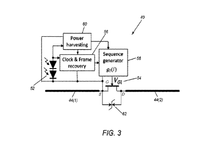
[0019] FIG. 3 is an exemplary embodiment of a schematic of a circuit that may
be included
in the marker of FIGS. 2A-2C.
[0020] FIG. 4 is a cross-sectional view of a breast including a plurality of
markers
implanted therein, and showing a probe being used to identify and located the
markers.
[0021] FIG. 4A is an end view of a distal end of the probe of FIG. 4.
[0022] FIG. 5 illustrates an exemplary set of periodic code sequences that may
be used to
switch a plurality of reflector tags triggered by IR pulses and a resulting
digitized waveform
of reflected signals received by a probe from the reflector tags and other
reflections.
[0023] FIG. 6 illustrates an exemplary set of code sequences using a balanced
Gold Code
that may be used to switch a plurality of reflector tags triggered by IR
pulses.
[0024] FIG. 7 is a graph showing the cross-correlation resulting from using
the
orthogonal balanced Gold Code code sequences shown in FIG. 6.
[0025] FIGS. 8A and 8B are graphs showing an example of a composite set of
reflected
radar pulses being separated into individual reflector signals in an ideal
noise free
environment.
[0026] FIGS. 9A and 9B are graphs showing an example of a composite set of
reflected
radar pulses being separated into individual reflector signals in an
environment including
noise.
DETAILED DESCRIPTION OF THE EXEMPLARY EMBODIMENTS
[0027] In the following description, numerous details are set forth in order
to provide a
more thorough description of the system. It will be apparent, however, to one
skilled in the
art, that the disclosed system may be practiced without these specific
details. In the other
instances, well known features have not been described in detail so as not to
unnecessarily
obscure the system.
[0028] Turning to the drawings, FIGS. 1A and 1B show an exemplary embodiment
of a
system 10 for localization of a target tissue region within a patient's body
that includes a
probe 20 and a plurality of reflectors, tags, or markers 40 (three shown
merely for
CA 03149936 2022-3-2
WO 2020/051236
PCT/U52019/049583
- 6 -
illustration) that may be implanted within a patient's body, e.g., within a
target tissue region,
such as within a breast 90, e.g., as shown in FIG 4. Optionally, as shown in
FIG. 1B the
system 10 may include one or more additional components, e.g., one or more
delivery
devices 70, each carrying one or more reflectors, tags, or markers 40 (one
shown) for
introduction/implantation in a patient's body, and a controller and/or display
unit 30 coupled
to the probe 20, e.g., using one or more cables 32, similar to embodiments
described in the
applications incorporated by reference herein.
[00291 As shown, the probe 20 generally includes one or more antennas 22 for
transmitting electromagnetic signals into a patient's body and receiving
reflected signals
from the patient's body, and a light source 24 for delivering light pulses
into a patient's
body synchronized with the electromagnetic signals, as described further
elsewhere herein.
In an exemplary embodiment, the antenna(s) 22 transmit ultrawide band (UVVB)
radar
pulses that are used for simultaneous detection of the markers 40 based on
modulated
reflective properties, e.g., using a switch inside each marker 40, which is
controlled by the
light pulses from the light source 24.
100301 FIGS. 2A-2C show an exemplary embodiment of a marker 40 that may be
used for each of the markers that may implanted within a patient's body.
Generally, the
marker 40 includes an electronics package 42 coupled to one or more antennas
44. In an
exemplary embodiment, each antenna 44 may be a wire or other elongate member
extending
from the package 42, e.g., a solid or hollow structure having a diameter or
other maximum
cross-section between about half and two millimeters (0.5-2 mm) and a length
between
about one and ten millimeters (1.0-10 mm). The antennas 44 may be formed from
elastic or
superelastic material and/or from shape memory material, e.g., stainless
steel, Nitinol, and
the like, such that the antennas 44 are biased to a predetermined shape when
deployed
within tissue, but may be elastically deformed, e.g., to facilitate delivery.
100311 As shown in FIGS. 2A-2C, the antennas 44 may be biased to assume a
substantially linear configuration, e.g., such that the antennas 44 extend
substantially
parallel to a longitudinal axis 48 of the marker 40. Alternatively, the
antennas 44 may be
substantially rigid such that the marker 40 remains in a substantially fixed,
e.g., linear or
curved, shape. Optionally, one or both antennas 44 may be offset from the
longitudinal axis
48, which may enhance loading the marker 40 within a delivery device (not
shown), as
described elsewhere herein or in the applications incorporated by reference
herein.
CA 03149936 2022-3-2
WO 2020/051236
PCT/U52019/049583
-7-
100321 As shown, each antenna 44 may include a first end 44a coupled to a
printed
circuit board (PCB) or other substrate 50 within the package 42 and a second
free end 44b,
e.g., terminating in an enlarged, rounded, and/or atraumatic tip 45.
Optionally, the first ends
44a may include one or more bends, e.g., to facilitate coupling the first ends
44a to the
substrate 50 and/or such that the antennas 44 extend tangentially from
opposite sides of the
package 42, as best seen in FIGS. 2A and 2B, e.g., to maximize an effective
length of the
antennas 44.
100331 Alternatively, the antennas 44 may be biased to assume a curvilinear or
other
configuration, e.g., a helical, serpentine or other curved shape, around the
longitudinal axis
48. For example, the antennas 44 may be formed from elastic or superelastic
material that is
shape set such that the antennas 44 are biased to a helical configuration (not
shown), yet
may be resiliently straightened to a substantially linear configuration, e.g.,
to facilitate
loading the marker 40 into a delivery device and/or otherwise introducing the
marker 40 into
a patient's body, e.g., as described in U.S. Patent Nos. 8,892,185 and
9,713,437, the entire
disclosures of which are expressly incorporated by reference herein.
100341 With additional reference to FIG. 3, the marker 40 may include one or
more
circuits or other electrical components encased or embedded in the electronics
package 42
and configured to modulate incident signals from the probe 20 (shown in FIG.
1) used to
identify and/or locate the marker 40. For example, the components may be
mounted on a
semiconductor chip, print circuit board (PCB), and/or other substrate 50
carried in the
package 42, and encased within the package 42 such that the components are
electrically
isolated from one another other than as shown in the schematic of FIG. 3. In
an exemplary
embodiment, the components may include an energy converter 52, a switch 54, a
clock
circuit or block 56 coupled to the energy converter 52, and a sequence
generator 58 coupled
to the clock circuit 56 and the switch 54, to generate a code sequence to open
and close the
switch 54 to modulate signals reflected by the marker 40 back to the probe 20
based on the
code sequence, as described elsewhere herein. Optionally, the marker 40 may
include one
or more additional components, e.g., a power harvesting circuit or block 60
coupled to the
energy converter 52 for generating electrical energy to operate one or more
electrical
components of the marker 40, e.g., the sequence generator 58, and/or an
Electro Static
Discharge (F SD) protection device 62 to provide protection against an
electrostatic
discharge event.
CA 03149936 2022-3-2
WO 2020/051236
PCT/U52019/049583
-8-
100351 As described further elsewhere herein, the sequence generator 58 of
each
marker 40 may be pre-programmed such that the code sequences generated by the
sequence
generators are orthogonal to one another, i.e., the sequence generators 58 may
open and
close the respective switches 54, based on the light pulses from the light
source 24 of the
probe 20, to modulate the reflective properties of the markers 40 differently
from one
another, and the probe 20 may be configured to analyze the reflected signals
to identify and
locate each of the markers 40 substantially simultaneously based on the
resulting modulation
in the reflected signals received by the probe 20.
100361 As shown in FIG. 3, the switch 54 may be a field effect transistor
(FET),
e.g., a junction field effect transistor (JFET), with the sequence generator
58 coupled to the
gate (G) and the diodes 52, clock circuit 56, and a first antenna wire 44(1)
coupled to the
drain (D). A second antenna wire 44(2) may be coupled to the source (S) of the
switch 54 to
provide a pair of antennas 44 for the marker 40. In an exemplary embodiment,
the switch
54 may include an enhancement mode pseudomorphic high electron mobility
transistor (E-
pHEMT), such as a VMMK-1225 manufactured by Avago Technologies US Inc.
100371 In an exemplary embodiment, the energy converter 52 includes a
plurality of
photosensitive diodes capable of transforming incident light (e g , infrared
light) striking
them into electrical energy (e.g., a predetermined minimum voltage). As shown,
multiple
pairs of diodes 52 may be connected in series, which may be arranged
orthogonally to one
another spatially within the package 42. For example, given that
photosensitive diodes are
directional, at least two pairs of diodes 52 may be mounted within the package
42 offset one
hundred eighty degrees (1801 or otherwise relative to one another, e.g., as
best seen in FIG.
1A, such that at least one pair of diodes 52 may receive light from the light
source 24 of the
probe 20 regardless of the orientation of the marker 40 relative to the probe
20 after
implantation. The package 42 may be at least partially transparent or the
diodes 52 may be
exposed such that light directed towards the package 42 may be received by the
diodes 52.
100381 Light from the light pulses intermittently striking the diodes 52 may
generate
a voltage that may be used by the clock circuit 56 to provide a control signal
that may be
used to activate the sequence generator 58 to open and close the switch 54,
e.g., based on a
pre-programmed code sequence, as described elsewhere herein. In addition, the
power
harvesting block 60 may harvest electrical energy, as needed, from the diodes
52 to provide
voltage and/or other electrical energy to the sequence generator 58 and/or
other components
of the marker 40. As a result of the sequence generator 58, the marker 40 is
made to change
CA 03149936 2022-3-2
WO 2020/051236
PCT/U52019/049583
- 9 -
its structure between two form factors, thereby providing a passive reflector.
By being able
to change the switch 54 from closed to open, the reflection properties of the
antennas 44
may be changed significantly and used by the probe 20 to identify, locate,
and/or distinguish
the markers 40 within the patient's body.
[0039] The ESD device 62 may be coupled in parallel across the switch 54,
e.g.,
between the drain (D) and source (5), to provide protection against an
electrostatic discharge
event. For example, use of an E-pHEMT device as switch 54 sets restrictions on
the
absolute maximal voltage between the drain (D) and source (S ) and, therefore,
across the
marker's antennas. In the exemplary embodiment of a VMMK-1225 E-pHEMT, the
maximal voltage across the switch 54 may be no more than about five Volts (5
V). Modern
breast surgery often involves the use of electro-cutting tools, electocautery
tools, and/or
other tools (not shown), which can generate electrical pulses of a few kV. If
such a tool gets
close to the marker 40, the tool can cause a very large voltage across antenna
wires 44 and
destroy the switch 54.
[0040] To increase survivability of the marker 40 during operation of such
tools, the
ESD protection device 62 truncates voltage on the switch 58 device when the
voltage
approaches the maximal value_ Generally, the ESD protection device 62 in the
marker 40
should have low capacitance that does not shunt the antennas 44 for the
frequency range of
the small amplitude lUVVB signal coming from the signals from the probe 20. In
exemplary
embodiments, the ESD protection device 62 may be a transient voltage
suppressor, such as a
Zener diode, a low-capacitance varistor, and the like. Alternatively or in
addition, other
ESD protection devices may be provided. For example, a capacitor (not shown)
may be
provided in series to one or both of the antennas 44 to provide additional ESD
protection of
the switch 58.
[0041] Returning to FIGS. 1A and 1B, the probe 20 may be a portable device
having
electromagnetic signal emitting and receiving capabilities, e.g., a micro-
power impulse radar
(MIR) probe, similar to embodiments described in the applications incorporated
by
reference herein. With additional reference to FIG. 4, the probe 20 may be a
handheld
device including a first or proximal end 20a configured to be held by a user,
and a second or
distal end 20b configured to be placed against or adjacent tissue, e.g., a
patient's skin or
underlying tissue. Generally, the probe 20 includes one or more antennas 22,
e.g., transmit
antennas 22t and receive antennas 22r (shown in phantom in FIG. 4A) mounted on
a
ceramic disk or other support structure 26 on the distal end 20b that transmit
incident signals
CA 03149936 2022-3-2
WO 2020/051236
PCT/U52019/049583
- 10 -23t and receive reflected signals 23r, as described in the applications
incorporated by
reference herein.
100421 In addition, the probe 20 includes a light source or transmitter 24
configured
to transmit light pulses 25a into tissue contacted by the distal end 24, e.g.,
into breast tissue
90, as shown in FIG. 4. For example, in one embodiment, a plurality of LEDs 24
may be
provided at the distal end 24, e.g., between the antennas 24 that are oriented
for transmitting
infrared light distally beyond the distal end 24. Alternatively, the probe 20
may include
light fibers (not shown) that terminate at the distal end 24 that are coupled
to a light source
(not shown), e.g., within the probe 20 or display unit 30, such that light
from the light source
passes through the light fibers distally from the distal end 24 of the probe
20. Optionally,
one or more filters, lenses, and the like (not shown) may be provided to
direct the light in a
desired manner from the probe 20 into the tissue.
100431 The probe 20 may include one or more processors within its housing or
within the display unit 30 including one or more controllers, circuits, signal
generators,
gates, and the like (not shown) needed to generate signals for transmission by
the transmit
antennas 22t and/or to process signals received from the receive antennas 22r.
The
components of the processor(s) may include discrete components, solid state
devices,
programmable devices, software components, and the like, as desired.
Optionally, the probe
and/or display unit 30 may include other features or components, such as one
or more
20 user interfaces, memory, transmitters, receivers, connectors, cables,
power sources, and the
like (not shown). In addition, the processor(s) may be coupled to a display 34
of the display
unit 30 for displaying information to a user of the probe 20, e.g., spatial or
image data
obtained using the probe 20.
100441 With additional reference to FIGS. IA and 1B, the system 10 may be used
during a medical procedure, to identify and locate a plurality of reflectors,
tags, or markers
40 implanted within a patient's body. For example, in a breast biopsy or
lumpectomy
procedure, the markers 40 may be used to facilitate localization of a lesion
or other target
tissue region and/or to facilitate dissection and/or removal of a specimen
from a breast 90, as
shown in FIG. 4. It should be noted that, although the system 10 may also be
used in
localization of other objects in other areas of the body, e.g., as described
in the applications
incorporated by reference herein.
100451 Before the procedure, a target tissue region, e.g., a tumor or other
lesion, may
be identified using conventional methods. For example, a lesion (not shown)
within a breast
CA 03149936 2022-3-2
WO 2020/051236
PCT/U52019/049583
-11-
90 may be identified, e.g., using mammography and/or other imaging, and a
decision may
be made to remove the lesion. A plurality of marker 40s may be implanted
within the breast
90 within or adjacent the target lesion, e.g., using individual delivery
devices or successively
from a single delivery device 70, similar to the methods described in the
applications
incorporated by reference.
100461 Once the markers 40 are implanted, e.g., as shown in FIG. 4, the probe
20
may be activated and/or placed against a patient's skin, e.g., against the
breast 90. For
example, as shown in FIG. 4, the distal end 24 of the probe 20 may be placed
adjacent or in
contact with the patient's skin, e.g., generally above the lesion, and/or
otherwise aimed
generally towards the lesion and markers 40, and activated to determine a
spatial
relationship between the markers 40 and the distal end 24 of the probe 20,
e.g., a distance
and/or orientation angle, to facilitate determining a proper direction of
dissection for the
surgeon,
100471 For example, the display 34 may include a readout providing distance,
angle,
orientation, and/or other data based on predetermined criteria, e.g., based on
the relative
distance from the markers 40 to the distal end 24 of the probe 20. The
distance information
may be displayed as a numerical value representing the distance in units of
length, such as in
inches (in.) or centimeters (cm). For example, as shown in FIG. 1B, a bar
graph may be
presented on the display 34 with the height of each bar corresponding to the
distance from
the respect markers 40. Alternatively, the display 34 may present a graphical
image (e.g., a
two-dimensional or three-dimensional image) depicting the markers 40, the
probe 20, the
distance from the probe 20 to the markers 40, and/or a physiological picture
of the body part
containing the markers 40 (e.g., the breast).
100481 Tissue may then be dissected, e.g., by creating an incision in the
patient's
skin and dissecting intervening tissue to a desired depth, e.g., corresponding
to a target
margin around the lesion is reached. A tissue specimen may be excised or
otherwise
removed using conventional lumpectomy procedures, e.g., with the markers 40
remaining
within the removed specimen.
100491 An exemplary method will now be presented describing operation of the
system 10 during use. Initially, when the probe 20 is activated, the transmit
antennas 22t
may periodically transmit relatively short ultrawide band (UWB) radio
frequency (RF)
pulses 23t, which are reflected by the markers 40, surrounding tissue, and/or
otherwise by
the patient's body. The receive antennas 22r receive the reflected signals
23r, which include
CA 03149936 2022-3-2
WO 2020/051236
PCT/U52019/049583
- 12 -
crosstalk, scattering, noise, and reflections from the implanted markers 40.
The processor(s)
of the probe 20 or display unit 30 may digitize the reflected signals and
generate waveform
data, e.g., generally including multiple RF pulses, e.g., as represented by
the top row in FIG.
5.
[0050] After acquisition of the waveform is completed, the light source 24 may
be
activated to generate a clock pulse, i.e., a plurality of light pulses 25a,
e.g., in spaced-apart
frames including a predetermined number of pulses (N), that triggers the
change of internal
states of the markers 40 in accordance with the preprogrammed code sequence
implemented
in each marker 40. As explained elsewhere herein (with particular reference to
FIG. 3), in
response to the light pulses, the clock circuit 56 of each marker 40 may
activate the
sequence generator 58 to open and close the switch 54 according to the code
sequence to
connect or disconnect the antennas 44 of each marker by voltage (VG) at the
gate(G) of
switch 54 connecting the antennas 44 and, therefore, modulate its reflective
properties
simultaneously with the light pulses. The same light pulses may power the
electrical
circuitry of the markers 40 via the diodes 52 and power harvesting block 60 to
support the
switching sequence.
100511 The clock circuit 56 of each marker 40 processes the light signals, i e
, by
detecting the changes in voltage output by the diodes 52 when the light pulses
strike the
diodes. The clock circuit 56 may detect clock pulses as the rising edge of the
light pulses
and framing events encoded as relatively long time intervals with no clocking
pulses. Thus,
when a frame event is detected (i.e., a relatively long period of time without
a change in
voltage from the diodes 52), the clock circuit 56 resets the sequence
generator 58 to its
initial state. The clock pulses following the frame event control timing for
generation of the
code sequence by the sequence generator 58, represented as gi(i), which is
preprogrammed
in each marker 40.
[0052] Turning to FIG. 5, an example of periodic code sequences of length N=8
is
shown that can be used for code multiplexing of four reflector markers. In
this example, the
probe 20 transmits a frame including eight clock pulses having predetermined
time lengths,
separated by a relatively long period of transmission of light (during which
the power
harvesting block 60 may be configured to harvest electrical energy from the
diodes 52). As
can be seen, the first marker (labeled Reflector 1) includes a sequence
generator that has a
code sequence configured to alternately open and close the switch of the first
marker with
each clock pulse, while the second marker (labeled Reflector 2) has a code
sequence that
CA 03149936 2022-3-2
WO 2020/051236
PCT/U52019/049583
- 13 -
opens and closes the switch with every other pulse. In this example, the four
markers
modulate their reflective properties in a different, i.e., orthogonal, manner
than each other,
which the processor(s) of the probe and/or display unit 30 may process to
identify and/or
locate each of the markers.
100531 The processor(s) of the probe 20 and/or display unit 30 may perform
separation and analysis of waveforms associated with individual reflectors
using the
orthogonal code sequences and the exemplary algorithm described below. To
describe a
method for the use of orthogonal sequences we consider a set of sequences in
the form of
si
= {-1.õ11, instead of g(i) = {OM,
where index i = 0 ...N ¨1. These sequences
contain the same and even number of symbols N=2m. They are balanced and
orthogonal,
i.e.,
N-1 N-1
and X st(i)sk(0 = IN' t = k'
0, I k.
i=o i=o
Waveforms acquired from the corresponding to reflected RF signals received
from a
reflector with index k for each state of sk(i) can be written as:
Wk(n, = Wk(n) + sk(owk(n)
where n is the index of the waveform sample, Wk(n) is the average shape of the
waveform
for and Wk (n) is the effect of antenna modulation caused by switching in the
k-th reflector.
Total signal received by the receive antennas 22r of the probe 20 may be
digitized,
e.g., in a synchronous Analog-to-Digital Converter, and include stationary
scattering and
crosstalk Ws(n), signals from reflectors and noise, which can be written as
follows:
WRx(n, = WS(n) I[Wk(n) + sk(i)wk(n)]+WNoise(n, .
k=1
The processor(s) may perform detection and localization of each marker by
separating the
modulation waveform from the specific marker, e.g., w1(n) for marker with
index 1 and
performing further analysis of the waveform characteristics. Separation of the
marker
modulation waveform w 1(n) from the received signal 14/8x (it, 0 is achieved
using
multiplication of WRx(n, 0 with the corresponding code symbol sk() and
calculating the
sum of the results for the complete number of symbols in the sequence. i.e.,
N. The result of
this multiplication and summation, i.e.,
CA 03149936 2022-3-2
WO 2020/051236
PCT/U52019/049583
- 14 -
R,,õ (n, 1) = Errol st(i) Wftx (t 1) ,
may be unfolded by substituting waveforms WR,c(n, 0 with its components, and
written as
follows:
Ri(n, 1) = si(i)WAn, + I(si(i)I[W k(n)
+ sk(i)wk(n)])
N -1
+ st(i)WNoise(n, 0.
i=o
The equation for I?õ,,(n, 1) is a sum three terms. The first one gives zero
due to the balance
property of the code sequence, i.e.,
s,(0 wsoo = wsoo sK0 = 0.
,=.
,=0
The second term may be written as two double sums:
EL1 Wk (ii) E7-731 Si (0 EL1wk (70 Zrso' si
where the first sum equals to zero, due to balance property, and the second
sum may be split
into a correlated part, that gives Nwi(n), and an uncorrelated part, that
equals zero due to
orthogonal property of the sequences, as shown below:
(7) I s1(os,(0 wk(n) I
s,(0,õ(i) = Nw,(n)
i=0 k=1
i=0
kil
k#1
Therefore, the result of the described processing gives the modulation of the
selected
marker and the remaining third term, corresponding to noise, can be written
as:
N -1
1) = Nwi(n) +
st(i) WArotse(n, 1) =
i=o
All other components of the received reflected RF signals equal zero due to
the orthogonal
properties and balanced selection of sequences.
To obtain waveforms of the modulation of the other markers, the processor(s)
may
perform the same processing, i.e., repeated using the code sequences
preprogrammed in the
respective markers. The sets of orthogonal sequences may be designed by
utilizing a
periodic sequence, such as that shown in FIG. 5 and described above, or using
other
CA 03149936 2022-3-2
WO 2020/051236
PCT/U52019/049583
- 15 -
methods. For example, FIG. 6 shows another exemplary embodiment using Gold
Code
sequences specially conditioned to support properties of balance and
orthogonality.
These sequences use a Gold Code algorithm to generate a set of sequences of
length
thirty one (31) symbols, modified to support the balance property by adding an
extra symbol
at the beginning of each sequence. As a result, the cross-correlation Elitol
si (0 sk +
'delay) between each two sequences has zero value as shown in FIG. 7 (see
'delay = (1).
100541 With the reflected signals separated for each marker, the processor(s)
may then
process the individual signals to locate the individual markers, i.e., process
the separated
signals to determine a distance from the probe 20 to the respective markers
40. This
processing may be performed substantially simultaneously, allowing information
regarding
each of the markers 40 to be presented to the user at the same time, e.g., on
the display 34 of
the display unit 30.
[0055] For example, each individual signal associated with a marker may be
processed
initially to identify the amplitude (or power envelope) of the signal
waveform, and then
determine the time delay of the return pulse in the signal to locate the
marker. For example,
to provide a distance measurement, time delay of the returned pulse may be
measured with
respect to the time of cross talk pulse, associated with a reflection from the
probe antenna
interfacing the tissue, to evaluate propagation delay in the path, e.g., from
the probe 20 to
the marker 40 and back to the probe 20, e.g., as shown in FIG. 1k and, then
the distance
between the tip of the probe 20 and the marker 40 may be calculated taking
into account the
propagation speed of the ultrawide band pulse in tissue.
100561 Alternatively, Gold Code sequences may be used in a continuous wave
(CW)
radar system, such as those disclosed in U.S. Publication No. 2017/
0319102,where
amplitude and phase shift of the separated signals characterizing the
propagation time and
attenuation of the CW signal in the tissue on the path from the probe 20 to
the marker 40
and back to the probe, e.g., as shown in FIG. 1A, may be used to identify and
locate each
marker.
[0057] It will be appreciated that the multiplexing processing, e.g., code
division
processing, described herein may be used with other radar systems and/or other
medical or
non-medical applications using radar.
[0058] Turning to FIGS. 8A and 8B, an example of a composite set of reflected
radar
pulses is shown that may be received by a probe in an ideal noise-free
environment, showing
the pulses being separated into individual signals (1= 0, 1, 2) for three
markers being
CA 03149936 2022-3-2
WO 2020/051236
PCT/U52019/049583
- 16 -
modulated by light pulses using Gold Code multiplexing. In this example, the
analysis may
be represented by
D(n, = ¨N1 Rw(n, = 1 Errol st Wabc(n, = wi (n)-
10059] FIGS. 9A and 9B show another example of a composite set of reflected
radar
pulses is shown that may be received by a probe in an environment including
noise. In this
example, the analysis may be represented by:
Dw(n, = ¨N1 E7-7)1 si (0 wR.(n, = (2) + ¨N1 Zjiv--0151(0 WNoise(n, 0 =
100601 It will be appreciated that elements or components shown with any
embodiment
herein are exemplary for the specific embodiment and may be used on or in
combination
with other embodiments disclosed herein.
100611 While the invention is susceptible to various modifications, and
alternative forms,
specific examples thereof have been shown in the drawings and are herein
described in
detail. It should be understood, however, that the invention is not to be
limited to the
particular forms or methods disclosed, but to the contrary, the invention is
to cover all
modifications, equivalents and alternatives falling within the scope of the
appended claims.
CA 03149936 2022-3-2