Note: Descriptions are shown in the official language in which they were submitted.
WO 2021/051098
PCT/US2020/050764
PATIENT-SPECIFIC SURGICAL METHODS AND INSTRUMENTATION
TECHNICAL FIELD
[0001] The present disclosure relates to surgical
devices, systems, instruments, and methods. More
specifically, the present disclosure relates to patient-specific cutting
guides and implants, and methods
of designing and using the same.
BACKGROUND
[0002] Various bone conditions may be corrected through
the use of an osteotomy, in which one
or more bones are cut, replaced, and/or reoriented. Cutting guides are often
used to help the surgeon
properly locate the cut. Unfortunately, many known cutting guides are not
patient-specific, and can be
difficult to properly position to perform the osteotomy on a specific patient
Even if properly positioned,
many known cutting guides are difficult to secure at the desired position,
without moving away from
the desired position prior to performance of the osteotomy. As a result, many
known osteotomy
procedures carry risk of an improper cut that fails to correct the underlying
condition, or even endangers
surrounding tissues.
SUMMARY
[0003] The various systems and methods of the present
disclosure have been developed in response
to the present state of the art, and in particular, in response to the
problems and needs in the art that
have not yet been fully solved by currently available osteotomy systems and
methods.
[0004] In some embodiments, a method may be used to
correct a condition present in a patient.
The method may include obtaining a first bone model of a first bone of one or
more bones of the
patient's foot, and using at least the first bone model to generate a cutting
guide model. The cutting
guide model may define a first bone engagement surface shaped to match a first
contour on the first
bone, and a first guide feature that, with the first bone engagement surface
overlying the first contour,
is positioned to guide resection of the one or more bones as part of a
surgical osteotomy for correcting
the condition. The surgical procedure may be selected from a first group
consisting of a bunion
correction osteotomy, an Evans calcaneal osteotomy, and a medializing cakaneal
osteotomy. The first
bone may be selected from a second group consisting a metatarsus, a cuneiform,
and a calcaneus.
[0005] The one or more bones may include the cuneiform
and the metatarsus. The surgical
osteotomy may be the bunion correction osteotomy. The condition may be a
bunion, and the first guide
feature may be positioned to guide resection of one of the cuneiform and the
metatarsus.
[0006] The rwst bone may be the cuneiform. The first
guide feature may be positioned to guide
resection of the cuneiform. The cutting guide model may further define a
second bone engagement
surface shaped to match a second contour of the metatarsus, and a second guide
feature that, with the
second bone engagement surface overlying the second contour, is positioned to
guide resection of the
metatarsus.
1
CA 03150081 2022-3-3
WO 2021/051098
PCT/US2020/050764
[0007] The method may further include
obtaining a second bone model of
the metatarsus, and
virtually repositioning the second bone model relative to the first bone model
to simulate reorientation
of the metatarsus relative to the cuneiform to correct the bunion.
[0008]
The cutting guide model may
further include a first end having the first bone engagement
surface,
a second end having the second
bone engagement surface, a first bone attachment feature
positioned to secure the rust end to the cuneiform, and a second bone
attachment feature positioned to
secure the second end to the metatarsus.
[0009]
The method may further include
using the cutting guide model to fabricate a cutting guide
having the first bone engagement surface, the second bone engagement surface,
the first bone
attachment feature, the second bone attachment feature, the first guide
feature, and the second guide
feature.
[0010]
The method may further include
placing the cutting guide such that the first bone
engagement surface overlies the first contour and the second bone engagement
surface overlies the
second contour, securing the first bone attachment feature to the cuneiform,
securing the second bone
attachment feature to the metatarsus, using the first guide feature to guide
motion of a cutter to resect
the cuneiform, and using the second guide feature to guide motion of a cutter
to resect the metatarsus.
[0011]
The method may further include
reorienting the metatarsus relative to the cuneiform and,
after reorienting the metatarsus relative to the cuneiform, promoting fusion
between the cuneiform and
the metatarsus.
[0012]
Obtaining the Lust bone model may
include obtaining CT scan data of the first bone. Using
the first bone model to generate the cutting guide model may include
converting the CT scan data to a
CAD models, using the CAD model to obtain the first contour, and using the
first contour to generate
the first bone engagement surface of the cutting guide model.
[0013]
The surgical osteotomy may be the
Evans calcaneal osteotomy. The first bone may be the
calcaneus. The cutting guide model may further have a second bone engagement
surface shaped to
match a second contour of the cakaneus such that, with the first bone
engagement surface overlying
the first contour and the second bone engagement surface overlying the second
contour, the first guide
feature is positioned to guide a cutter to resect the calcaneus to perform the
Evans calcaneal osteotomy.
[0014]
The surgical osteotomy may be the
medializing calcaneal osteotomy. The first bone may
be the calcaneus. The cutting guide model may further include a second bone
engagement surface
shaped to match a second contour of the calcaneus such that, with the first
bone engagement surface
overlying the first contour and the second bone engagement surface overlying
the second contour, the
first guide feature is positioned to guide a cutter to resect the calcaneus to
perform the medializing
calcaneal osteotomy.
[0015]
The method may further include
using at least the first bone model to generate an implant
model defining a first bone-facing surface with a first shape that matches a
first profile of a first resected
2
CA 03150081 2022-3-3
WO 2021/051098
PCT/US2020/050764
surface of the first bone after resection of the first bone with a cutting
guide fabricated using the cutting
guide model.
[0016]
The implant model may further
have a second bone-facing surface with a second shape that
matches a second profile of a second resected surface of the first bone or a
second bone of the one or
more bones after resection of the first bone or a second bone with the cutting
guide.
[0017]
The method may further include
using the cutting guide model to fabricate a cutting guide
having the first bone engagement surface and first guide feature, using the
implant model to fabricate
an implant having the first bone-facing surface and the second bone-facing
surface, placing the cutting
guide such that the first bone engagement surface overlies the first contour,
using at least the first guide
feature to guide motion of a cutter to resect the one or more bones to define
the first resected surface
and the second resected surface, and placing the implant between the first
resected surface and the
second resected surface such that the first shape is aligned with the first
profile and the second shape is
aligned with the second profile.
[0018]
According to one embodiment, a
system may be provided for correcting a condition present
in one or more bones of a patient's foot. The system may have a cutting guide
with a first bone
engagement surface shaped to match a first contour on a first bone of the one
or more bones, and a first
guide feature that, with the first bone engagement surface overlying the first
contour, is positioned to
guide resection of the one or more bones as part of a surgical osteotomy for
correcting the condition.
The surgical osteotomy may be selected from a first group consisting of a
bunion correction osteotomy,
an Evans calcaneal osteotomy, and a medializing calcaneal osteotomy. The first
bone may be selected
from a second group consisting of a metatarsus, a cuneiform, and a calcaneus.
[0019]
The first bone may be the
cuneiform. The surgical osteotomy may be the bunion correction
osteotomy. The condition may be a bunion. The first guide feature may be
positioned to guide resection
of the cuneiform. The cutting guide may further have a second bone engagement
surface shaped to
match a second contour of the metatarsus, and a second guide feature that,
with the second bone
engagement surface overlying the second contour, is positioned to guide
resection of the metatarsus.
[0020]
The cutting guide may further
have a first end having the first bone engagement surface, a
second end having the second bone engagement surface, a first bone attachment
feature positioned to
secure the first end to the cuneiform, and a second bone attachment feature
positioned to secure the
second end to the metatarsus_
[0021]
The surgical osteotomy may be the
Evans calcaneal osteotomy or the medializing calcaneal
osteotomy. The first bone may be the calcaneus. The cutting guide may further
have a second bone
engagement surface shaped to match a second contour of the calcaneus such
that, with the first bone
engagement surface overlying the first contour and the second bone engagement
surface overlying the
second contour, the first guide feature is positioned to guide a cutter to
resect the calcaneus to perform
the Evans calcaneal osteotomy or the medializing calcaneal osteotomy.
3
CA 03150081 2022-3-3
WO 2021/051098
PCT/US2020/050764
[0022]
The system may further have an
implant with a first bone-facing surface with a first shape
that matches a first profile of a first resected surface of the first bone
after resection of the first bone
with the cutting guide, and a second bone-facing surface comprising a second
shape that matches a
second profile of a second resected surface of the first bone or a second bone
of the one or more bones
after resection of the first bone or a second bone with the cutting guide.
[0023]
According to some embodiments, a
cutting guide may be provided for correcting a bunion
present a patient's foot. The cutting guide may have a first bone engagement
surface shaped to match
a first contour on cuneiform of the patient's foot, and a second bone
engagement surface shaped to
match a second contour on a metatarsus of the patient's foot. The cutting
guide may further have a first
slot that, with the first bone engagement surface overlying the first contour
and the second bone
engagement surface overlying the second contour, is positioned to guide
resection of the cuneiform to
define a first resected surface on the cuneiform. Further, the cutting guide
may have a second slot that,
with the first bone engagement surface overlying the first contour and the
second bone engagement
surface overlying the second contour, is positioned to guide resection of the
metatarsus to define a
second resected surface on the metatarsus. The first slot and the second slot
may be positioned and
oriented relative to each other such that, upon fusion of the cuneiform and
the metatarsus between the
first resected surface and the second resected surface, the bunion is at least
partially corrected.
BRIEF DESCRIPTION OF THE DRAWINGS
[0024]
The advantages, nature, and
additional features of exemplary embodiments of the
disclosure will become more fully apparent from the following description and
appended claims, taken
in conjunction with the accompanying drawings. Understanding that these
drawings depict only
exemplary embodiments and are, therefore, not to be considered limiting of the
disclosure's scope, the
exemplary embodiments of the disclosure will be described with additional
specificity and detail
through use of the accompanying drawings in which:
[0025]
Figure 1 A is a flowchart diagram
depicting a method for correcting a bone condition,
according to one embodiment
[0026]
Figure 1B is a flowchart diagram
depicting a method for correcting bunion deformity of
the human foot, according to one embodiment.
[0027]
Figure 2 is a perspective view of
a portion of a foot with a bunion deforrnity to be treated
through use of the methods of Figures 1A and/or 1B, according to one
embodiment.
[0028]
Figures 3A, 3B, 3C, and 313 are
top perspective, alternative top perspective, front elevation,
and bottom perspective views, respectively, of a patient-specific cutting
guide, according to one
embodiment
[0029]
Figure 4 is a perspective view of
the foot of Figure 2, with the cutting guide of Figures 3A,
3B, 3C and 3D properly positioned on the first cuneiform and the first
metatarsus, but as yet not attached
to the first cuneiform and the first metatarsus.
4
CA 03150081 2022-3-3
WO 2021/051098
PCT/US2020/050764
[0030]
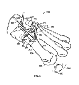
Figure 5 is a perspective view of
the foot of Figure 2, with the cutting guide of Figures 3A,
3B, 3C, and 3D properly positioned on the first cuneiform and the first
metatarsus, and attached to the
first cuneiform and the first metatarsus in preparation for resection of the
first cuneiform and the first
metatarsus, according to one embodiment.
[0031]
Figure 6A is a perspective view
of the foot of Figure 2, after resection of the first cuneiform
and the first metatarsus, removal of the cutting guide, and placement of the
first metatarsus to abut the
first cuneiform, according to one embodiment.
[0032]
Figures 6B and 6C are dorsal
views of the foot of Figure 2, before and after correction,
respectively, according to one embodiment.
[0033]
Figures 7A, 7B, 7C, and 71) are
top perspective, alternative top perspective, front elevation,
and bottom perspective views, respectively, of a patient-specific cutting
guide according to one
alternative embodiment.
[0034]
Figures 8A, 8B, and 8C are dorsal
pre-operative, dorsal post-operative, and lateral post-
operative views, respectively, of a foot treated with an Evans calcaneal
osteotomy, according to one
embodiment.
[0035]
Figures 9A and 9B are dorsal post-
operative and lateral post-operative views, respectively,
of a foot treated with a medializing calcaneal osteotomy, according to one
embodiment.
[0036]
Figure 10 is a rear, perspective
view of the foot of Figure 2, after performance of an Evans
calcaneal osteotomy and a medializing calcaneal osteotomy with patient-
specific instruments and/or
implants, according to one embodiment.
[0037]
Figure 11 is a perspective view
of the implant of Figure 10, in isolation, according to one
embodiment.
DETAILED DESCRIPTION
[0038]
Exemplary embodiments of the
disclosure will be best understood by reference to the
drawings, wherein like parts are designated by like numerals throughout. It
will be readily understood
that the components, as generally described and illustrated in the Figures
herein, could be arranged and
designed in a wide variety of different configurations. Thus, the following
more detailed description of
the embodiments of the apparatus, system, and method, as represented in
Figures 1A through 11, is not
intended to limit the scope of the disclosure but is merely representative
exemplary of exemplary
embodiments.
[0039]
The phrases 'connected to,"
"coupled to" and "in communication with" refer to any form
of interaction between two or more entities, including mechanical, electrical,
magnetic,
electromagnetic, fluid, and thermal interaction. Two components may be
functionally coupled to each
other even though they are not in direct contact with each other. The term
"abutting" refers to items
that are in direct physical contact with each other, although the items may
not necessarily be attached
together. The phrase "fluid communication" refers to two features that are
connected such that a fluid
within one feature is able to pass into the other feature.
CA 03150081 2022-3-3
WO 2021/051098
PCT/US2020/050764
[0040]
The word "exemplary" is used
herein to mean "serving as an example, instance, or
illustration." Any embodiment described herein as "exemplary" is not
necessarily to be construed as
preferred or advantageous over other embodiments. While the various aspects of
the embodiments are
presented in drawings, the drawings are not necessarily drawn to scale unless
specifically indicated.
[0041]
The present disclosure discloses
surgical systems and methods by which a bone condition,
such as a deformity, may be corrected through the use of patient-specific
instrumentation. Known
methods of correcting bone conditions are often limited to a finite range of
discretely sized instruments.
A patient with an unusual condition, or anatomy that falls between instrument
sizes, may not be readily
treated with such systems_ One example is correction of a bunion, in
particular, via adjustment of the
angulation between a cuneiform and a metatarsus.
[0042]
Figure 1A is a flowchart diagram
depicting a method 100 for correcting a bone condition,
according to one embodiment. The method 100 may be used for any of a wide
variety of bone
conditions, including but not limited to deformities, fractures, joint
failure, and/or the like. Further, the
method 100 may provide correction with a wide variety of treatments, including
but not limited to
arthroplasty, arthrodesis, fracture repair, and/or the like.
[0043]
As shown, the method 100 may
begin with a step 102 in which a CT scan (or another three-
dimensional image) of the patient's anatomy is obtained. The step 102 may
entail capturing a scan of
only the particular bone(s) to be treated, or may entail capture of additional
anatomic information, such
as the surrounding tissues. Additionally or alternatively, the step 102 may
entail receiving a previously
captured image, for example, at a design and/or fabrication facility.
Performance of the step 102 may
result in possession of a three-dimensional model of the patient's anatomy, or
three-dimensional surface
points that can be used to construct such a three-dimensional model.
[0044]
After the step 102 has been
carried out, the method 100 may proceed to a step 104 in which
a CAD model of the patient's anatomy is generated. The CAD model may be of any
known format,
including but not limited to SolidWorks, Catia, AutoCAD, or DXF. In some
embodiments, customized
software may be used to generate the CAD model from the CT scan. The CAD model
may only include
the bone(s) to he treated or may include surrounding tissues. In alternative
embodiments, the step 104
may be omitted, as the CT scan may capture data that can directly be used in
future steps without the
need for conversion.
[0045]
In a step 106, the CAD model
and/or CT scan data may be used to model patient-specific
instrumentation that can be used to correct the condition, as it exists in the
patient's anatomy. In some
embodiments, any known CAD program may be used to view and/or manipulate the
CAD model and/or
CT scan, and generate one or more instruments that are matched specifically to
the size and/or shape of
the patient's bone(s). In some embodiments, such instrumentation may include a
cutting guide that is
attachable to one or more bones, with one or more guide features that
facilitate resection of the one or
more bones pursuant to a procedure such as arthroplasty or arthrodesis. In
some embodiments,
performance of the step 106 may include modelling an instrument with a bone
apposition surface that
6
CA 03150081 2022-3-3
WO 2021/051098
PCT/US2020/050764
is shaped to match the contour of a surface of the bone, such that the bone
apposition surface can lie
directly on the corresponding contour.
[0046]
In a step 108, the model(s) may
be used to manufacture patient-specific instrumentation
and/or implants. This may be done via any known manufacturing method,
including casting, forging,
milling, additive manufacturing, and/or the like. Additive manufacturing may
provide unique benefits,
as the model may be directly used to manufacture the necessary instrumentation
and/or implants
(without the need to generate molds, tool paths, and/or the like beforehand).
Such instrumentation may
optionally include a cutting guide with the bone apposition surface and one or
more guide features as
described above.
[0047]
In addition to or in the
alternative to the step 108, the model(s) may be used to select from
available sizes of implants and/or instruments and advise the surgeon
accordingly. For example, where
a range of cutting guides ate available for a given procedure, analysis of the
CAD data may facilitate
pre-operative selection of the optimal cutting guide and/or optimal placement
of the cutting guide on
the bona Similarly, if a range of implants may be used for a given procedure,
analysis of the CAD data
may facilitate pre-operative selection of the optimal implant(s). More
particularly, properly-sized
spacers, screws, bone plates, and/or other hardware may be pre-operatively
selected.
[0048]
Thus, the result of the step 108
may be provision, to the surgeon, of one or more of the
following: (1) one or more patient-specific instruments; (2) one or more
patient-specific implants; (3)
an instrument, selected from one or more available instrument sizes and/or
configurations; (4) an
implant, selected from one or more available implant sizes and/or
configurations; (5) instructions for
which instrument(s) to select from available instrument sizes and/or
configurations; (6) instructions for
which implant(s) to select from available implant sizes and/or configurations;
(7) instructions for proper
positioning or anchorage of one or more instruments to be used in the
procedure; and (8) instructions
for proper positioning or anchorage of one or more implants to be used in the
procedure. These items
may be provided to the surgeon directly, or to a medical device company or
representative, for
subsequent delivery to the surgeon.
[0049]
In a step 110, the manufactured
instrumentation may be used in surgery to facilitate
treatment of the condition. hi some embodiments, this may entail placing the
modelled bone apposition
surface against the corresponding contour of the bone used to obtain its
shape, and then using the guide
feature(s) to guide resertion of one or more bones. Then the bone(s) may be
further treated, for example,
by attaching one or more joint replacement implants (in the case of joint
arthroplasty), or by attaching
bone segments together (in the case of arthrodesis or fracture repair). Prior
to completion of the step
110, the instrumentation may be removed from the patient, and the surgical
wound may be closed.
[0050]
As mentioned previously, the
method 100 may be used to correct a wide variety of bone
conditions. One particular example of the method 100 will be shown and
described in connection with
Figure 111, for correction of a bunion deformity of the foot.
7
CA 03150081 2022-3-3
WO 2021/051098
PCT/US2020/050764
[0051]
Figure 1B is a flowchart diagram
depicting a method 120 for correcting bunion deformity
of the human foot, according to one embodiment. The method 120 may be used to
carry out an
arthrodesis procedure by which the first metatarsocuneiform joint is removed
and the first cuneiform
and first metatarsus are secured together in a manner that properly aligns the
first metatarsus, providing
correction of the deformity.
[0052]
As shown, the method 120 may
begin with a step 122 in which a CT scan (or another three-
dimensional image) of the patient's foot is obtained. The step 122 may entail
capturing a scan of only
the first cuneiform and first metatarsus, or may entail capture of additional
anatomic information, such
as the entire foot Additionally or alternatively, the step 122 may entail
receipt of previously captured
image data. Capture of the entire foot in the step 122 may facilitate proper
alignment of the first
metatarsus with the rest of the foot (for example, with the second
metatarsus). Performance of the step
122 may result in generation of a three-dimensional model of the patient's
foot, or three-dimensional
surface points that can be used to construct such a three-dimensional model.
[0053]
After the step 122 has been
carried out, the method 120 may proceed to a step 124 in which
a CAD model of the relevant portion of the patient's anatomy is generated. The
CAD model may
optionally include the bones of the entire foot, like the CT scan obtained in
the step 122. In alternative
embodiments, the step 124 may be omitted in favor of direct utilization of the
CT scan data, as described
in connection with the step 104.
[0054]
In a step 126, the CAD model
and/or CT scan data may be used to model patient-specific
instrumentation that can be used to correct the bunion deformity. Such
instrumentation may include a
cutting guide that is attachable to the first cuneiform and the first
metatarsus, with two guide features
that facilitate resection of the cuneiform and the metatarsus in preparation
for arthrodesis. In some
embodiments, performance of the step 126 may include modelling the cutting
guide with a bone
apposition surface that is shaped to match contours of the surfaces of the
cuneiform and the metatarsus,
such that the bone apposition surface can lie directly on the corresponding
contours of the first
cuneiform and the first metatarsus.
[0055]
In a step 128, the model(s) may
be used to manufacture patient-specific instrumentation
and/or instruments. This may include manufacturing the cutting guide with the
bone apposition surface
and the guide features as described above. As in the step 108, the step 128
may additionally or
alternatively involve provision of one or more instruments and/or implants
from among a plurality of
predetermined configurations or sizes. Further, the step 128 may additionally
or alternatively involve
provision of instructions for placement and/or anchorage of one or more
instruments and/or instruments
to carry out the procedure.
[0056]
In a step 130, the manufactured
cutting guide may be used in surgery to facilitate treatment
of the condition. Specifically, the bone apposition surface of the cutting
guide may be placed against
the corresponding contours of the first cuneiform and the first metatarsus.
The guide features (for
example, slots) may then be positioned on either side of the joint between the
first cuneiform and the
8
CA 03150081 2022-3-3
WO 2021/051098
PCT/US2020/050764
first metatarsus to guide resection of the first metatarsus and the first
cuneiform to remove the
intervening joint. The cutting guide may then be removed, and the remaining
portions of the first
cuneiform and the first metatarsus may be placed to abut each other. The
cutting guide may have been
shaped such that the cuts made to the first cuneiform and the first metatarsus
are properly oriented to
bring the first metatarsus back into its proper orientation relative to the
rest of the foot. The first
cuneiform and the first metatarsus may be secured together through the use of
a bone plate or the like.
The surgical wound may be closed to allow the foot to heal, and to allow the
first cuneiform and the
first metatarsus to fuse together.
[0057]
The method 100 and the method 120
are merely exemplary. Those of skill in the art will
recognize that various steps of the method 100 and the method 120 may be
reordered, omitted, and/or
supplemented with additional steps not specifically shown or described herein.
[0058]
As mentioned previously, the
method 120 is only one species of the method 100; the present
disclosure encompasses many different procedures, performed with respect to
many different bones
and/or joints of the body. Exemplary steps and instrumentation for the method
120 will further be
shown and described in connection with Figures 2 through 711 Those of skill in
the art will recognize
that the method 120 may be used in connection with different instruments;
likewise, the instruments of
Figures 2 through 7D may be used in connection with methods different from the
method 100 and the
method 120.
[0059]
Figure 2 is a perspective view of
a portion of a foot 200 with a bunion deformity to be
treated through use of the method 100 (and more specifically, the method 120)
described above. The
foot 200 may have a first cuneiform 210, a second cuneiform 220, a first
metatarsus 230, and a second
metatarsus 240. The first cuneiform 210 and the second cuneiform 220 may be
joined together at a first
metatarsocuneiforrn joint, and the first metatarsus 230 and the second
metatarsus 240 may be joined
together at a second metatarsocurteiform joint.
[0060]
The first metatarsus 230 may be
excessively angled in a medial direction 270 (i.e., toward
the lower left-hand corner of the page), causing a painful protrusion at a
distal end 250 of the first
metatarsus 230, and further causing the phalanges (not shown) attached to the
distal end 250 to be
angled excessively in a lateral direction 260 (i.e., pointing toward the other
phalanges of the foot, rather
than pointing directly forward). The excessive medial angulaiion of the first
metatarsus 230 may also
result in an excessive gap between the first metatarsus 230 and the second
metatarsus 240_
[0061]
The first metatarsus 230 may
further be offset in a plantar direction 280 or in a dorsal
direction 290, relative to the remainder of the foot 200. Accordingly, the
orientation of the first
metatarsus 230 may need to be adjusted to move the distal end 250 in the
lateral direction 260 and in
the plantar direction 280 and/or in the dorsal direction 290.
[0062]
Every deformity is different;
accordingly, the degree of angular adjustment needed in each
direction may be different for every patient Use of a patient-specific cutting
guide may help the
surgeon obtain the optimal realignment in the lateral direction 260 and in the
plantar direction 280 or
9
CA 03150081 2022-3-3
WO 2021/051098
PCT/US2020/050764
the dorsal direction 290. Conversely, use of one of a number of differently-
sized cutting guides may
provide only approximate correction, as the surgeon may not have a guide that
precisely matches the
correction needed for the foot 200, and must thus choose the cutting guide
that most closely provides
the desired correction. Such differently sized cutting guides would not be
contoured to fit the first
cuneiform 210 or the fast metatarsus 230, thus introducing additional
potential for error as the surgeon
must properly align the selected cutting guide.
[0063]
Thus, providing a patient-
specific cutting guide may provide unique benefits. Specifically,
the patient-specific cutting guide may provide precise correction of the
deformity present in the foot
200 and may also reduce the likelihood of improper correction due to
misalignment of the cutting guide
on the foot 200. The optimal cut provided by such a cutting guide may further
reduce the likelihood
that additional procedures, such as attachment of the first metatarsus 230 to
the second metatarsus 240
to each other with screws or the like, will be needed to provide the desired
correction. Any such
additional procedure carries its own added surgical burden and risk of
failure. Thus, the use of patient-
specific instrumentation may shorten surgery, accelerate recovery, and reduce
the risk of complications.
[0064]
Figures 3A, 3B, 3C, and 313 are
top perspective, alternative top perspective, front elevation,
and bottom perspective views, respectively, of a patient-specific cutting
guide, or cutting guide 300,
according to one embodiment. The cutting guide 300 may be designed to
facilitate resection of the first
cuneiform 210 and the first metatarsus 230 with planar cuts at the proper
angles to provide dual-plane
correction of the orientation of the first metatarsus 230, thereby providing
correction in the lateral
direction 260 and in the plantar direction 280 or the dorsal direction 290.
[0065]
As shown, the cutting guide 300
may have a body 310 with a monolithic construction and
the general shape of a rectangular prism. The cutting guide 300 may further
have a joint alignment
feature that helps align the body 310 with the metatarsocuneiforrn joint
between the first cuneiform 210
and the first metatarsus 230. The joint alignment feature may consist of a
joint probe 320 that extends
from the body 310 and has a blade-like shape. The body 310 may reside on the
dorsal surfaces of the
first cuneiform 210 and the first metatarsus 230, while the joint probe 320
may protrude into the
metatarsocimeiform joint between the first cuneiform 210 and the first
metatarsus 230 to provide proper
alignment of the body 310 with the metatarsocuneiform joint.
[0066]
The body 310 may have a bone
apposition side 330 that, upon attachment of the body 310
to the first cuneiform 210 and the first metatarsus 230, is to face toward the
first cuneiform 210 and the
first metatarsus 230. The body 310 may also have an outward-facing side 332
that, upon attachment of
the body 310 to the first cuneiform 210 and the first metatarsus 230, faces
outward, away from the first
cuneiform 210 and the first metatarsus 230. Further, the body 310 may have one
or more bone
attachment features that facilitate attachment of the body 310 to the first
cuneiform 210 and/or the first
metatarsus 230. Such bone attachment features may comprise any of a wide
variety of holes, spikes,
fastening devices, ancUor the like. As embodied in Figures 3A through 3D, the
bone attachment features
may take the form of holes 340 that extend from the bone apposition side 330
to the outward-facing
CA 03150081 2022-3-3
WO 2021/051098
PCT/US2020/050764
side 332. The holes 340 may be shaped to accommodate pins, K-wires, and/or
other elongated bone
fixation elements that can be anchored in the first cuneiform 210 and/or the
first metatarsus 230 to keep
the cutting guide 300 in place.
[0067]
The bone apposition side 330 may
be custom contoured to match the shapes of the first
cuneiform 210 and/or the first metatarsus 230. As embodied in Figures 3A
through 3D, the bone
apposition side 330 may have a cuneiform apposition portion 342 shaped to lie
against the dorsal surface
of the first cuneiform 210, and a metatarsus apposition portion 344 shaped to
lie against the dorsal
surface of the first metatarsus 230. As shown, the cuneiform apposition
portion 342 may be contoured
to match the contour of the dorsal surface of the first cuneiform 210 on which
it is to rest, and the
metatarsus apposition portion 344 may similarly be contoured to match the
contour of the dorsal surface
of the first metatarsus 230 on which it is to rest. Thus, the body 310 may
have only one stable position
and orientation relative to the first cuneiform 210 and the first metatarsus
230.
[0068]
Generation of the contours of the
cuneiform apposition portion 342 and the metatarsus
apposition portion 344 may be performed relative easily in various CAD
programs_ In some
embodiments, the shapes of the corresponding dorsal surfaces of the first
cuneiform 210 and the first
metatarsus 230 may be obtained directly from the CAD models and/or CT scan
data, and simply copied
onto the model for the body 310 of the cutting guide 300. Various operations
may be used to copy
surfaces from one object to another. Additionally or alternatively, various
Boolean operations, such as
a Boolean subtraction operation, may be used to remove material from a model
for the body 310 with
a shape that matches the dorsal surfaces of the first cuneiform 210 and the
first metatarsus 230.
[0069]
The body 310 may further have
guide features that guide a cutter to resect the first
cuneiform 210 and the first metatarsus 230 in the manner needed to make the
desired correction. For
example, the guide features may be used to guide a planar cutting blade, an
arcuate cutting blade, a drill
or mill, a burr, and/or the like.
[0070]
In the embodiment of Figures 3A
through 3D, the guide features may guide a reciprocating
planar blade, such as that of a surgical bone saw, that forms planar cuts in
the first cuneiform 210 and
the first metatarsus 230. Thus, the guide features may take the form of a
first slot 350 and a second slot
352, which may be positioned toward the center of the body 310, on opposite
sides of the joint probe
320. Thus, upon proper positioning of the cutting guide 300, the first slot
350 may be positioned over
the first cuneiform 210 to facilitate resection of the first cuneiform 210,
while the second slot 352 may
be positioned over the first metatarsus 230 to facilitate resection of the
first metatarsus 230.
[0071]
In alternative embodiments, a
guide feature may be designed to guide a different type cutter,
such as a drill, mill, or side-cutting burr. In such embodiments, the guide
feature may not be a slot, but
may instead be a translatable or rotatable cutter retainer that guides
translation and/or rotation of the
cutter relative to the bone.
[0072]
Returning to Figures 3A through
3D, the body 310 may further have features that facilitate
proper positioning of the cutting guide 300 on the first cuneiform 210 and the
first metatarsus 230.
11
CA 03150081 2022-3-3
WO 2021/051098
PCT/US2020/050764
More specifically, the body 310 may have a first bone indicator 360 with the
text "CUN," indicating
that the end of the body 310 with the first bone indicator 360 is to be
positioned over the first cuneiform
210. Similarly, the body 310 may have a second bone indicator 362 with the
text "MET," indicating
that the end of the body 310 with the second bone indicator 362 is to be
positioned over the first
metatarsus 230. In addition, the body 310 may have a side indicator 370 with
the text "LEFT,"
indicating that the cutting guide 300 is to be used in connection with the
patient's left foot. The side
indicator 370 may be particularly helpful when bunion corrections are to be
provided on both of the
patient's feet. In such a case, the surgeon may manufacture or receive two
separate cutting guides: one
for the left foot (the foot 200 of Figure 2) and another for the right foot
(not shown).
[0073]
Figure 4 is a perspective view of
the foot 200 of Figure 2, with the cutting guide 300 of
Figures 3A, 3B, 3C and 313 properly positioned on the first cuneiform 210 and
the first metatarsus 230,
but as yet not attached to the first cuneiform 210 and the first metatarsus
230. The surgeon has made
the necessary incision(s) to expose the dorsal surfaces of the first cuneiform
210 and the first metatarsus
230, and has inserted the cutting guide 300 such that the cuneiform apposition
portion 342 (identified
by the first bone indicator 360 on the outward-facing side 332 of the body
310) is resting on the
corresponding dorsal surface of the first cuneiform 210, and the metatarsus
apposition portion 344
(identified by the second bone indicator 362 on the outward-facing side 332 of
the body 310) is resting
on the corresponding dorsal surface of the first metatarsus 230. Since the
cuneiform apposition portion
342 and the metatarsus apposition portion 344 are contoured to match the bone
surfaces on which they
rest, the body 310 may readily slide into its proper position on the first
cuneiform 210 and the first
metatarsus 230.
[0074]
Notably, the joint probe 320 (not
visible) may reside between the first cuneiform 210 and
the first metatarsus 230 (i.e., distal to the first cuneiform 210 and proximal
to the first metatarsus 230).
The surgeon may need to cut the metatarsocuneiform joint between the first
cuneiform 210 and the first
metatarsus 230 to form a space between the first cuneiform 210 and the first
metatarsus 230 to receive
the joint probe 320. Positioning the joint probe 320 in this space may further
help to ensure that the
cutting guide 300 is properly aligned relative to the first cuneiform 210 and
the first metatarsus 230.
[0075]
Figure 5 is a perspective view of
the foot 200 of Figure 2, with the cutting guide 300 of
Figures 3A, 3B, 3C, and 3D properly positioned on the first cuneiform 210 and
the first metatarsus 230,
and attached to the first cuneiform 210 and the first metatarsus 230 in
preparation for resection of the
first cuneiform 210 and the first metatarsus 230. Specifically, pins 500 may
be inserted through the
holes 340 in the body 310 and anchored in the first cuneiform 210 and the
first metatarsus 230. Each
of the pins 500 may have a sharp and/or threaded distal end that can penetrate
and/or readily be retained
in the bone of the first cuneiform 210 or the first metatarsus 230.
Additionally or alternatively, a drill
or other hole-forming instrument may be used to pre-form holes in the first
cuneiform 210 and/or the
first metatarsus 230 to receive the distal ends of the pins 500.
12
CA 03150081 2022-3-3
WO 2021/051098
PCT/US2020/050764
100761
As shown, the body 310 may have
two holes 340 positioned over the first cuneiform 210,
and two holes 340 positioned over the first metatarsus 230. This is merely
exemplary; in some
embodiments, a cutting guide may he secured to only one of the first cuneiform
210 and the first
metatarsus 230, or may be secured to either of the first cuneiform 210 and the
first metatarsus 230 with
only one pin 500, or with more than two pins 500. Further, in some alternative
embodiments, different
fasteners may be used, such as screws, clamps, clips, and/or the like.
[0077]
Once the cutting guide 300 has
been secured relative to the first cuneiform 210 and the first
metatarsus 230, the first cuneiform 210 and the first metatarsus 230 may be
resected. hi some
embodiments, a reciprocating blade may be inserted into the first slot 350 and
moved medially and
laterally, between opposite ends of the first slot 350, to make a planar cut
that removes the distal end of
the first cuneiform 210. Similarly, the reciprocating blade (or a different
reciprocating blade) may be
inserted into the second slot 352 and moved medially and laterally, between
opposite ends of the second
slot 352, to make a planar cut that removes the proximal end of the first
metatarsus 230. The cuts in
the first cuneiform 210 and the first metatarsus 230 may be made in either
order. In either case, once
both cuts are made, the metatarsocuneiform joint between the first cuneiform
210 and the first
metatarsus 230 may be removed, resulting in exposure of "bleeding" bone at the
distal end of the first
cuneiform 210 and the proximal end of the first metatarsus 230. The cutting
guide 300 may be removed,
along with some or all of the pins 500. If desired, at least two of the pins
500 may remain in place and
used to attach a distractor (not shown) to the first cuneiform 210 and the
first metatarsus 230, such that
the distractor can temporarily widen the space between the first cuneiform 210
and the first metatarsus
230 to allow for fenestration and/or other preparation of the cut surfaces of
the first cuneiform 210 and
the first metatarsus 230. Once such preparation has been carried out, the
remaining pins 500 may also
be removed.
[0078]
The resulting bleeding and/or
prepared bone may readily grow together and fuse, upon
abutment of the distal end of the first cuneiform 210 to the proximal end of
the first metatarsus 230,
particularly with application of some compression across the juncture of the
two bones. Since the
positions and orientations of the first slot 350 and the second slot 352 were
carefully selected to provide
the proper correction, the first metatarsus 230 may be positioned to abut the
first cuneiform 210,
resulting in reorientation of the first metatarsus 230 to a desired
orientation, relative to the lateral
direction 260 and the plantar direction 280 and/or the dorsal direction 290.
Further, the surgeon may
optionally rotate the first metatarsus 230, relative to the first cuneiform
210, about an axis perpendicular
to the cutting planes, if desired.
[0079]
Figure 6A is a perspective view
of the foot 200 of Figure 2, after resection of the first
cuneiform 210 and the first metatarsus 230, removal of the cutting guide 300,
and placement of the first
metatarsus 230 to abut the first cuneiform 210. As shown, the distal end 250
of the first metatarsus 230
may now be positioned much closer to the second metatarsus 240, in a more
natural position. Further,
Figure 6A depicts a first proximal phalanx 600, which may now be properly
oriented generally parallel
13
CA 03150081 2022-3-3
WO 2021/051098
PCT/US2020/050764
to the other phalanges (not shown), rather than pointing in the lateral
direction 260. If desired, further
steps may be performed relative to the joint between the first metatarsus 230
and the first proximal
phalanx 600 in order to keep them in the proper relative orientation. The
distal end 250 may also have
been shifted in the plantar direction 280 or in the dorsal direction 290 from
the position of Figure 2.
Thus, the desired dual-plane correction of the orientation of the first
metatarsus 230 may be complete.
[0080]
The first metatarsus 230 may be
secured to the first cuneiform 210, at least until proper
bone in-growth has occurred between the first cuneiform 210 and the first
metatarsus 230. In some
embodiments, a bone plate (not shown) or other fastener (not shown) may be
used to secure the first
cuneiform 210 and the first metatarsus 230 together. Additional hardware (not
shown) may be used to
stabilize the position and/or orientation of the first proximal phalanx 600
relative to the first metatarsus
230, if desired. The surgical wound may be closed, and the foot 200 may be
allowed to heal with the
bunion deformity corrected.
[0081]
Figures 611 and 6C are dorsal
views of the foot 200, before and after correction,
respectively. Figures 611 and 6C illustrate the correction of the angulation
of the first metatarsus 230,
by which the distal end 250 of the first metatarsus 230 is moved in the
lateral direction 260. In some
embodiments, an implant 610 may be inserted in the space between the first
metatarsus 230 and the first
cuneiform 210 in order hold the first metatarsus 230 and the first cuneiform
210 together and/or
facilitate bony fusion between the first metatarsus 230 and the first
cuneiform 210.
[0082]
In some embodiments, the implant
610 may be patient-specific. For example, the implant
610 may have a cuneiform-facing side 620 that is shaped and/or sized to be
secured to the adjoining,
resected surface of the rust cuneiform 210, and a metatarsus-facing side 630
that is shaped and/or sized
to be secured to the adjoining, resected surface of the first metatarsus 230.
As the resections made to
the first metatarsus 230 and the first cuneiform 210 may both planar, the
cuneiform-facing side 620
and/or the metatarsus-facing side 630 may also be planar. However, the
cuneiform-facing side 620
and/or the metatarsus-facing side 630 may advantageously each be shaped to
match the profile of the
resected surface of the first cuneiform 210 and the first metatarsus 230,
respectively.
[0083]
This shaping may be accomplished
by custom-designing the implant 610 for the patient,
using the same models (for example, from CT scans) of the first metatarsus 230
and the first cuneiform
210 that were used to generate the cutting guide 300. Thus, the implant 610
may have a shape that
provides centre attachment and/or fusion between the first metatarsus 230 and
the first cuneiform 210
while avoiding proud edges or other protruding features that could otherwise
interfere with surrounding
tissues.
[0084]
As indicated previously, the
cutting guide 300 is only one of many patient-specific
instruments that may be used in connection with the method 100 and/or the
method 120. An alternative
cutting guide suitable for use with the method 120 will be shown and described
in connection with
Figures 7A, 711, 7C, and 7D.
14
CA 03150081 2022-3-3
WO 2021/051098
PCT/US2020/050764
[0085]
Figures 7A, 7B, 7C, and 71) are
top perspective, alternative top perspective, front elevation,
and bottom perspective views, respectively, of a patient-specific cutting
guide, or cutting guide 700,
according to one alternative embodiment. The cutting guide 700 may be used to
correct a bunion
deformity, such as that of the foot 200 of Figure 2. Thus, the cutting guide
700 may also be designed
to facilitate resection of the first cuneiform 210 and the first metatarsus
230 with planar cuts at the
proper angles to provide dual-plane correction of the orientation of the first
metatarsus 230, thereby
providing correction in the lateral direction 260 and in the plantar direction
280 or the dorsal direction
290.
[0086]
As shown, the cutting guide 700
may have a body 710 with a monolithic construction and
the general shape of a rectangular prism. The cutting guide 700 may further
have a joint alignment
feature that helps align the body 710 with the metatarsocuneiform joint
between the first cuneiform 210
and the first metatarsus 230. The joint alignment feature may consist of a
joint probe 720 that extends
from the body 710 and has a blade-like shape. The body 710 may reside on the
dorsal surfaces of the
first cuneiform 210 and the first metatarsus 230, while the joint probe 720
may protrude into the
metatarsocuneiform joint between the first cuneiform 210 and the first
metatarsus 230 to provide proper
alignment of the body 710 with the metatarsocuneiform joint. Notably, the
joint probe 720 may have
surfaces that are not simply planar, but rather have some contouring by which
the shape of the joint
probe 720 is matched to the adjoining surfaces of the first cuneiform 210
and/or the first metatarsus
230. Such contouring of the joint probe 720 may enable more precise alignment
of the body 710 with
the metatarsocuneiform joint.
[0087]
The body 710 may have a bone
apposition side 730 that, upon attachment of the body 710
to the first cuneiform 210 and the first metatarsus 230, is to face toward the
first cuneiform 210 and the
first metatarsus 230. The body 710 may also have an outward-facing side 732
that, upon attachment of
the body 710 to the first cuneiform 210 and the first metatarsus 230, faces
outward, away from the first
cuneiform 210 and the first metatarsus 230. Further, the body 710 may have one
or more bone
attachment features that facilitate attachment of the body 710 to the first
cuneiform 210 and/or the first
metatarsus 230. Such bone attachment features may comprise any of a wide
variety of holes, spikes,
fastening devices, and/or the like. As embodied in Figures 7A through 7D, the
bone attachment features
may take the form of holes 740 that extend from the bone apposition side 330
to the outward-facing
side 332_ The holes 340 may be shaped to accommodate pins, K-wires, and/or
other elongated bone
fixation elements that can be anchored in the first cuneiform 210 and/or the
first metatarsus 230 to keep
the cutting guide 700 in place. As embodied in Figures 7A through 7D, only one
hole 340 may be
present on each side of the body 710. Thus, the body 710 may be secured to the
first cuneiform 210
with only a single pin or K-wire (not shown) and to the first metatarsus 230
with only another single
pin or K-wire (not shown).
[0088]
The bone apposition side 730 may
be custom contoured to match the shapes of the first
cuneiform 210 and/or the first metatarsus 230. As embodied in Figures 7A
through 713, the bone
CA 03150081 2022-3-3
WO 2021/051098
PCT/US2020/050764
apposition side 730 may have a cuneiform apposition portion 742 shaped to lie
against the dorsal surface
of the first cuneiform 210, and a metatarsus apposition portion 744 shaped to
lie against the dorsal
surface of the first metatarsus 230. As shown, the cuneiform apposition
portion 742 may be contoured
to match the contour of the dorsal surface of the first cuneiform 210 on which
it is to rest, and the
metatarsus apposition portion 744 may similarly be contoured to match the
contour of the dorsal surface
of the first metatarsus 230 on which it is to rest. Thus, the body 710 may
have only one stable position
and orientation relative to the first cuneiform 210 and the first metatarsus
230.
[0089]
Like the cuneiform apposition
portion 342 and the metatarsus apposition portion 344 of the
cutting guide 300, generation of the contours of the cuneiform apposition
portion 742 and the metatarsus
apposition portion 744 may be performed relative easily in various CAD
programs through surface copy
operations, Boolean operations, and/or the like.
[0090]
The body 710 may further have
guide features that guide a cutter to resect the first
cuneiform 210 and the first metatarsus 230 in the manner needed to make the
desired correction. For
example, the guide features may be used to guide a planar cutting blade, an
arcuate cutting blade, a drill
or mill, and/or the like.
[0091]
In the embodiment of Figures 7A
through 7D, the guide features may guide a reciprocating
planar blade, such as that of a surgical bone saw, that forms planar cuts in
the rust cuneiform 210 and
the rust metatarsus 230. Thus, the guide features may take the form of a first
slot 750 and a second slot
752, which may be positioned toward the center of the body 710, on opposite
sides of the joint probe
720. Thus, upon proper positioning of the cutting guide 700, the first slot
750 may be positioned over
the first cuneiform 210 to facilitate resection of the rust cuneiform 210,
while the second slot 752 may
be positioned over the first metatarsus 230 to facilitate resection of the
first metatarsus 230.
[0092]
In operation, the cutting guide
700 may be used in a manner similar to that of the cutting
guide 300. However, the cutting guide 700 may only be secured to each of the
first cuneiform 210 and
the rust metatarsus 230 with a single pin or K-wire (not shown), as mentioned
previously. Further, the
cutting guide 700 is smaller than the cutting guide 300. Thus, the cutting
guide 700 may be placed
through a smaller, less invasive incision. One advantage to patient-specific
instrumentation may be that
instruments may be made smaller, since they are not limited to certain sizes.
Many known instruments
come in discrete sizes, each of which is designed to accommodate a range of
patient anatomic
dimensions. Thus, for given patient anatomy, the instrument must be large
enough to treat the anatomy
at either end of its range. This typically requires the instrument to be
oversized for many anatomic
dimensions it is designed to treat. Notably, the cutting guide 700 is merely
one compact example; other
cutting guides may be made even smaller; in some embodiments, cutting guides
may be made that have
a smaller width between holes (e.g., holes 740 on the cutting guide 700). As
long as the holes are
sufficiently far apart to avoid interference of the pins 500 with the
operation of the cutting blade, the
cutting guide may function appropriately. Thus, Lapidus and other procedures
may be accomplished
through a very narrow incision through the use of patient-specific
instrumentation.
16
CA 03150081 2022-3-3
WO 2021/051098
PCT/US2020/050764
[0093]
Those of skill in the art will
recognize that a wide variety of differently configured cutting
guides may be used in conjunction with the method 120 set forth above.
Further, a wide variety of
patient-specific instruments may be used in connection with the method 100,
including but not limited
to cutting guides, gages, implant positioning guides, joint distractors, joint
compressors, soft tissue
retractors, and the like.
[0094]
Furthermore, patient-specific
cutting guides may be used for various other procedures on
the foot, or on other bones of the musculoskeletal system. Patient-specific
cutting guides may be used
for various procedures involving osteotomy, including but not limited to
arthroplasty, fusion, and
deformity correction procedures. According to one example, patient-specific
cutting guides similar to
the cutting guide 300 and the cutting guide 700 may be used for the
metatarsophalangeal ("MTP") joint.
A method similar to the method 100 may be employed.
[0095]
In some embodiments, one or more
articulating surfaces of a joint may be replaced and/or
resurfaced. For example, for the MTP joint, a patient-specific cutting guide
may be used to determine
the angles of cuts on the distal metatarsus or the proximal phalanx in
preparation for replacement or
resurfacing of the metatarsal head and/or the proximal phalangeal base.
Implants for either the
metatarsus or the phalanx may be customized to match the patient's original
anatomy, such as the
curvature of the MTP joint. In other embodiments, an MTP joint may be fused
through the use of
patient-specific cutting guides. Patient-specific cutting guides may be used
to treat (for example, via
fusion, resurfacing, and/or arthroplasty) any joint in the body, using methods
similar to the method 100.
[0096]
According to other examples,
patient-specific cutting guides may be used to carry out an
Evans calcaneal osteotomy and/or a medializing calcaneal osteotomy. Patient-
specific instruments will
be shown and described in connection with Figures 8A through 11, in relation
to an Evans calcaneal
osteotomy, and a medializing calcaneal osteotomy.
[0097]
Figures 8A, 8B, and 8C are dorsal
pre-operative, dorsal post-operative, and lateral post-
operative views, respectively, of a foot treated with an Evans calcaneal
osteotomy, according to one
embodiment. Outward rotation of the foot may occur in patients with flatfoot.
An Evans or lateral
column lengthening procedure is sometimes performed for these patients. An
incision is made on the
outside of the foot, and the front half of the heel bone is cut. A bone wedge
(typically either titanium or
a bone-based graft) is then placed into the cut area of the heel bone. This
wedge helps to "lengthen" the
heel bone and rotate the foot back into its correct position_ The wedge is
usually kept in place using
screws or a surgical staple.
[0098]
Figures 9A and 913 are dorsal
post-operative and lateral post-operative views, respectively,
of a foot treated with a medializing calcaneal osteotomy, according to one
embodiment. A medializing
calcaneal osteotomy (heel slide) procedure is often used when the calcaneus
(heel bone) has shifted out
from underneath the leg. An incision is made on the outside of the heel, and
the back half of the heel
bone is cut and slid back underneath the leg. The heel is then fixed in place
using metal screws or a
plate. This also helps to reposition the Achilles tendon towards the center of
the ankle/rearfoot. The
17
CA 03150081 2022-3-3
WO 2021/051098
PCT/US2020/050764
medializing calcaneal osteotomy can be used in place of, or in addition to, an
Evans calcaneal
osteotomy.
[0099]
Figure 10 is a rear, perspective
view of the foot 200 of Figure 2, after performance of an
Evans calcaneal osteotomy and a medializing calcaneal osteotomy with patient-
specific instruments
and/or implants, according to one embodiment. The foot 200 may have a
calcaneus 1000 and a talus
1010, in addition to the metacarpals 1020 and cuneiforms 1030 depicted in
Figure 2. Pursuant to the
Evans calcaneal osteotomy, an anterior portion of the calcaneus 1000 may be
cut along the medial-
lateral direction to separate a first bone segment 1040 of the calcaneus 1000
from a second bone segment
1042 of the calcaneus 1000. The second bone segment 1042 may be reoriented
medially, relative to the
first bone segment 1040, such that a heel 1050 of the calcaneus 1000 is moved
medially, simulating a
natural, healthy arch in the foot 200.
[00100]
The cut between the first bone
segment 1040 and the second bone segment 1042 may be
carried out virtually (for example, in CAD) on a model of the calcaneus 1000
obtained from a CT scan
or other imaging of the patient's foot. Thus, the optimal realignment of the
posterior end of the
calcaneus 1000 can be obtained. If desired, a patient-specific cutting guide,
or cutting guide 1043, may
be generated in order to facilitate resection of the calcaneus 1000.
[00101]
As shown, the cutting guide 1043
may have a first end 1044 and a second end 1045, each
of which has a bone attachment feature 1046. The bone attachment features 1046
may be used to secure
the first end 1044 and the second end 1045 to the first bone segment 1040 and
the second bone segment
1042, respectively. The first end 1044 may have a fast bone engagement surface
1047 that is shaped
to match a corresponding contour on the first bone segment 1040, and the
second end 1045 may have a
second bone engagement surface 1048 that is shaped to match a corresponding
contour on the second
bone segment 1042. Thus, the cutting guide 1043 may naturally lie flush with
the surface of the
calcaneus 1000, in the optimal position on the calcaneus 1000 to facilitate
resection of the calcaneus
1000 to divide the first bone segment 1040 from the second bone segment 1042.
The cutting guide
1043 may have a guide feature 1049, such as a slot, that can be used to guide
a cutter to form a single
cut between the first bone segment 1040 and the second bone segment 1042.
[00102]
After the cut has been made to
split the calcaneus 1000 into the first bone segment 1040
and the second bone segment 1042, the surgeon may angle the second bone
segment 1042 relative to
the first bone segment 1040 in the predetermined (previously modeled) relative
orientation_ This
reorientation between the first bone segment 1040 and the second bone segment
1042 may leave a
wedge-shaped gap between the first bone segment 1040 and the second bone
segment 1042. In order
to maintain the desired relative orientation, an implant 1060 with a wedge
shape may be inserted into
the gap and secured to the first bone segment 1040 and the second bone segment
1042. The implant
1060 may be fabricated specifically for the patient, since the precise
angulation and position of the
realignment may also be patient specific. As shown, the implant 1060 may have
exterior surfaces that
are contoured to match the contours of the adjoining portions of the first
bone segment 1040 and the
18
CA 03150081 2022-3-3
WO 2021/051098
PCT/US2020/050764
second bone segment 1042. Thus, the implant 1060 may provide secure fixation,
while not protrude
beyond the adjoining surfaces of the first bone segment 1040 and the second
bone segment 1042. Thus,
the implant 1060 may be devoid of proud edges or other protrusions that could
otherwise interfere with
motion between the calcaneus 1000 and the talus 1010, or with surrounding soft
tissues, thus interfering
with the patient's post-operative gait.
[00103]
The implant 1060 may be made of
any biocompatible material, including but not limited to
Titanium and alloys thereof, stainless steel, PEEK, and/or the like. The
implant 1060 may be formed
by any method known in the art, including but not limited to forging, casting,
milling, additive
manufacturing, and/or the like. In some embodiments, the implant 1060 may have
an interior void that
can be filled with bone graft or other material designed to promote boney in-
growth between the cut
surfaces of the first bone segment 1040 and the second bone segment 1042. In
alternative embodiments,
the implant 1060 may have a mesh and/or lattice structure that facilitates
such honey in-growth, which
structure may be formed via additive manufacturing.
[00104]
As mentioned previously, a
medializing calcaneal osteotomy may optionally be performed
in addition to or in place of the Evans calcaneal osteotomy. As shown, the
heel 1050 may be cut from
the remainder of the second bone segment 1042 and may be displaced medially.
This displacement
may also help to restore normal gait and tendon function in the foot 200,
particularly when coupled with
the Evans calcaneal osteotomy. The proper displacement of the heel 1050
relative to the remainder of
the second bone segment 1042 may be determined based on analysis of the CAD
models from scans of
the foot 200. If desired, the model of the calcaneus 1000 may be divided and
manipulated in CAD to
simulate the repositioning of the heel 1050 pursuant to the medializing
calcaneal osteotomy. Thus, the
alignment of the heel 1050 relative to the remainder of the foot 200 can
easily be assessed and optimized
prior to surgery.
[00105]
Such preoperative alignment and
planning may be particularly useful where multiple
procedures, such as the Evans calcaneal osteotomy and the medializing
calcaneal osteotomy, are
combined for a single patient. Without such planning, it may be difficult to
properly assess the effect
of the combined procedures on the patient's anatomy. For example, the effect
of the Evans calcaneal
osteotomy, and that of the medializing calcaneal osteotomy, is to shift the
heel 1050 medially. The
combined shift may be difficult to assess in the operating room but may be
much more easily and
accurately gauged via manipulation of the modeled anatomy_
[00106]
In some embodiments, one or more
additional procedures may be carried out, in addition
to or in the alternative to those of Figure 9. For example, in addition to or
in the alternative to the Evans
calcaneal osteotomy and the medializing calcaneal osteotomy, a cotton
osteotomy and/or a first
metatarsal midfoot osteotomy may be performed. Patient-specific cutting guides
may be designed,
fabricated, and surgically used to facilitate any of these procedures through
the presence of bone
engagement surfaces that are shaped to rest on the particular bony surfaces
adjacent to the osteotomy.
19
CA 03150081 2022-3-3
WO 2021/051098
PCT/US2020/050764
100107]
As in the case of the Evans
calcaneal osteotomy, a custom cutting guide, or cutting guide
1053, may be generated to help the surgeon obtain the correction that was
previously modeled and/or
planned using the computer models of the foot 200. The cutting guide may 1053
have a structure and
function similar to that of the cutting guide 1043 used for the Evans
calcaneal osteotomy. Such a cutting
guide may have contoured surfaces that match the contours of the adjoining
boney surfaces of the
remainder of the second bone segment 1042 and/or the heel 1050.
[00108]
More specifically, the cutting
guide 1053 may have a first end 1054and a second end 1055,
each of which has a bone attachment feature 1056. The bone attachment features
1056 may be used to
secure the first end 1054 and the second end 1055 to the second bone segment
1042 and the heel 1050,
respectively. The first end 1054 may have a first bone engagement surface 1057
that is shaped to match
a corresponding contour on the second bone segment 1042, and the second end
1055 may have a second
bone engagement surface 1058 that is shaped to match a corresponding contour
on the heel 1050. Thus,
the cutting guide 1053 may naturally lie flush with the surface of the
calcaneus 1000, in the optimal
position on the calcaneus 1000 to facilitate resection of the calcaneus 1000
to divide the second bone
segment 1042 from the heel 1050. The cutting guide 1053 may have a guide
feature 1059, such as a
slot, that can be used to guide a cutter to form a single cut between the
second bone segment 1042 and
the heel 1050.
[00109]
In order to maintain the heel
1050 in the proper position relative to the remainder of the
second bone segment 1042, a bone plate 1070 may be secured to the heel 1050
and to the remainder of
the second bone segment 1042. The bone plate 1070 may include a first end 1080
secured to the
remainder of the second bone segment 1042, a second end 1082 secured to the
heel 1050, and an
intermediate portion 1084 that extends from the first end 1080 to the second
end 1082, and provides the
desired medial shift between the first end 1080 and the second end 1082. The
first end 1080 and the
second end 1082 may be secured to the remainder of the second bone segment
1042 and to the heel
1050, respectively, through the use of screws 1090.
[00110]
Like the implant 1060, the bone
plate 1070 may be made of any known biocompatible
material, through the use of any manufacturing process known in the art. In
some embodiments, the
bone plate 1070 may also be fabricated specifically for the foot 200, enabling
the bone plate 1070 to
maintain precisely the desired level of correction. When made specifically for
the foot 200 in
combination with each other, the implant 1060 and the bone plate 1070 may
provide a highly
predictable, precise, and customizable level of correction of the flat foot
deformity.
[00111]
Figure 11 is a perspective view
of the implant 1060, in isolation. As shown, the implant
1060 may have a first bone-facing surface 1100 that is generally flat and
shaped to match the cut surface
of the first bone segment 1040. The first bone-facing surface 1100 is shown in
Figure 11 with a smooth
shape; however, in alternative embodiments, the first bone-facing surface 1100
may be roughened
and/or may have teeth, spikes, ridges, and/or other features intended to
penetrate the first bone segment
1040 in order to provide for more secure engagement of the implant 1060 with
the first bone segment
CA 03150081 2022-3-3
WO 2021/051098
PCT/US2020/050764
1040. Similarly, the implant 1060 may have a second bone-facing surface 1110
(not visible) that is also
generally flat and shaped to match the cut surface of the second bone segment
1042. Like the first bone-
facing surface 1100, the second bone-facing surface 1110 may be roughened or
have protruding features
that strengthen engagement of the implant 1060 with the second bone segment
1042. If desired, the
implant 1060 may be further held in place through the use of bone screws,
cement, one or more bone
plates, and/or other features known in the art to secure an implant to bone.
[00112]
The edges of the first bone-
facing surface 1100 and the second bone-facing surface 1110
may be shaped to line up with the edges of the cut surfaces of the first bone
segment 1040 and the
second bone segment 1042, respectively. The implant 1060 may also have a
contoured surface 1120
that extends between the edges of the first bone-facing surface 1100 and the
second bone-facing surface
1110. The contoured surface 1120 may also be contoured to match the contours
of the adjoining
portions of the first bone segment 1040 and the second bone segment 1042.
Thus, the contoured surface
1120 may provide a continuous surface, devoid of protrusions, that extends
between the adjoining
surfaces of the first bone segment 1040 and the second bone segment 1042.
[00113]
A threaded hole 1130 may
optionally be provided in the contoured surface 1120. The
threaded hole 1130 may be used to secure the implant 1060 to an insertion
instrument, a positioning
instrument, and/or a removal instrument. The threaded hole 1130 may be formed
in a recess 1140 in
the contoured surface 1120 so that the threaded hole 1130 can have the desired
orientation, without
affecting the shape of the contoured surface 1120 more than necessary. Of
course, many other features
may be used to secure an instrument to the implant 1060, including various
clips, clamps, fasteners, and
interfacing features, as known in the art.
[00114]
The present disclosure is not
limited to cutting guides or extremity procedures. In some
embodiments, patient-specific instrumentation may be used to correct a wide
variety of bone conditions.
Such conditions include, but are not limited to, any angular deformities from
within one bone segment
in either the lower or upper extremities (for example, tibial deformities,
calcaneal deformities, femoral
deformities, and radial deformities). The present disclosure may also be used
to treat an interface
between two bone segments (for example, the ankle joint, metatarsal cuneiform
joint, lisfranc's joint,
complex ch.arcot deformity, wrist joint, knee joint, etc.). As one example, an
angular deformity or
segmental malalignrnent in the forefoot may be treated, such as is found at
the metatarsal cuneiform
level, the midfoot level such as the navicular cuneiform junction, hindfoot at
the calcaneal cubiod or
subtalar joint or at the ankle between the tibia and talar junction.
Additionally, patient-specific
instruments could be used in the proximal leg between two bone segments or in
the upper extremity
such as found at the wrist or metacarpal levels.
[00115]
Any methods disclosed herein
comprise one or more steps or actions for performing the
described method. The method steps and/or actions may be interchanged with one
another. In other
words, unless a specific order of steps or actions is required for proper
operation of the embodiment,
the order and/or use of specific steps and/or actions may be modified.
21
CA 03150081 2022-3-3
WO 2021/051098
PCT/US2020/050764
[00116]
Reference throughout this
specification to "an embodiment" or "the embodiment" means
that a particular feature, structure or characteristic described in connection
with that embodiment is
included in at least one embodiment. Thus, the quoted phrases, or variations
thereof, as recited
throughout this specification are not necessarily all referring to the same
embodiment.
[00117]
Similarly, it should be
appreciated that in the above description of embodiments, various
features are sometimes grouped together in a single embodiment, Figure, or
description thereof for the
purpose of streamlining the disclosure. This method of disclosure, however, is
not to be interpreted as
reflecting an intention that any claim require more features than those
expressly recited in that claim_
Rather, as the following claims reflect, inventive aspects lie in a
combination of fewer than all features
of any single foregoing disclosed embodiment. Thus, the claims following this
Detailed Description
are hereby expressly incorporated into this Detailed Description, with each
claim standing on its own
as a separate embodiment. This disclosure includes all permutations of the
independent claims with
their dependent claims.
[00118]
Recitation in the claims of the
term "first" with respect to a feature or element does not
necessarily imply the existence of a second or additional such feature or
element. Elements recited in
means-plus-function format are intended to be construed in accordance with 35
U.S.C. 112 Para. 6.
It will be apparent to those having skill in the art that changes may be made
to the details of the above-
described embodiments without departing from the underlying principles set
forth herein.
[00119]
While specific embodiments and
applications of the present disclosure have been illustrated
and described, it is to be understood that the scope of this disclosure is not
limited to the precise
configuration and components disclosed herein. Various modifications, changes,
and variations which
will be apparent to those skilled in the art may be made in the arrangement,
operation, and details of the
methods and systems of the present disclosure set forth herein without
departing from it spirit and scope.
22
CA 03150081 2022-3-3