Note: Descriptions are shown in the official language in which they were submitted.
WO 2021/050767
PCT/US2020/050249
SYSTEMS AND METHODS FOR TISSUE MODULATION
RELATED APPLICATIONS
[0001] This application claims priority to US
Provisional Application No.
62/899,622 filed September 12, 2019, the entire content of which is hereby
incorporated by
reference herein.
FIELD
(0002) Described herein are various
implementations of systems and methods for
modulating tissue (for example, systems and methods for ablating nerves or
other tissue
within or surrounding a vertebral body to treat chronic lower back pain).
Several
embodiments comprise the use of biomarkers to confirm or otherwise assess
ablation, pain
relief, efficacy of treatment, etc. Some embodiments include robotic elements
for, as an
example, facilitating roboticallv controlled access, navigation, imaging,
and/or treatment.
Assessment of vertebral endplate degeneration or defects (e.g., pre-IS/Iodic
changes) to
facilitate identification of treatment sites and protocols are also provided
in several
embodiments. Systems or kits of access tools for accessing target treatment
locations within
vertebral bodies are also provided.
BACKGROUND
[0903] Back pain is a very common health
problem worldwide and is a major
cause for work-related disability benefits and compensation. At any given
time, low back
pain impacts nearly 30% of the US population, leading to 62 million annual
visits to
hospitals, emergency departments, outpatient clinics, and physician offices.
Back pain may
arise from strained muscles, ligaments, or tendons in the back and/or
structural problems
with bones or spinal discs. The back pain may be acute or chronic. Existing
treatments for
chronic back pain vary widely and include physical therapy and exercise,
chiropractic
treatments, injections, rest, pharmacological therapy such as opioids, pain
relievers or anti-
inflammatory medications, and surgical intervention such as vertebral fusion,
discectomy
(e.g., total disc replacement), or disc repair. Existing treatments can be
costly, addictive,
temporary, ineffective, and/or can increase the pain or require long recovery
times. In
-1-
CA 03150339 2022-3-7
WO 2021/050767
PCT/US2020/050249
addition, existing treatments do not provide adequate relief for the majority
of patients and
only a small percentage are surgically eligible.
SUMMARY
100041 Applicant's existing technology (the
Intracept* procedure by Relievante)
offers a safe and effective minimally invasive procedure that targets the
basivertebrai nerve
for the relief of chronic vertebrogenic low back pain_ As disclosed herein,
several
embodiments provide bone access tools, additional modalities of relief for
patients and/or
adjunct technologies.
100051 In accordance with several
embodiments, quantitative efficacy of
treatment or efficacy of nerve ablation may be performed by assessing levels
of one or more
biomarkers (e.g., biomarkers associated with pain, inflammation, or
neurotransmission).
Such assessment may be particular useful to assess pain, for example. Pain can
be very
subjective based on individual patient pain tolerance and perception.
Accordingly, it can be
difficult to assess or quantify efficacy of pain treatment based on patient
feedback. It has
also been difficult historically to assess efficacy of nerve ablation in real
time. For example,
patients may be under anesthetic and unable to provide feedback. In other
cases, patients
may be awake but unable to accurately assess pain. The use of biomarkers, in
some
embodiments can facilitate pain assessment or confirmation of efficacy of
nerve ablation.
[00061 For example, a level or activity of
one or more biomarkers may be
measured or otherwise obtained prior to performing a procedure and after
performing a
procedure. The pre-procedure and post-procedure levels may be compared in
order to
quantitatively (non-subjectively) assess efficacy. The biomarkers may be
associated with
pain levels or associated with lesion formation (e.g., efficacy of
neurotransmission or neural
communication). The assessment of the level of the one or more biomarkers may
advantageously be performed in a non-invasive or minimally-invasive (e.g., non-
surgical)
manner in accordance with several embodiments_ Biomarkers may also be used to
assess
whether a particular subject is likely to be a candidate for nerve ablation
treatment for
treatment of back pain. For example, the biomarkers may be indicative of pre-
Modic
changes or symptoms likely to result in Modic changes or endplate damage
(e.g.,
inflammation, edema, bone marrow lesions or fibrosis). The assessment of
biornarker levels
may indicate which vertebral bodies of a particular subject are candidates for
treatment to
-2-
CA 03150339 2022-3-7
WO 2021/050767
PCT/US2020/050249
prevent (or reduce the likelihood of) back pain from developing or worsening
or to treat
existing back pain. The pre-procedure biomarker assessment may also be
combined with
pre-procedure imaging_ Mechanisms other than using biomarkers may also be used
(in
addition or in the alternative) to assess lesion formation (e.g., infrared
sensing, heat markers,
neurotransmission assessments via stimulation, and/or ultrasound imaging).
100071 In some embodiments, automated systems
for accessing and/or treating
tissue (such as nerves) are provided. In accordance with several embodiments,
robotically-
ena bled or robotically-controlled surgical, access, and/or treatment tools
may provide a high
level of control and precision of movement and increased dexterity and range
of motion,
thereby providing increased assurance that injury will not occur to tissue not
desired to be
impacted. Robotically-controlled tools and techniques (e.g., computer-aided
tools and
techniques that may incorporate artificial intelligence learning and feedback)
may also be
used to facilitate navigation to, and surgical operation at, desired target
treatment regions that
may be difficult to access manually, thereby providing enhanced flexibility
and possibilities
thought not to be possible via manual human surgery. Robotically-controlled
tools and
techniques (e.g., computer-aided tools and techniques that may incorporate
artificial
intelligence learning and feedback) may further be used to facilitate capture
of images pre-
operatively or intra-operatively without exposing the target treatment regions
to radiation or
without requiring large incisions to be made. Nerve detection devices (e.g.,
nerve monitoring
devices or nerve finders) may also be used to detect nerves along access
routes that are
desired to be avoided during access. Robotic or automated tools and techniques
may reduce
numbers of and sizes of incisions (and therefore scars), may reduce blood
loss, may reduce
pain, and may decrease recovery time.
[0008] Because the target treatment regions
within vertebral bodies may be fairly
small in size, it may be desirable to control or adjust lesion formation so as
to exhibit specific
lesion shapes (e.g., football-shape, oval, elliptical, disc-shaped, cigar-
shaped, dumbbell-
shaped, UFO-shaped, rounded, rectangular, amorphous, etc.). Creating specific
lesion shapes
may allow clinicians to efficiently ablate a basivertebral nerve trunk within
specific vertebral
bodies (e.g., cervical, thoracic, lumbar, sacral vertebrae). The specific
lesion shapes may
provide increased confidence in the efficacy of ablation while limiting the
extent of thermal
injury within the vertebral body. The lesion shapes may be controlled by
applying voltage
-3-
CA 03150339 2022-3-7
WO 2021/050767
PCT/US2020/050249
differentials between different pairs of electrodes on the same energy
delivery probe or on
different energy delivery probes for different durations. The lesion formation
may be
monitored and controlled in real time (e.g., using feedback based on imaging,
thermal
sensing, andlor artificial intelligence) to further increase confidence and
efficiency. Use of
two probes and delivering energy between the two probes may result in
synergistic lesion
formation (e.g., larger lesions than could be formed by individual probes
alone).
100091 Treatment procedures may include
modulation of nerves within or
surrounding bones. The terms "modulation" or "neuromodulation", as used
herein, shall be
given their ordinary meaning and shall also include ablation, permanent
denervation,
temporary denervation, disruption, blocking, inhibition, electroporation,
therapeutic
stimulation, diagnostic stimulation, inhibition, necrosis, desensitization, or
other effect on
tissue. Neuromodulation shall refer to modulation of a nerve (structurally
and/or
functionally) and/or neurotransmission. Modulation is not necessarily limited
to nerves and
may include effects on other tissue, such as tumors or other soft tissue
[0010] In accordance with several
embodiments, a method of ablating a
basivertebral nerve within a vertebral body of a subject and confirming
efficacy of ablation
of the basivertebral nerve includes obtaining a first reading (e.g., baseline
reading) of a level
of a biomarker from the subject. The method further includes performing a
denervation
procedure on the subject. As one example, the denervation procedure includes
denervating
the basivertebral nerve within the vertebral body. The method also includes
obtaining a
second reading (e.g., post-procedure reading) of the level of the biomarker
from the subject
and determining an effect of the denervation procedure by comparing the second
reading to
the first reading to assess efficacy of the denervation procedure.
100111 The biomarkers may include one or more
of: an inflammatory cytokine
(e.g., interleukins, interferons, tumor necrosis factors, prostaglandins, and
chemokines), pain
indicators (e.g., substance P. calcitonin gene-related peptides (CGRPs)), an
edema factor,
and/or other inflammatory factor. The first reading (e.g., baseline reading)
and the second
reading (e.g., post-procedure reading) may be obtained from cerebrospinal
fluid adjacent the
vertebral body of the subject, from a blood draw (e.g., at a location within
or adjacent the
vertebral body of the subject or at a remote location systemically), from a
urine sample, or
other source. The biomarkers may be circulating inflammatory cells (e.g.,
cytokines). The
-4-
CA 03150339 2022-3-7
WO 2021/050767
PCT/US2020/050249
biomarkers may be obtained via one or more immunoassay techniques (e.g..
ELISAs,
cytokine bead arrays, cytokine microarrays, flow cytometry,
immunohistochemical assays,
and/or the like).
(0012) The step of denervating the
basivertebral nerve within the vertebral body
may include applying energy (e.g., radiofrequency energy, ultrasound energy,
microwave
energy) to a target treatment region within the vertebral body sufficient to
denervate (e.g.,
ablate, electroporate, molecularly dissociate, necrose) the basivertebral
nerve using a
radiefrequency energy delivery device. The step of denervating may
alternatively or
additionally include applying an ablative fluid (e.g., steam, chemical,
crvoablative fluid) to a
target treatment region within the vertebral body. In some implementations,
the step of
denervating may include delivering a water jet at a pressure sufficient to
denervate the nerve
(e.g., between 5 and 10 MPa, between 10 and 15 MPa, between 15 and 30 MPa,
between 30
and 50 MPa, overlapping ranges thereof, pressure greater than 50 MPa, or any
value within
the recited ranges).
[0013] In accordance with several
embodiments, a method of detecting and
treating back pain of a subject includes obtaining images of a vertebral body
of the subject,
analyzing the images to determine whether the vertebral body exhibits one or
more
symptoms associated with a pre-Modic change, and ablating a basivertebral
nerve within the
vertebral body if it is determined that the vertebral body exhibits one or
more symptoms
associated with a pre-Modic change. The one Of more symptoms associated with a
pre-
Medic change may include edema, inflammation, and/or tissue changes (e.g.,
tissue lesions,
fibrosis, or other changes in tissue type or characteristics) of bone, bone
marrow, and'or
endplate(s).
[0014] In accordance with several
embodiments, a method of treating a vertebral
body includes inserting a first access assembly into a first target location
of the vertebral
body. The first access assembly includes a first cannula and a first style
configured to be
inserted within the first cannula until a distal tip of the first st3;10 is
advanced to or beyond an
open distal tip of the first cannula. The method further includes removing the
first stylet
from the first cannula. The method also includes inserting a second access
assembly into a
second target location of the vertebral body. The second access assembly
including a second
cannula and a second style configured to be inserted within the second cannula
until a distal
_s_
CA 03150339 2022-3-7
WO 2021/050767
PCT/US2020/050249
tip of the second stylet is advanced to or beyond an open distal tip of the
second cannula.
The method further includes removing the second stylet. The method also
includes inserting
a first radiofrequency energy delivery device through the first cannula and
inserting a second
radiofrequency energy delivery device through the second cannula. The first
radiofrequency
energy delivery device and the second radiofrequency energy delivery device
earth include at
least two electrodes (e.g., an active electrode and a return electrode
configured to act as a
bipolar electrode pair). The method further includes positioning the at least
two electrodes of
the first radiofrequency energy delivery device within the vertebral body and
positioning the
at least two electrodes of the second radiofrequency energy delivery device
within the
vertebral body.
[0015]
The method also includes
applying power to the first and second
radiofrequency energy delivery devices sufficient to create a desired lesion
shape within the
vertebral body sufficient to ablate a basivertebral nerve within the vertebral
body (e.g.,
football-shaped lesion, an elliptical-shaped lesion having a length-to-width
ration of at least
a cross-shaped lesion, an X-shaped lesion, a cigar-shaped lesion). The lesion
may have a
maximum width of 20 mm and a maximum length of 30 mm. The lesion may have a
maximum width of 70-80% of the anteroposterior depth of the vertebral body and
a
maximum length of 70-85% of the transverse width of the vertebral body. In
some
implementations, the step of applying power to the first and second
radiofrequency energy
delivery devices includes independently applying power to the first and second
radiofrequency energy delivery devices for a first duration of time (e.g., 1
minute ¨ 2
minutes, 30 seconds ¨ 90 seconds, 2 ¨ 5 minutes, 5 ¨ 10 minutes, 10 ¨ 15
minutes,
overlapping ranges thereof, or any value within the recited ranges).
In some
implementations, the step of applying power to the first and second
radiofrequency energy
delivery devices further includes applying a voltage differential between at
least one of the at
least two electrodes of the first radiofrequency energy delivery device and at
least one of the
at least two electrodes of the second radiofrequency energy delivery device
for a second
duration of time (e.g., 1 minute ¨ 2 minutes, 30 seconds ¨ 90 seconds, 2 ¨ 5
minutes, 5 ¨ 10
minutes, 10 ¨ 15 minutes, overlapping ranges thereof, or any value within the
recited ranges).
The first duration of time and the second duration of time may be the same or
different.
-6-
CA 03150339 2022-3-7
WO 2021/050767
PCT/US2020/050249
100161 In accordance with several
embodiments, a method of ablating a
basivertebral nerve within a vertebral body includes inserting an access
assembly within a
vertebral body using a robotically-contolled system_ The access assembly
includes at least
one cannula. The method further includes inserting a radiofrequency energy
delivery device
through the cannula to a target treatment site within the vertebral body using
the robotically-
controlled system, and applying power to the target treatment site using the
rachofrequency
energy delivery device sufficient to ablate the basivertebral nerve.
100171 In some implementations, the
robotically-controlled system includes one
or more robotic arms and an operator control console including at least one
processor. The
system may include one or more imaging devices configured to provide feedback
(e.g., based
on artificial intelligence processing algorithms) to the robotically-
controlled system to
control insertion of the access assembly and/or the radiofrequeney energy
delivery device.
100181 In accordance with several
embodiments, a radiofrequericy ("RF")
generator for facilitating nerve ablation includes a display screen (e.g.,
color active matrix
display) and an instrument connection port configured to receive a
corresponding connector
of a radiofi-equency probe. The generator further includes a first indicator
light ring (e.g.,
circular LED indicator light ring) surrounding the instrument connection port
that is
configured to illuminate when a treatment device is connected to the
instrument connection
port. The first indicator light ring is configured to continuously illuminate
in a solid color
(e.g., white, green, blue) when the treatment device is connected to the
instrument connection
port, to flash at a first pulsing rate (e.g., I. Hz) to prompt a clinician to
connect the treatment
device to the instrument connection port, and to flash at a second pulsing
rate different than
(e.g., greater than IHz, such as 2 Hz, 3 Hz or 4 Hz) the first pulsing rate to
indicate an error
condition. The generator may optionally be configured to output an audible
alert or alarm to
indicate the error condition. The generator also includes an energy delivery
actuation button
configured to be pressed by an operator to start and stop delivery of
radiofrequency energy
and a second indicator light ring (e.g., circular LED light ring) surrounding
the actuation
button_ The second indicator light ring is configured to continuously
illuminate in a solid
color (e.g., white, blue, green) when the generator is powered on and ready to
initiate energy
delivery, to flash at a third pulsing rate (e.g., I Hz) to prompt the operator
to press the
actuation button to initiate energy delivery, and to flash at a fourth pulsing
rate different than
' -
CA 03150339 2022-3-7
WO 2021/050767
PCT/US2020/050249
(e.g., greater than 1 Hz, such as 2 Hz, 3 Hz, 4Hz) the third pulsing rate when
energy delivery
has been paused or stopped.
(00191 In accordance with several
embodiments, a system for facilitating nerve
ablation includes an operator control console comprising a computer-based
control system
including at least one processor that is configured to execute program
instructions stored on a
non-transitory computer-readable medium to carry out a nerve ablation
procedure to ablate a
basivertebral nerve within one or more vertebral bodies using automated
robotic surgical
arms. The one or more robotic surgical arms are configured to move with six or
more
degrees of freedom and to support or carry access tools (e.g., cannulas,
siylets, bone drills,
curettes), treatment devices (e.g., radiofrequency probes, microwave ablation
catheters,
ultrasound probes), and/or diagnostic devices (e.g., cameras, sensors, andlor
the like). The
system may optionally include one or more imaging devices configured to obtain
images of a
target treatment site prior to, during, and/or after a treatment procedure.
100201 In accordance with several
embodiments, a method of facilitating ablation
of a basivertebr-al nerve within a vertebral body comprising applying
radiofrequency energy
to a location within the vertebral body according to the following treatment
parameters: a
frequency between 400 kHz and 600 kHz (e.g., between 400 kHz and 500 kHz,
between 450
kHz and 500 kHz, between 470 kHz and 490 kHz, between 500 kHz and 600 kHz,
overlapping ranges thereof, or any value within the recited ranges); a target
temperature of
between 80 degrees Celsius and 90 degrees Celsius (e.g., 80 degrees Celsius,
85 degrees
Celsius, 90 degrees Celsius); a temperature ramp of between 0.5 and 3 degrees
Celsius per
second (e.g., 0.5 degree Celsius per second, 1 degree Celsius per second, 1.5
degrees Celsius
per second, 2 degrees Celsius per second, 2.5 degrees Celsius per second, 3
degrees Celsius
per second); and an active energy delivery time of between 10 minutes and 20
minutes (e.g.,
minutes, 12, minutes, 14 minutes, 15 minutes, 16 minutes, 18 minutes, 20
minutes). In
some implementations, a target ablation zone has a major diameter along a long
axis of
between 20 mm and 30 mm and a minor diameter along a short axis of between 5
mm and 15
mm.
100211 In accordance with several
embodiments, a kit for facilitating nerve
ablation includes one or more biological assays configured to determine at
least one
biological marker (e.g., cytokine, substance P or other indicator of pain,
heat shock protein).
-8-
CA 03150339 2022-3-7
WO 2021/050767
PCT/US2020/050249
The determination includes at least one of a binary detection of a presence of
the at least one
biological marker, and/or a quantification (e.g., total amount) of the at
least one biological
marker. The determination may also optionally include an indication of
location of any of
the at least one biomarker or a location of a highest concentration of the at
least one
biomarker.
[00221 The kit may optionally include one or
more access tools (e_g., stylets,
cannulas, curettes, bone drills) configured to access a target nerve to be
treated (e.g.,
basivertebral nerve). The kit may also or alternatively optionally include one
or more
treatment tools configured to modulate (e.g., ablate, stimulate, denervate,
inhibit, necrose,
electroporate, molecularly dissociate) the target nerve. The optional
treatment tool include
one or a combination of the following: a radiofrequency energy delivery
device, a microwave
energy delivery device, an ultrasound energy delivery device, a cryomodulation
device (e.g.,
cryoablation device), a laser energy delivery device, and/or a drug eluting
device (e.g.,
chemical or fluid ablation device configured to elute a fluid capable of
denervating or
ablating a nerve, such as alcohol or phenol).
100231 In accordance with several
embodiments, a method of detecting and
treating back pain of a subiect includes obtaining images of a vertebral body
of the subject
and analyzing the images to determine whether the vertebral body exhibits one
or more
symptoms associated with a pre-Modic change. The method also includes
modulating (e.g.,
ablating, deneivating, stimulating) an intraosseous nerve (e.g., basivertebral
nerve) within the
vertebral body if it is determined that the vertebral body exhibits one or
more symptoms
associated with a pre-Modic change.
[0024] The images may be obtained, for
example, using an MR1 imaging
modality, a CT imaging modality, an X-ray imaging modality, an ultrasound
imaging
modality, or fluoroscopy. The one or more symptoms associated with a pre-Modic
change
may comprise characteristics likely to result in Modic changes (e g., Type I
Modic changes,
Type 2 Modic changes). The one or more symptoms associated with a pre-Modic
change
may comprise initial indications or precursors of edema or inflammation at a
vertebral
endplate prior to a formal characterization or diagnosis as a Modic change.
The one or more
symptoms may include edema, inflammation, and/or tissue change within the
vertebral body
or along a portion of a vertebral endplate of the vertebral body. Tissue
changes may include
-9-
CA 03150339 2022-3-7
WO 2021/050767
PCT/US2020/050249
tissue lesions or changes in tissue type or characteristics of an endplate of
the vertebral body
and/of tissue lesions or changes in tissue type or characteristics of bone
marrow of the
vertebral body. The one or more symptoms may include focal defects, erosive
defects, rim
defects, and corner defects of a vertebral endplate of the vertebral body.
(00251 The thermal treatment dose applied may
include delivery of one or more
of radiofrequency energy, ultrasound energy, microwave energy, and laser
energy_ Ablating
the basivertebral nerve within the vertebral body may comprise applying a
thermal treatment
dose to a location within the vertebral body of at least 240 cumulative
equivalent minutes
("CENT) using a CEM at 43 degrees Celsius model. In some embodiments, the
thermal
treatment dose is between 200 and 300 CEM (e.g., between 200 and 240 CEM,
between 230
CEM and 260 CEM, between 240 CEM and 280 CEM, between 235 CEM and 245 CEM.
between 260 CEM arid 300 CEM) or greater than a predetermined threshold (e.g.,
greater
than 240 CEM).
100261 In some embodiments, ablating the
basivertebral nerve within the vertebral
body comprises advancing at least a distal end portion of a radiofrequency
energy delivery
probe comprising two electrodes (e.g., a bipolar probe having an active
electrode and a return
electrode) to a target treatment location within the vertebral body and
applying
radiofrequency energy to the location using the energy delivery probe to
generate a thermal
treatment dose sufficient to modulate (e.g., ablate, denenfate, stimulate) the
intraosseous
nerve (e.g., basivertebral nerve). The radiofrequency energy may have a
frequency between
400 kHz and 600 kHz (e.g., between 400 kHz and 500 kHz, between 425 kHz and
475 kHz,
between 450 kHz and 500 kHz, between 450 kHz and 550 kHz, between 475 kHz and
500
kHz, between 500 kHz and 600 kHz, overlapping ranges thereof, or any value
within the
recited ranges). In some embodiments, the thermal treatment dose is configured
to achieve a
target temperature of between 70 degrees Celsius and 95 degrees Celsius (e.g.,
between 70
degrees Celsius and 85 degrees Celsius, between 80 degrees Celsius and 90
degrees Celsius,
between 85 degrees Celsius and 95 degrees Celsius, overlapping ranges thereof,
or any value
within the recited ranges) at the location. The thermal treatment dose may be
delivered with
a temperature ramp of between 0.1 and 5 degrees Celsius per second (e.g.,
between 0.5 and
1.5 degrees Celsius per second, between 1 and 2 degrees Celsius per second,
between 1.5 and
3 degrees Celsius per second, between 0.5 and 3 degrees Celsius per second,
between 1.5 and
-10-
CA 03150339 2022-3-7
WO 2021/050767
PCT/US2020/050249
degrees Celsius per second, overlapping ranges thereof, or any value within
the recited
ranges. In some embodiments, the temperature ramp is greater than 5 degrees
Celsius per
second. The radiofrequency energy may be applied for an active energy delivery
time of
between 5 minutes and 30 minutes (e.g., between 5 minutes and 15 minutes,
between 10
minutes and 20 minutes, between 15 minutes and 30 minutes, overlapping ranges
thereof, or
any value within the recited ranges). The thermal treatment dose may form a
targeted lesion
zone at the target treatment location having a maximum cross-sectional
dimension of less
than 15 mm.
100271 Ablating the basivertebral nerve may
comprise generating a targeted
ablation zone formed by a lesion having a "football" or elliptical profile
shape Ablating the
basivertebral nerve may comprise generating a targeted ablation zone having a
maximum
cross-sectional dimension (e.g., diameter, height, width, length) of less than
15 mm. In some
embodiments, ablating the basivertebral nerve comprises generating a targeted
ablation zone
having a maximum cross-sectional dimension (e.g., major diameter) along a long
axis of
between 20 mm and 30 mm and a maximum cross-sectional dimension (e.g., minor
diameter)
along a short axis of between 5 mm and 15 ram.
100281 In some embodiments, the method is
performed without use of any
cooling fluid. The method may further include modulating (e.g., ablating,
denervating,
stimulating) an intraosseous nerve (e.g., basivertebral nerve) within a second
vertebral body
superior to or inferior to the first vertebral body.
100291 In accordance with several
embodiments, a method of detecting and
treating back pain of a subject includes identifying a candidate vertebral
body for treatment
based on a determination that the vertebral body exhibits one or more symptoms
or defects
associated with vertebral endplate degeneration and ablating a basivertebral
nerve within the
identified candidate vertebral body by applying a thermal treatment dose to a
location within
the vertebral body of at least 240 cumulative equivalent minutes ("CENT')
using a CEM at 43
degrees Celsius model. The one or more symptoms associated with vertebral
endplate
degeneration or defects include pre-Modic change characteristics.
100301 In some embodiments, the determination
is based on images of the
candidate vertebral body (eg., MM images, CT images, X-ray images,
fluoroscopic images,
ultrasound images). In some embodiments, the determination is based on
obtaining
-11-
CA 03150339 2022-3-7
WO 2021/050767
PCT/US2020/050249
biomarkers from the subject. The biomarkers may be obtained, for example, from
one or
more blood serum samples (e.g., blood plasma). The biomarkers may be obtained
over an
extended period of time (e.g., a period of days, weeks, or months) or at a
single instance in
time.
100311 In some embodiments, the location of
the applied thermal treatment dose
is in a posterior half of the vertebral body. The location may include a
geometric center of
the vertebral body. The location may be at least 5 mm (e.g., at least 1 cm)
from a posterior
border (e.g., posterior cortical aspect) of the vertebral body.
100321 In some embodiments, the method
includes advancing at least a distal end
portion of a bipolar radiofrequency energy delivery probe having two
electrodes to the
location. The method may further include forming a passageway through a
pedicle and into
the vertebral body, then advancing at least the distal end portion of the
bipolar
radiofrequency energy delivery probe along the passageway to the location, and
then
applying the thermal treatment dose to the location using the bipolar
radiofrequency energy
delivery probe.
100331 In some embodiments, the method further includes applying
radiofrequency energy to a second location within a second vertebral body. The
second
vertebral body may be of a vertebra of a different vertebral level than the
first vertebral body.
The second vertebral body may be of a vertebra adjacent to the first vertebral
body.
[0034] In accordance with several
embodiments, an introducer system adapted to
facilitate percutaneous access to a target treatment location within bone
(e.g., a vertebral
body) includes an introducer cannula comprising a proximal handle and a distal
elongate
hypotube extending from the proximal handle. The system further includes an
introducer
stylet comprising a proximal handle and a distal elongate shaft extending from
the proximal
handle. The proximal handle of the introducer includes a central opening in
its upper surface
that is coupled to a lumen of the distal elongate hypotube to facilitate
insertion of the
introducer stylet into the central opening and into the distal elongate
hypotube of the
introducer cannula. The proximal handle of the introducer cannula includes one
or more
slots configured to receive at least a portion of the proximal handle of the
introducer sty/et so
as to facilitate engagement and alignment between the introducer stylet and
the introducer
cannula. The proximal handle of the introducer stylet includes an anti-
rotation tab
-12-
CA 03150339 2022-3-7
WO 2021/050767
PCT/US2020/050249
configured to be received within one of the one or more slots so as to prevent
rotation of the
introducer stylet within the introducer cannula. A distal end of the distal
elongate shaft of the
introducer stylet includes a distal cutting tip and a scalloped section
proximal to the distal
cutting tip so as to provide gaps between an outer diameter of the distal end
of the distal
elongate shaft and the inner diameter of the introducer cannula.
100351 In some embodiments, the proximal
handle of the introducer stylet further
includes a press button that, when pressed: (a) disengages the anti-rotation
tab and allows for
rotation of the introducer stylet within the introducer stylet, and (b) allows
for removal of the
introducer stylet from the introducer cannula. The proximal handle of the
introducer stylet
may include a ramp configured to provide a mechanical assist for removal of
the introducer
stylet from the introducer cannula. The proximal handle of the introducer
cannula may
comprise a T-shaped, or smokestack shaped, design.
100361 The introducer system may further
include a curved cannula assembly.
The curved cannula assembly may include a cannula comprising a proximal handle
with a
curved insertion slot and a distal polymeric tube. The distal polymeric tube
may include a
curved distal end portion having a preformed curvature but configured to bend
when placed
under constraint (e.g., constraint by insertion through a straight introducer
cannula). The
curved cannula assembly may further include a stylet comprising a proximal
handle and a
distal elongate shaft. The distal elongate shaft includes a curved distal end
portion having a
preformed curvature but configured to bend when placed under constraint (e.g.,
constraint by
insertion through a cannula or bone tissue) and a distal channeling tip. A
length of the
curved distal end portion of the distal elongate shaft proximal to the distal
channeling tip
(e.g., a springboard or platform portion) may comprise a cross-section
circumference profile
that is less than a full cross-section circumference profile (e.g., cross-
section circumference
profile of neighboring or adjacent portions of the distal elongate shaft or of
the distal
channeling tip), such that there is a larger gap between an outer cross-
sectional dimension of
the curved distal end portion of the distal elongate shaft and the inner
diameter of the curved
distal end portion of the cannula along the length of the curved distal end
portion of the distal
elongate shaft proximal to the distal channeling tip. The less than full cross-
section
circumference profile may comprise a "D" shape. The overall cross-section
circumference
profile may thus be asymmetric (e.g., not uniform or constant along its entire
length).
-13-
CA 03150339 2022-3-7
WO 2021/050767
PCT/US2020/050249
100371 The proximal handle of the stylet may
include a bail mechanism
comprises a bail actuator that is adapted to cause axial movement (e.g.,
proximal movement
upon actuation) of the distal channeling tip of the distal elongate shaft of
the stylet with
respect to the cannula so as to facilitate insertion of the curved cannula
assembly through the
introducer cannula and withdrawal of the stylet of the curved cannula assembly
from the
cannula of the curved cannula assembly after formation of a curved path within
the bone.
100381 The introducer system may further
include an introducer drill adapted to
be introduced into and through the introducer cannula to form a further path
within the bone
after removal of the introducer stylet from the introducer cannula. The
introducer drill may
include a fluted distal portion and a distal drill tip, wherein drill flutes
of the fluted distal
portion taper away (e.g., flutes go from higher volume to lower volume) from
the distal drill
tip so as to facilitate improved bone chip packing within an open volume
defined by the drill
flutes as bone chips are generated by operation of the introducer drill. The
aforementioned
system components may be provided as a kit with instructions for use.
[0039] In accordance with several
embodiments, a system configured to provide
curved access within bone includes a cannula comprising a proximal handle with
a curved
insertion slot and a distal polymeric tube, with the distal polymeric tube
including a curved
distal end portion having a preformed curvature but configured to bend when
placed under
constraint. The system further includes a stylet comprising a proximal handle
and a distal
elongate shaft, wherein the distal elongate shaft includes a curved distal end
portion having a
preformed curvature but configured to bend when placed under constraint and a
distal
channeling tip. A length of the curved distal end portion of the distal
elongate shaft proximal
to the distal channeling tip comprises a cross-section circumference profile
that is less than a
full cross-section circumference profile such that there is a larger gap
between an outer cross-
sectional dimension of the curved distal end portion of the distal elongate
shaft and the inner
diameter of the curved distal end portion of the cannula along the length of
the curved distal
end portion of the distal elongate shaft proximal to the distal channeling
tip. In some
embodiments, the cross-section circumference profile comprises a "D" shape. An
upper
surface of the length of the curved distal end portion may be generally flat.
The proximal
handle of the stvlet may include a bail configured to be actuated so as to
cause proximal axial
retraction of the stylet with respect to the cannula when the proximal handle
of the stylet is
-14-
CA 03150339 2022-3-7
WO 2021/050767
PCT/US2020/050249
engaged with the proximal handle of the cannula,. In some embodiments, the
curved distal
end portion of the distal elongate shaft is constructed such that the
preformed curvature of the
curved distal end portion does not deviate by more than 20 degrees upon
insertion within the
bone. A maximum vertical cross-sectional dimension of the length of the curved
distal end
portion may be between 40% and 80% (e.g., between 40% and 60%, between 45% and
70%,
between 50% and 65%, between 60% and 80%, overlapping ranges thereof, or any
value
within the recited ranges) of a maximum cross sectional dimension of proximal
and distal
regions of the curved distal end portion bordering the length of the curved
distal end portion.
The system components may be provided as a kit with instructions for use.
100401 In accordance with several
embodiments, a method of accessing a target
treatment location within a vertebral body identified as having hard bone
includes advancing
an introducer assembly through skin adjacent the vertebral body and into a
pedicle connected
to the vertebral body, the introducer assembly including an introducer stylet
inserted within
an introducer cannula with a distal cutting tip of the introducer stylet
extending out of the
introducer cannula. The method further includes removing the introducer stylet
from the
introducer cannula while leaving the introducer cannula in place. The method
also includes
inserting an introducer drill through and beyond the introducer cannula and
through the
pedicle and into cancellous bone of the vertebral body. Inserting the
introducer drill includes
rotating the introducer drill. The introducer drill includes a fluted distal
portion and a distal
drill tip. The drill flutes of the fluted distal portion taper away from the
distal drill tip so as
to facilitate improved bone chip packing within an open volume defined by the
drill flutes as
bone chips are generated by operation of the introducer drill.
[0041] In accordance with several
embodiments, insetting the introducer drill
may involve not rnalleting on the introducer drill. In some embodiments,
inserting the
introducer drill does include malleting on a proximal handle of the introducer
drill. The
method may further include removing the introducer drill from the introducer
cannula. The
method may also include inserting a curved cannula assembly into a curved slot
of a
proximal handle of the introducer cannula. The curved cannula assembly may
include a
second cannula including a proximal handle with a curved insertion slot and a
distal
polymeric tube, wherein the distal polymeric tube includes a curved distal end
portion having
a preformed curvature but configured to bend when placed under constraint The
curved
-15-
CA 03150339 2022-3-7
WO 2021/050767
PCT/US2020/050249
cannula assembly may also include a second sty-let including a proximal handle
and a distal
elongate shaft The distal elongate shaft of the second stylet includes a
curved distal end
portion having a preformed curvature but configured to bend when placed under
constraint
and a distal channeling tip. A length of the curved distal end portion of the
distal elongate
shaft proximal to the distal channeling tip may comprise a cross-section
circumference
profile that is less than a full cross-section circumference profile such that
there is a larger
gap between an outer cross-sectional dimension of the curved distal end
portion of the distal
elongate shaft and the inner diameter of the curved distal end portion of the
second cannula
along the length of the curved distal end portion of the distal elongate shaft
proximal to the
distal channeling tip.
[0042] In some embodiments, the method
further includes removing the second
stylet from the second cannula. The method may also include inserting a third
stylet into a
slot of the proximal handle of the second cannula and beyond an open distal
tip of the second
cannula, wherein the third stylet is configured to form a straight path (e,v,
beyond a curved
path formed by the curved cannula assembly) starting from the open distal tip
of the second
cannula toward the target treatment location, and removing the third stylet
from the second
cannula after formation of the straight path. The method may include inserting
a treatment
device into the slot of the proximal handle of the second cannula and beyond
the open distal
tip of the second cannula to the target treatment location and performing
therapy at the target
treatment location using the treatment device. The therapy may include
ablating at least 75%
of the branches of a basivertebral nerve within the bone (e.g., vertebral
body).
[0043] Several embodiments of the invention
have one or more of the following
advantages: (i) increased treatment accuracy; (ii) increased efficacy and
enhanced safety; (iii)
increased efficiency; (iv) increased precision; (y) synergistic results; (vi)
"one-and-done"
procedure that does not require further surgical intervention; (vii) treatment
of chronic low
back pain; (viii) prevention of pain due to early detection of factors likely
to cause pain in the
future; (ix) reduction of unwanted stoppages or interruptions in treatment
procedure (x) ease
of use (e.g., due to reduced friction or force).
(0044) For purposes of summarizing the
disclosure, certain aspects, advantages,
and novel features of embodiments of the disclosure have been described
herein_ it is to be
understood that not necessarily all such advantages may be achieved in
accordance with any
-16-
CA 03150339 2022-3-7
WO 2021/050767
PCT/US2020/050249
particular embodiment of the disclosure provided herein. Thus, the embodiments
disclosed
herein may be embodied or carried out in a manner that achieves or optimizes
one advantage
or group of advantages as taught or suggested herein without necessarily
achieving other
advantages as may be taught or suggested herein.
(00451 The methods summarized above and set
forth in further detail below
describe certain actions taken by a practitioner; however, it should be
understood that they
can also include the instruction of those actions by another party. Thus,
actions such as "For
example, actions such as "applying thermal energy" include "instructing the
applying of
thermal energy." Further aspects of embodiments of the disclosure will be
discussed in the
following portions of the specification_ With respect to the drawings,
elements from one
figure may be combined with elements from the other figures.
BRIEF DESCRIPTION OF THE DRAWINGS
100461 Several embodiments of the disclosure
will be more fully understood by
reference to the following drawings which are for illustrative purposes only:
[0047] FIGURE 1 illustrates various vertebral
levels and vertebrae that may be
treated by the systems and methods described herein.
[0048] FIGURE 2 illustrates pelvic bones of a
human to illustrate potential
methods of accessing certain vertebral bodies.
(00491 FIGURE 3 illustrates an example kit or
system of access tools configured
to access a vertebral body.
100501 FIGURES 3A-3C include various views of
an introducer cannula of the kit
or system of FIGURE 3.
[0051] FIGURE 3D is a side view of an
introducer stylet of the kit or system of
FIGURE 3 and FIGURE 3E is a side view of a distal cutting tip of an introducer
stylet.
100521 FIGURES 3F-311 illustrate a proximal
portion of an introducer assembly
of the kit or system of FIGURE 3.
(0053) FIGURE 31 is a side view and FIGURE 3J
is a top view of a curved
cannula of the kit or system of FIGURE 3.
(0054) FIGURE 3K is a side view of a J-stylet
of the kit or system of FIGURE 3
and FIGURES 3L and 3M show a side view and a perspective view of a curved
distal end
portion of the J-stylet of FIGURE 3K.
-17-
CA 03150339 2022-3-7
WO 2021/050767
PCT/US2020/050249
100551 FIGURES 3N arid 30 illustrate
insertion of the J-stylet of FIGURES 3K-
3M into the curved cannula of FIGURES 31 and 31
100561 FIGURE 3P illustrates insertion of the
curved cannula assembly of the kit
or system of FIGURE. 3 into the introducer cannula of FIGURES 3A-3C. FIGURE 3Q
is a
side cross-section view of a proximal portion of the introducer cannula and
the curved distal
end portions of the curved cannula assembly.
100571 FIGURES 3R and 35 illustrate operation
of a gear wheel of the curved
cannula of FIGURES 31 and 33 in connection with insertion of the curved
cannula assembly
into the introducer cannula. FIGURES 3T and 3U illustrate operation of a bail
of the J-styiet
of FIGURES 3K-3M to facilitate insertion and retraction of the J-stylet from
the curved
cannula.
100581 FIGURE 3V is a side view of a straight
stylet of the kit or system of
FIGURE 3 and FIGURE 3W is side cross-section view of a distal end portion of
the straight
stvlet.
[0059] FIGURES 3X-3Z illustrate an optional
introducer drill of the kit or system
of FIGURE 3. FIGURE 3Z illustrates the introducer drill inserted fully within
the introducer
cannula.
100601 FIGURES 3AA-31H-11 illustrate various
steps of a method of accessing and
treating tissue within a vertebral body using one or more of the access tools
of the kit or
system of FIGURE 3.
[0061] FIGURE 4 illustrates an example
radiofrequency generator.
10062/ FIGURE 5A-5D illustrate example lesion
shapes configured to be formed
to ablate intraosseous nerves within bone (e.g., vertebral body).
[0063] FIGURE 6 illustrates an example of a
system including two probes and
two introducer assemblies configured to facilitate formation of a desired
lesion.
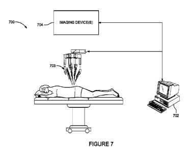
100641 FIGURE 7 illustrates a schematic block
diagram of a robotically-enabled
system.
DETAILED DESCRIPTION
(01165) Several implementations described
herein are directed to systems and
methods for modulating nerves within or adjacent (e.g., surrounding) bone. In
some
implementations, an intraosseous nerve (e.g., basivertebral nerve) within a
bone (es.,
-18-
CA 03150339 2022-3-7
WO 2021/050767
PCT/US2020/050249
vertebral body) of the spine is modulated for treatment, or prevention of;
chronic back pain.
The vertebral body may be located in any level of the vertebral column (e.g.,
cervical,
thoracic, lumbar and/or sacral). FIGURE 1 schematically illustrates a
vertebral column and
the various vertebral segments or levels. Multiple vertebral bodies may be
treated in a single
visit or procedure (simultaneously or sequentially). The multiple vertebral
bodies may be
located in a single spine segment (e.g., two adjacent vertebral bodies in the
sacral spine
segment (e.g.,. S1 and S2) or lumbar spine segment (e.g., L3, L4 and/or L5) or
thoracic spine
segment or cervical spine segment) or in different spine segments (e.g., an L5
vertebra in the
lumbar spine segment and an Si vertebra in the sacral spine segment).
Intraosseous nerves
within bones other than vertebral bodies may also be modulated. For example,
nerves within
a humerus, radius, femur, tibia, ealcaneus, tarsal bones, hips, knees, and/or
phalanges may be
modulated.
100661 In some implementations, the one or
more nerves being modulated are
extraosseous nerves located outside the vertebral body or other bone (e.g_, at
locations before
the nerves enter into, or after they exit from, a foramen of the bone). Other
tissue in addition
to, or alternative to, nerves may also be treated or otherwise affected (e.g.,
tumors or other
cancerous tissue or fractured bones). Portions of nerves within or on one or
more vertebral
endplates or interyertebral discs between adjacent vertebral bodies may be
modulated.
(00671 The modulation of nerves or other
tissue may be performed to treat one or
more indications, including but not limited to chronic low back pain, upper
back pain, acute
back pain, joint pain, tumors in the bone, and/or bone fractures. The
modulation of nerves
may also be performed in conjunction with bone fusion or arthrodesis
procedures so as to
provide synergistic effects or complete all-in-one, "one-and-done" treatment
that will not
require further surgical or minimally invasive interventions.
100681 In some implementations, fractures
within the bone may be treated in
addition to denervation treatment and/or ablation of tumors by applying heat
or energy and/or
delivering agents or bone filler material to the bone. For example, bone
morphogenetic
proteins and/or bone cement may be delivered in conjunction with
vertebroplasty or other
procedures to treat fractures or promote bone growth or bone healing. In some
implementations, energy is applied and then agents and/or bone filler material
is delivered in
a combined procedure. In some aspects, vertebral compression fractures (which
may be
-19-
CA 03150339 2022-3-7
WO 2021/050767
PCT/US2020/050249
caused by osteoporosis or cancer) are treated in conjunction with energy
delivery to modulate
nerves and/or cancerous tissue to treat back pam.
[0069] In accordance with several
implementations, the systems and methods of
treating back pain or facilitating neuromodulation of intraosseous nerves
described herein
can be performed without surgical resection, without general anesthesia,
without cooling
(e.g., without cooling fluid), and/or with virtually no blood loss. In some
embodiments, the
systems and methods of treating back pain or facilitating neurornodulation of
intraosseous
nerves described herein facilitate easy retreatment if necessary. In
accordance with several
implementations, successful treatment can be performed in challenging or
difficult-to-access
locations and access can be varied depending on bone structure or differing
bone anatomy.
One or more of these advantages also apply to treatment of tissue outside of
the spine (e.g.,
other orthopedic applications or other tissue).
ACCESS TO THE VERTEBRAL BODY
Methods of Access
[0070] Various methods of access may be used
to access a vertebral body or other
bone. In some implementations, the vertebral body is accessed transpedicularly
(though one
or both pedicles). In other implementations, the vertebral body is accessed
extrapedicularly
(e.g., without traversing through a pedicle). In some implementations, the
vertebral body is
accessed using an extreme lateral approach or a transforaminal approach, such
as used in
XLIF and TLIF interbody fusion procedures. In some implementations, an
anterior approach
is used to access the vertebral body.
[00711 Certain vertebrae in the sacral or
lumbar levels (e.g., Si vertebra, LS
vertebra) may also be accessed generally posterolaterally using a trans-ilium
approach (e.g.,
an approach through an ilium bone). With reference to FIGURE 2, an access hole
may be
formed through the ilium at a location designed to facilitate access to the
vertebral body or
bodies in the sacral or lumbar region. For example, access tools (e.g., an
introducer assembly
including a cannulaistylet combination) may be delivered through an ilium
and/or sacroiliac
joint or sacral ala into an SI vertebra under image guidance (e.g., CT image
guidance and/or
fluoroscopy) and/or using stereotactic or robotic-assisted surgical and/or
navigation systems,
such as the robotic system described in connection with FIGURE 7. A treatment
device
-20-
CA 03150339 2022-3-7
WO 2021/050767
PCT/US2020/050249
could then be inserted through an introducer andlor other access cannula of
the access tools
to a target treatment location within a sacral or lumbar vertebra. A trans-
ilium approach may
advantageously increase the ability of the clinician to access the target
treatment location in a
particular portion or region of the vertebral body (e.g., posterior portion or
region) that is not
capable of being adequately accessed using a transpedicular approach. In some
implementations, the vertebral body may be accessed directly through the
cerebrospinal fluid
and through the dura into a posterior region of the vertebral body.
100721 In some implementations, the vertebral
body may be accessed
transforarninally through a basivertebral foramen. Transforaminal access via
the spinal canal
may involve insertion of a "nerve finder" or nerve locator device and/or
imaging/diagnostic
tool to avoid damaging spinal cord nerves upon entry by the access tools or
treatment
devices. The nerve locator device may comprise a hand-held stimulation system
such as the
Checkpoint Stimulator and Locator provided by Checkpoint Surgica,r or the
FZstim
peripheral nerve stimulator/nerve locators provided by Avanos Medical, Inc.
The nerve
finder or nerve locator device could advantageously identify sensitive nerves
that should be
avoided by the access tools so as not to risk paralysis or spinal cord injury
upon accessing the
target treatment site. The nerve locator device may be configured to apply
stimulation
signals between two points or locations and then assess response to determine
presence of
nerves in the area between the two points or locations. The nerve locator
device may include
a bipolar pair of stimulation electrodes or monopolar electrodes. In some
implementations,
the nerve locator features may be implemented on the access tools or treatment
devices
themselves as opposed to a separate stand-alone device.
Access Tools and Treatment Devices
100731 Access tools may include an introducer
assembly including an outer
cannula and a sharpened stylet, an inner cannula configured to be introduced
through the
outer cannula, and/or one Or more additional stylets, curettes, or drills to
facilitate access to
an intraosseotts location within a vertebral body or other bone. The access
tools (e.g., outer
cannula, inner cannula, stylets, curettes, drills) may have pre-curved distal
end portions or
may be actively steerable or cm-veal:de. Any of the access tools may have
beveled or
otherwise sharp tips or they may have blunt or rounded, atraumatic distal
tips. Curved drills
-21-
CA 03150339 2022-3-7
WO 2021/050767
PCT/US2020/050249
may be used to facilitate formation of curved access paths within bone. Any of
the access
tools may be advanced over a guidewire in some implementations.
(00741 The access tools may be formed of a
variety of flexible materials (e.g.,
ethylene vinyl acetate, polyethylene, polyethylene-based polyolefin
elastomers,
polyetheretherketone, polypropylene, polypropylene-based elastomers, styrene
butadiene
copolymers, thermoplastic polyester elastomers, thermoplastic polyurethane
elastomers,
thermoplastic vulcanizate polymers, metallic alloy materials such as nitinol,
and/or the like).
Combinations of two or more of these materials may also be used. The access
tools may
include chevron designs or patterns or slits along the distal end portions to
increase flexibility
or bendabiljty. Any of the access tools may be manually or automatically
rotated (e.g., using
a robotic control system such as described in connection with FIGURE 7) to
facilitate a
desired trajectory.
100751 In some implementations, an outer
cannula assembly (e.g., introducer
assembly) includes a straight outer cannula and a straight stylet configured
to be received
within the outer cannula. The outer cannula assembly may be inserted first to
penetrate an
outer cortical shell of a bone and provide a conduit for further access tools
to the inner
cancellous bone. An inner cannula assembly may include a cannula having a pre-
curved or
steerable distal end portion and a stylet having a corresponding pre-curved or
steerable distal
end portion. Multiple stylets having distal end portions with different
curvatures may be
provided in a kit and selected from by a clinician. The inner cannula assembly
may
alternatively he configured to remain straight and non-curved.
100761 With reference to FIGURE 3, in one
implementation, a kit or system of
access tools includes an introducer assembly 110 comprised of an introducer
cannula 112 and
an introducer stylet 114, a curved cannula assembly 210 comprised of a curved
cannula 212
and a I-stylet 214, and a straight stylet 314. The introducer stylet 114 may
be bevel tipped,
trocar tipped, and/or diamond tipped. The introducer stylet 114 is configured
to be received
in a lumen of the introducer cannula 112 in a manner such that a distal tip of
the introducer
stylet 114 protrudes from an open distal tip of the introducer cannula 112,
thereby forming
the introducer assembly 110 in combination. The J-stvlet 214 is configured to
be received in
a lumen of the curved cannula 212 in a manner such that a distal tip of the .1-
stylet 214
protrudes from an open distal tip of the curved cannula 212, thereby forming
the curved
-22-
CA 03150339 2022-3-7
WO 2021/050767
PCT/US2020/050249
cannula assembly 210 in combination. The curved cannula 212 and the J-stvlet
214 may
each comprise a straight proximal main body portion and a curved distal end
portion. The
curves of the curved distal end portions of the curved cannula 212 and the J-
stylet 214 may
correspond to each other. The straight sty,-let 314 is a flexible channeling
sty let configured to
be delivered through the curved cannula 212 and then to form and maintain a
straight or
generally straight path upon exiting the open distal tip of the curved cannula
212.
100771 The access tools may be provided as a
kit that may optionally additionally
include one or more additional introducer cannulas, one or more additional
introducer stylets
(e.g., with different tips, such as one with a bevel tip and one with a
diamond or trocar tip),
one or two or more than two additional curved cannulas (e.g., having a curved
distal end
portion of a different curvature than a first curved cannula), an additional J-
style! (e.g.,
having a different curvature or different design configured to access hard
bone), an
introducer drill 440, and/of an additional straight stylet (e.g., having a
different length than
the first straight stylet
[0078] In some embodiments, the access tools
(e.g., kit) may be specifically
designed and adapted to facilitate access to hard, non-osteoporotic bone
(e.g., bone
surrounding or within a vertebral body, such as a cervical vertebra, a
thoracic vertebra, a
lumbar vertebra, or a sacral vertebra). Hard bone may be determined based on
bone mass
density testing, compressive strength determinations, compressive modulus
determinations,
imaging modalities, or based on tactile feel by the operator as access
instruments are being
advanced. In some implementations, hard bone may be determined as bone having
a bone
mineral density score within a standard deviation of a normal healthy young
adult (e.g., a T
score greater than or equal to -1). In some implementations, hard bone may be
identified as
bone having a compressive strength of greater than 4 MPa andlor a compressive
modulus of
greater than 80 MPa for cancellous bone and greater than 5.5 MPa .anclior a
compressive
modulus of greater than 170 MPa for cortical bone. Some kits may include at
least two of
every access instrument. Some kits may include optional add-on components or
accessory
kit modules for accessing hard bone (e.g., the introducer drill 440 and J-
stylet 214 specially
configured to access hard bone). Some kits may include optional additional
access tool
components or accessory kit modules adapted to access one or more additional
vertebrae in
-23-
CA 03150339 2022-3-7
WO 2021/050767
PCT/US2020/050249
the same spinal segment or in different spinal segments. The kit may also
include one or
more (e.g., at least two) treatment devices (such as radiofrequencyr energy
delivery probes).
[00791 FIGURES 3A-3C illustrate various views
of an embodiment of the
introducer cannula 112. The introducer cannula 112 includes a proximal handle
116 and a
distal hypotube 118 extending from the proximal handle 116. The illustrated
proximal
handle 116 comprises a "smokestack" or "T-Handle" design configuration adapted
to provide
sufficient finger clearance and gripping (e.g., two fingers on each side of a
lower flange 113
of the proximal handle 116 and along the lower surface of a crossbar portion
115) to facilitate
removal. However, alternative design configurations for the proximal handle
other than a
"smokestack" or "T-handle" design may be incorporated.
[0080] The proximal handle 116 includes an
upper central opening 120
configured to facilitate straight axial insertion of an introducer sty let 114
or other straight
access tool. The upper central opening 120 may be positioned so as to
correspond with (e.g.,
be coaxial with) a central lumen extending through the hypotube 118 of the
introducer
cannula 112 so as to facilitate insertion of straight instruments (e.g.,
introducer stylet 114 or
steerable cannulas or steerable stylets) therethrough. The proximal handle 116
may also
include coupling features 121 (e.g., recesses, notches, grooves, tabs) to
facilitate coupling or
mating of a proximal handle 216 of the introducer stylet 114 with the proximal
handle 116 of
the introducer cannula 112. The coupling features 121 may be adapted to
prevent rotation of
the introducer sty-let 114 andlor to provide assurance that a distal tip 125
of the introducer
stylet 114 extends beyond an open distal tip 122 of the hypotube 118 of the
introducer
cannula 112 so as to enable penetration of the distal tip 125 of the
introducer stylet 114
through bone. The upper surface of the proximal handle 116 of the introducer
cannula 112
also includes a curved lateral slot 117 and curved ramp 141 to facilitate
insertion of the
curved cannula assembly 210 into the proximal handle 116 and then into and
along the
central lumen of the hypotube 118_
100811 The central lumen of the hypotube 118
extends from the proximal handle
116 to the open distal tip 122 of the hypotube 118. The hypotube 118 may be
flared or
tapered such that the diameter of the hypotube 118 is not constant along its
entire length. For
example, the diameter may decrease abruptly at a certain distance (e.g., 1 cm
¨ 3 cm) from a
lower edge of the lower flange 113 of the proximal handle 116 and then
continue with a
-24-
CA 03150339 2022-3-7
WO 2021/050767
PCT/US2020/050249
constant diameter distally of an abrupt flare 119. In another embodiment, the
diameter may
decrease gradually (e.g., taper uniformly) along the length of the hypotube
118 from the start
of the flare 119 to the open distal tip 122 of the hypotube 118_ The central
lumen of the
hypotube 118 may be coated with a medical grade silicone lubricant to improve
tool insertion
and removal. The outer diameter of the hypotube 118 may range from 4.2 mm to
4.5 mm.
100821 The proximal handle 116 of the
introducer cannula 112 may also include
an overdrive indication mechanism configured to indicate when the curved
cannula assembly
210 has been fully deployed from the introducer cannula such that further
advancement of
the curved cannula would place the curved cannula assembly 210 at risk of
being overdriven
from the introducer cannula 112, which could result in damage to the curved
cannula
assembly 210. The overdrive indication mechanism may comprise two slots 123 in
the upper
surface of the crossbar portion 115 of the proximal handle 116 that display a
hi-stable (i.e.,
on-off states) indicator of a first color when overdrive is likely not a risk
and a second color
when overdrive is likely a risk (e.g., curved cannula assembly 210 has been
fully deployed).
In accordance with several embodiments, there are advantageously two distinct
states of
operation and there is no transition zone between the two states. The
overdrive indication
mechanism may be configured to be activated only when a gear wheel 221 of the
curved
cannula assembly 210 is bottomed out (e.g., fully engaged with the proximal
handle 116 of
the introducer cannula 112). As shown in FIGURE 3C a lower (bottom) side
surface of the
proximal handle 116 of the introducer cannula may include a cutout 124 adapted
to receive a
portion of a flexible shaft of a treatment device (e.g., radiofrequency probe
comprised of
nitinol or other flexible or shape memory material) and hold it in place and
out of the way
during a treatment procedure, thereby reducing stack height (e.g., by
approximately 3 inches
(or approximately 75 mm) or more).
100831 FIGURES 3D-3H illustrate various views
and portions of embodiments
of introducer stylets 114. FIGURE 3D illustrates a side view of an introducer
stylet 114.
The introducer stylet 114 includes a proximal handle 126 and a distal elongate
member or
shaft 128. The proximal handle 126 comprises an upper surface that is adapted
for malleting
by a mallet and a lower surface that is adapted to facilitate removal of the
introducer stylet
114 by an operator. The length of the distal elongate member 128 may range
from 8 mm to
14 mm (e.g., 8 mm, 8.5 mm, 9 mm, 9.5 mm, 10 mm, 10.5 mm, 11 mm, 11.5 mm, 12
mm,
-25-
CA 03150339 2022-3-7
WO 2021/050767
PCT/US2020/050249
12.5 mm, 13 mm, 13.5 rnm, 14 min). The distal end portion 132 of the
introducer stylet 114
may comprise a scalloped section 133 (as shown more closely in FIGURE 3E) to
provide a
release mechanism for bone compaction_ The scalloped section 133 may be
designed to have
a side profile shaped generally like an hourglass. The scalloped section 133
may gradually
taper from a full diameter proximal portion to a narrow-most middle potion and
then
gradually taper back to a full diameter distal portion_ The taper may be
symmetric or
asymmetric. The scalloped section 133 may comprise one scallop (or scooped-out
region) or
multiple scallops (or scooped-out regions) along the length of the distal end
portion 132. A
distal tip 125 of the distal end portion 132 may comprise a full diameter so
as to be adapted
to break apart bone (e.g., pedicle bone, cortical bone of a vertebral body).
As the bone is
broken up by the distal tip 125 of the distal end portion 132, bone shards or
chips can pack
into a gap formed between the distal end portion 132 of the introducer stylet
114 and the
inner surface of the distal end portion of the introducer cannula 112, thereby
making it more
difficult for the introducer stylet 114 to be removed from the introducer
cannula 111 In
accordance with several embodiments, the scalloped section 133 of the
introducer stylet 114
advantageously provides the bone shards and fragments a place to fall into
during removal of
the introducer stylet 114 so as to facilitate easier removal of the introducer
stylet 114.
100841 FIGURES 3F-311 illustrate the
introducer assembly 110 after the
introducer stylet 114 has been inserted within the introducer cannula 112. As
indicated
above, the proximal handle 116 of the introducer cannula 112 may include
mating or
engagement features (e.g., coupling features 121) that facilitate automatic
(e.g., snap-fit)
engagement of the introducer stylet 114 with the proximal handle 116 of the
introducer
cannula 112.
[0085] The proximal handle 126 of the
introducer stylet 114 includes an
alignment indicator 129, an anti-rotation tab 131, and a press button 134. As
shown best in
FIGURE 3G, the alignment indicator 129 is configured to align with a
corresponding
alignment indicator 130 on the upper surface of the crossbar portion 115 of
the proximal
handle 116 of the introducer cannula 112 in order to ensure proper insertion
and alignment of
the introducer stylet 114 with respect to the introducer cannula 112. The anti-
rotation tab
131 is configured to be positioned within the slot 117 of the proximal handle
116 of the
-26-
CA 03150339 2022-3-7
WO 2021/050767
PCT/US2020/050249
introducer cannula 112 and to prevent rotation of the introducer stylet 114
with respect to the
introducer cannula 112 during malleting and orienting_
(00861 The press button 134 is integrally
coupled to the anti-rotation tab 131 such
that pressing of the press button 134 extends the anti-rotation tab 131 out of
die constraint of
the slot 117, thereby allowing the introducer stylet 114 to rotate with
respect to the introducer
cannula 112 (as shown in FIGURE 3H)_ Pressing the press button 134 also
releases
engagement of the introducer stylet 114 with the introducer cannula 112 to
enable removal of
the introducer stylet 114 from the introducer cannula 112. The proximal handle
126 of the
introducer stylet 114 may include internal ramps (not shown) configured to
provide a
mechanical advantage to assist in removal of the introducer stylet 114 from
the introducer
cannula 112 (especially if bone shards have packed into gaps between the
introducer stylet
114 and introducer cannula 112 making removal more difficult) as the proximal
handle 126
is rotated (e_g., 120-degree rotation counter-clockwise). The combination of
the scalloped
distal end portion design and the internal ramps in the proximal handle 126
may provide
increased reduction of removal forces by 50% ¨ 70% compared to a full diameter
(e.g., no
scalloped section) distal end portion design with no ramps in the proximal
handle 126.
100871 FIGURES 31 and 3J illustrate a side
view and a top view of an
embodiment of the curved cannula 21.2. The curved cannula 212 includes a
proximal handle
216, a threaded proximal shaft portion 220, a gear wheel 221, a rigid support
portion 223,
and a distal polymeric shaft portion 224. The proximal handle 216 includes a
curved slot 217
and a curved ramp 231 configured to facilitate insertion of the .1-stylet 214
into and along a
central lumen of the curved cannula 212 extending from the proximal handle 216
to an open
distal tip 222 of the distal polymeric shaft portion 224. The central lumen of
the curved
cannula 212 may be coated with a medical grade silicone lubricant to improve
tool insertion
and removal.
00881 In the illustrated example, the gear
wheel 221 comprises threads
configured to interface with corresponding threads of the threaded proximal
shaft portion 220
such that rotation of the gear wheel 221 causes controlled proximal and distal
translation of
the gear wheel 221 along the threaded proximal shaft portion 220. The threaded
proximal
shaft portion 220 is sized such that when the gear wheel 221 is in its distal-
most position, the
distal tip 222 of the curved cannula 212 does not extend out of the open
distal tip 122 of the
-27-
CA 03150339 2022-3-7
WO 2021/050767
PCT/US2020/050249
introducer cannula 112 when the curved cannula assembly 210 is fully inserted
therein. The
gear wheel 221 may spin freely about the threaded proximal shaft portion 22a
The threads
may comprise triple threads and the gear wheel 221 may be configured to
traverse the entire
length of the threaded proximal shaft portion 220 with four complete rotations
of the gear
wheel 221.
100891 The rigid support portion 223 may
comprise a biocompatible metal or
other rigid material, such as stainless steel, titanium, platinum and/or the
like, so as to
provide additional support to the curved cannula 212 during insertion of the J-
stylet 214. The
distal polymeric shaft portion 224 may be comprised of a thermoplastic, shape-
memory
polymer material (such as polyether ether ketone (PEEK), polyurethane,
polyethylene
terephthalate (PET), and/or the like) and the distal end portion 225 is pre-
curved (e.g., shape-
set) to have a predetermined curve in a "resting" unconstrained configuration.
100901 FIGURES 3K-3P illustrate an embodiment
of the J-stylet 214. FIGURE
3K illustrates a side view of the J-stylet 214 in a 4'resting" normal,
unconstrained
configuration or state and FIGURES 31- and 3M are close-up views (side view
and
perspective view, respectively) of a curved distal end portion 227 of the J-
stylet 214. The J.-
st3r1et 214 comprises a proximal handle 226 and a distal elongate shaft 218.
The proximal
handle 226 comprises an upper surface that is adapted for malleting by a
mallet and a lower
surface that is adapted to facilitate removal of the J-stylet 214 by two or
more (e.g., two,
three, or four) fingers of an operator. The upper surface of the proximal
handle 216 includes
an alignment indicator 219 (shown, for example, in FIGURE 3P) configured to be
aligned
with the corresponding alignment indicator 130 of the introducer carmula. 112
to facilitate
insertion, removal, and deployment of the J-stylet 214 (and curved cannula
assembly 210).
[0091] The distal elongate shaft 218 includes
a curved distal end portion 227
haying an asymmetric curve profile along its length (e.g., the curved distal
end portion does
not have a constant full diameter along its length). A distal channeling tip
228 is sized and
shaped to facilitate channeling through cancellous bone along a curved path or
trajectory.
The curved distal end portion 227 comprises a springboard or platform section
229 having a
"13-shaped" cross-sectional profile, as shown, for example, by the cross-
section profile circle
in FIGURE 3M. The springboard or platform section 229 may be formed by
mechanical
grinding of a tubular wire until the desired D-shaped cross section profile is
achieved in
-28-
CA 03150339 2022-3-7
WO 2021/050767
PCT/US2020/050249
which a top (e.g., upper) surface of the springboard or platform section 229
is generally
smooth and flat. The thickness (eg,, vertical cross-sectional dimension) of
the springboard
or platform section 229, the predefined set angidation or radius of curvature,
and the starting
and ending points of the springboard or platform section 229 along the length
of the curved
distal end portion 227 may be varied to provide j-stylets having different
rigidity and
bending characteristics for different levels of vertebrae or different
densities of bone.
100921 In accordance with several
embodiments, a thickness (a g., a maximum
vertical cross-sectional dimension from an upper surface of the springboard or
platfomi
section 229 to a lower-most point on a lower surface of the curved distal end
portion) is
between 40% and 85% (e.g., between 40% and 60%õ between 50% and 70%, between
50%
and 75%, between 60% and 70%, between 65% and 80%, between 70% and 85%,
overlapping ranges thereof, or any value within the recited ranges) of the
thickness (e.g.,
diameter) of the adjacent regions of the curved distal end portion (e.g., the
regions just
proximal and just distal of the length of the springboard or platform section
229), Instead of
percentages, the difference in thickness dimensions could be represented as
ratios (e.g.,
between 2:5 and 4:5, between 2:5 and 3:5, between 1;2 and 3:4, between 3:5 and
4;5,
between 3:5 and 6:7). The ending point of the springboard or platform section
229 may be
between 4.5 and 9 mm from a distal terminus of the distal elongate shaft 218.
The starting
point of the springboard or platform section 229 may be between 230 mm and 245
mm from
a proximal terminus of the distal elongate shaft 218.
100931 According to several embodiments, the
asymmetric curve profile (e.g.,
profile with D-shaped cross-section) advantageously provides improved cephalad-
caudal
steering because the curved distal end portion 227 primarily bends inward and
not laterally.
In addition, the design and material of the curved distal end portion 227 of
the J-stvlet 214
may enable the angle of curvature of the curved distal end portion 227 to
advantageously
remain relatively consistent and reproducible across a variety of bone
densities, or regardless
of bone environment. For example, in one embodiment, the design and material
of the
curved distal end portion 227 of the J-stylet 214 facilitates consistent and
reproducible access
to a posterior location (e.g., in posterior half of the vertebral body or to a
location
approximately 30%-50% of the distance between the posterior-most aspect and
the anterior-
most aspect of the vertebral body along a sagittal axis or to a geometric
center or midpoint
-29-
CA 03150339 2022-3-7
WO 2021/050767
PCT/US2020/050249
within the vertebral body for vertebral bodies having varying bone densities
or other desired
target location in the vertebral body or other bone). In accordance with
several
embodiments, the curvature is designed to deviate by less than 25 degrees
(e.g., less than 20
degrees, less than 15 degrees, less than 10 degrees) or less than 30% from the
predefined set
curvature of the curved distal end portion 227 in an unconstrained
configuration (even in
hard bone).
100941 The .1-stylet 214 may be designed and
adapted to exert a lateral force of
between 6 pounds and 8 pounds. The angle of curvature of the curved distal end
portion 227
(with respect to the central longitudinal axis of the straight proximal
portion of the distal
elongate shaft 218) of the J-stylet 214 in the normal unconstrained state or
configuration may
be designed to be between 65 degrees and 80 degrees (e.g., 65 degrees, 70
degrees, 75
degrees, 80 degrees, or any other value within the recited range). The radius
of curvature of
the curved distal end portion 227 may range from 11.5 mm to 15 mm (e.g., from
11.5 mm to
12 nun, from 12 nun to 12,5 mm, from 12 mm to 13 mm, from 115 mm to 14 trim,
from 13
mm to 15 mm, overlapping ranges thereof, or any value within the recited
ranges). The .1-
stylet 214 may be comprised of nitinol or other metallic alloy material,
100951 FIGURES 3N and 30 are a perspective
view and a side cross-section
view, respectively, illustrating insertion of the curved distal end portion
227 of the J-stylet
214 into the slot 217 of the proximal handle 216 of the curved cannula 212. As
shown in
HG. 30, the slot 217 comprises a curved ramp 231 and a straight vertical
backstop support
233 (e.g., with no trumpeted section) to facilitate insertion of the curved
distal end portion
227 of the 1-stylet 214. As indicated above, the curved cannula 212 includes
the rigid
support portion 223 extending into and out of the threaded shaft portion 220
to provide
additional support upon insertion of the J-stvlet 214 within the central lumen
of the curved
cannula 212.
[0096] FIGURES 31? and 3Q illustrate
insertion of the curved cannula assembly
210 into the introducer cannula 112. The curved distal end portion 225 of the
curved cannula
assembly 210 is inserted from a side angle (e.g., at about a 65 to 75 degree
angle (such as a
70 degree starting angle in one embodiment) with respect to the central
longitudinal axis LA
of the distal lwpotube 118 of the introducer cannula 112) into the slot 117
and along the ramp
141 in the proximal handle 116 and then down the central lumen of the distal
hypotube 118
-30-
CA 03150339 2022-3-7
WO 2021/050767
PCT/US2020/050249
of the introducer cannula 112 while the gear wheel 221 of the curved cannula
212 is in a
distal-most position along the threaded proximal portion 220 of the curved
cannula 212 so as
to prevent inadvertent advancement of the curved distal portion of the curved
cannula
assembly 210 beyond the open distal tip 122 of the introducer cannula 112
until the operator
is ready to do so.
100971 FIGURE 3Q is a close-up side cross-
section view of the proximal portion
of the introducer cannula 112 and the curved distal portion of the curved
cannula assembly
210 and illustrates insertion of the curved distal end portions of the
assembled components of
the curved cannula assembly 210 into the introducer cannula 112. As shown, the
introducer
cannula 112 is shaped so as to provide a backstop support 143 generally
aligned with the
inner surface of the central lumen of the hypotube 118 so as to facilitate
insertion and so that
the curved distal end portion 225 of the distal polymeric shaft portion 224 of
the curved
cannula 212 does not pivot out of the introducer cannula 112 upon insertion.
In accordance
with several embodiments, the asymmetric "D-shaped" cross-sectional profile of
the J-stylet
214 is advantageously designed to prevent twisting during insertion.
100981 FIGURES 3R and 3S illustrate operation
of the gear wheel 221 of the
curved cannula 212. As shown in FIGURE 3R, the gear wheel 221 is rotated until
it is in its
distal-most position along the threaded proximal portion 220 prior to
insertion of the curved
cannula assembly 210 within the introducer cannula 112 so as to prevent
inadvertent
advancement of the curved distal end portions 225, 227 of the curved cannula
assembly 210
out of the introducer cannula 112. As shown in FIGURE 3S, the gear wheel 221
is rotated
to its proximal-most position along the threaded proximal portion 220 to
enable full insertion
of the curved cannula assembly 210 within the introducer cannula 112 such that
the curved
distal end portions 225, 227 of the curved cannula assembly 210 extend out of
the introducer
cannula 112 and along a curved path within the canceilous bone region of the
vertebral body
or other bone.
100991 FIGURES 3T and 3U illustrate operation
of a bail mechanism of the .1-
stylet 214. The proximal handle 226 of the .1-stylet 214 includes a bail
actuator 250
configured to be toggled between a first "resting" or "inactive" configuration
in which the
bail actuator 250 is generally aligned with (e.g., parallel or substantially
parallel to) the upper
surface of the proximal handle 226 (as shown in FIGURE 3T) and a second
"active"
-31-
CA 03150339 2022-3-7
WO 2021/050767
PCT/US2020/050249
configuration in which the bail actuator 250 is offset from the upper surface
of the proximal
handle 226 (as shown in FIGURE 311). The bail actuator 250 is configured to
act as a lever
to cause a slight axial (proximal-distal) movement of the J-stylet 214 with
respect to the
curved cannula 212 as the bail actuator 250 is pivoted. When the bail actuator
250 is toggled
to the "active" configuration, a flange 253 of the bail actuator 250 contacts
the proximal
handle 216 of the curved cannula to cause proximal refraction of the J-stylet
214 with respect
to the curved cannula 212 such that the distal channeling tip 228 of the J-
stylet 214 resides
completely within the curved cannula 212 and does not extend out of the open
distal tip of
the curved cannula 212. In accordance with several embodiments, the bail
actuator 250 is
advantageously toggled to the "active" configuration (in which the distal
channeling tip 228
of the J-stvlet 214 resides within the open distal tip of the curved cannulas
212) upon
insertion and removal of the curved cannula assembly 210 from the introducer
cannula 112
or the J-stylet 214 from the curved cannula 212 (e.g., so as to avoid friction
caused by
interaction between two metal components). The upper surface of the bail
actuator 250 may
include an indicator 252 (e.g., colored marking or other visual indicator)
that is visible to an
operator when the bail actuator 250 is in the active configuration and hidden
when the bail
actuator 250 is in the inactive configuration.
101001 FIGURE 3V illustrates a side view of
an embodiment of the straight stylet
314 and FIGURE 3W illustrates a distal portion of the straight stylet 314. The
straight stylet
314 includes a proximal handle 316 and a distal elongate shaft 318. The
proximal handle
316 includes an upper surface adapted for malleting by a mallet or application
of pressure by
a hand or fingers of an operator. A radiopaque marker band 317 may be
positioned along the
distal elongate shaft 318 at a position corresponding to the position when a
distal channeling
tip 319 of the straight stylet 314 is exiting the open distal tip of the
curved cannula 212 as the
straight stylet 314 is advanced through the curved cannula 212. The length of
the straight
stylet 314 may be sized such that, when the straight style 314 is fully
inserted within the
curved cannula 212, the length of the portion of the straight stylet 314
extending beyond the
open distal tip of the curved cannula 212 is between 25 and 50 mm (e.g.,
between 25 mm and
35 mm, between 30 mm and 40 mm, between 35 mm and 45 mm, between 40 and 50 mm,
overlapping ranges thereof, or any value within the recited ranges). The
diameter of the
-32-
CA 03150339 2022-3-7
WO 2021/050767
PCT/US2020/050249
straight stylet 314 is sized so as to be inserted within and through the
central lumen of the
curved cannula 212.
(010/1 The distal elongate shaft 318
comprises an inner flexible, shape memory
core 360 extending from the proximal handle 316 to the distal channeling tip
319 of the
straight stylet 314 and a polymeric outer layer 365 extending from the
proximal handle 316
to a distal end of the distal elongate shaft 318 but stopping short (or
proximal to) the distal
channeling tip 319 so that the inner core 360 protrudes out of the outer layer
365. The
straight stylet 314 is flexible enough to bend to traverse the curved distal
end portion 225 of
the curved cannula 212 without significant friction but sufficiently rigid so
as to maintain a
straight path once the straight stylet 314 exits the open distal tip of the
curved cannula 212.
The inner core 360 of the straight stylet 3/4 may comprise nitinol or other
metallic alloy or
other flexible material. The outer layer 365 may be comprised of a more rigid,
polymeric
material (such as PEEK, polyurethane, PET, and/or the like).
101021 FIGURES 3X-3Z illustrate an embodiment
of an introducer drill 440 and
its interaction with the introducer cannula 112. A kit or system of access
instruments (e.g., a
kit or kit module designed for accessing hard, or high-density, bone) may
optionally include
the introducer drill 440. FIGURE 3X is a side view of an embodiment of the
introducer drill
440. The introducer drill 440 includes a proximal handle 446 and an elongate
drill shaft 447.
The proximal handle 446 may comprise a generally T-shaped design and may
comprise a
soft-grip overrnolding. The length of the elongate drill shaft 447 may be
sized so as to
extend from 20 mm to 35 mm beyond the open distal tip of the introducer
cannula 112 when
the introducer drill 440 is fully inserted within the introducer cannula 112.
The elongate drill
shaft 447 may include a solid proximal portion 448 and a fluted distal portion
449.
[0103] FIGURE SY is a close-up perspective
view of the fluted distal portion
449. The fluted distal portion 449 may comprise a distal cutting tip 450
having a 90 degree
cutting angle. The drill flutes 452 of the fluted distal portion 449 may be
adapted to taper
away from the distal cutting tip 450 (which is a reverse taper or opposite the
direction of
taper of a typical drill bit) so as to facilitate improved bone chip packing
within the open
flute volume as bone chips and fragments are generated by operation of the
introducer drill
440. The distal cutting tip 450 may have a point angle of between 65 and 75
degrees and a
chisel edge angle of between 115 and 125 degrees. The flutes may
advantageously be deeper
-33-
CA 03150339 2022-3-7
WO 2021/050767
PCT/US2020/050249
and wider than typical drill bits because the elongate drill shaft 447 is
supported by a rigid
introducer cannula 112 surrounding at least a portion of the length of the
elongate drill shaft
(and a portion of the length of the fluted distal portion in most instances)
during use. The
drill flutes 452 may have a helix angle of between 12 degrees and 18 degrees
(e.g., between
12 degrese and 14 degrees, between 13 degrees and 17 degrees, between 14
degrees and 16
degrees, between 14 degrees and 18 degrees, overlapping ranges thereof, or any
value within
the recited ranges). The fluted distal portion 449 may include two flutes
having a length of
between 70 mm and 85 mm.
101041 The open flute volume of the fluted
distal portion 449 may be
advantageously configured to hold all or substantially all (e.g., more than
75%, more than
80%, more than 85%, more than 90%) of the significantly-sized bone chips or
fragments
removed by the introducer drill 440 as the introducer drill 440 is removed
from the
introducer cannula 112, thereby reducing the bone fragments left behind in the
bone (e.g.,
vertebral body) or in the introducer cannula 112. In some embodiments, the
open flute
volume of the fluted distal portion 449 is adapted to hold about 2 ccs of
bone. The fluted
distal portion 449 may exhibit web tapering (e.g., increase in width or depth,
or angle with
respect to longitudinal axis of the flutes) along its length from distal to
proximal (e.g., reverse
taper). There may be no web taper for approximately the first 25 mm at the
distal-most
region. The web taper may then increase gradually until a maximum web taper is
reached
near the proximal end of the fluted distal portion 449 so as to facilitate
pushing of the bone
fragments or chip upward (or proximally) along the fluted distal portion 449.
For example,
the fluted distal portion 449 may have a negative draft (e.g., 0.77" or -20 mm
negative draft).
101051 FIGURE 3Z illustrates the introducer
drill 440 fully inserted and engaged
with the proximal handle 116 of the introducer cannula 112. The introducer
drill 440 is sized
so as to be inserted within the central opening 120 of the proximal handle 116
of the
introducer cannula 112 and advanced through the central lumen of the hypotube
118 of the
introducer cannula 112. The proximal handle 446 of the introducer drill 440 is
configured to
engage with the coupling or mating features 121 of the proximal handle 116.
01061 FIGURES 3AA - 311 illustrate an
embodiment of steps of a method of
using the access tools to facilitate access to a location within a vertebral
body 500 for
treatment (e.g., modulation of intraosseous nerves, such as a basivertebral
nerve, bone
-34-
CA 03150339 2022-3-7
WO 2021/050767
PCT/US2020/050249
cement delivery for treatment of vertebral fractures, and/or ablation of bone
tumors). With
reference to FIGURE 3AA, the distal portion of the introducer assembly 110
(including the
distal tip 125 of the introducer stylet 114 and the distal tip of the
introducer cannula 112) are
inserted through a pedicle 502 adjacent the vertebral body 500 by malleting on
the proximal
handle of the introducer stylet 114 after insertion and aligned engagement of
the introducer
stylet 114 within the introducer cannula 112.
101071 In accordance with several
embodiments, the method may optionally
include removing the introducer stylet after initial penetration into the
pedicle 502 (for
example, if the operator can tell that the density of the bone is going to be
sufficiently dense
or hard that additional steps and/or tools will be needed to obtain a desired
curved trajectory
to access a posterior portion (e.g., posterior half) of the vertebral body
500. With reference
to FIGURE 3BB, the method may optionally include inserting the introducer
drill 440 into
and through the introducer cannula 112 to complete the traversal of the
pedicle 502 and
penetration through a conical bone 503 region of the vertebral body 500 until
a cancellous
bone region 504 of the vertebral body 500 is reached. The introducer drill 550
may be
advanced into the cancellous bone region 504 (especially if the cancellous
bone region 504 is
determined to be sufficiently hard or dense) or the advancement may stop at
the border
between the cortical bone region 503 and the cancellous bone region 504. This
step may
involve both rotating the introducer drill 440 and malleting on the proximal
handle 116 of the
introducer drill 440 or simply rotating the introducer drill 440 without
malleting on the
proximal handle 446. With reference to FIGURE 3CC, the introducer drill 440
may be
removed and the introducer stylet 114 may be re-inserted within the introducer
cannula 112.
With reference to FIGURE 39D, the introducer assembly 110 may then be malleted
so as to
advance the distal tip 122 of the introducer cannula 112 to the entry site
into (or within) the
cancellous bone region 504 of the vertebral body 500. The introducer stylet
114 may then be
removed from the introducer cannula 112.
/01081 The curved cannula assembly 210 may
then be inserted within the
introducer cannula 112 with the gear wheel 221 in the distal-most position so
as to prevent
inadvertent advancement of the curved cannula assembly 210 out of the open
distal tip 122 of
the introducer cannula 112 prematurely. With reference to -FIGURE 3EE, after
rotation of
the gear wheel 221 to a more proximal position, the curved cannula assembly
210 can be
-35-
CA 03150339 2022-3-7
WO 2021/050767
PCT/US2020/050249
malleted so as to advance the collective curved distal end portions of the
curved cannula
assembly 210 together out of the distal tip 122 of the introducer cannula 112
and along a
curved path within the cancellous bone region 504. With reference to FIGURE
3FF, the .1-
stylet 214 may then be removed from the curved cannula 212, with the curved
cannula 212
remaining in position. In accordance with several embodiments, the path formed
by the prior
instruments may advantageously allow the curved cannula assembly 210 to have a
head start
and begin curving immediately upon exiting the open distal tip 122 of the
introducer cannula
112.
101091 With reference to FIGURE 3GG, if a
further straight path beyond the
curved path is desired to reach a target treatment location, the straight
stylet 314 may be
inserted through the curved cannula 212 such that the distal channeling tip
319 of the straight
stylet extends beyond the open distal tip of the curved cannula 212 and along
a straight path
toward the target treatment location (e.g., a basivertebral nerve trunk or
basivertebral
foramen). In some embodiments, the straight stylet 314 may not be needed and
this step may
be skipped.
[01101 With reference to FIGURE 311H, a
treatment device 501 (e.g., a flexible
bipolar radiofrequency probe) may be inserted through the curved cannula 212
(after removal
of the straight stylet 314 if used) and advanced out of the open distal tip of
the curved
cannula 212 to the target treatment location. The treatment device 501 may
then perform the
desired treatment. For example, if the treatment device 501 is a
radiofrequency- probe, the
treatment device 501 may be activated to ablate intraosseous nerves (e.g., a
basivertebral
nerve) or a tumor within the vertebral body 500. Bone cement or other agent,
or a diagnostic
device (such as a nerve stimulation device or an imaging device to confirm
ablation of a
nerve) may optionally be delivered through the curved carinula. 212 after the
treatment device
501 is removed from the curved cannula 212.
101111 At certain levels of the spine (e.g.,
sacral and lumbar levels) and for
certain patient spinal anatomies that require a steeper curve to access a
desired target
treatment location within the vertebral body, a combination curette/curved
introducer may
first be inserted to start a curved trajectory (e.g., create an initial curve
or shelf) into the
vertebra. The curette may have a pre-curved distal end portion or be
configured such that the
distal end portion can be controllably articulated Or curved (e.g., manually
by a pull wire or
-36-
CA 03150339 2022-3-7
WO 2021/050767
PCT/US2020/050249
rotation of a handle member coupled to one or more pull wires coupled to the
distal end
portion or automatically by a robotic or artificial intelligence driven
navigation system).. The
combination curettelcurved introducer may then be removed and the outer
straight cannula
and inner curved cannula/curved sty let assembly may then be inserted to
continue the curve
toward the target treatment location.
[01121 In accordance with several
implementations, any of the access tools (e.g,
cannula or stylet) or treatment devices may comprise a theological and/or
magnetizable
material (e.g., magnetorheological fluid) along a distal end portion of the
access tool that is
configured to be curved in Mu after insertion to a desired location within
bone (e.g.,
vertebra). A magnetic field may be applied to the distal end portion of the
access tool and/or
treatment device with the magnetizable fluid or other material and adjusted or
varied using
one or more permanent magnets or electromagnets to cause the distal end
portion of the
access tool and/or treatment device to curve toward the magnetic field. In
some
implementations, a treatment probe may include a magnetic wire along a portion
of its length
(e.g., a distal end portion). Voltage applied to the magnetic wire may be
increased or
decreased to increase or decrease a curve of the magnetic wire. These
implementations may
advantageously facilitate controlled steering without manual pull wires or
other mechanical
mechanisms. The voltage may be applied by instruments controlled and
manipulated by an
automated robotic control system, such as the robotic system described in
connection with
FIGURE 7.
[0113] The treatment devices (e.g., treatment
probes) may be any device capable
of modulating tissue (e.g., nerves, tumors, bone tissue). Any energy delivery
device capable
of delivering energy can be used (e.g., RF energy delivery devices, microwave
energy
delivery devices, laser devices, infrared energy devices, other
electromagnetic energy
delivery devices, ultrasound energy delivery devices,, and the like). The
treatment device 501
may be an RF energy delivery device. The RF energy delivery device may include
a bipolar
pair of electrodes at a distal end portion of the device. The bipolar pair of
electrodes may
include an active tip electrode and a return ring electrode spaced apart from
the active tip
electrode. The RF energy delivery device may include one or more temperature
sensors
(e.g., thermocouples, therrnistors) positioned on an external surface of, or
embedded within, a
-37-
CA 03150339 2022-3-7
WO 2021/050767
PCT/US2020/050249
shaft of the energy delivery device. The RI. energy delivery device may not
employ
internally circulating cooling, in accordance with several implementations.
(01141 In some implementations, water jet
cutting devices may be used to
modulate (e.g., deneivate) nerves. For example, a water jet cutter may be
configured to
generate a very fine cutting stream formed by a very high-pressure jet of
water. For example,
the pressure may be in the range of 15 NIPa to 500 MPa (e.g., 15 MPa to 50MPa,
30 NIF'a ¨
60 /v1Pa, 50 MPa ¨ 100 MPa, 60 NIPa ¨ 120 MPa, 100 MPa ¨ 200 MPa, 150 MPa ¨
300
MPa, 300 NIPa ¨ 500 MPa, overlapping ranges thereof, or any value within the
recited
ranges). In some implementations, a chemical neuromodulation tool injected
into a vertebral
body or at an endplate may be used to ablate or otherwise modulate nerves or
other tissue.
For example, the chemical neuramodulation tool may be configured to
selectively bind to a
nerve or endplate. In some implementations, a local anesthetic (e.g.,
liposomal local
anesthetic) may be used inside or outside a vertebral body or other bone to
denervate or block
nerves. In some implementations, brachytherapv may be used to place
radioactive material
or implants within the vertebral body to deliver radiation therapy sufficient
to ablate or
otherwise derien,ate the vertebral body. In some implementations, chymopapain
injections
and/or cortdoliase injections may be used (e.g., under local anesthesia).
Phototherapy may be
used to ablate or otherwise modulate nerves after a chemical or targeting
agent is bound to
specific nerves or to a vertebral endplate.
(01151 In accordance with several
implementations, thermal energy may be
applied within a cancellous bone portion (e.g., by one or more radiofrequency
(RF) energy
delivery instruments coupled to one or more RE generators) of a vertebral
body. The thermal
energy may be conducted by heat transfer to the surrounding cancellous bone,
thereby
heating up the cancellous bone portion. In accordance with several
implementations, the
thermal energy is applied within a specific frequency range and having a
sufficient
temperature and over a sufficient duration of time to heat the cancellous bone
such that the
basivertebral nerve extending through the cancellous bone of the vertebral
body is
modulated. In several implementations, modulation comprises permanent ablation
or
denervation or cellular potation (e.g., electroporation). In some
implementations, modulation
comprises temporary &nervation or inhibition. In some implementations,
modulation
comprises stimulation or denervation without necrosis of tissue.
-38-
CA 03150339 2022-3-7
WO 2021/050767
PCT/US2020/050249
101161
For thermal energy,
temperatures of the thermal energy may range from
about 70 to about 115 degrees Celsius (e.g., from about 70 to about 90 degrees
Celsius, from
about 75 to about 90 degrees Celsius, from about 83 to about 87 degrees
Celsius, from about
80 to about 100 degrees Celsius, from about 85 to about 95 degrees Celsius,
from about 90 to
about 110 degrees Celsius, from about 95 to about 115 degrees Celsius, or
overlapping
ranges thereof). The temperature ramp may range from 0.1 ¨ 5 degrees
Celsius/second (e.g.,
0.1 ¨ 1.0 degrees Celsius/second, 0.25 to 2.5 degrees Celsius/second, 0.5 ¨
2.0 degrees
Celsius/second, 1.0¨ 3.0 degrc,cs Celsius/second, 1.5 ¨ 4.0 degree
Celsius/second, 2.0 ¨ 5.0
degrees Celsius/second). The time of treatment may range from about 10 seconds
to about 1
hour (e.g., from 10 seconds to 1 minute, 1 minute to 5 minutes, from 5 minutes
to 10
minutes, from 5 minutes to 20 minutes, from 8 minutes to 15 minutes, from 10
minutes to 20
minutes, from 15 minutes to 30 minutes, from 20 minutes to 40 minutes, from 30
minutes to
1 hour, from 45 minutes to 1 hour, or overlapping ranges thereof). Pulsed
energy may be
delivered as an alternative to or in sequence with continuous energy. For
radiofrequency
energy, the energy applied may range from 350 kHz to 650 kHz (e.g., from 400
kHz to 600
kHz, from 350 kHz to 500 kHz, from 450 kHz to 550 kHz, from 500 kHz to 650
kHz,
overlapping ranges thereof, or any value within the recited ranges, such as
450 kHz 5 kHz,
475 kHz 5 kHz, 487 kHz 5 kHz). A power of the radiofrequency energy may
range from
W to 30 W (e.g., from 5 W to 15 W, from 5 W to 20 W, from 8 W to 12 W, from 10
W to
25 Vi, from 15 W to 25 W, from 20 W to 30 W, from 8 W to 24 W, and overlapping
ranges
thereof, or any value within the recited ranges).
In accordance with several
implementations, a thermal treatment dose (e.g., using a cumulative equivalent
minutes
(CEM) 43 degrees Celsius thermal dose calculation metric model) is between 200
and 300
CEM (e.g., between 200 and 240 CEM, between 230 CEM and 260 CEM, between 240
CEM
and 280 CEM, between 235 CEM and 245 CEM., between 260 CEM and 300 CEM.) or
greater than a predetermined threshold (e.g., greater than 240 CEM). The CEM
number may
represent an average thermal cumulative dose value at a target treatment
region or location
and may represent a number that expresses a desired dose for a specific
biological end point
Thermal damage may occur through necrosis or apoptosis.
101171
Cooling may optionally be
provided to prevent surrounding tissues from
being heated during the nerve modulation procedure. The cooling fluid may be
internally
-39-
CA 03150339 2022-3-7
WO 2021/050767
PCT/US2020/050249
circulated through the delivery device from and to a fluid reservoir in a
closed circuit manner
(e.g., using an inflow lumen and an outflow lumen). The cooling fluid may
comprise pure
water or a saline solution having a temperature sufficient to cool electrodes
(e.g., 2 ¨ 10
degrees Celsius, 5 ¨ 10 degrees Celsius, 5 ¨ 15 degrees Celsius). Cooling may
be provided
by the same instrument used to deliver thermal energy (e.g., heat) or a
separate instrument.
In accordance with several implementations, cooling is not used
101181 In some implementations, ablative
cooling may be applied to the nerves or
bone tissue instead of heat (e.g., for cryoneurolysis or cryoablation
applications). The
temperature and duration of the cooling may be sufficient to modulate
intraosseous nerves
(e.g., ablation, or localized freezing, due to excessive cooling)_ The cold
temperatures may
destroy the myelin coating or sheath surrounding the nerves. The cold
temperatures may also
advantageously reduce the sensation of pain. The cooling may be delivered
using a hollow
needle under fluoroscopy or other imaging modality.
101191 In some implementations, one or more
fluids or agents may be delivered
to a target treatment site to modulate a nerve. The agents may comprise bone
morphogenetic
proteins, for example. In some implementations, the fluids or agents may
comprise
chemicals for modulating nerves (e.g., chemothlative agents, alcohols,
phenols, nerve-
inhibiting agents, or nerve stimulating agents). The fluids or agents may be
delivered using a
hollow needle or injection device under fluoroscopy or other imaging modality.
[0120] One or more treatment devices (e.g.,
probes) may be used simultaneously
or sequentially. For example, the distal end portions of two treatment devices
may be
inserted to different locations within a vertebral body or other bone or
within different
vertebral bodies or bones. Radiofrequency treatment probes may include
multiple electrodes
configured to act as rnonopolar, or unipolar, electrodes or as pairs of
bipolar electrodes. The
treatment device(s) may also be pre-curved or curveable such that the curved
stylet is not
needed or may have sharp distal tips such that additional sharpened stylets
are not needed. In
some implementations, any or all of the access tools and the treatment devices
are MR-
compatible so as to be visualized under MR imaging.
101211 The one or more treatment devices
(e.g., probes such as radicifrequency
probes, treatment device 501 of a kit or system) may include an indicator
configured to alert
a clinician as to a current operation state of the treatment device. For
example, the indicator
-40-
CA 03150339 2022-3-7
WO 2021/050767
PCT/US2020/050249
may include a light ring disposed along a length of, and extending around a
circumference of,
the treatment device. The light ring may be configured to light up with
different colors
and/or exhibit other visible effects (e.g., pulsing on and off with certain
patterns). The one or
more treatment devices may also be configured to provide audible alerts (e.g.,
beeps having a
certain frequency or intonation) corresponding to different operational
states. In one
implementation, the light ring may be dark or not lit up when the treatment
device is not
comiected to a radiofrequency generator or not ready for RF energy delivery.
The light ring
may pulse at a first rate (e.g., 1 pulse every 2-3 seconds) to indicate an
operational state in
which the treatment device and generator system are ready to initiate RE
energy delivery.
The light ring may be continuously lit up to indicate an operational state in
which the
treatment device is actively delivering RE energy. The light ring may pulse at
a second rate
different than (e.g., faster than, slower than) the first rate to indicate an
operational state in
which an error has been detected by the generator or if a particular treatment
parameter is
determined to be outside an acceptable range of values. In one implementation,
the second
rate is greater than the first rate (e.g., 2 pulses per second). Haptic
feedback may also be
Provided to the clinician for at least some of the operational states to
provide a further alert in
addition to a visible alert.
101221 In some implementations, the treatment
device (e.g., treatment device 501)
includes a microchip that is pre-programmed with treatment parameters (e.g.,
duration of
treatment, target temperature, temperature ramp rate). Upon electrical
connection of the
treatment device to the generator, the treatment parameters are transmitted to
the generator
and displayed on a display of the generator to provide confirmation of desired
treatment to a
[0123] FIGURE 4 illustrates a front view of
an embodiment of a generator 400
(e.g., radiofrequency energy generator). The generator 400 includes an
instrument
connection port 405 to which a treatment device (e.g., RE energy delivery
probe) may be
connected The generator 400 may be configured for use without a neutral
electrode (e.g.,
grounding pad). The instrument connection port 605 is surrounded by an
indicator light 406
configured to illuminate when the treatment device is properly connected to
the instrument
connection port 405. As shown, the indicator light 406 may comprise a circular
LED
indicator light The indicator light 406 may be configured to continuously
illuminate in a
-41-
CA 03150339 2022-3-7
WO 2021/050767
PCT/US2020/050249
solid color (e.g., white, blue, green) when a treatment device is connected to
the instrument
connection port 405. The indicator light 406 may flash at a first pulsing rate
(e.g., 1 Hz) to
prompt a clinician to connect the treatment device to the instrument
connection port 405.
The indicator light 406 may flash at a second pulsing rate different than
(e.g., faster than) the
first pulsing rate (e.g., 2 Hz, 3 Hz, 4 Hz) to indicate an error condition.
101241 The generator 400 also includes a
display 408 configured to display
information to the clinician or operator. During startup and use, the current
status of the
generator 400 and energy delivery (treatment) parameters may be displayed on
the display
408. During energy delivery, the display 408 may be configured to display
remaining
treatment time, temperature, impedance, and power information
(alphanumerically and/or
graphically). For example, graphical representations of power vs. time and
impedance vs.
time may be displayed. In one implementation, the display may comprise a
color, active
matrix display. The generator 400 further includes a start/pause button 410
configured to be
pressed by an operator to initiate and stop energy delivery. Similar to the
indicator light 406
surrounding the instrument connection port 405, a second indicator light 412
may surround
the start/pause button 410. The second indicator light 412 may also comprise a
circular LED
indicator light. The second indicator light 412 may be configured to
continuously illuminate
in a solid color (e.g., white, blue, green) when the generator 400 is powered
on and ready to
initiate energy delivery. The indicator light 412 may flash at a first pulsing
rate (e.g., 1 Hz)
to prompt a clinician to press the start/pause button 410 to initiate energy
delivery. The
indicator light 412 may flash at a second pulsing rate different than (e.g.,
faster than) the first
pulsing rate (e.g., 2 Hz, 3 Hz, 4 Hz) when energy delivery has been paused or
stopped. The
generator 400 may also be configured to output audible alerts indicative of
the different
operating conditions (e.g., to coincide with the output of the indicator
lights 406, 412.
101251 The generator 400 may also include a
power button 414 configured to
power on and off the generator 400, a standby power indicator light 416
configured to
illuminate (e.g., in solid green color) when an AC power switch (not shown) of
the generator
400 is switched on, an RF active indicator light 417 configured to illuminate
(e.g., in solid
blue color) during RF enemy delivery, and a system fault indicator light 418
configured to
illuminate (e.g., in solid red color) during a system fault condition. The
generator 400 may
also include user input buttons 420 configured to facilitate navigation and
selection of
-42-
CA 03150339 2022-3-7
WO 2021/050767
PCT/US2020/050249
options (e.g., menu options, configuration options, acknowledgement requests)
that appear
on the display 408 (e.g., arrow buttons to toggle up and down between options
and an "enter"
button for user selection of a desired option).
Access to Locations Outside Vertebral Body
[0126] For access to locations outside bone
(e.g., extraosseous locations, such as
outside a vertebral body), visualization or imaging modalities and techniques
may be used to
facilitate targeting. For example, a foramen of a vertebral body (e.g.,
basivertebral foramen)
may be located using MRI guidance provided by an external MR imaging system,
CT
guidance provided by an external tomography imaging system, fluoroscopic
guidance using
an external X-ray imaging system, and/or an endoscope inserted
laparoscopically. Once the
foramen is located, therapy (e.g., heat or energy delivery, chemoablative
agent delivery,
cryotherapy, brachytherapy, and/or mechanical severing) may be applied to the
foramen
sufficient to modulate (e.g., ablate, denervate, stimulate) any nerves
entering through the
foramen. For example, an endoscope may be used to locate the foramen under
direct
visualization and then the basivertebral nerve may be mechanically transected
near the
foramen. In some implementations, an intervertebral disc and vertebral body
may be
denervated by treating (e.g., ablating) a sinuvertebral nerve prior to the
sinuvertebral nerve
branching into the basivertebral nerve that enters the basivertebral foramen
of the vertebral
body. Because vertebral endplates are cartilaginous, radiation or high-
intensity focused
ultrasound energy may be applied to vertebral endplates from a location
external to a
subject's body altogether to denervate nerves in the vertebral endplates.
TARGET IDENTIFICATION AND PATIENT SCREENING
[0127] In accordance with several
implementations, target, or candidate,
vertebrae for treatment can be identified prior to treatment. The target, or
candidate,
vertebrae may be identified based on identification of various types of, or
factors associated
with, endplate degeneration and/or defects (e.g., focal defects, erosive
defects, rim defects,
corner defects, all of which may be considered pre-Modic change
characteristics). For
example, one or more imaging modalities (e.g., MRI, CT, X-ray, fluoroscopic
imaging) may
be used to determine whether a vertebral body or vertebral endplate exhibits
active Motile
characteristics or 'pre-IN:Iodic change" characteristics (e.g.,
characteristics likely to result in
-43-
CA 03150339 2022-3-7
WO 2021/050767
PCT/US2020/050249
Modic changes, such as Type 1 Modic changes that include findings of
inflammation and
edema or type 2 Medic changes that include changes in bone marrow (e.g.,
fibrosis) and
increased visceral fat content). For example, images obtained via MRI (e.g.,
IDEAL MRI)
may be used to identify (e.g., via application of one or more filters) initial
indications or
precursors of edema or inflammation at a vertebral endplate prior to a formal
characterization
or diagnosis as a Type 1 Modic change. Examples of pre-Modic change
characteristics could
include mechanical characteristics (e.g., loss of soft nuclear material in an
adjacent
intervertebral disc of the vertebral body, reduced disc height, reduced
hydrostatic pressure,
rnicrofractures, focal endplate defects, erosive endplate defects, rim
endplate defects, corner
endplate defects, osteitis, spondylodiscitis. Schmorl's nodes) or bacterial
characteristics (e.g.,
detection of bacteria that have entered an intervertebral disc adjacent to a
vertebral body, a
disc herniation or annulus tear which may have allowed bacteria to enter the
intervertebral
disc, inflammation or new capilarisation that may be caused by bacteria) or
other
pathogenetic mechanisms that provide initial indications or precursors of
potential Modic
changes or vertebral endplate degeneration or defects.
101281 Accordingly, vertebral bodies may be
identified as target candidates for
treatment before Modic changes occur (or before painful symptoms manifest
themselves to
the patient) so that the patients can be proactively treated to prevent, or
reduce the likelihood
of, chronic low back pain before it occurs. In this manner, the patients will
not have to suffer
from debilitating lower back pain for a period of time prior to treatment.
Modic changes may
or may not be correlated with endplate defects and may or may not be used in
candidate
selection or screening. Iii accordance with several embodiments, Medic changes
are not
evaluated and only vertebral endplate degeneration andlor defects (e.g., pre-
Modic change
characteristics prior to onset or prior to the ability to identify Modic
changes) are identified.
Rostra! and/or caudal endplates may be evaluated for pre-Modic changes (e.g.,
endplate
defects that manifest before Modic changes that may affect subchondral and
vertebral bone
marrow adjacent to a vertebral body endplate).
[0129] In some implementations, a level of
biomarker(s) (e.g., substance P,
cytokines, high-sensitivity C-reactive protein, or other compounds associated
with
inflammatory processes and/or pain and/or that correlate with
pathophysiological processes
associated with vertebral endplate degeneration or defects (e.g., pre-Modic
changes) or
-44-
CA 03150339 2022-3-7
WO 2021/050767
PCT/US2020/050249
Modic changes such as disc resorption, Type HI and Type IV collagen
degradation and
formation, or bone marrow fibrosis) may be obtained from a patient (e.g.,
through a blood
draw (e.g., blood serum) or through a sample of cerebrospinal fluid) to
determine whether the
patient is a candidate for basivertebral nerve ablation treatment (e.g.,
whether they have one
or more candidate vertebral bodies exhibiting factors or symptoms associated
with endplate
degeneration or defects (e.g., pre-Nlodic change characteristics)). Cytokine
biomarker
samples (e.g., pro-angiogenic serum cytokines such as vascular endothelial
growth factor
(VEGF)-C, VEGF-D, tyrosine-protein kinase receptor 2, VEGF receptor 1,
intercellular
adhesion molecule 1, vascular cell adhesion molecule I) may be obtained from
multiple
different discs or vertebral bodies or foramina of the patient and compared
with each other in
order to determine the vertebral bodies to target for treatment. Other
biomarkers may be
assessed as well, such as neo-epitopes of type HI and type IV pro-collagen
(e.g., PRO-C3,
PRO-C4) and type HI and type IV collagen degradation neo-epitopes (e.g., C3M,
C4M).
101301 In some implementations, samples are
obtained over a period of time and
compared to determine changes in levels over time. For example, biomarkers may
be
measured weekly, bi-monthly, monthly, every 3 months, or every 6 months for a
period of
time and compared to analyze trends or changes over time. If significant
changes are noted
between the biornarker levels (e.g., changes indicative of endplate
degeneration or defects
(e.g., pre-Modic change characteristics) or Medic changes, as described
above), treatment
may be recommended and performed to prevent or treat back pain. Biomarker
levels (e.g.,
substance P. cytokine protein levels, PRO-C3, PRO-C4, C3M, C4M levels) may be
measured
using various in vivo or in vitro kits, systems, and techniques (e.g., radio-
immunoassay
kits/methods, enzyme-linked immunosorbent assay kits, immunohistochemistry
techniques,
array-based systems, bioassay kits, in vivo injection of an anticytokine
immunoglobulin,
multiplexed fluorescent m icrosphere immune-assays, homogeneous time-resolved
fluorescence assays, bead-based techniques, interferometers, flow cytometry,
etc.). Cytokine
proteins may be measured directly or indirectly, such as by measuring mRNA
transcripts.
[013/] The identification of pre-Modic change
characteristics may involve
determining a quantitative or qualitative endplate score based on severity,
extent, and/or
quantity of the identified pre-Modic change characteristics (e.g., vertebral
endplate defects)
and vertebrae having a quantitative endplate score above a threshold may be
deemed as
-45-
CA 03150339 2022-3-7
WO 2021/050767
PCT/US2020/050249
potential candidates for treatment (e.g., basivertebral nerve ablation). The
pre-Niodic change
characteristics may be combined with age, gender, body mass index, bone
mineral density
measurements, back pain history, and/or other known risk factors for vertebral
endplate
degeneration or defects (such as smoking, occupational or recreational
physical demands or
situations) in identifying candidate patients and/or candidate vertebral
bodies for treatment
(e.g., basivertebral nerve ablation).
LESION SHAPLNG AND FORMATION
.ShaDing
(0132) In some implementations, a target
treatment region within a vertebral body
may be clarified using pre-operative imaging (e.g., using bilateral
fluoroscopy images or both
anterior-posterior and lateral fluoroscopy images) of the vertebral body. The
target treatment
region may be identified as where a tip of a channeling sty,rlet transects a
basivertebral
foramen (based on the images). In some implementations, an ideal target
treatment region
may be located at or about 1 cm from a posterior wall of the vertebral body
(e.g., between 10
mm and 11 mm, between 10 .5 min and 11.5 mm, 10 mm, 10.5 mm, 11 mm, 11_5 mm).
For
certain vertebral body levels, it may be desirable to target an edge of a
safety boundary_
[01331 In accordance with several
implementations, lesion zones, or ablation
zones, may advantageously be preferentially shaped to provide sufficient
coverage to ablate a
basivertebral nerve or other intraosseous nerve but not permanently ablate or
damage
surrounding or adjacent tissue, thereby minimizing extent of injury or damage.
The shape of
the lesion zone may be preferentially shaped by providing specific energy
treatment
algorithms or recipes. For example, a certain amount of power may be applied
to heat a
target treatment zone to within a certain temperature range for a period of
time within a
certain time range sufficient to form a lesion zone that ablates a targeted
nerve within bone
(e.g., basivertebral nerve) but limits the size of the lesion zone to isolate
the nerve (e.g., a
focused or targeted lesion zone).
[0134] In implementations involving
radiofrequency energy delivery devices,
multiple different sized electrodes may be included along the device and/or
the layout of the
electrodes may be varied to increase a diameter and/or length (e.g., major
diameter along a
-46-
CA 03150339 2022-3-7
WO 2021/050767
PCT/US2020/050249
long axis of the zone and/or minor diameter along a short axis of the zone) or
otherwise
adjust a shape of a lesion zone. The frequency applied to the electrodes, the
power applied to
the electrodes, the target temperature, cooling of the electrodes, duration of
treatment, and/or
the length or diameter of the electrodes may be varied to vary an overall
diameter or shape of
a lesion. Pulsing of the applied power may also be used to change lesion
shape. Power
output may be adjusted based on real-time temperature measurements obtained
from one or
more temperature sensors positioned within and/or along the treatment device
or in separate
temperature probes inserted within the target treatment zone. The treatment
device may also
be moved (e.g., rotated andfor translated) at various times during the
treatment procedure to
affect lesion shape. In other words, the lesion shape may be controlled by
rotational
attributes. In some implementations, shaping of lesions is effected by
controlling an amount
of electrode surface area that is exposed (e.g., masking of electrodes to
control delivery of
energy). In accordance with several implementations, a thermal treatment dose
(e.g., using a
cumulative equivalent minutes (CEM) 43 degrees Celsius model) is between 200
and 300
CEM (e.g., between 200 and 240 CEM, between 230 CEM and 260 CEM, between 235
CEM
and 245 CEM, between 240 CEM and 280 CEM, between 260 CENT and 300 CEM) or
greater than a predetermined threshold (e.g., greater than 240 CENT).
101351
In some implementations, a
heating, or lesion, zone is established and
controlled within a vertebral body so as not to heat any portion of the
vertebral body within 1
cm of the posterior wall (e.g., posterior-most border) of the vertebral body.
In some
implementations, the targeted heating zone is maintained to a region that is
between about
10% and about 80%, between about 5% and about 70%, between about 10% and about
65%,
between about 20% and about 60%, between about 30% and about 55%, or
overlapping
ranges thereof, of the distance from the posterior wall to the anterior wall
of the vertebral
body. The heating zone may be specifically designed and configured to
encompass a
terminus of a basivertebral nerve or other intraosseous nerve (or of a
basivertebral foramen).
The terminus may be located approximately mid-body in the vertebral body
(e.g.,
approximately 30%-50% across the sagittal vertebral body width).
In various
implementations, the heating zone may range from 8 mm to 20 mm (e.g., 8 to 10
mm, 10 to
12 mm, 11 to 13 mm, 12 to 14 mm, 13 to 15 ram, 14 to 20 mrn, overlapping
ranges thereof,
or any value within the recited ranges) in maximum dimension (e.g., largest
diameter)_
-47-
CA 03150339 2022-3-7
WO 2021/050767
PCT/US2020/050249
101361 In accordance with several
embodiments, a desired target treatment
location or region of a vertebral body may be any location at which 75% of the
basivertebral
nerve branches are sufficiently denervated (e.g., ablated) by applying a
thermal treatment
dose (e.g., using a cumulative equivalent minutes (CEM) 43 degrees Celsius
model) of
between 200 and 300 CEM (e.g., between 200 and 240 CEM, between 230 CEM and
260
CENT, between 235 CEM and 245 CENT, between 240 CENT and 280 CENT, between 260
CEM and 300 CEM) or greater than a predetermined threshold (e.g., greater than
240 CEM).
In some embodiments, the desired target treatment location or region of a
vertebral body is a
location that is no more anterior than a location corresponding to 25%
arborization of nerve
branches of the basivertebral nerve from the exit point at the basivertebral
foramen.
Arborization may be defined by its ordinary meaning in a medical dictionary
and may mean
branching off of nerve branches from a main origin nerve (e.g., terminus or
entry/exit point
of a basivertebral nerve in a vertebral body). 25% arborintion may mean that
25% of the
total nerve branches within a particular vertebral body have branched off from
a main origin
nerve. In some embodiments, the desired target treatment location comprises a
geometric
center or midpoint of the vertebral body. The treatment (e.g., basivertebral
nerve ablation)
may be performed within multiple different vertebral bodies simultaneously or
sequentially
using the same parameters. The vertebral bodies may be adjacent or spaced-
apart vertebral
bodies of the same spine level or a different spine level (e.g., sacral,
lumbar, thoracic,
cervical).
101371 In accordance with several
embodiments, a thermal treatment dose (e.g.,
using a cumulative equivalent minutes (CEM) 43 degrees Celsius model) of
between 200 and
300 CEM (e.g., between 200 and 240 CEM, between 230 CENT and 260 CEM, between
235
CEM and 245 CEM, between 240 CEM and 280 CEM, between 260 CEM and 300 CEM) or
greater than a predetermined threshold (e.g., greater than 240 CEM) to form a
lesion of a
smallest volume that still achieves denervation (e.g., ablation) of 75% of the
nerve branches
of a basivertebral nerve within a vertebral body. For example, the lesion zone
may form a 1
cm diameter sphere that may be elongated or adjusted so as to achieve the 75%
denervation
depending on the vertebral body characteristics (e.g., level, bone mass
density, etc.). A
major axis may be between 10 mm and 30 mm (e.g., between 10 mm and 20 mm,
between 10
mm and 15 mm, between 15 mm and 25 mm, between 10 mm and 25 mm, between 15 mm
-48-
CA 03150339 2022-3-7
WO 2021/050767
PCT/US2020/050249
and 30 mm, overlapping ranges thereof or any value within the recited ranges)
and a minor
axis may be between 5 mm and 20 mm (e.g., between 5 min and 10 mm, between 5
ram and
15 mm, between 8 mm and 15 mm, between 10 mm and 15 mm, between 15 mm and 20
mm,
overlapping ranges thereof, or any value within the recited ranges). A major
axis length to
minor axis length ratio may be between 1:1 and 5:1 (e.g., between 1:1 and
2.5:1, between 1:1
and 2:1, between 1:1 and 311, between 1.5:1 and 3:1, between 2:1 and 4:1,
overlapping
ranges thereof, or any value within the recited ranges, such as L2:1, 1.8:1,
1.5:1, 2:1, 2.5:1,
3:1, 3.5:1, 4:1, 4.5:1, or 5:1).
101381 The various treatment parameters
described herein may be adjusted to
effect a desired lesion shape. FIGURES 5A-5D Illustrate various lesion shapes
that may be
generated by one or more treatment devices 712. For example, as shown in
FIGURE 5A, a
desired lesion shape may be football-shaped or elliptical-shaped to obtain
more anterior-
posterior coverage. In some implementations, medial-lateral coverage could be
sacrificed to
obtain more anterior-posterior coverage. The desired maximum length (dimension
of longer
axis) and width (dimension of shorter axis) of the football-shaped lesion may
be, for
example, 30 mm x 10 mm, 25 mm x 10 mm, 20 mm x 10 min, 30 mm x 15 mm, 25 mm x
15
mm. In some implementations, the football-shaped lesion has a maximum length
to
maximum width ratio of about 1.8:1, 1.5:!, 2:1, 2.5:1, 3:1, 3.5:1, 4:1, 4.5:1,
or 5:1. The
lesion shape may be oval, elliptical (FIGURE 5B), cigar-shaped or disc-shaped
(FIGURE
5C), UFO-shaped (FIGURE 5D), rectangular, X-shaped, cross-shaped, or amorphous
in
various embodiments.
101391 Impedance may be monitored during
energy delivery and if impedance is
deemed to be outside of a safety threshold range, the energy delivery may be
automatically
terminated or an alert may be generated so as to prevent, or reduce the
likelihood of
occurrence of, charring. High impedance measurements may be triggered by
increased blood
flow in or near the treatment region, thereby resulting in undesired stoppages
or interruptions
in treatment even though these high impedance measurements do not present
safety risks. In
accordance with several implementations involving radiefrequency energy
delivery devices,
detection of regions of high blood flow in or near the target treatment region
may be
performed in order to position the electrodes in a location that does not have
high blood flow
in order to avoid these undesired interruptions in energy delivery once energy
delivery is
-49-
CA 03150339 2022-3-7
WO 2021/050767
PCT/US2020/050249
initiated. In addition, blood flow may be detected andior monitored during the
treatment
procedure and adjustments may be made to the energy delivery algorithm so as
not to
terminate or interrupt energy delivery when the "false positive" high
impedance
measurements are obtained as a result of increased blood flow. Multiple
thermocouples may
be positioned along a length of a treatment probe to steer the treatment probe
toward or away
from locations of high blood flow
101401 For implementations involving two
treatment probes, each treatment probe
can include two or more electrodes and voltage differentials may be applied
between
different pairs of electrodes on the two probes to adjust the shape of the
lesion. The paired
electrodes may vary or be toggled such that different pair combinations of
electrodes are
formed for various durations of time in a predetermined pattern or based on
feedback from
one or more sensors. The pairs of electrodes may include two electrodes on the
same probe
and/or two electrodes on different probes. In one implementation, a voltage
differential may
be applied between electrodes of the same probes for a certain duration and
then the voltage
differential may be applied between electrode "pairs" disposed on different
probes for a
certain duration. The durations may be the same or different, depending on the
shape of the
lesion desired. As an example of an implementation involving use of two probes
each having
two electrodes. a distal electrode of a first probe may be paired with a
distal electrode of a
second probe and a proximal electrode of the first probe may be paired with a
proximal
electrode of the second probe for a first duration of time. Then, the distal
electrode of the
first probe may be paired with the proximal electrode of the second probe and
the distal
electrode of the second probe may be paired with the proximal electrode of the
first probe for
a second duration of time. This pattern may be repeated multiple times over a
total treatment
duration. The energy delivery devices (e.g., probes) may be connected to a
single energy
source (e.g., generator) or separate energy sources (e.g., generators).
[0141] The durations may vary as desired
and/or required (e.g., 20 seconds to 60
seconds, 30 seconds to 90 seconds, 45 seconds to 90 seconds, 1 minute to 2
minutes, 90
seconds to 3 minutes, 2 minutes to 4 minutes, 3 minutes to 5 minutes, 4
minutes 1o6 minutes,
6 minutes to 15 minutes, overlapping ranges thereof, or any value within the
recited ranges).
The corresponding pairs of electrodes may be switched or toggled as many times
as desired
to form different lesion patters and to adjust overall lesion shape.
-50-
CA 03150339 2022-3-7
WO 2021/050767
PCT/US2020/050249
101421 Use of two devices or probes may
advantageously form synergistic lesions
that provide greater surface area or coverage (or the same amount of coverage
or treatment
efficacy but in a more efficient manner) than could be achieved by independent
lesions
formed by the separate devices or probes or by a single probe that is moved to
different
locations. In accordance with several embodiments, the use of two probes and
switching
patterns of energy delivery between pairs of electrodes may advantageously
allow for
replenishment of blood in the target treatment region to reduce impedance
stoppages.
101431 With reference to FIGURE 6, in
implementations involving two treatment
devices or probes, the access tools may include two cannulas or introducers
612 each having
a radial, or lateral side, window 613 at its distal end and a curved or angled
ramp 616 to
guide a treatment probe 617 (e.g., treatment device 312) inserted therethrough
in a curved or
angled direction upon exiting the radial window 613. The windows 613 of the
two
introducers 612 may be positioned to face toward each other so that the
treatment devices or
probes 617 curve out of the radial windows 613 toward each other to more
effectively control
lesion formation and shape as opposed to two probes simply being inserted
straight in to the
vertebral body (e.g., through separate pedicles). In some implementations, the
windows 613
of the introducersicannulas 612 are visible under fluoroscopy or CT imaging so
as to
facilitate positioning within a vertebral body or other bone. The introducers
612 may be
inserted in combination with an introducer stylet transpedicularly or
extrapedicularly into -the
vertebral body. The introducer stylets may then be removed and the treatment
devices or
probes 617 then inserted. In some implementations, the introducers 612 have a
sharp distal
tip and introducer stylets are not required. In some implementations, initial
paths are created
through the cortical shell of the vertebral body by a separate access
instrument and then the
introducers 612 are introduced into the vertebral body.
101441 Nerve detection and/or monitoring
techniques may be performed during
insertion of access tools or treatment devices to increase efficacy and/or
targeting.
Determined distances between the treatment device and target nerves may be
used to adjust
treatment parameters to increase efficacy of the treatment. For example, if it
is determined
from the techniques that a treatment device is in contact with a nerve or
within a certain
threshold distance from the nerve, ablation time duration may be decreased.
However, if it is
determined from the nerve detection and/or monitoring techniques that the
treatment device
-51-
CA 03150339 2022-3-7
WO 2021/050767
PCT/US2020/050249
(e.g., energy delivery device) is greater than a threshold distance away from
the nerve, the
ablation time duration may not change Of may be increased The distance between
(or
contact between) the treatment device and the target nerve may be monitored
inn-
procedurally and parameters may be adjusted in real time.
(01451
The nerve detection
techniques may he performed by a laparoscopic
device (e.g., catheter or probe with one or multiple stimulation and/or
sensory electrodes).
The device may be manually controlled or robotically controlled (e.g., using a
robotic system
such as the robotic system described in connection with FIGURE 7). The device
may be in
electrical communication with an analyzer unit programmed to analyze signals
from the
device to determine the proximity of the device to the nerve. The analyzer
unit may be
coupled to an output device (such as a speaker or visual display with a
graphical user
interface) that is configured to output a quantitative or qualitative output
indicative of
proximity. The qualitative output may comprise a change in intensity,
frequency, volume, or
sound of an audible output or a change in color corresponding to distance on a
visual display.
The quantitative output may comprise actual numeric values of distances
displayed on a
display screen (e.g., display 408 of generator 400).
Lesion Formation Assessment
101461
Lesion assessment may be
performed in real-time during treatment to
provide confirmation of treatment or other feedback to a clinician performing
the treatment.
For example, real-time input of lesion characteristics or lesion formation
(e.g., size,
temperature, tissue viability, nerve conduction) may be monitored to assure
coverage and/or
efficacy.
Such techniques may
advantageously provide intraoperative, real-time
confirmation of ablation. Lesion characteristics may be obtained from a
variety of sensors
(e.g., temperature sensors, impedance sensors) and/or from intra-procedural
images.
101471
In some implementations,
infrared sensing techniques may be performed
to confirm that the treatment device is in a desired treatment location within
the vertebral
body or other bone and providing sufficient coverage to effect ablation of the
basivertebral
nerve or tumor without over-extending the coverage. For example, the lesion
may be
thermally mapped using multiple thermocouples (e.g., two, three, four, five,
six, or more than
six) positioned at different locations within the vertebral body or other bone
and calculations
using bioheat transfer equations may be performed by a computer or processor
to transform
-52-
CA 03150339 2022-3-7
WO 2021/050767
PCT/US2020/050249
the measurements obtained from the multiple thermocouples into a graphical
visualization of
the lesion shape or zone in real time (e.g., thermal map). The graphical
visualization, or
thermal map, may be generated and displayed on a graphical user interface of a
display
device (e.g., display 608 of generator 600). Different colors may be used to
represent
different temperature ranges. The treatment procedure may be continued until
the lesion
reaches a certain desired size or shape as determined from the graphical
visualization. The
graphical visualization may be sufficiently sized such that it can be overlaid
on top of actual
anatomical images of the vertebral body so as to facilitate determination of
proper lesion
formation sufficient to ablate the basivertebral nerve within the vertebral
body.
101481
In some implementations, heat
markers (e.g., temperature-dependent
indicators) may be added to the target treatment zone that under MR or CT
imaging manifest
in a different way so that a clinician can visualize the lesion growing in
real time. For
example, once a particular temperature has been reached and maintained for an
amount of
time sufficient to ensure ablation, the heat marker may appear differently
under imaging.
[0149]
In other implementations, an
ultrasound balloon catheter (e.g., having a
sensor/emitter combination) may be inserted through one of the pedicles (e.g.,
on a
contralateral side) to map water density changes during ablation, which would
be indicative
of ablation, edema, etc.
(01501
In some implementations, a
high-frequency emitter and multiple
thermocouples may be used to generate a radar map of bone that can be
displayed on a
display device (e.g., of the radiofrequency generator). In some
implementations, a closed
loop system may be employed in which a robotic controller is actively moving a
device that
changes configuration (e.g., based on artificial intelligence feedback). For
example, a probe
may be driven to a preselected target using imaging and live feedback.
101511
In accordance with several
implementations, biomarkers may be used to
confirm treatment efficacy (e.g., whether the procedure resulted in effective
ablation of a
basivertebral nerve within a vertebral body or an intraosseous nerve within
another bone and
achieved a desirable therapeutic response).
Biornarkers can include
anatomical,
physiological, biochemical, molecular parameters or imaging features that can
be used to
confirm treatment efficacy. Biomarkers can be detected and measured by a
variety of
methods, including but not limited to, physical examination, laboratory assays
(such as blood
-53-
CA 03150339 2022-3-7
WO 2021/050767
PCT/US2020/050249
samples), and medical imaging. Biomarkers may be obtained via biological
tissue sampling
or in a minimally invasive manner (e.g., from blood, saliva, cerebrospinal
fluid, or urine).
Tissue imaging may also be used to detect and measure biomarkers. Biomarker
levels (e.g.,
substance P or cytokine or heat shock protein levels) may be measured using
various in vivo
or in vitro (ex vivo) kits, systems, and techniques (e.g., radio-immunoassay
kits/methods,
enzyme-linked iinniunosorbent assay kits, immunohistochemistry techniques,
array-based
systems, bioassay kits, in vivo injection of an anticytokine immunoglobulin,
multiplexed
fluorescent microsphere immune-assays, homogeneous time-resolved fluorescence
assays,
bead-based techniques, interferometers, flow cytometry, etc.). Cytokine
proteins may be
measured directly or indirectly, such as by measuring mRNA transcripts.
[0152]
The measurement of biomarker
levels can utilize one or more capture or
detection agents that specifically bind to the biomarker, such as a labeled
antibody to bind
and detect a biomarker. In some implementations, measurement of biomarkers may
utilize a
detection agent that has a functional interaction with the biomarker.
In other
implementations, measurement of biomarkers may be carried out using
imaging/spectroscopy
techniques that allow biomarkers levels to be assessed in a non-invasive
manner or by tissue
sampling. Capture or detection agents may be used. In some implementations,
binding of a
biomarker to a capture agent and/or interaction of the biomarker with a
detection agent
results in a quantitative, or detectable, signal. The signal may include, for
example, a
colorimetric, fluorescent, heat, energy, or electric signal. The detectable,
quantitative signal
may be transmitted to an external output or monitoring device. In some
implementations,
binding of a biomarker to a capture agent results in a signal that can be
transmitted to an
external monitoring device. For example, binding of a biomarker to a capture
or detection
agent may be detected using a high sensitivity fluorescence technique such as
a resonance
energy transfer method (e.g., Forster resonance energy transfer,
bioluminescence resonance
energy transfer, or surface plasnion resonance energy transfer).
/01531
In various implementations,
the measurement of pre- and post-treatment
biomarker levels may be carried out using the same device that is used to
carry out the
treatment (e.g., ablation, denervation) or a component attached to the
treatment device.
Alternatively, biomarker level or activity may be carried out using a separate
device from the
-54-
CA 03150339 2022-3-7
WO 2021/050767
PCT/US2020/050249
treatment device. The separate biomarker assessment device may be inserted
through the
same introducer as the treatment device or a separate introducer.
(01541
Biomarkers may include
genetic markers, products of gene expression,
autoantibodies, cytokine/growth factors, proteins or enzymes (such as heat
shock proteins),
and/or acute phase reactants. Biomarkers may include compounds correlated to
back pain,
such as inflammatory cytokines, Interleukin-1-beta
- 1-beta), interleukin-l-
alpha (1L-1-
alpha), interleukin-6 (1L-6), 1L-8, IL-10, IL-12, tumor necrosis factor-alpha
(TNF-alpha),
granulocyte-macrophage colony stimulating factor (GM-CSF), interferon gamma
(INF-
gamma), and prostaglandin E2 (PGE2). Biomarkers may also be indicative of
presence of
tumor cells or tissue if tumor tissue is being targeted by the treatment.
Biomarkers may be
found in blood serum/plasma, urine, synovial fluid, tissue biopsy, foramina,
intervertebral
discs, cerebrospinal fluid, or cells from blood, fluid, lymph node, and/or
tissue.
101551
One or more samples, images,
and/or measurements may be obtained from
a patient prior to treatment and after treatment and the presence of one or
more biomarkers in
the pre-treatment and post-treatment samples may be compared to confirm
treatment
efficacy. The comparison may involve comparison of levels or activity of the
biomarkers
within the samples. For example, there may be a burst or spike in biomarker
concentration
following ablation of the basivertebral nerve trunk or branches thereof that
can be detected or
measured within a collected biological sample
(01561
As another example, the
change in the level or activity of the biomarker(s)
may be an indirect response to ablation of the basi vertebral nerve trunk or
branches thereof
(e.g., an inflammatory or anti-inflammatory protein, such as a cytokine
protein, a heat shock
protein, or a stress response protein that is triggered in response to
ablative energy being
applied to the target treatment region or a non-protein biomarker associated
with nervous
activity, such as catecholamines, neurotransmitters, norepinephrine levels,
neuropeptide Y
levels, epinephrine levels, and/or dopamine levels) The post-treatment sampl--
, may be
obtained immediately following treatment (e.g, within seconds after treatment,
within about
15 minutes following treatment, or within about 30 minutes following
treatment) and/or may
be obtained after a more significant amount of time following treatment (e.g.,
24 hours after
treatment, 3 days after treatment, 1 week after treatment, 2 weeks after
treatment, 1 month
after treatment, 3 months after treatment, 6 months after treatment).
-55-
CA 03150339 2022-3-7
WO 2021/050767
PCT/US2020/050249
101571 Brain imaging or monitoring of brain activity (e.g.,
electroencephalography, magnetoencephalography) may also be used to confirm
efficacy of
treatment. The brain imaging or monitoring may be used to determine perception
of pain by
the patient. Such imaging and/or temperature and/or impedance measurements may
also be
used in combination with, or as an alternative to, biomarkers to assess lesion
formation or
confirmation of denervation. Various inputs (e.g., biomarker activity or
levels, physiological
parameter measurements indicative of neuronal activity, temperature
measurements,
impedance measurements, and/or images), may be combined (e.g., weighted
combinations)
to generate a quantitative pain score that can be used to confirm pain relief
(as an adjunct or
as an alternative to subjective pain relief confirmation). The pain score may
be generated
using an automated algorithm executed by a processor of a pain analyzer
system. The pain
analyzer system may receive input from various sensors, imaging devices,
and/or the like and
the input may be weighted and/or processed by one or more circuits or
processing modules of
the pain analyzer system to generate the quantitative pain score The
quantitative pain score
may be output on a display (e.g., of a generator).
ROBOTICALLY-ASSISTED ACCESS AND/OR TREATMENT
01581 Access to and/or treatment within or
adjacent bones (e.a., vertebral
bodies) may be facilitated by the use of robotic navigation systems or
robotically-controlled
devices (e.g., computer-aided or computer-assisted systems or devices). For
example,
robotics may be used to facilitate or assist in positioning, targeting,
deployment (e.g.,
hammering) so as to avoid over-insertion that might cause injury or damage,
and/or to
facilitate nerve sensing. FIGURE 7 schematically illustrates an example of a
robotically-
enabled system 700. The robotic system 700 may be a robotic control, surgical,
and/or
navigation system capable of performing a variety of medical and/or diagnostic
procedures
and/or providing guidance and enhanced imaging to a clinician. The robotic
system 700 may
be a robotic assisted spinal surgery system, or a spinal robotics system.
101591 The robotic system 700 may include an
operator workstation or control
console 702 from which a clinician can control movement of one or more robotic
arms 703 to
provide improved ease of use and fine control of movement. The workstation or
control
console 702 may include a computer-based control system that stores and is
configured to
execute (e.g., using one or more processors) program instructions stored on a
non-transitory
-56-
CA 03150339 2022-3-7
WO 2021/050767
PCT/US2020/050249
computer-readable storage medium (e.g., solid state storage drive, magnetic
storage drive,
other memory).
(01601 The robotic arms 703 may be configured
to move with six or more degrees
of freedom and to support or carry the access tools, treatment devices, and/or
diagnostic
devices. The robotic arms 703 may be coupled to a support system and
controlled by one or
more instrument drive systems that are in turn controlled by the control
console 702_ The
instrument drive systems may include electro-mechanical components and
mechanisms (e.g.,
gears, pulley's, joints, hydraulics, wires, etc) configured to actuate and
move the robotic arms
703.
101611 The robotic system 700 may also
include one or more imaging devices
704 (cameras, endoscopes, laparoscopes, ultrasound imaging modality,
fluoroscopic imaging
modality, MR imaging modality, and/or the like). The imaging devices 704 may
be
supported or carried by one or more of the robotic arms 703. The imaging
devices 704 may
be components of an imaging system that facilitates 360-degree scanning of a
patient. The
imaging devices 704 may include stereotactic cameras and/or electromagnetic
field sensors.
In some implementations, the imaging devices 704 of the robotic system 700
reduce an
amount of patient exposure to radiation. The imaging devices 704 may be
calibrated to
patient anatomy or using reference pins or trackers positioned at one or more
locations of the
patient's body by a registration, or localization, system. The registration
system may include
multiple computing devices (e.g., processors and computer-readable memory for
storing
instructions to be executed by the processor(s)). The registration may involve
identification
of natural landmarks of one or more vertebrae (e.g., using a pointer device or
the registration
system).
[0162] The imaging system may be configured
to communicate with software
(e.g., running on the operator workstation or control console 702 or the
registration system)
that is configured to generate a real-time 3D map that may be registered with
the robotic
arms 703 or instruments carried by the robotic arms 703. The software may
include surgery
planning software configured to plan, based on pre-operative images (e.g.,
obtained via CT,
MR.E, fluoroscopy, or other imaging modalities) a desired trajectory for
access to a target
treatment location within a vertebral body or other bone. However, pre-
operative planning
may not be used in some implementations and navigation may be performed
intraoperatively.
-57-
CA 03150339 2022-3-7
WO 2021/050767
PCT/US2020/050249
The software may include navigation software configured to control the robotic
arms 703 and
provide feedback regarding navigation (e.g., trajectory and positioning
information) to an
operator at the operator workstation or on a separate display device. A
computing device of
the control console 702 is configured to direct movement of the robotic arms
703 based on
instructions executed by the computing device (either via inputs (e.g.,
joystick controls) from
a clinician or via automated programs and artificial intelligence algorithms
stored in
memory). The computing device includes one or more specialized processors. The
robotic
system 700 may be used to carry out any of the methods of access, diagnosis,
or treatment
described herein while providing controlled movements to reduce likelihood of
injury caused
by manual operator error or error in judgment.
[0163] In some implementations, the robotic
system 700 includes a closed-loop
system that alters trajectory of access tools or treatment devices based on
feedback (e.g.,
artificial intelligence). The neuromodulation may also be robotically
implemented based on
intelligent (e.g., artificial intelligence) feedback. The robotic system 700
may include a
machine-driven navigation system deploying an energy source towards a target
within a
vertebral body to be treated. Detection and monitoring of the energy source's
proximity to
the target may be provided by the one or more imaging devices. The robotic
system 700 can
independently modify the trajectory in response to imaging or other
registration modalities.
Modification of the trajectory may be via change in the configuration of a
driving system
(e.g., robotic arms 703) and/or by change of the configuration of the energy
delivery device
or assembly. Modification of trajectory may be automatic (e.g., closed-loop)
or based on a
feedback mechanism to an operator (e.g., open-loop). The open-loop mode may
include
boundary conditions (e.g., haptic conditions) or not. The detection and
monitoring functions
may rely on pre-operative and/or intra-operative data. Registration and
targeting may be a
priori or interactive.
CONCLUSION
[0164] In some implementations, the system
comprises various features that are
present as single features (as opposed to multiple features). For example, in
one
embodiment, the system includes a single radiofrequency generator, a single
introducer
cannula with a single stylet, a single radiofrequency energy delivery device
or probe, and a
single bipolar pair of electrodes. A single thermocouple (or other means for
measuring
-58-
CA 03150339 2022-3-7
WO 2021/050767
PCT/US2020/050249
temperature) may also be included. Multiple features or components are
provided in
alternate embodiments.
(01651 In some implementations, the system
comprises one or more of the
following: means for tissue modulation (e.g., an ablation or other type of
modulation catheter
or delivery device), means for monitoring temperature (e.g., thermocouple,
thermistor,
infrared sensor), means for imaging (e_g., MRT, CT, fluoroscopy), means for
accessing (e.g.,
introducer assembly, curved cannulas, drills, curettes), etc.
101661 Although certain embodiments and
examples have been described herein,
aspects of the methods and devices shown and described in the present
disclosure may be
differently combined and/or modified to form still further embodiments.
Additionally, the
methods described herein may be practiced using any device suitable for
performing the
recited steps. Further, the disclosure (including the figures) herein of any
particular feature,
aspect, method, property, characteristic, quality, attribute, element, or the
like in connection
with various embodiments can be used in all other embodiments set forth
herein. The section
headings used herein are merely provided to enhance readability and are not
intended to limit
the scope of the embodiments disclosed in a particular section to the features
or elements
disclosed in that section.
101671 While the embodiments are susceptible
to various modifications, and
alternative forms, specific examples thereof have been shown in the drawings
and are herein
described in detail. It should be understood, however, that the embodiments
are not to be
limited to the particular forms or methods disclosed, but to the contrary, the
embodiments are
to cover all modifications, equivalents, and alternatives falling within the
spirit and scope of
the various embodiments described and the appended claims. Any methods
disclosed herein
need not be performed in the order recited. The methods disclosed herein
include certain
actions taken by a practitioner; however, they can also include any third-
party instruction of
those actions, either expressly or by implication. For example, actions such
as "applying
thermal energy" include "instructing the applying of thermal energy."
[01681 The terms "top," "bottom: "first,"
"second," "upper," "lower," "height,"
"width," "length," "end," "side," "horizontal," "vertical," and similar terms
may be used
herein; it should be understood that these terms have reference only to the
structures shown
in the figures and are utilized only to facilitate describing embodiments of
the disclosure
-59-
CA 03150339 2022-3-7
WO 2021/050767
PCT/US2020/050249
The terms "proximal" and "distal" are opposite directional terms. For example,
the distal end
of a device or component is the end of the component that is furthest from the
operator
during ordinary use. A distal end or tip does not necessarily mean an extreme
distal
terminus. The proximal end refers to the opposite end, or the end nearest the
operator during
ordinary use. Various embodiments of the disclosure have been presented in a
range format.
It should be understood that the description in range format is merely for
convenience and
brevity and should not be construed as an inflexible limitation on the scope
of the inventioa
The ranges disclosed herein encompass any and all overlap, sub-ranges, and
combinations
thereof, as well as individual numerical values within that range. For
example, description of
a range such as from 70 to 115 degrees should be considered to have
specifically disclosed
subranges such as from 70 to 80 degrees, from 70 to 100 degrees, from 70 to
110 degrees,
from 80 to 100 degrees etc., as well as individual numbers within that range,
for example, 70,
80, 90, 95, 100, 70.5, 90.5 and any whole and partial increments therebetween_
Language
such as "up to," "at least," "greater than," "less than," "between," and the
like includes the
number recited. Numbers preceded by a term such as "about" or "approximately"
include
the recited numbers. For example, "about 2:1" includes "2:1." For example, the
terms
"approximately", "about", and "substantially" as used herein represent an
amount close to the
stated amount that still performs a desired function or achieves a desired
result.
-60-
CA 03150339 2022-3-7