Note: Descriptions are shown in the official language in which they were submitted.
WO 2021/050709
PCT/U52020/050174
MEDICAL DEVICE, METHOD AND SYSTEM
CROSS REFERENCE TO RELATED APPLICATIONS
The present application is a Non-Provisional Application which claims priority
from U.S.
Provisional Patent Application Serial No. 62/898,336, filed September 10, 2019
and entitled
Medical Devices, Methods and Systems (Attorney Docket No. Z91), which is
hereby
incorporated herein by reference in its entirety.
FIELD OF THE INVENTION
This disclosure relates to medical devices. More specifically, this disclosure
relates to devices, methods and systems.
BACKGROUND OF THE INVENTION
Many potentially valuable medicines or compounds, including biologicals, are
not orally
active due to poor absorption, hepatic metabolism or other phannacokinetic
factors.
Additionally, some therapeutic compounds, although they can be orally
absorbed, are sometimes
required to be administered so often it is difficult for a patient to maintain
the desired schedule.
In these cases, parenteral delivery is often employed or could be employed.
Effective parenteral routes of drug delivery, as well as other fluids and
compounds, such
as subcutaneous injection, intramuscular injection, and intravenous (IV)
administration include
puncture of the skin with a needle or stylet. Insulin is an example of a
therapeutic fluid that is
self-injected by millions of diabetic patients. Users of parenterally
delivered drugs may benefit
from a wearable device that would automatically deliver needed drugs/compounds
over a period
of time.
To this end, there have been efforts to design portable and wearable devices
for the
controlled release of therapeutics. Such devices are known to have a reservoir
such as a
cartridge, syringe, or bag, and to be electronically controlled. These devices
suffer from a
number of drawbacks including the malfunction rate. Reducing the size, weight
and cost of these
devices is also an ongoing challenge. Additionally, these devices often apply
to the skin and pose
the challenge of frequent re-location for application. Providing power to or
charging of these
1
CA 03150523 2022-3-8
WO 2021/050709
PCT/U52020/050174
devices can be cumbersome or problematic in certain scenarios. The small size
of such devices
also limits the amount of power which these devices can store and puts
constraints on the size of
various components included therein. Additionally, the integration of these
devices into
networked systems, while beneficial, has not been perfected.
SUMMARY
In accordance with one aspect of the present invention, a medical device
system is
disclosed. The medical system includes a medical device including a first
portion and a second
portion; and an accessory, wherein the accessory configured to attach to the
medical device and
provide battery power to the medical device.
Some embodiments of this aspect of the invention may include one or more of
the
following. Wherein the accessory attaches to the second portion of the medical
device. Wherein
the accessory attaches to the first portion of the medical device.
In accordance with one aspect of the present invention, a medical system. The
medical
system may comprise a medical device accessory. The medical device accessory
may have a
mechanical coupling. The medical device may also include at least one
additional component
selected from a list consisting of charging circuitry, a user interface, a
wireless signal booster,
and an alarm. The system may also include a medical device which engages with
the mechanical
coupling to removably attach to the medical device accessory.
In some embodiments, the charging circuitry may be wireless charging
circuitry. In some
embodiments, the charging circuitry may be a wired connection charging
circuitry. In some
embodiments, the user interface may include a touch screen. In some
embodiments, the system
may further comprise an analyte monitor. In some embodiments, the wireless
signal booster may
boost an analyte monitor signal output from the analyte monitor. In some
embodiments, the
alarm may be a vibratory motor. In some embodiments, the alarm may be at least
one light
emitter. In some embodiments, the alarm may be an audio speaker. In some
embodiments, the
medical device may include a second alarm. The alarm of the medical device
accessory may be
the same type of alarm as the second alarm of the medical device, but may be a
more powerful or
stronger version of that alarm. In some embodiments, the medical device may be
a pump. In
some embodiments, the medical device may be a diabetes management device. In
some
embodiments, the medical device may be an insulin pump.
2
CA 03150523 2022-3-8
WO 2021/050709
PCT/U52020/050174
The details of one or more embodiments are set forth in the accompanying
drawings and
the description below. Other features and advantages will become apparent from
the description,
the drawings, and the claims.
BRIEF DESCRIPTION OF THE DRAWINGS
These and other aspects will become more apparent from the following detailed
description of the various embodiments of the present disclosure with
reference to the drawings
wherein:
HG. 1 depicts a block diagram of a system including a medical device and a
coupled
medical device accessory;
FIGS. 2-7 depict an embodiment of a medical device accessory;
FIG. 8 depicts a block diagram shown charging components included in a medical
device
and medical device accessory;
FIGS. 9-12 depict another embodiment of a medical device accessory;
FIGS. 13-16 depict another embodiment of a medical device accessory;
FIGS. 17-20 depict another embodiment of a medical device accessory;
FIGS. 21-26 depict yet another embodiment of a medical device accessory;
FIGS. 27-30 depict yet another embodiment of a medical device accessory;
FIGS. 31-34 depict yet another embodiment of a medical device accessory;
FIGS. 35-40 depict yet another embodiment of a medical device accessory;
FIGS. 41-47 depict yet another embodiment of a medical device accessory;
FIGS. 48-51 depict yet another embodiment of a medical device accessory;
FIGS. 52-55 depict yet another embodiment of a medical device accessory;
FIGS. 56-58 depict yet another embodiment of a medical device accessory;
FIG. 59 depicts a medical device accessory in an open state with a medical
device
installed therein;
FIG. 60 depicts a medical device accessory in a closed state with a medical
device
installed therein;
FIGS. 61-65 depict yet another embodiment of a medical device accessory;
FIGS 66-69 depict yet another embodiment of a medical device accessory;
3
CA 03150523 2022-3-8
WO 2021/050709
PCT/U52020/050174
FIGS. 70-72 depict yet another embodiment of a medical device accessory;
FIGS. 73-74 depict yet another embodiment of a medical device accessory;
FIG. 75 depicts an embodiment of a medical device accessory having contacts
for
establishing communication with cooperating contacts on a medical device;
FIG. 76 depicts a medical device accessory having a user interface; and
FIG. 77 depicts a medical device accessory having a user interface displaying
an
exemplary screen.
Like reference symbols in the various drawings indicate like elements.
DETAILED DESCRIPTION OF THE PREFERRED EMBODIMENTS
FIG. 1 depicts a block diagram of a medical device 10 and coupled accessory
12. The
medical device 10 may be any medical device including, but not limited to,
ambulatory medical
devices, drug delivery devices, physiological monitors, analyte sensors,
diabetes management
devices, and medical devices intended for use in a home or other non-
clinical/non-hospital
setting. A single accessory 12 may also be coupled to multiple medical devices
10 (e.g. an
insulin pump and glucose meter). The block diagram shown in FIG. 1 depicts the
medical device
10 as a drug delivery device which is in fluid communication via tubing 14
with a patient access
16 such as, for example, a needle, cannula, or subcutaneous infusion set. The
medical device 10
may deliver a drug or drugs such as insulin, glucagon, treprostinil, an
oncology drug, etc., or
some combination thereof to the patient.
The medical device 10 is depicted as having a first portion 18 and second
portion 20. The
second portion 20 may be a cartridge or other consumable including a drug
reservoir and perhaps
valve and/or acutatable pumping components which mates to the first portion
18. The first
portion 18 may include a controller, battery 22, pump actuation assembly,
sensors,
communication hardware, and other reusable components. An example of such a
drug delivery
device and/or the medical device having a first portion and a second portion
are shown and
described in U.S. Patent Application Serial No. 13/788,260, filed March 7,
2013 and entitled
Infusion Pump Assembly, now U.S. Publication No. US-2014-0107579, published
April 17,
2014 (Attorney Docket No. I(40); U.S. Patent No. 8,491,570, issued July 23,
2013 and entitled
Infusion Pump Assembly (Attorney Docket No. G75); and U.S. Patent No.
8,414,522, issued
April 9, 2013 and entitled Fluid Delivery Systems and Methods (Attorney Docket
No. E70), each
4
CA 03150523 2022-3-8
WO 2021/050709
PCT/U52020/050174
of which is incorporated herein by reference in its entirety. Though various
embodiments of this
disclosure are described in relation to particular medical devices 10 such as
drug delivery devices
or diabetes management devices, this is done for illustrative purposes and
other medical devices
may be used in place of the example medical devices 10 described.
5 An accessory 12 may be coupled the medical device 10
electrically, communicatively,
mechanically or some combination thereof. Where an accessory 12 is coupled to
multiple
medical devices 10, the type of coupling(s) between the medical devices 10 and
the accessory 12
may differ. For example, a first medical device may be electrically,
communicatively, and
mechanically coupled to the accessory 12, while a second medical device may
only be
10 communicatively coupled.
The accessory 12 may cooperate with the medical device 10 to aid in providing
power to
the medical device 10, augment existing functionality of the medical device
10, and/or provide
additionally functionality. For example, the accessory 12 may include a power
source such as a
battery 24. This battery 24 may be used as an auxiliary battery which may be
drawn from in
place of the battery 22 included in the medical device 10. The battery 24
included in the
accessory 12 may also be used to recharge the battery 22 included in the
medical device 10. In
such instances, a contact based electrical connection between the accessory 12
and medical
device 10 may be used to transmit power. Such embodiments may include a set of
conductive
contacts which may cooperate with contacts provided on the medical device 10
when the
accessory 12 is installed on the medical device 10. A male/female plug type
interface may also
be included on the accessory 12 and medical device 10 to provide electrical
communication.
Alternatively, the battery 22 of the medical device 10 may be recharged via a
wireless coupling.
The accessory 12 may be wirelessly (e.g. inductively, acoustically) coupled to
the medical device
10 and transfer power to the medical device 10 using, for example, but not
limited to a PMA,
Airfuel, A4WP, Open dots, Rezence, Qi, acoustic power transfer or other
wireless power transfer
standard. This may, for example, allow for a user to travel or perform various
activities with the
medical device 10 without needing to carry a supply of relatively heavy
consumable batteries or
various adapters and cabling. Moreover, a user may be able to charge the
medical device 10 in
scenarios when access to an electrical grid is not available or inconvenient
(e.g. camping, hiking,
beach, etc.). Additionally, it may facilitate recharging of a medical device
10 while the medical
device 10 is currently in use and attached to the patient. An insulin pump,
for example, may be
5
CA 03150523 2022-3-8
WO 2021/050709
PCT/U52020/050174
recharged by a user wearing the pump while sleeping via the installed
accessory 12. The absence
of cords may be beneficial/ desirable for many reasons, including, but not
limited to, making the
experience more hassle free, convenient, and user-friendly. Additionally,
concerns related to
damage of cords or charging ports may be eliminated. Wireless charging may
also simplify
certain aspects of medical device 10 design as it may, for example, increase
the ease of water-
proofing such devices.
An accessory 12 may include one or more of alarm 26. The alarm 26 may include
a
speaker, tactile stimulation arrangement (e.g. vibratory motor), illuminator,
or any combination
of one or more thereof. In some embodiments, the alarm 26 may augment an
existing alarm
system included in the medical device 10. As the accessory 12, in some
embodiments, may have
its own dedicated battery 24, the accessory 12 may be configured to issue
stronger or more
aggressive alarms than an alarm system included in the medical device 10. For
example, a larger
or more powerful vibratory motor may be included in the accessory 12 than
would be practical to
include in the medical device 10. Similarly, a larger or louder speaker may be
included in the
accessory 12. An illuminator included as part of the alarm 26 of the accessory
12 may have a
higher lumen output than any lights included as part of the medical device 10.
Such alarms may,
for example, aid in awakening a user or caregiver during sleep. This may be
particularly
advantageous for insulin pump users as awakening response may be impaired
during excursions
into nocturnal hypoglycemia. In some embodiments, an accessory 12 may include
a thermal
alarm which may have one or more heating element. The accessory 12 may, for
example,
generate heat with the heating element which may alert a user that an alarm
state or condition of
interest is in existence. The heat produced may be 5 F or more above body
temperature so as to
be noticeable, but not excessive.
The accessory 12 may include a wireless communicator 28. The wireless
communicator
28 may include one or more of a cellular, WiFi, Bluetooth, Zigbee, etc.
antenna. The wireless
communicator 28 may allow for the accessory 12 to download updates for the
medical device 10.
The wireless communicator 28 may provide wireless communications capability
for medical
devices 10 which do not have such capability. Additionally, the wireless
communicator 28 may
serve to supplement existing communicators in a medical device 10. For
example, the wireless
communicator in the accessory 12 may have a greater range or transmitted power
output than a
communicator or communicators included in the medical device 10. The accessory
12 may be
6
CA 03150523 2022-3-8
WO 2021/050709
PCT/U52020/050174
communicatively coupled to the medical device 10 and may boost any signals
output from the
medical device 10 or act as a repeater for signals output from the medical
device 10. This may
allow for the medical device 10 to have an increased communication range or be
less susceptible
to obstructions which may limit robustness of communication connections to
other components
of a medical system. For example, the accessory 12 may output a stronger
signal allowing for
remote monitoring of medical device 10 status. In one embodiment, the wireless
communicator
28 of the accessory 12 may receive data from a physiological or analyte sensor
and wirelessly
transmit the data to a receiver at a frequency or signal strength which would
be impractical if one
were to rely solely on a battery 22 included in the medical device 10. This
may be particularly
advantageous for certain medical devices 10 such as continuous glucose
monitors which are
worn during sleeping hours. As the user may shift position and move the
monitor into a position
in which the signal it outputs is obstructed, signal dropout presents an
issue. Alarms related to
dropout during sleep can be disruptive to a user, lower quality of life, and
may play a significant
role in decisions of patients to discontinue use of such monitors despite the
benefits they provide.
Additionally, such signal issues may lead to missed blood glucose data points
on a remote
monitoring device (e.g. smartphone or dedicated monitor). A wireless
communicator 28 in a
coupled accessory 12 may aid in mitigating these issues.
The accessory 12 may include a user interface 29. The user interface 29 may
include hard
buttons which are user actuated. When actuated, such buttons may, for example,
cause inputs to
be provided to buttons on the medical device 10. This may allow for a user to
maintain full
functionality of a medical device 10 in the event that placement of the
accessory 12 on the
medical device 10 covers one or more button of the medical device 10. Hard
buttons may also
have their own functionalities unrelated to buttons included on a medical
device 10. Such buttons
may, for example, aid in navigation through various screen flows displayed on
a user interface
29 including a display (see, e.g. HG. 77). Such a display may be a liquid
crystal display, LED
display, OLED display, plasma display, touch screen display, or any other
suitable display. In
such embodiments, the accessory 12 may allow for a medical device 10 to be
provided with large
and/or bright display having significant power demands as the accessory 12 may
have its own
battery 24. Thus, the accessory 12 may provide the user an aesthetically
pleasing and easy to use
graphical user interface which may convey information about the medical device
10, current
therapy, or patient related data. The accessory 12 may also allow for
programming or
7
CA 03150523 2022-3-8
WO 2021/050709
PCT/U52020/050174
modification of a therapy to be provided by the medical device 10. The
accessory 12 may thus
replace or provide redundancy to a user interface on a smart phone or similar
device used to
review and set therapy parameters and/or check analyte levels or trends.
In some embodiments, an accessory 12 may also allow for user customization of
the
appearance of the medical device 10. For example, the accessory 12 may have a
removable skin
which forms part of the housing 50 (see, e.g. FIGS. 2-5) of the accessory 12.
A number of
different skins may be attached to the accessory 12 depending on user
preference. Thus, the user
may modify the aesthetics of the medical device 10 to suit their particular
taste. Alternatively,
the appearance may be modified to make the accessory 12 readily
distinguishable from other
accessories 12. The removable skin may be coupled to the accessory 12 in any
suitable manner.
For example, a magnetic coupling or snap fit may be used or an adhesive may be
used to adhere
the skin to the accessory 12. The skin may also be coupled to the accessory 12
via a clip on
engagement.
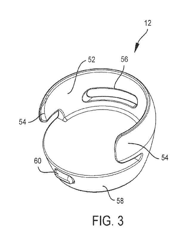
Referring now to FIGS. 2-5 a number of views of an example accessory 12 are
shown.
The accessory 12 includes a housing 50. The housing 50 includes a medical
device receiver
which is shown as a bay 52 that is sized and shaped to receive and retain a
medical device 10 or
portion thereof. In the example embodiment, the bay 52 includes coupling
members which are
depicted as arms 54 located on opposing sides of a docking opening in the
housing 50. The arms
54 may be cantilevered so as to resiliently deflect outward as the medical
device 10 is docked
into the bay 52. A portion of the medical device 10 which is wider than a
distance between the
interior faces of the arms 54 in the resting state may be passed through the
arms 54 and into the
bay 52 while the arms 54 are deflected outwards. The arms 54 may then restore
to a resting state
once the medical device 10 is in place within the bay 52. Thus, the arms 54
may clip onto the
medical device 10 mechanically retaining the accessory 12 in place on the
medical device 10.
Preferably, the resiliency of the arms 54 is chosen to allow retention of the
medical device 10
under some jostling, but also to allow for installation and removal of the
medical device without
excessive force or effort. The housing 50 may include one or more fenestration
56 which may
allow for a user to view or access a portion of the medical device 10.
Fenestrations 56 may for
example be included to allow a line of sight to user interface components of
the medical device
10 such as indicators (e.g. lights) and/or user input components of the
medical device (e.g. a
touchscreen or buttons such as a bolus button on a diabetes management pump).
Fenestrations 56
8
CA 03150523 2022-3-8
WO 2021/050709
PCT/U52020/050174
may also allow a user to access a portion of the medical device 10 to press
against in order to
remove the medical device 10 from the accessory 12.
The housing 50 may also include a second portion 58. The second portion 58 may
contain
one or more of, for example, a battery 24, charging circuitry, a controller
(e.g. microprocessor,
PLC, FPGA, etc.), memory, alarm 26, a wireless communicator 28, and a user
interface 29. As
shown, the accessory 12 also includes a button 60. The button 60 may turn the
accessory 12 on
and off. The button 60 may also be used to convey user input to accessory 12.
For example, the
button 60 may be used to acknowledge and silence (e.g. stop producing one or
more of an
audible, tactile, or visual output) or temporarily snooze an alarm being
generated by the
accessory 12. The accessory 12 may further include a port 62. The port 62 may
be used for data
(e.g. log transfer or medical device updates) or power communication. For
example, the port 62
may be used to charge the battery 24 included in the accessory 12. Any
suitable port 62 may be
used such as a USB, mini-USB, micro-USB, barrel jack, or any proprietary
connector port. In
alternative embodiments, no port 62 may be included. Instead, a battery 24 of
the accessory 12
may be wirelessly charged by a charging mat, platform, stand, or similar item.
Where
embodiments of accessories 12 are shown herein as port 62 free or having a
particular port 62
type, it should be understood that this is merely exemplary. Any type of port
may be used on any
of the embodiments depicted herein and any of the embodiments herein may be
wirelessly
charged. Likewise, any embodiment herein may include a charging port, but also
be capable of
wireless charging.
Referring now to also FIG. 6 and 7, the accessory 12 is respectively depicted
with a
medical device 10 retained therein and with a medical device 10 about to be
docked thereto. As
shown best in FIG. 6, the medical device 10 may be a drug delivery device such
as an
ambulatory infusion pump. The medical device 10 may be attached via tubing 14
to an infusion
set 16 (as shown) or may be a patch type drug delivery device. The medical
device 10 may be
retained on the body with a skin compatible adhesive. The accessory 12 may be
attached to the
medical device 10 while the medical device 10 remains in situ on the user.
Preferably, any edges
of the accessory 12 which may be adjacent to the skin of a patient when
clipped on a medical
device 10 in situ are rounded or blunted so as to be comfortable for a patient
wearing the medical
device 10. As shown in FIG. 6 and 7, the footprint of at least one medical
device receiving
portion of the housing 50 for the accessory 12 may mimic the footprint of the
medical device 10.
9
CA 03150523 2022-3-8
WO 2021/050709
PCT/U52020/050174
In the example embodiment, the medical device 10 is depicted as a disc like
device and the bay
52 of the accessory has a complimentary footprint. Other footprints for
medical devices 10 and
bays 52 are also possible such as various polygonal shapes (e.g. rectangular),
round shapes,
obrounds, or other shapes with both rounded and straight line features. When
retained on the
medical device 10 the arms of the accessory 12 may clip around an edge 64 of
the medical
device 10.
Referring now also to FIG. 8, a cross-sectional view of the accessory 12 shown
in FIG. 2-
7 retained on a medical device 10 is shown. For sake of illustration, only the
power transfer
components of the accessory 12 and medical device 10 are shown and are
depicted in block
diagram form. As shown, the accessory 12 includes a DC power source which is
shown as a
battery 24. The accessory 12 may also include a transmitter circuit 70 which
may include an
inverter for providing AC to a transmitting coil 72 also included in the
accessory 12. The
medical device 10 may include a corresponding receiver coil 74. The receiver
coil 74 may be in
electrical communication with receiver circuit 76 which may include a
rectifier. The receiving
circuit 76 may output direct current to a rechargeable battery 22 of the
medical device 10 to
charge the battery 22.
Another embodiment of an exemplary accessory 12 is depicted in FIGS. 9-12. As
shown,
the housing 50 includes a first portion medical device receiver which is
formed as a pocket. The
pocket is defined by arms 54 which serve as coupling members for retaining the
accessory 12 in
place on the medical device 10. The pocket is also defined by the second
portion 58 (which may
be furnished similarly to as described with respect to FIGS. 2-8) of the
housing 50 and a base
plate 80. The example embodiment also includes a fenestration 56, though
additional
fenestrations 56 may be included in alternative embodiments. The base plate 80
may allow for
the medical device 10 to be surrounded at least partially on all sides. Thus
the medical device 10
may be attached to the accessory 12 when removed from the body. In some
embodiments, a
bottom face of the base plate 80 may include an adhesive region 82 upon which
a skin
compatible adhesive may be applied. Thus the accessory 12 may be adhered to
the patient with
the medical device 10 retained therein.
In some embodiments, the charge rate of the medical device 10 may be altered
as the
medical device 10 is charged. For example, the medical device 10 may be
rapidly charged by the
accessory 12 until the battery level of the medical device 10 reaches a
certain level. For example,
CA 03150523 2022-3-8
WO 2021/050709
PCT/U52020/050174
the medical device 10 may be rapidly recharged until the battery 22 of the
medical device 10
reaches a percentage (e.g. 50% or greater) where the medical device 10 will be
capable of
functioning for a predefined period of time. This may allow a user to quickly
dock the accessory
12 to the medical device 10 to reach an acceptable charge state while
minimizing any disruption
to activities the patient is taking part in. The medical device 10 may be
charged to a full state
when it is more convenient for the user. Thus, convenience may be maximized
without
unnecessarily degrading the battery 22. In some embodiments, the accessory 12
may only rapidly
recharge the battery 22 of the medical device 10 upon receipt of a
communication from the
medical device 10 that the battery 22 included in the medical device 10 is
amenable to a rapid
recharging (of appropriate type and has no related errors or faults).
Another embodiment of an exemplary accessory 12 is depicted in FIGS. 13-15. As
shown, the housing 50 includes a bay 52 that is sized and shaped to receive
and retain a medical
device 10 or portion thereof. In the example embodiment, the bay 52 includes
coupling members
which are depicted as arms 54 located on opposing sides of a docking opening
in the housing 50.
The bay 52 is also defined by a top plate 84. The accessory 12 may be retained
on a medical
device 10 in situ. In the example embodiment, the accessory 12 includes a
second portion 58.
The second portion 58 is disposed laterally to the bay 52 giving the accessory
12 a profile that is
only slightly taller than a medical device 10 retained therein. The second
portion 58 may include
components such as those described in relation to FIGS. 2-8. The example
accessory 12 also
includes a release mechanism 86 which may be actuated by a user to detach the
accessory 12
from the medical device 10. In the example embodiment, the release mechanism
86 includes a
user displaceable button which may drive an extraction finger 90 (see, e.g.,
FIG. 15) into the
medical device 10 when the button is displaced. As best shown in FIG. 15, the
release
mechanism 86 may include a user contact face 88. The user contact face 88 may
include a curved
depression which serves as a pressing surface for a user's finger. The release
mechanism 86 may
also include a hinge 92 which connects the release mechanism 86 to the housing
50 of the
accessory 12. In the example, the hinge 92 is depicted as a living hinge
though a hinge including
a pivot pin about which the release mechanism 86 may displace may be used in
alternative
embodiments. When depressed, the release mechanism 86 may pivot about the
hinge 92 and the
extraction finger 90 may exert a force against the medical device 10. This
force may aid in
11
CA 03150523 2022-3-8
WO 2021/050709
PCT/U52020/050174
displacing the medical device 10 out of clipping engagement with the arms 54
of the accessory
12 so that the accessory 12 may be removed from the medical device 10.
While the example accessory 12 includes a release mechanism 86, other
embodiments
may include similar user actuatabk components which may be operated to
register user inputs to
the medical device 10. Thus, an accessory 12 may include a user interface 29.
For example, in
some embodiments, a user input mechanism may be included in an accessory 12.
The user input
mechanism may be a hinged displaceable component similar to the release
mechanism described
above. A user input mechanism may include an input finger instead of an
extraction finger. Such
an input finger may align with a button or the like included in a medical
device 10 retained
within the accessory 12. When the user input mechanism is actuated (e.g. by a
user's finger) the
input finger may be advanced against the input means included on the medical
device 10. This
may allow for the accessory 12 to be made without fenestrations 56 (see, e.g.
HG. 2-5), while
still allowing operation of buttons covered by the accessory 12 when the
accessory 12 is installed
on the medical device 10.
Referring now to FIGS. 17-20 another exemplary embodiment of an accessory 12
is
depicted. As shown, the housing 50 includes a bay 52 that is sized and shaped
to receive and
retain a medical device 10 or portion thereof. In the example embodiment, the
bay 52 includes
coupling members which are depicted as arms 54 located on opposing sides of a
docking
opening in the housing 50. The bay 52 is also defined by a top plate 84. The
accessory 12 may be
retained on a medical device 10 in situ. In the example embodiment, the
accessory 12 includes a
second portion 58. The second portion 58 is disposed laterally to the bay 52
giving the accessory
a profile that is only slightly taller than a medical device 10 retained
therein. The second portion
58 may include components such as those described in relation to FIGS. 2-8. As
shown, the top
plate 84 includes a fenestration 56.
Referring now to FIGS. 21-26 another exemplary embodiment of an accessory 12
is
depicted. As shown, the housing 50 is cap like and includes a medical device
receiver in the form
of a cavity 100 that is sized and shaped to receive and retain a medical
device 10 or portion
thereof. The accessory 12 also includes a second portion 58 which may include
the components
described above in relation to FIGS. 2-8. In the example embodiment, the
housing 50 includes a
peripheral wall 102 which substantially surrounds the medical device 10 when
retained on the
medical device 10. The peripheral wall 102 may also include a lip 105 at an
edge thereof which
12
CA 03150523 2022-3-8
WO 2021/050709
PCT/U52020/050174
may aid in retention of the accessory 12 on the medical device 10. In some
embodiments, the lip
105 may be sized to interface with a corresponding groove in a medical device
10. The
peripheral wall 102 includes a number of breaks 104. These breaks 104 in the
example
embodiment are spaced at regular angular intervals in the peripheral wall 102.
In the example,
breaks 104 are included about every 60 . In alternative embodiments, breaks
104 may be
included every 45-120' for example. Breaks 104 may also be irregularly spaced.
In embodiments
where the accessory 12 does not have a round footprint, there may, for
example, be at least one
break per side of the peripheral wall 102 of the accessory 12. In the example
embodiment, the
breaks 104 follow a straight line path generally parallel to the height axis
of the accessory 12 and
are cut into the peripheral wall 102 in a direction that is substantially
perpendicular to the height
axis of the accessory 12. It should be appreciated that as used herein, the
term "cut" may, but
does not necessarily mean that a feature is formed via a material removal
process. Features
described as cut out, cut into, etc. a component may be formed during molding,
casting, a
material additive process (e.g. 3D printing), or other manufacturing process
without removal of
material from the component. In other embodiments, the path of the breaks 104
need not follow a
straight line path and may be cut into the peripheral wall 120 at other angles
than that shown.
The breaks 104 may generate a number of wall segments which are cantilevered
to a top
portion of the accessory 12. Each segment may act as a coupling member which
may help to
retain the accessory 12 on a medical device 10. Each cantilevered segment may
resiliently
deflect outward as the medical device 10 is docked into the cavity 100. A
portion of the medical
device 10 which is wider than the opening in the bottom of the accessory 12
afforded by the lip
105 when the segments are in the resting state may be passed into the cavity
100 when the
segments are deflected outwards. The segments may then restore to a resting
state once the
medical device 10 is in place within the cavity 100. Thus, the accessory 12
may clip onto the
medical device 10 mechanically retaining the accessory 12 in place on the
medical device 10. As
indicated in FIG. 25, the medical device 10 may remain operating in situ on a
patient while the
accessory 12 is attached to the medical device 10 by pressing the accessory 12
onto the top of the
medical device 10.
The peripheral wall 102 of the accessory 12 may also have a niche 106. The
niche 106
may be included to accommodate a protrusion such as a tubing connector or
tubing 14 leading
from a medical device 10 to a point beyond the footprint of the accessory 12.
In the example
13
CA 03150523 2022-3-8
WO 2021/050709
PCT/U52020/050174
embodiment, the niche 106 is a cut out having the shape of a Norman window,
however, any
suitable shape may be used. Additionally, one of the breaks 104 in the
peripheral wall 102
extends to the niche 106. This need not be so in all embodiments.
The niche 106 of the example embodiment is flanked on each side by a flange
108. The
flanges 108 are included at a bottom of the peripheral wall 102 in the
example. The flanges 108
may provide a grasping or contact surface to facilitate removal of the
accessory 12 from the
medical device 10. The distance the flanges 108 extend from the peripheral
wall 102 may vary
and in the example increases with proximity to the niche 106.
Referring now to FIG. 27-30 another example accessory is depicted. In this
example
embodiment, the flange 108 is present along the entirety of the peripheral
wall 102 segments
adjacent the niche 106. The distance the flanges 108 project from the
peripheral wall 102 is
variable along a first section and substantially constant along a second
section which is proximal
the niche 106.
Referring now to FIG. 31-34 in an alternative embodiment, a flange 108 may be
included
at a top of the accessory 12. Additionally, breaks 104 in the peripheral wall
102 may only be
included over a portion of the peripheral wall 102. In the example embodiment,
breaks 104 are
included at regular angular intervals over the majority of the peripheral wall
102. The niche 106
is flanked on each side by a break 104 free segment of peripheral wall 102.
Referring now to FIGS. 35-40 another embodiment of an accessory 12 including a
flange
108 which extends from the top of the accessory 12 is shown. The example
embodiments shown
in FIGS. 35-38 also includes a peripheral wall 102 having a curvature. This
curvature may make
the opening in the bottom of the accessory 12 which leads to the cavity 100
smaller than at least
a portion of the medical device 10. Thus, as the accessory 12 is coupled onto
the medical device
10, the peripheral walls 102 may clip around the medical device 10 to retain
the accessory 12 in
place. As in other embodiments, the accessory 12 may include a second portion
58 which may
include components described in relation to FIGS. 2-8. The accessory 12 also
includes a larger
niche 106. The larger niche 106 may allow for some rotation of the accessory
12 relative to the
medical device 10.
Referring now to FIGS. 41-47 another example embodiment of an accessory 12 is
depicted. As shown, the housing 50 of the accessory 12 includes a retention
portion including a
number of clips 110. The housing 50 also includes a second portion 58 which
may include the
14
CA 03150523 2022-3-8
WO 2021/050709
PCT/US2020/050174
components described in relation to FIGS. 2-8. A medical device 10 may include
a number of
receiving recesses for the clips 110 of the accessory 12. In certain examples,
this may allow the
accessory 12 to mount onto the top of the medical device 10 while the medical
device 10 remains
operating in situ. In some embodiments, the accessory 12 may be mounted onto
the medical
device 10 by pressing the accessory 12 against the medical device 10. This may
cause each of the
clips 110 to deflect around a portion of a respective receiving recess in the
medical device 10. As
the accessory 12 is further advanced against the medical device 10, the clips
110 may progress
past an obstructing portion of the receiving recess and restore back to a
resting state. The clips
110 may include a hooked portion 112 which may latch the accessory 12 into
place once
advanced passed the obstructing portion of the receiving recess. As shown, the
housing 50 of the
accessory 12 may include an indentation 114. The indentation 114 may be sized
to allow a
fingertip to reach under the accessory 12 and pry the accessory 12 off of the
medical device 10.
In alternative embodiments, the clips 110 of the accessory 12 may interface
with a
bayonet type mount included in the medical device 10. In such embodiments, the
medical device
10 may include an "L" shaped slot for each of the clips 110. The clips 110 of
the accessory 12
may be advanced into an opening provided by the leg of the "L" shaped slot.
The accessory 12
may then be rotated such that the clips 110 are displaced along the remaining
portion of the slots
to a region of the slots where removal of the clips 110 from the slot is
obstructed. Thus the
accessory 12 may be mechanically coupled to the medical device 10. The
accessory 12 may be
rotated back to a position in which the clips 110 align with the opening
provided by the leg of the
"L" shaped slot to allow for removal of the accessory 12. In some embodiments,
the "L" shaped
slot may include a serifed portion in which the clips 110 reside when the
accessory 12 is
mechanically coupled to the medical device 10. A user may be required to press
down on the
accessory 12 to advance the clips 110 out of the serifed portion before
rotation to remove the
accessory 12 from the medical device 10 may be possible.
Referring now to FIGS. 48-51, another example accessory 12 is depicted. As
shown, the
housing 50 of the accessory 12 includes a retention portion including a
pivoting retention
member 120. The housing 50 also includes a second portion 58 which may include
the
components described in relation to FIGS. 2-8. The pivoting retention member
120 shown has an
arcuate shape and includes pins 122 included at terminal ends thereof. The
pins 122 may extend
into bearings included on a remaining portion of the housing 50. During
mechanical coupling of
CA 03150523 2022-3-8
WO 2021/050709
PCT/U52020/050174
the accessory 12 to the medical device 10, the pins 122 may allow the
retention member 120 to
pivot from a receiving position to a retaining position. In the receiving
position, the retention
member 120 may be pivoted over the top of the housing 50 allowing the housing
50 to be placed
onto the medical device 10 without removing the medical device 10 from a
patient. Once in
place, the retention member 120 of the accessory 12 may be displaced to the
retaining position
(shown in FIGS. 48-51). When in the retaining position, the retention member
120 may clip in
place around the medical device 10 holding the accessory 12 in place on the
medical device 10.
The retention member 120 may also include a flange 124. The flange 124 may be
included to
facilitate grasping and actuation by a user of the accessory 12. The housing
50 also includes a
notch 126. The notch 126 may allow access to user interface component of a
medical device 10
similarly to the fenestrations 56 described elsewhere herein.
Referring now to FIGS. 52-55, another example accessory 12 including a pivotal
retention member 120 is shown. The example embodiment depicted in FIGS. 52-55
is configured
to latch around the medical device 10 when installed on the medical device 10.
As best shown in
the detailed view of FIG. 53, the retention member 120 may include a
projection 128. When the
retention member 120 is in the retaining position, the projection 128 may
engage with a detent
130 included on another portion of the housing 50. This may aid in holding the
retention member
120 in the retaining position and keep the accessory 12 from being
inadvertently removed from
the medical device 10 as a user moves about while sleeping for example.
Referring now to FIGS. 56-60, another embodiment of an accessory 12 is shown.
As
shown, the accessory 12 may have a clip 142 which may be disposed on the
second portion 58.
The second portion 58 may also include the components described in relation to
FIGS. 2-8. The
second portion 58 may be pivotally coupled to the first portion 140 by a pivot
pin 144. The first
portion 140 may be pivoted to a loading position in which the medical device
10 may be placed
into the accessory 12. From the loading position, the first portion 140 may be
pivoted to a
retaining position in which the medical device 10 is held in place within the
accessory 12. A
latching arrangement similar to that shown in FIGS. 52-55 may be included to
help hold the first
portion 140 in place in the retaining position. The first portion 140 or
second portion 58 of the
accessory 12 may include a cradle 146 in which the medical device 100 may be
installed. As
shown in FIG. 59, the medical device 10 is shown in place in a cradle 146 of
the first portion
140. The cradle 146 may include one or more wings 148 which may surround at
least a portion
16
CA 03150523 2022-3-8
WO 2021/050709
PCT/U52020/050174
of the medical device 10 when the medical device 10 is installed in the cradle
146. Once the
medical device 10 is positioned in the cradle 146, the first portion 140 of
the accessory 12 may
be pivoted toward the second portion 58 of the accessory 12. This may sandwich
the medical
device 10 within the accessory 12 as shown in FIG. 60 for instance. The clip
142 may be
attached to a belt or waistband of a user allowing the medical device 10 to
operate with the
accessory 12 while being carried by the patient.
An alternative embodiment of the accessory 12 shown in FIGS. 56-60 is depicted
in
FIGS. 61-65. As shown the first portion 140 and second portion 58 are attached
to each other via
a living hinge 150. Thus, the first portion 140 may be displaced relative to
the second portion 58
without the need for a pivot pin 144 (see, e.g. FIGS. 56-60). Additionally, no
latching
arrangement may be included as the living hinge 150 may be constructed with
sufficient
resiliency to avoid inadvertent deflection once the medical device 10 has been
installed within
the accessory 12.
In another alternative embodiment as shown in FIGS. 66-69, an accessory 12
with a
housing 50 having a medical device receiver which is shown as a bay 52 that is
sized and shaped
to receive and retain a medical device 10 or portion thereof. In the example
embodiment, the bay
52 includes coupling members which are depicted as arms 54 located on opposing
sides of a
docking opening in the housing 50. The arms 54 may be contoured to cradle the
medical device
10 when the medical device 10 is placed in the accessory 12. As shown, the
accessory 12 may
include a clip 142 which may be disposed on the second portion 58. The second
portion 58 may
also include the components described in relation to FIGS. 2-8. The clip 142
may facilitate
attachment of the accessory 12 to a belt or waistband of a user. Additionally,
the clip 142 may
ensure that the arms 54 of the bay 52 are positioned such that the medical
device 10 may be
holstered in place within the accessory 12 by force of gravity.
In another embodiment and referring now to FIGS. 70-72, the accessory 12 may
include a
belt 160. Similar components such as strap(s) or slings which facilitate ease
of wearing the
accessory 12 may be included in various embodiments. As shown, the belt 160
may include a
buckle portion 162. The buckle portion 162 may contain one or more of, for
example, a battery
24, charging circuitry, a controller (e.g. microprocessor, PLC, FPGA, etc.),
memory, alarm 26,
and a wireless communicator 28. Additionally, the buckle portion 162 may
include a slot 164.
The slot 164 may be sized to accept the medical device 10. In some
embodiments, the arms 54
17
CA 03150523 2022-3-8
WO 2021/050709
PCT/U52020/050174
such as those depicted in FIGS. 9-12 may be included to aid in retaining the
medical device 10
within the slot 164.
Referring now to FIGS. 73-74 another embodiment of an accessory 12 is
depicted. As
shown, the accessory 12 may include a pad 170 having a depression 172 sized to
accept a
medical device 10. The depression 172 may include a number of magnets 174
which may couple
to magnets included in the medical device 10. Additionally, the pad 170 may
contain one or
more of, for example, a battery 24, charging circuitry, a controller (e.g.
microprocessor, PLC,
FPGA, etc.), memory, alarm 26, and a wireless communicator 28. The pad 170 may
further
include a port 62 which may be used for data (e.g. log transfer or medical
device updates) or
power communication. A USB type cable 178 is depicted coupled into the port 62
in FIG. 74. An
indicator light 176 is included on the pad 170 and may illuminate based on
status of the
accessory 12 and/or medical device 10. For example, the indicator light 176
may illuminate a
first color to indicate that the medical device 10 is being wirelessly
charged. The indicator light
may blink to indicate a low battery 24 in the accessory 12. The indicator
light 176 may
illuminate a second color in the event of an alarm encountered by the medical
device 10 or
accessory 12.
Referring now to FIG. 75, another exemplary accessory 12 is depicted. The
accessory 12
depicted is similar to that shown in FIGS. 2-8, however, the accessory 12
includes a set of
conductive contacts 180. When a medical device 10 is placed within the
accessory 12, the
conductive contacts 180 may interface with conductive zones included on the
medical device 10.
This may allow for the accessory 12 to interface with the medical device 10
for charging
purposes.
Referring now to FIGS. 76-77, another example accessory is depicted. As shown,
the accessory
includes a user interface 29. The user interface 29 may be a touch screen
display, though any
other suitable type of display may be included. A home screen of the user
interface 29 is depicted
in FIG. 77. The home screen may include information like a patient's current
insulin on board
(LOB). The home screen may also include a last reading from a glucose monitor
such as a CGM.
Other information like the date, a battery remaining indicia, and a medication
remaining indicia
may be included_ The user may also be able to access information about a last
bolus or blood
glucose trend data from the home screen. A bolus button may be included on the
home screen.
Additionally, the home screen may include a menu button which may be used to
access various
18
CA 03150523 2022-3-8
WO 2021/050709
PCT/U52020/050174
settings, program infusion profiles, view historical patient data, access
tutorials, etc. The user
interface 29 may show any number of other screens allowing a user to program
and use the
medical device 10. A number of example screens which may be generated for
presentation on the
user interface 29 are described in greater detail in: U.S. Patent No.
9,132,227, issued September
15, 2015 and entitled Methods and Systems for Controlling an Infusion Pump
(Attorney Docket
No. G98); U.S. Patent No. 9,656,031, issued May 23, 2017 and entitled Infusion
Pump Methods
and Systems (Attorney Docket No. 106); U.S. Patent No. 9,662,438, issued May
30, 2017 and
entitled Devices, Methods and Systems for Wireless Control of Medical Devices
(Attorney
Docket No. 198); U.S. Patent No. 10,238,794, issued March 26, 2019 and
entitled Devices,
Methods and Systems for Wireless Control of Medical Devices (Attorney Docket
No. K11); and
U.S. Patent No. 10,195,343, issued February 5, 20109 and entitled Devices,
Methods and
Systems for Wireless Control of Medical Devices (Attorney Docket No. L72),
each of which is
incorporated herein by reference in its entirety.
Various alternatives and modifications can be devised by those skilled in the
art without
departing from the disclosure. Accordingly, the present disclosure is intended
to embrace all
such alternatives, modifications and variances. Additionally, while several
embodiments of the
present disclosure have been shown in the drawings and/or discussed herein, it
is not intended
that the disclosure be limited thereto, as it is intended that the disclosure
be as broad in scope as
the art will allow and that the specification be read likewise. Therefore, the
above description
should not be construed as limiting, but merely as exemplifications of
particular embodiments.
And, those skilled in the art will envision other modifications within the
scope and spirit of the
claims appended hereto. Other elements, steps, methods and techniques that are
insubstantially
different from those described above and/or in the appended claims are also
intended to be
within the scope of the disclosure.
The embodiments shown in drawings are presented only to demonstrate certain
examples
of the disclosure. And, the drawings described are only illustrative and are
non-limiting. In the
drawings, for illustrative purposes, the size of some of the elements may be
exaggerated and not
drawn to a particular scale. Additionally, elements shown within the drawings
that have the
same numbers may be identical elements or may be similar elements, depending
on the context.
Where the term "comprising" is used in the present description and claims, it
does not
exclude other elements or steps. Where an indefinite or definite article is
used when referring to
19
CA 03150523 2022-3-8
WO 2021/050709
PCT/U52020/050174
a singular noun, e.g. "a" "an" or "the", this includes a plural of that noun
unless something
otherwise is specifically stated. Hence, the term "comprising" should not be
interpreted as being
restricted to the items listed thereafter, it does not exclude other elements
or steps, and so the
scope of the expression "a device comprising items A and B" should not be
limited to devices
consisting only of components A and B.
Furthermore, the terms "first", "second", "third" and the like, whether used
in the
description or in the claims, are provided for distinguishing between similar
elements and not
necessarily for describing a sequential or chronological order. It is to be
understood that the
terms so used are interchangeable under appropriate circumstances (unless
clearly disclosed
otherwise) and that the embodiments of the disclosure described herein are
capable of operation
in other sequences and/or arrangements than are described or illustrated
herein.
While the principles of the invention have been described herein, it is to be
understood by
those skilled in the art that this description is made only by way of example
and not as a limitation
as to the scope of the invention. Other embodiments are contemplated within
the scope of the
present invention in addition to the exemplary embodiments shown and described
herein.
Modifications and substitutions by one of ordinary skill in the art are
considered to be within the
scope of the present invention.
CA 03150523 2022-3-8