Note: Descriptions are shown in the official language in which they were submitted.
CA 03150788 2022-02-11
WO 2021/030092 PCT/US2020/044801
SHAPE-SENSING SYSTEMS AND METHODS FOR MEDICAL DEVICES
PRIORITY
[0001] This application claims the benefit of priority to U.S.
Provisional Application
No. 62/885,702, filed August 12, 2019, which is incorporated by reference in
its entirety into
this application.
BACKGROUND
[0002] At times, a tip of a peripherally inserted central catheter
("PICC") or central
venous catheter ("CVC") can move becoming displaced from an ideal position in
a patient's
superior vena cava ("SVC"). A clinician believing such a PICC or CVC has
displaced typically
checks for displacement by chest X-ray and replaces the PICC or CVC if
necessary. However,
X-rays expose patients to ionizing radiation. Therefore, there is a need for
clinicians to easily
and safely check for displacement of PICCs and CVCs for replacement thereof if
necessary.
[0003] Disclosed herein are shape-sensing systems and methods for medical
devices
that address the foregoing.
SUMMARY
[0004] Disclosed herein is a shape-sensing system for medical devices
including, in
some embodiments, a medical device, an optical interrogator, a console, and a
display screen.
The medical device includes a body of implementation including an optical
fiber, wherein the
optical fiber is comprised of a number of fiber Bragg grating ("FBG") sensors
along at least a
distal-end portion of the optical-fiber. One embodiment of the body of
implementation, as will
be discussed primarily throughout the disclosure, is an optical-fiber
integrated stylet. However,
other embodiments of body of implementation include, but are not limited to,
an integrated
optical-fiber guidewire, or an integrated optical-fiber catheter. The optical
interrogator is
configured to send input optical signals into the optical-fiber stylet and
receive FBG sensor-
reflected optical signals from the optical-fiber stylet. The console includes
memory and one or
more processors configured to convert the FBG sensor-reflected optical signals
from the
optical-fiber stylet into plottable data by way of a number of optical signal-
converter logic,
which may include one or more algorithms. The display screen is configured for
displaying
any plot of a number of plots of the plottable data. The number of plots
include at least a plot
of curvature vs. time for each FBG sensor of a selection of the FBG sensors in
the distal-end
-1-
CA 03150788 2022-02-11
WO 2021/030092 PCT/US2020/044801
portion of the optical-fiber stylet for identifying a distinctive change in
strain of the optical-
fiber stylet at a moment a tip of the medical device is advanced into an SVC
of a patient.
[0005] In some embodiments, the shape-sensing system further includes an
SVC-
determiner algorithm configured to automatically determine the distinctive
change in the strain
of the optical-fiber stylet at the moment the tip of the medical device is
advanced into the SVC
of the patient. The distinctive change in the strain is an instantaneous
increase in the strain
followed by an instantaneous decrease in the strain.
[0006] In some embodiments, the SVC-determiner algorithm is configured to
confirm
the tip of the medical device is in the SVC by way of periodic changes in the
strain of the
optical-fiber stylet sensed by the selection of the FBG sensors. The periodic
changes in the
strain result from periodic changes in blood flow within the SVC as a heart of
the patient beats.
[0007] In some embodiments, the shape-sensing system further includes an
optical-
fiber connector module configured to establish a first optical connection from
the medical
device to the optical-fiber connection module and a second optical connection
from the optical-
fiber connection module to the optical interrogator. The first optical
connection is through a
sterile drape with the medical device in a sterile field defined by the
sterile drape and the
optical-fiber connector module in a non-sterile field defined by the sterile
drape.
[0008] In some embodiments, the optical-fiber connector module includes
one or more
sensors selected from a gyroscope, an accelerometer, and a magnetometer. The
one or more
sensors are configured to provide sensor data to the console over one or more
data wires for
algorithmically determining a reference plane for shape sensing with the
optical-fiber stylet.
[0009] In some embodiments, the optical interrogator is an integrated
optical
interrogator integrated into the console.
[0010] In some embodiments, the display screen is an integrated display
screen
integrated into the console.
[0011] Also disclosed herein is a method for determining a tip of a
medical device is
located within an SVC of a patient. The method includes, in some embodiments,
advancing the
tip of the medical device through a vasculature of the patient toward the SVC.
The medical
device includes an integrated optical-fiber stylet having a number of FBG
sensors along at least
-2-
CA 03150788 2022-02-11
WO 2021/030092 PCT/US2020/044801
a distal-end portion of the optical-fiber stylet for shape sensing with a
shape-sensing system
including the medical device. The method also includes enabling input optical
signals (e.g.,
broadband incident light) to be sent into the optical-fiber stylet while
advancing the tip of the
medical device through the vasculature of the patient. In one embodiment, the
broadband
incident light is provided by a light source which may be a tunable swept
laser, although other
suitable light sources can also be employed in addition to a laser, including
semi-coherent light
sources, LED light sources, etc. The method also includes enabling FBG sensor-
reflected
optical signals to be received from the optical-fiber stylet while advancing
the tip of the medical
device through the vasculature of the patient. The method also includes
identifying on a display
screen of the shape-sensing system a distinctive change in strain of the
optical-fiber stylet
sensed by a selection of the FBG sensors in the distal-end portion of the
optical-fiber stylet at
a moment the tip of the medical device is advanced into the SVC, thereby
determining the tip
of the medical device is located within the SVC.
[0012] In some embodiments, the method further includes enabling the FBG
sensor-
reflected optical signals received from the optical-fiber stylet to be
algorithmically converted
into a number of different plots for display on the display screen.
[0013] In some embodiments, each plot of the number of different plots is
selected from
a plot of curvature vs. arc length, a plot of torsion vs. arc length, a plot
of angle vs. arc length,
and a plot of position vs. time for at least the distal-end portion of the
optical-fiber stylet.
[0014] In some embodiments, the number of different plots includes a plot
of curvature
vs. time for each FBG sensor selected from the FBG sensors of the optical-
fiber stylet.
[0015] In some embodiments, the method further includes enabling the FBG
sensor-
reflected optical signals received from the optical-fiber stylet to be
algorithmically converted
into a displayable shape for the medical device for display on the display
screen.
[0016] In some embodiments, the distinctive change in the strain of the
optical-fiber
stylet is an instantaneous increase in a plotted curvature of the optical-
fiber stylet followed by
an instantaneous decrease in the plotted curvature.
[0017] In some embodiments, a magnitude of the instantaneous decrease in
the plotted
curvature of the optical-fiber stylet is about twice that of the instantaneous
increase in the
plotted curvature.
-3-
CA 03150788 2022-02-11
WO 2021/030092 PCT/US2020/044801
[0018] In some embodiments, the selection of the FBG sensors is a last
three FBG
sensors in the distal-end portion of the optical-fiber stylet.
[0019] In some embodiments, the method further includes ceasing to
advance the tip
of the medical device through the vasculature of the patient after determining
the tip of the
medical device is located in the SVC. The method also includes confirming the
tip of the
medical device is in the SVC by way of periodic changes in the strain of the
optical-fiber stylet
sensed by the selection of the FBG sensors. The periodic changes in the strain
result from
periodic changes in blood flow within the SVC as a heart of the patient beats.
[0020] In some embodiments, advancing the tip of the medical device
through the
vasculature of the patient includes advancing the tip of the medical device
through a right
internal jugular vein, a right brachiocephalic vein, and into the SVC.
[0021] In some embodiments, the medical device is a CVC.
[0022] In some embodiments, advancing the tip of the medical device
through the
vasculature of the patient includes advancing the tip of the medical device
through a right
basilic vein, a right axillary vein, a right subclavian vein, a right
brachiocephalic vein, and into
the SVC.
[0023] In some embodiments, the medical device is a peripherally inserted
central
catheter (PIC C).
[0024] Also disclosed herein is a method for determining a tip of a
medical device is
located within an SVC of a patient. The method includes, in some embodiments,
advancing the
tip of the medical device through a vasculature of the patient toward the SVC.
The medical
device includes an integrated optical-fiber stylet having a number of FBG
sensors along at least
a distal-end portion of the optical-fiber stylet for shape sensing with a
shape-sensing system
including the medical device. The method also includes enabling input optical
signals to be
sent into the optical-fiber stylet while advancing the tip of the medical
device through the
vasculature of the patient. The method also includes enabling FBG sensor-
reflected optical
signals to be received from the optical-fiber stylet while advancing the tip
of the medical device
through the vasculature of the patient. The method also includes enabling the
FBG sensor-
reflected optical signals received from the optical-fiber stylet to be
algorithmically converted
into a plot of curvature vs. time for each FBG sensor of the FBG sensors. The
method also
-4-
CA 03150788 2022-02-11
WO 2021/030092 PCT/US2020/044801
includes identifying on a display screen of the shape-sensing system an
instantaneous increase
in strain of the optical-fiber stylet followed by an instantaneous decrease in
the strain as sensed
by each FBG sensor of a last three FBG sensors in the distal-end portion of
the optical-fiber
stylet at a moment the tip of the medical device is advanced into the SVC,
thereby determining
the tip of the medical device is located within the SVC. The method also
includes confirming
the tip of the medical device is in the SVC by way of periodic changes in the
strain of the
optical-fiber stylet as sensed by the last three FBG sensors in the distal-end
portion of the
optical-fiber stylet. The periodic changes in the strain result from periodic
changes in blood
flow within the SVC as a heart of the patient beats.
[0025] These and other features of the concepts provided herein will
become more
apparent to those of skill in the art in view of the accompanying drawings and
following
description, which describe particular embodiments of such concepts in greater
detail.
DRAWINGS
[0026] FIG. 1 is a block diagram of a first shape-sensing system in
accordance with
some embodiments.
[0027] FIG. 2 is a block diagram of a second shape-sensing system in
accordance with
some embodiments.
[0028] FIG. 3 illustrates the second shape-sensing system in accordance
with some
embodiments.
[0029] FIG. 4A illustrates a transverse cross-section of a catheter tube
of a medical
device in accordance with some embodiments.
[0030] FIG. 4B illustrates a longitudinal cross-section of the catheter
tube of the
medical device in accordance with some embodiments.
[0031] FIG. 5 illustrates a detailed section of an optical-fiber
connector module in
accordance with some embodiments.
[0032] FIG. 6 illustrates the second shape-sensing system with a first
optical-fiber
connector module in accordance with some embodiments.
-5-
CA 03150788 2022-02-11
WO 2021/030092 PCT/US2020/044801
[0033] FIG. 7 illustrates the second shape-sensing system with the first
optical-fiber
connector module within a fenestration of a surgical drape in accordance with
some
embodiments.
[0034] FIG. 8 illustrates the second shape-sensing system with a second
optical-fiber
connector module in accordance with some embodiments.
[0035] FIG. 9 illustrates the second shape-sensing system with the second
optical-fiber
connector module beneath a surgical drape in accordance with some embodiments.
[0036] FIG. 10 provides a number of different plots on a display screen
of a shape-
sensing system in accordance with some embodiments.
[0037] FIG. 11 provides a detailed plot of curvature vs. arc length and
torsion vs. arc
length for at least a distal-end portion of an optical-fiber stylet as one of
the plots of FIG. 10.
[0038] FIG. 12 provides a detailed plot of angle vs. arc length for at
least a distal-end
portion of an optical-fiber stylet as one of the plots of FIG. 10.
[0039] FIG. 13 provides a detailed plot of position vs. time for at least
a distal-end
portion of an optical-fiber stylet as one of the plots of FIG. 10.
[0040] FIG. 14 provides a displayable shape for at least a distal-end
portion of a
medical device or an optical-fiber stylet in accordance with some embodiments.
[0041] FIG. 15 provides detailed plots of curvature vs. time for each FBG
sensor
selected from a number of FBG sensors of an optical-fiber stylet as some of
the plots of FIG.
10.
DESCRIPTION
[0042] Before some particular embodiments are disclosed in greater
detail, it should be
understood that the particular embodiments disclosed herein do not limit the
scope of the
concepts provided herein. It should also be understood that a particular
embodiment disclosed
herein can have features that can be readily separated from the particular
embodiment and
optionally combined with or substituted for features of any of a number of
other embodiments
disclosed herein.
-6-
CA 03150788 2022-02-11
WO 2021/030092 PCT/US2020/044801
[0043] Regarding terms used herein, it should also be understood the
terms are for the
purpose of describing some particular embodiments, and the terms do not limit
the scope of the
concepts provided herein. Ordinal numbers (e.g., first, second, third, etc.)
are generally used to
distinguish or identify different features or steps in a group of features or
steps, and do not
supply a serial or numerical limitation. For example, "first," "second," and
"third" features or
steps need not necessarily appear in that order, and the particular
embodiments including such
features or steps need not necessarily be limited to the three features or
steps. Labels such as
"left," "right," "top," "bottom," "front," "back," and the like are used for
convenience and are
not intended to imply, for example, any particular fixed location,
orientation, or direction.
Instead, such labels are used to reflect, for example, relative location,
orientation, or directions.
Singular forms of "a," "an," and "the" include plural references unless the
context clearly
dictates otherwise.
[0044] With respect to "proximal," a "proximal portion" or a "proximal
end portion"
of, for example, a catheter disclosed herein includes a portion of the
catheter intended to be
near a clinician when the catheter is used on a patient. Likewise, a "proximal
length" of, for
example, the catheter includes a length of the catheter intended to be near
the clinician when
the catheter is used on the patient. A "proximal end" of, for example, the
catheter includes an
end of the catheter intended to be near the clinician when the catheter is
used on the patient.
The proximal portion, the proximal end portion, or the proximal length of the
catheter can
include the proximal end of the catheter; however, the proximal portion, the
proximal end
portion, or the proximal length of the catheter need not include the proximal
end of the catheter.
That is, unless context suggests otherwise, the proximal portion, the proximal
end portion, or
the proximal length of the catheter is not a terminal portion or terminal
length of the catheter.
[0045] With respect to "distal," a "distal portion" or a "distal end
portion" of, for
example, a catheter disclosed herein includes a portion of the catheter
intended to be near or in
a patient when the catheter is used on the patient. Likewise, a "distal
length" of, for example,
the catheter includes a length of the catheter intended to be near or in the
patient when the
catheter is used on the patient. A "distal end" of, for example, the catheter
includes an end of
the catheter intended to be near or in the patient when the catheter is used
on the patient. The
distal portion, the distal end portion, or the distal length of the catheter
can include the distal
end of the catheter; however, the distal portion, the distal end portion, or
the distal length of
the catheter need not include the distal end of the catheter. That is, unless
context suggests
-7-
CA 03150788 2022-02-11
WO 2021/030092 PCT/US2020/044801
otherwise, the distal portion, the distal end portion, or the distal length of
the catheter is not a
terminal portion or terminal length of the catheter.
[0046] The term "logic" may be representative of hardware, firmware or
software that
is configured to perform one or more functions. As hardware, the term logic
may refer to or
include circuitry having data processing and/or storage functionality.
Examples of such
circuitry may include, but are not limited or restricted to a hardware
processor (e.g.,
microprocessor, one or more processor cores, a digital signal processor, a
programmable gate
array, a microcontroller, an application specific integrated circuit "ASIC",
etc.), a
semiconductor memory, or combinatorial elements.
[0047] Additionally, or in the alternative, the term logic may refer to
or include
software such as one or more processes, one or more instances, Application
Programming
Interface(s) (API), subroutine(s), function(s), applet(s), servlet(s),
routine(s), source code,
object code, shared library/dynamic link library (dll), or even one or more
instructions. This
software may be stored in any type of a suitable non-transitory storage
medium, or transitory
storage medium (e.g., electrical, optical, acoustical or other form of
propagated signals such as
carrier waves, infrared signals, or digital signals). Examples of a non-
transitory storage medium
may include, but are not limited or restricted to a programmable circuit; non-
persistent storage
such as volatile memory (e.g., any type of random access memory "RAM"); or
persistent
storage such as non-volatile memory (e.g., read-only memory "ROM", power-
backed RAM,
flash memory, phase-change memory, etc.), a solid-state drive, hard disk
drive, an optical disc
drive, or a portable memory device. As firmware, the logic may be stored in
persistent storage.
[0048] Unless defined otherwise, all technical and scientific terms used
herein have the
same meaning as commonly understood by those of ordinary skill in the art.
[0049] As set forth above, there is a need for clinicians to easily and
safely check for
displacement of PICCs and CVCs for replacement thereof if necessary. Disclosed
herein are
shape-sensing system and methods for medical devices that address the
foregoing.
[0050] For example, a shape-sensing system can include a medical device,
an optical
interrogator, a console, and a display screen. In one embodiment, the medical
device includes
an integrated optical-fiber stylet having FBG sensors along at least a distal-
end portion of the
optical-fiber stylet. As noted above, alternatives to an optical-fiber stylet
include, but are not
limited or restricted to, an optical-fiber integrated guideway or an optical-
fiber integrated
-8-
CA 03150788 2022-02-11
WO 2021/030092 PCT/US2020/044801
guidewire. The optical interrogator is configured to send input optical
signals (e.g., broadband
incident light) into the optical-fiber stylet and receive FBG sensor-reflected
optical signals
therefrom.
[0051] In some embodiments, the optical-fiber stylet is configured to
return
information for use in identifying its physical state (e.g., shape length,
shape, and/or form) of
(i) a portion of the stylet (e.g., tip, segment of stylet, etc.) or a portion
of a catheter inclusive of
at least a portion of the stylet (e.g., tip, segment of catheter, etc.) or
(ii) the entirety or a
substantial portion of the stylet or catheter within the body of a patient
(hereinafter, described
as the "physical state of the stylet" or the "physical state of the
catheter"). According to one
embodiment of the disclosure, the returned information may be obtained from
reflected light
signals of different spectral widths, where each reflected light signal
corresponds to a portion
of broadband incident light propagating along a core of the optical fiber
(hereinafter, "core
fiber") that is reflected back over the core fiber by a particular sensor
located on the core fiber.
One illustrative example of the returned information may pertain to a change
in signal
characteristics of the reflected light signal returned from the sensor, where
wavelength shift is
correlated to (mechanical) strain on the core fiber. It should be understood
that the optical fiber
may include or more cores, where an optical fiber including a plurality of
cores is referred to
as a "multi-core optical fiber."
[0052] In some embodiments in which the stylet includes a multi-core
optical fiber,
each core fiber utilizes a plurality of sensors and each sensor is configured
to reflect a different
spectral range of the incident light (e.g., different light frequency range).
Based on the type and
degree of strain asserted on the each core fiber, the sensors associated with
that core fiber may
alter (shift) the wavelength of the reflected light to convey the type and
degree of stain on that
core fiber at those locations of the stylet occupied by the sensors. The
sensors are spatially
distributed at various locations of the core fiber between a proximal end and
a distal end of the
stylet so that shape sensing of the stylet may be conducted based on analytics
of the wavelength
shifts. In some embodiments, the shape sensing functionality is paired with
the ability to
simultaneously pass an electrical signal through the same member (stylet)
through conductive
medium included as part of the stylet.
[0053] More specifically, in some embodiments each core fiber of the
multi-core
optical fiber is configured with an array of sensors, which are spatially
distributed over a
prescribed length of the core fiber to generally sense external strain those
regions of the core
-9-
CA 03150788 2022-02-11
WO 2021/030092 PCT/US2020/044801
fiber occupied by the sensor. Given that each sensor positioned along the same
core fiber is
configured to reflect light of a different, specific spectral width, the array
of sensors enable
distributed measurements throughout the prescribed length of the multi-core
optical fiber.
These distributed measurements may include wavelength shifts having a
correlation with strain
experienced by the sensor.
[0054] During operation, multiple light reflections (also referred to as
"reflected light
signals") are returned to the console from each of the plurality of core
fibers of the multi-core
optical fiber. Each reflected light signal may be uniquely associated with a
different spectral
width. Information associated with the reflected light signals may be used to
determine a three-
dimensional representation of the physical state of the stylet within the body
of a patient. The
core fibers may be spatially separated with the cladding of the optical fiber
and each core fiber
is configured to separately return light of different spectral widths (e.g.,
specific light
wavelength or a range of light wavelengths) reflected from the distributed
array of sensors
fabricated in each of the core fibers. A comparison of detected shifts in
wavelength of the
reflected light returned by a center core fiber (operating as a reference) and
the surrounding,
periphery core fibers may be used to determine the physical state of the
stylet.
[0055] During vasculature insertion and advancement of the catheter, the
clinician may
rely on the console to visualize a current physical state (e.g., shape) of a
catheter guided by the
stylet to avoid potential path deviations. As the periphery core fibers reside
at spatially different
locations within the cladding of the multi-mode optical fiber, changes in
angular orientation
(such as bending with respect to the center core fiber, etc.) of the stylet
impose different types
(e.g., compression or tension) and degrees of strain on each of the periphery
core fibers as well
as the center core fiber. The different types and/or degree of strain may
cause the sensors of
the core fibers to apply different wavelength shifts, which can be measured to
extrapolate the
physical state of the stylet (catheter).
[0056] The console is configured to convert the reflected optical signals
into plottable
data for displaying plots thereof on the display screen. The plots include a
plot of curvature vs.
time for each FBG sensor of a selection of the FBG sensors in the distal-end
portion of the
optical-fiber stylet for identifying a distinctive change in strain of the
optical-fiber stylet as a
tip of the medical device is advanced into a superior vena cava of a patient.
CA 03150788 2022-02-11
WO 2021/030092 PCT/US2020/044801
[0057] The console may further be configured to receive one or more
electrical signals
from the stylet, which as referenced above, may be configured to support both
optical
connectivity as well as electrical connectivity. The electrical signals may be
processed by logic
of the console, while be executed by the processor, to determine ECG waveforms
for display.
[0058] These and other features of the shape-sensing systems and methods
provided
herein will become more apparent with reference to the accompanying drawings
and the
following description, which provide particular embodiments of the shape-
sensing systems and
methods thereof in greater detail.
Shape-sensing systems
[0059] FIG. 1 is a block diagram of a first shape-sensing system 100 in
accordance with
some embodiments. FIG. 2 is a block diagram of a second shape-sensing system
200 in
accordance with some embodiments. FIG. 3 illustrates the second shape-sensing
system 200 in
accordance with some embodiments. FIG. 10 provides a display screen 150 or 250
of the shape-
sensing system 100 or 200 in accordance with some embodiments. FIGS. 11-15
provide
detailed plots of a number of different plots on the display screen 150 or 250
of FIG. 10.
[0060] As shown, the shape-sensing system 100 includes a medical device
110, a stand-
alone optical interrogator 130, a console 140, and a display screen 150 such
as that of a stand-
alone monitor. The shape-sensing system 200 includes the medical device 110,
an integrated
optical interrogator 230, a console 240, and an integrated display screen 250,
wherein both the
integrated optical interrogator 230 and the integrated display screen 250 are
integrated into the
console 240. Each shape-sensing system of the shape-sensing systems 100 and
200 can further
include an optical-fiber connector module 120 configured for connecting the
medical device
110 to a remainder of the shape-sensing system 100 or 200 such as the optical
interrogator 130
or the console 240, which includes the integrated optical interrogator 230.
[0061] As set forth in more detail below, the medical device 110 includes
an integrated
optical-fiber stylet having a number of FBG sensors along at least a distal-
end portion of the
optical-fiber stylet for shape sensing with the shape-sensing system 100 or
200. (See integrated
the optical-fiber stylet 424 in FIG. 4B for an example of the integrated
optical-fiber stylet of
the medical device 110.)
-11-
CA 03150788 2022-02-11
WO 2021/030092 PCT/US2020/044801
[0062] Certain features of the medical device 110 are set forth in more
detail below
with respect to particular embodiments of the medical device 110 such as the
PICC 310. That
said, some features set forth below with respect to one or more embodiments of
the medical
device 110 are shared among two or more embodiments of the medical device 110.
As such,
"the medical device 110" is used herein to generically refer to more than one
embodiment of
the medical device 110 when needed for expository expediency. This is despite
certain features
having been described with respect to particular embodiments of the medical
device 110 such
as the PICC 310.
[0063] While only shown for the console 240, each console of the consoles
140 and
240 includes one or more processors 242 and memory 244 including a number of
algorithms
246 such as one or more optical signal-converter algorithms. The one or more
optical signal-
converter algorithms are configured to convert FBG sensor-reflected optical
signals from the
optical-fiber stylet of the medical device 110 into plottable data for
displayable shapes
corresponding to the medical device 110. The one or more optical signal-
convertor algorithms
are also configured to convert the reflected optical signals from the optical-
fiber stylet of the
medical device 110 into plottable data for a number of other plots of the
plottable data. The
display screen 150 or 250 is configured to display the displayable shapes for
the medical device
110 over a 3-dimensional grid 1002 or any plot of the number of plots of the
other plottable
data.
[0064] More specifically, in some embodiments, the algorithms 246 may
include shape
sensing logic configured to compare wavelength shifts measured by sensors
deployed in each
outer core fiber at the same measurement region of the stylet, catheter or
guidewire (or same
spectral width) to the wavelength shift at the center core fiber positioned
along central axis and
operating as a neutral axis of bending. From these analytics, the shape
sensing logic may
determine the shape the core fibers have taken in 3D space and may further
determine the
current physical state of the stylet, catheter or guidewire in 3D space for
rendering on the
display 150 or 250.
[0065] Referring to FIG. 10, the number of plots can include a plot of
curvature vs. arc
length 1004, a plot of torsion vs. arc length 1006, a plot of angle vs. arc
length 1008, or a plot
of position vs. time 1010 for at least the distal-end portion of the optical-
fiber stylet. The
number of plots can further include at least a plot of curvature vs. time
1012a, 1012b, 1012c,
..., 1012n, for each FBG sensor of a selection of the FBG sensors in the
distal-end portion of
-12-
CA 03150788 2022-02-11
WO 2021/030092 PCT/US2020/044801
the optical-fiber stylet. Any one or more of the plots of curvature vs. time
1012a, 1012b, 1012c,
..., 1012n, for the selection of the FBG sensors in the distal-end portion of
the optical-fiber
stylet can be used to manually identify a distinctive change in strain of the
optical-fiber stylet
by way of a distinctive change in plotted curvature of the optical-fiber
stylet at a moment a tip
of the medical device 110 is advanced into an SVC of a patient. However, the
three plots of
curvature vs. time 1012a, 1012b, and 1012c shown in FIGS. 10 and 15 are those
for a last three
FBG sensors in the distal-end portion of the optical-fiber stylet. The last
three FBG sensors in
the distal-end portion of the optical-fiber stylet are particularly useful in
identifying the
distinctive change in the plotted curvature of the optical-fiber stylet in
that the foregoing FBG
sensors directly experience a physical change in curvature resulting from
tensile strain and
compressive strain of the optical-fiber stylet when the tip of the medical
device 110 is advanced
into the SVC of the patient. The distinctive change in the plotted curvature
of the optical-fiber
stylet is exemplified by an instantaneous increase in the plotted curvature
followed by an
instantaneous decrease in the plotted curvature having a magnitude about twice
that of the
instantaneous increase in the plotted curvature as shown by the arrow in any
plot 1012a, 1012b,
or 1012c of curvature vs. time shown in FIG. 15.
[0066] In addition to being able to use any one or more of the plots of
curvature vs.
time to manually identify the distinctive change in the strain of the optical-
fiber stylet at the
moment the tip of the medical device 110 is advanced into the SVC of the
patient, any one or
more of the plots of curvature vs. time 1012a, 1012b, 1012c, ..., 1012n, for
the selection of the
FBG sensors in the distal-end portion of the optical-fiber stylet can be used
to manually confirm
the tip of the medical device 110 is in the SVC by way of periodic changes in
the strain of the
optical-fiber stylet. The periodic changes in the strain of the optical-fiber
stylet are evidenced
by periodic changes in the plotted curvature of the optical-fiber stylet
sensed by the selection
of the FBG sensors. (See the three plots of curvature vs. time 1012a, 1012b,
and 1012c in FIGS.
and 15, between about 860 s and 1175 s when the distal-end portion of the
optical-fiber
stylet is held in position in the SVC as shown by the plot of position vs.
time 1010.) The
periodic changes in the plotted curvature result from periodic changes in
blood flow within the
SVC sensed by the selection of the FBG sensors as a heart of the patient
beats.
[0067] Each console of the consoles 140 and 240 can further include an
SVC-
determiner algorithm of the one or more algorithms 246 configured to
automatically determine
the distinctive change in the strain of the optical-fiber stylet by way of a
distinctive change in
-13-
CA 03150788 2022-02-11
WO 2021/030092 PCT/US2020/044801
plotted curvature of the optical-fiber stylet, or the plottable data therefor,
at the moment the tip
of the medical device 110 is advanced into the SVC of the patient. Again, the
distinctive change
in the plotted curvature is an instantaneous increase in the plotted curvature
followed by an
instantaneous decrease in the plotted curvature having a magnitude about twice
that of the
instantaneous increase in the plotted curvature. The SVC-determiner algorithm
can also be
configured to confirm the tip of the medical device 110 is in the SVC by way
of automatically
determining periodic changes in the plotted curvature of the optical-fiber
stylet sensed by the
selection of the FBG sensors. (See the three plots of curvature vs. time
1012a, 1012b, and 1012c
in FIGS. 10 and 15, between about 860 s and 1175 s when the distal-end portion
of the optical-
fiber stylet is held in position in the SVC as shown by the plot of position
vs. time 1010.) The
periodic changes in the plotted curvature result from periodic changes in
blood flow within the
SVC sensed by the selection of the FBG sensors as a heart of the patient
beats.
[0068] The optical interrogator 130 or 230 is configured to send input
optical signals
into the optical-fiber stylet of the medical device 110 and receive the
reflected optical signals
from the optical-fiber stylet. When the optical-fiber connector module 120 is
present in the
shape-sensing system 100 or 200, the optical interrogator 130 or 230 is
configured to send the
input optical signals into the optical-fiber stylet of the medical device 110
by way of the optical-
fiber connector module 120 and receive the reflected optical signals from the
optical-fiber stylet
by way of the optical-fiber connector module 120.
[0069] In some embodiments, the optical interrogator 130 or 230 may be a
photodetector such as a positive-intrinsic-negative "PIN" photodiode,
avalanche photodiode,
etc. With respect to such embodiments, the optical interrogator 130 or 230 may
be configured
to: (i) receive returned optical signals, namely reflected light signals
received from optical
fiber-based reflective gratings (sensors) fabricated within each of the core
fibers deployed
within a stylet, catheter, guidewire, etc., and (ii) translate the reflected
light signals into
reflection data, namely data in the form of electrical signals representative
of the reflected light
signals including wavelength shifts caused by strain. The reflected light
signals associated with
different spectral widths include reflected light signals provided from
sensors positioned in the
center core fiber (reference) of a multi-core optical fiber of the stylet,
catheter, guidewire, etc.,
and reflected light signals provided from sensors positioned in the outer core
fibers of the stylet,
catheter, guidewire, etc.
-14-
CA 03150788 2022-02-11
WO 2021/030092 PCT/US2020/044801
[0070] The optical-fiber connector module 120 includes a housing 324, a
cable 326
extending from the housing 324, and an optical fiber 528 within at least the
cable 326. (For the
optical fiber 528, see FIG. 5.) The optical-fiber connector module 120 is
configured to establish
a first optical connection between the optical-fiber stylet of the medical
device 110 and the
optical fiber 528 of the optical-fiber connector module 120. The optical-fiber
connector module
120 is also configured with a plug 330 at a terminus of the cable 326 to
establish a second
optical connection between the optical fiber 528 of the optical-fiber
connector module 120 and
the optical interrogator 130 or 230. The optical fiber 528 of the optical-
fiber connector module
120 is configured to convey the input optical signals from the optical
interrogator 130 or 230
to the optical-fiber stylet of the medical device 110 and the reflected
optical signals from the
optical-fiber stylet to the optical interrogator 130 or 230.
[0071] The optical-fiber connector module 120 can further include one or
more sensors
222 selected from at least a gyroscope, an accelerometer, and a magnetometer
disposed within
the housing 324. The one or more sensors 222 are configured to provide sensor
data to the
console 140 or 240 by way of one or more data wires within at least the cable
326 for
determining a reference plane with a reference plane-determiner algorithm of
the one or more
algorithms 246 for shape sensing with the optical-fiber stylet of the medical
device 110.
[0072] Certain features of the optical-fiber connector module 120 are set
forth in more
detail below with respect to particular embodiments of the optical-fiber
connector module 120
such as the optical-fiber connector module 620 and 820. That said, some
features set forth
below with respect to one or more embodiments of the optical-fiber connector
module 120 are
shared among two or more embodiments of the optical-fiber connector module
120. As such,
"the optical-fiber connector module 120" is used herein to generically refer
to more than one
embodiment of the optical-fiber connector module 120 when needed for
expository
expediency. This is despite certain features having been described with
respect to particular
embodiments of the optical-fiber connector module 120 such as the optical-
fiber connector
modules 620 and 820.
Medical devices
[0073] FIG. 3 also illustrates a PICC 310 as the medical device 110 in
accordance with
some embodiments. FIG. 4A illustrates a transverse cross-section of a catheter
tube 312 of the
PICC 310 including an integrated optical-fiber stylet 424 in accordance with
some
embodiments. FIG. 4B illustrates a longitudinal cross-section of the catheter
tube 312 of the
-15-
CA 03150788 2022-02-11
WO 2021/030092 PCT/US2020/044801
PICC 310 including the integrated optical-fiber stylet 424 in accordance with
some
embodiments.
[0074] As shown, the PICC 310 includes the catheter tube 312, a
bifurcated hub 314,
two extension legs 316, and two Luer connectors 318 operably connected in the
foregoing
order. The catheter tube 312 includes two catheter-tube lumens 413 and the
optical-fiber stylet
424 disposed in a longitudinal bead 425 of the catheter tube 312 such as
between the two
catheter-tube lumens 413, as extruded. In some embodiments, the optical-fiber
stylet 424
includes a single core fiber. In other embodiments, the optical-fiber stylet
424 is a multi-core
optical fiber stylet. Optionally, in a same or different longitudinal bead of
the catheter tube 312,
the PICC 310 can further include an electrocardiogram ("ECG") stylet. The
bifurcated hub 314
has two hub lumens correspondingly fluidly connected to the two catheter-tube
lumens 413.
Each extension leg of the two extension legs 316 has an extension-leg lumen
fluidly connected
to a hub lumen of the two hub lumens. The PICC 310 further includes a stylet
extension tube
320 extending from the bifurcated hub 314. The stylet extension tube 320 can
be a skived
portion of the catheter tube 312 including the optical-fiber stylet 424 or the
skived portion of
the catheter tube 312 disposed in another tube, either of which can terminate
in a plug 322 for
establishing an optical connection between the optical fiber 528 of the
optical-fiber connector
module 120 and the optical-fiber stylet 424 of the PICC 310.
[0075] The optical-fiber stylet 424 includes a number of FBG sensors
426a, 426b, 426c,
..., 426n along at least a distal-end portion of the optical-fiber stylet 424
configured for shape
sensing with the shape-sensing system 100 or 200. The FBG sensors 426a, 426b,
426c, ...,
426n include periodic variations in refractive index of the optical fiber of
the optical-fiber stylet
424, thereby forming wavelength-specific reflectors configured to reflect the
input optical
signals sent into the optical-fiber stylet 424 by the optical interrogator 130
or 230. In
embodiments in which the optical-fiber stylet 424 is a multi-core optical
fiber stylet, each core
fiber includes a number of FBG sensors 426a, 426b, 426c, ..., 426n, FIG. 4B
illustrates, in
particular, a last three FBG sensors 426a, 426b, and 426c in the distal-end
portion of the optical-
fiber stylet 424, which FBG sensors 426a, 426b, and 426c, which in some
embodiments, are
particularly useful in identifying a distinctive change in plotted curvature
of the optical-fiber
stylet 424 as set forth above. This is because the last three FBG sensors
426a, 426b, and 426c
directly experience a physical change in curvature of the optical-fiber stylet
424 when, in this
case, a tip of the PICC 310 is advanced into an SVC of a patient. However, in
other
-16-
CA 03150788 2022-02-11
WO 2021/030092 PCT/US2020/044801
embodiments, reflected light received from FBG sensors in addition, or as an
alternative, to the
distal-most three FBG sensors 426a, 426b, and 426c may be used in shape
sensing
functionalities of the shape-sensing system 100 or 200.
[0076] While the PICC 310 is provided as a particular embodiment of the
medical
device 110 of the shape-sensing system 100 or 200, it should be understood
that any medical
device of a number of medical devices including catheters such as a CVC can
include at least
an optical-fiber stylet and a stylet extension tube terminating in a plug for
establishing an
optical connection between the optical-fiber stylet of the medical device and
the optical
interrogator 130 or 230, optionally by way of the optical fiber 528 of the
optical-fiber connector
module 120.
Optical-fiber connector modules
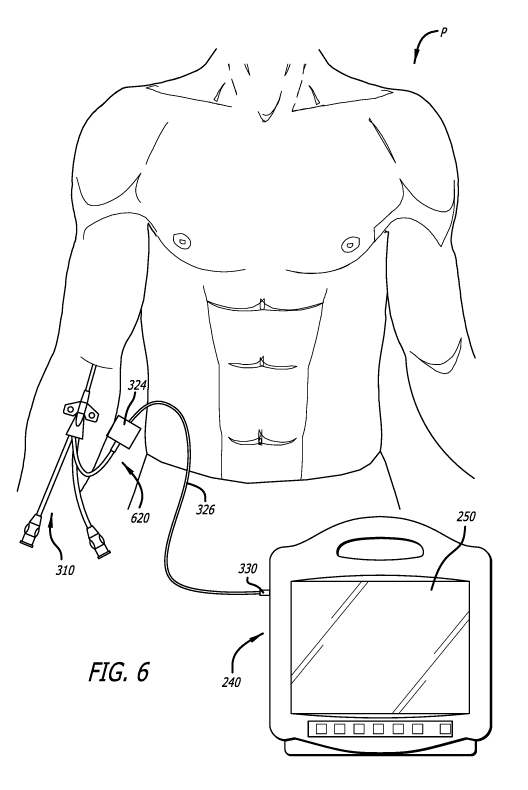
[0077] FIG. 6 illustrates the second shape-sensing system 200 with a
first optical-fiber
connector module 620 in accordance with some embodiments. FIG. 7 illustrates
the second
shape-sensing system 200 with the first optical-fiber connector module 620
within a
fenestration 601 of a surgical drape 603 in accordance with some embodiments.
FIG. 8
illustrates the second shape-sensing system 200 with a second optical-fiber
connector module
820 in accordance with some embodiments. FIG. 9 illustrates the second shape-
sensing system
200 with the second optical-fiber connector module 820 beneath the surgical
drape 603 in
accordance with some embodiments. FIG. 5 illustrates a detailed section of the
optical-fiber
connector module 120 in accordance with some embodiments thereof such as the
first optical-
fiber connector module 620 or the second optical-fiber connector module 820.
[0078] As shown, the optical-fiber connector module 620 or 820 includes
the housing
324, a receptacle 532 disposed in the housing 324, the cable 326 extending
from the housing
324, and an optical fiber 528 within at least the cable 326.
[0079] The receptacle 532 includes an optical receiver configured to
accept insertion
of an optical terminal of a plug of the medical device 110 (e.g., the plug 322
of the PICC 310)
for establishing an optical connection between the optical-fiber connector
module 620 or 820
and the optical-fiber stylet of the medical device 110 (e.g., the optical-
fiber stylet 424 of the
PICC 310) when the plug is inserted into the receptacle 532.
-17-
CA 03150788 2022-02-11
WO 2021/030092 PCT/US2020/044801
[0080] The cable 326 includes the plug 330 for establishing an optical
connection
between the optical-fiber connector module 620 or 820 and the optical
interrogator 230 of the
console 240.
[0081] The optical fiber 528 extends from the receptacle 532 through the
cable 326 to
the plug 330. The optical fiber 528 is configured to convey the input optical
signals from the
optical interrogator 230 to the optical-fiber stylet of the medical device 110
(e.g., the optical-
fiber stylet 424 of the PICC 310) and the reflected optical signals from the
optical-fiber stylet
to the optical interrogator 230.
[0082] As set forth above, the optical-fiber connector module 620 or 820
can further
include the one or more sensors 222 selected from the gyroscope, the
accelerometer, and the
magnetometer disposed within the housing 324. The one or more sensors 222 are
configured
to provide sensor data for determining a reference plane for shape sensing
with the optical-
fiber stylet of the medical device 110 (e.g., the optical-fiber stylet 424 of
the PICC 310).
[0083] While not shown, the optical-fiber connector module 620 or 820 can
further
include power and data wires extending from the one or more sensors 222
through the cable
326 to the plug 330 or another plug. The power and data wires are configured
to respectively
convey power to the one or more sensors 122 and data from the one or more
sensors 122 to the
console 240 when the one or more sensors 122 are present in the optical-fiber
connector module
620 or 820.
[0084] The optical-fiber connection module 620 is configured to sit
within the
fenestration 601 of the surgical drape 603 adjacent a percutaneous insertion
site for the medical
device 110 (e.g., a catheter such as the PICC 310). As the optical-fiber
connection module 620
is configured to sit within the fenestration 601 of the surgical drape 603,
the optical-fiber
connection module 620 is amenable to disinfection or sterilization. For
example, the housing
324 of the optical-fiber connection module 620 can be a non-porous or
chemically resistant to
oxidants. The optical-fiber connection module 620 can be configured for manual
disinfection
with a ChloraPrep product by Becton, Dickinson and Company (Franklin Lakes,
NJ), or the
optical-fiber connection module 620 can be configured for automatic high-level
disinfection or
sterilization with vaporized H202 by way of Trophon by Nanosonics Inc.
(Indianapolis, IN).
[0085] In contrast to the optical-fiber connection module 620, the
optical-fiber
connection module 820 is configured to sit beneath the surgical drape 603 on a
chest of a patient
-18-
CA 03150788 2022-02-11
WO 2021/030092 PCT/US2020/044801
P. As such, the optical-fiber connection module 820 need not require a same
level of
disinfection or sterilization as the optical-fiber connection module 620.
[0086] While not shown, the housing 324 the optical-fiber connection
module 820
includes a loop extending from the housing 324, a tether point integrated into
the housing 324,
a ball-lock-pin receiver integrated into the housing 324, or the like
configured for attaching a
neck strap to the optical-fiber connector module 820. The loop, the tether
point, the ball-lock-
pin receiver, or the like enables the optical-fiber connector module 820 to be
secured to a neck
of the patient P while sitting on the patient's chest. Additionally or
alternatively, the housing
324 includes a patient-facing surface (e.g., a back of the optical-fiber
connection module 820)
configured to be adhered to the patient's chest. The patient-facing surface
enables the optical-
fiber connector module 820 to be secured to the patient's chest while sitting
on the patient's
chest whether or not the optical-fiber connection module 820 is also secured
to the patient's
neck.
[0087] Again, the receptacle 532 includes an optical receiver configured
to accept
insertion of an optical terminal of a plug of the medical device 110 (e.g.,
the plug 322 of the
PICC 310) and form an optical connection when the plug is inserted into the
receptacle 532;
however, with the optical-fiber connector module 820, the optical connection
is formed with
the surgical drape 603 between the optical-fiber connector module 820 and the
medical device
110. The receptacle 532 and the plug of the medical device 110 enable at least
the optical
connection from a sterile field (e.g., above the surgical drape 603) including
the medical device
110 such as the PICC 310 to a non-sterile field (e.g., beneath the surgical
drape 603) including
the optical-fiber connection module 820 by way of breaching the surgical drape
603.
Methods
[0088] Each method of a number of methods for determining whether the tip
of the
medical device 110 is located within an SVC of a patient includes advancing
the tip of the
medical device 110 through a vasculature of the patient toward the SVC. As set
forth above,
the medical device 110 (e.g., the PICC 310) includes the integrated optical-
fiber stylet (e.g.,
the optical-fiber stylet 424) having the number of FBG sensors (e.g. the FBG
sensors 426a,
426b, 426c, ..., 426n) along at least the distal-end portion of the optical-
fiber stylet for shape
sensing with the shape-sensing system 100 or 200 including the medical device
110. When the
medical device 110 is the PICC 310, advancing the tip of the PICC 310 through
the vasculature
of the patient includes advancing the tip of the PICC 310 through a right
internal jugular vein,
-19-
CA 03150788 2022-02-11
WO 2021/030092 PCT/US2020/044801
a right brachiocephalic vein, and into the SVC. When the medical device is a
CVC, advancing
the tip of the CVC through the vasculature of the patient includes advancing
the tip of the DVC
through a right basilic vein, a right axillary vein, a right subclavian vein,
a right brachiocephalic
vein, and into the SVC.
[0089] The method can include enabling certain functions of the shape-
sensing system
100 or 200 by turning on the console 140 or 240, running one or more programs
on the console
140 or 240, making the selection of the FBG sensors (e.g., a selection of the
FBG sensors 426a,
426b, 426c ..., 426n) in the distal-end portion of the optical-fiber stylet
for the plots of
curvature vs. time 1012a, 1012b, 1012c, ..., 1012n, making the optical or
electrical
connections, or the like as needed for various functions of the shape-sensing
system 100 or
200. Enabling certain functions of the shape-sensing system 100 or 200 can
include enabling
the input optical signals to be sent into the optical-fiber stylet by the
optical interrogator 130
or 230 of the shape-sensing system 100 or 200 while advancing the tip of the
medical device
110 through the vasculature of the patient. Enabling certain functions of the
shape-sensing
system 100 or 200 can include enabling the FBG sensor-reflected optical
signals to be received
from the optical-fiber stylet by the optical interrogator 130 or 230 while
advancing the tip of
the medical device 110 through the vasculature of the patient. Enabling
certain functions of the
shape-sensing system 100 or 200 can include enabling the FBG sensor-reflected
optical signals
received from the optical-fiber stylet to be algorithmically converted into
the number of
different plots (e.g., the plot of curvature vs. arc length 1004, the plot of
torsion vs. arc length
1006, the plot of angle vs. arc length 1008, the plot of position vs. time
1010, one or more of
the plots of curvature vs. time 1012a, 1012b, 1012c ..., 1012n, etc.) for
display on the display
screen 150 or 250. Enabling certain functions of the shape-sensing system 100
or 200 can
include enabling the FBG sensor-reflected optical signals received from the
optical-fiber stylet
to be algorithmically converted into the displayable shapes over the 3-
dimensional grid 1002
for the medical device 110 for display on the display screen 150 or 250.
[0090] The method can include manually identifying on the display screen
150 or 250
the distinctive change in the plotted curvature of the optical-fiber stylet
sensed by the selection
of the FBG sensors in the distal-end portion of the optical-fiber stylet at
the moment the tip of
the medical device 110 is advanced into the SVC, thereby determining the tip
of the medical
device 110 is located within the SVC. Identifying on the display screen 150 or
250 the
distinctive change can include identifying the instantaneous increase in the
plotted curvature
-20-
CA 03150788 2022-02-11
WO 2021/030092 PCT/US2020/044801
of the optical-fiber stylet followed by the instantaneous decrease in the
plotted curvature as
sensed by each FBG sensor of the last three FBG sensors (e.g., the FBG sensors
426a, 426b,
and 426c) in the distal-end portion of the optical-fiber stylet at the moment
the tip of the
medical device 110 is advanced into the SVC. Additionally or alternatively,
the method can
include automatically determining with the SVC-determiner algorithm the
distinctive change
in the plotted curvature of the optical-fiber stylet, or the plottable data
therefor, sensed by the
selection of the FBG sensors in the distal-end portion of the optical-fiber
stylet at the moment
the tip of the medical device 110 is advanced into the SVC.
[0091] The method can include ceasing to advance the tip of the medical
device 110
through the vasculature of the patient after determining the tip of the
medical device 110 is
located in the SVC. The method can include confirming the tip of the medical
device 110 is in
the SVC by way of periodic changes in the plotted curvature of the optical-
fiber stylet sensed
by the selection of the FBG sensors. The periodic changes in the plotted
curvature result from
periodic changes in blood flow within the SVC as a heart of the patient beats.
[0092] In some alternative or additional embodiments, logic of the shape-
sensing
system 100 or 200 may be configured to generate a rendering of the current
physical state of
the stylet and, as a result, of the catheter, based on heuristics or run-time
analytics. For example,
the logic may be configured in accordance with machine-learning techniques to
access a data
store (library) with pre-stored data (e.g., images, etc.) pertaining to
different regions of the
stylet in which the core fibers experienced similar or identical wavelength
shifts. From the pre-
stored data, the current physical state of the stylet and/or the catheter may
be rendered.
Alternatively, as another example, the logic may be configured to determine,
during run-time,
changes in the physical state of each region of the stylet (and the catheter),
based on at least (i)
resultant wavelength shifts experienced by the core fibers, and (ii) the
relationship of these
wavelength shifts generated by sensors positioned along different outer core
fibers at the same
cross-sectional region of the stylet (or the catheter) to the wavelength shift
generated by a
sensor of the center core fiber at the same cross-sectional region. It is
contemplated that other
processes and procedures may be performed to utilize the wavelength shifts as
measured by
sensors along each of the core fibers to render appropriate changes in the
physical state of the
stylet and/or the catheter.
[0093] Notably, not one method of the shape-sensing system 100 or 200
requires an X-
ray for determining whether the tip of the medical device 110 is located
within the SVC of the
-21-
CA 03150788 2022-02-11
WO 2021/030092 PCT/US2020/044801
patient. As such, patients need not be exposed to ionizing X-ray radiation
when the shape-
sensing system 100 or 200 is used. In addition, not one method of the shape-
sensing system
100 or 200 requires an additional magnetic-sensor piece of capital equipment
for determining
whether the tip of the medical device 110 is located within the SVC of the
patient. In addition,
since, the shape-sensing system 100 or 200 does not require use of a reliable
ECG P-wave like
some existing systems for placing a tip of a medical device into an SVC of a
patient, the shape-
sensing system 100 or 200 can be used with patient having atrial fibrillation
or another heart
arrhythmia.
[0094] While some particular embodiments have been disclosed herein, and
while the
particular embodiments have been disclosed in some detail, it is not the
intention for the
particular embodiments to limit the scope of the concepts provided herein.
Additional
adaptations and/or modifications can appear to those of ordinary skill in the
art, and, in broader
aspects, these adaptations and/or modifications are encompassed as well.
Accordingly,
departures may be made from the particular embodiments disclosed herein
without departing
from the scope of the concepts provided herein.
-22-