Note: Descriptions are shown in the official language in which they were submitted.
CA 0315137 2022--14
WO 2021/048409
PCT/EP2020/075550
1
Medical Implant, Delivery Device, Method of Producing a Medical
Implant, and Method of Delivering a Medical Implant
The present invention relates to a medical implant, in particu-
lar a patch, a delivery device for a medical implant, and a
method of delivering a medical implant according to the preamble
of the independent claims.
Defects in tissue, for example atrial septal defects (ASDs) or
ventricular septal defects (VSDs), are fairly common conditions
in humans that are typically treated minimally invasively or
surgically. Such defects can cause a variety of symptoms such as
shortness of breath and a higher burden on the heart and lungs.
As a consequence, a myriad of implantable devices has been pro-
posed in the prior art, many of which can be deployed in a mini-
mally invasive way.
For example, closure of an atrial septal defect (ASD) by deploy-
ing umbrella-like implants through a catheter have been dis-
closed by Lock et al. (DOI: 10.1161/01.CIR.79.5.1091).
However, known implants exhibit several disadvantages. Typical-
ly, they are attached to a tissue wall, or held in place, me-
chanically. On one hand, this can lead to small injuries of the
tissue to be treated. On the other hand, mechanical attachment
places constraints on the material choice and mechanical
strength of the used components. Finally, mechanical anchoring
can also be difficult to realize in a minimally invasive treat-
ment.
Thus, the object of the present invention is to overcome the
drawbacks of the prior art, in particular to provide a medical
CA 0315137 2022--14
WO 2021/048409
PCT/EP2020/075550
2
implant, a device for delivering an implant, and a method of de-
livering an implant that is easy and safe to use.
This and other objects are achieved by the medical implant, the
delivery device and the methods according to the characterizing
portion of the independent claims of the invention.
The medical implant according to the invention is adapted to re-
pair or close a defect, in particular an opening in a ventricu-
lar, atrial, or septal wall. In particular, the medical implant
may be a patch, for example a polymeric or pericardial patch. It
comprises an adhesive composition. It further comprises two
states, wherein in the first state, the medical implant can be
deployed to an implant site while the adhesive composition is
inactive. It can be brought into a second state, preferably at
the implant site, by an activation mechanism. The adhesive com-
position, in the second state, is curable by a curing mechanism.
In particular, it is conceivable that the activation mechanism
is identical to the curing mechanism. Alternatively, two differ-
ent mechanisms may be employed for activation and curing.
The medical implant may also be suitable to close a cavity, such
as a left atrial appendage. In particular, the implant may be
sized and shaped such that the implant may be attached an ostium
of a cavity.
The curing mechanism may in particular be exposure to electro-
magnetic radiation, for example exposure to visible light, UV
light, IR light, and/or X-rays. Curing may, in particular, in-
clude cross-linking of the adhesive.
CA 0315137 2022--14
WO 2021/048409
PCT/EP2020/075550
3
A patch shall be understood as a substantially flat structure.
Preferably, it is mechanically flexible such that it can adapt
to an underlying surface shape or structure.
The implant is preferably adapted in size and shape to close an
opening in a ventricular and atrial septal wall, for example a
patent foramen ovale (PFO). Typically, a medical implant for
such an application has a substantially round, preferably circu-
lar, shape, though any shape such as a triangle, square, or more
complicated shape is possible. It is substantially flat, and has
a typical diameter of 20-30 mm, preferably 20 - 25 mm. It may of
course be larger or smaller, depending on a patient or the open-
ing to be treated. For example, an opening in the heart of a
child may be smaller, requiring a patch diameter as small as
10 mm. It may also be as large as 30 mm, for example if a pa-
tient is very tall. Typically, the thickness of the medical im-
plant is 100-200 pm. Of course, the thickness may also be
adapted and be as thin as 50 pm or as thick as 1.5 mm, preferably
as thick as 1 mm, particularly preferably as thick as 500 pm.
Preferably, the medical implant comprises a material with self-
healing and/or self-closing properties. Such properties enable
easier implantation because the implant can be temporarily held
with an instrument through a hole, for example with a needle
and/or a suture, wherein the hole closes automatically after im-
plantation.
Additionally or alternatively, the implant may comprise a hole
in a center region that enables or assists holding with a deliv-
ery instrument.
In general, materials with high flexibility are preferred mate-
rials for the implant according to the invention. A high flexi-
CA 0315137 2022--14
WO 2021/048409
PCT/EP2020/075550
4
bility reduces the risk of tear, rupture and/or dislodgment dur-
ing and after implantation. However, it will be understood that
rigid materials, in principle, are also suitable for the im-
plants disclosed herein.
Preferably, in the first state, the medical implant comprises at
least one cavity that contains the adhesive composition. Prefer-
ably, the medical implant comprises a plurality of cavities. It
may be adapted to, in the second state, release the adhesive
composition.
A cavity shall be understood as a closed structure in the medi-
cal implant that can retain another substance such as a liquid,
a viscous liquid, or even a solid. For example, the cavity may
be a large hollow structure or a small pore. Cavities are par-
ticularly advantageous to store the adhesive composition in the
first state because the adhesive is protected from the surround-
ing media, in particular humidity/moisture from the body, for
example originating from as bodily fluids. Thus, it does not ac-
cidentally engage tissue before activation, and prevents problem
while deploying the medical implant, such as clogging of a cath-
eter due to adhesive leaks.
In general, the adhesive may also be electrically activatable.
An example of an electrically activatable glue is voltaglue, for
example as disclosed in ACS Appl. Bio Mater. 2019, 2, 6, 2633-
2642, which is incorporated here by reference. However, any oth-
er electrically activatable glue is suitable. In general, such
glues may contain an element or molecule that can form a radical
when exposed to a voltage. The formed radicals may cause cross-
linking.
Preferably, the implant is manufactured by an additive manufac-
turing/3D-printing method.
CA 0315137 213-1314
WO 2021/048409
PCT/EP2020/075550
Preferably, the implant comprises optical fibers for distribu-
tion of light within the implant. For example, the optical fi-
bers may comprise or consist of glass, a polymer, or particular-
5 ly preferably a biodegradable polymer.
The cavities may have at least two boundary surfaces. At least
one property of the boundary surfaces is different between one
boundary surface and the other. Preferably, the property in-
cludes at least one of a permeability and solubility.
For example, the boundary surfaces may differ in the permeabil-
ity for an adhesive such as to preferentially release the adhe-
sive at a particular location or side of the cavity. Additional-
ly or alternatively, the permeability for blood or another bodi-
ly fluid may differ.
Additionally or alternatively, the boundary surfaces may differ
in their solubility in blood or another bodily fluid. Such a
difference in solubility may cause preferential release of an
adhesive at a particular location or side of the cavity.
In particular, at least one of the boundary surfaces may com-
prise, preferably consist of, at least one of PEG, PLA, PET,
PUs.
The two different surfaces may also be configured such that one
surface is adapted to provide adhesion to tissue, while the oth-
er surface is adapted to enhance tissue and/or cell growth.
The medical implant may comprise a radio-opaque element. A ra-
dio-opaque element may be any element that provides contrast in
radio-imaging. Preferably, the radio-opaque element comprises,
CA 0315137 2022--14
WO 2021/048409
PCT/EP2020/075550
6
in particular consists of, barium sulphate, platinum, iridium,
and/or tungsten. The radio-opaque element may, in particular, be
a wire, a particle, or another marker.
Preferably, the medical implant comprises a support structure,
wherein the radio-opaque element is arranged within or formed by
the support structure. For example, the medical implant may com-
prise a backbone made of a polymer that comprises at least one,
preferably a plurality of, barium sulphate particles. Alterna-
tively, a similar backbone may be made of a metal that is radio-
opaque.
Particularly preferably, a plurality of radio-opaque elements
may be arranged in a particular pattern, spacing, geometry or
alignment such that data on the relative positions of the radio-
opaque elements, for example their spacing and/or alignment de-
termined from imaging data, may provide information about proper
adhesion and/or positioning of the medical implant. For example,
three markers may be equally spaced around the circumference of
the implant. Additionally or alternatively, the radio-opaque el-
ement may have a particular shape that provides information on
adhesion and/or position of the implant. For example, the radio-
opaque element may have a bent shape, which is kept straight by
the adhesive force when the implant is attached to a straight
surface. Thus, if a bent shape is determined via radio imaging,
it would indicate that the medical device is detaching from the
tissue. In particular, the radio-opaque element may be adapted
such as to not exert a force that is sufficient to dislodge the
implant.
Preferably, the radio-opaque element is arranged within or
formed by the adhesive composition. For example, barium sulphate
particles may be dispersed in the adhesive composition. Addi-
CA 0315137 2022--14
WO 2021/048409
PCT/EP2020/075550
7
tionally or alternatively, the adhesive may comprise a coordina-
tion polymer comprising barium ions. Additionally or alterna-
tively, the radio-opaque element may comprise or consist of bar-
ium sulphate, iodine, tantalum, iridium, and/or iohexol, which
may also be dispersed in the adhesive.
Preferably, the radio-opaque element comprises at least one of
barium sulphate and iodine. Iodine is particularly advantageous
if the radio-opaque element is used to track the degradation of
an adhesive, a patch material, or other parts of the implant.
For example, if the medical implant is designed to degrade in
the human body while allowing for cell overgrowth, iodine may be
incorporated in a biodegradable material. The degradation of the
biodegradable material may be tracked by means of radio imaging.
If, for example, coagulation suppressant therapy is needed dur-
ing degradation, but only during degradation, the imaging data
may indicate whether coagulation suppressant are still needed.
Preferably, the implant comprises at least one discrete marker.
Discrete shall be understood as contained to a particular area
or location. For example, a discrete marker may be used to des-
ignate one side of the implant from the other, or to designate
an upper and a lower part. Additionally or alternatively, the
discrete marker may be deformable by pressure and indicate a
pressure at a particular location of the implant, for example a
pressure caused by the adhesive force between a tissue wall and
the implant. Particularly preferably, the discrete marker may be
a spring that may cause detachment if the adhesion force between
the implant and the tissue is below a certain threshold and thus
facilitates detection of detachment (similar to predetermined
breaking point).
CA 0315137 213-1314
WO 2021/048409
PCT/EP2020/075550
8
The marker may, in particular, be used to guide a robot, prefer-
ably a microrobot, to the implantation site after the implant
has been implanted. The marker may define a position in the body
and may in particular retain its function for a certain period
of time, for example one year. Thus, guiding of a robot may also
be done a certain amount after implantation. The marker may be
detectable by the robot and thus enable passive guiding. Alter-
natively, the marker may emit a signal that is detected by the
robot and thus enable active guiding.
Preferably, the discrete marker is at least one of radio-opaque
and echo-opaque/echogenic. Particularly preferably, the discrete
marker is configured as being the radio-opaque element.
The implant may comprise at least two discrete markers that are
arranged at a pre-defined distance and/or orientation from one
another.
Preferably, the medical implant has a generally flat shape with
a first and a second surface, wherein the first and the second
surfaces are facing in substantially opposite directions. At
least one property of the first surface is different from a cor-
responding property of the second surface.
The person skilled in the art will understand that "generally
flat" may encompass slightly curved flat surfaces and shapes, in
particular disk- or chip-like shapes.
The difference between the first and the second surface may be
with respect to any measurable quantity , wherein a measurement
of said quantity would yield significantly different values.
Particularly preferably, the first and second surface differ in
CA 0315137 213-1314
WO 2021/048409 PCT/EP2020/075550
9
polarity, charge, functionalization, surface structure, surface
pattern, material, coating, and/or porosity.
The first surface may be adapted to enhance cell ingrowth. In
particular, the porosity of the surface may be adapted to allow
for cell ingrowth, for example by having a pore size of adapted
to allow for cell ingrowth. The pore size may be in the range of
a few microns to several hundred microns. Preferably, the pores
have a diameter between 50 pm and 500 pm. Additionally or alter-
natively, the first surface may be biocompatible and in particu-
lar be functionalized with growth factors or cell adhesion mo-
tifs. The first surface may comprise surface charges that acti-
vate and/or attract cells. The first surface may also have a
surface roughness adapted to enhance cell ingrowth and/or com-
prise a velour-like surface.
In particular, at least one of a length, size, and 3D-
arrangement of pores and holes may be adapted to enhance cell
ingrowth. The length may in particular denominate the longest
extension along an axis of a pore or hole in case of non-
spherical pores/holes.
Preferably, the first and/or the second surface may comprise or
consist of derivates of polymer peptides.
Preferably, the second surface is adapted to provide adhesion to
biological tissue. In particular, the biological tissue may be
human or animal tissue such as one of endocardial, pericardial
and septal tissue. For example, the second surface may comprise
a glue layer.
Preferably, at least one surface of the implant, in particular
at least one of the first and the second surface of the implant,
CA 0315137 213-1314
WO 2021/048409
PCT/EP2020/075550
comprise a velour-like surface. Particularly preferably, all
surfaces of the implant comprise a velour-like surface.
Preferably, the adhesive composition is arranged on the medical
5 implant in a pattern. The pattern may be non-uniform. In partic-
ular, the pattern may be printed on the implant by inkjet or ex-
trusion printing. The pattern may also be regular, but comprise a
3D structure and/or a non-homogeneous topography.
10 Preferably, the adhesive composition comprises gelatin-
methacryloyl (GelMA), in particular a GelMA of animal original.
Particularly suited GelMAs are Fish GelMA, porcine GelMA, and
bovine GelMA, i.e. GelMA processed and originating from fish
and/or pigs and/or cows. GelMA originating from cold-water fish
is particularly suited because of its low-temperature (in par-
ticular at room temperature) mechanical flexibility. However,
any type of commercially available GelMA is suitable for the in-
vention.
In particular, the GelMA, in particular if derived from pigs,
may have a Bloom value of 250 to 325. GelMA derived from fish
may not have bloom strength.
Preferably, the GelMA has a molecular weight of 50 to 170 kDa.
Preferably, the GelMA may be formed by a mixture of at least two
GelMAs of animal origin. Particularly preferably, the GelMA is
formed by a mixture fish GelMA and porcine GelMA.
A mixture of two GelMAs enables to combine properties of differ-
ent GelMAs. For example, a mixture of (cold water) fish GelMA
with porcine GelMA may yield a GelMA with the solubility of por-
cine GelMA and the mechanical flexibility of fish GelMA. It is
CA 0315137 213-1314
WO 2021/048409
PCT/EP2020/075550
11
also possible to gradually tune properties, for example solubil-
ity and mechanical flexibility, by choosing the ratio of differ-
ent GelMAs, for example porcine and fish GelMA. Such GelMAs are
known to the skilled person and commercially available.
Additionally or alternatively, it is also possible to tune prop-
erties of the GelMA by varying and/or mixing of different molec-
ular masses.
Preferably, the adhesive composition further comprises at least
one photoinitiatior. The adhesive composition may comprise sev-
eral different photoinitiators, a single photoinitiator, or mix-
tures of different photoinitiators.
The photoinitiator may be a so-called type I photoninitatior,
preferably one of lithium pheny1-2,4,6-
trimethylbenzoylphosphinate, LAP, Irgacure, and camphorquinone.
Additionally or alternatively, the photoinitiator may be a so-
called type II photoinitiator, preferably one of Eosin Y and
mono/di/triethanolamine, Rose Bengal and
mono/di/triethanolamine, and Riboflavin and
mono/di/triethanolamine.
The GelMA may also be cross-linkable by X-ray radiation. Partic-
ularly preferably, the GelMA is cross-linkable via a photoiniti-
ator that can be activated by X-rays. Alternatively, the GelMA
may be crosslinkable without a photoinitiator.
The medical implant may also comprise a rivet, in particular a
blind rivet. A rivet may be used for attachment of the implant
to the tissue.
CA 0315137 2022--14
WO 2021/048409
PCT/EP2020/075550
12
Rivets are particularly advantageous because they are not sensi-
tive to, and thus securely attach in the presence of, different
temperatures, chemical environments, and humidity levels. Pref-
erably, a rivet can thus be used to at least temporarily attach
the medical implant to tissue, for example during curing of the
adhesive composition, during placement of the implant, and/or
while other manipulations on the implant are performed by a med-
ical professional.
The medical implant may comprise at least one retaining element
for retaining at least one suture at an outer circumference of
the implant. In addition, a suture can be arranged within said
loop.
Such a retaining element provides a particularly easy method of
temporarily attaching the medical implant to a delivery device.
In particular, the suture(s) may provide a connection to the de-
livery device. Tearing of the suture(s) and/or the retaining el-
ement can release the implant.
Preferably, the retaining element comprises or consists of a
loop of fabric. Alternatively, the retaining element may also be
formed by a backbone folded such as to form a loop in a circum-
ferential area of the implant.
The retaining element may also comprise a pre-determined break-
ing point.
Preferably, the at least one retaining element is formed from
the same material as the medical implant.
CA 0315137 2022--14
WO 2021/048409
PCT/EP2020/075550
13
Additionally or alternatively, the at least one retaining ele-
ment is formed of polyurethane with a thickness of 35 to 65 pm.
Preferably, the thickness is between 45 and 55 pm.
Alternatively, the at least one retaining element is configured
as a separate element arranged on the medical implant. For exam-
ple, the retaining element may be configured as a separate strip
of fabric attached to a circumferential area of the implant and
folded such as to form a loop. Alternatively, the retaining ele-
ment may comprise a loop formed by a suture.
Preferably, the at least one retaining element comprises a pre-
determined breaking point, particularly preferably an indenta-
tion. This allows to control where the retaining element breaks
when releasing the implant and as such provides better control
the implant procedure.
The adhesive composition may also comprise, in particular con-
sist of, a dried adhesive composition. The dried adhesive compo-
sition may swell and/or become at least partially liquid when
exposed to a liquid. The dried adhesive composition may also be
cross-linkable by exposure to a liquid, such as cyanoacrylate.
For example, GelMA as described herein may be dried and used in
this manner. Alternatively, any film-forming polymer, particu-
larly biopolymers, such as hyaluronic acid, collagen, heparin
and their photopolymerizable counterparts (such as collagen
methacrylate) are suitable as well.
The dried adhesive composition is preferably activatable by ex-
posure to a liquid, for example by rehydration in saline,blood,
and/or water. Additionally or alternatively, the dried adhesive
may also be activatable by exposure to cyanoacrylate.After rehy-
dration, the previously dried adhesive may be curable by expo-
CA 0315137 2022--14
WO 2021/048409
PCT/EP2020/075550
14
sure to electromagnetic radiation, such as visible and/or UV
light, in the presence of a photoinitator.
Preferably, in the first state, the medical implant comprises a
plurality of cavities, in particular micro-sized cavities.
Micro-sized cavities shall be understood as cavities with any
shape that have characteristic size in the micrometer range,
i.e. from 1 pm to 1000 pm. For example, the medical implant may
comprise a plurality of spherical cavities with a diameter of
10 pm to 100 pm. This is particularly advantageous because it
ensures a homogenous distribution, and if necessary mixing, of
the adhesive composition when it is released.
Preferably, the at least one cavity comprises an additive that,
upon exposure to humidity, swells. The at least one cavity may
be adapted to release the adhesive composition upon swelling.
This enables a particularly easy way of releasing the adhesive,
because exposure to blood automatically makes the cavities swell
and thus releases the adhesive.
Preferably, the medical implant comprises at least two different
kinds of cavities.
The two types of cavities may be different in size, composition,
shape, or any other property. For example, the two different
types of cavities may contain different additives that make them
swell at different rates. They may also have different wall
thickness, different radii, or be made of a different material.
They may also be adapted such that one type of cavity swells and
the other does not. This enables better control of the release
of the adhesive. For example, one component can selectively be
CA 0315137 213-1314
WO 2021/048409
PCT/EP2020/075550
released first, or one component can be released at a different
rate than the other. It may also enable a better control of the
rate of release if the adhesive only comprises one component.
For example, it may be advantageous to release a first fraction
5 of the adhesive, and release a second fraction at a later point.
It is also possible to adapt the cavities such that they release
the adhesive composition or a component thereof at different
pressures or temperatures.
10 Preferably, the at least two different types of cavities are
adapted to contain different components of an adhesive composi-
tion, in particular in a liquid, gel, dried, or gaseous form.
This may, in particular, include any of the features of two dif-
ferent types of cavities as described above. However, it may al-
15 so include particular properties that enable the storage of a
particular component of an adhesive. For example, a certain wall
material may be particularly advantageous for one component of
an adhesive composition, but may be incompatible with another.
Thus, it may be advantageous to adapt the cavities to the spe-
cific components of an adhesive composition. Of course, it may
also include different sizes or degradation rates of the cavi-
ties to account for desired ratios of two components in the fi-
nal (mixed) adhesive composition.
Preferably, the adhesive composition comprises two components
that are individually disposed in the cavities, such that in the
first state, the two components are separated. This enables the
controlled mixing in the second state, for example at the im-
plant site. This is of course particularly advantageous for two-
component adhesives that are not curable before mixing. In this
case, unplanned curing before adhesive release, for example by
accidental exposure to humidity or elevated temperature can be
prevented. However, it is of course conceivable to have an adhe-
CA 0315137 2022--14
WO 2021/048409
PCT/EP2020/075550
16
sive that comprises one component that only additionally hardens
the adhesive composition, for example through cross-linking. It
may be advantageous to dispose such a component separately as
well.
Preferably, the adhesive composition is adapted to be curable
upon mixing of the at least two components. This prevents acci-
dental curing before release of the adhesive. Any curing mecha-
nism to cure the curable adhesive composition after mixing is
conceivable. It may be an increase in temperature, exposure to
humidity, exposure to electromagnetic radiation such as visible
light, infrared light, ultraviolet light, or a combination
thereof.
Preferably, the adhesive composition is adapted to spontaneously
cure upon mixing of the at least two components. This offers a
particularly advantageous way of deploying the adhesive composi-
tion because it does not require additional processing steps be-
yond the release and the spontaneous mixing of the two compo-
nents. For example, a first component may comprise a primary
amine, and a second component may comprise an NHS ester. In the
presence of the functional groups of the first and second compo-
nents, the adhesive may cure upon mixing. However, it is of
course possible to combine such an adhesive composition with ad-
ditional curing mechanisms. For example, an adhesive composition
with two components may spontaneously cure upon mixing, but ex-
posure to an additional curing mechanism such as electromagnetic
radiation, humidity, or increased temperature may accelerate the
curing if necessary. It is also conceivable that a third compo-
nent is used as an additional hardener.
Preferably, the cavities are adapted to release the adhesive
composition upon a temperature increase, in particular to the
CA 0315137 2022--14
WO 2021/048409
PCT/EP2020/075550
17
temperature of a human body. This enables the automatic release
of the adhesive composition after implantation because the im-
plant is heated up to 37 C.
Preferably, the cavities are adapted to release the adhesive
composition upon exposure to electromagnetic radiation. For ex-
ample, the cavities may degrade or burst upon irradiation with
visible light, infrared light, ultraviolet light, or similar.
They may also be adapted to release the adhesive composition up-
on irradiation with a particular wavelength or wavelength range.
If more than one type of cavity is present, they may also be
adapted to release the adhesive upon irradiation with different
wavelength ranges, such that it is possible to selectively re-
lease one component at a time. In general, cavities that are
adapted to release the adhesive composition upon exposure to
electromagnetic radiation are particularly advantageous because
they enable the combination with a delivery device that can
transmit light for the release, for example delivery devices as
disclosed in WO 15/1756632. It is also a particularly safe way
of releasing the adhesive composition because light is typically
not prevalent in the human body, thus preventing accidental re-
lease.
Preferably, the cavities are adapted to release the adhesive
composition upon an increase in pressure. In particular, this
enables the release of the adhesive composition upon inflation
of a balloon. However, any other mechanism to provide a pressure
increase is also conceivable. For example, the cavities may be
adapted such that exposure to a liquid causes swelling due to an
osmotic pressure that provides a pressure difference. It may al-
so comprise a separate inflation reservoir to provide a pressure
on the cavities, the implant, or another part thereof.
CA 0315137 2022--14
WO 2021/048409
PCT/EP2020/075550
18
Preferably, the cavities are adapted to release the adhesive
composition upon a mechanical compression. For example, a com-
pression by a balloon of a delivery device may be employed and
is particularly advantageous because delivery devices with bal-
loons are known and thus easy to implement.
Preferably, the adhesive composition is adapted to be curable by
exposure to electromagnetic radiation. This provides the similar
advantages already described in the context of cavities that are
adapted to release upon exposure to electromagnetic radiation.
For example, the adhesive composition may be curable upon irra-
diation with visible light, infrared light, ultraviolet light,
or similar. It may also be adapted to be curable upon irradia-
tion with a particular wavelength or wavelength range. In gen-
eral, adhesive compositions that are adapted to release the ad-
hesive composition upon exposure to electromagnetic radiation
are particularly advantageous because they enable the combina-
tion with a delivery device that can transmit light for the re-
lease, for example delivery devices as disclosed in WO
15/1756632. It is also a particularly safe way of curing the ad-
hesive composition because light is typically not prevalent in
the human body, thus preventing accidental curing.
Preferably, the medical implant comprises a self-expanding sup-
port structure. A support structure may be any structure with a
higher mechanical stiffness and/or strength than the rest of the
implant. For example, it may comprise at least one strut of a
polymeric material that provides mechanical stiffness to the im-
plant. Additionally or alternatively, it may also comprise a
structure that holds the medical implant in place at a desired
implant location such as a PFO. For example, it may comprise
structure that is adapted to be placed in a defect and hold two
patches - one on each side of the defect. Alternatively, it may
CA 0315137 2022--14
WO 2021/048409
PCT/EP2020/075550
19
also only hold one patch. The self-expanding property of the
support structure enables particularly easy deployment through a
catheter. For example, it may be made of a shape memory material
such as a shape memory polymer or a shape memory metal such as a
Nitinol. It is also conceivable, however, to use an elastic ma-
terial that is compressed in a delivery device and expands into
its original shape at the implant site.
Preferably, the cavities are formed by closed capsules. In par-
ticular, the cavities may be formed by spherical capsules. They
may be adapted to be opened by the activation mechanism.
Preferably, the capsules are adapted to break open by the acti-
vation mechanism. They may be adapted to break open by any of
the activations described herein, for example exposure to elec-
tromagnetic radiation, increased pressure, increased tempera-
ture, or a combination thereof.
Preferably, the capsule walls are adapted to dissolve by the ac-
tivation mechanism. This is particularly advantageous if they
are adapted to dissolve upon exposure to water, blood, or anoth-
er bodily fluid, because such an activation mechanism does not
require additional handling or processing due the natural pres-
ence of these fluids at the implant site. Thus, this provides a
particularly easy and safe way of release the adhesive composi-
tion from the capsules.
Preferably, the capsule, in particular the capsule wall, com-
prises a filler material. Filler materials can increase the me-
chanical strength of the cured adhesive. Thus, such capsules can
serve a double purpose by holding the adhesive composition and
upon release provide a filler material.
CA 0315137 2022--14
WO 2021/048409 PCT/EP2020/075550
Preferably, the medical implant comprises a porous foam and, in
the first state, an adhesive composition is disposed in the po-
rous foam. Of course, it is possible to combine a foam with oth-
er cavities as described herein. For example, the medical im-
5 plant may comprise cavities that are larger than the pores of
the foam and contain a second component of the adhesive, a hard-
ener, or another substance. A foam shall, in particular, be un-
derstood as a material that substantially consists of pores sep-
arated by pore walls made of a solid or a liquid component. The
10 pores of a foam typically exhibit sizes and shapes that fluctu-
ate statistically. Typically, they also percolate.
In a particularly preferred embodiment, the medical implant com-
prises a braided structure that supports the porous foam.
Preferably, the porous foam comprises solid walls.
Preferably the porous foam comprises walls formed of a hydrogel.
Hydrogels are particularly advantageous because they are typi-
cally biocompatible. In addition, they can be manufactured from
a wide variety of different materials and can thus be adapted to
the application or even the patient to be treated. In addition,
hydrogels can functionalize and comprise active components. Hy-
drogels can also be adapted to biodegradable.
Preferably, the pore size of the foam is adapted to allow for
cell ingrowth. This facilitates the formation of tissue in or
around the foam. In particular, this is advantageous in combina-
tion with biodegradable materials such as a hydrogel that is
adapted in this way. The medical implant can then be adapted to
degrade at a rate that is slower than cell ingrowth. Thus, the
medical implant can serve as a template or scaffold and be de-
CA 0315137 2022--14
WO 2021/048409 PCT/EP2020/075550
21
graded in the body once the implant is replaced with body tis-
sue.
Preferably, the medical implant comprises at least one reser-
voir, and at least part of the adhesive composition is disposed
in said reservoir. A reservoir shall be understood as a compart-
ment in the medical implant with a characteristic size in the
order of magnitude of the implant itself. It may be disposed as
a cavity in the medical implant. However, it may also be ar-
ranged as a separate reservoir that is attached to the implant.
For example, it may be a blister on a surface of the medical im-
plant. A reservoir can be advantageous if a relatively large
amount of adhesive needs to be released fast, and/or it is only
needed at a specific location.
Preferably, the medical implant comprises at least one separate
inflation reservoir. This allows for localized inflation and
thus release of an adhesive disposed in a cavity or reservoir.
For example, it is possible to inflate a reservoir in a particu-
lar location of the medical implant first, releasing a first
portion of the adhesive. The rest of the adhesive can then be
released at a second point in time. Similarly, it would be con-
ceivable to arrange two inflation reservoirs to selectively re-
lease two portions at, for example, two different locations sep-
arately.
In a particularly preferred embodiment, the at least one reser-
voir filled with adhesive is adapted to release said adhesive
upon inflation of the at least one inflation reservoir. The med-
ical implant thus comprises at least one reservoir which is at
least partially filled with adhesive and at least one inflation
reservoir. However, it is of course also possible for the medi-
cal implant to comprise several inflation reservoirs and/or sev-
CA 0315137 213-1314
WO 2021/048409 PCT/EP2020/075550
22
eral reservoirs filled with adhesive. In particular, one infla-
tion reservoir may be used to release the adhesive from more
than one reservoir filled with adhesive. Similarly, two or more
inflation reservoirs may be adapted to release the adhesive from
one reservoir filled with adhesive. This can provide additional
safety through redundancy, or can also be used to provide an
easy way to release a defined first and second portion of the
adhesive from one reservoir.
Preferably, the reservoir is adapted such that the adhesive is
only released on one side of the medical implant. In particular,
it may only be released on a distal side of the patch, or on a
proximal side of the patch. However, it is also conceivable that
the adhesive would be released only on a side wall side of the
medical implant. This ensures proper placement of the adhesive
facing the tissue and prevents release of adhesive where the
medical does not and is not designed to be in operative contact
with tissue. As a consequence, the necessary amount of adhesive
composition is also reduced. This is more economical and safer
for the patient. The medical implant may adapted to only release
the adhesive on one side by selecting different materials on
each side, by varying the thicknesses and/or the density of the
material on each side, by varying the pores size and/or struc-
ture of a foam, or by changing any other property that may
change the permeability of the implant material for an adhesive
composition.
Preferably, the medical implant comprises at least one micro-
channel in fluid connection with the reservoir for the release
of the adhesive. A microchannel shall be understood as a channel
with a longitudinal shape that is in fluid connection to a sur-
face of the medical implant and has a diameter that is small
compared to the size of the medical implant and the reservoir.
CA 0315137 2022--14
WO 2021/048409 PCT/EP2020/075550
23
In particular it may have a channel diameter in the range of 1
to 1000 pm, preferably 25 to 750 pm, even more preferably 50-
300 pm. Such microchannels enable an easy release of the adhe-
sive composition from the reservoir.
In a particularly preferred embodiment, the medical implant is
adapted to only release the adhesive composition on one side of
the implant and comprises at least one microchannel. In particu-
lar, the implant may be adapted such that the adhesive is only
released through said microchannel. Thus, the arrangement of the
microchannels provides an easy way of adapting the medical im-
plant to release the adhesive in a particular location, for ex-
ample only on one side of the implant.
Preferably, the adhesive composition is disposed as fibers, in
particular solid fibers. Even more preferably, the adhesive com-
position comprises at least two components, at least one of
which is disposed as solid fibers. Solid fibers may comprise a
dried adhesive, or an adhesive that can be molten (such as a hot
melt), or an adhesive that swells if exposed to humidity or wa-
ter. In particular, an adhesive composition may also be disposed
as a coating on a fiber.
In particular, at least one of the fibers can comprise an alde-
hyde that is adapted to adhere to tissue.
[30] Preferably, the adhesive composition is adapted to at least
partially dissolve upon exposure to a liquid, in particular one
of an organic solvent, a saline, and blood, and form a gel, in
particular a hydrogel.
Preferably, the adhesive composition comprises at least one com-
ponent from the group of methacrylated gelatin, methacryloyl-
substituted tropoelastin, poly(acrylic) acid, and methacrylated
CA 0315137 2022--14
WO 2021/048409 PCT/EP2020/075550
24
collagen. Poly(acrylic) acid may be used with a diacry-
late/dimethacrylate/amide crosslinker
Preferably, the adhesive composition comprises a dried component
that is adapted to be curable by a curing mechanism upon expo-
sure to a liquid, in particular an organic solvent or blood.
This is particularly advantageous because it allows for an auto-
matic activation of the adhesive upon implantation due to the
presence of blood. Of course, it is also possible to adapt the
adhesive composition such that only a selective activation by an
organic solvent is possible. It may also be adapted such that
exposure to blood activates the adhesive, but an additional ex-
posure to an organic solvent accelerates the activation process.
In particular, it is also possible to adapt the adhesive such
that it can be activated by a liquid either inside the body of
outside the body before implantation.
In particular, the adhesive composition may be activatable by
rinsing with a solution comprising a photoinitiator. Alterna-
tively, the photoinitiator may also be comprised in a dried ad-
hesive and become activated by exposure to a liquid. For exam-
ple, it may only be reactive in an at least partially swollen
adhesive and be kinetically hindered in the dried solid adhe-
sive.
Preferably, the medical implant comprises a scaffold made of bi-
oabsorbable or biodegradable material (hereinafter, reference to
"biodegradable" materials shall be understood as encompassing
both, bioabsorbable und biodegradable materials), in particular
a woven, knitted, electrospun, melt spun, and/or nonwoven bioab-
sorbable material, and/or a biological implant made by 3-D
printing. A scaffold may in particular be used to facilitate
cell in-growth and tissue formation.
CA 0315137 2022--14
WO 2021/048409 PCT/EP2020/075550
The invention also relates to a method for deploying a medical
implant that comprises and adhesive. The method is particularly
advantageous in combination with a medical implant as described
5 herein, but of course it is possible to perform the method with
any other medical implant that comprises an adhesive. The method
comprises the steps of deploying the medical implant to a first
site in a first state, bringing the medical implant into a sec-
ond state by means of an activation mechanism, and curing the
10 adhesive by means of a curing mechanism. The first site is pref-
erably the implant site. However, it is also possible to bring
the implant into the second state either outside the body or in-
side the body, but not at the implant site.
15 Preferably, the method comprises a step of increasing the tem-
perature of the medical implant to bring it into the second
state. In particular, an increased temperature may be used to
release an adhesive as described herein, for example by breaking
open capsules and/or dissolving cavities.
Preferably, the temperature increase is at least partially pro-
vided by and external heat source. It may also be provided only
by an external heat source.
Alternatively or additionally, the temperature increase is at
least partially provided by a patient's body heat. It may also
be provided only by the patient's body heat.
Alternatively or additionally, the temperature increase is at
least partially provided by electromagnetic radiation, in par-
ticular infrared light. It may also be provided only by electro-
magnetic radiation.
CA 0315137 2022--14
WO 2021/048409 PCT/EP2020/075550
26
Preferably, the method comprises a step of applying a pressure
to bring the implant into the second state. For example, the ap-
plied pressure may squeeze the adhesive composition out of cap-
sules or cavities. It may also release the adhesive composition
from the pores of a foam.
Preferably, the pressure increase is at least partially caused
by osmotic pressure. It may also be only caused by osmotic pres-
sure. For example, the adhesive composition may comprise a con-
centration of ions that is higher than the one of blood and may
be comprised in a plurality of cavities with a wall through
which water can diffuse. Due to osmotic pressure, water diffuses
into the cavities. The cavities can be adapted such that they
burst at pressures lower than the osmotic pressure. Of course,
the same can be achieved with only one or any other number of
cavities.
Preferably, the pressure increase is at least partially caused
by applying mechanical deformation, in particular a mechanical
pressure, to the implant. The person skilled in the art will of
course understand that all the possibilities of increasing the
pressure described herein can be combined.
Preferably, the method comprises the step of exposing the im-
plant to humidity to bring it into the second state. Humidity
may, for example, cause the dissolution or bursting of cavities
or capsules. It may also cause swelling of an adhesive composi-
tion to bring it into a curable or cured state. Any other mecha-
nism of releasing the adhesive and/or bringing into a curable
state described herein is conceivable.
Preferably, the method comprises the step of exposing the medi-
cal implant to electromagnetic radiation to bring into the sec-
CA 0315137 2022--14
WO 2021/048409
PCT/EP2020/075550
27
and state. The electromagnetic radiation may be infrared light,
ultraviolet light, or visible light. It may cause degradation of
cavities or any other mechanism described herein to release and
adhesive from a cavity and/or bring it into a curable state.
Preferably, the method comprises the step of spontaneous mixing
of two components in the second state, which causes curing of
the adhesive. For example, the adhesive composition may comprise
two components that are released from separate capsules by any
of the mechanisms herein. Upon release, the mix spontaneously
mixes due to the loss of the separation in the cavities. The ad-
hesive composition may thus be adapted such that the two compo-
nents react with each other without any other external trigger.
This provides a particularly easy way of curing the adhesive
composition. However, it would of course be possible to combine
such an adhesive composition with an additional curing mechanism
to cure or harden it further.
Preferably, the method comprises the step of exposing the medi-
cal implant to electromagnetic radiation in the second state,
which causes curing of the adhesive.
Preferably, the method comprises the step of inflating a sepa-
rate inflation reservoir to bring the implant into the second
state. In particular, inflating of the separate inflation reser-
voir may squeeze the adhesive out of a reservoir containing the
adhesive.
Preferably, the method comprises the step of disposing a liquid
to bring the implant into the second state. The liquid may be an
organic solvent, in particular an organic solvent that is misci-
ble with human blood. The liquid may, however, also comprise or
CA 0315137 2022--14
WO 2021/048409 PCT/EP2020/075550
28
even consist of water, in particular a physiological saline so-
lution.
The invention is further directed at a medical implant. The med-
ical implant comprises at least one tear line that is arranged
in proximity to a circumference of a medical implant. The medi-
cal implant is adapted to repair or close a defect, preferably
an opening in a ventricular or atrial or vessel wall. In partic-
ular, the medical implant may be a patch or any other implant as
disclosed herein. The medical implant is further adapted such
that, upon deployment to an implant site, in particular a septal
defect site, the medical implant can be attached to a tissue
wall and torn along the tear line.
A tear line shall be understood as any sort of weakening in the
material that creates a site of predetermined failure. Thus, if
a mechanical stress is applied to the material, the material
will first break along the tear line. This allows for an attach-
ment of the medical implant to a delivery device and easy re-
lease from the delivery device by breaking or tearing along the
tear line. The tear line may, for example, be a weakening of the
material by aligned holes or perforations in the material, or be
a lower thickness along a certain line, or a region of a differ-
ent material.
The tear line may be arranged around a complete circumference of
the medical implant, or only at selected regions where the im-
plant is attached to a delivery device. In particular, the medi-
cal implant may also comprise elongated elements for attachment
to a delivery device and that are detachable from the medical
implant by means of a tear line. For example, such elongated el-
ements may be flaps or arms of the same material as the implant,
or struts of another material such as a polymer or a metal.
CA 0315137 2022--14
WO 2021/048409 PCT/EP2020/075550
29
A preferred way of tearing the tear line is by inflation of a
balloon on a delivery device. Thus, the tear line is preferably
arranged such that a mechanical stress can be applied to it by
means of a balloon. For example, the medical implant may com-
prise tearable flaps that are too short to reach around the cir-
cumference of a balloon. Thus, the tear lines would be arranged
perpendicularly to the longitudinal axis of the flaps. Inflation
of the balloon causes a mechanical stress along the longitudinal
axis of the flaps and thus rupture along the tear line.
Preferably, the medical implant comprises a biodegradable mate-
rial that is adapted to lose its mechanical strength in a human
body within three years, preferably twelve months, even more
preferably six months. Loss of mechanical strength shall in par-
ticular encompass molecular weight loss of a polymeric substance
which reduces the mechanical stiffness.
Preferably, the medical implant is coated with a non-adhering
coating, in particular silicone and/or poly(tetrafluoro eth-
ylene), between an outer edge and the tear line. This prevents
adhesion of the medical implant to tissue in areas that are de-
signed to be retracted with the delivery device.
Preferably, the medical implant comprises a cut, in particular a
cross-shaped cut, that is adapted such that a delivery device
can partially extend through said cut. The cut may have any
shape that provides an opening in the medical implant that can
be closed. For example, it may also be a semi-circular cut that
forms similarly shaped flap, or a square-shaped cut. Even a lin-
ear cut is conceivable, but would have to be long enough for a
delivery device to extend through it. In addition, the cut is
adapted such that the opening formed by it at least has a ten-
CA 0315137 2022--14
WO 2021/048409 PCT/EP2020/075550
dency to close itself. This allows for the delivery device to at
least partially extend on both sides of the implant during de-
livery, but to be retracted through the medical implant after
delivery. After retraction, the flaps close the opening.
5
Preferably, the medical implant comprises fibers, in particular
woven, spun, or knitted fibers. This is particularly advanta-
geous if the medical implant comprises, or consists, of a fabric
patch. The fibers can be adapted for certain functions, such as
10 being coated with an adhesive or a drug and/or being biodegrada-
ble. Of course, it is possible to combine different types of fi-
bers in one medical implant, for example fibers coated with dif-
ferent adhesive, adapted to biodegrade at different rates, or to
elute different drug, or any combination of those.
Preferably, the fibers are made of a biodegradable material.
Preferably, the biodegradable material is selected from a group
of poly(lactic-co-glycolic acid), poly(L-lactic acid, poly(D-
lactic acid), poly(glycolic acid), poly(caprolactone), any co-
polymer and/or blend of these materials. These materials are
particularly advantageous in that they are non-toxic, well-known
and approved for medical applications, and easily commercially
available.
Preferably, the medical implant comprises a spine structure. In
particular, the spine structure may have different mechanical
properties from the rest of the medical implant. For example, it
may be made from a different material, or have different dimen-
sion such as a greater thickness. The spine structure allows for
tuning of the implant properties with more flexibility because
the mechanical properties can be tuned without necessarily
CA 0315137 2022--14
WO 2021/048409 PCT/EP2020/075550
31
changing the implant material that may have been selected due to
other properties.
Preferably, the spine structure comprises, particularly prefera-
bly consists of, a polyurethane.
Preferably, the spine structure extends beyond the medical im-
plant to provide non-elastic tear arms. The non-elastic tear
arms can be used to connect the implant to the delivery device,
in particular a center lumen. This enables a more reliable im-
plant, in particular patch, release without constraining the
choice of material.
Particularly preferably, the retaining element for retaining at
least one suture is formed by or on the non-elastic tear arm.
It is also possible to configure the non-elastic tear arms as an
extension of the implant, for example by cutting out an implant
including extending flaps from a sheet of implant material.
Preferably, the medical implant comprises an adhesive composi-
tion, in particular an adhesive composition that is adapted to
attach the medical implant to human tissue. In particular, any
adhesive composition as described herein may be used. It may
preferably comprise glutaraldehyde for pre-treatment of the tis-
sue to which the medical implant is to be attached. In particu-
lar, the adhesive composition may comprise derivates of a poly-
mer with linkers that covalently bind to cell surface molecules.
Additionally or alternatively, the adhesive composition may in-
clude growth factors, chemotactic factors, coagulation or anti-
coagulation factors, and/or anti-inflammatory compounds incorpo-
rated into it.
CA 0315137 2022--14
WO 2021/048409 PCT/EP2020/075550
32
Preferably, the tear line comprises a laser-cut tear line. This
provides a particularly easy and precise way of manufacturing a
medical implant with a tear line.
Preferably, the medical implant comprises at least one extension
that radially extends beyond the periphery of the medical im-
plant. This allows for attachment to a delivery device.
In a particularly preferred embodiment, the medical implant com-
prises at least one extension that radially extends beyond the
periphery of the medical implant and a spine structure. In par-
ticular, the spine structure may also comprise non-elastic tear
arms as described herein, wherein the non-elastic tear arms are
arranged such as to be comprised in the extension.
Preferably, the at least one extension comprises a string and/or
a suture. In particular, the extension may consist of a string
and/or a suture. This is particularly advantageous because it
provides a simple way of attaching the medical implant to a de-
livery device, for example by means of a knot or a suture.
Additionally or alternatively, the at least one extension com-
prises a strip consisting of the same material as the medical
implant. This is particularly easy to manufacture because the
medical implant can be, for example, cut from the base material
with the extension directly.
The retaining element for retaining at least one suture may be
formed by the at least one extension.
Preferably, the at least one tear line is adapted, in particular
located and dimensioned, such as to separate the at least one
CA 0315137 2022--14
WO 2021/048409 PCT/EP2020/075550
33
extension from the medical implant upon tearing. If the medical
implant comprises a spine structure with non-elastic and/or non-
extensible tear arms, the tear line may also be arranged such as
to weaken the non-elastic tear arms at the same or a different
location as the at least one extension.
Preferably, the extensions are adapted to hold the medical im-
plant, preferably a patch, to a delivery device, in particular
balloon comprised in a delivery device.
Preferably, the implant is formed by a part of a surface of an
inflatable balloon of a delivery device. The balloon may be made
of an implant-grade material such as polyurethane. It may also
comprise a tear line that is adapted to break at a predetermined
pressure or pull force. In particular, the tear line may be
formed by a weakening of the material other than holes or cuts
to allow for efficient inflation. For example, a circumference
of the balloon may have a thinner wall such that the balloon
ruptures along said tear line. The balloon, or an area inside a
tear line, may also be coated with an adhesive.
The invention is further directed to a delivery device to deliv-
er a medical implant comprising a tear line as described herein,
in particular a medical implant comprising an adhesive. It com-
prises a shaft with an implant holder for holding the implant.
The implant holder is adapted to hold the implant, preferably at
least partially, along its periphery. In particular, it may hold
the implant by means of an adhesive ring on the periphery of the
medical implant. The delivery device further comprises and actu-
ation mechanism for increasing a distance between at least two
predefined points of the implant such that upon actuation of the
actuation mechanism, said tear line is at least partially rup-
tured.
CA 03150927 2022-02-14
WO 2021/048409 PCT/EP2020/075550
34
Preferably, the actuation mechanism includes an inflatable bal-
loon.
The delivery device, in particular the implant holder, may have
a retention element for a suture. The retention element is
adapted to retain at least one suture connected or connectable
to the medical implant.
A retention element is in particular adapted be in operable con-
nection, i.e. to hold, implants that comprise a retaining ele-
ment with a suture.
Preferably, the delivery device comprises a gauge, particularly
preferably arranged on a handle, for indicating an adhesion
force between the medical implant and a tissue.
The indicated adhesion force may be measured by the delivery de-
vice directly, for example be pulling a part of the implant and
measuring a force. If the implant detaches, which could be de-
tected by a sudden dislocation, the force could be determined.
Alternatively, the applied force could be measured without de-
taching, which would provide a minimum value for the adhesion
force.
Alternatively, it is also possible that the gauge indicates a
value determined by a marker, for example a marker which is de-
formable under pressure, on the implant.
The invention is further directed to a medical implant, prefera-
bly a patch, preferably a medical implant as described herein,
in particular a medical implant adapted to close a defect, pref-
erably an opening in a heart wall, in particular an atrial, ven-
CA 0315137 2022--14
WO 2021/048409 PCT/EP2020/075550
tricular and/or septal wall, or vessel wall, or any other defect
as described herein. The medical implant comprises at least one
connecting element, in particular a bead, that is located at the
outer edge of the medical implant. The connecting element has a
5 size that is larger than a size, in particular a thickness, of
the implant such that the connecting element is adapted to be
brought into engagement with a delivery device having an appro-
priate counter element for a connection with said connecting el-
ement.
Preferably, the beads comprise a polymeric or metallic material.
In particular, they may entirely consist of a polymeric or me-
tallic material. They may be attached to a wire or a suture.
The invention is further directed at a delivery device for a
medical implant, in particular a medical implant comprising a
connecting element as described herein. The delivery device com-
prises at least one tube comprising an actuation element. The
actuation element may, in particular, be a wire located within
said tube. It further comprises at least one holder, preferably
at least two holders, that are in operative connection with said
actuation element, in particular attached to said wire. The tube
and the at least one holder, preferably the at least two hold-
ers, are adapted such that a medical implant is held by the at
least one holder in a first state. The medical implant can be
released by actuation of the actuation element.
The invention further relates to a method of producing a medical
implant, preferably a medical implant as described herein. An
adhesive composition is at least partially liquid and is ar-
ranged on at least one surface of the medical implant. The adhe-
sive composition is dried.
CA 0315137 2022--14
WO 2021/048409 PCT/EP2020/075550
36
The adhesive composition may be dried subsequently to its ar-
rangement on the implant, or may be dried first and then ar-
ranged on the implant.
In particular, the adhesive composition may be dried under a
vacuum, or at least a pressure lower than atmospheric pressure.
Additionally or alternatively, an elevated temperature may also
be employed for drying the adhesive composition.
The invention further relates to a method of producing a medical
implant, preferably a method as described above. The method is
preferably used to produce a medical implant as described here-
in. At least one extension element is arranged at an outer cir-
cumference of the medical implant. The extension element prefer-
ably comprises a pre-determined breaking point. The at least one
extension element is configured to form a retaining element. To
this effect, an end of the extension element may in particular
be arranged on a surface of the medical implant such that the
extension element forms a substantially closed loop. The end of
the extension element is bonded such as to fix the extension el-
ement in a configuration comprising a retaining element. Prefer-
ably, the method further comprises the step of arranging a su-
ture in the retaining element, in particular in the substantial-
ly closed loop.
The substantially closed loop may in particular be formed by
folding of the extension, for example such as to arrange two
ends of the extension in proximity to each other and bonding the
two ends together.
Bonding is preferably achieved by application of a glue/adhesive
composition. Additionally or alternatively, bonding may also be
CA 0315137 2022--14
WO 2021/048409
PCT/EP2020/075550
37
achieved by welding, soldering, casting, and/or mechanical at-
tachment (rivets, hooks, Velcro, stitching).
Preferably, the adhesive composition is arranged on the medical
implant via inkjet or extrusion printing.
In particular, inkjet or extrusion printing enables complex
structures and patterns of adhesive to be arranged on medical
implants that may otherwise be difficult to achieve due to brit-
tleness of the adhesive composition upon drying.
Alternatively, it is also possible to arrange a continuous film
of adhesive, wherein a pattern is created with a stamp while the
adhesive is in a liquid state.
Particularly preferably, the adhesive composition is arranged in
a pre-defined pattern such as to enable flexibility of the im-
plant in certain directions. For example, the adhesive composi-
tion may be arranged as slices of a round disk. The implant may
then be flexible along the axes separating the individual slic-
es. Additionally or alternatively, the pre-defined pattern may
include spikes, pyramids, triangles, cubes, barbs, quills, or
other shapes.
The pre-defined pattern may be a two-dimensional pattern, i.e. a
substantially flat adhesive film with a patterned structure. Al-
ternatively, the pattern may also be three-dimensional, i.e. al-
so comprise a pattern along an axis perpendicular to the implant
surface on which the adhesive composition is arranged.
A three-dimensional pattern is particularly advantageous as it
allows for local tuning of pressure. For example, a pyramid that
extends from the surface may be pressed against tissue with a
CA 0315137 2022--14
WO 2021/048409 PCT/EP2020/075550
38
higher local pressure than a flat film. Such structures may thus
also enhance tissue integration through diffusion into the tis-
sue.
The invention further relates to a method of treating a defect,
in particular an opening in a ventricular or atrial or vessel
wall. The method comprises a step of implanting a medical de-
vice, preferably a medical device as described herein. The im-
plant comprises an adhesive composition. The adhesive composi-
tion may be hydrated in situ. Alternatively, the adhesive compo-
sition may be hydrated prior to delivery by flushing.
Hydration may be passive, i.e. via liquid water and/or vapour
that is naturally present in blood or other bodily fluids, or
may be active, i.e. via delivery of a liquid, for example
through a fluid canal in the delivery device.
The invention further relates to a method of treating a defect,
in particular an opening in a ventricular, atrial, septal, or
vessel wall. The implant is preferably an implant as disclosed
herein, in particular an implant comprising a retaining element.
Preferably, a delivery device as described herein is used to
perform the method, preferably a delivery device comprising a
retention element. The method comprises the steps of implanting
the implant, and pulling at least one suture. The medical im-
plant is released by pulling the suture.
In the following, the invention is described in detail with ref-
erence to the following figures, showing:
Fig. la-lb: an embodiment of a medical implant.
CA 03150927 2022-02-14
WO 2021/048409 PCT/EP2020/075550
39
Fig. 2: an embodiment of a medical implant implanted into a
patient.
Fig. 3: an embodiment of a delivery device with a medical
implant.
Fig. 4: an embodiment of a delivery device after release of
a medical implant.
Fig. 5a-5c: an embodiment of a medical implant and schematical-
ly a release mechanism.
Fig. 6: an embodiment of a medical implant.
Fig. 7: an embodiment of a medical implant with a delivery
device.
Fig. 8: an embodiment of a delivery device.
Fig. 9a-9d: different embodiments of a medical implant.
Fig. 10a-10b: an embodiment of a medical implant.
Fig. lla-11b: an embodiment of a medical implant.
Fig. 12: an embodiment of a medical implant and schematical-
ly a release mechanism.
Fig. 13: an embodiment of a medical implant and schematical-
ly a release mechanism.
Fig. 14: an embodiment of a medical implant and schematical-
ly a release mechanism.
CA 03150927 2022-02-14
WO 2021/048409 PCT/EP2020/075550
Fig. 15a-15c: schematically different embodiments of adhesive de-
livery.
5 Fig. 16a-16b: an embodiment of a medical implant.
Fig. 17: an embodiment of a medical implant implanted into a
patient.
10 Fig. 18a-18b: a patch with capsules containing a filler material.
Fig. 19a-19b: patches with different first and second surfaces in
a side view.
15 Fig. 20a-20b: patches with a patterned adhesive layer in a top
view.
Fig. 21a-21c: patches with different embodiments of radio-opaque
markers.
Fig. 22: a patch with a discrete marker.
Fig. 23a-23b: a patch with a dried adhesive before and after ac-
tivation.
Fig. 24a-24b: two embodiments of a handle of a delivery device
with a gauge.
Fig. 25: a patch with an adhesive having a three-dimensional
pattern.
Fig. 26a-26d: schematically a method of patterning an adhesive
layer on a patch.
CA 0315137 2022--14
WO 2021/048409 PCT/EP2020/075550
41
Fig. 27a-27d: schematically a method of producing a patch with a
retaining element.
Fig. 28a-28b: schematically an embodiment of a backbone for a
medical implant.
Fig. 29: a medical implant with a retaining element being
attached to a retention element and
Fig. 30 schematically an implant attached to a tissue wall.
Figs. la and lb show an embodiment of a medical implant 1 ac-
cording to the invention. The implant comprises a fabric patch 5
with a tear line 3. The fabric is a woven fabric of biocompati-
ble fibers made of polyglycolic acid and is coated with a bioad-
hesive. Additionally or alternatively, the fibers may be made of
another polymer such as PET.
Fig. la shows the medical implant 1 in a side view. Very well
visible in the perspective is the silicone layer 2 that is only
arranged on one side of the implant 1. This prevents adhesion
between the medical implant and the tissue of the patient in the
area that does not remain in the patient. Also visible in this
perspective is the thickness T of the implant, which is 150 pm.
Fig. lb shows a front view of the medical implant 1. The fabric
is mechanically flexible and can adapt to the anatomy of the pa-
tient and the surface structure of the tissue at the implant
site. The tear line 3 comprises a plurality of laser-cut parts
along the circumference of the patch 5 that together form a cir-
cular pre-determined breaking line. The tear line is adapted to
CA 0315137 2022--14
WO 2021/048409 PCT/EP2020/075550
42
break upon radial stretch of the patch 5 and does not have any
free fibers after tearing. Between the outer edge OC of the med-
ical implant 1 and the tear line 3 is a layer silicone 2 as a
non-adhesive material. The patch 5 further comprises a cross-
shaped cut 4 in the in its center that is adapted such that a
delivery device (not shown) can partially extend through it. The
patch is adapted to degrade in the human body within six months.
The woven structure facilitates tissue growth such that by the
time the implant is degraded, it has been replaced with tissue.
The implant has a diameter of 25 mm including the rim on the
outer edge OC, and the patch has a diameter of 20 mm after tear-
ing along the tear line 3.
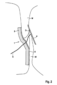
Fig. 2 shows a medical implant 1 in the form of a patch 5 at an
implant site. The implant site is a defect D in an atrial wall W
of a patient's heart. Here, the medical implant 1 is shown dur-
ing the implantation process. A delivery device C comprising a
positioning device P partially extends through the patch 5 and
cut in its center (not visible). The patch is coated with an ad-
hesive composition 6, in this case GelMA. Alternatively, glutar-
aldehyde may be employed. This provides an effective attachment
of the patch 5 to the atrial wall W.
Fig. 3 shows a similar medical implant 1 as shown in Fig. 2.
Here, the implant is shown during the implantation process, but
before detachment from the delivery device (not shown) compris-
ing a balloon B. The medical implant comprises a patch 5 made of
a knitted fabric that is attached to a balloon B by means of ad-
hesive rims 7 one the balloon-side of the medical implant 1 and
in between the outer edge OC of the medical implant 1 and its
tear line. On the other side, the implant 1 is coated with an
adhesive composition 6 within the area surround by the tear line
3. In between the tear line 3 and the outer edge OC, on the op-
CA 0315137 2022--14
WO 2021/048409 PCT/EP2020/075550
43
posite site of the adhesive rims 7, is a PTFE coating that pre-
vents wetting of the adhesive and thus adhesion. In the illus-
tration shown here, the balloon B is partially inflated.
As shown in Fig. 4, further inflation applies a force F on the
patch (not shown for clarity) and the tear lines 3 due to the
extension, in a direction orthogonal to the longitudinal axis L
of the delivery device, of the outer rims 8 of the medical im-
plant that are attached to the balloon B through the adhesive
rims 7. Thus, further inflation ruptures the tear line 3 and re-
leases the patch 5. Here, the delivery device C is shown and is
adapted to expose the patch to electromagnetic irradiation E.
Figs. 5a-5c show schematically a medical implant 1 and a release
mechanism for a medical implant 1. Here, the medical implant
comprises a patch of spun fibers of polylactic acid. However,
the person skilled in the art will of course understand that the
release mechanism could be combined with any patch material or
even any sort of medical implant. The implant 1 comprises beads
9 made of a polymeric material. Here, the beads are made of a
biodegradable polymeric material that is adapted to degrade in
the human body within typically two weeks. Of course, they could
also be adapted to degrade faster or slower. They are attached
to the edge OC of the medical implant 1.
Fig. 5a shows the medical implant 1 from the side. It is partic-
ularly well visible in this illustration that the beads 9 have a
diameter that is larger than the thickness of the medical im-
plant. Here, the beads 9 have a slightly elongated shape, but it
would be possible to arrange spherical beads as well.
In Fig. 5b, the medical implant 1 is shown from a top perspec-
tive. It is well visible that the beads are considerably smaller
CA 0315137 2022--14
WO 2021/048409 PCT/EP2020/075550
44
than the medical implant. Typically, they have a diameter of
300 pm, but could also be up to 1 mm in diameter. Here, the im-
plant 1 comprises four beads that are spaced equally around the
circumference of the medical implant 1. It would of course be
conceivable to arrange a higher or lower number of beads on the
medical implant, and/or to space them unequally.
Fig. 5c shows schematically how an implant 1 as shown in Figs.
5a and 5b can be released. The delivery device comprises at
least one tube 10, typically one tube 10 per bead 9 attached to
the medical implant 1. In said tube 10 a wire 11 with a lower
holder 12a and an upper holder 12b. The holders 12a, 12b are
adapted such that a bead 9 that is located between them in the
tube 10 cannot pass the holders along the longitudinal direction
of the tube. Thus, for implantation, the bead 9 is arranged in
the tube 10 in between the upper holder 12b and the lower holder
12a. This enables the release mechanism shown in the illustra-
tion wherein the wire 11 is actuated such that the upper holder
12b is released from the tube 10. This clears the way for the
bead 9 to also leave the tube 10, thus releasing the implant 1.
Fig. 6 shows another embodiment of a medical implant 1 and sche-
matically a release mechanism. Here, the medical implant 1 com-
prises a patch 5 made of a biodegradable fabric. The fabric com-
prises fibers that are coated with an adhesive composition (not
shown). The implant 1 comprises four extensions 13 that extend
away from the implant 1. Each extension 13 is separated from the
implant 1 by a tear line. Here, the extensions are made of the
same material as the patch 5. This is particularly simple, but
of course it would be possible to include other materials as
well. The tear lines are arranged such that upon tearing of the
tear lines, the patch 5 is substantially spherical and has a di-
ameter of 20 mm.
CA 0315137 2022--14
WO 2021/048409 PCT/EP2020/075550
Fig. 7 shows another embodiment of a medical implant 1. Here, a
balloon B is attached to a delivery device (C). The balloon is
made of implant-grade material, here from polyurethane. The bal-
5 loon is coated with an adhesive composition 6 on a distal side.
It further comprises a tear line 3 that is substantially circu-
lar and arranged in a plane that is substantially perpendicular
to the longitudinal axis L of the delivery device C. Here, the
tear line is formed as thinner wall part of the balloon B that
10 creates a pre-determined breaking point. However, it is still
sealed to allow for inflation of the balloon B. Inflation of the
balloon B creates a mechanical stress in the balloon wall in a
tangential direction. Due to the pre-determined breaking point,
the balloon rupture along the tear line 3. The patch formed by
15 the rupture adheres to the tissue by means of the adhesive com-
position 6. Thus, the medical implant was part of the balloon B
during delivery.
Fig. 8 shows another embodiment of a delivery device C. Here,
20 the delivery device C comprises a balloon B as described in oth-
er embodiments that can serve to deliver the medical implant 1.
Of course, any medical implant 1 described herein can be com-
bined with such a delivery device shown here. Here, the medical
implant consists of a fabric patch with adhesive fibers. The de-
25 livery device comprises a second balloon 14 that forms an outer
layer around the balloon B and the medical implant 1. It thus
protects the patch and the adhesive from being in contact with
tissue. Here, the outer balloon 14 comprises an opening 30 ar-
ranged approximately at the center of the medical implant 1.
30 This provides an advantageous way to implant the medical implant
1, but is optional. This allows for a preliminary attachment to
the tissue. For deployment, the outer balloon is retracted in a
direction away from the implant 1 along the longitudinal axis L
CA 0315137 2022--14
WO 2021/048409 PCT/EP2020/075550
46
of the delivery device. This exposes the implant 1 to tissue and
enables the adhesive composition to attach to the tissue.
Figs. 9a-9d show different embodiments of fabric patches that
are cut out from a fabric scaffold. Thus, the extensions 13
shown here consist of the same material as the patch 5. Through-
out the Figs. 9a-9d, only one reference sign is shown for cer-
tain identical features for clarity. The extensions 13 shown in
these embodiments have a typical length of 15 mm and a width of
3.5 mm. Of course these values can be adapted to reach a partic-
ular mechanical strength or to adapt the patch to a particular
delivery device.
Fig. 9a shows a patch with eight extensions 13 made from PET
fabric 15. The extensions 13 are separated from the patch 5 by
tear line 3 each. PET is a non-absorbable material. The shown
embodiment is thus particularly advantageous if the replacement
of the implant with tissue is not possible or undesired, for ex-
ample due to insufficient stability of the newly formed tissue.
Fig. 9b shows an embodiment of a patch 5 with only two exten-
sions 13. The fabric 15 consists of knitted poly(L-lactic acid)
(PLLA). PLLA absorbs in in the human body within two years.
Thus, the shown embodiment is particularly advantageous if cell-
ingrowth is slow or support by the patch 5 for one to two years
is desired.
Fig. 9c shows an embodiment of a patch 5 that is cut from a fab-
ric 15 made of electrospun polycaprolactone (PCL). It comprises
six extensions 13, each separated from the patch 5 by a tear
line 3. PCL is particularly advantageous for electrospinning and
thus provides an easy way to manufacture the fabric 15. It de-
grades in the human body within about six months and is thus the
CA 0315137 2022--14
WO 2021/048409
PCT/EP2020/075550
47
material of choice if relatively fast degradation is required or
desired.
Fig. 9d shows another embodiment of a patch that made of the
same electrospun PCL fabric 15 as shown in Fig. 9c. The patch 5
has a circular shape and a continuous circularly shaped tear
line 3 around its circumference that forms an outer rim 8.
Figs. 10a and 10b show another embodiment of a medical implant 1
in a cross-sectional view (Fig. 10a) and in a top view (Fig.
10b). For clarity, only one reference sign is shown for identi-
cal features. The medical implant 1 comprises an electrospun
patch wherein the fibers are functionalized and have direction-
ality. The implant 1 comprises a reservoir 16 that can be filled
with an adhesive composition. A region 18 of the patch 5 can be
functionalized such as to have a lower permeability. Here, this
prevents an adhesive disposed in the reservoir 16 from permeat-
ing through the patch and be released this side of the medical
implant. Instead, the shown embodiment comprises microchannels
17 integrated by selective laser welding. These microchannels 17
are in fluid connection with the outer surface of the medical
implant 1 and the reservoir 17. The adhesive can thus be re-
leased in a directional manner by means of the microchannels.
Figs. ha and llb show an embodiment of a spine structure 31.
Fig. ha shows a spine structure 31 by itself. The shown spine
structure is made of a polymer, similar or different from the
patch. It comprises three elongated structures 19, 20. This is
typically the most advantageous arrangement in that it provides
sufficient mechanical stability to the implant. However, it
would also be possible to adapt a spine structure with several
additional elongated structures, or with only one or two of
them, if necessary. The elongated structures comprise an inner
CA 0315137 2022--14
WO 2021/048409 PCT/EP2020/075550
48
part 20 and tear arms 19 that a separated by a predetermined
breaking point 33. The elasticity of the tear arms is generally
lower than that of the inner part. The inner part 20 is designed
to be arranged in an area close to a medical implant 1 and re-
main in the patient upon implantation. Thus, the length of one
arm of the inner part 20 is approximately half a diameter of the
implant, typically around 10 mm. Of course, the size of the
spine structure 31 as a whole and of the inner part 20 can be
adapted to a specific implant and thus be larger or smaller. The
inner part gives the implant mechanical stability during deliv-
ery and implantation, and also provides additional support after
implantation. The spine structure 31 further comprises a round-
shaped hole 32 in the middle of structure 31. This enables a de-
livery device and/or positioning device (both not shown) to ex-
tend through the spine structure 31 and be retracted through it
again. Furthermore, the less elastic tear arms 19 are can be at-
tached to a delivery device. Breaking at the predetermined
breaking point allows for the release of the implant 1. A spine
structure 31 as shown here provides a particularly advantageous
way of decoupling the force needed to release the patch from the
mechanical properties of the medical implant 1.
Fig. llb shows a spine structure with the same features as shown
in Fig. ha but in combination with a medical implant 1.
Fig. 12 shows a delivery device C for a medical implant 1 and
schematically a release mechanism. The delivery device C com-
prises an inflatable or expandable structure, such as a balloon
B and outer struts 21 to hold and release the medical implant 1.
Here, the medical implant comprises suture with notches 22 that
are held by the outer struts 21. The mechanism of holding and
releasing the notches substantially corresponds the ball release
schematically shown Fig. 5c, wherein the notches 22 have the
CA 03150927 2022-02-14
WO 2021/048409 PCT/EP2020/075550
49
technical effect of the balls shown in Fig. 5c. The balloon B
can thus be used to exert a pressure on the medical implant, but
is not necessary to release the implant 1 from the delivery de-
vice C.
Fig. 13 shows a similar delivery device C as shown in Fig. 12.
However, the outer struts 21 here are connected to the medical
implant 1 through sutures 23. The sutures are fixedly connected
to the outer struts 21 that do not comprise a mechanism to re-
lease the sutures 23. Instead, the balloon B, when inflated,
pushes the outer struts 21 away from the medical implant, thus
rupturing the connection and releasing the implant 1.
Fig. 14 shows schematically a delivery device C with a medical
implant 1 similar to the one shown in Fig. 6. The implant 1 com-
prises elongated flaps 3 that are connected to the implant 1
through tear lines 3. The shown embodiment of the implant 1 has
four such flaps 13, but could also be adapted to have smaller or
larger number of flaps. The delivery device comprises a balloon
B. Upon inflation, the balloon exerts a force on the tear lines
3, causing them to rupture and release the implant. The flaps 13
can then be retracted together with the delivery device C, while
the medical implant 1 remains in the patient. Although not shown
here, the embodiment illustrated is well suited to be combined
with spine structure as shown in Figs. ha and 11b.
Figs. 15a-15c show schematically different embodiments of patch-
es 5 comprising an adhesive composition.
Fig. 15a shows a patch 5 comprising two different types of cavi-
ties 24a, 24b. The cavities 24a, 24b are spherical and have a
diameter of approximately 1 mm. The patch has a diameter of
20 mm and consists of poly(lactic acid-co-glycolic acid). Other
CA 0315137 2022--14
WO 2021/048409 PCT/EP2020/075550
absorbable materials such as PLA- GA, PLGA, PCL, PU could also
be used. Alternatively, non-absorbable materials such as PET,
PE, and/or PP may be used. The cavities 24a, 24b contain two
different components, a resin and a hardener, of an adhesive
5 composition. The resin and the hardener are physically separated
from one another and become curable upon mixing. Thus, the adhe-
sive composition is not curable in the shown state where the two
components are separated. However, cavities 24a, 24b in the
shown embodiment are adapted to burst upon application of a me-
10 chanical pressure, for example exerted by an inflatable balloon.
The burst of the cavities causes the release of both components
of the adhesive composition, rendering it curable. Typical adhe-
sives might be GelMA (Metacrylated Gelatin), CollMA (methacry-
lated Collagen) or MeTro (methacrylated Tropoelastin).
Fig. 15b shows a patch that comprises a foam 25. The pores 35 of
the foam are surrounded by walls 34 made of a hydrogel. The hy-
drogel is adapted to degrade in the human body within 24 months.
The pores 35 are filled with an adhesive composition. The patch
is adapted to release the adhesive composition from the pores 35
upon a mechanical deformation. The adhesive composition here is
curable by exposure to electromagnetic radiation. It will be un-
derstood by the person skilled in the art that any adhesive com-
position could be combined with the shown patch, in particular
any curing mechanism. The pores 35 are adapted in their size to
facilitate cell in-growth such that after release of the adhe-
sive composition, the empty pores can serve as a scaffold for
tissue growth. The biodegradation of the patch 5 is adapted such
that the patch degrades after the formation of new tissue.
Fig. 15c shows yet another embodiment of a patch 5. The shown
patch is made of electrospun fibers 26 of polycaprolactone. The
fibers have a diameter of approximately 3 pm and a length of
CA 0315137 2022--14
WO 2021/048409 PCT/EP2020/075550
51
several 100 pm. The fibers are coated with methacrylated gelatin
as an adhesive composition. This patch can thus easily be at-
tached to tissue and is particularly easy to fabricate by elec-
trospinning. It will of course be understood by the person
skilled in the art that the fibers could also consist of an ad-
hesive composition instead of being coated by it. Similarly,
although polycaprolactone is particularly advantageous for elec-
trospinning, the fibers could be made of another material.
Figs. 16a-16b show an embodiment of medical implant 1 in a lat-
eral cross-section. The implant 1 comprises an inflation reser-
voir 27, a patch 5 and a separate layer 28 comprising reservoirs
16 for containing an adhesive. The shown embodiment is similar
to the medical implant shown in Figs. 10a and 10b.
Fig. 16a shows the medical implant 1 in a first state. The in-
flation reservoir is empty. The reservoirs 16 for comprising an
adhesive are filled with adhesive. Typically the adhesive can be
a poly(acrylic) acid which uses an acrylate/methacrylate/amuse
crosslinker.
The patch 5 is made of electrospun fibers of Dacron and is not
biodegradable. However, it would of course be possible to adapt
the patch 5 to be biodegradable as well. The layer 28 comprising
the reservoirs 16 for the adhesive is of solid Dacron.
Fig. 16b shows the medical implant 1 in a second state wherein
the inflation reservoir 27 is inflated. The inflation of the in-
flation reservoir 27 applies a mechanical pressure to the reser-
voirs 16 containing the adhesive which is subsequently squeezed
out, forming a layer of adhesive 6 on one side of the implant 1.
Here, the patch 5 is adapted to not be penetrable by the adhe-
sive composition, thus leading to a selective release of the ad-
CA 0315137 2022--14
WO 2021/048409 PCT/EP2020/075550
52
hesive composition on the other side of the implant. The infla-
tion reservoir is adapted to be removed after inflation. Howev-
er, it would also be conceivable to form it from an implant
grade material that remains in the patient.
Fig. 17 shows another embodiment of the medical implant accord-
ing to the invention in an implanted state closing a defect D in
a heart wall W. The implant 1 comprises a support structure 29
that extends around a circumference of the defect D. The support
structure is made of a shape memory polymer that provides self-
expanding at the implant site. Upon expansion, it is engaged in
the defect D. Two patches 5 are attached to the support struc-
ture 29. The patches in the shown embodiment comprise electro-
spun Dacron fibers coated with a methacrylated collagen that
swells upon exposure to humidity in the body. However, any
patches as described herein can be used, of course.
It will of course be understood by the person skilled in the art
that the embodiments described herein are examples are not re-
strictive to the scope of the invention. In particular, the dif-
ferent features described herein may be freely combined with
other features and/or used without certain features.
Fig. 18a and 18b show an embodiment of a patch 5 comprising cap-
sules 36 with a filler material 37 in the capsule wall 38.
Fig. 18a shows the patch in a first state. The capsules 36 are
spherically shaped and have a diameter of approximately 0.5-2 mm
and are evenly distributed in the patch 5. An adhesive composi-
tion 6 comprising methacryloyl-substituted tropoelastin is con-
tained in the inside of the capsules 36. Here, the capsules 36
are adapted to break open due to an osmotic pressure. For exam-
ple, exposure to blood or another liquid causes swelling of the
CA 0315137 2022--14
WO 2021/048409
PCT/EP2020/075550
53
capsules 36. The resulting pressure increase then causes burst-
ing of the capsules 36.
Fig. 18b shows the patch 5 in a second state after rupture of
the capsules 36. The adhesive composition 6 is evenly distribut-
ed over the surface of the patch 5. The filler material 37 re-
mains in the adhesive composition 6 and provides additional me-
chanical strengths to the adhesive layer.
Fig. 19a shows a patch 1 in a side view. The patch 1 has a first
surface 101' and a second surface 102. The first surface 101' is
configured as a velour-like surface having shorts strands of
fabric 101" extending away from the first surface 101'. The
second surface 102 comprises an adhesive layer. The medical im-
plant 1 is made of a polyurethane, and the velour-like surface
101' comprises polyurethane fibers. In the shown configuration,
the velour-like surface 101' enhances cell ingrowth and thus
tissue overgrowth, while the second surface 102 provides adhe-
sion to tissue.
Fig. 19b shows a similar embodiment as shown in Fig. 19a. The
medical implant 1 comprises a first surface 103' and a second
surface 103". The first surface 103' has a lower permeability
for an adhesive composition (not shown) than the first surface
103'. In the present case, this is achieved by a thicker layer
of porous material. The implant comprises the adhesive composi-
tion and can release it by means of a sponge-like mechanism when
mechanical pressure is applied. The adhesive composition prefer-
entially permeates the second surface 103" and thus, the second
surface 103" provides more adhesion as compared to the first
surface 103', when activated.
CA 0315137 2022--14
WO 2021/048409 PCT/EP2020/075550
54
Fig. 20a shows a patch 1 with an adhesive layer 104 that has a
patterned structure. The pattern is configured as five sectors
of a circular shape that forms the patch 1. Consequently, there
are four intermediate sectors 105 that do not comprise an adhe-
sive layer. The adhesive layer 104 was printed via inkjet print-
ing and is based on a mixture of porcine and fish GelMA. Alter-
natively, extrusion printing may also be employed. Presently,
the pattern is two-dimensional. The adhesive layer is thus sub-
stantially flat and the sectors 104,105 of the patch 1 differ in
whether or not they have an adhesive layer, but not in the
thickness of said adhesive layer.
Fig. 20b shows a medical implant similar to the one shown in
Fig. 20a. The patch 1 comprises an inkjet-printed pattern of ad-
hesive 104. The adhesive pattern is two-dimensional and is ar-
ranged substantially at the circumference of the patch 1. The
adhesive pattern comprises several curved lines.
It will be understood that any particular pattern of adhesive
may be arranged on a patch, in particular if the adhesive is
inkjet printed. Alternatively, extrusion printing may also be
employed.
Fig. 21a shows an embodiment of a patch 1 with a radio-opaque
element 106. The radio-opaque element 106 consists of four beads
comprising barium sulphate arranged at a circumferential area of
the patch 1 and spread substantially equally along in the direc-
tion of the circumference of the patch 1.
Fig. 21b shows an alternative embodiment of a patch 1 having a
radio-opaque element 107. The radio-opaque element 107 consists
of a cross-shaped metallic spine structure.
CA 0315137 2022--14
WO 2021/048409 PCT/EP2020/075550
Fig. 21c shows an alternative embodiment of a radio-opaque ele-
ment 108,109 on a patch 1. A spine structure 108 made of a poly-
urethane comprises small capsules 109 filled with iodine. The
iodine provides radio opacity.
5
Fig. 22 shows a patch 1 with a discrete marker 110. The discrete
marker 110 is configured as a spring-like element. The discrete
element 110 consists of titanium and is thus also radio-opaque
and echo-opaque. However, it would be possible to alternatively
10 configure the discrete marker 110 to not be radio-opaque and/or
echo-opaque. The discrete marker is deformable by pressure and
thus provides information on the pressure acting on the patch 1
at its location. Presently, the pressure can be read by measur-
ing the extension of the marker 110 along a longitudinal axis
15 (perpendicular to the surface of the patch 1), for example via
radiography.
Fig. 23a shows a medical implant 1 with a dried adhesive 111'.
The dried adhesive comprises fibers 112 of porcine GelMA spun
20 from an aqueous solution and then dried. Any gelatin may also be
employed as in addition or as an alternative to porcine GelMA.
Fig. 23b shows the medical implant 1 of Fig. 23a after exposure
to an aqueous liquid. The adhesive composition 111" is swollen
25 from water incorporation and the fibers 112" are thus larger in
diameter as compared to the dried fibers 111',112'. In the swol-
len state, the fibers 112" exhibit an adhesive force to human
tissue.
30 Fig. 24a shows a handle 113 for a delivery device. The handle
113 comprises a digital gauge 114' that is adapted to show a nu-
merical value representing an adhesive force between an implant
as shown herein and human tissue (not shown) to which it is at-
CA 0315137 2022--14
WO 2021/048409 PCT/EP2020/075550
56
tached. The gauge 114' is attached to the implant at a distal
end and measures the adhesive force.
Fig. 24b shows an alternative embodiment of a handle 113. An an-
alog gauge 114" provides a qualitative measure (for example
high, medium, low) of an adhesive force between an implant as
shown herein and human tissue (not shown) to which it is at-
tached.
It will be understood that a digital gauge may also be used to
show a qualitative measure and/or an analog gauge may be used to
show a numerical value.
Fig. 25 shows an embodiment of a medical implant 1 with a three-
dimensionally patterned adhesive 115. The adhesive is formed as
pyramids 116 evenly spaced over one surface of the implant. The
medical implant is configured as a patch made of pericardium.
The adhesive 115 consists of pyramids 116 of fish GelMA.
Figs. 26a-26d schematically show a method of patterning an adhe-
sive composition on a medical implant 1.
Fig. 26a shows a medical implant 1 made of polyurethane with a
smooth, homogeneous layer 117' of adhesive. The adhesive is
based on bovine GelMA.
Fig. 26b shows a stamp-like element 118. The stamp-like element
has a shape representing a negative of the desired adhesive pat-
tern on the medical implant 1. The stamp-like element 118 is
made of a metallic material with a PTFE coating. Thus, the
stamp-like element 118 does not adhere to the adhesive layer
117' and can be removed easily from the adhesive bearing sur-
face.
CA 0315137 2022--14
WO 2021/048409
PCT/EP2020/075550
57
Fig. 26c shows the stamp-like element 118 being pressed on the
adhesive layer 117' of the medical implant 1. The stamp-like el-
ement 118 pushes the adhesive laterally away.
Fig. 26d shows the medical implant 1 after removal of the stamp-
like element. The adhesive layer 117" has a pattern that sub-
stantially corresponds to a negative shape of the stamp-like el-
ement. The medical implant 1 hence comprises areas 119 that are
not covered by an adhesive layer. The aggregate of the area 119
substantially corresponds to the shape of the stamp-like ele-
ment.
Any of the implants and adhesives disclosed herein are suitable
to be patterned with the method shown in Figs. 26a-26d. It would
also be possible to pattern a three-dimensional pattern using
the method of Figs. 26a-26b.
Figs. 27a-27d schematically show a method to produce a medical
implant 1 comprising a retaining element for holding suture(s).
Fig. 27a shows a first step of the method. The medical implant 1
is arranged with three radially extending flaps 120. In the pre-
sent case, the extending flaps 120 are made of polyurethane and
separately attached to a patch 121 made of pericardium. One of
the three extending flaps 120 comprises an indentation 122 that
functions as a pre-determined breaking point to reduce the nec-
essary force to break the extending flap and/or to control where
breaking occurs. It would alternatively be possible to have any
number of extending flaps 120 with or without indentation.
Fig. 27b shows the medical implant 1 of Fig. 27a, wherein the
extending flaps have been folded towards the center of the medi-
CA 0315137 2022--14
WO 2021/048409 PCT/EP2020/075550
58
cal implant 1. After folding, the medical implant 1 has a sub-
stantially round shape. Around an area of the fold 123 a passage
is formed (not visible, see Fig. 27c).
Fig. 27c shows a cross-section of the medical implant 1 of Fig.
27b along the plane M. In the area of the fold 123, a passage
124 is formed that can be used, for example, to hold a suture
(not shown).
Fig. 27d shows schematically an embodiment of a medical implant
1 as shown in Figs. 27a-27c. The implant 1 is held at the fold
123 in the passage 124 by sutures 125. Pulling of the sutures
125 releases the implant 1 by tearing the extending flaps 120 at
the area of the fold 123.
Fig. 28a shows an embodiment of a spine structure 31 made of a
polyurethane. The spine structure 31 in generally suitable to be
combined with any of the disclosed medical implants. The spine
structure 31 comprises three arms 126. Each arm has a thickness
of 2 mm and further comprises an indentation 127, where the arm
126 has a reduced thickness of 1 mm. The indentation 127 is
placed at a distance of 10 mm from the center 128 of the spine
structure 31. Thus, when arranged on a medical implant, the in-
dentation is typically located at the circumference of the im-
plant. The spine structure is also configured to extend beyond
the circumference of an implant in this case, and is therefore
particularly suited to produce an implant as shown in Figs. 27a-
27c.
Fig. 28b shows a cross-section of a spine structure similar to
the one shown in Fig. 28a. The arm 126 is folded around the in-
dentation 127. In the shown embodiment, the spine structure 31
is attached to a medical implant 1 configured as a fabric patch.
CA 03150927 2022-02-14
WO 2021/048409
PCT/EP2020/075550
59
The arm 126 was bonded onto the fabric of the implant 1 by heat
bonding wherein the polyurethane was partially molten and dif-
fused into the fabric, thus providing adhesion. The folded arm
126 forms a passage 124 through which a suture is passed. The
folded arm 126 thus forms a retaining element and is held by a
suture 125. The suture is adapted in its mechanical strength
(i.e. thickness and material choice) such that pulling of the
suture may tear the arm 126 of the spine structure 31 at the in-
dentation 127. The implant further comprises a radio-opaque ele-
ment 106, configured as a platinum particle, held on the implant
1 by the folded arm 126. As an alternative to platinum, iridium
is also suited as a material for a radio-opaque marker. Addi-
tionally or alternatively, the spine structure 31 could be made
from a polymer filled with radiopaque agents, such as BaSO4.
Fig. 29 shows an implant 1 according to the invention. The im-
plant 1 is similar to the one shown in Fig. 27d. The implant
comprises extending arms 120 cut from the same base sheet mate-
rial as the body of the implant 1. The arms 120 were folded onto
the patch 1, thus leaving a small passage 124 at a circumferen-
tial area of the patch 1 to allow a suture 125 to pass through.
The sutures 125 can be mounted to a delivery system via reten-
tion elements 129. Any delivery system as shown in Figs. 12-14
is suitable to be combined with the retention elements 129.
Pulling the sutures backwards causes the sutures 129 to cut
through the polymeric sheet and thus releases the implant 1. The
required force for the cut can be controlled/adjusted by cutting
small indentations into the arm 120, as shown in Fig. 27a. The
implant further comprises a hole in a center region 128 that en-
ables additional holding with a delivery instrument, for example
for easier centering of the implant 1 at an implantation site.
CA 03150927 2022-02-14
WO 2021/048409 PCT/EP2020/075550
Fig.30 shows schematically an implant 1 attached to a tissue
wall W such as to close a defect D. The implant 1 is attached to
the tissue wall W by means of two rivets 130. The rivets consist
of a biodegradable material and degrade in a human body within a
5 year. Thus, the rivets 130 provide temporary attachment while
until, for example, a sufficient adhesive force has formed
and/or tissue has formed on the implant 1.