Note: Descriptions are shown in the official language in which they were submitted.
WO 2021/061710
PCT/US2020/052123
SYSTEMS AND METHODS FOR IMPROVING LOW DOSE VOLUMETRIC
CONTRAST-ENHANCED MRI
CROSS-REFERENCE TO RELATED APPLICATION
100011 This application claims priority to U.S. Provisional Application No.
62/905,689 filed on
September 25, 2019, the content of which is incorporated herein in its
entirety
STATEMENT AS TO FEDERALLY SPONSORED RESEARCH
100021 This invention was made with government support under Grant No. R44
EB027560
awarded by the National Institutes of Health. The government has certain
rights in the invention.
BACKGROUND
100031 Contrast agents such as Gadolinium-based contrast agents (GBCAs) have
been used in
approximately one third of Magnetic Resonance imaging (MRI) exams worldwide to
create
indispensable image contrast for a wide range of clinical applications, but
pose health risks for
patients with renal failure and are known to deposit within the brain and body
for patients with
normal kidney fimction. Recently, deep learning technique has been used to
reduce GBCA dose
in volumetric contrast-enhanced MRI, but challenges in general izability
remain due to variability
in scanner hardware and clinical protocols within and across sites.
SUMMARY
100041 The present disclosure provides improved imaging systems and methods
that can address
various drawbacks of conventional systems, including those recognized above.
Methods and
systems as described herein can improve image quality with reduced dose level
of contrast agent
such as Gadolinium-Based Contrast Agents (GBCAs). In particular, a generalized
deep learning
(DL) model is utilized to predict contrast-enhanced images with contrast dose
reduction across
different sites and scanners.
100051 Traditionally, contrast agent such as Gadolinium-Based Contrast Agents
(GBCAs) and
others has been used in a wide range of contrast-enhanced medical imaging such
as Magnetic
Resonance Imaging (MRI), or nuclear magnetic resonance imaging, for examining
pathology,
predicting prognosis and evaluating treatment response for gliomas, multiple
sclerosis (MS),
Alzheimer's disease (AD), and the like. GBCAs are also pervasive in other
clinical applications
such as evaluation of coronary artery disease (CAD), characterization of lung
masses, diagnosis
of hepatocellular carcinoma (HCC), imaging of spinal metastatic disease. In
2006, an association
between GBCA administration and the development of nephrogenic systemic
fibrosis (NSF) in
-1-
CA 03151320 2022-3-15
WO 2021/061710
PCT/US2020/052123
patients with impaired renal function was identified. Other acute side-effects
of GBCAs in
subjects with normal renal function include hypersensitivity, nausea, and
chest pain.
Subsequently, in 2017, US. FDA issued warnings and safety measures related to
Gadolinium
retention, while the regulatory bodies of Canada, Australia and other
countries issued similar
warnings. In addition to safety advisories, the European Medicines Agency has
suspended the
use of linear GBCAs. Gadolinium retention has not only been reported in the
CNS tissue in the
form of hyper-intensities on non-enhanced T1W MRI, but also in other parts of
the body.
Environmental sustainability concerns are also being raised as gadolinium is
an emerging water
pollutant. Other disadvantages of contrast-enhanced scans include patient
inconvenience during
intravenous injection, prolonged scan time, and an overall increase in imaging
costs. Even though
GBCAs have a good pharmacovigilance safety profile, there is a clear need for
dose reduction
due to the abovementioned safety issues and concerns. In particular, it is
desirable to provide a
safe imaging technique where the contrast dose can be reduced regardless the
properties or type
of the contrast materials without comprising the imaging quality or
introducing additional safety
issues.
1001061 Recent developments in Deep learning (DL) or machine learning (ML)
techniques enable
it as a potential alternative to the use of contrast dose. DL/ML has found a
plethora of
applications in medical imaging which includes denoising, super-resolution and
modality
conversion of, e.g., MRI to CT, Ti to T2. DL model has the potential to be
used for generating
contrast-enhanced images using a small fraction of the standard dose and the
pre-contrast images.
Although such method may be able to reduce dose levels while maintaining non-
inferior image
quality, the DL enhanced images often suffer from artifacts such as streaks on
a reformat image
(e.g., reformatted volumetric image or reconstructed 3D image viewed from
different planes,
orientations or angles).
00071 There exists a need for providing a robust DL model that is generalized
for (sometimes
agnostic to) diverse clinical settings such as different scanner vendors, scan
protocols, patient
demographics, and clinical indications. Such a model is also desired to
produce artifact-free
images and support a variety of clinical use cases such as multiplanar
reformat (MPR) for
oblique visualizations of 3D images, thus enabling the model to be deployed
and integrated
within a standard clinical workflow.
00081 Systems and methods described herein can address the abovementioned
drawbacks of the
conventional solutions. In particular, the provided systems and methods may
involve a DL model
including a unique set of algorithms and methods that improve the model
robustness and
-2-
CA 03151320 2022-3-15
WO 2021/061710
PCT/US2020/052123
generalizability. The algorithms and methods may include, for example, multi-
planar reconstruc-
tion, 2.5D deep learning model, enhancement-weighted Li, perceptual and
adversarial losses
algorithms and methods, as well as pre-processing algorithms that are used to
pre-process the
input pre-contrast and low-dose images prior to the model predicting the
corresponding contrast-
enhanced images.
[0009] In an aspect, a method is provided for computer-implemented method for
improving
image quality with reduced dose of contrast agent. The method comprises:
acquiring, using a
medical imaging apparatus, a medical image of a subject with a reduced dose of
contrast agent;
reformatting the medical image of the subject in multiple orientations to
generate a plurality of
reformat medical images; and applying a deep network model to the plurality of
reformat medical
images to generate a predicted medical image with improved quality.
[0010] In a related yet separated aspect, a non-transitory computer-readable
storage medium
including instructions that, when executed by one or more processors, cause
the one or more
processors to perform operations. The operations comprise: acquiring, using a
medical imaging
apparatus, a medical image of a subject with a reduced dose of contrast agent;
reformatting the
medical image of the subject in multiple orientations to generate a plurality
of reformat medical
images; and applying a deep network model to the plurality of reformat medical
images to
generate a predicted medical image with improved quality.
[0011] In some embodiments, the medical imaging apparatus is a transforming
magnetic
resonance (MR) device. In some embodiments, the medical image is a 2.5D
volumetric image.
[0012] In some embodiments, the multiple orientations include at least one
orientation that is not
in the direction of the scanning plane. In some embodiments, the method or the
operations further
comprise rotating each of the plurality of reformat medical images into
various angles to generate
a plurality of rotated reformat medical images. In some cases, the deep
network model is applied
to the plurality of rotated reformat medical images to output a plurality of
predicted images. The
plurality of predicted images as an output of the deep network model are
rotated to be aligned to
a scanning plane. In some instances, the method or the operations further
comprise averaging the
plurality of predicted images after rotated to be aligned to the scanning
plane to generate the
predicted medical image with improved quality. In some embodiments, the
predicted medical
image with improved quality is obtained by averaging a plurality of predicted
medical images
corresponding to the plurality of the reformat medical images.
[0013] Additionally, methods and systems of the present disclosure may be
applied to existing
systems without a need of a change of the underlying infrastructure. In
particular, the provided
-3-
CA 03151320 2022-3-15
WO 2021/061710
PCT/US2020/052123
methods and systems may reduce the dose level of contrast agent at no
additional cost of
hardware component and can be deployed regardless of the configuration or
specification of the
underlying infrastructure.
[0014] Additional aspects and advantages of the present disclosure will become
readily apparent
to those skilled in this art from the following detailed description, wherein
only illustrative
embodiments of the present disclosure are shown and described. As will be
realized, the present
disclosure is capable of other and different embodiments, and its several
details are capable of
modifications in various obvious respects, all without departing from the
disclosure.
Accordingly, the drawings and descriptions are to be regarded as illustrative
in nature, and not as
restrictive
INCORPORATION BY REFERENCE
[0015] All publications, patents, and patent applications mentioned in this
specification are
herein incorporated by reference to the same extent as if each individual
publication, patent, or
patent application was specifically and individually indicated to be
incorporated by reference. To
the extent publications and patents or patent applications incorporated by
reference contradict the
disclosure contained in the specification, the specification is intended to
supersede and/or take
precedence over any such contradictory material.
BRIEF DESCRIPTION OF THE DRAWINGS
[0016] The novel features of the invention are set forth with particularity in
the appended claims.
A better understanding of the features and advantages of the present invention
will be obtained
by reference to the following detailed description that sets forth
illustrative embodiments, in
which the principles of the invention are utilized, and the accompanying
drawings (also "Figure"
and "FIG." herein), of which.
[0017] FIG. 1 shows an example of a workflow for processing and reconstructing
magnetic
resonance imaging (MRI) volumetric image data.
[0018] FIG. 2 shows an example of data collected from the two different sites.
[0019] FIG. 3 shows the analytic results of a study.
[0020] FIG. 4 schematically illustrates a magnetic resonance imaging (MRI)
system in which an
imaging enhancer of the presenting disclosure may he implemented.
[0021] FIG. 5 shows an example of a scan procedure or scanning protocol
utilized for
collecting the experiment data in the study.
[0022] FIG. 6 illustrates an example of a reformat MPR reconstructed image
that have a quality
improved over the reformat MRI image generated using the conventional method.
-4-
CA 03151320 2022-3-15
WO 2021/061710
PCT/US2020/052123
100231 FIG. 7 shows an example of a pre-processing method, in accordance with
some
embodiments herein.
100241 FIG. 8 shows an example of a U-Net style encoder-decoder network
architecture, in
accordance with some embodiments herein.
100251 FIG. 9 shows an example of the discriminator, in accordance with some
embodiments
herein.
100261 FIG. 10 shows an experiment including data distribution and
heterogeneity of a study
dataset from three institutions, three different manufacturers, and eight
different scanner models.
100271 FIG. 11 schematically illustrates systems and methods that are utilized
to monotonically
improve the image quality.
100281 FIG. 12 shows examples of pre-contrast, low-dose, full-dose ground
truth image data and
synthesized images along with the quantitative metrics for cases from
different sites and
scanners.
100291 FIG. 13 shows examples illustrating effect of the number of rotation
angles in MPR on
the quality of the output image and processing time.
DETAILED DESCRIPTION
100301 While various embodiments of the invention have been shown and
described herein, it
will be obvious to those skilled in the art that such embodiments are provided
by way of example
only. Numerous variations, changes, and substitutions may occur to those
skilled in the art
without departing from the invention. It should be understood that various
alternatives to the
embodiments of the invention described herein may be employed.
100311 Gadolinium-based contrast agents (GBCAs) are widely used in magnetic
resonance
imaging (MRI) exams and have been indispensable for monitoring treatment and
investigating
pathology in myriad applications including angiography, multiple sclerosis and
tumor detection.
Recently, the identification of prolonged gadolinium deposition within the
brain and body has
raised safety concerns about the usage of GBCAs. Reducing the GBCA dose
reduces the degree
of deposition, but also degrades contrast enhancement and tumor conspicuity. A
reduced dose
exam that retains contrast enhancement is therefore greatly relevant for
patients who need
repeated contrast administration (e.g., multiple sclerosis patients) and are
at high risk of
gadolinium deposition (e.g., children).
100321 Though MRI, Gadolinium-based contrast agents, MRI data examples are
primarily
provided herein, it should be understood that the present approach can be used
in other imaging
modality contexts and/or other contrast-enhanced imaging. For instance, the
presently described
-5-
CA 03151320 2022-3-15
WO 2021/061710
PCT/US2020/052123
approach may be employed on data acquired by other types of tomographic
scanners including,
but not limited to, computed tomography (CT), single photon emission
computed tomography (SPECT) scanners, Positron Emission Tomography (PET),
functional
magnetic resonance imaging (fMRI), or various other types of imaging scanners
or techniques
wherein a contrast agent may be utilized for enhancing the contrast.
[0033] Deep learning (DL) framework has been used to reduce GBCA dose levels
while
maintaining image quality and contrast enhancement for volumetric MM. As an
example, a DL
model may use a U-net encoder-decoder architecture to enhance the image
contrast from a low-
dose contrast image. However, the conventional DL models may only work well
with scans from
a single clinical site without considering generalizability to different sites
with different clinical
workflows. Moreover, the conventional DL models may evaluate image quality for
individual 2D
slices in the 3D volume, even though clinicians frequently require volumetric
images to visualize
complex 3D enhancing structures such as blood vessels and tumors from various
angles or
orientations.
[0034] The present disclosure provides systems and methods that can address
various drawbacks
of conventional systems, including those recognized above. Methods and systems
of the
presenting disclosure capable of improving model robustness and deployment in
real clinical
settings. For instance, the provided methods and systems are capable of
adapting to different
clinical sites, each with different MRI scanner hardware and imaging
protocols. In addition, the
provided methods and systems may provide improved performance while retaining
multi-planar
reformat (MPR) capability to maintain the clinician workflow and enable
oblique visualizations
of the complex enhancing microstructure.
[0035] Methods and systems herein may provide enhancements to the DL model to
tackle real-
world variability in clinical settings_ The DL model is trained and tested on
patient scans from
different hospitals across different MRI platforms with different scanning
planes, scan times, and
resolutions, and with different mechanisms for administering GBCA. The
robustness of the DL
models may be improved in these settings with improved generalizability across
a heterogeneity
of data.
Multi-planar reformat (NIPR)
[0036] In a conventional DL pipeline, 2D slices from the 3D volume may be
separately
processed and trained with standard 2D data augmentation (e.g. rotations and
flips). The choice
of a 2D model is often motivated by memory limitations during training, and
performance
requirements during inference. In some cases, DL framework may process the
data in a "2.5D"
-6-
CA 03151320 2022-3-15
WO 2021/061710
PCT/US2020/052123
manner, in which multiple adjacent slices are input to a network and the
central slice is predicted.
However, both 2D and 2.5D processing may neglect the true volumetric nature of
the acquisition.
As the 3D volume is typically reformatted into arbitrary planes during the
clinical workflow
(e.g., oblique view, views from orientations/angles that are oblique to the
scanning
plane/orientation), and sites may use a different scanning orientation as part
of their MR1
protocol, 2D processing can lead to images with streaking artifacts in the
reformat volumetric
images (e.g., reformat into planes that are orthogonal to the scanning plane).
[0037] Methods and systems described herein may beneficially eliminate the
artifacts (e.g.,
streaking artifacts) in reformat images thereby enhancing the image quality
with reduced contrast
dose. As described above, reformatting a 3D volume image to view the image in
multiple planes
(e.g., orthogonal or oblique planes) is common in a standard clinical
workflow. In some cases,
though training a model to enhance the 2.5D image may reduce the streaking
artifacts in the
plane of acquisition, reformatting to other orientations may still cause
streaking artifacts.
Methods and systems as described herein may enable artifact-free
visualizations in any selected
plane or viewing direction (e.g., oblique view). Additionally, the model may
be trained to learn
intricate or complex 3D enhancing structures such as blood vessels or tumors.
[0038] FIG. 1 shows an example of a workflow for processing and reconstructing
Mill
volumetric image data. As illustrated in the example, the input image 110 may
be image slices
that are acquired without contrast agent (e.g., pre-contrast image slice 101)
and/or with reduced
contrast dose (e.g., low-dose image slice 103). In some cases, the raw input
image may be 2D
image slices. A deep learning (DL) model such as a U-net encoder-Decoder 111
model may be
used to predict an inference result 112. While the DL model 111 may be a 2D
model that is
trained to generate an enhanced image within each slice, it may produce
inconsistent image
enhancement across slices such as streaking artifacts in image reformats. For
instance, when the
inference result is reformatted 113 to generate a reformat image in the
orthogonal direction 114,
bcause the input 2D image 110 matches the scanning plane, the reformat image
114 may contain
reformat artifacts such as streaking artifacts in the orthogonal directions.
[0039] Such reformat artifacts may be alleviated by adopting a multi-planar
reformat (MPR)
method 120 and using a 2.5D trained model 131. The MPR method may beneficially
augment the
input volumetric data in multiple orientations. As shown in FIG. 1, a selected
number of input
slices of the pre-contrast or low-dose images 110 may be stacked channel-wise
to create a 2.5D
volumetric input image. The number of input slices for forming the 2.5D
volumetric input image
can be any number such as at least two, three, four, five, six, seven, eight,
nine, ten slices may be
-7-
CA 03151320 2022-3-15
WO 2021/061710
PCT/US2020/052123
stacked. In some cases, the number of input slices may be determined based on
the
physiologically or biochemically important structures in regions of interest
such as
microstructures where a volumetric image without artifacts are highly desired.
For instance, the
number of input slices may be selected such that microstructure (e.g., blood
vessels or tumors)
may be mostly contained in the input 2.5D volumetric image. Alternatively or
additionally, the
number of slices may be determined based on empirical data or selected by a
user. In some cases,
the number of slices may be optimized according the computational power and/or
memory
storage of the computing system.
100401 Next, the input 2.5D volumetric image may be reformatted into multiple
axes such as
principal axes (e.g., sagittal, coronal, and axial) to generate multiple
reformatted volumetric
images 121. The multiple orientations for reformatting the 2.5D volumetric
images may be in any
suitable directions that need not be aligned to the principal axes.
Additionally, the number of
orientations for reformatting the volumetric images can be any number greater
than one, two,
three, four, five and the like so long as at least one of the multiple
reformatted volumetric images
is along an orientation that is oblique to or orthogonal to the scanning
plane.
100411 At inference stage, each of the multiple reformatted volumetric images
may be rotated by
a series of angles to produce a plurality of rotated reformat volumetric
images 122 thereby
further augmenting the input data. For example, each of the three reformatted
volumetric images
121 (e.g., sagittal, coronal, and axial) may be rotated by five equispaced
angles between 0 ¨ 90*
resulting in 15 volumetric images 122. It should be noted that the angle step
and the angle range
can be in any suitable range. For example, the angle step may not be a
constant and the number
of rotational angles can vary based on different applications, cases, or
deployment scenarios. In
another example, the volumetric images can be rotated across any angle range
that is greater
than, smaller than or partially overlapping with 0 ¨ 90 . The effect of the
number of the rotational
angles on the predicted MPR images are described later herein.
[0042] The plurality of rotated volumetric 2.5D images 122 may then be fed to
the 2.5D trained
model 131 for inference. The output of the 2.5D trained model includes a
plurality of contrast-
enhanced 2.5 D volumetric images. In some cases, the final inference result
132, which is
referred to as the "MP R reconstruction" , may be an average of the plurality
of contrast-enhanced
2.5 D volumetric images after rotating back to the original
acquisition/scanning plane. For
instance, the 15 enhanced 2.5 D volumetric images may be rotated back to be
aligned to the
scanning plane and the mean of such volumetric images is the MPR
reconstruction or the final
inference result 132. The plurality of predicted 2.5 D volumetric images may
be rotated to be
-8-
CA 03151320 2022-3-15
WO 2021/061710
PCT/US2020/052123
aligned to the original scanning plane or the same orientation such that an
average of the plurality
of 2.5D volumetric images may be computed. The plurality of enhanced 2.5D
volumetric images
may be rotated to be aligned to the same direction that may or may not be in
the original
scanning plane. The MPR reconstruction method beneficially allows to add a 3D
context to the
network while benefitting from the performance gains of 2D processing.
[0043] As illustrated in FIG. 1, when the MPR reconstruction image 132 is
reformatted 133 into
a plane orthogonal to the original acquisition plane, the reformat image 135
does not present
streaking artifacts. The quality of the predicted MPR reconstruction image may
be quantified by
quantitative image quality metrics such as peak signal to noise ratio (PSNR),
and structural
similarity (SSIM). The image quality metrics are calculated for the
conventional model 111 and
the presented model 131, and an example of the result showing the quality of
the reformat images
114, 135 and ground truth 140 are illustrated in FIG. 3.
Data collection
[0044] In an example, under 1RB approval and patient consent, the scanning
protocol was
implemented in two sites. FIG. 2 shows the example of data collected from the
two sites. 24
patients (16 training, 8 testing) were recruited from Site 1 and 28 (23
training, 5 testing) from
Site 2. Differences between scanner hardware and protocol are highlighted in
Table 1. In
particular, the two sites used different scanner hardware, and had great
variability in scanning
protocol. Notably, Site 1 used power injection to administer GBCA, while Site
2 used manual
injection, leading to differences in enhancement time and strength.
[0045] As an example of collecting data for training the model, multiple scans
with reduced dose
level as well as a full-dose scan may be performed. The multiple scans with
reduced dose level
may include, for example, a low-dose (e.g., 10%) contrast-enhanced MRI and a
pre-contrast
(e.g., zero contrast) may be performed. For instance, for each participant,
two 3D Ti-weighted
images were obtained: pre-contrast and post-10% dose contrast (0.01 mmol/kg).
For training and
clinical validation, the remaining 90% of the standard contrast dose (full-
dose equivalent, 100%-
dose) was administrated and a third 3D TL-weighted image (100%-dose) was
obtained. Signal
normalization is performed to remove systematic differences (e.g., transmit
and receive gains)
that may have caused signal intensity changes between different acquisitions
across different
scanner platforms and hospital sites. Then, nonlinear affine co-registration
between pre-dose,
10%-dose, and 100%-dose images are performed. The DL model used a U-Net
encoder-decoder
architecture, with the underlying assumption that the contrast-related signal
between pre-contrast
and low-dose contrast-enhanced images was nonlinearly scaled to the full-dose
contrast images.
-9-
CA 03151320 2022-3-15
WO 2021/061710
PCT/US2020/052123
Additionally, images from other contrasts such as T2 and T2 -FLAIR can be
included as part of
the input to improve the model prediction.
100461 FIG. 5 shows an example of a scan procedure or scanning protocol 500
utilized for
collecting data for the studies or experiments shown in FIGs. 2, 3, and 10-12.
In the illustrated
scan protocol, each patient underwent three scans in a single imaging session.
Scan 1 was pre-
contrast 3D Ti-weighted MRI, followed by Scan 2 with 10% of the standard dose
of 0.1
mmol/kg. Images from Scan 1 and 2 were used as input to the DL network. Ground
truth images
were obtained from Scan 3, after administering the remaining 90% of the
contrast dose (i.e., full
dose).
100471 During inference, after deployment of the provided systems, only one
scan without
contrast agent (e.g., similar to scan 1), or a scan with reduced contrast dose
(e.g., similar to scan
2) may be performed. Such input image data may then be processed by the
trained model to
output a predicted MPR reconstructed image with enhanced contrast. In some
cases, after
deploying the model to a clinical site, a user (e.g., physician) may be
permitted to choose a
reduced dose level that can be any level in the range from 0 to 30% for
acquiring the medical
image data. It should be noted that depending on the practical implementation
and user
desired dose reduction level, the reduced dose level can be any number in a
range greater
than 30%.
Inter-site generalizability
100481 The conventional model may be limited by evaluating patients from a
single site with
identical scanning protocol. In real clinical settings, each site may tailor
its protocol based on the
capabilities of the scanner hardware and standard procedures. For example, a
model trained on
Site 2 may perform poorly on cases from Site 1 (FIG. 2, middle).
[0049] The provided DL model may have improved generalizability. The DL model
may be
trained with a proprietary training pipeline. For example, the training
pipeline may comprise first
scaling each image to a nominal resolution of 1 mm3 and in-plane matrix size
of 256x256,
followed by applying the MPR processing. As the DL model is fully
convolutional, inference can
be run at the native resolution of the acquisition without resampling.
100501 Based on the qualitative and quantitative results, the addition of MPR
processing,
resolution re-sampling, and inter-site training led to great improvement in
model robustness and
generalizability. In optional embodiments, the model may be a full 3D model.
For instance, the
model may be a 3D patch-based model that may alleviate both MPR processing,
and memory
-10-
CA 03151320 2022-3-15
WO 2021/061710
PCT/US2020/052123
usage. The provided training methods and model framework may be applied to
different sites
with different scanner platforms, and/or across different MR_I vendors.
Network architecture and processes
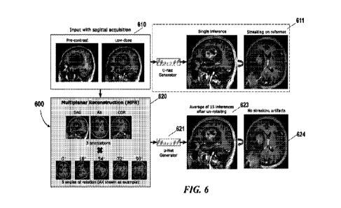
100511 FIG. 6 schematically illustrates another example of an MPR
reconstructed image 624 that
have improved quality compared to the MRI image predicted using the
conventional method 611.
The workflow 600 for processing and reconstructing MRI volumetric image data
623 and the
reformat MPR reconstructed image 624 can be the same as those as described in
FIG. 1. For
example, the input image 610 may include a plurality of 2D image slices that
are acquired
without contrast agent (e.g., pre-contrast image slice) and/or with reduced
contrast dose (e.g.,
low-dose image slice). The input images may be acquired in a scanning plane
(e.g., axial) or
along a scanning orientation. A selected number of the image slices are
stacked to form a 2.5D
volumetric input image which is further processed using the multiplanar
reconstruction (MPR)
method 620 as described above.
00521 For example, the input 2.5D volumetric image may be reformatted into
multiple axes
such as principal axes (e.g., sagittal, corona', and axial) to generate
multiple reformatted
volumetric images (e.g., SAG, AX, COR). It should be noted that the 2.5D
volumetric image can
be reformatted into any orientations that may or may not be aligned with the
principal axes.
10111531 Each of the multiple reformatted volumetric images may be rotated by
a series of angles
to produce a plurality of rotated reformat images. For example, each of the
three reformatted
volumetric images (e.g., sagittal, corona', and axial) may be rotated by five
angles between 0 ¨
90* resulting in 15 rotated reformat volumetric images. The multiple
reformatted volumetric
images (e.g., sagittal, corona', and axial) may or may not be rotated at the
same angle or rotated
into the same number of orientations.
[0054] The plurality of rotated volumetric images 122 may then be processed by
the trained
model 621 to produce a plurality of enhanced volumetric images. In some cases,
the MPR
reconstruction image 623 or the inference result image is the average of the
plurality of inference
volumes after rotating back to the original plane of acquisition. The MPR
reconstruction image
when is reformatted to be viewed at a selected orientation (e.g.,
orthogonal/oblique to the
scanning plane), the reformat image 624 may not contain streaking artifacts
compared to the
reformat image obtained using the single inference method 611 and/or the
single inference
model.
-11-
CA 03151320 2022-3-15
WO 2021/061710
PCT/US2020/052123
Network architecture and data processing
[0055] Using the multiplanar reconstruction (MPR) technique, the deep learning
model may be
trained with volumetric images (e.g., augmented 2.5D images) such as from the
multiple
orientations (e.g., three principal axes). The model may be a trained deep
learning model for
enhancing the quality of volumetric MRI images acquired using reduced contrast
dose. In some
embodiments, the model may include an artificial neural network that can
employ any type of
neural network model, such as a feedforward neural network, radial basis
function network.,
recurrent neural network, convolutional neural network, deep residual learning
network and the
like. In some embodiments, the machine learning algorithm may comprise a deep
learning
algorithm such as convolutional neural network (CNN). Examples of machine
learning
algorithms may include a support vector machine (SVM), a naive Bayes
classification, a random
forest, a deep learning model such as neural network, or other supervised
learning algorithm or
unsupervised learning algorithm. The model network may be a deep learning
network such as
CNN that may comprise multiple layers. For example, the CNN model may comprise
at least an
input layer, a number of hidden layers and an output layer. A CNN model may
comprise any
total number of layers, and any number of hidden layers. The simplest
architecture of a neural
network starts with an input layer followed by a sequence of intermediate or
hidden layers, and
ends with output layer. The hidden or intermediate layers may act as learnable
feature extractors,
while the output layer in this example provides 2.5D volumetric images with
enhanced quality
(e.g., enhanced contrast). Each layer of the neural network may comprise a
number of neurons
(or nodes). A neuron receives input that comes either directly from the input
data (e.g., low
quality image data, image data acquired with reduced contrast dose, etc.) or
the output of other
neurons, and performs a specific operation, e.g., summation. In some cases, a
connection from an
input to a neuron is associated with a weight (or weighting factor). In some
cases, the neuron
may sum up the products of all pairs of inputs and their associated weights In
some cases, the
weighted sum is offset with a bias. In some cases, the output of a neuron may
be gated using a
threshold or activation function. The activation function may be linear or non-
linear. The
activation function may be, for example, a rectified linear unit (ReLU)
activation function or
other functions such as saturating hyperbolic tangent, identity, binary step,
logistic, arcTan,
softsign, parametetic rectified linear unit, exponential linear unit,
softPlus, bent identity,
softExponential, Sinusoid, Sinc, Gaussian, sigmoid functions, or any
combination thereof
[0056] In some embodiments, the network may be an encoder-decoder network or a
U-net
encoder-decoder network. A U-net is an auto-encoder in which the outputs from
the encoder-
half of the network are concatenated with the mirrored counterparts in the
decoder-half of the
-12-
CA 03151320 2022-3-15
WO 2021/061710
PCT/US2020/052123
network. The U-net may replace pooling operations by upsampling operators
thereby increasing
the resolution of the output.
100571 In some embodiments, the model for enhancing the volumetric image
quality may be
trained using supervised learning. For example, in order to train the deep
learning network, pairs
of pre-contrast and low-dose images as input and the full-dose image as the
ground truth from
multiple subjects, scanners, clinical sites or databases may be provided as
training dataset.
[0058] In some cases, the input datasets may be pre-processed prior to
training or inference.
FIG. 7 shows an example of a pre-processing method 700, in accordance with
some
embodiments herein. As shown in the example, the input data including the raw
pre-contrast,
low-dose, and full-dose image (i.e., ground truth) may be sequentially
preprocessed to generate
preprocessed image data 710. The raw image data may be received from a
standard clinical
workflow, as a DICOM-based software application or other imaging software
applications. As an
example, the input data 701 may be acquired using a scan protocol as described
in FIG. 5. For
instance, three scans including a first scan with zero contrast dose, a second
scan with a reduced
dose level and a third scan with full dose may be operated. The reduced dose
image data used for
training the model, however, can include images acquired at various reduced
dose level such as
no more than 1%, 5%, 10%, 15%, 20%, any number higher than 20% or lower than
1%, or any
number in-between. For example, the input data may include image data acquired
from two
scans including a full dose scan as ground truth data and a paired scan at a
reduced level (e.g.,
zero dose or any level as described above). Alternatively, the input data may
be acquired using
more than three scans with multiple scans at different levels of contrast
dose. Additionally, the
input data may comprise augmented datasets obtained from simulation. For
instance, image data
from clinical database may be used to generate low quality image data
mimicking the image data
acquired with reduced contrast dose. In an example, artifacts may be added to
raw image data to
mimic image data reconstructed from images acquired with reduced contrast
dose.
[0059] In the illustrated example, pro-processing algorithm such as skull-
stripping 703 may be
performed to isolate the brain image from cranial or non-brain tissues by
eliminating signals from
extra-cranial and non-brain tissues using the DL-based library. Based on the
tissues, organs and
use application, other suitable preprocessing algorithms may be adopted to
improve the
processing speed and accuracy of diagnosis. In some cases, to account for
patient movement
between the three scans, the low-dose and full-dose images may be co-
registered to the pre-
contrast image 705. In some cases, given that the transmit and receive gains
may vary for
different acquisitions, signal normalization may be performed through
histogram equalization
-13-
CA 03151320 2022-3-15
WO 2021/061710
PCT/US2020/052123
707. Relative intensity scaling may be performed between the pre-contrast, low-
dose, and full-
dose for intra-scan image normalization. As the multi-institutional dataset
include images with
different voxel and matrix sizes, the 3D volume may be interpolated to an
isotropic resolution of
0.5mm' and wherever applicable, zero-padded images at each slice to a
dimension of 512 x 512.
The image data may have sufficiently high resolution to enable the DL network
to learn small
enhancing structures, such as lesions and metastases. In some cases, scaling
and registration
parameters may be estimated on the skull-stripped images and then applied to
the original images
709. The preprocessing parameters estimated from the skull-stripped brain may
be applied to the
original images to obtain the preprocessed image volumes 710.
[0060] Next, the preprocessed image data 710 is used to train an encoder-
decoder network to
reconstruct the contrast-enhanced image. The network may be trained with an
assumption that
the contrast signal in the full-dose is a non-linearly scaled version of the
noisy contrast uptake
between the low-dose and the pre-contrast images. The model may not explicitly
require the
difference image between low-dose and pre-contrast.
[0061] FIG. 8 shows an example of a U-Net style encoder-decoder network
architecture 800, in
accordance with some embodiments herein. In the illustrated example, each
encoder block has
three 2D convolution layers (3x3) with ReLU followed by a maxpool (2 x 2) to
downsample the
feature space by a factor of two. The decoder blocks have a similar structure
with maxpool
replaced with upsample layers. To restore spatial information lost during
downsampling and
prevent resolution loss, decoder layers are concatenated with features of the
corresponding
encoder layer using skip connections. The network may be trained with a
combination of L I
(mean absolute error) and structural similarity index (SSIM) losses. Such U-
Net style encoder-
decoder network architecture may be capable of producing a linear 10x scaling
of the contrast
uptake between low-dose and zero-dose, without picking up noise along with the
enhancement
signal.
[0062] As shown in FIG. 8, the input data to the network may be a plurality of
augmented
volumetric images generated using the MPR method as described above. In the
example, seven
slices each of pre-contrast and low-dose images are stacked channel-wise to
create a 14-channel
input volumetric data for training the model to predict the central full-dose
slices 803.
Enhancement and weighted LI lass
[0063] In some situations, even after signal normalization and scaling is
applied, the difference
between the low-dose and pre-contrast images may have enhancement-like noise
perturbations
which may mislead training of the network. To make the network pay more
attention to the
-14-
CA 03151320 2022-3-15
WO 2021/061710
PCT/US2020/052123
actual enhancement regions, the Li loss may be weighted with an enhancement
mask_ The mask
is continuous in nature and is computed from the skull-stripped difference
between low-dose and
pre-contrast images, normalized between 0 and 1 The enhancement mask can be
considered as a
normalized smooth version of the contrast uptake.
Perceptual and adversarial losses
[0064] It is desirable to train the network to focus on the structural
information in the areas of
enhancement as well as high frequency and texture details which are crucial
for making confident
diagnostic decisions. A simple combination of Li and structural similarity
index (SSIM) losses
may tend to suppress high-frequency signal information and the obtained
results may have a
smoother appearance, which is perceived as a loss of image resolution. To
address this issue, a
perceptual loss from a convolutional network (e.g., V456-19 network consisting
of 19 layers
including 6 convolution layers, 3 Fully connected layer, 5 MaxPool layers and
1 SoftMax layer
which is pre-trained on ImageNet dataset) is employed. The perceptual loss is
effective in style-
transfer and super-resolution tasks. For example, the perceptual loss can be
computed from the
third convolution layer of the third block (e.g., b1ock3 conv3) of a VGG-19
network, by taking
the mean squared error (MSE) of the layer activations on the ground truth and
prediction.
100651 In some cases, to further improve the overall perceptual quality, an
adversarial loss is
introduced through a discriminator, trained in parallel to the encoder-decoder
network, to predict
whether the generated image is real or fake. FIG. 9 shows an example of the
discriminator 900,
in accordance with some embodiments herein. The discriminator 900 has a series
of spectral
normalized convolution layers with Leaky ReLU activations and predicts a 32 x
32 patch. Unlike
a conventional discriminator, which predicts a binary value (e.g., 0 for fake
and 1 for real), the
"patch discriminator" 900 predicts a matrix of probabilities which helps in
the stability of the
training process and faster convergence. The spectral normalized convolution
layer employs a
weight normalization technique to further stabilize discriminator training.
The patch
discriminator, as shown in FIG. 9, can be trained with MSE loss, and Gaussian
noise may be
added to the inputs for smooth convergence.
100661 The function for configuring the network model can be formulated as
below:
[0067] G* = argininGRGANLGAN(G)+AuLL/(Menh.G)+AssimLssmiG)+AvGGING4G)]
[0068] where Menu is the enhancement mask and the adversarial loss LGAN can be
written as LGAN
= max-DLGAN(G, D), where G is the U-Net generator and D is the patch-
discriminator. The loss
weights AL, Assmn, AVGG and AGAN can be determined empirically. With the
abovementioned
-15-
CA 03151320 2022-3-15
WO 2021/061710
PCT/US2020/052123
processes and methods, a single model is trained to make accurate predictions
on images from
various institutions and scanners.
Example
100691 FIG. 3 shows an example of analytic results of a study to evaluate the
generalizability
and accuracy of the provided model. In the illustrated example, the results
show comparison of
ground-truth (left), original model (middle), and proposed model (right)
inference result on a test
case from Site 1 (red arrow shows lesion conspicuity). The conventional model
was trained on
data from Site 2 only. This example is consistent with the MRI scanning data
illustrated in FIG.
2. The provided model was trained on data from both sites, and used MPR
processing and
resolution resampling. In this study, the result qualitatively shows the
effect of MPR processing
on one example from the test set. By averaging the result of many MPR
reconstructions,
streaking artifacts that manifest as false enhancement are suppressed. As
shown in FIG. 3, one
slice of a ground-truth contrast-enhanced image (left) is compared to the
inference results from
the model trained on Site 2 (middle) and the model trained on Sites 1 and 2
simultaneously
(right). By accounting for differences in resolution and other protocol
deviations, the provided
model demonstrates qualitative improvement in generalizability. Quantitative
image quality
metrics such as peak signal to noise ratio (PSNR), and structural similarity
(SSIM) were
calculated for all the conventional model and the presented model. The average
PSNR and SSIM
on the test set for the conventional and presented model was 32.81 dB (38.12
dB) and 0.872
(0.951), respectively. Better image quality may be achieved using the methods
and systems in the
present disclosure.
100701 In the study as illustrated in FIG. 3, a deep learning (DL) framework
as described
elsewhere herein is applied for low-dose (e.g., 10%) contrast-enhanced MRI.
For each
participant, two 3D Ti-weighted images were obtained: pre-contrast and post-
10% dose contrast
(0.01 mmol/kg). For training and clinical validation, the remaining 90% of the
standard contrast
dose (full-dose equivalent, 100%-dose) was administrated and a third 3D Ti-
weighted image
(100%-dose) was obtained. Signal normalization was performed to remove
systematic
differences (e.g., transmit and receive gains) that may have caused signal
intensity changes
between different acquisitions across different scanner platforms and hospital
sites. Then,
nonlinear affine co-registration between pre-dose, 10%-dose, and 100%-dose
images were
performed. The DL model used a U-Net encoder-decoder architecture, with the
underlying
assumption that the contrast-related signal between pre-contrast and low-dose
contrast-enhanced
-16-
CA 03151320 2022-3-15
WO 2021/061710
PCT/US2020/052123
images was nonlinearly scaled to the full-dose contrast images. Images from
other contrasts such
as T2 and T2 -FLAIR can be included as part of the input to improve the model
prediction.
100711 As another example of an experiment in connection with FIG. 10- FIG.
13, data
distribution and heterogeneity of a study dataset from three institutions,
three different
manufacturers, and eight different scanner models are shown in FIG. 10. The
study
retrospectively identified 640 patients (323 females; 52 J 16 years),
undergoing clinical brain
MN exams from three institutions, three scanner manufacturers and eight
scanner models using
different institutional scan protocols, including different imaging planes,
field strengths, voxel
sizes, matrix sizes, use of fat suppression, contrast agents and injection
methods. The clinical
indications included suspected tumor, post-op tumor follow-up, routine brain,
and others
requiring MRI exams with GBCAs. Each subject underwent 3D pre-contrast Tlw
imaging,
followed by a low-dose contrast-enhanced Tlw scan with 10% (0.01 mmol/kg) of
the standard
dose (0.1 mmol/kg). For training and evaluation, a third 3D Tlw image was
obtained with the
remaining 90% (0.09 mmol/kg) of the full dose, which was considered as the
ground truth. All
three acquisitions were made in a single imaging session, and the patients did
not receive any
additional gadolinium dose compared to the standard protocol.
100721 Out of 640 cases, the model as shown in FIG. 11 was trained with 56
cases, and 13
validation cases were used to fine-tune the hyper-parameters and empirically
find the optimal
combination of loss weights, To ensure that the model generalizes well across
sites and vendors,
the train and validation sets consisted of approximately an equal number of
studies from all the
institutions and scanner manufacturers (refer FIG. 10). The remaining 571
cases were held-out
for testing and model evaluation. The model was implemented in Python 3.5
using Keras with
Tensorflow backend and was trained on Nvidia Tesla V100 (SXM2 32GB) GPU for
100 epochs
with a batch size of 8. Model optimization was performed using Adam optimizer
with a learning
rate of 0.001.
100731 The model is quantitatively evaluated using a plurality of metrics.
Peak signal-to-noise
ratio (PSNR) is the scaled version of pixel-wise differences, whereas
structural similarity index
(SSIM) is sensitive to changes in local structure and hence captures the
structural aspect of the
predicted image with respect to the ground truth. Using the 571 test cases,
the model was
quantitatively evaluated using the PSNR and SSIM metrics, computed between the
true full-dose
and synthesized images. These values were compared with the PSNR and SSIM
values between
low-dose and full-dose images. Per-site and per-scanner metrics were also
calculated and
compared to prove model generalizability,
-17-
CA 03151320 2022-3-15
WO 2021/061710
PCT/US2020/052123
100741 From the test set, a subset of images from 26 patients (13 males; 58
15 years), with
different types and grades of enhancing tumor cases (either pre- or post-
operative) were
identified and used for an in-depth evaluation of model performance. These
enhancing tumor
cases were similar to the training dataset in terms of heterogeneity and were
acquired using the
same scanning protocol as shown in FIG. 5. A binary assessment was performed
to find if the
enhancement pattern agreed without any false positives or false negatives
(with true full-dose
images as the reference). When present, image artifacts in the synthesized
images were recorded
and the image artifacts are proved to be reduced with aid of the provided
model.
100751 To further validate that the model predictions were similar to the full-
dose ground truth,
automatic tumor segmentation is performed on the 26 enhancing tumor cases. The
variant of the
model applied, used only post-contrast images to segment the tumor core. As
per the
requirements of the segmentation model, the ground truth and predicted full-
dose images were
skull-stripped, interpolated to 1 mm3 resolution and co-registered to an
anatomical template. The
evaluation is performed by computing the Dice scores of the predicted tumor
core between the
segmented masks of the ground-truth and those created using the synthesized
images.
100751 FIG. 11 schematically illustrates systems and methods are utilized to
monotonically
improve the image quality. The example is shown for a sagittally acquired MR
image with an
enhancing frontal tumor. Vertical streaks can be seen in the axial reformat of
the 2.5D model
result as shown in panel a, which was fixed by MPR training and inference as
shown in panel b.
Adding perceptual and adversarial losses further improves the texture inside
the tumor and
restored overall perceptual quality as shown panel c. Additionally, weighting
the Li loss with the
smooth enhancement mask matched the enhancement pattern to that of the ground
truth, as
shown in panel d. The monotonic increase in the metrics with respect to the
ground truth (as
shown in panel e) also illustrates the improvement of model. Below table shows
the model
improvement for each of the proposed technical solutions for the 26 enhancing
tumor cases.
Metric UNet 2D (35) UNet 2.5D -I- MP
-F VGG & GAN + Enhancement mask*
PSNR (c113)
31.84 4,8K 3238 4,67 33.56 5,19 34.28 4,8K 35.22 +
4.79
SSIM
0.88 0.06 0.89 0.06 0.90 0.06 0.92 0.05 0.93 +
0.04
100771 FIG. 12 shows pre-contrast, low-dose, full-dose ground truth and
synthesized images
along with the quantitative metrics for cases from different sites and
scanners. The metrics show
that the model with the proposed technical improvements performed better than
the original
model (with metrics 31.84+4.88 dB, 0.88+0.06). The best performing model used
MPR with five
rotations with a combination of SSB4, perceptual, adversarial, and enhancement
weighted Li
-18-
CA 03151320 2022-3-15
WO 2021/061710
PCT/US2020/052123
losses. For a 512 x 512 x 300 volume, preprocessing and inference of the best
model took about
135 seconds on a GeForce RTX 2080 (16 GB) GPU.
100781 FIG. 13 shows examples of different number of rotations and the
corresponding effect on
the quality of the image and the performance. The effect of the number of
rotation angles in MPR
as shown in FIG. 13 provides that greater number of angles may reduce the
horizontal streaks
inside the tumor (better quality), while it may also increase the inference
time. When deploy a
trained model to a physical site, the number of rotations and different angles
may be determined
based on the desired image quality and deployment environment (e.g.,
computational power,
memory storage, etc.).
System overview
100791 The provided DL framework for low-dose contrast-enhanced MRI is capable
of reducing
the dosage of GBCA for contrast-enhanced MRI while preserving image quality
and avoiding
degradation in contrast enhancement. The robustness and generalizability of
the DL model is
improved thereby allowing for improved adaptation to various applications
across a
heterogeneous patient and site population_ FIG. 4 schematically illustrates a
magnetic resonance
imaging (MRI) system 400 in which an imaging enhancer 440 of the presenting
disclosure may
be implemented. The MRI system 400 may comprise a magnet system 403, a patient
transport
table 405 connected to the magnet system, and a controller 401 operably
coupled to the magnet
system. In one example, a patient may lie on the patient transport table 405
and the magnet
system 403 would pass around the patient. The controller 401 may control
magnetic fields and
radio frequency (RF) signals provided by the magnet system 403 and may receive
signals from
detectors in the magnet system 403.
100801 The MRI system 400 may further comprise a computer system 410 and one
or more
databases operably coupled to the controller 401 over the network 430. The
computer system
410 may be used for implementing the volumetric MR imaging enhancer 440. The
volumetric
MR imaging enhancer 440 may implement the DL framework and methods described
herein. For
example, the volumetric MR imaging enhancer may employ the MPR reconstruction
method and
various other training algorithms, and data processing methods described
herein. The computer
system 410 may be used for generating an imaging enhancer using training
datasets_ Although
the illustrated diagram shows the controller and computer system as separate
components, the
controller and computer system can be integrated into a single component.
100811 The computer system 410 may comprise a laptop computer, a desktop
computer, a central
server, distributed computing system, etc. The processor may be a hardware
processor such as a
-19-
CA 03151320 2022-3-15
WO 2021/061710
PCT/US2020/052123
central processing unit (CPU), a graphic processing unit (GPU), a general-
purpose processing
unit, which can be a single core or multi core processor, or a plurality of
processors for parallel
processing. The processor can be any suitable integrated circuits, such as
computing platforms or
microprocessors, logic devices and the like. Although the disclosure is
described with reference
to a processor, other types of integrated circuits and logic devices are also
applicable. The
processors or machines may not be limited by the data operation capabilities.
The processors or
machines may perform 512 bit, 256 bit, 128 bit, 64 bit, 32 bit, or 16 bit data
operations.
100821 The MRI system 400 may include one or more databases 420 that may
utilize any suitable
database techniques. For instance, structured query language (SQL) or "NoSQL"
database may
be utilized for storing the reconstructed/reformat image data, raw collected
data, training
datasets, trained model (e.g., hyper parameters), weighting coefficients,
rotation angles, rotation
numbers, orientation for reformat reconstruction, etc. Some of the databases
may be implemented
using various standard data-structures, such as an array, hash, (linked) list,
struct, structured text
file (e.g., 3CML), table, JSON, NOSQL and/or the like. Such data-structures
may be stored in
memory and/or in (structured) files In another alternative, an object-oriented
database may be
used. Object databases can include a number of object collections that are
grouped and/or linked
together by common attributes; they may be related to other object collections
by some common
attributes. Object-oriented databases perform similarly to relational
databases with the exception
that objects are not just pieces of data but may have other types of
functionality encapsulated
within a given object. If the database of the present disclosure is
implemented as a data-structure,
the use of the database of the present disclosure may be integrated into
another component such
as the component of the present invention. Also, the database may be
implemented as a mix of
data structures, objects, and relational structures. Databases may be
consolidated and/or
distributed in variations through standard data processing techniques.
Portions of databases, e.g.,
tables, may be exported and/or imported and thus decentralized and/or
integrated.
100831 The network 430 may establish connections among the components in the
MRI platform
and a connection of the MRI system to external systems. The network 430 may
comprise any
combination of local area and/or wide area networks using both wireless and/or
wired
communication systems. For example, the network 430 may include the Internet,
as well as
mobile telephone networks. In one embodiment, the network 430 uses standard
communications
technologies and/or protocols. Hence, the network 430 may include links using
technologies such
as Ethernet, 802.11, worldwide interoperability for microwave access (WiMAX),
2G/3G/4G/5G
mobile communications protocols, InfiniBand, PCI Express Advanced Switching,
etc. Other
networking protocols used on the network 430 can include multiprotocol label
switching
-20-
CA 03151320 2022-3-15
WO 2021/061710
PCT/US2020/052123
(MPLS), the transmission control protocol/Internet protocol (TCP/IP), the User
Datagram
Protocol (UDP), the hypertext transport protocol (IITTP), the simple mail
transfer protocol
(SMTP), the file transfer protocol (FTP), and the like. The data exchanged
over the network can
be represented using technologies and/or formats including image data in
binary form (e.g.,
Portable Networks Graphics (PNG)), the hypertext markup language (HTML), the
extensible
markup language (XML), etc. In addition, all or some of links can be encrypted
using
conventional encryption technologies such as secure sockets layers (SSL),
transport layer
security (TLS), Internet Protocol security (IPsec), etc. In another
embodiment, the entities on the
network can use custom and/or dedicated data communications technologies
instead of, or in
addition to, the ones described above.
100841 Whenever the term "at least," "greater than," or "greater than or equal
to" precedes the
first numerical value in a series of two or more numerical values, the term
"at least," "greater
than" or "greater than or equal to" applies to each of the numerical values in
that series of
numerical values. For example, greater than or equal to 1, 2, or 3 is
equivalent to greater than or
equal to 1, greater than or equal to 2, or greater than or equal to 3.
100851 Whenever the term "no more than," "less than," or "less than or equal
to" precedes the
first numerical value in a series of two or more numerical values, the term
"no more than," "less
than," or "less than or equal to" applies to each of the numerical values in
that series of numerical
values. For example, less than or equal to 3, 2, or I is equivalent to less
than or equal to 3, less
than or equal to 2, or less than or equal to 1.
00861 As used herein A and/or B encompasses one or more of A or B, and
combinations thereof
such as A and B. It will be understood that although the terms "first,"
"second," "third" etc. are
used herein to describe various elements, components, regions and/or sections,
these elements,
components, regions and/or sections should not be limited by these terms.
These terms are
merely used to distinguish one element, component, region or section from
another element,
component, region or section. Thus, a first element, component, region or
section discussed
herein could be termed a second element, component, region or section without
departing from
the teachings of the present invention.
100871 The terminology used herein is for the purpose of describing particular
embodiments only
and is not intended to be limiting of the invention. As used herein, the
singular forms "a", "an"
and "the" are intended to include the plural forms as well, unless the context
clearly indicates
otherwise. It will be further understood that the terms "comprises" and/or
"comprising," or
"includes" and/or "including," when used in this specification, specify the
presence of stated
-21-
CA 03151320 2022-3-15
WO 2021/061710
PCT/US2020/052123
features, regions, integers, steps, operations, elements and/or components,
but do not preclude the
presence or addition of one or more other features, regions, integers, steps,
operations, elements,
components and/or groups thereof
[0088] Reference throughout this specification to "some embodiments," or "an
embodiment,"
means that a particular feature, structure, or characteristic described in
connection with the
embodiment is included in at least one embodiment. Thus, the appearances of
the phrase "in
some embodiment," or "in an embodiment," in various places throughout this
specification are
not necessarily all referring to the same embodiment. Furthermore, the
particular features,
structures, or characteristics may be combined in any suitable manner in one
or more
embodiments
[0089] While preferred embodiments of the present invention have been shown
and described
herein, it will be obvious to those skilled in the art that such embodiments
are provided by way of
example only. It is not intended that the invention be limited by the specific
examples provided
within the specification. While the invention has been described with
reference to the
aforementioned specification, the descriptions and illustrations of the
embodiments herein are not
meant to be construed in a limiting sense. Numerous variations, changes, and
substitutions will
now occur to those skilled in the art without departing from the invention.
Furthermore, it shall
be understood that all aspects of the invention are not limited to the
specific depictions,
configurations or relative proportions set forth herein which depend upon a
variety of conditions
and variables. It should be understood that various alternatives to the
embodiments of the
invention described herein may be employed in practicing the invention. It is
therefore
contemplated that the invention shall also cover any such alternatives,
modifications, variations
or equivalents. It is intended that the following claims define the scope of
the invention and that
methods and structures within the scope of these claims and their equivalents
be covered thereby.
-22-
CA 03151320 2022-3-15