Note: Descriptions are shown in the official language in which they were submitted.
WO 2021/055013 PCT/US2020/024592
1
PROCESSES AND AGENTS FOR GLAUCOMA
TECHNICAL FIELD
[0001] This invention relates to methods and compositions
for detecting, identifying and treating
glaucoma diseases. More particularly, this invention discloses compositions
and methods for affecting
intraocular pressure and increasing ocular outflows in glaucoma.
BACKGROUND
[0002] Glaucoma diseases are a world-wide leading cause of
vision loss and affect an estimated
70 million people. Glaucoma is a permanently blinding disease that is
asymptomatic until patients
experience advanced vision loss. Diagnosis of glaucoma is often delayed.
[0003] Forms of glaucoma are described as open angle
glaucoma or closed angle glaucoma.
Primary open-angle glaucoma (POAG) is most prevalent, about 75% of cases.
Narrow angle
glaucoma and other less common forms account for the other 25%. In POAG, the
anterior chamber
angle appears healthy and open and there is elevated intraocular pressure
(lOP) with no underlying
disease.
[0004] Risk factors for POAG include elevated IOP,
advancing age, family history, African
ancestry, myopia, and associations with diabetes or hypertension. The
pathophysiology responsible
for glaucoma is related to increased resistance to aqueous outflow, but the
direct mediator of this
process remains unknown. The etiology of glaucoma is poorly understood and the
factors contributing
to its progression have not been identified.
[0005] Signs and symptoms of glaucoma include damage to the
optic nerve, with degeneration of
retinal ganglion cells, changes to the optic nerve head, and corresponding
visual field loss. Elevation
of IOP is related to retinal ganglion cell (RGC) death and ultimately visual
field (VF) loss. IOP-related
optic nerve damage is important in the pathogenesis of POAG. Patients with
POAG and ocular
hypertension (OHT) have elevated IOP. Raised IOP is a significant risk factor
for the progression
from OHT to POAG. Raised IOP is the only common clinical finding in a wide
variety of secondary
glaucomas. Reduction of LOP was shown to lower the risk of progression in NTG.
In animal models,
raised IOP precedes glaucomatous nerve damage. In general, the etiology of
elevated IOP may be due
to reduced aqueous outflow.
CA 03151625 2022-3-17
WO 2021/055013 PCT/US2020/024592
2
[0006] Pharmaceutical treatment of glaucoma is directed to
lowering IOP which may slow disease
progression in some patients! Drawbacks of current treatments include lack of
efficacy and side effects
of medications.
[0007] What is needed are effective methods and
compositions for glaucoma, as well as modalities
for lowering IOP and improving ocular outflows.
[0008] There is an urgent need for methods, kits, and
compositions for detecting, identifying and
treating glaucoma.
BRIEF SUMMARY
[0009] This invention provides methods, compositions,
devices, kits and reagents for detecting,
identifying and treating glaucoma diseases.
[0010] In some aspects, this invention provides methods and
compositions for reducing
intraocular pressure and increasing ocular outflows in glaucoma subjects.
Aspects of this invention
can reduce formation and presence of aggregational features and structures in
ocular humor.
[0011] In further aspects, this disclosure provides
therapeutic compositions for glaucoma.
[0012] Embodiments of this invention provide devices for
measuring and characterizing
glaucoma aggregational features, as well as intraocular pressure and ocular
outflows.
[0013] Additional aspects of this disclosure include
diagnostic and screening modalities for
glaucoma. Further embodiments include kits and reagents for carrying out the
foregoing.
[0014] Embodiments of this invention include the following:
[0015] An aqueous pharmaceutical composition for ophthalmic
use comprising an active
agent selected from cetylpyridinium chloride, polymyxin B sulfate, a mycin,
and heparin
sodium. The mycin can be selected from neomycin, salinomycin, azithromycin,
rapamycin,
gentamycin, erythromycin, adriamycin, bleomycin, dactinomycin, mitomycin,
plicamycin,
dihydrostreptomycin, kanamycin, natamycin, rifamycin, and tobramycin. The
active agent
may comprise 0.01-2% w/v of the composition. The active agent can comprise
0.01-0.2%
w/v of the composition. The composition can have a pH of about 7.3.
[0016] The composition above may reduce intraocular
pressure when administered to the
eye.
[0017] The composition above may reduce ocular
extracellular vesicle complexes when
administered to the eye.
CA 03151625 2022-3-17
WO 2021/055013 PCT/US2020/024592
3
[0018] The composition above can be effective for treating
a glaucoma disease when
administered to the eye.
[0019] The composition above can further comprise one or
more of a solubilizer, a
surfactant, a tonicifier, and a preservative. The solubilizer may be selected
from a phosphate,
a citric acid monohydrate, a trisodium citrate, and combinations thereof. The
surfactant can
be selected from a phospholipid, a polyglycerol ester, a propylene glycol
ester, a
polyethylene glycol ester, a copolymer ester, a polyoxyethylene sorbitan
ester, a
cyclodextrin, a polyvinyl alcohol, povidone, a hydroxypropyl methyl cellulose,
a poloxamer,
a carboxymethyl cellulose, a hydroxyethyl cellulose, a polyacrylate, and
combinations
thereof. The tonicifier may be selected from sodium chloride, trehalose,
mannitol, sorbitol,
dextrose, potassium chloride, and combinations thereof The preservative can be
selected
from benzalkonium chloride, polyquaternium-1, benzododecinium bromide, sorbic
acid,
methyl paraben, propyl paraben, chlorobutanol, benzylic alcohol, phenylethyl
alcohol, an
oxychloro complex, thimerosal, sodium perborate, disodium edetate, and
combinations
thereof.
[0020] A composition above may be used in medical therapy,
in the treatment of the
human or animal body, in reducing intraocular pressure in the human or animal
body, in
reducing ocular extracellular vesicle complexes in the human or animal body,
or in preparing
or manufacturing a medicament for preventing, ameliorating, or treating a
disease or
condition associated with glaucoma in a subject in need.
[0021] Embodiments of this invention further contemplate
methods for treating a
glaucoma disease, reducing intraocular pressure, or reducing ocular
extracellular vesicle
complexes in a subject in need thereof, the method comprising administering a
composition
above to the eye of the subject. The administration may be by injection. The
ocular
extracellular vesicle complexes can be aggregates of extracellular vesicles
having a diameter
greater than about 300 manometers.
BRIEF DESCRIPTION OF THE DRAWINGS
[0022] FIG. 1 shows an illustration of normal eye (left)
anatomy compared to glaucomatous eye
(right).
[0023] FIG. 2 shows an electron micrograph of a sample of
trabecular meshwork (left), and an
illustration of aqueous humor flow in eye trabecular meshwork (right).
CA 03151625 2022-3-17
WO 2021/055013 PCT/US2020/024592
4
100241 FIG. 3 shows a diagram of an embodiment of a device
for detecting flow and pressure of
a fluid composition in a channel. The channel may contain a meshwork having
properties like a
trabecular meshwork of the eye. The channel meshwork may have a uveal meshwork
portion, a
corneoscleral meshwork portion, and a justacanalicular meshwork portion The
channel can be a
microfluidic channel.
100251 FIG. 4 shows an embodiment of a series of test
channels for detecting the effect or
various compounds, compositions and substances on the flow and pressure of a
fluid composition in
a channel.
100261 FIG. 5 shows an embodiment of a device and system
for detecting flow and pressure of a
fluid composition in a channel. A channel may contain a meshwork having
properties like a
trabecular meshwork of the eye. Alternatively, the device may utilize a
reservoir for a test sample of
a fluid composition. The channel can be a microfluidic channel.
100271 FIG 6 shows imaging of extracellular vesicles with
conventional fixation techniques
result in inefficient EV imaging due to failure of EVs to adhere. Imaging is
surprisingly improved
using a non-reversible crosslinking reagent. (a) Representative
photomicrographs of isolated bovine
vitreous EVs, 4 million loaded, fixed to the copper grid with glutaraldehyde
and subsequent UA and
lead citrate solution, show few negatively stained EVs (arrowhead) at low
(left) magnification, and
in other photographs no EVs are visualized (middle and right). Negatively
stained EVs are shown
with signal (black) surrounding the perimeter of the EV and lower signal
(white or grey) in the
center. (b) Schematic diagram shows that EVs in solution that were applied to
the electron
microscopy grid surface fail to attach_ The EVs are present in the discarded
solution (black box) and
we quantitated the size and number of EVs using nanoparticle tracking analysis
(NTA). (c) NTA
shows the size and concentration of EVs applied to the TEM grid surface. (d)
NTA shows the size
and concentration of EVs that were present in the discarded solution. This
number represents the size
and number of EVs that fail to adhere to the TEM grid surface. (g) Graphical
representation
comparing the amount of EVs applied to the electron microscopy grid surface
(black bar) and the
size and concentration of EVs present in the discarded solution (grey bars, n
= 3) (f) Representative
TEM photomicrographs of isolated bovine vitreous EVs after EDC-glutaraldehyde-
fixation, negative
staining and TEM imaging reveal substantially more EVs visualized at low
(left), medium (middle)
and high (right) magnification. (g) Graphical representation (1og2) of the
mean and standard
deviation that shows significantly more EVs counted per image from EDC fixed
specimens (350-
CA 03151625 2022-3-17
WO 2021/055013 PCT/US2020/024592
fold), when compared to Glut fixed grids (n = 3, counted on average seven
images per biological
replicate, sp <0.05). (h) Representative TEM photomicrographs of isolated
bovine aqueous humor
after EDC-glutaraldehyde-fixation show robust negative staining surrounding
the border of the EV.
(i) TEM images from human aqueous humor, without performing EV isolation, show
multiple EVs
present, in situ. Scale bars are (a) 1000 nm (left), 600 nm (middle), 125 nm
(right), (f) 600 nm (left),
125 nm (right); 00 500 nm; and (i) 1 pm.
100281 FIG. 7 shows healthy human aqueous humor, control,
Patient #1, shows a diffuse
distribution of non-aggregated EVs. (a-c) Representative photomicrographs of
diluted human
healthy control aqueous humor EVs (EVs were NOT isolated with
ultracentrifugation, only diluted
with buffered saline), fixed to the copper grid with EDC, glutaraldehyde and
subsequently stained
with UA solution. Photographs were captured with transmission electron
microscopy and recorded.
The images show many negatively stained EVs of various sizes, and the majority
of EVs exist in the
fluid independent without aggregation. One EV-aggregate was observed and was
in the minority of
samples imaged. Scale bars are marked on the photographs.
100291 FIG. 8 shows healthy human aqueous humor, control,
Patient #2, shows a diffuse
distribution of non-aggregated EVs. (a-d) Representative photomicrographs of
diluted human
healthy control aqueous humor EVs (EVs were NOT isolated with
ultracentrifugation, only diluted
with buffered saline), fixed to the copper grid with EDC, glutaraldehyde and
subsequently stained
with UA solution. Photographs were captured with transmission electron
microscopy and recorded.
The images show many negatively stained EVs of various sizes. No EV-aggregates
larger than 2 pm
were observed, nor were large clumps of EVs visualized. Scale bars are marked
on the photographs.
100301 FIG. 9 shows POAG aqueous humor and shows large glaucoma-associated-EV-
aggregates in Patient #1. (a-c) Representative transmission electron
microscopy photographs show
POAG aqueous humor samples (EVs were NOT isolated with ultracentrifugation,
only diluted with
buffered saline), fixed to the copper grid with EDC, glutaraldehyde and
subsequently stained with
UA solution. Photographs were captured with transmission electron microscopy
and recorded. The
diluted POAG specimens show evidence of glaucoma-associated-EV-aggregates
present in the fluid
that are large in size. Images show few free EVs observed (a-c). Scale bars
are marked on the
photographs.
100311 FIG. 10 shows POAG aqueous humor and shows large glaucoma-associated-EV-
aggregates in Patient #2. (a-c) Representative transmission electron
microscopy photographs from a
CA 03151625 2022-3-17
WO 2021/055013 PCT/US2020/024592
6
second POAG sample (EVs were NOT isolated with ultracentrifugation, only
diluted with buffered
saline), fixed to the copper grid with EDC, glutaraldehyde and subsequently
stained with UA
solution. Photographs were captured with transmission electron microscopy and
recorded The
second POAG aqueous humor specimens show evidence of sizeable glaucoma-
associated-EV-
aggregates and larger free EVs. Scale bars are marked on the photographs.
100321 FIG. 11 shows a known glaucoma treatment,
bimatoprost, reduces the size of POAG-
associated EV complexes in the aqueous humor of a patient with glaucoma, when
compared to
untreated glaucoma samples. (a-b) Representative transmission electron
microscopy photographs
show human aqueous humor collected from a patient with POAG and no treatment
(placebo, control,
buffered saline) shows evidence of extracellular matrix and glaucoma
associated-EV-complexes in
the aqueous humor. Images show large collagen-like matrix in all panels and
these electron dense
structures measure several microns in size. (c-d) Representative photographs
show POAG aqueous
humor samples treated with a glaucoma medication, bimatoprost, and show no
evidence of large
extracellular matrix nor glaucoma-associated-EV-aggregates. On high power
imaging, there appears
to be larger globules that are not aggregated. Scale bars are marked on the
figures.
100331 FIG. 12 shows treatment of a glaucoma patient's
aqueous humor with buffered saline has
no effect on POAG-associated EV complexes of a second subject diagnosed with
glaucoma. (FIG.
12) Representative transmission electron microscopy photographs show human
aqueous humor
collected from a patient with POAG and no treatment (placebo, control,
buffered saline) shows
evidence of glaucoma associated-EV-complexes. Images show large electron dense
glaucoma
associated-EV-complexes in all panels. The placebo treatment had no effect the
glaucoma
associated-EV--complexes. Scale bars are marked on the figures.
100341 FIG. 13 shows bimatoprost reduces the size of POAG-
associated EV complexes in the
aqueous humor of a second subject diagnosed with glaucoma, when compared to
control-treated
glaucoma samples. (FIG. 13) Representative photographs show POAG aqueous humor
from Subject
#2 samples treated with bimatoprost show a disruption of the large electron
dense glaucoma-
associated-EV-aggregates.. The glaucoma-associated-EV-aggregates were smaller
in size, when
compared to the controls (shown in FIG. 12). On high power imaging, we do not
observe many
aggregated EVs. Scale bars are marked on the figures.
00351 FIG. 14 shows glaucoma patients' aqueous humor
contains larger electron dense
structures in the aqueous humor that are not present in healthy controls.
Graphical representation
CA 03151625 2022-3-17
WO 2021/055013
PCT/US2020/024592
7
that depicts the size and number of an unidentified material that we termed,
"glaucoma-associated-
EV-aggregates" in healthy controls or POAG specimens. We obtained healthy
control or POAG
human aqueous humor, fixed the samples with EDC, and imaged the specimens with
transmission
electron microscopy. Photographs were analyzed and the number of glaucoma-
associated-EV-
aggregates were identified and plotted for each variable. In aqueous humor
obtained from a single
healthy control subject, the data shows that most EVs did not have glaucoma-
associated-EV-
aggregates. The data show that three subjects with a diagnosis of POAG had
numerous glaucoma-
associated-EV-aggregates that are several micrometers in size. Theses
aggregates were not observed
in healthy controls. The graph shows a substantial difference in the size of
the glaucoma-associated-
EV-aggregates in POAG, when compared to healthy controls.
00361 FIG. 15 shows glaucoma patients' aqueous humor
contains EVs that aggregate and
contact each other, when compared to healthy controls that have fewer EVs
touching each other_
Graphical representation that depicts the number of EVs that are in contact
with each other (X-axis)
and the count frequency (percent of total). We obtained healthy control or
POAG human aqueous
humor, fixed the samples with EDC, and imaged the specimens with transmission
electron
microscopy. Photographs were analyzed and the number of EVs contacting each
other as
determined for each variable. In aqueous humor obtained from a single healthy
control subject, the
data shows that most EVs are free and not present in aggregates. The data
shows that two subjects
with a diagnosis of POAG have aqueous humor that contains EVs that contact a
large number of
other EVs. The graph shows a substantial difference between the number of EVs
contacting each
other in POAG, when compared to healthy controls.
100371 FIG. 16 shows a curvilinear chart of the data in
FIG. 15. Glaucoma patients' aqueous
humor contains EVs that aggregate and contact each other, when compared to
healthy controls that
have fewer EVs touching each other.
00381 FIG. 17 shows extracellular vesicles are present in
human aqueous humor obtained from
healthy control patients and have a dominant population of EVs between 100 to
200 nm. Graphical
representation that depicts the EV population in human aqueous humor obtained
from a single
healthy control patient The EV count frequency is shown as a function of size.
We fixed healthy
control human aqueous humor after measuring EVs diameter after fixing human
aqueous humor
from a healthy control that was fixed with EDC fixation and imaged with
transmission electron
microscopy. The data shows that healthy control human aqueous humor contains
EVs. The majority
CA 03151625 2022-3-17
WO 2021/055013 PCT/US2020/024592
8
of EVs between 100-200 nm in diameter for this patient. All EVs that were
visualized were free
from contact with other EVs (not aggregated).
[0039] FIG. 18 shows extracellular vesicles in the aqueous
humor of subject #1 with the
diagnosis of POAG are located within the "glaucoma-associated-EV-aggregates,"
shows all EVs
located within the glaucoma-associated-EV-aggregate. (a) Graphical
representation of the EV
population in human aqueous humor obtained from subject #1 with the diagnosis
of POAG. We
fixed the aqueous humor with EDC fixation and imaged with transmission
electron microscopy. The
photographs were analyzed and EV size and number were quantitated and graphed.
We observed
that EVs were present within an aggregate (defined as EVs that contact each
other or glaucoma-
associated-EV-aggregate). The data show the number and size of individual EVs
present within the
glaucoma-associated-EV-aggregate (gray bar) as a function of the total number
EVs counted. A
substantial number of EVs are between 36 nm and 300 nm with a high number of
EVs 100 ¨ 200 nm
in size. In Subject #1's aqueous humor, we did not observe free EVs (EVs that
do not contact each
other).
100401 FIG. 19 shows extracellular vesicles in aqueous
humor obtained from human subjects
with the diagnosis of primary open angle glaucoma (POAG) are contacting each
other to create an
aggregate or the EVs were present as free EVs (non-aggregated). (a-b)
Graphical representation of
the EV population in human aqueous humor obtained from a single subject with
the diagnosis of
POAG. We fixed the aqueous humor with EDC fixation and imaged with
transmission electron
microscopy. The photographs were analyzed and EV size and number were
quantitated. We
observed EVs that were present within an aggregate (defined as EVs that
contact each other) or a
free EVs (EVs that do not contact each other). The data shows that a
population of EVs that are free
from contact with each other (non-aggregated EVs). There was a substantial
number of EVs
between 100-500 nm, and some that are larger in size.
[0041] FIG. 20 shows a graph representing the size and
frequency of EVs that are contacting
other EVs and were located within a "glaucoma-associated-EV-aggregate" for the
patient of FIG. 19.
A substantial amount of the EVs were between 36 nm and 300 nm, with a few
larger free EVs
observed.
[0042] FIG. 21 shows extracellular vesicles in the aqueous
humor of a single subject with the
diagnosis of POAG are located within the "glaucoma-associated-EV-aggregates,"
with a substantial
population of EVs present within the glaucoma-associated-EV-aggregate. (a)
Graphical
CA 03151625 2022-3-17
WO 2021/055013 PCT/US2020/024592
9
representation of the EV population in human aqueous humor obtained from a
single subject with
the diagnosis of POAG. We fixed the aqueous humor with EDC fixation and imaged
with
transmission electron microscopy. The photographs were analyzed and EV size
and number were
quantitated We observed EVs that were present within an aggregate (defined as
EVs that contact
each other) or a free EVs (EVs that do not contact each other). The data show
the number and size
of individual EVs present within the glaucoma-associated-EV-aggregate (black
bar) or the number
and size of EVs that are not aggregated (free EVs, black and white stripe
bar), as a function of the
total number EVs counted. A substantial number of EVs were located within the
glaucoma-
associated-EV-aggregate. A substantial number of EVs within the aggregate,
show a diameter from
100-300 nm in size. The larger EVs had higher populations show a substantial
number of free EVs
(non-aggregate EVs) in the larger size range.
100431 FIG. 22 shows extracellular vesicles in two separate
glaucoma patient shows similar sizes
and number of EVs that are located within the "glaucoma-associated-EV-
aggregate." (a) Graphical
representation of the EV population in human aqueous humor obtained from two
subjects with the
diagnosis of POAG. We fixed the aqueous humor of each subject with EDC and
imaged with the
specimen with transmission electron microscopy. The photographs were analyzed
and EV size and
number were quantitated. We observed EVs that were present within an aggregate
(defined as EVs
that contact each other). The data show the size and number of EVs present
within the glaucoma-
associated-EV-aggregate from Subject #1 (grey bar) or Subject #2 (black bar).
The data was
normalized and shows that both samples have a similar distribution of EVs
present within the
glaucoma-associated-EV-aggregate.
100441 FIG. 23 shows extracellular vesicles in the aqueous
humor of human subjects with the
diagnosis of POAG differ in size and frequency, when compared to aqueous humor
of healthy
control subjects. (a) Graphical representation of the EV population in human
aqueous humor
obtained from a subject with the diagnosis of POAG and normal healthy control.
We fixed the
aqueous humor of each subject with EDC and imaged with the specimen with
transmission electron
microscopy. The photographs were analyzed and EV size and number were
quantitated. We
observed EVs that were free (non-aggregate, defined as EVs that do not contact
each other) were
different is size and frequency when compared to healthy controls. The data
show the size and
number of free EVs from healthy controls (white bar) or POAG from Subject #2
(black bar). We
CA 03151625 2022-3-17
WO 2021/055013
PCT/US2020/024592
observed that free EVs present in patient #2's aqueous humor has substantially
larger EVs, when
compared to healthy controls.
[0045] FIG. 24 shows extracellular vesicles POAG aggregates
are similar in size and frequency
to healthy human subjects. (a) Graphical representation of the EV population
from human subject's
aqueous humor obtained from healthy controls (free EVs) or POAG subjects
(aggregated EVs). We
fixed the aqueous humor of each subject with EDC and imaged with the specimen
with transmission
electron microscopy. The photographs were analyzed and EV size and number were
quantitated. We
observed that free-EVs (non-aggregate, defined as EVs that do not contact each
other) were similar
in size and frequency when compared to EVs located within the aggregates of
two separate POAG
subjects. The data show the size and number of free EVs from healthy controls
(white bar),
aggregate-EVs from POAG Subject #1 (grey bar), aggregate-EVs from POAG Subject
#2 (stripe
bar).
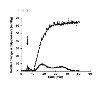
100461 FIG. 25 shows that agent cetylpridinium chloride
reduced intraocular pressure (tOP) in a
glaucoma model as compared to control. The agent was tested by controlling
flow and measuring
relative IOP using in a microfluidic device. The agent was compared against
placebo (buffered
saline) by preparing each in bovine vitreous humor (BVH) and pre-incubating at
37 C for 24 hours.
The timepoint of injection into the device is denoted by an arrow and the
letter "a." Referring to
FIG. 25, the IOP for placebo (dashed line) increased greatly after injection
of the placebo sample.
The LOP rose steadily to a maximum pressure of about 64 mmHg. To the contrary,
the LOP after
injection of the agent cetylpridinium chloride-BVH sample (solid line) was
markedly lower than for
placebo, and the difference was sustained. This result showed that the agent
cetylpridinium chloride
was surprisingly effective to reduce IOP in the glaucoma model.
100471 FIG. 26 shows that agent polymyxin B reduced
intraocular pressure (LOP) in a glaucoma
model as compared to control. The agent was tested by controlling flow and
measuring relative LOP
using in a microfluidic device The agent was compared against placebo
(buffered saline) by
preparing each in bovine vitreous humor (BVH) and pre-incubating at 37 C for
24 hours. The
timepoint of injection into the device is denoted by an arrow and the letter
"a?' Referring to FIG_ 26,
the IOP for placebo (dashed line) increased greatly after injection of the
placebo sample_ The IOP
rose steadily to a maximum pressure of about 250 mmHg. To the contrary, the
LOP after injection of
the agent polymyxin B (solid line) was 78% lower than for placebo, and the
difference was
CA 03151625 2022-3-17
WO 2021/055013 PCT/US2020/024592
11
sustained, This result showed that the agent polymyxin B was surprisingly
effective to reduce IOP
in the glaucoma model.
[0048] FIG. 27 shows that agent neomycin reduced
intraocular pressure (lOP) in a glaucoma
model as compared to control. The agent was tested by controlling flow and
measuring relative LOP
using in a microfluidic device. The agent was compared against placebo
(buffered saline) by
preparing each in bovine vitreous humor (BVH) and pre-incubating at 37 C for
24 hours. The
timepoint of injection into the device is denoted by an arrow and the letter
"a." Referring to FIG. 27,
the IOP for placebo (dashed line) increased greatly after injection of the
placebo sample. The IOP
rose steadily to a maximum pressure of about 64 mmHg. To the contrary, the LOP
after injection of
the agent neomycin (solid line) was 72% lower than for placebo, and the
difference was sustained,
This result showed that the agent neomycin was surprisingly effective to
reduce IOP in the glaucoma
model.
[00491 FIG. 28 shows that agent heparin sodium reduced
intraocular pressure (LOP) in a
glaucoma model as compared to control. The agent was tested by controlling
flow and measuring
relative IOP using in a microfluidic device, The agent was compared against
placebo (buffered
saline) by preparing each in bovine vitreous humor (BVH) and pre-incubating at
37 C for 24 hours.
The timepoint of injection into the device is denoted by an arrow and the
letter "a." Referring to
FIG. 28, the IOP for placebo (dashed line) increased greatly after injection
of the placebo sample.
The LOP rose steadily to a maximum pressure of about 67 mmHg. To the contrary,
the LOP after
injection of the agent heparin sodium (solid line) was 32% lower than for
placebo, and the difference
was sustained. This result showed that the agent heparin sodium was
surprisingly effective to reduce
IOP in the glaucoma model.
100501 FIG. 29 shows that compound sodium dodecyl sulfate
was a negative control for
intraocular pressure (lOP) in a glaucoma model. The compound was tested by
controlling flow and
measuring relative LOP using in a microfluidic device microfluidic. The
compound was compared
against placebo (buffered saline) by preparing each in bovine vitreous humor
(BVH) and pre-
incubating at 37 C for 24 hours The timepoint of injection into the device is
denoted by an arrow
and the letter "a." Referring to FIG. 29, the IOP for placebo (dashed line)
increased greatly after
injection of the placebo sample_ The TOP rose steadily to a maximum pressure
of about 60 mmHg_
However, the IOP after injection of sodium dodecyl sulfate (solid line) was
significantly higher than
CA 03151625 2022-3-17
WO 2021/055013
PCT/US2020/024592
12
for placebo. This result showed that sodium dodecyl sulfate was a negative
control that did not
reduce IOP in the glaucoma model.
DETAILED DESCRIPTION OF THE INVENTION
[0051] This invention provides methods, compositions,
devices, kits and reagents for detecting,
identifying and treating glaucoma disease. Embodiments of this invention
utilize ultrastructural
features of ocular aqueous humor as guides and markers for glaucoma
therapeutic modalities.
[0052] In some aspects, this invention provides methods and
compositions for reducing
intraocular pressure and increasing ocular outflows in glaucoma subjects.
Aspects of this invention
can reduce formation and presence of aggregational features and structures in
ocular humor.
[0053] In further aspects, this disclosure provides
therapeutic compositions for glaucoma.
[0054] Embodiments of this invention provide devices for
measuring and characterizing
glaucoma aggregational features, as well as intraocular pressure and ocular
outflows.
[0055] Additional aspects of this disclosure include
diagnostic and screening modalities for
glaucoma. Further embodiments include kits and reagents for carrying out the
foregoing.
[0056] Embodiments of this invention can provide glaucoma
diagnosis based on a
unique and reliable ultrastructural biomarker herein identified. The
ultrastructural
component can block the trabecular meshwork, increasing LOP and, over time,
ocular
aqueous outflow resistance increases leading to elevated intraocular pressure,
and eventual
vision loss. The ultrastructural component in the humor fluid of a patient
with POAG can
be reflected in EV aggregates formed together in large EV-complexes. The EV-
complexes
can be multiple microns in size and are glaucoma-associated-EV-complexes. The
EV-
complexes may be present in glaucoma patient samples and sizeable enough to
block the
trabecular meshwork.
[0057] Embodiments of this invention can provide
compositions and methods for
purifying and/or synthesizing EV-complexes of this ultrastructural component
for use in
therapeutic and biological methods.
[0058] Embodiments of this invention can provide
compositions and methods for
therapeutics and treatment of POAG and testing POAG aqueous humor specimens.
[0059] In some embodiments, glaucoma-associated-EV-
complexes can be reduced by
dissociation and other routes.
CA 03151625 2022-3-17
WO 2021/055013
PCT/US2020/024592
13
100601 In additional embodiments, compositions and methods
of this invention can
reduce intraocular pressure and/or increase ocular outflows.
00611 Embodiments of this invention further contemplate
methods for treating
glaucoma.
100621 In certain aspects, a glaucoma disease may be
treated by administering a surface
active agent for affecting EV-complexes. A surface active agent may be used
for
ameliorating, alleviating, inhibiting, lessening, delaying, and/or preventing
at least one
symptom or condition of a glaucoma disorder.
100631 The eye can be viewed as a closed chamber. (FIG. 1,
left) IOP can be
determined by the rate of aqueous humor formation and the rate fluid exit,
(FIG. 1, right)
In general, reduced aqueous outflow in glaucoma can be related to raised IOP.
Aqueous
humor outflow can be related to elevated IOP and glaucomatous visual damage.
Aqueous
humor exits the eye via two pathways: the trabecular meshwork and to a lesser
extent the
uveolscleral outflow. (FIG. 2)
100641 Abnormal aqueous humor outflow can cause elevated
IOP. The trabecular
meshwork (TM) can be a major site of outflow, The TM is a filter-like tissue
composed of
a series of fenestrated striations that allow aqueous humor to flow and exit
the anterior
chamber via Schlemmis canal. The primary function of the TM is to allow
aqueous humor
to exit the eye and establish IOP.
00651 The juxtacanalicular tissue (JCT) or cribriform
region is next to Schlemm's canal
and is the region of the TM that may be implicated in establishing IOP. The
site of most
resistance to the aqueous outflow can be the JCT tissue, which measures
approximately 2-
20 pm. JCT is composed of the loosely arranged extracellular matrix (ECM) into
which
cells are embedded.
00661 Abnormal regulation of aqueous flow through the TM may be associated
with
elevated IOP. The ECM of the TM can be a barrier that may isolate the ocular
fluid
outflow.
100671 Ultrastructural features or compositions in the
aqueous humor of patients with
glaucoma that are physically larger than the fenestrations of the JCT outlet,
or that of other
TM tissues, can block the TM.
CA 03151625 2022-3-17
WO 2021/055013
PCT/US2020/024592
14
[0068] Ultrastructural features or compositions in the
aqueous humor can include
structures based on extracellular vesicles (EV). EVs are transport nano-
vesicles related to
inter-cellular communication via transfer of biomolecules such as proteins,
lipids, and
nucleic acids from one cell to another
[0069] In general, various cell types secrete EVs into
fluids like blood, cerebrospinal
fluid, and urine. Examples include exomeres approximately 35 nm, exosomes
about 40-
100 nm, larger micro-vesicles about 100-1000 nm, and apoptotic bodies about 1-
5 gm.
EVs can be associated with pathophysiology of disease.
100701 In some embodiments of this invention,
ultrastructural features and compositions
based on EVs are utilized in characterizing ocular fluids.
[0071] In further embodiments, ultrastructural features and
compositions based on EVs
can be utilized in devices for determining IOP. Ultrastructural features and
compositions
based on EVs can be monitored for determining therapeutic effects in reducing
IOP
Ultrastructural features and compositions based on EVs can be monitored as
biomarkers for
determining therapeutic effects in reducing IOP.
[0072] In additional embodiments, ultrastructural features
and compositions based on
EVs can be utilized in devices for measuring ocular outflows. Ultrastructural
features and
compositions based on EVs can be monitored for determining therapeutic effects
in
increasing ocular outflows. Ultrastructural features and compositions based on
EVs can be
monitored as biomarkers for determining therapeutic effects in increasing
ocular outflows.
[0073] In further embodiments, ultrastructural features and
compositions based on EVs
are utilized in reducing formation and presence of aggregational features,
structures and particles
in ocular humor.
[0074] In certain embodiments, ultrastructural features and
compositions based on EVs
are utilized in devices for detecting ocular conditions and parameters.
[0075] In additional embodiments, ultrastructural features
and compositions based on
EVs are utilized in identifying glaucoma in a subject
[0076] In further embodiments, ultrastructural features and
compositions based on EVs
are utilized in methods, kits and reagents for glaucoma.
CA 03151625 2022-3-17
WO 2021/055013
PCT/US2020/024592
[0077] Without wishing to be bound by any particular
theory, ultrastructural features
and compositions based on EVs in glaucoma may have larger structures that
block the TM
and/or other outflows
[0078] Glaucoma disorders, referred to herein as
"glaucoma," that can be treated with
the methods and compositions as described herein include, but are not limited
to,
preglaucoma open angle with borderline findings, open angle, low risk,
anatomical narrow
angle primary angle closure suspect, steroid responder, ocular hypertension,
primary angle
closure without glaucoma damage (PAS or high IOP with no optic nerve or visual
field
loss), unspecified open-angle glaucoma, primary open-angle glaucoma, chronic
simple
glaucoma, low-tension glaucoma, pigmentary glaucoma, capsular glaucoma with
pseudo-
exfoliation of lens, residual stage of open-angle glaucoma, unspecified
primary angle-
closure glaucoma, acute angle-closure glaucoma attack, chronic angle-closure
glaucoma,
intermittent angle-closure glaucoma, residual stage of angle-closure glaucoma,
glaucoma
secondary to eye trauma, glaucoma secondary to eye inflammation, glaucoma
secondary to
other eye disorders including, retinal vascular occlusions, diabetes type 1
complicated,
diabetes type 2 complicated, disorders of lens, disorders of intraocular lens,
disorders after
other ocular symptoms, neoplasms, benign neoplasms, or malignant. Also
included is
glaucoma secondary to drugs, glaucoma with increased episcleral venous
pressure,
hypersecretion glaucoma, aqueous misdirection malignant glaucoma, glaucoma in
diseases
classified elsewhere, congenital glaucoma, axenfeld's anomaly, buphthalmos,
glaucoma of
childhood, glaucoma of newborn, hydrophthalmos, keratoglobus, congenital
glaucoma
macrocornea with glaucoma, macrophthalmos in congenital glaucoma, megalocornea
with
glaucoma, absolute glaucoma. Also included are adverse effect of
ophthalmological drugs
and preparations, acute follicular conjunctivitis, adverse effect of carbonic
anhydrase
inhibitors, and adverse effect of under dosing of ophthalmological drugs and
preparations.
[0079] In some embodiments, a composition of this
disclosure can be administered
systemically. Systemic administration can be achieved via intravenous
administration, oral
administration, intraarterial administration, inhalation, intranasal
administration, intra-
peritoneal administration, intra-abdominal administration, subcutaneous
administration,
intra-articular administration, intrathecal administration, transdural
administration,
transdermal administration, submucosal administration, sublingual
administration, enteral
CA 03151625 2022-3-17
WO 2021/055013
PCT/US2020/024592
16
administration, parenteral administration, percutaneous administration,
periarticular
administration, or intraventricular administration.
100801 In further embodiments, a composition of this
disclosure can be administered
locally. A composition may be administered locally to ocular tissue. As used
herein, the
term ocular tissue refers to the eye, including tissues within the sclera,
e.g., the retina, and
outside the sclera, e.g., ocular muscles within the orbit. Ocular tissue also
includes tissues
neurologically connected to, but distinct from the eye, such as the optic
nerve, the
geniculate nucleus and the visual cortex. Local administration to ocular
tissue can be
achieved via intraocular administration. Intraocular administration can be
carried out via
intracameral administration, intravitreal administration, or subretinal
administration.
100811 In additional embodiments, local administration to
ocular tissue can be achieved
via periocular administration. Periocular administration can be carried out
via sub-
conjunctival injection, sub-Tenon's injection, direct periocular injection, or
depot
periocular injection.
100821 A subject may be administered a therapeutically
effective amount of the
composition. A therapeutically effective amount can be an amount effective to
ameliorate,
alleviate, inhibit, lessen, delay, and/or prevent at least one symptom or
condition of the
condition being treated.
100831 In certain embodiments, a therapeutically effective
amount can be the amount
effective to ameliorate the ocular condition being treated. The dose may be
determined
according to various parameters, especially according to the severity of the
condition, age,
and weight of the patient to be treated; the route of administration; and the
required
regimen. A physician will be able to determine the required route of
administration and
dosage for any particular patient. Dosages may vary depending on the relative
potency of
the composition being administered, and can generally be estimated based on
the half
maximal effective concentration (EC50) found to be effective in in vitro and
in vivo
models
Extracellular vesicles and aggregates in glaucoma
100841 Embodiments of this invention provide methods for
detecting EVs in biological
fluids. In certain methods, a cross-linking agent can be used to provide
robust imaging of
CA 03151625 2022-3-17
WO 2021/055013
PCT/US2020/024592
17
EV ultrastructures with, for example, electron microscopy. In further methods,
a
glutaraldehyde-alternative cross-linker can be used.
[0085] Additional embodiments of this invention contemplate
detecting and
characterizing EV-complexes in glaucoma. EV-complexes in glaucoma can block
the TM
Of JCT and inhibit ocular aqueous outflow pathways.
[0086] In certain aspects, EVs in glaucoma can be
aggregated together in EV-
complexes. Glaucoma-associated EV-complexes may be up to multiple microns in
size or
diameter.
[0087] In glaucoma, EV-complexes can be an ultrastructural
feature of the disease. This
ultrastructural feature can be a target for characterizing glaucoma. EV-
complexes can be
used for detecting therapeutic parameters and modalities for glaucoma. In some
embodiments, EV-complexes can be used for diagnosis, prognostication, and/or
screening
of glaucoma compositions EV-complexes can also be used in devices for
determining
therapeutic compositions, doses and regimens.
[0088] As used herein, the term diameter refers to the
longest linear dimension of an irregularly-
shaped particle such as an extracellular vesicle complex. For a regularly-
shaped particle,
such as a spherical vesicle, the term diameter has its usual meaning as the
line segment passing
through the center with endpoints on the sphere.
[0089] In some aspects, this disclosure provides
compositions of purified EV-complexes
from glaucoma Purified EV-complexes from glaucoma can be used in a device for
assaying and detecting changes in EV ultrastructural components which can be
related to
intraocular pressure and ocular outflows. This invention provides devices
containing
purified EV-complexes which can be used for characterizing and measuring
ocular
blockage and ocular outflows. Purified EV-complexes from glaucoma can be used
in a
device for screening effects of therapeutic agents on ocular EV
ultrastructural components.
[0090] In further aspects, this disclosure provides
compositions of synthetic EV-
complexes for characterizing glaucoma. Synthetic EV-complexes for
characterizing
glaucoma can be used in a device for assaying and detecting changes in EV
ultrastructural
components which can be related to intraocular pressure and ocular outflows.
This
invention provides devices containing synthetic EV-complexes which can be used
for
characterizing and measuring ocular blockage and ocular outflows. Synthetic EV-
CA 03151625 2022-3-17
WO 2021/055013
PCT/US2020/024592
18
complexes from glaucoma can be used in a device for screening effects of
therapeutic
agents on ocular EV ultrastructural components.
00911 EV ultrastructural components such as EV-complexes can be composed of
complexes or aggregates of extracellular vesicles Examples of extracellular
vesicles
include exomeres, exosomes, multivesicular bodies, intraluminal vesicles
(ILVs),
multivesicular endosomes (MVEs), oncosomes, micro-vesicles, apoptotic bodies,
and
vesicles originating from endosome or plasma membranes.
100921 Complexes or aggregates of extracellular vesicles
can be protein-EV structures
having micrometer diameters, or diameters greater than about 1 micrometer.
100931 Extracellular vesicle aggregates can have a size of
from about 360 to about
21,000 nanometers (nm).
100941 For example, exomeres can be about 35 nm in
diameter, exosomes can be about
40-100 nm in diameter, micro-vesicles can be about 100-1000 nm in diameter,
and
apoptotic bodies can be about 1-5 micrometers in diameter.
100951 Complexes or aggregates of extracellular vesicles
may contain 10, 20, 30, 40, 50,
100, 200, 500 or more extracellular vesicles.
100961 For example, a healthy subject may have free EVs,
which are non-aggregated
EVs, about 100-200 nm in diameter, which are mainly exosomes, along with some
micrometer sized vesicles. A healthy subject may not have EV-aggregates or EV
ultrastructural features larger than 0.4-20 micrometers in diameter in aqueous
humor.
100971 For example, in glaucoma, a subject may have EV-
aggregates larger than 0.4-20
micrometers in diameter. A healthy subject may have small EV aggregates of
about 36-300
nm, which are mainly exomeres, along with some micro-vesicles. A glaucoma
subject may
have reduced amounts of free EVs, or very few remaining free EVs. Free EVs in
glaucoma
may be of larger sizes than free EVs in a healthy subject. EV aggregates in
glaucoma may
be composed of any of exomeres, exosomes, micro-vesicles, and/or apoptotic
bodies, as
well as other kinds of vesicles or bodies.
Devices for glaucoma ag,gregational features
100981 A device of this invention can be used to
characterize the activity of a
biologically active agent toward glaucoma. A device of this invention can be
used to
CA 03151625 2022-3-17
WO 2021/055013
PCT/US2020/024592
19
detect or characterize ocular conditions or parameters in a model system or
patient
pathology.
[0099] An active agent may be capable of providing a
therapeutic benefit, especially in
glaucoma In some embodiments, an active agent may be a known drug effective
for
treating a disease of the eye.
1001001 In some aspects, a fluid composition in a device of this invention can
be
analyzed by various techniques. For example, a fluid composition can be
analyzed by an
imaging technique.
1001011 Examples of imaging techniques include electron microscopy,
stereoscopic
microscopy, wide-field microscopy, polarizing microscopy, phase contrast
microscopy,
multiphoton microscopy, differential interference contrast microscopy,
fluorescence
microscopy, laser scanning confocal microscopy, multiphoton excitation
microscopy, ray
microscopy, and ultrasonic microscopy.
1001021 Examples of imaging techniques include positron emission tomography,
computerized tomography, and magnetic resonance imaging,
1001031 Examples of assay techniques include colorimetric assay,
chemiluminescence
assay, spectrophotometry, and light scattering.
1001041 In some embodiments, this invention can provide a device for measuring
pressure and flow rate of a fluid composition. (FIG. 3) The device may have a
channel
having an inlet at a first end and an outlet at a second end, wherein the
inlet and outlet are
in fluid communication. The device can have a meshwork composition lodged in
the
channel for providing resistance to flow. The meshwork composition may have
any one or
more, or all of the following portions. A uveal meshwork, a corneoscleral
meshwork, and a
juxtacanalicular meshwork.
1001051 In some embodiments, a meshwork composition can be composed of glass
beads,
micro beads, magnetic beads, gel particles, dextran particles, or polymer
particles. A
meshwork composition may also be composed of glass fibers, polymeric fibers,
inorganic
fibers, organic fibers, or metal fibers.
1001061 In certain embodiments, a uveal meshwork may have fenestrations of
about 25
micrometers. A corneoscleral meshwork may have fenestrations of about 2-15
CA 03151625 2022-3-17
WO 2021/055013
PCT/US2020/024592
micrometers. A juxtacanalicular meshwork may have fenestrations of about 1 to
4
micrometers or less.
1001071 A device may further include a fluid reservoir for holding the fluid
composition,
so that the fluid reservoir is in fluid communication with the inlet of the
channel for
introducing the fluid composition into the inlet of the channel.
1001081 A device of this disclosure can have a pressure source for applying
pressure to
the fluid composition in the fluid reservoir for introducing the fluid
composition into the
inlet of the channel.
1001091 A device of this invention can have a flow sensor in fluid
communication with
the fluid composition for measuring the flow rate and pressure of the fluid
composition at
the inlet of the channel and transmitting the flow rate and pressure to a
processor.
1001101 Signals and data from the device can be received by a processor. The
processor
can display the flow rate and pressure. Memory or media can store instructions
or files,
such as a machine-readable storage medium. A machine-readable storage medium
can be
non-transitory.
1001111 A processor of this disclosure can be a general purpose or special
purpose
computer. A processor can execute instructions stored in a machine readable
storage
device or medium. A processor can include an integrated circuit chip, a
microprocessor, a
controller, a digital signal processor, any of which can be used to receive
and/or transmit
data and execute stored instructions. A processor can also transform data,
and/or store data
in memory, media or a file. A processor may receive and execute instructions
which may
include performing one or more steps of a method of this invention. A device
of this
invention can include one or more non-transitory machine-readable storage
media, one or
more processors, one or more memory devices, and/or one or more user
interfaces. A
processor my have an integral display for displaying data or transformed data.
1001121 In some aspects, a device may have a microfluidic channel. One or more
channels can also be arranged in a microfluidic chip.
1001131 A device of this disclosure can include one or more detectors for
analyzing the
fluid composition within the channel or at the inlet or exiting the outlet of
the channel.
One or more detectors can also be arranged to detect the fluid composition
within the
channel.
CA 03151625 2022-3-17
WO 2021/055013 PCT/US2020/024592
21
1001141 A device of this invention may include a meshwork composition which
contains
extracellular vesicles or extracellular vesicle complexes. An EV-complex for
use in a
meshwork composition may be purified from glaucoma ocular humor, aqueous
humor, or
vitreous humor. The ocular humor may be from animal or clinical sources. An
EN/-
complex for use in a meshwork composition may be composed of extracellular
vesicles,
and may have a diameter from about 360 to about 21,000 nanometers.
1001151 In certain embodiments, an EV-complex for use in a meshwork
composition may
include a fixative, a stabilizing component, or a cross linking component
which can
transform the structure to a stable, uniform composition,
[00116] Examples of stabilizing components include fixatives as described
herein, cross
linking compounds as described herein, organic solvents, polypeptides, and
pharmaceutically-acceptable organic salts.
1001171 Examples of salts include ammonium salts, alkali metal salts including
sodium,
lithium, and potassium salts, alkaline earth metal salts including calcium and
magnesium
salts, salts with organic bases, for example, organic amines, such as
benzathines,
dicyclohexylamines, hydrabamines formed with N,N-
bis(dehydroabietyflethylenediamine),
N-methyl-D-glucamines, N-methyl-D-glucamides, t-butyl amines, and salts with
amino
acids including arginine and lysine.
100118] Examples of salts include acetates, adipates, alginates, ascorbates,
aspartates,
benzoates, benzenesulfonates, bisulfates, borates, butyrates, citrates,
camphorates,
camphorsulfonates, cyclopentanepropionates, hydrochlorides, hydrobromides,
hydroiodides, 2-hydroxyethanesulfonates, lactates, maleates,
methanesulfonates, 2-
napthalenesulfonates, nicotinates, nitrates, oxalates, pectinates,
persulfates, digluconates,
dodecylsulfates, ethanesulfonates, fumarates, glucoheptanoates,
glycerophosphates,
hemisulfates, heptanoates, hexanoates, 3-phenylpropionates, phosphates,
picrates,
pivalates, propionates, salicylates, succinates, sulfates, sulfonates,
tartarates, thiocyanates,
toluenesulfonates, and undecanoates.
1001191 Extracellular vesicle complexes that are cross linked can be
reversibly cross
linked, or non-reversibly cross linked.
1001201 Extracellular vesicles that are cross linked can be reversibly cross
linked, or non-
reversibly cross linked.
CA 03151625 2022-3-17
WO 2021/055013
PCT/US2020/024592
22
1001211 In some embodiments, a device of this invention may contain an EV-
complex
meshwork composition that can be used for identifying or screening active
agents for
effects in reducing IOP and/or increasing ocular outflows. An EV-complex for
use in a
meshwork composition may include a drug delivery excipient An EV-complex
meshwork
composition for a device may be a synthetic EV-complex or a purified EV-
complex.
1001221 An embodiment of an arrangement of channels of this invention is
illustrated in
FIG. 4.
1001231 An embodiment of a device of this invention is illustrated in FIG. 5,
1001241 In additional embodiments, a device of this invention may be used for
measuring
the quantity or level of an EV-complex in a test sample. Measuring the
quantity or level of
an EV-complex in a test sample can provide a diagnostic marker level for the
test sample.
A device of this invention can be used to identify glaucoma or pre-glaucoma in
a subject.
1001251 In further embodiments, a device of this invention may be used for
measuring a
pressure which can be related to a quantity or level of an EV-complex in a
test sample. A
pressure value in a channel can be related directly to a quantity or level of
an EV-complex
in a test sample.
1001261 In certain embodiments, a device of this invention may be used for
measuring an
assay value which can be related to a quantity or level of an EV-complex in a
test sample.
An assay value of a composition in a channel can be related directly to a
quantity or level
of an EV-complex in a test sample. For example, an assay may be a colorimetric
assay, a
chemiluminescence assay, a spectrophotometry assay, or a light scattering
assay.
1001271 In some aspects, an aqueous humor sample from a subject can be
provided and
analyzed for a quantity of glaucoma extracellular vesicle complexes. The
subject can be
identified as having glaucoma or pre-glaucoma based on the quantity exceeding
a reference
value A reference value can be a quantity or level of glaucoma extracellular
vesicle
complexes in a reference population of healthy individuals. The subject can be
diagnosed
as having glaucoma or pre-glaucoma Subsequent test samples from the subject
can be
used to monitor a quantity or level of glaucoma extracellular vesicle
complexes exceeding
or not exceeding a previous test sample, which can be related to reducing IOP
and/or
increasing ocular outflows in the subject.
CA 03151625 2022-3-17
WO 2021/055013
PCT/US2020/024592
23
1001281 In certain embodiments, a quantity or level of glaucoma extracellular
vesicle
complexes may include one or more of the number, size, density, morphology,
and spatial
distribution of the extracellular vesicle complexes.
1001291 In some embodiments, a reference value can be a quantity or level of
glaucoma
extracellular vesicle complexes in a reference population of healthy
individuals. The
reference value can be the average value in samples from the reference
population_
1001301 Glaucoma may be found in a subject where a test sample from the
subject
contains a quantity or level of glaucoma extracellular vesicle complexes
exceeding a
glaucoma reference value.
1001311 In certain embodiments, a glaucoma reference value can be that the
number of
extracellular vesicle complexes which contain 10 or more aggregated
extracellular vesicles
is zero per sample. In certain embodiments, a glaucoma reference value can be
that the
number of extracellular vesicle complexes which contain 10 or more aggregated
extracellular vesicles is 10 per sample. In certain embodiments, a glaucoma
reference
value can be that the number of extracellular vesicle complexes which contain
10 or more
aggregated extracellular vesicles is 50 per sample. In certain embodiments, a
glaucoma
reference value can be that the number of extracellular vesicle complexes
which contain 10
or more aggregated extracellular vesicles is 100 per sample.
1001321 In certain embodiments, a glaucoma reference value can be that the
number of
extracellular vesicle complexes which contain 10 or more aggregated
extracellular vesicles,
wherein the complexes are larger than 360 nm, is zero per sample. In certain
embodiments,
a glaucoma reference value can be that the number of extracellular vesicle
complexes
which contain 10 or more aggregated extracellular vesicles, wherein the
complexes are
larger than 360 nm, is 10 per sample. In certain embodiments, a glaucoma
reference value
can be that the number of extracellular vesicle complexes which contain 10 or
more
aggregated extracellular vesicles, wherein the complexes are larger than 360
nm, is 50 per
sample In certain embodiments, a glaucoma reference value can be that the
number of
extracellular vesicle complexes which contain 10 or more aggregated
extracellular vesicles,
wherein the complexes are larger than 360 nm, is 100 per sample.
1001331 In certain embodiments, a glaucoma reference value can be the number
of
extracellular vesicle complexes larger than 360 nm is 10 per sample. In
certain
CA 03151625 2022-3-17
WO 2021/055013
PCT/US2020/024592
24
embodiments, a glaucoma reference value can be the number of extracellular
vesicle
complexes larger than 360 nm is 50 per sample. In certain embodiments, a
glaucoma
reference value can be the number of extracellular vesicle complexes larger
than 360 nm is
100 per sample. In certain embodiments, a glaucoma reference value can be the
number of
extracellular vesicle complexes larger than 360 nm is 200 per sample.
1001341 In additional aspects, a meshwork composition in a device of this
invention can
be an anterior half or portion of an animal eye with lens, wherein the TM of
the eye is
oriented in between the inlet and the outlet of the channel.
Methods
1001351 Extracellular vesicles in the aqueous humor in patients with POAG may
be compared to a
population of healthy controls. EV complex ultrastructure in the aqueous humor
in subjects with
ocular pathology such as glaucoma can be compared to healthy controls such as
subjects with no
ocular pathology aside from cataracts. The level of EV complexes in the
aqueous humor in
glaucoma subjects can exceed the level in healthy subjects.
1001361 The kind of EVs in glaucoma aqueous humor can be larger than in
healthy subjects. In
some embodiments, the level of larger EV structures can be reduced to un-block
the aqueous humor
meshwork and increase humor outflows. The EV complexes in glaucoma aqueous
humor can be
larger than any EV in a healthy subject. In some embodiments, the level of EV
complexes can be
reduced to un-block the aqueous humor meshwork and increase humor outflows.
1001371 In certain embodiments, EVs in healthy human aqueous humor can be
diffusely and
evenly distributed without aggregation. Healthy control aqueous humor may
contain EVs that are
non-aggregated and have diffuse distribution.
1001381 Glaucoma EV-complexes can be larger than EVs observed in healthy
controls and may
block the trabecular meshwork.
1001391 In some embodiments, purified EV-complexes can be obtained from
aqueous humor in
POAG. The purified EV-complexes may be several microns in size. The glaucoma
EV-complex
can be larger than the opening of the JCT (1 to 4 gm, or up to 2 to 20 pm),
which may be large
enough to block the juxtacanalicular tissue. The EV-complexes in POAG may be
used to block the
trabecular meshwork and reduce aqueous outflow. In certain embodiments, the
level of EV-
complexes can be reduced to un-block the trabecular meshwork and increase
aqueous outflow.
CA 03151625 2022-3-17
WO 2021/055013
PCT/US2020/024592
1001401 For example, EV complexes can be contacted with a composition
containing an active
agent such as bimatoprost. In these embodiments, the level of EV-complexes can
be reduced to un-
block a trabecular meshwork and increase aqueous outflow.
1001411 In certain aspects, the level or quantity of glaucoma EV-complexes can
be reduced in a
POAG subject by administering an active agent such as bimatoprost.
1001421 In further embodiments, purified EV-complexes can have a size from
about 360 nm to
21,000 nm. A purified EV-complex can be substantially larger than any particle
found in healthy
aqueous humor.
1001431 In additional embodiments, a purified EV-complex can have a size from
about 360 nm to
about 21,000 nm, or 360 nm to about 10,000 nm, or 360 nm to about 5,000 nm, or
360 nm to about
3,000 nm, or 360 nm to about 2,000 nm, or 360 nm to about 1,000 nm.
1001441 In a purified EV complex, the number of EVs contacting each other can
be from about 5
to about 300, or from 10 to 300, or from 10 to 200, or from 10 to 100, or from
10 to 50, or from 10
to 40, or from 10 to 20.
1001451 In a purified EV complex, the number of EVs contacting each other can
be from 20 to
300, or from 30 to 300, or from 40 to 300, or from 50 to 300.
1001461 In a purified EV complex, the number of EVs contacting each other can
be from 20 to
200, or from 20 to 100, or from 30 to 200, or from 30 to 100, or from 40 to
200, or from 40 to 100,
or from 50 to 200, or from 50 to 100
1001471 In some embodiments, purified EV-complexes can provide particles of a
size for a
uveal meshwork. Purified EV-complexes for a uveal meshwork can be about 10,000
nm to
about 25,000 nm, or 15,000 nm to 25,000 nm, or 20,000 nm to 25,000 nm.
1001481 In further embodiments, purified EV-complexes can provide particles of
a size for a
corneoscleral meshwork. Purified EV-complexes for a corneoscleral meshwork can
be about
1,000 nm to about 15,000 nm, or 2,000 nm to 10,000 nm, or 2,000 nm to 5,000
nm.
1001491 In additional embodiments, purified EV-complexes can provide particles
of a size for
a juxtacanalicular meshwork Purified EV-complexes for a juxtacanalicular
meshwork can be
about 360 nm to about 1,000 nm, or 360 nm to 2,000 nm, or 260 nm to 3,000 nm,
or 1,000
nm to 3,000 nm.
1001501 The region of the TM that may be implicated in establishing IOP is
next to Schlemm's
canal and is called the juxtacanalicular tissue (1CT) or cribriform region.
CA 03151625 2022-3-17
WO 2021/055013
PCT/US2020/024592
26
1001511 EV complexes can be synthesized by contacting EVs with reagents to
form larger
structures. Reagents can include fixatives, cross linkers, and buffer
suspensions. Synthesized EV
complexes may be composed of many EVs contacting each other to form
aggregates.
1001521 In a synthesized EV complex, the number of EVs contacting each other
can be from
about 5 to about 300, or from 10 to 300, or from 10 to 200, or from 10 to 100,
or from 10 to 50, or
from 10 to 40, or from 10 to 20.
1001531 In a synthesized EV complex, the number of EVs contacting each other
can be from 20 to
300, or from 30 to 300, or from 40 to 300, or from 50 to 300.
1001541 In a synthesized EV complex, the number of EVs contacting each other
can be from 20 to
200, or from 20 to 100, or from 30 to 200, or from 30 to 100, or from 40 to
200, or from 40 to 100,
or from 50 to 200, or from 50 to 100.
001551 In additional embodiments, a synthesized EV-complex can have a size
from about 360
nm to about 25,000 nm, or 360 rim to 21,000 nm, or 360 nm to about 10,000 nm,
or 360 nm to about
5,000 nm, or 360 nm to about 3,000 nm, or 360 nm to about 2,000 nm, or 360 nm
to about 1,000 rim.
1001561 In some embodiments, synthesized EV-complexes can provide particles of
a size for a
uveal meshwork. Synthesized EV-complexes for a uveal meshwork can be about
10,000 nm
to about 25,000 nm, or 15,000 urn to 25,000 nm, or 20,000 nm to 25,000 nm.
1001571 In further embodiments, synthesized EV-complexes can provide particles
of a size for
a corneoscleral meshwork. Synthesized EV-complexes for a corneoscleral
meshwork can be
about 1,000 nm to about 15,000 nm, or 2,000 nm to 10,000 nm, or 2,000 nm to
5,000 nm.
001581 In additional embodiments, synthesized EV-complexes can provide
particles of a size
for a juxtacanalicular meshwork. Synthesized EV-complexes for a
juxtacanalicular meshwork
can be about 360 nm to about 1,000 nm, or 360 nm to 2,000 nm, or 260 nm to
3,000 nm, or
1,000 nm to 3,000 nm.
Synthesis and purification of extracellular vesicles and EV-complexes
1001591 In some embodiments, extracellular vesicles including exosomes and EV-
complexes can be synthesized, isolated, and/or purified by size exclusion
chromatography
or gel filtration chromatography.
001601 In certain embodiments, extracellular vesicles including exosomes and
EV-
complexes can be synthesized, isolated, and/or purified by centrifugation,
differential
centrifugation, density gradient centrifugation, or ultracentrifugation.
CA 03151625 2022-3-17
WO 2021/055013
PCT/US2020/024592
27
1001611 In additional embodiments, extracellular vesicles including exosomes
and EV-
complexes can be synthesized, isolated, and/or purified using precipitation
reagents, for
example polymeric precipitation reagents, protamine, sodium acetate, or
organic solvents.
1001621 In some embodiments, extracellular vesicles including exosomes and EV-
complexes can be synthesized, isolated, and/or purified using immunoaffinity
capture
techniques.
1001631 In further embodiments, extracellular vesicles including exosomes and
EV-
complexes can be synthesized, isolated, and/or purified using microfluidic
devices,
acoustic fluidic devices, and microfluidic chips.
1001641 In additional embodiments, extracellular vesicles including exosomes
and EV-
complexes can be synthesized, isolated, and/or purified using sequential
filtration
techniques.
1001651 In further embodiments, extracellular vesicles including exosomes can
be
detected by resistive pulse sensing using a tunable pore sensor, or tunable
resistive pulse
sensing.
1001661 In certain embodiments, extracellular vesicles including exosomes can
be
detected by electron microscopy, light microscopy and flow cytometry.
1001671 In additional embodiments, extracellular vesicles including exosomes
can be
detected by dynamic light scattering and/or mass spectrometry.
1001681 In some aspects, extracellular vesicles including exosomes and EV-
complexes
can be synthesized, isolated, and/or purified by first isolating the vesicles
from cell culture.
1001691 In certain aspects, extracellular vesicles including exosomes and EV-
complexes
can be synthesized, isolated, and/or purified by first isolating the vesicles
from bodily
fluids, such as ocular humor. The isolated vesicles can be diluted, filtered
and protected
with protease inhibitors.
1001701 In further embodiments, steps for purification of extracellular
vesicles including
exosomes and EV-complexes include contacting with a fixative
1001711 In some aspects, extracellular vesicles including exosomes can be
synthesized by
controlled biogenesis and release from in vitro grown cell lines.
CA 03151625 2022-3-17
WO 2021/055013
PCT/US2020/024592
28
Active agents
001721 Examples of active agents include small molecule drugs, proteins,
nucleic acids,
polysaccharides, biologics, and combinations thereof.
1001731 Examples of active agents include cytokines, growth factors, proteins,
peptides,
anti-metabolites, signaling modulators, antibiotics, antibodies,
chemotherapeutic
compounds, and combinations thereof.
1001741 Examples of active agents include antiinfective agents, anesthetic
agents, anti-
VEGF agents, anti-inflammatory agents, an intraocular pressure reducing agent,
and
combinations thereof.
1001751 Examples of active agents include anesthetics, analgesics, and
combinations
thereof.
1001761 Examples of active agents include cell transport or mobility impending
agents
such as colchicine, vincristine, cytochalasin B, and combinations thereof.
1001771 Examples of active agents include antiglaucoma drugs.
1001781 Examples of active agents include beta-blockers such as timolol,
betaxolol,
atenolol, prostaglandins, and combinations thereof
1001791 Examples of active agents include lipid-receptor agonists or
prostaglandin
analogues such as bimatoprost, travoprost, tafluprost, latanoprost,
unoprostone, and
combinations thereof.
1001801 Examples of active agents include alpha-adrenergic agonists including
brimonidine, dipivefrine, and combinations thereof
1001811 Examples of active agents include carbonic anhydrase inhibitors such
as
acetazolamide, methazolamide, dichlorphenamide, diamox, and combinations
thereof.
1001821 Examples of active agents include and neuroprotectants such as
nimodipine.
1001831 Examples of active agents include agents for dry AMD such as
rapamycin,
glatiramer acetate, complement C5aR blocker, ciliary neurotrophic factor,
fenretinide,
rheopheresis, and combinations thereof
1001841 Examples of active agents include agents for wet AMD such as
mecamylamine;
VEGF trap eye, complement inhibitor POT-4.
1001851 Examples of active agents include kinase inhibitors such as
bevacizumab, BIBW
2992, cetuximab, imatinib, trastuzumab, gefitinib, ranibizumab, pegaptanib,
sorafenib,
CA 03151625 2022-3-17
WO 2021/055013
PCT/US2020/024592
29
dasatinib, sunitinib, erlotinib, nilotinib, lapatinib, panitumumab,
vandetanib, E7080, and
combinations thereof.
1001861 Examples of active agents include antibiotics such as tetracycline,
chlortetracycline, bacitracin, neomycin, polymyxin, gramicidin,
oxytetracycline,
chloramphenicol, gentamycin, and erythromycin.
1001871 Examples of active agents include antibacterials such as sulfonamides,
sulfacetamide, sulfamethizole and sulfisoxazole.
1001881 Examples of active agents include anti-fungal agents such as
fluconazole,
nitrofurazone, amphotericin B, and ketoconazole.
1001891 Examples of active agents include anti-viral agents such as
trifluorothymidine,
acyclovir, ganciclovir, DDI, AZT, foscamet, vidarabine, trifluorouridine,
idoxuridine,
ribavirin, protease inhibitors, and anti-cytomegalovirus agents.
1001901 Examples of active agents include antiallergenics such as
methapyriline;
chlorpheniramine, pyrilamine and prophenpyridamine.
1001911 Examples of active agents include anti-inflammatories such as
hydrocortisone,
dexamethasone, fluocinolone, prednisone, prednisolone, methylprednisolone,
fluorometholone, betamethasone and triamcinolone.
1001921 Examples of active agents include decongestants such as phenylephrine,
naphazoline, and tetrahydrazoline; miotics, and muscarinics
1001931 Examples of active agents include anti-cholinesterases such as
pilocarpine,
carbachol, di-isopropyl fluorophosphate, phospholine iodine, and demecarium
bromide.
1001941 Examples of active agents include mydriatics such as atropine sulfate,
cyclopentolate, homatropine, scopolamine, tropicamide, eucatropine.
1001951 Examples of active agents include sympathomimetics such as
epinephrine.
1001961 Examples of active agents include ranibizumab, bevacizamab, and
triamcinolone.
1001971 Examples of active agents include antiinflammatories, such as non-
steroidal anti-
inflammatories (NSAID) including acetylsalicylic acid, ibuprofen,
indomethacin; and
COX-2 inhibitors.
1001981 Examples of active agents include immunosuppressive agents including
sirolimus.
CA 03151625 2022-3-17
WO 2021/055013
PCT/US2020/024592
1001991 Examples of active agents include matrix metalloproteinase (MMP)
inhibitors
such as tetracycline.
002001 Examples of active agents include anticlotting agents such as heparin,
antifibrinogen, fibrinolysin, and anti-clotting activase.
1002011 Examples of active agents include antidiabetic agents including
acetohexamide,
chlorpropamide, glipizide, glyburide, tolazamide, tolbutamide, insulin, and
aldose
reductase inhibitors.
1002021 Examples of active agents include amines such as Thonzonium.
1002031 Examples of active agents include detergents such as Taurocholic acid,
Glycocholic acid, Glycochenodeoxycholic Acid, Benzalkonium, Cetylpyridinium,
Taurochenodeoxycholic acid, Polidocanol, and Tyloxapol.
002041 Examples of active agents include lipids such as Sodium lauryl sulfate.
002051 Examples of active agents include antibacterials such as Polyrnyxin B
1002061 Examples of active agents include amines such as Thonzonium and
related
compounds Thonzylamine, mepyramine and Piribedil.
1002071 Examples of active agents include Ophthalmics such as Aceclidine,
Acetazolamide, Acetylcysteine, Acyclovir, Aflibercept, Alcaftadine,
Alclometasone,
Alteplase, Ampicillin, Anecortave, Ascorbic acid, Atropine, Azelastine,
Azithromycin,
Befunolol, Bendazac, Benzylpenicillin, Besifloxacin, Betamethasone, Betaxolol,
Bibrocathol, Bimatoprost, Brimonidine, Brinzolamide, Bromfenac,
Carbamoylcholine,
Carte lol, Cefuroxime, Cenegermin, Chloramphenicol, Chlorhexidine,
Chlortetracycline,
Chymotrypsin, Cinchocaine, Ciprofloxacin, Clobetasone, Clonidine, Cocaine,
Cortisone,
Cromoglicic acid, Cyclopentolate, Cyclosporine, Cysteamine, Dapiprazole,
Demecarium,
Desonide, Dexamethasone, Dexpanthenol, Dibrompropamidine, Diclofenac,
Diclofenamide, Dihydrostreptomycin, Dipivefrin, Dorzolamide, Echothiophate,
Edetate
sodium, Emedastine, Ephedrine, Epinastine, Epinephrine, Erythromycin,
Ethylmorphine,
Famciclovir, Fludrocortisone, Fluocinolone acetoni de, Fluocortolone,
Fluorescein,
Fluorometholone, Flurbiprofen, Fomivirsen, Formocortal, Framycetin, Fusidic
acid,
Ganciclovir, Gatifloxacin, Gentamicin, Guaiazulen, Guanethidine, Heparin,
Hexamidine,
Homatropine, Hyaluronic acid, Hydrocortisone, Hypromellose, Ibopamine,
Idoxuridine,
Indomethacin, Inosine, Kanamycin, Ketorolac, Ketotifen, Latanoprost,
Levobunolol,
CA 03151625 2022-3-17
WO 2021/055013 PCT/US2020/024592
31
Levocabastine, Levofloxacin, Lidocaine, Lifitegrast, Lodoxamide, Lomefloxacin,
Loteprednol, Medrysone, Methazolamide, Methscopolamine, Methylprednisolone,
Metipranolol, Micronomicin, Moxifloxacin, Nandrolone, Naphazoline, Natamycin,
Nedocromil, Neomycin, Neostigmine, Nepafenac, Netarsudil, Netilmicin,
Nitrofural,
Norfloxacin, Ocriplasmin, Ofloxacin, Olopatadine, Oxybuprocaine,
Oxymetazoline,
Oxyphenbutazone, Oxytetracycline, Paraoxon, Pegaptanib, Phenylephrine,
Physostigmine,
Picloxydine, Pilocarpine, Piroxicana, Polymyxin B, Potassium Iodide, Povidone-
iodine,
Pranoprofen, Prednisolone, Procaine, Propamidine, Propanoic acid,
Proparacaine,
Ranibizumab, Resorcinol, Riboflavin, Rifamycin, Rimexolone, Rose bengal free
acid,
Salicylic acid, Scopolamine, Sirolimus, Sodium borate, Spaglumic Acid,
Sulfacetamide,
Sulfadicramide, Sulfamethizole, Sulfaphenazole, Sulfisoxazole, Synephrine,
Tafluprost,
Tetracaine, Tetracycline, Tetryzoline, Timolol, Tobramycin, Travoprost,
Triamcinolone,
Trifluridine, Tropicamide, Tyrothricin, Unoprostone, Verteporfin, Vidarabine,
Vitamin A,
and Xylometazoline.
1002081 Examples of active agents include anti-cancer agents such as 5-
fluorouracil,
adriamycin, asparaginase, azacitidine, azathioprine, bleomycin, busulfan,
carboplatin,
carmustine, chlorambucil, cisplatin, cyclophosphamide, cyclosporine,
cytarabine,
dacarbazine, dactinomycin, daunorubicin, doxorubicin, estramustine, etoposide,
etretinate,
filgrastin, floxuridine, fludarabine, fluorouracil, fluoxymesterone,
flutamide, goserelin,
hydroxyurea, ifosfamide, leuprolide, levamisole, lomustine, nitrogen mustard,
melphalan,
mercaptopurine, methotrexate, mitomycin, mitotane, pentostatin, pipobroman,
plicamycin,
procarbazine, sargramostin, streptozocin, tamoxifen, taxol, teniposide,
thioguanine, uracil
mustard, vinblastine, vincristine and vindesine.
1002091 Examples of active agents include hormones, peptides, steroids,
nucleic acids,
saccharides, lipids, glycolipids, and glycoproteins.
1002101 Examples of active agents include endocrine hormones such as
pituitary, insulin,
insulin-related growth factor, thyroid, and growth hormones
1002111 Examples of active agents include heat shock proteins.
1002121 Examples of active agents include immunological response modifiers
such as
muramyl dipeptide, cyclosporins, interferons, interleukin-2, cytokines,
tacrolimus, tumor
necrosis factor, pentostatin, thymopentin, transforming factor beta2, and
erythropoietin.
CA 03151625 2022-3-17
WO 2021/055013
PCT/US2020/024592
32
1002131 Examples of active agents include brain nerve growth factor (BNGF),
ciliary
nerve growth factor (CNGF), and vascular endothelial growth factor (VEGF).
002141 Examples of active agents include anti-coagulants, anti-proliferatives,
quinoxalines, and potassium channel blockers.
1002151 Examples of active agents include guanylate cyclase inhibitor, such as
methylene
blue, butylated hydroxyani sole, N-methylhydroxylamine, 2-(4-
methylaminobutoxy)
diphenyl methane, and apraclonidine.
1002161 Examples of active agents include prostaglandins such as metabolite
derivatives
of arachidonic acid.
1002171 Examples of active agents include sugars, such as trehalose.
1002181 Examples of active ingredients include viscoelastic agents including
hyaluronic
acid, dimethicone, and Hypromellose.
1002191 Examples of active ingredients include ophthalmic viscosurgical
devices, sodium
hyaluronate, chondroitin sulfate, hydroxypropyl methycellulose, hydroxy-propyl-
methylcellulose, hyaluronic acid, dimethicone, and hypromellose.
1002201 Examples of active agents include detergents, purifying or cleansing
agents, for
example salts of long-chain aliphatic bases or acids, which can have
cleansing, oil-
dissolving, and/or antimicrobial effects. Examples of active agents include
Glycochenodeoxycholic Acid, Glycocholic acid, Peanut oil, Benzalkonium,
Cetylpyridinium, Taurochenodeoxycholic acid, Polidocanol, Tyloxapol,
Taurocholic acid,
and N-Dodecyl-N,N-Dimethy1-3-Ammonio-1-Propanesulfonate.
1002211 Embodiments of this invention further contemplate use of active agents
for
treating glaucoma disorders. In some aspects, a glaucoma disorder may be
treated by
administering a surface active agent for affecting EV-complexes. An effective
amount a
surface active agent can be administered for ameliorating, alleviating,
inhibiting, lessening,
delaying, and/or preventing at least one symptom or condition of a glaucoma
disorder. As
used herein, the term surface active agent refers broadly to surfactants,
detergents,
purifying or cleansing agents, soaps, and modified variations thereof.
1002221 Examples of active agents include surfactants including Lucinactant,
Calfactant,
Beractant, Tyloxapol, Sodium lauryl sulfoacetate, Thonzonium, Nonoxyno1-9
Cetalkonium,
Dimethicone, Polyethylene glycol 400, Sinapultide, Palmitoyloleoyl-
phosphatidylglycerol,
CA 03151625 2022-3-17
WO 2021/055013
PCT/US2020/024592
33
Lapyrium, Sodium Lauryl Sulfate (SLS), Polyethylene glycol 300, Trolamine,
Polysorbate
80, Poloxamer 407, Sodium tetradecyl sulfate, Polysorbate 20, Docusate,
Poractant alfa,
Colfosceril palmitate, Castor oil, Benzalkonium, N-alkyl ethylbenzyl dimethyl
ammonium
(c12-c14), Quaternium-15, Ambroxol, Pumactant, Ethanolamine, Lecithin,
soybean, and
Cocamidopropyl betaine_
1002231 Examples of active agents include agents as described herein can be
modified for
delivery or metabolic acceptability, or can be PEGylated with a polyethylene
glycol chain,
or have a polypropylene glycol chain attached. This disclosure includes the
agents
described above as modified active agents, or PEGylated, or with other chain
modifications.
1002241 Examples of active agents include antibodies, antibody fragments, VEGF
inhibitors, small molecules, corticosteroids, and combinations thereof.
1002251 Examples of active agents include tyrosine kinase inhibitors,
monoclonal
antibodies, and combinations thereof.
1002261 Examples of active agents include antibodies and antibody fragments.
The term
"antibody" as referred to herein includes whole antibodies, e.g., two heavy
chains and two
light chains, antibody binding fragments thereof, e.g., single chain
antibodies (scFv), single
domain antibodies, e.g., nanobodies or Fv, Fab, Fab', F(ab')2, and, variants
thereof, e.g.,
tandem scFv, Fd fragments, diabodies, triabodies. Antibody fragments may be
obtained
using conventional techniques known to those of skill in the art, and the
fragments may be
used in the same manner as intact antibodies.
1002271 Antibody and antibody fragments as disclosed herein can be mono-
valent, bi-
valent, or tri-valent with regard to binding domains, and the binding domains
may be
mono-specific, bi-specific, or tri-specific in binding specificity by design.
Suitable
antibodies include monoclonal antibodies or a polyclonal antibody mixture. The
antibody
may be a chimeric antibody, a CDR-grafted antibody, a humanized antibody or an
antigen
binding portion of any of the foregoing thereof. Therapeutic antibodies may be
derived
from a variety of species, including, without limitation, mouse, human, camel,
llama, goat,
rabbit, bovine, and cartilaginous fish.
1002281 In some embodiments, an antibody or antigen binding fragment thereof
can be
used for the treatment of an ocular disease or condition. Examples of
antibodies or antigen
CA 03151625 2022-3-17
WO 2021/055013
PCT/US2020/024592
34
binding fragments include those that bind to and preferentially block or
reduce the activity
of integrins associated with disease, such as an anti-avI33 integrin antibody
and an anti-
a4131 integrin antibody, anti-epidermal growth factor receptor antibody, anti-
vascular
endothelial growth factor (VEGF) receptor antibody, anti-VEGF antibodies,
e.g.,
bevacizumab, ranibizumab, anti-TNFa antibodies, e.g., infliximab and
adalimumab, an
anti-fibroblast growth factor antibody, an anti-epidermal growth factor
antibody, an anti-
CD20 antibody, an anti-CD52 antibody, an anti-CD1la antibody, and anti-IL-2
antibody.
1002291 In further embodiments, a therapeutic protein can be an antibody
mimetic.
1002301 As used herein, the term "antibody mimetic" encompasses any organic
compound, e.g., a peptide or polypeptide, that can specifically bind an
antigen like an
antibody and is about 3-20 kDa. An antibody mimetic may comprise a scaffold
which
binds its target antigen via amino acids in exposed loops similar to the CDR
loops of an
antibody. Antibody mimetics include adnectins, lipocalins, Kunitz domain-based
binders,
avimers, knottins, fynomers, atrimers, and cytotoxic T-lymphocyte associated
protein-4
(CTLA4)-based binders. Some examples are given in Weidle et at., The Emerging
Role of
New Protein Scaffold-based Agents for the Treatment of Cancer, Cancer Genomics
&
Proteomics 10:155-168 (2013).
1002311 Examples of active agents include agents used for Parkinson's such as
Benzatropine, Ropinirole, Tolcapone, Trihexyphenidyl, Procyclidine,
Pramipexole,
Entacapone, Biperiden, Amantadine, Selegiline, Bromocriptine, Levodopa,
Dexetimide,
Piribedil, Budipine, Melevodopa, Profenamine, Cabergoline, Lisuride,
Progabide,
Gabapentin, Memantine, Orphenadrine, 3,5-Dinitrocatechol, Pimavanserin,
Ifenprodil,
Opicapone, Benserazide, Metixene, Apomorphine, Pergolide, Rasagiline,
Rotigotine,
Etilevodopam, Tropatepine, Dihydroergocryptine, Phenglutarimide, Mazaticol,
Etybenzatropine, Bornaprine, Etanautine, Carbidopa, Safinamide, and
Dexpramipexole
1002321 [0002] Examples of active agents include
prostaglandins such as
Epoprostenol, Dinoprost, Carboprost tromethamine, Dinoprost tromethamine,
Dinoprostone, Prostaglandin D2, Prostalene, Reidispongiolide C, Unoprostone,
Gemeprost, Limaprost, Iloprost, Latanoprost, Cloprostenol, Sepetaprost,
Bimatoprost,
Fenprostalene, Latanoprostene Bunod, Travoprost, Carboprost Tromethamine,
Dinoprost,
Tafluprost, Cabazitaxel, Cloprostenol Sodium, Bimatoprost, (-)-Corey Lactone 4-
CA 03151625 2022-3-17
WO 2021/055013
PCT/US2020/024592
Phenylbenzoate Alcohol, Dutasteride, Isopropyl Unoprostone, Beraprost Sodium,
Prostaglandin El, Cloprostenol, Remodulin, Trenbolone Cyclohexylmethyl
carbonate,
Prostaglandin F2a, Iloprost, Misoprostol Acid, Gemeprost, 9-deoxy-9-methylene-
16,16-
dimethyl Prostaglandin E2, Enoprostil, Ornoprostil, epoprostenol, Sulprostone,
Iloprost,
Rosaprostol, (+)-Cloprostenol sodium, Carboprost tromethamine, Carboprost,
Misoprostol,
Prostacyclin sodium salt, Prostaglandin E2, and Limaprost.
1002331 Examples of active agents include vitamins such as 1-alpha, 25-
dihydroxy1-20-
epi-22-oxa-24, 26 ,27-trihomovitamin D3.
1002341 Examples of active agents include antibotics such as Brefeldin A,
Fusidic acid,
Ovalicin, Narasin, and Salinomycin.
002351 Examples of active agents include steroids such as Hydrocortisone
cypionate,
Hydrocortisone valerate, Hydrocortisone butyrate, Hydrocortisone probutate,
Hydrocortisone aceponate, Prednisolone tebutate, Trilostane Hydrocortisone
acetate,
Cholesteryl Linoleate, Methylprednisolone aceponate, Eldecalcitol Pregnenolone
acetate,
Testosterone propionate Drospirenone Clascoterone Norethindrone enanthate,
Prednisolone
hemisuccinate, Hydroxyprogesterone caproate, Trenbolone acetate, fluprostenol
Anecortave acetate Oleandrin Cortisone acetate, Calcipotriol Gestodene
Dimethyl carbate,
Calcipotriol lalpha,24S-Dihydroxyvitamin D2, Ethynodiol diacetate, Nandrolone
decanoate, Testosterone cypionate, Testosterone undecanoate, Carbenoxolone,
progesterone-11-alpha-ol-hemisuccinate, Eplerenone, Testosterone succinate,
and
Boldenone undecylenate_
1002361 Examples of active agents include statins such as Lovastatin,
atorvastatin,
pravastatin, rosuvastatin, fluvastatin, and simvastatin.
1002371 Examples of active agents include aggregation inhibitors such as
Pentoxifylline,
Argatroban, and Von Willebrand Factor Human.
1002381 Examples of active agents include prostacyclin such as Treprostinil.
1002391 Examples of active agents include amyloid targeting agents such as
caprospinol
and similar compounds including Cholesteryl Linoleate, Pregnenolone acetate,
and P-
57AS3.
1002401 Examples of active agents include lactones such as Reidispongiolide A
and
Soraphen A, and Canrenone.
CA 03151625 2022-3-17
WO 2021/055013
PCT/US2020/024592
36
1002411 Examples of active agents include Benzoquinones such as Antroquinonol.
1002421 Examples of active agents include Oxepanes such as Triptolide PG-701.
1002431 Examples of active agents include Lipids such as gibberellin A4 and
Fumagillin
1002441 Examples of active agents include blood glucose lowering agents such
as
Mitiglinide.
1002451 Examples of active agents include central nervous system depressant
such as
Glutethimide.
1002461 Examples of active agents include benzenoids such as sildenafil,
udenafil, and
vardenafil.
1002471 Examples of active agents include carbohydrates such as Fusicoccin and
treholose.
1002481 Examples of active agents include Cholinergic Agents such as
Biperiden,
Cycrimine, Procyclidine, and Trihexyphenidyl
1002491 Examples of active agents include Terpenes such as Cyclohexanes.
1002501 The molecules, compounds and/or compositions of this disclosure may be
asymmetric, having one or more chiral stereocenters. A compound containing one
or more
chiral centers can include substances described as an "isomer," a
"diastereomer," a
"stereoisomer," an "optical isomer," an "enantiomer," or as a "racemic
mixture."
Conventions for stereochemical nomenclature, for example the stereoisomer
naming rules
of Cahn, Ingold and Prelog, as well as methods for the determination of
stereochemistry
and the separation of stereoisomers are known in the an. See, e.g., March's
Advanced
Organic Chemistry (7th ed., 2013). The compounds, composition and structures
of this
disclosure are intended to encompass all possible isomers, stereoisomers,
diastereomers,
enantiomers, and/or optical isomers that exist for the compound, composition
and/or
structure, including any mixture, racemate, or racemic or other mixtures
thereof
1002511 A compound can exist in un-solvated and solvated forms, or hydrated
forms. In
this disclosure, solvated forms, with pharmaceutically acceptable solvents,
such as water or
ethanol, are to be taken as equivalent to the un-solvated forms. Compounds and
salts, or
solvates thereof, may also exist in tautomeric forms, which are to be taken as
equivalent_
1002521 The molecules, compounds and/or compositions of this disclosure may be
found
in different crystalline forms, which are intended to be encompassed by this
disclosure.
CA 03151625 2022-3-17
WO 2021/055013
PCT/US2020/024592
37
Compositions and formulations
1002531 An active agent of this disclosure can include drugs and agents for
diseases of
the eye, including small molecule drugs, peptides, antibodies and protein
agents.
1002541 A formulation of an active agent may be prepared by dissolving a
composition in
water to produce an aqueous solution and rendering the solution sterile.
1002551 A formulation of this disclosure can be in the form of a sterile
injectable aqueous
or oily suspension. A suspension can be formulated including a dispersing or
wetting
agent. A sterile injectable preparation can be a sterile injectable solution
or suspension in
a non-toxic, pharmaceutically acceptable diluent or solvent.
1002561 Examples of solvents include water, water for injection, Ringer's
solution,
balanced salt saline, isotonic sodium chloride solution, 1,3-butanediol,
synthetic mono-or
diglycerides, and fatty acids such as oleic acid.
1002571 A formulation of this disclosure can be in the form of eye drops for
topical
delivery.
1002581 An ophthalmic formulation can be a solution or suspension for topical
administration. A composition can be a viscous or semi-viscous gel, or other
solid or semi-
solid compositions.
1002591 An ophthalmic formulation can be locally delivered by direct injection
or by use
of an infusion pump.
1002601 An ophthalmic formulation can include artificial tears carriers
1002611 An ophthalmic formulation can include a phospholipid carrier.
1002621 An ophthalmic formulation can include a surfactant, a preservative, an
antioxidant, a tonicity adjusting excipient, a buffer, a co-solvent, and a
viscosity excipient.
1002631 An ophthalmic formulation may include an excipient to adjust
osmolarity of the
formulation.
1002641 An ophthalmic formulation can include a viscosity excipient such as a
polysaccharide, hyaluronic acid, chondroitin sulfate, a dextran, a cellulose
polymer, a vinyl
polymer, and an acrylic acid polymer.
1002651 An ophthalmic formulation may have a viscosity of from 1 to 400
centipoises, or
from 1 to 100 centipoises, or from 2 to 40 cps. An ophthalmic formulation may
have a
viscosity of about 15, 20, 25, 30, 40, or 50 centipoises.
CA 03151625 2022-3-17
WO 2021/055013
PCT/US2020/024592
38
1002661 Examples of excipients or carriers for a formulation of this invention
include
ophthalmologically acceptable preservatives, viscosity enhancers, penetration
enhancers,
buffers, sodium chloride, sterile water, water for injection, and combinations
thereof.
1002671 A dosage form of a composition of this invention can be liquid or an
emulsion.
A dosage form of the composition of this invention can be solid, which can be
reconstituted in a liquid prior to administration.
1002681 A composition of this disclosure can also be in the form of an oil-in-
water
emulsion. The oily phase can be a vegetable oil or a mineral oil.
1002691 Examples of emulsifying agents include naturally-occurring gums, gum
acacia,
gum tragacanth, phosphatides, esters of fatty acids, hexitol, sorbitan
monooleate, and
polyoxyethylene sorbitan monooleate.
002701 Embodiments of this invention can advantageously provide effective
activity of
an active agent at dosage levels significantly lower than conventional dosage
levels.
002711 An effective amount of an active agent composition of this disclosure
can be an
amount sufficient to ameliorate or reduce a symptom of the disease treated.
1002721 A composition may be administered as a single dosage or may be
administered in
a regimen with repeated dosing.
1002731 An appropriate dosage level of an active agent can be determined by a
skilled
artisan. In some embodiments, an active agent can be present in a composition
in an
amount from about 0.001% to about 40%, or from about 0.01 % to about 20%, or
from
about 0.1% to 10% by weight of the total formulation.
1002741 An active agent of this disclosure can be combined with one or more
pharmaceutically acceptable carriers. A carrier can be in a variety of forms
including
fluids, viscous solutions, gels, or solubilized particles. Examples of
carriers include
pharmaceutically acceptable diluents, solvents, saline, and various buffers.
002751 Some examples of carriers, excipients and additives are given in U.S.
Pharmacopeia National Formulary (2014); Handbook of Pharmaceutical Excipients
(7th
ed., 2013); Handbook of Preservatives (2004, Synapse Information Resources);
Remington: The Science and Practice of Pharmacy (22nd ed. 2013); Remington's
Pharmaceutical Sciences (Mack Publishing Co. 1990). Some examples of drugs and
CA 03151625 2022-3-17
WO 2021/055013
PCT/US2020/024592
39
delivery are given in Goodman and Gilman, The Pharmacological Basis of
Therapeutics
(13th ed. 2018, McGraw Hill, NY).
002761 In certain embodiments, an active agent may be delivered without a
carrier for
reducing extracellular vesicle complexes in glaucoma ocular humor.
1002771 Examples of carriers include pyrogen free water; isotonic saline,
Ringer's
solution, ethyl alcohol, and phosphate buffer solution.
1002781 A formulation of this disclosure may include a polymer such as a
polyethylene
glycol (PEG), polypropylene glycol, or poly(lactic-co-glycolic acid) having a
molecular
weight of about 0.2 to about 50 kDa.
1002791 Examples of carrier polymers include polyvinyl acetate, polyvinyl
alcohol,
polyvinylpyrrolidone, chitosan, collagen, sodium alginate, gelatin, hyaluronic
acid,
polylactic acid, poly(lactic acid-glycolic acid) copolymer, polyhydroxybutyric
acid,
poly(hydroxybutyric acid-glycolic acid) copolymer, cellulose, hydroxymethyl
cellulose,
hydroxypropylcellulose, fatty acid esters, and polyglycerins.
1002801 Examples of additives include saccharides, sucrose, mannitol, lactose,
L-
arabinose, D-erythrose, D-ribose, D-xylose, D-mannose, D-galactose, lactulose,
cellobiose,
gentibiose, glycerin, polyethylene glycol, N-methylpyrrolidone, oligovinyl
alcohol,
ethanol, ethylene glycol, and propylene glycol.
[00281] Examples of solubility enhancing agents include cyclodextrins.
1002821 A formulation can include galactose, lactose, mannitol,
monosaccharide,
fructose, maltose, galactose, glucose, D-mannose, sorbose, disaccharide,
lactose, sucrose,
trehalose, cellobiose, polysaccharide, maltodextrin, dextran, starch,
mannitol, or xylitol_
1002831 An ophthalmic formulation may include a lipid such as
dipalmitoylethylphosphocholine, dioleoyl phosphatidylethanolamine, or 3131N-
(N',N1-
Dimethylaminoethane)-carbamoyl] cholesterol.
1002841 An ophthalmic formulation may include a lipid such as 1,2-Dioleoyl-sn-
Glycero-
3-[Phospho-L-Serine], 1,2-Dioleoyl-sn-Glycero-3-Phosphate.
1002851 An ophthalmic formulation may include a lipid such as 1,2-Dipalmitoyl-
sn-
Glycero-3-Phosphocholine, distearoylphosphatidylcholine,
diarachidoylphosphatidylcholin,
dipalmitoyl phosphatidylethanolamine.
CA 03151625 2022-3-17
WO 2021/055013
PCT/US2020/024592
1002861 An ophthalmic formulation may include a fatty acid, oleic acid,
myristoleic, or
aracadonic acid.
002871 An ophthalmic formulation may include a phospholipid such as
phosphatidylcholine, lecithin, phosphatidylglycerol, phosphatidylinositol,
phosphatidylserine, and phosphatidylethanolamine.
002881 An ophthalmic formulation may include a polymer such as
polyvinylpyrrolidone,
hydroxymethylcellulose, hydroxyethyl cellulose, hydroxypropylmethyl cellulose,
hydroxyethylstarch, cyclodextrin, 2-hydroxypropy1-13-cyclodextrin,
sulfobutylether-it-
cyclodextrin, polyethylene glycol, pectin, poly(lactide-co-glycolide),
polylactide,
polyethylene imine, or poly-L-lysine.
002891 In some embodiments, an ophthalmic formulation may include one or more
of a
pH adjusting excipient, a buffering excipient, a tonicity excipient, a
viscosity excipient, or
a wetting excipient. In certain embodiments, an ophthalmic formulation may
include an
acidifying excipient, a preservative, an antioxidant, a solubilizing
excipient, a humectant,
or a suspending excipient.
1002901 An ophthalmic formulation may include additives, diluents, delivery
vehicles, or
carrier materials such as a polymer, a polyethylene glycol, a dextran, a
diethylaminoethyl
dextran, a cyclodextrin, or a carboxymethyl cellulose.
1002911 Examples of excipients include sodium chloride, sodium dihydrogen
phosphate
monohydrate, and di sodium hydrogen phosphate anhydrous.
1002921 Examples of formulation additives include vegetable oils, olive oil,
sesame oil,
coconut oil, mineral oil, and paraffin.
1002931 Examples of dispersing or wetting agents include lecithin,
polyoxyethylene
stearate, heptadecaethyleneoxycetanol, polyoxyethylene sorb itol monooleate,
and
polyethylene sorbitan monooleate.
002941 Examples of antioxidants include ascorbic acid, cysteine hydrochloride,
sodium
bisulfite, sodium metabisulfite, sodium sulfite, ascorbyl palmitate, butylated
hydroxyanisole, butylated hydroxytoluene, lecithin, propyl gallate, alpha-
tocopherol, citric
acid, ethylenediamine tetraacetic acid, sorbitol, tartaric acid, and
phosphoric acid.
1002951 Examples of formulation additives include a thickening agent, for
example
beeswax, paraffin, or cetyl alcohol.
CA 03151625 2022-3-17
WO 2021/055013 PCT/US2020/024592
41
1002961 Examples of formulation excipients include a suspending excipient,
sodium
carboxymethylcellulose, methylcellulose, hydropropyl-methylcellulose, sodium
alginate,
polyvinylpyrrolidone, gum tragacanth, or gum acacia.
1002971 An ophthalmic formulation may include a carrier or co-solvent such as
Polysorbate 20, 60 or 80, Pluronic F-68, F-84 or P-103, Tyloxapol, Cremophor,
sodium
dodecyl sulfate, glycerol, PEG 400, propylene glycol, cyclodextrin, and
combinations
thereof. A carrier or co-solvent can be used in concentrations from about
0.01% to about
2% by weight.
1002981 An ophthalmic formulation may include a gel excipient such as gellan,
xanthan
gum, and combinations thereof.
002991 An ophthalmic formulation may include a viscosity enhancer such as
polyvinyl
alcohol, methyl cellulose, hydroxy propyl carboxymethyl cellulose,
hydroxymethylcellulose, hydroxyethyl cellulose, hydroxypropylmethyl cellulose,
methylcellulose, polyvinylpyrrolidone, and combinations thereof. A viscosity
enhancer
can be used in concentrations from about 0.01% to about 2% by weight.
1003001 An ophthalmic formulation may include a preservative such as
benzalkonium
chloride, chlorobutanol, benzododecinium bromide, methyl paraben, propyl
paraben,
phenylethyl alcohol, edetate disodium, sorbic acid, onamer, polyquaternium-1,
hydroxybenzoate, sodium benzoate, phenol, cresol, p-chloro-m-cresol, benzyl
alcohol,
thimerosal, sorbic acid, benzethonium chloride, and combinations thereof. A
preservative
can be used in concentrations from about 0.001% to about 1.0% by weight.
1003011 A unit dose composition can be sterile, but may not contain a
preservative.
1003021 An ophthalmic formulation may include a pH adjusting excipient such as
citric
acid buffer, acetic acid buffer, succinic acid buffer, malic acid buffer, and
gluconic acid
buffer.
003031 An ophthalmic formulation may include an additional acid such as
hydrochloric
acid, or and additional base, such as sodium hydroxide for pH adjustment
1003041 Examples of pH control agents include arginine, sodium hydroxide,
glycine,
hydrochloric acid, and citric acid.
CA 03151625 2022-3-17
WO 2021/055013
PCT/US2020/024592
42
1003051 An ophthalmic formulation may include a buffer such as citric acid,
ascorbic
acid, gluconic acid, carbonic acid, tartaric acid, succinic acid, acetic acid,
phthalic acid,
tris, tromethamine hydrochloride, and phosphate buffer.
1003061 An ophthalmic formulation may include a surfactant.
1003071 Examples of a surfactant include nonionic surfactants, polysorbate-80,
polysorbate-20, polysorbates, sorbitan esters, a lipid, a phospholipid,
lecithin, a
phosphatidylcholine, a phosphatidylethanol amine, a phosphatidylglycerol, a
fatty acid, a
fatty ester, a cholesterol.
1003081 Examples of surfactants include oleic acid, sorbitan trioleate, and
long chain
diglycerides.
1003091 Examples of surfactants include beractant, poractant alfa, and
calfactant.
1003101 An ophthalmic formulation may include a tonicifier tonicity adjusting
excipient.
003111 Examples of a tonicity adjusting excipient, isotonizing excipient,
include sodium
chloride, mannitol, and sorbitol.
1003121 Examples of a tonicity adjusting excipient include sugars, polyols,
amino acids,
and organic and inorganic salts.
1003131 Embodiments of this invention include kits containing any of reagents,
pharmaceutical excipients, active agents, and instructions for use.
003141 A kit may include a container or formulation that contains one or more
active
agents formulated in a pharmaceutical preparation for delivery. An ophthalmic
formulation
kit can be a multidose form.
1003151 A kit may include a dispenser or dropping device for topical delivery
and use.
1003161 A kit can include one or more unit doses of a composition for
delivery. A unit
dose can be hermetically sealed to preserve sterility.
Sample preparation and detection of ultrastructural target
1003171 Sample preparation and processing is described. Aqueous humor or
vitreous
humor specimens collected for EV isolation were processed immediately without
fixation.
EVs were isolated from bovine vitreous humor or aqueous humor; or 4T1 cells by
using
ultracentrifugation protocols described below. EVs were isolated using methods
described.
Patients aqueous humor was NOT isolated and imaged without processing. The
samples
were diluted as described.
CA 03151625 2022-3-17
WO 2021/055013
PCT/US2020/024592
43
1003181 Extracellular vesicle isolation and purification is described. We
adapted
methods for isolating extracellular vesicles from fluids. For bovine vitreous
or aqueous
humor, approximately 8 ml of vitreous (or 100 Fil of aqueous humor) was placed
in 15 ml
tubes and centrifuged in Sorvall legend RT Swinging bucket (Sorvall) at 2,000
g (2500
rpm) at 4 C for 30 minutes. The supernatant was then transferred to a new 15
ml tube_ Then
the centrifugation step was repeated. The supernatant was then transferred to
new tube and
centrifuged at 10,000 g in a Sorvall RC-58 centrifuge (Sorvall) using an 55-34
rotor
(DuPont) for 30 min at 4 C. The supernatant was then transferred and the step
was
repeated. The sample was transferred to an ultracentrifuge tube (Beckman) and
in a
swinging bucket rotor (SW-41, Beckman) and centrifuged at 100,000 gin an L7-55
ultracentrifuge (Beckman) at 4 C for 1 hour. The supernatant was transferred
to a new tube.
The step was repeated. Samples were resuspended in 50 pi of sterile tris
buffered saline
(TBS, pH 8) and placed in a siliconized tube. Samples for imaging were
immediately
processed, and remaining sample was frozen at -80 C.
1003191 Nanoparticle tracking analysis is described. The NanoSight NS300
system
(Malvern) was used to perform nanoparticle tracking analysis to characterize
particles from
30 ¨ 800 nm in solution. We re-suspended extracellular vesicles isolated from
vitreous
humor, aqueous humor, or 4T1 cells in 100 p.l of tris buffered saline (TBS, pH
7.0).
Particles were loaded, the camera was focused, and 5 videos were captured for
60 sec each.
Videos were recorded and then analyzed using NanoSight software (Version 3.0)
to
determine the size distribution and particle concentration of EVs. Graphs were
created. The
Brownian motion of each particle is tracked between frames, ultimately
allowing
calculation of the size through application of the Stokes-Einstein equation.
1003201 Conventional glutaraldehyde only fixation of liquid samples for
electron
microscopy is described. BV solutions that were processed with conventional
TEM
fixation methods are referred to as, "glutaraldehyde only," or "Glut only".
EVs were
obtained and resuspended in buffered solution as described above We obtained
Formvar/carbon-coated EM grids (Electron Microscopy Sciences) and coated the
surface
with Poly-L-lysine solution (%, Sigma Aldrich). We applied approximately 15 pl
of poly-
L-lysine to the formvar/carbon-coated surface of the EM grid and incubated the
sample in a
CA 03151625 2022-3-17
WO 2021/055013
PCT/US2020/024592
44
humidified chamber for 15 min at room temperature. We removed the poly-L-
lysine
solution with a pipette. We allowed the grid to dry for 10 minutes at room
temperature.
10032111 Next, 5 fiL of EV-containing solution was pipetted onto a poly-L-
lysine-
formvar/carbon-coated EM grid and incubated in a humidified chamber for 30
minutes at
room temperature. Next, EV solution was removed with a pipette. The samples
were fixed
in a "glutaraldehyde fixation solution"; consisting of 2.5% glutaraldehyde, 4%
paraformaldehyde, 0.02% picric acid in 0.1M sodium cacodylate buffer. We
pipetted 151.t1
of glutaraldehyde solution on the EM grid and incubated the sample for 15 min
at room
temperature19. After, the glutaraldehyde fixation solution was removed with a
pipette.
Grids were washed with 150 of double distilled water for 5 minutes at room
temperature.
Samples were washed 2 times for 5 min each at room temperature. Samples were
dried at
room temperature and viewed on a JEM 1400 electron microscope (JEOL, USA, Inc)
as
described below. EDC-formalin fixed specimens were processed further as
described
below.
1003221 EDC-ETT solution preparation is described. Methods for EDC solution
fixation
were adapted from previous reports17, 18. We prepared 0.1 M 1-Methylimidazole
buffer
solution (0.1 M 1-methylimidazole, 300 mM NaCl, with an adjusted pH to 8.0
with 12 N
NaOH) and stored the solution for up to 3 months at room temperature. Next, we
freshly
prepared the EDC solution for each experiment. We measured 0.96 ml of 0.1 M 1-
Methylimidazole buffer solution and added 13 mg of 5-(Ethylthio)-1H-tetrazole
(ETT,
Sigma Aldrich, final concentration was 0.1 M). The pH was adjusted to 8.0 with
12 N
NaOH. Next, we added 19_2 mg of 1-ethyl-3-(3-dimethylaminopropyl) carbodiimide
(EDC) (Sigma Aldrich, final concentration 0.10 M) and then readjusted the pH
to 8.0 using
12 M HCI. The EDC-ETT solution was placed on ice until use.
1003231 EDC-glutaraldehyde fixation of liquid samples on electron microscopy
is
described. All isolated EVs were resuspended in 20 gl of TB S (pH 8.0) and
kept at 4 C.
We obtained Formvar/carbon-coated EM grids (Electron Microscopy Sciences) and
coated
the surface with Poly-L-lysine solution (%, Sigma Aldrich). We applied
approximately 15
gl of poly-L-lysine to the formvaricarbon-coated surface of the EM grid and
incubated the
sample in a humidified chamber for 15 min at room temperature. We removed the
poly-L-
lysine solution with a pipette and set aside the grid in a humidified chamber
until it is
CA 03151625 2022-3-17
WO 2021/055013
PCT/US2020/024592
ready for use. Next, we combined equal parts of freshly made EDC/ETT solution
and the
EVs solution, by adding 5 tat of ice cold EDC/ETT solution with 5 pl of ice-
cold EVs
suspended in TBS (pH 8.0) into a 1.5 ml pre-chilled siliconized tube. The
sample was
incubated for 30 min on ice. We then applied 10 pl of the EDC/ETT-EV solution
to the
surface of the formvar/carbon-coated EM grids and incubated the sample for 30
min at 4 C
in a humidified chamber. In order too active the EDC regent's crosslinking
capability, we
placed the samples in a humidified chamber in an incubator for 3 h at 50 C.
The samples
were removed from the incubator and the EDC-solution was removed using a
pipette. The
samples were fixed with a secondary fixation using a glutaraldehyde-based
crosslinking
solution containing; 2.5% glutaraldehyde, 4% paraformaldehyde, 0.02% picric
acid in 0.1M
sodium cacodylate buffer and incubated for 15 min at room temperature. The
glutaraldehyde solution was removed by pipetting the bubble from the EM grid.
The grid
was washed by placing 15 pl of double distilled water onto the grid and
incubating for 5
minutes at room temperature. The water was removed and washed a second time.
Finally,
the samples were negatively stained or stained for DNA, RNA and protein as
described
below. For negative staining, the samples were contrasted successively in 2%
uranyl
acetate, pH 7, and 2% methylcellulose/0.4% uranyl acetate, pH 4. After
staining with the
respective stain(s), the EM grids were then mounted for imaging on the
electron
microscope as described below.
003241 Transmission electron microscope imaging is described. All EM grids
were
viewed on a JEM 1400 electron microscope (JEOL, USA, Inc) operated at 100kV.
Digital
images were captured on a Veleta 2K x 2K CCD camera (Olympus-SIS). Electron
microscopy images were recorded and analyzed for size and frequency of EVs
using
ImageJ software.
1003251 Transmission Electron microscopy of vitreous humor and ocular tissues
is
described. Human or bovine vitreous tissue was obtained as described above.
Samples
were cleared of cells with low speed centrifugation and whole mount specimens
tested with
H and E staining and imaging as described below. For vitreous, 2 pL was
pipetted onto a
block and fixed in a solution of 2.5% glutaraldehyde, 4% paraformaldehyde,
0.02% picric
acid in 0.1M sodium cacodylate buffer and incubated for 60 min at room
temperature19.
Specimens were washed with excess volume of buffer (pH 7.3) for 5 minutes each
at room
CA 03151625 2022-3-17
WO 2021/055013
PCT/US2020/024592
46
temperature. Samples were post-fixed with 1% 0s04-1.5% K-ferricyanide
(aqueous) for 60
min at room temperature20. Samples were washed with buffer 3 times for 5 min
each at
room temperature. Samples were set en bloc and stained with 1.5% uranyl
acetate for 60
min at room temperature. Samples were dehydrated through graded ethanol series
and
transitioned through acetonitrile. Samples were infiltrated and embedded in
Embed 812
resin (Electron Microscopy Sciences). Tissue sections were cut at 60-65 nm
using a
Diatome diamond knife (Diatome) on Leica Ultracut T ultramicrotome (Leica
Microsystems). Sections were contrasted with lead citrate21 and viewed on a
JEM 1400
electron microscope (JEOL, USA, Inc) operated at 100kV. Digital images were
captured on
a Veleta 2K x 2K CCD camera (Olympus-SIS). Electron microscopy images were
recorded
and analyzed for size and frequency of EVs using ImageJ software. For TEM
staining of
nucleic acids, we incubated Acridine Orange stain solution (Exo-Red Exosome
RNA
Fluorescent Label, System Biosciences) with 5 pi of ultracentrifuge purified
EVs for 30
min at 25 C. For ethidium bromide (EtBr) stained EVs, we mixed 5 pg/m1 of EtBr
solution
with 5 ill of ultracentrifuge purified EVs for 30 min at 25 C. For protein
staining on TEM,
we mixed 500 1AM CFSE diluted in TBS (pH 7.4) with 5 Al of ultracentrifuge
purified EVs
for 30 min at 25 C. All samples above were then fixed, mounted, and imaged
with TEM as
above.
1003261 Statistical analyses are described. Graph visualization and
calculations were
performed using Excel (version 2011, Microsoft). All experiments, unless
otherwise
stated, were performed with n 3. For nanoparticle
tracking analysis we calculated
particle size, concentration, and distribution using Stokes- Einstein
equation. Statistical
analyses were carried out using unpaired Student's t-test using SPSS software,
and p values
<0.05 were taken to be significant.
1003271 Methods for imaging healthy control aqueous humor samples and POAG
aqueous
humor samples are described. Samples are shown in Table 1.
Table 1: Reagents/Samples
Experiment Sample Sample Storage Stain Notes
number Name
WCM1-8-1 ME-2-1
Cold Room UA 1:10 dilution
(technical repeat)
CA 03151625 2022-3-17
WO 2021/055013
PCT/US2020/024592
47
Experiment Sample Sample Storage Stain Notes
number Name
WCM1-8-2 ME-2-1
Cold Room AO 1:10 dilution,
-Label nucleic acids
1003281 Methods:
003291 For 1 mls of EDC/ETT solution in OA M 1-Methylimidazole buffer (pH 8M):
1. EDC-HC1 powder should be stored at ¨20 C under argon. To avoid
condensation of
humidity, only open bottles after they reached room temperature, i.e. take the
bottle out of
freezer 1 h before weighing out sample.
2. EDC/5-ETT solution should be made at most 1 h prior to use.
3. Pipette 5.76 ml of 0.1 M 1-Methylimidazole buffer (pH 8.0) into a 15 ml
tube, vortex
vigorously.
4. Carefully spoon out the 0.078 g of ETT found in the "G" group, onto a piece
of paper.
Carefully pour the 0.078 g into the tube.
5. Using the pH paper and pipet to check to be pH 8.0
6. Use a weighing paper to measure out 0.12 g of EDC. Carefully pour into the
tube.
Vortex vigorously.
7. Discard unused solution.
8. Using the pH paper and pipet to check to be pH 8.0
1003301 Poly L-lysine grids, EDC fixation, Glut-fixation, UA stain
1. Use formvar coated grids.
2. Place them facing formvar side down to poly-lysine drops and let them sit
for 5-10min.
3. Then you will withdraw the Poly-L-ly sine
4. Fasten down a piece of parchment paper to a 100mm dish with tape or weight.
Place
grid down on parchment paper.
003311 Mix EDC solution with EVs.
1. Mix 10 ul of (Cold) EVs with 10 ul of (Cold) EDC solution in 1.5 ml
siliconized tube.
2. In the cold room, apply 10 I sample (Aqueous humor) to the grid and wait
30 minutes
to dry and for the EVs to settle to the bottom of the grid.
3.Place in a humidified chamber and incubate at 50 deg for at least 3 hours.
1003321 Secondary Fixation with Glut.
CA 03151625 2022-3-17
WO 2021/055013
PCT/US2020/024592
48
1. Remove EDC-EV solution from the sample using a pipette.
2. Fix with Glut, add 2-5 ul of glut fix and wait for 5 min
3. Remove Glut with Pipet or lens paper
[00333] Rinse Glut and EDC
1. Apply water bubble to grid for 5 min
2. Remove water bubble with pipette
3. Add 5 ul of uranyl acetate onto grid (4 drops, blotting between each drop)
4. Let grid dry, and place in grid box
5. Take off stain with pipette
[00334] Imaging with TEM. Image shortly after completion at 25k (about 50
pictures)
with few 50K magnifications_
[00335] Methods for treating POAG aqueous humor samples with bimatoprost, and
fixation of fluid with EDC, negative staining and TEM imaging. We sought to
determine
the effect of bimatoprost on EVs in aqueous humor of patients with glaucoma.
To do this,
we diluted the POAG aqueous humor sample with iris buffered saline (1:10
dilution) and
added 1:1 volume of diluted POAG sample with either iris buffered saline
(control) or
bimatoprost.
[00336] Lumigan was obtained directly from the dispensing bottle and was NOT
diluted.
ul of POAG sample was mixed with 10u1 of TBS or 10u1 POAG sample was mixed
with
10 ul of undiluted Lumigan. The tubes were then incubated in a thermocycler
PCR
machine and were allowed to sit for 72 hours at 37 C. The samples were then
diluted
(1:10). Next, we fixed with EDC on an electron microscopy grid using the
following
protocol.
[00337] For 1 mls of EDC/ETT solution in 0.1 M 1-Methylimidazole buffer (pH
8.0).
1. EDC-HC1 powder should be stored at ¨20 C under argon. To avoid
condensation of
humidity, only open bottles after they reached room temperature, i.e. take the
bottle out of
freezer 1 h before weighing out sample.
2. EDC/5-ETT solution should be made at most 1 h prior to use.
3. Pipette 5.76 ml of 0.1 M 1-Methylimidazole buffer (pH 8.0) into a 15 ml
tube, vortex
vigorously.
CA 03151625 2022-3-17
WO 2021/055013
PCT/US2020/024592
49
4. Carefully spoon out the 0.078 g of ETT found in the "G" group, onto a piece
of paper.
Carefully pour the 0.078 g into the tube.
5. Using the pH paper and pipet to check to be pH 8.0
6. Use a weighing paper to measure out 0.12 g of EDC. Carefully pour into the
tube.
Vortex vigorously.
7. Discard unused solution.
8. Using the pH paper and pipet to check to be pH 8.0
1003381 Poly L-lysine grids, EDC fixation, Glut-fixation, UA stain.
1. Use formvar coated grids.
1 Place them facing formvar side down to poly-lysine drops and let them sit
for 5-10min.
3. Then you will withdraw the Poly-L-Iy sine
4. Fasten down a piece of parchment paper to a 100mm dish with tape or weight.
Place
grid down on parchment paper.
003391 Mix EDC solution with EVs
1. Mix 2.5 ul of (Cold) EVs with 5 ul of (Cold) EDC solution in 1.5 ml
siliconized tube.
1 In the cold room, apply 5 1 sample (Aqueous humor) to the grid and wait 15
minutes to
dry and for the EVs to settle to the bottom of the grid.
3. Place in a humidified chamber and incubate at 50 deg for at least 3 hours.
[00340] Secondary Fixation with Glut
1. Remove EDC-EV solution from the sample using a pipette.
2. Fix with Glut, add 2-5 ul of glut fix and wait for 5 min
3. Remove Glut with Pipet or lens paper
1003411 Rinse Glut and EDC
1. Apply water bubble to grid for 5 min
2. Remove water bubble with pipette
3. Add 5 ul of uranyl acetate onto grid (1 drop, blotting after the drop)
4. Let grid dry, and place in grid box
5. Take off stain with pipette
1003421 Samples were then imaged with TEM.
1003431 Additional embodiments of this invention are as follows.
CA 03151625 2022-3-17
WO 2021/055013
PCT/US2020/024592
1003441 A method of identifying glaucoma or pre-glaucoma in a subject, said
method
comprising: providing an aqueous humor sample from a subject potentially
having
glaucoma; analyzing the sample for glaucoma-associated-extracellular vesicle-
complexes
which are either. aggregates of extracellular vesicles, or individual
extracellular vesicles
having a diameter greater than 300 nanometers, or from 300 to 3,000 nm, or
from 300 to
5,000 nm, or from 300 to 10,000 nm; and identifying, based on said analyzing,
the subjects
having glaucoma or pre-glaucoma.
1003451 The glaucoma-associated-extracellular vesicle complexes are complexes
of
extracellular vesicles selected from the group consisting of exomeres,
exosomes,
multivesicular bodies, intraluminal vesicles (ILVs), multivesicular endosomes
(MVEs),
oncosomes, micro-vesicles, apoptotic bodies, and vesicles originating from
endosome or
plasma membranes.
003461 The glaucoma-associated extracellular vesicle complexes are complexes
of
individual extracellular vesicles, where the complexes may have a diameter of
from 360 to
21,000 nanometers.
1003471 The glaucoma-associated extracellular vesicle complexes can be
aggregates of
10, 20, 30, 40 or more extracellular vesicles.
1003481 The glaucoma-associated extracellular vesicle complexes can be
aggregates of
50, 100 or 200 or more extracellular vesicles.
100349] The method above, further comprising: fixing the glaucoma-associated
extracellular vesicles in the sample prior to said analyzing. The fixing the
glaucoma-
associated extracellular vesicles comprises: contacting the sample with a non-
reversible
cross-linking agent; and contacting the sample with an aldehyde-containing
fixative before,
after, or at the same time as said contacting the sample with a non-reversible
cross-linking
agent to fix the glaucoma- associated-extracellular vesicle-complexes.
003501 The method above, wherein the non-reversible cross-linking agent is
selected
from the group consisting of a water-soluble carbodilmide, cyanogen halide,
and mixtures
thereof.
1003511 The method above, wherein the non-reversible cross-linking agent is 1-
ethyl-3-
(3- dimethylaminopropy1)-carbodiimide.
CA 03151625 2022-3-17
WO 2021/055013 PCT/US2020/024592
51
[00352] The method above, wherein the non-reversible cross-linking agent is a
cyanogen
halide selected from the group consisting of cyanogen bromide, cyanogen
fluoride,
cyanogen chloride, and cyanogen iodide
[00353] The method above, further comprising: contacting the sample with a
further
cross-linking agent, independently of, and before, after, or at the same time
as said
contacting with said non-reversible cross-linking agent and as said contacting
with said
aldehyde-containing fixative, said further cross-linking agent being selected
from the group
consisting of ethylene glycol di(meth)acrylate, ethylene glycol diacrylate,
di(ethylene
glycol) diacrylate, tetra(ethyleneglycol) diacrylate, ethylene glycol
dimethacrylate,
di(ethylene glycol) dimethacrylate, tri(ethyllene glycol) dimethacrylate,
derivatives of
methylenebisacrylamide, N,N- methylenebisacrylamide, N,N-
methylenebisacrylamide,
N,N- (1,2-dihydroxyethylene)bisacrylamide, formaldehyde-free cross- linking
agents, N-
0-hydroxy-2,2-dimethoxyethypacrylamide, divinylbenzene, formalin fixatives,
formal
calcium, formal saline, zinc formalin (unbuffered), Zenker's fixative, Hefty's
fixative, B-5
fixative. Bouin's solution. Hollande's solution, Gendre's solution. Clarke' s
solution,
Camey's solution. methacam, alcoholic formalin, and formol acetic alcohol.
[00354] The method above, wherein said analyzing comprises: imaging the fixed
glaucoma-associated-extracellular vesicle-complexes.
[00355] The method above, wherein said imaging is carried out by transmission
electron
microscopy, scanning electron microscopy, cryoelectron microscopy, binocular
stereoscopic microscopy, wide-field microscopy, polarizing microscopy, phase
contrast
microscopy, multi-photon.microscopy, differential interference contrast
microscopy,
fluorescence microscopy, laser scanning confocal microscopy, multiphoton
excitation
microscopy, ray microscopy, ultrasonic microscopy, color metric assay,
chemiluminescence assay, spectrophotometry, positron emission tomography,
computerized
tomography, and magnetic resonance imaging.
[00356] The method above, further comprising: detecting the glaucoma-
associated-
extracellutar vesicle-complexes in the aqueous humor sample based on said
imaging.
[00357] The method above, wherein said identifying is based on said detecting.
[00358] The method above, wherein said identifying comprises: providing a
standard
image of a clinical aqueous humor sample containing the glaucoma- associated-
CA 03151625 2022-3-17
WO 2021/055013
PCT/US2020/024592
52
extracellular vesicle-complexes fixed with the non-reversible cross-linking
agent and the
aldehyde containing fixative, from a subject having glaucoma; comparing the
image of the
clinical sample of the subject to the standard image with regard to size,
density,
morphology, or spacial distribution of the fixed glaucoma-associated-
extracellular vesicle-
complexes; and determining if the subject has glaucoma or pre-glaucoma based
on said
comparing.
1003591 The method above, further comprising: administering a therapeutic
agent to the
subject based on said determining.
1003601 The method above, wherein the therapeutic agent reduces the size of
glaucoma-
associated extracellular-vesicle complexes by disrupting two or more
extracellular vesicles
that are in contact with each other.
1003611 The method above, wherein said identifying involves monitoring
progression or
regression of glaucoma and comprises: providing a prior image of a clinical
aqueous humor
sample of the subject, containing glaucoma-associated-extracellular vesic le-
complexes
fixed with a non-reversible cross-linking agent and the aldehyde containing
fixative;
comparing the image of the clinical aqueous humor sample of said subject
containing the
glaucoma-associated-extracellular vesicle-complexes fixed with the non-
reversible cross-
linking agent and the aldehyde containing fixative to the prior image with
regard to size,
density, morphology, or spacial distribution of the fixed glaucoma-associated-
extracellular
vesicle- complexes; and determining if the glaucoma is progressing or
regressing based on
said comparing.
1003621 The method above, wherein said identifying of glaucoma or pre-glaucoma
is
carried out before the subject providing the aqueous humor sample experiences
any vision
loss.
1003631 A method of screening compounds for their ability to treat glaucoma,
said
method comprising: providing candidate agents potentially useful in treating
glaucoma;
providing a sample containing glaucoma-associated extracellular vesicle-
complexes,
wherein the extracellular vesicles are either: aggregates of extracellular
vesicles, or
individual extracellular vesicles, having a diameter greater than 300
nanometers,
contacting the candidate agents with the glaucoma-associated extracellular
vesicle-
CA 03151625 2022-3-17
WO 2021/055013
PCT/US2020/024592
53
complexes; and identifying candidate compounds which are effective in reducing
the size
of glaucoma- associated-extracellular vesicle-complexes based on said
contacting.
[00364] The method above, wherein the glaucoma-associated extracellular
vesicle-
complexes are in aqueous humor.
[00365] The method above, further comprising: contacting a second sample
containing
the glaucoma-associated extracellular vesicle- complexes with a placebo and
comparing
size reduction of the glaucoma-associated extracellular vesicle-complexes of
the samples
contacted with the placebo versus that achieved with the candidate agent, to
identify
candidate compounds which are effective in reducing the size of glaucoma-
associated-
extracellular vesicle-complexes.
[00366] A reagent for detection of glaucoma comprising: an isolate sample
comprising
glaucoma-associated-extracellular vesicle-complexes which are either:
aggregates of
extracellular vesicles, or individual extracellular vesicles having a diameter
over 300
nanometers.
1003671 The reagent above, wherein the glaucoma-associated extracellular
vesicle-
complexes are in aqueous humor,
1003681 Embodiments of this invention further contemplate compositions for
ophthalmic
use.
[00369] In some embodiments, an aqueous pharmaceutical composition for
ophthalmic
use may contain an active agent selected from cetylpyridinium chloride,
polymyxin B
sulfate, neomycin sulfate, and heparin sodium. The active agent may comprise
0.01-2%
w/v of the composition. In some embodiments, the active agent can be 0.01-0.2%
w/v of
the composition.
003701 The pH of a composition can be adjusted using, for example, sodium
hydroxide
and hydrochloric acid. The composition may have a pH of from 6.8 to 7.9, or
about 7.3.
1003711 In some embodiments, the composition can reduce intraocular pressure
when
administered to the eye.
[00372] In additional embodiments, the composition can reduces ocular
extracellular vesicle
complexes when administered to the eye.
CA 03151625 2022-3-17
WO 2021/055013
PCT/US2020/024592
54
[00373] In further embodiments, the composition may be effective for treating
a glaucoma disease
when administered to the eye.
1003741 A composition of this invention may further contain one or more of a
solubilizer, a
surfactant, a tonicifier, and a preservative,
[00375] Examples of a solubilizer include a phosphate, a citric acid
monohydrate, a trisodium
citrate, and combinations thereof
[00376] Examples of a phosphate include monosodium dihydrogen phosphate,
disodium
monohydrogen phosphate, dipotassium monohydrogen phosphate, monopotassium
dihydrogen
phosphate, and combinations thereof
[00377] Examples of a surfactant include a phospholipid, a polyglycerol ester,
a propylene glycol
ester, a polyethylene glycol ester, a copolymer ester, a polyoxyethylene
sorbitan ester, a
cyclodextrin, a polyvinyl alcohol, povidone, a hydroxypropyl methyl cellulose,
a poloxamer, a
carboxymethyl cellulose, a hydroxyethyl cellulose, a polyacrylate, and
combinations thereof
[00378] Examples of a tonicifier include sodium chloride, trehalose, mannitol,
sorbitol, dextrose,
potassium chloride, and combinations thereof.
[00379] Examples of a preservative include benzalkonium chloride,
polyquaternium-1,
benzododecinium bromide, sorbic acid, methyl paraben, propyl paraben,
chlorobutanol, benzylic
alcohol, phenylethyl alcohol, an oxychloro complex, thimerosal, sodium
perborate, disodium
edetate, and combinations thereof
[00380] In the context of an ophthalmic composition, a solubilizer can also
have effect as a
buffer, or as a stabilizer, or as a thickener.
[00381] In the context of an ophthalmic composition, a surfactant can also
have effect as a
solubilizer.
[00382] A composition of this invention may consist only of an active agent
and a carrier.
1003831 For example, a composition may be an aqueous solution of an active
agent.
[00384] Examples of a carrier include water, sterile water, water for
injection, water for irrigation,
a phosphate buffer, and combinations thereof
[00385] A composition of this invention can be used in medical therapy.
[00386] A composition of this invention can be used in the treatment of the
human or animal
body.
CA 03151625 2022-3-17
WO 2021/055013
PCT/US2020/024592
[00387] A composition of this invention can be used in reducing intraocular
pressure in the
human or animal body.
[00388] A composition of this invention can be used in reducing ocular
extracellular vesicle
complexes in the human or animal body.
[00389] A composition of this invention can be used in preparing or
manufacturing a medicament
for preventing, ameliorating, or treating a disease or condition associated
with glaucoma in a subject
in need.
[00390] Embodiments of this invention also contemplate methods for treating a
glaucoma disease,
reducing intraocular pressure, or reducing ocular extracellular vesicle
complexes in a subject in need
thereof, by administering a composition of this disclosure to the eye of the
subject.
[00391] The administration may be by injection.
[00392] The ocular extracellular vesicle complexes can be aggregates of
extracellular
vesicles having a diameter greater than about 300 nanometers
[00393] All publications including patents, patent application publications,
and non-
patent publications referred to in this description are each expressly
incorporated herein by
reference in their entirety for all purposes.
[00394] Although the foregoing disclosure has been described in detail by way
of
example for purposes of clarity of understanding, it will be apparent to the
artisan that
certain changes and modifications are comprehended by the disclosure and may
be
practiced without undue experimentation within the scope of the appended
claims, which
are presented by way of illustration not limitation. This invention includes
all such
additional embodiments, equivalents, and modifications. This invention
includes any
combinations or mixtures of the features, materials, elements, or limitations
of the various
illustrative components, examples, and claimed embodiments.
[00395] The terms "a," "an," "the," and similar terms describing the
invention, and in the
claims, are to be construed to include both the singular and the plural.
EXAMPLES
[00396] Example 1. Using improved fixation to enhance
imaging of extracellular vesicles in
biological fluids. EV ultrastructure in fluids can be detected with
transmission electron microscopy
(TEM) combined with negative staining. However, in our laboratory, we found
that this technique
CA 03151625 2022-3-17
WO 2021/055013
PCT/US2020/024592
56
led to inconsistent or often negative results. When examining known quantities
of EVs applied to a
solution, we observed a substantial discrepancy between the high number of EVs
applied and the
few EVs that were ultimately imaged. Often, the results were inconsistent and
technical replicates
would vary. Therefore, a methodological gap exists and hinders efficient,
consistent and
representative EV imaging in solutions. Therefore, we evaluated each step of
the EV imaging
protocol and attempted to identify points at which EVs may be lost. We found
that conventional
TEM protocols result in inefficient binding of EVs to the electron microscopy
grid surface and that
the majority of EVs fail to adhere. To more efficiently attach EVs, we
crosslink with 1-ethy1-3-(3-
dimethylaminopropyl) carbodiimide (EDC), which retains EVs and enables robust
TEM imaging.
Finally, we demonstrate that this method can be used to image EVs in a variety
of biological fluids,
including: blood (plasma); cerebrospinal fluid; nipple aspirate fluid; aqueous
humor, or isolated EVs
suspended in buffers.
[00397] Example 2. Imaging EVs suspended in liquids with conventional methods
has a low
yield due to massive loss of vesicles to the discarded solution. To improve
established TEM and
negative staining procedures, we used EVs isolated from the bovine vitreous
humor (gel-like matrix,
located between the lens and the retina of the eye) and aqueous as a model
system. First, we
dissected the vitreous humor from the posterior chamber of the eye,
homogenized the sample,
isolated EVs using ultracentrifugation, and re-suspended the sample in
buffered saline. Next, we
quantified the number and size of EVs using nanoparticle-tracking analysis
(NTA; 3.98 x 10⁸ EVs
per ml). To visualize the ultrastructure of vitreous EVs suspended in a fluid,
we followed
conventional glutaraldehyde-based TEM imaging protocols (FIG. 6, top row). We
applied
approximately 4 x 10⁶ EVs to an electron microscopy grid and followed standard
protocols for
glutaraldehyde fixation and negative staining with a uranyl acetate solution.
We subsequently
imaged the specimens using TEM and the results showed few EVs were present
(0.033 0.182 EVs
observed per 25,000x high-powered micrographic field, n = 3 biological
replicates, and 10 photos
captured), which is an unexpectedly low number and incongruent with loading 4
million EVs. At
low power, we observed sparsely spaced EVs (FIG. 6, top row, left) and in most
photographic
frames, we were unable to identify clearly negatively stained EVs (FIG. 6, top
row, middle and
right). These results suggest that EVs were either destroyed during specimen
processing, failed to
adhere to the surface of the TEM grid or were lost during the protocol. To
verify that EVs were
present in the vitreous specimen, we imaged the vitreous base, a tightly
adherent section of the gel-
CA 03151625 2022-3-17
WO 2021/055013
PCT/US2020/024592
57
like matrix attached to the retina, sectioned the sample, and prepared it for
TEM. Indeed, we
observed many EV shaped electron dense signals in vitreous tissue sections
(data not shown),
confirming that EVs should be present in our sample prepared for TEM.
Therefore, we
hypothesized that the millions of EVs applied to the surface of the electron
microscopy grid were
discarded in the cast off solution and not attached to the grid surface (FIG.
6, rd row, left diagram).
To determine if there were significant amounts of EVs present in the discarded
solution, we
measured and compared the number and size of EVs that were isolated from 4T1
breast cancer cell
lines and re-suspended in a buffered saline. We found that the concentration
of 4T1 EVs was 8.63 x
108 particles per ml (+ 4.96 x 10⁸ particles per ml), and we applied 8.63 x
106 EVs to the grid
surface (FIG. 6, 2" row, middle and Table 2). Next, we measured the amount of
EVs present in the
cast-off solution using nanoparticle tracking analysis (NTA) and found that in
3 separate trials, at
least 8.16 x 10⁸ particles per ml ( 2.43 x 10⁷ particles per ml) or 8.16 x
106 particles were lost to
the wash buffer (FIG. 6, 2N1row, right, FIG. 6, 3'1 row, left and Table 2).
These data show that a
majority of the EVs applied to the surface of the grid failed to adhere to the
electron microscopy grid
surface and were lost to the cast-off solution (FIG. 6, 3"I row, left).
Therefore, we have inferred that
imaging EVs suspended in liquids using conventional fixation, negative
staining and electron
microscopy is severely hindered by poor cross-linking for EV to the grid
surface.
1003981 Table 2 shows extracellular vesicles poorly adhere to electron
microscopy grids.
We determined the size and concentration of EVs that were applied over the
grid surface
and compared this value to the EVs present in the discarded fraction. We
isolated EVs
from mouse mammary tumor cell line, 4T1 cell media using ultracentrifugation
methods
and measurements were conducted using nanoparticle tracking analysis (NTA),
using
identical settings for all variables in NTA 2.3 build 17 software. The mean
size, mode size
and mean concentration of EVs applied over the TEM grid surface, and EVs
present in the
discarded fraction are shown (n = 3).
Table 2: Extracellular vesicles and electron microscopy grids
Sample/ Vesicle type Mean
size Mode size Mean concentration
EV's isolated from media,
E8
SD
mouse mammary tumor cell line
(nm) Std error (nm) Std error Particles(4T1
)
/mL
EV's applied to grid surface
198.5 8.6 123.4 3.6 8.63e+08 4.96e+07
CA 03151625 2022-3-17
WO 2021/055013
PCT/US2020/024592
58
Sample/ Vesicle type Mean
size Mode size Mean concentration
EV's isolated from media,
E8
SD
mouse mammary tumor cell line
(4T1 (nm) Std error (nm)
SW error Particles
)
/mL
EV's present in discarded 156.3
th 12.9 119.2 th 6.0 8.16e+08 th 2.43e+07
solution. Trial #1
EV's present in discarded 154.6 th 2.1
118.9 th 3.3 9.12e+08 2.54e+07
solution. Trial #2
EV's present in discarded 163.3 th
6.2 117.0 th 9.5 1.02e+09 th 1.95e+07
solution. Trial #3
Samples were analyzed using NTA 3.1 build 54 software (Malvern)
1003991 Example 3. Using formalin-EDC fixation retains EVs and allows for
robust imaging of
EVs suspended in liquids. We assumed that EVs were poorly bound to the TEM
grid and attempted
to permanently adhere EVs suspended in a liquid on the grid by adding a heat
stable fixative, EDC, a
carbodiimide that creates a non-reversible crosslink between positively
charged amino group side
chains and carboxyl groups of proteins. To test our hypothesis, we combined 4
million isolated
bovine vitreous EVs and EDC fixation solution, applied this to the surface of
a poly-1-lysine coated
formvar TEM grid, and activated the EDC solution by applying heat (50 C) for 3
hr (FIG. 6, 3' row,
middle). After crosslinking, we removed the EDC solution, applied
glutaraldehyde fixation solution,
washed the sample, and conducted negative staining. The images show an
abundant number of EVs
in each photographic frame with 16.5 EVs (th 16.9) per 25,000x high-powered
field under matching
conditions of conventional negative staining and TEM imaging. Moreover, we
identified at least
357-fold more EVs in EDC fixed samples, when compared to glutaraldehyde only
(FIG. 6, 4th row,
left, p < 0.05, n=3). To broaden the scope of this technique to other fluids,
we visualized EVs
isolated from bovine aqueous humor, which shows robust detection of EVs with
electron dense
negative stain surrounding the EV perimeter (FIG. 6, 4th row, middle). We also
imaged native
aqueous humor fluid from a healthy patent to visualize the EVs in situ. We
define in situ imaging
as, "imaging a solution without EV isolation, and visualizing the contents of
the biological fluid, in
place." This is method of visualizing EVs is fluids differs from previous
methods to images EVs,
since we do not concentrate the EVs. Here, the images show a heterogeneous
population of EVs
with well-defined negative staining (FIG. 6, 4th row, right). Overall, we
found that cross-linking
CA 03151625 2022-3-17
WO 2021/055013
PCT/US2020/024592
59
EVs suspended in multiple fluids with EDC resulted in substantial improvement
transmission
electron microscopy imaging. These data show that for imaging EVs in a fluid,
EDC fixation
significantly improves the number of By observed, when compared to
conventional aldehyde
fixation
1004001 Example 4. Comparing extracellular vesicles in the aqueous humor from
healthy
controls and patients with POAG. The aqueous humor is a transparent, water-
like biological fluid
that is like plasma, but contains 98% water, along with amino acids,
electrolytes, ascorbic acid,
glutathione, and immunoglobulins. Aqueous humor has been shown to contain EVs.
In these
studies, the EVs were isolated using ultracentrifugation protocols and they
did not observe any
glaucoma-associated-EV-aggregates. We hypothesized that there are differences
in the EV
ultrastructure of the aqueous in subjects with ocular pathology (such as
glaucoma), as compared to
healthy controls (subjects with no ocular pathology aside from cataracts). We
choose to study the
aqueous because it is normally lost during cataract surgery and obtaining the
samples did not pose
any additional risk to the subjects. To observe the most natural state of the
aqueous humor, we did
not isolate EVs using ultracentrifugation or other EV-related protocols. We
collected aqueous humor
from healthy patient cohorts and those with a clinical diagnosis of POAG. We
predicted that EVs in
glaucoma patients' aqueous humor shall be larger, thus allowing these
structures to block the
aqueous humor outflow. To complete this study, we obtained aqueous humor
specimens from 1)
healthy control patients undergoing elective cataract surgery (control cohort)
or 2) from patients with
a diagnosis of glaucoma (POAG cohort). The study design is listed below.
004011 Example 5. Study design; obtaining aqueous humor from healthy and
glaucoma patients.
To determine the ultrastructure of EVs from aqueous humor donated by healthy
controls or
glaucoma patients, we conducted a prospective clinical trial. Control samples
were those with a
diagnosis of cataracts, but no other ocular comorbidity, nor another systemic
comorbidity. Ml
patients in the control cohort were undergoing elective outpatient, ambulatory
cataract surgery and
were generally healthy. We collected a small sample (50 to 100 1..1) of the
aqueous (which would
normally be lost as medical waste) at the beginning of the cataract surgery.
We then fixed the
samples with EDC crosslinking agent, negatively stain the samples, and
performed transmission
electron microscopy. Study samples will be those who in addition to the
cataract also have POAG
and none of the following conditions: diabetic retinopathy, or age-related
macular degeneration. The
pool of glaucoma patients will be chosen from patients who will have cataract
extraction and lens
CA 03151625 2022-3-17
WO 2021/055013
PCT/US2020/024592
implantation surgery. Inclusion criteria include the following; outpatient
cataract surgery patients,
age 18 years or older, who have either no ocular co-morbidities besides
cataract, or who also have a
documented clinical diagnosis of POAG. Exclusion criteria include ocular
comorbidities, other than
cataracts. An Institutional Review Board approved the protocol to collect
samples and complete
these studies.
1004021 Example 6. Obtaining aqueous humor samples from patients with and
without
glaucoma: During cataract surgery, incisions are made to enter the anterior
chamber and access the
cataractous lens. The aqueous egresses from the eye during the surgery and is
replaced by irrigation
solution infused through the instruments. We collected a small sample the
aqueous, which would
normally be lost as medical waste, at the beginning of the cataract surgery.
The sample was assigned
a numerical study ID code to de-identify it and transferred immediately from
the OR to the
laboratory for TEM imaging analysis. Patients did not undergo surgery or
additional interventions
for the purposes of this study, but were instead those individuals who were
scheduled to undergo
cataract surgery for therapeutic purposes. The cataract surgical schedule was
reviewed to identify
those patients that meet the inclusion and exclusion criteria delineated
above, for control or POAG
cohorts. Informed consent was obtained from each patient prior to inclusion in
the study. Patients
then underwent standard cataract surgery with the following exception. At the
beginning of the
cataract surgery a 30-gauge needle on a TB syringe was be inserted through the
clear cornea to
aspirate 0.05 ¨ Ice of aqueous humor. The wound self-sealed. Subsequently,
the corneal
paracentesis incision was made as per standard cataract surgery as per the
surgeon We collected
samples and transported the sample on ice to the laboratory. The study sample
was identified a
random study ID code to ensure it is de-identified. For all experiments, we
did not isolate EVs using
ultra-centrifugation methods, rather imaged the biological fluid in situ.
1004031 Example 7. Imaging the ultrastructure of healthy control aqueous humor
in situ shows a
diffuse distribution of EVs: To understand the morphology of EVs in the normal
physiological state,
we imaged healthy patients aqueous humor in situ. No prior glaucoma studies
have used the EDC
fixation to visualize EVs in aqueous humor in situ. To understand the
ultrastructural content of
aqueous humor, we diluted the sample 1:10 with buffered saline (note: we did
not isolate EVs), fixed
the sample with EDC, then glutaraldehyde, negatively stained the specimen, and
imaged with TEM.
The data showed that healthy human aqueous humor contains an abundant number
of EVs from
multiple control human subjects (FIG. 7-8). These studies suggest that EVs in
healthy human
CA 03151625 2022-3-17
WO 2021/055013 PCT/US2020/024592
61
aqueous humor are present, diffusely distributed and there is no evidence of
aggregating of the EVs.
Moreover, most EVs were not attached to another and had a relatively even
distribution. Therefore,
healthy control aqueous humor contains EVs that are non-aggregated and diffuse
distribution.
1004041 Example S. Imaging the ultrastructure of POAG aqueous humor in situ
shows a sizeable
glaucoma-associated-EV-complex that is larger than EVs observed in healthy
control& We
hypothesized that POAG patients have a previously unidentified material
present in the aqueous
humor that is responsible for blocking the trabecular meshwork. We hypothesize
that this
unidentified material has yet to be described, because it has been below the
threshold of detection
using current imaging technology. Therefore, we applied EDC fixation, negative
staining, and
transmission electron microscopy imaging to observe the morphology of EVs in
the aqueous humor
of a POAG patient. The aqueous humor imaged from two different POAG patients
was imaged in
situ, without processing (no EV isolation), under identical conditions that
were used for healthy
controls. Surprisingly, the aqueous humor of patients with POAG showed
numerous groups of EVs
aggregated together in large EV-complexes, which are several microns in size,
which we term
"glaucoma-associated-EV-complex" (FIG. 9 and FIG. 10). The glaucoma-associated-
EV-complex
was present is two different patient samples and measure larger than the
opening of the JCT (1 to 4
pm, or up to 2 to 20 gm), which is large enough to block the juxtacanalicular
tissue. These data
suggest that patients with POAG have an ultrastructural material present in
the aqueous humor that
could be responsible for blocking the trabecular meshwork, reducing aqueous
outflow, and
potentially causing vision loss. We propose that the glaucoma-associated-EV-
complex is the
unidentified material that contributes to the pathology of glaucoma.
1004051 Example 9. Treating large EV complexes with Bimatoprost breaks up the
EV-complex,
when compared to controls. To determine if glaucoma-associated-EV-aggregates
visualized using
the EDC fixation method were indeed a potential mediator of POAG, we
hypothesized that treating
it with a known glaucoma medication would change the morphology of the
glaucoma-associated-
EV-aggregates. Therefore, we opted to use Bimatoprost (lumigan), which is a
ocular hypotensive
agent that lowers TOP in normal, ocular hypertensive, and glaucomatous eyes.
Moreover,
bimatoprost is known to enhance the outflow of aqueous humor by remodeling the
extracellular
matrix, via regulation of matrix metallo-proteinases and remodeling of
extracellular matrix. Here,
we hypothesized that the glaucoma-associated-EV-complex would be reduced in
size by the addition
of bimatoprost. To test this, we incubated a POAG patient's aqueous humor with
bimatoprost or
CA 03151625 2022-3-17
WO 2021/055013
PCT/US2020/024592
62
buffered saline (placebo or control condition) at 37 C for 72 hours, and
imaged the EV ultrastructure
using EDC fixation, negative staining and transmission electron microscopy
(FIG. 11-13). After
bimatoprost treatment, we noted a substantial reduction in the number of
glaucoma-associated-EV-
complex in the POAG-bimatoprost sample, when compared to POAG sample with
placebo treatment
(FIG. 11). In a second human subject with a diagnosis of glaucoma, repeated
the experiment and
mixed POAG aqueous humor with placebo (buffered saline), incubated the sample
at 37 C for 72
hours and we observed many glaucoma-associated-EV-complexes (Fig. 12).
However, under
identical conditions, except we used bimatoprost, we observed a substantial
reduction in the number
of glaucoma-associated-EV-complexes (FIG. 13). These data suggest that a known
treatment for
POAG reduces size and alters the morphology of the glaucoma-associated-EV-
complex, suggesting
that this complex may be a potential pathological mediator of POAG.
1004061 Example 10. Glaucoma patients' aqueous humor contains larger electron
dense
structures in the aqueous humor that are not present in healthy controls. The
electron microscopy
images showed large electron dense structure in the POAG specimen aqueous
humor that were not
present in the healthy controls. We hypothesized that the POAG glaucoma-
associated-EV-
aggregates were larger in size and present in higher numbers when compared to
healthy controls.
Therefore, we obtained the electron microscopy photographs from healthy
controls or POAG
samples and measured the number and size of glaucoma-associated-EV-aggregates.
The data
showed that POAG aqueous humor has substantially larger glaucoma-associated-EV-
aggregates that
measure from 361 nm to 20,214 nm (FIG 14). Moreover, we observed substantially
more
glaucoma-associated-EV-aggregates in the POAG samples when compared to healthy
controls.
These data suggest that POAG aqueous humor EVs contain a large ultrastructural
complex that is
not present in healthy controls (FIG. 14). Moreover, the glaucoma-associated-
EV-aggregates are
large enough to block the drainage system of the eye, the trabecular meshwork.
It has been
postulated that abnormal aqueous humor outflow causes elevated IOP, which is a
major risk factor
for glaucoma. The region of the TM implicated in establishing IOP is next to
Schlemm's canal and is
called the juxtacanalicular tissue (JCT) or cribriform region. The site of
most resistance to the
aqueous outflow is the JCT, and it measures approximately 2-20 pm (J. Ocular
Biology 2013 June
1(1):3), with fenestrations of 1 to 4 p.m or larger. JCT is composed of the
loosely arranged
extracellular matrix (ECM) into which cells are embedded. ECM of JCT has been
implicated as a
barrier that may isolate the ocular fluid outflow. Therefore, we hypothesize
that this material
CA 03151625 2022-3-17
WO 2021/055013
PCT/US2020/024592
63
(glaucoma-associated-EV-aggregate) in the aqueous humor of patients with
glaucoma, that is
physically larger than the diameter of the JCT outlet, may block the aqueous
outflow and be related
to glaucoma pathology.
1004071 Example H. Glaucoma patients' aqueous humor contains EVs that contact
each other
that form larger structures called, "glaucoma-associated-EV-aggregates", that
are not present in
healthy control aqueous humor. To determine the composition of the glaucoma-
associated-EV-
aggregates we analyzed the TEM images and found that these ultra-structures
are composed of many
EVs contacting each other to form larger aggregates. To quantify the number of
EVs found within
the glaucoma-associated-EV-aggregates, we counted the total number of EVs
present in the image,
quantified the number of EVs in each aggregate, or calculated the number of
EVs that were not
contacting another EV (Free EVs). We classified the number of EVs contacting
each other from 0
(free EVs, without contact), less than 5 EVs contacting each other, 5 to 10
EVs contacting each
other, 10 to 50 EVs contacting each other, 50 to 100 EVs contacting each
other, or 100 to 300 EVs
contacting each other. The data showed that in healthy controls, few EVs were
found in aggregates
(FIG, 15 and FIG, 16), with most EVs contacting less than 5 other EVs, The
data suggest that in
healthy controls most EVs are "free EVs." Interestingly, for patients with
glaucoma, a substantial
amount of EVs contacted between 50 to 300 other EVs. The data suggests that
most EVs in
glaucoma are found within the glaucoma-associated-EV-aggregate, more than the
healthy control
sample (FIG. 15 and FIG. 16) These data suggest that patients with glaucoma
have EVs that contact
each other, in stark contrast to healthy controls, in which the majority of
EVs are free.
1004081 Example 12. Extracellular vesicles in the aqueous humor of healthy
control subjects
exist as "Free-EVs" and with a majority of EVs between 100 to 200 nm in size.
We sought to
determine and quantify the EV population in healthy controls aqueous humor. To
determine the size
distribution of EVs in healthy control aqueous humor, we characterized the EV
population in human
aqueous humor obtained from a single healthy control patient (FIG. 17). We
fixed healthy control
human aqueous humor using EDC, negatively stained the sample and imaged with
TEM. We
analyzed the photographs and measured the EVs diameter. The data shows that
healthy control
human aqueous humor contains EVs with a majority of EVs between 100-200 nm in
diameter for
this patient (FIG. 17). Furthermore, we observed that most EVs in the control
cohort were free from
contact from other EVs. This data suggests that healthy control aqueous humor
contain exosomes
CA 03151625 2022-3-17
WO 2021/055013
PCT/US2020/024592
64
and some microvesicles. We did not observe larger apoptotic bodies in these
samples, nor
glaucoma-associated-EV-aggregates.
1004091 Example 13. Extracellular vesicles in the aqueous humor of subjects
with the diagnosis
of POAG are located within the glaucoma-associated-EV-aggregates are similar
in size to "Free-
EVs" found in healthy controls. To study the EV population in the aqueous
humor of human subject
#1 who has a diagnosis of POAG, we obtained the aqueous humor, fixed the
sample with EDC,
negatively stained the specimen and imaged with TEM. We analyzed the
photographs and measured
the EV size and count frequency of those EVs located within the glaucoma-
associated-EV-aggregate
or we counted the free EVs. In POAG specimen #1, we did not observe "free-
EVs." We then
counted and measured the number and size of EVs that were located in the
glaucoma-associated-EV-
aggregate (FIG. 18). The data shows that a substantial number of EVs located
within the glaucoma-
associated-EV-aggregate measure in size between 36 nm and 300 nm in size with
a peak number of
EVs measuring 100 ¨ 200 mu in size (FIG. 18). In a second human subject with
the diagnosis of
POAG, we again found that the POAG aqueous humor has several glaucoma-
associated-EV-
aggregates. We observed EVs that were present within an aggregate (defined as
EVs that contact
each other) or as free EVs (EVs that do not contact each other, FIG. 19-20),
Furthermore, we found
that a substantial number of EVs located within the glaucoma-associated-EV-
aggregate were 36- 300
nm in size (FIG. 21). Next we compared the number and size of EVs that were
present in the
glaucoma-associated-EV-aggregate and the two sample EV populations were
similar (FIG. 22).
These data suggest that the EV size population in patients with POAG are
located within the
glaucoma-associated-EV-aggregate and the data is consistent between samples.
1004101 Example 14. Free-EVs in the aqueous humor of human subjects with the
diagnosis of
POAG differ in size and frequency, when compared to aqueous humor of healthy
control subjects.
To determine if there is a difference in free EVs in the aqueous humor of
patients with glaucoma and
healthy controls, we compared the size and count frequency of glaucoma and
control conditions.
We found that the EVs differ in size and frequency between POAG patients and
healthy controls
(FIG. 23). These data suggest that the EVs that do not contact other EVs are
larger in POAG
patients, relative to the healthy controls. These data may have implications
for the function of the
free EVs.
1004111 Example 15. Extracellular vesicles from POAG aqueous humor that are
present with the
glaucoma-associated-EV-aggregates are similar in size and frequency too free
EVs obtained from
CA 03151625 2022-3-17
WO 2021/055013
PCT/US2020/024592
healthy human subjects. We hypothesized that the EVs present in the glaucoma-
associated-EV-
aggregate are similar in size and count frequency to the healthy control EVs.
To test this, we
compared the count frequency and size of EVs from POAG and healthy control
specimens. The
EVs from POAG aqueous humor located within the glaucoma-associated-EV-
aggregate are similar
in size and frequency to the non-aggregated EVs in healthy human subjects
(FIG. 24). These data
suggest that the normal population of EVs from healthy controls may be blocked
from functioning
due to the EVs in POAG being caught in the glaucoma-associated-EV-aggregates.
1004121 Example 16. An active agent for use in treating glaucoma can be
cetylpyridinium
Formula XI, which may be used in a prodrug form or in a pharmaceutically-
acceptable salt form.
N+
1004131
Formula XI
which is 1-hexadecylpyridin-1-ium.
1004141 A solution of the agent compound was prepared by weighing out the
compound
in a microcentrifuge tube and dissolving the solid material in lx PBS buffer
at pH 7.2. For
a compound that was less soluble in water, a stock solution was prepared in
ethanol or
DMSO, and then diluted ten-fold to achieve the final concentration with a 10%
ethanol or
DMSO vehicle. Heat (37 C) and vortex mixing were applied to the solution of
the
compound to facilitate dissolution.
1004151 The concentration of cetylpyridinium chloride was 43 mg/ml.
1004161 To determine the effect of the compound on intraocular pressure (lOP),
the
compound was tested in bovine vitreous humor (BVH) in the microfluidic chip
device.
1004171 A solution of 25% homogenized bovine vitreous humor (BVH) was prepared
by
diluting 100% homogenized BVH with PBS buffer. 50 uL BVH was aliquoted into
0.5 mL
PCR tubes. 50 uL of the solution of the compound was added to the BVH,
bringing the
BVH total concentration to 12.5%. The sample was briefly vortexed and then
incubated at
37 C overnight. For a control experiment, 50 uL of either PBS buffer or PBS
with 10%
ethanol or DMSO were prepared and incubated with 25% BVH in the same
conditions.
1004181 The test BVH solution was introduced into the reservoir of the device.
A fluidic
probe was attached to the inlet of the microfluidic chip and a flow rate of
2u1/min was
CA 03151625 2022-3-17
WO 2021/055013
PCT/US2020/024592
66
established with PBS as the source fluid. Once fluid began exiting from the
outlet of the
chip and a steady flow of 2 ul/min was achieved, the flow rate and the
pressure change
within the microfluidic chip were recorded. Baseline flow rate and pressure
readings were
recorded for 5 minutes, after which 7 ul of the test BVH solution was injected
into the chip
through a sample injector Recording of the flow rate and pressure change was
continued
for 50 additional minutes after the sample injection. Recording was stopped
after 55
minutes, The relative change in chip pressure for the entire course of the
experiment was
plotted on a graph.
1004191 FIG. 25 shows that agent cetylpridinium chloride reduced intraocular
pressure (lOP) in a
glaucoma model as compared to control. The agent was tested by controlling
flow and measuring
relative IOP using in a microfluidic device. The agent was compared against
placebo (buffered
saline) by preparing each in bovine vitreous humor (BVH) and pre-incubating at
37 C for 24 hours.
The timepoint of injection into the device is denoted by an arrow and the
letter "a." Referring to
FIG. 25, the TOP for placebo (dashed line) increased greatly after injection
of the placebo sample.
The LOP rose steadily to a maximum pressure of about 64 mmHg, To the contrary,
the LOP after
injection of the agent cetylpridinium chloride-BVH sample (solid line) was
markedly lower than for
placebo, and the difference was sustained. This result showed that the agent
cetylpridinium chloride
was surprisingly effective to reduce IOP in the glaucoma model.
004201 Example 17. An active agent for use in treating glaucoma can be
polymyxin 13 Formula
)03, which may be used in a prodrug form or in a pharmaceutically-acceptable
salt form.
CA 03151625 2022-3-17
WO 2021/055013
PCT/US2020/024592
67
NH2
41 H4 0 NH2
= HN4
0 NH HN.1
7NH HN
H2N
0 NH
OH HNX----........--NH2
Lessix:
HN NH2
1004211 0
Formula XXI.
1004221 A solution of the agent compound was prepared by weighing out the
compound
in a microcentrifuge tube and dissolving the solid material in lx PBS buffer
at pH 7.2. For
a compound that was less soluble in water, a stock solution was prepared in
ethanol or
DMSO, and then diluted ten-fold to achieve the final concentration with a 10%
ethanol or
DMSO vehicle. Heat (37 C) and vortex mixing were applied to the solution of
the
compound to facilitate dissolution.
1004231 The concentration of Polymyxin B sulfate was 10 mg/ml.
1004241 To determine the effect of the compound on intraocular pressure (lOP),
the
compound was tested in bovine vitreous humor (BVH) in the microfluidic chip
device_
1004251 A solution of 25% homogenized bovine vitreous humor (BVH) was prepared
by
diluting 100% homogenized BVH with PBS buffer. 50 uL BVH was aliquoted into
0.5 mL
PCR tubes. 50 uL of the solution of the compound was added to the BVH,
bringing the
BVH total concentration to 12.5%. The sample was briefly vortexed and then
incubated at
37 C overnight. For a control experiment, 50 uL of either PBS buffer or PBS
with 10%
ethanol or DMSO were prepared and incubated with 25% BVH in the same
conditions.
CA 03151625 2022-3-17
WO 2021/055013
PCT/US2020/024592
68
1004261 The test BVH solution was introduced into the reservoir of the device.
A fluidic
probe was attached to the inlet of the microfluidic chip and a flow rate of
2u1/m1n was
established with PBS as the source fluid. Once fluid began exiting from the
outlet of the
chip and a steady flow of 2 ul/min was achieved, the flow rate and the
pressure change
within the microfluidic chip were recorded. Baseline flow rate and pressure
readings were
recorded for 5 minutes, after which 7 ul of the test BVH solution was injected
into the chip
through a sample injector. Recording of the flow rate and pressure change was
continued
for 50 additional minutes after the sample injection. Recording was stopped
after 55
minutes. The relative change in chip pressure for the entire course of the
experiment was
plotted on a graph.
1004271 FIG. 26 shows that agent polymyxin B reduced intraocular pressure
(LOP) in a glaucoma
model as compared to control. The agent was tested by controlling flow and
measuring relative LOP
using in a microfluidic device. The agent was compared against placebo
(buffered saline) by
preparing each in bovine vitreous humor (BVH) and pre-incubating at 37 C for
24 hours. The
timepoint of injection into the device is denoted by an arrow and the letter
"a." Referring to FIG. 26,
the IOP for placebo (dashed line) increased greatly after injection of the
placebo sample. The IOP
rose steadily to a maximum pressure of about 250 mmHg. To the contrary, the
IOP after injection of
the agent polymyxin B (solid line) was 78% lower than for placebo, and the
difference was
sustained. This result showed that the agent polymyxin B was surprisingly
effective to reduce TOP
in the glaucoma model.
1004281 Example 18. An active agent for use in treating glaucoma can be
neomycin Formula m.,
which may be used in a prodrug form or in a pharmaceutically-acceptable salt
form.
1004291
H2N
NH2 OH
i3/440...HOminquiNH2 c--NH2
0
Cie
H2
II H2
Formula XI.
CA 03151625 2022-3-17
WO 2021/055013
PCT/US2020/024592
69
[00430] A solution of the agent compound was prepared by weighing out the
compound
in a microcentrifuge tube and dissolving the solid material in lx PBS buffer
at pH 7.2. For
a compound that was less soluble in water, a stock solution was prepared in
ethanol or
DMSO, and then diluted ten-fold to achieve the final concentration with a 10%
ethanol or
DMSO vehicle. Heat (37 C) and vortex mixing were applied to the solution of
the
compound to facilitate dissolution.
[00431] The concentration of neomycin sulfate was 35 mg/ml.
[00432] To determine the effect of the compound on intraocular pressure (lOP),
the
compound was tested in bovine vitreous humor (BVH) in the microfluidic chip
device.
[00433] A solution of 25% homogenized bovine vitreous humor (BVH) was prepared
by
diluting 100% homogenized BVH with PBS buffer. 50 uL BVH was aliquoted into
0.5 mL
PCR tubes. 50 uL of the solution of the compound was added to the BVH,
bringing the
BVH total concentration to 12.5%. The sample was briefly vortexed and then
incubated at
37 C overnight. For a control experiment, 50 uL of either PBS buffer or PBS
with 10%
ethanol or DMSO were prepared and incubated with 25% BVH in the same
conditions.
[00434] The test BVH solution was introduced into the reservoir of the device.
A fluidic
probe was attached to the inlet of the microfluidic chip and a flow rate of
2u1/min was
established with PBS as the source fluid. Once fluid began exiting from the
outlet of the
chip and a steady flow of 2 ul/min was achieved, the flow rate and the
pressure change
within the microfluidic chip were recorded. Baseline flow rate and pressure
readings were
recorded for 5 minutes, after which 7 ul of the test BVH solution was injected
into the chip
through a sample injector Recording of the flow rate and pressure change was
continued
for 50 additional minutes after the sample injection. Recording was stopped
after 55
minutes, The relative change in chip pressure for the entire course of the
experiment was
plotted on a graph.
[00435] FIG. 27 shows that agent neomycin reduced intraocular pressure (TOP)
in a glaucoma
model as compared to control. The agent was tested by controlling flow and
measuring relative LOP
using in a microfluidic device. The agent was compared against placebo
(buffered saline) by
preparing each in bovine vitreous humor (BVH) and pre-incubating at 37 C for
24 hours. The
timepoint of injection into the device is denoted by an arrow and the letter
"a." Referring to FIG. 27,
the IOP for placebo (dashed line) increased greatly after injection of the
placebo sample. The IOP
CA 03151625 2022-3-17
WO 2021/055013
PCT/US2020/024592
rose steadily to a maximum pressure of about 64 mmHg. To the contrary, the LOP
after injection of
the agent neomycin (solid line) was 72% lower than for placebo, and the
difference was sustained.
This result showed that the agent neomycin was surprisingly effective to
reduce 1OP in the glaucoma
model.
1004361 Example 19. An active agent for use in treating glaucoma can be
heparin, which may be
used in a prodrug form or in a pharmaceutically-acceptable salt form.
1004371 A solution of the agent compound was prepared by weighing out the
compound
in a microcentrifuge tube and dissolving the solid material in lx PBS buffer
at pH 7.2. For
a compound that was less soluble in water, a stock solution was prepared in
ethanol or
DMSO, and then diluted ten-fold to achieve the final concentration with a 10%
ethanol or
DMSO vehicle. Heat (37 C) and vortex mixing were applied to the solution of
the
compound to facilitate dissolution.
004381 The concentration of heparin sodium was 10 mg/ml.
004391 To determine the effect of the compound on intraocular pressure (TOP),
the
compound was tested in bovine vitreous humor (BVH) in the microfluidic chip
device.
1004401 A solution of 25% homogenized bovine vitreous humor (BVH) was prepared
by
diluting 100% homogenized BVH with PBS buffer. 50 uL BVH was aliquoted into
0.5 mL
PCR tubes. 50 uL of the solution of the compound was added to the BVH,
bringing the
BVH total concentration to 12.5%. The sample was briefly vortexed and then
incubated at
37 C overnight. For a control experiment, 50 uL of either PBS buffer or PBS
with 10%
ethanol or DMSO were prepared and incubated with 25% BVH in the same
conditions.
1004411 The test BVH solution was introduced into the reservoir of the device_
A fluidic
probe was attached to the inlet of the microfluidic chip and a flow rate of
2u1/min was
established with PBS as the source fluid. Once fluid began exiting from the
outlet of the
chip and a steady flow of 2 ul/min was achieved, the flow rate and the
pressure change
within the microfluidic chip were recorded. Baseline flow rate and pressure
readings were
recorded for 5 minutes, after which 7 ul of the test BVH solution was injected
into the chip
through a sample injector Recording of the flow rate and pressure change was
continued
for 50 additional minutes after the sample injection. Recording was stopped
after 55
minutes. The relative change in chip pressure for the entire course of the
experiment was
plotted on a graph.
CA 03151625 2022-3-17
WO 2021/055013 PCT/US2020/024592
71
1004421 FIG. 28 shows that agent heparin sodium reduced intraocular pressure
(LOP) in a
glaucoma model as compared to control. The agent was tested by controlling
flow and measuring
relative IOP using in a microfluidic device. The agent was compared against
placebo (buffered
saline) by preparing each in bovine vitreous humor (BVH) and pre-incubating at
37 C for 24 hours.
The timepoint of injection into the device is denoted by an arrow and the
letter "a" Referring to
FIG. 28, the IOP for placebo (dashed line) increased greatly after injection
of the placebo sample.
The LOP rose steadily to a maximum pressure of about 67 mmHg. To the contrary,
the LOP after
injection of the agent heparin sodium (solid line) was 32% lower than for
placebo, and the difference
was sustained. This result showed that the agent heparin sodium was
surprisingly effective to reduce
10P in the glaucoma model.
1004431 Example 20. Sodium dodecyl sulfate was a negative control for
intraocular pressure
(LOP) in a glaucoma model.
1004441 A solution was prepared by weighing out the compound in a
microcentrifuge
tube and dissolving the solid material in lx PBS buffer at pH 7.2. For a
compound that
was less soluble in water, a stock solution was prepared in ethanol or DMSO,
and then
diluted ten-fold to achieve the final concentration with a 10% ethanol or DMSO
vehicle.
Heat (37 C) and vortex mixing were applied to the solution of the compound to
facilitate
dissolution.
1004451 The concentration of sodium dodecyl sulfate was 24 mg/ml.
1004461 To determine the effect of the compound on intraocular pressure (TOP),
the
compound was tested in bovine vitreous humor (BVH) in the microfluidic chip
device.
1004471 A solution of 25% homogenized bovine vitreous humor (BVH) was prepared
by
diluting 100% homogenized BVH with PBS buffer. 50 uL BVH was aliquoted into
0.5 mL
PCR tubes. 50 uL of the solution of the compound was added to the BVH,
bringing the
BVH total concentration to 12.5%. The sample was briefly vortexed and then
incubated at
37 C overnight. For a control experiment, 50 uL of either PBS buffer or PBS
with 10%
ethanol or DMSO were prepared and incubated with 25% BVH in the same
conditions.
1004481 The test BVH solution was introduced into the reservoir of the device.
A fluidic
probe was attached to the inlet of the microfluidic chip and a flow rate of
2u1/min was
established with PBS as the source fluid. Once fluid began exiting from the
outlet of the
chip and a steady flow of 2 ul/min was achieved, the flow rate and the
pressure change
CA 03151625 2022-3-17
WO 2021/055013
PCT/US2020/024592
72
within the microfluidic chip were recorded, Baseline flow rate and pressure
readings were
recorded for 5 minutes, after which 7 ul of the test BVH solution was injected
into the chip
through a sample injector. Recording of the flow rate and pressure change was
continued
for 50 additional minutes after the sample injection Recording was stopped
after 55
minutes. The relative change in chip pressure for the entire course of the
experiment was
plotted on a graph.
1004491 FIG. 29 shows that compound sodium dodecyl sulfate was a negative
control for
intraocular pressure (LOP) in a glaucoma model. The compound was tested by
controlling flow and
measuring relative LOP using in a microfluidic device. The compound was
compared against
placebo (buffered saline) by preparing each in bovine vitreous humor (BVH) and
pre-incubating at
37 C for 24 hours. The timepoint of injection into the device is denoted by an
arrow and the letter
"a." Referring to FIG. 29, the LOP for placebo (dashed line) increased greatly
after injection of the
placebo sample. The TOP rose steadily to a maximum pressure of about 60 mmHg.
However, the
IOP after injection of sodium dodecyl sulfate (solid line) was significantly
higher than for placebo.
This result showed that sodium dodecyl sulfate was a negative control that did
not reduce LOP in the
glaucoma model,
1004501 In general, intraocular administration can be carried out via
intracameral administration,
intravitreal administration, or subretinal administration. Periocular
administration can be carried out
via sub-conjunctival injection, sub-Tenon's injection, direct periocular
injection, or depot periocular
injection. Systemic administration may be carried out via intravenous
administration, oral
administration, intraarterial administration, inhalation, intranasal
administration, intra-peritoneal
administration, intra-abdominal administration, subcutaneous administration,
intra-articular
administration, intrathecal administration, transdural administration,
transdermal administration,
submucosal administration, sublingual administration, enteral administration,
parenteral
administration, percutaneous administration, periarticular administration, or
intraventricular
administration.
1004511 An ophthalmic formulation can be locally delivered by an eye drop, by
direct
injection or by use of an infusion pump. Intraocular administration can be
carried out via
intracameral administration, intravitreal administration, or subretinal
administration.
CA 03151625 2022-3-17