Note: Descriptions are shown in the official language in which they were submitted.
WO 2021/062157
PCT/US2020/052721
COMPOSITIONS AND METHODS FOR TREATING METASTATIC
GASTROINTESTINAL CANCER
CROSS-REFERENCE TO RELATED APPLICATIONS
This application claims priority under 35 U.S.C. 119(e) to U.S. Provisional
Patent
Application No. 62/907,113, filed September 27, 2019. The foregoing
applications are
incorporated by reference herein.
STATEMENT REGARDING FEDERALLY SPONSORED RESEARCH
The invention disclosed herein was made, at least in part, with government
support
under Grant No. 1DP20D006506-01 from the National Institutes of Health.
Accordingly, the
U.S. Government has certain rights in this invention.
FIELD OF THE INVENTION
This invention relates to agents and methods for treatment of gastrointestinal
cancer.
BACKGROUND OF THE INVENTION
Cancer is among the leading causes of death worldwide. There were 18 million
new
cases and 9 million mortality in 2018 worldwide. 90% of cancer-related
mortality is from
metastatic cancer. For example, the 5-year survival rate of colorectal cancer
patients with early
local disease is >90%, but it drops to 7% in patients with distant organ
metastasis. Although it
has become standard treatment to administer chemotherapeutics to patients with
a higher
likelihood of post-surgically developing metastatic disease, a subset of
cancer cells in the
patients being treated with the post-surgical chemotherapy will eventually
develop resistance
to 5-FU, the key compound of the current standard treatment, and evolve as
metastatic cancer.
Colorectal cancer (CRC) is a major cause of human death. Mortality is
primarily due to
metastatic organ colonization, with liver being the primary organ affected.
CRC remains a
challenging disease despite multiple advances over the last six decades. Some
patients with
metastatic CRC can experience regression responses to current therapies,
though most succumb
to their disease within three years.
Thus, there remains a pressing need for novel methods and therapeutic agents
to
suppress distant organ metastasis.
SUMMARY OF THE INVENTION
This disclosure addresses the above-mentioned need by providing agents and
methods
for suppressing cancer metastasis. In one aspect, the invention features a
method for treating
gastrointestinal cancer (e.g., metastatic colorectal cancer) in a subject in
need thereof The
1
CA 03152258 2022-3-23
WO 2021/062157
PCT/US2020/052721
method includes suppressing the enzymatic activity of DHODH and/or decreasing
the level of
creatine via suppression of creatine transporter channel SLC6a8 in the
subject.
In some embodiments, the suppression step can be carried out by administering
to the
subject a set of small molecule compounds. For example, the suppression step
can be carried
out by administering to the subject a DHODH inhibitor, such as leflunomide.
Other examples
of DHODH inhibitors include, without limitation, atovaquone, brequinar sodium,
teriflunomide, BAY-2402234, and AG-636.
In some embodiments, the decreasing step can be carried out by administering
to the
subject beta-guanidinopropionic acid (13-GPA), or a pharmaceutically
acceptable salt thereof.
In another aspect, also provided is a method for treating metastatic
gastrointestinal
cancers in a subject in need thereof. The method includes administering
compounds to the
subject an effective amount of a DHODH inhibitor, or a pharmaceutically
acceptable salt
thereof, and an effective amount of a P-GPA, or a pharmaceutically acceptable
salt thereof, to
suppress metastatic colonization of gastrointestinal cancer. In some
embodiments, the DHODH
inhibitor can be any one of: atovaquone, brequinar sodium, leflunomide,
teriflunomide, BAY-
2402234, AG-636, and a combination thereof.
In another aspect, also provided is a method for treating cancer (e.g.,
metastatic cancers)
in a subject in need thereof The method includes administering to the subject
an effective
amount of a DHODH inhibitor (e.g., atovaquone, brequinar sodium, leflunomide,
teriflunomide, BAY-2402234, AG-636, or a combination thereof), or a
pharmaceutically
acceptable salt thereof, and I3-GPA, or a pharmaceutically acceptable salt
thereof. In some
embodiments, the effective amount is an amount of the DHODH inhibitor and 13-
GPA, or a
pharmaceutically acceptable salt thereof that is together effective to
suppress metastatic
progression (e.g., metastatic colonization) of the cancer. In some
embodiments, the DHODH
inhibitor is leflunomide. In some embodiments, the cancer is gastrointestinal
cancer, such as
colorectal cancer, esophageal cancer, or gastric cancer, pancreatic cancer,
liver cancer, breast
cancer, prostate cancer, lung cancer, and melanoma. In some embodiments, the
cancer is
gastrointestinal cancer. In some embodiments, the cancer is lung cancer. In
some
embodiments, the effective amount is an amount effective to suppress
metastatic colonization
of the cancer to the liver and/or brain.
In some embodiments, the DHODH inhibitor or the pharmaceutically acceptable
salt
thereof and/or 13-GPA or the pharmaceutically acceptable salt thereof are
administered to the
subject intratumorally, intravenously, subcutaneously, intraosseously, orally,
transdermally, in
sustained release, in controlled release, in delayed release, as a
suppository, or sublingually.
2
CA 03152258 2022-3-23
WO 2021/062157
PCT/US2020/052721
In some embodiments, the DHODH inhibitor or the pharmaceutically acceptable
salt
thereof is administered to the subject before (e.g., at least one day before,
at least one week
before, at least one month before), after (e.g., at least one day after, at
least one week after, at
least one month after), or concurrently with the P-GPA or the pharmaceutically
acceptable salt
thereof
In some embodiments, the DHODH inhibitor or the pharmaceutically acceptable
salt
thereof and the 13-GPA or the pharmaceutically acceptable salt thereof are
provided in a single
composition. Alternatively, the DHODH inhibitor or the pharmaceutically
acceptable salt
thereof and the 13-GPA or the pharmaceutically acceptable salt thereof can be
provided in
separate compositions.
In some embodiments, the method further comprises administering to the subject
an
additional anti-cancer therapy (e.g., surgery, radiation therapy, and/or one
or more therapeutic
agents, such as an anti-tumor or anti-cancer agent (e.g., irinotecan,
oxaliplatin, cetuximab,
avastin, leucovorin, and/or 5-fluorouracil (5-FU)). In some embodiments, the
subject has
previously been administered at least one prior anti-cancer therapy (e.g.,
surgery, radiation
therapy, and/or one or more therapeutic agents, such as an anti-tumor or anti-
cancer agent). In
some embodiments, the therapeutic agent is cyclocreatine, an RNAi agent, a
nucleic acid, a
vector, 5-FU, Oxaliplatin, irinotecan, oxaliplatin, capecitabine, gemcitabine,
cetuximab, taxol,
avastin, folinic acid (leucovorin), regorafenib, zaltrap, topoisomerase I
inhibitors, etirinotecan
pegol, tivantinib, sonolisib, sorafenib, linifanib, kinase inhibitors,
telatinib, XL281 (BMS-
908662), robatumumab, IGFI-R inhibitors, or combinations thereof
This disclosure further provides a method for treating colorectal cancer,
gastric cancer,
esophageal cancer, or pancreatic cancer in a subject in need thereof In some
embodiments, the
cancer is cholangial cancer.
In some embodiments, the cancer expresses creatine kinase brain-type (CKB)
(e.g., the
cancer cells express CKB). In some embodiments, the cancer has been determined
to express
CKB based on histological examination of a tissue sample from the subject. In
some
embodiments, the subject has been identified as likely to respond to treatment
with a DHODH
inhibitor and I3-GPA, or a pharmaceutically acceptable salt thereof (e.g.,
based on histological
examination of a tissue sample from the subject to determine the level of CKB
expression).
In some embodiments, the cancer expresses SLC6a8 (e.g., the cancer cells
express
SLC6a8). In some embodiments, the cancer has been determined to express SLC6a8
based on
histological examination of a tissue sample from the subject. In some
embodiments, the subject
has been identified as likely to respond to treatment with a DHODH inhibitor
and I3-GPA, or a
3
CA 03152258 2022-3-23
WO 2021/062157
PCT/US2020/052721
pharmaceutically acceptable salt thereof (e.g., based on histological
examination of a tissue
sample from the subject to determine the level of SLC6a8 expression)
This disclosure additionally provides a method comprising suppressing
metastatic
colonization of lung cancer in the liver of a subject in need thereof.
In another aspect, the invention provides a pharmaceutical composition
comprising (i)
an effective amount of a dihydroorotate dehydrogenase (DHODH) inhibitor, or a
pharmaceutically acceptable salt thereof, (ii) an effective amount of a beta-
guanidinopropionic
acid (P-GPA), or a pharmaceutically acceptable salt thereof, and (iii)
pharmaceutically
acceptable carrier. The invention also provides a kit comprising (i) an
effective amount of a
dihydroorotate dehydrogenase (DHODH) inhibitor, or a pharmaceutically
acceptable salt
thereof, and (ii) an effective amount of a beta-guarridinopropionic acid (P-
GPA), or a
pharmaceutically acceptable salt thereof The pharmaceutical composition or the
kit can
further comprise an additional therapeutic agent described above.
In a further aspect, the invention provides a method for evaluating a clinical
survival
outcome of a subject having a cancer. The method comprises obtaining from the
subject a
sample containing cancer cells; grafting the sample into a non-human animal
(such as a mouse);
maintaining the animal for a period of time to allow the grafted cancer cells
to form a tumor;
determining a growth level, an engraft level, or a metastasis level of the
tumor, and comparing
one or more of the levels to a predetermined reference value.
In some embodiments of any of the foregoing methods, the DHODH inhibitor is
atovaquone administered in an amount of 500 to 1500 mg per day, e.g., 500 mg
once daily, 750
mg once daily, 1000 mg once daily, 1500 mg once daily, or 250 mg twice daily,
500 mg twice
daily, or 750 mg twice daily.
In some embodiments of any of the foregoing methods, the DHODH inhibitor is
leflunomide administered in an amount of 10 to 100 mg per day, e.g., 10 mg
once daily, 20 mg
once daily, or 100 mg once daily.
In some embodiments of any of the foregoing methods, the DHODH inhibitor is
teriflunomide administered in an amount of 7 to 14 mg per day, e.g., 7 mg once
daily, 14 mg
once daily, or 7 mg twice daily.
In some embodiments of any of the foregoing methods, the 13-GPA, or a
pharmaceutically acceptable salt thereof, is administered in an amount of 0.01
to 100 mg/kg
per day.
The foregoing summary is not intended to define every aspect of the
disclosure, and
additional aspects are described in other sections, such as the following
detailed description.
4
CA 03152258 2022-3-23
WO 2021/062157
PCT/US2020/052721
The entire document is intended to be related as a unified disclosure, and it
should be
understood that all combinations of features described herein are
contemplated, even if the
combination of features are not found together in the same sentence, or
paragraph, or section
of this document. Other features and advantages of the invention will become
apparent from
the following detailed description. It should be understood, however, that the
detailed
description and the specific examples, while indicating specific embodiments
of the disclosure,
are given by way of illustration only, because various changes and
modifications within the
spirit and scope of the disclosure will become apparent to those skilled in
the art from this
detailed description.
BRIEF DESCRIPTION OF THE DRAWINGS
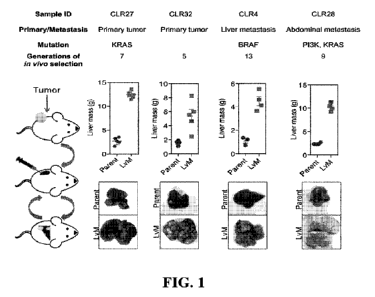
FIG. 1 is a set of diagrams and photographs showing in vivo selection that
generates
derivatives with enhanced ability to colonize mouse livers. In vivo selection
was performed on
four different CRC patient-derived primary and metastatic tumor xenograft
(PDX) samples
with varied anatomical locations and mutational backgrounds. The illustration
on the left
depicts the process used to generate the liver metastatic derivatives. Tumor
samples from
surgical specimens were inoculated subcutaneously into NSG mice. When the
tumor reached
the threshold size, it was removed from the mouse, dissociated into a single-
cell suspension,
and injected into the spleens of another set of mice as a means of introducing
the colorectal
cancer cells into the portal circulation. When the mice were deemed ill, the
liver tumors were
removed, dissociated, and re-injected to establish a next-generation liver
derivative. This
process was repeated numerous times (Range: 5-13) with each PDX sample to
obtain a distant
liver metastatic CRC PDX derivative requiring euthanasia of mice in 3 weeks
after cancer cells
injection. Each of the CRC PDX liver derivatives grew significantly faster in
the livers
compared to their parent samples.
FIGS. 2A and 2B (collectively "FIG. 2") are a set of diagrams showing the
unsupervised
hierarchical clustering of 170 polar metabolites' profiling data. FIG. 2B is a
close-up of the
highlighted area of the diagram of FIG. 2A. Pyrimidine nucleotide precursors
were up-
regulated in the highly liver metastatic PDXs. Parental PDXs were used as
references to the
corresponding highly metastatic PDXs.
FIGS. 3A and 3B are a set of diagrams showing the effects of the DHODH
inhibitor in
inhibiting liver metastatic colonization. FIG. 3A shows that leflunomide
inhibited liver
metastatic colonization of Lvm3b cells. 1 x 10⁶ Lvm3b cells were
intrasplenically injected into
athymic nude mice (n =4 per each cohort) on day 1, and leflunomide (7.5 mg/kg
mouse body
CA 03152258 2022-3-23
WO 2021/062157
PCT/US2020/052721
weight) or DMSO treatment was begun on day 1. The mice were imaged every week.
Firefly
luciferase bioluminescent images are shown (p<0.0001, Student's t-test). FIG.
3B shows a
Kaplan-Meier plot of the experiment (n =4 per each cohort) (p=0.007, log-rank
test).
FIGS. 4A, 4B, 4C, and 4D (collectively "FIG. 4") are a set of diagrams showing
the
combination of DHODH inhibition and SLC6a8 inhibition can be therapeutically
exploited in
gastrointestinal cancer models. FIG. 4A shows that 1 million MC38 cells were
subcutaneously
injected into C57BL/6 mice (n=4 per each cohort). Intraperitoneal leflunomide
injection was
started at the time that the average size of tumors reached 100 mm3. The
leflunomide treatment
was given daily. MPK: mg/kg body weight of mouse. Tumor size was measured by a
digital
caliper, and tumor volume was calculated as volume = (the longest diameter of
tumor/2)*(the
shortest diameter of tumor). FIG. 48 shows that 1 million MC38 cells were
subcutaneously
injected to C57BL/6 mice (n=4 per each cohort). Intraperitoneal leflunomide
injection and oral
I3-GPA administration were started at the time that the average size of tumors
reached 100 mm3.
Leflunomide and13-GPA were given daily. Leflunomide/P-GPA combo treatment
significantly
reduced the growth of MC38 tumor (p=0.0004, Student t-test). FIG. 4C shows
that 1 million
HS746T cells were subcutaneously injected to NOD.Cg-Prkdc"' 112ren1WO/SzJ (Nod-
Scid-
Gamma; NSG) mice (n=4 per each cohort). Intraperitoneal leflunomide injection
and oral 13-
GPA administration were started at the time that the average size of tumors
reached to 100mm3.
Leflunomide and I3-GPA were given daily. Leflunomide/ [3-GPA combo treatment
significantly
reduced the growth of HS746T tumor (p=0.0045, Student t-test). Pyrimidine
precursor
nucleoside, uridine administration rescued the leflunomide-induced tumor
growth reduction
supporting the on-target efficacy of leflunomide. FIG. 4D shows that 1 million
KPC LM2 cells
were subcutaneously injected to C57BL/6 mice (n=4 per each cohort).
Intraperitoneal
leflunomide injection and oral (3-GPA administration were started at the time
that the average
size of tumors reached to 100mm3. Leflunomide and (3-GPA were given daily.
Leflunomide/
13-GPA combo treatment significantly reduced the growth of KPC LM2 tumor
(p<0.0001,
Student t-test).
FIGS. 5A and 5B (collectively "FIG. 5") are a set of diagrams demonstrating
the
combinational therapeutic targeting of DHODH and SLC6a8 suppresses two
independent
human patient-derived tumor growth. FIG. 5A shows that 30 mm3 fragments of the
patient-
derived tumor were surgically sutured into the subcutaneous tissue of athymic
nude mice (n=4
per each cohort). Intraperitoneal leflunomide injection and oral [3-GPA
administration were
started at the time that the average size of tumors reached 100 mm3.
Leflunomide and 13-GPA
were given daily. Leflunomide/ [3-GPA combo treatment significantly reduced
the growth of
6
CA 03152258 2022-3-23
WO 2021/062157
PCT/US2020/052721
CLR1 tumor (p=0.0011, Student t-test). FIG. 5B shows that 30 mm3 fragments of
patient-
derived tumors were surgically sutured into the subcutaneous tissue of athymic
nude mice (n=4
per each cohort). Intraperitoneal leflunomide injection and oral I3-GPA
administration were
started at the time that the average size of tumors reached 100 mm3.
Leflunomide and I3-GPA
were given daily. Leflunomide/ P-GPA combo treatment significantly reduced the
growth of
GAS HI tumor (13=0.008, Student t-test). Uridine administration rescued the
leflunomide
induced tumor growth suppression.
DETAILED DESCRIPTION OF THE INVENTION
This disclosure is based, at least in part, on unexpected discoveries that
therapeutic
inhibition of the pyrimidine biosynthetic enzyme DHODH with a DHODH inhibitor
(e.g.,
leflunomide) substantially impaired CRC liver metastatic colonization and
hypoxic survival
Given that most colorectal cancer deaths occur as a result of complications of
metastatic
disease, a model that can predict which patients with advanced CRC harbor more
aggressive
disease could aid in appropriately positioning patients for experimental
clinical trials and
treatments. Accordingly, this disclosure also provides a colorectal cancer
liver metastasis
patient-derived xenograft model, as well as methods to identify candidate
genes that may drive
colorectal cancer liver colonization using this model. Also, metastatic CRC
(mCRC) liver
colonization was modeled using patient-derived primary and metastatic tumor
xenografts
(PDX). Such PDX modeling predicted patient survival outcomes. In vivo
selection of multiple
PDXs for enhanced metastatic capacity upregulated the gluconeogenic enzyme
PCK1, which
enhanced metastatic hypoxic survival by driving anabolic pyrimidine nucleotide
biosynthesis.
Consistently, highly metastatic tumors upregulated multiple pyrimidine
biosynthesis
intermediary metabolites. It was demonstrated in this disclosure that
therapeutic inhibition of
DHODH substantially diminished CRC liver metastatic colonization and hypoxic
survival.
Thus, the present disclosure provides a mechanistic basis for the
epidemiologic
association of anti-gluconeogenic drugs with improved CRC metastasis outcomes,
reveals the
exploitation of a gluconeogenesis enzyme for pyrimidine biosynthesis during
hypoxia, and
implicates DHODH and PCK1 as metabolic therapeutic targets in colorectal
cancer metastasis.
A. METHODS FOR TREATING GASTROINTESTINAL CANCER
This disclosure provides agents and methods for suppressing cancer metastasis.
In one
aspect, this disclosure provides a method for treating gastrointestinal cancer
(e.g., metastatic
colorectal cancer) in a subject in need thereof. The method includes
suppressing the enzymatic
7
CA 03152258 2022-3-23
WO 2021/062157
PCT/US2020/052721
activity of DHODH and/or decreasing the level of creatine via suppression of
creatine
transporter channel SLC6a8 in the subject.
In some embodiments, the enzymatic activity of DHODH can be suppressed by
administering to the subject one or more DHODH inhibitors, or a
pharmaceutically acceptable
prodrug, a pharmaceutically active metabolite, a pharmaceutically acceptable
salt thereof For
example, the suppression step can be carried out by administering to the
subject a DHODH
inhibitor, such as leflunomide. Other examples of DHODH inhibitors include,
but are not
limited to, atovaquone, brequinar sodium, teriflunomide, BAY-2402234, and AG-
636.
In some embodiments, the level of creatine can be decreased by administering
to the
subject beta-guanidinopropionic acid (P-GPA), or a pharmaceutically acceptable
prodrug, a
pharmaceutically active metabolite, a pharmaceutically acceptable salt
thereof.
In another aspect, also provided is a method for treating metastatic
gastrointestinal
cancers in a subject in need thereof. The method includes administering
compounds to the
subject an effective amount of a DHODH inhibitor, or a pharmaceutically
acceptable salt
thereof, and a P-GPA, or a pharmaceutically acceptable salt thereof, to
suppress metastatic
colonization of gastrointestinal cancer. In some embodiments, the DHODH
inhibitor can be
any one of atovaquone, brequinar sodium, leflunomide, teriflunomide, BAY-
2402234, AG-636,
and a combination thereof
In another aspect, also provided is a method for treating cancer (e.g.,
metastatic cancers)
in a subject in need thereof The method includes administering to the subject
an effective
amount of a DHODH inhibitor (e.g., atovaquone, brequinar sodium, leflunomide,
teriflunomide, BAY-2402234, AG-636, or a combination thereof), or a
pharmaceutically
acceptable salt thereof, and p-GPA, or a pharmaceutically acceptable salt
thereof In some
embodiments, the effective amount is an amount of the DHODII inhibitor and P-
GPA, or a
pharmaceutically acceptable salt thereof that is together effective to
suppress metastatic
progression (e.g., metastatic colonization) of the cancer. In some
embodiments, the DHODH
inhibitor is leflunomide. In some embodiments, the cancer is gastrointestinal
cancer, such as
colorectal cancer, esophageal cancer, or gastric cancer, pancreatic cancer,
liver cancer, breast
cancer, prostate cancer, lung cancer, and melanoma. In some embodiments, the
cancer is
gastrointestinal cancer. In some embodiments, the cancer is lung cancer. In
some
embodiments, the effective amount is an amount effective to suppress
metastatic colonization
of the cancer to the liver and/or brain.
In some embodiments, the method further comprises administering to the subject
an
additional anti-cancer therapy (e.g., surgery, radiation therapy, and/or one
or more therapeutic
8
CA 03152258 2022-3-23
WO 2021/062157
PCT/US2020/052721
agents, such as an anti-tumor or anti-cancer agent (e.g., irinotecan,
oxaliplatin, cetuximab,
avastin, leucovorin, and/or 5-fluorouracil (5-FU)). In some embodiments, the
subject has
previously been administered at least one prior anticancer therapy (e.g.,
surgery, radiation
therapy, and/or one or more therapeutic agents, such as an anti-tumor or anti-
cancer agent). In
some embodiments, the therapeutic agent is cyclocreatine, an RNAi agent, a
nucleic acid, a
vector, 5-FU, Oxaliplatin, irinotecan, oxaliplatin, capecitabine, gemcitabine,
cetuximab, taxol,
avastin, folinic acid (leucovorin), regorafenib, zaltrap, topoisomerase I
inhibitors, etirinotecan
pegol, tivantinib, sonolisib, sorafenib, linifanib, kinase inhibitors,
telatinib, XL281 (BMS-
908662), robatumumab, IGF1-R inhibitors, or combinations thereof.
In some embodiments of any of the foregoing methods, the DHODH inhibitor is
atovaquone administered in an amount of 500 to 1500 mg per day, e.g., 500 mg
once daily, 750
mg once daily, 1000 mg once daily, 1500 mg once daily, or 250 mg twice daily,
500 mg twice
daily, or 750 mg twice daily.
In some embodiments of any of the foregoing methods, the DHODH inhibitor is
leflunomide administered in an amount of 10 to 100 mg per day, e.g., 10 mg
once daily, 20 mg
once daily, or 100 mg once daily.
In some embodiments of any of the foregoing methods, the DHODH inhibitor is
teriflunomide administered in an amount of 7 to 14 mg per day, e.g., 7 mg once
daily, 14 mg
once daily, or 7 mg twice daily.
In some embodiments of any of the foregoing methods, the I3-GPA, or a
pharmaceutically acceptable salt thereof, is administered in an amount of 0.01
to 100 mg/kg
per day.
A subject to be treated for a disorder can be identified by standard
diagnosing
techniques for the disorder. Optionally, the subject can be examined for
mutation, expression
level, or activity level of one or more of DHODH, CKB, SLC6a8, miR-483-5p, and
miR-55 Ia
mentioned above by methods known in the art or described above before
treatment. If the
subject has a particular mutation in the gene, or if the gene expression or
activity level is, for
example, greater (in the case for CKB or SLC6a8) in a sample from the subject
than that in a
sample from a normal person, the subject is a candidate for treatment of this
invention.
To confirm the inhibition or treatment, one can evaluate and/or verify the
inhibition of
cancer cell survival, hypoxic survival, metastatic survival, or metastatic
colonization using
technologies known in the art before and/or after the administering step.
Exemplary
technologies include CT-scans or PET-scans of organs of the body.
9
CA 03152258 2022-3-23
WO 2021/062157
PCT/US2020/052721
The DHODH inhibitor or the pharmaceutically acceptable salt thereof and/or [3-
GPA or
the pharmaceutically acceptable salt thereof can be administered to the
subject intratumorally,
intravenously, subcutaneously, intraosseously, orally, transdermally, in
sustained release, in
controlled release, in delayed release, as a suppository, or sublingually.
A therapeutic agent can be administered in vivo or ex vivo, alone or co-
administered in
conjunction with other drugs or therapy, i.e., a cocktail therapy. As used
herein, the term "co-
administration" or "co-administered" refers to the administration of at least
two agent(s) or
therapies to a subject. For example, in the treatment of tumors, particularly
malignant tumors,
the agents can be used alone or in combination with, e.g., chemotherapeutic,
radiotherapeutic,
apoptopic, anti-angiogenic agents and/or immunotoxins or coaguligands.
In some
embodiments, the co-administration of two or more agents/therapies is
concurrent. In other
embodiments, a first agent/therapy is administered prior to a second
agent/therapy. Those of
skill in the art understand that the formulations and/or routes of
administration of the various
agents/therapies used may vary.
In some embodiments, the DHODH inhibitor or the pharmaceutically acceptable
salt
thereof is administered to the subject before (e.g., at least one day before,
at least one week
before, at least one month before), after(e.g., at least one day after, at
least one week after, at
least one month after), or concurrently with the I3-GPA or the
pharmaceutically acceptable salt
thereof
In some embodiments, this disclosure additionally provides a method comprising
suppressing metastatic colonization of lung cancer in the liver of a subject
in need thereof.
The dosage required depends on the choice of the route of administration; the
nature of
the formulation; the nature of the patient's illness; the subject's size,
weight, surface area, age,
and sex; other drugs being administered; and the judgment of the attending
physician. Suitable
dosages are in the range of 0.01-100 mg/kg. Variations in the needed dosage
are to be expected
in view of the variety of compounds available and the different efficiencies
of various routes
of administration. For example, oral administration would be expected to
require higher
dosages than administration by i.v. injection. Variations in these dosage
levels can be adjusted
using standard empirical routines for optimization as is well understood in
the art.
Encapsulation of the compound in a suitable delivery vehicle (e.g., polymeric
microparticles
or implantable devices) can increase the efficiency of delivery, particularly
for oral delivery.
In some embodiments, a dosage of the DHODH inhibitor or13-GPA can be one of:
trace
amount, 0.01-0.05 mg, 0_05-0.1 mg, 0.1-0.5 mg, 0.25-1 mg, 0.5-15 mg, 0.5-2.5
mg, 1.0-2.5
mg, 2.5-5 mg, 5.0-7.5 mg, 5.0-10 mg, 1.0-25 mg, 25-50 mg, 50-75 mg, 75-100 mg,
10-20 mg,
CA 03152258 2022-3-23
WO 2021/062157
PCT/US2020/052721
10-15 mg, and 15-20 mg, 20-30 mg, 30-40 mg, 40-50 mg, 50-60 mg, 60-70 mg, 70-
80 mg, 80-
90 mgõ 90-100 mg, 1-100 mg, 100-125 mg, 125-150 mg, 150-175 mg, 175-200 mg,
and >200
mg.
I3-GPA has the structure:
NH 0
H2NAN..--,)11-OH
I3-GPA is zwitterionic and highly soluble in water (> 50 mg/mL), but has low
solubility
in organic solvents. I3-GPA possesses a basic guanidino group and is thus
capable of forming
acid addition salts.
This disclosure further provides a method for treating colorectal cancer,
gastric cancer,
esophageal cancer, or pancreatic cancer in a subject in need thereof In some
embodiments, the
cancer is cholangial cancer.
In some embodiments, the cancer expresses creatine kinase brain-type (CKB)
(e.g., the
cancer cells express CKB). In some embodiments, the cancer has been determined
to express
CKB based on histological examination of a tissue sample from the subject. In
some
embodiments, the subject has been identified as likely to respond to treatment
with a DHODH
inhibitor and I3-GPA, or a pharmaceutically acceptable salt thereof (e.g.,
based on histological
examination of a tissue sample from the subject to determine the level of CKB
expression).
In some embodiments, the cancer expresses SLC6a8 (e.g., the cancer cells
express
SLC6a8). In some embodiments, the cancer has been determined to express SLC6a8
based on
histological examination of a tissue sample from the subject In some
embodiments, the subject
has been identified as likely to respond to treatment with a DHODH inhibitor
and I3-GPA, Of a
pharmaceutically acceptable salt thereof (e.g., based on histological
examination of a tissue
sample from the subject to determine the level of SLC6a8 expression)In some
embodiments,
the method further comprises administering to the subject one or more
additional therapeutic
agents, such as antitumor/anticancer agents, including chemotherapeutic agents
and
immunotherapeutic agents.
A "chemotherapeutic agent" is a chemical compound useful in the treatment of
cancer.
Examples of chemotherapeutic agents include alkylating agents such as thiotepa
and
cyclophosphamide (CYTOXANTM); alkyl sulfonates such as busulfan, improsulfan
and
piposulfan; aziridines such as benzodopa, carboquone, methyldopa, and uredopa;
ethylenimines and methylamelamines including altretamine, triethylenemelamine,
tri etyl enephosphorami de, tri ethyl enethiophosphaorami de and
trimethylolomelamine;
11
CA 03152258 2022-3-23
WO 2021/062157
PCT/US2020/052721
acetogenins (especially bullatacin and bullatacinone); a camptothecin
(including the synthetic
analogue topotecan), bryostatin; callystatin; CC-1065 (including its
adozelesin, carzelesin and
bizelesin synthetic analogues); cryptophycins (particularly cryptophycin 1 and
cryptophycin
8); dolastatin; duocarmycin (including the synthetic analogues, KW-2189 and
CBI-TMI);
eleutherobin; pancratistatin; a sarcodictyin; spongistatin; nitrogen mustards
such as
chlorambucil, chlornaphazine, chol ophosphami
de, estramustine, ifosfamide,
mechlorethamine, mechlorethamine oxide hydrochloride, m el phal an, novembi
chi n,
phenesterine, prednimustine, trofosfamide, uracil mustard; nitrosureas such as
cannustine,
chlorozotocin, fotemustine, lomustine, nimustine, ranimustine; antibiotics
such as the enediyne
antibiotics (e.g. calicheamicin, see, e.g., Agnew Chem. Intl. Ed. Engl. 33:183-
186 (1994);
dynemicin, including dynemicin A; an esperamicin; as well as neocarzinostatin
chromophore
and related chromoprotein enediyne antibiotic chromomophores), aclacinomysins,
actinomycin, authramycin, azaserine, bleomycins, cactinomycin, carabicin,
caminomycin,
carzinophilin, chromomycins, dactinomycin, daunorubicin, detorubicin, 6-diazo-
5-oxo-L-
norleucine, doxorubicin (including morpholino-doxorubicin, cyanomorpholino-
doxorubicin,
2-pyrrolino-doxorubicin and deoxydoxorubicin), epirubicin, esorubicin,
idarubicin,
marcellomycin, mitomycins, mycophenolic acid, nogalamycin, olivomycins,
peplornycin,
potfiromycin, puromycin, quelamycin, rodorubicin, streptonigrin, streptozocin,
tubercidin,
ubenimex, zinostatin, zorubicin; anti-metabolites such as methotrexate and 5-
fluorouracil (5-
FU); folic acid analogues such as denopterin, methotrexate, pteropterin,
trimetrexate; purine
analogs such as fludarabine, 6-mercaptopurine, thiamiprine, thioguanine;
pyrimidine analogs
such as ancitabine, azacitidine, 6-azauridine, carmofur, cytarabine,
dideoxyuridine,
doxifluridine, enocitabine, floxuridine, 5-FU; androgens such as calusterone,
dromostanolone
propionate, epitiostanol, mepitiostane, testolactone; anti-adrenals such as
aminoglutethimide,
mitotane, trilostane; folic acid replenisher such as frolinic acid;
aceglatone; aldophosphamide
glycoside; aminolevulinic acid; amsacrine; bestrabucil; bisantrene;
edatraxate; defofamine;
demecolcine; diaziquone; elformithine; elliptinium acetate; an epothilone;
etoglucid; gallium
nitrate; hydroxyurea; lentinan; lonidamine; maytansinoids such as maytansine
and
ansamitocins; mitoguazone; mitoxantrone; mopidamol; nitracrine; pentostatin;
phenamet;
pirarubicin; podophyllinic acid; 2-ethylhydrazide; procarbazine; PSKO.;
razoxane; rhizoxin;
sizofuran; spirogermanium, tenuazonic acid; triaziquone; 2,2',2"-
trichlorotriethylamine;
trichothecenes (especially T-2 toxin, verracurin A, roridin A and anguidine);
urethan;
vindesine; dacarbazine; mannomustine; mitobronitol; mitolactol; pipobroman;
gacytosine;
arabinoside ("Ara-C"); cyclophosphamide; thiotepa; taxoids, e.g. paclitaxel
(TAXOL ,
12
CA 03152258 2022-3-23
WO 2021/062157
PCT/US2020/052721
Bristol-Myers Squibb Oncology, Princeton, N.J.) and doxetaxel (TAXOTERE ,
Rhone-
Poulenc Rorer, Antony, France); chlorambucil; gemcitabine; 6-thioguanine;
mercaptopurine;
methotrexate; platinum analogs such as cisplatin and carboplatin; vinblastine;
platinum;
etoposide (VP-16); ifosfamide; mitomycin C; mitoxantrone; vincristine;
vinorelbine;
navelbine; novantrone; teniposide; daunomycin; aminopterin; xeloda;
ibandronate; CPT-11;
topoisomerase inhibitor RFS 2000; difluoromethylornithine (DMF0); retinoic
acid;
capecitabine; and pharmaceutically acceptable salts, acids or derivatives of
any of the above.
Also included in this definition are anti-hormonal agents that act to regulate
or inhibit hormone
action on tumors such as anti-estrogens including for example tamoxifen,
raloxifene, aromatase
inhibiting 4(5)-imi dazol es, 4-hydroxytamoxifen, trioxifene, keoxifene,
LY117018,
onapristone, and toremifene (Fareston); and anti-androgens such as flutamide,
nilutamide,
bicalutamide, leuprolide, xeloda, gemcitabine, 1CRAS mutation covalent
inhibitors and
goserelin; and pharmaceutically acceptable salts, acids or derivatives of any
of the above.
Additional examples include irinotecan, oxaliplatinum, and other standard
colon cancer
regimens.
An "immunotherapeutic agent" is a biological agent useful in the treatment of
cancer.
Examples of immunotherapeutic agents include atezolizumab, avelumab,
blinatumomab,
daratumumab, cemiplimab, durvalumab, elotuzumab, laherparepvec, ipilimumab,
nivolumab,
obinutuzumab, ofatumumab, pembrolizumab, cetuximab, and talimogene.
B. COMPOSITIONS AND KITS
In some embodiments, the DHODH inhibitor or the pharmaceutically acceptable
salt
thereof and the I3-GPA or the pharmaceutically acceptable salt thereof are
provided in a single
composition. Alternatively, the DHODH inhibitor or the pharmaceutically
acceptable salt
thereof and the P-GPA or the pharmaceutically acceptable salt thereof can be
provided in
separate compositions.
Pharmaceutical compositions for use in accordance with the present methods may
be
formulated in a conventional manner using one or more physiologically
acceptable carriers or
excipients. Thus, the DHODH inhibitor and/or 0-GPA, or their analogs/variants,
described
herein and their physiologically acceptable salts and solvates may be
formulated for
administration by, for example, injection, inhalation or insufflation (either
through the mouth
or the nose) or oral, buccal, parenteral or rectal administration. In one
embodiment, the agent
is administered locally, e.g., at the site where the target cells are present,
such as by the use of
a patch.
13
CA 03152258 2022-3-23
WO 2021/062157
PCT/US2020/052721
Phannaceutical compositions can be formulated for a variety of loads of
administration,
including systemic and topical or localized administration. Techniques and
formulations
generally may be found in Remmington's Pharmaceutical Sciences, Meade
Publishing Co.,
Easton, PA. For systemic administration, injection is preferred, including
intramuscular,
intravenous, intraperitoneal, and subcutaneous. For injection, the agents can
be formulated in
liquid solutions, preferably in physiologically compatible buffers such as
Hank's solution or
Ringer's solution. In addition, the agents may be formulated in solid form and
redissolved or
suspended immediately prior to use. Lyophilized forms are also included.
For oral administration, the pharmaceutical compositions may take the form of,
for
example, tablets, lozenges, or capsules prepared by conventional means with
pharmaceutically
acceptable excipients such as binding agents (e.g., pregelatinized maize
starch,
pol yvi nyl pyrroli done or hydroxypropyl methylcellulose); fillers (e.g.,
lactose, microcrystalline
cellulose or calcium hydrogen phosphate); lubricants (e.g., magnesium
stearate, talc or silica);
disintegrants (e.g., potato starch or sodium starch glycolate); or wetting
agents (e.g., sodium
!miry' sulfate). The tablets may be coated by methods well known in the an
Liquid preparations
for oral administration may take the form of, for example, solutions, syrups
or suspensions, or
they may be presented as a dry product for constitution with water or other
suitable vehicles
before use. Such liquid preparations may be prepared by conventional means
with
pharmaceutically acceptable additives such as suspending agents (e.g.,
sorbitol syrup, cellulose
derivatives or hydrogenated edible fats); emulsifying agents (e.g., lecithin
or acacia); non-
aqueous vehicles (e.g., ationd oil, oily esters, ethyl alcohol or fractionated
vegetable oils); and
preservatives (e.g., methyl or propyl-p-hydroxybenzoates or sorbic acid). The
preparations may
also contain buffer salts, flavoring, coloring and sweetening agents as
appropriate. Preparations
for oral administration may be suitably formulated to give controlled release
of the active
compound.
Pharmaceutical compositions that may oxidize and lose biological activity,
especially
in a liquid or semisolid form, may be prepared in a nitrogen atmosphere or
sealed in a type of
capsule and/or foil package that excludes oxygen (e.g., Capsugerm).
For administration by inhalation, the agents may be conveniently delivered in
the form
of an aerosol spray presentation from pressurized packs or a nebulizer, with
the use of a suitable
propellant, e.g., di chlorodi fluoronriethane, trichlorofluoromethane,
dichlorotetrafluoroethane,
carbon dioxide or other suitable gas. In the case of a pressurized aerosol,
the dosage unit may
be determined by providing a valve to deliver a metered amount. Capsules and
cartridges of,
14
CA 03152258 2022-3-23
WO 2021/062157
PCT/US2020/052721
e.g., gelatin, for use in an inhaler or insufflator may be formulated
containing a powder mix of
the agent and a suitable powder base such as lactose or starch.
Pharmaceutical compositions may be formulated for parenteral administration by
injection, e.g., by bolus injection or continuous infusion. Formulations for
injection may be
presented in unit dosage form, e.g., in ampoules or in multi-dose containers,
with an added
preservative. The agents may take such forms as suspensions, solutions or
emulsions in oily or
aqueous vehicles, and may contain formulatory agents such as suspending,
stabilizing and/or
dispersing agents. Alternatively, the active ingredient may be in powder form
for constitution
with a suitable vehicle, e.g., sterile pyrogen-free water, before use. The
agents may also be
formulated in rectal compositions such as suppositories or retention enemas,
e.g., containing
conventional suppository bases such as cocoa butter or other glycerides.
In addition to the formulations described previously, pharmaceutical
compositions may
also be formulated as a depot preparation. Such long acting formulations may
be administered
by implantation (for example, subcutaneously or intramuscularly) or by
intramuscular injection.
Thus, for example, the agents may be formulated with suitable polymeric or
hydrophobic
materials (for example, as an emulsion in an acceptable oil) or ion exchange
resins, or as
sparingly soluble derivatives, for example, as a sparingly soluble salt.
Controlled release
formula also includes patches, e.g., transdermal patches. Patches may be used
with a sonic
applicator that deploys ultrasound in a unique combination of waveforms to
introduce drug
molecules through the skin that normally could not be effectively delivered
transdermally.
Pharmaceutical compositions (including cosmetic preparations) may comprise
from
about 0.00001 to 100%, such as from 0.001 to 10% or from 0.1% to 5% by weight
of one or
more agents described herein.
A pharmaceutical composition described herein can also be incorporated into a
topical
formulation containing a topical earner that is generally suited to topical
drug administration
and comprising any such material known in the art. The topical carrier may be
selected so as
to provide the composition in the desired form, e.g., as an ointment, lotion,
cream,
microemulsion, gel, oil, solution, or the like, and may be comprised of a
material of either
naturally occurring or synthetic origin. It is preferable that the selected
carrier not adversely
affect the active agent or other components of the topical formulation.
Examples of suitable
topical carriers for use herein include water, alcohols, and other nontoxic
organic solvents,
glycerin, mineral oil, silicone, petroleum jelly, lanolin, fatty acids,
vegetable oils, parabens,
waxes, and the like.
CA 03152258 2022-3-23
WO 2021/062157
PCT/US2020/052721
Formulations may be colorless, odorless ointments, lotions, creams,
microemulsions,
and gels. Pharmaceutical compositions may be incorporated into ointments,
which generally
are semisolid preparations which are typically based on petrolatum or other
petroleum
derivatives. The specific ointment base to be used, as will be appreciated by
those skilled in
the art, is one that will provide for optimum drug delivery, and, preferably,
will provide for
other desired characteristics as well, e.g., emolliency or the like. As with
other carriers or
vehicles, an ointment base should be inert, stable, nonirritating and
nonsensitizing. As
explained in Remington's, ointment bases may be grouped in four classes:
oleaginous bases;
emulsifiable bases; emulsion bases, and water-soluble bases. Oleaginous
ointment bases
include, for example, vegetable oils, fats obtained from animals, and
semisolid hydrocarbons
obtained from petroleum. Emulsifiable ointment bases, also known as absorbent
ointment bases,
contain little or no water and include, for example, hydroxystearin sulfate,
anhydrous lanolin,
and hydrophilic petrolatum. Emulsion ointment bases are either water-in-oil
(W/O) emulsions
or oil-in-water (01W) emulsions, and include, for example, cetyl alcohol,
glyceryl
monostearate, lanolin, and stearic acid. Exemplary water-soluble ointment
bases are prepared
from polyethylene glycols (PEGs) of varying molecular weight; again, reference
may be had
to Remington's, supra, for further information.
Pharmaceutical compositions may be incorporated into lotions, which generally
are
preparations to be applied to the skin surface without friction, and are
typically liquid or
semiliquid preparations in which solid particles, including the active agent,
are present in a
water or alcohol base. Lotions are usually suspensions of solids, and may
comprise a liquid
oily emulsion of the oil-in-water type. Lotions are preferred formulations for
treating large
body areas, because of the ease of applying a more fluid composition. It is
generally necessary
that the insoluble matter in a lotion be finely divided. Lotions will
typically contain suspending
agents to produce better dispersions as well as compounds useful for
localizing and holding the
active agent in contact with the skin, e.g., methylcellulose, sodium
carboxymethylcellulose, or
the like. An exemplary lotion formulation for use in conjunction with the
present method
contains propylene glycol mixed with hydrophilic petrolatum such as that which
may be
obtained under the trademark AquaphorTM from Beiersdorf, Inc. (Norwalk,
Conn.).
Pharmaceutical compositions may be incorporated into creams, which generally
are
viscous liquid or semisolid emulsions, either oil-in-water or water-in-oil.
Cream bases are
water-washable and contain an oil phase, an emulsifier and an aqueous phase.
The oil phase is
generally comprised of petrolatum and a fatty alcohol such as cetyl or stearyl
alcohol; the
aqueous phase usually, although not necessarily, exceeds the oil phase in
volume, and generally
16
CA 03152258 2022-3-23
WO 2021/062157
PCT/US2020/052721
contains a humectant. The emulsifier in a cream formulation, as explained in
Remington's,
supra, is generally a nonionic, anionic, cationic or amphoteric surfactant.
Pharmaceutical compositions may be incorporated into microemulsions, which
generally are thermodynamically stable, isotropically clear dispersions of two
immiscible
liquids, such as oil and water, stabilized by an interfacial film of
surfactant molecules
(Encyclopedia of Pharmaceutical Technology (New York: Marcel Dekker, 1992),
volume 9).
For the preparation of microemulsions, surfactant (emulsifier), co-surfactant
(co-emulsifier),
an oil phase and a water phase are necessary. Suitable surfactants include any
surfactants that
are useful in the preparation of emulsions, e.g., emulsifiers that are
typically used in the
preparation of creams. The co-surfactant (or "co-emulsifier) is generally
selected from the
group of polyglycerol derivatives, glycerol derivatives, and fatty alcohols.
Preferred
emulsifier/co-emulsifier combinations are generally although not necessarily
selected from the
group consisting of: glyceryl monostearate and polyoxyethylene stearate;
polyethylene glycol
and ethylene glycol palmitostearate, and caprylic and capric trig,lycerides
and oleoyl
macrogolglycerides. The water phase includes not only water but also,
typically, buffers,
glucose, propylene glycol, polyethylene glycols, preferably lower molecular
weight
polyethylene glycols (e.g., PEG 300 and PEG 400), and/or glycerol, and the
like, while the oil
phase will generally comprise, for example, fatty acid esters, modified
vegetable oils, silicone
oils, mixtures of mono- di- and triglycerides, mono- and di-esters of PEG
(e.g., oleoyl macrogol
glycerides), etc.
Pharmaceutical compositions may be incorporated into gel formulations, which
generally are semisolid systems consisting of either suspension made up of
small inorganic
particles (two-phase systems) or large organic molecules distributed
substantially uniformly
throughout a carrier liquid (single-phase gels). Single-phase gels can be
made, for example, by
combining the active agent, a carrier liquid and a suitable gelling agent such
as tragacanth (at
2 to 5%), sodium alginate (at 2-10%), gelatin (at 2-15%), methylcellulose (at
3-5%), sodium
carboxymethylcellulose (at 2-5%), carbomer (at 0.3-5%) or polyvinyl alcohol
(at 10-20%)
together and mixing until a characteristic semisolid product is produced.
Other suitable gelling
agents include methylhydroxycellulose,
polyoxyethylene-
polyoxypropylene,
hydroxyethylcellulose, and gelatin. Although gels commonly employ aqueous
carrier liquid,
alcohols and oils can be used as the carrier liquid as well.
Various additives, known to those skilled in the art, may be included in
formulations,
e.g, topical formulations. Examples of additives include, but are not limited
to, solubilizers,
skin permeation enhancers, pacifiers, preservatives (e.g., anti-oxidants),
gelling agents,
17
CA 03152258 2022-3-23
WO 2021/062157
PCT/US2020/052721
buffering agents, surfactants (particularly nonionic and amphoteric
surfactants), emulsifiers,
emollients, thickening agents, stabilizers, humectants, colorants, fragrance,
and the like.
Inclusion of solubilizers and/or skin permeation enhancers is particularly
preferred, along with
emulsifiers, emollients, and preservatives. An optimum topical formulation
comprises
approximately: 2 wt. % to 60 wt. %, preferably 2 wt. % to 50 wt. %,
solubilizer and/or skin
permeation enhancer; 2 wt. % to 50 wt. %, preferably 2 wt. % to 20 wt. %,
emulsifiers; 2 wt. %
to 20 wt. % emollient; and 0.01 to 0.2 wt. % preservative, with the active
agent and carrier (e.g.,
water) making of the remainder of the formulation. A skin permeation enhancer
serves to
facilitate passage of therapeutic levels of active agent to pass through a
reasonably sized area
of unbroken skin. Suitable enhancers are well known in the art and include,
for example: lower
alkanols such as methanol ethanol and 2-propanol; alkyl methyl sulfoxides such
as
dimethylsulfoxide (DMSO), decylmethylsulfoxide (Cio MSO) and tetradecylmethyl
sulfoxide;
pyrrolidones such as 2-pyrrolidone,
N-methyl-2-pyrrol i done and N-(-
hydroxyethyl)pynrolidone; urea, N,N- diethyl-m-toluamide, C2-C6 alkane diols;
miscellaneous
solvents such as dimethylformamide (DMF), N,N-dimethylacetamide (DMA) and
tetrahydrofurfuryl alcohol; and the 1 -substituted azacycloheptan-2-ones,
particularly 1-n-
dodecyleyelazacycloheptan-2-one (laurocapram; available under the trademark
AzoneRTM
from Whitby Research Incorporated, Richmond, Va.).
Examples of solubilizers include, but are not limited to, the following:
hydrophilic
ethers such as diethylene glycol monoethyl ether (ethoxydiglycol, available
commercially as
TranscutolTm) and diethylene glycol monoethyl ether oleate (available
commercially as
SoftcutolTm); polyethylene castor oil derivatives such as polyoxy 35 castor
oil, polyoxy 40
hydrogenated castor oil, etc.; polyethylene glycol, particularly lower
molecular weight
polyethylene glycols such as PEG 300 and PEG 400, and polyethylene glycol
derivatives such
as PEG-8 caprylie/capric glycerides (available commercially as LabrasolTm);
alkyl methyl
sulfoxides such as DMSO; pyrrolidones such as 2-pyrrolidone and N-methyl-2-
pyrrolidone;
and DMA. Many solubilizers can also act as absorption enhancers. A single
solubilizer may be
incorporated into the formulation, or a mixture of solubilizers may be
incorporated therein.
Suitable emulsifiers and co-emulsifiers include, without limitation, those
emulsifiers
and co-emulsifiers described with respect to mieroemulsion formulations.
Emollients include,
for example, propylene glycol, glycerol, isopropyl myristate, polypropylene
glycol- 2 (PPG-2)
myristyl ether propionate, and the like.
Other active agents may also be included in formulations, e.g., anti-
inflammatory
agents, analgesics, antimicrobial agents, antifungal agents, antibiotics,
vitamins, antioxidants,
18
CA 03152258 2022-3-23
WO 2021/062157
PCT/US2020/052721
and sunblock agents commonly found in sunscreen formulations including, but
not limited to,
anthranilates, benzophenones (particularly benzophenone-3), camphor
derivatives, cinnamates
(e.g., octyl methoxycinnamate), dibenzoyl methanes (e.g., butyl
methoxydibenzoyl methane),
p-aminobenzoic acid (PABA) and derivatives thereof, and salicylates (e.g.,
octyl salicylate). In
certain topical formulations, the active agent is present in an amount in the
range of
approximately 0.25 wt. % to 75 wt. % of the formulation, preferably in the
range of
approximately 0.25 wt. % to 30 wt. % of the formulation, more preferably in
the range of
approximately 0.5 wt. % to 15 wt. % of the formulation, and most preferably in
the range of
approximately 1.0 wt. % to 10 wt. % of the formulation. Topical skin treatment
compositions
can be packaged in a suitable container to suit its viscosity and intended use
by the consumer.
For example, a lotion or cream can be packaged in a bottle or a roll-ball
applicator, or a
propellant-driven aerosol device or a container fitted with a pump suitable
for finger operation.
When the composition is a cream, it can simply be stored in a non-deformable
bottle or squeeze
container, such as a tube or a lidded jar. The composition may also be
included in capsules
such as those described in U.S. Pat. No. 5,063,507. Accordingly, also provided
are closed
containers containing a cosmetically acceptable composition.
In some embodiments, a pharmaceutical formulation is provided for oral or
parenteral
administration, in which case the formulation may comprise an activating
compound-
containing microemulsion as described above, and may contain alternative
pharmaceutically
acceptable carriers, vehicles, additives, etc particularly suited to oral or
parenteral drug
administration. Alternatively, an activating compound-containing microemulsion
may be
administered orally or parenterally substantially as described above, without
modification.
A composition described herein can be provided in a kit. In one embodiment,
the kit
includes (a) a container that contains the composition, and optionally (b)
informational
material. The informational material can be descriptive, instructional,
marketing or other
material that relates to the methods described herein and/or the use of the
agents for therapeutic
benefit. In an embodiment, the kit includes also includes an additional
therapeutic agent. For
example, the kit includes a first container that contains the composition and
a second container
for the additional therapeutic agent.
The informational material of the kits is not limited in its form. In one
embodiment,
the informational material can include information about production of the
composition,
concentration, date of expiration, batch or production site information, and
so forth. In one
embodiment, the informational material relates to methods of administering the
composition,
e.g., in a suitable dose, dosage form, or mode of administration (e.g., a
dose, dosage form, or
19
CA 03152258 2022-3-23
WO 2021/062157
PCT/US2020/052721
mode of administration described herein), to treat a subject in need thereof.
In one
embodiment, the instructions provide a dosing regimen, dosing schedule, and/or
route of
administration of the composition or the additional therapeutic agent. The
information can be
provided in a variety of formats, including printed text, computer-readable
material, video
recording, or audio recording, or information that contains a link or address
to substantive
material.
In addition to the composition, the kit can include other ingredients, such as
a solvent
or buffer, a stabilizer, or a preservative. The composition can be provided in
any form, e.g.,
liquid, dried or lyophilized form, preferably substantially pure and/or
sterile. When the agents
are provided in a liquid solution, the liquid solution preferably is an
aqueous solution. When
the agents are provided as a dried form, reconstitution generally is by the
addition of a suitable
solvent and acidulant. The acidulant and solvent, e.g., an aprotic solvent,
sterile water, or a
buffer, can optionally be provided in the kit.
The kit can include one or more containers for the composition or compositions
containing a DHODH inhibitor and/or (3-GPA. In some embodiments, the kit
contains separate
containers, dividers or compartments for the composition and informational
material. For
example, the composition can be contained in a bottle, vial, or syringe, and
the informational
material can be contained in a plastic sleeve or packet. In other embodiments,
the separate
elements of the kit are contained within a single, undivided container. For
example, the
composition is contained in a bottle, vial or syringe that has attached
thereto the informational
material in the form of a label. In some embodiments, the kit includes a
plurality (e.g., a pack)
of individual containers, each containing one or more unit dosage forms (e.g.,
a dosage form
described herein) of the agents. The containers can include a combination unit
dosage, e.g., a
unit that includes both the DHODH inhibitor and fl-GPA in a desired ratio. For
example, the
kit includes a plurality of syringes, ampules, foil packets, blister packs, or
medical devices,
e.g., each containing a single combination unit dose. The containers of the
kits can be airtight,
waterproof (e.g., impermeable to changes in moisture or evaporation), and/or
light-tight.
The kit optionally includes a device suitable for administration of the
composition, e.g.,
a syringe or other suitable delivery device. The device can be provided pre-
loaded with one or
both of the agents or can be empty, but suitable for loading.
C. DEFINITIONS
To aid in understanding the detailed description of the compositions and
methods
according to the disclosure, a few express definitions are provided to
facilitate an unambiguous
disclosure of the various aspects of the disclosure. Unless otherwise defined,
all technical and
CA 03152258 2022-3-23
WO 2021/062157
PCT/US2020/052721
scientific terms used herein have the same meaning as commonly understood by
one of
ordinary skill in the art to which this disclosure belongs.
As used herein, a "subject" refers to a human and a non-human animal. Examples
of a
non-human animal include all vertebrates, e.g., mammals, such as non-human
mammals, non-
human primates (particularly higher primates), dog, rodent (e.g., mouse or
rat), guinea pig, cat,
and rabbit, and non-mammals, such as birds, amphibians, reptiles, etc. In one
embodiment, the
subject is a human. In another embodiment, the subject is an experimental
animal or animal
suitable as a disease model.
"Treating" or "treatment" as used herein refers to administration of a
compound or
agent to a subject who has a disorder with the purpose to cure, alleviate,
relieve, remedy, delay
the onset of, prevent, or ameliorate the disorder, the symptom of a disorder,
the disease state
secondary to the disorder, or the predisposition toward the disorder.
An "effective amount" or "therapeutically effective amount" refers to an
amount of the
compound or agent that is capable of producing a medically desirable result in
a treated subject.
The treatment method can be performed in vivo or ex vivo, alone or in
conjunction with other
drugs or therapy. A therapeutically effective amount can be administered in
one or more
administrations, applications or dosages and is not intended to be limited to
a particular
formulation or administration route.
As used herein, the term "in vitro" refers to events that occur in an
artificial environment,
e.g., in a test tube or reaction vessel, in cell culture, etc., rather than
within a multi-cellular
organism.
As used herein, the term "in vivo" refers to events that occur within a multi-
cellular
organism such as a non-human animal.
The term "disease" as used herein is intended to be generally synonymous, and
is used
interchangeably with, the terms "disorder" and "condition" (as in medical
condition), in that
all reflect an abnormal condition of the human or animal body or of one of its
parts that impairs
normal functioning, is typically manifested by distinguishing signs and
symptoms, and causes
the human or animal to have a reduced duration or quality of life.
The terms "decrease," "reduced," "reduction," "decrease," or "inhibit" are all
used
herein generally to mean a decrease by a statistically significant amount.
However, for
avoidance of doubt, "reduced," "reduction" or "decrease" or "inhibit" means a
decrease by at
least 10% as compared to a reference level, for example, a decrease by at
least about 20%, or
at least about 30%, or at least about 40%, or at least about 50%, or at least
about 60%, or at
least about 70%, or at least about 80%, or at least about 90% or up to and
including a 100%
21
CA 03152258 2022-3-23
WO 2021/062157
PCT/US2020/052721
decrease (e.g., absent level as compared to a reference sample), or any
decrease between 10-
100% as compared to a reference level.
As used herein, the term "modulate" is meant to refer to any change in
biological state,
i.e., increasing, decreasing, and the like.
The terms "increased," "increase" or "enhance" or "activate" are all used
herein to
generally mean an increase by a statically significant amount; for the
avoidance of any doubt,
the terms "increased," "increase" or "enhance" or "activate" means an increase
of at least 10%
as compared to a reference level, for example, an increase of at least about
20%, or at least
about 30%, or at least about 40%, or at least about 50%, or at least about
60%, or at least about
70%, or at least about 80%, or at least about 90% or up to and including a
100% increase or
any increase between 10-100% as compared to a reference level, or at least
about a 2-fold, or
at least about a 3-fold, or at least about a 4-fold, or at least about a 5-
fold or at least about a 10-
fold increase, or any increase between 2-fold and 10-fold or greater as
compared to a reference
level.
The term "effective amount," "effective dose," or "effective dosage" is
defined as an
amount sufficient to achieve or at least partially achieve a desired effect. A
"therapeutically
effective amount" or "therapeutically effective dosage" of a drug or
therapeutic agent is any
amount of the drug that, when used alone or in combination with another
therapeutic agent,
promotes disease regression evidenced by a decrease in severity of disease
symptoms, an
increase in frequency and duration of disease symptom-free periods, or a
prevention of
impairment or disability due to the disease affliction. A "prophylactically
effective amount" or
a "prophylactically effective dosage" of a drug is an amount of the drug that,
when administered
alone or in combination with another therapeutic agent to a subject at risk of
developing a
disease or of suffering a recurrence of disease, inhibits the development or
recurrence of the
disease. The ability of a therapeutic or prophylactic agent to promote disease
regression or
inhibit the development or recurrence of the disease can be evaluated using a
variety of methods
known to the skilled practitioner, such as in human subjects during clinical
trials, in animal
model systems predictive of efficacy in humans, or by assaying the activity of
the agent in in
vitro assays.
Doses are often expressed in relation to bodyweight. Thus, a dose which is
expressed
as [g, mg, or other unit]/kg (or g, mg etc.) usually refers to [g, mg, or
other unit] "per kg (or g,
mg etc.) bodyweight," even if the term "bodyweight" is not explicitly
mentioned.
The term "agent" is used herein to denote a chemical compound, a mixture of
chemical
compounds, a biological macromolecule (such as a nucleic acid, an antibody, a
protein or
22
CA 03152258 2022-3-23
WO 2021/062157
PCT/US2020/052721
portion thereof, e.g., a peptide), or an extract made from biological
materials such as bacteria,
plants, fungi, or animal (particularly mammalian) cells or tissues. The
activity of such agents
may render it suitable as a "therapeutic agent," which is a biologically,
physiologically, or
pharmacologically active substance (or substances) that acts locally or
systemically in a subject.
The terms "therapeutic agent," "therapeutic capable agent," or "treatment
agent" are
used interchangeably and refer to a molecule or compound that confers some
beneficial effect
upon administration to a subject. The beneficial effect includes enablement of
diagnostic
determinations; amelioration of a disease, symptom, disorder, or pathological
condition;
reducing or preventing the onset of a disease, symptom, disorder or condition;
and generally
counteracting a disease, symptom, disorder or pathological condition.
"Combination" therapy, as used herein, unless otherwise clear from the
context, is
meant to encompass administration of two or more therapeutic agents in a
coordinated fashion,
and includes, but is not limited to, concurrent dosing. Specifically,
combination therapy
encompasses both co-administration (e.g., administration of a co-formulation
or simultaneous
administration of separate therapeutic compositions) and serial or sequential
administration,
provided that administration of one therapeutic agent is conditioned in some
way on
administration of another therapeutic agent. For example, one therapeutic
agent may be
administered only after a different therapeutic agent has been administered
and allowed to act
for a prescribed period of time. See, e.g., Kohn et al. (2011) Blood 117:2423.
"Sample," "test sample," and "patient sample" may be used interchangeably
herein.
The sample can be a sample of, serum, urine plasma, amniotic fluid,
cerebrospinal fluid, cells
(e.g., antibody-producing cells) or tissue. Such a sample can be used directly
as obtained from
a patient or can be pre-treated, such as by filtration, distillation,
extraction, concentration,
centrifugation, inactivation of interfering components, addition of reagents,
and the like, to
modify the character of the sample in some manner as discussed herein or
otherwise as is
known in the art. The terms "sample" and "biological sample" as used herein
generally refer
to a biological material being tested for and/or suspected of containing an
analyte of interest
such as antibodies. The sample may be any tissue sample from the subject. The
sample may
comprise protein from the subject.
The terms "inhibit" and "antagonize," as used herein, mean to reduce a
molecule, a
reaction, an interaction, a gene, an mRNA, and/or a protein's expression,
stability, function or
activity by a measurable amount or to prevent entirely. Inhibitors are
compounds that, e.g.,
bind to, partially or totally block stimulation, decrease, prevent, delay
activation, inactivate,
23
CA 03152258 2022-3-23
WO 2021/062157
PCT/US2020/052721
desensitize, or down-regulate a protein, a gene, and an mRNA stability,
expression, function
and activity, e.g., antagonists.
"Parenteral" administration of a composition includes, e.g., subcutaneous
(s.c.),
intravenous (i.v.), intramuscular (i.m.), or intrasternal injection, or
infusion techniques.
As used herein, the term "pharmaceutical composition" refers to a mixture of
at least
one compound useful within the invention with other chemical components, such
as carriers,
stabilizers, diluents, dispersing agents, suspending agents, thickening
agents, and/or excipients.
The pharmaceutical composition facilitates administration of the compound to
an organism.
Multiple techniques of administering a compound exist in the art including,
but not
limited to, intravenous, oral, aerosol, parenteral, ophthalmic, pulmonary and
topical
administration.
As used herein, the term "pharmaceutically acceptable" refers to a material,
such as a
carrier or diluent, which does not abrogate the biological activity or
properties of the
composition, and is relatively non-toxic, i.e., the material may be
administered to an individual
without causing undesirable biological effects or interacting in a deleterious
manner with any
of the components of the composition in which it is contained.
The term "pharmaceutically acceptable carrier" includes a pharmaceutically
acceptable
salt, pharmaceutically acceptable material, composition or carrier, such as a
liquid or solid filler,
diluent, excipient, solvent or encapsulating material, involved in carrying or
transporting a
compound(s) of the present invention within or to the subject such that it may
perform its
intended function. Typically, such compounds are carried or transported from
one organ, or
portion of the body, to another organ, or portion of the body. Each salt or
carrier must be
"acceptable" in the sense of being compatible with the other ingredients of
the formulation,
and not injurious to the subject. Some examples of materials that may serve as
pharmaceutically acceptable carriers include: sugars, such as lactose, glucose
and sucrose;
starches, such as corn starch and potato starch; cellulose, and its
derivatives, such as sodium
carboxymethyl cellulose, ethyl cellulose and cellulose acetate; powdered
tragacanth; malt;
gelatin; talc; excipients, such as cocoa butter and suppository waxes; oils,
such as peanut oil,
cottonseed oil, safflower oil, sesame oil, olive oil, corn oil and soybean
oil; glycols, such as
propylene glycol; polyols, such as glycerin, sorbitol, mannitol and
polyethylene glycol; esters,
such as ethyl oleate and ethyl laurate; agar; buffering agents, such as
magnesium hydroxide
and aluminum hydroxide; alginie acid; pyrogen-free water; isotonic saline;
Ringer's solution;
ethyl alcohol; phosphate buffer solutions; diluent; granulating agent;
lubricant; binder;
disintegrating agent; wetting agent; emulsifier; coloring agent; release
agent; coating agent;
24
CA 03152258 2022-3-23
WO 2021/062157
PCT/US2020/052721
sweetening agent; flavoring agent; perfuming agent; preservative; antioxidant;
plasticizer;
gelling agent; thickener; hardener; setting agent; suspending agent;
surfactant; humectant;
carrier; stabilizer; and other non-toxic compatible substances employed in
pharmaceutical
formulations, or any combination thereof. As used herein, "pharmaceutically
acceptable cattier"
also includes any and all coatings, antibacterial and antifungal agents, and
absorption delaying
agents, and the like that are compatible with the activity of the compound,
and are
physiologically acceptable to the subject. Supplementary active compounds may
also be
incorporated into the compositions.
As used herein, the language "pharmaceutically acceptable salt" refers to a
salt of the
administered compounds prepared from pharmaceutically acceptable non-toxic
acids,
including inorganic acids, organic acids, solvates, hydrates, or clathrates
thereof.
It is noted here that, as used in this specification and the appended claims,
the singular
forms "a," "an," and "the" include plural reference unless the context clearly
dictates otherwise.
The terms "including," "comprising," "containing," or "having" and variations
thereof
are meant to encompass the items listed thereafter and equivalents thereof as
well as additional
subject matter unless otherwise noted.
The phrases "in one embodiment," "in various embodiments," "in some
embodiments,"
and the like are used repeatedly. Such phrases do not necessarily refer to the
same embodiment,
but they may unless the context dictates otherwise.
The terms "and/or" or "1' means any one of the items, any combination of the
items, or
all of the items with which this term is associated.
The word "substantially" does not exclude "completely," e.g., a composition
which is
"substantially free" from Y may be completely free from Y. Where necessary,
the word
"substantially" may be omitted from the definition of the invention.
As used herein, the term "approximately" or "about," as applied to one or more
values
of interest, refers to a value that is similar to a stated reference value. In
some embodiments,
the term "approximately" or abour refers to a range of values that fall within
25%, 20%, 19%,
18%, 17%, 16%, 15%, 14%, 13%, 12%, 11%, 10%, 9%, 8%, 7%, 6%, 5%, 4%, 3%, 2%,
1%,
or less in either direction (greater than or less than) of the stated
reference value unless
otherwise stated or otherwise evident from the context (except where such
number would
exceed 100% of a possible value). Unless indicated otherwise herein, the term
"about" is
intended to include values, e.g., weight percents, proximate to the recited
range that are
equivalent in terms of the functionality of the individual ingredient, the
composition, or the
embodiment.
CA 03152258 2022-3-23
WO 2021/062157
PCT/US2020/052721
It is to be understood that wherever values and ranges are provided herein,
all values
and ranges encompassed by these values and ranges, are meant to be encompassed
within the
scope of the present invention. Moreover, all values that fall within these
ranges, as well as the
upper or lower limits of a range of values, are also contemplated by the
present application.
As used herein, the term "each," when used in reference to a collection of
items, is
intended to identify an individual item in the collection but does not
necessarily refer to every
item in the collection. Exceptions can occur if explicit disclosure or context
clearly dictates
otherwise.
The use of any and all examples, or exemplary language (e.g., "such as")
provided
herein, is intended merely to better illuminate the invention and does not
pose a limitation on
the scope of the invention unless otherwise claimed. No language in the
specification should
be construed as indicating any non-claimed element as essential to the
practice of the invention.
All methods described herein are performed in any suitable order unless
otherwise
indicated herein or otherwise clearly contradicted by context. In regard to
any of the methods
provided, the steps of the method may occur simultaneously or sequentially.
When the steps of
the method occur sequentially, the steps may occur in any order, unless noted
otherwise.
In cases in which a method comprises a combination of steps, each and every
combination or sub-combination of the steps is encompassed within the scope of
the disclosure,
unless otherwise noted herein.
Each publication, patent application, patent, and other reference cited herein
is
incorporated by reference in its entirety to the extent that it is not
inconsistent with the present
disclosure. Publications disclosed herein are provided solely for their
disclosure prior to the
filing date of the present invention. Nothing herein is to be construed as an
admission that the
present invention is not entitled to antedate such publication by virtue of
prior invention.
Further, the dates of publication provided may be different from the actual
publication dates,
which may need to be independently confirmed.
It is understood that the examples and embodiments described herein are for
illustrative
purposes only and that various modifications or changes in light thereof will
be suggested to
persons skilled in the art and are to be included within the spirit and
purview of this application
and scope of the appended claims.
D. EXAMPLES
EXAMPLE 1
This example describes the materials and methods used in EXAMPLES 2-8 below.
26
CA 03152258 2022-3-23
WO 2021/062157
PCT/US2020/052721
Cell Culture
SW480, LS174T, and CT26 cell lines were obtained from ATCC. HEK-293LTV cells
were obtained from Cell Biolabs. LS174T, HEK-293LTV, and CT26 cells were grown
in
Dulbecco's Modified Eagle Medium (Gibco) supplemented with 10% v/v fetal
bovine serum
(Corning), L-glutamine (2mM; Gibco), penicillin-streptomycin (100U/m1; Gibco),
Amphotericin (11.1g/m1; Lonza), and sodium pyruvate (1mM; (iibco). SW480 cells
were grown
in McCoy's 5A modified media with L-glutamine (Corning) supplemented with 10%
v/v fetal
bovine serum, penicillin-streptomycin (100U/m1), Amphotericin (ltig/m1), and
sodium
pyruvate (1mM). All cells were grown at 37 C under 5% CO2 and passaged when
the
monolayer reached 80% confluency.
In vitro cell growth assays
CT26 cells that had been stably transduced with PCK1-targeting shRNA hairpins
or
control hairpins were grown in vitro for 3 days and counted on day 3 using the
Scepter 2.0
automated Cell counter (Millipore).
In vitro hypoxia cell growth assays
Lvm3b cells or LS174T cells were grown under normoxia for 24 hours followed by
incubation for 5 days under 0.5% oxygen and then counted using the Sceptor 2.0
automated
Cell Counter (Millipore).
3-Alercaptopicolinic Acid in vitro growth assay
LS174T cells were seeded in 6-well plates. On day 1, the media was replaced
with
either control media or media supplemented with 1mM 3MPA. On day 2, all the
media was
replaced with control media. The experiment was terminated on day 5.
A 24-hour exposure to 1mM 31%4PA in media does not alter LS174T cell growth in
vitro.
2 x 10⁴ LS174T cells were seeded in triplicate. On day 1, the media was
replaced with either
control media or media supplemented with 1mM 3MPA. On day 2, all the media was
replaced
with control media. The experiment was terminated on day 5.
Stable cell lines
Lentiviral particles were created using the ViraSafe lentiviral packaging
system (Cell
Biolabs). ShRNA oligo sequences were based upon the Sigma-Aldrich MISSION
shRNA
library and were obtained from Integrated DNA technologies. The following
shRNAs were
used in this study: PCK1 sh3 (TRCN0000196706), PCK1 sh4 (TRCN0000199286), PCK1
sh5
(TRCN0000199573), shControl (SHC002), mouse PCK1 sh64 (TRCN0000025064), mouse
PCK1 sh66 (TRCN0000025066), DHODH sh2 (TRCN0000221421), and DHODH sh3
(TRCNO000221422). Forward and reverse complement oligos were annealed, cloned
into
27
CA 03152258 2022-3-23
WO 2021/062157
PCT/US2020/052721
pLKO, and transformed into XL10-Gold E. coil (200314, Agilent). For PCK1
overexpression,
PCK1 cDNA (plasmid ID HsCD00045535) was obtained from the PlasinTD Repository
at
Harvard Medical School and cloned into pBabe-puromycin. For tetracycline-
inducible
experiments, the seed sequences of shRNA control (SHC002) or PCK1 sh4
(TRCNO000199286) were cloned into pLKO-Tet-On (Wiederschain etal., 2009). All
plasmids
were isolated using the plasmid plus midi kit (Qiagen). Transduction and
transfection were
performed as described previously (F. Yu, et al. Cancer Lett 368, 135-143
(2015).).
Animals studies
All animal work was conducted in accordance with a protocol approved by
the Institutional Animal Care and Use Committee (IACUC) at The Rockefeller
University and
Memorial Sloan Kettering Cancer Center. Either NOD.Cg-Prkdecid Il2relwil/SzJ
(Nod-Scid-
Gamma; NSG) aged 6-10 weeks or Foxn1"(Null; athymic nude) aged 6-10 weeks were
used
for all mouse experiments. For functional studies of PCK1, colorectal cancer
cells (SW480 or
LS174T) that had been stably transduced with a luciferase reporter (Ponomarev
et al., 2004)
were subjected to portal circulation injection in NSG mice; after two minutes,
a splenectomy
was performed. For functional and pharmacology studies of DHODH, colorectal
cancer cells
(Lvm3b) that had been stably transduced with a luciferase reporter were
subjected to portal
circulation injection in athymic nude mice; after two minutes, a splenectomy
was performed.
Mice were imaged weekly; experiments were terminated when the luciferase
signal had
saturated or the mice were too ill, whichever occurred first.
Administration of 3-Alercaptopicolinic Acid and Lejltinomide in vivo
Chow was removed from cages four hours prior to injection of LS174T cells.
Gavage
with either 3-MPA (200mg/kg in aqueous solution) or placebo was performed one
hour prior
to the injection of LS174T cells. 1 x 10⁶ LS174T cells were injected into the
portal systemic
circulation as described in the Animals section above. Chow was returned to
cages after
injection. On day 1, chow was removed from cages; 3-MPA or placebo was
administered via
gavage four hours post-chow removal. Chow was returned to cages four hours
after drug
administration. Mice were imaged hi-weekly. Leflunomide (Tocris Cat # 2228)
7.5mg/kg
mouse body weight were intraperitoneally injected every day. An equivalent
volume of DMSO
was intraperitoneally injected every day to the control cohort.
Histology
Patient colorectal tumors were prepared and stained with hematoxylin and eosin
(H&E)
per standard clinical procedures following surgical resection of the tumor
specimen.
Subcutaneous and liver xenograft samples were removed from the mice at the
time of sacrifice
28
CA 03152258 2022-3-23
WO 2021/062157
PCT/US2020/052721
and fixed in 4% paraformaldehyde solution for 48 hours at 4 . The xenografts
samples were
subsequently rinsed in PBS twice followed by one-hour incubations in 50%
ethanol, then 70%
ethanol. The xenografts samples were stained with maintained in 70% ethanol at
40. The fixed
xenografts samples were embedded in paraffin, sectioned, and stained with H&E
(Histoserv).
Quantitative RT-PCR
qRT-PCR was performed to confirm expression of PCK1. Total RNA was extracted
(37500, Norgen) from CRC PDXs, SW480, LS174T, or CT26 cells that had been
stably
transduced with PCK I-targeting shRNA hairpins, control hairpins, pBabe-PCK1,
or pBabe-
control. cDNA was generated using Superscript III first strand cDNA synthesis
kit (18080051,
Invitrogen) per manufacturer's protocol. For quantification of cDNA, Fast SYBR
Green
Master Mix (4385612, Applied Biosystems) was used for sample analysis. Gene
expression
was normalized to HPRT expression. The following sequences were used as
primers for CRC
PDXs, SW480, and LS174T cells: PCK1-F, AAGGTGTTCCCATTGAAGG (SEQ ID NO: 1);
PCK1-R, GAAGTTGTAG-CCAAAGAAGG (SEQ ID NO: 2); HPRT-F,
GACCAGTCAACAGGGGACAT (SEQ ID NO: 3); HPRT-R,
CCTGACCAAGGAAAGCAAAG (SEQ ID NO: 4). The following sequences were used as
primers for CT26 cells: PCK1-F, CTGCATAACGGTCTGGACTTC (SEQ ID NO: 5); PCK1-
R, CAGCAACTGCCCGTACTCC (SEQ ID NO: 6); b-actin-F,
GGCTGTATTCCCCTCCATCG (SEQ
ID NO: 7); b-actin-R,
CCAGTTGGTAACAATGCCATGT (SEQ ID NO: 8). The following primers were used for
Lvm3b cells: DHODH-F, CCACGGGAGATGAGCGTTTC (SEQ ID NO: 9); DHODH-R,
CAGGGAGGTGAAGCGAACA (SEQ ID NO: 10)
Clinical analysis
GEO data sets G5E41258, GSE 507060, GSE14297, and GSE6988 were used to
evaluate for expression of PCK1 as described previously (F. Yu, et al. Cancer
Lett 368, 135-
143 (2015); S. K, Kim, etal. Mol Oncol 8, 1653-1666 (2014)).
Patient-derived colorectal cancer xenografi's
Within 2 hours of surgical resection, colorectal cancer tumor tissue that was
not needed
for diagnosis was implanted subcutaneously into NSG mice at the MSKCC
Antitumor
Assessment Core facility. When the tumor reached the pre-determined end-point
of 1,000 mm3,
the tumor was excised and transferred to the Rockefeller University. Xenograft
tumor pieces
of 20-30 mm3 were reimplanted. When the subcutaneous tumor reached 1,000 mm3,
the tumor
was excised. Part of the tumor was cryogenically frozen in FBS:DMSO (90:10)
for future use.
The rest of the tumor was chopped finely with a scalpel and placed in a 50ml
conical tube with
29
CA 03152258 2022-3-23
WO 2021/062157
PCT/US2020/052721
a solution of Dulbecco's Modified Eagle Medium (Gibco) supplemented with 10%
v/v fetal
bovine serum (Corning), L-glutamine (2mM; Gibco), penicillin-streptomycin
(100U/m1;
Gibco), Amphotericin (lpg/m1; Lonza), sodium pyruvate (1mM; Gibco) and
Collagenase,
Type IV (200U/m1; Worthington) and placed in a 37 C shaker at 220 rpm for 30
minutes.
After centrifugation and removal of the supernatant, the sample was subjected
to ACK
lysis buffer (Lonza) for 3 minutes at room temperature to remove red blood
cells. After
centrifugation and removal of ACK lysis buffer, the sample was subjected to a
density gradient
with Optiprep (1114542, Axis-Shield) to remove dead cells. The sample was
washed in media
and subjected to a 100gm cell strainer and followed by a 70 gm cell strainer.
Mouse cells were
removed from the single-cell suspension via magnetic-associated cell sorting
using the Mouse
Cell Depletion Kit 0130-104-694, Miltenyi), resulting in a single cell
suspension of
predominantly colorectal cancer cells of human origin. One million PDX
colorectal cancer
cells were injected into the portal circulation of NSG mice via the spleen.
Two minutes after
injection, the spleen was removed using electrocautery. When the mouse was
deemed ill by
increased abdominal girth, slow movement, and pale footpads, it was euthanized
and the
tumors were removed and sectioned in a manner similar to the subcutaneous
implants. For a
subset of mice (CLR4, CLR27, CLR28, CLR32) the CRC liver metastatic tumor
cells were
injected into the spleens of another set of NSG mice in order to obtain
metastatic derivatives
with enhanced ability to colonize the liver.
Flow cytometric cell sorting and RNA sequencing
To ensure minimal contamination from mouse stromal or blood cells during RNA
sequencing, flow cytometric cell sorting of the PDX cell suspension was
performed after it had
been processed through the magnetic-based mouse cell depletion kit (130-104-
694, Miltenyi).
Single cells that bound an APC-conjugated anti-human CD326 antibody (324208,
BioLegend)
and did not bind to a FITC-conjugated anti-mouse H-2Kd antibody (116606,
BioLegend) were
positively selected and considered to be PDX colorectal cancer cells. RNA was
isolated from
these double-sorted CRC PDX cells (37500, Norgen), ribosomal RNA was removed
(MRZH11124, fllumina), and the samples were prepared for RNA-sequencing using
script-seq
V2 (SSV21124, Iflumina). RNA sequencing was performed by the RU Genomics
Resource
Center on an Illumina HiSeq 2000 with 50 basepair single read sequencing. The
sequencing
data was cleaned of low-quality base pairs and trimmed of linker sequences
using CUTADAPT
(v1.2) and aligned to the reference transcriptome (Hg19) using TopHat (v2).
Cufflinks (v2)
was used to estimate transcript abundances. Upon merging assemblies
(Cuffmerge),
comparison of samples was made using Cuffdiff (v2) to determine genes that
were
CA 03152258 2022-3-23
WO 2021/062157
PCT/US2020/052721
differentially expressed between parental and liver-metastatic derivative
xenografts_ Fisher's
method was used to determine genes that were differentially expressed across
all analyzed gene
sets.
Gene expression profile clustering
Correlation matrix of gene expression profiles from RNA sequencing was
generated
using Spearman's correlation coefficient. Clustering was performed in R using
Euclidean
distance and complete agglomeration method.
Gene Set Enrichment Analysis (GSEA)
Each isogenic tumor pair (parental and liver-metastatic derivative) was
evaluated for
changes in the Hallmark gene sets using GSEA (v2.2.1, Broad Institute).
Additionally, a
composite gene set using Fisher's method as described in the section above was
analyzed using
GSEA.
Metabolite extraction
Metabolite extraction and subsequent Liquid-Chromatography coupled to High-
Resolution Mass Spectrometry (LC-FIRMS) for polar metabolites of cells were
carried out
using a Q Exactive Plus. Shctrl or shPCK1 LS174T were plated at 300,000
cells/well in
triplicate with RPM:11640 + dialyzed FBS + 6mM glucose and remained in 0.5% 02
or
normoxia for 24 hr. For PDX metabolite profiling, 100mg of frozen PDXs were
used. For all
metabolite profiling, cells were washed with ice-cold 0.9% NaC1 and harvested
in ice-cold
80:20 LC-MS methanol:water (v/v). Samples were vortexed vigorously and
centrifuged at
20,000 g at maximum speed at 4 C for 10 min. The supernatant was transferred
to new tubes.
Samples were then dried to completion using a nitrogen dryer. All samples were
reconstituted
in 30 rtl 2:1:1 LC-MS water:methanol:acetonitrile. The injection volume for
polar metabolite
analysis was 5 rd.
Liquid chromatography
A ZIC-pHILIC 150 x 2.1 mm (5 pm particle size) column (END Millipore) was
employed on a Vanquish Horizon UHPLC system for compound separation at 40 C.
The
autosampler tray was held at 4 C. Mobile phase A is water with 20mM Ammonium
Carbonate,
0.1% Ammonium Hydroxide, pH 9.3, and mobile phase B is 100% Acetonitrile. The
gradient
is linear as follows: 0 min, 90% B; 22 min, 40% B; 24 min, 40% B; 24.1 min,
90% B; 30 min,
90% B. The flow rate was 0.15 mlimin. All solvents are LC-MS wade and
purchased from
Fisher Scientific.
Mass Spectrometry
31
CA 03152258 2022-3-23
WO 2021/062157
PCT/US2020/052721
The Q Exactive Plus MS (Thermo Scientific) is equipped with a heated
electrospray
ionization probe (HESI) and the relevant parameters are as listed: heated
capillary, 250 C;
HES1 probe, 350 C; sheath gas, 40; auxiliary gas, 15; sweep gas, 0; spray
voltage, 3_0 kV. A
full scan range from 55 to 825 (m/.z) was used. The resolution was set at
70,000. The maximum
injection time was 80 ms. Automated gain control (AGC) was targeted at 1x10⁶
ions.
Maximum injection time was 20 msec.
Peak extraction and data analysis
Raw data collected from LC-Q Exactive Plus MS was processed on Skyline
(https://skyline.ms/project/home/software/Skyline/begin.view?) using a 5 ppm
mass tolerance
and an input file of m/z and detected retention time of metabolites from an in-
house library of
chemical standards. The output file including detected nez and relative
intensities in different
samples was obtained after data processing. Quantitation and statistics were
calculated using
Microsoft Excel, GraphPad Prism 8.1, and Rstudio 1Ø143.
Statistics
Kaplan-Meier analysis was used to evaluate patient survival based upon PDX
parameters. Sample size in mouse experiments was chosen based on the
biological variability
observed with a given genotype. Non-parametric tests were used when normality
could not be
assumed. Mann Whitney test and t-test were used when comparing independent
shRNAs to
shControl. One-tailed tests were used when a difference was predicted to be in
one direction;
otherwise, a two-tailed test was used. A P value less than or equal to 0.05
was considered
significant (*: P<0.05, **:p<0.01, ***:p<0.001, and ****: p<0.0001)Error bars
represent
SEM unless otherwise indicated.
Study approval
Approval for the study was obtained through the MSKCC Institutional Review
Board/Privacy Board (protocol 10-018A), the MSKCC Institutional Animal Care
and Use
Committee (protocol 04-03-009), The Rockefeller University Institutional
Review Board
(protocol STA-0681), and The Rockefeller University Institutional Animal Care
and Use
Committee (protocol 15783-H). Written consent was obtained from all human
participants who
provided samples for patient-derived xenografts.
EXAMPLE 2
Liver growth and engraftment rates of CRC PDXs predict patient outcomes
In order to establish a PDX model of CRC liver metastatic colonization, a
small sample
of colorectal cancer tissue, taken either from a primary or metastatic site,
was dissociated and
32
CA 03152258 2022-3-23
WO 2021/062157
PCT/US2020/052721
injected subcutaneously into the flanks of NOD. Cg-Prkdend 112rrlSzJ (Nod-Scid-
Gamma; NSG) mice within two hours of surgical resection at MSKCC. Thirty-one
subjects
provided forty tumor samples; 48.3% of the subjects' samples engrafted. The
majority of
subjects in this study were classified as Stage IV colorectal cancer according
to the American
Joint Committee on Cancer (AJCC). However, AJCC stage was not associated with
increased
xenograft engraftment (p = 0.35; x2 test). The engraftment rates for tumor
tissues that originated
from the colon and the liver were similar (40% vs 37.5%). Most subjects had
undergone
chemotherapy prior to surgical resection of metastases (67.7%). When
categorizing tumors by
commonly tested clinical mutations (KRAS, high microsatellite instability (MSI-
H), NRAS,
BRAF, P1K3CA, and none), MSI-11 tumors exhibited the highest engraftment rates
(83.3%),
while tumors lacking these commonly tested for clinical mutations exhibited
the lowest
engraftment rates (18.2%).
It was found that subcutaneous tumor engraftment was associated with worse
patient
survival (p=0.045). The time from subcutaneous tumor implantation to tumor
harvest ranged
from 35 to 88 days. Among those CRC tumors that did grow subcutaneously, the
time to reach
the pre-determined tumor size (1,000 mm3) was not significantly associated
with patient
survival (p=0.27). When the estimated subcutaneous tumor volume reached 1,000
mm3, the
mice were euthanized, and the tumors were removed. For each sample, a portion
of the
xenografted tumor was set aside for cryopreservation, and the rest of the
tumor was dissociated
into a single cell suspension for portal circulation injection via the spleen.
Portal circulation injection has been demonstrated to be a reliable means of
establishing
liver growth via a hematogenous spread of CRC cells, simulating the entry of
cells into the
portal circulation which is typical of clinical CRC progression. After
injection of cells, the mice
were observed until they were deemed ill by increased abdominal girth, slow
movement, and
pale footpads, at which point euthanization and tumor extractions were
performed. Successful
liver metastatic colonization was achieved upon injection of 15/17 patient
samples. The time
to mouse sacrifice for the CRC patient-derived liver xenografts ranged from 51
to 407 days
and did not correlate with subcutaneous tumor growth rates (1(2=0.046,
p=0.50). The mCRC
liver PDXs fell into two biologically distinct groups based on their growth
rates: one set grew
quickly, requiring mouse euthanasia within three months of implantation; the
other set grew
more slowly, requiring animal sacrifice after six months, or even one-year,
post-engraftment.
Importantly, these two groups of PDXs exhibited similar growth rates when
implanted
subcutaneously (p=0.09). This suggests distinct selective pressures for PDX
growth existing in
the liver relative to the subcutaneous microenvironment. It was found that the
liver colonization
33
CA 03152258 2022-3-23
WO 2021/062157
PCT/US2020/052721
model mimicked clinical outcomes, as patients whose xenografts rapidly
colonized mouse
livers fared poorly relative to patients whose xenografts colonized the liver
slowly or not at all
(p).031). Taken together, these results establish that the CRC liver
metastasis PDX modeling
described above is prognostic of clinical outcomes.
A key objection to cell line xenografts is that the histology of animal tumors
is often
not representative of clinical sample histology. Contrary to this, it was
observed that both
subcutaneous and liver engrafted tumors re-capitulated the architecture of the
primary tumor
from which they were derived. CLR4 was established from a poorly
differentiated liver
metastatic colon adenocarcinoma; it remained poorly differentiated in both the
subcutaneous
and liver xenografts. Similarly, CLR32 and CLR28 were derived from moderate-to-
well
differentiated primary colon and peritoneal metastatic adenocarcinomas and
retained their
moderate-to-well differentiated histology when passaged subcutaneously and
hepatically.
EXAMPLE 3
Generation of in-vivo selected highly liver metastatic PDXs
Liver-directed in vivo selection through iterative splenic injections of four
distinct CRC
PDXs was performed with varying mutational and metastatic backgrounds to
obtain derivatives
with increased capacity for liver colonization and growth (FM.1). Tumors were
only passaged
in vivo without the use of in vitro culture. When a mouse bearing a liver
colonization graft had
met its pre-determined endpoint, it was euthanized and the liver tumor was
removed and
dissociated into a single cell suspension in a similar manner to that of the
subcutaneous tumors
described above. Dissociated cells were subsequently injected into the spleen
of another mouse
to generate a second-generation liver metastatic derivative. This process was
repeated multiple
times to create a highly metastatic derivative for each of the four distinct
CRC PDXs. The
number of rounds of in vivo selection varied between tumor samples (range: 5-
13) and in
general, tended to represent the number of rounds required to plateau enhanced
metastatic
colonization capacity. In the last round of in vivo selection, a cohort of
mice was subjected to
portal circulation injection with either the parental CRC PDX cells or the
liver-metastatic
derivative CRC PDX cells in order to assess the relative liver colonization
capacities among
the liver-metastatic derivatives. In each of the four CRC PDX comparisons, the
in vivo-selected
CRC PDX liver metastatic derivatives colonized the mouse liver more
efficiently than their
parental counterparts (FIG.!).. The two extreme isogenic populations of each
patient, the
parental CRC PDX and its liver-metastatic derivative, were then subjected to
transcriptomic
34
CA 03152258 2022-3-23
WO 2021/062157
PCT/US2020/052721
and metabolite profiling as described below to identify candidate regulators
of metastatic
colonization.
EXAMPLE 4
Candidate metastasis promoting genes identified through transcriptomic
profiling of
metastatic CRC PDXs
Candidate mCRC liver colonization promoters were identified through mRNA
sequencing and differential gene expression analyses from parental CRC PDXs
(anatomical
locations included subcutaneous graft, cecal graft, or first-generation liver
graft) and last
generation liver-metastatic CRC PDXs. Comparisons between liver metastatic
derivatives and
their parental counterparts allowed for isogenic comparisons. A phylogenetic
tree using
complete clustering and Euclidian distance function based upon the gene
expression profiles
demonstrated that isogenic pairs mostly clustered together with one exception.
Each of the four pairs of tumors individually and as a composite were
interrogated using
gene set enrichment analysis (GSEA) to identify cancer-related pathways and
signatures that
were significantly altered in liver metastatic derivatives compared to their
isogenic parental
xeriografts (A. Subramanian, et at Proc Nat! Acad See US A 102, 15545-15550
(2005)). The
hypoxia signature was found to be upregulated in all of the liver metastatic
derivatives
individually and in the composite, where it was the most significantly
enriched gene signature
(normalized enrichment score (NES)=2.12, q-value<0.001). Upregulation of
hypoxia genes in
the liver metastatic derivatives is consistent with previous reports
demonstrating that hypoxia
exerts selective pressure in the liver metastatic microenvironment . M. Loo,
et at Cell 160,
393-406 (2015); A. Nguyen, etal. J Clin Invest 126, 681-694 (2016)).
With each CRC PDX pair, upregulated genes were identified in each liver-
metastatic
derivative compared to its parental counterpart through a generalized linear
model. The number
of upregulated genes (p<0.05) in the liver-metastatic derivatives ranged from
200 (CLR28) to
345 (CLR27) out of a possible list of more than 12,000 genes. Fisher's
combined probability
test was used to construct a list of candidate liver colonization promoting
genes that were
statistically significantly upregulated across the four pairs of CRC PDXs with
an effect size of
greater than 1.5 10g2 fold change (logFC). Using this approach, 24 highly up-
regulated genes
were identified in the liver metastatic derivatives, with the ten most highly
up-regulated genes
annotated on the volcano plot. Interestingly, two of the top ten up-regulated
genes (IFITMI,
and CAB) have been previously implicated as promoters of colorectal cancer
metastasis. The
most common `druggable' targets for cancer therapeutics are enzymes and cell-
surface
CA 03152258 2022-3-23
WO 2021/062157
PCT/US2020/052721
receptors. In the list of candidate genes, three were enzymes (ACSL6, CKB, and
PCK1) and
one was a cell-surface receptor (CDHR1).
One of the genes on this list, creatine kinase-brain (CAR), was identified by
us in a prior
study using established colorectal cancer cell lines and shown to regulate
tumoral
phosphocreatine and ATP levels in the hypoxic microenvironment of the liver
(J. M. Loo, et
al. Cell 160, 393-406 (2015)). Of the remaining three enzymes on the list, the
analysis was
focused on evaluating the role of phosphoenolpyruvate carboxykinase 1 (PCKI),
given the
availability of a pharmacological inhibitor and its heightened expression in
normal liver,
suggesting potential mimicry of hepatocytes by colorectal cancer cells during
adaptation to the
liver microenvironment.
Whether the 24-gene candidate CRC liver colonization signature was enriched in
liver
metastases from patients with colorectal cancer was investigated by querying a
publicly
available dataset in which transcriptomes of primary CRC tumors and liver
metastases were
profiled. Of the twenty-four genes, twenty-two were represented in this
previously published
dataset (M. Sheffer, et al. Proc Nall Acad Sci USA 106, 7131-7136 (2009)). The
patient data
was binned based upon differential gene expression in the primary CRC tumors
versus the CRC
liver metastatic tumors. The up-regulated genes were significantly enriched
(p=0.007) in the
bin with the most upregulated genes in CRC liver metastases, supporting the
clinical relevance
of the in vivo-selected CRC PDX liver colonization mouse model. In further
support of the
clinical relevance of the findings presented herein, it was found that the
gene expression up-
regulation in the metastatic CRC system significantly correlated (rho=0.39,
p::).047) with the
gene expression up-regulation in human liver CRC metastases relative to CRC
primary tumors.
Interestingly, PCK1 was highly up-regulated in human CRC liver metastases
relative to
primary tumors. QPCR quantification confirmed PCKI gene expression up-
regulation in liver
metastatic derivatives relative to isogenic parental counterparts. Other
publicly available
colorectal cancer gene expression datasets were analyzed and consistently
observed PCK1 to
be significantly upregulated (p=0.01, Student t-test) in CRC liver metastases
relative to primary
tumors. Additionally, PCKI was upregulated (p=0.01; Fig.3F, p<0.0001) in CRC
liver
metastases in datasets containing only paired CRC primary tumors and CRC liver
metastases
obtained from the same patients.
EXAMPLE 5
PCK1 promotes colorectal cancer liver metastatic colonization
36
CA 03152258 2022-3-23
WO 2021/062157
PCT/US2020/052721
Functional in vivo studies were performed using human colorectal cancer cell
lines in
which PCK1 expression was modulated through stable gene knockdown or
overexpression.
Depletion of PCKI in SW480 cells by two independent shRNAs significantly
impaired
(p<0.0001 in both comparison) colorectal cancer liver metastatic colonization
of cells
introduced into the portal circulation of NSG mice. PCK1 depletion in another
colorectal cell
line (LS174T) also significantly decreased (p<0.0001) liver metastatic
colonization.
Conversely, PCKI over-expression in SW480 cells significantly increased
(p=0.003) liver
metastatic colonization. In contrast, PCKI depletion did not impact
subcutaneous tumor growth
in the SW480 or LS174T cell lines.
To assess whether PCKI modulation regulated cancer progression in a fully
immunocompetent model as well, PCKI in the murine colorectal cancer cell line
CT26 was
depleted. Consistent with the observations in human cancer lines, PCKI
depletion decreased
murine colorectal cancer cell liver colonization in an immune-competent model
(p=0.039 and
p:).005 for shCTRL vs. shPCK1064, shCTRL vs. shPCK1-66, respectively) and did
not
impair in vitro proliferation under basal cell culture conditions.
It is important to determine the cellular mechanism by which PCK1 impacts
metastatic
colonization; that is, whether PCKI influences initial colorectal cancer cell
liver colonization,
apoptosis, or population growth. To identify whether initial liver
colonization was the sole step
in the metastatic cascade influenced by PCKI or whether it could provide
continued impact on
colorectal cancer liver growth, SW480 cells were generated expressing an
inducible PCKI
shRNA. Four days after portal-systemic injection of cancer cells, at which
time CRC cells have
extravasated into the liver and begun initial outgrowth, administration of
doxycycline or a
control diet was started. It was found that even after the initial liver
colonization phase (days
0-4), PCKI depletion continued to impair (p=0.004) colorectal cancer
metastatic liver growth.
Increased apoptosis was not observed using the caspase 3/7 reporter in both
PCKI depleted
cell populations in viva Evaluation of the in vivo growth rate through natural
log slope
calculations demonstrated that in each PCKI modulation experiment, either
knockdown or
overexpression, in which a luciferase reporter was used, the rate of growth
after the first
measured time point (day 4-7) did not equal the rate of growth of the
controls. These results
reveal that PCKI promotes the rate of metastatic growth in viva
EXAMPLE 6
Metabolic profiling reveals PCK1-dependent pyrimidine nucleotide biosynthesis
in CRC under
hypoxia
37
CA 03152258 2022-3-23
WO 2021/062157
PCT/US2020/052721
Given inventors' identification of PCK I as a metabolic regulator of CRC liver
metastatic colonization as well as the enrichment of a hypoxic signature by
GSEA in highly
metastatic PDXs, it was speculated that PCKI may promote metabolic adaptation
that enables
growth under hypoxia¨a key feature of the hepatic microenvironment. Consistent
with this,
depletion of PCK1 in CRC cells significantly impaired hypoxic viability.
Interestingly,
depletion of PCKI in CRC cells did not affect viability under normoxic
conditions.
Hypoxia poses a metabolic challenge for cancer cell growth as metabolites
needed for
biosynthesis of macromolecules required for cell proliferation can become
limiting. In vivo
selected cancer cells can alter cellular metabolism in order to better respond
to the metastatic
microenvironment. To search for such adaptive metastatic metabolic alterations
that associate
with enhanced PCK1 expression, metabolomic profiling of the four highly/poorly
metastatic
CRC PDX pairs was performed. Unsupervised hierarchical clustering analysis was
then
performed on the differentially expressed metabolite profiles for each pair.
Interestingly, the
most salient observation was increased abundance in three out of four PDX
pairs of multiple
nucleoside base precursors and specific metabolites in the pyrimidine
biosynthetic pathway
(FIGS. 2A and 2B). These metabolites comprised orotate, dihydroorotate, and
ureidopropionate. These findings revealed that metastatic colonization by
human CRC cells
selects for induction of multiple metabolites in the pyrimidine biosynthetic
pathway.
It was thus hypothesized that enhanced levels of pyrimidine precursors were
selected
for in metastatic CRC cells to enable adaptation to hypoxia where precursors
for pyrimidine
biosynthesis, such as aspartate, are known to become depleted ( K. Birsoy,
etal. Cell 162, 540-
551 (2015); L. B. Sullivan, et al. Nat Gel/ Bid 20, 782-788 (2018)). Without
such an adaptation,
cells would experience deficits in pyrimidine bases and consequently
nucleotide pools, which
would curb growth. Thus, it remained to be investigated how PCK1 upregulation
contributes
to the maintenance of nucleotide pools. Nucleotides contain nitrogenous bases
covalently
coupled to ribose and phosphate. PCK1 was previously shown to promote ribose
generation by
CRC cells under pathophysiological levels of glucose via the pentose phosphate
pathway (E.
D. Montal, R. Dewi, etal. Mol Cell 60, 571-583 (2015)). It was thus
hypothesized that PCK1
depletion may reduce pyrimidine and purine nucleotide pools in CRC cells. To
test this,
metabolite profiling of control and PCKI depleted CRC cells under hypoxia were
performed.
While metabolites related to glycolysis and the citric acid (TCA) cycle were
significantly
increased, the most salient finding was a significant depletion of nucleosides
and nucleotides
including uridine, guanine, UMP, CMP, CDP, IMP, GMP, and AMP. Consistent with
the cell
viability findings, these decreases in nucleoside and nucleotide levels were
abrogated under
38
CA 03152258 2022-3-23
WO 2021/062157
PCT/US2020/052721
normoxic conditions. These findings reveal that PCK I expression is required
for nucleotide
pool maintenance in CRC cells in the context of hypoxia.
The above findings reveal that liver metastatic CRC cells enhance pyrimidine
levels
and that PCKI drives pyrimidine nucleotide levels under hypoxia. These
findings also suggest
that hypoxia acts as a barrier to growth for metastatic CRC by limiting
pyrimidine nucleoside
levels. To directly test this, whether the growth defect of PCKI depletion
upon hypoxia could
be rescued by the pyrimidine nucleoside uridine was determined. Indeed,
supplementation of
CRC cells with uridine rescued the hypoxic growth defect observed upon PCK1
depletion. The
results reveal PCK1 induction to be a mechanism employed by CRC cells to
enhance
pyrimidine nucleotide levels under hypoxia to promote growth.
EXAMPLE 7
Inhibition of PCK1 or DHODH suppresses CRC liver metastatic colonization
Due to the strong reduction in mCRC liver colonization observed upon PCKI
depletion,
it was hypothesized that PCK1 inhibition may represent a potential therapeutic
strategy for
impairing CRC metastatic progression. In vivo proof-of-principle experiments
were performed
in two independent CRC cell lines with a PCK1-inhibitor, 3-mercaptopicolinic
acid (3-MPA)
(N W. DiTullio, etal. Biochem J138, 387-394 (1974)). CRC cells were treated in
vitro for 24
hours at a dose that did not alter cell proliferation in vitro, The following
day, mice were
subjected to portal circulation injections with either control or 3-MPA-
treated cells. Similar to
PCK1 inhibition by shRNA, pre-treatment of cells with 3-MPA significantly
reduced (p=0.01)
mCRC liver colonization in viva Pre-treatment of LS174T cells with 3-MPA did
not, however,
alter subcutaneous tumor growth. Next, whether experimental therapeutic
delivery of 3-MPA
could suppress metastatic colonization was tested. Prior to portal-systemic
injection of LS174T
cells, oral gavage treatment of mice was started with either 200 mg/kg of 3-
MPA in aqueous
solution or control. On day one, the 3-MPA or control gavage was repeated. It
was found that
even such short-term treatment of 3-MPA decreased colorectal cancer liver
colonization in this
model. Taken together, these results indicate that PCKI promotes colorectal
cancer liver
colonization and represents a potential target for which therapeutics could be
developed as a
means of reducing CRC for metastatic relapse.
Dihydroorotate Dehydrogenase (DHODH) is a key enzyme in the metabolic pathway
that reduces dihydroorotate to orotate, which is ultimately converted to the
pyrimidine
nucleotides UTP and CTP. To further confirm that pyrimidine biosynthesis
promotes CRC
hypoxic growth, CRC growth upon DHODH inhibition was assessed. Leflunomide is
an
39
CA 03152258 2022-3-23
WO 2021/062157
PCT/US2020/052721
approved, well-tolerated, and high-affinity (Kd=12 nM) small-molecule
inhibitor of DHODH
used in the treatment of rheumatoid arthritis. Leflunomide treatment
significantly impaired
CRC growth in the context of hypoxia¨an effect that was more significant under
hypoxia than
normoxia. These results confirm that metastatic CRC cell growth under hypoxia
is sensitive to
pyrimidine biosynthesis inhibition
The results presented herein suggest that metastatic CRC liver metastatic
colonization
may be sensitive to inhibition of the pyrimidine biosynthetic pathway. To
directly test this,
highly metastatic LVM3b CRC cells of DHODH were depleted. DHODH depletion
substantially reduced CRC liver metastatic colonization, indicating a critical
role for DHODH
activity and pyrimidine biosynthesis in CRC liver metastatic colonization. To
determine if
leflunomide can therapeutically inhibit CRC liver metastasis, animals were
treated with a dose
of this drug similar to that used for rheumatoid arthritis (7.5 mWkg body
weight). Treatment of
animals injected with highly metastatic Lvm3b cells with leflunomide caused a
¨90-fold
reduction in CRC liver metastatic colonization (FIG. 3A). The leflunomide
treated mice
experienced significantly longer survival (p=0.006) than the control mice
(FIG. 3B).
Importantly, these cells are known to be highly resistant to 5-FU ( K. Bracht,
etal. Br LI Cancer
103, 340-346 (2010)), the backbone chemotherapeutic used in CRC, indicating
that inhibition
of DHODH can exert therapeutic benefit despite cellular resistance to an anti-
metabolite that
targets the pyrimidine pathway. Leflunomide treatment only modestly impacted
primary tumor
growth by two distinct CRC populations, suggesting a preferential sensitivity
of CRC cells to
leflunomide-mediated DHODH inhibition during liver metastatic colonization.
To determine if the metastatic colonization defect caused by leflunomide
treatment is
caused by pyrimidine depletion, cell growth suppression was tested in the
presence or absence
of uridine¨the downstream metabolic product of the pyrimidine biosynthetic
pathway. It was
found that the impaired growth upon hypoxia was rescued upon uridine
supplementation.
Importantly, leflunomide treatment impaired proliferation significantly more
in the context of
hypoxia than under normoxia. These observations show that highly metastatic
cells exhibit
enhanced dependence on pyrimidine biosynthesis and upregulation of metabolites
in this
pathway as a selective adaptive trait of highly metastatic CRC cells. Overall,
the results identify
DHODH as a therapeutic target in CRC progression and provide proof-of-concept
for the use
of leflunomide in therapeutic inhibition of CRC metastatic progression.
EXAMPLE 8
Synergetic effects of the DHODH inhibitor and 13-GPA
CA 03152258 2022-3-23
WO 2021/062157
PCT/US2020/052721
To test whether a combination of DHODH inhibition and SLC6a8 inhibition can be
therapeutically exploited in gastrointestinal cancer models, 1 million MC38
cells were
subcutaneously injected into C57BL/6 mice (n=4 per each cohort) (FIG. 4A).
Intraperitoneal
leflunomide injection was started at the time that the average size of tumors
reached 100 MM3,
The leflunomide treatment was given daily. Tumor size was measured by a
digital caliper and
tumor volume was calculated as volume= (the longest diameter of tumor/2)*(the
shortest
diameter of tumor). FIG. 4B shows that 1 million MC38 cells were
subcutaneously injected to
C57BL/6 mice (n=4 per each cohort). Intraperitoneal leflunomide injection and
oral P-GPA
administration were started at the time that the average size of tumors
reached 100 mm3.
Leflunomide and 13-GPA were given daily_ As shown in FIGS. 4A and 413,
leflunomide/ 0-
GPA combo treatment significantly reduced the growth of MC38 tumor (p=0.0004,
Student t-
test).
FIG. 4C shows that 1 million HS746T cells were subcutaneously injected to NOD
Cg-
Prkdcseld 112relwii/SzJ (Nod-Scid-Gamma; NSG) mice (n=4 per each cohort).
Intraperitoneal
leflunomide injection and oral P-GPA administration were started at the time
that the average
size of tumors reached 100 mm3. Leflunomide and I3-GPA were given daily.
Leflunomide/ J3-
GPA combo treatment significantly reduced the growth of 115746T tumor
(p=0.0045, Student
t-test). Pyrimidine precursor nucleoside, uridine administration rescued the
leflunomide-
induced tumor growth reduction supporting the on-target efficacy of
leflunomide FIG. 4D
shows that 1 million KPC LM2 cells were subcutaneously injected to C57BL/6
mice (n=4 per
each cohort). Intraperitoneal leflunomide injection and oral 13-CPA
administration were started
at the time that the average size of tumors reached to 100mm3. Leflunomide and
13-CPA were
given daily. As shown in FIGS_ 4C and 4D, Leflunomide/ I3-GPA combo treatment
significantly reduced the growth of KPC LM2 tumor (p<0.0001, Student t-test).
FIGS. 5A and 5B further demonstrated the combinational therapeutic targeting
of
DHODH and SLC6a8 suppressed two independent human patient-derived tumor
growth. FIG.
5A shows that 30 mm3 fragments of the patient-derived tumor were surgically
sutured into the
subcutaneous tissue of athymic nude mice (n=4 per each cohort).
Intraperitoneal leflunomide
injection and oral I3-GPA administration were started at the time that the
average size of tumors
reached 100 mm3. Leflunomide and P-GPA were given daily. Leflunomide/ 13-CPA
combo
treatment significantly reduced the growth of CLR1 tumor (p=0.0011, Student t-
test). FIG. 5B
shows that 30mm3 fragments of patient-derived tumors were surgically sutured
into the
subcutaneous tissue of athymic nude mice (n=4 per each cohort).
Intraperitoneal leflunomide
injection and oral I3-GPA administration were started at the time that the
average size of tumors
41
CA 03152258 2022-3-23
WO 2021/062157
PCT/US2020/052721
reached 100 mm3. Leflunomide and 13-GPA were given daily. Leflunomide/ I3-GPA
combo
treatment significantly reduced the growth of GAS HI tumor (p=0.008, Student t-
test). As
shown in FIGS. 5A and 5B, Uridine administration rescued the leflunomide
induced tumor
growth suppression.
DISCUSSION
Most patient-derived xenograft models consist of subcutaneous tumor tissue
implantation. It was found that successful subcutaneous tumor engraftment
associated with
worse patient survival in those with colorectal cancer. However, among those
tumors in the
present study that did engraft subcutaneously, the subcutaneous tumor growth
rate did not
significantly correlate with patient survival. In contrast, liver metastasis
growth rate was
significantly correlated with patient survival. The reason for this
discrepancy in the prognostic
power of subcutaneous tumor growth versus liver metastatic growth is likely
the greater
selective pressure inherent to the liver microenvironment. Thus, the
clinically predictive
colorectal cancer liver metastatic PDX models described in this disclosure
represents a valuable
resource for the cancer community.
PCKI is the rate-limiting enzyme in gluconeogenesis and is often upregulated
in
patients with metabolic syndrome and diabetes mellitus. Epidemiologic data
suggest that those
patients with diabetes that are on metformin, a gluconeogenic-antagonist, and
exhibit improved
colorectal cancer clinical outcomes relative to their metformin-free
counterpart. The
observations as presented herein indicate one potential mechanistic basis for
the sensitivity of
CRC metastatic progression to inhibition of this pathway.
Metabolic rewiring in cancer has been well-established to provide tumor cells
with the
necessary nutrients and anabolic components to sustain proliferative and
energetic demands.
While numerous pathways are involved in metabolic reprogramming, metabolic
shunting into
pathways including glucose metabolism, the citric acid (TCA) cycle, and
lipogenesis largely
support macromolecule synthesis for cancer cells. In line with these notions,
there have been
two reports on PCKI and its role in cancer. Li et al. found that PCK1 enhanced
melanoma
tumor re-initiation (Y. Li, a al. Cancer Res 75, 1191-1196 (2015)). Li c/at
demonstrated that,
in tissue culture, melanoma 'tumor re-initiating cells' (TRC) consumed more
glucose and
produced more lactate and glycerate-3-phosphate; PCKI silencing elicited the
opposite
phenotype in culture. Using cell culture metabolomics, Montal et at recently
described a
mechanism by which PCKI promotes colorectal cancer growth through its
increased ability to
metabolize glutamine into lipids and ribose (E. D. Montal, et at Mol Cell 60,
571-583 (2015)).
42
CA 03152258 2022-3-23
WO 2021/062157
PCT/US2020/052721
PCK1 silencing in a colorectal cancer cell line in vitro was shown to decrease
glutamine
utilization and TCA cycle flux. They further found that cells with increased
expression of
PCK1 consumed more glucose and produced more lactate. The authors performed
PCK1
staining on a primary colorectal cancer tissue microarray, finding that PCK I
was overexpressed
in many primary CRC biopsies, but PCK1 expression was not associated with
tumor grade.
The present disclosure, however, demonstrates a major role for PCKI in liver
metastatic
colonization by CRC. While it was not determined that PCK2 upregulation occurs
in the mCRC
model, three recent studies demonstrated that PCK2 upregulation in lung cancer
cells in vitro
can enhance cancer cell survival in glucose-depleted conditions (K. Leithner,
et al. Oncogene
34, 1044-1050 (2015); E. E. Vincent, et at itiol Cell 60, 195-207 (2015)).
Vincent et al. found
that in glucose-depleted conditions, lung cancer cells increased consumption
of glutamine as
an energy source in a PCK2-dependent manner. Zhao et al. observed PCK2
upregulation in
tumor-initiating cells (TIC) and demonstrated that PCK2 promoted tumor
initiation through
reducing TCA cycle flux by lowering Acetyl-CoA (J. Zhao, et al. Oncotarget 8,
83602-83618
(2017)).
The present disclosure provides for three novel insights underlying the role
of PCK1 in
cancer progression. First, the was demonstrated that increased PCKI strongly
drives liver
metastatic colonization relative to primary tumor growth. Second, it provides
the first reported
evidence that PCK1 can promote hypoxic survival. Third, it uncovered a key
role for PCK1
and gluconeogenesis in pyrimidine biosynthesis under hypoxia. As disclosed
herein, PCKI and
PCK2 support important roles for these gluconeogenesis enzymes in cancer
initiation,
progression, and potential novel therapies.
Because metabolic programs are altered within tumor cells in the tumor
microenvironment, metabolic liabilities emerge that provide therapeutic
opportunities. Past
research by White et al. implicated DHODH as a regulator of melanoma formation
via its
effects on transcriptional elongation (R. M. White, et at Nature 471, 518-522
(2011)). More
recent work has implicated DHODH as a regulator of differentiation in certain
myeloid
leukemias and pancreatic adenocarcinoma (D. B. Sykes, et at Cell 167, 171-186
e115 (2016)).
Furthermore, Bajzikova et al. found that de-novo pyrimidine biosynthesis is
essential for
mouse breast cancer tumorigenesis in a DHODH dependent manner (M. Bajzikova,
eta!, Cell
Metal) 29, 399-416 e310 (2019)). The present disclosure reveals that beyond
effects on cell
growth in vitro and primary tumor growth, CRC metastatic progression selects
for upregulation
of pyrimidine biosynthesis. Moreover, the use of leflunomide to
therapeutically target DHODH
has been implicated under various cancer contexts as a metabolic inhibitor. As
disclosed herein,
43
CA 03152258 2022-3-23
WO 2021/062157
PCT/US2020/052721
it was observed that molecular or pharmacological inhibition with leflunomide
of this pathway
strongly impairs CRC metastatic colonization relative to primary tumor growth.
The present
disclosure also demonstrated that hypoxia enhanced the sensitivity of cells to
DHODH
inhibition, suggesting that enhanced pyrimidine biosynthesis enables enhanced
growth upon
hypoxia¨a key feature of the hepatic tumor microenvironment.
5-Fluorouracil (5-FU) was the first chemotherapeutic to demonstrate efficacy
in
reducing the risk of CRC recurrence (C. G. Moertel, et al. N Engl JMed 322,
352-358(1990)).
This agent remains the backbone of the current FOLFOX regimen, which is
administered to
patients after surgical resection to reduce the risk of metastatic relapse.
Interestingly, 5-FU
targets thymidylate synthase, an enzyme downstream of DHODH in the pyrimidine
biosynthetic pathway¨supporting the premise of the dependence of and
susceptibility to
inhibition of this pathway in CRC metastasis. Despite its activity, a large
fraction of patients
treated with 5-FU nonetheless relapse. Multiple mechanisms of resistance to 5-
FU have been
described (C. Holohan, et al. Nat Rev Cancer 13, 714-726 (2013)).
As disclosed herein, inhibition of DHODH can suppress metastatic progression
of a
CRC cell line that is resistant to 5-FU¨revealing promise for clinical testing
of this agent in
patients at high risk for relapse and whose tumors may exhibit resistance to 5-
FU. The
disclosure further demonstrated that (i) PDX modeling of CRC can be predictive
of clinical
survival outcomes; (ii) integration of PDX modeling with in vivo selection can
give rise to
highly metastatic PDX derivatives which can be profiled transcriptomically and
metabolically
to identify key drivers of metastatic progression; and (iii) PCK1 and DHODH
represent key
metabolic drivers of CRC metastasis and therapeutic targets in CRC.
The foregoing examples and description of the preferred embodiments should be
taken
as illustrating, rather than as limiting the present invention as defined by
the claims. As will
be readily appreciated, numerous variations and combinations of the features
set forth above
can be utilized without departing from the present invention as set forth in
the claims. Such
variations are not regarded as a departure from the scope of the invention,
and all such
variations are intended to be included within the scope of the following
claims. All references
cited herein are incorporated herein in their entireties.
44
CA 03152258 2022-3-23