Note: Descriptions are shown in the official language in which they were submitted.
Cur Ref [P21419377CA]
C D3 -Ta rgeti ng Antibody, Bispecific Antibody and Use Thereof
[0001] This application claims the priority of Chinese patent application
2019109413286 filed on September 30, 2019, the contents of which are
incorporated
herein by its entirety.
Technical Field
[0002] The present invention belongs to the field of biopharmaceuticals, in
particular
to a CD3-targeting antibody, bispecific antibody and use thereof.
Background
[0003] T-lymphocytes are an important class of cells involved in the adaptive
immune
response, and T cells recognize antigens through the T cell receptor (TCR).
The TCR
does not recognize antigen surface epitopes directly, but specifically
recognizes
antigen-peptide-MHC molecular complexes (pMHC) presented on the surface of
antigen-presenting cells (APCs) or target cells. The specificity of the T cell
response is
mediated by the recognition of pMHC by molecular complexes of the TCR and CD3.
TCR is a heterodimer composed of two different transmembrane polypeptide
chains
with four peptide chains including a, 13, y and 6; according to the different
combinations
of peptide chains, TCR is divided into TCRaf3 and TCR76. CD3 has different
transmembrane polypeptide chains, i.e., y, 6, c, and
which interact to form
homodimers or heterodimers as part of the TCR-CD3 complexes. For example, the
TCR-CD3 complexes include TCRaf3 dimer, CD37E dimer, CD361; dimer, and CD3
dimer. Since the cytoplasmic region of the TCR peptide chain is very short, it
is
generally assumed that the activation signal generated by antigen recognition
by the
TCR is transduced into the T cell by the CD3 peptide chain.
[0004] Due to the important role of CD3 in initiating the immune response, the
signal
transduction targeting TCR-CD3 signaling, particularly monoclonal antibodies
targeting CD3, are considered to be effective agents that can modulate the
immune
process and be used to treat inflammatory or autoimmune diseases. In fact, the
anti-
CD3 antibody Orthoc lone OKT3 was the first approved therapeutic antibody.
OKT3
was first approved by the US FDA in 1985 for the treatment of acute rejection
after
organ transplantation. Although the immunosuppressive capacity resulting from
repeated administration of OKT3 provided an effective treatment for rejection
after
kidney transplantation, its application was limited by the first toxic dose
response
syndrome; the syndrome thought to be associated with OKT3-mediated T cell
activation and cytokine release. Subsequently, OKT3 was withdrawn from the
market
in 2010 due to severe cytokine storm and immunogenicity problems associated
with
murine antibodies, among other factors.
[0005] Another problem with CD3 antibodies is that many CD3 antibodies have
been
found to be species-specific, for example, OKT3 reacts with CD3 of chimpanzee
but
not with CD3 of other primates, such as macaque CD3 homologs, or murine CD3
homologs. The species specificity of CD3 monoclonal antibodies is a
significant barrier
1
CA 03152438 2022- 3- 24
Cur Ref [P21419377CA]
to their development as antibody drugs for the treatment of human diseases.
Any new
candidate drug must undergo rigorous preclinical validation before it can be
used in
clinical trials in human patients. The purpose of preclinical testing is to
confirm that the
candidate drug has the desired activity and, most importantly, that the
candidate drug is
safe. Prec linica I safety testing involves the administration of the
candidate drug to the
species of interest, preferably Non-Human Primates. However, higher primates,
particularly chimpanzees, are considered endangered species and the use of
such
animals for drug safety testing is highly restricted. The species described in
the art
suitable for safety evaluation testing may be macaques, in particular
cynomolgus
monkeys. However, it is difficult to provide valid preclinical safety
evaluation data of
CD3 antibodies that lack primate species-specific cross-reactivity. Among the
known
antibodies that bind to human CD3, 5P34 is one of the very few that can bind
to multiple
primate CD3s (e.g., human and cynomolgus monkey CD3) (See, Salmeron, A. et al,
J
Immuno1147 (1991) 3047-3052; Conrad MI., et. al, CytometryA 71 ( 2007) 925-
933).
[0006] Although monoclonal antibodies of CD3 have been clinically validated
for
their effectiveness in certain diseases, however, in recent years, CD3
antibodies have
been more often used in the development of bispecific antibody drugs.
Currently, CD3-
based Bi-specific T cell engager antibodies (BsTCE) account for more than half
of the
bispecific antibody programs in the clinical or preclinical stage worldwide.
The CD3
bispecific antibodies BsTCE, on the one hand, show the same strong efficacy as
CAR-
T cell therapy, and on the other hand, they can be produced and commercialized
like
traditional monoclonal antibodies. Among the bispecific antibodies currently
approved
for marketing worldwide, the earliest Catumaxomab (approved by Europe EMA in
2009, and withdrawn from the US in 2013) and Blinatumomab (approved by FDA in
2014) are both BsTCEs. CD3 antibody is an important component in the
construction
of BsTCE. BsTCE bispecific antibody can bind to two targets at the same time,
one end
recognizes Tumor-associated antigen (TAA) on the surface of tumor cells, while
the
other end binds to the CD3 molecule on T cells. In the presence of tumor
cells, the
binding of BsTCE bispecific antibody to the surface of tumor cells can recruit
and
activate T cells near the tumor cells, which in turn kills the tumor cells.
When designing
and constructing various structures of BsTCE bispecific antibodies, the
selection and
optimization of CD3 antibodies is of paramount importance. First, the species
specificity of CD3 monoclonal antibodies is very important, especially monkey
cross-
reaction. Second, the affinity of the CD3 antibody to the CD3 complex is also
very
important; CD3 antibody with high affinity may confine the antibody to the
spleen and
other areas, making it difficult to contact with the tumor; and high affinity
may also
over stimulate T cells, resulting in high level of cytokine release. Third,
CD3 antibody
binding valence bonds also play an important role, it was previously found
that
multivalent forms of CD3 bispecific antibodies may cause side effects by
activating T
cells without binding tumor-associated antigens, and thus the vast majority of
CD3
bispecific antibodies under investigation are in the form of monovalent CD3.
[0007] In addition to CD3 antibodies, the structural design of BsTCE
bispecific
antibodies is also very important. There are various structures of BsTCE
bispecific
2
CA 03152438 2022- 3- 24
Cur Ref [P21419377CA]
antibodies, which can be divided into two main categories: IgG-like structures
containing Fc and antibody fragment structures without Fc. For example,
Blinatumomab is a single polypeptide chain structure consisting of two single-
chain
variable region antibody fragments (scFv) in series, but this structure has a
short half-
life, requireing continuous intravenous perfusion, and is very inconvenient to
use. Fc-
containing structures are used in many BsTCE bispecific antibodies therefore
to
improve molecular stability and pharmacokinetic properties. However, since the
CD3-
binding domain in BsTCE usually requires a monovalent form, Fc-containing
structures
are often asymmetric. There are many technical difficulties to be overcome in
these
asymmetric structures containing Fc, such as the heavy chain homodimerization
problem in the asymmetric structure, the light chain mismatch problem, the
molecular
cross-linking caused by Fcy receptor and the functional effects such as ADCC
or CDC,
etc. Different asymmetric structures can be chosen for the construction of
BsTCE
bispecific antibodies from anti-TAA IgG antibodies and anti-CD3 IgG antibodies
[Figure 16(A)], one of the commonly used structures is an IgG-like structure
that retains
two independent Fab domains, which contains four different polypeptide chains
[two
different heavy chains and two different light chains, structure shown in
Figure16 (B)],
with an approximate molecular weight to that of a conventional monoclonal
antibody;
this structure may bring about by-products containing multiple combinations
due to
containing many different polypeptide chains, which poses a great challenge to
the
expression purification and production process of the antibody. If the Fab of
the CD3
antibody is modified into a scFv structure, the "four-chain" structure can be
changed
into a "three-chain" structure [shown in Figure16 (C)], further reducing the
number of
by-product combinations and thus the complexity of its production. In order to
construct
BsTCE bispecific antibody, the present inventors tried to convert SP34 mouse
anti-I gG
into scFv, but no matter which (VHNL) arrangement mode was adopted or the
length
of the linking peptide was changed, no stable scFv could be obtained, so a
stable anti-
CD3 monoclonal antibody, especially its stable scFv structure, is urgently
needed in the
art.
[0008] In summary, there is an urgent need in the artfor a CD3 antibody that
is capable
of binding to primate CD3, has a suitable CD3 binding capacity, and has a
stable single-
chain scFv structure.
Content of the present invention
[0009] The technical problem to be solved in the art is to overcome the defect
of
lacking low-antigenic, effective and safe anti-CD3 antibodies and bispecific
antibodies
with asymmetric structures, the present invention provides a CD3-targeting
antibody,
bispecific antibody and use thereof.
[0010] To solve the above-mentioned technical problem, the technical solution
provided by the first aspect of the present invention is: a CD3-targeting
antibody,
comprising a light chain variable region (VL) and a heavy chain variable
region (VH);
3
CA 03152438 2022- 3- 24
Cur Ref [P21419377CA]
wherein the amino acid sequence of the VL is set forth in SEQ ID NO: 56 or a
mutant
thereof, the VH is a mutant of the amino acid sequence set forth in SEQ ID NO:
42
comprising one or more mutations at positions 30, 73, 76, 78, 93 and 94
(according to
Chothia numbering scheme). The mutation can cause addition, deletion or
substitution
of one or more amino acid residues in the original amino acid sequence. The
CD3-
targeting antibody of the present invention alters the binding capacity to T
cells and
reduces the level of cytokine release, and thus is expected to reduce the
toxicity
associated with cytokine release syndrome.
[0011] In a preferred example, the VH has mutations at positions selected from
the
following groups:
[0012] (a) position 30;
[0013] (b) positions 30, 73 and 76;
[0014] (c) positions 30, 93 and 94;
[0015] (d) positions 30, 73 and 93;
[0016] (e) positions 30 and 93;
[0017] (f) positions 30, 76 and 78;
[0018] (g) positions 73, 76, 93 and 94;
[0019] (h) positions 76, 78 and 93;
[0020] (I) positions 30, 73, 76, 93 and 94;
[0021] (j) positions 30, 76, 78 and 93.
[0022] In a preferred example, the VH has mutations selected from the
following
groups:
[0023] (a) N305;
[0024] (b) N305, D73N and 576N;
[0025] (c) N305, V93A, and R94K;
[0026] (d) N305, D73N, and V93A;
[0027] (e) N305 and V93T;
[0028] (f) N305, 576N and L78A;
[0029] (g) D73N, 576N, V93A, and R94K;
[0030] (h) 576N, L78A, and V93T;
[0031] (i) N305, D73N, 576N, V934, and R94K;
[0032] (j) N305, 576N, L78A, and V93T
4
CA 03152438 2022- 3- 24
Cur Ref [P21419377CA]
[0033] Provided that the VH of the antibody has the above-defined mutations,
the
antibody of the present invention is further mutated on the amino acid
sequence of the
VL set forth in SEQ ID NO: 56, or on the amino acid sequence of the VH set
forth in
SEQ ID NO: 42, and the resulted amino acid sequence has 80%, 85%, 90%, 95%,
98%,
99% or more identity with the original amino acid sequence, and the amino acid
sequences that maintain or improve the function of the antibody are also
within the
scope of protection of the present invention.
[0034] In a preferred example, the amino acid sequence of the VH is set forth
in any
one of SEQ ID NOs: 43-55, and/or, the amino acid sequence of the VL is set
forth in
any one of SEQ ID NOs: 57-60.
[0035] In a preferred example,
[0036] the amino acid sequence of the VH is set forth in SEQ ID NO: 44, and
the
amino acid sequence of the VL is set forth in SEQ ID NO: 58; or,
[0037] the amino acid sequence of the VH is set forth in SEQ ID NO: 51, and
the
amino acid sequence of the VL is set forth in SEQ ID NO: 58; or,
[0038] the amino acid sequence of the VH is set forth in SEQ ID NO: 44, and
the
amino acid sequence of the VL is set forth in SEQ ID NO: 60; or,
[0039] the amino acid sequence of the VH is set forth in SEQ ID NO: 51, and
the
amino acid sequence of the VL is set forth in SEQ ID NO: 60; or,
[0040] the amino acid sequence of the VH is set forth in SEQ ID NO: 45, and
the
amino acid sequence of the VL is set forth in SEQ ID NO: 58; or,
[0041] the amino acid sequence of the VH is set forth in SEQ ID NO: 52, and
the
amino acid sequence of the VL is set forth in SEQ ID NO: 58; or,
[0042] the amino acid sequence of the VH is set forth in SEQ ID NO: 43, and
the
amino acid sequence of the VL is set forth in SEQ ID NO: 58; or,
[0043] the amino acid sequence of the VH is set forth in SEQ ID NO: 43, and
the
amino acid sequence of the VL is set forth in SEQ ID NO: 60; or,
[0044] the amino acid sequence of the VH is set forth in SEQ ID NO: 50, and
the
amino acid sequence of the VL is set forth in SEQ ID NO: 58; or,
[0045] the amino acid sequence of the VH is set forth in SEQ ID NO: 47, and
the
amino acid sequence of the VL is set forth in SEQ ID NO: 58; or,
[0046] the amino acid sequence of the VH is set forth in SEQ ID NO: 48, and
the
amino acid sequence of the VL is set forth in SEQ ID NO: 58; or,
[0047] the amino acid sequence of the VH is set forth in SEQ ID NO: 49, and
the
amino acid sequence of the VL is set forth in SEQ ID NO: 58; or,
[0048] the amino acid sequence of the VH is set forth in SEQ ID NO: 53, and
the
CA 03152438 2022- 3- 24
Cur Ref [P21419377CA]
amino acid sequence of the VL is set forth in SEQ ID NO: 58; or,
[0049] the amino acid sequence of the VH is set forth in SEQ ID NO: 54, and
the
amino acid sequence of the VL is set forth in SEQ ID NO: 58; or,
[0050] the amino acid sequence of the VH is set forth in SEQ ID NO: 43, and
the
amino acid sequence of the VL is set forth in SEQ ID NO: 57; or,
[0051] the amino acid sequence of the VH is set forth in SEQ ID NO: 44, and
the
amino acid sequence of the VL is set forth in SEQ ID NO: 57; or,
[0052] the amino acid sequence of the VH is set forth in SEQ ID NO: 43, and
the
amino acid sequence of the VL is set forth in SEQ ID NO: 59; or,
[0053] the amino acid sequence of the VH is set forth in SEQ ID NO: 44, and
the
amino acid sequence of the VL is set forth in SEQ ID NO: 59; or,
[0054] the amino acid sequence of the VH is set forth in SEQ ID NO: 51, and
the
amino acid sequence of the VL is set forth in SEQ ID NO: 57; or,
[0055] the amino acid sequence of the VH is set forth in SEQ ID NO: 55, and
the
amino acid sequence of the VL is set forth in SEQ ID NO: 58; or,
[0056] the amino acid sequence of the VH is set forth in SEQ ID NO: 46 and the
amino acid sequence of the VL is set forth in SEQ ID NO: 58.
[0057] In a preferred example, the antibody comprises a single chain variable
antibody (scFv) of VL-Linker-VH orVH-Linker-VL; preferably, the Linker (i.e.,
linker
peptide) is (GGGGS) , [abbreviation (G4S),] or a variant thereof, wherein n is
a non-
zero natural number, preferably 1 to 20, more preferably the amino acid
sequence of
the Linker is set forth in SEQ ID NO: 65, SEQ ID NO: 66, or SEQ ID NO: 67;
more
preferably, the amino acid sequence of the scFv is set forth in SEQ ID NO: 73,
SEQ ID
NO: 74, SEQ ID NO: 75, SEQ ID NO: 78, SEQ ID NO: 79 or SEQ ID NO: 80; further
preferably, the antibody further comprises fragment crystallizable (Fc), the
Fc linked to
the scFv by a Hinge.
[0058] In a preferred example, the antibody further comprises a constant
region,
preferably a human constant region; preferably, the human constant region
comprises a
human light chain constant region and a human heavy chain constant region, and
the
human light chain constant region is preferably a human lc light chain
constant region
as shown in SEQ ID NO: 61 or a human A light chain constant region as shown in
SEQ
ID NO: 62; more preferably, the human heavy chain constant region is hIgG1, hl
gG2,
hIgG3, hIgG4, or a variant thereof, preferably a heavy chain constant region
as shown
in SEQ ID NO: 63 or SEQ ID NO: 64.
[0059] To solve the above-mentioned technical problem, the technical solution
provided by the second aspect of the present invention is: a bispecific
antibody. The
bispecific antibody of the present invention has a three-chain structure,
which can
reduce the number of by-product combinations and thus the complexity of its
6
CA 03152438 2022- 3- 24
Cur Ref [P21419377CA]
production; but it is not possible to develop the bispecific antibody by
slightly
modifying the antibody of the existing technology. As described in the
background, in
order to construct BsTCE bispecific antibody, the present inventors tried to
convert
5P34 mouse anti-IgG into scFv, but no matter which (VHNL) arrangement mode was
adopted or the length of the linker peptide was changed, no stable scFv could
be
obtained. After several mutation designs and validations, the inventors found
that only
some of these mutations could keep the scFv in a stable structure. The
bispecific
antibody of the present invention comprising a first protein functional region
and a
second protein functional region, wherein the first protein functional region
comprises
the CD3-targeting antibody of the first aspect of the present invention;
preferably, the
bispecific antibody comprises the following three chains: (1) VL1-Linker-VH1-
Hinge-
CH2-CH3 (knob) or VH1-Linker-VL1-Hinge-CH2-CH3 (knob) of the first protein
functional region, (2) VH2-CH1-Hinge-CH2-CH3 (hole) of the second protein
functional region, and (3) VL2-CL of the second protein functional region; the
second
protein functional region is a no-CD3-targeting antibody, preferably a B7H4-
targeting
antibody or a ROR1-targeting antibody, and the linker is preferably (G4S),,
wherein n
is a non-zero natural number, preferably 1 to 20, and more preferably the
amino acid
sequence of the Linker is set forth in SEQ ID NO: 65, SEQ ID NO: 66, or SEQ ID
NO:
67; more preferably, the bispecific antibody comprises VL1-Linker-VH1-H inge-
CH2-
CH3 (knob) as shown in SEQ ID NO: 88, VH2-CH1-Hinge-CH2-CH3 (hole) as shown
in SEQ ID NO: 86, and VL2-CL as shown in SEQ ID NO: 83, or, VL1-Linker-VH1-
Hinge-CH2-CH3 (knob) as shown in SEQ ID NO: 88, VH2-CH1-Hinge-CH2-CH3
(hole) as shown in SEQ ID NO: 87, and VL2-CL as shown in SEQ ID NO: 85. The
bispecific antibody of the present invention overcome the defect of
instability of the
CD3-targeting single chain antibody arm, which is stable and has the ability
to bind to
T cells. The bispecific antibody containing only three chains is easily to be
prepared,
the production difficulty of which is reduced.
[0060] To solve the above-mentioned technical problem, the technical solution
provided by the third aspect of the present invention is: an isolated nucleic
acid,
encoding the CD3-targeting antibody of the first aspect of the present
invention or the
bispecific antibody of the second aspect of the present invention.
[0061] To solve the above-mentioned technical problem, the technical solution
provided by the forth aspect of the present invention is: an expression
vector,
comprising the isolated nucleic acid of the third aspect of the present
invention;
preferably, the expression vector is selected from a retroviral vector, a
lentiviral vector,
an adenovirus vector, and an adeno-associated virus vector.
[0062] To solve the above-mentioned technical problem, the technical solution
provided by the fifth aspect of the present invention is: a genetically
modified cell,
transfected with the expression vector of the forth aspect of the present
invention;
preferably, the genetically modified cell is a eukaryotic cell.
[0063] To solve the above-mentioned technical problem, the technical solution
provided by the sixth aspect of the present invention is: a pharmaceutical
composition,
7
CA 03152438 2022- 3- 24
Cur Ref [P21419377CA]
comprising the CD3-targeting antibody of the first aspect of the present
invention, the
bispecific antibody of the second aspect of the present invention, the
genetically
modified cell of the fifth aspect of the present invention, and a
pharmaceutically
acceptable carrier; preferably, the pharmaceutical composition further
comprises an
immune checkpoint antibody.
[0064] To solve the above-mentioned technical problem, the technical solution
provided by the seventh aspect of the present invention is: a use of the CD3-
targeting
antibody of the first aspect of the present invention, the bispecific antibody
of the
second aspect of the present invention, the isolated nucleic acid of the third
aspect of
the present invention, the expression vector of the forth aspect of the
present invention,
the genetically modified cell of the fifth aspect of the present invention or
the
pharmaceutical composition of the sixth aspect of the present invention in the
manufacture of a medicament for the treatment of tumor.
[0065] To solve the above-mentioned technical problem, the technical solution
provided by the eighth aspect of the present invention is: a kit combination,
comprising
a kit A and a kit B; the kit A comprises the CD3-targeting antibody of the
first aspect of
the present invention, the bispecific antibody of the second aspect of the
present
invention, the genetically modified cell of the fifth aspect of the present
invention or
the pharmaceutical composition of the sixth aspect of the present invention;
the kit B
comprises other antibodies, bispecific antibodies, genetically modified cells
or
pharmaceutical compositions, the other antibodies, bispecific antibodies,
genetically
modified cells or pharmaceutical compositions targeting CD3, B7H4, ROR1 or
other
targets. The kit A and kit B can be used in any order, kit A can be used
before kit B, or
kit B can be used before kit A. The drug in kit A is present in an injectable
form such
as an injection, and the drug in kit B is present in an injectable form such
as an injection,
or in a swallowable form such as a tablet or pill.
[0066] The CD3-targeting antibody of the first aspect of the present
invention, the
bispecific antibody of the second aspect of the present invention, the
genetically
modified cell of the fifth aspect of the present invention, the pharmaceutical
composition of the sixth aspect of the present invention or the kit
combination of the
eighth aspect of the present invention may be administered to a patient for
the treatment
of the relevant tumor.
[0067] On the basis of common sense in the art, the above-mentione preferred
conditions can be combined arbitrarily to obtain preferred examples of the
present
invention.
[0068] The reagents and raw materials used in the present invention are all
commercial ly available.
[0069] The positive and progressive effects of the present invention are:
[0070] 1. The monoclonal antibody of the present invention alters the binding
capacity
to T cells and reduces the level of cytokine release, and thus is expected to
reduce the
8
CA 03152438 2022- 3- 24
Cur Ref [P21419377CA]
toxicity associated with cytokine release syndrome.
[0071] 2. The bispecific antibody prepared from it overcome the defect of
instability
of the CD3-targeting single chain antibody arm, which is stable and has the
ability to
bind to T cells.
[0072] 3. The bispecific antibody containing only three chains is easily to be
prepared,
the production difficulty of which is reduced.
Brief description of the drawings
[0073] Fig.1 shows the HPLC-SEC results of the CD3 single-chain antibody after
one-step purification: (A) PR000275, (B) PR000276, (C) PR000307, and (D)
PRO00308.
[0074] Fig. 2 shows the sequence alignment of the humanized mutants of SP34
VH.
[0075] Fig. 3 shows the sequence alignment of humanized mutants of SP34 VL.
[0076] Fig. 4 shows the differences in significant sites of different VHNL
mutant
sequences, wherein (A) shows the VH mutant sequence and (B) shows the VL
mutant
sequence.
[0077] Fig. 5 shows (A) SDS-PAGE results and (B) HPLC-SEC results of the CD3
single chain antibody PR000510 after one-step purification.
[0078] Fig. 6 shows the binding capacity of the CD3 antibody PR000260 to (A)
recombinant CHOK1 cells overexpressing human CD3 and (B) recombinant CHOK1
cells overexpressing cynomolgus monkey CD3.
[0079] Fig. 7 shows the binding capacity of the CD3 antibody to human T cells,
including the binding curve and MA relative intensity (fluorescence intensity
MFI of
the antibody binding to human T cells at specific concentrations, and relative
ratio
compared to the initial antibody PR000260(SP34)) or MFI maximum, wherein (A)
PR000511, PR000512, PR000513, PR000514 and PR000260 bind to human T cells, (B)
PR001848, PR001849 and PR000260 bind to human T cells, (C) PR002467, PR002468,
PR002469, PR002470, PR002471, PR002472, PR001848 and PR000260 bind to
human T cells, (D) PR001848, PR002742, PR002743 and PR000260 bind to human T
cells, (E) PR002833, PR002834, PR002835, PR002836, PR002837, PR002742,
PR001848, PR002469 and PR000260 bind to human T cells, (F) PR003886, PR001848
and PR002742 bind to human T cells, (G) PR001848, PR002469 and PR004616 bind
to human T cells.
[0080] Fig. 8 shows the binding capacity of the CD3 single-chain antibody to
human
T cells, including the binding curve and MFI relative intensity (fluorescence
intensity
MFI of the antibody binding to human T cells at specific concentration, and
relative
ratio compared to the initial antibody PR000260 (SP34)), wherein (A) PR000510,
9
CA 03152438 2022- 3- 24
Cur Ref [P21419377CA]
PR000624, PR000627 and PR000260 bind to human T cells, (B) PR001850 and
PR000260 bind to human T cells.
[0081] Fig. 9 shows the binding capacity of the CD3 antibody to cynomolgus
monkey
T cells.
[0082] Fig. 10 shows the capacity of the CD3 antibody to activate human T
cells to
produce cytokine IFN-y, wherein (A) PRO00511, PRO00512, PRO00513, PRO00514
and PR000260 activate T cells, (B) PR001848, PR001849 and PR000260 activate T
cells, (C) PR002468, PR002469, PR002471 and PR001848 activate T cells, (D)
PR002742, PR001848 and PR000260 activate T cells, (E) PR002833, PR002834,
PR002835, PR002836, PR002837 and PR000260 activate T cells, (F) PR003886,
PR001848 and PR002742 activate T cells, (G) PR001848, PR002469 and PR004616
activate T cells.
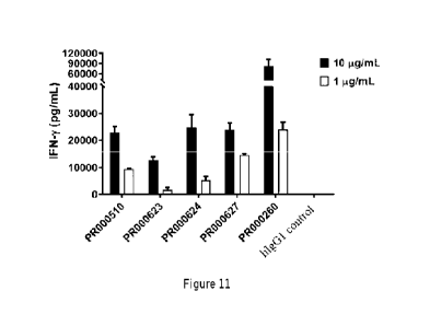
[0083] Fig. 11 shows the capacity of the CD3 single-chain antibody (PR000510,
PR000623, PR000624, PR000627 and PR000260) to activate human T cells to
produce
cytokine IFN-y.
[0084] Fig. 12 shows the SDS-PAGE results of samples of the bispecific
antibody (A)
PR002883 and (B) PR002885 obtained one-step purification.
[0085] Fig. 13 shows the binding capacity of the monoclonal antibody and the
bispecific antibody to (A) SK-BR-3 cells and (B) human T cells.
[0086] Fig. 14 shows the target cell killing capacity mediated by the
bispecific
antibody PR002883 in vitro; wherein, (A) shows SK-BR-3 cell killing and (B)
shows
IFN-y release levels.
[0087] Fig. 15 shows the binding capacity of the monoclonal antibody and the
bispecific antibody to (A) Panc-1 cells and (B) human T cells.
[0088] Fig. 16 shows the structure of the monoclonal antibody or the
bispecific
antibody; (A) IgG structure, (B) asymmetric "four chain" structure, (C)
asymmetric
"three chain" structure containing the single chain antibody.
Detailed description of the preferred embodiment
[0089] The present invention is further illustrated below by way of examples,
but the
invention is not thereby limited to the scope of the described examples. The
experimental methods for which specific conditions are not indicated in the
following
examples were selected according to the conventional methods and conditions,
or
according to the commodity specification.
[0090] In this application, the term "antibody" generally refers to a protein
comprising
a moiety that binds to an antigen and optionally allows the moiety that binds
to the
antigen to adopt a scaffold or skeleton moiety of the conformation that
promotes
CA 03152438 2022- 3- 24
Cur Ref [P21419377CA]
binding of the antibody to the antigen. The antibody typically may comprise an
antibody light chain variable region (VL), an antibody heavy chain variable
region
(VH), or both. The VH and VL regions may be further divided into hypervariable
regions called complementarily determining regions (CDR), which are scattered
in
more conservative regions called framework regions (FR). Each of the VH and VL
may
consist of three CDR and four FR regions, which may be arranged in the
following
order from the amino end to the carboxyl end: FR1, CDR1, FR2, CDR2, FR3, CDR3,
and FR4. The variable regions of the heavy and light chains contain binding
domains
that interact with the antigen. Examples of the antibody include, but are not
limited to,
antibody, antigen-binding fragment (Fab, Fab', F(ab)2, Fv fragment, F(abl)2,
scFv, di-
scFv and/or dAb), immunoconjugate, multispecific antibody (e.g., bispecific
antibody),
antibody fragment, antibody derivative, antibody analog or fusion protein, and
the like,
as long as they show the desired antigen-binding activity.
[0091] In this application, the term "variable" generally refers to the fact
that certain
parts of the sequence of the variable domain of the antibody differ
substantially, which
forms the binding and specificity of various specific antibodies to their
particular
antigen. However, the variability is not uniformly distributed throughout the
variable
region of the antibody. It is concentrated in three segments of the light and
heavy chain
variable regions, known as the complementary determining region (CDR) or high
variability region (HVR). The more highly conserved part of the variable
domain is
known as the framework (FR). The variable structure domains of the natural
heavy and
light chains each contain four FR regions, most of which adopt the f3-fold
conformation
and are connected by three CDRs that form a loop linkage and, in some cases,
form part
of the f3-fo1d structure. The CDRs in each chain are in close proximity to
each other
through the FR region and form the antigen binding site of the antibody
together with
the CDR from the other chain, the constant regions are not directly involved
in
antibody-antigen binding, but exhibit different effector functions, such as
involvement
in antibody-dependent cytotoxicity of the antibody. In the present art, the
CDR of an
antibody can be defined by a variety of methods, such as the Kabat definition
scheme
based on sequence variability (see, Kabat et al., Sequences of proteins of
immunological interest, Fifth Edition, National Institutes of Health,
Bethesda,
Maryland (1991)) and the Chothia definition scheme based on the location of
structural
loop regions (see, Al- Lazikani et al., J Mol Biol 273:927-48, 1997). In the
present
application, the Combined definition scheme comprising the Kabat definition
and the
Chothia definition is also used to identify amino acid residues in variable
domain
sequences and full-length antibody sequences (Table 1).
Table 1 The CDR definition rules of antibodies in the present application (see
http://bioinforgiuklabs/)
CDR region Kabat definition Chothia
definition Combined definition
LCDR1 L24--L34 L24--L34
L24--L34
LCDR2 L50--L56 L50--L56
L50--L56
11
CA 03152438 2022- 3- 24
Cur Ref [P21419377CA]
LCDR3 L89--L97 L89--L97
L89--L97
HCDR1 H31--H35 H26--H32
H26--H35
HCDR2 H50--H65 H52--H56
H50--H65
HCDR3 H95--H102 H95--H102
H95--H102
[0092] Wherein, Laa-Lbb may refer to the amino acid sequence from position aa
(Chothia numbering scheme) to position bb (Chothia numbering scheme) starting
from
the N-terminal of the antibody light chain; Haa-Hbb may refer to the amino
acid
sequence from position aa (Chothia coding scheme) to position bb (Chothia
numbering
scheme) starting from the N-terminal of the antibody heavy chain. For example,
L24-
L34 may refer to the amino acid sequence from position 24 to position 34
starting from
the N-terminal of the light chain of the antibody according to the Chothia
numbering
scheme; H26-H32 may refer to the amino acid sequence from position 26 to
position
32 starting from the N-terminal of the heavy chain of the antibody according
to the
Chothia numbering scheme.
[0093] The antibody Fc domain-mediated effector functions such as ADCC and CDC
also have very important biological functions, different IgG isoforms have
different
ADCC or CDC functions, for example, IgG1 and IgG3 have strong ADCC and CDC
effects, while IgG2 and IgG4 have relatively weak effects. In addition, the
original
effector function of Fc can also be modulated by amino acid mutations or
modifications
to alter the binding ability of Fc to the Fc receptor. For example, the
''LALA" double
mutation (1_2344/L2354) in IgG1 significantly reduces the affinity to FcyRII
IA
(CD16A) and thus reduces the ADCC effect. In addition, the P329G mutation
significantly reduces the binding of IgG1 multiple FCy receptors (see,
Schlothauer T,
Herter S, Koller CF, et al. Protein Eng Des Sel. 2016 Oct:29(10):457-466). In
this
application, in order to reduce the binding of CD3 antibodies to the FCy
receptor,
"LALA" double mutation (1_234A/235A) or the "LALAPG" triple mutation
(1_234A/L235A/P329G) was introduced into the Fc of these CD3 antibodies.
Example 1 Preparation and characterization analysis of recombinant antibodies
1.1 Preparation of IgG recombinant antibodies
[0094] After obtaining the light and heavy chain variable domain sequences
encoding
the antibody molecule, the recombinant antibody molecule can be prepared by
fusion
expression of the light and heavy chain variable domain sequences with the
corresponding human antibody light and heavy chain constant domain sequences
using
conventional recombinant DNA techniques. In this example, the antibody heavy
chain
variable domain sequence (VH) is genetically synthesized and cloned into the
mammalian cell expression plasmid vector encoding the human IgG1 antibody
heavy
chain constant domain sequence to obtain the full-length heavy chain of I gG1
antibody
by encoding, and the "LALA" double mutation (1_234A/L234A)(SEQ ID NO: 63) or
the "LALAPG" triple mutation (1_234A/L235A/P329G)(SEQ ID NO: 64) is introduced
12
CA 03152438 2022- 3- 24
Cur Ref [P21419377CA]
in the IgG1 heavy chain constant region to reduce the antibody binding to the
Fey
receptor The sequence of antibody light chain variable domain (VL) is
genetically
synthesized and cloned into the mammalian cell expression plasmid vector
encoding
the sequence of human antibody K light chain constant domain (SEQ ID NO: 61)
to
obtain the full length K light chain of antibody by encoding; alternatively,
VL is
genetically synthesized and cloned into the mammalian cell expression plasmid
vector
encoding the sequence of human antibody X light chain constant domain (SEQ ID
NO:
62) to obtain the full length X light chain to produce antibody by encoding.
[0095] By co-transfecting mammalian host cell (e.g., human embryonic kidney
cell
HEK293) with plasmids encoding the antibody heavy chain and plasmids encoding
the
antibody light chain, the purified recombinant antibody with correctly paired
assembly
of light and heavy chains can be obtained using conventional recombinant
protein
expression and purification techniques. Specifically, the HEK293 cells were
expanded
in medium FreeStyleTM F17 Expression Medium (Thermo, #A1383504). Before
transient transfecti on, adjusted the cell concentration to 6-8x10⁵ cel Is/m1
and incubated
them in shaker at 37 C 8% CO2 for 24 hours at the cell concentration of 1.2x
106 cells/ml.
30 ml of cultured cells were prepared. The plasmid encoding the heavy chain
and the
plasmid encoding the light chain were mixed in a ratio of 2:3, a total of 30
jig plasmid
were dissolved in 1.5 mL Opti-MEM reduced serum medium (Thermo, #31985088)
and filtered through the 0.22 prm membrane. Then 1.5 mL opti-MEM was dissolved
into 120 tit 1 mg/mL PEI (Polysciences, #23966-2), and left standing for 5
minutes.
PEI was slowly added to the plasmid, thereafter incubating for 10 minutes at
room
temperature. The mixed solution of plasmid PEI was slowly added into a culture
flask
dropwise while shaking the culture flask. The transfected cells were incubated
at 37t,
8% CO2 in a shaker for 5 days. The cell viability were observed after 5 days.
Then
cultures were harvested by centrifugation at 3300g for 10 minutes to collect
the
supernatant. Impurities in the supernatant was removed by centrifugation at
high speed.
The gravity column (Bio-Rad, #7311550) containing MabSelect Tm(GE Healthcare
Life
Science, #71-5020-91 AE) was equilibrated with PBS (pH 7.4) and rinsed with 2-
5
times of the column volume of PBS. The column was loaded with the supernatant
sample and rinsed with 5-10 times of the column volume of PBS. Then the target
protein
was elutd with 0.1 M glycine at pH 3.5, later adjusted to neutral pH with Tris-
HCI at
pH 8.0, finally concentrated using the ultrafiltration tube (Millipore,
#UFC901024) and
exchanged to PBS buffer and to obtain the purified recombinant antibody
solution. At
last, measured the concentration by NanoDrop (Thermo ScientificTM NanoDropTM
One),
dispensed and stored the purified recombinant antibody solution for backup.
1.2 Preparation of monovalent scFv-his recombinant antibodies
[0096] The VH and VL sequences of the antibody were linked by a flexible
peptide
(Linker) to obtain a single polypeptide chain encoding both VH and VL, i.e., a
single
chain antibody variable region fragment (scFv). If a linker peptide of
suitable length,
such as (G45)3 (SEQ ID NO: 65) or (G45)4 (SEQ ID NO: 66), is selected, VH and
VL
13
CA 03152438 2022- 3- 24
Cur Ref [P21419377CA]
can be correctly folded and assembled into functional antibodies. Different
scFv
structures (VH-linker-VL or VL-linker-VH) can be constructed depending on the
different arrangements of VH and VL and difference of the linker peptide. A
single scFv
contains an antigen-binding region consisting of a pair of VH and VL, which
usually
binds only one antigen molecule and is thus called a monovalent binding
molecule.
[0097] To facilitate purification, in this example, The C-terminal of the scFv
was fused
with His tag composed of 6-H istidine. The plasmid encoding the scFv and His
tag was
genetically synthesized and cloned into expression plasmid vectorfor mammalian
cells
to obtain the plasmid encoding scFv-his, which was transfected into mammalian
host
cell (e.g., human embryonic kidney cell HEK293), and then the recombinant
protein
can be purified using conventional recombinant protein expression and
purification
techniques. Specifically, the HEK293 cells were expanded in medium FreeStyleTM
F17
Expression Medium (Thermo, #41383504). Before transient transfection, adjusted
the
cell concentration to 6-8x10⁵ cells/ml and incubated them in shaker at 37 C 8%
CO2
for 24 hours at the cell concentration of 1.2x10⁶ cells/ml. 30 ml of cultured
cells were
prepared. 30 jig of the plasmid were dissolved in 1.5 mL Opti-MEM reduced
serum
medium (Thermo, #31985088) and filtered through the 0.22 pm membrane. Then 1.5
mL opti-MEM was dissolved into 120 !IL 1 mg/mL PEI (Polysciences, #23966-2),
and
left standing for 5 minutes. PEI was slowly added to the plasmid, thereafter
incubating
for 10 minutes at room temperature. The mixed solution of plasmid-PEI was
slowly
added into a culture flask dropwise while shaking the culture flask. The
transfected cells
were incubated at 37 t , 8% CO2 in a shaker for 5 days. The cell viability
were
observed after 5 days. Then cultures were harvested by centrifugation at 3300g
for 10
minutes to collect the supernatant. Impurities in the supernatant was removed
by
centrifugation at high speed. The gravity column (Bio-Rad, #7311550)
containing Ni
Sepharose excel (GE Healthcare Life Science, #17-3712-01) Ni Sepharose excel
(GE
Healthcare Life Science, #17-3712-01) was equilibrated with PBS (pH 7.4) and
rinsed
with 2-5 times of the column volume of PBS. The column was loaded with the
supernatant sample; and rinsed with 5-10 times of the column volume of PBS.
Non-
specifically adsorbed heteroproteins was eluted with buffer A (containing 20
mM
imidazole, 150 mM phosphate, pH 8.0) first, and then the target protein with
buffer B
(containing 500 mM imidazole, 150 mM phosphate, pH 8.0), finally using the
ultrafiltrati on tube (Millipore, #UFC901024) to concentrate and exchange the
solution
to PBS buffer and to obtain the purified recombinant antibody solution. At
last,
measured the concentration by NanoDrop (Thermo ScientificTM NanoDropTM One),
dispensed and stored the purified recombinant antibody solution for backup.
1.3 Preparation of bivalent scFv-Fc recombinant antibodies
[0098] In this example, the scFv-Fc recombinant molecule was constructed by
fusing
the sequence of human I gG1 constant region Fc (G1u216-Lys447, containing the
hinge
region, CH2 domain and CH3 domain) at the C-terminus of scFv, and through
homodimerization of Fc, a bivalent scFv-Fc dimer molecule was formed, which is
14
CA 03152438 2022- 3- 24
Cur Ref [P21419377CA]
capable of binding two antigen molecules simultaneously. And the "LALA" double
mutation (1_234A/L235A) or the "LALAPG" triple mutation (1_2344/L2354/P329G)
was introduced into Fc to reduce the binding of the antibody to the Fcy
receptor. The
polypeptide sequence encoding scFv-Fc was genetically synthesized and cloned
into
the expression plasmid vector for mammalian cells to obtain the plasmid
encoding
scFv-Fc, which was thereafter transfected into mammalian host cells (e.g.,
human
embryonic kidney cell HEK293), and then using the protein expression
purification
method described in the Example 1.1 to obtain the purified recombinant
protein.
1.4 Protein purity analysis by HPLC-SEC
[0099] The purity and aggregate form of protein samples were analyzed by
molecular
size exclusion chromatography (SEC). The analytical column TSKgel G3000SWx1
(Tosoh Bioscience, #08541, 5 pm, 7.8 mm x 30 cm) was connected to the high
pressure
liquid chromatograph (HPLC) (Agilent Technologies, Agilent 1260 Infinity II)
and
equilibrated with PBS buffer at room temperature for at least lhour. An
appropriate
amount of protein sample (at least 10 pg) filtered through the 0.22 pm
membrane were
injected into the system, and the HPLC program was set: the column was loaded
by the
sample with PBS buffer at a flow rate of 1.0 ml/min for a maximum of 20
minutes.
HPLC will generate Analytical reports of HPLC would be generated, which
reports the
retention time of the different molecular size components in the sample.
Example 2 Recombinant expression of murine-human chimeric antibody of CD3
antibody 5P34
[0100] 5P34 is a murine -derived anti-human CD3e antibody that binds a variety
of
primate CD3 and functions to activate T cells. The sequences of variable
region VH
and VL of 5P34 have been disclosed in W02016071004A1. In this application, the
amino acid sequence of VH of 5P34 is set for in SEQ ID NO: 42, and its
corresponding
murine germline V gene is IGHV10- 1; the amino acid sequence of VL of 5P34 is
set
forth in SEQ ID NO: 56, and its corresponding murine germline V gene is I
GLV1. In
this example, the VH sequence of 5P34 was fused with the human IgG1 antibody
heavy
chain constant domain sequence (SEQ ID NO: 63) comprising a "LALA" double
mutation(L234A/235A) to produce the full-length heavy chain of 5P34 murine-
human chimeric IgG1 antibody; the amino acid sequence of VL of SP34 was fused
with
the human antibody A. light chain constant domain sequence (SEQ ID NO: 62) to
produce the full length A. light chain of the SP34 murine-human chimeric
antibody.
[0101] The 5P34 murine -human chimeric recombinant antibody PR000260 was
prepared according to the method of the Example 1.1. The following Table 2
shows the
data of recombinant expression of PR000260.
Table 2 Expression and purification of recombinant antibody PR000260
Antibody Expression system Purification
HPLC-SEC (monomer
ld
number (volume) method Yie
(mg/L) purity%)
PR000260 HEK293 (100 ml) MabSelect
19.30 99.75 %
CA 03152438 2022- 3- 24
Cur Ref [P21419377CA]
Example 3 Convert murine CD3 5P34 antibody to recombinant scFv antibody
[0102] The VH sequence (SEQ ID NO: 42) and VL sequence (SEQ ID NO: 56) of
5P34 were linked by the flexible peptide (Linker) to obtain the single
polypeptide chain
encoding both VH and VL, i.e., the single chain antibody variable region
fragment
(scFv). Depending on the different arrangements of VH and VL and different
lengths
of the linker peptides (SEQ ID NO: 65, SEQ ID NO: 66), different scFv
structures can
be constructed, and a His tag consisting of a 6-Histidine was fused at the C-
terminus of
the scFv for purification. The linker peptide as shown in SEQ ID NO: 67 can
also be
used for the construction of scFv in this application.
[0103] In this Example, four recombinant scFv antibody molecules (PR000275,
PR000276, PR000307, PR000308) were prepared according to the method of the
Example1.2. The sequence numbers of these four recombinant scFv antibody
molecules
are listed in Table 3 below; the following Table 4 shows the expression data
of of these
four recombinant molecules; and the HPLC-SEC results of these four molecules
after
one-step purification are shown in Figure 1, wherein (A) shows the result of
PR000275,
(B) shows the result of PR000276, (C) shows the result of PR000307, and (D)
shows
the result of PR000308. It can be seen that using the sequences of VH and VL
of 5P34
to construct scFv, no matter which (VHNL) alignment pattern was used or the
length
of the linking peptide was changed, no stable scFv could be obtained.
Table 3 Structure and sequence number of the four recombinant antibodies
VH VL
Molecular HI Linker Full length
Antibody number
scFv StructureVH
Va ri ant Vari ant structure peptide sequence
(SEQ ID NOs:)
PR000275 SP34_VHSP34_VL scFv-his VL-linker-
VH 42 56 65 68
PR000276 5P34_VHSP34_VL scFv-his VH-linker-
VL 42 56 65 69
PR000307 SP34_VHSP34_VL scFv-his VL-linker-
VH 42 56 66 70
PR000308 5P34_VHSP34_VL scFv-his VH-linker-
VL 42 56 66 71
Table 4 Expression and purification of recombinant scFv antibodies
HEK293
HPLC-SEC
Antibody Structure Expression
Purification Yield (monomer
number
method (mg/L)
Volume
purity %]
PR000275 VL-(G45)3-VH 100 ml
Nickel 2.60 46.61 %
PR000276 VH-(G4S)3-VL 100 ml
Nickel 0.26 Weak signal
PR000307 VL-(G45)4-VH 30 ml
Nickel 3.33 Weak signal
PR000308 V H-(G4S)4-VL 30 ml
Nickel 3.67 Weak signal
16
CA 03152438 2022- 3- 24
Cur Ref [P21419377CA]
Example 4 Sequence optimization of SP34
4.1 Humanization of variable region sequences and mutation of frame region
[0104] The "CDR transplantation" method is used for sequence humanization in
this
example, i.e., transplantation of the CDR of the murine antibody VH to the
frame region
of the human antibody VH, and transplantation of the CDR of the murine
antibody VL
to the frame region of the human antibody VL. The sequence of the frame region
of
human antibody VH or VL can be derived from human germline gene sequences or
antibody sequences that have been rearranged by V(D)J or the consensus
sequences of
the specific VH or VL gene family of the human antibody. In this example, the
frame
region sequences provided by human germline gene sequences are used as
humanized
template sequences, i.e., the human germline V gene fragment provides the
sequences
of the frame regions FR1, FR2, and FR3, and the human germline J gene fragment
provides the sequence of the frame region FR4. Finally, the sequences of
humanized
variable region (VH or VL) were constructed in the arrangement of (human)FR1-
(murine)CDR1-(human)FR2-(murine)CDR2-(human)FR3-(murine)CDR3-
(human)FR4.
[0105] In this example, the sequence of the human germline V gene fragment I
GHV3-
73*01 or the human germline V gene fragment IGHV3-23*01 was used as the
humanized template in combination with the sequence of the human germline J
gene
fragment IGHJ 1*01 to provide the frame region sequence. Amino acid mutations
at one
or more sites were introduced in at the position 30, position 73, position 76,
position
78, position 93 or position 94 (according to Chothia numbering rules) to
obtain several
different VH mutant sequences.
[0106] In this example, the sequence of the human germline V gene fragment
IGLV7-
46*02 combined with the sequence of the human germline J gene fragment
IGLJ2*01
or the sequence of the human germline V gene fragment IGKV1-39*01 combined
with
the sequence of the human germline J gene fragment IGKJ 4*01 was used as the
humanized template to provide the frame region sequence. Aamino acid mutations
at
zero or more sites were introduced in at the position 2, position 36, position
46, position
49, position 66, position 69, position 71 or position 87 (according to Chothia
numbering
scheme) to obtain several different VL mutant sequences.
[0107] The following Table 5 lists the sequence numbers of the antibody
variable
region, its optimized mutant sequences (FV) and the sequences of the CDR and
FR
regions defined by CHOTH IA.
Table 5 Variable region of 5P34 antibody and optimized mutant sequences (FV)
thereof
and the sequence list of CDR and FR regions defined by CHOTHIA
ID FV FR1 CDR1 FR2 CDR2 FR3 CDR3 FR4
5P34 _VH 42 5 1 8
3 11 4 21
VH3730 50 7 1 10
3 17 4 22
17
CA 03152438 2022- 3- 24
Cur Ref [P21419377CA]
VH3731 51 7 1 10
3 18 4 22
VH3732 52 7 2 10
3 18 4 22
VH3733 53 7 2 10
3 19 4 22
VH3734 54 7 2 10
3 20 4 22
VH3735 55 7 2 10
3 17 4 22
VH3230 43 6 1 9
3 12 4 22
VH3231 44 6 1 9
3 13 4 22
VH3232 45 6 2 9
3 13 4 22
VH3233 46 6 2 9
3 12 4 22
VH3234 47 6 2 9
3 14 4 22
VH3235 48 6 2 9
3 15 4 22
VH3236 49 6 2 9
3 16 4 22
5P34_VL 56 26 23 30
24 35 25 40
VL7460 57 27 23 33
24 38 25 40
VL7461 58 27 23 34
24 39 25 40
VK1392 59 28 23 31
24 36 25 41
VK1393 60 29 23 32
24 37 25 41
[0108] Figure 2 lists the comparison of VH mutant sequences. Figure 3 lists
the
comparison of VL mutant sequences. The differences in the sequences of VH
mutants
and VL mutants at significant sites are listed in (A) and (B) of Figure 4,
respectively.
As can be seen from Figure 2 to 4, the mutations of the antibody of the
present invention
on VH occured at one or more sites selected from position 30, position 73,
position 76,
position 78, position 93 or position 94tof the amino acid sequence as shown in
SEQ ID
NO: 42. The mutations on the VL occurred at position 2, position 36, position
46,
position 49, position 66, position 69, position 71and/or position 87 of the
sequence as
shown in SEQ ID NO: 56. More detailed mutation information can be found in
Table 5
for sequence specifics of VH3730, VH3731, VH3732, VH3733, VH3734, VH3735,
VH3230, VH3231, VH3232, VH3233, VH3234, VH3235, VH3236, VL7460, VL7461,
VK1392, and VK1393.
4.2 Recombinant antibodies of mutants with optimized sequences
[0109] The sequences of VH mutants and VL mutants obtained in the Example 4.1
were paired and combined, and the IgG recombinant antibody constructed
according to
the method in the Example 1.1, wherein the "LALA" double mutation or the
"LALAPG" triple mutation was introduced into the constant region of the IgG1
heavy
18
CA 03152438 2022- 3- 24
Cur Ref [P21419377CA]
chain to reduce the Fc effector function. Table 6 lists the sequences of
recombinant
antibody molecules that have been sequence optimized. Table 7 lists the
expression data
of the recombinant antibodies. Except for the three I gG molecules constructed
with the
VH mutant VH3230, which have very low expression yields, all other I gG
molecules
have reasonable expression yields.
Table 6 Sequence list of the 5P34 chimeric antibodies or antibodies with
optimized
sequence
Heavy
Light
Antibody VH VL VH
VL chain chain
number Mutant Mutant
constant constant
region
region
( SEQ ID NOs: )
PR000260 5P34_VH 5P34_VL 42
56 63 62
PR000511 VH3231 VL7461 44
58 63 62
PR000512 VH3731 VL7461 51
58 63 62
PR000513 VH3231 VK1393 44
60 63 61
PR000514 VH3731 VK1393 51
60 63 61
PRO01848 VH3232 VL7461 45
58 64 62
PR001849 VH3732 VL7461 52
58 64 62
PR002467 VH3230 VL7460 43
57 63 62
PR002468 VH3231 VL7460 44
57 63 62
PR002469 VH3230 VL7461 43
58 63 62
PR002470 VH3230 VK1392 43
59 63 61
PR002471 VH3231 VK1392 44
59 63 61
PR002472 VH3230 VK1393 43
60 63 61
PR002742 VH3730 VL7461 50
58 63 62
PR002743 VH3731 VL7460 51
57 63 62
PR002833 VH3234 VL7461 47
58 64 62
PR002834 VH3235 VL7461 48
58 64 62
PR002835 VH3236 VL7461 49
58 64 62
PR002836 VH3733 VL7461 53
58 64 62
PR002837 VH3734 VL7461 54
58 64 62
PR003886 VH3735 VL7461 55
58 63 62
PR004616 VH3233 VL7461 46
58 63 62
19
CA 03152438 2022- 3- 24
Cur Ref [P21419377CA]
Table 7 Expression yield and purity of the recombinant antibodies
HPLC-SEC
Antibody
Yield of HEK293
VH Mutant VL Mutant
monomer purity
number
(mg/L}
WO
PR000260 5P34_VH 5P34_VL
64.33 99.75%
PR000511 VH3231 VL7461
48.33 99.88%
PR000512 VH3731 VL7461
150.00 100.00%
PR000513 VH3231 VK1393
45.00 100.00%
PR000514 VH3731 VK1393
48.33 100.00%
PR001848 VH3232 VL7461
38.50 99.03%
PR001849 VH3732 VL7461
43.50 99.10%
PR002467 VH3230 VL7460
0.60 n/a
PR002468 VH3231 VL7460
107.25 98.84%
PR002469 VH3230 VL7461
10.20 96.85%
PR002470 VH3230 VK1392
0.60 n/a
PR002471 VH3231 VK1392
62.50 94.96%
PR002472 VH3230 VK1393
0.30 n/a
PR002742 VH3730 VL7461
14.25 100.00%
PR002743 VH3731 VL7460
44.25 100.00%
PR002833 VH3234 VL7461
18.00 98.92%
PR002834 VH3235 VL7461
24.60 95.88%
PR002835 VH3236 VL7461
13.50 95.58%
PR002836 VH3733 VL7461
26.25 94.68%
CA 03152438 2022- 3- 24
Cur Ref [P21419377CA]
PR002837 VH3734 VL7461
17.20 97.36%
PR003886 VH3735 VL7461
116 98.49%
PR004616 VH3233 VL7461
22 n/a
4.3 Recombinant scFv molecules containing mutants with optimized sequences
[0110] The sequences of VH mutants and VL mutants obtained in the Example 4.1
were paired and combined, and a plurality of recombinant bivalent scFv
antibody
molecules were obtained according to the method in the Example 1.3. The
following
Table 8, 9 respectively lists the sequence information and protein expression
of the scFv.
As can be seen in Table 9, PR000510 and PR000627 are especially better
expressed
and stable molecules. Figure5 shows the results of (A) SDS-PAGE and (B) HPLC-
SEC
of PR000510, which can be seen that it has a good monomeric purity with no
obvious
aggregates.
Table 8 Structure and sequence information of the scFv molecules constructed
based on
the mutants with optimized sequences
Antibody VH VL Molecular scFv
Linker Full
number Mutant Mutant structure
Structure VH VL peptide length
sequence
( SEQ ID NOs: )
PR000509 VH3731 VL7461 scFv-Fc VH-linker-VL 51 58 67
72
PR000510 VH3731 VL7461 scFv-Fc VL-linker-VH 51 58 67
73
PR000623 VH3231 VK1393 scFv-Fc VH-linker-VL 44 60 67
74
PR000624 VH3731 VK1393 scFv-Fc VH-linker-VL 51 60 67
75
PR000625 VH3231 VK1393 scFv-Fc VL-linker-VH 44 60 67
76
PR000626 VH3731 VK1393 scFv-Fc VL-linker-VH 51 60 67
77
PR000627 VH3731 VL7461 scFv-Fc VL-linker-VH 51 58 67
78
PR000914 VH3231 VL7461 scFv-Fc VH-linker-VL 44 58 67
79
PR000915 VH3231 VL7461 scFv-Fc VL-linker-VH 44 58 67
80
PRO01850 VH3732 VL7461 scFv-Fc VL-linker-VH 52 58 67
81
Table 9 Expression data of the scFv antibody after sequence optimization
HPLC-SEC
Antibody
VH Mutant VL Mutant Yield
of HEK293
monomer purity
number
(mg/i..)
(%)
21
CA 03152438 2022- 3- 24
Cur Ref [P21419377CA]
PR000509 VH3731 VL7461
0.00 n/a
PR000510 VH3731 VL7461
7.00 99.84%
PR000623 VH3231 VK1393
0.60 93.29%
PR000624 VH3731 VK1393
1.80 100.00%
PR000625 VH3231 VK1393
0.00 n/a
PR000626 VH3731 VK1393
0.00 n/a
PR000627 VH3731 VL7461
6.00 100.00%
PR000914 VH3231 VL7461
3.33 78.22%
PR000915 VH3231 VL7461
2.67 86.14%
PR001850 VH3732 VL7461
4.88 50.76%
Example 5 Determination of the binding capacity of CD3 antibody to CD3
expressing cells by FACs
[0111] The flow cytometryFACS was used to analyse the binding of the CD3
antibody
to CD3-expressing cells, where the CD3-expressing cells could be: CHOK1 cells
overexpressing human CD3 or HEK 293ce11s (plasmids encoding the ORF of y, 6,
E,
and chains of human-derived CD3 and p1asmids
encoding the ORF of a and f3 chains
of human TCR were co-transfected with host cells CHOK1 (ATCC, CCL-61) or
HEK293 (ATCC, CRL-1573) to construct stable cell lines expressing the
structure of
the human TCRICD3 complex); CHOK1 or HEK293 cells overexpressing cynomolgus
monkey CD3; human pan-T cells (isolated with the human pan-T cell isolation
kit
(Miltenyi, #130-096-535) from PBMC); cynomolgus monkey pan-T cells.
Specifically,
the collected cells were washed twice with PBS containing 2% FBS (FACS
buffer), and
resuspended with FACS buffer, divided into 96 well plates with 1x10⁵ cells per
well,
centrifuged at 500g for 5 minutes. The supernatant was discarded, and 100 pl
of pre-
gradient diluted CD3 antibody was added, then incubating for 1 hour at room
temperature and washing twice with FACS buffer. The cells were resuspended
with
FACS buffer diluted with secondary antibody Alexa Fluor 488 AffiniPure Goat
Anti-
Human IgG, Fcy fragment specific (Jackson ImmunoResearch, #109-545-098), then
incubated at room temperature in the dark for 30 minutes. The cells were
washed twice
with FACS buffer, and resuspended with 200 1.11 FACS buffer. The fluorescent
luminescence signal value were read by flow cytometry (BD FACS CANTO!! orACEA
NovoCyte), and the resulted data were processed and analyzed by software Flow]
o v10
22
CA 03152438 2022- 3- 24
Cur Ref [P21419377CA]
(Flowi
LLC). The software GraphPad Prism 8 was
used for data processing and
graphical analysis, and parameters such as binding curves and EC50 values can
be
obtained by four-parameter nonlinear fitting.
[0112] Figure 6 shows the binding ability of the CD3 antibody obtained in the
Example 2 to recombinant CHOK1 cells overexpressing human CD3 (Figure6 (A))
and
recombinant CHOK1 cells overexpressing cynomolgus monkey CD3 (Figure 6(B)).
The results indicate that the 5P34 chimeric antibody PR000260 has a strong
binding
ability to both human CD3 and cynomolgus monkey CD3.
[0113] Figure 7 (A) to (G) show the binding ability of the CD3 antibodies
obtained in
the Example 4.2 (including PR000260 and its mutants) to human pan-T cells,
respectively. The fluorescence intensity MFI of the CD3 antibody binding to
human
pan-T cells and the relative ratio relative to the initial antibody PR000260
were
calculated when the antibody concentration was 7.4 or 10 g/ml. Specifically,
after
5P34 IgG antibody sequence optimization, PR000512, PR000513, PR001849, and
PR002837 have comparable binding ability as PR000260 (i.e., 5P34 chimeric
antibody);
while PR000514 has slightly higher binding ability than PR000260; PR000511,
PR001848 PR002469, PR002472, PR002742, PR002833, PR002834, PR002835,
PR002836, PR003886, and PR004616 have lower binding ability to T cells;
PR002467,
PR002468, PR002470, PR002471, and PR002743, on the other hand, barely bind T
cells (or the signal cannot be detected at the current antibody
concentration). The above
results indicate that the present invention has obtained several new
antibodies by
sequence optimization of the CD3 antibody, which have different binding
abilities to
human T cells and can be applied to different application scenarios.
[0114] Figure 8 (A) and (B) show the binding capacity of the anti-CD3 scFv-Fc
single
chain antibody obtained in the Example 4.3 to human pan-T cellsThe
fluorescence
intensity MFI of the CD3 antibody binding to human pan-T cells and the
relative ratio
relative to the initial antibody PR000260 were calculated when the antibody
concentration was 7.4 or 10 g/ml. Specifically, after optimization of the
humanization
of SP34 scFv antibody, PR000624 has a comparable or slightly higher binding
capacity
than PR000260; PR000510 and PR000627 have a comparable or slightly lower
binding
capacity than PR000260; and PR001850 has a significantly lower binding
capacity to
T cells than PR000260. The above results indicate that, the present invention
also
obtained several stable single-chain antibodies in the form of scFv by
sequence
optimization of the CD3 antibody, which are able to bind to human T cells and
are
suitable for application scenarios such as the construction of bispecific
antibodies.
[0115] Figure 9 shows the binding ability of some of the CD3 antibodies
obtained in
the Example 4.2 to cynomolgus monkey pan-T cells. It can be seen that
different
molecules have different binding abilities to cynomolgus monkey pan-T cells
and are
positively correlated with their binding abilities to human pan-T cells; that
is, molecules
that strongly bind to human pan-T cells also strongly bind to cynomolgus
monkey pan-
T cells, and vice versa.
Example 6 Determination of activation of CD3 antibody on human T cells
23
CA 03152438 2022- 3- 24
Cur Ref [P21419377CA]
[0116] The gradient dilutions of CD3 antibody (e.g., 50, 10, 5, 1, 0.5, 0.05
g/m1)
were coated in 96 well cell culture plates at three replicate wells per
concentration and
50 I per well, incubating overnight at 4 C. The cell density of human PBMC
(MiaoTong Biology) or human pan-T cells (isolated with the human pan-T cell
isolation
kit (Miltenyi, #130-096-535) from PBMC) was adjusted to 7.5x10⁵/ml, and human
CD28 antibody was added at a concentration of 1 g/ml, thereafter adding 200
pi the
resulted mixture per well to the cell culture plate and incubating in CO2
incubator. After
72 hours of incubation, the supernatant was taken and the content of IFN-y
therein was
determined using use the IFN-y ELISA kit (Thermo, #88-7316-77). Used the
software
GraphPad Prism 8 for data processing and graphical analysis.
[0117] Figure 10 (A) to (G) show the ability of each CD3 antibody (including
5P34
chimeric antibody) obtained in the Example 4.2 to activate human T cells,
respectively.
When the antibody concentration is 1 gg/mL, PR000511, PR000512, PR000513, and
PRO00514 produced significantly lower levels of IFN-y after activating T cells
than
PR000260; when the antibody concentration is 10 g/mL, PR000512, PR000513, and
PRO00514 activate slightly lower levels of IFN-y than PR000260 (Figure 10
(A)). When
the antibody concentration is 0.5 Kg/mL and 5 g/mL, the level of IFN-y
activated by
PR001848 is significantly lower than that of PR000260 (Figure 10(B)). In
addition, the
effect of activating T cells of antibodies PR002468, PR002469, PR002471,
PR002742,
PR002833, PR002834, PR002835, PR002836, PR002837, PR001848, PR003886 and
PRO04616 was also detected (Figure 10 (C) to (G)), in concentration of 0.5
pg/mL, 5
i_ig/mL and 50 pg/mE, the results indicated that the level of IFN-y produced
by T cells
activated by these antibodies are much lower than those activated by PR000260,
wherein, no release of IFN-y was detected by T cells activated by PR002468 and
PR002471, and only weak level of IFN-y were detected at 50 pg/mE of PR002469
and
PR002835; the level of IFN-y produced by T cells activated by PR002742 and
PR003886 were comparable and slightly weaker than PR001848; the level of IFN-y
produced by T cells activated by PR002469 and PR004616 were comparable and
significantly weaker than PR001848. The above results indicate that the
present
invention has obtained several new antibodies by sequence optimization of the
CD3
antibody, which have different activation abilities on human T cells, and can
control
different levels of cytokine release and can be applied to different
application scenarios.
[0118] Figure 11 shows the ability of the anti-CD3 scFv-Fc antibody obtained
in the
Example 4.3 to activate human T cells. PR000510, PR000623, PR000624, and
PR000627 at concentrations of 1 iig/mE and 10 Kg/mL all exhibited lower levels
of
IFN-y than PR000260 but higher than the isotype control antibody, indicating
that these
four molecules limited the release of cytokines by regulating the activation
levels of T
cells. The above results indicate that the present invention also obtained
several stable
single-chain antibodies in the form of scFv by sequence optimization of the
CD3
antibody, which have weaker activation on human T cells showing lower levels
of
cytokine release, and are suitable for application scenarios such as the
construction of
bispecific antibodies.
Example 7 Bispecific antibody targeting B7H4 containing anti-CD3 sc Fy
antibody
24
CA 03152438 2022- 3- 24
Cur Ref [P21419377CA]
[0119] B7H4, a member of the B7 family of transmembrane proteins, is highly
expressed in a variety of solid tumor tissues such as breast, ovarian and
endometrial
cancers, while it is not expressed or very faintly expressed in normal
tissues, making
B7H4 a very specific tumor-associated target antigen. A bispecific antibody
molecule
targeting both B7H4 and CD3 were constructed, which can selectively activate T
cells
near tumor cells by targeting and binding to B7H4 on the surface of tumor
cells, thus
providing specific killing of tumor cells.
7.1 Preparation of B7H4 antibody
[0120] The sequence of the variable region of the B7H4 antibody can be derived
from
W02016040724, and the recombinant IgG antibody PR000014 target B7H4 was
constructed according to the methods of the Example 1.1. The following Table
10 lists
the sequence information of the B7H4 antibody PR000014.
Table 10 Sequence information of light chain and heavy chain of B7H4 antibody
PRO00014
Heavy chain SEQ ID Light chain SEQ ID
Antibody number Target
NO:
NO:
PR000014 B7H4
82 83
7.2 Preparation of bispecific antibody targeting B7H4 containing anti-CD3 scFv
antibody
[0121] Using the sequence of the B7H4 antibody PR000014 obtained in the
Example
7.1 and the sequence of the CD3 single chain antibody PR000627 obtained in the
Example 4.3 to construct the bispecific antibody molecule PR002883 targeting
B7H4
x CD3, which contains three polypeptide chains: a heavy chain containing the
CD3
single chain antibody scFv (SEQ ID NO: 88), a heavy chain containing theVH of
B7H4
antibody (SEQ ID NO: 86), and a light chain containing the VL of B7H4 antibody
(SEQ
ID NO: 83). The structure is shown in Figure 16(C). Since the molecule has a
special
asymmetric structure, different amino acid mutations were introduced in the
constant
regions of the two heavy chains in order to reduce the generation of
homologous heavy
chain dimers. At the same time, the "LALAPG" triple mutation
(L234A/L235A/P329G)
was introduced in the constant region of the heavy chain to prevent cross-
linking and
reduce effector function caused by Fcy receptor binding.
[0122] Recombinant protein of the bispecific antibody PR002883 was prepared by
using the method described in the Example 1.1 in combination with plasmids in
ratio
(e.g., 1:1:1 or other ratios), and the follow-up one-step affinity
purification. The
sequence of bispecific antibody PR002883 is listed in Table 11; the expression
of the
bispecific antibody is listed in Table 12.
Table 11 Chains of the bispecific antibody and the corresponding sequence
information
CA 03152438 2022- 3- 24
Cur Ref [P21419377CA]
Bispecific Anti-B7H4 Anti-CD3 Heavy Chain 1 Heavy Chain 2 Light chain
antibodies antibody scFv SEQ ID
NO: SEQ ID NO: SEQ ID NO:
PR002883 PR000014 PR000627 88
86 83
Table 12 Expression of the bispecific antibody
Bispecific SDS-PAGE purity
Yield in HEK293 (mg/L)
antibody(%)
PR002883 94.0
70
[0123] Figure 12(A) shows the results of the bispecific antibody PR002883
after one-
step purification by SOS-PAGE analysis. It shows that its main by-products are
incompletely assembled molecules with few high polymer components, which can
be
reduced by optimizing the purification step or by optimizing the plasmid
transfection
ratio.
7.3 Binding to tumor cells expressing B7H4
[0124] This Example investigates the ability of the bispecific antibody
binding tumor
cell SK-BR-3 (ATCC, HTB-30) expressing human B7H4. Specifically, collected
cell
SK-BR-3 suspension, adjusted the cell density to lx 106/ml, and inoculated
them at 100
i/well in a 96 well V-bottom plate (Corning, #3894); subsequently, the
antibody to be
tested with a concentration 2-fold of final concentration obtained by 3-fold
gradient
dilution was added at volume of 100 l/well. Cells were incubated at 4 C in
the dark
for 2 hours. Afterwards, the cells were rinsed twice with 100 I/well of pre-
chilled PBS,
centrifuged at 500 g, 4 C for 5 minutes. The supernatant was discarded. Then
added
100 p1/well of secondary antibody Alexa Fluor 488 AffiniPure GoatAnti-Human
IgG,
Fcy fragment specific (Jackson ImmunoResearch, #109-545-098), and incubated
the
cells from light at 4 C in the dark for 1 hour. Then the cells were rinsed
twice with 100
p1/well of pre-chilled PBS, centrifuged at 500 g, for 5 minutes. The
supernatant was
discarded. Finally, the cells were resuspended with 200 L/well of pre-chilled
PBS. The
fluorescent luminescence signal value were read by flow cytometry (BD FACS
CANTOR orACEA NovoCyte), and the resulted data were processed and analyzed by
software Flowi o v10 (Flow.j o, LLC). The software GraphPad Prism 8 was used
for data
processing and graphical analysis, and parameters such as binding curves and
EC50
values can be obtained by four-parameter nonlinear fitting.
[0125] Figure 13(A) shows the binding ability of the monoclonal antibody
obtained
in the Example 7.1 and the bispecific antibody obtained in the Example 7.2 to
cell 5K-
BR-3. It can be seen that the bispecific antibody PR002883 has a comparable or
even
better binding capacity compared to the monoclonal antibody PR000014.
7.4 Binding to human T cells
26
CA 03152438 2022- 3- 24
Cur Ref [P21419377CA]
[0126] The ability of the bispecific antibody PR002883 binding to human pan-T
cells
was detected by the method described in the Example 5. As shown in Figure 13
(B),
PR002883 is able to bind to human pan-T cells.
7.5 Killing and cytokine releasing effect of the bispecific antibody On B7H4
high-
expressing cell line SK-BR-3 in vitro
[0127] To investigate the ability of killing target cell mediated by B7H4 x
CD3
bispecific antibody in vitro, human PBMC was used as effector cells, and B7H4
high-
express cell line SK-BR-3 (ATCC, HTB-30) was used as target cells for killing
assays
in vitro and cytokine release detection. Specifically, 50 1_11_ of
RPM11640110% FBS
medium was added to each well of E-plate (ACEA Biosciences Inc.,
#05232368001),
thereafter balancing in incubator containing 5% CO2 at 37 C for 30 minutes,
and then
the E-plate was placed in the instrument xCELLigence RTCA (ACEA Biosciences)
to
test for normality. The density of SK-BR-3 was adjusted to 0.4 x 10⁶ cells/mL
with
RPM 11640110% FBS medium, then inoculated into E-plate at 50 1_ cells/well,
and
then the E-plate was placed on xCELLigence RTCA overnight to detect the cell
index.
Thedensity of PBMC was adjusted to 4x10⁶ cells/mL with RPMI1640/10% FBS
medium, and inoculated into E-plate at 50 1_ cells/well, then the antibody to
be tested
with a concentration 4-flod of final concentration obtained by 5-fold gradient
dilution
was added at 50 pl/well, wherein the highest final concentration of the
antibody was
0.2 nM, and there were 7 concentrations for each antibody, the final ratio of
effector
cells to target cells was 10: 1, with two replicates set up. Meanwhile, blank
control was
set up in the plate: SKBR3 + PBMC + RPM 11640/10% FBS medium; the E-plate was
incubated in 37 C, incubator containing 5% CO2 for 24 hours. After incubation,
the E-
plate was placed on the xCELLigence RTCA instrument to detect the cell index.
[0128] The specific killing effect of the antibody was calculated by applying
the
assayed cell index to the following formula:
Cell Killing% = (1-Test Sample/Blank Control)* 100%.
[0129] The supernatant of cell culture was collected for detecting the release
of
cytokine IFN-y. Refer to the operation instructions of IFN-y kit (IFN gamma
Human
Uncoated ELISA Kit, Thermo, #88-7316-77) for ELI SA detection.
[0130] As shown in Figure 14 (A) and (B), the bispecific antibody PR002883 can
activate =T cells to release cytokines such as IFN-y and kill tumor cell SK-BR-
3
effectively. Almost 100% of tumor cells were killed when the concentration of
the
bispecific antibody was at 0.01 Fig/m1 (Figure 14(A)).
Example 8 Bispecific antibody targeting ROR1 containing anti-CD3 scFv
antibody
[0131] ROR1 is an inactive tyrosine protein kinase transmembrane protein that
is
overexpressed in many tumors but virtually not expressed in normal tissues.
ROR1
contributes to cell proliferation and migration in chronic lymphocytic
leukemia by
transducing the Wnt signaling pathway after interacting with Wnt5a as a
receptor, and
27
CA 03152438 2022- 3- 24
Cur Ref [P21419377CA]
contributes to epithelial-mesenchymal-transformation (EMT) in solid tumors.
The
tumor-specific expression of ROR1 makes it a suitable tumor-associated antigen
target
for the development of therapeutic agents. The construction of a bispecific
antibody
molecule targeting both ROR1 and CD3 can selectively activate T cells near
tumor cells
by targeting and binding to ROR1 on the surface of tumor cells, thus providing
specific
killing of tumor cells.
8.1 Preparation of ROR1 antibody
[0132] The sequence of the variable region of the ROR1 antibody can be derived
from
W02016094873, and construct the recombinant IgG antibody PR000374 targeting
ROR1 according to the methods of the Example 1.1. The following Table 13 lists
the
sequence information of the ROR1 antibody PR000374.
Table 13 Sequence list of the ROR1 antibody PR000374.
Heavy chain
Light chain
Antibody number Target
SEQ ID NO:
SEQ ID NO:
PR000374 ROR1
84 85
8.2 Preparation of bispecific antibody targeting ROR1 containing anti-CD3 scFv
antibody
[0133] Construction of the bispecific antibody molecule PR002885 targeting
ROR1
x CD3 was performed by using the sequence of the ROR1 antibody PR000374
obtained
in the Example 8.1 and the sequence of the CD3 single chain antibody PR000627
obtained in the Example 4.3, which contains three polypeptide chains: a heavy
chain
containing the CD3 single chain antibody scFv (SEQ ID NO: 88), a heavy chain
containing the ROR1 antibody VH (SEQ ID NO: 87), and a light chain containing
the
ROR1 antibody VL (SEQ ID NO: 85). The structure is shown in Figure 16(C).
Since
the molecule has a special asymmetric structure, different amino acid
mutations were
introduced into the constant regions of the two heavy chains in order to
reduce the
generation of homologous heavy chain dimers. At the same time, the "LALAPG"
triple
mutation (L234A/L235A/P329G) was introduced into the constant region of the
heavy
chain to prevent cross-linking and reduce effector function caused by Fcy
receptor
binding.
[0134] Recombinant protein of the bispecific antibody PR002885 was prepared by
using the method described in the Example 1.1 in combination with plasmids
ratio (e.g.,
1:1:1 or other ratios), and the follow-up one-step affinity purification. The
sequence of
bispecific antibody PR002885 is listed in Table 14; the expression of the
bispecific
antibody is listed in Table 15.
Table 14 Chains of the bispecific antibody and the corresponding sequence
number
28
CA 03152438 2022- 3- 24
Cur Ref [P21419377CA]
Bispecific Anti-ROR1 Anti-CD3 Heavy chainl
Heavy chain2 Light chain
antibody antibody scFv SEQ ID
NO: SEQ ID NO: SEQ ID NO:
PR002885 PR000374 PR000627
88 87 85
Table 15 Expression of the bispecific antibody
Bispecific
Yield in HEK293 (mg/L)
SDS-PAGE purity (%)
antibody
PR002885 90.0
70
[0135] Figure 12(B) shows the results of the bispecific antibody PRO02885
after one-
step purification by SOS-PAGE analysis. It shows that its main by-products are
incompletely assembled molecules with few high polymer components, which can
be
reduced by optimizing the purification step or by optimizing the plasmid
transfection
ratio.
8.3 Binding to tumor cells expressing ROR1
[0136] This Example investigates the ability of the bispecific antibody
binding tumor
cell Panc-1(ATCC, CRL-1469) expressing human ROR1. Specifically, collected
cell
Panc-1 suspension, adjusted the cell density to 1x 10⁶/ml, and inoculated them
at 100
ktliwel I in a 96 well V-bottom plate (Corning, #3894); subsequently, the
antibody to be
tested with a concentration 2-hod of final concentration obtained by 3-fold
gradient
dilution was added at volume of 100 ktl/well. Cells were incubated at 4 C in
the dark
for 2 hours. Afterwards, the cells were rinsed twice with 100 I/well of pre-
chilled PBS,
centrifuged at 500 g, 4 C for 5 minutes.The supernatant was discarded. Then
added 100
l/well of fluorescent secondary antibody Alexa Fluor 488 AffiniPure Goat Anti-
Human IgG, Fcy fragment specific (Jackson ImmunoResearch, #109-545-098), and
incubated the cells from light at 4 C in the dark for 1 hour. Then the cells
were rinsed
twice with 100 l/wel I of pre-chilled PBS, centrifuged at 500 g, for 5
minutes. The
supernatant was discarded. Finally, the cells were resuspended with 200
pL/well of pre-
chilled PBS. The fluorescent luminescence signal value were read by flow
cytometry
(BD FACS CANTOR or ACEA NovoCyte), and the resulted data were processed and
analyzed by software Flow.] o v10 (Flow.] o, LLC). The software GraphPad Prism
8 was
used for data processing and graphical analysis, and parameters such as
binding curves
and EC50 values can be obtained by four-parameter nonlinear fitting.
[0137] Figure 15(A) shows the binding ability of the monoclonal antibody
obtained
in the Example 8.1 and the bispecific antibody obtained in the Example 8.2 to
cell Panc-
1. It can be seen that both the bispecific antibody PR002885 and the
monoclonal
antibody PR000374 can bind Panc-1.
8.4 Binding to human T cells
29
CA 03152438 2022- 3- 24
Cur Ref [P21419377CA]
[0138] The ability of the bispecific antibody PR002885 binding to human pan-T
cells
was detected by the method described in the Example 5. As shown in Figure 15
(B),
PR002885 is able to bind to human pan-T cells.
CA 03152438 2022- 3- 24