Note: Descriptions are shown in the official language in which they were submitted.
CA 03152541 2022-02-24
WO 2021/041710
PCT/US2020/048242
SYSTEMS AND METHODS FOR PERFORMING ANTIMICROBIAL
SUSCEPTIBILITY TESTING
Cross-Reference to Related Applications
[0001] This application claims the benefit of priority under 35 USC 119(e) to
United States
provisional patent application no. 62/892,305 filed August 27, 2019 by
Flentie, et al., which
is incorporated by reference in its entirety and for all purposes herein.
Field of the Disclosure
[0002] This disclosure relates to clinical microbiological compositions,
systems and methods,
more particularly to compositions, systems and methods for the assessment of
microbial
susceptibility or resistance to antimicrobial agents.
Background
[0003] Phenotypic AST is the gold-standard method for determining clinical
efficacy of
antibiotics. The key test output is the minimum inhibitory concentration
(MIC). The MIC is
determined for each antimicrobial by testing the growth of a microbe sample in
multiple
antimicrobial dilutions in parallel and determining the lowest antimicrobial
concentration that
effectively inhibited microbial growth. As dictated by the Centers for
Laboratory Standards
Institute (CLSI) M100 reference manual for broth microdilution (BMD) AST,
doubling
(serial) dilutions of antibiotics are the standard and all wells are to be
inoculated with the
same microorganism concentration, since MIC determinations are based on
assessments of
relative growth.
[0004] The desire to have same-shift AST results, important for improving
infectious
diseases patient care as well as for combatting the current antimicrobial
resistance epidemic,
poses an accuracy challenge for antimicrobials to which microorganisms have
developed
resistance mechanisms that are slowly induced and/or non-uniformly expressed
(heteroresistance). Such strains either directly or effectively express
resistant phenotypes as
only a fraction of the total number of cells in a given reservoir, generally
<103-104 cells out of
in 105- 106 cells. For example, it is well known to those skilled in the art
that some strains of
Enterococci harbor slowly-induced vancomycin resistance mechanisms. Thus,
rapid assays
may be prone to false susceptibility calls, a particularly acute issue in the
case of
1
CA 03152541 2022-02-24
WO 2021/041710
PCT/US2020/048242
vancomycin, which is commonly relied upon by physicians as a broad-spectrum
gram-
positive agent.
Summary
[0005] In an aspect, this disclosure describes a method for determining the
minimum
inhibitory concentration (MIC) and/or a qualitative susceptibility result of
an antimicrobial
for a microorganism-comprising sample. The method may comprise inoculating
panels
wherein a microorganism is present at concentration C.0 in a plurality of
reservoirs in a
dilution series of antimicrobial A that extends 3 or more dilutions from a low
concentration
of antimicrobial (CAL) that is at or below a susceptible breakpoint to a high
concentration of
antimicrobial (CAH) that is at or above a resistant breakpoint, the
microorganism is present at
a concentration >2xCmo in one or more high microorganism load reservoirs
(HMLRs)
comprising antimicrobial A at a concentration ?CAL/4, and no microorganism is
present in
one or more control HMLR, wherein the control HMLR optionally comprises the
same
antimicrobial concentration as in the HMLR. The method may also comprise
incubating the
panels under conditions promoting microorganism growth, determining growth at
a
minimum of two discrete time intervals, wherein the first interval, Ti, is
between 30 and 240
minutes and the second interval, 7'2, is between 30 and 120 minutes, and
measuring relative
growth during T2 and based on said growth measurement, determining the MIC
and/or
qualitative susceptibility interpretations from the dilution series
reservoirs.
[0006] In various embodiments, growth in the HMLR may be determined optically
following
addition of a metabolic viability probe. The viability probe may be added
prior to the onset
of panel incubation. The viability probe may comprise resazurin and methylene
blue. The
viability probe may comprise ferrous and ferric potassium salts. The viability
probe may
comprise 1-methoxy-5-methlyphenasinum methyl sulfate. The viability probe may
generate
an optical signal. The viability probe may be measured by fluorescence. The
conditions
promoting microorganism growth may comprise incubation at 31-39 C, 33-37 C.
The
conditions promoting microorganism growth may comprise orbital shaking. C.0
may be
between lx105 and 5x10⁶ CFU/mL, preferably between 2x10⁵ and 2x10⁶ CFU/mL. Ti
may
be 30-240, preferably 120-210, minutes. T2 may be 30-120, preferably 45-90,
minutes. One
or more additional growth periods (T,,) beyond T2 may be performed. The data
from the
additional growth periods up to 7,, may be used to determine MIC. Multiple
growth
measurements in HMLR reservoirs may be performed during Ti and/or T2. The data
may
comprise multiple growth measurements during one or more of the growth
periods. A
2
CA 03152541 2022-02-24
WO 2021/041710
PCT/US2020/048242
sufficient growth assay may be used to determine when sufficient growth has
been achieved
in a plurality of dilution series reservoirs on the panel to initiate one or
more MIC-
determining assays. The sufficient growth assay may be performed with a
metabolic dye.
The sufficient growth assay may be used to determine the number of growth
periods for the
HMLR assay. Determining MIC may comprise a viability assay where a probe is
added
following a sufficient growth assay threshold being achieved and a surface
area assay
following the viability assay. Each reservoir may be in a panel comprising
approximately
96, 384, or 1536 unique reservoirs.
[0007] In various embodiments, the microorganisms may be bacteria, fungi,
protozoa, and/or
archaea. The bacteria may be selected from the group consisting of
Enterococcus spp.,
Staphylococcus spp., Klebsiella spp., Acinetobacter spp., Pseudomonas spp.,
Enterobacter
spp., Streptococcus spp., Proteus spp., Aerococcus spp., Actinomyces spp.,
Bacillus spp.,
Bartonella spp., Bordetella spp., Brucella spp., Campylobacter spp., Chlamydia
spp.,
Chlamydophila spp., Clostridium spp., Corynebacterium spp., Ehrlichia spp.,
Francisella
spp., Gardenerella spp., Haemophilius spp., Helicobacter spp., Lactobacillus
spp.,
Legionella spp., Leptospira spp., Listeria spp., Mycobacterium spp.,
Mycoplasma spp.,
Neisseria spp., Nocardia spp., Pasteurella spp., Rickettsia spp., Salmonella
spp., Shigella
spp., Stenotrophomonas spp., Treponema spp., Ureaplasma spp., Vibrio spp.,
Yersinia spp.,
and combinations thereof. The fungi may be selected from the group consisting
of Candida
spp., Issatchenkia spp., Blastomyces spp., Coccidioides spp., Aspergillus
spp., Cryptococcus
spp., Histoplasma spp., Pneumocystis spp., Stachybotrys spp., Sporothrix,
Exserohilum,
Cladosporium, ringworm, mucormycetes, and combinations thereof. The sample may
be one
or more inoculates derived from samples selected from blood, cerebrospinal
fluid, urine,
stool, vaginal, sputum, bronchoalveolar lavage, throat, nasal/wound swabs, and
combinations thereof. The sample may be an unprocessed raw biological sample.
The
sample may be a processed biological sample.
[0008] In various embodiments, a signaling agent may be associated with one or
more
binding moieties capable of binding directly or indirectly to the intact
microorganisms. A
may be one or more of, but not limited to, the following: Amikacin, Amikacin-
fosfomycin,
Amoxicillin, Amoxicillin-clavulanate, Ampicillin, Ampicillin-sulbactam,
Azithromycin,
Azlocillin, Aztreonam, Aztreonam-avibactam, Besifloxacin, Biapenem, Cadazolid,
Carbenicillin, Cefaclor, Cefamandole, Cefazolin, Cefdinir, Cefditoren,
Cefepime, Cefepime-
tazobactam, Cefetamet, Cefixime, Cefmetazole, Cefonicid, Cefoperazone,
Cefotaxime,
Cefotetan, Cefoxitin, Ceftolozane-tazobactam, Cefpodoxime, Cefprozil,
Ceftaroline,
3
CA 03152541 2022-02-24
WO 2021/041710
PCT/US2020/048242
Ceftaroline-avibactam, Ceftazidime, Ceftazidime-avibactam, Ceftazidime-
avibactam,
Ceftibuten, Ceftizoxime, Ceftobiprole, Ceftolozane-tazobactam, Ceftriaxone,
Cefuroxime,
Cephalothin, Chloramphenicol, Cinoxacin, Ciprofloxacin, Clarithromycin,
Clinafloxacin,
Clindamycin, Colistin, Dalbavancin, Daptomycin, Delafloxacin, Dirithromycin,
Doripenem,
Doxycycline, Enoxacin, Eravacycline, Ertapenem, Erythromycin, Faropenem,
Fidaxomicin,
Finafloxacin, Fleroxacin, Fosfomycin, Fusidic acid, Garenoxacin, Gatifloxacin,
Gemifloxacin, Gentamicin, Gepotidacin, Grepafloxacin, Iclaprim, Imipenem,
Imipenem-
relebactam, Kanamycin, Lefamulin, Levofloxacin, Levonadifloxacin, Linezolid,
Linopristin-
flopristin, Lomefloxacin, Loracarbef, Mecillinam, Meropenem, Methicillin,
Mezlocillin,
Minocycline, Moxalactam, Moxifloxacin, Nafcillin, Nalidixic acid, Netilmicin,
Nitrofurantoin, Norfloxacin, Ofloxacin, Omadacycline, Oritavancin, Oxacillin,
Penicillin,
Piperacillin, Piperacillin-tazobactam, Plazomicin, Polymyxin B, Quinupristin-
dalfopristin,
Razupenem, Rifampin, Solithromycin, Sparfloxacin, Sulfisoxazole, Sulopenem,
Tedizolid,
Teicoplanin, Televancin, Telithromycin, Tetracycline, Ticarcillin, Ticarcillin-
clavulanate,
Tigecycline, Tobramycin, Trimethoprim, Trimethoprim-sulfamethoxazole,
Trospectomycin,
Vancomycin, Aculeacin A, Amphotericin B, Caspofungin, Clotrimazole,
Fluconazole,
Flucytosine, 5-Fluorocytosine, Griseofulvin, Itraconazole, Ketoconazole,
Nystatin, Sordarin,
Terbinafine, Voriconazole and a salt or hydrate form thereof.
[0009] In an aspect, the disclosure describes a method of determining a
qualitative
susceptibility result of an antimicrobial for a microorganism-comprising
sample. The
method may comprise inoculating panels comprising a plurality of fluid
reservoirs wherein
the panels comprise a microorganism and an antimicrobial A, the microorganism
is present
at concentration Cmo in a positive growth control well comprising no
antimicrobial, the
microorganism is present at a concentration >2xCmo in one or more high
microorganism load
reservoirs (HMLRs) comprising antimicrobial A, and no microorganism is present
in one or
more control HMLR, wherein the control HMLR optionally comprises the same
antimicrobial concentration as in the HMLR. The method may also comprise
incubating the
panels under conditions promoting microorganism growth, determining growth at
a growth
interval, measuring growth in the HMLR and the positive growth control well
during the
growth interval and based on said growth measurement, comparing the growth
measured in
the HMLR and the positive growth control wells to one another, and, based on
said
comparison determining the qualitative susceptibility interpretation for
antimicrobial A and
the microbe containing sample.
4
CA 03152541 2022-02-24
WO 2021/041710
PCT/US2020/048242
[0010] In various embodiments, the panels may comprise a microorganism is
present at
concentration C.0 in a plurality of reservoirs in a dilution series of
antimicrobial A that
extends 3 or more dilutions from a low concentration of antimicrobial (CAL)
that is at or
below a susceptible breakpoint to a high concentration of antimicrobial (CAH)
that is at or
above a resistant breakpoint, wherein antimicrobial A is present in the HMLR
at a
concentration ?CAL/4. The comparison may comprise evaluating the level of
growth of the
positive control well and the HMLR well against a look up table. The
comparison may
comprise normalizing the level of growth of HMLR well to the positive control
well and
comparing the normalized value against a predetermined threshold. Growth in
the HMLR
may be determined optically following addition of a metabolic viability probe.
The viability
probe may be added prior to the onset of panel incubation. The viability probe
may comprise
resazurin and methylene blue. The viability probe may comprise ferrous and
ferric
potassium salts. The viability probe may comprise 1-methoxy-5-methlyphenasinum
methyl
sulfate. The viability probe may generate an optical signal. The viability
probe may be
measured by fluorescence. The conditions promoting microorganism growth may
comprise
incubation at 31-39 C, 33-37 C. The conditions promoting microorganism growth
may
comprise orbital shaking. C.0 may be between 1x10⁵ and 5x10⁶ CFU/mL,
preferably
between 2x10⁵ and 2x10⁶ CFU/mL. Each reservoir may be in a panel comprising
approximately 96, 384, or 1536 unique reservoirs.
[0011] In various embodiments, the microorganisms may be bacteria, fungi,
protozoa, and/or
archaea. The bacteria may be selected from the group consisting of
Enterococcus spp.,
Staphylococcus spp., Klebsiella spp., Acinetobacter spp., Pseudomonas spp.,
Enterobacter
spp., Streptococcus spp., Proteus spp., Aerococcus spp., Actinomyces spp.,
Bacillus spp.,
Bartonella spp., Bordetella spp., Brucella spp., Campylobacter spp., Chlamydia
spp.,
Chlamydophila spp., Clostridium spp., Corynebacterium spp., Ehrlichia spp.,
Francisella
spp., Gardenerella spp., Haemophilius spp., Helicobacter spp., Lactobacillus
spp.,
Legionella spp., Leptospira spp., Listeria spp., Mycobacterium spp.,
Mycoplasma spp.,
Neisseria spp., Nocardia spp., Pasteurella spp., Rickettsia spp., Salmonella
spp., Shigella
spp., Stenotrophomonas spp., Treponema spp., Ureaplasma spp., Vibrio spp.,
Yersinia spp.,
and combinations thereof. The fungi may be selected from the group consisting
of Candida
spp., Issatchenkia spp., Blastomyces spp., Coccidioides spp., Aspergillus
spp., Cryptococcus
spp., Histoplasma spp., Pneumocystis spp., Stachybotrys spp., Sporothrix,
Exserohilum,
Cladosporium, ringworm, mucormycetes, and combinations thereof. The sample may
be one
or more inoculates derived from samples selected from blood, cerebrospinal
fluid, urine,
CA 03152541 2022-02-24
WO 2021/041710
PCT/US2020/048242
stool, vaginal, sputum, bronchoalveolar lavage, throat, nasal/wound swabs, and
combinations thereof. The sample may be an unprocessed raw biological sample.
The
sample may be a processed biological sample.
[0012] In various embodiments, a signaling agent may be associated with one or
more
binding moieties capable of binding directly or indirectly to the intact
microorganisms. A
may be one or more of, but not limited to, the following: Amikacin, Amikacin-
fosfomycin,
Amoxicillin, Amoxicillin-clavulanate, Ampicillin, Ampicillin-sulbactam,
Azithromycin,
Azlocillin, Aztreonam, Aztreonam-avibactam, Besifloxacin, Biapenem, Cadazolid,
Carbenicillin, Cefaclor, Cefamandole, Cefazolin, Cefdinir, Cefditoren,
Cefepime, Cefepime-
tazobactam, Cefetamet, Cefixime, Cefmetazole, Cefonicid, Cefoperazone,
Cefotaxime,
Cefotetan, Cefoxitin, Ceftolozane-tazobactam, Cefpodoxime, Cefprozil,
Ceftaroline,
Ceftaroline-avibactam, Ceftazidime, Ceftazidime-avibactam, Ceftazidime-
avibactam,
Ceftibuten, Ceftizoxime, Ceftobiprole, Ceftolozane-tazobactam, Ceftriaxone,
Cefuroxime,
Cephalothin, Chloramphenicol, Cinoxacin, Ciprofloxacin, Clarithromycin,
Clinafloxacin,
Clindamycin, Colistin, Dalbavancin, Daptomycin, Delafloxacin, Dirithromycin,
Doripenem,
Doxycycline, Enoxacin, Eravacycline, Ertapenem, Erythromycin, Faropenem,
Fidaxomicin,
Finafloxacin, Fleroxacin, Fosfomycin, Fusidic acid, Garenoxacin, Gatifloxacin,
Gemifloxacin, Gentamicin, Gepotidacin, Grepafloxacin, Iclaprim, Imipenem,
Imipenem-
relebactam, Kanamycin, Lefamulin, Levofloxacin, Levonadifloxacin, Linezolid,
Linopristin-
flopristin, Lomefloxacin, Loracarbef, Mecillinam, Meropenem, Methicillin,
Mezlocillin,
Minocycline, Moxalactam, Moxifloxacin, Nafcillin, Nalidixic acid, Netilmicin,
Nitrofurantoin, Norfloxacin, Ofloxacin, Omadacycline, Oritavancin, Oxacillin,
Penicillin,
Piperacillin, Piperacillin-tazobactam, Plazomicin, Polymyxin B, Quinupristin-
dalfopristin,
Razupenem, Rifampin, Solithromycin, Sparfloxacin, Sulfisoxazole, Sulopenem,
Tedizolid,
Teicoplanin, Televancin, Telithromycin, Tetracycline, Ticarcillin, Ticarcillin-
clavulanate,
Tigecycline, Tobramycin, Trimethoprim, Trimethoprim-sulfamethoxazole,
Trospectomycin,
Vancomycin, Aculeacin A, Amphotericin B, Caspofungin, Clotrimazole,
Fluconazole,
Flucytosine, 5-Fluorocytosine, Griseofulvin, Itraconazole, Ketoconazole,
Nystatin, Sordarin,
Terbinafine, Voriconazole and a salt or hydrate form thereof.
[0013] In an aspect, the disclosure describes a rapid method of determining a
qualitative
susceptibility of a microbe-containing sample to an antimicrobial. This method
may
comprise the steps of inoculating at least two reference wells of an assay
panel comprising a
plurality of fluid wells with a first quantity of the microbe-containing
sample, inoculating at
least one experimental well of the assay panel with a second quantity of the
microbe-
6
CA 03152541 2022-02-24
WO 2021/041710
PCT/US2020/048242
containing sample, the experimental well comprising a quantity of the
antimicrobial, storing
the assay panel under conditions that promote microbial growth, interrogating
a first
reference well of the panel to assess a level of microbial growth, and if the
assessed level of
growth exceeds a predetermined threshold, and measuring a signal from the at
least one
experimental well and a second reference well, wherein the signal is
proportional to one of a
microbial surface area or a microbial metabolic process. The method may also
comprise,
based on the signal measured in the second reference well, determining a
cutoff value for a
signal measured in the experimental well, and comparing the signal measured in
the
experimental well to the cutoff value, and determining, based on said
comparing, the
qualitative susceptibility of the microbe-containing sample to the
antimicrobial.
[0014] The systems, compositions and methods of the present disclosure can
include one or
more of the following enumerated embodiments:
1. A method for determining the minimum inhibitory concentration (MIC) and/or
a
qualitative susceptibility result of an antimicrobial for a microorganism-
comprising
sample, the method comprising:
inoculating panels wherein
a microorganism is present at concentration C.0 in a plurality of reservoirs
in a
dilution series of antimicrobial A that extends 3 or more dilutions from a low
concentration of antimicrobial (CAL) that is at or below a susceptible
breakpoint to a high
concentration of antimicrobial (CAH) that is at or above a resistant
breakpoint;
the microorganism is present at a concentration >2xCmo in one or more high
microorganism load reservoirs (HMLRs) comprising antimicrobial A at a
concentration
?CAL/4;
incubating the panels under conditions promoting microorganism growth;
measuring relative growth in dilutions series reservoirs and in HMLRs; and
determining the MIC and/or qualitative susceptibility interpretations from the
relative
growth measurements.
2. The method of embodiment 1, wherein growth in the HMLRs is determined
optically.
3. The method of embodiment 2, wherein growth in the HMLRs is determined
following
addition of a metabolic viability probe.
4. The method of embodiment 2, wherein the viability probe is added prior to
the onset of
panel incubation.
7
CA 03152541 2022-02-24
WO 2021/041710
PCT/US2020/048242
5. The method of embodiment 2, wherein the viability probe comprises resazurin
and
methylene blue.
6. The method of embodiment 2, wherein the viability probe comprises ferrous
and ferric
potassium salts.
7. The method of embodiment 2, wherein the viability probe comprises 1-methoxy-
5-
methlyphenasinum methyl sulfate.
8. The method of embodiment 2, wherein the viability probe generates an
optical signal.
9. The method of embodiment 2, wherein the viability probe is measured by
fluorescence.
10. The method of embodiment 2, wherein two or more fluorescent measurements
of the
viability probe are made at distinct intervals.
11. The method of embodiment 2, wherein growth in the HMLRs is determined by
time-
resolved fluorescence or time-gated luminescence.
12. The method of embodiment 2, wherein growth in the HMLRs is determined
following the
addition of a probe capable of binding microbial surfaces.
13. The method of embodiment 2, wherein the surface binding probe is
fluorescent.
14. The method of embodiment 1, wherein the conditions promoting microorganism
growth
comprise incubation at 31-39 C, 33-37 C.
15. The method of embodiment 1, wherein the conditions promoting microorganism
growth
comprise orbital shaking.
16. The method of embodiment 1, wherein Gio is between 1x10⁵ and 5x10⁶ CFU/mL,
preferably between 2x10⁵ and 2x10⁶ CFU/mL.
17. The method of embodiment 1, wherein one or more optical probes are added
following an
initial incubation period.
18. The method of embodiment 1, wherein determining MIC comprises a viability
assay
where a probe is added following a growth threshold being achieved and a
surface area
assay following the viability assay, wherein the surface area assay comprises
8
CA 03152541 2022-02-24
WO 2021/041710
PCT/US2020/048242
incubating a liquid suspension of microorganisms in the presence of an
antimicrobial
under conditions that promote growth of the microorganisms,
adding a signaling agent that binds to a surface of the microorganisms;
separating the microorganisms bound by the signaling agent from unbound
signaling
agent; and
measuring signal levels associated with the microorganisms as compared to one
or
more controls, thereby measuring the antimicrobial susceptibility of the
microorganisms;
wherein the signaling agent comprises a linker group L, and an amplifier group
104
comprises an Europium coordination complex; and wherein,
L forms a covalent bond to the amplifier group 104; or
L forms one or more non-covalent interactions with an amplifier group 104.
19. The method of embodiment 18, wherein the antimicrobial susceptibility of
the
microorganisms is determined in less than 5 hours.
20. The method of embodiment 18, wherein adding the signaling agent occurs
during
the incubating step.
21. The method of embodiment 18, wherein adding the signaling agent occurs
after
the incubating step.
22. The method of embodiment 18, wherein the linker group L comprises:
a microorganism binding chemical moiety 101, which forms a covalent bond
or a non-covalent interaction with the surface of a microorganism;
a spacer moiety 102, covalently attached to the chemical moiety 101 and to
another
chemical moiety 103; and
the chemical moiety 103, which forms a covalent or non-covalent interaction
with the
amplifier group 104.
23. The method of embodiment 22, wherein chemical moiety 101 forms covalent
bond in the
presence of one or more agents that promote coupling, selected from the group
consisting
of glutaraldehyde, formaldehyde, paraformaldehyde, EDC, DCC, CMC, DIC, HATU,
Woodward's Reagent, N,N' -carbonyl diimidazole, acrylates, amides, imides,
anhydrides,
chlorotriazines, epoxides, isocyanates, isothiocyanates, organic acids,
monomers,
polymers, silanes, silcates, NHS, sulfo-NHS, and a combination thereof.
9
CA 03152541 2022-02-24
WO 2021/041710
PCT/US2020/048242
24. The method of embodiment 22, wherein chemical moiety 101 forms a non-
covalent
interaction with the surface of a microorganism, wherein the non-covalent
interaction
comprises ionic interactions, van der Waals interactions, hydrophobic
interactions, 7E-7E
interactions, or hydrogen bonding, or any combination thereof.
25. The method of embodiment 22, wherein chemical moiety 101 comprises a
nucleophilic
functional group, wherein said nucleophilic functional group is amino,
hydrazino,
hydroxyamino, or thiol.
26. The method of embodiment 22, wherein chemical moiety 101 comprises an
electrophilic
functional group, wherein said electrophilic functional group comprises an
aldehyde, an
a-halo ketone, a maleimide, a succinimide, a hydroxysuccinimide, an
isothiocyanate, an
isocyanate, an acyl azide, a sulfonyl chloride, a tosylate ester, a glyoxal,
an epoxide, an
oxirane, a carbonate, an imidoester, an anhydride, a fluorophenyl ester, a
hydroxymethyl
phosphine derivative, a carbonate, a haloacetyl, a chlorotriazine, a
haloacetyl, an alkyl
halide, an aziridine, an acryloyl derivative, aldehyde, ketone, carboxylic
acid, ester, acetyl
chloride, or acetic anhydride.
27. The method of embodiment 22, wherein spacer moiety 102 is hydrophobic.
28. The method of embodiment 22, wherein spacer moiety 102 is hydrophilic.
29. The method of embodiment 22, wherein spacer moiety 102 is oligomeric or
polymeric,
derived from peptide linkages, or comprised of inorganic linkages.
30. The method of embodiment 22, wherein spacer moiety 102 comprises a
repeating group
that is:
=
* '* *rci*
*
0 ?
0 B
N yL* R NIs* or*- -413¨*
p
0
R , -t
CA 03152541 2022-02-24
WO 2021/041710
PCT/US2020/048242
wherein,
each of n, m, o, p, and q independently is an integer of 1 to 300.
31. The method of embodiment 22, wherein spacer moiety 102 is
0
11, A
32. The method of embodiment 22, wherein chemical moiety 103 comprises a
nucleophilic
group.
33. The method of embodiment 22, wherein chemical moiety 103 comprises an
electrophilic
group.
34. The method of embodiment 22, wherein chemical moiety 103 comprises a group
that is
carbonyl, alkenyl, alkynyl, hydroxyl, amino, thiol, maleimide, succinimide,
hydroxysuccinimide, or biotinyl.
35. The method of embodiment 22, wherein chemical moiety 103 comprises a group
that is
0
0 0 00__rN 0
*0 /`
H,cIycI
==r.
0 0
H
HN6A02 0
*A
0 NN
0
-*
*C'
,or S
36. The method of embodiment 22, wherein chemical moiety 103 is formed from a
chemical
structure comprising a group that is carbonyl, alkenyl, alkynyl, hydroxyl,
amino, thiol,
maleimide, succinimide, hydroxysuccinimide, biotinyl, anhydride,
chlorotriazine,
epoxide, isocyanate, or isothiocyanate.
37. The method of embodiment 36, wherein said group that is carbonyl, alkenyl,
alkynyl,
hydroxyl, amino, thiol, maleimide, succinimide, hydroxysuccinimide, or
biotinyl forms a
11
CA 03152541 2022-02-24
WO 2021/041710 PCT/US2020/048242
covalent bond to the amplifier group 104, or forms a non-covalent bond to the
amplifier
group 104.
38. The method of embodiment 18, wherein linker group L has the following
structure or is
formed from the following structure,
X +1%1R/1-+Y
; k (I), wherein
H H
*
* NH2 * I
= *
Xis NH2 0 , = 0¨NH2, 0, 0 ,
I 0
i ciNyc I
0 N N 0 0
Oy.0 A A N N
Y \-7,,,,
0 *
%.,
iN ,N
0*C/
, or S'... =
,
*1 0
H
. . ,
* * ==="*.sy:
*H* *Of * N* *N*I' id)(
*
R iS n m , R' , - H P , HO , 0 ,
_ -
1-1% IF!
R 0 B=N%
P *{IsiHrNHY(* i NI
*¨isl B¨*
,. I,
p¨n
H k
o R
H H
¨r , R R ,or - -t =
12
CA 03152541 2022-02-24
WO 2021/041710 PCT/US2020/048242
I 0
I
0 N 0 N 0r0
*
Y is /13 * =c,,_,
0 0 CI NrCI
.----:1-6
* 0 0
HN :
0
I *
=
*
NH2,
H H
N, * N.
NH2 *\ =
NH2 \ 7(.1 *
0 0¨NH2 µ.., NH 0 0¨NH2
N ,N
C/ C' * ,or ...=
c* =
each of j and k independently is an integer of 0 to 100; and
each of n, m, o, p, and q independently is an integer of 1 to 100.
39. The method of embodiment 38, wherein X forms a covalent bond or a non-
covalent
interaction with the surface of a microorganism;
and/or
Y forms a covalent bond to an amplifier group 104 that is a chemical or
biochemical
amplifier.
40. The method of embodiment 18, wherein said Europium coordination complex
comprises
a structure that is:
0 0
HOrs....) (......
/
iiMON
N".... N''''N, %.**N
/ \ \
/
N\ N....
13
CA 03152541 2022-02-24
WO 2021/041710
PCT/US2020/048242
H 0
/
I
N
( 10(
COO' COO' COO" COO' ;
(1)
o$ _o 0
,5:KN R
;14-7E.
\.1/ H
OH
14
CA 03152541 2022-02-24
WO 2021/041710
PCT/US2020/048242
oA
0.iP ,0
\
I N'Eu'N
OL,
0 (Si
0\
4110
yL/N
/0
0\
N
1101
I N)L
; or
CA 03152541 2022-02-24
WO 2021/041710
PCT/US2020/048242
0
-0
0:k
41. The method of embodiment 40, wherein the signaling agent comprises or is
formed from
a structure selected from the group consisting of:
0 0 I-1
0 0
/ N)1?
N 0
N
Eu-cryptate-maleimide ;
16
CA 03152541 2022-02-24
WO 2021/041710
PCT/US2020/048242
0 0 H
H011.) 0
N
, 0
NN /N
N N._
Eu-cryptate-NHS ;
17
CA 03152541 2022-02-24
WO 2021/041710
PCT/US2020/048242
H 0 0 11
H2N NH2
\
-N digg3V N
N N,
N
/
Eu-cryptate-diamine;
SCN
N
*P('
COO" COO" COO" COO"
Eu-Nl-ITC;
CI
)¨N H
N )¨N = N
)=N N
CI
coo- coo- coo-coo-
Eu-Nl-DTA;
18
CA 03152541 2022-02-24
WO 2021/041710
PCT/US2020/048242
H2N
N
COO- COO- COO-000-
Eu-N1-amino;
19
CA 03152541 2022-02-24
WO 2021/041710
PCT/US2020/048242
I f---1H
N 100
/--\
0 rN riropro.r
coo- coo- coo- COO-
Eu-N1-iodoacetamido ;
YaYrN I I
N
10001
I N I
I
I I
H2N N N
1101
N ======."-." N N H2
I I
CA 03152541 2022-02-24
WO 2021/041710 PCT/US2020/048242
I
I N 1
SCN 0VWNN N 0 NCS
I INL I
I / .
,
7F3c 1
H
1 0 E 71 Ai N
I
3
; and
7F3C \ I
H
1 0 Eel NI .
¨ I
S¨/Cs' NINI 7 N
c..
0=W
3 .
42. The method of embodiment 1, wherein the HMLRs comprise antimicrobial A at
a
concentration ?CAL.
43. The method of embodiment 1, wherein the HMLRs comprise a microorganism
concentration >3xCmo, >4xCmo, >5xCmo, >6xCmo, >7xCmo, >8xCmo, >9xCmo, >10xCmo,
>11xCm0, >12xCm0, >13xCm0, >14xCm0, >15xCm0, >16xCm0, >17xCm0, >18xCm0,
>19xCm0, >20xCm0.
21
CA 03152541 2022-02-24
WO 2021/041710
PCT/US2020/048242
44. The method of embodiment 1, wherein no microorganism and no antimicrobials
are
present in one or more positive control reservoirs.
45. The method of embodiment 1, wherein each reservoir is in a panel
comprising
approximately 96, 384, or 1536 unique reservoirs.
46. The method of embodiment 1, wherein the microorganisms are bacteria,
fungi, protozoa,
and/or archaea.
47. The method of embodiment 46, wherein the bacteria are selected from the
group
consisting of Enterococcus spp., Staphylococcus spp., Klebsiella spp.,
Acinetobacter spp.,
Pseudomonas spp., Enterobacter spp., Streptococcus spp., Proteus spp.,
Aerococcus spp.,
Actinomyces spp., Bacillus spp., Bartonella spp., Bordetella spp., Brucella
spp.,
Campylobacter spp., Chlamydia spp., Chlamydophila spp., Clostridium spp.,
Corynebacterium spp., Ehrlichia spp., Francisella spp., Gardenerella spp.,
Haemophilius
spp., Helicobacter spp., Lactobacillus spp., Legionella spp., Leptospira spp.,
Listeria spp.,
Mycobacterium spp., Mycoplasma spp., Neisseria spp., Nocardia spp.,
Pasteurella spp.,
Rickettsia spp., Salmonella spp., Shigella spp., Stenotrophomonas spp.,
Treponema spp.,
Ureaplasma spp., Vibrio spp., Yersinia spp., and combinations thereof.
48. The method of embodiment 46, wherein the fungi are selected from the group
consisting
of Candida spp., Issatchenkia spp., Blastomyces spp., Coccidioides spp.,
Aspergillus spp.,
Cryptococcus spp., Histoplasma spp., Pneumocystis spp., Stachybotrys spp.,
Sporothrix,
Exserohilum, Cladosporium, ringworm, mucormycetes, and combinations thereof.
49. The method of embodiment 1, wherein the sample is one or more inoculates
derived from
samples selected from blood, cerebrospinal fluid, urine, stool, vaginal,
sputum,
bronchoalveolar lavage, throat, nasal/wound swabs, and combinations thereof.
50. The method of embodiment 49, wherein the sample is an unprocessed raw
biological
sample.
51. The method of embodiment 49, wherein the sample is a processed biological
sample.
52. The method of embodiment 1, wherein a signaling agent is associated with
one or more
binding moieties capable of binding directly or indirectly to the intact
microorganisms.
22
CA 03152541 2022-02-24
WO 2021/041710
PCT/US2020/048242
53. The method of embodiment 1, wherein A is one or more of, but not limited
to, the
following: Amikacin, Amikacin-fosfomycin, Amoxicillin, Amoxicillin-
clavulanate,
Ampicillin, Ampicillin-sulbactam, Azithromycin, Azlocillin, Aztreonam,
Aztreonam-
avibactam, Besifloxacin, Biapenem, Cadazolid, Carbenicillin, Cefaclor,
Cefamandole,
Cefazolin, Cefdinir, Cefditoren, Cefepime, Cefepime-tazobactam, Cefetamet,
Cefixime,
Cefmetazole, Cefonicid, Cefoperazone, Cefotaxime, Cefotetan, Cefoxitin,
Ceftolozane-
tazobactam, Cefpodoxime, Cefprozil, Ceftaroline, Ceftaroline-avibactam,
Ceftazidime,
Ceftazidime-avibactam, Ceftazidime-avibactam, Ceftibuten, Ceftizoxime,
Ceftobiprole,
Ceftolozane-tazobactam, Ceftriaxone, Cefuroxime, Cephalothin, Chloramphenicol,
Cinoxacin, Ciprofloxacin, Clarithromycin, Clinafloxacin, Clindamycin,
Colistin,
Dalbavancin, Daptomycin, Delafloxacin, Dirithromycin, Doripenem, Doxycycline,
Enoxacin, Eravacycline, Ertapenem, Erythromycin, Faropenem, Fidaxomicin,
Finafloxacin, Fleroxacin, Fosfomycin, Fusidic acid, Garenoxacin, Gatifloxacin,
Gemifloxacin, Gentamicin, Gepotidacin, Grepafloxacin, Iclaprim, Imipenem,
Imipenem-
relebactam, Kanamycin, Lefamulin, Levofloxacin, Levonadifloxacin, Linezolid,
Linopristin-flopristin, Lomefloxacin, Loracarbef, Mecillinam, Meropenem,
Methicillin,
Mezlocillin, Minocycline, Moxalactam, Moxifloxacin, Nafcillin, Nalidixic acid,
Netilmicin, Nitrofurantoin, Norfloxacin, Ofloxacin, Omadacycline, Oritavancin,
Oxacillin, Penicillin, Piperacillin, Piperacillin-tazobactam, Plazomicin,
Polymyxin B,
Quinupristin-dalfopristin, Razupenem, Rifampin, Solithromycin, Sparfloxacin,
Sulfisoxazole, Sulopenem, Tedizolid, Teicoplanin, Televancin, Telithromycin,
Tetracycline, Ticarcillin, Ticarcillin-clavulanate, Tigecycline, Tobramycin,
Trimethoprim, Trimethoprim-sulfamethoxazole, Trospectomycin, Vancomycin,
Aculeacin A, Amphotericin B, Caspofungin, Clotrimazole, Fluconazole,
Flucytosine, 5-
Fluorocytosine, Griseofulvin, Itraconazole, Ketoconazole, Nystatin, Sordarin,
Terbinafine, Voriconazole and a salt or hydrate form thereof.
54. The method of embodiment 1, wherein the antimicrobial is vancomycin.
55. The method of embodiment 1, wherein the method further comprises
performing one or
more checkpoint assays to determine if microorganism growth has achieved a
threshold
value; and
(a) if the threshold value is achieved, performing at least one assay to
measure the
relative growth in the reservoirs of the dilution series and the HMLRs, and
based upon
23
CA 03152541 2022-02-24
WO 2021/041710
PCT/US2020/048242
said measuring, determining a qualitative susceptibility result (QSR) and
obtaining a
minimum inhibitory concentration (MIC); or
(b) if the threshold value is not achieved, performing one or more
additional
incubation periods under conditions promoting microorganism growth until
(i) the threshold value is achieved, and thereafter performing step (a); or
(ii) a maximum of 18 hours has transpired without the threshold value being
achieved and no further assays are performed.
56. The method of embodiment 55, wherein at least one assay is selected from
the group
consisting of: a metabolic probe assay, a surface-binding probe assay, a
chemical probe
assay, a biochemical probe assay, an enzymatic biochemical probe assay, an ATP
assay, a
nucleic acid probe assay, a double-stranded nucleic acid probe assay, an
optical density
assay, a visual assay, and a pH molecular probe assay.
57. The method of embodiment 55, wherein each of the assays is selected from
the group
consisting of: a metabolic probe assay, a surface-binding probe assay, a
chemical probe
assay, a biochemical probe assay, an enzymatic biochemical probe assay, an ATP
assay, a
nucleic acid probe assay, a double-stranded nucleic acid probe assay, an
optical density
assay, a visual assay, and a pH molecular probe assay.
58. The method of embodiment 57, wherein the metabolic probe comprises 7-
hydroxy-10-
oxidophenoxazin-10-ium-3-one (resazurin).
59. The method of embodiment 57, wherein the surface-binding probe comprises a
coordination complex of a lanthanide with diethylenetriaminetetraacetic acid
or a cryptate
ligand.
24
CA 03152541 2022-02-24
WO 2021/041710 PCT/US2020/048242
60. The method of embodiment 59, wherein the surface-binding probe comprises
H /0 0.
,
H2N
1H2
---;
\\
c Eu3+ ,)
/
N =N
\ If
\ (1: I \;
N
,1
61. The method of embodiment 59, wherein the surface binding probe comprises
europium,
strontium, terbium, samarium, and dysprosium, or a combination thereof.
62. The method of embodiment 55, wherein the plurality of assays for
determining
microorganism growth comprises (a) nucleic acid amplification, (b) nucleic
acid
sequencing, (c) use of adenosine triphosphate, (d) light scattering, (e)
optical microscopy,
or (f) measuring microorganism mass.
63. The method of embodiment 55, wherein the different growth assays are
performed (a)
sequentially (b) or concurrently.
64. The method of embodiment 55, wherein at least one well of the panel is a
checkpoint
assay well comprising one of:
(a) a growth indicator during the initial incubation period and/or additional
incubation
period; and/or
(b) no growth indicator, wherein the checkpoint assay is performed by
absorbance,
nephelometry, mass resonance, or acoustically.
65. The method of embodiment 55, wherein at least one well of the panel is a
checkpoint
assay well and does not comprise antimicrobials.
66. The method of embodiment 55, wherein the threshold value comprises a value
dependent
on the microorganism.
67. The method of embodiment 64, wherein the growth indicator comprises
resazurin.
SUBSTITUTE SHEET (RULE 26)
CA 03152541 2022-02-24
WO 2021/041710
PCT/US2020/048242
68. The method of embodiment 55, wherein the one or more microorganisms derive
from a
clinical sample.
69. The method of embodiment 68, wherein the clinical sample comprises a
microorganism
from the group consisting of: Escherichia spp., Enterococcus spp.,
Staphylococcus spp.,
Klebsiella spp., Acinetobacter spp., Pseudomonas spp., Enterobacter spp.,
Streptococcus
spp., Proteus spp., Aerococcus spp., Actinomyces spp., Bacillus spp.,
Bartonella spp.,
Bordetella spp., Brucella spp., Campylobacter spp., Chlamydia spp.,
Chlamydophila spp.,
Clostridium spp., Corynebacterium spp., Ehrlichia spp., Francisella spp.,
Gardenerella
spp., Haemophilius spp., Helicobacter spp., Lactobacillus spp., Legionella
spp.,
Leptospira spp., Listeria spp., Mycobacterium spp., Mycoplasma spp., Neisseria
spp.,
Nocardia spp., Pasteurella spp., Rickettsia spp., Salmonella spp., Shigella
spp.,
Stenotrophomonas spp., Treponema spp., Ureaplasma spp., Vibrio spp., Yersinia
spp.,
Candida spp., Issatchenkia spp., Blastomyces spp., Coccidioides spp.,
Aspergillus spp.,
Cryptococcus spp., Histoplasma spp., Pneumocystis spp., Stachybotrys spp.,
Sporothrix,
Exserohilum, Cladosporium, ringworm, mucormycetes, and a combination thereof.
70. The method of embodiment 55, wherein the steps are performed in an
automated platform
for antimicrobial susceptibility testing.
71. A method of determining a qualitative susceptibility result of an
antimicrobial for a
microorganism-comprising sample, the method comprising:
inoculating panels comprising a plurality of fluid reservoirs wherein
the panels comprise a microorganism and an antimicrobial A;
the microorganism is present at concentration Cmo in a positive growth control
well comprising no antimicrobial;
the microorganism is present at a concentration >2xCno in one or more high
microorganism load reservoirs (HMLRs) comprising antimicrobial A; and
incubating the panels under conditions promoting microorganism growth;
determining growth at a growth interval;
measuring growth in the HMLR and the positive growth control well during the
growth interval and based on said growth measurement;
comparing the growth measured in the HMLR and the positive growth control
wells
to one another;
based on said comparison, determining the qualitative susceptibility
interpretation for
26
CA 03152541 2022-02-24
WO 2021/041710
PCT/US2020/048242
antimicrobial A and the microbe containing sample; and
combining these data with growth measurements from the antimicrobial dilution
series to determine a minimal inhibitory concentration (MIC).
72. The method of embodiment 71, wherein the panels comprise a microorganism
is present
at concentration C.0 in a plurality of reservoirs in a dilution series of
antimicrobial A that
extends 3 or more dilutions from a low concentration of antimicrobial (CAL)
that is at or
below a susceptible breakpoint to a high concentration of antimicrobial (CAH)
that is at or
above a resistant breakpoint,
wherein antimicrobial A is present in the HMLR at a concentration ?CAL/4.
73. The method of embodiment 71, wherein the comparison comprises evaluating
the level of
growth of the positive control well and the HMLR well against a look up table.
74. The method of embodiment 71, wherein the comparison comprises normalizing
the level
of growth of HMLR well to the positive control well and comparing the
normalized value
against a predetermined threshold.
75. The method of embodiment 71, wherein growth in the HMLR is determined
optically
following addition of at least one of a metabolic viability probe and a
surface binding
probe.
76. The method of embodiment 75, wherein the viability probe is added prior to
the onset of
panel incubation.
77. The method of embodiment 75, wherein the viability probe comprises
resazurin and
methylene blue.
78. The method of embodiment 75, wherein the viability probe comprises ferrous
and ferric
potassium salts.
79. The method of embodiment 75, wherein the viability probe comprises 1-
methoxy-5-
methlyphenasinum methyl sulfate.
80. The method of embodiment 75, wherein the viability probe generates an
optical signal.
81. The method of embodiment 75, wherein the viability probe is measured by
fluorescence.
27
CA 03152541 2022-02-24
WO 2021/041710
PCT/US2020/048242
82. The method of embodiment 75, wherein the viability probe is fluorescently
measured two
or more discrete times.
83. The method of embodiment 71, wherein one or more optical probes are added
following
an initial incubation period.
84. The method of embodiment 71, wherein determining MIC comprises a viability
assay
where a probe is added following a growth threshold being achieved and a
surface area
assay following the viability assay, wherein the surface area assay comprises
incubating a liquid suspension of microorganisms in the presence of an
antimicrobial
under conditions that promote growth of the microorganisms,
adding a signaling agent that binds to a surface of the microorganisms;
separating the microorganisms bound by the signaling agent from unbound
signaling
agent; and
measuring signal levels associated with the microorganisms as compared to one
or
more controls, thereby measuring the antimicrobial susceptibility of the
microorganisms;
wherein the signaling agent comprises a linker group L, and an amplifier group
104
comprises an Europium coordination complex; and wherein,
L forms a covalent bond to the amplifier group 104; or
L forms one or more non-covalent interactions with an amplifier group 104.
85. The method of embodiment 84, wherein the antimicrobial susceptibility of
the
microorganisms is determined in less than 5 hours.
86. The method of embodiment 84, wherein adding the signaling agent occurs
during
the incubating step.
87. The method of embodiment 84, wherein adding the signaling agent occurs
after
the incubating step.
88. The method of embodiment 84, wherein the linker group L comprises:
a microorganism binding chemical moiety 101, which forms a covalent bond
or a non-covalent interaction with the surface of a microorganism;
a spacer moiety 102, covalently attached to the chemical moiety 101 and to
another
chemical moiety 103; and
28
CA 03152541 2022-02-24
WO 2021/041710
PCT/US2020/048242
the chemical moiety 103, which forms a covalent or non-covalent interaction
with the
amplifier group 104.
89. The method of embodiment 88, wherein chemical moiety 101 forms covalent
bond in the
presence of one or more agents that promote coupling, selected from the group
consisting
of glutaraldehyde, formaldehyde, paraformaldehyde, EDC, DCC, CMC, DIC, HATU,
Woodward's Reagent, IV,N' -carbonyl diimidazole, acrylates, amides, imides,
anhydrides,
chlorotriazines, epoxides, isocyanates, isothiocyanates, organic acids,
monomers,
polymers, silanes, silcates, NHS, sulfo-NHS, and a combination thereof.
90. The method of embodiment 88, wherein chemical moiety 101 forms a non-
covalent
interaction with the surface of a microorganism, wherein the non-covalent
interaction
comprises ionic interactions, van der Waals interactions, hydrophobic
interactions, 7E-7E
interactions, or hydrogen bonding, or any combination thereof.
91. The method of embodiment 88, wherein chemical moiety 101 comprises a
nucleophilic
functional group, wherein said nucleophilic functional group is amino,
hydrazino,
hydroxyamino, or thiol.
92. The method of embodiment 88, wherein chemical moiety 101 comprises an
electrophilic
functional group, wherein said electrophilic functional group comprises an
aldehyde, an
a-halo ketone, a maleimide, a succinimide, a hydroxysuccinimide, an
isothiocyanate, an
isocyanate, an acyl azide, a sulfonyl chloride, a tosylate ester, a glyoxal,
an epoxide, an
oxirane, a carbonate, an imidoester, an anhydride, a fluorophenyl ester, a
hydroxymethyl
phosphine derivative, a carbonate, a haloacetyl, a chlorotriazine, a
haloacetyl, an alkyl
halide, an aziridine, an acryloyl derivative, aldehyde, ketone, carboxylic
acid, ester, acetyl
chloride, or acetic anhydride.
93. The method of embodiment 88, wherein spacer moiety 102 is hydrophobic.
94. The method of embodiment 88, wherein spacer moiety 102 is hydrophilic.
95. The method of embodiment 88, wherein spacer moiety 102 is oligomeric or
polymeric,
derived from peptide linkages, or comprised of inorganic linkages.
96. The method of embodiment 88, wherein spacer moiety 102 comprises a
repeating group
that is:
29
CA 03152541 2022-02-24
WO 2021/041710
PCT/US2020/048242
*= )10
H
* No //B ¨*
N NHy(* N
H 0
B¨Isk R s
or -t
wherein,
each of n, m, o, p, and q independently is an integer of 1 to 300.
97. The method of embodiment 88, wherein spacer moiety 102 is
H
* N
*
0
98. The method of embodiment 88, wherein chemical moiety 103 comprises a
nucleophilic
group.
99. The method of embodiment 88, wherein chemical moiety 103 comprises an
electrophilic
group.
100. The method of embodiment 88, wherein chemical moiety 103 comprises a
group that
is carbonyl, alkenyl, alkynyl, hydroxyl, amino, thiol, maleimide, succinimide,
hydroxysuccinimide, or biotinyl.
101. The method of embodiment 88, wherein chemical moiety 103 comprises a
group that
is
0
0 0 sorirl 0
SH
CA 03152541 2022-02-24
WO 2021/041710 PCT/US2020/048242
0 0
....-NH H CI N CI
0 0 r
*
A A NN
1 \-7
S *
0 * 0
,N
C"* C*Isi*
0* , or S*
102. The method of embodiment 88, wherein chemical moiety 103 is formed from a
chemical structure comprising a group that is carbonyl, alkenyl, alkynyl,
hydroxyl, amino,
thiol, maleimide, succinimide, hydroxysuccinimide, biotinyl, anhydride,
chlorotriazine,
epoxide, isocyanate, or isothiocyanate.
103. The method of embodiment 102, wherein said group that is carbonyl,
alkenyl,
alkynyl, hydroxyl, amino, thiol, maleimide, succinimide, hydroxysuccinimide,
or biotinyl
forms a covalent bond to the amplifier group 104, or forms a non-covalent bond
to the
amplifier group 104.
104. The method of embodiment 84, wherein linker group L has the following
structure or
is formed from the following structure,
X +1%R/1-+Y
; k (I), wherein
H H
*)( NH2 *\N, N)ri
*
= *
X is NH2 0
, 0 ,
*
I
ci N CI
0
I
N 0 0
0 NN
0__r0 0 N
*A0A* 0
,N ,N
C' *
LI or 0'... =
, ,
31
CA 03152541 2022-02-24
WO 2021/041710
PCT/US2020/048242
*1 0
. .
. . , 1
*ti.**.0f* 0 0 *,.Ni *4-
**==Thr, ).r .N)L *
1 H
R iS n m R. , - H P , HO 0 ,
- -
R 0 1-1% IF!
,B=N,
*¨N B¨*
* N-HroyL *.k.N.1.* ,,-,,,, {
H
0 R A s .
HI H
-r , R R ,or - -t =
*
I
=,..,
u
I
0 N 0 soriri 0
*
*
Y is
SH ==r.
, ,
0 0 ciyNyCl
HNos0
* 0 0
OA* NN
1 *
= NH2
H H
*
N.
=rN,NH2 \ NH2 \
\-7 *
= r
0 0-NH2 0 NH2 co 0-NH2
, ,
Asi ,N
' C' === *
0*C
, or S #.. =
each of j and k independently is an integer of 0 to 100; and
each of n, m, o, p, and q independently is an integer of 1 to 100.
105. The method of embodiment 104, wherein X forms a covalent bond or a non-
covalent
interaction with the surface of a microorganism;
and/or
Y forms a covalent bond to an amplifier group 104 that is a chemical or
biochemical
amplifier.
32
CA 03152541 2022-02-24
WO 2021/041710 PCT/US2020/048242
106. The method of embodiment 84, wherein said Europium coordination complex
comprises a structure that is:
HO 0 0 \
/ \ --
N
W*. N, .srq
/ x
Ut
N N.....
/......./N/ \
H2N
N MiWi: N
0-0
N N__
/ \
1 \ ¨ \Hf \NIT¨\..-. N s,.
( OW( )
Coo- COO. COO' COO;
33
CA 03152541 2022-02-24
WO 2021/041710
PCT/US2020/048242
0,,k0
,N
4,H
r OH
A
0
13'0
Ne` N
N
-P
0
-L //
0\
0\ =
34
CA 03152541 2022-02-24
WO 2021/041710
PCT/US2020/048242
I
N....Y.
140 N N
I N)
;or
0
0-
107. The method of embodiment 106, wherein the signaling agent comprises or is
formed
from a structure selected from the group consisting of:
0 0 H
HO 0 0
/
N 0
N N,
/
Eu-cryptate-maleimide ;
CA 03152541 2022-02-24
WO 2021/041710
PCT/US2020/048242
0 0 H
H011.) 0
N
, 0
Ni.::::iiiiiiiiiii::!, N 74...N
N N._
Eu-cryptate-NHS ; 1;
36
CA 03152541 2022-02-24
WO 2021/041710
PCT/US2020/048242
H 0 0 11
H2N NH2
\
-N digg3V N
N N,
N
/
Eu-cryptate-diamine;
SCN
N
*P('
COO" COO" COO" COO"
Eu-Nl-ITC;
CI
)¨N H
N )¨N = N
)=N N
CI
coo- coo- coo-coo-
Eu-Nl-DTA;
37
CA 03152541 2022-02-24
WO 2021/041710
PCT/US2020/048242
H2N
N
COO- COO- COO-000-
Eu-N1-amino;
38
CA 03152541 2022-02-24
WO 2021/041710
PCT/US2020/048242
I f---1H
N 100
/--\
0 rN riropro.r
coo- coo- coo- COO-
Eu-N1-iodoacetamido ;
YaYrN I I
N
10001
I N I
I
I I
H2N N N
1101
N ======."-." N N H2
I I
39
CA 03152541 2022-02-24
WO 2021/041710 PCT/US2020/048242
I
I N 1
SCN 0VWNN N 0 NCS
I INL I
I / .
,
7F3c 1
H
1 0 E 71 Ai N
I
3
; and
7F3C \ I
H
1 0 Eel NI .
¨ I
S¨/Cs' NINI 7 N
c..
0=W
3 .
108. The method of embodiment 71, wherein the HMLRs comprise antimicrobial A
at a
concentration ?CAL.
109. The method of embodiment 71, wherein the HMLRs comprise a microorganism
concentration >3xCmo, >4xCmo, >5xCmo, >6xCmo, >7xCmo, >8xCmo, >9xCmo, >10xCmo,
>11xCm0, >12xCm0, >13xCm0, >14xCm0, >15xCm0, >16xCm0, >17xCm0, >18xCm0,
>19xCm0, >20xCm0=
CA 03152541 2022-02-24
WO 2021/041710
PCT/US2020/048242
110. The method of embodiment 71, wherein the conditions promoting
microorganism
growth comprise incubation at 31-39 C, 33-37 C.
111. The method of embodiment 71, wherein the conditions promoting
microorganism
growth comprise orbital shaking.
112. The method of embodiment 71, wherein Cmo is between lx105 and 5x10⁶
CFU/mL,
preferably between 2x10⁵ and 2x10⁶ CFU/mL.
113. The method of embodiment 71, wherein each reservoir is in a panel
comprising
approximately 96, 384, or 1536 unique reservoirs.
114. The method of embodiment 71, wherein the method further comprises
performing one
or more checkpoint assays to determine if microorganism growth has achieved a
threshold value; and
(a) if the threshold value is achieved, performing at least one assay to
measure the
relative growth in the reservoirs of the dilution series and the HMLRs, and
based upon
said measuring, determining a qualitative susceptibility result (QSR) and
obtaining a
minimum inhibitory concentration (MIC); or
(b) if the threshold value is not achieved, performing one or more
additional
incubation periods under conditions promoting microorganism growth until
(i) the threshold value is achieved, and thereafter performing step (a); or
(ii) a maximum of 18 hours has transpired without the threshold value being
achieved and no further assays are performed.
115. The method of embodiment 114, wherein at least one assay is selected from
the group
consisting of: a metabolic probe assay, a surface-binding probe assay, a
chemical probe
assay, a biochemical probe assay, an enzymatic biochemical probe assay, an ATP
assay, a
nucleic acid probe assay, a double-stranded nucleic acid probe assay, an
optical density
assay, a visual assay, and a pH molecular probe assay.
116. The method of embodiment 114, wherein each of the assays is selected from
the
group consisting of: a metabolic probe assay, a surface-binding probe assay, a
chemical
probe assay, a biochemical probe assay, an enzymatic biochemical probe assay,
an ATP
assay, a nucleic acid probe assay, a double-stranded nucleic acid probe assay,
an optical
density assay, a visual assay, and a pH molecular probe assay.
41
CA 03152541 2022-02-24
WO 2021/041710 PCT/US2020/048242
117. The method of embodiment 116, wherein the metabolic probe comprises 7-
hydroxy-
10-oxidophenoxazin-10-ium-3-one (resazurin).
118. The method of embodiment 116, wherein the surface-binding probe comprises
a
coordination complex of a lanthanide with diethylenetriaminetetraacetic acid
or a cryptate
ligand.
119. The method of embodiment 118, wherein the surface-binding probe comprises
0 0
H2N'
\ "2
\\ ____________________
7/
Eu4+
N N
\\.; ................
-\\
NN N
õ \
/
/ /
120. The method of embodiment 118, wherein the surface binding probe comprises
europium, strontium, terbium, samarium, and dysprosium, or a combination
thereof.
121. The method of embodiment 114, wherein the plurality of assays for
determining
microorganism growth comprises (a) nucleic acid amplification, (b) nucleic
acid
sequencing, (c) use of adenosine triphosphate, (d) light scattering, (e)
optical microscopy,
or (f) measuring microorganism mass.
122. The method of embodiment 114, wherein the different growth assays are
performed
(a) sequentially (b) or concurrently.
123. The method of embodiment 114, wherein at least one well of the panel is a
checkpoint
assay well comprising one of:
(a) a growth indicator during the initial incubation period and/or additional
incubation
period; and/or
(b) no growth indicator, wherein the checkpoint assay is performed by
absorbance,
nephelometry, mass resonance, or acoustically.
42
SUBSTITUTE SHEET (RULE 26)
CA 03152541 2022-02-24
WO 2021/041710
PCT/US2020/048242
124. The method of embodiment 114, wherein at least one well of the panel is a
checkpoint
assay well and does not comprise antimicrobials.
125. The method of embodiment 124, wherein the checkpoint assay wells that do
not
comprise antimicrobials are different from the positive control wells for the
one or more
AST assays that does not comprise antimicrobials.
126. The method of embodiment 114, wherein the threshold value comprises a
value
dependent on the microorganism.
127. The method of embodiment 71, wherein the microorganisms are bacteria,
fungi,
protozoa, and/or archaea.
128. The method of embodiment 127, wherein the bacteria are selected from the
group
consisting of Enterococcus spp., Staphylococcus spp., Klebsiella spp.,
Acinetobacter spp.,
Pseudomonas spp., Enterobacter spp., Streptococcus spp., Proteus spp.,
Aerococcus spp.,
Actinomyces spp., Bacillus spp., Bartonella spp., Bordetella spp., Brucella
spp.,
Campylobacter spp., Chlamydia spp., Chlamydophila spp., Clostridium spp.,
Corynebacterium spp., Ehrlichia spp., Francisella spp., Gardenerella spp.,
Haemophilius
spp., Helicobacter spp., Lactobacillus spp., Legionella spp., Leptospira spp.,
Listeria spp.,
Mycobacterium spp., Mycoplasma spp., Neisseria spp., Nocardia spp.,
Pasteurella spp.,
Rickettsia spp., Salmonella spp., Shigella spp., Stenotrophomonas spp.,
Treponema spp.,
Ureaplasma spp., Vibrio spp., Yersinia spp., and combinations thereof.
129. The method of embodiment 127, wherein the fungi are selected from the
group
consisting of Candida spp., Issatchenkia spp., Blastomyces spp., Coccidioides
spp.,
Aspergillus spp., Cryptococcus spp., Histoplasma spp., Pneumocystis spp.,
Stachybotrys
spp., Sporothrix, Exserohilum, Cladosporium, ringworm, mucormycetes, and
combinations thereof.
130. The method of embodiment 71, wherein the sample is one or more inoculates
derived
from samples selected from blood, cerebrospinal fluid, urine, stool, vaginal,
sputum,
bronchoalveolar lavage, throat, nasal/wound swabs, and combinations thereof.
131. The method of embodiment 130, wherein the sample is an unprocessed raw
biological
sample.
43
CA 03152541 2022-02-24
WO 2021/041710
PCT/US2020/048242
132. The method of embodiment 130, wherein the sample is a processed
biological sample.
133. The method of embodiment 71, wherein a signaling agent is associated with
one or
more binding moieties capable of binding directly or indirectly to the intact
microorganisms.
134. The method of embodiment 71, wherein A is one or more of, but not limited
to, the
following: Amikacin, Amikacin-fosfomycin, Amoxicillin, Amoxicillin-
clavulanate,
Ampicillin, Ampicillin-sulbactam, Azithromycin, Azlocillin, Aztreonam,
Aztreonam-
avibactam, Besifloxacin, Biapenem, Cadazolid, Carbenicillin, Cefaclor,
Cefamandole,
Cefazolin, Cefdinir, Cefditoren, Cefepime, Cefepime-tazobactam, Cefetamet,
Cefixime,
Cefmetazole, Cefonicid, Cefoperazone, Cefotaxime, Cefotetan, Cefoxitin,
Ceftolozane-
tazobactam, Cefpodoxime, Cefprozil, Ceftaroline, Ceftaroline-avibactam,
Ceftazidime,
Ceftazidime-avibactam, Ceftazidime-avibactam, Ceftibuten, Ceftizoxime,
Ceftobiprole,
Ceftolozane-tazobactam, Ceftriaxone, Cefuroxime, Cephalothin, Chloramphenicol,
Cinoxacin, Ciprofloxacin, Clarithromycin, Clinafloxacin, Clindamycin,
Colistin,
Dalbavancin, Daptomycin, Delafloxacin, Dirithromycin, Doripenem, Doxycycline,
Enoxacin, Eravacycline, Ertapenem, Erythromycin, Faropenem, Fidaxomicin,
Finafloxacin, Fleroxacin, Fosfomycin, Fusidic acid, Garenoxacin, Gatifloxacin,
Gemifloxacin, Gentamicin, Gepotidacin, Grepafloxacin, Iclaprim, Imipenem,
Imipenem-
relebactam, Kanamycin, Lefamulin, Levofloxacin, Levonadifloxacin, Linezolid,
Linopristin-flopristin, Lomefloxacin, Loracarbef, Mecillinam, Meropenem,
Methicillin,
Mezlocillin, Minocycline, Moxalactam, Moxifloxacin, Nafcillin, Nalidixic acid,
Netilmicin, Nitrofurantoin, Norfloxacin, Ofloxacin, Omadacycline, Oritavancin,
Oxacillin, Penicillin, Piperacillin, Piperacillin-tazobactam, Plazomicin,
Polymyxin B,
Quinupristin-dalfopristin, Razupenem, Rifampin, Solithromycin, Sparfloxacin,
Sulfisoxazole, Sulopenem, Tedizolid, Teicoplanin, Televancin, Telithromycin,
Tetracycline, Ticarcillin, Ticarcillin-clavulanate, Tigecycline, Tobramycin,
Trimethoprim, Trimethoprim-sulfamethoxazole, Trospectomycin, Vancomycin,
Aculeacin A, Amphotericin B, Caspofungin, Clotrimazole, Fluconazole,
Flucytosine, 5-
Fluorocytosine, Griseofulvin, Itraconazole, Ketoconazole, Nystatin, Sordarin,
Terbinafine, Voriconazole and a salt or hydrate form thereof.
135. The method of embodiment 71, wherein the antimicrobial is vancomycin.
44
CA 03152541 2022-02-24
WO 2021/041710
PCT/US2020/048242
136. A rapid method of determining a qualitative susceptibility of a microbe-
containing
sample to an antimicrobial, comprising the steps of:
inoculating at least two reference wells of an assay panel comprising a
plurality of
fluid wells with a first quantity of the microbe-containing sample;
inoculating at least one experimental well of the assay panel with a second
quantity of
the microbe-containing sample, the experimental well comprising a quantity of
the
antimicrobial;
storing the assay panel under conditions that promote microbial growth;
interrogating a first reference well of the panel to assess a level of
microbial growth,
and if the assessed level of growth exceeds a predetermined threshold,
measuring a signal from the at least one experimental well and a second
reference well, wherein the signal is proportional to one of a microbial
surface area or a
microbial metabolic process;
based on the signal measured in the second reference well, determining a
cutoff value
for a signal measured in the experimental well, and comparing the signal
measured in the
experimental well to the cutoff value; and
determining, based on said comparing, the qualitative susceptibility of the
microbe-
containing sample to the antimicrobial.
[0015] These and other features and advantages of the present disclosure will
be readily
apparent from the following detailed description, the scope of the claimed
invention being set
out in the appended claims.
[0016] This summary of the disclosure is given to aid understanding, and one
of skill in the
art will understand that each of the various aspects and features of the
disclosure may
advantageously be used separately in some instances, or in combination with
other aspects
and features of the disclosure in other instances. No limitation as to the
scope of the claimed
subject matter is intended by either the inclusion or non-inclusion of
elements, components,
or the like in this summary. Accordingly, while the disclosure is presented in
terms of
aspects or embodiments, it should be appreciated that individual aspects can
be claimed
separately or in combination with aspects and features of that embodiment or
any other
embodiment.
CA 03152541 2022-02-24
WO 2021/041710
PCT/US2020/048242
Description of Figures
[0017] The detailed description will be better understood in conjunction with
the
accompanying drawings, wherein like reference characters represent like
elements, as
follows:
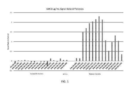
[0018] Figure 1. Depiction of the difference of the ratios of fluorescent
signal in wells
containing vancomycin to uninoculated control wells at 4 and 3 hours following
inoculation.
[0019] Figure 2A and 2B. Depiction of the difference of the ratios of signal
in wells
containing vancomycin to uninoculated control wells at 4 and 3 hours following
inoculation
for larger organism inoculum concentrations.
[0020] Figure 3A and 3B. Depiction of the normalized viability signal from
Vancomycin
wells for confused accessions.
[0021] Figure 4. Depiction of the clustering of signals measured in positive
control wells
comprising no antimicrobial, inoculated with a first quantity of microbe-
containing sample
Cmo, and signals measured in experimental HMLR wells comprising 4 ug/mL
vancomycin
within the range of the AST dilution series.
[0022] Figure 5 is a schematic that illustrates the confounding effect that
filamentous growth
has on volumetric-based determinations of microorganism's antimicrobial
susceptibilities.
Susceptible bacteria entering filamentous growth may appear falsely resistant
due to their
increased volume.
[0023] Non-limiting embodiments of the present disclosure are described by way
of example
with reference to the accompanying drawings, which are schematic and not
intended to be
drawn to scale. The accompanying drawings are provided for purposes of
illustration only,
and the dimensions, positions, order, and relative sizes reflected in the
figures in the drawings
may vary. In the figures, identical or nearly identical or equivalent elements
are typically
represented by the same reference characters. For purposes of clarity and
simplicity, not
every element of each embodiment is shown where illustration is not necessary
to allow those
of ordinary skill in the art to understand the disclosure.
Detailed Description
[0024] The following detailed description should be read with reference to the
drawings,
which, as noted above, depict illustrative embodiments. It will be appreciated
that the present
disclosure is set forth in various levels of detail in this application. In
certain instances,
details that are not necessary for one of ordinary skill in the art to
understand the disclosure,
46
CA 03152541 2022-02-24
WO 2021/041710
PCT/US2020/048242
or that render other details difficult to perceive may have been omitted. All
of the
compositions, systems and/or methods disclosed and claimed herein can be made
and
executed without undue experimentation in light of the present disclosure. It
should be
understood that the claimed subject matter is not necessarily limited to the
particular
embodiments or arrangements described or illustrated herein, the scope of the
claimed
invention being set out in the appended claims.
Definitions
[0025] The term "biological sample" refers to any sample that contains a
microorganism,
e.g., a bacterium and a fungal cell. Exemplary biological samples include, but
are not limited
to, whole blood, plasma, serum, sputum, urine, stool, white blood cells, red
blood cells, buffy
coat, tears, mucus, saliva, semen, vaginal fluids, lymphatic fluid, amniotic
fluid, spinal or
cerebrospinal fluid, peritoneal effusions, pleural effusions, exudates,
punctates, epithelial
smears, biopsies, bone marrow samples, fluids from cysts or abscesses,
synovial fluid,
vitreous or aqueous humor, eye washes or aspirates, bronchoalveolar lavage,
bronchial
lavage, or pulmonary lavage, lung aspirates, and organs and tissues, including
but not limited
to, liver, spleen, kidney, lung, intestine, brain, heart, muscle, pancreas,
and the like, swabs
(including, without limitation, wound swabs, buccal swabs, throat swabs,
vaginal swabs,
urethral swabs, cervical swabs, rectal swabs, lesion swabs, abscess swabs,
nasopharyngeal
swabs, and the like), and any combination thereof. Also included are bacteria
cultures or
bacteria isolates, fungal cultures or fungal isolates. The ordinary-skilled
artisan will also
appreciate that isolates, extracts, or materials obtained from any of the
above exemplary
biological samples are also within the scope of this disclosure.
[0026] As used herein, the terms "infection" and "infectious agent" are meant
to include any
infectious agent of a microbial origin, e.g., a bacterium, a fungal cell, an
archaeon, and a
protozoan. In preferred examples, the infectious agent is a bacterium, e.g., a
gram-positive
bacterium, a gram-negative bacterium, and an atypical bacteria. The term
"antimicrobial
resistant microorganism" is a microorganism (e.g., bacterium, fungus,
archeaon, and
protozoan) that is resistant to one or more distinct antimicrobials, i.e.,
anti-bacterial drugs,
antifungal drugs, anti-archaea medications, and anti-protozoan drugs.
[0027] "Microorganisms" as used in this specification refers to e.g., a liquid
suspension of
microorganisms, and may include one strain of microorganism, or more than one
strain of
microorganism. The microorganisms may include one species of microorganism.
The
microorganisms may include more than one strain of microorganism. The
microorganisms
47
CA 03152541 2022-02-24
WO 2021/041710
PCT/US2020/048242
may include one order of microorganism. The microorganisms may include one
class of
microorganism. The microorganisms may include one family of microorganism. The
microorganisms may include one kingdom of microorganism.
[0028] The microorganism may be a bacterium. Examples of bacterium include and
are not
limited to Acetobacter aura ntius, Acinetobacter bitumen, Acinetobacter spp.,
Actinomyces
israelii, Actinomyces spp., Aerococcus spp., Agrobacterium radiobacter,
Agrobacterium
tumefaciens, Anaplasma, Anaplasma phagocytophilum, Azorhizobium caulinodans,
Azotobacter vinelandii, Bacillus, Bacillus anthracis, Bacillus brevis,
Bacillus cereus, Bacillus
fusiformis, Bacillus licheniformis, Bacillus megaterium, Bacillus mycoides,
Bacillus spp.,
Bacillus stearothermophilus, Bacillus subtilis, Bacillus Thuringiensis,
Bacteroides,
Bacteroides fragilis, Bacteroides gingivalis, Bacteroides melaninogenicus
(also known as
Prevotella melaninogenica), Bartonella, Bartonella henselae, Bartonella
quintana,
Bartonella spp., Bordetella, Bordetella bronchiseptica, Bordetella pertussis,
Bordetella spp.,
Borrelia burgdorferi, Brucella, Brucella abortus, Brucella melitensis,
Brucella spp., Brucella
suis, Burkholderia, Burkholderia cepacia, Burkholderia mallei, Burkholderia
pseudomallei,
Calymmatobacterium granulomatis, Campylobacter, Campylobacter coli,
Campylobacter
fetus, Campylobacter jejuni, Campylobacter pylori, Campylobacter spp.,
Chlamydia,
Chlamydia spp., Chlamydia trachomatis, Chlamydophila, Chlamydophila pneumoniae
(previously called Chlamydia pneumoniae), Chlamydophila psittaci (previously
called
Chlamydia psittaci), Chlamydophila spp., Clostridium, Clostridium botulinum,
Clostridium
difficile, Clostridium perfringens (previously called Clostridium welchii),
Clostridium spp.,
Clostridium tetani, Corynebacterium, Corynebacterium diphtheriae,
Corynebacterium
fusiforme, Corynebacterium spp., Coxiella bumetii, Ehrlichia chaffeensis,
Ehrlichia spp.,
Enterobacter cloacae, Enterobacter spp., Enterococcus, Enterococcus avium,
Enterococcus
durans, Enterococcus faecalis, Enterococcus faecium, Enterococcus galllinarum,
Enterococcus maloratus, Enterococcus spp., Escherichia coli, Francisella spp.,
Francisella
tularensis, Fusobacterium nucleatum, Gardenerella spp., Gardnerella vaginalis,
Haemophilius spp., Haemophilus, Haemophilus ducreyi, Haemophilus influenzae,
Haemophilus parainfluenzae, Haemophilus pertussis, Haemophilus vaginalis,
Helicobacter
pylori, Helicobacter spp., Klebsiella pneumoniae, Klebsiella spp.,
Lactobacillus,
Lactobacillus acidophilus, Lactobacillus bulgaricus, Lactobacillus casei,
Lactobacillus spp.,
Lactococcus lactis, Legionella pneumophila, Legionella spp., Leptospira spp.,
Listeria
monocytogenes, Listeria spp., Methanobacterium extroquens, Microbacterium
multiforme,
Micrococcus luteus, Moraxella catarrhalis, Mycobacterium, Mycobacterium avium,
48
CA 03152541 2022-02-24
WO 2021/041710
PCT/US2020/048242
Mycobacterium bovis, Mycobacterium diphtheriae, Mycobacterium intracellulare,
Mycobacterium leprae, Mycobacterium lepraemurium, Mycobacterium phlei,
Mycobacterium
smegmatis, Mycobacterium spp., Mycobacterium tuberculosis, Mycoplasma,
Mycoplasma
fermentans, Mycoplasma genitalium, Mycoplasma hominis, Mycoplasma penetrans,
Mycoplasma pneumoniae, Mycoplasma spp., Neisseria, Neisseria gonorrhoeae,
Neisseria
meningitidis, Neisseria spp., Nocardia spp., Pasteurella, Pasteurella
multocida, Pasteurella
spp., Pasteurella tularensis, Peptostreptococcus, Porphyromonas gingivalis,
Prevotella
melaninogenica (previously called Bacteroides melaninogenicus), Proteus spp.,
Pseudomonas aeruginosa, Pseudomonas spp., Rhizobium radiobacter, Rickettsia,
Rickettsia
prowazekii, Rickettsia psittaci, Rickettsia quintana, Rickettsia rickettsii,
Rickettsia spp.,
Rickettsia trachomae, Rochalimaea, Rochalimaea henselae, Rochalimaea quintana,
Rothia
dentocariosa, Salmonella, Salmonella enteritidis, Salmonella spp., Salmonella
typhi,
Salmonella typhimurium, Serratia marcescens, Shigella dysenteriae, Shigella
spp., Spirillum
volutans, Staphylococcus, Staphylococcus aureus, Staphylococcus epidermidis,
Staphylococcus spp., Stenotrophomonas maltophilia, Stenotrophomonas spp.,
Streptococcus,
Streptococcus agalactiae, Streptococcus avium, Streptococcus bovis,
Streptococcus cricetus,
Streptococcus faceium, Streptococcus faecalis, Streptococcus ferus,
Streptococcus
gallinarum, Streptococcus lactis, Streptococcus mitior, Streptococcus mitis,
Streptococcus
mutans, Streptococcus oralis, Streptococcus pneumoniae, Streptococcus
pyogenes,
Streptococcus rattus, Streptococcus salivarius, Streptococcus sanguis,
Streptococcus
sobrinus, Streptococcus spp., Treponema, Treponema denticola, Treponema
pallidum,
Treponema spp., Ureaplasma spp., Vibrio, Vibrio cholerae, Vibrio comma, Vibrio
parahaemolyticus, Vibrio spp., Vibrio vulnificus, viridans streptococci,
Wolbachia, Yersinia,
Yersinia enterocolitica, Yersinia pestis, Yersinia pseudotuberculosis., and
Yersinia spp.
[0029] The microorganism may be a fungus. Examples of fungi include and are
not limited
to Aspergillus spp., Blastomyces spp., Candida spp., Cladosporium,
Coccidioides spp.,
Cryptococcus spp., Exserohilum, fusarium, Histoplasma spp., Issatchenkia spp.,
mucormycetes, Pneumocystis spp., ringworm, scedosporium, Sporothrix, and
Stachybotrys
spp.
[0030] The microorganism may be a protozoan. Examples of protozoan include and
are not
limited to Entamoeba histolytica, Plasmodium spp., Giardia lamblia, and
Trypanosoma
brucei.
[0031] The terms "kits" and "systems," as used herein in the present
disclosure, are intended
to refer to such things as combinations of multiple signaling agents with one
or more other
49
CA 03152541 2022-02-24
WO 2021/041710
PCT/US2020/048242
types of elements or components (e.g., other types of biochemical reagents,
signal detection
reagents, controls (i.e., positive and negative controls, e.g., chemically
sensitive/resistant
microorganisms), separation means (e.g., filters and magnetic beads),
containers, packages
such as packaging intended for commercial sale, substrates/cartridges to which
microorganism suspensions can be cultured, processed, or contained, electronic
hardware
components, and software recorded on a non-transitory processor-readable
medium). Certain
embodiments of this disclosure relate to kits for Rapid AST, for MIC or QSR
determinations,
etc.
[0032] The term "panel," refers to an arrangement of fluid wells configured to
permit the
performance of a multivariate or multiplex microbial assay. Panels used in the
present
disclosure, for example, may be dedicated to rapid AST determinations, and may
include one
or more positive control wells (e.g., wells for growth checkpoint assays) and
a series of
experimental wells defining, e.g., one or more dilution series for an
antimicrobial being
examined, and/or wells that allow for variation of other conditions such as
media conditions,
etc. Panels may be embodied on consumables, such as AST cassettes, multi-well
plates, etc.
The terms "well," and "reservoir" are used interchangeably to refer to the
fluid wells of a
panel or consumable.
Overview
[0033] Embodiments of the present disclosure relate to the novel finding that
the accuracy of
rapid AST results can be improved by utilizing different microorganism
concentrations for
AST. This represents a significant departure from the broth microdilution
standard and from
existing AST assay platforms, which reflect significant efforts in equipment
and consumables
design to ensure an approximately equal volume and/or number of microorganisms
is
inoculated into each reservoir on each panel, with the exception of negative,
contamination
control reservoirs.
[0034] The CLSI M07 manual follows the broth microdilution definition in the
International
Organization for Standardization (ISO) 16782 standard Clinical laboratory
testing ¨ Criteria
for acceptable lots of dehydrated Mueller-Hinton agar and broth for
antimicrobial
susceptibility testing in defining that "Each well [other than contamination
controls] should
contain approximately 5x10⁵ colony forming units (CFU)/mL (range, 2-8x10⁵
CFU/mL)."
The M07 further describes "The most convenient method for preparing
microdilution trays is
by using a dispensing device ... [which] delivers 0.1 ( 0.02) mL into each of
the 96 wells of
a standard tray."
CA 03152541 2022-02-24
WO 2021/041710
PCT/US2020/048242
[0035] Automated and semi-automated systems are thus designed to provide
approximately
equal concentrations of microbe to each reservoir of a panel. For example, the
Vitek 2
(bioMerieux) and PhoenixTM (Becton-Dickinson) platforms use capillary forces
in custom
cartridges to fill a plurality of reservoirs with approximately the same
concentration of
microbes and the MicroScanTM (Danaher/Beckman-Coulter) and SensiTitreTm
(Thermo
Fisher) platforms provide custom inoculators.
[0036] In cases of slowly induced- and/or hetero-resistance, it may be
challenging to
differentiate the absence of growth resulting from such resistance mechanisms
from the
absence of growth resulting from true susceptibility. To meet this challenge,
automated
systems either follow the reference standards and wait approximately 18 or
more hours (or, in
some cases for vancomycin, 24 or more hours), as is the case for the MicroScan
and the
SensiTitre, or regularly assess growth in each reservoir and perform
algorithmic
interrogations to determine relative growth trends across each dilution, as is
the case for the
Vitek2 and Phoenix.
[0037] This latter approach typically requires longer times for AST
determinations for
microbes exhibiting slowly induced- and/or hetero-resistance mechanisms,
resulting in slower
results being delivered for patients infected with pathogens that may not
respond to standard
broad-spectrum therapies. Furthermore, as described in U.S. Patent Application
Publication
2020-0149086 (which is incorporated by reference herein), repetitive growth
measurements
of every reservoir on each panel restrict the number of reservoirs that can be
present and
thereby limit the number of antimicrobial agents that can be tested in
parallel.
[0038] However, the inventors have discovered that accurate AST determinations
for a
plurality of microbial strains, including those harboring slowly induced-
and/or hetero-
resistance, may be achieved by performing growth determinations using two or
more
microorganism concentrations. For example, if a microorganism concentration of
approximately C.0 is inoculated into each reservoir of a dilution series of an
antimicrobial,
this new method teaches that the inoculation into each of an additional two or
more reservoirs
of a concentration of >5xCmo of the same microorganism, termed high
microorganism load
reservoirs (HMLRs), can improve the accuracy with which rapid AST
determinations can be
made.
[0039] The methods used to perform growth determinations in the dilution
reservoirs and
HMLRs may be similar or different. Growth in a plurality of dilution
reservoirs may be
assessed by metabolic (as described in U.S. Patent Application Publication
2020-0149086)
and/or surface area (as described in U.S. Patent 9,834,808) assays after the
performance of a
51
CA 03152541 2022-02-24
WO 2021/041710
PCT/US2020/048242
sufficient growth assay (as described in U.S. Patent Application 16/472,714).
Growth in
HMLRs may be assessed with a viability probe in parallel with the sufficient
growth assay.
[0040] In the context of rapid AST assays such as those described by the
inventors in, e.g.,
US Pat. No. 9,834,808, which is incorporated by reference in its entirety, the
use of HMLRs
may facilitate the goal of providing single-shift AST results.
[0041] The inventors have examined dose-response relationships of reference
microbial
strains that are known to be susceptible to, or resistant to, vancomycin.
Figure 3, depicts, for
individual susceptible (panel A) or resistant (Panel B) reference strains,
fluorescence signals
observed across a representative vancomycin dilution series. The figure shows
that, for many
susceptible strains, the dose curves overlap with those of resistant strains,
preventing
consistent differentiation therebetween.
[0042] By increasing the number of cells being interrogated, however,
susceptible and
resistant samples can be clearly delineated. Figure 4 shows, for individual
reference strains,
the clustering of (x) signals measured in positive control wells comprising no
antimicrobial,
inoculated with a first quantity of microbe-containing sample Cmo, and (y)
signals measured
in experimental HMLR wells comprising a concentration of vancomycin within the
range of
the AST dilution series, in this case 4 ug/mL. For each strain, multiple
measurements were
taken on multiple days to determine the ranges in which measurements cluster.
As the figure
demonstrates, the resistant reference strains tend to cluster at higher HMLR
signal values
than susceptible strains that cluster around similar positive control signal
values.
[0043] Based on this finding, the inventors have determined that a series of
threshold values,
or a threshold function, can be used to differentiate between resistant and
susceptible
samples. For example, in certain methods of this disclosure, a positive
control signal of an
AST panel is used to select an HMLR cutoff value, and measured HMLR signals
above the
cutoff value are called as resistant while those below the cutoff are called
susceptible.
HMLR cutoff values can be pre-determined and stored in a look-up table or
database, or may
be determined at the time of each measurement by a function, formula, or
mathematical
model.
[0044] In the context of rapid AST panels, some embodiments of this disclosure
relate to an
AST panel comprising one or more dedicated HMLR wells. Methods utilizing such
a panel
will typically include inoculating a larger volume of patent-sample derived
and/or
microorganism-containing fluid into each HMLR well than is inoculated into the
non-HMLR
wells of the AST panel; the HMLR wells may then receive a smaller volume of
other fluids
such as microbial broths or buffers, or they may receive concentrated
solutions to equalize
52
CA 03152541 2022-02-24
WO 2021/041710
PCT/US2020/048242
concentrations of, e.g., nutrients or antimicrobials with those of the non-
HMLR wells.
Alternatively, similar fluid volumes are inoculated into both HMLR and non-
HMLR wells,
but the HMLR wells receive fluids with higher microbial concentrations.
[0045] Signals from the HMLR and non-HMLR wells may be based on, e.g., surface
area
measurements as described in 11808 patent], or measurements of metabolic
activity.
[0046] Broth microdilution AST determinations for an antimicrobial agent, A,
are performed
by assessing relative growth in each of a range of different concentrations of
A, spanning
from a low concentration CAL to a high concentration CAH. The microorganism
concentration
is, by definition, held constant at Cmo = 2-8 x 10⁵ CFU/mL. The value of CAL
is typically less
than or equal to the lowest susceptible breakpoint concentration for the
organisms for which
the panel is designed to test and the jurisdiction where the panel is designed
to be used (ie.
US, Europe, etc.). The value of CAH is typically greater than or equal to the
highest resistant
breakpoint concentration for the organisms for which the panel is designed to
test and the
jurisdiction where the panel is designed to be used (ie. US, Europe, etc.). In
some cases, such
as where no resistant breakpoint exists, the value of CAH may alternately be
greater than or
equal to the intermediate or susceptible breakpoint.
[0047] A typical dilution series from CAL to CAH may span 3-15 independent
reservoirs, each
with a different concentration of antimicrobial A. These may be doubling
(serial) dilutions, as
detailed in the ISO and CLSI standards or may be non-twofold dilutions.
Dilution series may
be comprised of a combination of doubling dilutions and non-twofold dilutions.
The number
of dilutions is dependent upon the drug and the species supported for testing
with the panel.
[0048] It may be advantageous, in certain embodiments, for a minimum of one
high
microorganism load reservoir (HMLR) to be utilized together with a control
comprising no
microorganisms and no antimicrobial. The HMLR reservoir comprises
antimicrobial A at a
concentration ?CAL/4, preferably >CAL, and a microorganism concentration
>2xCmo,
preferably >5xCmo. Additional HMLR reservoirs may also be present. These may
comprise,
for example, additional reservoirs at the same concentration of A and/or
additional reservoirs
at different concentrations of A. In some cases, the HMLRs comprise a
microorganism
concentration >3xCmo, >4xCmo, >5xCmo, >6xCmo, >7xCmo, >8xCm0, >9xCm0, >10xCmo,
>11xCmo, >12xCmo, >13xCmo, >14xCmo, >15xCmo, >16xCmo, >17xCmo, >18xCmo,
>19xCmo,
or >20xCm0.
[0049] In an embodiment, growth in the HMLR(s) and HMLR control(s) is assessed
with a
probe capable of associating with microorganism surfaces. The present
disclosure permits
rapid determination of antibiotic susceptibility of microbial infections. This
disclosure is
53
CA 03152541 2022-02-24
WO 2021/041710
PCT/US2020/048242
based in part upon the surprising discovery of non-specific surface binding
assays that
provide accurate and rapid Antimicrobial Susceptibility Testing (AST)
determinations in
fewer than twelve hours ¨ and, specifically, under four hours. The present
disclosure ("Fast-
AST") provides accurate results that are consistent with results obtained
using the Clinical
Laboratory Standards Institute (CLSI) reference methods when tested with
multiple
antimicrobials and on a plurality of microorganisms; however, the present
disclosure takes
significantly less time to obtain results than the CLSI methods. Moreover, the
present
disclosure accurately differentiates an antimicrobial's MIC for clinically-
relevant microbial
strains that are resistant to one or more antimicrobials and the
antimicrobial's MIC for strains
of the same microorganism that are sensitive to the antimicrobials.
Furthermore, the present
disclosure may include signaling agents (e.g., Europium compounds) that are
bound to
microorganisms non-specifically rather than specifically (e.g., via chemically
conserved
groups or biochemically conserved binding sites on microorganisms), thereby
expanding the
generalization of the present disclosure to any microorganism and allowing
onset of an
appropriate treatment without first needing to identify the particular
infectious
microorganism. Also, the present disclosure permits signal amplification such
that microbes
may be rapidly detected at lower concentrations, e.g., from a dilute culture
of microorganisms
or via a patient's biological sample. Additionally, the present disclosure may
use Europium
formulations as chemical moiety, thereby expanding the dynamic range of the
methods and
allowing for more accurate determinations from a range of microbial samples.
Finally, the
present disclosure is compatible with existing equipment, thereby enabling
rapid adoption in
current clinical laboratories. Accordingly, the present disclosure, in a
greatly reduced amount
of time and expense, relative to standard methods, can provide a patient with
an appropriate
treatment regimen, i.e., a specific antimicrobial and at a particular dosage.
Thus, the present
disclosure will improve patient outcomes, lower hospital costs, and help
reduce further
evolution of antimicrobial resistant microorganisms; thus, the present
disclosure represents a
significant breakthrough in the AST field.
[0050] An aspect of the present disclosure is a method for determining
antimicrobial
susceptibility of microorganisms. The method includes steps of incubating a
liquid
suspension of microorganisms in the presence of an antimicrobial and a
signaling agent,
which is capable of binding to a surface of the microorganisms, under
conditions that
promote growth of the microorganisms; separating the microorganisms bound by
the
signaling agent from the unbound signaling agent; and determining signal
levels associated
with the microorganisms as compared to one or more controls.
54
CA 03152541 2022-02-24
WO 2021/041710
PCT/US2020/048242
[0051] Another aspect of the present disclosure is a method for determining
antimicrobial
susceptibility of microorganisms. The method includes steps of incubating a
liquid
suspension of microorganisms in the presence of an antimicrobial under
conditions that
promote growth of the microorganisms; adding a signaling agent capable of
binding to a
surface of the microorganisms; separating the microorganisms bound by the
signaling agent
from the unbound signaling agent; and determining signal levels associated
with the
microorganisms as compared to one or more controls.
[0052] Yet another aspect of the present disclosure is a method for
determining
antimicrobial susceptibility of microorganisms. The method includes steps of
incubating a
liquid suspension of microorganisms in a cartridge including a plurality of
chambers, each
chamber containing one or more antimicrobials, under conditions that promote
growth of the
microorganisms; adding a signaling agent, which is capable of binding to a
surface of the
microorganisms, to the plurality of chambers; removing unbound signaling
agent; and
determining signaling levels in the plurality of chambers as compared to one
or more
controls.
[0053] An aspect of the present disclosure is a method for determining
antimicrobial
susceptibility of microorganisms. The method includes incubating
microorganisms in the
presence of an antimicrobial and a signaling agent, which includes a signal
amplifier and one
or more chemical moieties capable of binding non-specifically to a surface of
the
microorganisms, under conditions that promote growth of the microorganisms;
separating the
microorganisms bound by the signaling agent from the unbound signaling agent;
and
determining signal levels associated with the microorganisms as compared to
one or more
controls.
[0054] Another aspect of the present disclosure is a method for determining
antimicrobial
susceptibility of microorganisms. The method includes incubating
microorganisms in the
presence of an antimicrobial under conditions that promote growth of the
microorganisms;
adding a signaling agent including a signal amplifier and one or more chemical
moieties
capable of binding non-specifically to a surface of the microorganisms;
separating the
microorganisms bound by the signaling agent from the unbound signaling agent;
and
determining signal levels associated with the microorganisms as compared to
one or more
controls.
[0055] Yet another aspect of the present disclosure is a kit for determining
antimicrobial
susceptibility of microorganisms. The kit includes a signaling agent capable
of binding to a
CA 03152541 2022-02-24
WO 2021/041710
PCT/US2020/048242
surface of the intact microorganisms of interest; a solution for incubating a
sample containing
microorganisms; and one or more reagents for generating signals from the
signaling agent.
[0056] In an embodiment, growth in the HMLR(s) and HMLR control(s) is assessed
through
the addition of one or more metabolic (viability) probes prior to a plurality
of the incubation
period. It is most preferred that the metabolic probe be added to the
reservoirs following
panel inoculation and prior to the onset of panel incubation. Suitable probes
are detailed in
U.S. Patent Application Publication 2020-0149086 at paragraphs [0211] through
[0237],
which is incorporated in its entirety herein. This probe may comprise
resazurin and
methylene blue together with ferric and ferrous salts. The probe may further
comprise 1-
methoxy-5-methlyphenasinum methyl sulfate. In alternative embodiments, growth
may be
assessed by any number of methods including, but not limited to, absorbance,
scattering,
thermal measurements, mass measurements, electrical measurements.
[0057] In an embodiment, growth in the HMLR(s) is normalized by growth in the
HMLR
control(s). The HMLR growth or the normalized HMLR growth may then be
evaluated based
on the rate of growth of the strain. Strain growth rate may be determined
based upon one or
more positive control reservoirs inoculated with a microorganism concentration
of C.o.
Growth in these reservoirs may be be assessed by one or more of Europium and
viability
probes.
[0058] In an embodiment, the addition of the viability and Europium probes is
performed
following the performance of a sufficient growth assay. An exemplary method of
this
disclosure comprises the following steps:
-introducing suspensions of one or more microorganisms to a cartridge
comprising a plurality
of chambers, wherein a plurality of chambers comprise one or more
antimicrobial agents;
-incubating the cartridge under conditions promoting microorganism growth for
an initial
incubation period;
- performing in a subset of the cartridge chambers, one or more checkpoint
assays to
determine if microorganism growth has achieved a threshold value; and
(a) if the threshold value is achieved, performing a plurality of different
growth assays
in a plurality of the cartridge chambers to determine the microorganism's
susceptibility to the
one or more antimicrobials, and obtaining a minimum inhibitory concentration
(MIC) and/or
a qualitative susceptibility result (QSR); or
(b) if the threshold value is not achieved, performing one or more
additional incubation
periods under conditions promoting microorganism growth until
(i) the threshold value is achieved, and thereafter performing step (a) ; or
56
CA 03152541 2022-02-24
WO 2021/041710
PCT/US2020/048242
(ii) a maximum of 18 hours has transpired without the threshold value being
achieved and no
further assays are performed.
[0059] In one aspect, a method of determining antimicrobial susceptibility of
one or more
microorganisms is provided, where the method comprises performing a growth
assay
comprising: incubating a suspension of a microorganism in the presence of one
or more
antimicrobials without a metabolic probe present; introducing a metabolic
probe in an
aqueous-miscible solvent after the incubation of the one or more
microorganisms; and
determining antimicrobial susceptibility of the one or more microorganisms
based on relative
microorganism growth.
[0060] In some embodiments, the method for determining antimicrobial
susceptibility of one
or more microorganisms comprises incubating a suspension of microorganisms in
a plurality
of chambers in a cartridge comprising antimicrobial agents for an initial time
period to
promote microorganism growth, performing one or more checkpoint assays in a
subset of the
cartridge chambers to determine if relative microorganism growth achieved a
threshold value,
wherein achieving the threshold value indicates a sufficient growth for the
assay system to
provide MIC or QSR data for the microorganism, then performing the assay for
obtaining the
MIC or QSR data.
[0061] In an embodiment, the fluorescent or absorbent signal resulting from
resazurin
reduction is then determined in the HMLR and HMLR control reservoirs at a
minimum of
two timepoints, preferably after 60-240 minutes of incubation, timepoint Ti,
and then after a
further 30-120 minutes of incubation, timepoint T2. As one example, Ti is
approximately 180
minutes and T2 is approximately 60 minutes.
[0062] In an embodiment, the HMLR assay is performed by comparing the optical
signal at
Ti with that at T2 after normalization with a control signal such as the HMLR
control
reservoir signal. The absolute value of the signal may also be used for
interpretation. An
optical signal at T2 is greater than that at Ti is indicative of continued
microorganism growth
at antimicrobial concentration A. An optical signal at T2 is less than or
equal to that at Ti
suggests that microorganism growth has subsided.
Surface Area Measurements for Rapid AST
[0063] Some embodiments of this disclosure relate to Rapid AST methods
utilizing specific
or non-specific surface area binding agents to measure the aggregate surface
area of cells
grown under varying conditions such as multiple antimicrobial agents or
multiple
concentrations of the same. Accordingly, one aspect of the present disclosure
relates to s a
57
CA 03152541 2022-02-24
WO 2021/041710
PCT/US2020/048242
method for determining antimicrobial susceptibility of microorganisms. The
method includes
steps of incubating a liquid suspension of microorganisms in the presence of
an antimicrobial
and a signaling agent under conditions that promote growth of the
microorganisms, wherein
the signaling agent is capable of binding to a surface of the microorganisms;
separating the
microorganisms bound by the signaling agent from the unbound signaling agent;
and
determining signal levels associated with the microorganisms as compared to
one or more
controls, thereby determining the antimicrobial susceptibility of the
microorganisms.
[0064] Another aspect of the present disclosure is a method for determining
antimicrobial
susceptibility of microorganisms. The method includes steps of incubating a
liquid
suspension of microorganisms in the presence of an antimicrobial under
conditions that
promote growth of the microorganisms; adding a signaling agent capable of
binding to a
surface of the microorganisms; separating the microorganisms bound by the
signaling agent
from the unbound signaling agent; and determining signal levels associated
with the
microorganisms as compared to one or more controls, thereby determining the
antimicrobial
susceptibility of the microorganisms. In embodiments, adding the signaling
agent occurs
prior to or during the incubating step or adding the signaling agent occurs
after the incubating
step.
[0065] Another aspect of the present disclosure is a method for determining
antimicrobial
susceptibility of microorganisms. The method includes steps of incubating a
liquid
suspension of microorganisms in a cartridge comprising a plurality of
chambers, each
chamber containing one or more antimicrobials, under conditions that promote
growth of the
microorganisms; adding a signaling agent to the plurality of chambers, wherein
the signaling
agent is capable of binding to a surface of the microorganisms; removing
unbound signaling
agent; and determining signaling levels in the plurality of chambers as
compared to one or
more controls, thereby determining the susceptibility of microorganisms to the
one or more
antimicrobials. In embodiments, the cartridge further includes one or more
control chambers
(e.g., at least 2, 4, 6, 8, 12, 24, 48, 96, 192, 384, 1536 or more chambers)
that do not contain
antimicrobials or one or more antimicrobials for which the microorganisms are
not
susceptible.
[0066] In embodiments of an above aspect, binding to a surface of the
microorganisms is
non-specific, e.g., comprising a non-covalent interaction and via forming a
covalent bond.
[0067] In embodiments of an above aspect, the signaling agent may include a
chemical
and/or biochemical group capable of binding a surface of the microorganisms,
wherein the
58
CA 03152541 2022-02-24
WO 2021/041710
PCT/US2020/048242
surface comprises one or more of membranes, walls, proteins, organelles,
saccharides, lipids,
cell envelope, and/or nucleic acids.
[0068] In embodiments of an above aspect, the signaling agent may include a
chemical
and/or biochemical group capable of binding a biomolecule of the surface of
the
microorganisms, wherein the surface biomolecule is selected from
peptidoglycans, mureins,
mannoproteins, porins, beta-glucans, chitin, glycoproteins, polysaccharides,
lipopolysaccharides, lipooligosaccharides, lipoproteins, endotoxins,
lipoteichoic acids,
teichoic acids, lipid A, carbohydrate binding domains, efflux pumps, other
cell-wall and/or
cell-membrane associated proteins, other anionic phospholipids, and a
combination thereof.
[0069] In embodiments of an above aspect, the signaling agent may include a
signal amplifier
and one or more chemical moieties capable of binding non-specifically to a
surface of the
microorganisms.
[0070] Another aspect of the present disclosure is a method for determining
antimicrobial
susceptibility of microorganisms. The method includes incubating
microorganisms in the
presence of an antimicrobial and a signaling agent under conditions that
promote growth of
the microorganisms, wherein the signaling agent comprises a signal amplifier
and one or
more chemical moieties capable of binding non-specifically to a surface of the
microorganisms; separating the microorganisms bound by the signaling agent
from the
unbound signaling agent; and determining signal levels associated with the
microorganisms
as compared to one or more controls, thereby determining the antimicrobial
susceptibility of
the microorganisms.
[0071] Another aspect of the present disclosure is a method for determining
antimicrobial
susceptibility of microorganisms. The method includes incubating
microorganisms in the
presence of an antimicrobial under conditions that promote growth of the
microorganisms;
adding a signaling agent comprising a signal amplifier and one or more
chemical moieties
capable of binding non-specifically to a surface of the microorganisms;
separating the
microorganisms bound by the signaling agent from the unbound signaling agent;
and
determining signal levels associated with the microorganisms as compared to
one or more
controls, thereby determining the antimicrobial susceptibility of the
microorganisms. In
embodiments, the signaling agent occurs prior to, at the beginning of, or
during the
incubating step, preferably during the incubating step. In embodiments, the
microorganisms
are incubated in a liquid suspension.
[0072] In embodiments of an above aspect, the liquid suspension may be
prepared by
inoculating a liquid media with a microbial isolate grown from a biological
sample.
59
CA 03152541 2022-02-24
WO 2021/041710
PCT/US2020/048242
[0073] In embodiments of an above aspect, the liquid suspension of
microorganisms may be
prepared from an unprocessed biological sample, e.g., an unprocessed
biological sample has
not undergone a culturing step.
[0074] In embodiments of an above aspect, the biological sample is selected
from blood,
cerebrospinal fluid, urine, stool, vaginal, sputum, bronchoalveolar lavage,
throat,
nasal/wound swabs, and a combination thereof.
[0075] In embodiments of an above aspect, the method does not involve a step
of capturing
microorganisms on a solid surface prior to or during incubation.
[0076] In embodiments of an above aspect, the method does not include a step
of growing
microorganisms on a solid surface during or subsequent to the incubating step.
[0077] In embodiments of an above aspect, the incubating may include agitating
the liquid
suspension of microorganisms.
[0078] In embodiments of an above aspect, the liquid suspension of
microorganisms may be
agitated by means of mechanical, acoustic, and/or magnetic agitation
continuously or
discretely during the incubating.
[0079] In embodiments of an above aspect, the incubating occurs at 31-37 C.
[0080] The present disclosure is superior to currently-used AST methods, in
part because it
provides accurate AST results in significantly less time. For instance,
comparisons of the
instant methods to currently used automated AST systems -- BioMerieux's
Vitek2, Beckman
Dickinson's Phoenix, and Beckman-Coulter's MicroScan ¨ are provided in US
Patent
10,501,772 at column 20, line 30 through column 22, line 29 and Figures 3 and
4, which is
incorporated by reference for all purposes.
[0081] Measurements of relative microorganism surface area, as used in the
present
disclosure, overcome the pitfalls of metabolic probes for AST. First, since
relative surface
area is not confounded by shifts in metabolic activity, fast-AST enables
rapid, accurate
resistance calls. Second, surface area measurements prevent over-resistance
calls. In
contrast to volumetric measurements obtained with metabolic probes of the
currently-used
AST systems, surface area measurements enable accurate differentiation between
true
resistance and filamentous growth. As illustrated in the schematic of FIG. 5,
volumes of
resistant and susceptible filamentous bacteria are difficult to distinguish.
But the lack of
septation creates a filamentous surface area significantly lower than that of
truly resistant
bacteria. Thus, by amplifying each bacteria's surface area, the present
disclosure is able to
accurately call four-hour, 0-lactam (ampicillin) MICs for E. coli samples
(see, the below
CA 03152541 2022-02-24
WO 2021/041710
PCT/US2020/048242
Examples). As illustrated in FIG. 5, the surface area differential between
elongation and
"true" resistance approaches 2/3, which may be detected with an amplified
signal.
[0082] Another aspect of the present disclosure is a kit for determining
antimicrobial
susceptibility of microorganisms. The kit includes a signaling agent capable
of binding to a
surface of the intact microorganisms of interest; a solution for incubating a
sample containing
microorganisms; and one or more reagents for generating signals from the
signaling agent.
[0083] In embodiments, the signaling agent is associated with one or more
binding moieties
capable of binding directly or indirectly to the intact microorganisms of
interest.
[0084] In embodiments, this disclosure features a signaling agent capable of
binding to the
surface of a microorganism. In embodiments, said binding is non-specific. In
embodiments,
said binding is specific.
[0085] In embodiments, a signaling agent is present during an incubating step
of a method
described herein. In embodiments, a signaling agent is present after an
incubating step of a
method described herein.
[0086] In embodiments, binding comprises the formation of a covalent bond. In
embodiments, a signaling agent is capable of binding to the surface of a
microorganism,
wherein said binding comprises the formation of a covalent bond. In
embodiments, a method
as described herein results in the formation of a covalent bond between a
group on a
microorganism surface (e.g., via a reactive group such as an electrophilic or
nucleophilic
group as described herein) and a signaling agent as described herein. In
embodiments, a
signaling agent has formed a covalent bond to the surface of a microorganism.
[0087] In embodiments, binding comprises the formation of a non-covalent
interaction. In
embodiments, a signaling agent is capable of binding to the surface of a
microorganism,
wherein said binding comprises the formation of a non-covalent interaction. In
embodiments,
a method as described herein results in the formation of non-covalent
interaction between a
group on a microorganism surface (e.g., via a reactive group such as an
electrophilic or
nucleophilic group as described herein) and a signaling agent as described
herein. In
embodiments, a signaling agent has formed a non-covalent interaction with the
surface of a
microorganism.
[0088] In embodiments, a non-covalent interaction comprises: ionic
interaction, ion-ion
interaction, dipole-dipole interaction, ion-dipole interaction, electrostatic
interaction, London
dispersion, van der Waals interaction, hydrogen bonding, 7E- it interaction,
hydrophobic
interaction, or any combination thereof. In embodiments, a non-covalent
interaction is: ionic
interaction, ion-ion interaction, dipole-dipole interaction, ion-dipole
interaction, electrostatic
61
CA 03152541 2022-02-24
WO 2021/041710
PCT/US2020/048242
interaction, London dispersion, van der Waals interaction, hydrogen bonding,
7E- it
interaction, hydrophobic interaction, or any combination thereof.
[0089] In embodiments, a non-covalent interaction comprises ionic
interactions, van der
Waals interactions, hydrophobic interactions, 7E-7E interactions, or hydrogen
bonding, or any
combination thereof. In embodiments, a non-covalent interaction comprises
ionic interaction,
van der Waals interaction, hydrogen bonding, or 7E- it interaction, or any
combination thereof.
[0090] In embodiments, a signaling agent capable of binding to a
microorganism's surface
comprises a group (e.g., a chemical or biochemical group) capable of binding
microorganism
membranes, walls, proteins, organelles, saccharides, lipids, cell envelope, or
nucleic acids, or
any combination thereof. In embodiments, a signaling agent capable of binding
to a
microorganism's surface comprises a chemical group (e.g., a nucleophilic group
or an
electrophilic group) capable of binding microorganism membranes, walls,
proteins,
organelles, saccharides, lipids, cell envelope, or nucleic acids, or any
combination thereof. In
embodiments, a signaling agent capable of binding to a microorganism's surface
comprises a
biochemical group capable of binding microorganism membranes, walls, proteins,
organelles,
saccharides, lipids, cell envelope, or nucleic acids, or any combination
thereof.
[0091] In embodiments, the surface may include a biomolecule to which the
signaling agent
binds or associates. Exemplary biomolecules include peptidoglycans, mureins,
mannoproteins, porins, beta-glucans, chitin, glycoproteins, polysaccharides,
lipopolysaccharides, lipooligosaccharides, lipoproteins, endotoxins,
lipoteichoic acids,
teichoic acids, lipid A, carbohydrate binding domains, efflux pumps, other
cell-wall and/or
cell-membrane associated proteins, other anionic phospholipids, and a
combination thereof.
[0092] In embodiments, a signaling agent capable of binding to a
microorganism's surface
comprises a biochemical group capable of binding microorganism membranes,
walls,
proteins, organelles, saccharides, lipids, cell envelope, or nucleic acids, or
any combination
thereof.
[0093] In embodiments, a signaling agent capable of binding to a
microorganism's surface
comprises a chemical group (e.g., a nucleophilic or electrophilic functional
group) capable of
binding microorganism membranes, walls, proteins, organelles, saccharides,
lipids, cell
envelope, or nucleic acids, or any combination thereof. In embodiments, said
chemical group
is a nucleophilic functional group. In embodiments, said chemical group is an
electrophilic
functional group.
62
CA 03152541 2022-02-24
WO 2021/041710
PCT/US2020/048242
[0094] In embodiments, a signaling agent is a biochemical signaling agent. In
embodiments,
a biochemical signaling agent comprises a biomolecule such as an antibody,
ligand, protein,
aptamer, ss-DNA, ss-RNA, or ss-PNA).
[0095] In embodiments, a signaling agent is a chemical signaling agent. In
embodiments, a
chemical signaling agent is a chemical compound (e.g., a synthetic chemical
compound). In
embodiments, a chemical signaling agent does not comprise a biomolecule such
as an
antibody, ligand, protein, aptamer, ss-DNA, ss-RNA, or ss-PNA).
[0096] In embodiments, a signaling agent capable of binding to a
microorganism's surface
comprises
[0097] a linker group L; and
[0098] an amplifier group (e.g., an amplifier group 104 that is a chemical or
biochemical
amplifier).
[0099] In embodiments, an amplifier group is an amplifier group 104, which is
a chemical or
biochemical amplifier. In embodiments, an amplifier group 104 is a chemical
amplifier. In
embodiments, an amplifier group 104 is a biochemical amplifier.
[0100] In embodiments, a signaling agent is a chemical compound. In
embodiments, a
chemical compound comprises a chemical amplifier group such as those described
herein).
[0101] In embodiments, a linker group L comprises the conserved (Fc) region of
an antibody.
[0102] In embodiments, a linker group L is capable of forming a covalent bond
to an
amplifier group (e.g., an amplifier group 104 that is a chemical or
biochemical amplifier).
[0103] In embodiments, a linker group L forms a covalent bond to a signal
amplifier group
(e.g., an amplifier group 104 that is a chemical or biochemical amplifier).
[0104] In embodiments, a linker group L is capable of forming one or more non-
covalent
interactions to an amplifier group (e.g., an amplifier group 104 that is a
chemical or
biochemical amplifier).
[0105] In embodiments, a linker group L forms one or more non-covalent
interactions to an
amplifier group (e.g., an amplifier group 104 that is a chemical or
biochemical amplifier).
[0106] In embodiments, a linker group L comprises a group (e.g., a chemical or
biochemical
group) capable of binding a microorganism surface. In embodiments, a linker
group L
comprises a group (e.g., a chemical or biochemical group) that binds a
microorganism
surface.
[0107] In embodiments, a linker group L comprises a group (e.g., a chemical or
biochemical
group) that is capable of forming a covalent bond to a microorganism's
surface. In
63
CA 03152541 2022-02-24
WO 2021/041710
PCT/US2020/048242
embodiments, a linker group L comprises a group (e.g., a chemical or
biochemical group)
that forms a covalent bond to a microorganism's surface.
[0108] In embodiments, a linker group L comprises a group (e.g., a chemical or
biochemical
group) that is capable of forming one or more non-covalent interactions with a
microorganism's surface. In embodiments, a linker group L comprises a group
(e.g., a
chemical or biochemical group) that forms one or more non-covalent
interactions with a
microorganism's surface.
[0109] In embodiments, a linker group L comprises a chemical moiety 101,
wherein said
chemical moiety is capable of forming a non-covalent interaction with the
surface of a
microorganism. In embodiments, a linker group L comprises a chemical moiety
101, wherein
said chemical moiety is capable of forming a covalent bond with the surface of
a
microorganism. In embodiments, a linker group L comprises a chemical moiety
101, wherein
said chemical moiety forms a non-covalent interaction with the surface of a
microorganism.
In embodiments, a linker group L comprises a chemical moiety 101, wherein said
chemical
moiety forms a covalent bond with the surface of a microorganism.
[0110] In embodiments, a linker group L comprises a spacer moiety 102. In
embodiments,
spacer moiety 102 is covalently attached to chemical moiety 101 and/or to
chemical moiety
103. In embodiments, spacer moiety 102 is covalently attached to chemical
moiety 101. In
embodiments, spacer moiety 102 is covalently attached to chemical moiety 103.
In
embodiments, spacer moiety 102 is covalently attached to chemical moiety 101
and to
chemical moiety 103. In embodiments, spacer moiety 102 forms a non-covalent
interaction
with chemical moiety 101 and/or with chemical moiety 103. In embodiments,
spacer moiety
102 forms a non-covalent interaction with chemical moiety 101. In embodiments,
spacer
moiety 102 forms a non-covalent interaction with chemical moiety 103. In
embodiments,
spacer moiety 102 forms a non-covalent interaction with chemical moiety 101
and with
chemical moiety 103.
[0111] In embodiments, a linker group L comprises a chemical moiety 103,
wherein said
chemical moiety is capable of forming a covalent bond to an amplifier group
(e.g., an
amplifier group 104 that is a chemical or biochemical amplifier). In
embodiments, a linker
group L comprises a chemical moiety 103, wherein said chemical moiety has
formed a
covalent bond to an amplifier group (e.g., an amplifier group 104 that is a
chemical or
biochemical amplifier). In embodiments, a linker group L comprises a chemical
moiety 103,
wherein said chemical moiety is capable of forming a non-covalent interaction
with an
amplifier group (e.g., an amplifier group 104 that is a chemical or
biochemical amplifier). In
64
CA 03152541 2022-02-24
WO 2021/041710
PCT/US2020/048242
embodiments, a linker group L comprises a chemical moiety 103, wherein said
chemical
moiety has formed a non-covalent interaction with an amplifier group (e.g., an
amplifier
group 104 that is a chemical or biochemical amplifier).
[0112] In embodiments, a signaling agent is a chemical compound comprising a
linker group
L that comprises: a chemical moiety 101, wherein said chemical moiety is
capable of forming
a covalent bond or a non-covalent interaction with the surface of the
microorganisms; a
spacer moiety 102, wherein spacer moiety is covalently attached to chemical
moiety 101 and
to chemical moiety 103; and a chemical moiety 103, wherein said chemical
moiety has
formed or can form a covalent bond to an amplifier group 104 that is a
chemical or
biochemical amplifier.
[0113] In embodiments, a signaling agent is a chemical compound comprising a
linker group
L that comprises: a chemical moiety 101, wherein said chemical moiety is
capable of
forming a covalent bond or a non-covalent interaction with the surface of a
microorganism; a
spacer moiety 102, wherein spacer moiety is covalently attached to chemical
moiety 101 and
to chemical moiety 103; and a chemical moiety 103, wherein said chemical
moiety has
formed or can form a non-covalent interaction with an amplifier group 104 that
is a chemical
or biochemical amplifier.
[0114] In embodiments, a linker group comprises one chemical moiety 101. In
embodiments, a linker group comprises more than one chemical moiety 101 (e.g.,
a linker
group comprises 1, 2, 3, 4, 5, or 6 chemical moieties 101).
[0115] In embodiments, a linker group comprises one spacer moiety 102. In
embodiments, a
linker group comprises more than one spacer moiety 102 (e.g., a linker group
comprises 1, 2,
3, 4, 5, or 6 spacer moieties 102).
[0116] In embodiments, a linker group comprises one chemical moiety 103. In
embodiments, a linker group comprises more than one chemical moiety 103 (e.g.,
a linker
group comprises 1, 2, 3, 4, 5, or 6 chemical moieties 103).
[0117] In embodiments, a linker group comprises: one chemical moiety 101, one
spacer
moiety 102, and one chemical moiety 103. In embodiments, a linker group
consists of: one
chemical moiety 101, one spacer moiety 102, and one chemical moiety 103.
[0118] In embodiments, a linker group has the structure of substructure (I):
-101-102-103- , (I)
wherein
CA 03152541 2022-02-24
WO 2021/041710
PCT/US2020/048242
"101" represents a chemical moiety 101;
"102" represents a spacer moiety 102; and
"103" represents a chemical moiety 103.
[0119] In embodiments, a chemical moiety 101 is capable of forming a covalent
bond with
the surface of a microorganism.
[0120] In embodiments, a chemical moiety 101 is capable of forming a covalent
bond with
the surface of a microorganism in the presence of one or more agents that
promote coupling
(also referred to herein as coupling agents).
[0121] In embodiments, agents that promote coupling include glutaraldehyde,
formaldehyde,
paraformaldehyde, 1-ethyl-3-(3-dimethylaminopropyl)carbodiimide (EDC), N,N'-
dicyclohexylcarbodiimide (DCC), N-cyclohexyl-N'-(2-
morpholinoethyl)carbodiimide-
methyl-p-toluenesulfonate (CMC), diisopropylcarbodiimide (DIC), (1-
lbis(dimethylamino)methylenel-1H-1,2,3-triazolol4,5-blpyridinium 3-oxid
hexafluorophosphate) (HATU), Woodward's Reagent, /V,N'-carbonyl diimidazole, N-
hydroysuccinimide (NHS), or N-hydroxysulfosuccinimide (sulfo-NHS), or any
combination
thereof.
[0122] In embodiments, agents that promote coupling include aldehydes,
acrylates, amides,
imides, anhydrides, chlorotriazines, epoxides, isocyanates, isothiocyanates,
organic acids,
monomers, polymers, silanes, or silcates, or any combination thereof.
[0123] In embodiments, agents that promote coupling include a carbodiimide, a
phosphonium salt, or an ammonium salt, or any combination thereof.
[0124] In embodiments agents that promote coupling include glutaraldehyde, N-
(3-
dimethylaminopropy1)-N' -ethylcarbonate (EDC), (1-
lbis(dimethylamino)methylenel-1H-
1,2,3-triazolo114,5-blpyridinium 3-oxid hexafluorophosphate) (HATU), (0-
benzotriazol-1-yl-
N,N,N ,N -tetramethyluronium hexafluorophosphate) (HBTU), N-
hy droxy succinimide (NHS), /V,N'-dicyclohexylcarbodiimide (DCC),
diisopropylcarbodiimide
(DIC), hydroxy-3,4-dihydro-4-oxo-1,2,3-benzotriazine (HOOBt),
hydroxybenzotriazole (HOBT), 1-hydroxy-7-azabenzotriazole (HOAt), (N-(3-
dimethylaminopropy1)-N'-ethylcarbodiimide (EDAC), 4-(N,N-
dimethylamino)pyridine
(DMAP), benzotriazol-1-yloxy-tris(dimethylamino)- phosphonium
hexafluorophosphate
(BOP), benzotriazol-l-yloxy-tripyrrolidino-phosphonium hexafluorophosphate
(PyB OP),
bromo-tripyrrolidino-phosphonium hexafluorophosphate (PyBrOP), 7-aza-
benzotriazol-1-
66
CA 03152541 2022-02-24
WO 2021/041710
PCT/US2020/048242
yloxy-tripyrrolidinophosphonium hexafluorophosphate (PyA0P), ethyl
cyano(hydroxyimino)acetato-02 )- tri-(1-pyrrolidiny1)-phosphonium
hexafluorophosphate
(PyOxim), 3-(diethoxy-phosphoryloxy)-1,2,3-benzoldl triazin-4(3H)-one (DEPBT),
2-(6-
chloro-1H-benzotriazol-1-y1)- N,N,N',N' -tetramethylaminium
hexafluorophosphate (HCTU),
N-R5-chloro-1H-benzotriazol-1-y1)- dimethylamino-morpholinol-uronium
hexafluorophosphate N-oxide (HDMC), 1-l1-(cyano-2-ethoxy-2-
oxoethylideneaminooxy)-
dimethylamino-morpholinol- uronium hexafluorophosphate (COMU), 2-(1-oxy-
pyridin-2-
y1)-1,1,3,3-tetramethylisothiouronium tetrafluoroborate (TOTT),
tetramethylfluoroformamidinium hexafluorophosphate (TFFH), N-Ethoxycarbony1-2-
ethoxy-
1,2-dihydroquinoline (EEDQ), 2-propanephosphonic acid anhydride (PPA),
triphosgene,
1,1'-carbonyldiimidazole (CDI), R6-nitrobenzotriazol-1-
yl)oxyltris(pyrrolidino)phosphonium hexafluorophosphate (PyNOP), ll6-
(trifluoromethyl)benzotriazol-1-ylloxyltris(pyrroli-dino)phosphonium
hexafluorophosphate
(PyFOP), P-nitro-6-(trifluoromethyl)benzotriazol-1-
ylloxyltris(pyrrolidino)phosphonium
hexafluorophosphate (PyNFOP), R6-nitrobenzo-triazol-1-yl)oxyltris(dimethyl-
amino)phosphonium hexafluorophosphate (NOP), 1-fl-naphthalenesulfonyloxy
benzotriazole
(NSBt), 1-0-naphthalenesulfonyloxy-6-nitrobenzotriazole (N-NSBt),
tetramethylfluoroformamidinium hexafluorophosphate (TFFH),
bis(tetramethylene)fluoroformamidinium hexafluorophosphate (BTFFH), 1,3-
dimethy1-2-
fluoro-4,5-dihydro-1H-imidazolium hexafluorophosphate (DFIH), Cyanuric
chloride (CC), or
2,4-dichloro-6-methoxy-1,3,5-triazine (DCMT), and 2-chloro-4,6-dimethoxy-1,3,5-
triazine
(CDMT), or any combination thereof.
[0125] In embodiments, agents that promote coupling include EDC, HATU, HBTU,
NHS,
DCC, HOBT, or PyBOP, or any combination thereof.
[0126] In embodiments, agents that promote coupling include EDC, DCC, CMC,
DIC, or
HATU, or any combination thereof.
[0127] In embodiments, agents that promote coupling include glutaraldehyde,
formaldehyde,
or paraformaldehyde, or any combination thereof.
[0128] In embodiments, a chemical moiety 101 is capable of forming a non-
covalent
interaction with the surface of a microorganism (e.g., any non-covalent
interaction described
herein). In embodiments, a non-covalent interaction comprises: ionic, ion-ion,
dipole-dipole,
ion-dipole, electrostatic, London dispersion, van der Waals, hydrogen bonding,
or 7E- it, or
any combination thereof.
67
CA 03152541 2022-02-24
WO 2021/041710
PCT/US2020/048242
[0129] In embodiments, a chemical moiety 101 comprises a nucleophilic
functional group.
In embodiments, a chemical moiety 101 comprises a group formed from a
nucleophilic
functional group.
[0130] In embodiments, a nucleophilic functional group is: amino, amido,
hydrazino,
hydroxyamino, hydroxy, or thio. In embodiments, a nucleophilic functional
group is: amino,
hydrazino, hydroxyamino, or thio. In embodiments, a nucleophilic functional
group
comprises: amino, hydrazino, hydroxyamino, hydroxy, or thio. In embodiments, a
nucleophilic functional group is carboxamide, N-hydroxycarboxamide, carboxyl
hydrazide,
or guanidino.
[0131] In embodiments, a nucleophilic functional group is ¨NH2, -NHNH2, -
CONHOH,
-CONHNH2,¨ONH2, -OH, or -SH. In embodiments, a nucleophilic functional group
is ¨NH2,
-NHNH2, -CONHNH2, or ¨ONH2.
[0132] In embodiments, a chemical moiety 101 comprises an electrophilic
functional group.
[0133] In embodiments, a chemical moiety 101 comprises a group formed from an
electrophilic functional group.
[0134] In embodiments, an electrophilic functional group comprises an
aldehyde, a ketone, a
carboxylic acid, a carboxylic ester, a carboxylic acid halide (e.g., acetyl
chloride), or a
carboxylic acid anhydride (e.g., acetic anhydride).
[0135] In embodiments, an electrophilic functional group comprises an
aldehyde, an a-halo
ketone, a maleimide, a succinimide, a hydroxysuccinimide, an isothiocyanate,
an isocyanate,
an acyl azide, a sulfonyl chloride, a tosylate ester, a glyoxal, an epoxide,
an oxirane, a
carbonate, an imidoester, an anhydride, a fluorophenyl ester, a hydroxymethyl
phosphine
derivative, a carbonate, a haloacetyl, a chlorotriazine, a haloacetyl, an
alkyl halide, an
aziridine, or an acryloyl derivative. In embodiments, an electrophilic
functional group is an
aldehyde, an a-halo ketone, a maleimide, a succinimide, a hydroxysuccinimide,
an
isothiocyanate, an isocyanate, an acyl azide, a sulfonyl chloride, a tosylate
ester, a glyoxal, an
epoxide, an oxirane, a carbonate, an imidoester, an anhydride, a fluorophenyl
ester, a
hydroxymethyl phosphine derivative, a carbonate, a haloacetyl, a
chlorotriazine, a haloacetyl,
an alkyl halide, an aziridine, or an acryloyl derivative.
[0136] In embodiments, an electrophilic functional group comprises an
aldehyde, an a-halo
ketone, a maleimide, a succinimide, or a hydroxysuccinimide group.
[0137] In embodiments, an electrophilic functional group comprises ¨CHO,
-C(0)CH2I, 0)CH2I,
68
CA 03152541 2022-02-24
WO 2021/041710
PCT/US2020/048242
CI N CI
1 0
oç50 0 T' if
N
0 0__rN 0 A 1
* N
0* 0
0* ,or S'
[0138] In embodiments, an electrophilic functional group comprises ¨CHO,
0
Ni
=f.0
-C(0)CH2I, \=/ , or
[0139] In embodiments, chemical moiety 101 comprises a group that is alkyl,
alkenyl,
alkynyl, phenyl, heteroaryl, haloalkyl, hydroxy, carbonyl, acyl halide,
alkoxycarbonyl)oxy,
carboxy, haloketone, alkoxy, alkoxyol (hemiacetal or) hemiketal, dialkoxy
(e.g., ketal or
acetal), trialkoxy(orthoether), carbamoyl, amino, ammonio, imino, imido,
succinamido,
maleidido, hydroxysuccinamido, biotin, D-Biotin, azido, azo, cyanate,
isocyanato, nitroxy,
cyano, isocyano, nitrosooxy, nitro, nitroso, oxime, sulfanyl, sulfinyl,
sulfonyl, sulfino, sulfo,
thiocyanato, isothiocyanato, thioyl, phosphate, or boronate.
[0140] In embodiments, spacer moiety 102 is hydrophobic. In embodiments,
spacer moiety
102 is hydrophilic.
[0141] In embodiments, spacer moiety 102 is peptidic (e.g., derived from
peptide linkages).
[0142] In embodiments, spacer moiety 102 comprises inorganic linkages. In
embodiments,
spacer moiety 102 comprises organic linkages. In embodiments, spacer moiety
102
comprises only organic linkages.
[0143] In embodiments, spacer moiety 102 is oligomeric. In embodiments, spacer
moiety
102 is polymeric. In embodiments, spacer moiety 102 comprises segments (e.g.,
1 to about
300, 1 to about 200, 1 to about 100, 1 to about 50, 1 to about 25, or 1 to
about 10, or 1, 2, 3,
4, 5, 6, 7, 8, 9, or 10 segments) of methylene (-CH2-), ethylene glycol (-
CH2CH20-),
iminoethylene (-CH2CH2NH-), vinyl alcohol (-CH2CHOH-)x, lactic acid (-CH(CH3)-
C(0)-0-
), acrylic acid (-CH2CH2(CO2H)-), methacrylic acid (-CH2C(CH3)(CO2H)-1, or
methyl
methacrylate (-CH2C(CH3)(CO2CH3)-).
69
CA 03152541 2022-02-24
WO 2021/041710
PCT/US2020/048242
-
*/*
[0144] In embodiments, spacer moiety 102 comprises a segment that is "n
N*
m H P , or HO
[0145] In embodiments, n, m, p, and q independently is an integer of 1 to
about 300 (e.g., 1
to about 200, 1 to about 100, 1 to about 50, 1 to about 25, or 1 to about 10).
In embodiments,
each of n, m, p, and q is independently 1, 2, 3, 4, 5, 6, 7õ 9, or 10.
0 0*
[0146] In embodiments, spacer moiety 102 comprises R'. In embodiments, R'
is independently hydrogen or a group that is Ci-C12 alkyl, C2-C12 alkenyl, or
C2-C12 alkynyl.
In embodiments, o is an integer of 1 to about 300 (e.g., 1 to about 200, 1 to
about 100, 1 to
about 50, 1 to about 25, or 1 to about 10). In embodiments, o is independently
1, 2, 3, 4, 5, 6,
7, 8, 9, or 10.
0
0 R
[0147] In embodiments, spacer moiety 102 comprises . In
embodiments, R is independently hydrogen or a group that is Ci-C12 alkyl, C2-
C12 alkenyl, or
C2-C12 alkynyl. In embodiments, r is an integer of 1 to about 300 (e.g., 1 to
about 200, 1 to
about 100, 1 to about 50, 1 to about 25, or 1 to about 10). In embodiments, r
is independently
1, 2, 3, 4, 5, 6, 7, 8, 9, or 10.
,rµ s
[0148] In embodiments, spacer moiety 102 comprises R R . In embodiments, R
is
independently hydrogen or a group that is Ci-C12 alkyl, C2-C12 alkenyl, or C2-
C12 alkynyl. In
embodiments, s is an integer of 1 to about 300 (e.g., 1 to about 200, 1 to
about 100, 1 to about
50, 1 to about 25, or 1 to about 10). In embodiments, s is independently 1, 2,
3, 4, 5, 6, 7, 8,
9, or 10.
CA 03152541 2022-02-24
WO 2021/041710
PCT/US2020/048242
1-1,
,B
*_N% B_*
[0149] In embodiments, spacer moiety 102 comprises - . In embodiments, t is
an integer of 1 to about 300 (e.g., 1 to about 200, 1 to about 100, 1 to about
50, 1 to about 25,
or 1 to about 10). In embodiments, t is independently 1, 2, 3, 4, 5, 6, 7, 8,
9, or 10.
0
0, A
II 11
[0150] In embodiments, spacer moiety 102 comprises: 0
[0151] In embodiments, spacer moiety 102 is a polymer comprising repeating
groups,
comprising alkyl, alkoxy, ester, acrylic, amino, hydroxyl, or acyl hydrazine
functional
groups, or any combination thereof.
wf*
[0152] In embodiments, the spacer moiety 102 is: n , =
m ,
H P , or HO , wherein n, m, p, and q are as defined herein.
[0153] In embodiments, each of n, m, o, p, q, r, s, or t independently is an
integer of 1 to 100,
of 10 to 90, of 10 to 80, of 10 to 70, of 10 to 60, of 10 to 50, of 10 to 40,
of 10 to 30, of 10 to
20, or of 1 to 10.
[0154] In embodiments, a chemical moiety 103 comprises a group that is a
nucleophilic
functional group.
[0155] In embodiments, a chemical moiety 103 comprises a group formed from a
nucleophilic functional group.
[0156] In embodiments, a nucleophilic functional group is: amino, amido,
hydrazino,
hydroxyamino, hydroxy, or thio. In embodiments, a nucleophilic functional
group is: amino,
hydrazino, hydroxyamino, or thio.
[0157] In embodiments, a nucleophilic functional group comprises: amino,
hydrazino,
hydroxyamino, hydroxy, or thio. In embodiments, a nucleophilic functional
group is
carboxamide, N-hydroxycarboxamide, carboxyl hydrazide, or guanidino.
[0158] In embodiments, a nucleophilic functional group is ¨NH2, -NHNH2, -
CONHOH,
-CONHNH2,¨ONH2, -OH, or -SH. In embodiments, a nucleophilic functional group
is ¨NH2,
-NHNH2, -CONHNH2, or ¨ONH2.
71
CA 03152541 2022-02-24
WO 2021/041710
PCT/US2020/048242
[0159] In embodiments, a chemical moiety 103 comprises a group that is a
electrophilic
functional group.
[0160] In embodiments, a chemical moiety 103 comprises a group formed from a
electrophilic functional group.
[0161] In embodiments, an electrophilic functional group comprises an
aldehyde, a ketone, a
carboxylic acid, a carboxylic ester, a carboxylic acid halide (e.g., acetyl
chloride), or a
carboxylic acid anhydride (e.g., acetic anhydride).
[0162] In embodiments, an electrophilic functional group comprises an
aldehyde, an a-halo
ketone, a maleimide, a succinimide, a hydroxysuccinimide, an isothiocyanate,
an isocyanate,
an acyl azide, a sulfonyl chloride, a tosylate ester, a glyoxal, an epoxide,
an oxirane, a
carbonate, an imidoester, an anhydride, a fluorophenyl ester, a hydroxymethyl
phosphine
derivative, a carbonate, a haloacetyl, a chlorotriazine, a haloacetyl, an
alkyl halide, an
aziridine, or an acryloyl derivative. In embodiments, an electrophilic
functional group is an
aldehyde, an a-halo ketone, a maleimide, a succinimide, a hydroxysuccinimide,
an
isothiocyanate, an isocyanate, an acyl azide, a sulfonyl chloride, a tosylate
ester, a glyoxal, an
epoxide, an oxirane, a carbonate, an imidoester, an anhydride, a fluorophenyl
ester, a
hydroxymethyl phosphine derivative, a carbonate, a haloacetyl, a
chlorotriazine, a haloacetyl,
an alkyl halide, an aziridine, or an acryloyl derivative.
[0163] In embodiments, an electrophilic functional group comprises an
aldehyde, an a-halo
ketone, a maleimide, a succinimide, or a hydroxysuccinimide group.
[0164] In embodiments, an electrophilic functional group comprises ¨CHO,
CI N CI
0
0 0
N
Or_s_ri 0 0__rN 0 A A
-C(0)CH2I, 0
0* ,or S"
[0165] In embodiments, an electrophilic functional group comprises ¨CHO,
1 0
00
-C(0)CH2I, \=¨/ , or
[0166] In embodiments, a chemical moiety 103 comprises a chemical structure
that is
carbonyl, alkenyl, alkynyl, hydroxyl, amino, thiol, maleimide, succinimide,
hydroxysuccinimide, biotinyl, anhydride, chlorotriazine, epoxide, isocyanate,
or
72
CA 03152541 2022-02-24
WO 2021/041710
PCT/US2020/048242
isothiocyanate. In embodiments, said group that is carbonyl, alkenyl, alkynyl,
hydroxyl,
amino, thiol, maleimide, succinimide, hydroxysuccinimide, biotinyl, anhydride,
chlorotriazine, epoxide, isocyanate, or isothiocyanate is capable of forming a
covalent bond
to an amplifier group (e.g., an amplifier group 104). In embodiments, said
group that is
carbonyl, alkenyl, alkynyl, hydroxyl, amino, thiol, maleimide, succinimide,
hydroxysuccinimide, biotinyl, anhydride, chlorotriazine, epoxide, isocyanate,
or
isothiocyanate is capable of forming non-covalent interaction with an
amplifier group (e.g.,
an amplifier group 104).
[0167] In embodiments, a chemical moiety 103 is formed from a chemical
structure
comprising a group that is carbonyl, alkenyl, alkynyl, hydroxyl, amino, thiol,
maleimide,
succinimide, hydroxysuccinimide, biotinyl, anhydride, chlorotriazine, epoxide,
isocyanate, or
isothiocyanate. In embodiments, said group that is carbonyl, alkenyl, alkynyl,
hydroxyl,
amino, thiol, maleimide, succinimide, hydroxysuccinimide, biotinyl, anhydride,
chlorotriazine, epoxide, isocyanate, or isothiocyanate has formed a covalent
bond to an
amplifier group (e.g., an amplifier group 104). In embodiments, said group
that is carbonyl,
alkenyl, alkynyl, hydroxyl, amino, thiol, maleimide, succinimide,
hydroxysuccinimide,
biotinyl, anhydride, chlorotriazine, epoxide, isocyanate, or isothiocyanate
has formed a non-
covalent interaction with an amplifier group (e.g., an amplifier group 104).
[0168] In embodiments, a chemical moiety 103 comprises a group that is
carbonyl, alkenyl,
alkynyl, hydroxyl, amino, thiol, maleimide, succinimide, hydroxysuccinimide,
or biotinyl.
[0169] In embodiments, a chemical moiety 103 comprises a carbonyl, alkenyl,
alkynyl,
hydroxyl, amino, thiol, maleimide, succinimide, hydroxysuccinimide, or
biotinyl functional
group.
*()
[0170] In embodiments, a chemical moiety 103 comprises:
0 0
0 H
00 0 HN60/
on \=/
CI N CI
0 0
*A A* NN
C*N* C*N
0 0 0* , or S'
73
CA 03152541 2022-02-24
WO 2021/041710
PCT/US2020/048242
[0171] In embodiments, a chemical moiety 103 comprises a group formed from a
chemical
structure comprising a group that is carbonyl, alkenyl, alkynyl, hydroxyl,
amino, thiol,
maleimide, succinimide, hydroxysuccinimide, or biotinyl functional group.
[0172] In embodiments, a linker group L has the structure of substructure
(II):
. .
X +I% R`I
1
; ' .I( (II)
wherein:
X represents a chemical moiety 101 (e.g., any chemical moiety 101 as described
herein;
R represents a spacer moiety 102 (e.g., any spacer moiety 102 as described
herein);
Y represents a chemical moiety 103 (e.g., any chemical moiety 103 as described
herein); and
each of j and k independently is an integer of 0 to 100.
H
*N,NH2
*
= *\ *
[0173] In embodiments, X is NH2 0
, 0¨NH2 /C)
,
*
*
0
* H I 0
i 0 N N N
).r1 0Y. 0 , 0__r0 *A A
0 0 *
, .= .
c I yN yC I
N N )k\ ...= N., ,N
0 0 o r , .
*1 *
*
*H* *0.1-
i
[0174] In embodiments, R is n , m, R' , - H P 9
1-1% H
*
* * { ;.rH 0 B =N1
0 H )( N )3
* /yq* *.r N , N YLs * 1 NI*
N H P f B¨N, 1-
H
or* t ,
wherein each of n, m, o, p, q, r, s, or t is as described herein (e.g., an
integer of 1 to about
300).
74
CA 03152541 2022-02-24
WO 2021/041710 PCT/US2020/048242
=
[0175] In embodiments, Y is ,.. ¨
0 0
0
* 0 0
0121y0 HN AnA
c I I
N,
N N NH *
_2 N
= =
0 NH 0 or
/N
[0176] In embodiments, X is capable of forming a covalent bond to a
microorganism's
surface. In embodiments, X forms a covalent bond to a microorganism's surface.
[0177] In embodiments, X is capable of forming one or more non-covalent
interactions with
a microorganism's surface. In embodiments, X forms one or more non-covalent
interactions
with a microorganism's surface.
[0178] In embodiments, Y is capable of forming a covalent bond to an amplifier
group 104
(e.g., a chemical or biochemical amplifier). In embodiments, Y forms a
covalent bond to an
amplifier group such as an amplifier group 104 (e.g., a chemical or
biochemical amplifier.)
[0179] In embodiments, Y is capable of forming one or more non-covalent
interactions to an
amplifier group 104 (e.g., a chemical or biochemical amplifier). In
embodiments, Y forms
one or more non-covalent interactions to an amplifier group such as an
amplifier group 104
(e.g., a chemical or biochemical amplifier.)
CA 03152541 2022-02-24
W02021/041710
PCT/US2020/048242
/-PEG-0Me
HN-/
HNiq
)7,--NH
[0180]In embodiments, a linker group L is: 0 (13),
0 0
0 )\---- )\---
HN-PEG )7"--- HN-PEG
Y
/' 0
0
rS) /-/-µ0 --S / 0
HNhr HNT---?
eNH
O (14), 0
HN-PEG-NH2 HN-PEG __ /-SH
..--S
HN/q HNI-Y
eNH eNH
(15), 0 (16), 0 (17),
f--0O2H
HN-PEG-f
HN-....?
eNH
O (18),
[p--.NF/1 NO
HN-PEG-NH H
/' ?i
_,S / 0 0
HN/q
eNH
O (19),
76
CA 03152541 2022-02-24
WO 2021/041710
PCT/US2020/048242
0 O. ,p
O¨N
0 0
HN/q
eNH
0 (20), WGA-Biotin, PolymixinB-Biotin, monoclonal
antibody, polyclonal antibody, biotinylated monoclonal antibody, biotinylated
polyclonal
antibody, europium chelate-antibody, horseradish peroxidase-conjugated
antibody, and
antibody variants (e.g., Fab: fragment, antigen-binding (one arm); F(ab')2:
fragment, antigen-
binding, including hinge region (both arms); Fab': fragment, antigen-binding,
including hinge
region (one arm); scFv: single-chain variable fragment; di-scFv: dimeric
single-chain variable
fragment; sdAb: single-domain antibody; Bispecific monoclonal antibodies;
trifunctional
antibody; and BiTE: bi-specific T-cell engager).
[0181] Exemplary amplifier groups include those described in, e.g.,
International Publication
No. WO 2016/015027 and in International Application No. PCT/US16/42589, each
of which
is incorporated by reference in its entirety.
[0182] In embodiments, an amplifier group (e.g., an amplifier group 104)
comprises a
catalyst, a fluorophore, or a colormetric dye. In embodiments, an amplifier
group (e.g., an
amplifier group 104) is a catalyst, a fluorophore, or a colormetric dye.
[0183] In embodiments, an amplifier group (e.g., an amplifier group 104)
comprises an
enzyme, a catalyst, or a nanoparticle. In embodiments, an amplifier group
(e.g., an amplifier
group 104) is an enzyme, a catalyst, or a nanoparticle.
[0184] In embodiments, a chemical amplifier group comprises a catalyst, a
fluorophore, a
nanoparticle, or a colormetric dye. In embodiments, a chemical amplifier group
is a catalyst,
a fluorophore, a nanoparticle, or a colormetric dye.
[0185] In embodiments, an amplifier group (e.g., an amplifier group 104)
comprises a
catalyst. In embodiments, an amplifier group (e.g., an amplifier group 104) is
a catalyst.
[0186] In embodiments, an amplifier group (e.g., an amplifier group 104)
comprises a
fluorophore. In embodiments, an amplifier group (e.g., an amplifier group 104)
is a
fluorophore. Exemplary fluorophores include those described in Table 1 of
International
Application No. PCT/US16/42589, which is incorporated by reference in its
entirety.
77
CA 03152541 2022-02-24
WO 2021/041710
PCT/US2020/048242
[0187] In embodiments, an amplifier group (e.g., an amplifier group 104)
comprises a
colormetric dye. In embodiments, an amplifier group (e.g., an amplifier group
104) is a
colormetric dye.
[0188] In embodiments, an amplifier group (e.g., an amplifier group 104)
comprises an
enzyme. In embodiments, an amplifier group (e.g., an amplifier group 104) is
an enzyme.
[0189] In embodiments, an amplifier group (e.g., an amplifier group 104)
comprises a
nanoparticle. In embodiments, an amplifier group (e.g., an amplifier group
104) is a
nanoparticle.
[0190] In embodiments, an amplifier group (e.g., an amplifier group 104)
comprises a
lanthanide.
[0191] In embodiments, an amplifier group (e.g., an amplifier group 104)
comprises a
lanthanide that is europium, strontium, terbium, samarium, or dysprosium. In
embodiments,
an amplifier group (e.g., an amplifier group 104) comprises a lanthanide
selected from the
group consisting of: europium, strontium, terbium, samarium, and dysprosium.
[0192] In embodiments, an amplifier group (e.g., an amplifier group 104)
comprises an
organic fluorophore.
[0193] In embodiments, an amplifier group (e.g., an amplifier group 104)
comprises a
fluorophore that is a coordination complex.
[0194] In embodiments, an amplifier group (e.g., an amplifier group 104)
comprises a
europium coordination complex. In embodiments, a coordination complex is a
europium
coordination complex. In embodiments, an amplifier group (e.g., an amplifier
group 104)
comprises a ruthenium coordination complex. In embodiments, a coordination
complex is a
ruthenium coordination complex. In embodiments, an amplifier group (e.g., an
amplifier
group 104) comprises a rhenium coordination complex. In embodiments, a
coordination
complex is a rhenium coordination complex. In embodiments, an amplifier group
(e.g., an
amplifier group 104) comprises a palladium coordination complex. In
embodiments, a
coordination complex is a palladium coordination complex. In embodiments, an
amplifier
group (e.g., an amplifier group 104) comprises a platinum coordination
complex. In
embodiments, a coordination complex is a platinum coordination complex.
[0195] In embodiments, an amplifier group (e.g., an amplifier group 104)
comprises a
chemiluminophore, a quantum dot, an enzyme, an iron coordination catalyst, a
europium
coordination complex, a ruthenium coordination complex, a rhenium coordination
complex, a
palladium coordination complex, a platinum coordination complex, a samarium
coordination
complex, a terbium coordination complex, or a dysprosium coordination complex.
78
CA 03152541 2022-02-24
WO 2021/041710
PCT/US2020/048242
[0196] In embodiments, an amplifier group (e.g., an amplifier group 104)
comprises a
chemiluminophore. In embodiments, an amplifier group (e.g., an amplifier group
104)
comprises a quantum dot. In embodiments, an amplifier group (e.g., an
amplifier group 104)
comprises an enzyme. In embodiments, an amplifier group (e.g., an amplifier
group 104)
comprises an iron coordination catalyst. In embodiments, an amplifier group
(e.g., an
amplifier group 104) comprises a europium coordination complex. In
embodiments, an
amplifier group (e.g., an amplifier group 104) comprises a ruthenium
coordination complex.
In embodiments, an amplifier group (e.g., an amplifier group 104) comprises a
rhenium
coordination complex. In embodiments, an amplifier group (e.g., an amplifier
group 104)
comprises a palladium coordination complex. In embodiments, an amplifier group
(e.g., an
amplifier group 104) comprises a platinum coordination complex. In
embodiments, an
amplifier group (e.g., an amplifier group 104) comprises a samarium
coordination complex.
In embodiments, an amplifier group (e.g., an amplifier group 104) comprises a
terbium
coordination complex. In embodiments, an amplifier group (e.g., an amplifier
group 104)
comprises a dysprosium coordination complex.
[0197] In embodiments, an amplifier group 104 comprises a moiety that is:
H
0 0 H 0 0 \ \
H 2N/---/N
i
Eu3--N + , N
N..(...)
\ ¨ - R¨. /4....N
\ /
(........)_, µ N`...'
/ \ /
N N._ N\ N__
(III), (IV), or
1 =N/¨= /¨\N
z [ r Ea+ n
coo- coo- coo-coo-
(V).
[0198] In embodiments, an amplifier group 104 comprises a moiety that is:
79
CA 03152541 2022-02-24
WO 2021/041710
PCT/US2020/048242
1101
I I
0.:
.p,
N
I N'Eu'N,
H
= OH
(VI);
OA
(001
I I
0 Ov
= p
XN
N7 I \
N.Eu.N
(001 N
0
//
4litt
(VII);
CA 03152541 2022-02-24
WO 2021/041710
PCT/US2020/048242
0
*
r,`1"--o
N-Eu. 0
/12
\ =
(VIII);
IN NI
N N
N
(IX); or
0
0-
/
Eu
/
N N-
/
-0
0\
(x).
[0199] In embodiments, an amplifier group 104 is a catalyst or enzyme. In
embodiments, an
amplifier group is horseradish peroxidase, alkaline phosphatase, acetyl
cholinesterase,
glucose oxidase, beta-D-galactosidase, or beta-lactamase.
[0200] In embodiments, amplifier group 104 is horseradish peroxidase.
[0201] In embodiments, amplifier group 104 is a fluorophore or colormetric
dye.
81
CA 03152541 2022-02-24
WO 2021/041710
PCT/US2020/048242
[0202] Suitable fluorophores and colormetric dyes are well known to those
skilled in the art
and are described in The Molecular Probes Handbook: A Guide to Fluorescent
Probes and
Labeling Technologies, 11th Ed. (2010) and Gomes, Fernandes, and Lima J.
Biochem.
Biophys. Methods 65 (2005) pp 45-80, which are herein incorporated by
reference in their
entirety. Exemplary fluorophores also include those described in, e.g.,
International
Publication No. WO 2016/015027 and in International Application No.
PCT/US16/42589,
each of which is incorporated by reference in its entirety.
[0203] Examples of suitable fluorophore or colormetric dyes include, but are
not limited to,
ethidium bromide, propidium iodide, SYTOX green, phenanthridines, acridines,
indoles,
imidazoles, cyanine, TOTO, TO-PRO, SYTO, 5-carboxy-2,7-dichlorofluorescein , 5-
Carboxyfluorescein (5-FAM), 5-Carboxynapthofluorescein, 5-
Carboxytetramethylrhodamine
(5-TAMRA), 5-FAM (5-Carboxyfluorescein), 5-HAT (Hydroxy Tryptamine), 5-ROX
(carboxy-X-rhodamine), 6-Carboxyrhodamine 6G, 7-Amino-4-methylcoumarin, 7-
Aminoactinomycin D (7-AAD), 7-Hydroxy-4-methylcoumarin, 9-Amino-6-chloro-2-
methoxyacridine, ACMA (9-Amino-6-chloro-2- methoxyacridine), Acridines, Alexa
Fluors,
Alizarin, Allophycocyanin (APC), AMCA (Aminomethylcoumarin), Bodipy, Carboxy-X-
rhodamine, Catecholamine, Fluorescein (FITC), Hydroxycoumarin, Lissamine
Rhodamine,
Monobromobimane, Oregon Green, Phycoerythrin, SYTO, Thiadicarbocyanine
(DiSC3),
Thioflavin, X-Rhodamine, C or TetramethylRodamineIsoThioCyanate.
[0204] In embodiments, amplifier group 104 is an organometallic compound,
transition metal
complex, or coordination complex. Exemplary examples are described in but not
limited to
EP 0 180 492, EP 0 321 353, EP 0 539 435, EP 0 539 477, EP 0 569 496,
EP139675,
EP64484, US 4,283,382, US 4,565,790, US 4,719,182, US 4,735,907, US 4,808,541,
US 4,927,923, US 5,162,508, US 5,220,012, US 5,324,825, US 5,346,996, US
5,373,093,
US 5,432,101, US 5,457,185, US 5,512,493, US 5,527,684, US 5,534,622, US
5,627,074,
US 5,696,240, US 6,100,394, US 6,340,744, US 6,524,727, US 6,717,354, US
7,067,320,
US 7,364,597, US 7,393,599, US 7,456,023, US 7,465,747, US 7,625,930, US
7,854,919,
US 7,910,088, US 7,955,859, US 7,968,904, US 8,007,926, US 8,012,609, US
8,017,254,
US 8,018,145, US 8,048,659, US 8,067,100, US 8,129,897, US 8,174,001, US
8,183,586,
US 8,193,174, US 8,221,719, US 8,288,763, US 8,362,691, US 8,383,249, US
8,492,783,
US 8,632,753, US 8,663,603, US 8,722,881, US 8,754,206, US 8,890,402, US
8,969,862,
US 9,012,034, US 9,056,138, US 9,118,028, US 9,133,205, US 9,187,690, US
9,193,746,
US 9,312,496, US 9,337,432, US 9,343,685, US 9,391,288, and US 9,537,107,
which are
incorporated by reference in their entirety. Exemplary organometallic
compounds, transition
82
CA 03152541 2022-02-24
WO 2021/041710
PCT/US2020/048242
metal complexes, or coordination complexes also include those described in,
e.g.,
International Publication No. WO 2016/015027 and in International Application
No.
PCT/US16/42589, each of which is incorporated by reference in its entirety.
[0205] In embodiments, amplifier group 104 is a lanthanide coordination
complex.
[0206] In embodiments, a lanthanide coordination complex is a complex between
a
lanthanide (e.g., Eu or Tb) and a tetradentate ligand.
[0207] In embodiments, a lanthanide coordination complex is a complex between
a
lanthanide (e.g., Eu or Tb) and a cryptate ligand.
[0208] In embodiments, amplifier group 104 is a coordination complex of
Lanthanum (La),
Cerium (Ce), Praseodymium (Pr), Neodymium (Pm), Samarium (Sm), Europium (Eu),
Gadolinium (Gd), Terbium (Tb), Dysprosium (Dy), Holmium (Ho), Erbium (Er),
Thulium
(Tm), Ytterbium (Yb), Lutetium (Lu), Ruthenium (Ru), Rhodium (Rh), Palladium
(Pd),
Osmium (Os), Iridium (Ir), or Platinum (Pt).
[0209] In embodiments, amplifier group 104 is a coordination complex of a rare
earth metal
collectively refers to 17 elements consisting of a group of 15 elements from
lanthanum
having an atomic number of 57 to lutetium having an atomic number of 71
(lanthanides), and
two additional elements consisting of scandium having an atomic number of 21
and yttrium
having an atomic number of 39. Specific examples of rare earth metals include
europium,
terbium, lanthanum, cerium, praseodymium, neodymium, promethium, samarium,
gadolinium, dysprosium, holmium, erbium, thulium, ytterbium, lutetium,
scandium and
yttrium, with europium and terbium being preferable, and europium being more
preferable.
[0210] In embodiments, amplifier group 104 is a coordination complex of a
lanthanide (e.g.,
Europium or Terbium) with diethylenetriaminetetraacetic acid or a cryptate
ligand.
[0211] In embodiments, amplifier group 104 is a coordination complex of a
lanthanide (e.g.,
Europium or Terbium) with diethylenetriaminetetraacetic acid.
[0212] In embodiments, amplifier group 104 is a coordination complex of a
lanthanide (e.g.,
Europium or Terbium) with a cryptate ligand.
[0213] In embodiments, a signaling agent (e.g., a chemical signaling agent)
comprises or is
formed from:
83
CA 03152541 2022-02-24
WO 2021/041710
PCT/US2020/048242
0 = H
HO 0 0
/ H N
Euu N 0
N
gN N,
/ (1);
Eu-cryptate-maleimide
0 0 H
0
0
3
3+ N 0
Eu
N N N
N N__
(2);
Eu-cryptate-NHS
ri 0 0 Ill
H2 N NH2
Eu3+; "
NN__
N N__
(3);
Eu-cryptate-diamine
SCN =
(1(
COO- COO- COO- COO'
(4);
Eu-Nl-ITC (Delfia)
84
CA 03152541 2022-02-24
WO 2021/041710 PCT/US2020/048242
CI
/\H
=
)=N N/¨= /--\N
CI (1(
COO - coo- COO-COO- (5) ;
Eu-Ni-DTA
H2N =N/--\mN
rEtir
COO' COO' C00-000-
(6) ;
Eu-Ni-amino
N
0 (70(
CO0' C00- COO' COO'
(7) ;
Eu-N1-iodoacetamido
I
N
Eu3+
N N
))1
(8);
YaYI 1 I
H2N
Eu
N N NH2
/4
CA 03152541 2022-02-24
WO 2021/041710 PCT/US2020/048242
(9)
SCN
Eu3+ )
N N NCS
I N I
(10);
7F3cco
E AI NI is,
0
- 0 µN N
S
3
(11); or
86
CA 03152541 2022-02-24
WO 2021/041710 PCT/US2020/048242
7F3C \ I
Eu / N N = 0=W
.4'
3 (12).
[0214] In embodiments, a signaling agent may comprise one or more paramagnetic
metal
chelates in order to form a contrast agent. Preferred paramagnetic metal ions
have atomic
numbers 21-29, 42, 44, or 57-83. This includes ions of the transition metal or
lanthanide
series which have one, and more preferably five or more, unpaired electrons
and a magnetic
moment of at least 1.7 Bohr magneton. Preferred paramagnetic metals include
chromium
(III), manganese (II), manganese (III), iron (II), iron (III), cobalt (II),
nickel (II), copper (II),
praseodymium (III), neodymium (III), samarium (III), gadolinium (III), terbium
(III),
dysprosium (III), holmium (III), erbium (III), europium (III) and ytterbium
(III).
Additionally, a signaling agent of the present disclosure may also comprise
one or more
superparamagnetic particles.
[0215] In embodiments, a signaling agent may comprise one or more metals that
are included
in a metal complex along with or as a part of a fluorescent compound: The
metal complex
includes metal complexes having Al, Zn, Be, or the like; a rare-earth metal
such as Tb, Eu, or
Dy; or a transition metal such as Pt or Ir as a central metal, and having an
oxadiazole,
thiadiazole, phenylpyridine, phenylbenzimidazole, or quinoline structure as a
ligand, such as
aluminum quinolinol complexes, benzoquinolinol beryllium complexes,
benzoxazole zinc
complexes, benzothiazole zinc complexes, azomethyl zinc complexes, porphyrin
zinc
complexes, and europium complexes.
[0216] In embodiments, a signaling agent may comprise a luminophore (donor)
which
features high luminescence quantum efficiency and long luminescence decay time
(>100 ns).
Preferred luminophores are cationic, metalorganic complexes of palladium,
rhodium,
platinum, ruthenium, osmium, rare earths (in particular, europium and
lanthanum). The
organic portion of these metalorganic complexes may consist, for example, of
ligands from
the group of porphyrins, bipyridyls, phenanthrolines or other heterocyclical
compounds.
[0217] In embodiments, a signaling agent capable of binding a microorganism
surface
comprises an antibody (e.g., monoclonal or polyclonal), modified antibodies
(e.g.,
87
CA 03152541 2022-02-24
WO 2021/041710
PCT/US2020/048242
biotinylated monoclonal antibody, biotinylated polyclonal antibody, europium
chelate-
antibody, horseradish peroxidase-conjugated antibody), antibody variants
(e.g., Fab:
fragment, antigen-binding (one arm); F(ab')2 fragment, antigen-binding,
including hinge
region (both arms); Fab': fragment, antigen-binding, including hinge region
(one arm); scFv:
single-chain variable fragment; di-scFv: dimeric single-chain variable
fragment; sdAb:
single-domain antibody; Bispecific monoclonal antibodies; trifunctional
antibody; and BiTE:
bi-specific T-cell engager), WGA-Biotin, PolymixinB-Biotin, lectin, natural
peptide,
synthetic peptides, synthetic and/or natural ligands, synthetic and/or natural
polymers,
synthetic and/or natural glycopolymers, carbohydrate-binding proteins and/or
polymers,
glycoprotein-binding proteins and/or polymers, charged small molecules, other
proteins,
bacteriophages, and/or aptamers.
[0218] In embodiments, a signaling agent capable of binding a microorganism
surface
comprises an amplifier group 104 that comprises a lanthanide coordination
complex, and/or
an enzyme and streptavidin and/or an antibody and/or aptamer.
[0219] In embodiments, a signaling agent capable of binding to a microorganism
surface
comprises:
an antibody; and
a europium coordination complex.
[0220] In embodiments, a signaling agent capable of binding to a microorganism
surface
comprises a linker group L that comprises NH2-PEG-Biotin (2K), NH2-PEG-Biotin
(4K),
sulfo-NHS-Biotin, WGA-Biotin, or polymixinB-Biotin.
[0221] In embodiments, a signaling agent capable of binding to a microorganism
surface
comprises a Europium complex comprising:
88
CA 03152541 2022-02-24
WO 2021/041710
PCT/US2020/048242
HO ---.7.
.-----\ H2 N /-----,_,.../ N ---"/ ).-s'----- µ
\ 1 \
1 ....--.
õ
rõ - = - -: z..--: N 7- "` Nõ..2-(....,
i \
/ \
, / = , 1,
4'----N\\------/N-:---- /kid N '''\\ ''',-----N .\\-------N---------
__________ %.'/ -
\¨J "L.\ _ , 1 \ `'''.-\ --,1 \\ 2_,/,'" i
--,.._ õ.., µ.. .,...-- ,,, 2
.
N),-- N Nz.:----,7 N>------ N - N ---------=, ,
.1., .-7
' ........ /
1µ
/ ., ., )it \(\
_____________________________________________ \
/ V
\:,--.--------/ `µ-----/ iii,1 \----::-----I `----,
(IV) or
/N N ror ) N
c00- (00- Car (00-
(v).
[0222] In embodiments, a signaling agent capable of binding to a microorganism
surface
comprises a Europium complex comprising:
89
SUBSTITUTE SHEET (RULE 26)
CA 03152541 2022-02-24
WO 2021/041710
PCT/US2020/048242
I I
O4 .O\ 0
N
,N
N R
I NN
OH
;or
A
0
1101
0,i 0
Fro" A:o
N
\
E
N
0
/I
[0223] As disclosed throughout the Specification and Drawings, the present
disclosure
provides, at least:
>89.9% MIC agreement ( 1 dilution) between presently-disclosed methods and
CLSI
standard with no major/very major errors for seventy-five strains of twelve
bacterial species
(including 0-lactams with gram-negative rods);
Equivalent MICs between the presently-disclosed method for direct-from-
positive-blood
culture and CLSI standard blood culture sample processing,
CA 03152541 2022-02-24
WO 2021/041710
PCT/US2020/048242
Detection of gram-positive and negative species down to 2x10³ CFU/ml;
Non-specific binding of a microorganism by a signaling agent;
Use of Europium formulations;
Semi-automated device use with data output.
[0224] Additional teaching relevant to the present disclosure are described in
one or more of
the following: EP139675; EP64484; US 2013/0217063; US 2014/0278136; US
2014/0323340; US 2014/0363817; US 2015/0064703; US 2015/0337351; US
2016/0010138;
US 3,798,320; US 4,565,790; US 4,647,536; US 4,808,541; US 4,927,923; US
5,457,185;
US 5,489,401; US 5,512,493; US 5,527,684; US 5,627,074; US 5,665,554; US
5,695,946;
US 6,284,470; US 6,385,272; US 6,844,028; US 7,341,841; US 7,629,029; US
7,868,144;
US 8,178,602; US 8,895,255; PCT/U52016/042589; and WO/2016015027 each of which
is
incorporated herein by reference in their entireties.
[0225] Any of the above aspects and embodiments can be combined with any other
aspect or
embodiment as disclosed in the Drawings, in the Summary of this disclosure,
and/or in the
Detailed Description, including the below Examples.
[0226] Although methods and materials similar or equivalent to those described
herein can be
used in the practice or testing of the present disclosure, suitable methods
and materials are
described below. All publications, patent applications, patents, and other
references
mentioned herein are incorporated by reference in their entirety. The
references cited herein
are not admitted to be prior art to the claimed invention. In addition, the
materials, methods,
and examples are illustrative only and are not intended to be limiting.
AST Assays Utilizing Sufficient Growth Checkpoints
[0227] The rapid AST methods described herein can provide accurate results
that are
consistent with results obtained using the Clinical Laboratory Standards
Institute (CLSI)
reference methods when tested with multiple antimicrobials and on a plurality
of
microorganisms; however, these methods can require significantly less time to
provide results
than the CLSI methods. The methods described herein, in a greatly reduced
amount of time
and expense, relative to standard methods, can provide a patient with an
appropriate
treatment regimen, i.e., a specific antimicrobial and at a particular dosage.
Thus, the methods
described herein can improve patient outcomes, lower hospital costs, and help
reduce further
evolution of antimicrobial resistant microorganisms; thus, the methods
described herein
represent a significant breakthrough in the AST field.
91
CA 03152541 2022-02-24
WO 2021/041710
PCT/US2020/048242
[0228] The methods provided by the present application are, in one aspect,
intended to be
performed in conjunction with rapid AST methods, such as those described in
PCT/US17/14343 and devices such as those described in PCT/US17/28906, which
are
incorporated by reference herein in their entirety.
[0229] For example, a rapid AST method can provide for introducing a
suspension of
microorganisms to a cartridge comprising a plurality of chambers comprising
antimicrobials
at pre-determined antimicrobial concentrations. A cartridge can be a multi-
well plate. A
cartridge comprises one or more reservoirs of wells. In some embodiments, the
cartridge is a
microplate. The cartridge can comprise at least 2, 4, 6, 8, 12, 24, 48, 96,
192, 384, or 1536
chambers. Further, cartridge chambers can be wells or reservoirs on a
microplate. The
suspension of microorganisms can comprise medium that comprises at least one
nutrient.
[0230] Further, a rapid AST method can include incubating the cartridge for a
time period
under conditions promoting microorganism growth. The incubation time period
can occur
for about 1 hour, 2 hours, 3 hours, 4 hours, 5 hours, or 6 hours. The initial
incubation, in
some embodiments, occurs for a time period from about 1 to 2 hours, from about
1 to 3
hours, from about 1 to 4 hours, from about 1 to 5 hours, from about 1 to 6
hours, from about
2 to 3 hours, from about 2 to 4 hours, from about 2 to 5 hours, from about 2
to 6 hours, from
about 3 to 4 hours, from about 3 to 5 hours, from about 3 to 6 hours, from
about 4 to 5 hours,
from about 4 to 6 hours, or from about 5 to 6 hours. In some embodiments, the
initial
incubation period is about 3 hours.
[0231] Finally, a rapid AST method can provide for performing a growth assay
in order to
determine a microorganism's susceptibility to an antimicrobial. Growth assays
can be
viability assays. Non-limiting examples of growth assays can include a
metabolic probe
assay, a surface-binding probe assay, a chemical probe assay, a biochemical
probe assay, an
ATP assay, a nucleic acid probe assay, a double-stranded nucleic acid probe
assay, an optical
density assay, a visual assay, or a pH molecular probe assay.
[0232] As is known to those skilled in the art, AST platforms can yield
minimum inhibitory
concentration (MIC) results and/or qualitative susceptibility results (QSRs)
for each
antimicrobial tested. According to CLSI Microbiology standards, an MIC of a
given
antibiotic for a given species and strain of a microorganism can be defined as
the lowest
concentration of the antibiotic in two-fold dilution series that inhibits
growth of the
microorganism and can provide physicians with dosing information. QSRs can
also provide
physicians with similar dosing information but cannot provide a numerical MIC.
92
CA 03152541 2022-02-24
WO 2021/041710
PCT/US2020/048242
[0233] AST assays can be predominantly configured to test multiple
antimicrobials in
parallel for each obtained biological sample. In order to produce MIC or QSR
results,
dilution series can be required for each antimicrobial. Thus, for liquid-based
ASTs, termed
"broth microdilution" by the CLSI, assays are commonly performed in cartridges
and/or
microplates, which enable parallel testing of different antimicrobials at
different
concentrations. These MICs, along with the microorganism species and
antimicrobial, are
used to determine the Clinical & Laboratory Standards Institute (CLSI)
breakpoint
interpretation to provide the clinical AST result for each combination of
microorganism
species and antimicrobial. Such results take the form of Susceptible (S),
Intermediate (I),
Resistant (R), Not Susceptible (NS), and No Interpretation (NI) per CLSI
publication M-
100S.
[0234] As disclosed, (e.g., in the Examples), the methods described herein
have been shown
to deliver equivalent results to the gold-standard for a broad range of
microorganism species,
including all six (Enterococcus faecium, Staphylococcus aureus, Klebsiella
pneumoniae,
Acinetobacter baumannii, Pseudomonas aeruginosa, and Enterobacter species)
("ESKAPE")
pathogens. The methods described herein can be easily and cheaply adapted to
new
microorganism species strains and diagnostic tests.
[0235] In some embodiments, the method provides for determining antimicrobial
susceptibility of a microorganism by introducing a suspension of
microorganisms to a
cartridge comprising a plurality of chambers comprising an antimicrobial;
incubating the
cartridge under conditions promoting microorganism growth for an initial time
period;
performing a checkpoint assay to determine if the relative microorganism
concentration has
reached a threshold value; and performing a plurality of different growth
assays to determine
the microorganism's susceptibility to the antimicrobial.
[0236] In some embodiments, the methods described herein are performed in an
automated
platform for antimicrobial susceptibility testing.
[0237] AST methods can perform assays that can be useful for determining MICs
or QSRs in
certain bacterial strains. Instances occur where one type of assay is more
effective for
particular strains of microorganisms over others in determining the
microorganism's
susceptibility to an antimicrobial. The methods described herein provide for a
way to
determine which of the plurality of different assays, if any, can be
appropriate for
determining a microorganism's susceptibility to an antimicrobial. In some
embodiments, the
method uses a different assay for a different antimicrobial-antibiotic
combination.
93
CA 03152541 2022-02-24
WO 2021/041710
PCT/US2020/048242
[0238] Each growth assay can be selected from a group of endpoint assays such
as a
metabolic probe assay, a surface-binding probe assay, a chemical probe assay,
a biochemical
probe assay, an ATP assay, a nucleic acid probe assay, a double-stranded
nucleic acid probe
assay, an optical density assay, measurement for microorganism mass, a visual
assay, or a pH
molecular probe assay.
[0239] The plurality of different assays can be performed in parallel, where
the growth assay
(e.g., an endpoint assay) provides a determination of antimicrobial
susceptibility for a given
microorganism. The AST method can be run on a cartridge as described above. In
some
embodiments, the plurality of different assays is performed in different
cartridge chambers.
In some embodiments, the same assay is performed in a particular row or column
of
chambers on a cartridge.
[0240] In some embodiments, a plurality of different assays run in parallel
means that the
assays share an incubation period for microorganism growth. In some
embodiments, the
assays run in parallel are performed sequentially. In some embodiments, the
assays run in
parallel are performed in the same cartridge chamber. In some embodiments, the
assays run
in parallel overlap.
[0241] In some embodiments, this disclosure provides for performing a
metabolic probe
assay and a surface-binding probe assay in order to enable accurate rapid
determination of a
microorganism's susceptibility to an antimicrobial in less than 3, 3.5, 4,
4.5, 5, 5.5, 6, 6.5, 7,
7.5, or 8 hours, as compared to the Clinical Laboratory Standards Institute
(CLSI) overnight
reference method. In some embodiments, the metabolic probe assay is performed
before the
surface-binding probe assay. Cumulatively, data from these two assays can
enable accurate
determination of the antimicrobial's MICs; thus, in some embodiments, this
disclosure, in a
greatly reduced amount of time relative to standard methods, provides a
patient with an
appropriate treatment regimen, e.g., a specific antimicrobial and at a
particular dosage.
[0242] The metabolic probe assay can utilize a metabolic probe that is present
in an aqueous-
miscible solvent. Thus, in some embodiments, the introduction of the metabolic
probe does
not result in an emulsion. Introducing a probe in an emulsion can be
inconvenient in small
chambers and can lead to inconsistent results. In some embodiments, the
metabolic probe is
hydrophilic or substantially hydrophilic. In some embodiments, the metabolic
probe assay
uses a metabolic probe that is a redox active probe. Non-limiting examples of
redox active
probes that can be introduced during the metabolic probe assay can include 7-
hydroxy-10-
oxidophenoxazin-10-ium-3-one (resazurin), 3-(4,5-dimethylthiazol-2-y1)-2,5-
diphenyltetrazolium bromide (MTT), 3-(4,5-dimethylthiazol-2-y1)-5-(3-
94
CA 03152541 2022-02-24
WO 2021/041710
PCT/US2020/048242
carboxymethoxypheny1)-2-(4-sulfopheny1)-2H-tetrazolium (MTS), 3,3'-(3,3'-
Dimethoxy-
4,4'-biphenylene)bis112,5-bis(p-nitropheny1)-2H-tetrazolium chloride] (TNBT),
2,3-bis-(2-
methoxy-4-nitro-5-sulfopheny1)-2H-tetrazolium-5-carboxanilide (XTT), water-
soluble
tetrazolium salts (WSTs), (2-(4-Iodopheny1)-3-(4-nitropheny1)-5-(2,4-
disulfopheny1)-2H-
tetrazolium sodium salt (WST-1), 4-113-(4-Iodopheny1)-2-(2,4-dinitropheny1)-2H-
5-
tetrazolio1-1,3-benzene disulfonate (WST-3), 2,2'-Dibenzothiazoly1-5,5'-bis[4-
di(2-
sulfoethyl)carbamoylpheny1]-3,3'-(3,3'-dimethoxy 4,4'-
biphenylene)ditetrazolium, disodium
salt (WST-5), 5-(2,4-disulfopheny1)-3-(2-methoxy-4-nitropheny1)-2-(4-
nitropheny1)-2H-
tetrazolium, inner salt, monosodium salt (WST-8), 2,3,5-triphenyl-tetrazolium
chloride
(TTC), 5-cyano-2,3-di(p-tolyl)tetrazolium chloride (CTC), 3,3'(3,3'-dimethoxy-
111,1'-
bipheny11-4,4'-diy1)bis(2-(4-nitropheny1)-5-phenyl-2H-tetrazol-3-ium)(DBNPT),
3-
(naphthalen-l-y1)-2,5-dipheny1-2H-tetrazol-3-ium (NDT), Thiazolyl Blue
Tetrazolium
Bromide (TBTB), 2-(4-lodopheny1)-3-(4-nitropheny1)-5-phenyl-2H-tetrazolium
chloride
(INT), phenazine methyl sulfate (PMS), phenazine ethyl sulfate (PES),
glycylphenylalanyl-
aminofluorocoumarin (GF-AFC), 2,2'-bis(4-Nitropheny1)-5,5'-dipheny1-3,3'-(3,3'-
dimethoxy-
4,4'-diphenylene)ditetrazolium chloride (NBT), 2,5-Dipheny1-3-(1-
naphthyl)tetrazolium
chloride (TV), 3,3'-(3,31-Dimethoxy[1,11-bipheny11-4,4'-diy1)-bis(2,5-dipheny1-
2H-
tetrazolium) dichloride (BTC), 5-Cyano-2,3-bis(4-methylpheny1)-2H-tetrazolium
chloride
(CTC), 2,3-Bis(2-methoxy-4-nitro-5-sulfopheny1)-2H-tetrazolium-5-carboxanilide
inner salt
(XTT), RealTime-GloTm, Caspase-Glo , acetoxymethyl ester of BATDA, ferrocene,
dodecylresazurin, dihydrorhodamine 123, dihydrofluorescein, 6-carboxy-2',7' -
dichlorodihydro fluorescein diacetate and its acetoxymethyl ester, 2',7'-
dichlorodihydrofluorescein diacetate, 5-carboxy-2',7'-
dichlorodihydrofluorescein diacetate
and its acetoxymethyl ester, chloromethy1-2',7'-dichlorodihydrofluorescein
diacetate acetyl
ester, dihydrocalcein AM, dihydroethidium, luminol, or 2,3,4,5,6-
pentafuorotetramethyldihydrorosamine.
[0243] In some embodiments, suitable metabolic probes are well known to those
skilled in
the art and are described in The Molecular Probes Handbook: A Guide to
Fluorescent
Probes and Labeling Technologies, 11th Ed. (2010) (see, e.g., Chapter 15,
"Assays for Cell
Viability, Proliferation and Function") and Riss TL, Moravec RA, Niles AL, et
al. Cell
Viability Assays. 2013 May 1 [Updated 2016 Jul 11. In: Sittampalam GS,
Coussens NP,
Nelson H, et al., editors. Assay Guidance Manual [Internet]. Bethesda (MD):
Eli Lilly &
Company and the National Center for Advancing Translational Sciences; 2004-.
and US
7,897,331, which are herein incorporated by reference in their entirety.
CA 03152541 2022-02-24
WO 2021/041710
PCT/US2020/048242
[0244] In some embodiments, the metabolic probes used are described in US
Patent
Application No. 2020-0149086 at paragraphs [0213] through [0237].
[0245] Checkpoint assays can be performed to ascertain microorganism growth.
For
example, in order to obtain accurate AST determinations, the assay can account
for slow-
growing strains of bacteria, and thus, the methods herein can provide for a
checkpoint assay
that occurs after an initial incubation period in order to ascertain whether
sufficient
microorganism growth has occurred. Growth, as in growth of microorganisms, can
include a
proliferation in number, an increase in length, an increase in volume, and/or
an increase in
nucleic acid and/or protein content of the microorganisms.
[0246] Although various endpoint measurements, such as ATP, DNA, RNA and
surface-
binding measurements, have previously been shown to be applicable to AST
determinations,
these assays have failed to date commercially due to their inability to
account for slow-
growing strains of microorganisms, such as the vancomycin-intermediate
Staphylococcus
aureus that can have significantly slower growth kinetics than other S. aureus
strains,
including methicillin-resistant and methicillin-susceptible strains.
[0247] Although various endpoint measurements, such as ATP, DNA, RNA and
surface-
binding measurements, have previously been shown to be applicable to AST
determinations,
these assays have failed to date commercially due to their inability to
account for slow-
growing strains of microorganisms, such as the vancomycin-intermediate
Staphylococcus
aureus that can have significantly slower growth kinetics than other S. aureus
strains,
including methicillin-resistant and methicillin-susceptible strains.
[0248] Although various endpoint measurements, such as ATP, DNA, RNA and
surface-
binding measurements, have previously been shown to be applicable to AST
determinations,
these assays have failed to date commercially due to their inability to
account for slow-
growing strains of microorganisms, such as the vancomycin-intermediate
Staphylococcus
aureus that can have significantly slower growth kinetics than other S. aureus
strains,
including methicillin-resistant and methicillin-susceptible strains.
[0249] Conventional AST methods can be performed on automated instruments that
utilize a
broth microdilution procedure in a microplate, where a growth indicator is
included in the
broth during inoculation and incubation in order to determine AST results by
measuring
indicator signals with respect to time. It was found, however, that these
growth indicators,
such as resazurin, can, in fact, be harmful to the microorganisms when they
are added during
the incubation period.
96
CA 03152541 2022-02-24
WO 2021/041710
PCT/US2020/048242
[0250] Although some growth indicators can suppress microbial growth, they can
serve as a
proxy for uninhibited growth through their incorporation in a growth threshold
checkpoint
well during microbial incubation. In order to address the slow-growing
bacteria limitation, a
checkpoint assay using a growth indicator can be first performed to measure
that sufficient
microorganism growth has reached a threshold, and then a final measurement of
relative
microorganism concentrations can be performed in separate wells to determine
AST results
(e.g. MIC or QSR). If the checkpoint assay shows that the microorganism growth
has failed
to reach the threshold, the microplate can be allowed to incubate for a
further period of time
and does not commence to the final measurement of relative microorganism
concentrations
until the growth threshold has been reached. In some embodiments, the
additional incubation
time period is performed between 1 and 20 hours, between 2 and 20 hours,
between 3 and 20
hours, between 4 and 20 hours, between 5 and 20 hours, between 6 and 20 hours,
between 8
and 20 hours, between 9 and 20 hours, between 10 and 20 hours, between 11 and
20 hours,
between 12 and 20 hours, between 13 and 20 hours, between 14 and 20 hours,
between 15
and 20 hours, between 16 and 20 hours, between 17 and 20 hours, between 18 and
20 hours,
or between 19 and 20 hours. In some embodiments, the incubation period is
between 2 and 19
hours, or between 3 and 18 hours, between 4 and 16 hours, between 3 and 14
hours, 3 and 12
hours or every possible time intervals in between.
[0251] In some embodiments, the threshold value is a ratio between a positive
control and a
background control. In some embodiments, the positive control comprises a
suspension of
microorganisms and a growth indicator incubated without an antimicrobial. In
some
embodiments, the background control comprises a medium and a growth indicator
incubated
without microorganisms. In some embodiments, a signal to noise ratio is
measured by
determining a ratio of a growth indicator such as alamar blue signal in an
inoculated versus
an uninoculated well. In certain embodiments, the ratio of the positive
control to the
background control is 1.1, 1.2, 1.3, 1.4, 1.5, 1.6, 1.7, 1.8, 1.9, 2.0, 2.1,
2.2, 2.3, 2.4, 2.5, or
greater. In some embodiments, the signal to noise ratio is measured by
determining the signal
from a surface binding agent in an inoculated versus uninoculated well.
[0252] In some embodiments, the wells of the microplate used for these
checkpoint assays do
not comprise antimicrobials, nor are they utilized for the final measurements
to determine an
antimicrobial's efficacy. In certain embodiments, the checkpoint assay is
performed in a
chamber without an antimicrobial. In some embodiments, the checkpoint assay is
performed
in a chamber without one or more microorganisms. In some embodiments, the
checkpoint
97
CA 03152541 2022-02-24
WO 2021/041710
PCT/US2020/048242
assay is performed in a chamber with one or more antimicrobials of known
efficacy against
the microorganism.
[0253] When the threshold checkpoint assays indicate sufficient growth to
initiate the AST
growth assay, a plurality of different assays can be performed. AST growth
assays, as
previously discussed, can be utilized, such as assays for ATP, such as
BacTiter-Glo ,
RealTime-GloTM, Caspase-GloCi; DNA stains, such as ethidium bromide, propidium
iodide,
SYTOX green, phenanthridines, acridines, indoles, imidazoles, and cyanine,
including
TOTO, TO-PRO, SYTO; and binding assays, such as enzyme-linked immunosorbent
assays,
antibody assays, lectin-based assays, polymyxin B-based assays, and chemical
probe-based
assays.
[0254] In some embodiments, the checkpoint assay comprises nucleic acid
amplification or
nucleic acid sequencing. In some embodiments, the checkpoint assay comprises
microscopy
or mass spectrometry. In some embodiments, the checkpoint assay comprises
measuring
microorganism mass.
Examples
Example 1. Metabolic Dye
[0255] A metabolic (viability) dye formulation was made according to the
following:
= 62.5 mg Potassium Hexacyanoferrate (III; Sigma)
= 80 mg Potassium Hexacyanoferrate (II; Sigma)
= 110 mg Resazurin Sodium Salt (Sigma)
= 320 pg Methylene Blue (Sigma; 100 mL of 0.32 mg Methylene Blue/100 mL
distilled
H20 solution was used)
= 900 mL distilled H20
Example 2. HMLR Testing
[0256] HMLR testing was performed as follows. Bacteria were prepared by
diluting colonies
in saline to obtain a 0.5 McFarland, which was measured using a
spectrophotometer (Den1B).
The suspensions were then inoculated into 96-well plates which contained
cation-adjusted
Mueller Hinton broth (MHB; BD Diagnostics) and an 8 lig/mL concentration of
vancomycin
(Sigma) to achieve a 1:10 or a 1:200 final dilution of the McFarland
suspension. A 1:200
final dilution of the McFarland suspension is the dilution commonly used for
broth
microdilution AST testing, thus the 1:10 dilution represents a 20-fold
concentration higher
than this standard. 10 pL of Metabolic Dye containing resazurin was added to
every well and
the plates were incubated at 35 C for 3 hours, shaking at 150 rpm. After
incubation,
98
CA 03152541 2022-02-24
WO 2021/041710
PCT/US2020/048242
fluorescence was read for every well at Ex: 560 nm/Em: 590 nm. The plates were
incubated
at 35 C for one more hour with shaking at 150 rpm and the fluorescence read
was repeated.
Data graphed in Figure 1 is the difference of the ratios of fluorescent signal
in wells
containing vancomycin to uninoculated control wells at 4 and 3 hours following
inoculation.
Organisms resistant to vancomycin have larger changes in signal ratios from 3
to 4 hours than
sensitive organisms. Additionally, the data in Figures 2-3 suggest the
differences between
these ratios are more pronounced for larger organism inoculum concentrations.
Example 3. Vancomycin Resistance Determination
[0257] Bacteria were prepared by diluting colonies in saline to obtain a 0.5
McFarland,
which was measured using a calibrated spectrophotometer. Suspensions were then
diluted
into cation-adjusted Mueller Hinton Broth (MHB) and inoculated into 384-well
plates that
contained dried-down antimicrobials, including doubling concentrations of
vancomycin
across multiple series. The diluted suspensions were added to achieve 1:200 or
1:5 final
dilutions of the McFarland suspensions for vancomycin and supplemental
vancomycin tests,
respectively. 5pL of Metabolic Dye, containing 1pM Methylene Blue, 0.438mM
Resazurin
Sodium Salt, 0.190mM Potassium Hexacyanoferrate (III), and 0.190mM Potassium
Hexacyanoferrate (II) Trihydrate in deionized water was added to all test
wells. Plates were
then incubated at 35 C for 3 hours, shaking at 150 rpm. After incubation,
fluorescence was
read for all wells at Ex: 560nm/Em: 590nm (Viability).
[0258] The reagents used in the subsequent steps are comprised as follows:
Selux Blast
Buffer, containing 1.65mM Cetrimonium Bromide in a solution of 0.5% n-Hexanol
by
volume in deionized water. Selux Binding Dye, containing 8.92mM Tween 20,
136.9mM
Sodium Chloride, 2.68mM Potassium Chloride, 10.14mM Sodium Phosphate Dibasic,
1.76mM Potassium Phosphate Monobasic, 556.3nM Europium Cryptate Diamine, and
0.05%
DMSO by volume in deionized water. Selux Wash Buffer, containing 0.446mM Tween
20,
136.9mM Sodium Chloride, 2.68mM Potassium Chloride, 10.14mM Sodium Phosphate
Dibasic, and 1.76mM Potassium Phosphate Monobasic in deionized water. These
reagents
were then added to all test wells in a series of operations, each followed by
a 5-minute
shaking step at 150rpm, centrifugation at 2500rpm for 150 seconds, and
aspiration of the
supernatant in the wells. First, 50pL of Selux Blast buffer was added to all
wells. Next, 50pL
of Selux Binding Dye was added to all test wells. Next, 50pL of Selux Wash
Buffer was
added to all test wells. After this third aspiration step was completed, 50pL
of Selux Wash
Buffer was added to all wells, and fluorescence was read for all wells at Ex:
330nm/Em:
615nm (Surface Area). Plotted data outlines normalized viability and surface
area signals in
99
CA 03152541 2022-02-24
WO 2021/041710
PCT/US2020/048242
standard vancomycin test wells, as well as surface area signal of supplemental
vancomycin
test wells as normalized to positive control signal.
Conclusion
[0259] While the foregoing description and drawings represent various
embodiments, it will
be understood that various additions, modifications, and substitutions may be
made therein
without departing from the spirit and scope of the present disclosure. In
particular, it will be
clear to those skilled in the art that principles of the present disclosure
may be embodied in
other forms, structures, arrangements, proportions, compositions or processes
and with other
elements, materials, steps and components, without departing from the spirit
or essential
characteristics thereof. One skilled in the art will appreciate that the
disclosure may be used
with many modifications of structure, arrangement, order, proportions,
materials, and
components and otherwise, used in the practice of the disclosure, which are
particularly
adapted to specific environments and operative requirements without departing
from the
principles of the present disclosure. The presently disclosed embodiments are
therefore to be
considered in all respects as illustrative and not restrictive, the scope of
the invention being
indicated by the appended claims, and not limited to the foregoing
description.
100