Note: Descriptions are shown in the official language in which they were submitted.
CA 03152631 2022-02-24
WO 2021/040870 PCT/US2020/039306
1
REPRODUCTIVE SPECIMEN PROCESSING SYSTEMS AND METHODS
CROSS REFERENCE TO RELATED APPLICATIONS
This application claims the benefit of prior U.S. Provisional Application No.
62/894,202, filed on August 30, 2019, which is incorporated herein by
reference in its
entirety.
TECHNICAL FIELD
This disclosure relates to specimen processing systems, such as automated
vitrification systems, and methods of tracking a position of a specimen within
a
specimen container undergoing a specimen processing protocol at such specimen
processing systems.
BACKGROUND
Cryopreservation containers are used in the field of assisted reproductive
technology (ART) to store and preserve living reproductive specimens (e.g.,
oocytes,
embryos, and blastocysts). Cryopreservation refers to a process in which
specimens
are preserved over extended periods of time by cooling to sub-zero
temperatures. For
example, a cryopreservation container can house and support specimens
undergoing
vitrification, which is the rapid transition of a substance from a liquid
phase to a solid
phase (e.g., glass) without the formation of ice crystals within cells of the
specimen.
Typical protocols for vitrifying a reproductive specimen include exposing the
specimen to multiple processing solutions according to a detailed
vitrification
protocol, subsequently transferring the specimen to a cryopreservation
container, and
then exposing the cryopreservation container, containing the specimen therein,
to a
low temperature cooling medium (e.g., liquid nitrogen) to cause the cells of
the
specimen to rapidly cool to a glass state before ice crystals can form within
the cells.
The cryopreservation container can be stored in the cooling medium until the
specimen is ready to be used in reproductive procedures.
CA 03152631 2022-02-24
WO 2021/040870
PCT/US2020/039306
2
SUMMARY
In general, this disclosure relates to specimen processing systems that can be
used to prepare a biological specimen for cryopreservation within a specimen
container according to a specimen processing protocol (e.g., a vitrification
protocol)
in an automated manner.
In one aspect, a specimen processing system includes a plate for supporting a
specimen system, wherein the specimen system includes a container and a
specimen
contained therein. The specimen processing system further includes a camera
disposed above the plate and configured to generate images of the specimen
system, a
light source disposed beneath the plate for radiating light towards the plate,
a light
stop for blocking a portion of the light from reaching the specimen system to
produce
darkfield illumination of the specimen at the camera, and one or more
processors
electronically coupled to the camera and configured to track a position of the
specimen within the specimen container during a specimen processing protocol
based
on the images.
Embodiments may include one or more of the following features.
In some embodiments, the specimen processing system further includes an
adjustable lens for focusing the light onto the specimen system.
In some embodiments, the specimen processing system further includes a
processing station that locates the camera.
In some embodiments, the processing station defines a receptacle adjacent the
plate for positioning the specimen container.
In some embodiments, the processing station includes a mount for selectively
positioning the camera at the processing station.
In some embodiments, the specimen processing system further includes a
rotatable platform to which the processing station is secured for applying a
centripetal
force to the specimen to cause the specimen to move within the specimen
container.
In some embodiments, the one or more processors are further configured to
convert the images from color to greyscale.
In some embodiments, the one or more processors are further configured to
remove noise from the images.
In some embodiments, the one or more processors are further configured to
detect an object corresponding to the specimen in the images.
CA 03152631 2022-02-24
WO 2021/040870
PCT/US2020/039306
3
In some embodiments, the one or more processors are further configured to
determine parameters including one or more of a position, a speed, and a
direction of
the specimen as the specimen moves within the specimen container.
In some embodiments, the one or more processors are configured to output
one or more of the parameters.
In some embodiments, the specimen processing system further includes a
motor that can adjust movement of the rotatable platform based on one or more
of the
parameters.
In some embodiments, the light stop is arranged to block the portion of the
light from reaching a central axis of the specimen container such that edges
of the
specimen remain visible to produce darkfield illumination at the camera.
In some embodiments, the light source includes multiple light-emitting diodes.
In some embodiments, the camera is configured to scan an identification label
of the specimen container.
In some embodiments, the one or more processors are configured to track
respective positions of multiple specimens within the specimen container based
on the
images during the specimen processing protocol.
In some embodiments, the specimen processing system further includes a
vibration assembly configured to direct movement of the specimen within the
specimen container during the specimen processing protocol.
In some embodiments, the specimen processing system further includes a
cutting station configured to cut and release a distal portion of the specimen
container
with the specimen contained therein following completion of the specimen
processing
protocol.
In some embodiments, the specimen is a reproductive specimen.
In some embodiments, the specimen processing protocol includes a
vitrification protocol.
In another aspect, a method of processing a specimen within a specimen
container includes generating images of the specimen within the specimen
container
at a camera disposed above a plate supporting the specimen container,
directing light
towards the plate from a light source disposed beneath the plate, blocking a
portion of
the light from reaching the specimen with a light stop to produce darkfield
illumination of the specimen at the camera, and tracking a position of the
specimen
CA 03152631 2022-02-24
WO 2021/040870
PCT/US2020/039306
4
within the specimen container based on the images at one or more processors in
electronic communication with the camera during a specimen processing
protocol.
Embodiments, may include one or more of the following features.
In some embodiments, the method further includes focusing the light onto the
specimen at an adjustable lens.
In some embodiments, the method further includes locating the camera at a
processing station.
In some embodiments, the method further includes positioning the specimen
container within a receptacle of the processing station that is adjacent the
plate.
In some embodiments, the method further includes selectively positioning a
mount supporting the camera at the processing station.
In some embodiments, the method further includes applying a centripetal force
to the specimen to cause the specimen to move within the specimen container by
rotating a platform to which the processing station is secured.
In some embodiments, the method further includes converting the images
from color to greyscale at the one or more processors.
In some embodiments, the method further includes removing noise from the
images at the one or more processors.
In some embodiments, the method further includes detecting an object
corresponding to the specimen in the images at the one or more processors.
In some embodiments, the method further includes determining, at the one or
more processors, parameters including one or more of a position, a speed, and
a
direction of the specimen as the specimen moves within the specimen container.
In some embodiments, the method further includes outputting one or more of
the parameters from the one or more processors.
In some embodiments, the method further includes adjusting movement of the
platform based on one or more of the parameters via a motor.
In some embodiments, the method further includes blocking the portion of the
light from reaching a central axis of the specimen container such that edges
of the
specimen remain visible to produce darkfield illumination at the camera.
In some embodiments, the light source includes multiple light-emitting diodes.
In some embodiments, the method further includes scanning an identification
label of the specimen container at the camera.
CA 03152631 2022-02-24
WO 2021/040870
PCT/US2020/039306
In some embodiments, the method further includes tracking respective
positions of multiple specimens within the specimen container based on the
images at
the one or more processors during the specimen processing protocol.
In some embodiments, the method further includes directing movement of the
5 specimen within the specimen container at vibration assembly during the
specimen
processing protocol.
In some embodiments, the method further includes cutting and releasing a
distal portion of the specimen container, with the specimen contained therein,
following completion of the specimen processing protocol at a cutting station.
In some embodiments, the specimen is a reproductive specimen.
In some embodiments, the specimen processing protocol includes a
vitrification protocol.
Embodiments may provide one or more of the following advantages.
In some embodiments, the specimen processing system includes one or more
processing stations that are configurable owing to multiple mounting and
support
components for particularly positioning the specimen container as desired. The
specimen processing system also includes a microcontroller that can
advantageously
adjust a rotational speed of a platform on which the specimen container
rotates and a
duration of one or more phases of a specimen processing protocol based on
feedback
from a vision system.
For example, in some embodiments, a vision system located at each
processing station is configured to provide darkfield illumination of the
specimen for
optimal visualization and tracking of the specimen during the specimen
processing
protocol. The configuration and functionality of the various components of the
vision
system for achieving dark field illumination advantageously allow for fine
control and
constraint of intensity, exposure time, and wavelength of light radiating from
a light
source to the specimen, which can be important to the survival of delicate
biological
specimens.
Furthermore, in some embodiments, a camera of the vision system can track a
linear movement of the specimen throughout the specimen processing protocol in
real
time by continuously generating images of the specimen and feeding the images
in a
real-time video feed to a computing device running a software algorithm that
processes the images to track a position of the specimen. Based on feedback
from the
software algorithm, the microcontroller advantageously can control the
rotational
CA 03152631 2022-02-24
WO 2021/040870
PCT/US2020/039306
6
speed, spin direction, and acceleration of the platform to ensure that the
specimen is
exposed to a substantially constant centripetal force as programmed by the
user. Such
protocol adjustments can optimize time periods of specimen exposure to the
processing media.
DESCRIPTION OF DRAWINGS
FIG. 1 is a front perspective view of a specimen processing system that can be
used to prepare a specimen disposed within a specimen container.
FIG. 2 is a side perspective view of the specimen processing system of FIG. 1.
FIG. 3 is a rear perspective view of the specimen processing system of FIG. 1.
FIG. 4 is a top perspective view of the specimen processing system of FIG. 1
with certain components of a processing station omitted.
FIG. 5 is a side view of a specimen container that can be processed at the
specimen processing system of FIG. 1.
FIG. 6 is a cross-sectional view of a proximal end region of the specimen
container of FIG. 5, including an identification (ID) label provided as an
RFID tag.
FIG. 7 is a cross-sectional view of a proximal end region of the specimen
container of FIG. 5, including an ID label provided as a barcode tag.
FIG. 8 is a cross-sectional view of a proximal end region of the specimen
container of FIG. 5, including an ID label provided as a QR code tag.
FIG. 9 is a front perspective view of the specimen processing system of FIG. 1
with certain portions of a housing omitted to expose certain internal
components.
FIG. 10 is a bottom perspective view of the specimen processing system of
FIG. 1 with certain portions of a housing omitted to expose certain internal
components.
FIG. 11 is a front perspective view of a platform and certain other associated
components of the specimen processing system of FIG. 1.
FIG. 12 is a top perspective view of the platform of FIG. 11.
FIG. 13 is an exploded perspective view of a vision system of the specimen
processing system of FIG. 1.
FIGS. 14-18 illustrate a series of movements of a specimen within the
specimen container of FIG. 1 for processing the specimen according to a
protocol
carried out at the specimen processing system of FIG. 1.
CA 03152631 2022-02-24
WO 2021/040870
PCT/US2020/039306
7
FIG. 19 illustrates a flowchart of a software algorithm that processes images
of a specimen during a protocol carried out at the specimen processing system
of FIG.
1.
FIG. 20 is a perspective view of vibration assembly of the specimen
processing system 100.
FIG. 21 is a side view of a cut-and-seal station of a specimen processing
system.
FIG. 22 is a side view of a cutting station of a specimen processing system.
DETAILED DESCRIPTION
FIGS. 1-4 illustrate various views of a specimen processing system 100 that
can be used to prepare a biological specimen for cryopreservation within a
specimen
container 1000 according to a specimen processing protocol (e.g., a
vitrification
protocol) in an automated manner. Referring to FIG. 5, the specimen 1001 is
disposed within the specimen container 1000, and the specimen container 1000
is
designed for cryopreparation and cryopreservation of the specimen 1001 in a
viable
and vitrified state within a low temperature substance (e.g., liquid nitrogen,
cryogenic
plasma, or liquid helium) until the specimen 1001 is desired for use (e.g.,
over a
period of up to about 30 years). The specimen 1001 may be a single cell, a
collection
of free (e.g., unattached) cells, or a collection of attached cells (e.g., a
multicellular
tissue). The specimen 1001 may be a reproductive specimen (e.g., a sperm cell,
an
oocyte, a zygote, a blastocyst, a gastrula, or an embryo) or a non-
reproductive
specimen (e.g., one or more T-cells or blood cells). The specimen 1001 may be
a
mammalian tissue sample or a non-mammalian tissue sample. In some examples,
the
specimen 1001 may be an agricultural specimen, such as canola. In other
examples,
the specimen 1001 may be a non-biological specimen, such as various chemicals
or
other non-biological specimens.
The specimen processing system 100 and the specimen container 1000 are
together designed to exploit mass properties (e.g., density and fluid
mechanics) of the
specimen 1001 with respect to mass properties of various processing media.
Accordingly, the specimen container 1000 is provided as an elongate tube 1002
that is
internally preloaded with multiple fluids to which the specimen 1001 will be
exposed
during a cryopreservation process. In particular, the specimen 1001 can be
moved in
an axial direction 1003 within the specimen container 1000 by centrifugal
forces
CA 03152631 2022-02-24
WO 2021/040870
PCT/US2020/039306
8
acting on the specimen 1001 within the processing system 100, as will be
discussed in
more detail below.
The elongate tube 1002 is hermetically sealed at proximal and distal closures
1004, 1006. In some embodiments, the elongate tube 1002 is preloaded with an
equilibration solution 1008 (e.g., a cryoprotectant of relatively low density)
and a
vitrification solution 1010 (e.g., a cryoprotectant of relatively high
density) that are
separated by a separation fluid 1012 (e.g., an air bubble or an immiscible
media).
Such separation of the equilibration solution 1008 and the vitrification
solution 1010
enables appropriate processing of the specimen 1001 (e.g., sequential exposure
of the
specimen 1001 to particular solutions for desired periods of time) during a
vitrification protocol. In some embodiments, the elongate tube 1002 is further
preloaded with a proximal air pocket 1014 that separates the equilibration
solution
1008 from the proximal closure 1004 and a distal air pocket 1016 (e.g.,
occupying a
portion of an interior volume of a tapered portion 1018 of the elongate tube
1002) that
separates the vitrification solution 1010 from the distal closure 1006.
The elongate tube 1002 is a thin capillary tube of very small diameter (e.g.,
having an internal diameter on the order of 10-4 m). The elongate tube 1002
has a
substantially constant diameter along a main portion 1020 (e.g., a cylindrical
portion)
and has a variable diameter that gradually decreases along the tapered portion
1018.
A lumen of the elongate tube 1002, at a smallest inner diameter, is large
enough to
accommodate a specimen 1001, which typically has a diameter or a width in a
range
of about 50 p.m to about 150 pm. The specimen container 1000 typically has a
total
length of about 15 mm to about 260 mm (e.g., about 150 mm). The elongate tube
1002 is typically made of one or more materials that are transparent or
translucent to
allow viewing of the specimen 1001 contained within the elongate tube 1002 and
that
can withstand the low temperature substance. Example materials from which the
elongate tube 1002 may be made include polymers such as polystyrene,
polypropylene, polyvinyl acetate, and polycarbonate, and fluoropolymers.
Referring to FIGS. 6-8, the specimen container 1000 further includes,
respectively, an identification (ID) label 1022, 1024, or 1026 attached to the
elongate
tube 1002 near the proximal closure 1004. The ID label may be attached to the
elongate tube 1002 with a self-adhesive sticker or embedded within the wall of
the
elongate tube 1002. The ID label includes machine readable information and may
additionally include human readable information that is written on an outer
surface of
CA 03152631 2022-02-24
WO 2021/040870
PCT/US2020/039306
9
the ID label. Either or both of the machine readable information and the human
readable information may include various patient data, such as a name, a
birthdate, a
unique reference code (e.g., an alphanumeric sequence), and other patient
data. The
ID label of the specimen container 1000 can be detected and read by a scanning
component of the specimen processing system 100, as will be discussed in more
detail
below. As shown respectively in FIGS. 6-8, the ID label may be embodied as a
radio-
frequency identification (RFID) tag 1022 (e.g., including an internal
antenna), a
barcode 1024 tag (e.g., including a one-dimensional code format), or a quick
response
(QR) code 1026 tag (e.g., including a two-dimensional code format).
Referring to FIGS. 1, 2, and 4, the specimen processing system 100 is
provided as a console that includes multiple processing stations 102 at which
respective specimen containers 1000 can be secured to carry out the specimen
processing protocol, a platform 104 along which the processing stations 102
are
disposed, a housing 106 that encloses internal components located beneath the
platform 104, handles 188 for lifting or otherwise moving the specimen
processing
system 100, and a lid 108. The lid 108 is openable from the housing 106 to
permit
access to the processing stations 102 and closeable upon the housing 106 to
prevent
access to or otherwise protect the processing stations 102. The specimen
processing
system 100 further includes a display screen 110 for presenting various user
interfaces, multiple selectors 112 (e.g., buttons) for setting various
operational
parameters of the specimen processing system 100 and process parameters of the
specimen protocol, a power switch 192, and a cable port 114 that are
positioned along
a front wall of the housing 106, and a power connector 116 that is positioned
along a
rear wall of the housing 106.
The housing 106 is designed to rest atop a table surface, a floor surface, or
another flat surface. The housing 106 defines air vents 118 positioned along
lateral
walls and air vents 120 positioned along the rear wall. The air vents 118
allow air to
circulate into and out of the housing 106 to prevent internal components
disposed
within the housing 106 from exceeding a threshold temperature of about 80 C.
The
housing 106 also defines a power connector 122 along the rear wall. The
housing 106
is connected to the lid 108 via hinges 124.
In some embodiments, the housing 106 and the lid 108 of the specimen
processing system 100 together have a total length of about 0.2 m to about 1.0
m, a
total width of about 0.2 m to about 1.0 m, and a total height of about 0.2 m
to about
CA 03152631 2022-02-24
WO 2021/040870
PCT/US2020/039306
1.0 m. In some embodiments, the specimen processing system 100 has a weight in
a
range of about 5 kg to about 50 kg and is typically stored on a laboratory
floor, a
storage facility floor, a table, or a countertop, that has an ambient
environmental
temperature of about 18 C to about 28 C. In some embodiments, a receptacle 162
of
5 a processing station 102 has a length of about 5 cm to about 15 cm and a
width of
about 1 cm to about 5 cm. The housing 106 and the lid 108 are typically made
of
materials that provide a significant degree of thermal insulation, such as
polymers.
Additionally, the specimen processing system 100 includes a timer 126 for
tracking durations of various phases of the specimen processing protocol, a
reader
10 component 128 that is programmed to read ID labels of specimen
containers 1000,
and a microcontroller 130 that is programmed to control various features and
functionalities of the specimen processing system 100. The timer 126, the
reader
component 129, and the microcontroller 130 (all illustrated schematically in
FIG. 1)
may be located at positions that are suitable for their respective functions.
For
.. example, any of the timer 126, the reader component 129, and the
microcontroller 130
may be mounted on any sidewall of the housing 106 (e.g., a base portion, a
lateral
portion, a top portion, or a bottom portion) or a support member attached
thereto].
For example, in some embodiments, the microcontroller 130 may be located
adjacent
the display screen 110.
The display screen 110 allows a user to input several parameters that govern
operation of the specimen processing system 100 to process (e.g., vitrify) one
or more
specimens 1001. In some examples, such input parameters are related to a
specimen
1001, such as a developmental stage of the specimen 1001 (e.g., resulting in a
selection of an oocyte protocol or a blastocyst protocol). The display screen
110 may
be an integrated touchscreen or a touchless screen associated with tactile
control
elements, such as buttons, knobs, dials, or the like.
The microcontroller 130 includes one or more processors that are in
communication with and/or are programmed to control various actuators and
sensors
of the specimen processing system 100 related to various automated features,
such as
receiving and instantiating user selections input at the display screen 110,
reading an
ID label of a specimen container 1001, executing the timer 126, spinning the
platform
104 at a specified spin speed for a specified duration, detecting an open or
closed state
of the lid 108, and providing audible and/or visual feedback regarding a
progression
of the specimen processing protocol. In some embodiments, the platform 104 can
CA 03152631 2022-02-24
WO 2021/040870
PCT/US2020/039306
11
only be activated to spin once the lid 108 is closed and interlocked with the
housing
106. Furthermore, once the platform 104 is spinning as part of a specimen
processing
protocol, the lid 108 may not be openable until spinning of the platform 104
has
ceased.
Referring particularly to FIG. 4, the platform 104 defines multiple (e.g.,
six)
slots 132 at which a processing station 102 can be secured (e.g., bolted) to
the
platform 104 in a fixed position. The slots 132 are formed as elongate
openings along
which a specimen container 1000 can be aligned and therefore define multiple,
optional locations at which a specimen container 1000 can be positioned on the
platform 104. Each slot 132 is flanked by a set of four holes 134 and two sets
of two
holes 136 that are distributed in arrangements that are parallel to the slot
132. A
processing station 102 can therefore be attached to the platform 104 at the
holes 134,
136 for examination of a specimen 1001 inside of a specimen container 1000
positioned along the slot 132. According to an arrangement of the multiple
slots 132,
sizes of the various components of a processing station 102, and functional
requirements of the specimen processing system 100 (e.g., maintaining a
substantially
balanced mass across the platform 104 during a protocol), only two or three
processing stations 102 may be installed to the platform 104 at any given time
in
some examples, and the two or three processing stations 102 should be spaced
circumferentially, substantially equally apart from one another about the
platform
104. In other examples, a different number and spacing of processing stations
102
may be implemented, as long as a method of balancing mass across the platform
104
is employed, such as by strategically placing counterweights along the
platform 104.
Referring to FIGS. 9 and 10, in which certain portions of the housing 106 and
the lid 108 have been omitted to expose certain interior features, the
specimen
processing system 100 further includes a printed circuit board (PCB) 138 and a
motor
assembly 140 that are assembled with the platform 104 and a PCB 154 that is
positioned along a front wall (omitted from FIGS. 9 and 10 for clarity) of the
housing
106. In some embodiments, the timer 126 and the microcontroller 130
(illustrated
.. schematically in FIG. 1) are implemented at the PCB 154. An assembly of the
platform 104 and the motor assembly 140 ensures fast and smooth acceleration
between rotational speed changes of the platform 104.
Referring particularly to FIG. 10, the PCB 138 is attached (e.g., bolted) to a
bottom surface of the platform 104 and includes multiple (e.g., six) extension
plates
CA 03152631 2022-02-24
WO 2021/040870
PCT/US2020/039306
12
142 that are sized, positioned, and oriented to align with the multiple slots
132 of the
platform 104. A matrix (e.g., two arrays) of multiple light emitting diodes
(LEDs)
144 are mounted to an upper surface of each extension plate 142 and are
exposed
through the slots 132 of the platform 104 (refer to FIG. 4). The motor
assembly 140
includes a rotatable motor block 190, a support plate 146 attached to an upper
surface
of the motor block 190, support columns 148 that extend from the support plate
146 to
the platform 144, and a cylindrical coupling unit 150 that extends from the
motor
block 190 (e.g., through the support plate 146) to the platform 104. The motor
block
190 may be a servo motor or a stepper motor with an attached encoder to
provide
continuous monitoring of motor speed and position such that specific commands
can
be executed to move the platform 104 to specific positions as desired for
carrying out
various actions (e.g., mounting or dismounting a specimen container 1000 from
the
specimen processing system 100). The cylindrical coupling unit 150 is attached
to
both the platform 104 and the motor block 190, such that rotation of the motor
block
190 causes rotation of the coupling unit 150 and rotation of the platform 104
about a
central axis 152 of the platform 104. Additionally, the specimen processing
system
100 also includes a motor power supply and heat sink 196 and a power converter
198
that converts source electricity (e.g., 110Volts/220Volts) for some, or all,
of the
components requiring electricity in the specimen processing system 100. The
cylindrical coupling unit 150 is equipped with multiple cylindrical electrical
contacts
156 (e.g., slip rings) that transmit data and control signals among the
processing
stations 102, the motor block 190, and the microcontroller 130.
Referring to FIGS. 11 and 12, each processing station 102 includes a lower
bracket 158 and an upper bracket 160 that together define a receptacle 162 for
holding
a specimen container 1000 along a slot 132 of the platform 104. In some
embodiments, the processing station 102 further includes one or more spring-
loaded
retaining strips or clamps that help to secure the specimen container 1000
within the
receptacle 162. The lower bracket 158 defines holes 164 and holes 166 that are
positioned to be aligned with one or more of the holes 134 and one or more of
the
holes 136 for attaching the processing station 102 to the platform 104 along a
particular slot 132. The lower bracket 158 also defines multiple (e.g., four)
flanges
168 that secure the upper bracket 160 to the lower bracket 158. Each
processing
station 102 also includes a post 170 that passes through alignment holes
defined
respectively by the upper and lower brackets 160, 158 to ensure a correct
positioning
CA 03152631 2022-02-24
WO 2021/040870
PCT/US2020/039306
13
of the upper bracket 160 along the lower bracket 158. The lower bracket 158
further
defines oppositely disposed, raised slots 176 and lateral through channels
178. The
upper and lower brackets 160, 158 of the processing station 102 and the
platform 104
are typically made of one or more metals, such as aluminum, magnesium,
stainless
steel, and other metals.
Each processing station 102 further includes a camera 180 by which
movement of a specimen 1001 within a specimen container 1000 can be observed,
a
mounting bracket 182 that supports the camera 180, and a cover plate 194 for
containing the camera 180 within the mounting bracket 182. The mounting
bracket
182 defines two oppositely disposed elongate projections 184 that are sized
and
positioned to slide within the raised slots 176 to position the camera 180 at
a desired
location along the lower bracket 158. The mounting bracket 182 further defines
two
sets of oppositely disposed holes 186 along the projections 184 that can be
selectively
aligned with the through channels 178 to secure the mounting bracket 182 to
the
lower bracket 158 at the desired location.
When the specimen 1001 is to be processed within the specimen container
1000 at the specimen processing system 100, an operator inputs detailed
information
about the specimen 1001 at the display screen 110, or such information may be
automatically imported into the specimen processing system 100 from another
device
through a data connection. In some embodiments for which the specimen
container
1000 is not pre-equipped with an ID label (e.g., an ID label 1022, 1024, or
1026), the
specimen processing system 100 may be configured to print human readable
information or a barcode onto an ID label using the automatically imported
information and further attach the ID label to the specimen container 1000, or
the
printed ID label may then be manually attached to the specimen container 1000
by the
operator.
In any case, once the detailed information about the specimen 1001 is inputted
manually or imported automatically, the operator then loads the specimen
container
1000, equipped with the ID label, into a receptacle 162 at a processing
station 102.
.. The reader component 128 can detect a presence of the specimen container
1000
within the receptacle 162 by reading the ID label and can communicate such
detection
to the microcontroller 130. In some embodiments, the reader component 128 may
be
a feature of the camera 180. For example, if the ID label is provided as a
barcode
CA 03152631 2022-02-24
WO 2021/040870
PCT/US2020/039306
14
label 1024 or as a QR code label 1026, then the camera 180 may be configured
and
programmed to read such label.
If the manually inputted or automatically imported information does not match
the information that the reader component 128 reads from the ID label, then
the
specimen processing system 100 generates and displays an error on the display
screen
110 and prevents activation of a specimen processing protocol. If the manually
inputted or automatically imported information does match the information that
the
reader component 128 reads from the ID label, then the specimen processing
system
100 can cause the timer 126 to be activated for processing the specimen 1001
according to a specified protocol. According to one or more signals received
from the
microcontroller 130, the platform 104 can spin about the central axis 152 to
exert
enough centripetal force on the specimen 1001 to cause the specimen 1001 to
move
along the axial direction 1003 within the specimen container 1000 toward the
distal
closure 1006 (refer to FIG. 5) according to the protocol. While the platform
104 is
spinning, the specimen 1001 and the various processing media (e.g., the
equilibration
and vitrification solutions 1008, 1010, and any other media) within the
specimen
container 1000 can be visualized (e.g., imaged) by the camera 180. In some
embodiments, one or more parameters of the protocol may be determined by or
associated with the type of ID label (e.g., RFID, bar code, or QR code)
present on the
specimen container 1000.
The microcontroller 130 can adjust either or both of a rotational speed of the
platform 104 and a duration of one or more phases of the protocol based on
feedback
from a vision system (e.g., including the camera 180) regarding an axial
position of
the specimen 1001, as will be discussed in more detail. Such protocol
adjustments
can optimize time periods of specimen exposure to the processing media within
the
specimen container 1000. Upon completion of the processing protocol, the
specimen
container 1000 may be removed from the receptacle 162 and placed within a low
temperature substance for vitrification and cryopreservation of the specimen
1001
contained within the specimen container 1000.
As discussed above, a camera 180 can be used to track a position of a
specimen 1001 within the specimen container 1000 during a specimen processing
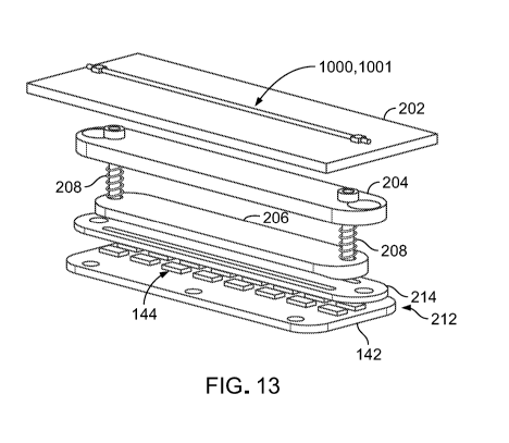
protocol. As shown in FIG. 13, each camera 180 is a component of a vision
system
200 located at each processing station 102 of the specimen processing system
100. In
addition to a camera 180, each vision system 200 further includes an optically
clear
CA 03152631 2022-02-24
WO 2021/040870
PCT/US2020/039306
plate 202 on which the specimen container 1000 can be supported, upper and
lower
lenses 204, 206 (e.g., plano-convex lenses), two adjustment screws 208 that
extend
between the upper and lower lenses 204, 206, two compression springs 210 that
respectively surround the adjustment screws 208, a light source 212 that
includes an
5 .. extension plate 142 of the PCB 138 and a matrix (e.g., one or more
arrays) of LEDs
144 distributed along the extension plate 142, and an opaque light stop 214
that
blocks centrally directed light rays (e.g., light rays directed substantially
towards a
central axis 1028 of the specimen container 1000) from impinging on the
specimen
container 1000. In some embodiments, a plate 202 of a vision system 200 may be
it) disposed within the a slot 132 of the platform 104. The camera 180 is
typically
located at a distance of about 1 cm to about 5 cm above the platform 104.
The upper and lower lenses 204, 206 are focusing lenses that can collimate
light radiating from the LEDs 144 into a light beam and focus the light beam
onto an
expected path of the specimen 1001 (e.g., generally along the central axis
1028 of the
15 specimen container 1000). Accordingly, the adjustment screws 108 and the
surrounding compression springs 210 allow a height adjustment of the upper and
lower lenses 204, 206 such that the focal point of the light beam coincides
with a
height of the support plate 202 on which the specimen container 1000 is held.
The
support plate 202 is typically positioned at a distance of about 0.1 cm to
about 1.5 cm
above the light source 212.
The light stop 214 blocks centrally directed light rays from the LEDs 144 such
that when the upper and lower lenses 204, 206 are focused correctly,
peripheral edges
(e.g., located off-axis) of the specimen 1001 are illuminated. Therefore, the
peripheral edges of the specimen 1001 appear brighter than an interior region
of the
specimen 1001 to make the specimen 1001 more visible to the camera 180 in a
manner similar to that of dark field illumination. Furthermore, the vision
system 200
may include a filtering functionality that blocks light with wavelengths of
less than
about 500 nm from reaching the specimen 1001, as exposure to such wavelengths
over the extended period of specimen tracking may be detrimental to the health
and
subsequent biological development of the specimen 1001. Accordingly, the
configuration and functionality of the various components of the vision system
200
for achieving dark field illumination advantageously allow for fine control
and
constraint of intensity, exposure time, and wavelength of light radiating from
the light
CA 03152631 2022-02-24
WO 2021/040870
PCT/US2020/039306
16
source 212 to the specimen 1001, which can be important to the survival of the
delicate biological specimen 1001.
The camera 180 can track a linear movement of the specimen 1001 throughout
a specimen processing protocol in real time by continuously generating images
of the
.. specimen 1001 and feeding the images at regular intervals or in the form of
a real-
time video feed wirelessly to a remote computing device running a software
algorithm
300 (refer to FIG. 19) that processes the images to track a position of the
specimen
1001 or through a wired connection via the electrical contacts 156 to one or
more
processors of the microcontroller 130 running the software algorithm 300.
Referring
to FIG. 14, the specimen container 1000 is allowed to sit in place (e.g.,
stationary) in
the receptacle 162 for a first predetermined exposure period during the
specimen
processing protocol so that the specimen 1001 can equilibrate in the
equilibration
solution 1020. The first exposure period may range from about 5 minutes to
about 15
minutes, depending on various parameters of typical ART protocols.
During the first exposure period, the equilibration solution 1020 draws water
molecules out from the specimen 1001 and infuses cryoprotectants into the
specimen
1001 according to osmotic potential. The reduction of water content and
addition of
cryoprotectants aids in minimizing damage to cellular components of the
specimen
1001 during freeze and warming cycles. Although the specimen 1001 is denser
than
the equilibration solution 1020 and will therefore very gradually descend
through the
equilibration solution 1020 due to gravitational forces over time, the
specimen 1001
will typically still be suspended within the equilibration solution 1020 and
will not
have yet reached the separation fluid 1024 by the end of the first exposure
period, as
shown in FIG. 14.
Referring to FIGS. 15-18, once the specimen 1001 has been exposed to the
equilibration solution 1020 for the predetermined exposure period, the
platform 104 is
activated to spin the specimen container 1000 at a select low speed to advance
the
equilibration solution 1020 and the specimen 1001 axially through the
separation fluid
1024 to the vitrification solution 1022. The specimen container 1000 is
typically spun
for about 0.5 minutes to about 5 minutes at an angular speed of about 50 rpm
to about
1200 rpm, which exerts enough centripetal force on the specimen 1001 to cause
the
specimen 1001 to descend into the vitrification solution 1022 in a timely
manner, but
not enough to cause mechanical damage to the specimen 1001. Such speed (e.g.,
corresponding to about 5 g to about 200 g) is significantly slower than speeds
of even
CA 03152631 2022-02-24
WO 2021/040870
PCT/US2020/039306
17
very low-speed conventional laboratory centrifuges, which are typically
capable of
revolving specimens about a centrifuge axis at speeds in a range of about 4000
rpm to
about 300,000 rpm (e.g., corresponding to about 2,500 g to about 65,000 g).
Referring particularly to FIG. 15, during an initial phase of spinning, the
.. specimen 1001 descends within the equilibration solution 1020 while the
equilibration
solution 1020, containing the specimen 1001, descends via bulk motion through
the
separation fluid 1024 (e.g., thereby displacing the separation fluid 1024)
toward the
vitrification solution 1022. Referring particularly to FIG. 16, during a
subsequent
phase of spinning, the equilibration solution 1020 reaches the vitrification
solution
1022, and the specimen 1001 passes from the equilibration solution 1020 into
the
vitrification solution 1022. Referring particularly to FIG. 17, during a next
phase of
spinning, the equilibration solution 1020 merges with the vitrification
solution 1022 to
form a combined vitrification solution 1030 (e.g., including the equilibration
solution
1020, the vitrification solution 1022, and a mixed solution interface layer
between the
equilibration solution 1020 and the vitrification solution 1022), and the
specimen
1001 continues to descend through the combined vitrification solution 1030.
Referring particularly to FIG. 18, during a final phase of spinning, the
specimen 1001 rests on a meniscus 1032 of the distal air pocket 1028 due to
surface
tension and thereby avoids contact with the relatively hard wall of the
elongate tube
.. 1002. For example, due to a balance between surface tension at the
interface of the
combined vitrification solution 1030 and the distal air pocket 1028, and
tension
between combined vitrification solution 1030 and an interior wall of the
tapered
portion 1016, the potential buoyancy force of the distal air pocket 1016 is
not
sufficient to break through meniscus 132. Therefore, the specimen 1001 cannot
penetrate the meniscus 1032.
With the specimen 1001 resting on the meniscus 1032 of the distal air pocket
1028 upon completion of spinning, the timer 126 is activated, and the specimen
container 1000 is allowed to sit in place (e.g., stationary) in the receptacle
162 for a
second predetermined exposure period for the specimen 1001 to be exposed to
the
combined vitrification solution 1030. The second exposure period may range
from
about 0.5 minutes to about 2 minutes, depending on various parameters of
typical
ART protocols. During the second exposure period, permeation of
cryoprotectants
within the combined vitrification solution 1030 into the specimen 1001
replaces water
within the specimen 1001, thereby dehydrating the specimen and further
infusing the
CA 03152631 2022-02-24
WO 2021/040870
PCT/US2020/039306
18
specimen 1001 with cryoprotectants. Such a stage-like progression of media
concentrations avoids an excessively high initial osmotic differential that
could
otherwise cause cells of the specimen 1001 to shrink too much and too rapidly
as the
water leaves the cells at a rate faster than the cryoprotectants can enter the
cells.
Owing to a preloaded state of the equilibration solution 1020 and the
vitrification solution 1022 within the specimen container 1000, a specimen
1001 can
be prepared for vitrification within a single, isolated environment (e.g., the
lumen of
the specimen container 1000) without being exposed to contamination,
mechanical
damage (e.g., from a micropipette or other specimen holding or fluid delivery
device),
or other accidental mishandling that may otherwise occur when a container that
houses a specimen is accessed multiple times to deliver and remove various
processing mediums or when a specimen is moved to various containers (e.g.,
petri
dishes, test tubes, or flask) during an ART process.
In some implementations, once the second exposure period has ended, the
specimen container 1000, containing the specimen 1001, is then manually
transferred
from the receptacle 162 to a long-term low temperature storage structure,
where the
specimen 1001 can be maintained in a cryogenic state for a period of up to
about 20
years. In some instances, the specimen container 1000 may be stored in the
long-term
low temperature storage structure for a much shorter period (e.g., as short as
few
hours).
The software algorithm 300 used to track the position of the specimen 1001
may be executed on the microcontroller 130 or on a separate, external
computing
device (e.g., a desktop computer, a laptop, a tablet, or a single board
computer)
running an operating system that is electrically coupled to the specimen
processing
system 100 via a data connection (e.g., a USB connection, an R5232 connection,
or a
wireless data connection). Referring to FIG. 19, the software algorithm 300
enters a
process flow loop in which the software acquires a single color image from the
camera feed (302), converts the image to greyscale, and stores the greyscale
image in
an array that holds a grey value for each pixel of the greyscale image (304).
The
algorithm 300 then performs an edge detection routine on the greyscale image
to
detect edges (e.g., an outline) of the specimen container 1000 and therefore
define a
size and a position of an area of interest with respect to a field of view of
the camera
180 in which the position of the specimen 1001 will be tracked (306).
CA 03152631 2022-02-24
WO 2021/040870
PCT/US2020/039306
19
The algorithm 300 then captures a first subsequent color image from the
camera feed (308), waits for a period of time (310), and then captures a
second
subsequent color image from the camera feed (312). As the first and second
subsequent color images are captured, the images are cropped to the area of
interest.
The algorithm 300 also converts the first and second subsequent color images
to
greyscale, and stores the first and second greyscale images in an array that
holds a
grey value for each pixel of the greyscale images (314). The algorithm 300
then
compares the first and second greyscale images to each other and generates an
additional array that stores the pixilation differences between the first and
second
grayscale images as a difference image (316). The algorithm 300 converts
luminosity
data from the difference image to a binary value based on an upper constraint
and a
lower constraint to generate a first binary threshold difference image (318).
For
example, all image data that falls between the upper and lower constraints is
maintained in the first binary threshold difference image, whereas all image
data that
falls outside of the range defined between the upper and lower constraints is
discarded.
The algorithm 300 then blurs the first binary threshold difference image to
remove noise and thereby generates a first blur difference image (320). The
algorithm
300 again converts luminosity data from the first blur difference image to a
binary
value based on an upper constraint and a lower constraint to generate a second
binary
threshold difference image with even less noise as compared to the first
binary
threshold difference image (322). In this case, the binary value, upper
constraint, and
lower constraint are independent of those used to generate the first binary
threshold
difference image. The algorithm 300 also blurs the second binary threshold
difference image to further remove noise and thereby generate a second blur
difference image (324).
The algorithm then passes the second binary threshold difference image to an
object detection routine (326) in which a specimen 1001 may be identified in
the
image. If a specimen 1001 is not identified in the image (328), then the
algorithm 300
returns to the step of capturing a first subsequent color image from the
camera feed
(308). If a specimen 1001 is identified in the image (328), then the algorithm
300
categorizes (e.g., determines) a location of the specimen 1001 and stores the
location
in an array of object positions (330). Using a predetermined maximum and
minimum
threshold, the algorithm 300 identifies the specimen 1001 based on the number
of
CA 03152631 2022-02-24
WO 2021/040870
PCT/US2020/039306
pixels (e.g., for a known camera resolution), which represents a generally
circular area
of the specimen 1001 under a known magnification within the array (332). The
algorithm 300 stores a center position, a speed (based on a time elapsed
between the
previously processed image and positions of the specimen 1001 in the current
and
5 previously processed image), and a direction of the specimen 1001 in
another array
(334). For example, the maximum and minimum thresholds provide maximum and
minimum limits count of at least partially contiguous pixels forming a
generally
circular area that represents approximate geometry limits of the specimen
1001.
Records of speed and position of the center position are tracked to verify the
motion
10 of at least one specimen 1001.
The algorithm 300 outputs the center position, speed, and direction data of
the
specimen 1001 for further processing (336). For example, in some embodiments,
the
algorithm 300 outputs the data to the display screen 110 for viewing by an
operator
(338) and to a component of the microcontroller 130. In some embodiments, the
15 algorithm 300 additionally outputs the data to a component of the
external computing
device via the data connection. If the algorithm 300 has finished tracking the
specimen 1001 (340), then the algorithm 300 exits the process flow loop. If
the
algorithm 300 has yet to finish tracking the specimen 1001 (340), then the
algorithm
300 returns to capture a first subsequent color image from the camera feed
(308).
20 Using the information from the algorithm 300, the microcontroller 130
can
control the rotational speed, spin direction, and acceleration of the platform
104 via
communication with the motor assembly 140 to ensure that the specimen 1001 is
exposed to a substantially constant centripetal force as programmed by the
user,
irrespective of an axial position of the specimen 1001 within the specimen
container
1000 (e.g., a radial position of the specimen 1001 along the platform 104).
For
example, according to one or more signals transmitted by the microcontroller
130, the
platform 104 can spin about the central axis 152 to exert enough centripetal
force on
the specimen 1001 to cause the specimen 1001 to move along the central axis
1028 of
the specimen container 1000 toward the distal closure 1006 according to a
specified
protocol. The one or more signals can be used to adjust an angular speed of
the
platform 104 and/or a duration of one or more phases of the protocol. Such
protocol
adjustments can optimize time periods of specimen exposure to the processing
media
within the specimen container 1000.
CA 03152631 2022-02-24
WO 2021/040870
PCT/US2020/039306
21
In some embodiments, a specimen container that is otherwise similar to the
specimen container 1000 may itself include one or more embedded optical
elements
(e.g., one or more lenses) that enable a specimen 1001 to be more clearly seen
by the
naked eye or visualized by the camera 180 of the vision system 200 during or
separate
from an automated specimen tracking routine carried out at the specimen
processing
system 100.
In some embodiments, the specimen processing system 100 is further
equipped with one or more vibration assemblies designed to excite movement of
either or both of a specimen 1001 or fluids within a specimen container 1000
while
the specimen container 1000 is processed at the specimen processing system
100. For
example, FIG. 20 illustrates such a vibration assembly 400 that is designed to
securely
support a specimen container 1000. One or more vibration assemblies 400 can be
respectively installed to one or more of the processing stations 102 of the
specimen
processing system 100 in place of a respective upper bracket 160 of a
processing
station 102.
The vibration assembly 400 includes a base 402 that can be secured to the
platform 104 at a processing station 102 and to which the other components of
the
vibration assembly 400 are mounted. The vibration assembly 400 further
includes a
mounting platform 404 that is formed to support a specimen container 1000 and
that
is movable (e.g., suspended in free space) with respect to the base 402. For
example,
the vibration assembly 400 further includes two frames 406 along which the
mounting
platform 404 can move laterally and longitudinally, two dynamic spacers 408
(e.g.,
springs or other members made of compliant materials) that limit excessive
outward
movement due to centripetal force during spinning, and an adjustable stop 410
that
permits some free movement against the dynamic spacers 408 without the need to
rigidly attach the mounting platform 404 to the base 402. At least a central
portion of
the mounting platform 404 is made of an optically transparent material to
allow
focused light from the vision system 200 to pass through and illuminate the
specimen
1001 within the specimen container 1000. The vibration assembly 400 also
includes
opposed restraining clamps 412 that can clamp the specimen container 1000 to
the
mounting platform 404.
The vibration assembly 400 further includes a motor 414 that vibrates the
mounting platform 404 along an x axis and a motor 416 that vibrates the
mounting
platform 404 along ay axis. The motors 414, 416 may be activated via
electrical
CA 03152631 2022-02-24
WO 2021/040870
PCT/US2020/039306
22
signals received from the electrical contacts 156 within the housing 106. The
motors
414, 416 may be activated simultaneously or at different times to achieve a
desired
movement direction. A drive voltage of the motors 414, 416 may also be
adjusted to
change vibration frequencies of the motors 414, 416.
In some embodiments, a specimen processing system that is similar in
construction and function to the specimen processing system 100 may be further
equipped with features for cutting and subsequently sealing a specimen
container
1000. For example, in some examples, there may be a need to cut excess length
from
the specimen container 1000 after the specimen 1001 has been processed and is
disposed at a distal end of the specimen container 1000. FIG. 21 illustrates a
cut-and-
seal station 502 of a specimen processing system 500 at which a specimen
container
1000 can be simultaneously cut and sealed (e.g., via a heat seal, an
ultrasonic seal, or
a crimp) prior to a distal storage portion 503 of the specimen container 1000
being
placed in a low temperature substance 501. In some embodiments, a specimen
container 1000 may be cut and sealed in two separate operations. For example,
FIG.
22 illustrates a cutting station 602 of a specimen processing system 600 at
which a
specimen container 1000 can first be cut and then be automatically sealed,
capped, or
plugged using dedicated equipment that is part of the specimen processing
system 600
prior to a distal storage portion 603 of the specimen container 1000 being
placed in a
low temperature substance 601.
While the above-discussed specimen processing system 100, specimen
processing system 500, specimen processing system 600, specimen container
1000,
vision system 200, and vibration assembly 400 have been described and
illustrated as
including components with certain dimensions, sizes, shapes, materials, and
configurations, and with respect to the software algorithm 300, in some
embodiments,
specimen processing systems, specimen containers, vision systems, vibration
assemblies, and software algorithms that are otherwise substantially similar
in
structure and function to the above-discussed embodiments may include one or
more
components with different dimensions, sizes, shapes, materials, and
configurations or
one or more different process flow steps.
For example, while the specimen processing system 100, the vision system
200, and the algorithm 300 have been described and illustrated with respect to
tracking one specimen 1001 within a specimen container 1000, in some
embodiments,
a specimen processing system that is substantially similar in construction and
function
CA 03152631 2022-02-24
WO 2021/040870
PCT/US2020/039306
23
to the specimen processing system 100 may be operated with an algorithm that
is
designed to track more than one specimen 1001 within the same specimen
container
1000 during a specimen processing protocol.
Accordingly, other embodiments are within the scope of the following claims.