Note: Descriptions are shown in the official language in which they were submitted.
CA 03152793 2022-02-25
WO 2021/046339
PCT/US2020/049387
NONINVASIVE METHOD TO QUANTIFY KIDNEY FUNCTION AND
FUNCTIONAL DECLINE
CROSS-REFERENCES TO RELATED APPLICATIONS
[1] The present application claims priority to U.S. Provisional Patent
Application No.
62/896,296, filed on September 5, 2019, which application is incorporated
herein by
reference in its entirety for all purposes.
BACKGROUND
[2] Fourteen percent of the United States population is afflicted with
chronic kidney
disease (CKD). Approximately 661,000 Americans have renal failure, of which,
approximately 468,000 are on dialysis. The high incidence of CKD contributes
to health care
costs of approximately S80,000 US dollars, per patient, per year. Further,
approximately
193,000 Americans have a functioning kidney transplant and 30,000 patients
receive new
kidney transplants every year ¨ incurring health care costs of approximately
S100,000 ¨
S200,000 US dollars yearly for each transplant, and S20,000 US dollars, per
patient, per year
1
CA 03152793 2022-02-25
WO 2021/046339
PCT/US2020/049387
for immunosuppression. In 2015, Medicare alone spent 64 Billion USD for
treatment of
chronic kidney disease alone (11% of total covered patients).
1131 Accurate assessment of renal function is imperative to allow for
proper tracking of
kidney function or injury. In the case of renal transplantation, renal
dysfunction can be
associated with 15% ¨ 30% of cases that require treatment with augmented
immunosuppression. Usually, 130,000 transplants are rejected after the first
10 years of graft
transplantation, making it very important to have accurate and non-invasive
tests for kidney
function or kidney injury. Nonetheless, the current standard of care does not
allow for proper
distinction between various underlying causes of either kidney function or
injury, such as
acute insult/recovery, chronic damage/ progression of injury, time assessment
for renal
replacement therapy, or assessment of dialysis/transplantation needs.
[4] Consider for instance the severe limitations of a widely used test for
measuring
renal function, namely the serum creatinine (SCr) blood test. First, the SCr
test is a blood test,
and the requirement for a blood draw limits its utility in a non-invasive
manner. Second,
serum creatinine is a late marker of advanced kidney injury and is not
specific for the
diagnosis of acute rejection (AR). In fact, SCr blood test is confounded by
multiple variables
as serum creatinine can rise with multiple causes unrelated to kidney
function, such as
volume depletion, infection, and obstruction. Furthermore, SCr measurements
are further
confounded by variables such as gender, hydration status, diet and muscle
mass. As a result,
while the cost of a SCr test is low, the lack of specificity of the SCr test
and the influence of
confounding variables limit the clinical utility of the test.
1151 Additionally, the current standard of care for measuring kidney
function ¨ the
glomerular filtration rate (GFR; also referred to as estimated glomerular
filtration rate eGFR)
¨ is perhaps even more limited than the SCr test. In chronic kidney disease,
the kidneys lose
their ability to effectively filter waste products in the blood because of
damage to the
glomeruli of nephrons. The standard glomerular filtration rate tests evaluate
some level of
kidney function by measuring the volume of plasma that the kidneys filter
through the
glomeruli per unit time. However, there are numerous limitations with the
current eGFR tests.
First, eGFR requires a blood sample and thus cannot readily be used in a non-
invasive
setting. Second, eGFR measurements are limited by demographic data collection.
eGFR tests
provide a measure of how well the kidneys are removing wastes and excess fluid
from the
blood by inputting a detected serum creatinine level in an equation, along
with parameters for
age and gender adjustments, and in some instances additional adjustments for
those of
African American descent. However, there is a lack of a consensus about what
formula
2
CA 03152793 2022-02-25
WO 2021/046339
PCT/US2020/049387
should be used to estimate glomerular function, where some prefer the
Modification of Diet
in Renal Disease (MDRD) equation and others the Cockcroft-Gault (CG) equation,
as some
believe the MDRD equation significantly underestimates the measured GFR when
compared
with the CG formula. Recently, calculation of the eGFR is done by the chronic
kidney
disease epidemiology collaboration formula (CKD-EPI).
[6] Lastly, more sensitive measurements of eGFR are available through the
use of
inulin, a molecule that is not endogenous in humans. When inulin is used to
measure eGFR, a
specified mass comprising inulin is injected into a person's bloodstream and
the amount of
inulin cleared through the urine is indicative of the amount of plasma
filtered by the body's
glomeruli. Unfortunately, inulin eGFR not only requires a blood draw, but it
also typically
requires a patient to stay in an outpatient setting, further limiting its
utility. Moreover, the
inulin GFR test is quite costly relative to SCr or non-inulin eGFR tests,
making this test
something that is rarely used in an actual clinical setting.
171 There currently do not exist any urine-based methods for eGFR
prediction or
estimation of kidney function. Current methods for kidney function assessment
largely
consist of semi-quantitative measures of leukocytes, nitrite, urobilinogen,
protein, pH, blood,
specific gravity, ketone, bilirubin, and glucose. These tests merely identify
the presence of
late-stage kidney disease and functional decline and do not provide a
quantitative estimate of
kidney function.
BRIEF SUMMARY
[8] Provided herein are methods for determining kidney function that
quantify the
presence of biomarkers in urine and transform the input of these biomarkers to
estimate the
glomerular filtration rate (GFR) of a subject.
191 In one aspect, provided are methods for determining kidney function in
a subject,
the method comprising: contacting a urine sample from the subject with a
coupling agent;
detecting the amount of asymmetric dimethylarginine (ADMA), or symmetrical
dimethylarginine (SDMA), or both ADMA and SDMA in the sample; and determining
kidney function in the subject based on the amount of ADMA, SDMA, or both ADMA
and
SDMA in the sample. In some aspects, the detection of ADMA and SDMA is
combined with
the detection of other markers in a lateral flow assay (LFA) test.
[10] In some embodiments, the coupling agent is selected from N-
hydroxysuccinimido
carbonic acid; (2,5-dioxopyrrolidin-1-y1) hydrogen carbonate (also known as
succinimidocarbonate); N,N'-Disuccinimidyl carbonate; carbonic acid
(chloromethyl ester)
(N-hydroxysuccinimide ester); or (2,5-dioxopyrrolidin-1-y1) prop-2-enyl
carbonate.
3
CA 03152793 2022-02-25
WO 2021/046339
PCT/US2020/049387
11111 In some aspects provided herein is method comprising: (a) contacting
a urine
sample with an antibody that specifically binds to asymmetric dimethylarginine
(ADMA);
and (b) detecting an amount of the antibody that is in a bound state; (c)
determining an
amount of ADMA from the urine sample based on the amount of the antibody that
is in the
bound state; and (c) either contacting the urine sample with a probe to
determine an amount
of a urinary biomarker that is indicative of the subject's hydration level; or
determining a
urine specific gravity of the urine sample. In some cases, the antibody that
specifically binds
ADMA has a reactivity for symmetric dimethylarginine (SDMA) that is less than
25%, less
than 10%, less than 5%, or less than 1% of its reactivity for ADMA. In some
cases, the
method further comprises contacting the urine sample with the probe to
determine an amount
of the urinary biomarker that is indicative of the subject's hydration level,
and the urinary
biomarker that is indicative of the subject's hydration level may be urine
SDMA or urine
creatinine. In some embodiments, the probe that is specific to SDMA is an
antibody that has a
reactivity for ADMA that is less than 25%, less than 10%, less than 5%, or
less than 1% of its
reactivity for SDMA. In some cases, the method further comprises determining
the urine
specific gravity of the urine sample. In some aspects, the method further
comprises
determining the specific gravity of the urine sample. In some aspects, the
method further
comprises contacting the urine sample with a reagent that reacts with free
ADMA to form an
ADMA conjugate prior to contacting the urine sample with the antibody that
specifically
binds to ADMA. ADMA may be bound to the antibody as either free ADMA or the
conjugate that results after the aforementioned coupling. The reagents may be
selected from
N-hydrosuccinimido carbonic acid; (2,5-dioxopyrrolidin-lyl)hydrogen carbonate
(also
known as succinimidocarbonate); N,N'-disuccinimidyl carbonate; carbonic acid
(choloromethyl ester) (N-hydroxysuccinimide ester); or (2,5-dioxopyrrolidin-1-
yl)prop-2-
enyl carbonate. In some instances, the amount of ADMA is determined via an
enzyme-linked
immunosorbent assay (ELISA), such as a competitive ELISA. In some instances,
the amount
of ADMA is determined via a lateral flow assay. In some instances, the urine
sample is a
diluted urine sample. In some instances, the method further comprises
detecting an amount of
at least one, at least two, at least three, at least four, or at least five
biomarkers in the urine
sample, wherein the biomarkers are selected from creatinine, total protein, 5-
methylcytosine,
cell-free DNA, methylated cell-free DNA, CXCL10, and clusterin. In some cases
the subject
is a mammal, such as human, a domesticated cat or a dog. In some instances,
the method
further comprises administering a treatment to the subject, including, but not
limited to
administering a diabetic kidney disease-targeted drug, a SGLT-2 receptor
inhibitor, a SIRT1
4
CA 03152793 2022-02-25
WO 2021/046339
PCT/US2020/049387
agonist, or a bromodomain inhibitor to the subject if the subject is diagnosed
with diabetic
kidney disease. In other cases the treatment comprises administering a steroid
therapy to the
subject if the subject is diagnosed with IgA/Non-IgA mesangial proliferative
glomerulonephritis or membrano-proliferative glomerulonephritis. In some
instances, the
treatment comprises dialysis.
[12] In some aspects provided herein is a method for determining kidney
function of a
subject from a urine sample, the method comprising: detecting an amount of
asymmetric
dimethylarginine (ADMA) from a urine sample of a subject; assaying the urine
sample to
determine a hydration status of the subject; and generating a value indicative
of the kidney
function of the subject based on the amount of ADMA from the urine sample and
the
hydration status of the subject; determining the kidney function of subject
based on the value.
In some instances, generating a value indicative of the kidney function of the
subject
comprises inputting the amount of ADMA and the hydration status of the subject
into an
algorithm to produce the value. In such instances, the algorithm may be
implemented via a
computer system. In some instances, determining the kidney function of the
subject
comprises comparing the value to a threshold and determining the kidney
function of the
subject based on the comparison. In some instances, generating the value
indicative of the
kidney function of the subject comprises inputting a determined amount of a
hydration
marker from the urine sample into the algorithm to produce the value, the
hydration marker
can be creatinine, SDMA, or both. In some instances, generating the value
indicative of the
kidney function of the subject comprises inputting a specific gravity of the
urine sample into
the algorithm to produce the value, inputting an amount of total protein from
the sample into
the algorithm, inputting an age of the subject into the algorithm, or
inputting a gender of the
subject into the algorithm. In some instances, a race of the subject is not
input into the
algorithm. In some cases, the amount of urine ADMA from the urine sample of
the subject
positively correlates with glomerular filtration rate (GFR). In some
instances, the value
indicative of the kidney function of the subject is an estimated GFR. In some
instances, the
hydration status of the subject is an amount of a urinary marker that is
indicative of a
hydration level in the subject, and the hydration mark can be urine SDMA or
urine creatinine.
In some instances, assaying the urine sample to determine the hydration status
of the subject
comprises determining a urine specific gravity of the urine sample. In some
cases, the
hydration status of the subject is represented by the specific gravity of the
urine sample. In
some cases, the method further comprises coupling a reagent to ADMA prior to
detecting the
amount of ADMA from the urine sample, and the reagent can be selected from N-
CA 03152793 2022-02-25
WO 2021/046339
PCT/US2020/049387
hydrosuccinimido carbonic acid; (2,5-dioxopyrrolidin-lyl)hydrogen carbonate
(also known
as succinimidocarbonate); N,N'-disuccinimidyl carbonate; carbonic acid
(choloromethyl
ester) (N-hydroxysuccinimide ester); or (2,5-dioxopyrrolidin-1-yl)prop-2-enyl
carbonate. In
some cases, the step of detecting the amount of ADMA from the urine sample of
the subject
comprises: contacting the urine sample with an antibody that specifically
binds to ADMA;
and detecting an amount of the antibody that is in a bound state. In some
aspects, the
antibody that specifically binds ADMA has a reactivity for symmetric
dimethylarginine
(SDMA) that is less than 25%, less than 10%, less than 5%, or less than 1% of
its reactivity
for ADMA. In some aspects, the subject is identified as having impaired kidney
function
when the ADMA is in the urine sample at a concentration of less than 19.4 M.
In some
instances, the subject is identified as having impaired kidney function when
the
ADMA/creatinine ratio or ADMA/SDMA ratio is less than 0.3 uM/mg/dL or 0.7
uM/mg/dL,
respectively. In some aspects, the method further comprises (1) identifying
the subject as
having impaired kidney function and (2) administering a treatment to the
subject based on the
identified impairment of kidney function.
[13] In some aspects, provided herein is a method for detecting kidney
injury in a
subject, the method comprising: determining the kidney function of the subject
according to
the methods described above; and detecting amounts of two or more biomarkers
in the urine
sample of the subject, wherein the two or more biomarkers are selected from
the group
consisting of creatinine, total protein, 5-methyclytosine, cell-free DNA,
methylated cell-free
DNA, CXCL10, and clusterin. In some aspects, the method further comprises
administering a
treatment to the subject if the subject has decreased kidney function
indicative of kidney
disease or kidney injury. The treatment may comprise administering a diabetic
kidney
disease-targeted drug, a SGLT-2 receptor inhibitor, a SIRT1 agonist, or a
bromodomain
inhibitor to the subject if the subject is diagnosed with diabetic kidney
disease, a steroid
therapy to the subject if the subject is diagnosed with IgA/Non-IgA mesangial
proliferative
glomerulonephritis or membrano-proliferative glomerulonephritis. The IGA
nephropathy
may be identified with a sensitivity of at least 95% and a specificity of at
least 98%. In some
instances, the treatment comprises dialysis.
[14] In another aspect, a kit is provided, the kit comprising: (i) reagents
for detecting
ADMA, SDMA, or both ADMA and SDMA in a urine sample; and (ii) a coupling
agent. In
some embodiments, the coupling agent is selected from N-hydroxysuccinimido
carbonic
acid; (2,5-dioxopyrrolidin-1-y1) hydrogen carbonate (also known as
succinimidocarbonate);
6
CA 03152793 2022-02-25
WO 2021/046339
PCT/US2020/049387
N,N'-Disuccinimidyl carbonate; carbonic acid (chloromethyl ester) (N-
hydroxysuccinimide
ester); or (2,5-dioxopyrrolidin-l-y1) prop-2-enyl carbonate.
[15] In some aspects, the disclosure provides a kit for use in detecting
kidney function in
a subject, the kit comprising: an antibody for detecting ADMA; a reagent for
covalent
conjugation to ADMA; and a reagent for assessing hydration status of the
subject. In some
aspects, the kit further comprises a detection reagent for detecting total
urinary protein. In
some aspects, the antibody for detecting ADMA has a reactivity for symmetric
dimethylarginine (SDMA) that is less than 25%, less than 10%, less than 5%, or
less than 1%
of its reactivity for ADMA. In some aspects, the reagent for covalent
conjugation to ADMA
is selected from N-hydrosuccinimido carbonic acid; (2,5-dioxopyrrolidin-
lyl)hydrogen
carbonate (also known as succinimidocarbonate); N,N'-disuccinimidyl carbonate;
carbonic
acid (choloromethyl ester) (N-hydroxysuccinimide ester); or (2,5-
dioxopyrrolidin-1-yl)prop-
2-enyl carbonate. In other cases, the kit further comprises a reagent for
binding to cell-free
DNA, a reagent for binding to CXCL10, a reagent for binding to creatinine, a
reagent for
binding to 5-methyclytosine, a reagent for binding to methylated cell-free
DNA, a reagent for
binding to clusterin, or a combination of two of more of the aforementioned
reagents. In
some instances, the kit comprises a receptacle for containing a urine sample
and a lateral flow
device.
[16] In some aspects, the disclosure provides a reaction mixture
comprising: a urine
sample of a subject, a reagent for covalent conjugation to ADMA, and an
antibody to
ADMA. In some instances, the reagent for covalent conjugation to ADMA is
selected from
N-hydrosuccinimido carbonic acid; (2,5-dioxopyrrolidin-lyl)hydrogen carbonate
(also
known as succinimidocarbonate); N,N'-disuccinimidyl carbonate; carbonic acid
(choloromethyl ester) (N-hydroxysuccinimide ester); or (2,5-dioxopyrrolidin-1-
yl)prop-2-
enyl carbonate. In some instances, the antibody to ADMA has a reactivity for
symmetric
dimethylarginine (SDMA) that is less than 25%, less than 10%, less than 5%, or
less than 1%
of its reactivity for ADMA. In some cases the subject is a mammal, such as a
human, a
domesticated cat or a dog. The coupling agent may be selected from N-
hydroxysuccinimido
carbonic acid; (2,5-dioxopyrrolidin-1-y1) hydrogen carbonate (also known as
succinimidocarbonate); N,N'-Disuccinimidyl carbonate; carbonic acid
(chloromethyl ester)
(N-hydroxysuccinimide ester); or (2,5-dioxopyrrolidin-1-y1) prop-2-enyl
carbonate.
[17] In another aspect, provided is a reaction mixture comprising a urine
sample, a
coupling agent, and antibodies that specifically bind ADMA, SDMA, or both ADMA
and
SDMA. In some embodiments, the coupling agent is selected from N-
hydroxysuccinimido
7
CA 03152793 2022-02-25
WO 2021/046339
PCT/US2020/049387
carbonic acid; (2,5-dioxopyrrolidin-l-y1) hydrogen carbonate (also known as
succinimidocarbonate); N,N'-Disuccinimidyl carbonate; carbonic acid
(chloromethyl ester)
(N-hydroxysuccinimide ester); or (2,5-dioxopyrrolidin-1-y1) prop-2-enyl
carbonate.
[18] In another aspect, a method of treating a disease associated with
decreased kidney
function in a subject is described, the method comprising the steps of: (a)
selecting a subject
having decreased kidney function as determined by (i) an ADMA concentration
less than
19.421 uM; or (ii) an ADMA/creatinine ratio less than 0.312 (uM/mg/dL); or
(iii) an
ADMA/SDMA ratio less than 0.694; or (iv) an eGFR less than 90 mL/min per 1.73
m2; or (v)
a KITGFR less than 90 mL/min per 1.73 m2; and (b) administering a treatment to
the subject.
BRIEF DESCRIPTION OF THE DRAWINGS
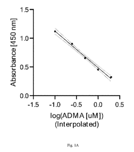
[19] Fig. 1A shows an example fit for ADMA using the analysis methods
described
herein.
[20] Fig. 1B shows an example fit for SDMA using the analysis methods
described
herein.
[21] Fig. 2A shows that the kidney health of an individual can be
determined by
comparing the quantity of ADMA in the urine sample to a cutoff value
indicative of kidney
injury status, which may be a pre-determined clinical threshold or relative to
a patient's
baseline ADMA value.
[22] Fig. 2B shows that the kidney health of an individual can be
determined by
comparing the ratio of the ADMA/SDMA in the urine sample to a cutoff value
indicative of
kidney injury status, which may be a pre- determined clinical threshold or
relative to a
patient's baseline ADMA/SDMA ratio value.
[23] Fig. 2C shows that the ratio of ADMA/SDMA in the urine sample can be
used to
calculate an approximation of a known clinical parameter, the estimated
glomerular filtration
rate (eGFR).
[24] Fig. 2D shows that a representative functional score can be used to
determine CKD
in a subject.
[25] Fig. 2E shows that a representative multiple linear regression can be
used to
determine kidney function in a patient. The data in Figs. 1A-2E are from a
dataset of 80
urine samples.
[26] Fig. 3 shows data from a larger data set of 300 urine samples. Based
on these data.
KITFunction (also referred to as KITGFR) was calculated using a formula that
incorporates
SDMA, ADMA, urine creatinine, urine protein and age of patient. The upper row
of panels
8
CA 03152793 2022-02-25
WO 2021/046339
PCT/US2020/049387
shows eGFR plotted against ADMA concentration, the ratio of ADMA/SDMA, and the
ratio
of ADMA to creatinine. The lower row of panels shows eGFR plotted against
KITFunction,
CKD stage plotted against KITFunet,on, specificity and sensitivity of the
assay for KITFunction
and proteinuria, and mean of actual eGFR and KITFunction versus actual eGFR
minus
KITFunction=
[27] Fig. 4 shows a representative study design and patient disposition.
(Left) In the
original trial, 34 patients met inclusion criteria and were randomized into
rituximab and
standard of care treatment groups. At least one urine sample was available
from 28 of the 34
patients, with 14 having urine samples at all three designated time-points.
(Right) Pictorial
depiction of patients, treatment, and sample availability. Patients were
segregated based on
treatment, either with standard of care (turquoise) or rituximab (coral), with
individual
patients as rows. A yellow square indicates a urine sample was available at
the indicated
time-point, while gray indicates that no urine sample was available.
[28] Figs. 5A and 5B show that the urinary KIT biomarkers could segregate
healthy
controls from those with IgA nephropathy. Fig. 5A. An IgA Risk Score ranging
from 0 to
100 segregated healthy control patients from those with IgA nephropathy. Urine
samples
were collected from healthy controls (n = 64) who had no evidence of kidney
disease or
injury as assessed by both absence of proteinuria and eGFR greater than 120
mL/min per 1.73
m2. All urine samples from IgA patients (n = 67) were used, as none of these
patients had
remission of IgA during the treatment duration. Fig. 5B. Receiver-operator
characteristic
(ROC) curves of the IgA Risk Score with AUC of 0.994 (P < 0.0001) and
proteinuria. For the
IgA Risk Score, at a threshold of 57.4, the sensitivity and specificity were
95.5% and 98.4%
respectively. **** P < 0.0001.
[29] Fig. 6 shows a representative example of biomarker modeling of disease
progression status after one year of treatment. Modeling was performed on
endpoint,
midpoint, and baseline biomarker data. The y-axis shows the probability of
progression as
determined by a nominal logistic regression model.
[30] Fig. 7 is a schematic illustrating that SDMA is renally cleared
regardless of the
degree of kidney functional impairment, in contrast ADMA is degraded.
[31] Fig. 8 shows that urinary ADMA was inversely correlated with blood
SDMA in
canine samples, suggesting its utility in noninvasively determining kidney
function.
[32] Fig. 9 shows a linear relationship between the ratio of urinary
ADMA/SDMA and
blood SDMA, suggesting that SDMA may be used as a normalization factor.
9
CA 03152793 2022-02-25
WO 2021/046339
PCT/US2020/049387
[33] Fig. 10 shows a linear relationship between the ratio of urinary
ADMA/creatinine
and blood SDMA, suggesting that creatine may be used as a normalization
factor.
[34] Fig. 11 shows a linear relationship between SDMA and creatinine
measurements of
feline and canine kidney function.
[35] Fig. 12 is a graph illustrating the development of a one-biomarker
formula only
considering urinary ADMA and predicting blood SDMA.
[36] Fig. 13 is a graph illustrating the development of a formula for
kidney function
considering urinary SDMA, ADMA, and creatine together.
[37] Fig. 14 illustrates results obtained for the detection of SDMA and
ADMA in
mammalian urine samples with the protocols described herein.
[38] Fig. 15 illustrates a comparison of the performance of the method
described herein
in urine sample versus serum samples in five different stages of chronic
kidney disease.
DEFINITIONS
[39] The terms "a," "an," or "the" as used herein not only include aspects
with one
member, but also include aspects with more than one member. For instance, the
singular
forms "a," "an," and "the" include plural referents unless the context clearly
dictates
otherwise. Thus, for example, reference to "a cell" includes a plurality of
such cells and
reference to "the agent" includes reference to one or more agents known to
those skilled in
the art, and so forth.
[40] The terms "subject", "patient" or "individual" are used herein
interchangeably to
refer to a human or animal. For example, the animal subject may be a mammal, a
primate
(e.g., a monkey), a livestock animal (e.g., a horse, a cow, a sheep, a pig, or
a goat), a
companion animal (e.g., a dog, a cat), a laboratory test animal (e.g., a
mouse, a rat, a guinea
pig, a bird), an animal of veterinary significance, or an animal of economic
significance.
[41] The term "biofluid" or "biofluidic sample" refers to a fluidic
composition that is
obtained or derived from an individual that is to be characterized and/or
identified, for
example based on physical, biochemical, chemical and/or physiological
characteristics. Non-
limiting examples of biofluid include blood, serum, plasma, saliva, phlegm,
gastric juices,
semen, tears, and sweat. In one embodiment the biofluid is urine.
[42] As used herein, the term "AUC" refers to "area under the curve" or C-
statistic,
which is examined within the scope of ROC (receiver-operating characteristic)
curve
analysis. AUC is an indicator that allows representation of the sensitivity
and specificity of a
test, assay, or method over the entire range of test (or assay) cut points
with just a single
CA 03152793 2022-02-25
WO 2021/046339
PCT/US2020/049387
value. An AUC of an assay is determined from a diagram in which the
sensitivity of the
assay on the ordinate is plotted against 1-specificity on the abscissa. A
higher AUC indicates
a higher accuracy of the test; an AUC value of 1 means that all samples have
been assigned
correctly (specificity and sensitivity of 1), an AUC value of 50% means that
the samples have
been assigned with guesswork probability and the parameter thus has no
significance.
[43] Using AUCs through the ROC curve analysis to evaluate the accuracy of
a
diagnostic or prognostic test are well known in the art, for example, as
described in, Pepe et
al., "Limitations of the Odds Ratio in Gauging the Performance of a
Diagnostic, Prognostic,
or Screening Marker," Am. J. Epidemiol 2004, 159 (9): 882-890, and "ROC Curve
Analysis:
An Example Showing The Relationships Among Serum Lipid And Apolipoprotein
levels In
Identifying Subjects With Coronary Artery Disease," Clin. Chem., 1992, 38(8):
1425-1428.
See also, CLSI Document EP24-A2: Assessment of the Diagnostic Accuracy of
Laboratory
Tests Using Receiver Operating Characteristic Curves; Approved Guideline -
Second
Edition. Clinical and Laboratory Standards Institute; 2011; CLSI Document
I/LA21-A2:
Clinical Evaluation of Immunoassays; Approved Guideline - Second Edition.
Clinical and
Laboratory Standards Institute; 2008.
[44] As used herein, the term "diagnose" means assigning symptoms or
phenomena to a
disease or injury. For the purpose of this invention, diagnosis means
determining the
presence of organ injury in a subject.
[45] As used herein, the term "predict" refers to predicting as to whether
organ injury is
likely to develop in a subject.
[46] As used herein, the terms "glomerular filtration rate" ("GFR"),
"estimated
glomerular filtration rate" ("eGFR"), and "actual glomerular filtration rate"
("actual GFR"),
refer to a measure of kidney function that uses a person's age, gender, and
blood creatinine
level.
[47] As used herein, the terms "KITFunction", "KITGFR", is a measurement of
kidney
function that incorporates measurements of SDMA, ADMA, urine creatinine, urine
protein,
and age of patient as inputs into a suitable formula, such as the formula
below.
11
CA 03152793 2022-02-25
WO 2021/046339
PCT/US2020/049387
277 + 140 x ADMA
eGFR = SDMA + ________________________ + 83 .321
GenderF + SDMA
* min ( greaterorequal(37 x ADMA, Protein), SDMA2
min(0.288, Citrate))
X __________________________________
Age x ADMA
¨ min(Protein, min(48.065 + ADMA,Creatinine))
Alternative suitable formulas are further described in the specifications.
[48] As used herein, a urinary biomarker that is indicative of the
subject's hydration
level, or a "hydration marker", or "a marker of hydration status", refers to
creatinine and
SDMA, either used jointly or individually.
[49] As used herein, the abbreviation SDMA refers to symmetric
dimethylarginine.
[50] As used herein, the abbreviation ADMA refers to asymmetric
dimethylarginine.
[51] As used herein, the abbreviation "KIT biomarkers" refers to a
composite of six
biomarkers, namely cell-free DNA (cfDNA), methylated cfDNA, clusterin,
creatinine,
protein, and CXCL10 biomarkers used in kidney injury test (KIT) assay urinary
biomarkers
to detect kidney injury.
[52] As used herein, the term "probe" refers to an agent that binds to a
biomarker in
urine. The term "probe" includes antibodies that bind to biomarkers, including
biomarkers
that indicate a subject's hydration level.
[53] As used herein, and as generally used in the art, "urine specific
gravity" (USG) is a
measure of the concentration of particles in urine and the density of urine
compared with the
density of water.
DETAILED DESCRIPTION
[54] The present disclosure provides methods, compositions and kits for the
quantitative
measurement of SDMA and ADMA in a urine sample. Asymmetric dimethylarginine
(ADMA) is an endogenous inhibitor of NO-synthase. It is formed during
proteolysis of
methylated proteins and removed by renal excretion or metabolic degradation by
the enzyme
dimethylarginine dimethylaminohydrolase (DDAH). Several cell types, including
human
endothelial and tubular cells are capable of synthesizing and metabolizing
ADMA. The
disclosure demonstrates that SDMA and ADMA can be detected in urine samples
from a
subject using a simple and inexpensive assay which can be easily performed in
most clinical
laboratories. Notably, the instant disclosure demonstrates that urinary ADMA
is both
12
CA 03152793 2022-02-25
WO 2021/046339
PCT/US2020/049387
positively and strongly correlated with kidney function, and thus can be used
as a biomarker
for kidney function, an unexpected result in view of the characterization in
the art of serum
ADMA as being negatively and weakly correlated with GI-R. Consider for
example, that
SDMA is almost entirely renally cleared (see Fig. 7). Meanwhile, the majority
of ADMA is
instead degraded. When the kidney is injured the enzymes that degrade ADMA may
be
upregulated, providing different patterns of ADMA and SDMA for kidney function
and
injury.
11551 The methods described herein provide the following advantages. The
methods are
fully noninvasive and only require urine samples for the prediction of kidney
function. No
blood draws are required and thus skilled technicians/phlebotomists are not
required.
Minimal sample processing is required prior to quantification, as the
metabolic biomarkers of
interest do not degrade rapidly, or they are amenable to being treated with a
stabilizing
solution.
11561 In one aspect, the method comprises a microwell assay format and
analysis methods
that integrates the SDMA and ADMA biomarkers, additional biomarkers, and
clinical/demographic parameters of the subject to provide a functional kidney
score and/or
predicted eGFR measurement. In one aspect, the method is a urinary ELISA assay
for
detecting dimethylarginine (ADMA) and symmetrical dimethylarginine (SDMA) in a
urine
sample from a subject. In some embodiments, the method is a competitive enzyme-
linked
immunoassay. In some aspects, the method comprises (a) contacting a urine
sample with an
antibody that specifically binds to asymmetric dimethylarginine (ADMA); and
(b) detecting
an amount of the antibody that is in a bound state; (c) determining an amount
of ADMA from
the urine sample based on the amount of the antibody that is in the bound
state; and (c) either
contacting the urine sample with a probe to determine an amount of a urinary
biomarker that
is indicative of the subject's hydration level; or determining a urine
specific gravity of the
urine sample. In some instances, the urine sample is contacted with a reagent
that reacts with
free ADMA to form an ADMA conjugate prior to contacting the urine sample with
the
antibody that specifically binds to ADMA. The ADMA that is bound to the
antibody can be
either free ADMA or the conjugate that results after coupling.
11571 In some embodiments, urinary ADMA and SDMA are derivatized by
contacting the
urine sample with a coupling agent. In some embodiments, the coupling agent is
a compound
that comprises an NHS ester moiety. In some embodiments, the compound is based
on
amine-reactive crosslinker chemistry whereby primary amines (¨ NH2) are
reacted with
various chemical groups that enable subsequent conjugation of other chemicals
of interest
13
CA 03152793 2022-02-25
WO 2021/046339
PCT/US2020/049387
and typically conjugate based on acylation or alkylation. The conjugation
chemistry is
described below:
.0 004 0 0
r
A + Ke
MiS Amit* = Sa Otu*:m NHS
Pi(lte*t Wokie ktoi*
[58]
[59] In some embodiments, the coupling agent is selected from the group
consisting of
N-hydroxysuccinimido carbonic acid; (2,5-dioxopyrrolidin-1-y1) hydrogen
carbonate (also
known as succinimidocarbonate); N,N'-Disuccinimidyl carbonate; carbonic acid
(chloromethyl ester) (N-hydroxysuccinimide ester); and (2,5-dioxopyrrolidin-1-
y1) prop-2-
enyl carbonate.
[60] The coupling agent provides the following advantages. First, without
being bound
by theory, ADMA and SDMA are small molecules, and antibodies may bind to the
derivatized ADMA with higher affinity than the non-derivatized ADMA because
this class of
antibodies are generated against ADMA conjugated to either KLH or BSA. As
such, while
the antibody binds ADMA, it actually has higher binding affinity for a region
consisting of
ADMA and the derivatization linker. Furthermore, ADMA and SDMA can occur
internally
within a protein sequence, as they are derivatives of arginine, a common amino
acid. By
using a derivatization agent, the likelihood of cross-reactivity towards
internal
ADMA/SDMA moieties (i.e., within a protein sequence) versus free ADMA/SDMA is
reduced as the antibodies can bind to the derivatization linker in addition to
the
ADMA/SDMA molecule in the free-form, but cannot bind ADMA/SDMA within the
amino
acid sequence of a protein as the derivatization agent does not chemically
react with internal
ADMA/SDMA. The urinary biomarker that is indicative of the subject's hydration
level can
be selected from the group consisting of urine SDMA and urine creatinine and
it may be
detected with suitable methods described in the art, including ELISA. In some
instances, the
antibody that specifically binds ADMA has a reactivity for symmetric
dimethylarginine
(SDMA) that is less than 50%, less than 45%, less than 40%, less than 35%,
less than 30%,
less than 25%, less than 20%, less than 15%, less than 10%, less than 5%, or
less than 1% of
its reactivity for ADMA. In some instances, the antibody that specifically
binds SDMA has a
reactivity for asymmetric dimethylarginine (ADMA) that is less than 50%, less
than 45%,
less than 40%, less than 35%, less than 30%, less than 25%, less than 20%,
less than 15%,
less than 10%, less than 5%, or less than 1% of its reactivity for SDMA.
14
CA 03152793 2022-02-25
WO 2021/046339
PCT/US2020/049387
[61] Second, while it is possible to create competitive immunoassays that
do not use a
derivatization agent (e.g. by direct conjugation of the small molecule to the
adsorbent
component, such as BSA), this creates significant steric hindrance that
reduces the ability of
the antibody to bind the molecule of interest, thus reducing overall affinity.
Thus,
derivatization allows detecting the small molecules ADMA and SDMA in a
competitive
immunoassays with high sensitivity.
[62] In some embodiments, ELISA wells are coated with ADMA or SDMA, and an
antibody against ADMA or SDMA is mixed with the diluted urine sample of
interest and is
added to these wells. The endogenous ADMA or SDMA in the sample that has been
derivatized competes with the well-bound ADMA or SDMA for antibody binding. In
some
embodiments, the sample is washed, and antibody binding is detected using a
detectable
label. In some embodiments, the detectable label is a peroxidase-conjugated
antibody that
can added to each microtiter well to detect the anti-ADMA or anti-SDMA
antibodies. In
some embodiments, the detectable label is detected contacting the sample with
tetramethylbenzidine (TMB) or a chemiluminescent substrate solution such as
SuperSignal
FEMTO ELISA (Thermo Fisher), which is a substrate for peroxidase. In
embodiments where
the substrate is TMB, the enzymatic reaction can be terminated by an acidic
stop solution. In
some embodiments, the absorbance is measured by a spectrophotometer at 450 nm
or the
luminescence by a luminometer. In a competitive enzyme-linked immunoassay, the
intensity
of the signal is inversely proportional to the ADMA or SDMA concentration in
the urine
sample, as a high ADMA or SDMA concentration in the sample reduces the urine
specific
gravity of well-bound antibodies and lowers the signal.
[63] In some instances, a lateral flow assay LFA dipstick is configured for
the detection
ADMA, SDMA, creatinine or both. These markers are indicative of various
different kidney
failure modes. The results of the test may be read using a benchtop lateral
flow assay reader
such as the Qiagen LR3, Axxin readers, or another suitable reader. The output
of these tests
may be plugged into an injury test of the disclosure.
[64] In some embodiments, the urine sample is diluted to ensure that
interference from
other urinary components do not interfere with the assay and to ensure that
the concentration
of ADMA or SDMA falls within the linear and/or quantifiable range of the
assay. Dilution of
the urine sample can be done with 1X PBS, bovine serum albumin (BSA) in 1X PBS
(where
the concentration can range from 1% to 5%), or human serum albumin (HSA) in
the range of
3.5 to 5.5 g/dL in 1X PBS.
CA 03152793 2022-02-25
WO 2021/046339
PCT/US2020/049387
[65] In some embodiments, unknown samples are interpolated to the values
from a
known standard of ADMA or SDMA values via curve-fitting such as that done by a
4-
parameter or 5-parameter logistic, or a log-linear fit. An example fit for
ADMA is shown in
Fig. IA. An example fit for SDMA is shown in Fig. 1B. The fits from these
curves can be
used to create a score for the quantitation of kidney function based on
ADMA/SDMA
measurements. In some instances, generating the value indicative of the kidney
function of
the subject comprises inputting an age and a gender of the subject into the
algorithm, but do
not require an input of the race of a subject.
[66] A non-limiting example of how ADMA/SDMA measurements can be computed
and
transformed into a score that is representative of kidney function is as
follows:
[67] eGFR = SDMA + 277+140xADMAGenderF+SDMA 83.321 * min
(greaterorequal(37 X
,
ADMA Protein) min(0.288Citratel
, SDMA2 x min(Protein, min(48.065 +
Age xADMA
ADMA,Creatinine)).
[68] In some aspects, the sensitivity of the test can be increased by
detecting an amount
of at least one, at least two, at least three, at least four, or at least five
biomarkers in the urine
sample, wherein the biomarkers are selected from creatinine, total protein, 5-
methylcytosine,
cell-free DNA, methylated cell-free DNA, CXCL10, and clusterin.
DATA ANALYTICS
[69] In some embodiments, the method is used to determine kidney function
or kidney
health in a subject. In some embodiments, determination of kidney health of an
individual
comprises comparing the quantity of ADMA in the urine sample to a cutoff value
indicative
of kidney injury status, which can be a pre-determined clinical threshold or
is relative to a
patient's baseline ADMA value (See Fig. 2A).
[70] In some instances, a cut off value that is indicative of kidney injury
status can be an
ADMA value less than 30 uM, less than 29 uM, less than 28 uM, less than 27 uM,
less than
26 uM, less than 25 uM, less than 24 uM, less than 23 uM, less than 22 uM,
less than 21
uM, less than 20 uM, less than 19 uM, less than 18 uM, less than 17 uM, less
than 16 uM,
less than 15 uM, less than 14 uM, less than 13 uM, less than 12 uM, less than
11 uM, less
than 10 uM, less than 9 uM, less than 8 uM, less than 7 uM, less than 6 uM, or
less than 5
M. In some embodiments, an ADMA value less than 19.421 uM indicates the
subject has
reduced kidney function or kidney disease.
16
CA 03152793 2022-02-25
WO 2021/046339
PCT/US2020/049387
[71] In some embodiments, kidney health or kidney function is determined by
a ratio of
ADMA to a biomarker of a subject's hydration level. In some embodiments, the
biomarker of
the subject's hydration level is creatinine, and kidney health or kidney
function is determined
by the ADMA/creatinine ratio. In some instances, an ADMA/creatinine ratio that
is indicative
of kidney injury status is a ratio of less than 2.0 (uM/mg/dL), less than 1.9
(uM/mg/dL), less
than 1.8 (uM/mg/dL), less than 1.7 (uM/mg/dL), less than 1.6 (uM/mg/dL), less
than 1.5
(uM/mg/dL), less than 1.4 (uM/mg/dL), less than 1.3 (uM/mg/dL), less than 1.2
(uM/mg/dL), less than 1.1 (uM/mg/dL), less than 1.0 (uM/mg/dL), less than 0.9
(uM/mg/dL), less than 0.8 (uM/mg/dL), less than 0.7 (uM/mg/dL), less than 0.6
(uM/mg/dL), less than 0.5 (uM/mg/dL), less than 0.4 (uM/mg/dL), less than 0.3
(uM/mg/dL), less than 0.2 (uM/mg/dL), or less than 0.1 (uM/mg/dL). In some
embodiments,
an ADMA/creatinine ratio of less than 0.312 (uM/mg/dL) indicates the subject
has reduced
kidney function or kidney disease.
[72] In some embodiments, the determination of kidney health of an
individual
comprises comparing the ratio of the ADMA/SDMA in the urine sample to a cutoff
value
indicative of kidney injury status, which may be a pre- determined clinical
threshold or
relative to a patient's baseline ADMA/SDMA ratio value (Fig. 2B) This ratio
may be
multiplied by a constant in the form of c * ADMA / SDMA. In a specific case,
the value of c
is 165.7 when ADMA and SDMA are measured in micromolar [ M]. This particular
form
enables the calculation to approximate a known clinical parameter, the
estimated glomerular
filtration rate (eGFR) (Fig. 2C). In some instances, an ADMA/SDMA ratio that
is indicative
of kidney injury status is a ratio of less than 5.0, less than 4.9, less than
4.8, less than 4.7, less
than 4.6, less than 4.5, less than 4.4, less than 4.3, less than 4.2, less
than 4.1, less than 4.0,
less than 3.9, less than 3.8, less than 3.7, less than 3.6, less than 3.5,
less than 3.4, less than
3.3, less than 3.2, less than 3.1, less than 3.0, less than 2.9, less than
2.8, less than 2.7, less
than 2.6, less than 2.5, less than 2.4, less than 2.3, less than 2.2, less
than 2.1, less than 2.0,
less than 1.9, less than 1.8, less than 1.7, less than 1.6, less than 1.5,
less than 1.4, less than
1.3, less than 1.2, less than 1.1, less than 1.0, less than 0.9, less than
0.8, less than 0.7, less
than 0.6, less than 0.5, less than 0.4, less than 0.3, less than 0.2, or less
than less than 0.1. In
some embodiments, an ADMA/SDMA ratio less than 0.694 indicates the subject has
reduced
kidney function or kidney disease.
[73] In some embodiments, a functional score is used to determine kidney
function in a
subject. In some embodiments, the functional score is estimated GI-R (eGFR).
In some
embodiments, the functional score is a composite value that is calculated
based on the
17
CA 03152793 2022-02-25
WO 2021/046339
PCT/US2020/049387
quantity of ADMA and SDMA detected in the urine samples. The functional score
can be,
for example, calculated from the mathematical relationships described in Figs.
1-3, along
with the input of other relevant data, such as age and gender. In some
embodiments,
additional biomarkers, such as citrate, can be used to calculate the composite
value. In some
embodiments, additional biomarkers present in the urine sample, including but
not limited to
creatinine, total protein, 5-methylcytosine, cell-free DNA, methylated cell-
free DNA,
CXCL10, and clusterin, can be used to calculate the composite value. In some
embodiments,
clinicodemographic features are included to refine the functional score,
including age and
gender. In a specific case, the score may take the form of c * ADMA / (d *
SDMA + age *
creatinine), where c and d are specific constants, ADMA and SDMA are measured
in uM,
age is measured in years, and creatinine is measured in mg/dL. In some
embodiments, the
constants c and d are 3.932 * 104 and 149.3 respectively, in which case the
score
approximates the eGFR. In some embodiments, an eGFR less than 120 mL/min per
1.73 m2,
less than 110 mL/min per 1.73 m2, less than 100 mL/min per 1.73 m2, less than
95 mL/min
per 1.73 m2, less than 90 mL/min per 1.73 m2, less than 85 mL/min per 1.73 m2,
less than 80
mL/min per 1.73 m2, less than 75 mL/min per 1.73 m2, less than 70 mL/min per
1.73 m2, less
than 65 mL/min per 1.73 m2, less than 60 mL/min per 1.73 m2, less than 55
mL/min per 1.73
m2, less than 50 mL/min per 1.73 m2, less than 45 mL/min per 1.73 m2, less
than 40 mL/min
per 1.73 m2, less than 35 mL/min per 1.73 m2, or less than 30 mL/min per 1.73
m2 is
indicative of kidney injury status. In some embodiments, an eGFR less than 90
mL/min per
1.73 m2 indicates the subject has reduced kidney function or kidney disease.
[74] In some embodiments, the functional score is calculated based on the
following
equation: SDMA + 83.321 * A ¨ D + E, where A is the minimum of B or (SDMA2* C
/ (age
* ADMA)) where B is 1 if (37 * ADMA) >= total protein and 0 otherwise, where C
is
minimum of 0.288 or citrate, where D is the minimum of total protein or 48.065
+ ADMA or
creatinine, and where E is (277 + 140 * ADMA)(GenderF + SDMA), where GenderF =
1 if
the Gender is female. In this case, ADMA, SDMA, and citrate are measured in
uM, age is
measured in years, total protein is measured in ug/mL, and creatinine is
measured in mg/dL.
(See Fig. 2D).
11751 In some embodiments, a multiple linear regression of the above
parameters can be
used in order to create a functional score. In some embodiments, an intercept
is included. In
some embodiments, two-way interactions, three-way interactions, and transforms
such as
logarithm, square, cube, and square root are included (Fig. 2E). In some
embodiments,
logistic regression or bootstrap random forest ensemble models are used to
determine a
18
CA 03152793 2022-02-25
WO 2021/046339
PCT/US2020/049387
functional score. The present disclosure contemplates variations of the
analysis that can be
similarly used to transform the data into a functional score.
[76] In some embodiments, kidney function (KITFunction or KITGFR) is
calculated using a
formula that incorporates ADMA, and a marker of hydration that can be either
SDMA or
urine creatinine. The formula can further incorporate the biological gender
and age of patient.
The formula can also incorporate the total amount of protein. Additional model
development
can also include gender and race (Fig. 3). In some instances a race of the
subject is not
inputed into the algorithm. In some embodiments, the following formula is used
to calculate
KITGFR: KITGFR = 141.922734943398 + 44.1991850006697*ADMA/Creatinine -
max(Age,
min(150.839900231942 + -200.429015237454*ADMA/Creatinine - ADMA, Age*Protein -
1403.95919636272 - Creatinine*ADMA)).
[77] Kidney function and injury are related, but injury can occur at
severely low function
or at normal function. Acute kidney injury (AM), formerly called acute kidney
failure, for
example can be associated with a sudden decline in glomerular filtration rate
(GFR). The
assays and biomarkers described herein can also be used to discriminate
healthy control
subjects from patients with IgA nephropathy. As shown in the Fig. 5A and 5B,
an IgA risk
score was developed using the biomarkers using a Bootstrap Forest ensemble
model, as
described in the Examples. For the IgA Risk Score, at a threshold of 57.4, the
sensitivity and
specificity were 95.5% and 98.4% respectively.
[78] The assays and biomarkers described herein can also be used
discriminate kidney
disease progressors from non-progressors. As shown in Fig. 6, urinary
biomarkers alone
could be used to classify progressor status. In some embodiments, progressor
status was
classified using nominal logistic regression with 100% accuracy based on
urinary
measurements alone (P = 0.0154).
KITS
[79] Also provided are kits that can be used to detect kidney function in a
subject. In
some embodiments, the kit comprises reagents for detecting ADMA and SDMA in a
urine
sample. In some embodiments, the reagents comprise antibodies that
specifically bind
ADMA and SDMA.
[80] In some embodiments, the kit comprises a coupling agent. In some
embodiments,
the coupling agent is selected from N-hydroxysuccinimido carbonic acid; (2,5-
dioxopyrrolidin-1-y1) hydrogen carbonate (also known as succinimidocarbonate);
N,N'-
19
CA 03152793 2022-02-25
WO 2021/046339
PCT/US2020/049387
Disuccinimidyl carbonate; carbonic acid (chloromethyl ester) (N-
hydroxysuccinimide ester);
or (2,5-dioxopyrrolidin-l-y1) prop-2-enyl carbonate.
[81] In some embodiments, the kit further comprises reagents for detecting
one or more
additional biomarkers in a urine sample, such as citrate, creatinine, total
protein, 5-
methylcytosine, cell-free DNA, methylated cell-free DNA, CXCL10, or clusterin.
[82] In specific embodiments, provided herein is a kit for use in detecting
kidney
function in a subject, the kit comprising: an antibody for detecting ADMA; a
reagent for
covalent conjugation to ADMA; and a reagent for assessing hydration status of
the subject. In
some instances, the kit further comprises a detection reagent for detecting
total urinary
protein, an antibody for detecting ADMA that has a reactivity for symmetric
dimethylarginine (SDMA) that is less than 25%, less than 10%, less than 5%, or
less than 1%
of its reactivity for ADMA. In some cases the reagent for covalent conjugation
to ADMA is
selected from N-hydrosuccinimido carbonic acid; (2,5-dioxopyrrolidin-
1yl)hydrogen
carbonate (also known as succinimidocarbonate); N,N'-disuccinimidyl carbonate;
carbonic
acid (choloromethyl ester) (N-hydroxysuccinimide ester); or (2,5-
dioxopyrrolidin-1-yl)prop-
2-enyl carbonate. In other cases the kit further comprising a reagent for
binding to cell-free
DNA, a reagent for binding to CXCL10, a reagent for binding to creatinine, a
reagent for
binding to 5-methyclytosine, a reagent for binding to methylated cell-free
DNA, a reagent for
binding to clusterin, a receptacle for containing a urine sample.
[83] In specific instances, the reagents are part of a lateral flow device
and are used in a
lateral flow assay. Also described herein is a lateral flow assay (LFA)
platform for the
detection and quantification of analytes in complex mixtures, where the sample
is placed on
a LFA device and the results are displayed within less then 30 min. An LFA-
based test of the
disclosure can be used for the qualitative and quantitative detection of
specific antigens,
nucleic acids, antibodies, as well as products of gene amplification,
including, but not limited
to cell-free DNA (cfDNA), 5-methylcytosine, CXCL10, clusterin, albumin,
creatinine, total
protein, amongst others. A variety of biological samples can be tested using
LFAs, including
urine, saliva, sweat, serum, plasma, whole blood, and other fluids.
REACTION MIXTURES
[84] Also provided are reactions mixtures comprising a urine sample, a
coupling agent,
and antibodies that specifically bind ADMA and SDMA. In some embodiments, the
coupling agent is N-hydroxysuccinimido carbonic acid.
CA 03152793 2022-02-25
WO 2021/046339
PCT/US2020/049387
[85] In some instances, the disclosure provides a reaction mixture
comprising: a urine
sample of a subject, a reagent for covalent conjugation to ADMA, and an
antibody to
ADMA. The reaction mixture may also have a reagent for covalent conjugation to
ADMA is
selected from N-hydrosuccinimido carbonic acid; (2,5-dioxopyrrolidin-
1yl)hydrogen
carbonate (also known as succinimidocarbonate); N,N'-disuccinimidyl carbonate;
carbonic
acid (choloromethyl ester) (N-hydroxysuccinimide ester); or (2,5-
dioxopyrrolidin-1-yl)prop-
2-enyl carbonate. In some instances, the antibody to ADMA has a reactivity for
symmetric
dimethylarginine (SDMA) that is less than 25%, less than 10%, less than 5%, or
less than 1%
of its reactivity for ADMA.
METHODS OF TREATMENT
[86] Also provided are methods of treating a disease or disorder associated
with
decreased kidney function or kidney disease in a subject. In some embodiments,
the subject
is an animal such as a mammal, a companion animal (dog, cat, or other
companion animal),
or a human. In some embodiments, the methods comprise identifying or selecting
a subject
for treatment based on the amount or concentration of ADMA in a urine sample.
In some
embodiments, the methods comprise identifying or selecting a subject for
treatment based on
the amount or concentration of ADMA and SDMA in a urine sample. In some
embodiments,
the subject is selected for treatment if the amount or concentration of ADMA
in the urine
sample is below a threshold value. In some embodiments, the subject is
selected for
treatment if the amount or concentration of ADMA and SDMA in the urine sample
is below a
threshold value. In some embodiments, the threshold value is determined as
described above.
In some embodiments, the threshold ADMA concentration is 19.421 uM (i.e., an
ADMA
value less than this value indicates the subject has reduced kidney function
associated with
kidney disease). In some embodiments, the threshold ADMA value corresponds to
an eGFR
of less than 90 mL/min per 1.73 m2. In some embodiments, an eGFR value of less
than 90
indicates the subject has reduced kidney function or kidney disease. Thus, in
some
embodiments, a subject is selected for treatment if the eGFR value is less
than 90 mL/min per
1.73 m2.
[87] In some embodiments, the subject is selected for treatment based on
the ratio of
ADMA/creatinine in the sample. In some embodiments, the subject is selected
for treatment
if the ADMA/creatinine ratio is less than 0.312 (uM/mg/dL). In some
embodiments, an
ADMA/creatinine ratio less than 0.312 (uM/mg/dL) indicates the subject has
reduced kidney
function or kidney disease.
21
CA 03152793 2022-02-25
WO 2021/046339
PCT/US2020/049387
[88] In some embodiments, the subject is selected for treatment based on
the ratio of
ADMA/SDMA in the sample. In some embodiments, the subject is selected for
treatment if
the ADMA/SDMA ratio is less than 0.694. In some embodiments, an ADMA/SDMA
ratio
less than 0.694 indicates the subject has reduced kidney function or kidney
disease.
[89] In some embodiments, a method of treating a disease associated with
decreased
kidney function in a subject is provided, the method comprising the steps of:
(a) selecting a
subject having decreased kidney function as determined by (i) an ADMA
concentration less
than 19.421 uM; or (ii) an ADMA/creatinine ratio less than 0.312 (uM/mg/dL);
or (iii) an
ADMA/SDMA ratio less than 0.694; or (iv) an eGFR less than 90 mL/min per 1.73
m2; or (v)
a KITGFR less than 90 mL/min per 1.73 m2; and (b) administering a treatment to
the subject.
[90] In some embodiments, the disease associated with decreased kidney
function is
kidney disease. In some embodiments, the disease associated with decreased
kidney function
is chronic kidney disease.
[91] In some embodiments, the amount or concentration ADMA and SDMA in a
urine
sample is used to determine kidney function. In some embodiments, the amount
or
concentration ADMA and SDMA in a urine sample is used to approximate eGFR. In
some
embodiments, additional biomarkers are used to determine kidney function in
the subject. In
some embodiments, the additional biomarkers are selected from one or more of
citrate,
creatinine, total protein, 5-methylcytosine, cell-free DNA, methylated cell-
free DNA,
CXCL10, or clusterin, or combinations thereof.
[92] After a subject is selected or identified as having decreased kidney
function using
the methods described herein, and appropriate treatment therapy can be
determined by a
health care professional. The treatment can comprise administering a
pharmaceutically
effective amount of a pharmaceutical drug, agent or compound to the subject.
Pharmaceutically effective amounts can be determined by a health care
professional, such as
a physician, based on the specific condition or disease presented by each
patient. For
example, if the subject is diagnosed with IgA/Non-IgA mesangial proliferative
glomerulonephritis, or membrano-proliferative glomerulonephritis, the subject
can be treated
with a steroid therapy, such as methylprednisone or prednisolone.
[93] In some embodiments, the subject may have one or more additional
disorders or
diseases that contribute to reduced kidney function, such as diabetic kidney
disease. If a
subject is diagnosed with diabetic kidney disease, the subject can be treated
with an effective
amount of a drug or compound appropriate for diabetic kidney disease, such as
the SGLT-2
receptor inhibitor empaglifozin. a SIRT1 agonist, or a bromodomain inhibitor.
22
CA 03152793 2022-02-25
WO 2021/046339
PCT/US2020/049387
[94] In some embodiments, the methods described herein can be used to
determine
kidney function associated with kidney disease or a particular stage of CKD.
In some
embodiments, the subject is treated by dialysis if the methods described
herein identify the
subject as having stage 5 CKD.
[95] In some embodiments, administering the treatment comprises
administering a
diabetic kidney disease-targeted drug, a SGLT-2 receptor inhibitor, a SIRT1
agonist, or a
bromodomain inhibitor to the subject if the subject is diagnosed with diabetic
kidney disease.
In some instances, administering the treatment comprises administering a
steroid therapy to
the subject if the subject is diagnosed with IgA/Non-IgA mesangial
proliferative
glomerulonephritis or membrano-proliferative glomerulonephritis. In other
cases, the
treatment comprises dialysis.
[96] Specifically, diabetic nephropathy is a serious kidney-related
complication of type 1
diabetes and type 2 diabetes. It is also called diabetic kidney disease. About
25% of people
with diabetes eventually develop kidney disease. Diabetic nephropathy affects
the kidneys'
ability to do their usual work of removing waste products and extra fluid from
your body. In
some instances, the methods provided herein further contemplate administering
a treatment
plan that may include various medications for treatment of diabetic
nephropathy, such as
those that help:
[97] a) Control high blood pressure. Medications called angiotensin-
converting enzyme
(ACE) inhibitors and angiotensin II receptor blockers (ARBs) are used to treat
high blood
pressure. Using both of these together isn't advised because of increased side
effects. Studies
support the goal of a blood pressure reading below 140/90 millimeters of
mercury (mm Hg)
depending on your age and overall risk of cardiovascular disease;
[98] b) Manage high blood sugar. Several medications have been shown to
help control
high blood sugar in people with diabetic nephropathy. Studies support the goal
of an average
hemoglobin A 1C of less than 7%. SGLT2 is the major cotransporter involved in
glucose
reabsorption in the kidney. SGLT2 is located in the early proximal tubule, and
is responsible
for reabsorption of 80-90% of the glucose filtered by the kidney glomerulus.
In some
instances, administering the treatment comprises administering a SGLT-2
receptor inhibitor
to the subject;
[99] c) Lower high cholesterol. Cholesterol-lowering drugs called statins
are used to treat
high cholesterol and reduce protein in the urine;
[100] d) Foster bone health. Medications that help manage calcium phosphate
balance are
important in maintaining healthy bones. Examples of medications that regulate
calcium
23
CA 03152793 2022-02-25
WO 2021/046339
PCT/US2020/049387
phosphate balance include calcium-based phosphate binders, and noncalcium-,
nonaluminum-, and nonmagnesium-containing phosphate binding agents (such as
sevelamer
HC1); and
11011 e) Control protein in urine. Medications can often reduce the level
of the protein
albumin in the urine and improve kidney function. Examples of medications that
reduce
protein in urine include angiotensin-converting enzyme (ACE) inhibitors and
angiotensin
receptor blockers (ARBs).
KIDNEY INJURY STATUS
11021 Compositions and methods are provided that can be used to assess
kidney injury
status, i.e., the presence or absence of kidney injury in an individual. Such
an assessment is
helpful for diagnosing when an individual is in need of medical intervention,
such as being
given more medication to address the medical problem or having medication
decreased
(including cessation) where it is no longer medically necessary. For example,
compositions
and methods described herein can be used to determine when an individual has
kidney injury
due to kidney transplant or kidney disease.
11031 Kidney injury can develop in patients who have undergone a kidney
transplant. This
can happen because of several immune and non-immune factors such as ischemia
reperfusion
injury, size disparity, donor related factors, cell-mediated rejection, and
antibody-mediated
rejection, by way of example. Problems after a transplant may include:
transplant rejection
(hyperacute, acute or chronic), infections and sepsis due to the
immunosuppressant drugs that
are required to decrease risk of rejection, post- transplant
lymphoproliferative disorder (a
form of lymphoma due to the immune suppressants), imbalances in electrolytes
including
calcium and phosphate which can lead to bone problems among other things, and
other side
effects of medications including gastrointestinal inflammation and ulceration
of the stomach
and esophagus, hirsutism (excessive hair growth in a male-pattern
distribution), hair loss,
obesity, acne, diabetes mellitus type 2, hypercholesterolemia, and
osteoporosis.
11041 Kidney injury can also develop in patients having kidney disease.
Kidney diseases
are diverse, but individuals with kidney disease frequently display
characteristic clinical
features. Common clinical conditions involving the kidney include but are not
limited to the
nephritic and nephrotic syndromes, renal cysts, acute kidney injury, chronic
kidney disease,
diabetes-induced nephropathy, urinary tract infection, nephrolithiasis, and
urinary tract
obstruction, glomerular nephritis (GN), focal segmental glomerular sclerosis
(FSGS), IgA
nephropathy (IgAN), mesangiocapillary, lupus, membranous, hypertensive
nephropathy, and
24
CA 03152793 2022-02-25
WO 2021/046339
PCT/US2020/049387
drug induced nephropathy. Kidney diseases can also include the various cancers
of the kidney
which exist. For example such cancers include, but are not limited to, renal
cell carcinoma,
urothelial cell carcinoma of the renal pelvis, squamous cell carcinoma,
juxtaglomerular cell
tumor (reninoma), angiomyolipoma, renal oncocytoma, bellini duct carcinoma,
clear-cell
sarcoma of the kidney, mesoblastic nephroma, Wilms tumor, mixed epithelial
stromal
tumors, clear cell adenocarcinoma, transitional cell carcinoma, inverted
papilloma, renal
lymphoma, teratoma, carcinosarcoma, and carcinoid tumor of the renal pelvis.
Kidney
disease can also be virally induced and include, but are not limited to BKV
nephropathy and
nephropathy induced by EBV and CMV. Kidney disease can also be drug-induced as
some
medications are nephrotoxic (they have an elevated risk for harming the
kidneys). In the
worst case, the drug causes kidney failure, while in other cases, the kidneys
are damaged, but
do not fail. Common nephrotoxic drugs include, but are not limited to,
nonsteroidal anti-
inflammatory drugs (NSAIDs), some antibiotics, some painkillers, and
radiocontrast dyes
used for some imaging procedures.
111051 In some embodiments, a urine sample is from an individual having a
kidney
transplant, or one of the above-listed kidney disorders or kidney transplant
clinical conditions
described above is assayed as described herein.
EXAMPLES
111061 Example 1
111071 This example describes a representative assay of the methods
described herein. The
assay was performed based on the following protocol:
111081 1. Raw urine samples received by the lab are collected in standard
urine collection
containers (100 mL maximum volume)
111091 2. The urine specimen is equally aliquoted into 50 mL conical tubes.
111101 3. The urine is centrifuged for 20 minutes at 2,000 x g at 4 C.
[1111 4. The urine is pooled into a separate container and the pellet
discarded. This
removes contaminating debris and cells. Alternatively, a 5 micron ( M) cell
strainer may be
employed to similar effect.
111121 5. Tris 1M pH 7.0 is added to the pooled urine at 1/10th volume of
the urine. This
ensures that all samples will behave similarly in the downstream analysis
procedures and
ensures similar stability of urine components.
111131 6. For long-term storage (>1 month), the sample is stored at -80C.
For short-term
storage prior to analysis, the sample is stored at -20C.
CA 03152793 2022-02-25
WO 2021/046339
PCT/US2020/049387
[114] 7. For the ADMA ELISA, the urine is diluted 1:20 in 1X PBS to ensure
proper
osmolality for downstream derivatization and antibody binding.
[115] 8. 1X PBS with 0.05% Tween-20 is used as the wash buffer (PBST).
[116] 9. For each sample, 50 uL of the pre-diluted urine is mixed with 150
uL of 1 M
Tris-HC1, pH 9.1. To this, 50 uL of the derivatization solution (0.833 mg of N-
hydroxysuccinimido carbonic acid in 50 uL of DMSO) is mixed on a horizontal
shaker at 400
RPM for 45 minutes at room temperature. Standards (diluted from stock solution
in 1X PBS)
are treated similarly.
[117] 10. 250 uL of 1X PBS is added to the mixture and mixed on a
horizontal shaker at
400 RPM for 45 minutes at room temperature.
[118] 11. Onto a clear-bottom, functionalized ELISA microplate which has
been coated
with BSA conjugated to ADMA, 50 uL of the derivatized samples are added.
[119] 12. 50 uL of mouse monoclonal ADMA IgM antibody is added to each of
the wells
and allowed to incubate overnight at 4 C.
[120] 13. The wells are washed 5X with PBST.
[121] 14. 100 uL of biotinylated rabbit anti-mouse IgM is added to each of
the wells and
allowed to incubate for 1 hour at RT at 400 RPM on a horizontal shaker.
[122] 15. The wells are washed 5X with PB ST.
[123] 16. 100 uL of 1-Step Ultra TMB-ELISA Substrate Solution is added and
covered
with a foil plate cover.
[124] 17. The plate is allowed to incubate for ¨15 minutes at RT at 400 RPM
on a
horizontal shaker.
[125] 18. 100 uL of 2N sulfuric acid is added to each well.
[126] 19. Absorption is determined by using a colorimetric plate reader at
450 nm with
620 nm as a reference wavelength.
[127] 20. A 4-parameter logarithmic fit is used to interpolate the unknown
values against
the standard curve values.
[128] Example 2
[129] This example describes a representative study design and patient
disposition.
[130] From 34 total patients, 69 urine samples were collected from 28
patients (Fig. 4).
Two time-points or more was available from 25 patients, and a complete set of
three time-
points collected at baseline, 1/2 year, and 1 year was available from 14
patients. The baseline
characteristics and disposition of the 14 patients are listed in Table 1.
26
CA 03152793 2022-02-25
WO 2021/046339
PCT/US2020/049387
111311 Table 1. Baseline characteristics of IgA nephropathy patients with
urine collected at
all designated three time-points.
Baseline Characteristics IgA Cohort (N = 14)1
Age, year 39.5 (29 ¨ 59)
Sex
= Female 4
= Male 10
Race
= Caucasian 7
= Asian / Pacific
Islander
= Hispanic / Latino 2
BMI, kg/m2 27.9 (20.5 ¨ 37.4)
eGFR, mL/min per 1.73 m2 44.7 (30.6 ¨ 69.3)
Treatment
= Rituximab 7
= Standard of Care 7
Data are reported as median (range) or count.
111321 There was no statistically significant difference between the change
in eGFR over
the course of the study by treatment with rituximab over standard of care
(data not shown).
However, while some patients maintained or even recovered kidney function
(corresponding
to an increase in eGFR), some patients had IgAN progression with functional
decline. We
therefore investigated whether the KIT Assay biomarkers could be used to not
only detect
IgA nephropathy through urine alone, but also monitor kidney function changes
longitudinally.
111331 Example 3
27
CA 03152793 2022-02-25
WO 2021/046339
PCT/US2020/049387
[134] This example shows that the biomarkers described herein can
discriminate healthy
controls from patients with IgA Nephropathy.
[135] Urine samples from healthy control patients were assessed and
compared to those
collected from the IgA nephropathy patients for the KIT biomarkers. An IgA
Risk Score,
ranging from 0 to 100, was developed on these biomarkers using a Bootstrap
Forest ensemble
model. The scores for each of the patients in the two groups are depicted in
Fig. 4A. This
Score could distinguish between healthy control (median 14.03, 95% CI 8.94 ¨
18.52) and
IgA patients (median 87.76, 95% CI 83.39 ¨ 90.32) (P < 0.0001). Receive-
operator
characteristic curves (Fig. 4B) comparing the discrimination abilities of the
IgA Risk Score
and proteinuria identifies the IgA Risk Score (AUC 0.9935, 95% CI 0.985 ¨
1.000) as
performing better than proteinuria (AUC 0.9100, 95% CI 0.855 ¨ 0.965), even in
this
comparison against healthy control patients. For the IgA Risk Score, at a
threshold of 57.4,
the sensitivity and specificity were 95.5% and 98.4% respectively.
[136] Example 4
[137] This example shows that the biomarkers described herein can
discriminate disease
progressors versus non-progressors and predict disease progression.
[138] Progression was defined as a composite clinical evaluation of changes
in proteinuria
and eGFR from baseline and, as such, was dependent on both urine and serum
biomarker
values. We first sought to investigate whether urinary biomarkers alone could
be used to
classify progressor status. Looking at the 1-year endpoint biomarkers (Fig.
6), progressor
status could be classified using nominal logistic regression with 100%
accuracy based on
urinary measurements alone (P = 0.0154). We then investigated whether midpoint
(1/2 year
prior to progression determination) and baseline (1 year prior) urinary
biomarkers could
predict progression status. We found that the KIT Assay biomarkers could
predict progressor
status with 100% accuracy at both time-points (midpoint P = 0.0269, baseline P
= 0.0383).
For both the baseline and midpoint predictions, the cfDNA values were the most
important
predictors, with chi square likelihood ratios of 25.92 and 141.98, and with P
< 0.0001 for
both.
[139] Example 5
[140] This example describes a representative method for selecting a
subject for treatment
using the methods described herein.
28
CA 03152793 2022-02-25
WO 2021/046339
PCT/US2020/049387
111411 The disclosed assay, alongside blood glucose and HbA lc testing, can
be performed
in a community clinic as part of a screening program targeting low resource
individuals. The
results of the assay may show that a patient has a KITFunet,or, (a measure of
kidney function
which approximates eGFR) of 22 mL/min/1.73m2. Confirmatory serum creatinine
results
confirm that the eGFR is in the range of 15 ¨ 30 mL/min/1.73m2, which is
clinically
recognized as Stage 4 CKD. The patient's blood glucose and HbAlc testing may
also reveal
that the patient has longstanding, untreated Type II diabetes. The patient
will thus be
diagnosed with diabetic kidney disease. The patient can be treated with a DKD-
targeted drug,
such as the SGLT-2 receptor inhibitor empaglifozin at the usually prescribed
levels. The
patient's kidney function can stabilize and the disclosed assay can be used as
a monitoring
tool to ensure maintenance of this kidney function over time.
111421 Example 6
111431 This example describes another representative method for selecting a
subject for
treatment using the methods described herein.
111441 The disclosed assay can be performed as part of a nationwide
screening program in
primary school age children targeted towards early detection of IgA/Non-IgA
mesangial
proliferative glomerulonephritis as well as membrano-proliferative
glomerulonephritis. In
one scenario, a child is referred with a low KITFunction for kidney biopsy,
which has
confirmatory findings of IgA nephropathy as based on the Oxford classification
with
concurrent nephrotic syndrome. The patient is prescribed and receives pulse
steroid therapy
consisting of i.v. methylprednisone 500 mg/m2 for 3 consecutive days.
Continued oral steroid
therapy consists of prednisolone of 30 mg/m2 daily. Additionally, this patient
could receive
supportive care of a renin angiotensin system blockade. Recovery of kidney
function in the
patient can assessed by both an increase in eGFR/serum creatinine and the
KITFunction.
111451 Example 7
111461 This example describes another representative method for selecting a
subject for
treatment using the methods described herein.
111471 The disclosed assay can be performed on a patient presenting with
extreme fatigue,
loss of appetite, vomiting, nausea, and changes in urination volume. The
results of the assay
may show that the patient has a KITFunction of 8 mL/min/1.73m2, indicating
Stage 5 CKD. The
patient will start renal replacement therapy, involving dialysis at an in-
center dialysis clinic,
29
CA 03152793 2022-02-25
WO 2021/046339
PCT/US2020/049387
where the patient receives nocturnal dialysis three times a week. The patient
will continue
this therapy while being placed on the kidney transplant waiting list.
[148] Example 8
[149] This example describes a LFA dipstick prototype for the reliable and
accurate
detection of ADMA/SDMA in addition to one, two, three, or four additional
markers in
human urine that have been associated with kidney injury and disease. The
markers include
cell-free DNA (cfDNA), 5-methylcytosine, CXCL10, and albumin. These four
markers are
indicative of various different kidney failure modes.
[150] Normal ranges of cfDNA in urine range from 0 to 5000 GE/ml (where 1
GE = 6.6
pg). Normal ranges of CXCL10 typically are from 0 to 50 pg/mL. Normal ranges
of 5-mC
typically range from ¨0.7 to ¨4 ng/ul. Normal ranges of albumin typically
range from 0 to 8
mg/dL (i.e. 0 to 80 ug/mL).
[151] The LFA dipstick prototype described herein is designed to detect the
following
minimum amounts of the four analytes combined with a threshold of ADMA:
Analyte Minimum concentrations
cfDNA 3000 GE/mL
CXCL10 7.8 pg/mL
5-mC 0.5 ng
albumin 1.5 ug/mL
[152] The results of the test are read using a benchtop lateral flow assay
reader such as the
Qiagen LR3 or the Axxin readers.
[153] Example 9
[154] This example describes a representative assay of the methods
described herein
conducted in ten (10) veterinary subjects. The assay was performed based on
the general
protocols described in examples above.
[155] Urine samples from felines and canines with matched blood-based
kidney function
biomarkers were characterized for urinary ADMA, SDMA, total protein, and
creatinine. Ten
canines were included in this preliminary analysis for further validation of
the markers in
other mammalian models. Briefly, the standard-of-care test for veterinary
applications,
CA 03152793 2022-02-25
WO 2021/046339
PCT/US2020/049387
namely IDEXX test, was used to measure performance of kidney function in 10
canines. The
kidney function of the canines as measured by the IDEXX SMDA test is described
below:
Animal ID Sample Date IDEXX SDMA [ug/dL]
CANINE-1003 7/1/2020 15
CANINE-1004 7/1/2020 14
CANINE-1005 7/1/2020 19
CANINE-1006 7/1/2020 72
CANINE-1007 5/29/2020 17
CANINE-1009 7/10/2020 14
CANINE-1010 7/14/2020 43
CANINE-1013 7/17/2020 12
CANINE-1015 7/17/2020 20
CANINE-1016 7/17/2020 12
[156] The analysis indicated that urinary ADMA was inversely correlated
(exponential
relationship) with blood SDMA in these canine samples, suggesting its utility
in
noninvasively determining kidney function. See Fig. 8. The analysis also
identified a linear
relationship between the ratio of ADMA/SDMA and the ratio of ADMA/SDMA with
blood
SDMA, demonstrating the utility of SDMA or creatinine as normalization
factors. See Fig. 9
and Fig. 10. Further, SDMA and creatinine correlated strongly with one
another. See Fig. 11.
[157] Based on this analysis a one-biomarker formula was developed for
predicting the
levels of blood SDMA. See Fig. 12.
1.290
eGFR = 4.457 + 0.183 ¨ 0.154 x ADMA
[158] Further, a composite analysis of SDMA, ADMA, and creatine together
provided the
following equation for predicting the levels of blood SDMA. See Fig. 13.
eGFR = 12.831 + 176.293 x SDMA + 50.745 x ADMA + 180.145 x ADMA2x SDMA2
¨ 0.137 x CR ¨ 0.434 x SDMA x CR ¨ 230.623 x ADMA x SDMA
¨ 121.514x SDMA2
[159] Example 10
[160] This example describes a representative ELISA assay for the detection
of SDMA.
The assay is performed with antibodies purchased from Immundiagnostik AG,
Stubenwald-
31
CA 03152793 2022-02-25
WO 2021/046339
PCT/US2020/049387
Allee 8a, 64625 Bensheim, Germany, and resold by various suppliers, including
Enzo Life
Sciences. In some embodiments, detection of SDMA was performed as described
below:
[161] The assay is based on the method of competitive enzyme linked
immunoassays.
[162] The sample preparation includes the addition of a derivatisation
reagent for SDMA
derivatisation. Afterwards, the treated samples and the polyclonal SDMA
antiserum are
incubated in wells of a microtiter plate coated with SDMA derivative (tracer).
During the
incubation period, the target SDMA in the sample competes with the tracer,
immobilised on
the wall of the microtiter wells, for the binding of the polyclonal
antibodies.
[163] During the second incubation step, a peroxidase conjugated antibody
is added to
detect the anti-SDMA antibodies. After washing away the unbound components,
tetramethylbenzidine (TMB) is added as a peroxidase substrate. Finally, the
enzymatic
reaction is terminated by an acidic stop solution. The colour changes from
blue to yellow and
the absorbance is measured in a photometer at 450 nm. The intensity of the
yellow colour is
inverse proportional to the SDMA concentration in the sample; this means high
SDMA
concentration in the sample reduces the concentration of tracer-bound
antibodies and lowers
the photometric signal. A dose response curve of absorbance unit (optical
density, OD at 450
nm) vs. concentration is generated using the values obtained from the
standards. SDMA,
present in the patient samples, is determined directly from this curve.
[164] Urine and SDMA detection sample preparation procedure
[165] Bring all reagents and samples to room temperature (15-30 C) and mix
well.
Derivatisation of standards, controls and samples is carried out in single
analysis in vials (e.g.
1.5 ml polypropylene vials).
1 Add 200 pl standard (STD), 200 pl control (CTRL) and 50 pl of urine
sample in the
corresponding vials.
2 Add 150 pl reaction buffer (DERBUF) only to the samples.
3 Add 50 pl derivatisation reagent (DER) into each vial (STD, CTRL,
sample), mix
thoroughly by repeated inversion or several seconds on a vortex mixer.
Incubate for 45
mm at room temperature (15-30 C) on a horizontal shaker.
[166] 2 x 50 pl of the derivatised standards, controls and samples are used
in the ELISA
as duplicates.
[167] Test procedure
32
CA 03152793 2022-02-25
WO 2021/046339
PCT/US2020/049387
111681 Mark the positions of standards/controls/samples in duplicate on a
protocol sheet.
Take as many microtiter strips as needed from the kit. Store unused strips
covered with foil at
2-8 C. Strips are stable until expiry date stated on the label.
4 For the analysis in duplicate, take 2 x 50 pl of the derivatised
standards/
controls/samples out of the vials and add into the respective wells of the
microtiter plate.
Add 50 pl SDMA antibody (AB) into each well of the microtiter plate.
6 Cover the strips and incubate for 2 hours at room temperature (15-30 C)
on a horizontal
shaker.
7 Discard the content of each well and wash 5 times with 250 pl wash
buffer. After the
final washing step, remove residual wash buffer by firmly tapping the plate on
absorbent
paper.
8 Add 100 pl conjugate (CONJ) into each well.
9 Cover the strips and incubate for 1 hour at room temperature (15-30 C)
on a horizontal
shaker.
Discard the content of each well and wash 5 times with 250 pl wash buffer.
After the
final washing step, remove residual wash buffer by firmly tapping the plate on
absorbent
paper.
11 Add 100 pl substrate (SUB) into each well.
12 Incubate for 10-15 min* at room temperature (15-30 C) in the dark.
13 Add 100 pl stop solution (STOP) into each well and mix well.
14 Determine absorption immediately with an ELISA reader at 450 nm against 620
nm (or
690 nm) as a reference. If no reference wavelength is available, read only at
450 nm. If
the extinction of the highest standard exceeds the range of the photometer,
absorption
must be measured immediately at 405 nm against 620 nm (690 nm) as a reference.
111691 Example 11
111701 This example describes a representative ELISA assay for the
detection of ADMA.
The assay is performed with antibodies purchased from Immundiagnostik AG,
Stubenwald-
Allee 8a, 64625 Bensheim, Germany. In some embodiments, detection of ADMA was
performed as described below:
111711 The assay is based on the method of competitive enzyme linked
immunoassays. The
sample preparation includes the addition of a derivatisation-reagent for ADMA
derivatisation. Afterwards, the treated samples and the polyclonal ADMA-
antiserum are
33
CA 03152793 2022-02-25
WO 2021/046339
PCT/US2020/049387
incubated in the wells of a microtiter plate coated with ADMA-derivative
(tracer). During the
incubation period, the target ADMA in the sample competes with the tracer
immobilised on
the wall of the microtiter wells for the binding of the polyclonal antibodies.
111721 During the second incubation step, a peroxidase-conjugated antibody
is added to
detect the anti-ADMA antibodies. After washing away the unbound components,
tetramethylbenzidine (TMB) is added as a peroxidase substrate. Finally, the
enzymatic
reaction is terminated by an acidic stop solution. The colour changes from
blue to yellow, and
the absorbance is measured in the photometer at 450 nm. The intensity of the
yellow colour is
inverse proportional to the ADMA concentration in the sample; this means, high
ADMA
concentration in the sample reduces the concentration of tracer-bound
antibodies and lowers
the photometric signal. A dose response curve of the absorbance unit (optical
density, OD at
450 nm) vs. concentration is generated, using the values obtained from the
standard. ADMA,
present in the patient samples, is determined directly from this curve.
111731 Sample preparation procedure
111741 Bring all reagents and samples to room temperature (15-30 C) and
mix well.
111751 Derivatisation of standards, controls and samples is carried out in
single analysis in
vials (e.g. 1.5 ml polypropylene vials). We recommend preparing one
derivatisation per
standard, control and sample and transferring it in duplicate determinations
into the wells of
the microtiter plate.
1 Add 200 pl standard (STD), 200 pl control (CTRL) and 50 pl urine sample in
the
corresponding vials.
2 Add 150 pl reaction buffer (DERBUF) only to the urine samples.
3 Add 50 pl derivatisation reagent into each vial (STD, CTRL, sample)
and mix thoroughly
by repeated inversion or several seconds on a vortex mixer. Incubate for 45 mm
at room
temperature (15-30 C) on a horizontal shaker.
4 Add 250 pl dilution buffer (CODIL) into each vial, mix well and incubate for
45 min at
room temperature (15-30 C) on a horizontal shaker.
111761 2 x 50 pl of the derivatised standards, controls and samples are
used in the ELISA
as duplicates.
111771 Test procedure
34
CA 03152793 2022-02-25
WO 2021/046339
PCT/US2020/049387
111781 Mark the positions of standards/controls/samples in duplicate on a
protocol sheet.
Take as many microtiter strips as needed from the kit. Store unused strips
covered with foil at
2-8 C. Strips are stable until expiry date stated on the label.
For the analysis in duplicate take 2 x 50 pl of the derivatised standards/
controls/samples
out of the vials and add into the respective wells of the microtiter plate.
6 Add 50 pl ADMA antibody into each well of the microtiter plate.
7 Cover the strips tightly with foil and incubate overnight at 2-8 C.
8 Discard the content of each well and wash 5 times with 250 pl wash
buffer. After the
final washing step, remove residual wash buffer by firmly tapping the plate on
absorbent
paper.
9 Add 100 pl conjugate (CONJ) into each well.
Cover the strips and incubate for 1 hour at room temperature (15-30 C) on a
horizontal
shaker.
11 Discard the content of each well and wash 5 times with 250 pl wash
buffer. After the
final washing step, remove residual wash buffer by firmly tapping the plate on
absorbent
paper.
12 Add 100 pl substrate (SUB) into each well.
13 Incubate for 10-14 min* at room temperature (15-30 C) in the dark.
14 Add 100 pl stop solution (STOP) into each well and mix well.
Determine absorption immediately with an ELISA reader at 450 nm against 620 nm
(or
690 nm) as a reference. If no reference wavelength is available, read only at
450 nm. If
the extinction of the highest standard exceeds the range of the photometer,
absorption
must be measured immediately at 405 nm against 620 nm (690 nm) as a reference.
111791 For automated ELISA processors, the given protocol may need to be
adjusted
according to the specific features of the respective automated platform.
111801 Example 12
111811 This example describes a representative ELISA assay for the parallel
detection of
ADMA and SDMA. The assay is performed with antibodies purchased from
Immundiagnostik AG, Stubenwald-Allee 8a, 64625 Bensheim, Germany. In some
embodiments, detection of ADMA and SDMA was performed as described below:
CA 03152793 2022-02-25
WO 2021/046339
PCT/US2020/049387
[182] In these ELISAs, standards and controls are provided by the
manufacturer as ready-
to-use vials. The ADMA antibody and derivatization reagents come lyophilized
and must be
reconstituted. The SDMA antibody does not come lyophilized and is ready-to-
use.
[183] Pre-Prep
[184] The SDMA standards are stored at -20 C. These should be removed prior
to the
start of the experiment to thaw.
[185] The ADMA DMSO and the SDMA DER derivatization reagents are frozen at 4
C.
These should be thawed prior to use by thawing or on a heat block.
[186] The ADMA DER derivatization reagent is lyophilized and must be
reconstituted in
6 mL of DMSO 10 minutes prior to use.
[187] The ADMA antibody is lyophilized and must be reconstitute in 6 mL of
1X wash
buffer.
[188] Sample Preparation
[189] Bring all reagents from both ADMA and SDMA kits and samples to room
temperature (20 ¨ 30 C) and mix well. Make wash buffer by diluting wash
buffer
concentrate (WASHBUF A) 1:10 with ultrapure water.
[190] Depending on how many replicates are to be run, dilute the urine
sample 1:20 in lx
PBS in a 96-well plate. All proceeding steps will describe how to run this set
of assays in
duplicate with controls and standards also run in duplicate. Modifications to
the plate plan
and amount of sample can be made to run this in singlicate or triplicate.
[191] Derivatization of standards, controls and samples is carried out in 2
mL deep 96-
well plates, with one plate per assay.
[192] Add 200 uL of standard (STD), 200 uL of control (CTRL), and 50 uL of
diluted
sample in the corresponding wells.
[193] Add 150 uL of reaction buffer (DERBUF) only to the sample wells.
[194] Add 50 uL derivatization reagent into the sample, STD, and CTRL
wells.
[195] Incubate for 1 hour at room temperature on a circular, horizontal
shaker. This time
may require modification. This is a chemical reaction step, as it is a
chemical reaction that
may come to completion faster in a hotter environment.
[196] ADMA only: Add 250 uL dilution buffer (CODIL) into the sample, STD,
and
CTRL wells.
[197] For SDMA, continue incubating during this period.
[198] Incubate for 45 minutes at room temperature on a circular, horizontal
shaker.
[199] Assay Procedure
36
CA 03152793 2022-02-25
WO 2021/046339
PCT/US2020/049387
[200] Take out the microtiter strips from the kit matching the number of
CTRL/STD/sample wells needed for the ADMA and SDMA assays.
[201] For ADMA and SDMA plates respectively: Take 2 x 50 uL from the
sample, STD,
and CTRL wells into the appropriate wells in the microtiter strips for
duplicates.
[202] Add 50 uL ADMA or SDMA antibody into each well for the ADMA or SDMA
plate respectively.
[203] Cover the microtiter plates tightly with foil and incubate overnight
at 4 C.
[204] Discard the content of each well and wash 5 times with 250 uL wash
buffer on an
automated plate washer.
[205] Add 100 uL conjugate (CONJ) into each well.
[206] Cover the microtiter plate tightly with foil and incubate for 1 hour
at room
temperature on a circular, horizontal shaker.
[207] Discard the content of each well and wash 5 times with 250 uL wash
buffer on an
automated plate washer.
[208] Add 100 uL substrate (SUB) into each well.
[209] Incubate for 10-15 minutes at room temperature covered by a foil
plate sealer.
[210] Add 100 uL stop solution (STOP) into each well and mix on a plate
shaker.
[211] Determine absorption immediately with an ELISA reader at 450 nm and
at 620 nm.
[212] Standard Curve Generation and Interpolation
[213] Using a 4-parameter logistic fit, create a curve correlating the
concentration of the
standards with the difference in absorption at 450 nm and 620 nm.
[214] Interpolate sample unknown values to the curve. Although there is no
sample
concentration between 0 and 0.1 uM, the manufacturer sets the 0 value as 0.001
uM in
generating the standard curve. This can be done if sample values are expected
to fall below
0.1 M.
[215] Multiply the interpolated value by 20 to get the actual urine
concentrations for
SDMA and ADMA in the samples.
[216] Expected Results
[217] Fig. 14 illustrates expected results for the SDMA and ADMA assay.
Squares
indicate the interpolation of the urine samples. All urine samples were within
the range of the
assay.
[218] The quantitative numbers detected by this assay can be inputted into
one or more of
the algorithms described above and the kidney function of the subject can be
estimated.
37
CA 03152793 2022-02-25
WO 2021/046339
PCT/US2020/049387
[219] Example 13
[220] On 346 unique urine samples from 346 human patients, the KITFunction
/ eGFR score
was determined by measuring ADMA and using the equation:
[221] KIT_GFR = 141.922734943398 + 44.1991850006697*ADMA/Creatinine -
max(Age, min(150.839900231942 + -200.429015237454*ADMA/Creatinine - ADMA,
Age*Protein - 1403.95919636272 - Creatinine*ADMA)). Detection of six
biomarkers
(CXCL10, cfDNA, m-cfDNA, creatinine, total protein, clusterin) had previously
been
measured on the same urine samples as described by PCT/US2017/047372. This
KITFunction /
eGFR score was inputted into the KIT Score algorithm, thus providing a
substitute for other
blood-based eGFR tests described in the art.
[222] Fig. 15 illustrates a comparison of the performance of the method
described herein
in urine sample versus serum samples in five different stages of chronic
kidney disease.
[223] It is understood that the examples and embodiments described herein
are for
illustrative purposes only and that various modifications or changes in light
thereof will be
suggested to persons skilled in the art and are to be included within the
spirit and purview of
this application and scope of the appended claims. All publications, patents,
and patent
applications cited herein are hereby incorporated by reference in their
entireties for all
purposes.
38