Note: Descriptions are shown in the official language in which they were submitted.
WO 2021/048445
PCT/EP2020/075665
NOVEL BIOMARKERS AND DIAGNOSTIC PROFILES FOR PROSTATE CANCER INTEGRATING
CUNICAL VARIABLES AND GENE EXPRESSION DATA
Field of the invention
The present invention relates to prostate cancer (PC), in particular the use
of biomarkers in biological samples
for the diagnosis of such conditions, such as early stage prostate cancer. The
present invention also relates
to the use of biomarkers in biological samples for the classification of PC,
and/or as a prognostic method for
predicting the disease progression of prostate cancer.
Introduction
Prostate cancer exhibits extreme clinical heterogeneity; 10-year survival
rates following diagnosis approach
84%, yet prostate cancer is still responsible for 13% of all cancer deaths in
men in the UK [1]. Coupled with
the high rates of diagnosis, prostate cancer is more often a disease that men
die with rather than from. This
illustrates the need for clinically implementable tools able to selectively
identify those men that can be safely
removed from treatment pathways without missing those men harbouring disease
that requires intervention.
An opportune point to intervene or supplement current clinical practices would
be prior to an initial biopsy in
men suspected of having prostate cancer, reducing costs to men, healthcare
systems and providers alike. In
current clinical practice men are selected forfurther investigations for
prostate cancer if they have an elevated
PSA (a4 ng/mL) and an adverse finding on digital rectal examination (ORE) or
lower urinary tract symptoms;
other factors such as age and ethnicity are also considered [2,3,4].
Damico stratification [5], which classifies patients as Low- Intermediate- or
High-risk of PSA-failure
post-radical therapy, is based on Gleason Score (Gs) [6], PSA and clinical
stage, and has been used as a
framework for guidelines issued in the UK, Europe and USA [7,8,9]. Low-risk,
and some favourable
Intermediate-risk patients are generally offered Active Surveillance (AS)
while unfavourable Intermediate-,
and High-risk patients are considered for radical therapy [7,10]. Other
classification systems such as CAPRA
score [11] use additional clinical information, assigning simple numeric
values based on age, pre-treatment
PSA, Gleason Score, percentage of biopsy cores positive for cancer and
clinical stage for an overall 0-10
CAPRA score. The CAPRA score has shown favourable prediction of PSA-free
survival, development of
metastasis and prostate cancer-specific survival [12].
However, the rates of negative biopsies in men with a clinical suspicion of
prostate cancer are overwhelming;
a recent population-level study of 419,582 men from Martin et al observed that
60% of all biopsies in the
control arm of the Cluster Randomized Trial of PSA Testing for Prostate Cancer
(CAP) were negative for
prostate cancer [13], similar to the rates observed by Donovan et al as part
of the PratecT trial [14]. Needle
biopsy is invasive, and not without complications: 44% of patients report pain
as a result of the biopsy, and
detection of clinically insignificant disease can result in years of
monitoring, causing patients undue stress [4].
Multiparametric MRI (MP-MRI) has been developed as a triage tool to reduce the
rates of negative biopsy
and its use has become increasingly widespread since its validation [15].
However, MP-MRI is relatively
CA 03152887 2022-3-29
WO 2021/048445
PCT/EP2020/075665
expensive and has shown a high rate of inter-operator and inter-machine
variability, leading to mpMRI missing
up to 28% of clinically significant diseases in practice 14,16,17,181
The interconnected nature of the male urological system makes it an ideal
candidate for liquid biopsy and
non-invasive biomarkers for prostate cancer. There is sizeable interest in the
development of such
non-invasive tests and classifiers capable of reducing the rates of initial
biopsy in men, whilst retaining the
sensitivity to detect aggressive disease. Single-gene or expression panels of
few genes, such as the PCA3
[19], SelectMDx [20], ExoDx Prostate(IntelliScore) [21] tests have published
promising results to date for the
non-invasive detection of significant disease (Gleason score (Gs)
Similarly, several urine methylation panels have been developed; the ProCUrE
assay from Zhao et al
quantities the methylation of HOXD4 and GSTP1 for the detection of CAPRA score
7-10 disease [22], whilst
Brikun et al assessed the binary presence/absence of CpG island methylation
associated with 18 genes to
predict the presence of any prostate cancer on biopsy [23]. However, these
biomarker panels have yet to be
widely implemented in clinical settings, and none are currently recommended
within the NICE guidelines [4],
suggesting that improvements are required.
Other studies have aimed to detect the most aggressive cancers by utilising
tissue samples taken at the time
of biopsy, resulting in moderate success and wider clinical adoption
[24,25,26]. However, due to their
proposed implementation within current clinical pathways, these tests may not
take into consideration the
considerable economic, psychological and societal costs of unnecessarily
subjecting men with low volume,
indolent disease to biopsy [27,28,29].
In 2012, the Movember Global Action Plan 1 (GAP1) initiative was launched, a
collaborative effort between
multiple instdutes focusing on prostate cancer biomarkers in urine, plasma,
serum and wdracellular vesicles.
The prime aim of the GAP1 initiative was to develop a multi-modal urine
biomarker panel for the discrimination
of disease state. The authors have previously published analyses from two of
the GAP1 studies that
measured differing molecular aspects within urine; epiCaPture assayed
hypermethylation of urinary cell DNA
[30], and PUR assessed transcript levels in cell-free extracellular vesicle
mRNA (cf-RNA) using NanoString
[31]. Both of these tests were able to discriminate some level of clinically
significant disease and exhibited
differing characteristics; where epiCaPture was well suited to detecting the
highest grade disease (Gleason
score PUR was better matched to the deconvolution of lower
risk and indolent disease, as detailed by its
prognostic ability in active surveillance use.
With a suitable overlap in the numbers of patient samples analysed by both
methods, we hypothesised that
these two methods could be complementary, and the integration of both datasets
could result in a more
holistic model with predictive ability greater than the sum of its parts, able
to encapsulate the clinical
heterogeneity of prostate cancer and reach the levels of accuracy and utility
required for widespread adoption.
In this study, we report the diagnostic accuracy of such an integrated model,
determined by the ability to
predict the presence of Gs ?7 and Gs a4+3 disease on biopsy, both critical
distinctions, where patients with
Gs a 7 are recommended radical therapy [4], whilst patients with Gs 4+3 have
significantly worse outcomes
than Gs 3+4 patients [32]. Mindful that many cancer biomarkers fail to
translate to the clinic, the development
2
CA 03152887 2022-3-29
WO 2021/048445
PCT/EP2020/075665
of the presented model has been carded out adhering to the transparent
reporting of a multivariable prediction
model for individual prognosis or diagnosis (TRIPOD) guidelines [331
Summary of the invention
Urine biomarkers offer the prospect of a more accurate assessment of cancer
status prior to invasive tissue
biopsy and may also be used to supplement standard clinical stratification
using Gleason scores, Clinical
Staging, PSA levels, and/or imaging techniques, such as magnetic resonance
imaging (MRI).
In a first aspect of the invention, there is provided a method of providing a
cancer diagnosis or prognosis
based on one or more clinical variables and/or the expression status of a
plurality of genes, comprising:
(a) providing a plurality of patient profiles each comprising the one or
more clinical variables
and/or the expression status of the plurality of genes in at least one sample
obtained from each patient,
wherein each of the patient profiles is associated with one of (n) biopsy
outcome groups, wherein each biopsy
outcome group is assigned a risk score and is associated with a different
cancer prognosis or cancer
diagnosis;
(b) applying a first supervised machine learning algorithm (for example
random forest analysis)
to the patient profiles to select a subset of one or more clinical variables
and/or a subset of expression
statuses of one or more genes from the plurality of genes in the patient
profile that are associated with each
biopsy outcome group;
(c) inputting the values of the subset of one or more clinical variables
and/or subset of
expression statuses of one or more genes into a second supervised machine
learning algorithm (for example
random forest analysis) comprising one or more decision trees;
(d) calculating a cut point for each of the one or more clinical variables
and/or expression
statuses of the one or more genes within the one or more decision trees to
optimise the discrimination of each
biopsy outcome group within the patient profiles, wherein the cut point can be
used to generate a risk score
for each decision tree;
(e) calculating an average risk score for each patient using the risk
scores from each decision
tree in (d); and
(1) providing a cancer diagnosis or prognosis for each patient or
determining whether each
patient has a poor prognosis based on whether the risk score for each patient
is associated with a poor
prognosis biopsy outcome group.
In a second aspect of the invention, there is provided a method of providing a
cancer diagnosis or prognosis
based on one or more clinical variables and/or the expression status of a
plurality of genes, comprising:
(a) providing a reference dataset comprising a plurality of patient
profiles each comprising the
one or more clinical variables and expression status values of one or more
genes in at least one sample
obtained from each patient wherein the biopsy outcome group of each patient
sample in the dataset is known
and wherein each biopsy outcome group is assigned a risk score and is
associated with a different cancer
prognosis or cancer diagnosis;
(b) using the one or more clinical variables and/or expression status
values for one or more
genes to apply a supervised machine learning algorithm (for example random
forest analysis) to the reference
dataset to obtain a predictor for biopsy outcome group;
3
CA 03152887 2022-3-29
WO 2021/048445
PCT/EP2020/075665
(c) determining the same one or more clinical variables and/or expression
status values for the
same one or more genes in a sample obtained from a test subject to provide a
test subject profile;
(d) applying the predictor to the test subject profile to generate a risk
score for the test subject
profile; and
(e)
providing a cancer diagnosis or prognosis for the
test subject or determining whether the
test subject has a poor prognosis based on whether the risk score for the test
subject profile is associated
with a poor prognosis biopsy outcome group.
In a third aspect of the invention, there is provided a method of providing a
cancer diagnosis or prognosis
based on one or more clinical variables and/or the expression status of a
plurality of genes, comprising:
(a) providing a reference dataset comprising a plurality of patient
profiles each comprising the
one or more clinical variables and expression status values of one or more
genes in at least one sample
obtained from each patient wherein the biopsy outcome group of each patient
sample in the dataset is known
and wherein each biopsy outcome group is assigned a risk score and is
associated with a different cancer
prognosis or cancer diagnosis;
(b) inputting the values of the one or more clinical variables and
expression status values of
one or more genes into a supervised machine learning algorithm (for example
random forest analysis)
comprising one or more decision trees;
(c) calculating a cut point for each of the one or more clinical variables
and/or expression status
of the one or more genes within the one or more decision trees to optimise the
discrimination of each biopsy
outcome group within the patient profiles, wherein the cut point can be used
to generate a risk score for each
decision tree;
(d) providing a test subject profile comprising values for the same one or
more clinical variables
and/or expression status of the same one or more genes in at least one sample
obtained from the test subject;
(e)
inputting the test subject profile into the
supervised machine learning algorithm comprising
the calculated cut points to generate a test subject risk score for each
decision tree;
(1)
calculating an average risk
score for the test subject profile based on the risk scores for
each decision tree calculated in step (e); and
(9)
providing a cancer diagnosis
or prognosis for the test subject or determining whether the
test subject has a poor prognosis based on whether the average risk score for
the test subject profile is
associated with a poor prognosis biopsy outcome group.
In some embodiments of the second and third aspects of the invention, the one
or more clinical variables and
expression status values of one or more genes comprises the expression status
of one or more of GSTP1,
APC, SFRP2, IGFBP3, IGF6P7, PTGS2, ERG exons 4-5, ERG exons 6-7, GJB1, HOXC6,
HPN, PCA3,
SNORA20, TIMP4 and TMPRSS2/ERG fusion and optionally PSA level (e.g. serum PSA
level).
In some embodiments of the second and third aspects of the invention, the
expression status of one or more
of GSTP1, APC, SFRP2, IGFBP3, IGFBP7, PTGS2, ERG exons 4-5, ERG exons 6-7,
GJB1, HOXC6, HPN,
PCA3, SNORA20, TIMP4 and TMPRSS2/ERG fusion is determined by methylation
status. In a preferred
embodiment of the second and third aspects of the invention, the expression
status of one or more of GSTP1,
APC, SFRP2, IGFBP3, IGFBP7, PTGS2 is determined by methylation status. In a
further preferred
embodiment of the second and third aspects of the invention, the expression
status of all of GSTP1, APC,
4
CA 03152887 2022-3-29
WO 2021/048445
PCT/EP2020/075665
SFRP2, IGFI3F.3, IGFI3F7, PTGS2 are determined by methylation status. In a
preferred embodiment of the
second and third aspects of the invention, the expression status of all of
GSTP1, APC, SFRP2, IGFBP3,
IGFBP7, PTGS2 are determined by melhylation status and the expression status
of ERG exons 4-5, ERG
exons 6-7, GJB1, HOXC6, HPN, PCA3, SNORA20, TIMP4 and TMPRSS2/ERG fusion are
determined by
RNA microarray.
In some embodiments of the second and third aspects of the invention, the one
or more clinical variables and
expression status values of one or more genes comprises the expression status
of one or more of EN2, ERG
exons 4-5, ERG exons 6-7, GJB1, HOXC6, HPN, PCA3, PPFIA2, TMPRSS2/ERG fusion,
SLC12A1 and
TMEM45B fusion and optionally PSA level (e.g. serum PSA level).
In some embodiments of the second and third aspects of the invention, the
expression status of one or more
of EN2, ERG exons 4-5, ERG exons 6-7, GJB1, HOXC6, HPN, PCA3, PPFIA2,
TMPRSS2/ERG fusion,
SLC12A1 and TMEM45B fusion is determined by protein concentration in the
sample. In a preferred
embodiment of the second and third aspects of the invention, the expression
status of EN2 is determined by
protein concentration in the sample. In a preferred embodiment of the second
and third aspects of the
invention, the expression status of EN2 is determined by protein concentration
in the sample and the
expression status of ERG exons 4-5, ERG exons 6-7, GJB1, HOXC6, HPN, PCA3,
PPFIA2, TMPRSS2/ERG
fusion, SLC12A1 and TMEM45B fusion are determined by RNA microarray.
In a fourth aspect of the invention, there is provided a method of diagnosing
or testing for prostate cancer in
a subject comprising determining the expression status of one or more genes
selected from the group
consisting of GSTP1, APE), SFRP2, IGFBP3, IGFBP7, PTGS2, ERG exons 4-5, ERG
exons 6-7, GJB1,
HOXC6, HPN, PCA3, SNORA20, TIMP4 and TMPRSS2/ERG fusion in a biological sample
from the subject,
optionally wherein the PSA level (e.g. serum PSA level) of the subject is also
used in the method of diagnosing
or testing for prostate cancer.
In a fifth aspect of the invention, there is provided a method of diagnosing
or testing for prostate cancer in a
subject comprising determining the expression status of one or more genes
selected from the group
consisting of EN2, ERG exons 4-5, ERG exons 6-7, GJB1, HOXC6, HPN, PCA3,
PPFIA2, TMPRSS2/ERG
fusion, SLC12A1 and TMEM45B fusion in a biological sample from the subject,
optionally wherein the PSA
level (e.g. serum PSA level) of the subject is also used in the method of
diagnosing or testing for prostate
cancer.
In some aspects of the invention the biopsy outcome group is classified by
Gleason score (Gs). In some
aspects of the invention the number of possible biopsy outcome groups (n) is
1, 2, 3, 4, 5, 6, 7, 8, 9 or 10.
In some aspects of the invention the n biopsy outcome groups comprise a group
associated with no cancer
diagnosis and one or more groups (e.g. 1, 2, 3 groups) associated with
increasing risk of cancer diagnosis,
severity of cancer or chance of cancer progression. In some aspects of the
invention the higher a risk score
is the higher the probability a given patient or test subject exhibits or will
exhibit the clinical features or outcome
of the corresponding biopsy outcome group.
5
CA 03152887 2022-3-29
WO 2021/048445
PCT/EP2020/075665
In some aspects of the invention at least one of the biopsy outcome groups is
associated with a poor prognosis
of cancer. In some aspects of the invention the number of biopsy outcome
groups (n) is 4. In a preferred
aspect of the invention the 4 biopsy outcome groups are (i) no evidence of
cancer, (ii) Gleason score (Gs) =
6, (iii) Gleason score (Gs) = 3+4 and (iv) Gleason score (Gs) 4+3.
In some methods of the invention step (b) further comprises discarding any
genes that are not associated
with any of the n biopsy outcome groups.
In some aspects of the invention the one or more clinical variables and/or
expression status of the plurality of
genes is selected from one or more clinical variables and/or genes typically
associated with the development
of prostate cancer.
In some aspects of the invention, the biopsy outcome groups are classified
based on a known clinical
diagnosis, for example a biopsy outcome. In some aspects of the invention, the
biopsy outcome groups can
be cancer risk groups. In some aspects of the invention the biopsy outcome
groups are classified by Gleason
score, wherein patients with different ranges of Gleason scores are grouped
into the same biopsy outcome
group. In some aspects of the invention, the biopsy outcome groups can act as
cancer classification groups.
In some aspects of the invention the association of each biopsy outcome group
with a different cancer
prognosis or cancer diagnosis corresponds to a known clinical diagnosis (for
example a biopsy score on the
Gleason scale) which can been provided as part of the patient profile. In some
aspects of the invention, each
patient profile in a reference or training dataset is associated with a biopsy
outcome group based on a known
clinical diagnosis (for example a biopsy score on the Gleason scale).
In some aspects of the invention the test subject profile does not comprise a
known biopsy score or clinical
classification.
In some aspects of the invention the one or more clinical variables and/or
expression status of the plurality of
genes is selected from the list in Table 1 (i.e. 1, 2, 3, 4, 5, 6, 7, 8, 9,
10, 11, 12, 13, 14, 15, 16, 17, 18, 19, 20,
21, 22, 23, 24, 25, 26, 27,25, 29, 30, 31, 32, 33, 34, 35, 36, 37, 38, 39, 40,
41, 42,43, 44, 45, 46, 47, 48,49,
50, 51, 52, 53, 54, 55, 56,57, 58, 59, 60, 61, 62, 63, 64, 65, 66, 67, 68, 69,
70, 71, 72, 73, 74, 75, 76, 77, 78,
79, 80, 81, 82, 83, 84, 85, 86, 87, 88, 89, 90, 91, 92, 93, 94, 95, 96, 97,
98, 99, 100, 101, 102, 103, 104, 105,
106, 107, 108, 109, 110, 111, 112, 113, 114, 115, 116, 117, 118, 119, 120,
121, 122, 123, 124, 125, 126,
127, 128, 129, 130, 131, 132, 133, 134, 135, 136, 137, 138, 139, 140, 141,
142, 143, 144, 145, 146, 147,
148, 149, 150, 151, 152, 153, 154, 155, 156, 157, 158, 159, 160, 161, 162,
163, 164, 165, 166, 167, 168,
169, 170, 171, 172, 173, 174, 175, 176 or 177 of the items in Table 1). In a
preferred aspect the one or more
clinical variables and/or expression status of the plurality of genes is all
177 variables listed in Table 1.
In some aspects of the invention the one or more clinical variables and/or
expression status of the plurality of
genes is selected from the list in the ExoRNA column of Table 1 (i.e. 1, 2,
3,4, 5, 6,7, 8, 9, 10, 11, 12, 13,
14, 15, 16, 17, 18, 19, 20, 21, 22, 23, 24, 25, 26, 27, 28, 29, 30, 31, 32,
33, 34, 35, 36, 37,38, 39, 40, 41, 42,
43, 14, 45, 46, 47, 48, 49,50, 51, 52, 53, 54, 55, 56, 57, 58, 59, 60, 61, 62,
63, 64,65, 66, 67, 68, 69, 70, 71,
72, 73, 74, 75, 76, 77, 78, 79, 80, 81, 82, 83, 84, 85, 86, 87, 88, 89, 90,
91, 92, 93, 94, 95, 96, 97, 98, 99,
6
CA 03152887 2022-3-29
WO 2021/048445
PCT/EP2020/075665
100, 101, 102, 103, 104, 105, 106, 107, 108, 109, 110, 111, 112, 113, 114,
115, 116, 117, 118, 119, 120,
121, 122, 123, 124, 125, 126, 127, 128, 129, 130, 131, 132, 133, 134, 135,
136, 137, 138, 139, 140, 141,
142, 143, 144, 145, 146, 147, 148, 149, 150, 151, 152, 153, 154, 155, 156,
157, 158, 159, 160, 161, 162,
163, 164, 165, 166 or 167 of the items in the ExoRNA column of Table 1). In a
preferred aspect the one or
more clinical variables and/or expression status of the plurality of genes is
all 167 variables listed in the
ExoRNA column of Table 1.
In some aspects of the invention the one or more clinical variables and/or
expression status of the plurality of
genes is selected from the list in the ExoMeth column of Table 1 (i.e. 1, 2,
3, 4, 5, 6, 7, 8, 9, 10, 11, 12, 13,
14, 15, 16, 17, 18, 19, 20, 21, 22, 23, 24, 25, 26, 27, 28, 29, 30, 31, 32,
33, 34, 35, 36, 37, 38, 39, 40, 41, 42,
43, 44, 45, 46, 47, 48, 49,50, 51, 52, 53, 54, 55, 56, 57, 58, 59, 60, 61, 62,
63, 64,65, 66, 67, 68, 69, 70, 71,
72, 73, 74, 75, 76, 77, 78, 79, 80, 817 82, 83, 84, 85, 86, 87, 88, 89, 90,
91, 92, 93, 94, 95, 96, 97, 98, 99,
100, 101, 102, 103, 104, 105, 106, 107, 108, 109, 110, 111, 112, 113, 114,
115, 116, 117, 118, 119, 120,
121, 122, 123, 124, 125, 126, 127, 128, 129, 130, 131, 132, 133, 134, 135,
136, 137, 138, 139, 140, 141,
142, 143, 144, 145, 146, 147, 148, 149, 150, 151, 152, 153, 154, 155, 156,
157, 158, 159, 160, 161, 162,
163, 164, 165, 166, 167, 168, 169, 170, 171, 172, 173, 174, 175, 176 or 177 of
the items in the ExoMeth
column of Table 1). In a preferred aspect the one or more clinical variables
and/or expression status of the
plurality of genes is all 177 variables listed in the ExoMeth column of Table
1.
In some aspects of the invention the one or more clinical variables and/or
expression status of the plurality of
genes is selected from the list in the ExoGrail column of Table 1 (i.e. 1, 2,
3,4, 5, 6,7, 8, 9, 10, 11, 12, 13,
14, 15, 16, 17, 18, 19, 20, 21, 22, 23, 24, 25, 26, 27, 28, 29, 30, 31, 32,
33, 34, 35, 36, 37,38, 39, 40, 41, 42,
43, 44, 45, 46, 47, 48, 49,50, 51, 52, 53, 54, 55, 56, 57, 58, 59, 60, 61, 62,
63, 64,65, 66, 67, 68, 69, 70, 71,
72, 73, 74, 75, 76, 77, 78, 79, 80, 81, 82, 83, 84, 85, 86, 87, 88, 89, 90,
91, 92, 93, 94, 95, 96, 97, 98, 99,
100, 101, 102, 103, 104, 105, 106, 107, 108, 109, 110, 111, 112, 113, 114,
115, 116, 117, 118, 119, 120,
121, 122, 123, 124, 125, 126, 127, 128, 129, 130, 131, 132, 133, 134, 135,
136, 137, 138, 139, 140, 141,
142, 143, 144, 145, 146, 147, 148, 149, 150, 151, 152, 153, 154, 155, 156,
157, 158, 159, 160, 161, 162,
163, 164, 165, 166, 167, 168, 169, 170, 171 or 172 of the items in the
ExoGrail column of Table 1). In a
preferred aspect the one or more clinical variables and/or expression status
of the plurality of genes is all 172
variables listed in the ExoGrail column of Table 1.
In some aspects of the invention the subset of one or more clinical variables
and/or expression status of the
plurality of genes is selected from the list of genes in the ExoMeth column of
Table 3 (i.e. 1, 2, 3, 4, 5, 6, 7,
8,9, 10, 11, 12, 13,14, 15 or 16 of the genes in Table 3). In a preferred
embodiment, the subset of one or
more clinical variables and/or expression status of the plurality of genes is
all 16 variables listed the ExoMeth
column of Table 3.
In some aspects of the invention the subset of one or more clinical variables
and/or expression status of the
plurality of genes is selected from the list of genes in the ExoGrail column
of Table 5 (i.e. 1, 2, 3, 4, 5, 6, 7, 8,
9, 10, 11 or 12 of the genes in Table 5). In a preferred embodiment, the
subset of one or more clinical variables
and/or expression status of the plurality of genes is all 12 variables listed
the ExoGrail column of Table 3.
7
CA 03152887 2022-3-29
WO 2021/048445
PCT/EP2020/075665
In some aspects of the invention the expression status of one or more genes is
determined by methylation
status, optionally wherein the expression status of one or more of GSTP1, APO,
SFRP2, IGFBP3, IGFBP7
and PTGS2 is determined by methylation status.
In some aspects of the invention the expression status of one or more genes is
determined by protein
quantification, optionally wherein the expression status of EN2 is determined
by protein quantification. In a
preferred aspect of the invention the expression status of one or more genes
is determined by protein ELISA.
In a preferred aspect of the invention the method can be used to determine
whether a patient should be
biopsied. In some aspects of the invention the method is used in combination
with MRI imaging data to
determine whether a patient should be biopsied. In some aspects of the
invention the MRI imaging data is
generated using muttiparametric MRI (MP MRI). In some aspects of the invention
the MRI imaging data is
used to generate a Prostate Imaging Reporting and Data System (PI RADS) grade.
In some aspects of the
invention the method can be used to predict disease progression in a patient.
In some aspects of the invention
the patient is currently undergoing or has been recommended for active
surveillance.
In some aspects of the invention the patient is currently undergoing active
surveillance by PSA monitoring,
biopsy and repeat biopsy and/or MRI, at least every 1 week, 2 weeks, 3 weeks,
4 weeks, 5 weeks, 6 weeks,
7 weeks, 8 weeks, 9 weeks, 10 weeks, 11 weeks, 12 weeks, 13 weeks, 14 weeks,
15 weeks, 16 weeks, 17
weeks, 18 weeks, 19 weeks, 20 weeks, 21 weeks, 22 weeks, 23 weeks or 24 weeks.
In some aspects of the
invention the method can be used to predict disease progression in patients
with a Gleason score of s 10, s
9, 5 8, 5 7 or 5 6. In some aspects of the invention the method can be used to
predict:
(i) the volume of Gleason 4 or Gleason prostate
cancer, and/or
(ii) low risk disease that will not require treatment for 1, 2, 3, 4, 5 or
more years.
In some aspects of the invention the biological sample is processed prior to
determining the expression status
of the one or more genes in the biological sample. In some aspects of the
invention determining the
expression status of the one or more genes comprises extracting RNA from the
biological sample. In some
aspects of the invention the RNA is extracted from extracellular vesicles.
In some aspects of the invention determining the expression status of the one
or more genes comprises the
step of quantifying the expression status of the RNA transcript or cDNA
molecule and wherein the expression
status of the RNA or cDNA is quantified using any one or more of the following
techniques: microarray
analysis, real time quantitative PCR, DNA sequencing, RNA sequencing, Northern
blot analysis, in situ
hybridisation and/or detection and quantification of a binding molecule. In
some aspects of the invention
determining the expression status of the RNA or cDNA comprises RNA or DNA
sequencing. In some aspects
of the invention determining the expression status of the RNA or cDNA
comprises using a microarray.
In some aspects of the invention the microarray detection further comprises
the step of capturing the one or
more RNAs or cDNAs on a solid support and detecting hybridisation. In some
aspects of the invention the
microarray detection further comprises sequencing the one or more RNA or cDNA
molecules.
8
CA 03152887 2022-3-29
WO 20211048445
PCT/EP2020/075665
In some aspects of the invention the microarray comprises a probe having a
nucleotide sequence with at
least 80%, 85%, 90%, 95%, 96%, 97%, 98% or 99% identity to a nucleotide
sequence selected from any one
of SEQ ID NOs 1 to 334. In some aspects of the invention the microarray
comprises a probe having a
nucleotide sequence selected from any one of SEQ ID NOs 1 to 334. In some
aspects of the invention the
microarray comprises 334 probes each having a nucleotide sequence with at
least 80%, 85%, 90%, 95%,
96%, 97%, 98% or 99% identity to a unique nucleotide sequence selected from
any one of SEQ ID NOs 1 to
334. In some aspects of the invention the microarray comprises 334 probes,
each having a unique nucleotide
sequence selected from SEQ ID NOs 1 to 334.
In some aspects of the invention the microarray comprises a pair of probes
having a nucleotide sequence
with at least 80%, 85%, 90%, 95%, 96%, 97%, 98% or 99% identity to a pair of
nucleotide sequences selected
from the following list: SEQ ID NO: 83 and SEQ ID NO: 84, SEQ ID NO: 87 and
SEQ ID NO: 88, SEQ ID NO:
89 and SEQ ID NO: 90, SEQ ID NO: 103 and SEQ ID NO: 104, SEQ ID NO: 121 and
SEQ ID NO: 122, SEQ
ID NO: 123 and SEQ ID NO: 124, SEQ ID NO: 211 and SEQ ID NO: 212, SEQ ID NO:
277 and SEQ ID NO:
278, and SEQ ID NO: 313 and SEQ ID NO: 314.
In some aspects of the invention the microarray comprises a pair of probes for
every gene of interest having
nucleotide sequences selected from the following list: SEQ ID NO: 83 and SEQ
ID NO: 84, SEQ ID NO: 87
and SEQ ID NO: 88, SEQ ID NO: 89 and SEQ ID NO: 90, SEQ ID NO: 103 and SEQ ID
NO: 104, SEQ ID
NO: 121 and SEQ ID NO: 122, SEQ ID NO: 123 and SEQ ID NO: 124, SEQ ID NO: 211
and SEQ ID NO:
212, SEQ ID NO: 277 and SEQ ID NO: 278, and SEQ ID NO: 313 and SEQ ID NO: 314.
In some aspects of the invention the microarray comprises a pair of probes
having a nucleotide sequence
with at least 80%, 85%, 90%, 95%, 96%, 97%, 98% or 99% identity to a pair of
nucleotide sequences selected
from the following list: SEQ ID NO: 83 and SEQ ID NO: 84, SEQ ID NO: 87 and
SEQ ID NO: 88, SEQ ID NO:
89 and SEQ ID NO: 90, SEQ ID NO: 103 and SEQ ID NO: 104, SEQ ID NO: 121 and
SEQ ID NO: 122, SEQ
ID NO: 123 and SEQ ID NO: 124, SEQ ID NO: 211 and SEQ ID NO: 212, SEQ ID NO:
219 and SEQ ID NO:
220, SEQ ID NO: 265 and SEQ ID NO: 266, and SEQ ID NO: 317 and SEQ ID NO: 318.
In some aspects of the invention the microarray comprises a pair of probes for
every gene of interest having
nucleotide sequences selected from the following list: SEQ ID NO: 83 and SEQ
ID NO: 84, SEQ ID NO: 87
and SEQ ID NO: 88, SEQ ID NO: 89 and SEQ ID NO: 90, SEQ ID NO: 103 and SEQ ID
NO: 104, SEQ ID
NO: 121 and SEQ ID NO: 122, SEQ ID NO: 123 and SEQ ID NO: 124, SEQ ID NO: 211
and SEQ ID NO:
212, SEQ ID NO: 219 and SEQ ID NO: 220, SEQ ID NO: 265 and SEQ ID NO: 266, and
SEQ ID NO: 317
and SEQ ID NO: 318.
In some aspects of the invention determining the expression status of the one
or more genes comprises
extracting protein from the biological sample. In some aspects of the
invention the protein is extracted directly
from the biological sample.
In some aspects of the invention determining the expression status of the one
or more genes comprises
determining the methylation status of one or more genes. In some aspects of
the invention the method further
9
CA 03152887 2022-3-29
WO 2021/048445
PCT/EP2020/075665
comprises a step of comparing or normalising the expression status of one or
more genes with the expression
status of a reference gene.
In some aspects of the invention the biological sample is a urine sample, a
semen sample, a prostatic exudate
sample, or any sample containing macromolecules or cells originating in the
prostate, a whole blood sample,
a serum sample, saliva, or a biopsy (such as a prostate tissue sample or a
tumour sample). In a preferred
aspect of the invention the biological sample is a urine sample. In a
preferred aspect of the invention the
sample is from a human.
In an sixth aspect of the invention, there is provided a method of treating
prostate cancer, comprising
diagnosing a patient as having or as being suspected of having prostate cancer
using a diagnostic method of
the invention and administering to the patient a therapy for treating prostate
cancer.
In a seventh aspect of the invention, there is provided a method of treating
prostate cancer in a patient,
wherein the patient has been determined as having prostate cancer or as being
suspected of having prostate
cancer according to a diagnostic method of the invention, comprising
administering to the patient a therapy
for treating prostate cancer.
In some aspects of the invention the therapy for prostate cancer comprises
surgery, brachytherapy, active
surveillance, chemotherapy, hormone therapy, immunotherapy and/or
radiotherapy. In some aspects of the
invention the chemotherapy comprises administration of one or more agents
selected from the following list:
abiraterone acetate, apalutamide, bicalutamide, cabazitaxel, bicalutamide,
degarelix, docetaxel, leuprolide
acetate, enzalutamide, apalutamide, flutamide, goserelin acetate,
mitoxantrone, nilutamide, sipuleucel T,
radium 223 dichloride and docetaxel.
In some aspects of the invention the therapy for prostate cancer comprises
resection of all or part of the
prostate gland or resection of a prostate tumour.
In a eighth aspect of the invention, there is provided an RNA, DNA, cDNA or
protein molecule of one or more
genes selected from the group consisting of GSTP1, APC, SFRP2, IGFBP3, IGFBP7,
PTGS2, ERG exons
4-5, ERG exons 6-7, GJB1, HOX06, HPN, PCA3, SNORA20, TIMP4 and TMPRSS2/ERG
fusion for use in a
method of diagnosing or testing for prostate cancer comprising determining the
expression status of the one
or more genes, optionally wherein the PSA level (e.g. serum PSA level) of the
subject is also used in the
method of diagnosing or testing for prostate cancer.
In some aspects of the invention the expression status of one or more genes is
determined by methylation
status, optionally wherein the expression status of one or more of GSTP1, APO,
SFRP2, IGFBP3, IGFBP7
and PTGS2 is determined by methylation status.
In an ninth aspect of the invention, there is provided an RNA, DNA, cDNA or
protein molecule of one or more
genes selected from the group consisting of EN2, ERG exons 4-5, ERG exons 6-7,
GJB1, HOXC6, HPN,
PCA3, PPFIA2, TMPRSS2/ERG fusion for use in a method of diagnosing or testing
for prostate cancer
CA 03152887 2022-3-29
WO 2021/048445
PCT/EP2020/075665
comprising determining the expression status of the one or more genes,
optionally wherein the PSA level
(e.g. serum PSA level) of the subject is also used in the method of diagnosing
or testing for prostate cancer.
In some aspects of the invention the expression status of one or more genes is
determined by protein
quantification, optionally wherein the expression status of EN2 is determined
by protein quantification, further
optionally wherein the expression status is determined by protein ELISA.
In a tenth aspect of the invention there is provided a kit for testing for
prostate cancer comprising a means for
measuring the expression status of:
(i) one or more genes selected from the group consisting of: GSTP1, APC,
SFRP2, IGFBP3, IGFBP7,
PTGS2, ERG exons 4-5, ERG exons 6-7, GJB1, HOXC6, HPN, PCA3, SNORA20, TIMP4
and
TMPRSS2/ERG fusion; or
(ii) one or more genes selected from the group consisting of: EN2, ERG exons 4-
5, ERG exons 6-7,
GJB1, HOXC6, HPN, PCA3, PPFIA2, TMPRSS2/ERG fusion,
in a biological sample, optionally wherein the kit further comprises a means
for measuring PSA level (e.g.
serum PSA level).
In some kits of the invention the expression status of one or more genes is
determined by methylation status,
optionally wherein the expression status of one or more of GSTP1, APC, SFRP2,
IGFBP3, IGFBP7 and
PTGS2 is determined by methylation status.
In some kits of the invention the expression status of one or more genes is
determined by protein
quantification, optionally wherein the expression status of EN2 is determined
by protein quantification, further
optionally wherein the expression status is determined by protein ELISA.
In a eleventh aspect of the invention there is provided a kit of parts for
providing a cancer diagnosis or
prognosis based on one or more clinical variables and/or the expression status
of a plurality of genes,
comprising a means for quantifying biomarkers, such as the expression status
of one or more gene
transcripts, methylation status of one or more genes, and/or the concentration
of (i.e. measuring) one or more
proteins selected from the group consisting of GSTP1, APC, SFRP2, IGFBP3,
IGFBP7, PTGS2, ERG exons
4-5, ERG exons 6-7, GJB1, HOXC6, HPN, PCA3, SNORA20, TIMP4 and TMPRSS2/ERG
fusion, optionally
wherein the kit further comprises a means for measuring PSA level (e.g. serum
PSA level).
The means may be any suitable detection means that can measure the quantity or
expression status of
biomarkers in the sample. In some embodiments of the invention, the expression
status, methylation status
or concentration of one or more biomarkers can be combined with one or more
clinical parameters (such as
PSA level (e.g. serum PSA level), age at sample collection, DRE impression and
urine volume collected) to
provide a cancer diagnosis or prognosis. In a preferred embodiment the
expression status, methylation status
or concentration of one or more biomarkers can be combined with PSA level
(e.g. serum PSA level) to provide
a cancer diagnosis or prognosis.
In a preferred embodiment of the invention the methylation status of one or
more of GSTP1, APC, SFRP2,
IGFBP3, IGFBP7 and PTGS2 can be used to provide a prostate cancer diagnosis or
prognosis. In a preferred
11
CA 03152887 2022-3-29
WO 2021/048445
PCT/EP2020/075665
embodiment, the invention provides a kit of parts for providing a prostate
cancer diagnosis or prognosis
comprising a means for quantifying the methylation status of one or more of
GSTP1, APC, SFRP2, IGFBP3,
IGFBP7 and PTGS2 and the transcript levels of one or more of ERG exons 4-5,
ERG exons 6-7, GJB1,
HOXC6, HPN, PCA3, SNORA20, TIMP4 and TMPRSS2/ERG fusion, optionally wherein
the kit further
comprises a means for measuring PSA level (e.g. serum PSA level).
In a still further embodiment of the invention there is provided a kit of
parts for providing a prostate cancer
diagnosis or prognosis comprising a means for quantifying biomarkers, such as
the expression status of one
or more gene transcripts, methylation status of one or more genes, and/or the
concentration of (i.e.
measuring) one or more proteins selected from the group consisting of EN2, ERG
exons 4-5, ERG exons
6-7, GJB1, HOXC6, HPN, PCA3, PPFIA2, TMPRSS2/ERG fusion, SLC12A1 and TMEM45B,
optionally
wherein the kit further comprises a means for measuring PSA level (e.g. serum
PSA level).
The means may be any suitable detection means that can measure the quantity of
biomarkers in the sample.
In some embodiments of the invention, the expression status, methylation
status or concentration of one or
more gene transcripts can be combined with one or more clinical parameters
(such as PSA level (e.g. serum
PSA level), age at sample collection, DRE impression and urine volume
collected) to provide a cancer
diagnosis or prognosis. In a preferred embodiment the expression status,
methylation status or concentration
of one or more gene transcripts can be combined with PSA level (e.g. serum PSA
level) to provide a cancer
diagnosis or prognosis.
In a preferred embodiment the protein concentration (as established by ELISA,
for example) of EN2 can be
used to provide a cancer diagnosis or prognosis. In a preferred embodiment,
the invention provides a kit of
parts for providing a prostate cancer diagnosis or prognosis comprising a
means for quantifying the protein
concentration of EN2 and the transcript levels of one or more of ERG exons 4-
5, ERG exons 6-7, GJB1,
HOXC6, HPN, PCA3, PPFIA2, TMPRSS2/ERG fusion, SLC12A1 and TMEM45B, optionally
wherein the kit
further comprises a means for measuring PSA level (e.g. serum PSA level).
In one embodiment, the means may be a biosensor. The kit may also comprise a
container for the sample or
samples and/or a solvent for extracting the biomarkers from the biological
sample. The kits of the present
invention may also comprise instructions for use.
The kit of parts of the invention may comprise a biosensor. A biosensor
incorporates a biological sensing
element and provides information on a biological sample, for example the
presence (or absence) or
concentration of an analyte. Specifically, they combine a biorecognition
component (a bioreceptor) with a
physiochemical detector for detection and/or quantification of an analyte
(such as an RNA, a cDNA or a
protein).
The bioreceptor specifically interacts with or binds to the analyte of
interest and may be, for example, an
antibody or antibody fragment, an enzyme, a nucleic acid, an organelle, a
cell, a biological tissue, imprinted
molecule or a small molecule. The bioreceptor may be immobilised on a support,
for example a metal, glass
or polymer support, or a 3-dimensional lattice support, such as a hydrogel
support.
12
CA 03152887 2022-3-29
WO 2021/048445
PCT/EP2020/075665
Biosensors are often classified according to the type of biotransducer
present. For example, the biosensor
may be an electrochemical (such as a potentiometric), electronic,
piezoelectric, gravimetric, pyroelectric
biosensor or ion channel switch biosensor. The transducer translates the
interaction between the analyte of
interest and the bioreceptor into a quantifiable signal such that the amount
of analyte present can be
determined accurately. Optical biosensors may rely on the surface plasmon
resonance resulting from the
interaction between the bioreceptor and the analyte of interest. The SPR can
hence be used to quantify the
amount of analyte in a test sample. Other types of biosensor include
evanescent wave biosensors,
nanobiosensors and biological biosensors (for example enzymatic, nucleic acid
(such as DNA), antibody,
epigenetic, organelle, cell, tissue or microbial biosensors).
The invention also provides microarrays (RNA, DNA or protein) comprising
capture molecules (such as RNA
or DNA oligonucleotides) specific for each of the biomarkers or biomarker
panels being quantified, wherein
the capture molecules are immobilised on a solid support. The microarrays are
useful in the methods of the
invention.
The binding molecules may be present on a solid substrate, such an array (for
example an RNA microarray,
in which case the binding molecules are DNA or RNA molecules that hybridise to
the target RNA or cDNA).
The binding molecules may all be present on the same solid substrate.
Alternatively, the binding molecules
may be present on different substrates. In some embodiments of the invention,
the binding molecules are
present in solution.
These kits may further comprise additional components, such as a buffer
solution. Other components may
include a labelling molecule for the detection of the bound RNA and so the
necessary reagents (i.e. enzyme,
buffer, etc) to perform the labelling: binding buffer washing solution to
remove all the unbound or
non-specifically bound RNAs. Hybridisation will be dependent on the size of
the putative binder, and the
method used may be determined experimentally, as is standard in the art. As an
example, hybridisation can
be performed at ¨20 C below the melting temperature (Tm), over-night.
(Hybridisation buffer 50% deionised
forrnamide, 0.3 M NaCI, 20 mM Tris¨HCI, pH 8.0, 5 mM EDTA, 10 mM phosphate
buffer, pH 8.0, 10% dextran
sulfate, lx Denhardt's solution, and 0.5 mg/mL yeast tRNA). Washes can be
performed at 4-6 C higher than
hybridisation temperature with 50% Formamide/2x SSG (20x Standard Saline
Citrate (SSC), pH 7.5: 3 M
NaCI, 0.3 M sodium citrate, the pH is adjusted to 7.5 with 1 M HCl). A second
wash can be performed with
1xPBS/0.1% Tween 20.
Binding or hybridisation of the binding molecules to the target analyte may
occur under standard or
experimentally determined conditions. The skilled person would appreciate what
stringent conditions are
required, depending on the biomarkers being measured. The stringent conditions
may include a hybridisation
buffer that is high in salt concentration, and a temperature of hybridisation
high enough to reduce non-specific
binding.
In some kits of the invention the means for detecting is a biosensor or
specific binding molecule. In some kits
of the invention the biosensor is an electrochemical, electronic,
piezoelectric, gravimetric, pyroelectric
biosensor, ion channel switch, evanescent wave, surface plasmon resonance or
biological biosensor. In some
kits of the invention the means for detecting the expression status of the one
or more genes is a microarray.
13
CA 03152887 2022-3-29
WO 2021/048445
PCT/EP2020/075665
In some kits of the invention the means for detecting the expression status of
the one or more genes is an
ELISA.
In some kits of the invention the kit comprises multiple means for detecting
the expression status of the one
or more genes. In some kits of the invention the multiple means for detecting
the expression status of the one
or more genes is a microarray and an ELISA. In some kits of the invention the
multiple means for detecting
the expression status of the one or more genes is multiple microarrays (e.g.
an expression microarray and a
methylation microarray).
In some kits of the invention the microarray comprises specific probes that
hybridise to one or more genes
selected from the group consisting of: GSTP1, APC, SFRP2, IGFBP3, IGFBP7,
PTGS2, ERG exons 4-5,
ERG exons 6-7, GJB1, HOXC6, HPN, PCA3, SNORA20, TIMP4 and TMPRSS2/ERG fusion.
In some kits of
the invention the microarray comprises specific probes that hybridise to one
or more genes selected from the
group consisting of: EN2, ERG exons 4-5, ERG exons 6-7, GJB1, HOXC6, HPN,
PCA3, PPFIA2,
TMPRSS2/ERG fusion.
In some kits of the invention the rnicroarray comprises a probe having a
nucleotide sequence with at least
80%, 85%, 90%, 95%, 96%, 97%, 98% or 99% identity to a nucleotide sequence
selected from any one of
SEQ ID NOs 1 to 334. In some kits of the invention the microarray comprises a
probe having a nucleotide
sequence selected from any one of SEQ ID NOs 1 to 334. In some kits of the
invention the microarray
comprises 334 probes each having a nucleotide sequence with at least 80%, 85%,
90%, 95%, 96%, 97%,
98% or 99% identity to a unique nucleotide sequence selected from any one of
SEQ ID NOs 1 to 334. In
some kits of the invention the microarray comprises 334 probes, each having a
unique nucleotide sequence
selected from SEQ ID NOs 1 to 334.
In some kits of the invention the microarray comprises a pair of probes having
a nucleotide sequence with at
least 80%, 85%, 90%, 95%, 96%, 97%, 98% or 99% identity to a pair of
nucleotide sequences selected from
the following list: SEQ ID NO: 83 and SEQ ID NO: 84, SEQ ID NO: 87 and SEQ ID
NO: 88, SEQ ID NO: 89
and SEQ ID NO: 90, SEQ ID NO: 103 and SEQ ID NO: 104, SEQ ID NO: 121 and SEQ
ID NO: 122, SEQ ID
NO: 123 and SEQ ID NO: 124, SEQ ID NO: 211 and SEQ ID NO: 212, SEQ ID NO: 277
and SEQ ID NO:
278, and SEQ ID NO: 313 and SEQ ID NO: 314. In some kits of the invention the
microarray comprises a
pair of probes for every gene of interest having nucleotide sequences selected
from the following list: SEQ
ID NO: 83 and SEQ ID NO: 84, SEQ ID NO: 87 and SEQ ID NO: 88, SEQ ID NO: 89
and SEQ ID NO: 90,
SEQ ID NO: 103 and SEQ ID NO: 104, SEQ ID NO: 121 and SEQ ID NO: 122, SEQ ID
NO: 123 and SEQ ID
NO: 124, SEQ ID NO: 211 and SEQ ID NO: 212, SEQ ID NO: 277 and SEQ ID NO: 278,
and SEQ ID NO:
313 and SEQ ID NO: 314. In some kits of the invention the microarray comprises
a pair of probes having a
nucleotide sequence with at least 80%, 85%, 90%, 95%, 96%, 97%, 98% or 99%
identity to a pair of
nucleotide sequences selected from the following list: SEQ ID NO: 83 and SEQ
ID NO: 84, SEQ ID NO: 87
and SEQ ID NO: 88, SEQ ID NO: 89 and SEQ ID NO: 90, SEQ ID NO: 103 and SEQ ID
NO: 104, SEQ ID
NO: 121 and SEQ ID NO: 122, SEQ ID NO: 123 and SEQ ID NO: 124, SEQ ID NO: 211
and SEQ ID NO:
212, SEQ ID NO: 219 and SEQ ID NO: 220, SEQ ID NO: 265 and SEQ ID NO: 266, and
SEQ ID NO: 317
and SEQ ID NO: 318. In some kits of the invention the microarray comprises a
pair of probes for every gene
of interest having nucleotide sequences selected from the following list: SEQ
ID NO: 83 and SEQ ID NO: 84,
14
CA 03152887 2022-3-29
WO 2021/048445
PCT/EP2020/075665
SEQ ID NO: 87 and SEQ ID NO: 88, SEQ ID NO: 89 and SEQ ID NO: 90, SEQ ID NO:
103 and SEQ ID NO:
104, SEQ ID NO: 121 and SEQ ID NO: 122, SEQ ID NO: 123 and SEQ ID NO: 124, SEQ
ID NO: 211 and
SEQ ID NO: 212, SEQ ID NO: 219 and SEQ ID NO: 220, SEQ ID NO: 265 and SEQ ID
NO: 266, and SEQ
ID NO: 317 and SEQ ID NO: 318.
In some kits of the invention the kit further comprises one or more solvents
for extracting RNA and/or protein
from the biological sample.
In a further aspect of the invention there is provided a computer apparatus
configured to perform a method
of the invention. In a fourteenth aspect of the invention there is provided a
computer readable medium
programmed to perform a method of the invention. In some kits of the invention
the kit further comprises a
computer readable medium programmed to perform a method of the invention.
In a further aspect the invention provides a method of providing a cancer
diagnosis or prognosis based on
one or more clinical variables and/or the expression status of a plurality of
genes comprising determining the
methylation status of one or more genes selected from the group consisting of
GSTP1, APC, SFRP2, IGFBP3,
IGFBP7, PTGS2, and the expression status of one or more genes selected from
the group consisting of f
ERG exons 4-5, ERG exons 6-7, GJB1, HOXG6, HPN, PCA3, SNORA20, TIMP4 and
TMPRSS2/ERG fusion
in a biological sample from the subject, optionally wherein the serum PSA
level of the subject is also used in
the method of diagnosing or testing for prostate cancer.
In a further aspect the invention provides a method of providing a cancer
diagnosis or prognosis based on
one or more clinical variables and/or the expression status of a plurality of
genes comprising determining the
expression status of EN2 by protein quantification and the expression of one
or more genes selected from
the group consisting of ERG exons 4-5, ERG exons 6-7, GJB1, HOXC6, HPN, PCA3,
PPFIA2,
TMPRSS2/ERG fusion, 5LC12A1 and TMEM45B fusion in a biological sample from the
subject, optionally
wherein the serum PSA level of the subject is also used in the method of
diagnosing or testing for prostate
cancer.
In a further aspect of the invention, there is provided a method of providing
a cancer diagnosis or prognosis
based on one or more clinical variables and/or the expression status of one or
more genes comprising:
(a) providing a reference dataset wherein the biopsy outcome group of each
patient sample in
the dataset is known;
(b) using the one or more clinical variables and/or the expression status
of one or more genes
to apply a supervised machine learning algorithm on the dataset to obtain a
predictor for biopsy outcome
group;
(c) providing or determining the same one or more clinical variables and/or
the expression
status of the same one or more genes in a sample obtained from a test subject
to provide a test subject
profile;
(d) applying the predictor to the test subject profile to classify the
cancer, or to predict the biopsy
outcome group of the test subject.
CA 03152887 2022-3-29
WO 2021/048445
PCT/EP2020/075665
In some aspects of the invention the expression status of one or more genes is
determined by one or more
methods including, protein quantification, methylation status, RNA extraction,
RNA hybridisation or
sequencing, optionally wherein the expression status of EN2 is determined by
protein quantification.
In some aspects of the invention calculating an average risk score involves
generating the mean, median or
modal value of the risk scores generated by each decision tree. In a preferred
embodiment, calculating an
average risk score involves generating the mean value of the risk scores
generated by each decision tree.
In some aspects of the invention the one or more clinical variables can
include one or more quantitative
parameters typically associated with the diagnosis or monitoring of patients
suspected of or having prostate
cancer. In some aspects of the invention the one or more clinical variables
can include one or more of PSA
level (e.g. serum PSA level), urine volume, age and/or prostate size, as
assessed by digital rectal examination
(DREsize). In a preferred embodiment of the invention, the clinical variable
includes PSA level (e.g. serum
PSA level).
In some aspects of the invention providing a cancer diagnosis or prognosis or
determining whether the patient
or test subject has a poor prognosis comprises comparing the average risk
value generated by the predictor
or supervised machine learning algorithm with the risk values assigned to the
biopsy outcome groups and
assessing whether the average risk score is more closely aligned with risk
scores assigned to higher-risk
biopsy outcome groups or lower-risk biopsy outcome groups. In some aspects of
the invention "higher risk"
and "lower risk" refer to the risk of a patient ortest subject having or
developing prostate cancer. For example,
if three biopsy outcome groups (low-, medium- and high-risk) are assigned
values of 0, 0.5 and 1 for the
purposes of generating a predictor or applying a supervised machine learning
algorithm then a patient or test
subject with an average risk score of 0.75 would have a cancer diagnosis or
prognosis corresponding to
between medium- and high-risk. In the same example, a patient or test subject
with an average risk score of
0.9 would have a cancer diagnosis or prognosis corresponding to a higher-risk
and a patient or test subject
with an average risk score of 0.2 would have a cancer diagnosis or prognosis
corresponding to a lower-risk.
In some aspects of the invention selecting a subset of one or more clinical
variables and/or expression status
of one or more genes comprises using a random forest classifier applied to a
training or reference dataset,
wherein the training or reference dataset comprises shadow features generated
by randomly shuffling the
dataset for each variable. The random forest classifier can compare each of
the input features against the
shadow features and select only those which are important for classifying the
patient profiles. In some aspects
of the invention feature selection is conducted using the Boruta algorithm.
In some aspects of the invention selecting a subset of one or more clinical
variables and/or expression status
of one or more genes from the plurality of genes in the patient profile that
are associated with each biopsy
outcome group comprises applying a supervised machine learning algorithm (for
example a random forest
analysis, such as the Boruta algorithm) constrained with a predefined set of
criteria for determining feature
significance. In some aspects of the invention, the predefined set of criteria
can comprise a predefined number
of iterations (or resamples) and/or a predefined proportion of iterations (or
resamples) in which a feature must
be selected. In some aspects of the invention, the predefined number of
iterations is 1000 and/or the
predefined proportion of iterations (or resamples) in which a feature must be
selected to be considered
16
CA 03152887 2022-3-29
WO 2021/048445
PCT/EP2020/075665
associated with a biopsy outcome group is 90%. In a preferred embodiment of
the invention the predefined
number of iterations is 1000 and the predefined proportion of iterations (or
resamples) in which a feature must
be selected to be considered associated with a biopsy outcome group is 90%.
In some aspects of the invention a resample is a new random selection of the
original dataset which is
constructed by randomly drawing observations/samples from the original dataset
one at a time and returning
them to the original dataset after they have been chosen until the size of the
new and original dataset are the
same.
In some aspects of the invention, calculating a cut point for each of the one
or more clinical variables and/or
expression statuses of the one or more genes within the one or more decision
trees is based on the values
of the one or more clinical variables and/or expression statuses of the one or
more genes. In some aspects
of the invention, the values of the one or more clinical variables and/or
expression statuses of the one or more
genes are provided in the same units in the patient profiles and in the test
subject profile (for example age in
years). In some aspects of the invention, the values of the one or more
clinical variables and/or expression
statuses of the one or more genes are provided in the same units in the
reference dataset and in the test
subject profile. In some aspects of the invention, the values of the one or
more clinical variables and/or
expression statuses of the one or more genes are numerical values. In some
aspects of the invention, the
values of the one or more clinical variables and/or expression statuses of the
one or more genes are
continuous values (i.e. not discrete). In some aspects of the invention, the
values of the one or more clinical
variables and/or expression statuses of the one or more genes are continuous
numerical values.
Supervised machine leaming algorithms or general linear models are used to
produce a predictor of cancer
risk. The preferred approach is random forest analysis but alternatives such
as support vector machines,
neural networks or naive Bayes classifier could be used. Such methods are
known and understood by the
skilled person.
Random forest analysis can be used to predict whether a patient profile
(comprising one or more clinical
variables such as PSA level (e.g. serum PSA level), gene expression data, gene
methylation data and/or
protein concentration data) is associated with a particular biopsy outcome
group.
A random forest analysis is an ensemble learning method for classification,
regression and other tasks, which
operates by constructing a multitude of decision trees during training and
outputting the class that is the mode
of the classes (classification) or mean prediction (regression) of the
decision trees. Accordingly, a random
forest corrects for oven-Kling of data to any one decision tree.
A decision tree comprises a tree-like graph or model of decisions and their
possible consequences, including
chance event outcomes. Each internal node of a decision tree typically
represents a test on an attribute or
multiple attributes (for example whether an expression level of a gene in a
cancer sample is above a
predetermined threshold), each branch of a decision tree typically represents
an outcome of a test, and each
leaf node of the decision tree typically represents a class (classthcation)
label or value along a continuous
scale (regression).
17
CA 03152887 2022-3-29
WO 2021/048445
PCT/EP2020/075665
In a random forest analysis, an ensemble classifier is typically trained on a
training dataset (also referred to
as a reference dataset) wherein the biopsy outcome group for each patient
profile of the training dataset is
known. The training produces a model that is a predictor for membership of
each biopsy outcome group or
the average predicted value in the case of regression trees. Once trained the
random forest classifier can
then be applied to a dataset from an unknown sample. This step is
deterministic i.e. if the classifier is
subsequently applied to the same dataset repeatedly, it will consistently sort
each cancer of the new dataset
into the same class each time.
In a preferred embodiment of the invention, a predictor is a trained random
forest based algorithm which has
been provided with a reference dataset comprising a plurality of patient
profiles each comprising one or more
clinical variables and expression status values of one or more genes in at
least one sample obtained from
each patient wherein the biopsy outcome group of each patient sample in the
dataset is known and wherein
each biopsy outcome group is assigned a risk score and is associated with a
different cancer prognosis or
cancer diagnosis.
When the random forest analysis is undertaken, the ensemble classifier splits
the patient profiles in the
dataset being analysed into a number of classes, each associated with a biopsy
outcome group in the training
or reference dataset. The number of groups may be 2, 3, 4, 5, 6, 7, 8, 9, 10
or more (e.g. the biopsy outcome
groups may be associated with different Gleason scores, for example wherein
there are four groups
associated with (i) no evidence of cancer, (ii) Gs = 6, (iii) Gs = 3+4 and
(iv) Gs 4+3). In the present case
these groups are treated as being along a continuum, that is where any value
between the individual groups
can also exist.
Each decision tree in the random forest is an independent predictor that,
given a patient profile, provides a
risk score (a score along a single continuous variable) for each of the
classes which it has been trained to
recognize, (e.g. no evidence of cancer, (ii) Gs = 6, (iii) Gs = 3+4 and (iv)
Gs ? 4+3). Each node of each
decision tree comprises a test concerning one or more genes of the same
plurality of genes as obtained in
the patient profile from the patient. Several genes may be tested at the node.
For example, a test may ask
whether the expression level(s) of one or more genes of the plurality of genes
is above a predetermined
threshold.
Variations between decision trees will lead to each decision tree assigning a
sample a score or class in a
different way. The ensemble classifier takes the classification produced by
all the independent decision trees
and assigns the sample to the class on which the most decision trees agree
(classification) or mean prediction
of the individual decision trees (regression).
The reference dataset may have been obtained previously and, in general, the
obtaining of these dataseis is
not part of the claimed method. However, in some embodiments, the method may
further comprise obtaining
the additional datasets for inclusion in the analysis. The reference dataset
is in the form of a plurality of
patient profiles (i.e. one or more clinical variables and/or one or more
expression status values) that comprise
the same variables measured in the test subject sample.
18
CA 03152887 2022-3-29
WO 2021/048445
PCT/EP2020/075665
Brief description of the figures
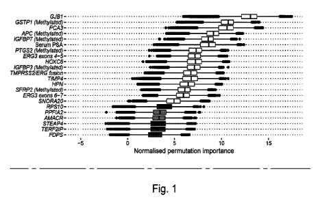
Figure 1 - Boruta analysis of variables available for the training of the
ExoMeth model. Variable
importance was determined over 1,000 bootstrap resamples of the available data
and the decision reached
recorded at each resample. Colour indicates the proportion of the 1,000
resamples a variable was confirmed
to be important in. Variables confirmed in at least 90% of resamples were
selected for predictive modelling.
Those variables rejected in every single resample are not shown here, but the
full list of inputs for all models
can be seen in Table 1.
Figure 2 - Waterfall plot of the ExoMeth risk score for each patient. Each
coloured bar represents an
individual patient's calculated risk score and their true biopsy outcome,
coloured according to Gleason score
(Gs) . Green - No evidence of cancer, Blue ¨ Gs 6, Orange - Gs 3+4, Red - Gs a
4+3.
Figure 3 - Density plots detailing risk score distributions generated from
four trained models. Models
A to D were trained with different input variables: A: SoC clinical risk
model, including Age and PSA,
B: Methylation model, C: ExoRNA model and D: ExoMeth model, combining the
predictors from all three
previous models. The full list of variables in each model is available in
Table 1. Fill colour shows the risk score
distribution of patients with a significant biopsy outcome of Gs a 3+4
(Orange) or Gs S 6 (Blue).
Figure 4- Cumming estimation plot of the ExoMeth risk signature. The top row
details individual patients
as points, separated according to Gleason score on the x-axis and risk score
on the y-axis. Points are
coloured according to clinical risk category; NEC - No evidence of cancer,
Raised PSA - Raised PSA with
negative biopsy, L -D'Amico Low-Risk, I - D'Amico Intermediate Risk, H -
D'Amico High-Risk. Gapped vertical
lines detail the mean and standard deviation of each group's risk scores. The
lower panel shows the mean
differences in risk score of each group, as compared to the NEC samples. Mean
differences and 95%
confidence interval are displayed as a point estimate and vertical bar
respectively, using the sample density
distributions calculated from a bias-corrected and accelerated bootstrap
analysis from 1,000 resamples.
Figure 5 - Decision curve analysis (DCA) plots detailing the standardised net
benefit (sNB) of adopting
different risk models for aiding the decision to biopsy patients who present
with a PSA 4 nWmL.
The x-axis details the range of risk a clinician or patient may accept before
deciding to biopsy. Panels show
the sNB based upon the detection of varying levels of disease severity: A:
detection of Gleason 4+3,
B: detection of Gleason a 3+4, C - any cancer; Blue- biopsy all patients with
a PSA >4 ng/mL, Orange - biopsy
patients according to the SOC model, Green - biopsy patients based on the
methylation model, Purple - biopsy
patients based on the ExoRNA model, Red - biopsy patients based on a the
ExoMeth model. To assess the
benefit of adopting these risk models in a non-PSA screened population we used
data available from the
control arm of the CAP study [13]. DCA curves were calculated from 1,000
bootstrap resamples of the
available data to match the distribution of disease reported in the CAP trial
population. Mean sNB from these
resampled DCA results are plotted here.
Figure 6 - Net percentage reduction in biopsies, as calculated by DCA
measuring the benefit of
adopting different risk models for aiding the decision to biopsy patients who
would otherwise
undergo biopsy by current clinical guidelines. The x-axis details the range of
accepted risk a clinician or
19
CA 03152887 2022-3-29
WO 2021/048445
PCT/EP2020/075665
patient may accept before deciding to biopsy. Panels show the reduction in
biopsies per 100 patients based
upon the detection of varying levels of disease severity: A: detection of
Gleason a 4+3, B: detection of
Gleason a 3+4 and C - any cancer. Coloured lines show differing comparator
models; Blue- biopsy all patients
with a PSA >3 ng/mL, Orange - biopsy patients by according the to the SoC
model, Green - biopsy patients
based on the methylation model, Purple - biopsy patients based on the ExoRNA
model, Red - biopsy patients
based on a the ExoMeth model. To assess the benefit of adopting these risk
models in a non-PSA screened
population we used data available from the control arm of the CAP study 1131.
DCA curves were calculated
from 1,000 bootstrap resamples of the available data to match the distribution
of disease reported in the CAP
trial population. Mean sNB from these resampled DCA results are used to
calculate the potentially reductions
in biopsy rates here.
Figure 7 - Boruta analysis of variables available for the training of the SoC
model. Variable importance
was determined over 1,000 bootstrap resamples of the available data and the
decision reached recorded at
each resample. Variable origins are denoted by font; clinical variables are
italicised and emboldened. Colour
indicates the proportion of the 1,000 resamples a variable was confirmed to be
important in.. Variables
confirmed in at least 90% of resamples were selected for training predictive
models.
Figure 8 - Boruta analysis of variables available for the training of the
Methylation model. Variable
importance was determined over 1,000 bootstrap resamples of the available data
and the decision reached
recorded at each resample_ Variable origins are denoted by font; methylation
variables are italicised. Colour
indicates the proportion of the 1,000 resamples a variable was confirmed to be
important in. Variables
confirmed in at least 90% of resamples were selected for training predictive
models.
Figure 9 - Boruta analysis of variables available for the training of the
ExoRNA model (ExoMeth
comparator). Variable importance was determined over 1,000 bootstrap resamples
of the available data and
the decision reached recorded at each resample. Variable origins are denoted
by font; clinical variables are
emboldened. Colour indicates the proportion of the 1,000 resamples a variable
was confirmed to be important
in. Variables confirmed in at least 90% of resamples were selected for
training predictive models. Those
variables rejected in every single resample are not shown here, but the full
list of inputs for the ExoRNA model
can be seen in Table 1.
Figure 10- Density plots detailing risk score distributions generated from
four trained models. Models
A to D were trained with different input variables; A: SoC clinical risk
model, including Age and PSA,
B: Methylation model, C: ExoRNA model and D: ExoMeth model, combining the
predictors from all three
previous models. The full list of variables in each model is available in
Table 1. Fill colour shows the risk score
distribution of patients with with respect to biopsy outcome: No evidence of
cancer (Blue). Gleason 6 or 3+4
(Orange), Gleason a 4+3 (Green).
Figure 11- Cumming estimation plot of the ExoMeth risk signatures in No
evidence of cancer (NEC)
and raised PSA, negative biopsy samples. The left panel details individual
patients as points with ExoMeth
risk score on the y-axis. Points are coloured according to clinical risk
category; NEC - No evidence of cancer,
Raised PSA - Raised PSA with negative biopsy. The right panel shows the mean
differences in risk score
between each NEC and Raised PSA samples. Mean differences and 95% confidence
interval are displayed
CA 03152887 2022-3-29
WO 2021/048445
PCT/EP2020/075665
as a point estimate and vertical bar respectively, using the sample density
distributions calculated from a
bias-corrected and accelerated bootstrap analysis from 11000 resamples.
Figure 12 - Boruta analysis of variables available for the training of the
ExoGrail model. Variable
importance was determined over 1,000 bootstrap resamples of the available data
and the decision reached
recorded at each resample. Colour indicates the proportion of the 11000
resamples a variable was confirmed
to be important in. Variables confirmed in at least 90% of resamples were
selected for predictive modelling
(Green). Those variables rejected in every single resample are not shown here,
but the full list of inputs for
all models can be seen in Table 1_
Figure 13 - Waterfall plot of the ExoGrail risk score for each patient. Each
coloured bar represents an
individual patient's calculated risk score and their true biopsy outcome,
coloured according to Gleason score
(Gs). Green - No evidence of cancer, Blue ¨ Gs 6, Orange - Gs 3+4, Red - Gs a
4+3.
Figure 14 - Density plots detailing risk score distributions generated from
four trained models. Models
A to 0 were trained with different input variables; A - SoC clinical risk
model, including Age and PSA, B - EN2
model, C -ExoRNA model and D - ExoGrail model, combining predictors from all
three modes of analysis.
The full list of variables in each model is available in Table 1. Fill colour
shows the risk score distribution of
patients with with respect to biopsy outcome: No evidence of cancer (Green),
Gleason 6 (Blue), Gleason 3+4
(Orange), Gleason a 4+3 (Red). AUCs of each model's predictive ability for
clinically relavent biopsy outcomes
are detailed underneath each plot.
Figure 15- Cumming estimation plot of the ExoGrail risk signature. The top row
details individual patients
as points, separated according to Gleason score on the x-axis and risk score
on the y-axis. Points are
coloured according to clinical risk category; NEC - No evidence of cancer,
Raised PSA - Raised PSA with
negative biopsy, L -D'Amico Low-Risk, I - D'Amico Intermediate Risk, H -
D'Amico High-Risk. Gapped vertical
lines detail the mean and standard deviation of each group's risk scores. The
lower panel shows the mean
differences in risk score of each group, as compared to the NEC samples. Mean
differences and 95%
confidence interval are displayed as a point estimate and vertical bar
respectively, using the sample density
distributions calculated from a bias-corrected and accelerated bootstrap
analysis from 1,000 resamples.
Figure 16 - Decision curve analysis (DCA) plots detailing the standardised net
benefit (sNB) of
adopting different risk models for aiding the decision to biopsy patients who
present with a PSA a 4
ngimL_ The x-axis details the range of risk a clinician or patient may accept
before deciding to biopsy. Panels
show the sNB based upon the detection of varying levels of disease severity: A
- detection of Gleason a 4+3,
B - detection of Gleason a 3+4, C - any cancer, Blue- biopsy all patients with
a PSA >4 ng/mL, Orange - biopsy
patients according to the SOC model, Green - biopsy patients based on the
methylation model, Purple - biopsy
patients based on the ExoRNA model, Red - biopsy patients based on a the
ExoGrail model. To assess the
benefit of adopting these risk models in a non-PSA screened population we used
data available from the
control arm of the CAP study [13]. DCA curves were calculated from 1,000
bootstrap resamples of the
available data to match the distribution of disease reported in the CAP trial
population. Mean sNB from these
resampled DCA results are plotted here.
21
CA 03152887 2022-3-29
WO 2021/048445
PCT/EP2020/075665
Figure 17 ¨ Net percentage reduction in biopsies, as calculated by DCA
measuring the benefit of
adopting different risk models for aiding the decision to biopsy patients who
would otherwise
undergo biopsy by current clinical guidelines. The x-axis details the range of
accepted risk a clinician or
patient may accept before deciding to biopsy. Panels show the reduction in
biopsies per 100 patients based
upon the detection of varying levels of disease severity: A - detection of
Gleason 4+3, B - detection of
Gleason 3+4 and C - any cancer. Coloured lines show differing comparator
models; Blue- biopsy all patients
with a PSA >3 ng/mL, Orange - biopsy patients by according the to the SoC
model, Green - biopsy patients
based on the methylation model, Purple - biopsy patients based on the ExoRNA
model, Red - biopsy patients
based on a the ExoGrail model. To assess the benefit of adopting these risk
models in a non-PSA screened
population we used data available from the control arm of the CAP study [13].
DCA curves were calculated
from 11000 bootstrap resamples of the available data to match the distribution
of disease reported in the CAP
trial population. Mean sNB from these resampled DCA results are used to
calculate the potentially reductions
in biopsy rates here.
Figure 18- Boruta analysis of variables available for the training of the SoC
model. Variable importance
was determined over 1,000 bootstrap resamples of the available data and the
decision reached recorded at
each resample. Variable origins are denoted by font; clinical variables are
italicised and emboldened. Colour
indicates the proportion of the 1,000 resamples a variable was confirmed to be
important in.. Variables
confirmed in at least 90% of resamples were selected for training predictive
models (Green).
Figure 19 - Boruta analysis of variables available for the training of the
Methylation model. Variable
importance was determined over 1,000 bootstrap resamples of the available data
and the decision reached
recorded at each resample_ Variable origins are denoted by font; rnethylation
variables are italicised. Colour
indicates the proportion of the 1,000 resamples a variable was confirmed to be
important in. Variables
confirmed in at least 90% of resamples were selected for training predictive
models (Green).
Figure 20 - Boruta analysis of variables available for the training of the
ExoRNA model (ExoGrail
comparator). Variable importance was determined over 1,000 bootstrap resamples
of the available data and
the decision reached recorded at each resample. Variable origins are denoted
by font; clinical variables are
emboldened. Colour indicates the proportion of the 1,000 resamples a variable
was confirmed to be important
in. Variables confirmed in at least 90% of resamples were selected for
training predictive models. Those
variables rejected in every single resample are not shown here, but the full
list of inputs for the ExoRNA model
can be seen in Table 1.
Figure 21 - Partial dependency plots detailing the marginal effects and
interactions of SLC12A1 and
urinary EN2 on predicted ExoGrail Risk Score. A - Partial dependency of
ExoGrail on urinary EN2,
B - Partial dependency of ExoGrail on SLC12A1, C - Partial dependency of
ExoGrail on both SLC12A1 and
urinary EN2.
Figure 22 - Density plots detailing risk score distributions generated from
four trained models. Models
A to D were trained with different input variables; A - SoC clinical risk
model, including Age and PSA,
B - Methylation model, C -ExoRNA model and D - ExoGrail model, combining the
predictors from all three
22
CA 03152887 2022-3-29
WO 20211048445
PCT/EP2020/075665
previous models. The full list of variables in each model is available in
Table 1. Fill colour shows the risk score
distribution of patients with a significant biopsy outcome of Gs 3+4 (Orange)
or Gs 5 6 (Blue)
Figure 23 - Cumming estimation plot of the ExoGrail risk signatures in No
evidence of cancer (NEC)
and raised PSA, negative biopsy samples. The left panel details individual
patients as points with ExoGrail
risk score on the y-axis. Points are coloured according to clinical risk
category; NEC - No evidence of cancer,
Raised PSA - Raised PSA with negative biopsy. The right panel shows the mean
differences in risk score
between each NEC and Raised PSA samples. Mean differences and 95% confidence
interval are displayed
as a point estimate and vertical bar respectively, using the sample density
distributions calculated from a
bias-corrected and accelerated bootstrap analysis from 1,000 resamples.
Figure 24- Example computer apparatus.
Detailed description of the invention
Extracellular vesicles
It is well documented that eukaryotic cells release extracellular vesicles
including apoptotic bodies, exosomes,
and other microvesicles [34,35]. Here we will use the term Extracellular
Vesicle (EV) to include any
membranous vesicles found in the urine such as exosomes. Extracellular
vesicles differ in their cellular origins
and sizes, for example, apoptotic bodies are released from the cell membrane
as the final consequence of
cell fragmentation during apoptosis, and they have irregular shapes with a
range of 1-5 pm in size 1351
Exosomes are specialised vesicles, 30 to 100nm in size that are actively
secreted by a variety of normal and
tumour cells and are present in many biological fluids, including serum and
urine. They carry membrane and
cytosolic components including protein and RNA into the extracellular space
[36,37]. These microvesicles
form as a result of inward budding of the cellular endosomal membrane
resulting in the accumulation of
intraluminal vesicles within large multivesicular bodies. Through this process
trans-membrane proteins are
incorporated into the invaginating membrane while the cytosolic components are
engulfed within the
intraluminal vesicles that form the exosomes, which will then be released,
into the extracellular space 138,391
So far urine exosomes have been examined in several studies for renal and
prostatic pathology and have
been reported to be stable in urine. RNA isolated from urine EVs had a better-
preserved profile than
cell-isolated RNA from the same samples 140] which makes them much better for
potential biomarker use.
EV Function
EVs such as exosomes function as a means of transport for biological material
between cells within an
organism. As a consequence of their origin, EVs such as exosomes exhibit the
mother-cell's membrane and
cytoplasmic components such as proteins, lipids and genomic materials. Some of
the proteins they exhibit
regulate their docking and membrane fusion, for example the Rab proteins,
which are the largest family of
small GTPases[41]. Annexins and flotillin aid in membrane-trafficking and
fusion events 1421 Exosomes also
contain proteins that have been termed exosomal-marker-proteins, for example
Alix, TSG101, HSP70 and
23
CA 03152887 2022-3-29
WO 2021/048445
PCT/EP2020/075665
the tetraspanins CD63, CD61 and C09. Exosome protein composition is very
dependent on the cell type of
origin. So far a total of 13,333 exosomal proteins have been reported in the
ExoCarta database, mainly from
dendritic, normal and malignant cells.
Besides proteins, 2,375 mRNAs and 764 microRNAs have been reported
(Exocarta.org) which can be
delivered to recipient cells. Exosomes are rich in lipids such as cholesterol,
sphingolipids, ceramide and
glycerophospolipids which play an important role in exosome biogenesis,
especially ILV formation.
EVs in malignancy
The role of EVs such as EVs in cancer remains to be fully elucidated; they
appear to function as both pro- and
anti-tumour effectors. Either way cancer cell-derived EVs appear to have
distinct biologic roles and molecular
profiles. They can have unique gene expression signatures (RNAs, mRNAs) and
proteomics profiles
compared to EVs from normal cells [43,44]. Reference 43 reports large numbers
of differentially expressed
RNAs in EVs from melanocytes compared with melanoma-derived EVs. This
indicates that exosomal RNAs
may contribute to important biological functions in normal cells, as well as
promoting malignancy in tumour
cells. Reference 43 also suggests that cancer cell-derived EVs have a closer
relationship to the originating
cancer cell than normal cell derived EVs do to a normal cell, which highlights
the potential of using EVs as a
source of diagnostic biomarkers. RNA expression in melanoma EVs has been
linked to the advancement of
the disease supporting the idea that EVs such as exosomes can promote tumour
growth. A similar finding
was reported in glioblastoma, highlighting their potential as prognostic
markers.
Experiments in mice have shown that cancer-derived EVs can induce an anti-
tumour immune response. It
has been demonstrated that EVs such as exosomes isolated from malignant
effusions are an effective source
of tumour antigens which are used by the host to present to CDS+ cytotoxic T
cells, dramatically increasing
the anti-tumour immune response.
EVs and prostate cancer
Several studies have examined the role of EVs such as exosomes in prostate
cancer. Reference 45 suggests
that prostate cancer derived EVs can stimulate fibroblast activation and lead
to cancer development by
increasing cell motility and preventing cell apoptosis. Similarly, vesicles
from activated fibroblasts are, in turn,
able to induce migration and invasion in the PC3 cell line. Another study
reported that EVs from hormone
refractory PC cells are able to induce osteoblast differentiation via the Ets1
which they contained, suggesting
a role for vesicles in cell-to-cell communication during the osteoblastic
metastasis process. Cell-to-cell
communication was also emphasised in another study that showed that vesicles
released from the human
prostate carcinoma cell line DU145 are able to induce transformation in a non-
malignant human prostate
epithelial cell line.
Besides the in vivo evidence on the active role of EVs in cancer and cancer
metastasis, Reference 46
suggests that EVs are present in high levels in the urine of cancer patients,
and that unlike cells, EVs have
remarkable stability in urine [47]. Other studies suggest the presence of EVs
in prostatic secretions, identifying
them as a potential source of prostate cancer biomarkers.
24
CA 03152887 2022-3-29
WO 2021/048445
PCT/EP2020/075665
Using a nested PCR-based approach, the authors of reference 48 suggest that
tumour EVs are harvestable
from urine samples from PC patients and that they carry biomarkers specific to
PC including KLK3, PCA3
and TMPRSS2/ERG RNAs. PCA3 transcripts were detectable in all patients
including subjects with low grade
disease, however TMPRSS2/ERG transcripts were only detectable in high Gleason
grades. They also
demonstrated in this study that i) mild prostate massage increased the
extracellular vesicle secretion into the
urethra and subsequently into the collected urine fraction ii) that tumour EVs
are distinct from EVs shed by
normal cells, and iii) they are more abundant in cancer patients.
In the present invention the RNA may be harvested from all extracellular
vesicles (EV) present in urine that
are below 0.8pm. The EVs will consist of exosomes and other extracellular
vesicles. In further embodiments
of the invention different subtypes of EVs may be harvested and analysed.
In some embodiments of the invention RNA is extracted from urine supernatant.
In some embodiments of the
invention RNA is extracted from whole urine.
Apparatus and media
The present invention also provides an apparatus configured to perform any
method of the invention.
Figure 18 shows an apparatus or computing device 100 for carrying out a method
as disclosed herein. Other
architectures to that shown in Figure 18 may be used as will be appreciated by
the skilled person_
Referring to the Figure, the meter 100 includes a number of user interfaces
including a visual display 110 and
a virtual or dedicated user input device 112. The meter 100 further includes a
processor 114, a memory 116
and a power system 118. The meter 100 further comprises a communications
module 120 for sending and
receiving communications between processor 114 and remote systems. The meter
100 further comprises a
receiving device or port 122 for receiving, for example, a memory disk or non-
transitory computer readable
medium carrying instructions which, when operated, will lead the processor 114
to perform a method as
described herein.
The processor 114 is configured to receive data, access the memory 116, and to
act upon instructions
received either from said memory 116, from communications module 120 or from
user input device 112. The
processor controls the display 110 and may communicate date to remote parties
via communications module
120.
The memory 116 may comprise computer-readable instructions which, when read by
the processor, are
configured to cause the processor to perform a method as described herein.
The present invention further provides a machine-readable medium (which may be
transitory or
non-transitory) having instructions stored thereon, the instructions being
configured such that when read by
a machine, the instructions cause a method as disclosed herein to be carried
out.
CA 03152887 2022-3-29
WO 2021/048445
PCT/EP2020/075665
Active surveillance
Active surveillance (AS) is a means of disease-management for men with
localised PCa with the intent to
intervene if the disease progresses. AS is offered as an option to men whose
prostate cancer is thought to
have a low risk of causing harm in the absence of treatment. It is a chance to
delay or avoid aggressive
treatment such as radiotherapy or surgery, and the associated morbidities of
these treatments. Entry criteria
for men to go on active surveillance varies widely and can include men with
Low risk and Intermediate risk
prostate cancer.
Patients on AS are currently monitored by a wide range of means that include,
for example, PSA monitoring,
biopsy and repeat biopsy and MP-MRI. The timing of repeat biopsies, PSA
testing and MP-MR! varies with
the hospital, and a widely accepted method for monitoring men on AS has not
yet been achieved.
In some embodiments, active surveillance comprises assessment of a patient by
PSA monitoring, biopsy and
repeat biopsy and/or imaging techniques such as MRI, for example MP-MRI. In
some embodiments, active
surveillance comprises assessment of a patient by any means appropriate for
diagnosing or prognosing
prostate cancer.
In some embodiments of the invention, active surveillance comprises assessment
of a patient at least every
1 month, 2 months, 3 months, 4 months, 5 months, 6 months, 7 months, 8 months,
9 months, 10 months, 11
months or 12 months.
In some embodiments of the invention, active surveillance comprises assessment
of a patient at least every
1 year, 2 years, 3 years, 4 years or 5 or more years.
In some embodiments of the invention the ExoMeth and/or ExoGrail risk score
will be used alone or in
conjunction with other means of testing to improve shared decision making with
the multi-disciplinary team
and the patient. The ExoMeth and/or ExoGrail risk score could be used to
decide whether radical intervention
is necessary, or to decide the optimal time between re-monitoring by, for
example, biopsy, PSA testing or
MP-MRI.
Biological samples
In the present invention, the biological sample may be a urine sample, a semen
sample, a prostatic exudate
sample, or any sample containing macromolecules or cells originating in the
prostate, a whole blood sample,
a serum sample, saliva, or a biopsy (such as a prostate tissue sample or a
tumour sample), although urine
samples are particularly useful. The method may include a step of obtaining or
providing the biological
sample, or alternatively the sample may have already been obtained from a
patient, for example in ex vivo
methods.
Biological samples obtained from a patient can be stored until needed.
Suitable storage methods include
freezing immediately, within 2 hours or up to two weeks after sample
collection. Maintenance at -80 C can be
26
CA 03152887 2022-3-29
WO 2021/048445
PCT/EP2020/075665
used for long-term storage. Preservative may be added, or the urine collected
in a tube containing
preservative. Urine plus preservative such as Norgen urine preservative, can
be stored between room
temperature and -80 C.
Methods of the invention may comprise steps carried out on biological samples.
The biological sample that is
analysed may be a urine sample, a semen sample, a prostatic exudate sample, or
any sample containing
macromolecules or cells originating in the prostate, a whole blood sample, a
serum sample, saliva, or a biopsy
(such as a prostate tissue sample or a tumour sample). Most commonly for
prostate cancer the biological
sample is from a prostate biopsy, prostatectomy or TURP. The method may
include a step of obtaining or
providing the biological sample, or alternatively the sample may have already
been obtained from a patient,
for example in ex vivo methods. The samples are considered to be
representative of the expression status of
the relevant genes in the potentially cancerous prostate tissue, or other
cells within the prostate, or
microvesicles produced by cells within the prostate or blood or immune system.
Alternatively, the samples
can be considered to be representative of the potentially cancerous
microenvironment of the prostate,
comprising gene expression or methylation and protein expression. Hence the
methods of the present
invention may use quantitative data on RNA, methylation and proteins produced
by cells within the prostate
and/or the blood system and/or bone marrow in response to cancer, to determine
the presence or absence
of prostate cancer.
The methods of the invention may be carried out on one test sample from a
patient. Alternatively, a plurality
of test samples may be taken from a patient, for example at least 2, 3, 4 or 5
samples. Each sample may be
subjected to a separate analysis using a method of the invention, or
alternatively multiple samples from a
single patient undergoing diagnosis could be included in the method.
The sample may be processed prior to determining the expression status of the
biome rkeis. The sample may
be subject to enrichment (for example to increase the concentration of the
biomarkers being quantified),
centrifugation or dilution. In other embodiments, the samples do not undergo
any pre-processing and are
used unprocessed (such as whole urine).
In some embodiments of the invention, the biological sample may be
fractionated or enriched for RNA prior
to detection and quantification (i.e. measurement). The step of fractionation
or enrichment can be any suitable
pre-processing method step to increase the concentration of RNA in the sample
or select for specific sources
of RNA such as cells or wdracellular vesicles. For example, the steps of
fractionation and/or enrichment may
comprise centrifugation and/or filtration to remove cells or unwanted analytes
from the sample, or to increase
the concentration of EVs in a urine fraction. Methods of the invention may
include a step of amplification to
increase the amount of gene transcripts that are detected and quantified.
Methods of amplification include
RNA amplification, amplification as cDNA, and PCR amplification. Such methods
may be used to enrich the
sample for any biomarkers of interest.
Generally speaking, the RNAs will need to be extracted from the biological
sample. This can be achieved by
a number of suitable methods_ For example, extraction may involve separating
the RNAs from the biological
sample. Methods include chemical extraction and solid-phase extraction (for
example on silica columns).
Preferred methods include the use of a silica column. Methods comprise lysing
cells or vesicles (if required),
27
CA 03152887 2022-3-29
WO 2021/048445
PCT/EP2020/075665
addition of a binding solution, centitfugation in a spin column to force the
binding solution through a silica gel
membrane, optional washing to remove further impurities, and elution of the
nucleic acid. Commercial kits
are available for such methods, for example from Qiagen or Exigon.
If RNAs are extracted from a sample, the extracted solution may require
enrichment to increase the relative
abundance of RNA transcripts in the sample.
The methods of the invention may be carried out on one test sample from a
patient. Alternatively, a plurality
of test samples may be taken from a patient, for example at least 2, at least
3, at least 4 or at least 5 samples.
Each sample may be subjected to a single assay to quantify one of the
biomarker panel members, or
alternatively a sample may be tested for all of the biomarkers being
quantified.
Methods of the invention
Expression status
Determining the expression status of a gene may comprise determining the level
of expression of the gene.
Expression status also encompasses the determination of any parameter of a
gene or protein which impacts
the functional effect of the gene or protein in question. For example, this
encompasses, among other
parameters, the methylation status, the level of mRNA (i.e. gene transcripts)
and/or the concentration of
protein. Expression status and levels of expression as used herein can be
determined by methods known to
the skilled person. For example, this may refer to the up or down-regulation
of a particular gene or genes, as
determined by methods known to a skilled person. Epigenetic modifications may
be used as an indicator of
expression, for example determining DNA methylation status, or other
epigenetic changes such as histone
marking, RNA changes or conformation changes. Epigenetic modifications
regulate expression of genes in
DNA and can influence efficacy of medical treatments among patients. Aberrant
epigenetic changes are
associated with many diseases such as, for example, cancer. DNA methylation in
animals influences dosage
compensation, imprinting, and genome stability and development. Methods of
determining DNA methylation
are known to the skilled person (for example methylation-specific PCR, matrix-
assisted laser
desorption/ionization time-of-flight mass spectrometry, use of microarrays,
reduced representation bisulfate
sequencing (RRBS) or whole genome shotgun bisulfate sequencing (VVGBS). In
addition, epigenetic changes
may include changes in conformation of chromatin. The impact of different
parameters (for example
methylation status) is known to the skilled person. In many cases, the impact
of the altered parameterwill be
clear, for example higher protein concentration leading to a greater
availability of the protein to achieve its
effect.
Expression analysis
NanoStringe technology is based on double hybridisation of two adjacent ¨50bp
probes to their target
RNA/cONA. The first probe hybridisation is used to pull the target RNA/cONA
down on to a hard surface. The
excess unbound nucleic acid is then washed away. The second probe is then
hybridised to the RNA/cDNA.
This probe has a multi-colour barcode attached to it. The nucleotides are then
stretched out under an electrical
current, and the image is recorded. The barcodes number and type are counted,
and this is the data output.
28
CA 03152887 2022-3-29
WO 2021/048445
PCT/EP2020/075665
Up to 800 different barcodes are possible, and therefore up to 800 different
target RNAs can be detected in
a single assay.
Methods of real-time qPCR may involve a step of reverse transcription of RNA
into complementary DNA
(cDNA). PCR amplification can use sequence specific primers or combinations of
other primers to amplify
RNA species of interest. Micnoarray analysis may comprise the steps of
labelling RNA or cDNA, hybridisation
of the labelled RNAs to DNA (or RNA or LNA) probes on a solid-substrate array,
washing the array, and
scanning the array.
RNA sequencing is another method that can benefit from RNA enrichment,
although this is not always
necessary. RNA sequencing techniques generally use next generation sequencing
methods (also known as
high-throughput or massively parallel sequencing). These methods use a
sequencing-by-synthesis approach
and allow relative quantification and precise identification of RNA sequences.
In situ hybridisation techniques
can be used on tissue samples, both in vivo and ex vivo.
In some methods of the invention, detection and quantification of cDNA-binding
molecule complexes may be
used to determine RNA expression. For example, RNA transcripts in a sample may
be converted to GOMA by
reverse-transcription, after which the sample is contacted with binding
molecules specific for the RNAs being
quantified, detecting the presence of a of cDNA-specific binding molecule
complex, and quantifying the
expression of the corresponding gene. There is therefore provided the use of
cDNA transcripts corresponding
to one or more of the RNAs of interest, or combinations thereof, for use in
methods of detecting, diagnosing
or predicting prognosis of prostate. In some embodiments of the invention, the
method may therefore
comprise a step of conversion of the RNAs to cDNA to allow a particular
analysis to be undertaken and to
achieve RNA quantification.
DNA and RNA arrays (microarrays) for use in quantification of the mRNAs of
interest comprise a series of
microscopic spots of DNA or RNA sequences, each with a unique sequence of
nucleotides that are able to
bind complementary nucleic acid molecules. In this way the oligonucleotides
are used as probes to which
only the correct target sequence will hybridise under high-stringency
condition. In the present invention, the
target sequence can be the coding DNA sequence or unique section thereof,
corresponding to the RNA
whose expression is being detected. Most commonly the target sequence is the
RNA biomarker of interest
itself.
Capture molecules include antibodies, proteins, aptamers, nucleic adds,
biotin, streptavidin, receptors and
enzymes, which might be preferable if commercial antibodies are not available
for the analyte being detected.
Capture molecules for use on the arrays can be externally synthesised,
purified and attached to the array.
Alternatively, they can be synthesised in-situ and be directly attached to the
array. The capture molecules
can be synthesised through biosynthesis, cell-free DNA expression or chemical
synthesis. In-situ synthesis
is possible with the latter two. The appropriate capture molecule will depend
on the nature of the target (e.g.
RNA, protein or cDNA).
Once captured on a microarray, detection methods can be any of those known in
the art. For example,
fluorescence detection can be employed. It is safe, sensitive and can have a
high resolution. Other detection
29
CA 03152887 2022-3-29
WO 2021/048445
PCT/EP2020/075665
methods include other optical methods (for example colorimetric analysis,
chemiluminescence, label free
Surface Plasmon Resonance analysis, microscopy, reflectance etc.), mass
spectrometry, electrochemical
methods (for example vottammetry and amperometry methods) and radio frequency
methods (for example
multipolar resonance spectroscopy).
Once the expression status or concentration has been determined, the level can
be compared to a threshold
level or previously measured expression status or concentration (either in a
sample from the same subject
but obtained at a different point in time, or in a sample from a different
subject, for example a healthy subject,
i.e. a control or reference sample) to determine whether the expression status
or concentration is higher or
lower in the sample being analysed. Hence, the methods of the invention may
further comprise a step of
correlating said detection orquantification with a control or reference to
determine if prostate cancer is present
(or suspected) or not. Said correlation step may also detect the presence of a
particular type, stage, grade or
risk group of prostate cancer and to distinguish these patients from healthy
patients, in which no prostate
cancer is present or from men with indolent or low risk disease. For example,
the methods may detect early
stage or low risk prostate cancer. Said step of correlation may include
comparing the amount (expression or
concentration) of one, two, or three or more of the panel biomarkers with the
amount of the corresponding
biomarker(s) in a reference sample, for example in a biological sample taken
from a healthy patient. The
methods of the invention may include the steps of determining the amount of
the corresponding biomarker in
one or more reference samples which may have been previously determined.
Alternatively, the method may
use reference data obtained from samples from the same patient at a previous
point in time. In this way, the
effectiveness of any treatment can be assessed and a prognosis for the patient
determined.
Internal controls can be also used, for example quantification of one or more
different RNAs not part of the
biomarker panel. This may provide useful information regarding the relative
amounts of the biomarkers in the
sample, allowing the results to be adjusted for any variances according to
different populations or changes
introduced according to the method of sample collection, processing or
storage.
Methods of normalisation can involve correction of the counts of the measured
levels of NanoStringe
gene-probes in order to account for, for example; differences in the input
amount of RNA, variability in RNA
quality and to centre data around RNA originating from prostatic material, so
that all the genes being analysed
are on a comparable scale.
As would be apparent to a person of skill in the art, any measurements of
analyte concentration or expression
may need to be normalised to take in account the type of test sample being
used and/or and processing of
the test sample that has occurred prior to analysis. Data normalisation also
assists in identifying biologically
relevant results. Invariant RNAs/mRNAs may be used to determine appropriate
processing of the sample.
Differential expression calculations may also be conducted between different
samples to determine statistical
significance.
Further analytical methods used in the invention
CA 03152887 2022-3-29
WO 2021/048445
PCT/EP2020/075665
The expression status of a gene or protein from a biomarker panel of the
invention can be determined in a
number of ways. Levels of expression may be determined by, for example,
quantifying the biomarkers by
determining the concentration of protein in the sample, if the biomarkers are
expressed as a protein in that
sample. Alternatively, the amount of RNA or protein in the sample (such as a
tissue sample) may be
determined. Once the expression status has been determined, the level can
optionally be compared to a
control. This may be a previously measured expression status (either in a
sample from the same subject but
obtained at a different point in time, or in a sample from a different subject
or subjects, for example one or
more healthy subjects or one or more subjects with non-aggressive cancer, i.e.
a control or reference sample)
or to a different protein or peptide or other marker or means of assessment
within the same sample to
determine whether the expression status or protein concentration is higher or
lower in the sample being
analysed. Housekeeping genes can also be used as a control. Ideally, controls
are one or more RNA, protein
or DNA markers that generally do not vary significantly between samples or
between tissue from different
people or between normal tissue and tumour.
Other methods of quantifying gene expression include RNA sequencing, which in
one aspect is also known
as whole transcriptome shotgun sequencing (WTSS). Using RNA sequencing it is
possible to determine the
nature of the RNA sequences present in a sample, and furthermore to quantify
gene expression by measuring
the abundance of each RNA molecule (for example, RNA or microRNA transcripts).
The methods use
sequencing-by-synthesis approaches to enable high throughout analysis of
samples.
There are several types of RNA sequencing that can be used, including RNA
PolyA tail sequencing (there
the polyA tail of the RNA sequences are targeting using polyT
oligonucleotides), random-primed sequencing
(using a random oligonucleotide primer), targeted sequence (using specific
oligonudeotide primers
complementary to specific gene transcripts), small RNA/non-coding RNA
sequencing (which may involve
isolating small non-coding RNAs, such as microRNAs, using size separation),
direct RNA sequencing, and
real-time PCR. In some embodiments, RNA sequence reads can be aligned to a
reference genome and the
number of reads for each sequence quantified to determine gene expression. In
some embodiments of the
invention, the methods comprise transcription assembly (de-novo or genome-
guided).
RNA, DNA and protein arrays (microarrays) may be used in certain embodiments.
RNA and DNA microarrays
comprise a series of microscopic spots of DNA or RNA oligonucleotides, each
with a unique sequence of
nucleotides that are able to bind complementary nucleic acid molecules. In
this way the oligonucleotides are
used as probes to which the correct target sequence will hybridise under high-
stringency condition. In the
present invention, the target sequence can be the transcribed RNA sequence or
unique section thereof,
corresponding to the gene whose expression is being detected. Protein
microarrays can also be used to
directly detect protein expression. These are similar to DNA and RNA
microarrays in that they comprise
capture molecules fixed to a solid surface.
Methods for detection of RNA or cDNA can be based on hybridisation, for
example, Northern blot,
Microarrays, NanoString , RNA-FISH, branched chain hybridisation assay, or
amplification detection
methods for quantitative reverse transcription polymerase chain reaction (qRT-
PCR) such as TagMan, or
SYBR green product detection. Primer extension methods of detection such as:
single nucleotide extension,
Sanger sequencing. Alternatively, RNA can be sequenced by methods that include
Sanger sequencing, Next
31
CA 03152887 2022-3-29
WO 2021/048445
PCT/EP2020/075665
Generation (high throughput) sequencing, in particular sequencing by
synthesis, targeted RNAseq such as
the Precise targeted RNAseq assays, or a molecular sensing device such as the
Oxford Nanopore MinION
device. Combinations of the above techniques may be utilised such as
Transcription Mediated Amplification
(TMA) as used in the Gen-Probe PCA3 assay which uses molecule capture via
magnetic beads, transcription
amplification, and hybridisation with a secondary probe for detection by, for
example chemiluminescence.
RNA may be converted into cDNA prior to detection. RNA or cDNA may be
amplified prior or as part of the
detection.
The test may also constitute a functional test whereby presence of RNA or
protein or other macromolecule
can be detected by phenotypic change or changes within test cells. The
phenotypic change or changes may
include alterations in motility or invasion.
Commonly, proteins subjected to electrophoresis are also further characterised
by mass spectrometry
methods. Such mass spectrometry methods can include matrix-assisted laser
desorption/ionisation
time-of-flight (MALDI-TOF).
MALDI-TOF is an ionisation technique that allows the analysis of biomolecules
(such as proteins, peptides
and sugars), which tend to be fragile and fragment when ionised by more
conventional ionisation methods.
Ionisation is triggered by a laser beam (for example, a nitrogen laser) and a
matrix is used to protect the
biomolecule from being destroyed by direct laser beam exposure and to
facilitate vaporisation and ionisation.
The sample is mixed with the matrix molecule in solution and small amounts of
the mixture are deposited on
a surface and allowed to dry. The sample and matrix co-crystallise as the
solvent evaporates.
Additional methods of determining protein concentration include mass
spectrometry and/or liquid
chromatography, such as LC-MS, UPLC, a tandem UPLC-MS/MS system, and ELISA
methods. Other
methods that may be used in the invention include Agilent bait capture and PCR-
based methods (for example
PCR amplification may be used to increase the amount of analyte).
Methods of the invention can be carried out using binding molecules or
reagents specific for the analytes
(RNA molecules or proteins being quantified). Binding molecules and reagents
are those molecules that have
an affinity for the RNA molecules or proteins being detected such that they
can form binding
molecule/reagent-analyte complexes that can be detected using any method known
in the art. The binding
molecule of the invention can be an oligonucleotide, or oligoribonucleotide or
locked nucleic acid or other
similar molecule, an antibody, an antibody fragment, a protein, an aptamer or
molecularly imprinted polymeric
structure, or other molecule that can bind to DNA or RNA. Methods of the
invention may comprise contacting
the biological sample with an appropriate binding molecule or molecules. Said
binding molecules may form
part of a kit of the invention, in particular they may form part of the
biosensors of in the present invention.
Aptamers are oligonucleotides or peptide molecules that bind a specific target
molecule. Oligonuc.leotide
aptamers include DNA aptamer and RNA aptamers. Aptamers can be created by an
in vitro selection process
from pools of random sequence oligonucleotides or peptides. Aptamers can be
optionally combined with
ribozyrnes to self-cleave in the presence of their target molecule. Other
oligonucleotides may include RNA
32
CA 03152887 2022-3-29
WO 2021/048445
PCT/EP2020/075665
molecules that are complimentary to the RNA molecules being quantified. For
example, polyT oligos can be
used to target the polyA tail of RNA molecules.
Aptamers can be made by any process known in the art. For example, a process
through which aptamers
may be identified is systematic evolution of ligands by exponential enrichment
(SELEX). This involves
repetitively reducing the complexity of a library of molecules by partitioning
on the basis of selective binding
to the target molecule, followed by re-amplification. A library of potential
aptamers is incubated with the target
protein before the unbound members are partitioned from the bound members. The
bound members are
recovered and amplified (for example, by polymerase chain reaction) in order
to produce a library of reduced
complexity (an enriched pool). The enriched pool is used to initiate a second
cycle of SELEX. The binding of
subsequent enriched pools to the target protein is monitored cycle by cycle.
An enriched pool is cloned once
it is judged that the proportion of binding molecules has risen to an adequate
level. The binding molecules
are then analysed individually. SELEX is reviewed in 1491
Statistical analysis
Decision curve analysis (DCA)
Decision curve analysis is a method of evaluating predictive models. It
assumes that the threshold probability
of a disease or event at which a patient would opt for treatment is
informative of how the patient weighs the
relative harms of a false-positive and a false-negative prediction. This
theoretical relationship is then used to
derive the net benefit of the model across different threshold probabilities.
Plotting net benefit against
threshold probability yields the "decision curve." Decision curve analysis can
be used to identify the range of
threshold probabilities in which a model is of value, the magnitude of
benefit, and which of several models is
optimal 1501
Boruta algorithm
The Boruta algorithm is a wrapper built around the random forest
classification algorithm. It duplicates a
dataset, and randomly shuffles the values in each column. These values are
called shadow features. It then
trains a classifier, such as a Random Forest Classifier, on the dataset. By
doing this, it can provide an idea
of the importance -via the Mean Decrease Accuracy or Mean Decrease Impurity-
for each of the features of
the data set. The higher the score, the better or more important the feature
is.
The algorithm checks whether each of the "real" features have higher
importance than the "shadow" features.
In other words, whether the feature has a higher Z-score (i.e. the number of
standard deviations from the
mean a data point is) than the maximum Z-score of the best of the shadow
features. If the algorithm identities
a "real" feature with a better association than the "shadow" features then it
will record this as a hit. After a
predefined set of iterations, the algorithm provides a table of hits.
At every iteration (or resample), the algorithm compares the Z-scores of the
shuffled copies of the features
and the original features to see if the latter performed better than the
former. If it does, the algorithm will mark
the feature as important. In essence, the algorithm validates the importance
of the feature by comparing with
33
CA 03152887 2022-3-29
WO 2021/048445
PCT/EP2020/075665
random shuffled copies, which increases the robustness. This is done by simply
comparing the number of
times a feature did better with the shadow features using a binomial
distribution.
The number of iterations can be predefined. In some aspects of the invention
the number of iterations (or
resamples) is at least about 100, about 200, about 300, about 400, about 500,
about 600, about 700, about
800, about 900, about 1000, about 1500, about 2000, about 3000, about 4000,
about 5000. In a preferred
embodiment of the invention the number of iterations (or resamples) is 1000.
The proportion of iterations (or resamples) in which a feature must be
selected in order to be considered
associated with a biopsy outcome group can be predefined. In some aspects of
the invention the proportion
of iterations (or resamples) in which a feature must be selected in order to
be considered associated with a
biopsy outcome group is at least about 70%, about 75%, about 80%, about 85%,
about 90%, about 95%,
about 98% or about 99%. In a preferred embodiment of the invention the
proportion of iterations (or
resamples) in which a feature must be selected in order to be considered
associated with a biopsy outcome
group is 90%.
Gene Transcript detection
The present invention provides probes suitable for use in cDNA or RNA sequence
detection such as
NanoStringe or microarray techniques which can be used to determine the
expression status of genes of
interest. Methods of the invention can be operated using any suitable probe
sequence to detect a gene
transcript and methods of generating probe sequences are known to those
skilled in the art.
In another embodiment the gene transcripts may be detected by sequencing, or
qRT-PCR.
Methvlation detection
The methylation status of genes can be determined by any suitable means. For
example, methylation
detection assays which rely on the digestion of genomic DNA with a methylation-
sensitive restriction enzyme
followed by either Southern blot analysis or PCR. Other suitable assays use
treatment of genomic DNA with
sodium bisultite followed by alkaline treatment to convert unmethylated
cytosines to uracil, while leaving
methylated cytosine residues intact. Sequence variants at a particular locus
can subsequently be analyzed
by PCR amplification with primers designed to anneal with bisultlie-converted
DNA. Preferably, methylation
status of genes is established using high-throughput assays that utilize
highly sensitive and accurate
fluorescence-based real-time quantitative PCR (qPCR). Other suitable methods
will be known in the art.
Protein quantification
The concentration of a urinary protein can be established by any suitable
method. Individual protein
quantitation methods include enzyme-linked immunosorbent assay (ELISA) assay,
westem blot analysis, and
more recently, mass spectrometry, among others. ELISAs are used to
qualitatively and quantitatively analyze
the presence or concentration of a particular soluble antigen, peptide or
protein in liquid samples, such as
biological fluids. These assays make use of the ability of polystyrene plates
to bind proteins, including
34
CA 03152887 2022-3-29
WO 2021/048445
PCT/EP2020/075665
antibodies, as well as the particular specificities of antibodies for target
antigens. Generally, these assays
incorporate a calorimetric endpoint that can be detected via absorbance
wavelength and quantitated from a
known standard curve of antigen or antibody dilutions. Western blotting is a
method in which proteins that
have been electrophoretically separated on a gel are transferred to an
absorbent membrane via an electric
charge. Once blotted, the proteins can be detected with labeled specific
antibodies. Preferably the
concentration of protein is detected by ELISA assay. Other suitable methods
will be known in the art.
Biopsies
A prostate biopsy involves taking a sample of the prostate tissue, for example
by using thin needles to take
small samples of tissue from the prostate. The tissue is then examined under a
microscope to check for
cancer.
There are two main types of prostate biopsy ¨ a TRUS (trans-rectal ultrasound)
guided or transrectal biopsy,
and a template (transperineal) biopsy. TRUS biopsy involves insertion of an
ultrasound probe into the rectum
and scanning the prostate in order to guide where to extract the cells from.
Normally 10 to 12 small pieces of
tissue are taken from different areas of the prostate.
A template biopsy involves inserting the biopsy needle into the prostate
through the skin between the testicles
and the rectum (the perineum). The needle is inserted through a grid
(template). A template biopsy takes
more tissue samples from more areas of the prostate than a TRUS biopsy. The
number of samples taken will
vary but can be around 20 to 50 from different areas of the prostate.
Prostate cancer treatment
Patients with metastatic disease are primarily treated with hormone
deprivation therapy. However, the cancer
invariably becomes resistant to treatment leading to disease progression and
eventually death. Treatment of
patients with metastatic prostate cancer is clinically very challenging for a
number of reasons, which include:
i) the variability in patient response to hormone treatment (i.e. time prior
to relapse and becoming castrate
resistant), ii) the detrimental effects of hormone manipulation therapy on
patients and iii) the myriad new
treatment options available for castrate resistant patients. In some cases,
treatment of prostate cancer can
be placing the patient under active surveillance.
The response to hormone manipulation/ablation therapy is highly variable. Some
men fail to respond to
treatment while others relapse early (i.e. within 6 months), the majority
relapse within 18 months (late relapse)
and the rest respond well to the treatment often taking several years before
relapsing (delayed relapse). Early
identification of patients who will have a poor response will provide a
clinical opportunity to offer them a
different treatment approach that may perhaps improve their prognosis.
However, there is no means currently
to identify such patients except for when they exhibit biochemical progression
with rising PSA level (e.g.
serum PSA level), or become clinically symptomatic, in which case they get
offered a different treatment
strategy. This regime however goes hand in hand with a number of detrimental
effects such as bone loss,
increased obesity, decreased insulin sensitivity increasing the incidence of
diabetes, adversely altered lipid
profiles leading to cardiovascular disease and an increased rate of heart
attacks. For these reasons offering
CA 03152887 2022-3-29
WO 2021/048445
PCT/EP2020/075665
hormone manipulation requires a lot of clinical consideration particularly as
most of the patients requiring
such treatment are elderly patients and such treatment could overall be
detrimental rather than beneficial.
Due to ever-emerging new treatments or second line therapies for patients with
advanced metastatic cancer
in the past decade, the treatment of men with castrate resistant prostate
cancer is dramatically changing.
Prior to 2004, the only treatment option for these patients was medical or
surgical castration then palliation.
Since then several chemotherapy treatments have emerged starting with
docetaxel, which has shown to
improve survival for some patients_ This was followed by five additional
agents (FDA-approved) including new
hormonal agents targeting the androgen receptor (AR) such as the AR antagonist
Enzalutamide, agents to
inhibit androgen biosynthesis such as Abiraterone, two agents designed
specifically to affect the androgen
axis, sipuleucel-T, which stimulates the immune system, cabazitaxel
chemotherapeutic agent and
radium-223, a radionuclide therapy. Other treatments include targeted
therapies such as the PI3K inhibitor
BKM120 and an Aid inhibitor AZ05363. Therefore, it is crucially important to
be able to identify patients that
would benefit from these treatments and those that will not. Identification of
prognostic indicators capable of
predicting response to hormone manipulation and to the above list of
alternative treatments is very important
and would have great clinical impact in managing these patients. In addition,
the only current clinically
available means to diagnose metastasis is by imaging. Markers that are being
put forward include circulating
tumour cells and urine bone degradation markers. A test for metastasis per se
could radically alter patient
treatment. The data presented here in suggest that extracellular vesicle RNA
may have the potential to
overcome these issues, particularly as studies have shown a role for EVs such
as exosomes in aiding
metastasis. A test for metastasis per se could radically after patient
treatment.
Prostate cancers can be staged according to how advanced they are. This is
based on the TMN scoring as
well as any other factors, such as the Gleason score and/or the PSA test. The
staging can be defined as
follows:
Stage I:
Ti, NO, MO, Gleason score 6 or less, PSA less than 10
OR
T2a, NO, MO, Gleason score 6 or less, PSA less than 10
Stage IIA:
Ti, NO, MO, Gleason score of 7, PSA less than 20
OR
Ti, NO, MO, Gleason score of 6 or less, PSA at least 10 but less than 20:
OR
T2a or T2b, NO, MO, Gleason score of 7 or less, PSA less than 20
Stage IIB:
T2c, NO, MO, any Gleason score, any PSA
OR
Ti or T2, NO, MO, any Gleason score, PSA of 20 or more:
36
CA 03152887 2022-3-29
WO 2021/048445
PCT/EP2020/075665
OR
Ti or T2, NO, MO, Gleason score of 8 or higher, any PSA
Stage III:
T3, NO, MO, any Gleason score, any PSA
Stage IV:
T4, NO, MO, any Gleason score, any PSA
OR
Any T, Ni, MO, any Gleason score, any PSA:
OR
Any T, any N, Ml, any Gleason score, any PSA
In the present invention, an aggressive cancer is defined functionally or
clinically: namely a cancer that can
progress. This can be measured by PSA failure. When a patient has surgery or
radiation therapy, the prostate
cells are killed or removed. Since PSA is only made by prostate cells the PSA
level in the patients blood
Deduces to a very low or undetectable amount. If the cancer starts to recur,
the PSA level increases and
becomes detectable again. This is referred to as "PSA failure". An alternative
measure is the presence of
metastases or death as endpoints.
Prostate cancer can be scored using the Prostate Imaging Reporting and Data
System (PI-RADS) grading
system designed to standardise non-invasive MRI and related image acquisition
and reporting, potentially
useful in the initial assessment of the risk of clinically significant
prostate cancer. A PI-RADS score is given
according to each variable parameter. The scale is based on a score "Yes" or
"No" for Dynamic
Contrast-Enhanced (DCE) parameter, and from 1 to 5 for T2-weighted (T2VV) and
Diffusion-weighted imaging
(DWI). The score is given for each lesion, with 1 being most probably benign
and 5 being highly suspicious
of malignancy:
P1-RAIDS 1: very IOW (clinically significant cancer is highly unlikely to be
present)
P1-RAIDS 2: low (clinically significant cancer is unlikely to be present)
PI-RADS 3: intermediate (the presence of clinically significant cancer is
equivocal)
P1-RAIDS 4: high (clinically significant cancer is likely to be present)
PI-RADS 5: very high (clinically significant cancer is highly likely to be
present)
Increase in Gleason score, stage as defined above or PI-RADS grade can also be
considered as progression.
However, a ExoMeth and/or ExoGrail risk score is independent of Gleason, stage
and PI-FtADS. It provides
additional information about the development of aggressive cancer in addition
to Gleason, stage and
P1-RAIDS. It is therefore a useful independent predictor of outcome.
Nevertheless, ExoMeth and/or ExoGrail
risk score can be combined with Gleason, tumour stage and/or PI-RADS score.
In some methods of the invention the ExoMeth and/or ExoGrail risk score can be
used alongside MRI to aid
decision making on whether to biopsy or not, particularly in men with PI-RADS
3 and 4. ExoMeth and/or
37
CA 03152887 2022-3-29
WO 2021/048445
PCT/EP2020/075665
ExoGrail risk scores could also be used to confirm the absence of clinically
significant prostate cancer in men
with PI-RADS 1 and 2.
Thus, the methods of the invention provide methods of providing a cancer
diagnosis or prognosis based on
one or more clinical variables and/or the expression status of a plurality of
genes comprising determining the
expression status of one or more members of a biomarker panel and/or one or
more clinical variables. The
expression of one or more members of the panel of markers may be determined
using a method of the
invention.
By "clinical outcome" it is meant that for each patient whether the cancer has
progressed. For example, as
part of an initial assessment, those patients may have prostate specific
antigen (PSA) levels monitored. When
it rises above a specific level, this is indicative of relapse and hence
disease progression. Histopathological
diagnosis may also be used. Spread to lymph nodes, and metastasis can also be
used, as well as death of
the patient from the cancer (or simply death of the patient in general) to
define the clinical endpoint. Gleason
scoring, cancer staging and multiple biopsies (such as those obtained using a
coring method involving hollow
needles to obtain samples) can be used. Clinical outcomes may also be assessed
after treatment for prostate
cancer. This is what happens to the patient in the long term. Usually the
patient will be treated radically
(prostatectomy, radiotherapy) to effectively remove or kill the prostate. The
presence of a relapse or a
subsequent rise in PSA level (e.g. serum PSA level) (known as PSA failure) is
indicative of progressed cancer.
The high ExoMeth and/or ExoGrail risk score cancer populations identified
using methods of the invention
comprise subpopulations of cancers that may progress more quickly.
Accordingly, any of the methods of the invention may be carried out in
patients in whom prostate cancer is
suspected. Importantly, the present invention allows a prediction of cancer
progression before treatment of
cancer is provided. This is particularly important for prostate cancer, since
many patients will undergo
unnecessary treatment for prostate cancer when the cancer would not have
progressed even without
treatment.
Proteins can also be used to determine expression status, and suitable methods
to determine expressed
protein levels are known to the skilled person.
The present invention shall now be further described with reference to the
following examples, which are
present for the purposes of illustration only and are not to be construed as
being limiting on the invention.
SoC Methylation ExoRNA ExoMeth
ExoGrail
PSA GSTP1 Ma rch5 PSA
PSA
UrineVol APC AATF
UrineVol UrineVol
DRESize SFRP2 ABCB9
DRESize DRESize
Age IGFBP3 ACTR5 Age
Age
IGFBP7 AGR2 GSTP1
Urinary EN2
PTGS2 ALAS1 APC
March5
AMACR SFRP2 AATF
AMH IGFBP3 ABCB9
38
CA 03152887 2022-3-29
WO 2021/048445
PCT/EP2020/075665
SoC Methylation ExoRNA
ExoMeth ExoGrai I
ANKRD34B IGFBP7 ACTR5
ANPEP PTGS2 AGR2
APOC1 March5 ALAS1
ARexon9 AATF AMACR
ARexons4-8 ABC BY AMH
ARHGEF25 ACTR5 ANKRD34B
AURKA AGR2 ANP EP
B2M ALAS1 APOC1
B4GALNT4 AMACR ARexon9
BRAF AMH ARexons4-8
BTG2 ANKRD34B ARHGEF25
CACNA1D AN PEP AURKA
CADPS APOC1 B2M
CAMK2N2 ARexon9 B4GALNT4
CAMKK2 ARexons4-8 BRAF
CASKIN1 ARHGEF25 BTG2
CCDC88B AURKA CACNA1D
CD10 B2M CADPS
C DC20 B4GALNT4 CAMK2N2
CDC37L1 BRAF CAMKK2
CDKN3 BTG2 CASKIN1
CKAP2L CAC NA1D CCDC88B
CLIC2 CADPS CD10
CLU CAMK2N2 C DC20
COL10A1 CAMKK2 CDC37L1
COL9A2 CASKIN1 CDKN3
CP CCDC88B CKAP2L
CTA-211A9.5/MIATNB CD10 CLIC2
DLX1 CDC20 CLU
DNAH5 CDC37L1 COL10A1
DPP4 CDKN3 COL9A2
ElF2D CKAP2L CP
EN2 CLIC2 CTA-211A9.5/MIATNB
ERG exons 4-5 CLU DLX1
ERG exons 6-7 COL10A1 DNAH5
ERG5 COL9A2 DPP4
FDPS CP ElF2D
FOLH1/PSMA/NAALAD1 CTA-211A9.5/MIATNB EN2
GABARAPL2 DLX1 ERG exons 4-5
GAPDH DNAH5 ERG exons 6-7
GCNT1 DPP4 ERG5
GJB1 ElF2D FDPS
GOLM1 EN2 FOLH1/PSMA/NAALAD1
HIST1H1C ERG exons 4-5 GABARAPL2
HIST1 H1 E ERG exons 6-7 GAPDH
HIST1H2BF ERG5 GCNT1
HIST1H2BG FDPS GJB1
39
CA 03152887 2022-3-29
WO 2021/048445
PCT/EP2020/075665
SoC Methylation ExoRNA
ExoMeth ExoGrai I
HIST32HA FOLI-11 /PSMA/NAALAD1 GOLM1
HMBS GABARAPL2 HIST1H1C
HOXC4 GAPDH HIST1H1E
HOXC6 GCNT1 HIST1H2BF
HPN GJ B1 H IST1H2BG
HPRT GOLM1 HIST32HA
IFT57 HIST1H1C HMBS
IGFBP3 HIST1H1E HOXC4
IMPDH2 HIST1H2BF HOXC6
ISX HIST1H2BG HPN
ITGBL1 HIST32HA HPRT
ITPR1 HMBS IFT57
KLK2 HOXC4 IGFBP3
KLK3/PSA(exons1-2 HOXC6 IMPDH2
KLK3/PSA(exons2-3 HPN ISX
KLK4 HPRT ITGBL1
LASS1 IFT57 ITPR1
LBH IGFBP3 KLK2
MAK IMPDH2 KLK3/PSA(exons1-2
MAPK8IP2 ISX KLK3/PSA(exons2-3
MCM7 ITGBL1 KLK4
MCTP1 ITPR1 LASS1
MDK KLK2 LBH
MED4 KLK3/PSA(exons1-2 MAK
MEM01 KLK3/PSA(exons2-3 MAPK8IP2
Met KLK4 MCM7
MEX3A LASS1 MCTP1
MFSD2A LBH MDK
MGAT5B MAK MED4
MIC1 MAPK8I P2 MEM01
MIR146A/DO658414 MCM7 Met
MIR4435-1HGA0C541471 MCTP1 MEX3A
MK1157 MDK MFSD2A
MMP11 MED4 MGAT5B
MMP25 MEM01 MIC1
MMP26 Met MIR146A/D0658414
MNX1 MEX3A M1R4435-11-
1G/10C541471
MSMB MFSD2A MK167
MXI1 MGAT5B MMP11
MYOF MIC1 MMP25
NAALADL2 MI R146A/DQ658414 MMP26
NEAT1 M1R4435-1HG110C541471 MNX1
NKAIN1 MK67 MS MB
NLRP3 MMP11 MXIl
OGT MMP25 MYOF
0R52A2/PSGR MMP26 NAALADL2
PALMS MNX1 N EAT1
CA 03152887 2022-3-29
WO 20211048445
PCT/EP2020/075665
SoC Methylation ExoRNA
ExoMeth ExoGrai I
PCA3 MSMB NKAIN1
PCSK6 MXIl NLRP3
PDLI M5 MYOF OGT
PECI NAALADL2 0R52A2/PSGR
PPAP2A NEAT1 PALM3
PPFIA2 NKAINI PCA3
PPP1R12B NLRP3 PCSK6
PSTPIP1 OGT PDLIM5
PIN 0R52A2/PSGR PECI
PTPRC PALM3 PPAP2A
PVTI PCA3 PPFIA2
RAB17 PCSK6 PPP1R12B
RI0K3 MUMS PSTPIP1
RNF157 PECI PTN
RP11-244H18.1/P712P PPAP2A PTPRC
RP11-97012.7 PPFIA2 PVT1
RPLI 8A PPP1R12B RAB17
RPL23AP53 PSTPIPI RI0K3
RPLP2 PIN RNF157
RPSI 0 PTPRC RPI 1-244H18.1/P712P
RPS11 PVT1 RP11-97012.7
SACMI L RAB17 RPL18A
SChLAP1 RIOK3 RPL23AP53
SEC61A1 RNF157 RPLP2
SERPINB5/Maspin RP11-244H18.1/P712P RPS10
SFRP4 RP11-97012.7 RPS11
SIM2.1ong RPL18A SACM1L
SIM2.short RPL23AP53 SChLAP1
SI RT1 RPLP2 SEC61A1
SLC12A1 RPS10 SERPINB5/Maspin
SLC43A1 RPS11 SFRP4
SLC4A1 S SACM1L SIM2.1ong
SMAP1 ex 7-8 SChLAP1 SIM2.short
S MI MI 8EC61A1 SIRT1
SNCA 5ERPINB5/Maspin SLC12A1
SNORA20 SFRP4 SLC43A1
SPINKI SIM2.1ong SLC4A1 S
SPON2 SIM2.short SMAPI ex 7-8
SRSF3 SIRTI SMIM1
SSPO SLC12A1 SNCA
SSTR1 SLC43A1 SNORA20
ST6GALNAC1 SLC4A1 S SP INK1
STEAP2 SMAPI ex 7-8 SPON2
STEAP4 SMIM1 SRSF3
STOM SNCA SSPO
SULF2 SNORA20 SSTR1
SULT1A1 SPINK1 ST6GALNAC1
41
CA 03152887 2022-3-29
WO 2021/048445
PCT/EP2020/075665
SoC Methylation ExoRNA
ExoMeth ExoGrail
SYNM SPON2 STEAP2
TBP SRSF3 STEAP4
TDRD SSPO STOM
TERF2IP SSTR1 SULF2
TERT ST6GALNAC1 SULT1A1
TFDP1 STEAP2 SYNM
TIMP4 STEAP4 TBP
TMCC2 STOM TDRD
TMEM45B SULF2 TERF2IP
TMEM47 SULT1A1 TERT
TMEM86A SYNM TFDP1
TMPRSS2/ERG fusion TBP TIMP4
TRPM4 TDRD TMCC2
TWIST1 TERF2IP TMEM45B
UPK2 TERT TMEM47
VAX2 TFDP1 TMEM86A
VPS13A TIMP4 TMPRSS2/ERG fusion
ZNF577 TMCC2 TRPM4
TMEM45B TWIST1
TMEM47 UPK2
TMEM86A VAX2
TMPRSS2/ERG fusion VPS13A
TRPM4 ZNF577
TWIST1
UPK2
VAX2
VPS13A
ZNF577
Table 1_ List of all features available for selection as input variables for
each model used in the ExoMeth
model design prior to bootstrapped Boruta feature selection.
Examples
Example 1
Patient population and characteristics
The full Movember GAP1 urine cohort comprises of 1,257 first-catch post-DRE,
pre-TRUS biopsy urine
samples collected between 2009 and 2015 from urology clinics at multiple
sites. Samples within the
Movember cohort that were analysed for both methylation and cf-RNA were
eligible for selection for model
development in the current study (a = 207).
42
CA 03152887 2022-3-29
WO 2021/048445
PCT/EP2020/075665
Exclusion criteria for model development included a recent prostate biopsy or
trans-urethral resection of the
prostate (<6 weeks) and metastatic disease (confirmed by a positive bone-scan
or PSA >100 ng/mL),
resulting in a cohort of 197 samples, deemed the ExoMeth cohort (Table 2). The
samples analysed in the
ExoMeth cohort were collected from the Norfolk and Norwich University Hospital
(NNUH, Norwich, UK) and
St. James's Hospital (SJH, Dublin, Republic of Ireland). Sample collections
and processing were ethically
approved in their country of origin: NNUH samples by the East of England REC
(n = 181), Dublin samples by
St. James's Hospital (n = 16).
Sample Processing and analysis
Urine samples were processed according to the Movember GAP1 standard operating
procedure
(Supplementary Methods). Hypermethylation at the 5'-regulatory regions of six
genes (GSTP1, SFRP2,
IGFBP3, IGFBP7, APC and PTSG2) in urinary cell-pellet DNA was assessed using
quantitative
rnethylation-specific PCR as described by O'Reilly et al (2019) [30]. Cell-
free mRNA was isolated and
quantified from urinary extracellular vesicles using NanoString technology,
with 167 gene-probes (ExoRNA
column of Table 1), as described in Connell et al (2019)131], with the
modification that NanoString data were
normalised according to NanoString guidelines using NanoString internal
positive controls, and 1og2
transformed. Clinical variables that were considered are serum PSA, age at
sample collection, DRE
impression and urine volume collected.
Statistical Analysis
All analyses, model construction and data preparation were undertaken in R
version 33.3 [51], and unless
otherwise stated, utilised base R and default parameters. All data and code
required to reproduce these
analyses can be found at https://github.comfUEA-Cancer-Genetics-LabtExoMeth.
Example 2 ¨ ExoMeth model desion
Feature Selection
In total 177 variables available for prediction (cf-RNA (n = 167), methylation
(n = 6) and clinical variables (n
= 4). For full list see Table 1), making feature selection a key task for
minimising model yearning and
increasing the robustness of trained models. To avoid dataset-specific
features being positively selected [61]
we implemented a robust feature selection workflow utilising the Boruta
algorithm [62] and bootstrap
resampling. Boruta is a random forest-based algorithm that iteratively
compares feature importance against
random predictors, deemed "shadow features". Features that perform
significantly worse compared to the
maximally performing shadow feature at each permutation, (p s 0.01, calculated
by Z-score difference in
mean accuracy decrease) are consecutively dropped until only confirmed, stable
features remain.
Boruta was applied on 1,000 datasets generated by resampling with replacement.
Features were only
positively selected for model construction when confirmed as stable features
in 90% of resampled Boruta
runs.
43
CA 03152887 2022-3-29
WO 2021/048445
PCT/EP2020/075665
Comparator Models
To evaluate potential clinical utility, additional models were trained as
comparators using subsets of the
available variables across the patient population: a clinical standard of care
(SoC) model was trained by
incorporating age, PSA. T-staging and clinician DRE impression; a model using
only the available DNA
methylation probes (Methylation, n = 6); and a model only using NanoString
gene-probe information
(NanoString, n = 167). The fully integrated ExoMeth model was trained by
incorporating information from all
of the above variables (n = 177). Each set of variables for comparator models
were independently selected
via the bootstrapped Boruta feature selection process described above to
select the most optimal subset of
variables possible for each predictive model.
Model Construction
All models were trained via the random forest algorithm 1631 using the
randomForest package [64] with
default parameters except for: resampling without replacement and 401 trees
being grown per model. Risk
scores from trained models are presented as the out-of-bag predictions; the
aggregated outputs from decision
trees within the forest where the sample in question has not been included
within the resampled dataset [63].
Bootstrap resamples were identical for feature selection and model training
for all models and used the same
random seed.
Models were trained on a modified continuous label, based on biopsy outcome
and constructed as follows:
samples were scored on a continuous scale (range: 0 ¨ 1) according to Gleason
score: where 0 represents
no evidence of cancer, Gleason scores 6 & 3+4 are equal to 0.5 and Gleason
scores ?4+3 are set to 1. This
recognises that two patients with the same Gleason scored TRUS-biopsy detected
cancer will not share the
exact same proportions of tumour pattern, or overall disease burden. This
scale is solely used for model
training and is not represented in any endpoint measurements, or for
determining predictive ability and clinical
utility.
Statistical evaluation of model predictivkty
Area Under the Receiver-Operator Characteristic curve (AUG) metrics were
produced using the package [65],
with confidence intervals calculated via 1,000 stratified bootstrap resamples.
Density plots of model risk
scores, and all other plots were created using the ggp1a12 package [66].
Cumming estimation plots and
calculations were produced using the dabestr package [67] and 1,000 bootstrap
resamples were used to
visualise robust effect size estimates of model predictions.
Decision curve analysis (DCA) (34) examined the potential net benefit of using
PUR-signatures in the clinic.
Standardised net benefit (sNB) was calculated with the rmda package [69] and
presented throughout our
decision curve analyses as it is a more directly interpretable metric compared
to net benefit got In order to
ensure DCA was representative of a more general population, the prevalence of
Gleason scores within the
ExoMeth cohort were adjusted via bootstrap resampling to match those observed
in a population of 219,439
men that were in the control arm of the Cluster Randomised Trial of PSA
Testing for Prostate Cancer (CAP)
Trial [13], as described in Connell et al (2019). Briefly, of the biopsied men
within this CAP cohort, 23.6%
were Gs 6, 8.7% Gs 7 and 7.1% Gs as, with 60.6% of biopsies showing no
evidence of cancer. These ratios
44
CA 03152887 2022-3-29
WO 2021/048445
PCT/EP2020/075665
were used to perform stratified bootstrap sampling with replacement of the
Movember cohort to produce a
-new" dataset of 197 samples with risk scores from each comparator model. sNB
was then calculated for this
resampled dataset, and the process repeated for a total of 1,000 resamples
with replacement. The mean sNB
for each risk score and the "treat-all" options over all iterations were used
to produce the presented figures to
account for variance in resampling. Net reduction in biopsies, based on the
adoption of models versus the
default treatment option of undertaking biopsy in all men with PSA 4 ng/mL was
calculated as:
1 ¨ Threshold
BioPsYNetReduction = (NBModel NBAII)
_____________________________________________________________________
Threshold
Where the decision threshold (Threshold) is determined by accepted
patient/clinician risk [en For example,
a clinician may accept up to a 25% perceived risk of cancer before
recommending biopsy to a patient,
equating to a decision threshold of 0.25.
Example 3 ¨ ExoMeth data
The ExoMeth development cohort
Linked methylation and transcriptomic data were available for 197 patients
within the Movember GAP1 cohort,
with the majority originating from the NNUH and forming the ExoMeth
development cohort (Table 2). The
proportion of Gleason disease in the ExoMeth cohort was 49%.
Biopsy Negative:
Biopsy Positive
Collection Centre:
NNUH, n (%) 68(88)
113 (94)
SJH, n (%) 9(12)
7(6)
Age:
minimum 42.00
53.00
median (IQR) 66.00
(59.00, 71.00) 69.50 (65.00, 76.00)
mean (sd) 65.70 8.53
69.97 7.44
maximum 82.00
86.00
PSA:
minimum 0.20
3.60
median (IQR) 6.70 (4.20,
8.80) 10.05 (6.90, 18.20)
mean (sd) 7.44 5.59
17.50 18.82
maximum 30.30
95.90
Prostate Size (DRE Estimate):
Small, n (%) 14 (18)
12 (10)
Medium, n (%) 29 (38)
56 (47)
Large, n (%) 22 (29)
37 (31)
Unknown, n (%) 12 (16)
15 (12)
Gleason Score:
CA 03152887 2022-3-29
WO 2021/048445
PCT/EP2020/075665
0(0)
31(26)
0, n (%) 77(100)
0(0)
3+4, n (cY0) 0 (0)
42 (35)
4+3, n (%) 0(0)
23(19)
6, n (%) 0(0)
24(20)
Biopsy_Result
Biopsy Negative 77 (100)
0 (0)
Biopsy Positive 0 (0)
120 (100)
Table 2. Characteristics of the ExoMeth development cohort.
Feature selection and model development
Using a robust feature selection framework four models were produced in total;
a standard of care (SoC)
model using only clinical information (age and PSA), a model using only
methylation data (Methylation, 6
genes), a model using only cf-RNA information (ExoRNA, 12 gene-probes) and the
integrated model, deemed
ExoMeth (16 variables) (Table 3). The ExoMeth model is a muttivariable risk
prediction model incorporating
clinical, methylation and cf-RNA variables. When the resampling strategy was
applied for feature reduction
using Boruta, 16 variables were selected for the ExoMeth model. Each of the
retained variables were
positively selected in every resample and notably included information from
clinical, methylation and cf-RNA
variables (Figure 1). Full resample-derived Boruta variable importances for
the SoC. Methylation and ExoRNA
comparator models can be seen in Supplementary Figures 1 ¨3, respectively.
In the SoC comparator model only PSA and age were selected as important
predictors. All methylation probes
were selected as important in both the independent Methylation model and the
ExoMeth models (Table 3).
12 NanoString gene-probes were selected for the ExoRNA model, notably
containing both variants of the
ERG gene-probe and TMPRSS2/ERG fusion gene-probe, alongside PCA3. All features
within the ExoMeth
model were also selected in one of the comparator models.
Models:
SoC Methylation
ExoRNA ExoMeth
Clinical Serum PSA
Serum PSA
Parameters: Age
GSTP1
GSTP1
APC
APC
Methylation SFRP2
SFRP2
Targets: a IGFBP3 a
1GFBP3
IGFBP7
1GFBP7
PTGS2
a PTGS2
AMACR
ERG exons 4-5
ERG exons 4-5
Transcript
ERG exons 6-7
ERG exons 6-7
Targets
GJB1
GJB1
HOXC6 HOXC6
46
CA 03152887 2022-3-29
WO 2021/048445
PCT/EP2020/075665
HPN
HPN
PCA3
PC43
PPFIA2
RPS 1 0
SNORA20
SNOR420
77MP4
77MP4
TMPRSS2/ERG fusion TMPRSS2/ERG fusion
Table 3. Boruta-derived features positively selected for each model. Features
are selected for each model by
being confirmed as important for predicting biopsy outcome, categorised as a
modified ordinal variable by
Boruta in a. 90% of bootstrap resamples. Variables selected for the fully
integrated model (ExoMeth) are in
the highlighted column; for example; Age is selected within the SoC model, but
not in ExoMeth.
ExoMeth predictive ability
As ExoMeth Risk Score (range 0-1) increased, the likelihood of high-grade
disease being detected on biopsy
was significantly greater (Proportional odds ratio = 2.04 per 0.1 ExoMeth
increase, 95% Cl: 1.78- 2.35; ordinal
logistic regression, Figure 2). The median ExoMeth risk score was 0.83 for
metastatic patients (n = 10). These
were excluded from model training and can be considered as a positive control.
One metastatic sample had
a lower than expected ExoMeth score of 0.55: where no methylation was
quantified for this sample, which
may reflect a technical failure of the sample.
Initial biopsy outcome: SoC Methylation
ExoRNA ExoMeth
Gleason a-4+3: 0.75 (0.67 -0.82) 0.77 (0.68- 0.85) 0.74 (0.66 -
0.81) 0.81 (0.75 - 0.87)
Gleason 3-1-4: 0.73 (0.65 - 0.79) 0.78 (0.71 - 0.84) 0.81 (0.75 -
0.87) 0.89 (0.84- 0.93)
Any Cancer 0.70 (0.62- 0.77) 0.73 (0.66- 0.79) 0.86 (0.81 -
0.91) 0.91 (0.87 - 0.95)
Table 4. AUC of all trained models (ExoMeth) for detecting outcomes of an
initial biopsy for varying clinically
significant thresholds. Brackets show 95% confidence intervals of the AUC,
calculated from 1,000 stratified
bootstrap resamples. Input variables for each model are detailed in Table 1.
ExoMeth was superior to all other models, retuming an AUC of 0.89 (95% Cl:
0.84 - 0.93) for Gleason a3+4
and 0.81 (95% Cl: 0.75 - 0.87) for Gleason 4+3 (Table 4).
As revealed by the distributions of risk scores and AUC, ExoMeth achieved a
better discrimination of Gleason
3+4 disease from other outcomes when compared to any of the other models
(ExoMeth all p < 0.01
bootstrap test, 1,000 resamples, Figure 3). The SoC model, whilst retuming
respectable AUCs, would
misclassify more men with indolent disease as warranting further investigation
than all other models (Figure
3A), for example, to classify 90% of Gleason 7 men correctly, an SoC risk
score of 0237 would misclassify
65% of men with less significant disease. The methylation comparator model
improves upon SoC, by drawing
the risk distribution of Gs <6 men into a more pronounced peak but featured a
bimodal risk score distribution
extending to higher-risk men; almost 50% of men with Gs 3+4 have risk scores
equal to benign patients
47
CA 03152887 2022-3-29
WO 2021/048445
PCT/EP2020/075665
(Figure 3B). The opposite occurred in the ExoRNA comparator model exhibited a
broad bimodal distribution
for lower-risk men (Figure 3C). This discriminatory ability of the ExoMeth
model over all comparators was
improved when biopsy outcomes are considered as biopsy negative, Gleason 6 or
3+4, or Gleason 14+3
(Supplementary Figure 4).
Resampling of ExoMeth predictions via estimation plots allowed for comparisons
of mean ExoMeth signatures
between groups (1,000 bias-corrected and accelerated bootstrap resamples,
Figure 4). The mean ExoMeth
differences between patients with no evidence of cancer were: Gleason 6 = 0.22
(95% Cl: 0.14 ¨ 0.30),
Gleason 3+4 = 0.36 (95% Cl: 0.28 ¨ 0.42) and Gleason .14-3 = 0.44 (95% Cl:
0.37 ¨ 0.51). Notably, there
were no differences in ExoMeth risk signatures of patients with a raised PSA
but negative for cancer on biopsy
and men with no evidence of cancer (mean difference = 0.03 (95% Cl: 0.05 ¨
0.10), Figure 4, Supplementary
Figure 5).
Decision curve analysis examined the net benefit of adopting ExoMeth in a
population of patients suspected
with prostate cancer and to have a PSA level suitable to trigger biopsy (= 4
ng/mL). The biopsy of men based
upon their ExoMeth risk score consistently provided a net benefit over current
standards of care across all
decision thresholds examined and was the most consistent amongst all
comparator models across a range
of clinically relevant endpoints for biopsy (Figure 5). Of the patients with
Gs 7 disease, 95% had an ExoMeth
Fisk score a= 0.283. At a decision threshold of 0.25, ExoMeth could result in
up to 66% fewer unnecessary
biopsies of men presenting with a suspicion of prostate cancer, without
missing substantial numbers of men
with aggressive disease, whilst if Gleason 4+3 were considered the threshold
of clinical significance, the
same decision threshold of 0.25 could save 79% of men from receiving an
unnecessary biopsy (Figure 6).
Example 4 ¨ ExoMeth results
The accurate discrimination of disease state in men prior to a confirmatory
initial biopsy would mark a
significant development and impact large numbers of men suspected of
harbouring prostate cancer. Up to
75% of men with a raised PSA (a4 ng/mL) are negative for prostate cancer on
biopsy [4,13,52]. This has
resulted in concentrated research efforts to address this problem non-
invasively, and resulting in the
development of several biome rker panels capable of detecting Gleason 3+4
disease with superior accuracy
to current clinically implemented methods [19,20,21,31]. However, in each of
these examples, only a single
quantification method or biological process is assayed and with the molecular
heterogeneity of prostate
cancer considered [53], a more holistic approach is necessary.
It is becoming apparent from published data that urine can contain a wealth of
useful cancer biomarkers within
RNA, DNA, cell-free DNA, DNA methylation and proteins [22,30,31,54,55].
However, the analyses presented
here are, to the authors knowledge, the first attempt to integrate such
biomarker information within the same
samples for the detection of prostate cancer prior to biopsy. There has
recently been reported that a
combination of miRNA and methylation markers can be used to predict outcome
following radical
prostatectomy [56]. Our results show an improved diagnostic marker can be
produced from the synergistic
relationship of information derived from different urine fractions in men
suspected to have prostate cancer.
The methylation of six previously identified genes [30] was quantified via
methylation specific qPCR, whilst
the transcript levels of 167 cell-free mRNAs were quantified using NanoString
technology. The final model
48
CA 03152887 2022-3-29
WO 2021/048445
PCT/EP2020/075665
integrating this information with serum PSA levels was deemed ExoMeth. Markers
selected for the model
include well known genes associated with prostate cancer and proven in other
diagnostic tests, such as
HOXC6 [20], PCA3 [19] and the TMPRSS2/ERG gene fusion [57]. ExoMeth
additionally incorporated GJB1
as the most important variable for predicting biopsy outcome. Whilst GJB1 is
known to be a prognostic marker
for favourable outcome in renal cancers, there is no evidence of its use as a
diagnostic biomarker in prostate
cancer [58,59].
ExoMeth was able to correctly predict the presence of significant prostate
cancer on biopsy with an AUG of
0.89, representing a significant uplift when compared to other published tests
(AUCs for Gs PUR = 0.77
[31], ProCUrE = 0.73 [22], ExoDX Prostate !MethScore = 0.77 [21], SelectMDX =
0.78 [20], epiCaPture Gs
M+3 AUG = 0.73 [30]). Furthermore, ExoMeth resulted in accurate predictions
even when serum PSA levels
alone were inaccurate; where patients with a raised PSA but negative biopsy
result possessed similar
ExoMeth scores as clinically benign men, whilst still able to discriminate
between Gleason grades (Figure 4).
These are men that would be unnecessarily subjected to biopsy by current
guidelines. Of the three patients
with no evidence of cancer on biopsy with an ExoMeth risk score >0.55, two
were positive for the
TMPRSSVERG fusion transcript in NanoString analyses (data not shown), implying
that PCa may have been
missed and re-biopsy may be necessary [60].
Whilst every step has been taken to robustly develop ExoMeth to minimise
potential overrating and bias
through extensive bootstrap resampling and the use of out-of-bag predictions,
ExoMeth nonetheless was
developed on a small dataset and requires validation in an independent cohort
before its use a clinical marker
can be considered. Additionally, as MP-MRI can misrepresent disease state in
patients, even when rigorous
protocols are implemented [15] the clinical utility of supplementing MP-MRI
with ExoMeth needs to be
assessed. For many men harbouring indolent prostate cancer, ExoMeth could
greatly impact their experience
of prostate cancer care when compared to current clinical pathways.
Example 5 ¨ ExoGrail model desiqn
Feature Selection
With many variables available for prediction (n = 172, NanoString, EN2 ELISA
and clinical variables. For full
list see Table 1), feature selection was a key task for minimising overfill
and increasing the robustness of
trained models. However, applying feature selection to a complete dataset can
result in dataset-specific
features being positively selected [61]. With this considered, we implemented
a robust feature selection
workflow utilising the Boruta algorithm [62] and bootstrap resampling. Boruta
is a random forest-based
algorithm that iteratively compares feature importance against random
predictors, deemed "shadow features".
These shadow features are created by permutation of original features rather
than arbitrary "randomness".
Features that perform significantly worse compared to the maximally performing
shadow feature at each
permutation, (pS0.01, calculated by Z-score difference in mean accuracy
decrease) are consecutively
dropped until only confirmed, stable features remain.
49
CA 03152887 2022-3-29
WO 2021/048445
PCT/EP2020/075665
Boruta is implemented within a bootstrap resampling loop here, with the
normalised permutation featured
importance aggregated over 1,000 resamples with replacement. Features were
only positively selected for
model construction when confirmed in a 90% of resampled Boruta runs.
Model Construction
To evaluate potential clinical utility, additional models were trained as
comparators using subsets of the
available variables across the patient population: a clinical standard of care
(SOC) model was trained by
incorporating age, PSA, T-staging and clinician DRE impression; a model using
only the EN2 ELISA result
(EN2); and a model only using NanoStang gene-probe information (ExoRNA). The
fully integrated ExoGrail
model was trained by incorporating information from all of the above
variables. Each set of variables for
comparator models were independently selected via the bootstrapped Boruta
feature selection process
described above to select the most optimal subset of variables possible for
each predictive model.
All models were trained via the random forest algorithm [62], using the
randomForest package [63], with
default parameters except for resampling without replacement and 401 trees
grown per model. Risk scores
from trained models are presented as the out-of-bag predictions; the
aggregated outputs from decision trees
within the forest where the sample in question has not been included within
the i-esampled dataset
[621200l5trap resamples were identical between the comparators for feature
selection and model training
and used the same random seed.
Models were trained on a modified continuous label, based on biopsy outcome
and constructed as follows:
samples were first categorised as an ordinal variable according to the biopsy
Gleason score as either, no
evidence of cancer (NEC), lower-grade cancer - Gleason 6 & 3+4 (LC), and
higher-grade cancer - Gleason
a 4+3 (HC). In order to recognise that no two patients with the same Gleason
graded TRUS-biopsy detected
cancer will share the exact same proportions of tumour pattern, or overall
disease burden, this ordinal variable
was further treated as a continuous predictor, where 0 represents NEC, 0.5 the
LC label and 1 the HC label
of aggressive disease Gleason a 4+3.
Statistical evaluation of model predictivity
Area Under the Receiver-Operator Characteristic curve (AUROC) analyses were
produced using the pROC
package [64], with confidence intervals calculated via 2,000 stratified
bootstrap resamples. Density plots of
model risk scores, and all other plots used the ggplot2 package [65]. Cumming
estimation plots and
calculations were produced using the dabestr package [66] and 5,000 bootstrap
resamples to visualise robust
effect size estimates of model predictions.
Decision curve analysis (DCA) [67] examined the potential net benefit of using
PUR-signatures in the clinic.
Standardised net benefit (sNB) was calculated with the rmda package [68] and
presented throughout our
decision curve analyses as it is a more directly interpretable metric compared
to net benefit [69]. In order to
ensure DCA was representative of a more general population, the prevalence of
Gleason grades within the
Movember cohort were adjusted via bootstrap resampling to match that observed
in a population of 219,439
men that were in the control arm of the Cluster Randomised Trial of PSA
Testing for Prostate Cancer (CAP)
Trial [70], similarly to those methods previously reported in Connell et al
(2019). Briefly, of the biopsied men
CA 03152887 2022-3-29
WO 2021/048445
PCT/EP2020/075665
within this CAP cohort, 23.6% were Gs 6, 8.7% Gs 7 and 7.1% Gs 8 or greater,
with 60.6% of biopsies
showing no evidence of cancer. These ratios were used to perform stratified
bootstrap sampling with
replacement of the Movember cohort to produce a "new" dataset of 150 samples
with risk scores from each
comparator model. sNB was then calculated for this resampled dataset, and the
process repeated for a total
of 500 resamples with replacement. The mean sNB for each risk score and the
"treat-all" options over all
iterations were used to produce the presented figures to account for variance
in resampling. Net reduction in
biopsies, based on the adoption of models versus the default treatment option
of undertaking biopsy in all
men with PSA ng/mL was calculated as:
1¨ Threshold
Biopsy
NetReduction = Iffimodei ¨ NBA,!)
_______________________________________________________________________
Threshold
Example 6¨ ExoGrail Results
As ExoGrail Risk Score (range 0-1) increased, the likelihood of high-grade
disease being detected on biopsy
was significantly greater (Proportional odds ratio =2.21 per 0.1 ExoGrail
increase, 95% Cl: 1.91 -2.59; ordinal
logistic regression. Figure 13). The median ExoGrail risk score was 01645677
for metastatic patients (n =
11). These were excluded from model training and can be considered as a
positive control.
Models:
SoC EN2
ExoRNA ExoGrail
Clinical Serum PSA
Serum PSA
Parameters: Age
EN2 EN2 Protein
EN2 Protein
ERG exons 4-5
ERG exons 4-5
ERG exons 6-7
ERG exons 6-7
GJB1
GJB1
HOXC6
HOXC6
HPN
HPN
Transcript a a
NKAIN1
Targets
PC/i3 PC/i3
PPF1A2
PPF142
RPLP2
TMEM458
TMEM45,5
TiWPRSS2/ERG fusion TMPRSSVERG fusion
SLC12A1
Table 5. Boruta-derived features positively selected for each model. Features
are selected for each model by
being confirmed as important for predicting biopsy outcome, categorised as a
modified ordinal variable by
Boruta in a 90% of bootstrap resamples. Variables selected for the fully
integrated model (ExoGrail) are in
the highlighted column; for example; Age is selected within the SoC model, but
not in ExoGrail.
51
CA 03152887 2022-3-29
WO 2021/048445
PCT/EP2020/075665
Initial biopsy outcome: Sot EN2
ExoRNA ExoGrail
Gleason =4+3: 0.77 (0.69 - 0.85) 0.81 (0.73 - 0.88) 0.67 (0.60 -
0.75) 0.84 (0.78 - 0.90)
Gleason =3+4: 0.72 (0.65 - 0.79) 0.83 (0.77 - 0.88) 0.77 (0.70 -
0.82) 0.90 (0.86 - 0.94)
Any Cancer 0.75 (0.68 - 0.82) 0.81 (0.74 - 0.87) 0.81 (0.75 -
0.87) 0.89 (0.85 - 0.94)
Table 6. AUC of all trained models (ExoGrail) for detecting outcomes of an
initial biopsy for varyinq
clinically sionificant thresholds. Brackets show 95% confidence intervals of
the AUG. calculated from 1,000
stratified bootstrap resamples. Input variables for each model are detailed in
Table 1.
As revealed by the distributions of risk scores and AUG, ExoGrail achieved a
better discrimination of Gleason
a 3+4 disease from other outcomes when compared to any of the other models
(ExoGrail all pc 0.01 bootstrap
test, 1,000 resamples, Figure 14).
The SoC model, whilst returning respectable AUCs, displayed a realtive
inability to clearly stratify disease
status, and would cause large numbers of men to be inappropriately selected
for further investigation (Figure
14A). For example, to classify 90% of Gleason 7 men correctly, an SoC risk
score of 0.251 would misclassify
64.5% of men with less significant, or no disease. The EN2 model detailed much
clearer discrimination,
though featured a biomodal distribution of patients without prostate cancer
(Figure 14B, green density plot),
falsely identifying 51.4% of patients with low grade disease as warranting
invasive followup (Figure 14B). As
more molecular markers were considered in the ExoRNA model, the bimodal
distribution flattened and,
despite attaining lower AUCs, ExoRNA could more accurately discriminate cancer
from non-cancer than
either the SoC or EN2 models (Figure 14C). The greater discriminatory ability
of the ExoGrail model when
biopsy outcomes are considered as a binary Gleason a3+4 threshold can also be
seen in Figure 21.
Resampling of ExoGrail predictions via estimation plots allowed for
comparisons of mean ExoGrail signatures
between groups (1,000 bias-corrected and accelerated bootstrap resamples,
Figure 15). The mean ExoGrail
differences between patients with no evidence of cancer on biopsy were:
Gleason 6 = 0.3 (95% Cl:
0.22 - 0.37), Gleason 3+4 = 0.48 (95% Cl: 0.41 - 0.53) and Gleason a4+3 = 0.56
(95% Cl: 0.5 - 0.61).
Interestingly, patients with no evidence of cancer had a lower ExoGrail risk
score (mean difference = 0.17
(95% Cl: 0.11 - 0.23)) than those men with a raised PSA but negative for
cancer on biopsy (Figure 22).
Decision curve analyses examined the net benefit of adopting ExoGrail in a
population of patients suspected
with prostate cancer and to have a PSA level suitable to trigger biopsy ca 4
ng/mL). The biopsy of men based
upon their ExoGrail risk score consistently provided a net benefit over
current standards of care across all
decision thresholds examined and was the most consistent amongst all
comparator models across a range
of clinically relevant endpoints for biopsy (Figure 16).
At a decision threshold of 0.25, ExoGrail could result in up to 69% fewer
unnecessary biopsies of men
presenting with a suspicion of prostate cancer, without missing substantial
numbers of men with aggressive
disease, whilst if Gleason a 4+3 were considered the threshold of clinical
significance, the same decision
threshold of 0.25 could save 80% of men from receiving an unnecessary biopsy
(Figure 17).
52
CA 03152887 2022-3-29
WO 2021/048445
PCT/EP2020/075665
Example 7 - Expression analyses
Gene transcript analysis
NanoString expression analysis (167 probes, 164 genes, Table 7) was
performed. 137 probes were selected
based on previously proposed controls plus prostate cancer diagnostic and
prognostic biomarkers within
tissue and control probes. 30 additional probes were selected as overexpressed
in prostate cancer samples
when next generation sequence data generated from 20 urine EV RNA samples were
analysed. Target gene
sequences were provided to NanoString , who designed the probes according to
their protocols 1711 Data
were adjusted relative to internal positive control probes as stated in
Nano&dirge's protocols.
Gene Full name
Accession number
AATF
apoptosis antagonizing transcription factor NM 012138.3
ABCB9 ATP binding cassette subfamily B
member 9 NM 001243013.1
ACTR5 ARP5 actin-related protein 5
homolog NM 024855.3
anterior gradient 2, protein disulphide isomerase
AGR2 NM 006408.2
family member
ALAS1 5'-aminolevulinate synthase
1 NM 000688.4
AMACR alpha-methylacyl-CoA racemase
NM 014324.4
AMH anti-Mullerian hormone
NM 000479.3
ANKRD34B ankyrin repeat domain 34B
NM 001004441.2
ANPEP alanyl aminopeptidase,
membrane NM 001150.1
APOC1 apolipoprotein Cl
NM 001645.3
AR ex 9 Androgen Receptor splice
variant ENST00000514029.1
AR ex 4-8 Androgen Receptor
NM_000044.2
ARHGEF25
Rho guanine nucleotide exchange factor 25 NM 001111270.2
AURKA aurora kinase A
NM_003600.2
B2M beta-2-microglobulin
NM 004048.2
B4GALNT4 beta-1,4-N-acetyl-
galactosaminyliransferase 4 NM 178537.4
BRAF B-Raf proto-oncogene,
serine/threonine kinase NM_004333.3
BTG2 Bit anti-proliferation
factor 2 NM 006763.2
CACNA1D calcium voltage-gated channel subunit
alpha1 D NM 000720.3
CADPS calcium dependent secretion
activator NM 183394.2
calcium/calmodulin dependent protein kinase II
CAMK2N2
NM 033259.2
inhibitor 2
CAMKK2 calcium/calmodulin dependent protein
kinase kinase 2 NM 006549.3
CASKIN1 CASK interacting protein 1
NM 020764.3
CCDC88B coiled-coil domain containing
88B NM 032251.5
CDC20 cell division cycle 20
NM 001255.2
CDC37L1 cell division cycle 37
like 1 NM 017913.2
CDKN3 cyclin dependent kinase
inhibitor 3 NM 005192.3
CERS1 ceramide synthase 1
NM 198207.2
CKAP2L cytoskeleton associated protein
2 like NM 152515.3
53
CA 03152887 2022-3-29
WO 2021/048445
PCT/EP2020/075665
Gene Full name
Accession number
CLIC2 chloride intracellular
channel 2 NM 001289.4
CLU clusterin
NM_203339.1
COL10A1 collagen type X alpha 1
chain NM 000493.3
COL9A2
collagen type IX alpha 2 chain NM 001852.3
CP ceruloplasmin
NM_000096.3
MIATNB
MIAT neighbour CTA_211A95.1
DLX1 distal-less homeobox 1
NM 001038493.1
DNAH5
dynein axonemal heavy chain 5 NM 001369.2
DPP4 dipeptidyl peptidase 4
NM 001935.3
ECI2 enoyl-CoA delta isomerase 2
NM_006117.2
ElF2D eukaryotic translation initiation factor 2D
NM 006893.2
EN2 engrailed homeobox 2
NM_001427.3
TMPRSS2/ERG transmembrane protease, serine 2/ERG
fusion Fusion_0120.1
EU432099.1
ERG ERG, ETS
transcription factor NM 001243428.1
ERG ex 4-5 ERG, ETS transcription
factor NM 004449.4
ERG ex 6-7 ERG, ETS transcription
factor NM 182918.3
FDPS farnesyl diphosphate
synthase NM 001135822.1
FOLH1 folate hydrolase 1
NM 004476.1
GABARAPL2 GABA type A receptor associated
protein like 2 NM 007285.6
GAPDH glyceraldehyde-3-phosphate
dehydrogenase NM 002046.3
GCNTI glucosaminyl (N-acetyl) transferase 1, core 2
NM 001097633.1
GDF15 growth differentiation
factor 15 NM 004864.2
GJB1 gap junction protein
beta 1 NM 000166.5
GOLM1 golgi membrane protein 1
NM 016548.3
HIST1H1C histone cluster 1 H1 family member c
NM_005319.3
HISTI H1E histone duster 1 H1 family member e
NM 005321.2
HIST1H2BF histone cluster 1 H2B family member f
NM_003522.3
HIST1H2BG histone cluster 1 H2B family member g
NM 003518.3
HIST3H2A histone cluster 3 H2A
NM 033445.2
HMBS hydroxymethylbilane
synthase NM 000190.3
HOXC4 homeobox C4
NM 014620.4
HOXC6 homeobox CO
NM_153693.3
HPN hepsin
NM 182983.1
HPRT1 hypoxanthine phosphoribosyttransferase 1
NM_000194.1
IFT57 intraflagellar transport
57 NM 018010.2
IGFBP3 insulin like growth factor binding
protein 3 NM 000598.4
IMPDH2 inosine monophosphate dehydrogenase
2 NM 000884.2
ISX intestine specific homeobox
NM 001008494.1
ITGBL1 integrin subunit beta
like 1 NM 004791.2
ITPR1 inositol 1,4,5-trisphosphate receptor type 1
NM 001099952.1
54
CA 03152887 2022-3-29
WO 2021/048445
PCT/EP2020/075665
Gene Full name
Accession number
KLK2 kallikrein related peptidase 2
NM 005551.3
KLK3 ex 1-2 kallikrein related
peptidase 3 NM 001030048.1
KLK3 ex 2-3 kallikrein related
peptidase 3 NM 001648.2
KLK4 kallikrein related peptidase 4
NM 004917.3
LBH limb bud and heart
development NM_030915.3
POTEH antisense RNA 1 (POTEH-AS1), long
POTEH-AS1 NR 110505.1
non-coding RNA. prostate-specific P712P mRNA
MAK male germ cell associated
kinase NM 005906.3
mitogen-activated protein kinase 8 interacting protein
MAPK8IP2 NM 012324.2
2
MARCH5 membrane associated ring-CH-type
finger 5 NM 017824.4
MCM7 minichromosome maintenance complex component 7
NM 182776.1
MCTP1 multiple 02 and transmembrane domain containing 1
NM 024717.4
MDK midkine (neurite growth-promoting
factor 2) NM_001012334.1
MED4 mediator complex subunit 4
NM 001270629.1
MEM01
mediator of cell motility 1 NM_001137602.1
MET MET proto-oncogene, receptor tyrosine
kinase NM 001127500.1
MEX3A mex-3 RNA binding family member
A NM_001093725.1
MFSD2A major facilitator superfamily domain
containing 2A NM 032793.4
mannosyl (alpha-1,6-)-glycoprotein
MGAT5B NM 144677.2
beta-1,6-N-acetyl-glucosaminyltransferase, isozyme B
MIR146A microRNA 146a
ENST00000517927.1
MIR4435-2HG MIR4435-2 host gene
ENST00000409569b.1
MKI67 marker of proliferation Ki-67
NM 002417.2
MME membrane metalloendopeptidase
NM 000902.2
MMP11 matrix metallopeptidase 11
NM 005940.3
MMP25 matrix metallopeptidase 25
NM 022468.4
MMP26 matrix metallopeptidase 26
NM_021801.3
MNX1 motor neuron and pancreas homeobox 1
NM 005515.3
MSMB microseminoprotein beta
NM_002443.2
MXIl MAX interactor 1, dimerization
protein NM 001008541.1
MYOF myoferlin
NM_013451.3
NAALADL2 N-acetylated alpha-linked acidic
dipeptidase like 2 NM_207015.2
nuclear paraspeckle assembly transcript 1
NEAT-1 nuclear
028272.1
(non-protein coding)
NKAIN1 Na+/K+ transporting ATPase
interacting 1 NM 024522.2
NLRP3 NLR family pyrin domain containing 3
NM 001079821.2
OGT 0-linked N-acetylglucosamine (GIchlAc)
transferase NM 181672.1
OR51E2 olfactory receptor family 51 subfamily
E member 2 NM 030774.2
PALM3 paralemmin 3
NM 001145028.1
PCA3 prostate cancer associated 3 (non-protein coding)
NR 015342.1
PCSK6 proprotein convertase subtilisin/kexin type 6
NM 138320.1
PDLIM5
PDZ and LIM domain 5 NR 046186.1
CA 03152887 2022-3-29
WO 20211048445
PCT/EP2020/075665
Gene Full name
Accession number
PLPP1 phospholipid phosphatase 1
NM 176895.1
PPFIA2 PTPRF interacting protein alpha 2
NM 003625.2
PPP1R12B protein phosphatase 1 regulatory
subunit 12B NM 001167857.1
proline-serine-threonine phosphatase interacting
PSTPIP1
XM_006720737.1
protein 1
PTN pleiotrophin
NM 002825.5
PTPRC protein tyrosine phosphatase, receptor type C
NM 080923.2
PVT1 Pvt1 oncogene (non-protein
coding) NR 003367.2
RAB17 RAB17, member RAS oncogene family
NR 033308.1
RI0K3 RIO kinase 3
NM 003831.3
RNF157 ring finger protein 157
NM 052916.2
MRPL46 mitochondrial ribosomal protein L46
ENST00000561140.1
RPL18A ribosomal protein L18a
NM 000980.3
RPL23AP53 ribosomal protein L23a pseudogene
53 NR 003572.2
RPLP2 ribosomal protein lateral stalk
subunit P2 NM 001004.3
RPS10
ribosomal protein S10 NM 001014.3
RPS11
ribosomal protein S11 NM 001015.3
SAC MIL SAC1 suppressor of actin mutations 1-
like (yeast) NM 014016.3
SWI/SNF complex antagonist associated with
SCHLAP1 NR 104320.1
prostate cancer 1 (non-protein coding)
SEC61A1 Sec61 translocon alpha 1
subunit NM 013336.3
SERPINB5 serpin family B member 5
NM 002639.4
SFRP4 secreted frizzled related
protein 4 NM 003014.2
SIM2 single-minded family bHLH
transcription factor 2 NM 005069.3
SIM2 single-minded family bHLH
transcription factor 2 NM 009586.3
SIRT1 sirtuin 1
NM 012238.4
SLC12A1 solute carrier family 12
member 1 NM 000338.2
SLC43A1 solute carrier family 43
member 1 NM 003627.5
SLC4A1 solute carrier family 4 member 1
NM 000342.3
SMAP1 small ArfGAP 1
NM 021940.3
SMIM1 small integral membrane protein 1 (Vel
blood group) EN8T00000444870.1
SNCA synuclein alpha
NM 007308.2
SNORA20 Small nucleolar RNA SNORA20
NR 002960.1
SPINK1 serine peptidase inhibitor, Kazal
type 1 NM 003122.2
SPON2 spondin 2
NM 012445.1
SRSF3 serine and arginine rich splicing
factor 3 NM 003017.4
SSPO SCO-spondin
NM 198455.2
SSTR1 somatostatin receptor 1
NM 001049.2
ST6 N-acetylgalactosaminide
ST6GALNAC1 ENST00000592042.1
alpha-2,6-sialyltransferase 1
STEAP2 STEAP2 metalloreductase
NM 152999.2
STEAP4 STEAP4 metalloreductase
NM 024636.2
56
CA 03152887 2022-3-29
WO 2021/048445
PCT/EP2020/075665
Gene Full name
Accession number
STOM stomatin
NM 004099.5
SULF2 sulfatase 2
NM 001161841.1
SULT1A1 sulfotransferase family 1A
member 1 NM 177534.2
SYNM synemin
NM 015286.4
TBP TATA-box binding protein
NM_001172085.1
TDRD1 Tudor domain containing 1
NM 198795.1
TERF2IP TERF2 interacting protein
NM 018975.3
TERT telomerase reverse transcriptase
NM 198253.1
TFDP1 transcription factor Dp-1
NM 007111.4
TIMP4 TIMP metallopeptidase inhibitor 4
NM_003256.2
TMCC2 transmembrane and coiled-coil domain
family 2 NM 014858.3
TMEM45B transmembrane protein 45B
NM_138788.3
TMEM47 transmembrane protein 47
NM 031442.3
TMEM86A transmembrane protein 86A
NM_153347.1
transient receptor potential cation channel subfamily
TRPM4 NM_001195227.1
M member 4
TWIST1 twist family bHLH transcription
factor 1 NM 000474.3
UPK2 uroplakin 2
NM 006760.3
VAX2 ventral anterior homeobox 2
NM 012476.2
VPS13A vacuolar protein sorting 13
homolog A NM 1333305.2
ZNF577 zinc finger protein 577
NM 032679.2
Table 7 ¨ Genes initially identified for analysis with NanoStringe
Gene
Official Accession Capture probe Reporter probe
name
symbol number sequence
sequence
Long
apoptosis
TCATCATCTTCACTAGAA
CTCTTTGCAGGGACCCTTC
NM 012138.3
antagonizing
ATCTCCTCACTTCCCGCA
TTCGTTGCTGCTTCTTCTC
AATF (Accessed 5th
transcription TTGGGCTTTGTCCC TTCTACCAGC (SEQ ID
September 2019)
factor
(SEQ ID NO: 1) NO: 2)
ATP binding
ACGAA.GAGGCACACGAGGG
NM 001243013.1 GGGCCCCAGCGCACTGTT
cassette
TGATGACCAGCCACGAGGC
ABCB9 (Accessed 5th
CTTGGCCACACCAATGGT
subfamily B
CCGCAGCCGCCG (SEQ
September 2019) GG
(SEQ ID NO: 3)
member 9
ID NO: 4)
AR PS
GGCAGGTACATCTAGCACA
NM 024855.3 CAAGGCATGGCGTGCAGG
actin-related
ATCACAGTCCTGTCACACT
ACTR5 (Accessed 5th
GCAGTCTCTCTGGAGGG
protein 5
GCCAACGTGGCC (SEQ
September 2019)
(SEQ ID NO: 5)
homolog
ID NO: 6)
anterior
gradient 2,
TGCCTCATCAACACGTCA TGCCACAGCCTTTCACGTT
protein NM 006408.2
CCACCCTTTGCTCTTCTT TCCTAAACCCTAGTAACCT
AGR2 disulphide (Accessed 5th
CCAATTAGTCACAT
CTGATCTCCATC ( SEQ
isomerase September 2019)
(SEQ ID NO: 7) ID NO: 8)
family
member
57
CA 03152887 2022-3-29
WO 2021/048445
PCT/EP2020/075665
Gene
Official Accession Capture probe Reporter probe
name
symbol number sequence sequence
Long
AGTGTTCCAGAAATGATG GAGAACTCGTGCTGGCGAT
5'-aminolevu NM 000688.4
TCCATTTTTGGCATGACT GTACCCTCCAACACAACCA
ALAS1 linate (Accessed 5th
CCATCCCGATCCCC
AAGGCTTTGCCA ( SEQ
synthase 1 September 2019)
(SEQ ID NO: 9) ID NO: 10)
TGGAATCTACCCCTTCCT CAACATCCATTCTCTACTC
alpha-methy NM_014324.4
CACATGCCTTTAGGAAGT CCTCTACTCTGATGGCACC
AMACR lacyl-CoA (Accessed 5th
TGAGTCCAGGGAAG
CGGATTAGATTG ( SEQ
racemase September 2019) (SEQ
ID NO: 11) ID NO: 12)
NM 000479.3 TTGGCCTGGTAGGTCTCG CGGACTGAGGCCAGCCGCA
anti-Mulleria
AMH (Accessed 5th GGGATGAGTACGGAGCG CACGCCCTGGCAATTG
n hormone
September 2019) (SEQ
ID NO: 13) (SEQ ID NO: 14)
TTTATAGGATAGTTCTTC ATGCTTTGGTGCCTAGTGA
ankyrin NM 001004441.2
CTCTGGTGTAATATCCTG TGAACCGCTTGGAAAGTGC
ANKRD34B repeat (Accessed 5th
GAGCTCCTCTTGCA
CAGCCCATTGGT ( SEQ
domain 34B September 2019) (SEQ
ID NO: 15) ID NO: 16)
alanyl GTAATGCTGATGATGGTA AGTTGCTCTGGACAAAGTC
NM 001150.1
aminopeptid
GAGGTGGCGTCCTGCTTC CCAGACCAGACCTTGCCCA
AN PEP (Accessed 5th
ase,
CGGATTAAGTC ( SEQ ATGACGTTGTTG ( SEQ
September 2019)
membrane
ID NO: 17) ID NO: 18)
CGGAGGGGCACTCTGAAT CAGAACCACCACCAGGACC
NM 001645.3
apolipoprote
CCTTGCTGGAGGGCTTGG GGGAGCGACAGGAAGAGCC
APOC1 (Accessed 5th
in Cl
TTGGGAGGTC ( SEQ ID TCATGGCGAGGC ( SEQ
September 2019)
NO: 19) ID NO: 20)
Androgen TTTGAAGAGAGGGGTTGG CAGTAAGGCTAGATGTAAG
ENST0000051402
Receptor
CTGGCTTCTTCTCCTGGA AGGGAAAGTCGGACTGTAG
AR ex 9 9_1 (Accessed 5th
splice
GAAGCAGAAATCTG TCTCTCAGTGTG ( SEQ
September 2019)
variant (SEQ
ID NO: 21) ID NO: 22)
GACTTGTGCATGCGGTAC CAAACTCTTGAGAGAGGTG
NM 0000/11.2
Androgen T
CATTGAAAACCAGAT CA CCTCATTCGGACAC.ACTGG
AR ex 4-8 (Accessed 5th
Receptor
GGGGCGAAGTAGAG CTGTACATCCGG ( SEQ
September 2019) (SEQ
ID NO: 23) ID NO: 24)
Rho guanine
NM 001111270.2 CAGCGCTTGGGCACAAAG CTCAAATCCCCGCAATCTC
nucleotide
ARHGEF25 (Accessed 5th
CACATGACCTCCACAGCT CCCAGCGTCATCATATCGT
exchange
September 2019) TG (SEQ ID NO: 25)
TG (SEQ ID NO: 26)
factor 25
AAGGAAATTGCTGAGTCA ACACAAGACCCGCTGAGCC
NM 003600.2
aurora
CGAGAACACGTTTTGGAC TGGCCACTATTTACAGGTA
AURKA (Accessed 5th
kinase A
CTCCAACTGGAGCT ATGGATTCTGAC ( SEQ
September 2019) (SEQ
ID NO: 27) ID NO: 28)
NM 004048.2 CACGGAGCGAGACATCTC CAGGCCAGAAAGAGAGAGT
beta-2-micro
B2M (Accessed 5th GGCCCGAATGCTGTCAGC AGCGCGAGCACAGCT.AAGG
globulin
September 2019) TT (SEQ ID NO: 29)
C (SEQ ID NO: 30)
beta-1,4-N-a TCCCTCGCCGGGTGGATG CAGAACTCCGAGTTGTCGT
NM 178537.4
cetyl-galacto
AAACCAAAAATACGGAGT CTGAGGCCACAGAAAACTG
B4GAINT4 (Accessed 5th
saminyltrans
CCATAGTTCTTCCA GACGTCTCCG ( SEQ ID
September 2019)
ferase 4 (SEQ
ID NO: 31) NO: 32)
B-Rat
AGTGCTTTCTTTAGACTG CCTGAATTCTGTAAA.CAGC
proto-oncog NM 004333.3
TCTCGGACTGTAACTCCA ACAGCACTCTGGGATTAGA
BRAF ene, (Accessed 5th
CACCTTGCAGGTAC
CCTCTCATCATC ( SEQ
serine/threo September 2019) (SEQ
ID NO: 33) ID NO: 34)
nine kinase
58
CA 03152887 2022-3-29
WO 2021/048445
PCT/EP2020/075665
Gene
Official Accession Capture probe
Reporter probe
name
symbol number sequence
sequence
Long
BTG
CAAGGAATACATGCAAGG
ACAAGAATACCAAGTAGTC
NM 006763.2
anti-prolifer
CT GACTAGCCAGCCATCA
TTGCAGAACATGGGGCACT
BTG2 (Accessed 5th
ation factor
TCCCAAGGAGAG ( SEQ CTCCCATTCAGC ( SEQ
September 2019)
2
ID NO: 35) ID NO: 36)
calcium
GTACTTCTGGGCTTTACT GTTGCTGGAGGGGTGGCCC
voltage-gate NM_000720.3
TGAATCTAGGCCGGCAAC ACGACCGGGTCGAGTGACT
CACNA1D d channel (Accessed 5th
TGCCATGATCTGTT
CGGTGA (SEQ ID NO:
subunit September 2019) (SEQ ID
NO: 37) 38)
alpha1 D
calcium
TT GAGGCTTATCCATTCG TTCCAGACATTCTTACCGA
NM 183394.2
dependent
GACAGCAAGTTTGATTTT
TGGCCCATAAATACCCAGA
CADPS (Accessed 5th
secretion
GAGATCTTGGTCGG ATGCTTCATGTT ( SEQ
September 2019)
activator (SEQ
ID NO: 39) ID NO: 40)
calcium/cal
modulin
AAATACAAATGTGCTGAG GGGAGGGCAGGAACCATGA
NM 033259.2
dependent
GAAGTCCCTTAGAAAGAG
GCAGAGCCAGTAAACAAAG
CAMK2N2 (Accessed 5th
protein GCTGAGGCTGGGGT AGTCGGATATAA ( SEQ
September 2019)
kinase II (SEQ ID NO: 41)
ID NO: 42)
inhibitor 2
calcium/cal
modulin
GGTGGATGATCTTCTGGT CTTGATGTGCCCATCTTCT
NM 006549.3
dependent AGTGTAAGTACTCGAT GC CCGACCAGGAGGTTGGAAG
CAM KK2 (Accessed 5th
CTTTGATCAGATCC
GTTTGATGTCAC (SEQ
protein
September 2019)
kinase (SEQ ID NO: 43)
ID NO: 44)
kinase 2
AGGTGATGTCGGTGATGAA
CASK NM 020764.3
ACCTTGTAGTACTGGGCC
ATCAATGTTCTCGTAGCCA
CASKIN1 interacting (Accessed 5th AGGCCGATCATGGACAG
TTGTCCACCAAC ( SEQ
protein 1 September 2019) (SEQ
ID NO: 45)
ID NO: 46)
coiled-coil
TCCACCGCTTCTTCTGAG
NM 032251.5
TGACGCTCCCAACAGTAGC
domain AGAGGGTCAAATCCCAAT
CCDC88B (Accessed 5th
CGAAGAACGCCTTCCAGCT
containing GTCTG
(SEQ ID NO:
September 2019)
GC (SEQ ID NO: 48)
8813
47)
CCTCTACATCAAAACCGT ACCCTCTGGCGCATTTTGT
NM 001255.2
cell division TCAGGTTCAAAGCCCAGG
GGTTTTCCACTGAGCCGAA
CDC20 (Accessed 5th
CTTTCTGATGTTCC
GGATCTTGGCTT (SEQ
cycle 20
September 2019) (SEQ
ID NO: 49) ID NO: 50)
TCATCTTCTTTATGTACC GGCCTCAGCAGTCTTAACC
cell division NM 017913.2
ACCGAGTTTAAGCTGCAG AAATTATACAGTGTCCATC
CDC37L1 cycle 37 like (Accessed 5th
AGAGCTGTACTGAT
ATTTTGGGTTCA ( SEQ
1 September 2019) (SEQ
ID NO: 51) ID NO: 52)
cyclin
AGACAAGATCTCCCAAGT
CTCTGGTGATATT GT GTCA
NM 005192.3
dependent
CCTCCATAGCAGTGTATT
GACAGGTATAGTAGGAGAC
CDKN3 (Accessed 5th
kinase AAGGTTTTTCGGTA
AAGCAGCTACA ( SEQ ID
September 2019)
inhibitor 3 (SEQ
ID NO: 53) NO: 54)
GCATCTCGCACCTCCCGT
NM 198207.2
CTGCCTGGCTACAGCCCCG
ceramide
TCCAAAAAACGTCACGGA
CERS1 (Accessed 5th
GAT GTGTTAAATGTCT
synthase 3.
GCTCTGAG ( SEQ ID
September 2019)
NO: 55) (SEQ ID NO: 56)
cytoskeleton
TGAGGTATACAAACTTGG
AATTAGGCCTCTGGCTTAT
NM 152515.3
associated
CTGGACTTCTGATCTTGC
GGCTTTTGACTTTTGCAGT
CKAP2L (Accessed 5th
protein 2
TTGATGTTTGGATG ACACATGATGTC ( SEQ
September 2019)
like (SEQ
ID NO: 57) ID NO: 58)
59
CA 03152887 2022-3-29
WO 2021/048445
PCT/EP2020/075665
Gene
Official Accession Capture probe Reporter probe
name
symbol number sequence sequence
Long
CCAGTCTCTTCTCTCAAG TGCTTTAAGAAGACCGTCT
chloride NM 001289.4
AGGTGTGACGCAGAAAAT AGCTTGTAGTGGACTGAGT
CLIC2 intracellular (Accessed 5th
TCTAGATGCTTAAG
CAGACCTGGAG ( SEQ ID
channel 2 September
2019) (SEQ ID NO: 59) NO: 60)
GCCTGTGGTCCAGGGAAA AGCGTAGGGTACTGCAGCC
NM 203339.1
GGTATGAAGATCATATAA CAGCTATGGTTCAGACTAA
CLU clusterin (Accessed 5th
ACCGGCGGTGGACA
AAGCCGAGAAAC ( SEQ
September 2019) (SEQ ID NO: 61) ID
NO: 62)
CCTGTGGGCATTTGGTAT TGTAGGGAATGAAGAACTG
collagen NM 000493.3
CGTTCAGCGTAAAA.CACT TGTCTTGGTGTTGGGTAGT
COL10A1 type X alpha (Accessed 5th
CCATGAACCAAGTT
GGGCCTTTTATG ( SEQ
1 chain September
2019) (SEQ ID NO: 63) ID NO: 64)
CGATAGCGCCCACCATGC
collagen NM 001852.3
CCTAGGACCTTCCTCACCC
CTTTATATCCATGAGGGC
COL9A2 type IX alpha (Accessed 5th
GGTGGCCCAGTGGCAC
CCGTCTCTCCCTTG
2 chain September
2019) (SEQ ID NO: 66)
(SEQ ID NO: 65)
CTTGCCCGTGAAAGAAAG AGCAGGAAAGAGGTT GATT
NM 000096.3
ceruloplasmi
CTGCGTGCACATCAACTT
GTGTCAATACGGTAGTTCT
CP (Accessed 5th
n
CATTACCCATACCA TGTTAGTCAGTG ( SEQ
September 2019) (SEQ ID NO: 67) ID
NO: 68)
CT GGAGGTATCCAAGAGT GAAGAGCCCAAACCTGCCT
CI 211A95.1 _ MIAT
CTGCCGAGGGACTTCAAG
GGCTTCAAAACAGGTGGTG
MIATNB (Accessed 5th
neighbour
TATTCA.GGAAGGGG AGCTCCCCATTG ( SEQ
September 2019) (SEQ ID NO: 69) ID
NO: 70)
CAGCCTCAGGCGAA.GTCC CGTTTGAA.CAGTGCGTTCC
NM 001038493.1
distal-less
ATTTCTCAATAAATAAAA
TTGCGCCCAGCAGAACCCT
DLX1 (Accessed 5th
homeobox 1
CCCCCTCCCTCCAA GAATTGGCAAA ( SEQ ID
September 2019) (SEQ ID NO: 71) NO: 72)
dynein
GGCGGAACGCATCATGTA
CTGAAGGAGTGTAAT GGGA
NM 001369.2
axonernal
CAAGCTCAGTTTCTATGA
AACTGCTTATGAGCCTCGG
DNAH5 (Accessed 5th
heavy chain
TTATGTCCATCAGC TGGTCATCCAGA ( SEQ
September 2019)
(SEQ ID NO: 73) ID NO: 74)
AAATCCACTCCAACATCG CTGCTAGCTATTCCATGGT
NM 001935.3
dipeptidyl
ACCAGGGCTTTGGAGATC
CTTCATCAGTATACCACAT
DPP4 (Accessed 5th
peptidase 4
TGAGCTGACTGCTG TGCCTGG (SEQ II) NO:
September 2019) (SEQ ID NO: 75) 76)
GAAAACTTCAGTAACAAG CAAATGCCTTCAGCCTGGT
enoyl-CoA NM 006117.2
TCCTTGAGCACATGCCTC CCAGACTTCTTTCTGAAAA
ECI2 delta (Accessed 5th
TCCCGCTGTTAACT
GTGCTATCAGG ( SEQ ID
isomerase 2 September 2019) (SEQ ID NO: 77) NO: 78)
eukaryotic
GCTCTTGTCCGGGAAGGG
TTGTGCTAGGGTGATGTCA
NM 006893.2
translation
TCACTTGATAGGCAGGCT
ATTGGACAGATTCTCCCTT
ElF2D (Accessed 5th
initiation GTAATTTTTCCAAA TCTTCACAATGG ( SEQ
September 2019)
factor 2D (SEQ
ID NO: 79) ID NO: 80)
AAGGTAGCCACATGTTTC CTTTCTTCCTTCTTCTAGA
NM 001427.3
engrailed
AGAACTGTGGACTCAAAC
TCCTGG7kGGATTCTGAGTT
EN2 (Accessed 5th
homeobox 2
ACGCCTGGTGTGTG CTTTTGAAAGAC (SEQ
September2019) (SEQ ID NO: 81) ID
NO: 82)
transmembr
TAGGCACACTCAAACAACG
ane Fusion 0120.1
CTGCCGCGCTCCAGGCGG
ACTGGTCCTCACTCACAAC
TMPR5S2/ERG protease, (Accessed 5th
CGCTCCCCGCCCCTCGC
TGATAAGGCTTC ( SEQ
serine 2/ERG September 2019) (SEQ ID NO: 83)
ID NO: 84)
fusion
CA 03152887 2022-3-29
WO 20211048445
PerEP2020/075665
Gene
Official Accession Capture probe Reporter probe
name
symbol number sequence sequence
Long
CCATCTTTTTTCTCTGTG CCATCTACCAGCTGTTCAG
ERG, ETS NM
001243428.1
AGTCATTTGTCTTGCTTT AACCTGACGGCTTTAGTTG
ERG transcription (Accessed 5th
TGGTCAACACGGCT CCCTTGGTTCTG (SEQ
factor September 2019) (SEQ ID NO: 85) ID NO:
86)
TGAGCCATTCACCTGGCT CCACCATCTTCCCGCCTTT
ERG, ETS NM 004449.4
AGGGTTACATTCCATTTT GGCCACACTGCATTCATCA
ERG ex 4-5 transcription (Accessed 5th
GATGGTGACCCTGG GGAGAGTTCCT (SEQ ID
factor September 2019) (SEQ ID NO: 87) NO: 88)
ACATCATCTGAAGTCAAA CTGTGTTTCTAGCATGCAT
ERG, ETS NM 182918.3
TGTGGAAGAGGAGTCTCT TAACCGTGGAGAGTTTTGT
ERG ex 6-7 transcription (Accessed 5th
CTGAGGTAGTGGAG AAGGCTTTATCA (SEQ
factor September 2019) (SEQ ID NO: 89) ID NO:
90)
CATCCTGTTTCCTTGGCT
farnesyl NM
001135822.1 CCAGCCCACAGTCCAGGCC
CCACCAGCTCCCGGAATG
FDPS diphosphate
(Accessed 5th CGCTGGAGACTATCAG
CTACTAC (SEQ ID
synthase September 2019) NO: 91) (SEQ ID NO: 92)
TGAAAGGTGGTACAATAT GTTAACATACACTAGATCG
NM 004476.1
folate
CCGAAACATTTTCATATC CCCTCTGGCATTCCTTGAG
FOLH1 (Accessed 5th
hydrolase 1 CTGGAGGAGGTGGT GAGA
GAAAGCAC ( SEQ
September 2019) (SEQ ID NO: 93) ID NO: 94)
GABA type A
GGGACTGTCTTATCCACA CTTCATCTTTTTCCTTCTC
receptor NM 007285.6
AACAGGAAGATCGCCTTT GTAAAGCTGTCCCATAGTT
GABARAPL2 associated (Accessed 5th
TCAGAAGGAAGCTG AGGCTGGACTGT (SEQ
protein like September 2019) (SEQ ID NO: 95) ID NO:
96)
2
glyceraldehy
AAGTGGTCGTTGAGGGCA CCCTGTTGCTGTAGCCAAA
de-3-phosph NM_002046.3
ATGCCAGCCCCAGCGTCA TTCGTTGTCATACCAGGAA
GAPDH ate (Accessed 5th
AAG (SEQ ID NO: ATGAGCTTGACA (SEQ
dehydrogen September 2019) 97) ID NO: 98)
ase
glucosaminyl
TTTCAAACAATAATCAGG GTATTTGGTGGGATAAGAA
NM 001097633.1
(N-acetyl)
GA.TTTCCTTTGTGAAGGG
AAAAGTCTCCTTCGCAGCA
GCNT1 (Accessed 5th
transferase CAGTCTTCTATGCT
ACGTCCTCAGCA (SEQ
September 2019)
1, core 2
(SEQ ID NO: 99) ID NO: 100)
growth NM 004864.2 CCTGGTTAGCAGGTCCTC GTGTTCGAATCTTCCCAGC
GDF15 differentiati (Accessed 5th
GTAGCGTTTCCGCAACTC TCTGGTTGGCCCGCAG
on factor 15 September 2019) (SEQ ID NO: 101) (SEQ ID NO: 102)
TGAAGATGAAGATGACCG TTTCTCATCACCCCACACA
gap junction NM_000166.5
AGAGCCATACTCGGCCAA CTCTCTGCAGCCACCACCA
GJB1 protein beta
(Accessed 5th T GGCAGTAGAAT GC GCACCATGATTC (SEQ
1 September
2019) (SEQ ID NO: 103) ID NO: 104)
GGATGAGCCTCTCACCTG
golgi NM 016548.3
TAATTCCTCTGCAGGGTCT
T GGT GATGTTATTCACCA
GOLM1 membrane
(Accessed 5th TTAACTGGTCTTGCAGCAC
AAACCGC ( SEQ ID
protein 1 September
2019) NO: 105) TC (SEQ ID NO: 106)
histone
CTTGGCTGCCCCAACTGG
NM 005319.3
TTCGGAGTTGCGCCGCCAG
cluster 1H1 CTTCTTAGGTTTGGTTCC
HIST1H1C (Accessed 5th
CCGCCTTCTTGGGCTT
GCCCGCCTTTTTAA.
September 2019) (SEQ ID NO: 108)
family
member c (SEQ ID NO: 107)
histone
GCGCTCCTTGG.AGGCGGC
NM 005321.2
CTGCCAGCGCTTTCTTGAG
cluster 1 H1 AACAGCTTTAGTAATGAG
HIST1H1E (Accessed 5th
AGCGGCCAAAGATAC GC CG
family CTCGG (SEQ ID NO:
September 2019) CT (SEQ ID NO: 110)
member e 109 )
61
CA 03152887 2022-3-29
WO 2021/048445
PCT/EP2020/075665
Gene
Official Accession Capture probe Reporter probe
name
symbol number sequence sequence
Long
histone
AGCCTTTGGGATTGGGTAT
NM 003522.3
CTTGGTGACGGCCTTGGT
cluster 1
GAAGACGTTAGAATTACTT
HIST1H2BF (Accessed 5th
GCCCTCTGACACGGCGTG
H2B family AGAGCTGGTGTA ( SEQ
September 2019) ( SEQ ID NO: 111)
member f ID NO: 112)
histone
TATACTTGGTGACAGCCT AAGAGCCTTTGAGTTTTAA
NM 003518.3
cluster 1
TGGTACCTTCGGACACTG AGCACCTAAGCACACATTT
HIST1H2BG (Accessed 5th
H2B family CGTGCTTGG (SEQ ID ACTTGGAGCTTG (SEQ
September 2019)
member g NO: 113) ID NO: 114)
histone NM 033445.2
CGGAGCAACCGGTGCACG CGCCGGCGCCCACGCGCTC
HIST3H2A cluster 3
(Accessed 5th CGGCCCACGGGGAACTG CGAATAGTTGCCCTTG
H2A September 2019) (SEQ ID NO: 115)
(SEQ ID NO: 116)
AGT GAT GCCTACC.AA.CT GT
hydroxyrnet NM_000190.3 GCTGGGCAGGGACATGGA
GGGTCATCCTCAGGGCCAT
HMBS hylbilane (Accessed 5th
TGGTAGCCTGCATGGTC
CTT CAT (SEQ ID NO:
synthase September
2019) (SEQ ID NO: 117)
118)
TGAATTTTTTTCATCCAT CGCTTGGGTTCCCCTCCGT
NM 014620.4
homeobox
GGGTAGACTAT GGGTT GC TATAATTGGGGTT CAC C GT
HOXC4 (Accessed 5th
C4
TTGCTGGCGGCG ( SEQ GCTAACG
( SEQ ID NO:
September 2019) ID NO: 119) 120)
GGT CGAGAAAT GCCT CAC GAATAAAAGGGAGTCGAGT
NM 153693.3
homeobox
TGGATCATAGGCGGTGGA AGATCCGGTTCTGGGCAAC
HOXC6 (Accessed 5th
ATTGAGGGCGACGT
GGCCGCTCCATA. (SEQ
C6
September 2019) ( SEQ ID NO: 121) ID NO: 122)
CCGAGAGATGCTGTCCTC
NM 182983.1 CCAACTCACAATGCCACAC
ACACACAAAGGGAC CAC C
HPN hepsin (Accessed 5th
AGC C GCCAAC GT GGC GT
GCTG ( SEQ ID NO:
September 2019) 123) (SEQ ID NO: 124)
hypoxanthin
TGAGCACACAGAGGGCTA CAGT GCTTT GAT GTAAT CC
e NM 000194.1
CART GTGAT GGCCT CCCA AGCAGGTCAGCAAAGAATT
HPRT1 phosphoribo (Accessed 5th
TCTCCTTCATCACA.
TATAGCCCCCCT ( SEQ
syltransferas September 2019) (SEQ ID NO: 125) ID NO: 126)
el
AATCGTGACTTTCAGTTG TGCT GGTGCATTT GGT CAA
NM 018010.2
intraflagellar
CGGTAGTACACGTTCCAC CAT
GGATTCT CCAAT CCTT
IFT57 (Accessed 5th
transport 57
TTCTAGGCTCCATT ATTGTCAGTCCT ( SEQ
September 2019) (SEQ ID NO: 127) ID NO: 128)
insulin like
CGGGCGCAT GAAGT CT GG
growth NM 000598.4
TGGTCGGCCGCTTCGACCA
GT GCTGTGCTCGAGTCT C
IGFBP3 factor (Accessed 5th
ACAT GT GGT GAGCATT C CA
TGAATATTTTGATA
binding September 2019) (SEQ ID NO: 129)
(SEQ ID NO: 130)
protein 3
inosine
TCTTTGAGAAAATCAATG TCCCTCTTTGTCATTATCT
monophosp NM 000884.2
T CCCTGGAGGAGAT GAT G CTT CCAAGAAACAGT CAT G
IMPDH2 hate (Accessed 5th
CCCACCAAGCGGCT TTCCTCC (SEQ ID NO:
dehydrogen September 2019) (SEQ ID NO: 131) 132)
ase 2
AT CT GGCATTTTTAAGAT TGCTAGAGACCTGGTGTTG
intestine NM
001003494.1
GGCAAAGCACTTTTGCAT ATATCCACATTCATAGGCT
ISX specific (Accessed 5th
CCTGTGGGCTGTTG
CTGAGTG (SEQ ID NO:
homeobox September 2019) (SEQ ID NO:
133) 134)
AGACCACACCATCGAGGT TCCT CT CTCACAAACACAG
integrin NM 004791.2
CTT CACAGC GGCGAT CAT CGAC CACAGGAA CAT GT GC
ITGBL1 subunit beta (Accessed 5th
CACACTCACAAGTC
CGTGGCCTCCAC ( SEQ
like 1 September 2019) (SEQ ID NO:
135) ID NO: 136)
62
CA 03152887 2022-3-29
WO 2021/048445
PCT/EP2020/075665
Gene
Official Accession Capture probe Reporter probe
name
symbol number sequence sequence
Long
inositol
GACAATCTCTATCTGCGC CATATGCTGGGCACGGGAA
1,4,5-trispho NM_001099952.1
CGTGTGCTTGGCATAAAA AGACTATCTGTTCCATTGT
ITPR1 sphate (Accessed 5th
CTCCAGGGC (SEQ ID
TCGGTCTAATCT ( SEQ
receptor September 2019)
NO: 137) ID NO: 138)
Wipe 1
CTTGGACACTAAGGAT CA GTCAATTATTCAAGTACTC
kallikrein NM 005551.3
GGTGAGCTTCCTCAGTTG CATACTCGTCCTACAGACC
KLK2 related (Accessed 5th
GAATTACTTTGTAC
CCCA.GTAAAAAC ( SEQ
peptidase 2 September 2019) (SEQ
ID NO: 139) ID NO: 140)
TGAGGAA.GACAACCGGGA AATCCGAGACAGGATGAGG
kallikrein NM 001030048.1
CCCACATGGTGACACAGC GGTGCAGCACCAATCCACG
KLK3 ex 1-2 related (Accessed 5th
TCTCCGGGTG ( SEQ ID TCACGGACAGGG ( SEQ
peptidase 3 September 2019)
NO : 141) ID NO: 142)
CCTGTGTCTTCAGGATGAA
kallikrein NM 001648.2
ATCACGCTTTTGTTCCTG
ACAGGCTGTGCCGACCCAG
KLK3 ex 2-3 related (Accessed 5th AT
GCAGTGGGCAGCTGT G
CAAG ( SEQ ID NO:
peptidase 3 September 2019) (SEQ
ID NO: 143)
144)
kallikrein NM 004917.3
CCCAGCCAGAAACGAGGC CAGCACGGTAGGCATTCTG
KLK4 related (Accessed 5th
AAGAGTTCCCCGCGGTAG CCGTTCGCCAGCAGAC
peptidase 4 September 2019) (SEQ
ID NO: 145) (SEQ ID NO: 146)
limb bud GAGAGTATGGATGAACCA ACAGGAATTGAAAAGGCAA
NM 0309153
and heart CT
CTCTGCAGCCAAAACA GACCCCCGTCCACAAGGGG
LB I-1 (Accessed 5th
developmen
GAACGAAGCGGGGA AGGCGAGGGAAT ( SEQ
September 2019)
t (SEQ
ID NO: 147) ID NO: 148)
POTEH
antisense
RNA 1
(POTEH-AS1) ATTTATTTTACCCCCTAG TCTTACCATTATTATTAAT
NR 110505.1
,long CT
GATTTT CTATTACAGC CTTACTTGCTTTCAGCATG
POTEH-AS1(Accessed 5th
non-coding
ATATCAGTCTAGGG CAGAG7kGCTCTT ( SEQ
September 2019)
RNA. (SEQ
ID NO: 149) ID NO: 150)
prostate-spe
cific P712P
mRNA
male germ TATCTCCAGACTTGAAGA CTTCTTGGAATGGGAGGCT
NM 005906.3
cell
TAGTCTGACCCCAA.CGCC CCGAAA.TCATAGTCCTCCA.
MAK (Accessed 5th
associated
TCCTACCACTTTTA ACTCTTCCCAGC ( SEQ
September 2019)
kin ase (SEQ
ID NO: 151) ID NO: 152)
mitogen-acti
vated
CTCTCGCTCCTCGCCGTT
NM 012324.2
CCGCGGGATGAACCTGAAC
protein
GACCAGACAGGAGAAAAG
MAPK8IP2 (Accessed 5th
ACAGCCCGGTGAGTCTG
kinase 8
GCCAAAGGACTCG ( SEQ
September 2019)
(SEQ ID NO: 154)
interacting ID
NO: 153)
protein 2
membrane TGTGCTGAAACTAGACTG AAACAAAGAGCTCAA.GGCC
NM 017824.4
associated
TCAACTCTGTAAGAGCTT TCACCTTGGTTTATTCACT
MARCHS (Accessed 5th
ring-CH-type
GGACCAAGTCTGTC GCTGGTTTTCTA ( SEQ
September 2019)
fingers (SEQ
ID NO: 155) ID NO: 156)
63
CA 03152887 2022-3-29
WO 2021/048445
PCT/EP2020/075665
Gene
Official Accession Capture probe
Reporter probe
name
symbol number sequence
sequence
Long
minichromo
some
TGTGTTCTCTCCTTCTAC
CAAGAAAATACCAGTGACG
NM 182776.1
maintenanc
CAGC.ACCGTGATACTACG
CTGACGTGGTCTCCAGGCT
MCM7 (Accessed 5th
e complex
AGGGATATT (SEQ ID GGGCAATCCT ( SEQ ID
September 2019)
component
NO: 157) NO: 150)
7
multiple C2
AACTCCAATTGTGTCA GA GATAATGAGGATCTTTCAG
and NM 024717.4
TCCAGAAAGGCTGAGCCC AGTAAGGGTCACATCTGTG
MCTP1 transmembr (Accessed 5th
ATAAAGTCATCCTG
GGCCTGTTT (SEQ ID
ane domain September 2019) (SEQ
ID NO: 159) NO: 160)
containing 1
midkine
CGAGCAGA.CAGAAGGCAC GGGGCTGGGGAGTGAGAGG
(neurite NM 001012334.1
TGGTGGGTCACATCTCGG GACAAGGCAGGGCATGATT
MDK growth-pro (Accessed 5th
GC (SEQ ID NO:
GATTAAAGCTAA ( SEQ
moting September 2019)
161) ID NO: 162)
factor 2)
TCTTGCTTTTTCTATTGA CTGATCCTATGTGCATACT
mediator NM 001270629.1
CTTGAGTTTCTCCTTCGC TAATTATTTCTTCAGAGGA
MED4 complex (Accessed 5th
TTGGTAAACAGCTG
GATAGCACCTTT ( SEQ
subunit 4 September 2019) (SEQ
ID NO: 163) ID NO: 164)
TATCGTGGTAAAGGCTAGG
mediator of NM 001137602.1 GAATGTGCAGGTGGCATC
CTGGGACCCCGGACAGAGT
MEM01 cell motility (Accessed 5th CCTGAGGATTCAGAGCT
ATGA (SEQ ID NO:
1 September 2019) (SEQ
ID NO: 165)
1 6 6)
MET
proto-oncog
AAATTTATTATTCCTCCG
GTCAAGGTGCAGCTCTCAT
NM 001127500.1
ene,
AAATCCAAAGTCCCAGCC
TTCCAAGGAGAACTCTAGT
MET (Accessed 5th
receptor
ACATA.TGGTCAGCC TTTCTTTAAA.TC ( SEQ
September 2019)
tyrosine (SEQ
ID NO: 167) ID NO: 168)
kinase
mex-3 RNA
GATCTATGCAACTTCTGA
CCTTTCAGCCACAGAAACG
NM 001093725.1
binding
TAGGACTCCAACTCCCTT
ATTGACATGCTTCTCTCCC
MEX3A (Accessed 5th
family
ACACTGCTGGAAAC CAACCCCTAGAA ( SEQ
September 2019)
member A (SEQ
ID NO: 169) ID NO: 170)
major
facilitator
AAGAGGCAATAGAAAAGC
ACATGGTGAGAGCCGAGTA
NM 032793.4
superfamily
AGGTACCAATAGGTCTGG
GGGAACATGGAAACACGTG
MFSD2A (Accessed 5th
domain
CCGTGTGGGAAGTC ACCATTGTTTCA ( SEQ
September 2019)
containing ( SEQ
ID NO: 171) ID NO: 172)
2A
mannosyl
(alpha-1,6+
glycoprotein
GGTTGGAACAAGCAGGAG
CAGGTCATGCCAGGATGGG
NM 144677.2
beta-1,6-N-a
AGAGAAACAATTCAACCA
TTTTGGGAGAAGCCCAGAG
MGAT5B (Accessed 5th
cetyl-glucosa
GGGTCTGGGTGGTC TGAAAAG ( SEQ ID NO:
September 2019)
minyltransfe (SEQ
ID NO: 173) 174)
rase,
isozyme B
CGGTTGAGATTTCACCAA TTCTGGATTTTCTCCATCA
ENST0000051792
microRNA
GGTTCTGGTTCTGGAATG
GTCTAGGACTGAAGACACC
MIR146A 7.1 (Accessed 5th
146a
AGTCACTGGCTAAG GATCTCTGGTGT ( SEQ
September 2019) ( SEQ
ID NO: 175) ID NO: 176)
64
CA 03152887 2022-3-29
WO 2021/048445
PCT/EP2020/075665
Gene
Official Accession Capture probe Reporter probe
name
symbol number sequence sequence
Long
ENST0000040956 AAAGCAGCGACCATCCAG CAGGCACGGGCT CAGGCAC
MIR4435-2
9b.1 (Accessed T
CATTTATTTCCCTCCAT CGCTT GTCT GGAATGT CAA
M I R4435-2HG
host gene 5th September
TCCCAAT GATGTAC TTTGAAACTTAA ( SEQ
2019) (SEQ
ID NO: 177) ID NO: 178)
CT GATGGCATTAGATT CC GT CTTT CTCTTCACCTACT
marker of NM 002417.2
T GCACGCTAAGAGTT CT C GAT GGTTTAGGCGTGT GCA
MKI67 proliferation (Accessed 5th
CCTCTACATCTG ( SEQ
TGGCTTTGCCTG ( SEQ
Ki-67 September 2019) ID
NO: 179) ID NO: 180)
TAGGGCTGGAACAAGGAC CCAAAGGAATATT GCAAAT
membrane NM 000902.2
T CTTTTCT CT GGACAGCT ACCCAAGGTCACCCT GT CA
MME metalloendo (Accessed 5th T GCACCTACAAT CC
GGAGT GGCAGAA ( SEQ
peptidase September 2019) ( SEQ
ID NO: 181) ID NO: 182)
T CAGTGGGTAGCGAAAGG ATATAGGTGTTGAACGCCC
matrix NM 005940.3
T GTAGAAGGCGGACAT CA CT GCAGTCAT CT GGG CT GA
MMP11 metallopepti (Accessed 5th
GGGCCTTGG ( SEQ ID
GAC ( SEQ ID NO:
dase 11 September 2019) NO: 183) 184)
CATTTAGAT CCTAAAACT CCCAGT GATT CT GAT GT GG
matrix NM 022468.4
GT GGGGAGT GGGGACA GG GATAGT CTAGAA GAATA GT
MMP25 metallopepti (Accessed 5th
GT GAACGAGGTGCC
TCCAGAGGCAAT ( SEQ
dase 25 September 2019) (SEQ ID NO: 185) ID NO: 186)
CAGGATTT CCAGAATTT G TCCAGT GTCTGAAGCTGAC
matrix NM 021801.3
GTAAAAAGGCATGGCCTA CAGT GTTCATT CT TGT CAA
MMP26 metallopepti (Accessed 5th
AGATACCACCTGGC
AATGGACAACT C ( SEQ
dase 26 September 2019) (SEQ ID NO: 187) ID NO: 188)
motor
TTTCTTGAAGAGCAGGT G TTAAAAGAACCAGAGTT CA
NM 005515.3
neuron and
AGGCGCCCTTGCTTAAAA AGTTT
CAGCCCCC TGGGTC
MNX1 (Accessed 5th
pancreas
GGGAAGCGCCCAGG TCCCTCTCGCTG ( SEQ
September 2019)
homeobox 1 (SEQ
ID NO: 189) ID NO: 190)
TTTTTGGGT CCTTCTT CT GT GCCTACTAGAAGCACAT
NM 002443.2
microsemino
CCACCACGATATACTT GC
TAGATTATCCATT CACT GA
MSMB (Accessed 5th
protein beta
AGTCCT CCTTCTTG CAGAACAGGT CT ( SEQ
September 2019) (SEQ
ID NO: 191) ID NO: 192)
MAX
GAAGTGAAT GAAAGTTT G
TGGCCCAGTGAATATTTTG
NM 001008541.1
interactor 1,
ACACTGGCACTGGAGTAA CCCT
GCACT GTTATGT CAT
MXIl (Accessed 5th
dimerization
CCCTCGT CACTCCC GCTGGGTTCTAT ( SEQ
September 2019)
protein (SEQ ID NO: 193) ID NO: 194)
AT GATCGT GT GACGCAAG TGAGGT CCGGAAT CAT GT C
NM 013451.3
T CAAGTTCTAGGAAACCC CAAT CT GCATTT CTCTGGT
MYOF myoferlin (Accessed 5th AAGTAGT CATCCAG
GATTTTGCAGGA. ( SEQ
September 2019) (SEQ
ID NO: 195) ID NO: 196)
N-acetylated
ATTCTCAGCACCGTCTAG TGAAT GGAAT CAAGATT GA
alpha-linked NM_207015.2
CT GGAATT GGTCAAAACC GGT CTATAGT CT CTGAATG
NAALADL2 acidic (Accessed 5th
AGACT C CT CTAGTT CCCTAGGTT CT G ( SEQ
dipeptidase September 2019) (SEQ
ID NO: 197) ID NO: 198)
like 2
nuclear
paraspeckle
TTTCTCACACACAGATTT TT CT
CCTAGTAAT CT GCAA
NR 028272.1 _ assembly
AGGAATGACCAACTTGTA TGCAAT
CACAAT GCCCAAA
N EAT1 (Accessed 5th
transcript 1
CCCTCCCAGCGTTT CTAGACCTGCCA ( SEQ
(non-protein September 2019) (SEQ
ID NO: 199) ID NO: 200)
coding)
CA 03152887 2022-3-29
WO 2021/048445
PCT/EP2020/075665
Gene
Official Accession Capture probe Reporter probe
name
symbol number sequence sequence
Long
Na+/K+
CACTGTGTTCAAGGCCCA GAACTCAGAGAGCAGACAC
NM 024522.2
transporting
CTT C CAC CAAAAAT CTAG
TGGGTTTTACAGTCAGAAA
NKAIN1 (Accessed 5th
ATPase CTGTGTGGCCTCAA CTGCAGAAAGTA ( SEQ
September 2019)
interacting 1 (SEQ
ID NO: 201) ID NO: 202)
NLR family
CT GGCATAT CACAGT GGG CT
CGAAAGGTACT CCAGTA
NM 001079821.2
pyrin NLRP3
ATTCGAAACACGTGCATT AACCCATCCACTCCTCTTC
(Accessed 5th
domain AT CTGAAC C CCACT AATGCTGTCTTC ( SEQ
September 2019)
containing 3 (SEQ
ID NO: 203) ID NO: 204)
0-linked
CTTTGAGAGCATTGGCTA ACGGAGAGCTGTATTATAA
N-acetylgluc NM_181672.1
GOTT GCAGTAAGCAT CAG CAATCTTCTGCTTCAGCAA
OGT osamine (Accessed 5th
GGAAA.T GT GGTT GT
CACTGCCCTTCT ( SEQ
(GIcNAc) September 2019) (SEQ
ID NO: 205) ID NO: 206)
transferase
olfactory
GAGCGTGCAGGCTGCGTT GGATAAGGCCAGGTCAATG
receptor NM 030774.2
CC GT CCTTAC GATGAAGA GCTGCAAGCATGCAGAGAA
OR51E2 family 51 (Accessed 5th
CCACGATGCAGTTT
AGAGGTACATCG ( SEQ
subfamily E September 2019) (SEQ
ID NO: 207) ID NO: 208)
member 2
AGCT GGGACT GGAGT GT G
GCT GGGCACCT GT GGAAGC
paralemmin NM W1145028.1 AACAAACT GT CTTCCAGG
PALM3 (Accessed 5th
ACTTTGCAACAGTTGC
3 TTCCG
( SEQ ID NO:
September 2019)
209) (SEQ ID NO: 210)
prostate
TAAGGAACACATCAATTC TCCCGTTCAAATAAATATC
cancer N R_015342.1
ATTTTCTAAT GTCCTT CC CACAACAGGAT CT GTTTT C
PCA3 associated 3 (Accessed 5th CT
CACAAGC GGGAC CTGCCCATCCTT ( SEQ
(non-protein September 2019) (SEQ
ID NO: 211) ID NO: 212)
coding)
proprotein
ACATCGCCGTCCAGCATG
CGATGTAGTTGGGTCTGAT
NM 138320.1
conyertase
CGGATGCCTCCTATTTTG
GCCCAGCGACTTT GC CT CG
PCSK6 (Accessed 5th
subtilisin/ke
GCATTGTACGCTAT ACCA.CATCTGTG ( SEQ
September 2019)
xin type 6 (SEQ
ID NO: 213) ID NO: 214)
CT CAAAGT CCAATGA CAG GGCCAACCAGTGACACACT
N R046186.1
_ PDZ and LIM
AAAATGAAATATGCTCGG GTAGTT
GCT CAT GGTT CTA
PDLIM5 (Accessed 5th
domains
GTCCGGCGCGGCGC AT GG ( SEQ ID NO:
September 2019) (SEQ
ID NO: 215) 216)
GT GATTGCT C GGATAGT G TTAGAAAACAGGCCAGCTT
phospholipid NM_176895.1
ATTCCCAGTTGTTGGT GT CACCTGGGCACCCTGCTGC
PLPP1 phosphatase (Accessed 5th
TTCATGCAGAGTTG
CTTTCAAGGCTG ( SEQ
1 September 2019) (SEQ
ID NO: 217) ID NO: 218)
PTPRF
CACTTTCATCCAGTCGCC
AGGAGGAAACTGCCTTCTC
NM 003625.2
interacting
TTTCAGTTCCCAGGGCCA CAGGTT
GAT CCACGT CT GA
PPFIA2 (Accessed 5th
protein AGAGGTTATTGTAT AGTTCTTGTCAT ( SEQ
September 2019)
alpha 2 (SEQ ID NO: 219) ID NO: 220)
protein
TGCTCTGTGATACTACTC CTAGCAGAAGAGGCAGAGA
NM 001167857.1
phosphatase
TT GCTTTCA GAGTTGGAA
AGGTATTTTGAGCTGGTGC
PPP1R1213 (Accessed 5th
1 regulatory T
GATT GACAAAGGC TGGTATC ( SEQ ID NO:
September 2019)
subunit 128 (SEQ
ID NO: 221) 222)
proline-serin
e-threonine XM 006720737.1 TCAAAGGAGGCCCTCAGG AGCT GC CCACATT CT C CAT
PSTPIP1 phosphatase (Accessed 5th
GAGTTGATCTCCGTCTG TT GCTGCTTCAAGGAG
interacting September 2019) (SEQ
ID NO: 223) (SEQ ID NO: 224)
protein 1
66
CA 03152887 2022-3-29
WO 2021/048445
PCT/EP2020/075665
Gene
Official Accession Capture probe Reporter probe
name
symbol number sequence sequence
Long
TTTCTTCCCTGCTTCAGC CCATTCTCCACAGTCAGAC
NM 002825.5
AGTATCCACAGCTGCCAG TTCTTCACTTTTTTTTCTG
PTN pleiotrophin (Accessed 5th
TATGAAAATGAATG
GTTTCTC (SEQ ID NO:
September 2019)
(SEQ ID NO: 225) 226)
protein
CAAGAGTTT.AAGCCACAA CTTTGCCCTGTCACAAATA
tyrosine NM 080923.2
ATACATGGTCATATCTGG CTTCTGTGTCCAGAAAGGC
PTPRC phosphatase (Accessed 5th
AAGTCAGCCGTGTC
AAAGCCAAATGC ( SEQ
,receptor September 2019)
(SEQ ID NO: 227) ID NO: 228)
Wile C
Pvtl
AAAATACTTGAACGAAGC
AGCGTTATTCCCCAGACCA
N R003367.2
oncogene _
TCCATGCAGCTGACAGGC
CTGAAGATCACTGTAAATC
PVT1 (Accessed 5th
(non-protein
ACAGCCATCTTGAG CATCAGGCTCAG ( SEQ
September 2019)
coding)
(SEQ ID NO: 229) ID NO: 230)
RAB17,
ACAGCACTTTCCTGGGAG
GGAACAGGCACAGGCATCG
NR 033308_1
_ member RAS
C CAT GTGAC GC CAGAT CT
GGGAATCAGATGGTATCAG
RAI317 (Accessed 5th
oncogene
TCCTCTGGCAGTTC TGGGGATAGGGC ( SEQ
September 2019)
family
( SEQ ID NO: 231) ID NO: 232)
CT GGAAAAACTGCGAGAC ACAGCATTGAAGAGTTCTC
NM 003831.3
ATTCCTGCAGTCCCGGAA GTTCACTAAGGGCTTCCTT
RIOK3 RIO kinase 3 (Accessed 5th
CAAGAACTCCAGGC GACTCCTCCTTT ( SEQ
September 2019)
(SEQ ID NO: 233) ID NO: 234)
ACTAGAGGGTAAACTT CT CATGGCAATGGCCAAAATA
NM 052916.2
ring finger
CGGTCTAAATCAAAGCCA
CTCGTCTCCTTCATCCACC
RN 1157 (Accessed 5th
protein 157
AGCTCCTCTTCGGC ACGGCATGTACC ( SEQ
September 2019)
(SEQ ID NO: 235) ID NO: 236)
TT GCCAGT CGCTGGTTTT CAGCAATATATCCTGTTCA
mitochondri ENST0000056114
CATCCAGAGCACGAAGCT TCTTCTTCATCATGAAGGT
MRPL46 al ribosomal 0_1 (Accessed 5th
CGTGGTCTGAATAC
CAGCTTTCTTCT ( SEQ
protein L46 September 2019)
(SEQ ID NO: 237) ID NO: 238)
GAGATACAAAGTACCAGA CTGCCCACAGTAGACAATC
NM 000980.3
ribosomal
AGCGGGACTTGGCGACGA
TCCCCTGAAGACTTCTTCA
RPL18A (Accessed 5th
protein L18a
CATGATTAGGCGCA TCTTCTTTAACT ( SEQ
September 2019)
( SEQ ID NO: 239) ID NO: 240)
ribosomal
AAATCCGAAAGGATCT CA
CATTTATGGCTGTCAACCC
N R_0035722
protein L23a
T CCCATTA GGACCCTT GT
GCCAGTTCTCAGGAGTTTG
RPL23AP53 (Accessed 5th
pseudogene
CTCCTTTTCTGTTG TATAAAAGCCT ( SEQ ID
September 2019)
53
(SEQ ID NO: 241) NO: 242)
ribosomal
CTGATAACCTTGTTGAGC
TGCCAATACCCTGGGCAAT
NM 001004.3
protein
CGGTCGTCGTCCGCCTCG
GACGTCTTCAATGTTTTTT
RPLP2 (Accessed 5th
lateral stalk
ATAC ( SEQ ID NO: CCATTCAGCTCA ( SEQ
September 2019)
subunit P2
243) ID NO: 244)
GAAATGTCTCCAGGCAAA TGAAGGTAATCACGGAGAT
NM 001014.3
ribosomal
CT GTTCCTT CACGTAGCC
ACTGGATACCCTCATTGGT
RPS10 (Accessed 5th
protein 510
TCGGGACTTGAGAG AAGGTACCAGTA ( SEQ
September 2019)
(SEQ ID NO: 245) ID NO: 246)
CAGCAGGACCCTCTTCTT AGACCGATGTTCTTGTAGT
NM 001015.3
ribosomal
GTTTTGAAAGATGGTCGG
ACCGCGGGAGCTTCTCCTT
RPS11 (Accessed 5th
protein S11
CTGCTTTTGGTAGG GCCAGTTTCTCC ( SEQ
September 2019)
(SEQ ID NO: 247) ID NO: 248)
SAC1
AGAAAGTTCTCTTAGAAG ATAAAGCCATGTAACACTG
suppressor NM 014016.3
AT GACCATT CCATACAAA GAAGGGCAAACCGATGAAC
SACM1L of actin (Accessed 5th
CCGCTGATCTGCCC CTCTGGCTGTGC ( SEQ
mutations September 2019)
(SEQ ID NO: 249) ID NO: 250)
1-like (yeast)
67
CA 03152887 2022-3-29
WO 20211048445
PCT/EP2020/075665
Gene
Official Accession Capture probe
Reporter probe
name
symbol number sequence
sequence
Long
SWI/SNF
complex
antagonist
CCAGGTACATGGTGAAAG ACCTT GTGTCCCCAGCATC
associated NR 104320_1
TGCCTTATACAGGTTGAA TAGATTGCTGAAAAAGATG
SCHLAP1 with (Accessed 5th
TAAAAAT CACTGCC
TAGAT GTTGCTT ( SEQ
prostate September
2019) (SEQ ID NO: 251) ID NO: 252)
cancer 1
(non-protein
coding)
Sec61 CT CTAAGCCCAACCAGAA GAGCT GAT GACCCAAGT GG
NM 013336.3
translocon GAGTCAGCTAGAAGAGCC ACTAAACACGGAGCTAGCA
SEC61A1 (Accessed 5th
AATAGGT GCACAGA
GAAACAGGCAGA (SEQ
alpha 1
September 2019)
subunit (SEQ
ID NO: 253) ID NO: 254)
CGGGCCTGGAGTCACAGT GAACAGATCAACGGCAAAA
NM 002639.4
serpinfamMy
TATCCTGGAAAATGCGTG GCCGAATTTGCTAGTTGCA
SERMNB5 (Accessed 5th
B member 5
GAAAAGGAACAGGC GGGCATCCATT G ( SEQ
September 2019) (SEQ ID NO: 255) ID NO: 256)
secreted CAGCCTCTCTTCCCACTG CCCGGCTGTTTTCTTCTTG
NM 003014.2
frizzled
TATGGATCTTTTACTAAG TCCTGAACTGTT CTC CGCT
SFRP4 (Accessed 5th
related
CTGATCTCTCCATT GTTCCTG (SEQ ID NO:
September 2019)
protein 4 (SEQ
ID NO: 257) 258)
single-minde
TTAATGTAGGTCGTGCGC AT CCGCAAGT CGGCGGCGG
d family NM 005069.3
ATTTGCCGGGCTCGGT GG GGTCCAATTCAAACAGCTG
S1 M2 bHLH (Accessed 5th
CGCCGCAGCC ( SEQ ID T CT CT GCATAAA ( SEQ
transcription September 2019) NO: 259) ID NO: 260)
factor 2
single-minde
GAAGCAGAAAGAGGGCAAG
d family NM 009586.3 CT
GCCACCCACCGCCAT G
TTTGCCCAAAGCGTGAGGG
S1 M2 bHLH (Accessed 5th
GCTGCTTCGGCTCCCGG
TTCTGTCTCCAT (SEQ
transcription September 2019) (SEQ ID NO: 261)
ID NO: 262)
factor 2
GGTGTGGGT GGCAACT CT CTGGTGGTGAAGTTCTTTC
NM 012238.4
GACAAATAAGCCAATT CT TGGT GAACTT GAGTCTT CT
SIRT1 sirtuin 1 (Accessed 5th
TTTTGTGTTCGTGG
GAAACATGAAGA. ( SEQ
September 2019) (SEQ ID NO: 263) ID NO: 264)
solute
CCATATACAACAAATCCG TCTAACTAGTAAGACAGGT
NM 000338.2
carrier
ATATGGAT CCCT T T CT T G GGGAGGT T CT T T GT GAGGA
SLC12A1 (Accessed 5th
family 12
CCACGGGAAGGCTC TTTCCAACCAAG ( SEQ
September 2019)
member 1 (SEQ
ID NO: 265) ID NO: 266)
solute TT GACTTCCTCAGGGGCA CTT GT GGTCCAGGGCCAGC
NM 003627.5
carrier
GGAAAGGCTTCGATGGGC CCACT CAGCTT GATCTT CT
51C43A1 (Accessed 5th
family 43
CAGTTGAGGGTGCA TCGTGTAA (SEQ ID
September 2019)
member 1 (SEQ
ID NO: 267) NO: 268)
solute CAT CAT CAGCATCCAGAC CAC TT CGTCGTAT TCAT CC
NM 000342.3
carrier ACT
GAAGCT CCACGTT CC CGACCTTCCTCCT CAT CAA
SLC4A1 (Accessed 5th
family 4 T
GAAGATGAGCGG (SEQ AGGTT GCCTTGG ( SEQ
September 2019)
member 1 ID
NO: 269) ID NO: 270)
GAGTACTTTGCTGTTGAA TGGTGCTTGTGAGGTAAAT
NM 021940.3
small T
GGTTCCT GT GCCATACA GGTATAT T T GT GGGT CCCA
SMAP1 (Accessed 5th
ArfGAP 1
GAGATAAGATGGAG TAAATACACCAG ( SEQ
September 2019) (SEQ ID NO: 271) ID NO: 272)
68
CA 03152887 2022-3-29
WO 2021/048445
PCT/EP2020/075665
Gene
Official Accession Capture probe Reporter probe
name
symbol number sequence sequence
Long
small
integral
TTCATGGCGATGCCCAGC
GGTAGCCCAGGATGAAGAT
ENST0000044487
membrane
TTGCCCGTGC.ACAGCCTC
GATCCAGAAGAGGGCCACG
SMIM1 0.1 (Accessed 5th
protein 1 TGGGAGAT (SEQ ID CCGCCCAGCACC (SEQ
September 2019)
(WI blood
NO: 273) ID NO: 274)
group)
ACTGGGAGCAAAGATATT GGAACTGAGCACTTGTACA
NM 007308.2
synuclein
TCTTAGGCTTCAGGTTCG GGATGGAACATCTGTCAGC
SNCA (Accessed 5th
alpha TAGTCTTGATACCC AGATCTCAAGAA ( SEQ
September 2019) (SEQ
ID NO: 275) ID NO: 276)
Small
CGTATAACTGCTCGTATC ATGGTTACTTCATCTCAAT
NR 002960.1
_ nucleolar
ACTGTGAGACTACAAGCA TTACAGTGGCCCAATGTTA
SNORA20 (Accessed 5th
RNA GCAAATAAATGGGA TTTTATCCCATG (SEQ
September 2019)
SNORA20 (SEQ ID NO: 277) ID NO: 278)
serine
AAGTTCTGCGTCCAGAGG CAACAGGGCCAAGGCACTG
NM 003122.2
peptidase
TCAGTTGAAAACTGCACC AGAAGAAAGATGCCTGTTA
SPINK1 (Accessed 5th
inhibitor, GCACTTACCACGTC CCTTCATGGCTG ( SEQ
September 2019)
Kazal type 1 (SEQ
ID NO: 279) ID NO: 280)
CATTTATTCACTTCTCAA AACGCAGAGAGATCCATAA
NM 012445.1
GTGGCCCCCGCTTGGATG CATGGAAACACTGACGCTT
SPON2 spondin 2
(Accessed 5th CGCCCTCG (SEQ ID CCGAAACCGCCC (SEQ
September 2019)
NO: 281) ID NO: 282)
serine and
TAAAGTAACTGCCAACTG
CCATGTTCTAAAGTTTCTA
NM 003017.4
arginine rich
GGACTGTATGTCACCTAA
AGAGTCTTGAGGTTATGCT
SRSF3 (Accessed 5th
splicing
GTCAGGATAACTCC AGGGCTCCTGGT (SEQ
September 2019)
factor 3 (SEQ
ID NO: 283) ID NO: 284)
CCACAAGGCAGGGAGAGA
NM 198455.2 ATGGTAGGCATCATGAAGG
AGGGAGCCACATAAGTAG
SSPO SCO-spondin (Accessed 5th
GCACAGTGCTCGCTGC
ATTCCTGGCG (SEQ ID
September 2019)
NO: 285) (SEQ ID NO: 286)
TCCGACCCCGCAATCTTA GGTCTTTGAAAACGCGCAG
NM 001049.2
somatostati
TAAAAACTCCTCATTCGG
TAGGAGGGTGATTCCTATT
SSTR1 (Accessed 5th
n receptor 1
CTTGTTCTCAGCTC ACGCGCCCACAC ( SEQ
September 2019) (SEQ
ID NO: 287) ID NO: 288)
ST6
N-acetylgala
TTTTTCCTCAAAATCCCA
TTCACAGAGTCAGGGCAAG
EN ST0000059204
ctosaminide
CCGAGGCTCAGATTTGAA
TCGTCTGAAGGCCTCCTAT
ST6GALNAC1 2.1 (Accessed 5th
alpha-2,6-sia
GTTGGCGGCCTTCA TTCGAAGCTGTA (SEQ
September 2019)
lyltransferas (SEQ
ID NO: 289) ID NO: 290)
el
ATATATAAACCTGCCGGC CTGGCGGGCAAGTTCAATA
STEAP2 NM 152999.2
TGGCATCCTTAGGTCCTA ACCTGTTGTCGCGCTTGAA
STEAP2 metalloredu (Accessed 5th
ACTGAAGTGCCCAA
TATTGTTGCTGC (SEQ
ctase September 2019) (SEQ ID NO: 291) ID NO: 292)
ATCAAAGATAAGTTGAAG CCATGACTCTACTCAATGT
STEAP4 NM 024636.2
GAGCGTGTGTTCTGTGTA CGTCCAACTTTTTGTATCC
STEAP4 metalloredu (Accessed 5th
CCTTTGCAACCAGT
TTGCTTGGGTTT (SEQ
ctase September 2019) (SEQ ID NO: 293) ID NO: 294)
CCAAAATCCATCCGCAAGG
NM 004099.5 GAGTCGGGGAGCCGCTGG
TCCAAGGCCCTTACTGGGG
STOM stomatin
(Accessed 5th GCTTCGGAGTCCCGTGT
CTGTCCTTGAAG (SEQ
September 2019) (SEQ
ID NO: 295)
ID NO: 296)
69
CA 03152887 2022-3-29
WO 2021/048445
PCT/EP2020/075665
Gene
Official Accession Capture probe Reporter probe
name
symbol number sequence sequence
Long
GTACATCTTCTTGGACGTG
NM 001161841.1 AT GAGGT CT GT GAGGTAA
CGGAAGAAGCT CACGCT GT
SULF2 sulfatase 2 (Accessed 5th
TCCTTGGAGTAGTCGGAG
CATTGGTG ( SEQ ID
September 2019) C ( SEQ ID NO: 297)
NO: 293)
sulfotransfer
CCCTCAATTCATATTTTA
TCAGCCTCCAAATTGCTGG
NM 177534.2
ase family
TT CTTGAGCCGCTTGGT C
GATTACAGACATGACCTAC
SULT1A1 (Accessed 5th
1A member
AGGTTTGATTCGCA CGTCCCGGG (SEQ ID
September 2019)
1 (SEQ
ID NO: 299) NO: 300)
AATGTGACATCGCTTT CT TCGTGTTCTCCTGAGGCTG
NM 015286.4
CCATAACCTTCCTCCT CC CTTGGTCCTTCGATGCTGA
SYNM synemin (Accessed 5th
TTAACCAACCCCCA TTAACTGAG (SEQ ID
September 2019) (SEQ ID NO: 301) NO: 302)
GCACGAAGTGCAATGGTC TCCTCATGATTACCGCAGC
TATA-box NM
001172085.1
TTTAGGTCAAGTTTACAA AAACCGCTTGGGATTATAT
TBP binding (Accessed 5th
CCAAGATTCACTGT
TCGGCGTTTCGG (SEQ
protein September 2019) ( SEQ ID NO:
303) ID NO: 304)
TGTTTCTAGACTGTATAT CCCAGCAACACACATCTGG
Tudor NM 198795.1
CT GCTAACT GGCAC C GTA AATCTTGTTATGGCTTCTT
TDRD1 domain (Accessed 5th
TT CCCT GAAAGGGA
CAGACCAATGTT ( SEQ
containing 1 September 2019) (SEQ ID NO: 305) ID NO: 306)
GCCTGTGTAACTGTTGAT ACGCTAAGAAGGCGGAAGT
TERF2 NM 018975.3
AGATCCAAGTTAAA.CTTC AGCCTCCAGCTCACCACTA
TERF2IP interacting (Accessed 5th
TCCATTAACTGCCG
TTTTTTAGGAAG ( SEQ
protein September 2019) (SEQ ID NO:
307) ID NO: 308)
telomerase
CGCAAGACCCCAAAGAGT
TCTGGAGGCTGTTCACCTG
NM 198253.1
reverse
TT GCGACGCAT GTTCCT C CAAATCCAGAAACAGGCTG
TERT (Accessed 5th
transcriptas
CCAGCCTTGAAGCC TGACACTTCAGC ( SEQ
September 2019)
C (SEQ
ID NO: 309) ID NO: 310)
TTCCTCTGCACCTTCTCG TGAACTCCGCAACCAGCTC
NM 007111.4
transcription (Accessed 5th
CAGACCTTCATGGAGAAA
GTCTGCCACTTCGTTGTAG
TFDP1
TGCCGTAGGCCCTT
GAAGTGGTCCCT (SEQ
factor Dp-1
September 2019) (SEQ ID NO: 311) ID NO: 312)
TIMP
TCTGCAGGGAAGGAGAAC GGCACTTCTTATTAGCTGG
metallopepti NM 003256.2
TGGCTTGATCTTCAGGAC CAGCAAGAGGTCAGGTGGT
TIMP4 (Accessed 5th
dase T CTTGAAGGGAT GT AATGGCCAAAGC ( SEQ
September 2019)
inhibitor 4 (SEQ
ID NO: 313) ID NO: 314)
transmembr
ACGTTGCTGCCGTCGGCC
ane and NM 014858.3
CCCCGATGCCTTCGGCCTC
AGCAGCAGAGCAGT GT CG
TMCC2 coiled-coil (Accessed 5th GTG
(SEQ ID NO: CTCAGCCAGGAGGTAC
domain September 2019) 315) (SEQ ID NO:
316)
family 2
GCATACAGCAGGAGT GAG GGTCCCGGAAGATCACCTC
transmembr NM 138788.3
TGGATGTGCTGGTCCAGC TAGGGAGATACTAACACAC
TMEM458 ane protein (Accessed 5th
GGAGGCCGG ( SEQ ID
CCTCCGAACAGA ( SEQ
458 September 2019) NO: 317)
ID NO: 318)
AGCAAATAACCAACAGCC CCCATTAGATGCTGAAGGG
transmembr NM 031442.3
AATGTAGTCATTGGGTAG CAGTTCATTTTTCAAGGGC
TMEM47 ane protein (Accessed 5th
GATAAGCAGGCGGT
TCACTCA ( SEQ ID NO:
47 September
2019) (SEQ ID NO: 319) 320)
AATGAATCAGCCAATCTA GCT CCT GGAGCAGAGT GAT
transmembr NM 153347.1
AT CCCATT GCT CCCAGCT GTATTATTCTGCCAGGGCT
TMEM86A ane protein (Accessed 5th
GTTCAACTAAGCCC
TTACAACTAATG (SEQ
86A September2019) (SEQ ID NO:
321) ID NO: 322)
CA 03152887 2022-3-29
WO 2021/048445
PCT/EP2020/075665
Gene
Official Accession Capture probe Reporter probe
name
symbol number sequence sequence
Long
transient
receptor
CTTCCAGTAGAGATCGCT GCCAGCGCGGGCCGAGAGT
potential
NIV1_001195227.1
GTTGCCCTGTACTTTGCC GGAATTCCCGGATGAGGCG
TRPM4 cation (Accessed 5th
GAATGTGTAACTGA
GTAACGCTGCGC ( SEQ
channel September
2019) (SEQ ID NO: 323) ID NO: 324)
subfamily M
member 4
twist family
NM 000474.3 CTCGGCGGCTGCTGCCGG TGCTGCTGCGCCGCTTGCG
bHLH
TINIST1 (Accessed 5th
TCTGGCTCTTCCTCGCTG TCCCCCGCGCTTGCCG
transcription
September 2019) (SEQ ID NO: 325) (SEQ ID NO: 326)
factor 1
ACGAGGTTTGTCACCTGG TCCCCTTCTTCACTAGGTA
NM 006760.3
TATGCACTGAGCCGAGTG GGAAATGTAGAATTTGGTT
tJPK2 uroplakin 2 (Accessed 5th
ACTG (3E¾ ID NO:
CCTGGC (3E¾ ID NO:
September 2019) 327) 328)
TCACAGGGTGGGAGTCTT
ventral NM 012476.2
ACAGGAGACTGGGAAGGTG
AAGTGTTAGCTTTCTTGC
VAX2 anterior (Accessed 5th
CTGTGCTCGGGACTCAGTG
AG (SEQ ID NO:
homeobox 2 September 2019) 329) (SEQ ID NO: 330)
vacuolar
TAAAGGGCTTTGGTGCTG
ACGTGATATCTGGGAATGT
NM 033305.2
protein
AATCCATGGTGACCGACT CCT
GCAGAT CT CATGACAA
VPS13A (Accessed 5th
sorting 13
TTGGAGGTTTAACA TACT GACATCT G ( SEQ
September 2019)
homolog A (SEQ
ID NO: 331) ID NO: 332)
TCTCTCTTCTGTCTATTC GCCTTGCCCATTTCGTTCA
NM 032679.2
zinc finger
TGGGCCTTCCCAGAAGTG
ACTCTTAGGGGCTAGCAAC
ZNF577 (Accessed 5th
protein 577
GTGGTCAG ( SEQ ID TCTAGTATGTTC ( SEQ
September 2019) NO: 333) ID NO: 334)
Table 8 - Genes of interest and associated capture probes
Methylation analysis
Hyperrnethylation at the 57-regulatory regions of six genes (GSTP1, SFRP2,
IGFBP3, IGFBP7, APC and
PTSG2) in urinary cell-pellet DNA was assessed using quantitative methylation-
specific PCR as described
by O'Reilly et al (2019)1301
DNA methylation of each gene is indicated by the NIM (normalised index of
methylation), and for the collective
panel, by the epiCaPture score (NIM sum G1-G6). For ease of interpretation,
DNA methylation is plotted on
a logarithmic axis, and samples with no methylation are not shown. DNA
methylation was measured using
the lnfinium HumanMethylation450 BeadChip (E1M450k). Genomic DNA is used in
bisulfite conversion to
convert the unmethylated cytosine into uracil. The product contains
unconverted cytosine where they were
previously methylated, but cytosine converted to uracil if they were
previously unmethylated.
The bisulfite treated DNA is subjected to whole-genome amplification (VVGA)
via random hexamer priming
and Phi29 DNA polymerase. The products are then enzymatically fragmented,
purified from dNTPs, primers
and enzymes, and applied to the chip.
71
CA 03152887 2022-3-29
WO 2021/048445
PCT/EP2020/075665
On the chip, there are two bead types for each CpG (or "CG", as per Figure 1)
site per locus. Each locus
tested is differentiated by different bead types. Both bead types are attached
to single-stranded 50-mer DNA
oligonucleotides that differ in sequence only at the free end; this type of
probe is known as an allele-specific
oligonucleotide. One of the bead types will correspond to the methylated
cytosine locus and the other will
correspond to the unmethylated cytosine locus, which has been converted into
uracil during bisulfite treatment
and later amplified as thymine during whole-genome amplification. The
bisulfite-converted amplified DNA
products are denatured into single strands and hybridized to the chip via
allele-specific annealing to either
the methylation-specific probe or the non-methylation probe. Hybridization is
followed by single-base
extension with hapten-labeled dideoxynucleotides. The ddCTP and ddGTP are
labeled with biotin while
ddATP and ddUTP are labeled with 2,4-dinitrophenol (DNP).
After incorporation of these hapten-labeled ddNTPs, multi-layered
immunohistochemical assays are
performed by repeated rounds of staining with a combination of antibodies to
differentiate the two types. After
staining, the chip is scanned to show the intensities of the unmethylated and
methylated bead types. The raw
data are analyzed by the software, and the fluorescence intensity ratios
between the two bead types are
calculated. For a given individual at a given locus, a ratio value of 0 equals
to non-methylation of the locus
(i.e., homozygous unmethylated); a ratio of 1 equals to total methylation
(i.e., homozygous methylated); and
a value of 03 means that one copy is methylated and the other is not (i.e.,
heterozygosity), in the diploid
human genome.
The scanned microarray images of methylation data are further analyzed by the
system, which normalizes
the raw data to reduce the effects of experimental variation, background and
average normalization, and
performs standard statistical tests on the results. The data can then be
compiled into several types of figures
for visualization and analysis. Scatter plots are used to correlate the
methylation data; bar plots to visualize
relative levels of methylation at each site tested; heat maps to cluster the
data to compare the methylation
profile at the sites tested.
Oligonucleotide Sequence (5r-3)
Gene
Forward
yffirosiorawnwik.w4},xp,,makas,,,,,:numean,
.
awrimmistasksis
SFRP2 (G2) agt ttt teg gag ttg
ege g
rr1,73',Tih2SCW4iNtnai'L,u;,1010AStriOgitnalecniethaMtcliKa
IA:if 4. :4 in A I t1/44+4$54,1rOnle 0 5 :Ls;
a 4 s s briCWIM INdESNAgtEM
11;:ir.rak0-6-101 c't41607d-Att`
;71i nietaii ik A -14P j-*.'1,7]Ps=
IGFBP7 (G4) aag egg geg tga gat
cg
1=;V:3.6' t' iP" St* ;), -,1,0?1;s3946-licAtiaStiqathi
LÃPb: fiS 'th961.4.SLIf ft-L &g,
341#0.4t1161,0000;
ow, 134;41.114'il:VrV)
N't ItH.Tvimire 410AN 0,:rpiA, RA 64 14m-tiftvh .
!ts.R0 4f.%,.? n0,1,! virt,pA(!`
PTGS2 (G6) cgg aag cgt tcg ggt
aaa g
72
CA 03152887 2022-3-29
WO 2021/048445
PCT/EP2020/075665
Oligonucleotide Sequence (5-3.')
Gene
Reverse
, ,wit,,,,,,,,,,,,,,,..,",õ,õ,,,,,,...õ,,,,,,iõ,,,,.,2õ.,õ ,,,4 -.:...!J A ,-.-
.-,,, S ,õlic*,,' A ,c4,1IjAle kt d ,,, ,,, f..q.,' .1, 6,1: vb 7=u.V. 1*,
=.,.07.
blttc:''''')IY:7-1 ' c. 4 : " ''t.4"Vitt0f.61-Witirffir il -Iil-Vj-f ' '05-1-
Iiiiffy ii.,1p- +Ept, i L-IcCf:* 4 L ,1µ00,1A--'54-4)1V Aqh):0
0
01i1,-m 4.cii...,,s,,a=:,6:..= ,: ;:m-,..it.L marvAliNfr.tiitAtikrA .:=:,
knprftrwtntry:. 24,- i .!-,ht _co . A.,;:,...:4-alaSmi.c.tc-aw
.1.,.4t!t!iitia:,-,aack-..: Lo 1,: 11;1
SFRP2 (G2) gct ctc ttc get aaa
tac gat tcg
vvp, 7, ..:.!lit .1.1', cai,i
14.4.0",v .4riktv,ii:.t.:,..ii:0, 4,
Qi: tC1-,;7j,INE: rtEi4i)M4413_, iitkv,Ijk,7;7c til-' ilifi, '1:1/1,11K:' AP74
:*1;: ! '500 i
-,,, --,i; õõ,.. OA ,,-
,= 1 W =s;i = = .:f N Ti A' ''
PViriiit', Ohtlyki),..4,00, ,J 6 ,y; VI
. ,
:".:. Afr-`4111 ``n*I'1"- t.); '"V'O'll t!) Es ''''N=i till%P.' I'',
ler 444 A' . 9 '41 ' .4 t"'== li:I"I''
: ' 'tit = .0-6 '''' t nii:03-4:.0v4. 'van -''' WO
IGFBP7 (G4) cgc get tet act aac
gtc g
= - = = -,,õõzi .. õ- - ,Th-, ,-,,- , 4,1, 0. Jr vivre ,3740,..0%, :6=1)=?
Avolritip elm'. ::-:vri. co:A- In-romp-1 otq.,0 l'i, 1
'iligtirclr"ra ----it,' kykq...-2-4 ...=,-15 vrac-- ;.:1- = .= 4; --..: ai.,k,
1 ,-,',4,,-, _ ,.,,,, t, , E. 0,a- õK., 4 ,.* :F., .. Ht.. p 0-Er: , r ,,
4,* it:. g ., \ . e 5L.P 1 ti
UM '''' ' 15 4i. :,' t1/4L ' ' . . f'44y,(Mtlit.4,4:* 0 -, 3,i 4-,c 4.-inthir
. ' ., ifr-- - ii-tites-eri,, - -, - - = -. : rtq 2. ay iir,f4: ,,
1:1v:4v 4, I,. ,,,,u,
µ 0
le - 'II. = -' ' i' fr1:44-1#4 11 Vri:PMP rs ' - N
S'')4 ' '? fe;r41'6:1AMX4k *;444/tri Pil
4i, ':;"M :t'' 'iN,l'= A.or
N.,-)- - . VAt:',0 Yi-W '11.-0,24=. E-I -' ; OA o';?'J.Q! E,--3 fri,,z.ri,
f i',/ rids'i. -... !P&M'il .-!,:.t. t.rw,t, ii.i'.0;(5-.42,';:,:'
!=:1'.-; ;C.14,,ik krt'AV,-,:';--C+ 4'4 5t
PTGS2 (G6) pa tic tic cge ccc
aaa cg
Oligonucleotide Sequence (5 ' -3 ')
Gene
Probe
;,-u:,),,02
it:vionreziRuNruwepisikfiiiorricikairpcfry,iirrilin.,µ.'esinzpw,rmovxMlifina
0 0
iliic!,-1)õ,./.., !õ, me ;, ,, \,4004,..44443A 1.3-9.41. - sho.tr.
õ4õ,b.m.sEi.:..rmlooaktgoo,:.iegyityktit:4.oky-Aramkgi
SFR.P2 (G2) tgt agc gtt tcg tatc gc
. i...-;,v rqw --s:itczcoplor Mitx.k-0.54.4011;O:Air, ,66. i.:144,3Zsii.:9;
Niciarci:-,f4,-,_. :ifrArniltrilcrOr44-Vggilirig
' 1! ''' ' -N- ' 1 A\&-A 1 r
.,1374,1,,kitil>. R, ,12,:.. rt= tit - ! ==:s.,,M-, 4.19-; `,.
.11 -. , 3RmLo.. I 1,1111.4%,w40,:ziclil,TotitrArric..
It, .. t.) 1.1 ..., , S . ,'...'-'
7.0411\11')610141.- :,,. ., '-i" =,, ; -. ;,.i .
Tr.,:..it.r.. 0.{04;1416;VV461:.cilt Aeot
Ad:LMILAWS1.70,,V ,3/44,1i_ .-AE-t:r-,,:e.,ite4dilIAIAMSC:Cit.
WM47.144:004Ara,9 g1.4.. t.. .$0i: ..W., . .:.
..;VoYfr.P.MIng4thISINCV:":4`44NATIeldiaN
IGFBP7 (G4) tta tgg gtc gift tat
gtc g
cio= rt.o.,,,,s,-k-ty4. :,-;,=-ii.q1,0344-40ai,winv.-Atoge
mANAPIWAPsnirlttirnatqacigal,B4 Wigh$40;
tv v= i.= let pviolzitalir44-Enx, ' :- . . ti'; .1r:::-!µ" 'W. "f' ':-.f '
' ' ''' teary 4,1;:,,,fli. 41,e,tittaid,R.,,,5c;
doo
i,..- - - 4.: ., -6 ,.
4 ku, ,. /way Atkoto, P = ',I. ===%. ;- A, - 4 - el --i,,,, '.-
iwritit Vi!,; ,l,p-,.:,,,vilV,-54Drpr,IN tql,
PTGS2 (Go) ttt teg tea aat ate ttt
tet tet teg ea
Table 9¨ epiCaPture qMSP primers and probes
Protein Quantification
Urinary EN2 protein concentration was quantified by ELISA using a monoclonal
anti-mouse EN2 antibody, as
described by Morgan et at (2011)1721
EN2 protein detection ¨ ELISA
Two monoclonal mouse anti-EN2 antibodies were raised using the synthetically
produced C-terminal 100
amino acids (Biosynthesis Inc.) of EN2 as an antigen (Antibody Production
Services Ltd.). One of these,
73
CA 03152887 2022- 3- 29
WO 2021/048445
PCT/EP2020/075665
APS1, was conjugated to alkaline phosphatase using the Lightning Link alkaline
phosphatase conjugation kit
(Innova Biosciences), whilst the other, APS2, was conjugated to biotin using
the Lightning Link Biotin
Conjugation kit (Innova Biosciences). APS2-biotin was captured onto a 96-well
streptavidin-coated plate
(Nunc 436014) at a concentration of 4 mg/mL. After washing, 100 mL of urine or
a dilution of the EN2 fragment
in buffer was incubated in each well for 1 hour at room temperature. The plate
was then washed 8 times in
buffer (PBS with 0.1% Tween-20) and the secondary detection antibody¨APS1-
alkaline phosphatase was
added to each well at a concentration of 4 mg/mL (1 hour at room temperature).
After a final wash step a
colormetric agent ¨ pNPP (Sigma) was added and the absorption of light at 405
nm was measured after 1
hour. The dilution series was used to generate a standard curve by which the
concentration of EN2 in each
sample was measured.
Alternatively EN2 protein can be detected by Western blotting
An amount of 1.5 mL urine was centrifuged at 10,000 g for 5 minutes to remove
cells and cellular debris.
Twenty microliters of the supematant were then mixed directly with gel running
buffer (Invitrogen). Proteins
were resolved by 10% SDS¨polyacrylamide gel electrophoresis and transferred to
a polyvinylidene fluoride
membrane (Invitrogen). Anti-EN2 antibody (ab45867; Abcam) was used at
concentration of 0.5 mg/mL, and
a goat-anti human IgG peroxidize-labeled antibody was used together with the
ECL chemiluminescent system
for detection.
A sequence listing is provided with the present application for search
purposes. In the event that there is any
variation in the sequences in the description and the sequences in the
sequence listing, the sequence in the
description is to be used as the definitive version of the sequence.
All publications, patents and patent applications discussed and cited herein
are incorporated herein by
reference in their entireties. It is understood that the disclosed invention
is not limited to the particular
methodology, protocols and materials described as these can vary. It is also
understood that the terminology
used herein is for the purposes of describing particular embodiments only and
is not intended to limit the
scope of the present invention which will be limited only by the appended
claims.
While the invention has been described in terms of preferred embodiments,
those skilled in the art will
recognize that the invention can be practiced with modification within the
scope of the appended claims.
Embodiments
Further embodiments of the present invention are described below:
1.
A method of providing a
cancer diagnosis or prognosis based on one or more clinical variables
and/or the expression status of a plurality of genes, comprising:
(a) providing a plurality of patient profiles each comprising the one or more
clinical variables
and/or the expression status of the plurality of genes in at least one sample
obtained from
each patient, wherein each of the patient profiles is associated with one of
(n) biopsy
outcome groups, wherein each biopsy outcome group is assigned a risk score and
is
associated with a different cancer prognosis or cancer diagnosis;
74
CA 03152887 2022-3-29
WO 2021/048445
PCT/EP2020/075665
(b) applying a first supervised machine learning algorithm (for example random
forest analysis)
to the patient profiles to select a subset of one or more clinical variables
and/or a subset of
expression statuses of one or more genes from the plurality of genes in the
patient profile
that are associated with each biopsy outcome group;
(c) inputting the values of the subset of one or more clinical variables
and/or subset of
expression statuses of one or more genes into a second supervised machine
learning
algorithm (for example random forest analysis) comprising one or more decision
trees;
(d) calculating a cut point for each of the one or more clinical variables
and/or expression
statuses of the one or more genes within the one or more decision trees to
optimise the
discrimination of each biopsy outcome group within the patient profiles,
wherein the cut point
can be used to generate a risk score for each decision tree;
(e) calculating an average risk score for each patient using the risk scores
from each decision
tree in (d); and
(f) providing a cancer diagnosis or prognosis for each patient or determining
whether each
patient has a poor prognosis based on whether the risk score for each patient
is associated
with a poor prognosis biopsy outcome group.
2.
A method of providing a cancer diagnosis or
prognosis based on one or more clinical variables
and/or the expression status of a plurality of genes, comprising:
(a) providing a reference dataset comprising a plurality of patient profiles
each comprising the
one or more clinical variables and expression status values of one or more
genes in at least
one sample obtained from each patient wherein the biopsy outcome group of each
patient
sample in the datasel is known and wherein each biopsy outcome group is
assigned a risk
score and is associated with a different cancer prognosis or cancer diagnosis;
(b) using the one or more clinical variables and/or expression status values
for one or more
genes to apply a supervised machine learning algorithm (for example random
forest
analysis) to the reference dataset to obtain a predictor for biopsy outcome
group;
(c) determining the same one or more clinical variables and/or expression
status values for the
same one or more genes in a sample obtained from a test subject to provide a
test subject
profile;
(d) applying the predictor to the test subject profile to generate a risk
score for the test subject
profile; and
(e) providing a cancer diagnosis or prognosis for the test subject or
determining whether the
test subject has a poor prognosis based on whether the risk score for the test
subject profile
is associated with a poor prognosis biopsy outcome group.
3.
A method of providing a cancer diagnosis or
prognosis based on one or more clinical variables
and/or the expression status of a plurality of genes, comprising:
(a) providing a reference dataset comprising a plurality of patient profiles
each comprising the
one or more clinical variables and expression status values of one or more
genes in at least
one sample obtained from each patient wherein the biopsy outcome group of each
patient
sample in the dataset is known and wherein each biopsy outcome group is
assigned a risk
score and is associated with a different cancer prognosis or cancer diagnosis;
CA 03152887 2022-3-29
WO 2021/048445
PCT/EP2020/075665
(b) inputting the values of the one or more clinical variables and expression
status values of
one or more genes into a supervised machine learning algorithm (for example
random forest
analysis) comprising one or more decision trees;
(c) calculating a cut point for each of the one or more clinical variables
and/or expression status
of the one or more genes within the one or more decision trees to optimise the
discrimination
of each biopsy outcome group within the patient profiles, wherein the cut
point can be used
to generate a risk score for each decision tree;
(d) providing a test subject profile comprising values for the same one or
more clinical variables
and/or expression status of the same one or more genes in at least one sample
obtained
from the test subject;
(e) inputting the test subject profile into the supervised machine learning
algorithm comprising
the calculated cut points to generate a test subject risk score for each
decision tree;
(f) calculating an average risk score for the test subject profile based on
the risk scores for
each decision tree calculated in step (e); and
(g) providing a cancer diagnosis or prognosis for the test subject or
determining whether the
test subject has a poor prognosis based on whether the average risk score for
the test
subject profile is associated with a poor prognosis biopsy outcome group_
4.
The method of any one of embodiments 2 or 3
wherein the one or more clinical variables and
expression status values of one or more genes comprises the expression status
of one or more
of GSTP1, APC, SFRP2, IGFBP3, IGFBP7, PTGS2, ERG exons 4-5, ERG exons 6-7,
GJB1,
HOXC6, HPN, PCA3, SNORA20, TIMP4 and TMPRSS2/ERG fusion and optionally PSA
level
(e.g. serum PSA level).
5.
The method of any one of embodiments 2-4 wherein
the expression status of one or more of
GSTP1, APC, SFRP2, IGFBP3, IGFBP7, PTGS2, ERG exons 4-5, ERG exons 6-7, GJB1,
HOXC6, HPN, PCA3, SNORA20, TIMP4 and TMPRSS2/ERG fusion is determined by
methylation status.
6.
The method of any one of embodiments 2-5 wherein
the expression status of one or more of
GSTP1, APC, SFRP2, IGFBP3, IGFBP7, PTGS2 is determined by methylation status.
7. The method of any one of embodiments 2-6 wherein the expression status
of all of GSTP1, APC,
SFRP2, IGFBP3, IGFBP7, PTGS2 are determined by methylation status.
8. The method of any one of embodiments 2-7 wherein the expression status
of all of GSTP1, APC,
SFRP2, IGFBP3, IGFBP7, PTGS2 are determined by methylation status and the
expression
status of ERG exons 4-5, ERG exons 6-7, GJB1, HOXC6, HPN, PCA3, SNORA20, TIMP4
and
TMPRSS2/ERG fusion are determined by RNA microarray.
9. The method of any one of embodiments 2 or 3 wherein the one or more
clinical variables and
expression status values of one or more genes comprises the expression status
of one or more
76
CA 03152887 2022-3-29
WO 2021/048445
PCT/EP2020/075665
of EN2, ERG exons 4-5, ERG exons 6-7, GJB1, HOXC6, HPN, PCA3, PPFIA2,
TMPRSS2/ERG
fusion, SLC12A1 and TMEM45B fusion and optionally PSA level (e.g. serum PSA
level).
10. The method of any one of embodiments 2, 3 or 9 wherein the expression
status of one or more
of EN2, ERG exons 4-5, ERG exons 6-7, GJB1, HOXC6, HPN, PCA3, PPFIA2,
TMPRSS2/ERG
fusion, SLC12A1 and TMEM45B fusion is determined by protein concentration.
11. The method of any one of embodiments 2, 3 or 9-10 wherein the
expression status of EN2 is
determined by protein concentration in the sample.
12. The method of any one of embodiments 2, 3 or 9-11 wherein the
expression status of EN2 is
determined by protein concentration in the sample and the expression status of
ERG exons 4-5,
ERG exons 6-7, GJB1, HOXC6, HPN, PCA3, PPFIA2, TIMPRSS2/ERG fusion, SLC12A1
and
TMEM45B fusion are determined by RNA microarray.
13. The method according to any preceding embodiment, wherein the biopsy
outcome group is
classified by Gleason score (Gs).
14. The method according to any preceding embodiment, wherein the number of
possible biopsy
outcome groups (n) is 1, 2, 3, 4, 5, 6, 7, 8, 9 or 10.
15. The method according to any preceding embodiment, wherein the n biopsy
outcome groups
comprise a group associated with no cancer diagnosis and one or more groups
(e.g. 1, 2, 3
groups) associated with increasing risk of cancer diagnosis, severity of
cancer or chance of
cancer progression.
16. The method according to any preceding embodiment, wherein the higher a
risk score is the
higher the probability a given patient or test subject exhibits or will
exhibit the clinical features or
outcome of the corresponding biopsy outcome group.
17. The method according to any preceding embodiment, wherein at least one
of the biopsy outcome
groups is associated with a poor prognosis of cancer.
18. The method according to any preceding embodiment, wherein the number of
biopsy outcome
groups (n) is 4.
19. The method according to embodiment 18, wherein the 4 biopsy outcome
groups are (i) no
evidence of cancer, (ii) Gleason score (Gs) = 6, (iii) Gleason score (Gs) =
3+4 and (iv) Gleason
score (Gs) a 4+3.
20. The method according to embodiment 1 or 13-19, wherein the step of
selecting a subset of
variables further comprises discarding any variables that are not associated
with any of the n
biopsy outcome groups.
77
CA 03152887 2022-3-29
WO 2021/048445
PCT/EP2020/075665
21. The method according to any preceding embodiment, wherein the one or
more clinical variables
and/or expression status of the plurality of genes is selected from one of the
lists in Table 1 (i.e.
1, 2, 3, 4, 5, 6, 7, 8, 9, 10, 11, 12, 13, 14, 15, 16, 17, 18, 19, 20, 21, 22,
23, 24, 25, 26, 27, 28,
29, 30, 31, 32, 33, 34, 35, 36, 37, 38, 39, 40, 41, 42, 43, 44, 45, 46, 47,
48, 49, 50, 51, 52, 53,
54, 55, 56, 57, 58, 59, 60, 61, 62, 63, 64, 65, 66, 67, 68, 69, 70, 71, 72,
73, 74, 75, 76, 77, 78,
79, 80, 81, 82, 83, 84, 85, 86, 87, 88, 89, 90, 91, 92, 93, 94, 95, 96, 97,
98, 99, 100, 101, 102,
103, 104, 105, 106, 107, 108, 109, 110, 111, 112, 113, 114, 115, 116, 117,
118, 119, 120, 121,
122, 123, 124, 125, 126, 127, 128, 129, 130, 131, 132, 133, 134, 135, 136,
137, 138, 139, 140,
141, 142, 143, 144, 145, 146, 147, 148, 149, 150, 151, 152, 153, 154, 155,
156, 157, 158, 159,
160, 161, 162, 163, 164, 165, 166, 167, 168, 169, 170, 171, 172, 173, 174,
175, 176 or 177 of
the items in Table 1).
22. The method according to any preceding embodiment, wherein the one or
more clinical variables
and/or expression status of the plurality of genes is selected from the list
in the ExoRNA column
of Table 1 (i.e. 1, 2, 3, 4, 5, 6, 7, 8, 9, 10, 11, 12, 13, 14, 15, 16, 17,
18, 19, 20, 21, 22, 23, 24,
25, 26, 27, 28, 29, 30, 31, 32, 33, 34, 35, 36, 37, 38, 39, 40, 41, 42, 43,
44, 45, 46, 47, 48, 49,
50, 51, 52, 53, 54, 55, 56, 57, 58, 59, 60, 61, 62, 63, 64, 65, 66, 67, 68,
69, 70, 71, 72, 73, 74,
75, 76, 77, 78, 79, 80, 81, 82, 83, 84, 85, 86, 87, 88, 89, 90, 91, 92, 93,
94, 95, 96, 97, 98, 99,
100, 101, 102, 103, 104, 105, 106, 107, 108, 109, 110, 111, 112, 113, 114,
115, 116, 117, 118,
119, 120, 121, 122, 123, 124, 125, 126, 127, 128, 129, 130, 131, 132, 133,
134, 135, 136, 137,
138, 139, 140, 141, 142, 143, 144, 145, 146, 147, 148, 149, 150, 151, 152,
153, 154, 155, 156,
157, 158, 159, 160, 161, 162, 163, 164, 165, 166 or 167 of the items in the
ExoRNA column of
Table 1).
23. The method according to any preceding embodiment, wherein the one or
more clinical variables
and/or expression status of the plurality of genes is selected from the list
in the ExoMeth column
of Table 1 (i.e. 1, 2, 3, 4, 5, 6, 7, 8, 9, 10, 11, 12, 13, 14, 15, 16, 17,
18, 19, 20, 21, 22, 23, 24,
25, 26, 27, 28, 29, 30, 31, 32, 33, 34, 35, 36, 37, 38, 39, 40, 41, 42, 43,
44, 45, 46, 47, 48, 49,
50, 51, 52, 53, 54, 55, 56, 57, 58, 59, 60, 61, 62, 63, 64, 65, 66, 67, 68,
69, 70, 71, 72, 73, 74,
75, 76, 77, 78, 79, 80, 81, 82, 83, 84, 85, 86, 87, 88, 89, 90, 91, 92, 93,
94, 95, 96, 97, 98, 99,
100, 101, 102, 103, 104, 105, 106, 107, 108, 109, 110, 111, 112, 113, 114,
115, 116, 117, 118,
119, 120, 121, 122, 123, 124, 125, 126, 127, 128, 129, 130, 131, 132, 133,
134, 135, 136, 137,
138, 139, 140, 141, 142, 143, 144, 145, 146, 147, 148, 149, 150, 151, 152,
153, 154, 155, 156,
157, 158, 159, 160, 161, 162, 163, 164, 165, 166, 167, 168, 169, 170, 171,
172, 173, 174, 175,
176 or 177 of the items in the ExoMeth column of Table 1).
24. The method according to any preceding embodiment, wherein the one or
more clinical variables
and/or expression status of the plurality of genes is selected from the list
in the ExoG rail column
of Table 1 (i.e. 1, 2, 3, 4, 5, 6, 7, 8, 9, 10, 11, 12, 13, 14, 15, 16, 17,
18, 19, 20, 21, 22, 23, 24,
25, 26, 27, 28, 29, 30, 31, 32, 33, 34, 35, 36, 37, 38, 39, 40, 41, 42, 43,
44, 45, 46, 47, 48, 49,
50, 51, 52, 53, 54, 55, 56, 57, 58, 59, 60, 61, 62, 63, 64, 65, 66, 67, 68,
69, 70, 71, 72, 73, 74,
75, 76, 77, 78, 79, 80, 81, 82, 83, 84, 85, 86, 87, 88, 89, 90, 91, 92, 93,
94, 95, 96, 97, 98, 99,
78
CA 03152887 2022-3-29
WO 2021/048445
PCT/EP2020/075665
100, 101, 102, 103, 104, 105, 106, 107, 108, 109, 110, 111, 112, 113, 114,
115, 116, 117, 118,
119, 120, 121, 122, 123, 124, 125, 126, 127, 128, 129, 130, 131, 132, 133,
134, 135, 136, 137,
138, 139, 140, 141, 142, 143, 144, 145, 146, 147, 148, 149, 150, 151, 152,
153, 154, 155, 156,
157, 158, 159, 160, 161, 162, 163, 164, 165, 166, 167, 168, 169, 170, 171 01
172 of the items
in the ExoGrail column of Table 1).
25. The method according to any one of embodiments 1-21 and 23, wherein the
subset of one or
more clinical variables and/or expression status of the plurality of genes is
selected from the list
of items in the ExoMeth column of Table 3 (i.e. 1, 2, 3,4, 5,6, 7, 8, 9, 10,
11, 12, 13, 14, 1501
16 of the items in Table 3).
26. The method according to any one of embodiments 1-21 and 24, wherein the
subset of one or
more clinical variables and/or expression status of the plurality of genes is
selected from the list
of items in the ExoGrail column of Table 5 (i.e. 1,2, 3, 4, 5, 6, 7, 8,9, 10,
11 or 12 of the items
in Table 5).
27. A method of diagnosing or testing for prostate cancer in a subject
comprising determining the
expression status of one or more genes selected from the group consisting of
GSTP1, APC,
SFRP2, IGFBP3, IGFBP7, PTGS2, ERG exons 4-5, ERG exons 6-7, GJB1, HOXC6, HPN,
PCA3, SNORA20, TIMP4 and TRAPRSS2/ERG fusion in a biological sample from the
subject,
optionally wherein the serum PSA level of the subject is also used in the
method of diagnosing
or testing for prostate cancer.
28. A method of diagnosing or testing for prostate cancer in a subject
comprising determining the
expression status of one or more genes selected from the group consisting of
EN2, ERG exons
4-5, ERG exons 6-7, GJB1, HOXC6, HPN, PCA3, PPFIA2, IMPRSSVERG fusion, SLC12A1
and TMEM45B fusion in a biological sample from the subject, optionally wherein
the serum PSA
level of the subject is also used in the method of diagnosing or testing for
prostate cancer.
29. The method of any preceding embodiment wherein the expression status
of one or more genes
is determined by methylation status, optionally wherein the expression status
of one or more of
GSTP1, APC, SFRP2, IGFBP3, IGFBP7 and PTGS2 is determined by methylation
status.
30. The method of embodiment 29 wherein the methylation status of the one
or more genes is
determined by methylation microarray.
31. The method of any preceding embodiment wherein the expression status of
one or more genes
is determined by protein quantification, optionally wherein the expression
status of EN2 is
determined by protein quantification.
32. The method of embodiment 31 wherein the expression status of one or
more genes is
determined by protein ELISA.
79
CA 03152887 2022-3-29
WO 2021/048445
PCT/EP2020/075665
33. A method of diagnosing or testing for prostate cancer in a subject
comprising determining the
methylation status of one or more genes selected from the group consisting of
GSTP1, APC,
SFRP2, IGFBP3, IGFBP7, PTGS2, and the expression status of one or more genes
selected
from the group consisting of ERG exons 4-5, ERG exons 6-7, GJB1, HOXC6, HPN,
PCA3,
SNORA20, 11MP4 and TMPRSS2/ERG fusion in a biological sample from the subject,
optionally
wherein the serum PSA level of the subject is also used in the method of
diagnosing or testing
for prostate cancer.
34. A method of diagnosing or testing for prostate cancer in a subject
comprising determining the
expression status of EN2 by protein quantification and the expression of one
or more genes
selected from the group consisting of ERG exons 4-5, ERG exons 6-7, GJB1,
HOXC6, HPN,
PCA3, PPFIA2, TMPRSS2/ERG fusion, SLC12A1 and TMEM45B fusion in a biological
sample
from the subject, optionally wherein the serum PSA level of the subject is
also used in the method
of diagnosing or testing for prostate cancer.
35. The method according to any preceding embodiment wherein the expression
status of one or
more genes is determined by one or more methods including, protein
quantification, methylation
status, RNA extraction, RNA hybridisation or sequencing optionally wherein the
expression
status of EN2 is determined by protein quantification.
36. The method according to any preceding embodiment, wherein the method
can be used to
determine whether a patient should be biopsied.
37. The method according to embodiment 36, wherein the method is used in
combination with MRI
imaging data to determine whether a patient should be biopsied.
38. The method according to embodiment 37, wherein the MRI imaging data is
generated using
multiparametric-MRI (MP-MR!).
39. The method according to any one of embodiments 37-38, wherein the MRI
imaging data is used
to generate a Prostate Imaging Reporting and Data System (PI-RAIDS) grade.
40. The method according to any preceding embodiment, wherein the method
can be used to predict
disease progression in a patient.
41. The method according to any preceding embodiment, wherein the patient
is currently undergoing
or has been recommended for active surveillance.
42. The method according to embodiment 41, wherein the patient is currently
undergoing active
surveillance by PSA monitoring, biopsy and repeat biopsy and/or MRI, at least
every 1 week, 2
weeks, 3 weeks, 4 weeks, 5 weeks, 6 weeks, 7 weeks, 8 weeks, 9 weeks, 10
weeks, 11 weeks,
12 weeks, 13 weeks, 14 weeks, 15 weeks, 16 weeks, 17 weeks, 18 weeks, 19
weeks, 20 weeks,
21 weeks, 22 weeks, 23 weeks or 24 weeks.
CA 03152887 2022-3-29
WO 2021/048445
PCT/EP2020/075665
43. The method according to any preceding embodiment, wherein the method
can be used to predict
disease progression in patients with a Gleason score of 10, 9, 8, ,S 7 or 6.
44. The method according to any preceding embodiment, wherein the method
can be used to
predict:
(i) the volume of Gleason 4 or Gleason a4 prostate cancer and/or
(ii) low risk disease that will not require treatment for 1, 2, 3, 4, 5 or
more years.
45. The method according to any preceding embodiment, wherein the
biological sample is
processed prior to determining the expression status of the one or more genes
in the biological
sample.
46. The method according to any preceding embodiment, wherein determining
the expression status
of the one or more genes comprises extracting RNA from the biological sample.
47. The method according to embodiment 46, wherein the RNA is extracted
from extracellular
vesicles.
48. The method according to any preceding embodiment wherein determining
the expression status
of the one or more genes comprises the step of quantifying the expression
status of the RNA
transcript or cDNA molecule and wherein the expression status of the RNA or
cDNA is quantified
using any one or more of the following techniques: microarray analysis, real-
time quantitative
PCR, DNA sequencing, RNA sequencing, Northern blot analysis, in situ
hybridisation and/or
detection and quantification of a binding molecule.
49. The method according to embodiment 48, wherein the determining the
expression status of the
RNA or cDNA comprises RNA or DNA sequencing.
50. The method according to embodiment 48, wherein the determining the
expression status of the
RNA or cDNA comprises using a microarray.
51. The method according to embodiment 50, further comprising the step of
capturing the one or
more RNAs or cDNAs on a solid support and detecting hybridisation.
52. The method according to embodiment 51, further comprising sequencing
the one or more RNA
or cDNA molecules.
53. The method according to any one of embodiments 50-52, wherein the
microarray comprises a
probe having a nucleotide sequence with at least 80%, 85%, 90%, 95%, 96%, 97%,
98% or 99%
identity to a nucleotide sequence selected from any one of SEQ ID NOs 1 to
334.
81
CA 03152887 2022-3-29
WO 2021/048445
PCT/EP2020/075665
54. The method according to any one of embodiments 50-52, wherein the
microarray comprises a
probe having a nucleotide sequence selected from any one of SEQ ID NOs 1 to
334.
55. The method according to any one of embodiments 50-52, wherein the
microarray comprises 334
probes each having a nucleotide sequence with at least 80%, 85%, 90%, 95%,
96%, 97%, 98%
or 99% identity to a unique nucleotide sequence selected from any one of SEQ
ID NOs 1 to 334.
56. The method according to any one of embodiments 50-52, wherein the
microarray comprises 334
probes, each having a unique nucleotide sequence selected from SEQ ID NOs 1 to
334.
57. The method according to any one of embodiments 50-52, wherein the
microarray comprises a
pair of probes having a nucleotide sequence with at least 80%, 85%, 90%, 95%,
96%, 97%, 98%
or 99% identity to a pair of nucleotide sequences selected from the following
list: SEQ ID NO:
83 and SEQ ID NO: 84, SEQ ID NO: 87 and SEQ ID NO: 88, SEQ ID NO: 89 and SEQ
ID NO:
90, SEQ ID NO: 103 and SEQ ID NO: 104, SEQ ID NO: 121 and SEQ ID NO: 122, SEQ
ID NO:
123 and SEQ ID NO: 124, SEQ ID NO: 211 and SEQ ID NO: 212, SEQ ID NO: 277 and
SEQ ID
NO: 278, and SEQ ID NO: 313 and SEQ ID NO: 314.
58. The method according to embodiment 57, wherein the microarray comprises
a pair of probes for
every gene of interest having nucleotide sequences selected from the following
list: SEQ ID NO:
83 and SEQ ID NO: 84, SEQ ID NO: 87 and SEQ ID NO: 88, SEQ ID NO: 89 and SEQ
ID NO:
90, SEQ ID NO: 103 and SEQ ID NO: 104, SEQ ID NO: 121 and SEQ ID NO: 122, SEQ
ID NO:
123 and SEQ ID NO: 124, SEQ ID NO: 211 and SEQ ID NO: 212, SEQ ID NO: 277 and
SEQ ID
NO: 278, and SEQ ID NO: 313 and SEQ ID NO: 314.
59. The method according to any one of embodiments 50-52, wherein the
microarray comprises a
pair of probes having a nucleotide sequence with at least 80%, 85%, 90%, 95%,
96%, 97%, 98%
or 99% identity to a pair of nucleotide sequences selected from the following
list: SEQ ID NO:
83 and SEQ ID NO: 84, SEQ ID NO: 87 and SEQ ID NO: 88, SEQ ID NO: 89 and SEQ
ID NO:
90, SEQ ID NO: 103 and SEQ ID NO: 104, SEQ ID NO: 121 and SEQ ID NO: 122, SEQ
ID NO:
123 and SEQ ID NO: 124, SEQ ID NO: 211 and SEQ ID NO: 212, SEQ ID NO: 219 and
SEQ ID
NO: 220, SEQ ID NO: 265 and SEQ ID NO: 266, and SEQ ID NO: 317 and SEQ ID NO:
318.
60. The method according to embodiment 59, wherein the microarray comprises
a pair of probes for
every gene of interest having nucleotide sequences selected from the following
list: SEQ ID NO:
83 and SEQ ID NO: 84, SEQ ID NO: 87 and SEQ ID NO: 88, SEQ ID NO: 89 and SEQ
ID NO:
90, SEQ ID NO: 103 and SEQ ID NO: 104, SEQ ID NO: 121 and SEQ ID NO: 122, SEQ
ID NO:
123 and SEQ ID NO: 124, SEQ ID NO: 211 and SEQ ID NO: 212, SEQ ID NO: 219 and
SEQ ID
NO: 220, SEQ ID NO: 265 and SEQ ID NO: 266, and SEQ ID NO: 317 and SEQ ID NO:
318.
61. The method according to any preceding embodiment, wherein determining
the expression status
of the one or more genes comprises extracting protein from the biological
sample.
82
CA 03152887 2022-3-29
WO 2021/048445
PCT/EP2020/075665
62. The method according to embodiment 61, wherein the protein is extracted
directly from the
biological sample.
63. The method according to any preceding embodiment, wherein determining
the expression status
of the one or more genes comprises determining the methylation status of one
or more genes.
64. The method according to any preceding embodiment, further comprising
the step of comparing
or normalising the expression status of one or more genes with the expression
status of a
reference gene.
65. The method according to any preceding embodiment wherein the biological
sample is a urine
sample, a semen sample, a prostatic exudate sample, or any sample containing
macromolecules
or cells originating in the prostate, a whole blood sample, a serum sample,
saliva, or a biopsy
(such as a prostate tissue sample or a tumour sample).
66. The method according to any preceding embodiment wherein the biological
sample is a urine
sample.
67. The method according to any preceding embodiment wherein the sample is
from a human_
68. A method of treating prostate cancer, comprising diagnosing a patient
as having or as being
suspected of having prostate cancer using a method as defined in any one of
embodiments 1 to
67, and administering to the patient a therapy for treating prostate cancer.
69. A method of treating prostate cancer in a patient, wherein the patient
has been determined as
having prostate cancer or as being suspected of having prostate cancer
according to a method
as defined in any one of embodiments 1 to 67, comprising administering to the
patient a therapy
for treating prostate cancer_
70. The method according to embodiment 65 or 66, wherein the therapy for
prostate cancer
comprises surgery, brachytherapy, active surveillance, chemotherapy, hormone
therapy,
immunotherapy and/or radiotherapy.
71. The method according to embodiment 70, wherein the chemotherapy
comprises administration
of one or more agents selected from the following list: abiraterone acetate,
apalutamide,
bicalutamide, cabazitaxel, bicalutamide, degarelix, docetaxel, leuprolide
acetate, enzalutamide,
apalutamide, fiutamide, goserelin acetate, mitoxantrone, nilutamide,
sipuleucel-T, radium 223
dichloride and docetaxel.
72. The method according to embodiment 69 or 70, wherein the therapy for
prostate cancer
comprises resection of all or part of the prostate gland or resection of a
prostate tumour.
73. An RNA, cDNA or protein molecule of one or more genes selected from the
group consisting of:
GSTP1, APC, SFRP2, IGFBP3, IGFBP7, PTGS2, ERG exons 4-5, ERG exons 6-7, GJB1,
83
CA 03152887 2022-3-29
WO 2021/048445
PCT/EP2020/075665
HOXC6, HPN, PCA3, SNORA20, TIMP4 and TMPRSS2/ERG fusion for use in a method of
diagnosing or testing for prostate cancer comprising determining the
expression status of the
one or more genes, optionally wherein the serum PSA level of the subject is
also used in the
method of diagnosing or testing for prostate cancer.
74. The RNA, cDNA or protein molecule for use of embodiment 73 wherein the
expression status of
one or more genes is determined by methylation status, optionally wherein the
expression status
of one or more of GSTP1, APC, SFRP2, IGFBP3, IGFBP7 and PTGS2 is determined by
methylation status.
75. An RNA, cDNA or protein molecule of one or more genes selected from the
group consisting of
EN2, ERG exons 4-5, ERG exons 6-7, GJB1, HOXC6, HPN, PCA3, PPFIA2, TMPRSS2/ERG
fusion for use in a method of diagnosing or testing for prostate cancer
comprising determining
the expression status of the one or more genes, optionally wherein the serum
PSA level of the
subject is also used in the method of diagnosing or testing for prostate
cancer.
76. The RNA, cDNA or protein molecule for use of embodiment 75 wherein the
expression status of
one or more genes is determined by protein quantification, optionally wherein
the expression
status of EN2 is determined by protein quantification, further optionally
wherein the expression
status is determined by protein ELISA.
77. An RNA, cDNA or protein molecule for use according to any one of
embodiments 73-76, wherein
expression status of one or more genes can be used to determine whether a
patient should be
biopsied.
78. An RNA, cDNA or protein molecule for use according to any one of
embodiments 73-76, wherein
expression status of one or more genes can be used to predict disease
progression in a patient.
79. An RNA, cDNA or protein molecule for use according to any one of
embodiments 73-76, wherein
the patient is currently undergoing or has been recommended for active
surveillance.
80. An RNA, cDNA or protein molecule for use according to embodiment 78,
wherein the patient is
currently undergoing active surveillance by PSA monitoring, biopsy and repeat
biopsy and/or
MRI, at least every 1 week, 2 weeks, 3 weeks, 4 weeks, 5 weeks, 6 weeks, 7
weeks, 8 weeks,
9 weeks, 10 weeks, 11 weeks, 12 weeks, 13 weeks, 14 weeks, 15 weeks, 16 weeks,
17 weeks,
18 weeks, 19 weeks, 20 weeks, 21 weeks, 22 weeks, 23 weeks or 24 weeks.
81. An RNA, cDNA or protein molecule for use according to any one of
embodiments 73-80, wherein
the method can be used to predict disease progression patients wdh a Gleason
score of S 10, S
9, 5 8, s 7 or 5 6.
82. An RNA, cDNA or protein molecule for use according to any one of
embodiments 73-81, wherein
the method can be used to predict:
84
CA 03152887 2022-3-29
WO 2021/048445
PCT/EP2020/075665
(I) the volume of Gleason 4 or Gleason
.tt prostate cancer; and/or
(ii) low risk disease that will not
require treatment for 1, 2, 3, 4, 5 or more years.
83. A kit for testing for proslate cancer comprising a means for measuring
the expression status of:
(i) one or more genes selected from the group consisting of: GSTP1, APC,
SFRP2, IGFBP3,
IGFBP7, PTGS2, ERG exons 4-5, ERG exons 6-7, GJB1, HOXC6, HPN, PCA3, SNORA20,
TIMP4 and TMPRSS2/ERG fusion; or
(ii) one or more genes selected from the group consisting of: EN2, ERG exons 4-
5, ERG
exons 6-7, GJB1, HOXC6, HPN, PCA3, PPFIA2, TMPRSS2/ERG fusion,
in a biological sample, optionally wherein the kit further comprises a means
for measuring serum
PSA levels.
84. The kit according to embodiment 83 wherein the expression status of one
or more genes is
determined by methylalion status, optionally wherein the expression status of
one or more of
GSTP1, APC, SFRP2, IGFBP3, IGFBP7 and PTGS2 is determined by methylation
status.
85. The kit according to embodiment 83 wherein the expression status of one
or more genes is
determined by protein quantification, optionally wherein the expression status
of EN2 is
determined by protein quantification, further optionally wherein the
expression status is
determined by protein ELISA.
86. The kit according to any one of embodiments 83-85, wherein the means
for detecting is a
biosensor or specific binding molecule.
87. The kit according to any one of embodiments 83-86, wherein the
biosensor is an electrochemical,
electronic, piezoelectric, gravimetric, pyroeleciric biosensor, ion channel
switch, evanescent
wave, surface plasmon resonance or biological biosensor
88. The kit according to any one of embodiments 83-87, wherein the means
for detecting the
expression status of the one or more genes is a microarray.
89. The kit according to any one of embodiments 83-87, wherein the means
for detecting the
expression status of the one or more genes is an ELISA.
90. The kit according to any one of embodiments 83-89, wherein the kit
comprises multiple means
for detecting the expression status of the one or more genes.
91. The kit according to embodiment 90 wherein the multiple means for
detecting the expression
status of the one or more genes is a microarray and an ELISA.
CA 03152887 2022-3-29
WO 2021/048445
PCT/EP2020/075665
92. The kit according to embodiment 91 wherein the multiple means for
detecting the expression
status of the one or more genes is multiple microarrays (e.g. an expression
microarray and a
methylation microarray).
93. The kit according to any one of embodiments 83-92, wherein the
microarray comprises specific
probes that hybridise to one or more genes selected from the group consisting
of: GSTP1, APC,
SFRP2, IGFBP3, IGFBP7, PTGS2, ERG exons 4-5, ERG exons 6-7, GJB1, HOXC6, HPN,
PCA3, SNORA20, TIMP4 and TMPRSS2/ERG fusion.
94. The kit according to any one of embodiments 83-92, wherein the
microarray comprises specific
probes that hybridise to one or more genes selected from the group consisting
of: EN2, ERG
exons 4-5, ERG exons 6-7, GJB1, HOXC6, HPN, PCA3, PPFIA2, TMPRSS2/ERG fusion.
95. The kit according to any one of embodiments 83-92, wherein the
microarray comprises a probe
having a nucleotide sequence with at least 80%, 85%, 90%, 95%, 96%, 97%, 98%
or 99%
identity to a nucleotide sequence selected from any one of SEQ ID NOs 1 to
334.
96. The kit according to any one of embodiments 83-92, wherein the
microarray comprises a probe
having a nucleotide sequence selected from any one of SEQ ID NOs 1 to 334.
97. The kit according to any one of embodiments 83-92, wherein the
microarray comprises 334
probes each having a nucleotide sequence with at least 80%, 85%, 90%, 95%,
96%, 97%, 98%
or 99% identity to a unique nucleotide sequence selected from any one of SEQ
ID NOs 1 to 334.
98. The kit according to any one of embodiments 83-92, wherein the
microarray comprises 334
probes, each having a unique nucleotide sequence selected from SEQ ID NOs 1 to
334.
99. The kit according to any one of embodiments 83-92, wherein the
microarray comprises a pair of
probes having a nucleotide sequence with at least 80%, 85%, 90%, 95%, 96%,
97%, 98% or
99% identity to a pair of nucleotide sequences selected from the following
list: SEQ ID NO: 83
and SEQ ID NO: 84, SEQ ID NO: 87 and SEQ ID NO: 88, SEQ ID NO: 89 and SEQ ID
NO: 90,
SEQ ID NO: 103 and SEQ ID NO: 104, SEQ ID NO: 121 and SEQ ID NO: 122, SEQ ID
NO: 123
and SEQ ID NO: 124, SEQ ID NO: 211 and SEQ ID NO: 212, SEQ ID NO: 277 and SEQ
ID NO:
278, and SEQ ID NO: 313 and SEQ ID NO: 314.
100. The kit according to embodiment 99, wherein the microarray comprises a
pair of probes for every
gene of interest having nucleotide sequences selected from the following list:
SEQ ID NO: 83
and SEQ ID NO: 84, SEQ ID NO: 87 and SEQ ID NO: 88, SEQ ID NO: 89 and SEQ ID
NO: 90,
SEQ ID NO: 103 and SEQ ID NO: 104, SEQ ID NO: 121 and SEQ ID NO: 122, SEQ ID
NO: 123
and SEQ ID NO: 124, SEQ ID NO: 211 and SEQ ID NO: 212, SEQ ID NO: 277 and SEQ
ID NO:
278, and SEQ ID NO: 313 and SEQ ID NO: 314.
86
CA 03152887 2022-3-29
WO 2021/048445
PCT/EP2020/075665
101. The kit according to any one of embodiments 83-92, wherein the
microarray comprises a pair of
probes having a nucleotide sequence with at least 80%, 85%, 90%, 95%, 96%,
97%, 98% or
99% identity to a pair of nucleotide sequences selected from the following
list: SEQ ID NO: 83
and SEQ ID NO: 84, SEQ ID NO: 87 and SEQ ID NO: 88, SEQ ID NO: 89 and SEQ ID
NO: 90,
SEQ ID NO: 103 and SEQ ID NO: 104, SEQ ID NO: 121 and SEQ ID NO: 122, SEQ ID
NO: 123
and SEQ ID NO: 124, SEQ ID NO: 211 and SEQ ID NO: 212, SEQ ID NO: 219 and SEQ
ID NO:
220, SEQ ID NO: 265 and SEQ ID NO: 266, and SEQ ID NO: 317 and SEQ ID NO: 318.
102. The kit according to embodiment 101 wherein the microarray comprises a
pair of probes for
every gene of interest having nucleotide sequences selected from the following
list: SEQ ID NO:
83 and SEQ ID NO: 84, SEQ ID NO: 87 and SEQ ID NO: 88, SEQ ID NO: 89 and SEQ
ID NO:
90, SEQ ID NO: 103 and SEQ ID NO: 104, SEQ ID NO: 121 and SEQ ID NO: 122, SEQ
ID NO:
123 and SEQ ID NO: 124, SEQ ID NO: 211 and SEQ ID NO: 212, SEQ ID NO: 219 and
SEQ ID
NO: 220, SEQ ID NO: 265 and SEQ ID NO: 266, and SEQ ID NO: 317 and SEQ ID NO:
318.
103. The kit according to any one of embodiments 83-102, wherein the kit
further comprises one or
more solvents for extracting RNA and/or protein from the biological sample.
104. A computer apparatus configured to perform a method according to any
one of embodiments 1
to 67.
105. A computer readable medium programmed to perform a method according to
any one of
embodiments 1 to 67.
106. A kit of any one of embodiments 83-103, further comprising a computer
readable medium as
defined in embodiment 105.
87
CA 03152887 2022-3-29
WO 2021/048445
PCT/EP2020/075665
References
[1] Cancer Research UK. Prostate cancer incidence statistics [Internet].
2019 [cited 2019 Jun 29]. Available from:
htto://www.ca nce rase a rch u k.org/hea Ith-brafessiona I/cancer-
statistics/statistics- by-cancer-tvae/Drostate-ca n
cer/incidence
[2] Sanda MG, Cadeddu JA, Kirkby E, Chen RC, Crispino T, Fontanarosa J, et al.
Clinically Localized Prostate
Cancer: AUA/ASTRO/SUO Guideline. Part I: Risk Stratification, Shared Decision
Making, and Care Options.
Journal of Urology [Internet]. 2018;199(3):683-90. Available from:
https://doi.orn/10.1016/1.1uro.2017.11.095
[3] Cornford P. Bellmunt J, BoIla M, Briers E, De Santis M, Gross T, et al.
EAU-ESTRO-SIOG Guidelines on
Prostate Cancer. Part II: Treatment of Relapsing, Metastatic, and Castration-
Resistant Prostate Cancer.
European Urology [Internet].
2017;71(4):630-42. Available from:
htta://dx.doi.om/10.1016/1.eururo.2016.08.002
[4] National Institute for Health and Care Excellence. Prostate cancer:
diagnosis and management (update).
NICE.
[5] D'Amico A V., Whittington R, Bruce Malkowicz S. et al. Biochemical outcome
after radical prostatectorny,
external beam radiation therapy, or interstitial radiation therapy for
clinically localized prostate cancer. J Am
Med Assoc. 1998;280(11):969-974. doi:10.1001/jama.280.11.969.
[6] Epstein JI, Zelefsky MJ, Sjoberg DD, et al. A Contemporary Prostate
Cancer Grading System: A Validated
Alternative to the Gleason Score. Err Um!. 2016;69(3):428-435.
doi:10.1016/j.eururo.2015.06.046.
[7] Sanda MG, CadecIdu JA, Kirkby E, et al. Clinically Localized Prostate
Cancer: AUA/ASTRO/SUO Guideline.
Part I: Risk Stratification, Shared Decision Making, and Care Options. J Urol.
2018;199(3):683-690.
doi:10.1016/j.juro.2017.11.095.
[8] Monet N, Bellmunt J, Balla M, et al. EAU-ESTRO-SIOG Guidelines on
Prostate Cancer. Part 1: Screening,
Diagnosis, and Local Treatment with Curative Intent. Eur Urol. 2017;71(4):618-
629.
doi:10.1016/j.eururo.2016.08.003.
[9] National Institute for Health and Care Excellence. Prostate Cancer
Diagnosis and Treatment; 2014.
[10] Selvadurai ED, Singhera M, Thomas K, et al. Medium-term outcomes of
active surveillance for localised
prostate cancer. Err Lira 2013;64(6):981-987.
doi:10.1016/j.eururo.2013.02.020.
[11] Cooperbeng MR, Freedland SJ, Pasta DJ, et al. Multiinstitutional
validation of the UCSF cancer of the prostate
risk assessment for prediction of recurrence after radical prostatectomy.
Cancer. 2006;107(10):2384-2391.
doi:10.1002/cncr.22262.
[12] Brajtbond JS, Leapman MS, Cooperberg MR. The CAPRA Score at 10 Years:
Contemporary Perspectives
and Analysis of Supporting Studies. Err Unal. 2017;71(5):705-709.
do1:10.1016/j.eururo.2016.08.065.
[13] Martin RM, Donovan JL, Turner EL, Metcalfe C, Young GJ, Walsh El, et al.
Effect of a low-intensity
PSA-based screening intervention on prostate cancer mortality: The CAP
randomized clinical trial.
JAMA - Journal of the American Medical Association [Internet]. 2018 Ma
r;319(9):883-95. Available from:
htto://iarna.iamatietwork.com/articie.asox?doi=10.1001/iarna.2018.0154
[14] Donovan JL, Hamdy FC, Lane JA, Mason M, Metcalfe C, Walsh E, et al.
Patient-Reported Outcomes after
Monitoring, Surgery, or Radiotherapy for Prostate Cancer. New England Journal
of Medicine [Internet]. 2016
Oct;375(15):1425-37. Available from:
http://www.nernoruidoill0.1056/NEJMoa1606221
[15] Ahmed HU, El-Shater Bosaily A, Brown LC, Gabe R, Kaplan R, Parrnar MK, et
al. Diagnostic accuracy of
multi-parametric MRI and TRUS biopsy in prostate cancer (PROMIS): a paired
validating confirmatory study.
The Lancet [Internet]. 2017 Feb;389(10071):815-22. Available from:
http://www.ncbi.nim.nih.qovipubrned/28110982
[16] Pepe P. Pennisi M. Gleason score stratification according to age at
diagnosis in 1028 men. Wspolczesna
Onkologia [Internet]. 2015;19(6):471-3. Available from:
httos://vvyvw. riebistlatnih,aoviorndarticles/PMC4731454/odf/W0-19-26451.cdf
[17] Sonn GA, Fan RE, Ghanouni P, Wang NN, Brooks JD, Loening AM, et al.
Prostate Magnetic Resonance
Imaging Interpretation Varies Substantially Across Radiologists [Internet].
Elsevier; 2018. Available from:
httios://www.sciencedirectoom/science/arlide/pii/S2405456917302663
88
CA 03152887 2022-3-29
WO 2021/048445
PCT/EP2020/075665
[181 Walz J. The PROMS" of Magnetic Resonance Imaging Cost Effectiveness in
Prostate Cancer Diagnosis?
European Urology [Internet]. 2018 Jan;73(1):31-2. Available from:
fitto://www.ricbi.nlm.riih.eovipubmed/28965689
[19] HesseIs D, Klein Gunnewiek JM, Van Oort I, Karthaus HF, Van Leenders GJ,
Van Balken B, et al.
DD3PCA3-based molecular urine analysis for the diagnosis of prostate cancer.
European Urology [Internet].
2003 Jul;44(1):8-16. Available from:
ittrn://1inkinahula.elseviereorn/retrievefoii/S030228380300201X
[20] Van Neste L, Hendriks RJ, Dijkstra 5, Trooskens G, Comet EB, Jannink SA,
et al. Detection of High-grade
Prostate Cancer Using a Urinary Molecular Biomarker¨Based Risk Score. European
Urology.
2016;70(5):740-8.
[21] McKieman .4 Donovan MJ, O'Neill V, Bentink S, Noerholm M, Belzer S, et
al. A novel urine exosome gene
expression assay to predict high-grade prostate cancer at initial biopsy. JAMA
Oncology [Internet]. 2016
Jul;2(7):882-9. Available from:
http://oricoloovia mart etwork.com/a rticle.asiax?do 1=10.100 1
itamaoncol.2016.0097
[22] Zhao F, Olkhov-Mitsel E, Kamdar S, Jeyapala R, Garcia J, Hurst R, et al.
A urine-based DNA methylation
assay, ProCUrE, to identify clinically significant prostate cancer. Clinical
Epigenetics [Internet]. 2018
Dec;10(1):147. Available from:
htkos://clinicalehiaeneticsiournat.biornedeentral corniarticies/10.1186/s13148-
018-0575-z
[23] Brikun I, Nusskern D, Decatus A, Harvey E, Li L, Freije D. A panel of DNA
methylation markers for the
detection of prostate cancer from FV and DRE urine DNA. Clinical Epigenetics
[Internet]. 2018;10(1).
Available from: https Wel oi.orcill 0.1186/513148-018-0524-x
[24] Luca BA, Brewer DS, Edwards DR, Edwards S, Whitaker HC, Merson S, et al.
DESNT: A Poor Prognosis
Category of Human Prostate Cancer [Internet]. Elsevier, 2017. Available from:
https://www.sciencedirect.conVscience/articie/pii/S2405456917300251
[25] Knezevic D, Goddard AD, Natraj N, Cherbavaz DB, Clark-Langone KM, Snable
.J, et al. Analytical validation
of the Oncotype DX prostate cancer assay- a clinical RT-PCR assay optimized
for prostate needle biopsies.
BMC Genomics. 2013 Oct;14(1):690.
[26] Cuzick J, Berney DM, Fisher G, Mesher D, Moller H, Reid JE, et al.
Prognostic value of a cell cycle
progression signature for prostate cancer death in a conservatively managed
needle biopsy cohort. British
Journal of Cancer. 2012 Mar;106(6):1095-9.
[27] Eklund M, Nordstrom T, Aly M, Adolfsson J, Wiklund P, Brandberg V. et al.
The Stockholm-3 (STFILM3)
Model can Improve Prostate Cancer Diagnostics in Men Aged 50-69 yr Compared
with Current Prostate
Cancer Testing. European Urology Focus. 2016;3:4-7.
[28] Tosoian JJ, Carter HB, Lepor A, Loeb S. Active surveillance for prostate
cancer: Current evidence and
contemporary state of practice [Internet]. Vol. 13. Nature Publishing Group;
2016. pp. 205-15. Available
from: fittpl/www,nature.com/afficlesinrurol.2016,45
[29] Loeb S, Bjurlin MA, Nicholson J, Tammela TL, Penson DF, Carter HB, et al.
Overdiagnosis and
overtreatment of prostate cancer [Internet]. Vol. 65. Elsevier; 2014. pp. 1046-
55. Available from:
htto://www.sciencedirect.corniscience/articleipti/S0302283813014905?via%3Dihub
[30] O'Reilly E, Tuzova AV, Walsh AL, Russell NM, O'Brien 0, Kelly S, et al.
epiCaPture: A Urine DNA
Methylation Test for Early Detection of Aggressive Prostate Cancer. JCO
Precision Oncology [Internet].
2019 Jan;(3):1-18. Available from: httb://as000ubs.oraidoi/10.1200/P0 18.00134
[31] Connell SP and, Hanna M, McCarthy F, Hurst R, Webb M, Curley H, et al. A
Four-Group Urine Risk
Classifier for Predicting Outcome in Prostate Cancer Patients. BJU
International [Internet]. 2019 May;
Available from: hilp://dol.wilev.com/10 .1111/biu .14811
[32] Stark JR, Pemer S, Stampfer MJ, Sinnott JA, Finn S, Eisenstein AS, et al.
Gleason score and lethal prostate
cancer: Does 3 + 4 = 4 + 3? Journal of Clinical Oncology. 2009;27(21):3459-64.
[33] Collins GS, Reitsma JB, Altman DG, Moons KG. Transparent reporting of a
mukivariable prediction model
for individual prognosis or diagnosis (tripod): The tripod statement. European
Urology [Internet].
2015;67(6):1142-51. Available from:
httplArvww.sciencedirect.com/science/articie/pii/S0302283814011993
pal Rak J. Microparticles in cancer. Semin Thromb Hemost 2010 Nov;36(8):888-
906.
89
CA 03152887 2022-3-29
WO 2021/048445
PCT/EP2020/075665
[35] Mathivanan S, Ji H, Simpson RJ. Exosomes: Extracellular organelles
important in intercellular communication.
Journal of Proteornics. Elsevier B.V; 2010 Sep 10;73(10):1907-20.
[36] van der Pol E, Boing AN, Harrison P, Stud< A, Nieuwland R.
Classification, Functions, and Clinical Relevance
of edracellular Vesicles. Pharmacological Reviews. 2012 Jul 2;64(3):676-705.
[37] Keller S, Sanderson MP, Stoeck A, Aftevogt P. Exosomes: from biogenesis
and secretion to biological
function. Immunol Lett 2006 Nov 15;107(2):102-8.
[38] Simons M, Raposo G. Exosomes ¨ vesicular carriers for intercellular
communication. Current Opinion in Cell
Biology. 2009 Aug;21(4):575-81.
[39] van Niel G. Exosomes: A Common Pathway for a Specialized Function.
Journal of Biochemistry. 2006 Jul
1;140(1):13-21.
[40] Miranda KC, Bond DT, McKee M, et al. Nucleic acids within urinary
exosomes/microvesicles are potential
biomarkers for renal disease. Kidney Int. 2010;78(2):191-199. dot
10.1038/k1.2010.106.
[41] Mears R, Craven RA, Hanrahan S, Toffy N. Proteomic analysis of melanoma-
derived exosomes by two-
dimensional polyacryl amide gel electrophoresis and mass spectrometry.
Proteomics 2004 Dec;4(12):4019-31.
[42] Futter CE, White IJ. Annexins and endocytosis. Traffic 2007 Aug;8(8):951-
8.
[43] Xiao D, Ohlendorf J, Chen V. Taylor DD, Rai SN, Waigel 8, et al.
Identifying mRNA, microRNA and protein
profiles of melanoma exosomes. PLoS ONE. 2012;7(10):e46874.
[44] Wieckowski E, Whiteside TL. Human tumour-derived vs dendritic cell-
derived exosornes have distinct biologic
roles and molecular profiles. Immunol Res. 2006;36(1-3):247-54.
[45] Castellana D, Zobairi F, Martinez MC, Panaro MA, Mitolo V. Freyssinet J-
M, et al. Membrane microvesicles
as actors in the establishment of a favorable prostatic turnoural niche: a
role for activated fibroblasts and
CX3CL1-CX3CR1 axis. Cancer Research. 2009 Feb 1;69(3):785-93.
[46] Mitchell PJ, Welton Staffurth J, Court J, Mason MD, Tabi Z, et al. Can
urinary exosomes act as treatment
response markers in prostate cancer? J Trans! Med. 2009;7(1):4.
[47] Schostak M, Schwan GP, Poznanovit 5, Groebe K, MUller M, Messinger D, et
al. Annexin AS in Urine: A
Highly Specific Noninvasive Marker for Prostate Cancer Early Detection. The
Journal of Urology. 2009
Jan ;181(1):343-53.
[48] Nilsson J, Skog J, Nordstrand A, Baranov V. Mincheva-Nilsson L,
Breakefield X0, et al. Prostate
cancer-derived urine exosomes: a novel approach to biomarkers for prostate
cancer. Nature Publishing Group;
2009 Apr 28;100(10):1603-7.
[49] Fitzwater & Polisky (1996) Methods Enzyme!, 267:275-301
[50] Vickers AJ, Elkin EB. Decision Curve Analysis: A Novel Method for
Evaluating Prediction Models. Med Decis
Mak. 2006;26(6):565-574. 0.1177/0272989X06295361.
[51] R Core Team. R: A Language and Environment for Statistical Computing
[Internet]. Vienna, Austria: R
Foundation for Statistical Computing; 2019. Available from: httos://www.r-
proiectorq/
[52] Lane JA, Donovan JL, Davis M, Walsh E, Dedman D, Down L, et al. Active
monitoring, radical
prostatectomy, or radiotherapy for localised prostate cancer: Study design and
diagnostic and baseline
results of the Protect randomised phase 3 trial. The Lancet Oncology
[Internet]. 2014 Sep;15(10):1109-18.
[53.1 Ciccarese C, Massari F, lacovelli R, Fiorentino M, Montironi R, Nunno
VD, et al. Prostate cancer
heterogeneity: Discovering novel molecular targets for therapy. Cancer
Treatment Reviews [Internet].
2017;54:68-73.
[54] Xia Y, Huang C-C, Dittmar R, Du M, Wang Y, Liu H, et al. Copy number
variations in urine cell free DNA as
biomarkers in advanced prostate cancer. Oncotarget [Internet]. 2016
Jun;7(24):35818-31.
[55] Killick E, Morgan R, Launchbury F, Bancroft E, Page E, Castro E, et al.
Role of Engrailed-2 (EN2) as a
prostate cancer detection biomarker in genetically high risk men. Scientific
Reports [Internet]. 2013;3:2059.
CA 03152887 2022-3-29
WO 2021/048445
PCT/EP2020/075665
[56] Strand SH, Bavafaye-Haghighi E, Kristensen H, Rasmussen AK, Hoyer S,
Borre M, et al. A novel combined
miRNA and methylation marker panel (miMe) for prediction of prostate cancer
outcome after radical
prostatectomy. International Journal of Cancer [Internet]. 2019 Jun;ijc.32427.
[57] Tomlins SA, Day JR, Lonigro RJ, Hove!son DH, Siddiqui J, Kunju LP, et al.
Urine TMPRSS2:ERG Plus
PCA3 for Individualized Prostate Cancer Risk Assessment. European Urology
[Internet]. 2016;70(1):45-53.
[58] Ricketts CJ, De Cubes AA, Fan H, Smith CC, Lang M, Reznik E, et al. The
Cancer Genome Atlas
Comprehensive Molecular Characterization of Renal Cell Carcinoma. Cell Reports
[Internet].
2018;23(1):313-326.e5.
[59] The Human Protein Atlas. Expression of GJB1 in cancer [Internet]. [cited
2019 May 241. Available from:
https://wvay.proteinatlas.org/ENSG00000169562-GJB1/pathology
[60] Tomlins SA, Laxman B, Varambally S. Cao X, Yu J, Helgeson BE, et al. Role
of the TMPRSS2-ERG gene
fusion in prostate cancer. Neoplasia (New York, NY) [Internet]. 2008
Feb;10(2):1 77-88.
[61] Guyon I, Elisseeff A. An introduction to variable and feature selection.
Journal of machine learning research.
2003;3(Mar):1157-82.
[62] Kursa MB, Rudnicki VVR. Feature Selection with the Boruta Package.
Journal of Statistical Software.
2010;36(11).
[63] Breiman L. Random forests. Machine Learning [Internet]. 2001;45(1):5-32.
Available from:
http://link.springercorn/10.1023/A:1010933404324
[64] Liaw A, Wiener M. Classification and regression by randomForest. R News
[Internet]. 2002;2(3):18-22.
Available from: https://CRAN.R-project.org/doc/Rnews/
[65] Robin X, Turck N, Hainard A, Tiberti N, Lisacek F, Sanchez J-C, et al.
PROC: An open-source package for r
and s+ to analyze and compare roc curves. BMC Bioinforrnatics. 2011;12:77.
[66] Wickham H. Ggplot2: Elegant graphics for data analysis [Internet].
Springer-Verlag New York; 2016.
Available from: https://ggplotatidyverse.org
pi Ho J, Tumkaya T, Aryal S, Choi H, Claridge-Chang A. Moving beyond P values:
data analysis with
estimation graphics. Nature Methods [Internet]. 2019 Jun;1. Available from:
http://wmv.nature.comtarticles/s41592-019-0470-3
[68] Vickers AJ, Elkin EB. Decision Curve Analysis: A Novel Method for
Evaluating Prediction Models. Medical
Decision Making [Internet]. 2006;26(6):565-74. Available from:
http://joumals.sagepub.com/doi/10.1177/0272989X06295361
[69] Brown M. rmda: Risk Model Decision Analysis [Internet]. 2018. Available
from:
https://cran.r-project.org/package=rrrida
[70] Kerr KF, Brown MD, Zhu K, Janes H. Assessing the clinical impact of risk
prediction models with decision
curves: Guidance for correct interpretation and appropriate use. Journal of
Clinical Oncology [Internet].
2016;34(21):2534-40. Available from: www.jco.org
[71] Geiss GK, Bumgarner RE, Birditt B, et al. Direct multiplexed measurement
of gene expression with
color-coded probe pairs. Nat Blotechnol. 2008;26(3):317-325.
doi:10.1038/nbt1385.
[72] Morgan R, Boxall A, Bhatt A, Bailey M, Hindley R, Langley S, et al.
Engrailed-2 (EN2): A tumor specific
urinary biomarker for the early diagnosis of prostate cancer. Clinical Cancer
Research [Internet]. 2011
Mar;17(5):1090-8. Available from: http://www.ncbi.nlm.nih.gov/pubmed/21364037
http://clincancerres.aacrjournals.org/cgi/doi/10.1158/1078-0432.CCR-10-2410
91
CA 03152887 2022-3-29