Note: Descriptions are shown in the official language in which they were submitted.
CA 03152973 2022-02-25
Description
Title of Invention
PHARMACEUTICAL COMPOSITION FOR TREATING CANCER,
COMPRISING VACCINIA VIRUS AND GRANULOPOIESIS INHIBITOR AS
ACTIVE INGREDIENTS
Technical Field
The present invention relates to a pharmaceutical composition for treating
cancer, comprising, as active ingredients, a vaccinia virus and a
granulopoiesis
inhibitor.
Background Art
Oncolytic viruses have excellent tumor-specific targeting ability,
proliferation
ability in cancer cells, and cancer cell-killing ability. Recently, various
clinical studies
based on oncolytic viruses have been conducted. In the year 2015, an era of
oncolytic
virus field began in the US and Europe, as talimogene laherparepvec (T-Vec),
which is
an oncolytic virus based on herpes simplex virus, was successfully
commercialized as
a therapeutic agent for advanced melanoma.
Recently, the usefulness of oncolytic viruses exceeds their own efficacy and
the
viruses activate tumor immunity, thereby showing their potential as a
therapeutic agent
that is used in combination with another immunotherapeutic agent. Until the
year
2000 that was an early stage of development of oncolytic viruses, a direct
killing effect
of the viruses, which is caused by cancer cell-specific proliferation thereof,
was
relatively more important. However, subsequent clinical studies have found
that
activation of tumor immunity is a key mechanism rather than a direct cancer
cell-killing
effect. Based on this finding, therapeutic agents which include an oncolytic
virus and
an immunotherapeutic agent such as an immune checkpoint inhibitor, both being
administered in combination, are recently being developed. This is because it
is
1
Date Recue/Date Received 2022-02-25
CA 03152973 2022-02-25
known that oncolytic viruses convert the tumor microenvironment, in which
immunity
is suppressed, into a tumor microenvironment appropriate for immunotherapy.
In a number of clinical studies on vaccinia virus-based oncolytic viruses,
oncolytic virus therapy may result in acute tumor necrosis, durable response,
or
complete response, but in some cases, may lead to a difficult-to-predict
result
(pharmacodynamics variability) such as progressive disease or early death. For
example, for Pexa-vec that is based on a vaccinia virus, in the phase 1
clinical trial,
some patients died prematurely within a month after the oncolytic virus
therapy and this
was associated with persistent systemic inflammatory response and main organs
dysfunction. In addition, transient flu symptoms (high fever) and low blood
pressure
observed after oncolytic virus treatment are the most frequent adverse events
following
the oncolytic virus therapy.
Meanwhile, in the treatment using oncolytic viruses, there has been no
accurate
report on the effect of a drug-induced increase in neutrophils on the
treatment result.
The first innate immune cell that responds to oncolytic virus administration
is a
neutrophil, which has a short half-life of less than 20 hours in the human
body.
Clinically, although a high number of neutrophils were observed in patients
treated with
drugs (e.g., clozapine) or with acute inflammation and acute injury (Liao Y et
al, PloS
One, 8(7), 2013), the increase of the absolute neutrophil count (ANC) is not
recognized
as an abnormal response because this is not included in the Common Terminology
Criteria of Adverse Events (CTCAE).
Therefore, there is a need for the study of the effect of changes in the
number of
neutrophils on oncolytic virus treatment.
Disclosure of Invention
Technical Problem
Accordingly, as a result of conducting studies to enhance the anticancer
effect
of a vaccinia virus used as an oncolytic virus, the present inventors have
found that in
administering a vaccinia virus to an individual having cancer, when an
inhibitor that
2
Date Recue/Date Received 2022-02-25
CA 03152973 2022-02-25
lowers neutrophil levels is administered in combination, the co-administration
could
significantly reduce the systemic inflammatory response to ensure safe use, as
compared with the existing case where a vaccinia virus is administered alone.
Additionally, the present inventors have found that when the inhibitor is
administered
in combination, the cancer cell-specific selectivity and proliferative
capacity of the
vaccinia virus are improved, thereby completing the present invention. It is
speculated
that the inhibitor inhibits the granulopoiesis, thereby lowering the
neutrophil level, and
thus improving the anticancer effect of the oncolytic virus.
Solution to Problem
To achieve the above-mentioned object, in an aspect of the present invention,
there is provided a pharmaceutical composition for treating cancer,
comprising, as
active ingredients, a vaccinia virus and granulopoiesis inhibitor.
In another aspect of the present invention, there is provided a method for
treating
cancer, comprising administering, to an individual having cancer, a vaccinia
virus and
granulopoiesis inhibitor.
In yet another aspect of the present invention, there is provided a use of a
composition including a vaccinia virus and granulopoiesis inhibitor, for the
prevention
or treatment of cancer.
In still yet another aspect of the present invention, there is provided a use
of a
composition including a vaccinia virus and granulopoiesis inhibitor, for the
manufacture of a medicament for preventing or treating cancer.
In still yet another aspect of the present invention, there is provided an
anticancer adjuvant, comprising granulopoiesis inhibitor as an active
ingredient.
Advantageous Effects of Invention
The pharmaceutical composition for treating cancer, which comprises, as active
ingredients, a vaccinia virus and granulopoiesis inhibitor, of the present
invention has
excellent anticancer effect and safety as compared with a conventional case
where only
a vaccinia virus is administered. Accordingly, the pharmaceutical composition,
which
3
Date Recue/Date Received 2022-02-25
CA 03152973 2022-02-25
comprises, as active ingredients, a vaccinia virus and granulopoiesis
inhibitor, of the
present invention may be effectively used for the treatment of cancer.
Brief Description of Drawings
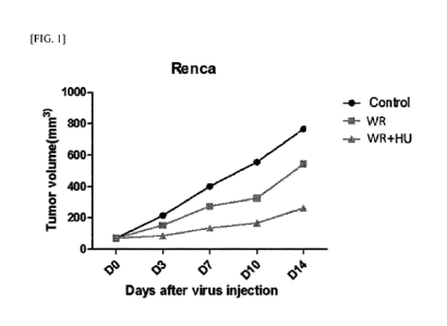
FIG. 1 illustrates results obtained by administering, to mouse renal cancer
cell-
transplanted mice (Renca), a wild-type vaccinia virus (Western Reserve strain
vaccinia
virus, WR) and hydroxyurea (HU), and then measuring tumor volumes on days 0,
3, 7,
10, and 14.
FIG. 2 illustrates results obtained by administering, to the mouse renal
cancer
cell-transplanted mice (Renca), the wild-type vaccinia virus (WR) and HU, and
then
measuring body weights on days 0, 3, 7, 10, and 14.
FIG. 3 illustrates results obtained by administering, to mouse renal cancer
cell-
transplanted mice (Renca), a recombinant vaccinia virus (WR VV'), which has
been
obtained by deleting TK gene from WR, and HU (60 mg/kg), and then measuring
tumor
volumes on days 0, 3,7, 10, 14, 17, and 21.
FIG. 4 illustrates results obtained by administering, to mouse renal cancer
cell-
transplanted mice (Renca), the recombinant vaccinia virus (WR VV') and HU (30
mg/kg), and then measuring tumor volumes on days 0, 3, 7, 10, and 14.
FIG. 5 illustrates results obtained by measuring tumor volumes 1 day before
and
on days 4 and 7 after administering, to mouse melanoma-transplanted mice
(B16F10),
a recombinant vaccinia virus (VV DD), which has been obtained by
simultaneously
deleting TK gene and vaccinia virus growth factor (VGF) gene from WR, and HU.
FIG. 6 illustrates results obtained by administering, to human lung cancer
cell
(NCI-H460)-transplanted mice, a recombinant vaccinia virus (WOTS-418) and HU,
and then measuring tumor volumes on days 0, 5, 10, 12, and 15.
FIG. 7 illustrates results obtained by administering, to human lung cancer
cell
(NCI-H460)-transplanted mice, the recombinant vaccinia virus (WOTS-418) and
HU,
and then measuring survival rates.
FIG. 8 illustrates results obtained by administering, to mouse renal cancer
cell-
transplanted mice (Renca), a recombinant vaccinia virus (VV') and human
granulocyte
4
Date Recue/Date Received 2022-02-25
CA 03152973 2022-02-25
colony stimulating factor (rhG-CSF) or HU, and then measuring tumor volumes in
the
mice.
FIG. 9 illustrates results obtained by isolating lymphocytes in the spleen
from
the mouse renal cancer cell-transplanted mice (Renca), to which the
recombinant
vaccinia virus (VV') and the human granulocyte colony stimulating factor (rhG-
CSF)
or HU have been administered, administering the lymphocytes to new mice, and
then
measuring tumor volumes in the new mice.
FIG. 10 illustrates results obtained by administering, to mouse renal cancer
cell-
transplanted mice (Renca), a recombinant vaccinia virus (Wyeth VV') and HU,
and
then measuring tumor volumes in the mice.
FIG. 11 illustrates results obtained by isolating T lymphocytes from mouse
renal
cancer cell-transplanted mice (Renca), to which a recombinant vaccinia virus
(Wyeth
VV') and HU have been administered, administering the T lymphocytes to new
mice,
and then measuring tumor volumes in the new mice.
FIG. 12 illustrates results obtained by isolating splenocytes isolated from
the
mouse renal cancer cell-transplanted mice (Renca), to which the recombinant
vaccinia
virus (Wyeth VV') and HU have been administered, administering the splenocytes
to
new mice, and then measuring tumor volumes in the new mice.
FIG. 13 illustrates results obtained by administering, to mouse renal cancer
cell-
transplanted mice (Renca), a recombinant vaccinia virus (Wyeth VV') and HU,
and
then measuring tumor volumes on day 22.
FIG. 14 illustrates results obtained by administering, to mouse renal cancer
cell-
transplanted mice (Renca), a recombinant vaccinia virus (Wyeth VV') and HU,
and
then observing the proliferation of CD4+ T cells or CD8+ T cells in the spleen
tissue.
FIG. 15 illustrates results obtained by administering, to mouse breast cancer
cell-transplanted mice (4T1), a recombinant vaccinia virus (OTS-412) and HU,
and then
observing the proliferation of CD4+ T cells or CD8+ T cells in the blood and
spleen
tissue.
FIG. 16 illustrates results obtained by administering, to the left tumor in
mouse
breast cancer cell-transplanted mice (4T1), a recombinant vaccinia virus (WR
VV')
5
Date Recue/Date Received 2022-02-25
CA 03152973 2022-02-25
and HU, and then measuring left tumor volumes.
FIG. 17 illustrates results obtained by administering, to the left tumor in
mouse
breast cancer cell-transplanted mice (4T1), a recombinant vaccinia virus (WR
VV')
and HU, and then measuring right tumor volumes.
FIG. 18 illustrates results obtained by administering, to mouse renal cancer
cell-
transplanted mice (Renca), a recombinant vaccinia virus (WR) and HU, and then
performing staining on day 22 to identify distribution of the recombinant
vaccinia virus
in mouse tumor tissues.
FIG. 19 illustrates results obtained by administering, to normal mice, a wild-
type vaccinia virus (WR) or a wild-type vaccinia virus (WR) and HU, and then
identifying distribution of the wild-type vaccinia virus in liver and kidney
tissues.
FIG. 20 illustrates the absolute neutrophil count of mice in each group after
administering, to mouse renal cancer cell-transplanted mice (Renca), saline,
HU, a
recombinant vaccinia virus (OTS-412), a recombinant vaccinia virus and a
recombinant
human granulocyte colony-stimulating factor (OTS-412+rhG-CSF), or a
recombinant
vaccinia virus and HU (OTS-412+HU).
FIG. 21 illustrates the blood neutrophil count of mice measured in each group
after administering, to mouse renal cancer cell-transplanted mice (Renca),
saline, a
recombinant vaccinia virus, or a recombinant vaccinia virus (WR VV') and HU.
FIG. 22 illustrates the blood neutrophil count of mice measured in each group
after administering, to mouse renal cancer cell-transplanted mice (Renca),
saline, a
recombinant vaccinia virus, or a recombinant vaccinia virus (WOTS-418) and HU.
FIG. 23 illustrates the blood neutrophil count of mice measured in each group
after administering, to mouse renal cancer cell-transplanted mice (Renca),
saline,
lenalidomide, or HU.
FIG. 24 illustrates the blood neutrophil count of mice measured in each group
after administering, to mouse renal cancer cell-transplanted mice (Renca),
saline, a
recombinant vaccinia virus, a recombinant vaccinia virus (WOTS-418) and
lenalidomide, or a recombinant vaccinia virus (WOTS-418) and HU.
FIG. 25 illustrates the tumor volume of mice measured after administering, to
6
Date Recue/Date Received 2022-02-25
CA 03152973 2022-02-25
mouse renal cancer cell-transplanted mice (Renca), a recombinant vaccinia
virus (WR
VV') and lenalidomide.
FIG. 26 illustrates the tumor volume of mice measured after administering, to
mouse renal cancer cell-transplanted mice (Renca), a recombinant vaccinia
virus (WR
VV") and palbociclib.
FIG. 27 illustrates the body weight of mice measured after administering, to
mouse renal cancer cell-transplanted mice (Renca), a recombinant vaccinia
virus (WR
VV') and palbociclib.
FIG. 28 illustrates the tumor volume of mice measured on day 0, day 4, day 10,
day 14, day 17, and day 21 after administering, to mouse renal cancer cell-
transplanted
mice (Renca), an oncolytic virus (Wyeth VV'), a PD-1 inhibitor, and HU.
FIG. 29 illustrates the tumor volume of mice measured on day 0, day 4, day 10,
day 14, and day 17 after administering, to mouse renal cancer cell-
transplanted mice
(Renca), an oncolytic virus (Wyeth VV'), a CTLA-4 inhibitor, and HU.
FIG. 30 illustrates the tumor volume of mice measured on day 0, day 4, day 10,
day 14, day 17, and day 21 after administering, to mouse renal cancer cell-
transplanted
mice (Renca), an oncolytic virus (Wyeth VV'), a PD-Li inhibitor, and HU.
FIG. 31 illustrates the tumor volume of mice measured on day 0, day 3, day 7,
day 10, and day 14 after administering, to mouse breast cancer cell-
transplanted mice
(4T1), an oncolytic virus (WR VVtk-), a CTLA-4 inhibitor, and HU.
FIG. 32 illustrates the tumor volume of mice measured on day 0, day 3, day 7,
day 10, day 14, and day 18 after administering, to mouse breast cancer cell-
transplanted
mice (4T1), an oncolytic virus (WOTS-418), a PD-Li inhibitor, and HU.
FIG. 33 illustrates the tumor volume of mice measured on day 0, day 3, and day
7 after administering, to mouse renal cancer cell-transplanted mice (Renca), a
Western
Reserve strain vaccinia virus (WR), a CTLA-4 inhibitor, and HU.
Best Mode for Carrying out the Invention
Hereinafter, the present invention will be specifically described.
In an aspect of the present invention, there is provided a pharmaceutical
7
Date Recue/Date Received 2022-02-25
CA 03152973 2022-02-25
composition for preventing or treating cancer, comprising, as active
ingredients, a
vaccinia virus and granulopoiesis inhibitor.
The vaccinia virus and granulopoiesis inhibitor contained in the
pharmaceutical
composition may be administered in combination simultaneously, sequentially,
or in
reverse order. Specifically, the vaccinia virus and granulopoiesis inhibitor
may be
administered simultaneously. In addition, the granulopoiesis inhibitor may be
first
administered, followed by the vaccinia virus. Furthermore, the vaccinia virus
may be
first administered, followed by the granulopoiesis inhibitor.
In addition, the
granulopoiesis inhibitor may be first administered, followed by the vaccinia
virus, and
the granulopoiesis inhibitor may be administered again.
The vaccinia virus may belong to, but is not limited to, Western Reserve (WR),
New York vaccinia virus (NYVAC), Wyeth (The New York City Board of Health;
NYCBOH), LC16m8, Lister, Copenhagen, Tian Tan, USSR, Tashkent, Evans,
International Health Division-J (IHD-J), or International Health Division-
White (IHD-
W) vaccinia virus strain. In an embodiment of the present invention, Western
Reserve
strain vaccinia virus and Wyeth strain vaccinia virus were used.
The vaccinia virus may be a wild-type vaccinia virus or a recombinant vaccinia
virus. Specifically, the recombinant vaccinia virus may be obtained by
deleting a gene
from a wild-type vaccinia virus or inserting a foreign gene thereinto. Here,
among the
genes of the wild-type vaccinia virus, a gene related to viral virulence may
be deleted
which encodes any one selected from the group consisting of thymidine kinase
(TK),
vaccinia growth factor (VGF), WR53.5, F13.5L, F14.5L, A56R, B 1 8R, or
combinations
thereof
In addition, the inserted foreign gene may be a gene that promotes immunity
and encodes any one selected from the group consisting of herpes simplex virus
thymidine kinase (HSV-TK), mutated HSV-TK, granulocyte-macrophage colony-
stimulating factor (GM-CSF), granulocyte colony-stimulating factor (G-CSF),
cytosine
deaminase (CD), carboxyl esterase type 1, carboxyl esterase type 2, interferon
beta
(INF-13), somatostatin receptor 2, and combinations thereof
Specifically, the recombinant vaccinia virus may be obtained by deleting TK
8
Date Recue/Date Received 2022-02-25
CA 03152973 2022-02-25
gene from a vaccinia virus that belongs to Western Reserve (WR), New York
vaccinia
virus (NYVAC), Wyeth (The New York City Board of Health; NYCBOH), LC16m8,
Lister, Copenhagen, Tian Tan, USSR, Tashkent, Evans, International Health
Division-J
(IHD-J), or International Health Division-White (IHD-W) vaccinia virus strain.
In an
embodiment of the present invention, a recombinant vaccinia virus obtained by
deleting
TK gene from a Western Reserve strain vaccinia virus was used, and this virus
was
designated "WR VV". In addition, in an embodiment of the present invention, a
recombinant vaccinia virus obtained by deleting TK gene from a Wyeth strain
vaccinia
virus was used, and this virus was designated "Wyeth VV'".
In addition, the recombinant vaccinia virus may be obtained by deleting TK
gene and VGF gene from a vaccinia virus that belongs to Western Reserve,
NYVAC,
Wyeth, LC16m8, Lister, Copenhagen, Tian Tan, USSR, Tashkent, Evans, IHD-J, or
IHD-W vaccinia virus strain.
In an embodiment of the present invention, a
recombinant vaccinia virus obtained by deleting TK gene and VGF gene from a
Western
Reserve strain vaccinia virus was used, and this virus was designated "VV DD".
Furthermore, the recombinant vaccinia virus may be obtained by deleting TK
gene from and inserting HSV-TK gene into a vaccinia virus that belongs to
Western
Reserve, NYVAC, Wyeth, LC16m8, Lister, Copenhagen, Tian Tan, USSR, Tashkent,
Evans, IHD-J, or IHD-W vaccinia virus strain.
In addition, the recombinant vaccinia virus may be obtained by deleting TK
gene from and inserting mutated HSV-TK gene into a vaccinia virus that belongs
to
Western Reserve, NYVAC, Wyeth, LC16m8, Lister, Copenhagen, Tian Tan, USSR,
Tashkent, Evans, IHD-J, or IHD-W vaccinia virus strain. In an embodiment of
the
present invention, a recombinant vaccinia virus obtained by deleting TK gene
from a
Wyeth strain vaccinia virus and inserting, into the deleted position, a gene
encoding the
HSV-TK fragment (1-330 aa) of SEQ ID NO: 1 was used, and this virus was
designated
"OTS-412". In addition, in an embodiment of the present invention, a
recombinant
vaccinia virus obtained by deleting TK gene from a Western Reserve strain
vaccinia
virus and inserting, into the deleted position, a gene encoding the HSV-TK
variant of
SEQ ID NO: 2 of HSV-TK gene was used, and this virus was designated "WOTS-
418".
9
Date Recue/Date Received 2022-02-25
CA 03152973 2022-02-25
Furthermore, the recombinant vaccinia virus may be obtained by deleting TK
gene from and inserting GM-CSF gene into a vaccinia virus that belongs to
Western
Reserve, NYVAC, Wyeth, LC16m8, Lister, Copenhagen, Tian Tan, USSR, Tashkent,
Evans, IHD-J, or IHD-W vaccinia virus strain.
In addition, the recombinant vaccinia virus may be obtained by deleting TK
gene from and inserting G-CSF gene into a vaccinia virus that belongs to
Western
Reserve, NYVAC, Wyeth, LC16m8, Lister, Copenhagen, Tian Tan, USSR, Tashkent,
Evans, IHD-J, or IHD-W vaccinia virus strain.
Furthermore, the recombinant vaccinia virus may be obtained by deleting TK
gene from and inserting cytosine deaminase (CD) gene into a vaccinia virus
that belongs
to Western Reserve, NYVAC, Wyeth, LC16m8, Lister, Copenhagen, Tian Tan, USSR,
Tashkent, Evans, IHD-J, or IHD-W vaccinia virus strain.
In addition, the recombinant vaccinia virus may be obtained by deleting TK
gene from and inserting somatostatin receptor 2 gene into a vaccinia virus
that belongs
to Western Reserve, NYVAC, Wyeth, LC16m8, Lister, Copenhagen, Tian Tan, USSR,
Tashkent, Evans, IHD-J, or IHD-W vaccinia virus strain.
Furthermore, the recombinant vaccinia virus may be obtained by deleting TK
gene from and inserting any two or more genes, which are selected from the
group
consisting of genes, each of which encodes herpes simplex virus thymidine
kinase
(HSV-TK), mutated HSV-TK, granulocyte-macrophage colony-stimulating factor
(GM-CSF), granulocyte colony-stimulating factor (G-CSF), cytosine deaminase
(CD),
or somatostatin receptor 2, into a vaccinia virus that belongs to Western
Reserve,
NYVAC, Wyeth, LC16m8, Lister, Copenhagen, Tian Tan, USSR, Tashkent, Evans,
IHD-J, or IHD-W vaccinia virus strain.
In addition, the recombinant vaccinia virus may be obtained by deleting TK
gene and VGF gene from and inserting any one gene, which is selected from the
group
consisting of genes, each of which encodes herpes simplex virus thymidine
kinase
(HSV-TK), mutated HSV-TK, granulocyte-macrophage colony-stimulating factor
(GM-CSF), granulocyte colony-stimulating factor (G-CSF), cytosine deaminase
(CD),
or somatostatin receptor 2, and combinations thereof, into a vaccinia virus
that belongs
Date Recue/Date Received 2022-02-25
CA 03152973 2022-02-25
to Western Reserve, NYVAC, Wyeth, LC16m8, Lister, Copenhagen, Tian Tan, USSR,
Tashkent, Evans, IHD-J, or IHD-W vaccinia virus strain.
As used herein, the term "gene deletion" means that a gene is not expressed
due
to partial or complete deletion of the gene, or insertion of a foreign gene
thereinto. In
a case where partial deletion occurs in the gene, some amino acids at the N-
terminus or
C-terminus of a polypeptide expressed by the gene may be deleted.
As used herein, the term "thymidine kinase (TK)" refers to an enzyme that is
called thymidine kinase and involved in nucleotide biosynthesis. TK is an
enzyme
used for nucleotide biosynthesis in both cells and viruses. Here, for the
cells, normal
cells do not divide anymore, and thus no TK exists therein; and even for
rapidly dividing
cells such as hair follicle cells, TK is not present in an amount sufficient
for viruses to
utilize. From these viewpoints, a virus is allowed to proliferate only in the
presence
of cancer cells, in which TK is present, by deletion of TK gene therein, so
that the cancer
cells may be selectively killed.
As used herein, the term "vaccinia growth factor (VGF)" refers to a
polypeptide
that has sequence homology to epidermal growth factor and stimulates cell
proliferation
around infected cells. A vaccinia virus replicates better in proliferating
cells, and thus
may be advantageously used for viral replication in vivo. In order to cause an
oncolytic virus to proliferate more specifically only in cancer cells, the
virus may
additionally undergo deletion of VGF gene in addition to deletion of the TK
gene.
As used herein, the term "GM-CSF", which is called granulocyte-macrophage
colony-stimulating factor, refers to a protein secreted by macrophages, T
cells, mast
cells, natural killer cells, endothelial cells, and fibroblasts. GM-CSF
stimulates stem
cells to produce granulocytes (neutrophils, basophils, eosinophils) and
monocytes. In
addition, GM-CSF rapidly increases the number of macrophages, thereby inducing
an
immune response. GM-CSF may be of human origin and may be a protein having the
sequence of GenBank: AAA52578.1.
As used herein, the term "CD", which is called cytosine deaminase, refers to
an
enzyme that catalyzes hydrolytic deamination of cytosine into uracil and
ammonia.
As used herein, the term "G-CSF", which is called granulocyte colony-
11
Date Recue/Date Received 2022-02-25
CA 03152973 2022-02-25
stimulating factor, refers to a cytokine produced by macrophages, fibroblasts,
endothelial cells, and the like upon stimulation by inflammation or endotoxin.
G-CSF
promotes production of neutrophils. The G-CSF may be of human origin (rhGCSF)
and may be a protein having the sequence of GenBank: AAA03056.1.
As used herein, the term "somatostatin receptor 2" refers to a protein encoded
by SSTR2 gene in humans. The somatostatin receptor 2 is expressed mainly in
tumors,
and patients with neuroendocrine tumors, who overexpress somatostatin receptor
2,
show improved prognosis. The somatostatin receptor 2 has capacity to stimulate
apoptosis in many cells, including cancer cells.
A myeloid cell may be granulocytes, and specifically, the myeloid cells may be
neutrophils, eosinophils, or basophils.
The granulopoiesis inhibitor may be a substance that inhibits granulocytes
(e.g.,
neutrophils, eosinophils or basophils) mainly produced in bone marrow. The
granulopoiesis inhibitor, when reducing or inhibiting the number of
neutrophils (i.e.,
one of myeloid cells) in the body, may be referred to as a neutrophil
inhibitor or include
the same. The neutrophil is also called a neutrophilic leukocyte, and refers
to a
neutrophil cell circulating in the blood, which is a type of a granulocyte
mainly
produced in the bone marrow. Neutrophils are the main component of
granulocytes,
and the normal number is about 1,500 to about 8,000 per 1 mm3 of blood. The
neutrophil absorbs, through phagocytosis, foreign substances such as bacteria
that have
invaded the body and breaks the foreign substances down with a digestive
enzyme (e.g.,
hydrogen peroxide, lysosome, etc.).
As used herein, the term "absolute neutrophil count (ANC)" refers to the
number
obtained by multiplication of the number of white blood cells x the percentage
of
neutrophils.
The granulopoiesis inhibitor may be hydroxyurea, lenalidomide, thalidomide,
tadalafil, palbociclib, alkylating agents, anthracyclines, antimetabolites,
camptothecins,
epipodophyllotoxins, mitomycin C, taxanes, or vinblastine. The hydroxyurea may
be
a compound having the structure of Formula 1 below:
[Formula 1]
12
Date Recue/Date Received 2022-02-25
CA 03152973 2022-02-25
0
00,0H
H N
The hydroxyurea is known as an anticancer agent that inhibits DNA synthesis;
however, the exact mechanism of action thereof is not elucidated. In addition,
the
hydroxyurea may be included in the pharmaceutical composition in the form of a
commercialized drug that contains hydroxyurea. Examples of the commercialized
drug that contains hydroxyurea may include, but are not limited to,
Hydroxyurea0,
Hydrea0, DroxiaTM, MylocelTM, Siklos0, and Hydrine0 capsule. The hydroxyurea
may be taken orally, and parenteral administration thereof is also possible.
The lenalidomide may be a compound having the structure of Formula 2 below:
[Formula 2]
"
= wiz
The lenalidomide is an anticancer agent used for the treatment of multiple
myeloma, etc. In addition, the lenalidomide stops the growth cycle of cancer
cells and
inhibits cancer proliferation by activating tumor suppressor genes as an
anticancer effect.
.. As an immunomodulatory effect, the lenalidomide eliminates tumor cells by
activating
immune cells (e.g., T cells, natural killer cells (NK cells), B cells, etc.).
In addition,
the lenalidomide has an angiogenesis inhibitory effect that inhibits the
formation of new
blood vessels to supply nutrients to cancer cells.
The thalidomide may be a compound having the structure of Formula 3 below:
[Formula 3]
13
Date Recue/Date Received 2022-02-25
CA 03152973 2022-02-25
0
045
N
4 ,
=
The exact mechanism of action of thalidomide is not known, but is used for the
treatment of multiple myeloma and severe skin lesions in patients with leprosy
(Hansen's disease).
The tadalafil may be a compound having the structure of Formula 4 below:
[Formula 4]
=0 H
1
N I. N
H
4,1.61/4 0
IIP
O\
The palbociclib may be a compound having the structure of Formula 5 below:
[Formula 5]
9
WM
L,NII
The alkylating agent may be nitrogen mustard, an ethylene derivative, an
alkylsulfonic acid derivative, nitrosoureas, or triazines compounds among
chemotherapeutic agents for malignant tumors. These may also be called in the
name
of an alkylating agent because they substitute the hydrogen in many organic
compounds,
proteins, or nucleic acids with an alkyl group. By alkylation with the
alkylating agent,
DNA replication and mRNA transcription of tumor cells may be inhibited and an
14
Date Recue/Date Received 2022-02-25
CA 03152973 2022-02-25
antitumor action may be exhibited. As a common pharmacological action, the
alkylating agent acts non-specifically on each phase of a cell cycle, and may
inhibit cell
division with a high proliferative potential. Since the alkylating agent
exhibits a
radiation-like action, dyshematopoiesis may be strong and immunosuppression
may be
caused.
The anthracycline is a drug extracted from Streptomyces bacteria, is used for
cancer chemotherapy, and is used to treat many cancers including leukemia,
lymphoma,
breast cancer, gastric cancer, uterine cancer, ovarian cancer, bladder cancer,
and lung
cancer.
The first discovered anthracycline was daunorubicin (trademark:
Daunomycin), which was naturally produced by Streptomyces peucetius, which is
a
species of Actinomycetes. The most clinically important anthracyclines include
doxorubicin, daunorubicin, epirubicin, idarubicin, etc.
The antimetabolite may be a substance that inhibits the development and
proliferation of cells by antagonizing essential metabolites for the
metabolism or growth
of microorganisms or tumor cells. Slupamine, which is used as a
chemotherapeutic
agent and is antagonistic to the bacterial para-aminobenzoate (PABA), was
historically
first developed. The antimetabolites may include sulfa drugs for bacteria,
purine
antimetabolite drugs for malignant tumors (8-azaguanine, 6-thioguanine, 6-
mercaptopurine), pyrimidine antimetabolite drugs (5 -fluororuacil, cytarabine,
azauridine), folate antimetabolite drugs (4-aminopterin, methotrexat), or
glutamine
antimetabolite drugs (azerin, DON).
The camptothecin may be a natural anticancer substance isolated from plants
such as Camptotheca acuminate (Camptotheca, Happy tree), and Chonemorpha
fragrans.
The camptothecin may be a compound having the structure of Formula 6 below:
[Formula 6]
Date Recue/Date Received 2022-02-25
CA 03152973 2022-02-25
0
0
Noõµ.
HO 0
The epipodophyllotoxin may be a natural anticancer substance produced
naturally in the root of Podophyllum peltatum. A derivative of
epipodophyllotoxin
may be used for cancer treatment at present.
The epipodophyllotoxin may be a compound having the structure of Formula 7
below:
[Formula 7]
o 0
HOF1
.õ
o ''H
HO" 0
0
0
0 OH
\--0 0
The mitomycin C may be an antibiotic substance isolated by Streptomyces
griseus. The mitomycin C is thermally stable, has the lowest toxicity, and has
a strong
anticancer effect. The mitomycin C inhibits the cellular enzyme system and
nucleic
acid metabolism, thus inhibiting the division of cell nuclei, and thereby
preventing the
proliferation of malignant tumor cells. Examples of the side effects of
mitomycin C
include bleeding accompanied with leukopenia and thrombocytopenia, etc.
The taxane is also called a cell division inhibitor or anti-microtubule
inhibitor,
and may be an anticancer agent which inhibits cancer cell growth by inhibiting
cell
division. The taxane may kill cancer cells by disrupting the microtubules
through
which chromosomes move during cell mitosis. The taxane is used to treat
various
types of cancer such as breast cancer, ovarian cancer, and non-small cell lung
cancer.
16
Date Recue/Date Received 2022-02-25
CA 03152973 2022-02-25
Specifically, the taxane includes paclitaxel, docetaxel, etc.
The vinblastine may be an anticancer agent of a vinca alkaloid component used
for the treatment of various types of cancer. The vinblastine was first
extracted from
the periwinkle plant belonging to the Oleander family, and a synthetic
material is used
at present. The vinblastine is the most widely used agent among anticancer
agents and
is also widely used in combination therapy with other anticancer agents. The
vinblastine prevents the division of cancer cells by interfering with the
normal function
of microtubules. The vinblastine may widely be used for testicular cancer,
breast
cancer, lymphoma, Kaposi's sarcoma, etc. The most important side effect of
vinblastine is a decrease in leukocytes and thrombocyte, and side effects such
as
gastrointestinal disorders, increased blood pressure, excessive sweating,
depression,
muscle pain, nausea, and headache may appear.
A dosage of the vaccinia virus varies depending on the individual's condition
and body weight, the severity of disease, the type of drug, the route and
period of
administration, and may be appropriately selected by a person skilled in the
art. The
dosage may be such that a patient receives a vaccinia virus at lx105 to lx1018
of virus
particles, infectious virus units (TCID50), or plaque forming units (pfu).
Specifically,
the dosage may be such that a patient receives a vaccinia virus at lx105,
2x10⁵, 5x10⁵,
1x10⁶, 2x10⁶, 5x10⁶, 1x10⁷, 2x10⁷, 5x10⁷, 1x10⁸, 2x10⁸, 5x10⁸, 1x10⁹, 2x10⁹,
5x10⁹,
1x10¹⁰, 5x10¹⁰, 1x10¹¹, 5x10¹¹, 1x10¹², 1x10¹³, 1x10¹⁴, 1x10¹⁵, 1x10¹⁶,
1x10¹⁷, or
higher of virus particles, infectious virus units, or plaque forming units,
and various
numerical values and ranges between the above-mentioned numerical values may
also
be included therein. Preferably, the vaccinia virus may be administered at a
dose of
1 x10⁵ to 1 x101 pfu. More preferably, the vaccinia virus may be administered
at a
dose of equal to or greater than 1x10⁵ and lower than 1 x10⁹ pfu. In an
embodiment of
the present invention, the vaccinia virus was administered at lx105 or lx107
pfu.
In addition, the granulopoiesis inhibitor may be administered at a dose of 1
mg/kg/day to 100 mg/kg/day, or 10 mg/kg/day to 90 mg/kg/day. Specifically, the
granulopoiesis inhibitor may be administered at a dose of 10 mg/kg/day to 90
mg/kg/day,
15 mg/kg/day to 80 mg/kg/day, 20 mg/kg/day to 70 mg/kg/day, 25 mg/kg/day to 65
17
Date Recue/Date Received 2022-02-25
CA 03152973 2022-02-25
mg/kg/day, or 30 mg/kg/day to 60 mg/kg/day. In an embodiment of the present
invention, hydroxyurea, lenalidomide, or palbociclib, as granulopoiesis
inhibitors, was
administered at a dose of 25 mg/kg/day, 30 mg/kg/day, 50 mg/kg/ day, 60
mg/kg/day,
or 100 mg/kg/day. Depending on the dosage, the granulopoiesis inhibitor may be
administered in divided doses several times a day. Specifically, the
granulopoiesis
inhibitor may be administered 1 to 4 times a day or 1 to 2 times a day.
The pharmaceutical composition may further include an immune checkpoint
inhibitor (ICI).
The immune checkpoint inhibitor refers to a substance that inhibits the
mechanism of cancer cells that interferes with the activation of T cells, and
may be any
one selected from the group consisting of an anti-PD-Li antibody, an anti-PD-1
antibody, an anti-CTLA4 antibody, an anti PD-L2 antibody, an LTF2 modulating
antibody, an anti-LAG3 antibody, an anti-A2aR antibody, an anti-TIGIT
antibody, an
anti-TIM-3 antibody, an anti-B7-H3 antibody, an anti-B7-H4 antibody, an anti-
VISTA
antibody, an anti-CD47 antibody, an anti-BTLA antibody, an anti-MR antibody,
an
anti-IDO antibody, and a combination thereof
Cancer cells hijack the immune checkpoint system as a mechanism to evade an
immune response. Specifically, cancer cells use immune checkpoint receptors to
evade immune responses, and representative receptors include PD-L1, PD-1, CTLA-
4,
etc. In order to prevent immune evasion of these cancer cells, immune
checkpoint
inhibitors, which are molecules that specifically bind to immune checkpoint
receptors,
are used for cancer treatment. The first immune checkpoint inhibitor is
ipilimumab
(Yervoy0), which is a monoclonal antibody that specifically binds to cytotoxic
T-
lymphocytes associated antigen-4 (CTLA-4). The immune checkpoint therapy
developed next is monoclonal antibodies against programmed cell death-1 (PD-1)
and
the corresponding ligand, programmed death ligand-1 (PD-L1). Representative
drugs
include anti-PD-1 antibodies (e.g., nivolumab (Opdivo0), pembrolizumab
(Keytruda0),
etc.) and anti-PD-Li antibodies (e.g., avelumab (Bavencio0), atezolizumab
(Tecentriq0), durvalumab (Imfinzi0), etc.).
In addition thereto, studies on monoclonal antibodies that specifically bind
to
18
Date Recue/Date Received 2022-02-25
CA 03152973 2022-02-25
various immune checkpoint receptors (e.g., glucocorticoid-induced TNFR-related
protein (GITR), killer cell immunoglobulin-like receptor (KIR), lymphocyte-
activation
gene-3 (LAG-3), T-cell immunoglobulin and mucin-domain containing-3 (TIM-3),
tumor-necrosis factor receptor superfamily member 4 (TNFRSF4)) are underway.
The dose of the immune checkpoint inhibitor may be administered in
compliance with the dosage regimen of each manufacturer. The dose of the
immune
checkpoint inhibitor may be 0.1 mg/kg to 10 mg/kg or 1 mg/kg to 5 mg/kg. For
example, in the case of Opdivo Inj. containing the nivolumab as an active
ingredient, 3
mg/kg may be intravenously instilled over 60 minutes at intervals of 2 weeks,
and
regarding the dosage regimen as a combination therapy, 1 mg/kg may be
intravenously
instilled over 30 minutes. In addition, in the case of Keytruda Inj.
containing
pembrolizumab as an active ingredient, 200 mg may be intravenously instilled
over 30
minutes at intervals of 3 weeks. As such, since even the same anti-PD-1
antibody has
different dosage regimen depending on the product, it is preferred that the
antibody be
administerd in compliance with the manufacturer's dosage regimen.
When the pharmaceutical composition further includes an immune checkpoint
inhibitor, the oncolytic virus, granulopoiesis inhibitor, and immune
checkpoint inhibitor
included in the pharmaceutical composition may be administered simultaneously,
sequentially, or in reverse order.
Specifically, the oncolytic virus, granulopoiesis inhibitor, and immune
checkpoint inhibitor may be administered simultaneously.
In addition, the
granulopoiesis inhibitor may be administered first, and the immune checkpoint
inhibitor
may be administered thereafter, and then the oncolytic virus may be
administered. The
granulopoiesis inhibitor may be administered first, and the oncolytic virus
may be
administered thereafter, and then the immune checkpoint inhibitor may be
administered.
The granulopoiesis inhibitor may be administered first, and then, the
oncolytic virus
and the immune checkpoint inhibitor may be administered simultaneously.
Furthermore, the oncolytic virus may be administered first, and the
granulopoiesis inhibitor may be administered thereafter, and then the immune
checkpoint inhibitor may be administered. The oncolytic virus may be
administered
19
Date Recue/Date Received 2022-02-25
CA 03152973 2022-02-25
first, and the immune checkpoint inhibitor may be administered thereafter, and
then the
granulopoiesis inhibitor may be administered.
The oncolytic virus may be
administered first, and then the granulopoiesis inhibitor and the immune
checkpoint
inhibitor may be administered simultaneously.
In addition, the immune checkpoint inhibitor may be administered first, and
the
granulopoiesis inhibitor may be administered thereafter, and then the
oncolytic virus
may be administered. The immune checkpoint inhibitor may be administered
first,
and the oncolytic virus may be administered thereafter, and then the
granulopoiesis
inhibitor may be administered. The immune checkpoint inhibitor may be
administered
first, and then the oncolytic virus and the granulopoiesis inhibitor may be
administered
simultaneously.
Furthermore, the granulopoiesis inhibitor may be administered first, and the
oncolytic virus may be administered thereafter, and then the immune checkpoint
inhibitor may be administered, and once again the granulopoiesis inhibitor may
be
administered. The granulopoiesis inhibitor may be administered first, and the
immune
checkpoint inhibitor may be administered thereafter, and then the oncolytic
virus may
be administered, and once again the granulopoiesis inhibitor may be
administered.
The granulopoiesis inhibitor may be administered first, and the oncolytic
virus and the
immune checkpoint inhibitor may be administered simultaneously, and once again
the
granulopoiesis inhibitor may be administered.
In addition, the granulopoiesis inhibitor may be administered first, and the
oncolytic virus may be administered thereafter, and once again the
granulopoiesis
inhibitor may be administered, and then the immune checkpoint inhibitor may be
administered. The granulopoiesis inhibitor may be administered first, and the
immune
checkpoint inhibitor may be administered thereafter, and once again the
granulopoiesis
inhibitor may be administered, and then the oncolytic virus may be
administered.
Furthermore, the granulopoiesis inhibitor may be administered first, and the
oncolytic virus may be administered thereafter, and once again the
granulopoiesis
inhibitor may be administered, and then the immune checkpoint inhibitor may be
administered, and once again the granulopoiesis inhibitor may be administered.
The
Date Recue/Date Received 2022-02-25
CA 03152973 2022-02-25
granulopoiesis inhibitor may be administered first, and the immune checkpoint
inhibitor
may be administered thereafter, and once again the granulopoiesis inhibitor
may be
administered, and then the oncolytic virus may be administered, and once again
the
granulopoiesis inhibitor may be administered.
In addition, the oncolytic virus may be administered first, and the
granulopoiesis
inhibitor may be administered thereafter, and then the immune checkpoint
inhibitor may
be administered, and once again the granulopoiesis inhibitor may be
administered.
The immune checkpoint inhibitor may be administered first, and the
granulopoiesis
inhibitor may be administered thereafter, and then the oncolytic virus may be
administered, and once again the granulopoiesis inhibitor may be administered.
The cancer may be solid cancer or blood cancer. Specifically, the blood cancer
may be any one selected from the group consisting of lymphoma, acute leukemia,
and
multiple myeloma. The solid cancer may be any one selected from the group
consisting of lung cancer, colorectal cancer, prostate cancer, thyroid cancer,
breast
cancer, brain cancer, head and neck cancer, esophageal cancer, skin cancer,
thymic
cancer, gastric cancer, colon cancer, liver cancer, ovarian cancer, uterine
cancer, bladder
cancer, rectal cancer, gallbladder cancer, biliary tract cancer, pancreatic
cancer, and
combinations thereof
In addition, the pharmaceutical composition of the present invention may
further comprise a physiologically acceptable carrier. In addition, the
pharmaceutical
composition of the present invention may further comprise suitable excipients
and
diluents commonly used in the preparation of pharmaceutical compositions. In
addition, the pharmaceutical composition may be formulated in the form of an
injection
according to a conventional method.
In a case of being formulated as preparations for parenteral administration,
the
pharmaceutical composition may be formulated into sterilized aqueous
solutions, non-
aqueous solutions, suspensions, emulsions, freeze-dried preparations,
suppositories, or
the like.
For the non-aqueous solution or the suspension, propylene glycol,
polyethylene glycol, vegetable oil such as olive oil, injectable ester such as
ethyl oleate,
or the like may be used. As the base of the suppository, WitepsolTM, macrogol,
21
Date Recue/Date Received 2022-02-25
CA 03152973 2022-02-25
TweenTm 61, cacao butter, laurin fat, glycerogelatin, or the like may be used.
Regarding the administration route, dosage, and frequency of administration,
the pharmaceutical composition may be administered to a subject in a variety
of ways
and amounts depending on the patient's condition and the presence or absence
of side
effects; and the optimal administration route, dosage, and frequency of
administration
therefor may be selected by a person skilled in the art within a suitable
range. In
addition, the pharmaceutical composition may be administered in combination
with
another drug or physiologically active substance whose therapeutic effect is
known for
the disease to be treated, or may be formulated in the form of a combination
preparation
.. with the other drug.
The pharmaceutical composition may be administered parenterally, and such
administration may be performed by any suitable method, such as intratumoral,
intraperitoneal, subcutaneous, intradermal, intranodal, intravenous, or
intraarterial
administration.
Among these, intratumoral, intraperitoneal, or intravenous
administration may be preferred. On the other hand, the dosage of the
pharmaceutical
composition may be determined depending on the administration schedule, the
total
dosage, and the patient's health condition.
The pharmaceutical composition for treating cancer may be characterized by
increased cancer selectivity of the vaccinia virus.
Another aspect of the present invention provides a kit for preventing or
treating
cancer, which includes a first composition including a vaccinia virus as an
active
ingredient and a second composition including a granulopoiesis inhibitor as an
active
ingredient. The kit may further include a third composition which includes an
immune
checkpoint inhibitor as an active ingredient.
The vaccinia virus, granulopoiesis inhibitor, and immune checkpoint inhibitor
are the same as those described above in the pharmaceutical composition.
The second composition that includes the granulopoiesis inhibitor as an active
ingredient may be a commercialized drug. Examples of the commercialized drug
that
contains hydroxyurea as an active ingredient as the granulopoiesis inhibitor
may include
Hydroxyurea0, Hydrea0, DroxiaTM, MylocelTM, Siklos0, and Hydrine0 capsule.
22
Date Recue/Date Received 2022-02-25
CA 03152973 2022-02-25
The second composition may be taken orally, and parenteral administration
thereof is
also possible.
A dosage of the first composition varies depending on the individual's
condition
and body weight, the severity of disease, the type of drug, the route and
period of
administration, and may be appropriately selected by a person skilled in the
art. The
dosage may be such that a patient receives a vaccinia virus at lx 105 to
lx1018 of virus
particles, infectious virus units (TCID50), or plaque forming units (pfu).
Specifically,
the dosage may be such that a patient receives a vaccinia virus at lx105,
2x10⁵, 5x10⁵,
1x10⁶, 2x10⁶, 5x10⁶, 1x10⁷, 2x10⁷, 5x10⁷, 1x10⁸, 2x10⁸, 5x10⁸, 1x10⁹, 2x10⁹,
5x10⁹,
1x10¹⁰, 5x10¹⁰, 1x10¹¹, 5x10¹¹, 1x10¹², 1x10¹³, 1x10¹⁴, 1x10¹⁵, 1x10¹⁶,
1x10¹⁷, or
higher of virus particles, infectious virus units, or plaque forming units,
and various
numerical values and ranges between the above-mentioned numerical values may
also
be included therein. Preferably, the first composition may be administered at
a dose
of lx105 to lx101 pfu. More preferably, the first composition may be
administered at
a dose of equal to or greater than 1x10⁵ and lower than 1x10⁹ pfu. In an
embodiment
of the present invention, the first composition was administered at lx105 or
lx107 pfu.
In addition, the second composition may be administered at a dose of 1
mg/kg/day to 100 mg/kg/day, or 10 mg/kg/day to 90 mg/kg/day. Specifically, the
second composition may be administered at a dose of 10 mg/kg/day to 90
mg/kg/day,
15 mg/kg/day to 80 mg/kg/day, 20 mg/kg/day to 70 mg/kg/day, 25 mg/kg/day to 65
mg/kg/day, or 30 mg/kg/day to 60 mg/kg/day. In an embodiment of the present
invention, the second composition was administered at 25 mg/kg/day, 30
mg/kg/day, 50
mg/kg/day, 60 mg/kg/day or 100 mg/kg/day. Depending on the dosage, the second
composition may be administered in divided doses several times a day.
Specifically,
.. the second composition may be administered 1 to 4 times a day or 1 to 2
times a day.
The dose of the third composition may be administered in compliance with the
dosage regimen of each manufacturer of the immune checkpoint inhibitor
included in
the third composition. The dose of the third composition may be 0.1 mg/kg to
10
mg/kg or 1 mg/kg to 5 mg/kg.
The cancer may be solid cancer or blood cancer. Specifically, the blood cancer
23
Date Recue/Date Received 2022-02-25
CA 03152973 2022-02-25
may be any one selected from the group consisting of lymphoma, acute leukemia,
and
multiple myeloma. The solid cancer may be any one selected from the group
consisting of lung cancer, colorectal cancer, prostate cancer, thyroid cancer,
breast
cancer, brain cancer, head and neck cancer, esophageal cancer, skin cancer,
thymic
.. cancer, gastric cancer, colon cancer, liver cancer, ovarian cancer, uterine
cancer, bladder
cancer, rectal cancer, gallbladder cancer, biliary tract cancer, pancreatic
cancer, and
combinations thereof
The first composition, the second composition and the third composition may
further comprise a physiologically acceptable carrier. In addition, the
composition
included in the kit of the present invention may further comprise suitable
excipients and
diluents commonly used in the preparation of pharmaceutical compositions. In
addition, the compositions may be formulated in the form of an injection
according to
a conventional method.
In a case of being formulated as preparations for parenteral administration,
the
first composition, the second composition and the third composition may be
formulated
into sterilized aqueous solutions, non-aqueous solutions, suspensions,
emulsions,
freeze-dried preparations, suppositories, or the like. For the non-aqueous
solution or
the suspension, propylene glycol, polyethylene glycol, vegetable oil such as
olive oil,
injectable ester such as ethyl oleate, or the like may be used. As the base of
the
suppository, WitepsolTM, macrogol, TweenTm 61, cacao butter, laurin fat,
glycerogelatin,
or the like may be used.
Regarding the administration route, dosage, and frequency of administration,
the first composition, the second composition and the third composition may be
administered to a subject in a variety of ways and amounts depending on the
patient's
condition and the presence or absence of side effects; and the optimal
administration
route, dosage, and frequency of administration therefor may be selected by a
person
skilled in the art within a suitable range. In addition, the pharmaceutical
composition
may be administered in combination with another drug or physiologically active
substance whose therapeutic effect is known for the disease to be treated, or
may be
formulated in the form of a combination preparation with the other drug.
24
Date Recue/Date Received 2022-02-25
CA 03152973 2022-02-25
The second composition may be administered orally or parenterally.
Specifically, the second composition may be administered parenterally, and
such
administration may be performed by intraperitoneal, intraarterial, or
intravenous
administration.
The first composition may be administered parenterally, and such
administration
may be performed by any suitable method, such as intratumoral,
intraperitoneal,
subcutaneous, intradermal, intranodal, intraarterial, or intravenous
administration.
Among these, intratumoral, intraperitoneal, or intravenous administration may
be
preferred. On the other hand, dosages of the first composition and the second
composition may be determined depending on the administration schedule, the
total
dosage, and the patient's health condition.
In addition, the first composition may be administered 1 to 10 times or 2 to 5
times, and administration thereof to an individual may be performed at
intervals of 7 to
30 days. Specifically, the first composition may be administered at intervals
of 7 days,
14 days, 21 days, or 30 days.
The second composition may be administered before or after administration of
the first composition. Specifically, the second composition may be
continuously
administered once a day starting from 3 to 5 days before administration of the
first
composition, and may be continuously administered once a day for 9 to 28 days
starting
from within 24 hours of or after 24 hours of administration of the first
composition. In
an embodiment of the present invention, the second composition may be
continuously
administered once a day starting from 1 to 3 days before administration of the
first
composition, and may be administered once a day for 13 days, 17 days, 18 days,
or 28
days after administration of the first composition.
The third composition may be continuously administered for 1 to 10 weeks at
least once a week after administering the first composition. Specifically, the
third
composition may be continuously administered for 1 to 8 weeks at least twice a
week
after administering the first composition.
In yet another aspect of the present invention, there is provided a method for
treating cancer, comprising administering, to an individual having cancer, a
vaccinia
Date Recue/Date Received 2022-02-25
CA 03152973 2022-02-25
virus and granulopoiesis inhibitor.
The treatment method may further comprise administering an immune
checkpoint inhibitor to an individual having cancer.
The oncolytic virus, granulopoiesis inhibitor, and immune checkpoint inhibitor
are the same as described above in the pharmaceutical composition.
The vaccinia virus may belong to, but is not limited to, Western Reserve,
NYVAC, Wyeth, LC 16m8, Lister, Copenhagen, Tiantan, USSR, Tashkent, Evans, IHD-
J, or IHD-W vaccinia virus strain.
The vaccinia virus and granulopoiesis inhibitor may be administered in
combination simultaneously, sequentially, or in reverse order.
Specifically, the
vaccinia virus and granulopoiesis inhibitor may be administered
simultaneously. In
addition, the granulopoiesis inhibitor may be first administered, followed by
the
vaccinia virus. Furthermore, the vaccinia virus may be first administered,
followed by
the granulopoiesis inhibitor. In addition, the granulopoiesis inhibitor may be
first
administered, followed by the vaccinia virus, and then the granulopoiesis
inhibitor may
be administered again.
In addition, when the treatment method further includes administering an
immune checkpoint inhibitor, the oncolytic virus, granulopoiesis inhibitor,
and immune
checkpoint inhibitor may be administered simultaneously, sequentially, or in
reverse
order.
Specifically, the oncolytic virus, granulopoiesis inhibitor, and immune
checkpoint inhibitor may be administered simultaneously.
In addition, the
granulopoiesis inhibitor may be administered first, and the immune checkpoint
inhibitor
may be administered thereafter, and then the oncolytic virus may be
administered. The
granulopoiesis inhibitor may be administered first, and the oncolytic virus
may be
administered thereafter, and then the immune checkpoint inhibitor may be
administered.
The granulopoiesis inhibitor may be administered first, and then the oncolytic
virus and
the immune checkpoint inhibitor may be administered simultaneously.
Furthermore, the oncolytic virus may be administered first, and the
granulopoiesis inhibitor may be administered thereafter, and then the immune
checkpoint inhibitor may be administered. The oncolytic virus may be
administered
26
Date Recue/Date Received 2022-02-25
CA 03152973 2022-02-25
first, and the immune checkpoint inhibitor may be administered thereafter, and
then the
granulopoiesis inhibitor may be administered.
The oncolytic virus may be
administered first, and then the granulopoiesis inhibitor and the immune
checkpoint
inhibitor may be administered thereafter, and then the granulopoiesis
inhibitor may be
administered simultaneously.
In addition, the immune checkpoint inhibitor may be administered first, and
the
granulopoiesis inhibitor may be administered thereafter, and then the
oncolytic virus
may be administered. The immune checkpoint inhibitor may be administered
first,
and the oncolytic virus may be administered thereafter, and then the
granulopoiesis
inhibitor may be administered. The immune checkpoint inhibitor may be
administered
first, and then the oncolytic virus and the granulopoiesis inhibitor may be
administered
simultaneously.
Furthermore, the granulopoiesis inhibitor may be administered first, and the
oncolytic virus may be administered thereafter, and then the immune checkpoint
inhibitor may be administered, and once again the granulopoiesis inhibitor may
be
administered. The granulopoiesis inhibitor may be administered first, and the
immune
checkpoint inhibitor may be administered thereafter, and then the oncolytic
virus may
be administered, and once again the granulopoiesis inhibitor may be
administered.
The granulopoiesis inhibitor may be administered first, and then the oncolytic
virus and
the immune checkpoint inhibitor may be administered simultaneously, and once
again
the granulopoiesis inhibitor may be administered.
In addition, the granulopoiesis inhibitor may be administered first, and the
oncolytic virus may be administered thereafter, and once again the
granulopoiesis
inhibitor may be administered, and then the immune checkpoint inhibitor may be
administered. The granulopoiesis inhibitor may be administered first, and the
immune
checkpoint inhibitor may be administered thereafter, and once again the
granulopoiesis
inhibitor may be administered, and then the oncolytic virus may be
administered.
Furthermore, the granulopoiesis inhibitor may be administered first, and the
oncolytic virus may be administered thereafter, and once again the
granulopoiesis
inhibitor may be administered, and then the immune checkpoint inhibitor may be
27
Date Recue/Date Received 2022-02-25
CA 03152973 2022-02-25
administered, and once again the granulopoiesis inhibitor may be administered.
The
granulopoiesis inhibitor may be administered first, and the immune checkpoint
inhibitor
may be administered thereafter, and once again the granulopoiesis inhibitor
may be
administered, and then the oncolytic virus may be administered, and once again
the
granulopoiesis inhibitor may be administered.
In addition, the oncolytic virus may be administered first, and the
granulopoiesis
inhibitor may be administered thereafter, and then the immune checkpoint
inhibitor may
be administered, and once again the granulopoiesis inhibitor may be
administered.
The immune checkpoint inhibitor may be administered first, and the
granulopoiesis
inhibitor may be administered thereafter, and then the oncolytic virus may be
administered, and once again the granulopoiesis inhibitor may be administered.
A dosage of the vaccinia virus varies depending on the individual's condition
and body weight, the severity of disease, the type of drug, the route and
period of
administration, and may be appropriately selected by a person skilled in the
art. The
dosage may be such that a patient receives a vaccinia virus at lx105 to lx1018
of virus
particles, infectious virus units (TCID50), or plaque forming units (pfu).
Specifically,
the dosage may be such that a patient receives a vaccinia virus at lx105,
2x10⁵, 5x10⁵,
1x10⁶, 2x10⁶, 5x10⁶, 1x10⁷, 2x10⁷, 5x10⁷, 1x10⁸, 2x10⁸, 5x10⁸, 1x10⁹, 2x10⁹,
5x10⁹,
1x10¹⁰, 5x10¹⁰, 1x10¹¹, 5x10¹¹, 1x10¹², 1x10¹³, 1x10¹⁴, 1x10¹⁵, 1x10¹⁶,
1x10¹⁷, or
higher of virus particles, infectious virus units, or plaque forming units,
and various
numerical values and ranges between the above-mentioned numerical values may
also
be included therein. Preferably, the vaccinia virus may be administered at a
dose of
1 x10⁵ to 1 x10¹⁰ pfu. More preferably, the vaccinia virus may be administered
at a
dose of equal to or greater than 1 x10⁵ and lower than 1 x10⁹ pfu. In an
embodiment of
the present invention, the vaccinia virus was administered at lx105 or lx107
pfu.
In addition, the granulopoiesis inhibitor may be administered at a dose of 1
mg/kg/day to 100 mg/kg/day, or 10 mg/kg/day to 90 mg/kg/day. Specifically, the
granulopoiesis inhibitor may be administered at a dose of 10 mg/kg/day to 90
mg/kg/day,
15 mg/kg/day to 80 mg/kg/day, 20 mg/kg/day to 70 mg/kg/day, 25 mg/kg/day to 65
mg/kg/day, or 30 mg/kg/day to 60 mg/kg/day. In an embodiment of the present
28
Date Recue/Date Received 2022-02-25
CA 03152973 2022-02-25
invention, hydroxyurea, lenalidomide, or palbociclib, as granulopoiesis
inhibitors, was
administered at a dose of 25 mg/kg/day, 30 mg/kg/day, 50 mg/kg/ day, 60
mg/kg/day,
or 100 mg/kg/day. Depending on the dosage, the granulopoiesis inhibitor may be
administered in divided doses several times a day. Specifically, the
granulopoiesis
inhibitor may be administered 1 to 4 times a day or 1 to 2 times a day.
In addition, the vaccinia virus may be administered 1 to 10 times or 2 to 5
times,
and may be administered to an individual at intervals of 7 to 30 days.
Specifically, the
vaccinia virus may be administered at intervals of 7 days, 14 days, 21 days,
or 30 days.
The granulopoiesis inhibitor may be administered before, during, or after
administration of the vaccinia virus. Specifically, the granulopoiesis
inhibitor may be
administered before or after administration of the vaccinia virus. The
granulopoiesis
inhibitor may be continuously administered once a day starting from 3 to 5
days before
administration of the vaccinia virus, and may be continuously administered
once a day
for 9 to 28 days starting from 24 hours after administration of the vaccinia
virus. In
an embodiment of the present invention, the granulopoiesis inhibitor may be
continuously administered once a day starting from 1 to 3 days before
administration of
the vaccinia virus, and may be administered once a day for 13 days, 17 days,
18 days,
or 28 days after administration of the vaccinia virus.
The cancer may be solid cancer or blood cancer. Specifically, the blood cancer
may be any one selected from the group consisting of lymphoma, acute leukemia,
and
multiple myeloma. The solid cancer may be any one selected from the group
consisting of lung cancer, colorectal cancer, prostate cancer, thyroid cancer,
breast
cancer, brain cancer, head and neck cancer, esophageal cancer, skin cancer,
thymic
cancer, gastric cancer, colon cancer, liver cancer, ovarian cancer, uterine
cancer, bladder
cancer, rectal cancer, gallbladder cancer, biliary tract cancer, pancreatic
cancer, and
combinations thereof
The granulopoiesis inhibitor may be administered orally or parenterally.
Specifically, the granulopoiesis inhibitor may be administered parenterally,
and such
administration may be performed by intraperitoneal, intraarterial, or
intravenous
administration.
29
Date Recue/Date Received 2022-02-25
CA 03152973 2022-02-25
The vaccinia virus and granulopoiesis inhibitor may be administered
parenterally, and such administration may be performed by any suitable method,
such
as intratumoral, intraperitoneal, subcutaneous, intradermal, intranodal,
intravenous, or
intraarterial administration.
Among these, intratumoral, intraperitoneal, or
intravenous administration may be preferred. On the other hand, the dosages of
the
vaccinia virus and granulopoiesis inhibitor may be determined depending on the
administration schedule, the total dosage, and the patient's health condition.
As used herein, the term "individual" refers to a person who has or is
suffering
from a disease in a state that may be alleviated, inhibited, or treated by
administering
.. the pharmaceutical composition of the present invention.
As used herein, the term "administration" means introducing an effective
amount of a substance into an individual by an appropriate method, and
administration
of the vaccinia virus and the granulopoiesis inhibitor may be performed via a
common
route that allows the substances to reach a target tissue.
In addition, the vaccinia virus and the granulopoiesis inhibitor may be
administered in combination with another drug or physiologically active
substance
whose therapeutic effect is known for the disease to be treated, or may be
formulated in
the form of a combination preparation with the other drug.
In still yet another aspect of the present invention, there is provided a use
of a
composition, which includes a vaccinia virus and granulopoiesis inhibitor, for
the
prevention or treatment of cancer.
In still yet another aspect of the present invention, there is provided a use
of a
composition, which includes a vaccinia virus and granulopoiesis inhibitor, for
the
manufacture of a medicament for preventing or treating cancer.
In still yet another aspect of the present invention, there is provided an
anticancer adjuvant, comprising granulopoiesis inhibitor as an active
ingredient. Here,
the granulopoiesis inhibitor is as described above for the pharmaceutical
composition.
In addition, the anticancer adjuvant may be characterized in that it is used
as an
anticancer adjuvant for an anticancer agent that includes a vaccinia virus as
an active
ingredient. The anticancer adjuvant may be characterized in that it improves,
Date Recue/Date Received 2022-02-25
CA 03152973 2022-02-25
enhances, or increases anticancer activity of the vaccinia virus. The
anticancer
adjuvant may be characterized in that it increases cancer selectivity of the
vaccinia virus.
The granulopoiesis inhibitor may be hydroxyurea, lenalidomide, thalidomide,
tadalafil, palbociclib, alkylating agents, anthracyclines, antimetabolites,
camptothecins,
epipodophyllotoxins, mitomycin C, taxanes, or vinblastine.
Mode for the Invention
Hereinafter, the present invention will be described in more detail by way of
examples. However, the following examples are for illustrative purposes only,
and the
scope of the present invention is not limited thereto.
Preparation Example 1. Production of recombinant vaccinia viruses
(Wyeth VVtk-, WR VVti)
Preparation Example 1.1. Construction of shuttle plasmid vector
To produce recombinant vaccinia viruses in which thymidine kinase (TK) gene
is deleted, the wild-type vaccinia viruses, that is, Wyeth strain (NYC
Department of
Health) and Western Reserve strain were purchased from the American Type
Culture
Collection (ATCC). For recombination, a TK region in the wild-type vaccinia
virus
was subjected to substitution using a shuttle plasmid vector that contains
firefly
luciferase reporter (p7.5 promoter) gene or GFP gene.
Preparation Example 1.2. Production of recombinant vaccinia viruses
To obtain recombinant viruses, HeLa cells (ATCC) were seeded in 6-well plates
at 4 x10⁵ cells per well, and then culture was performed in EMEM medium
containing
10% fetal bovine serum. Subsequently, treatment with the wild-type vaccinia
virus
was performed at an MOI of 0.05. 2 hours later, the medium was replaced with
EMEM
medium containing 2% fetal bovine serum, and then the cells were transfected
with 4
lug of the shuttle plasmid vector, which was constructed in Preparation
Example 1.1 and
linearized, using XfectTM polymer (Clonetech 631317, USA). Culture was
performed
for 4 hours. Subsequently, the medium was replaced with EMEM medium containing
2% fetal bovine serum, and then culture was additionally performed for 72
hours.
31
Date Recue/Date Received 2022-02-25
CA 03152973 2022-02-25
Finally, the infected cells were collected, and then freezing and thawing were
repeated
3 times. Subsequently, the cells were lysed by sonication, and a sucrose
cushion
method was used to obtain free recombinant vaccinia viruses, which were
designated
Wyeth VV' or WR \TV '.
Preparation Example 2. Production of recombinant vaccinia virus (OTS-
412)
To produce a recombinant vaccinia virus in which thymidine kinase (TK) gene
is deleted and which expresses a mutated herpes simplex virus thymidine kinase
(HSV-
TK) gene, a TK region in the Wyeth strain wild-type vaccinia virus was
subjected to
substitution using as a shuttle vector pUC57amp+ plasmid (Genewiz, USA) into
which
synthesized mutated type 1 HSV-TK gene (pSE/L promoter) of SEQ ID NO: 1 and
firefly luciferase reporter (p7.5 promoter) gene were recombined. A
recombinant
vaccinia virus was obtained in the same manner as in Preparation Example 1.2
using
the shuttle vector as constructed above, and this virus was designated OTS-
412.
Preparation Example 3. Production of recombinant vaccinia virus (WOTS-
418)
To produce a recombinant vaccinia virus in which thymidine kinase (TK) gene
is deleted and which expresses a mutated herpes simplex virus thymidine kinase
(HSV-
TK) gene, a TK region in the Western Reserve strain wild-type vaccinia virus
was
subjected to substitution using as a shuttle vector pUC57amp+ plasmid
(Genewiz, USA)
into which synthesized mutated type 1 HSV-TK gene (pSE/L promoter) of SEQ ID
NO:
2 and firefly luciferase reporter (p7.5 promoter) gene were recombined.
A
recombinant vaccinia virus was obtained in the same manner as in Preparation
Example
1.2 using the shuttle vector as constructed above, and this virus was
designated WOTS-
418.
Preparation Example 4. Production of recombinant vaccinia virus (VV_DD)
To produce a recombinant vaccinia virus in which thymidine kinase (TK) gene
and vaccinia growth factor (VGF) gene are deleted, a TK region in the Western
Reserve
strain wild-type vaccinia virus was subjected to substitution using a shuttle
plasmid that
contains enhanced green fluorescent protein (EGFP) gene, and a VGF gene region
in
32
Date Recue/Date Received 2022-02-25
CA 03152973 2022-02-25
the same virus was subjected to substitution using a shuttle plasmid that
contains lacZ
gene. A recombinant vaccinia virus was obtained in the same manner as in
Preparation
Example 1.2 using the shuttle plasmid that contains EGFP gene and the shuttle
plasmid
that contains lacZ gene, and this virus was designated VV DD.
I. Identification of synergistic anticancer effect by co-administration of
vaccinia virus and hydroxyurea
Experimental Example 1. Identification of cancer therapeutic effect of
wild-type vaccinia virus (WR) and hydroxyurea in mouse renal cancer cell-
transplanted mice: Renca (I)
Experimental Example 1.1. Production of mouse renal cancer cell-
transplanted mice and drug administration
Balb/c mice (female, 10-week-old) purchased from ORIENT BIO (Busan,
Korea) were subjected to a 2-day acclimatization period, and then
subcutaneously
transplanted with Renca cancer cell line (Korea Cell Line Bank) at 5x10⁶
cells. The
tumor volume was observed until it reached 50 mm3 to 80 mm3, and then
administration
of a wild-type vaccinia virus was started. On the other hand, the Western
Reserve
strain wild-type vaccinia virus (WR) has stronger proliferative capacity in an
allograft
model than a Wyeth strain wild-type vaccinia virus.
The produced mouse renal cancer cell-transplanted mice were divided into 3
groups (n=6). The group receiving intraperitoneal administration of saline was
set as
a negative control group, and the group receiving administration of the wild-
type
vaccinia virus (WR, 1x10⁵ pfu) as a positive control group. In addition, the
group
receiving co-administration of the wild-type vaccinia virus (WR, 1 x10⁵ pfu)
and
hydroxyurea (30 mg/kg) was set as an experimental group. The wild-type
vaccinia
virus was intratumorally administered once; and the hydroxyurea was
intraperitoneally
administered 5 times per week starting from 1 day before administration of the
wild-
type vaccinia virus to day 14 after the administration, except for the day of
administration of the wild-type vaccinia virus.
Experimental Example 1.2. Checking for changes in tumor volume
33
Date Recue/Date Received 2022-02-25
CA 03152973 2022-02-25
Tumor volumes were measured on days 0, 3, 7, 10, and 14 after the drug
administration to the mice of each group in Experimental Example 1.1. As a
result, it
was identified that the tumor volume in the mice of the positive control group
was
suppressed as compared with the negative control group, whereas the tumor
volume in
the mice of the experimental group was remarkably suppressed (FIG. 1).
Experimental Example 1.3. Checking for changes in body weight
Body weights were measured on days 3, 7, 10, and 14 after each drug
administration to the mice of the negative control group, the positive control
group, and
the experimental group in Experimental Example 1.1. As a result, there was no
significant body weight loss in all three groups (FIG. 2).
Experimental Example 2. Identification of cancer therapeutic effect of
recombinant vaccinia virus (WR VVtk-) and hydroxyurea in mouse renal cancer
cell-transplanted mice: Renca (II)
Experimental Example 2.1. Production of mouse renal cancer cell-
transplanted mice and drug administration
Balb/c mice (female, 8-week-old) purchased from ORIENT BIO (Busan, Korea)
were subjected to a one-week acclimatization period, and then allografted with
Renca
cancer cell line (Korea Cell Line Bank) at 5x10⁶ cells. The tumor volume was
observed until it reached 100 mm3 to 150 mm3, and then administration of a
recombinant vaccinia virus was started. On the other hand, Western Reserve
strain-
derived recombinant vaccinia virus (WR VV') has stronger proliferative
capacity in an
allograft model than a Wyeth strain-derived recombinant vaccinia virus.
The produced mouse renal cancer cell-transplanted mice were divided into 3
groups (n=8). The group receiving intraperitoneal administration of saline was
set as
a negative control group, and the group receiving administration of
recombinant
vaccinia virus (WR VV', 1x10⁷ pfu) was set as a positive control group. In
addition,
the group receiving co-administration of the recombinant vaccinia virus and
hydroxyurea (60 mg/kg) was set as an experimental group. The recombinant
vaccinia
virus was intratumorally administered twice; and the hydroxyurea was
intraperitoneally
administered 6 times per week starting from 1 day before administration of the
34
Date Recue/Date Received 2022-02-25
CA 03152973 2022-02-25
recombinant vaccinia virus to day 21 after the administration, except for the
day of
administration of the recombinant vaccinia virus.
Experimental Example 2.2. Checking for changes in tumor volume
Tumor volumes were measured on days 0, 3, 7, 10, 14, 17, and 21 after the drug
administration to the mice of each group in Experimental Example 2.1. As a
result, it
was identified that the tumor volume in the mice of the experimental group was
significantly suppressed as compared with the tumor volume in the mice of the
positive
control group (FIG. 3).
Experimental Example 3. Identification of cancer therapeutic effect of
recombinant vaccinia virus (WR VVtk-) and hydroxyurea in mouse renal cancer
cell-transplanted mice: Renca (III)
Balb/c mice (female, 10-week-old) purchased from ORIENT BIO (Busan,
Korea) were subjected to a 2-day acclimatization period, and then
subcutaneously
transplanted in the left thigh with Renca cancer cell line (Korea Cell Line
Bank) at
5x10⁶ cells. The tumor volume was observed until it reached 50 mm3 to 150 mm3,
and
then administration of a recombinant vaccinia virus was started.
The produced mouse renal cancer cell-transplanted mice were divided into 3
groups (n=6). The group receiving intraperitoneal administration of saline was
set as
a negative control group, and the group receiving administration of a
recombinant
vaccinia virus (WR VV', 1x10⁵ pfu) as a positive control group. In addition,
the
group receiving co-administration of the recombinant vaccinia virus and
hydroxyurea
(30 mg/kg) was set as an experimental group. The recombinant vaccinia virus
was
intratumorally administered once; and the hydroxyurea was intraperitoneally
administered 6 times per week starting from 1 day before administration of the
recombinant vaccinia virus to day 14 after the administration, except for the
day of
administration of the recombinant vaccinia virus.
Tumor volumes were measured on days 0, 3, 7, 10, and 14 after the drug
administration to the mice of each group. As a result, it was identified that
the tumor
volume in the mice of the experimental group was suppressed by about 25% in
growth
as compared with the tumor volume in the mice of the positive control group
(FIG. 4).
Date Recue/Date Received 2022-02-25
CA 03152973 2022-02-25
Experimental Example 4. Identification of cancer therapeutic effect of
recombinant vaccinia virus (VV_ DD) and hydroxyurea in mouse melanoma-
transplanted mice: B16F10
C57BL/6 mice (female, 7-week-old) purchased from KOATECH (Korea) were
subjected to a 2-day acclimatization period, and then subcutaneously
transplanted with
a mouse melanoma cancer cell line (ATCC, B16F10) at 5x10⁵ cells. The tumor
volume was observed until it reached 50 mm3 to 100 mm3, and then
administration of a
recombinant vaccinia virus (VV DD) was started. The recombinant vaccinia virus
(VV DD) was obtained by performing double deletion of thymidine kinase (TK)
and
vaccinia growth factor (VGF) regions in a Western Reserve strain vaccinia
virus, and
has limited proliferation capacity in an allograft model.
The produced mouse melanoma-transplanted mice were divided into 4 groups
(n=6). The group receiving intraperitoneal administration of saline was set as
a
negative control group, and the group receiving administration of hydroxyurea
(30
mg/kg) or the recombinant vaccinia virus (VV DD, 1x10⁶ pfu) alone as a
positive
control group. In addition, the group receiving co-administration of the
recombinant
vaccinia virus and hydroxyurea (30 mg/kg) was set as an experimental group.
The
recombinant vaccinia virus was intraperitoneally administered on days 0 and 5;
and the
hydroxyurea was intraperitoneally administered 6 times per week starting from
1 day
before administration of the recombinant vaccinia virus to day 15 after the
administration, except for the day of administration of the recombinant
vaccinia virus.
Tumor volumes were measured 1 day before drug administration to the mice of
each group and days 4 and 7 after the administration. As a result, it was
identified that
the tumor volume in the mice of the experimental group was significantly
suppressed
as compared with the tumor volume in the mice of the positive control group
(FIG. 5).
From these results, it was identified that a synergistic effect was observed
in a case
where the recombinant vaccinia virus (VV DD) and the hydroxyurea were co-
administered.
Experimental Example 5. Identification of cancer therapeutic effect of
recombinant vaccinia virus (WOTS-418) and hydroxyurea in human lung cancer
36
Date Recue/Date Received 2022-02-25
CA 03152973 2022-02-25
cell line-transplanted mice: NCI-11460
Balb/c nu/nu mice (female, 7-week-old) purchased from ORIENT BIO (Busan,
Korea) were subjected to a 2-day acclimatization period, and then
subcutaneously
xenografted with NCI-H460 human lung cancer cell line (Korea Cell Line Bank)
at
5x10⁶ cells. The tumor volume was observed until it reached 100 mm3 to 150
mm3,
and then administration of a recombinant vaccinia virus (WOTS-418) was
started. On
the other hand, the Western Reserve strain-derived recombinant vaccinia virus
(WOTS-
418) has proliferation capacity in human lung cancer cell line (NCI-H460)-
xenografted
mice.
The produced human lung cancer cell line-transplanted mice were divided into
2 groups (n=4). The group receiving intraperitoneal administration of saline
was set
as a control group, and the group receiving co-administration of the
recombinant
vaccinia virus (WOTS-418, 1x10⁷ pfu) and hydroxyurea (30 mg/kg) was set as an
experimental group.
The recombinant vaccinia virus was intraperitoneally
administered once; and the hydroxyurea was intraperitoneally administered 6
times per
week starting from 1 day before administration of the recombinant vaccinia
virus to day
15 after the administration, except for the day of administration of the
recombinant
vaccinia virus.
Tumor volumes were measured on days 0, 5, 10, 12, and 15 after drug
administration to the mice of each group. As a result, it was identified that
the tumor
volume in the mice of the experimental group was suppressed by about 40% as
compared with the control group (FIG. 6).
Experimental Example 6. Analysis of survival for recombinant vaccinia
virus (WOTS-418) and hydroxyurea in mouse colorectal cancer cell-transplanted
mice: CT-26
Balb/c mice (female, 7-week-old) purchased from ORIENT BIO (Busan, Korea)
were subjected to a 2-day acclimatization period, and then subcutaneously
transplanted
with a mouse colorectal cancer cell line (CT-26, Korea Cell Line Bank) at
lx106 cells.
After 7 days, administration of a recombinant vaccinia virus (WOTS-418) and
hydroxyurea was started. On the other hand, the Western Reserve strain-derived
37
Date Recue/Date Received 2022-02-25
CA 03152973 2022-02-25
recombinant vaccinia virus (WOTS-418) has stronger proliferation capacity in
an
allograft model as compared with a Wyeth strain-derived recombinant vaccinia
virus.
The produced mouse colorectal cancer cell line-transplanted mice were divided
into 2 groups (n=12), that is, the group receiving intraperitoneal
administration of the
recombinant vaccinia virus (WOTS-418, 1x10⁷ pfu) and the group receiving co-
administration of the recombinant vaccinia virus and hydroxyurea (30 mg/kg).
The
recombinant vaccinia virus was intraperitoneally administered once; and the
hydroxyurea was intraperitoneally administered 5 times consecutively starting
from day
1 after administration of the recombinant vaccinia virus.
Survival curves for the mice of respective groups were analyzed. As a result,
for the group having received administration of the recombinant vaccinia virus
alone,
all mice died 25 days after the administration; however, 30% or higher of the
mice,
which had received co-administration of the hydroxyurea and the recombinant
vaccinia
virus, survived for 55 days or longer (FIG. 7). From these results, it was
identified
that in a case where the recombinant vaccinia virus and hydroxyurea were co-
administered, enhanced safety was obtained as compared with a case where only
the
recombinant vaccinia virus was administered.
Experimental Example 7. Identification of cancer therapeutic effect of
recombinant vaccinia virus (Wyeth VVtk-) and hydroxyurea in mouse renal cancer
cell-transplanted mice: Renca (IV)
Experimental Example 7.1. Production of mouse renal cancer cell-
transplanted mice and drug administration
Balb/c mice (female, 7-week-old) purchased from ORIENT BIO (Busan, Korea)
were subjected to a 2-day acclimatization period, and then allografted with
Renca
cancer cell line (Korea Cell Line Bank) at 5x10⁶ cells. The tumor volume was
observed until it reached 100 mm3 to 150 mm3, and then administration of a
recombinant vaccinia virus was started.
The produced mouse renal cancer cell-transplanted mice were divided into 4
groups (n=4). The group receiving intratumoral administration of saline was
set as a
negative control group, and the group receiving administration of the
recombinant
38
Date Recue/Date Received 2022-02-25
CA 03152973 2022-02-25
vaccinia virus (Wyeth VV', 1x10⁷ pfu) as a positive control group. In
addition, the
group receiving co-administration of the recombinant vaccinia virus (Wyeth
VV',
1x10⁷ pfu) and a recombinant human granulocyte colony-stimulating factor (rhG-
CSF,
75 tig/kg) and the group receiving administration of the recombinant virus
(Wyeth VV',
1x10⁷ pfu) and hydroxyurea (30 mg/kg) was set as experimental groups. The
recombinant vaccinia virus was intratumorally administered, and rhG-CSF or the
hydroxyurea was intraperitoneally administered 5 times per week starting from
3 days
before administration of the recombinant vaccinia virus until sacrifice.
Experimental Example 7.2. Checking for changes in tumor volume
The mice of each group in Experimental Example 7.1 were sacrificed on day 16
after drug administration, and tumor volumes were measured. As a result, the
mice of
the positive control group and the mice of the experimental group having
received co-
administration of the recombinant vaccinia virus and rhG-CSF showed a nearly
10-fold
increase as compared with the initial tumor volume. On the other hand, the
mice of the
experimental group having received co-administration of the recombinant
vaccinia
virus and hydroxyurea showed a nearly 8-fold increase as compared with the
initial
tumor volume, and this was the most suppressed tumor volume observed (FIG. 8).
Experimental Example 7.3. Identification of antigen-specific cytotoxic T
lymphocyte (CTL) activation
To identify whether a tumor-specific anticancer effect is obtained in a case
where a recombinant vaccinia virus and hydroxyurea are co-administered, the
mice of
each group in Experimental Example 7.1 were sacrificed on day 16, and then
lymphocytes in the spleen were isolated from each group. Then, the isolated
lymphocytes were injected respectively into new normal mice. Cancer
transplantation
was performed and tumor volumes were observed. Specifically, one week later,
the
mice were allografted with Renca cancer cell line (Korea Cell Line Bank) at
5x10⁶ cells,
and tumor volumes were measured on day 19.
As a result, tumor growth was remarkably suppressed in the mice injected with
the splenocytes collected from the mice of the group having received co-
administration
of the recombinant vaccinia virus and hydroxyurea. On the other hand, tumor
growth
39
Date Recue/Date Received 2022-02-25
CA 03152973 2022-02-25
was not significantly suppressed in each of the mice injected with the
splenocytes
collected from the mice of the remaining groups (FIG. 9). From these results,
it was
identified that for the group having received co-administration of the
recombinant
vaccinia virus and hydroxyurea, not only immune cells such as cytotoxic T
cells were
produced, but also adaptive immunity was activated.
Experimental Example 8. Identification of cancer therapeutic effect of
recombinant vaccinia virus (Wyeth VVtk-) and hydroxyurea in mouse renal cancer
cell-transplanted mice: Renca (V)
Experimental Example 8.1. Production of mouse renal cancer cell-
.. transplanted mice and drug administration
Balb/c mice (female, 7-week-old) purchased from ORIENT BIO (Busan, Korea)
were subjected to a one-week acclimatization period, and then allografted with
Renca
cancer cell line (Korea Cell Line Bank) at 5x10⁶ cells. The tumor volume was
observed until it reached 50 mm3 to 100 mm3, and then administration of a
recombinant
vaccinia virus was started. On the other hand, the Wyeth strain-derived
recombinant
vaccinia virus (Wyeth VV') hardly proliferates in a mouse renal cancer cell-
transplanted mouse model.
The produced mouse renal cancer cell-transplanted mice were divided into 4
groups (n=4). The group receiving intratumoral administration of saline was
set as a
negative control group, and the group receiving administration of hydroxyurea
(30
mg/kg) alone and the group receiving administration of the recombinant
vaccinia virus
(Wyeth VV', 1x10⁷ pfu) alone were set as positive control groups. In addition,
the
group receiving co-administration of the recombinant vaccinia virus (Wyeth
VV',
1x10⁷ pfu) and hydroxyurea (30 mg/kg) was set as an experimental group. The
recombinant vaccinia virus was intratumorally administered, and the
hydroxyurea was
intraperitoneally administered 5 times per week starting from 3 days before
administration of the recombinant vaccinia virus until sacrifice.
Experimental Example 8.2. Checking for changes in tumor volume
Tumor volumes were measured on days 0, 4, 10, 15, and 22 after the drug
administration to the mice of each group in Experimental Example 8.1. As a
result,
Date Recue/Date Received 2022-02-25
CA 03152973 2022-02-25
the tumor volume in the mice of the positive control group increased by about
11 to 13
fold as compared with the initial tumor volume. On the other hand, the tumor
volume
in the mice of the experimental group increased by about 4 fold as compared
with the
initial tumor volume (FIG. 10).
Experimental Example 8.3. Identification of tumor-specific cytotoxic T
lymphocyte (CTL) activation
To identify whether a tumor-specific anticancer effect is obtained in a case
where a recombinant vaccinia virus and hydroxyurea are co-administered, the
mice of
each group in Experimental Example 8.1 were sacrificed on day 16, and then
splenocytes and cytotoxic T lymphocytes (CD8+ T cells) were isolated from each
group.
Then, the isolated splenocytes or cytotoxic T lymphocytes were injected
respectively
into new normal mice. Cancer transplantation was performed and tumor volumes
were observed. Specifically, one week later, the mice were allografted with
Renca
cancer cell line (Korea Cell Line Bank) at 5x10⁶ cells, and tumor volumes were
measured on days 7, 10, 14, 18, and 21.
As a result, tumor growth was remarkably suppressed in the mice injected with
the splenocytes or T lymphocytes collected from the mice of the experimental
group.
On the other hand, tumor growth was not significantly suppressed in the mice
injected
with the splenocytes or T lymphocytes collected from the mice of the remaining
groups
(FIG. 11). From these results, it was identified that for the group having
received co-
administration of the recombinant vaccinia virus and hydroxyurea, adaptive
immunity
with anticancer efficacy was activated not only due to T lymphocytes but also
other
immune cells formed in the spleen (FIGS. 11 and 12).
Experimental Example 9. Identification of cancer therapeutic effect of
recombinant vaccinia virus (Wyeth VVtk-) and hydroxyurea in mouse renal cancer
cell-transplanted mice: Renca (VI)
Experimental Example 9.1. Production of mouse renal cancer cell-
transplanted mice and drug administration
Balb/c mice (female, 8-week-old) purchased from ORIENT BIO (Busan, Korea)
were subjected to a one-week acclimatization period, and then allografted with
Renca
41
Date Recue/Date Received 2022-02-25
CA 03152973 2022-02-25
cancer cell line (Korea Cell Line Bank) at 5x10⁶ cells. The tumor volume was
observed until it reached 100 mm3 to 150 mm3, and then administration of a
recombinant vaccinia virus was started. On the other hand, the Wyeth strain-
derived
recombinant vaccinia virus (Wyeth VV') hardly proliferates in a mouse renal
cancer
cell-transplanted mouse model.
The produced mouse renal cancer cell-transplanted mice were divided into 3
groups (n=6). The group receiving intratumoral administration of saline was
set as a
negative control group, and the group receiving administration of the
recombinant
vaccinia virus (Wyeth VV', 1x10⁷ pfu) as a positive control group. In
addition, the
group receiving administration of the recombinant vaccinia virus (Wyeth VV',
lx107
pfu) and hydroxyurea (30 mg/kg) was set as an experimental group. The
recombinant
vaccinia virus was intratumorally administered, and the hydroxyurea was
intraperitoneally administered 6 times per week starting from 1 day before
administration of the recombinant vaccinia virus until sacrifice.
Experimental Example 9.2. Checking for changes in tumor volume
The mice of each group in Experimental Example 9.1 were sacrificed on day 22
after drug administration, and tumor volumes were measured. As a result, the
tumor
volume in the mice of the positive control group was suppressed by about 25%
as
compared with the tumor volume in the mice of the negative control group. In
particular, the tumor volume in the mice of the experimental group was
suppressed by
about 37.5% as compared with the tumor volume in the mice of the negative
control
group, and was suppressed by about 15% as compared with the tumor volume in
the
mice of the positive control group (FIG. 13).
Experimental Example 9.3. Identification of spleen tissue
microenvironment
Distribution of immune cells in the tumor microenvironment was analyzed
when a recombinant vaccinia virus and hydroxyurea were co-administered. For
analysis, immunohistochemical staining using diaminobenzidine (DAB) was
performed.
Specifically, the spleen was collected from the mice of each group. The spleen
tissue
was cut into 0.4 ium and dried. Subsequently, the tissue was washed with PBS,
and
42
Date Recue/Date Received 2022-02-25
CA 03152973 2022-02-25
then treated with bovine serum albumin (BSA). The tissue was subjected to
treatment
with primary antibodies (anti-CD3 antibody (Abcam), anti-CD4 antibody (BD
Biosciences), anti-CD8 antibody (BD Biosciences)) that were diluted at a ratio
of 1:50,
and reaction was allowed to proceed at 4 C overnight. The next day, the tissue
was
.. washed with PBS, and then allowed to react with a secondary antibody (Dako)
at room
temperature for 30 minutes. The tissue was washed again with PBS, allowed to
react
using the ABC kit (Dako), and then allowed to develop by addition of H202.
Then,
the tissue was subjected to dehydration, and then encapsulated.
As a result, it was identified that CD4+ T cells and CD8+ T cells were
.. distributed more abundantly in the tumor tissue of the mice of the
experimental group
(FIG. 14). From these results, it was identified that in a case where the
recombinant
vaccinia virus and hydroxyurea were co-administered, CD4+ T cells and CD8+ T
cells
in the spleen tissue were more differentiated and activated than a case where
only the
recombinant vaccinia virus was administered. That is, it was identified that
in a case
where the recombinant vaccinia virus and hydroxyurea were co-administered,
adaptive
immunity was better activated than a case where only the recombinant vaccinia
virus
was administered.
Experimental Example 10. Identification of antigen-specific cytotoxic T
lymphocyte (CTL) activation caused by recombinant vaccinia virus (OTS-412)
and hydroxyurea in mouse breast cancer cell-transplanted mice: 4T1(I)
Balb/c mice (female, 7-week-old) purchased from ORIENT BIO (Busan, Korea)
were subjected to a one-week acclimatization period, and then allografted with
4T1
cancer cell line (Korea Cell Line Bank) at 1 x10⁶ cells. The tumor volume was
observed until it reached 100 mm3 to 150 mm3, and then administration of a
recombinant vaccinia virus was started. On the other hand, the Wyeth strain-
derived
recombinant vaccinia virus (OTS-412) hardly proliferates in a mouse breast
cancer cell-
transplanted mouse model. In addition, the breast cancer cell line-
transplanted mouse
is an animal model in which metastasis progresses throughout the body
including lung
tissue, and the metastasis is generally evaluated by the number of nodules on
the tumor
surface.
43
Date Recue/Date Received 2022-02-25
CA 03152973 2022-02-25
The produced mouse breast cancer cell-transplanted mice were divided into 4
groups (n=5). The group receiving intratumoral administration of saline was
set as a
negative control group, and the group receiving administration of the
recombinant
vaccinia virus (OTS-412, 1x10⁷ pfu) or hydroxyurea (30 mg/kg) were set as a
positive
control group. The group receiving administration of the recombinant vaccinia
virus
and hydroxyurea was set as an experimental group. The recombinant vaccinia
virus
was firstly intratumorally administered, and then secondly administered on day
7 after
the first administration. The hydroxyurea was intraperitoneally administered
once a
day starting from 3 days before administration of the recombinant vaccinia
virus to 3
days before sacrifice, except for the day of administration of the recombinant
vaccinia
virus.
On day 18 after drug administration, the mice of each group were sacrificed,
and the blood and spleen were collected therefrom. Distribution of immune
cells in
the blood and splenocytes was analyzed by flow cytometry. As a result, it was
identified that distribution of CD4+ T cells and CD8+ T cells, which induce
tumor
immune responses, in the blood and spleen was highest in the mice of the
experimental
group. In addition, it was identified that the number of myeloid-derived
suppressor
cells (MDSCs) having an immunosuppressive function was remarkably low in the
mice
of the experimental group as compared with the mice of the negative control
group and
the positive control group (FIG. 15).
Experimental Example 11. Identification of adaptive immunity increase
effect of recombinant vaccinia virus (WR VVtk-) and hydroxyurea in mouse
breast
cancer cell-transplanted mice: 4T1(II)
Balb/c mice (female, 10-week-old) purchased from ORIENT BIO (Busan,
Korea) were subjected to a 2-day acclimatization period, and then
subcutaneously
transplanted in the left thigh with 4T1 cancer cell line (Korea Cell Line
Bank) at lx106
cells. Two days later, the mice were subcutaneously transplanted in the right
thigh
with the same number of 4T1 cancer cell line. The tumor subcutaneously
transplanted
in the left thigh was observed until its volume reached 50 mm3 to 200 mm3, and
then
administration of a recombinant vaccinia virus was started.
44
Date Recue/Date Received 2022-02-25
CA 03152973 2022-02-25
The produced mouse breast cancer cell-transplanted mice were divided into 3
groups (n=6). The group receiving intratumoral administration of saline was
set as a
negative control group, and the group receiving administration of the
recombinant
vaccinia virus (WR VV', 1x10⁵ pfu) was set as a positive control group. In
addition,
the group receiving co-administration of the recombinant vaccinia virus and
hydroxyurea (90 mg/kg) was set as an experimental group. The recombinant
vaccinia
virus was administered once into the left tumor, and the hydroxyurea was
intraperitoneally administered 6 times per week starting from 1 day before
administration of the recombinant vaccinia virus to day 14 after the
administration,
except for the day of administration of the recombinant vaccinia virus.
The volumes of the tumors subcutaneously transplanted in both thighs were
measured on days 0, 3, 7, 10, and 14 after drug administration to the mice of
each group.
As a result, it was identified that the volume of the left tumor in the mice
of the
experimental group was suppressed by about 35% in growth as compared with the
volume of the left tumor in the mice of the positive control group (FIG. 16).
In
addition, it was identified that the volume of the right tumor in the mice of
the
experimental group was suppressed by about 45% in growth as compared with the
volume of the right tumor in the mice of the positive control group (FIG. 17).
From
these results, it was identified what effect co-administration of the
recombinant vaccinia
virus and hydroxyurea had on the surrounding tumor.
That is, it was identified that in a case where the tumor was locally treated
by
co-administration with the vaccinia virus and hydroxyurea, an anticancer
effect was
observed even in the tumor into which the virus had not been administered.
Experimental Example 12. Identification of increased cancer selectivity
upon co-administration of wild-type vaccinia virus (WR) and hydroxyurea in
mouse renal cancer cell-transplanted mice (I)
Balb/c mice (female, 8-week-old) purchased from Orient Bio (Busan, Korea)
were subjected to a one-week acclimatization period, and then allografted with
Renca
cancer cell line (Korea Cell Line Bank) at 5 x 10⁶ cells. The tumor volume was
observed until it reached 100 mm3 to 150 mm3, and then administration of a
wild-type
Date Recue/Date Received 2022-02-25
CA 03152973 2022-02-25
Western Reserve strain vaccinia virus (WR) was started. Meanwhile, the wild-
type
Wyeth strain vaccinia virus has limited proliferative capacity in syngeneic
mice.
The produced mouse renal cancer cell-transplanted mice were divided into 3
groups (n=8). The group receiving intraperitoneal administration of saline was
set as
a negative control group, and the group receiving administration of the wild-
type
vaccinia virus (WR, 1x10⁷ pfu) as a positive control group. In addition, the
group
receiving co-administration of the wild-type vaccinia virus and hydroxyurea
(60 mg/kg)
was set as an experimental group. The wild-type vaccinia virus was
intratumorally
administered twice; and the hydroxyurea was intraperitoneally administered 6
times per
week starting from 1 day before administration of the wild-type vaccinia virus
to day
21 after the administration, except for the day of administration of the wild-
type
vaccinia virus.
The mice of each group were sacrificed on day 22, and the tumors were isolated
therefrom. Virus proliferation was compared through immunohistochemical
staining
using diaminobenzidine (DAB). Specifically, the tumor tissue was collected
from the
mice of each group. The tumor tissue was cut into 0.4 ium and dried.
Subsequently,
the tissue was washed with PBS, and then treated with bovine serum albumin
(BSA).
The tissue was subjected to treatment with a primary antibody (cat no.
ABIN1606294,
Antibodies-Online) that was diluted at a ratio of 1:50, and reaction was
allowed to
proceed at 4 C overnight. The next day, the tissue was washed with PBS, and
then
allowed to react with a secondary antibody (Alexa 594, cat no. A21205,
Invitrogen) at
room temperature for 30 minutes. The tissue was washed again with PBS, allowed
to
react using the ABC kit (Dako), and then allowed to develop by addition of
H202.
Then, the tissue was subjected to dehydration, and then encapsulated.
As a result, it was identified that the wild-type vaccinia virus was
distributed
more abundantly in the tumor tissue of the mice of the experimental group
(FIG. 18).
From these results, it was identified that more effective tumor-specific
proliferation of
the wild-type vaccinia virus was observed in a case where hydroxyurea was co-
administered at the time of systemic administration of the wild-type vaccinia
virus.
Experimental Example 13. Identification of increased survival and cancer
46
Date Recue/Date Received 2022-02-25
CA 03152973 2022-02-25
selectivity upon co-administration of wild-type vaccinia virus (WR) and
hydroxyurea in normal mice (II)
Balb/c nu/nu mice (female, 7-week-old) purchased from ORIENT BIO (Busan,
Korea) were subjected to a 2-day acclimatization period, and then
administration of a
wild-type Western Reserve strain vaccinia virus (WR) was started. On the other
hand,
the wild-type Wyeth strain vaccinia virus has limited proliferation capacity
in syngeneic
mice.
The mice were divided into two groups (n=12). The group receiving
administration of the wild-type Western Reserve strain vaccinia virus (1x10⁷
pfu) was
set as a control group, and the group receiving co-administration of the wild-
type
Western Reserve strain vaccinia virus and hydroxyurea (50 mg/kg) was set as an
experimental group. The wild-type vaccinia virus was intranasally administered
once;
and the hydroxyurea was intraperitoneally administered 5 times per week
starting from
1 day before administration of the wild-type vaccinia virus, except for the
day of
administration of the wild-type vaccinia virus.
On day 8, the mice of the control group and the experimental group were
sacrificed, and the kidney and liver tissues were isolated therefrom.
Immunohistochemical staining was performed. Paraffin blocks were created, and
each block was deparaffinized using xylene and ethyl alcohol. The resulting
block
was subjected to antigen retrieval using a decloaking chamber. Then, a primary
antibody (cat no. ABIN1606294, Antibodies-Online) was attached to this block
and a
FITC-labeled secondary antibody (Alexa 594, cat no. A21205, Invitrogen) was
attached
thereto. Then, observation was made using a fluorescence microscope.
As a result, it was identified that the virus was distributed and proliferated
in a
small number in the liver and kidney tissues of the mice of the experimental
group as
compared with the liver and kidney tissues of the mice of the control group
(FIG. 19).
Experimental Example 14. Identification of absolute neutrophil count
(ANC) in mouse renal cancer cell-transplanted mice upon co-administration of
recombinant vaccinia virus (OTS-412) and hydroxyurea
Balb/c mice (female, 7-week-old) purchased from Orient Bio (Busan, Korea)
47
Date Recue/Date Received 2022-02-25
CA 03152973 2022-02-25
were subjected to a 7-day acclimatization period, and then allografted with
Renca
cancer cell line (Korea Cell Line Bank) at 5 x 10⁶ cells. The tumor volume was
observed until it reached 100 mm3 to 150 mm3, and then administration of a
recombinant vaccinia virus was started. Meanwhile, the recombinant vaccinia
virus
(OTS-412) hardly proliferates in a mouse renal cancer cell-transplanted mice
model.
The prepared mouse renal cancer cell-transplanted mice were divided into 4
groups (n = 4). The group receiving intratumoral administration of saline was
set as a
negative control group, and the group receiving administration of hydroxyurea
(30
mg/kg) was set as a positive control group. The group receiving administration
of the
recombinant vaccinia virus (OTS-412, 1 x 10⁷ pfu), the group receiving
administration
of the recombinant vaccinia virus (OTS-412, 1 x 10⁷ pfu) and the recombinant
human
granulocyte colony-stimulating factor (rhG-CSF, 75 tig/kg), and the group
receiving
administration of the recombinant vaccinia virus (OTS-412, 1 x 10⁷ pfu) and
hydroxyurea (30 mg/kg) were set as experimental groups. The recombinant
vaccinia
virus was administered intratumorally, and the second administration was
performed 13
days after the first administration. The rhG-CSF or hydroxyurea was
intraperitoneally
administered from 2 days before the administration of the recombinant vaccinia
virus
until sacrificing the mice.
As a result of performing a complete blood count (CBC) by sacrificing 3 mice
in each group on day 8 after administering the recombinant vaccinia virus, it
was
confirmed that the absolute neutrophil count (ANC) of the positive control
group was
decreased as compared with that of the negative control group. In the
experimental
groups, the absolute neutrophil count was measured to be similar to that of
the positive
control group only in the group in which the recombinant vaccinia virus and
hydroxyurea were co-administered (FIG. 20).
Experimental Example 15. Measurement of neutrophil count in mouse
renal cancer cell-transplanted mice upon co-administration of recombinant
vaccinia virus (WR VVil) and hydroxyurea
Balb/c mice (female, 8-week-old) purchased from Orient Bio (Busan, Korea)
were subjected to a one-week acclimatization period, and then allografted with
Renca
48
Date Recue/Date Received 2022-02-25
CA 03152973 2022-02-25
cancer cell line (Korea Cell Line Bank) at 5 x 10⁶ cells. The tumor volume was
observed until it reached 100 mm3 to 150 mm3, and then administration of a
recombinant vaccinia virus was started. Meanwhile, the recombinant vaccinia
virus
(WR VV') can proliferate in a mouse renal cancer cell-transplanted mice model.
The prepared mouse renal cancer cell-transplanted mice were divided into 3
groups (n = 8). The group receiving intratumoral administration of saline was
set as a
control group. The group receiving administration of the recombinant vaccinia
virus
(WR VV', 1 x 10 pfu) and the group receiving co-administration of the
recombinant
recombinant vaccinia virus (WR VV', 1 x 10' pfu) and hydroxyurea (60 mg/kg)
were
set as experimental groups. The recombinant vaccinia virus was administered
twice
intraperitoneally, and hydroxyurea was administered 6 times per week
intraperitoneally
from 1 day before the administration of the recombinant vaccinia virus to day
21 after
the administration, except for the day of administering the recombinant
vaccinia virus.
As a result of performing a complete blood count (CBC) after sacrificing 3
mice
in each group on day 8 after administering the recombinant vaccinia virus, the
neutrophil count in the blood of the mice in the group receiving
administration of only
the recombinant vaccinia virus was increased as compared with the control
group.
Meanwhile, the neutrophil count in the blood of the mice in the group co-
administered
with the recombinant vaccinia virus and hydroxyurea was significantly reduced
as
compared with the control group (FIG. 21).
Experimental Example 16. Identification of absolute neutrophil count
(ANC) in mouse renal cancer cell-transplanted mice upon co-administration of
recombinant vaccinia virus (WOTS-418) and hydroxyurea
Balb/c mice (female, 8-week-old) purchased from Orient Bio (Busan, Korea)
were subjected to a one-week acclimatization period, and then allografted with
Renca
cancer cell line (Korea Cell Line Bank) at 5 x 10⁶ cells. The tumor volume was
observed until it reached 100 mm3 to 150 mm3, and then administration of a
Western
Reserve strain vaccinia virus-derived oncolytic virus (WOTS-418) was started.
The prepared mouse renal cancer cell-transplanted mice were divided into 3
groups (n = 3). The group receiving intraperitoneal administration of saline
was set as
49
Date Recue/Date Received 2022-02-25
CA 03152973 2022-02-25
a negative control group, the group receiving administration of oncolytic
virus (WOTS-
418, 1 x 10⁷ pfu) was set as a positive control group, and the group receiving
co-
administration of the oncolytic virus and hydroxyurea (30 mg/kg) was set as an
experimental group. The oncolytic virus was administered once
intraperitoneally, and
hydroxyurea was administered intraperitoneally daily from 1 day before the
administration of the oncolytic virus to day 2 after the administration.
As a result of performing a complete blood count (CBC) by sacrificing the mice
in each group on day 3 after administering the oncolytic virus, it was
confirmed that
there was a tendency of a decrease in the neutrophil count of the experimental
group
(FIG. 22).
II. Identification of synergistic anticancer effect by co-administration of
vaccinia virus and lenalidomide
Experimental Example 17. Identification of absolute neutrophil count
(ANC) in mouse renal cancer cell-transplanted mice upon administration of
lenalidomide
Balb/c mice (female, 8-week-old) purchased from Orient Bio (Busan, Korea)
were subjected to a one-week acclimatization period, and then allografted with
Renca
cancer cell line (Korea Cell Line Bank) at 5 x 10⁶ cells. After another one-
week
acclimatization period, the mice were divided into 3 groups (n = 3) and
administered
intraperitoneally with saline, hydroxyurea (30 mg/kg), and lenalidomide (30
mg/kg),
respectively, daily from 1 day before the administration of the oncolytic
virus to day 4
after the administration.
As a result of performing a complete blood count (CBC) by sacrificing the mice
in each group on day 5 after administering the oncolytic virus, it was
confirmed that the
neutrophil count of the mice in the group administered with lenalidomide was
reduced
as compared with the group administered with saline, except for one outlier
mouse. In
addition, it was confirmed that the neutrophil count of the mice of the group
administered with hydroxyurea was significantly lower than that of the group
administered with saline (FIG. 23).
Experimental Example 18. Identification of absolute neutrophil count
Date Recue/Date Received 2022-02-25
CA 03152973 2022-02-25
(ANC) in mouse renal cancer cell-transplanted mice upon co-administration of
recombinant vaccinia virus (WOTS-418) and lenalidomide
Balb/c mice (female, 8-week-old) purchased from Orient Bio (Busan, Korea)
were subjected to a one-week acclimatization period, and then allografted with
Renca
cancer cell line (Korea Cell Line Bank) at 5 x 10⁶ cells. The tumor volume was
observed until it reached 100 mm3 to 150 mm3, and then administration of a
Western
Reserve strain vaccinia virus-derived anticancer (WOTS-418) was started.
The prepared mouse renal cancer cell-transplanted mice were divided into 4
groups (n = 5). The group receiving intraperitoneal administration of saline
was set as
a negative control group, the group receiving administration of oncolytic
virus (WOTS-
418, 1 x 10 pfu) was set as a positive control group, and the group receiving
co-
administration of the oncolytic virus and hydroxyurea (30 mg/kg) and the group
receiving co-administration of the oncolytic virus and lenalidomide (30 mg/kg)
were
set as experimental groups.
The oncolytic virus was administered once
intraperitoneally, and hydroxyurea or lenalidomide was administered
intraperitoneally
daily from 1 day before the administration of the oncolytic virus to day 2
after the
administration.
As a result of performing a complete blood count (CBC) by sacrificing the mice
in each group on day 5 after administering the oncolytic virus, it was
confirmed that the
neutrophil count of the mice of the experimental groups was significantly
lower than
that of the positive control group administered with the oncolytic virus alone
(FIG. 24).
Experimental Example 19. Identification of cancer therapeutic effect upon
co-administration of recombinant vaccinia virus (WR VVTIc) and lenalidomide in
mouse renal cancer cell-transplanted mice
Balb/c mice (female, 8-week-old) purchased from Orient Bio (Busan, Korea)
were subjected to a one-week acclimatization period, and then allografted with
Renca
cancer cell line (Korea Cell Line Bank) at 5 x 10⁶ cells. The tumor volume was
observed until it reached 100 mm3 to 150 mm3, and then administration of an
oncolytic
virus was started. Meanwhile, the Western Reserve strain vaccinia virus-
derived
oncolytic virus (WR VV') can proliferate in a mouse renal cancer cell-
transplanted
51
Date Recue/Date Received 2022-02-25
CA 03152973 2022-02-25
mice model.
The prepared mouse renal cancer cell-transplanted mice were divided into 6
groups (n = 8). Experiments were performed by dividing the mice as follows:
the
group receiving intraperitoneal administration of saline was set as a control
group, and
the group receiving administration of the oncolytic virus (WR VVtk-, 1 x10'
pfu) alone,
the group receiving co-administration of the oncolytic virus and hydroxyurea
(60
mg/kg), and the group receiving co-administration of the oncolytic virus and
lenalidomide (25 mg/kg).
The oncolytic virus was administered twice
intraperitoneally, and hydroxyurea or lenalidomide was administered 6 times
per week
intraperitoneally 1 day before the administration of the oncolytic virus to
day 21 after
the administration, except for the day of administering the oncolytic virus.
As a result of measuring the tumor voume by sacrificing mice of each group on
day 21, it was confirmed that the tumor growth was significantly inhibited in
the group
administered with the oncolytic virus alone as compared with the control group
(p<0.001). The group co-administered with oncolytic virus and lenalidomide
showed
a tendency to inhibit tumor growth as compared with the group treated with
lenalidomide alone. In particular, it was observed that the group co-
administered with
the oncolytic virus and hydroxyurea significantly inhibited tumor growth as
compared
with the group treated with the oncolytic virus alone (p<0.05), and it was
confirmed that
the group co-administered with lenalidomide and hydroxyurea showed higher
inhibition
of tumor growth than the group co-administered with the oncolytic virus and
hydroxyurea (FIG. 25).
III. Identification of synergistic anticancer effect by co-administration of
vaccinia virus and palbociclib
Experimental Example 20. Identification of cancer therapeutic effect upon
co-administration of recombinant vaccinia virus (WR VVTIc) and palbociclib in
mouse renal cancer cell-transplanted mice
Experimental Example 20.1. Preparation of mouse renal cancer cell-
transplanted mice and drug administration
Balb/c mice (female, 8-week-old) purchased from Orient Bio (Busan, Korea)
52
Date Recue/Date Received 2022-02-25
CA 03152973 2022-02-25
were subjected to a one-week acclimatization period, and then allografted with
Renca
cancer cell line (Korea Cell Line Bank) at 5 x 10⁶ cells. The tumor volume was
observed until it reached 100 mm3 to 150 mm3, and then administration of an
oncolytic
virus was started. Meanwhile, a Western Reserve strain vaccinia virus-derived
oncolytic virus (WOTS-418) can proliferate in a mouse renal cancer cell-
transplanted
mice model.
The prepared mouse renal cancer cell-transplanted mice were divided into 5
groups (n = 5). Experiments were performed by dividing the mice as follows:
the
group receiving intraperitoneal administration of saline was set as a control
group, and
the group receiving administration of the oncolytic virus (WR VVtk-, 1 x10'
pfu) alone,
the group receiving co-administration of the oncolytic virus and hydroxyurea
(60
mg/kg), and the group receiving co-administration of the oncolytic virus and
palbociclib
(50 mg/kg or 100 mg/kg).
The oncolytic virus was administered once
intraperitoneally, and palbociclib was orally administered once per week from
5 days
before the administration of oncolytic virus, and hydroxyurea was administered
6 times
per week intraperitoneally from 1 day before the administration of the
oncolytic virus
to day 19 after the administration, except for the day of administering the
oncolytic
virus.
Experimental Example 20.2. Identification of changes in tumor volume
As a result of measuring the tumor volume by sacrificing mice in each group of
Experimental Example 20.1 on day 19, it was confirmed that tumor growth was
significantly inhibited statistically in the group administered with the
oncolytic virus
alone as compared with the control group (p<0.05). It was confirmed that the
group
co-administered with the oncolytic virus and hydroxyurea or palbociclib
significantly
inhibited tumor growth as compared with the group administered with the
oncolytic
virus alone (p<0.0001) (FIG. 26).
Experimental Example 20.3. Identification of changes in body weight
After administration of each drug to the control group and each group of
Experimental Example 20.1, the weight of mice was measured on days 3, 6, 9,
12, 16,
and 19. As a result, there was no tendency of a decrease in body weight in all
of the
53
Date Recue/Date Received 2022-02-25
CA 03152973 2022-02-25
co-administration groups, and even though there was a tendency of a continuous
decrease in body weight in the group administered with the oncolytic virus
alone, the
body weight on day 19 was maintained close to 90% as compared with the body
weight
at the start of administration, thus confirming that the safety was not at a
level of concern
(FIG. 27).
IV. Identification of synergistic anticancer effect by co-administration of
vaccinia virus, granulopoiesis inhibitor (hydroxyurea), and immune checkpoint
inhibitor
Experimental Example 21. Identification of cancer therapeutic effect of co-
administration of oncolytic virus (Wyeth VVt1), PD-1 inhibitor, and
hydroxyurea
in mouse renal cancer cell-transplanted mice: Renca (I)
In order to confirm the additional effect of administering hydroxyurea when an
oncolytic virus and a PD-1 inhibitor (CD279, BioXCell) (i.e., one of the
immune
checkpoint inhibitors) are co-administered, an experiment was performed using
mouse
renal cancer cell-transplanted mice.
First, Balb/c mice (female, 8-week-old) purchased from Orient Bio (Busan,
Korea) were subjected to a 7-day acclimatization period, and then allografted
with
Renca cancer cell line (Korea Cell Line Bank) at 5 x 10⁶ cells. The tumor
volume was
observed until it reached 200 mm3 to 300 mm3, and then administration of an
oncolytic
virus (Wyeth VV') was started. The oncolytic virus has limited proliferative
capacity
in an allograft model.
The prepared mouse renal cancer cell-transplanted mice were divided into 5
groups (n = 5). The group receiving intraperitoneal administration of saline
was set as
a negative control group, and the group receiving administration of a mouse PD-
1
inhibitor (200 jig/mouse), the group receiving intratumoral administration of
the
oncolytic virus (Wyeth VV', 1x10⁷ pfu), and the group receiving co-
administration of
the oncolytic virus (Wyeth VV', 1x10⁷ pfu) and a PD-1 inhibitor were set as
positive
control groups. In addition, the group receiving co-administration of the
oncolytic
virus (Wyeth VV', 1 x10⁷ pfu), a PD-1 inhibitor, and hydroxyurea (30 mg/kg)
was set
as an experimental group. In this case, the oncolytic virus was administered
once
54
Date Recue/Date Received 2022-02-25
CA 03152973 2022-02-25
intratumorally, the PD-1 inhibitor was administered intraperitoneally once
every two
days on days 14, 16, 18, and 20, and hydroxyurea was administered 6 times per
week
intraperitoneally.
The tumor volume was measured on days 0, 4, 10, 14, 17, and 21 after the
administration of the drugs to the mice of each group. As a result, it was
confirmed
that the tumor volume of the mice in the experimental group was significantly
inhibited
as compared with the tumor volume of the mice in the positive control group
(FIG. 28).
Experimental Example 22. Identification of cancer therapeutic effect of co-
administration of oncolytic virus (Wyeth VVt1), CTLA4 inhibitor, and
hydroxyurea in mouse renal cancer cell-transplanted mice: Renca (II)
In order to confirm the additional effect of administering hydroxyurea when an
oncolytic virus and a CTLA-4 inhibitor (B7-H1, BioXCell)) (i.e., immune
checkpoint
inhibitor) are co-administered, an experiment was performed using mouse renal
cancer
cell-transplanted mice.
First, Balb/c mice (female, 8-week-old) purchased from Orient Bio (Busan,
Korea) were subjected to a 7-day acclimatization period, and then allografted
with
Renca cancer cell line (Korea Cell Line Bank) at 5 x 10⁶ cells. The tumor
volume was
observed until it reached 50 mm3 to 150 mm3, and then administration of an
oncolytic
virus (Wyeth VV') was started. The oncolytic virus has limited proliferative
capacity
in an allograft model.
The prepared mouse renal cancer cell-transplanted mice were divided into 5
groups (n = 6). The group receiving intraperitoneal administration of saline
was set as
a negative control group, and the group receiving administration of a CTLA-4
inhibitor
(150 jig/mouse), the group receiving intratumoral administration of the
oncolytic virus
.. (Wyeth VV', 1x10⁷ pfu), and the group receiving co-administration of the
oncolytic
virus (Wyeth VV', 1 x10⁷ pfu) and a CTLA-4 inhibitor were set as positive
control
groups. In addition, the group receiving co-administration of the oncolytic
virus
(Wyeth VV', ix i0 pfu), a CTLA-4 inhibitor, and hydroxyurea (30 mg/kg) was set
as
an experimental group. In this case, the oncolytic virus was administered once
intratumorally, the CTLA-4 inhibitor was administered intraperitoneally once
every two
Date Recue/Date Received 2022-02-25
CA 03152973 2022-02-25
days on days 3, 5, 7, and 9, and hydroxyurea was administered 6 times per week
intraperitoneally.
The tumor volume was measured on days 0, 4, 7, 10, 14, and 17 after the
administration of the drugs to the mice of each group. As a result, it was
confirmed
.. that the tumor volume of the mice in the experimental group was
significantly inhibited
as compared with the tumor volume of the mice in the positive control group
(FIG. 29).
From these results, it was confirmed that when co-administering an oncolytic
virus and
an immune checkpoint inhibitor (CTLA-4 inhibitor), an additional
administration of
hydroxyurea thereto resulted in exhibition of an excellent effect of
inhibiting mouse
renal cancer.
Experimental Example 23. Identification of cancer therapeutic effect of co-
administration of oncolytic virus (Wyeth VVt1), PD-Li inhibitor, and
hydroxyurea in mouse renal cancer cell-transplanted mice: Renca (III)
In order to confirm the additional effect of administering hydroxyurea when an
oncolytic virus and a PD-Li inhibitor (CD152, BioXCell) (i.e., one of the
immune
checkpoint inhibitors) are co-administered, an experiment was performed using
mouse
renal cancer cell-transplanted mice.
First, Balb/c mice (female, 8-week-old) purchased from Orient Bio (Busan,
Korea) were subjected to a 7-day acclimatization period, and then allografted
with
Renca cancer cell line (Korea Cell Line Bank) at 5 x 10⁶ cells. The tumor
volume was
observed until it reached 50 mm3 to 100 mm3, and then administration of an
oncolytic
virus (Wyeth VV') was started. The oncolytic virus has limited proliferative
capacity
in allograft model.
The prepared mouse renal cancer cell-transplanted mice were divided into 5
groups (n = 6). The group receiving intraperitoneal administration of saline
was set as
a negative control group, and the group receiving administration of a PD-Li
inhibitor
(300 jig/mouse), the group receiving intratumoral administration of the
oncolytic virus
(Wyeth VV', 1x10⁷ pfu), and the group receiving co-administration of the
oncolytic
virus (Wyeth VV', 1 x10⁷ pfu) and a PD-Li inhibitor were set as positive
control
groups. In addition, the group receiving co-administration of the oncolytic
virus
56
Date Recue/Date Received 2022-02-25
CA 03152973 2022-02-25
(Wyeth VV', ix 107 pfu), a PD-Li inhibitor, and hydroxyurea (30 mg/kg) was set
as
an experimental group. In this case, the oncolytic virus was administered once
intratumorally, the PD-Li inhibitor was administered intraperitoneally on days
0, 3, 7,
10, 14, 17, and 21, and hydroxyurea was administered 6 times per week
intraperitoneally.
The tumor volume was measured on days 0, 3, 7, 10, 14, 17, and 21 after the
administration of the drugs to the mice of each group. As a result, it was
confirmed
that the tumor volume of the mice in the experimental group was significantly
inhibited
as compared with the tumor volume of the mice in the positive control group
(FIG. 30).
In particular, comparing the tumor volume before sacrificing the mice, it was
confirmed
that the tumor volume of the experimental group was about 46% smaller than
that of
the group receiving the co-administration of the oncolytic virus and the PD-Li
inhibitor.
From these results, it was confirmed that when co-administering an oncolytic
virus and an immune checkpoint inhibitor (PD-Li inhibitor), an additional
administration of hydroxyurea thereto resulted in exhibition of an excellent
effect of
inhibiting mouse renal cancer.
Experimental Example 24. Identification of cancer therapeutic effect of
oncolytic virus (WR VVt1), CTLA-4 inhibitor, and hydroxyurea in mouse breast
cancer cell-transplanted mice: 4T1 (I)
Experimental Example 24.1. Preparation of mouse breast cancer cell-
transplanted mice and drug administration
In order to confirm the additional effect of administering hydroxyurea when an
oncolytic virus and a CTLA-4 inhibitor (B7-H1, BioXCell) are co-administered,
an
experiment was performed using mouse breast cancer cell-transplanted mice.
First, Balb/c mice (female, 8-week-old) purchased from Orient Bio (Busan,
Korea) were subjected to a 7-day acclimatization period, and then allografted
with 4T1
cancer cell line (Korea Cell Line Bank) at 1 x 10⁶ cells. The tumor volume was
observed until it reached 50 mm3 to 150 mm3, and then administration of an
oncolytic
virus (WR VV') was started. The Western Reserve strain vaccinia virus-derived
oncolytic virus (WR VV') has a stronger proliferative capacity in an allograft
model
57
Date Recue/Date Received 2022-02-25
CA 03152973 2022-02-25
than the Wyeth strain vaccinia virus-derived oncolytic virus.
The prepared mouse breast cancer cell-transplanted mice were divided into 5
groups (n = 5). The group receiving intraperitoneal administration of saline
was set as
a negative control group, and the group receiving administration of a CTLA-4
inhibitor
(300 jig/mouse), the group receiving intratumoral administration of the
oncolytic virus
(WR VV', lx107pfu), and the group receiving co-administration of the oncolytic
virus
(Wyeth VV', lx 107 pfu) and a CTLA-4 inhibitor were set as positive control
groups.
In addition, the group receiving co-administration of the oncolytic virus (WR
VV',
lx107pfu), a CTLA-4 inhibitor, and hydroxyurea (30 mg/kg) was set as an
experimental
group. In this case, the oncolytic virus was administered twice
intraperitoneally, the
CTLA-4 inhibitor was administered intraperitoneally on days 3, 5, 7, and 9,
and
hydroxyurea was administered 6 times per week intraperitoneally.
Experimental Example 24.2. Identification of changes in tumor volume
The tumor volume was measured on days 0, 3, 7, 10, and 14 after the
administration of a drug to the mice of each group. As a result, it was
confirmed that
the tumor volume of the mice of the experimental group was significantly
inhibited as
compared with the tumor volume of the mice of the positive control group (FIG.
31).
Experimental Example 24.3. Analysis of survival rate
Additionally, for the survival period, it was confirmed that the survival
period
and survival rate of mice in the experimental group were shown to be the best.
From
this result, it was confirmed that when the oncolytic virus and the CTLA-4
inhibitor
were co-administered in mouse breast cancer cell-transplanted mice, additional
administration of hydroxyurea showed a significant effect.
Experimental Example 25. Identification of cancer therapeutic effect of
oncolytic virus (WOTS-418), PD-Li inhibitor, and hydroxyurea in mouse breast
cancer cell-transplanted mice: 4T1 (II)
Experimental Example 25.1. Preparation of mouse breast cancer cell-
transplanted mice and drug administration
In order to confirm the additional effect of administering hydroxyurea when an
oncolytic virus and a PD-Li inhibitor (CD152, BioXCell) are co-administered,
an
58
Date Recue/Date Received 2022-02-25
CA 03152973 2022-02-25
experiment was performed using mouse breast cancer cell-transplanted mice.
First, Balb/c mice (female, 8-week-old) purchased from Orient Bio (Busan,
Korea) were subjected to a 7-day acclimatization period, and then allografted
with 4T1
cancer cell line (Korea Cell Line Bank) at 1 x 10⁶ cells. The tumor volume was
observed until it reached 50 mm3 to 100 mm3, and then administration of an
oncolytic
virus (WOTS-418) was started.
The Western Reserve strain has a stronger
proliferative capacity in an allograft model than the Wyeth strain.
The prepared mouse breast cancer cell-transplanted mice were divided into 5
groups (n = 6). The group receiving intraperitoneal administration of saline
was set as
a negative control group, and the group receiving administration of a PD-Li
inhibitor
(300 jig/mouse), the group receiving intratumoral administration of the
oncolytic virus
(WOTS-418, 1x10⁷ pfu), and the group receiving co-administration of the
oncolytic
virus (WOTS-418, 1 x10⁷ pfu) and a PD-Li inhibitor were set as positive
control groups.
In addition, the group receiving co-administration of the oncolytic virus
(WOTS-418,
1 x10⁷ pfu), a PD-Li inhibitor, and hydroxyurea (30 mg/kg) was set as an
experimental
group. In this case, the oncolytic virus was administered twice
intraperitoneally, the
PD-Li inhibitor was administered intraperitoneally on days 3, 5, 7, and 9, and
hydroxyurea was administered 6 times per week intraperitoneally.
Experimental Example 25.2. Identification of changes in tumor volume
The tumor volume was measured on days 0, 3, 7, 10, and 14 after the
administration of the drugs to the mice of each group of Experimental Example
5.1.
As a result, it was confirmed that the tumor volume of the mice in the
experimental
group was significantly inhibited as compared with the tumor volume of the
mice in the
positive control group (FIG. 32). In particular, comparing the tumor volume
before
sacrificing the mice, it was confirmed that the tumor volume of the
experimental group
was about 30% smaller than that of the group receiving the co-administration
of the
oncolytic virus and the PD-Li inhibitor.
From these results, it was confirmed that when co-administering an oncolytic
virus and an immune checkpoint inhibitor (PD-Li inhibitor), an additional
administration of hydroxyurea thereto resulted in exhibition of a synergistic
effect of
59
Date Recue/Date Received 2022-02-25
CA 03152973 2022-02-25
inhibiting mouse breast cancer.
Experimental Example 25.3. Analysis of survival rate
The survival rate for 30 days of mice in each group of Experimental Example
5.1 was analyzed. As a result, it was confirmed that the survival rate of mice
in the
experimental group was higher than that of the mice in the negative and
positive control
groups.
Experimental Example 26. Analysis of survival rate by oncolytic virus (WR,
WOTS-418), PD-Li inhibitor, and hydroxyurea in mouse colorectal cancer cell-
transplanted mice: CT-26 I
In order to confirm the safety upon co-administration of Western Reserve
strain
vaccinia virus (WR), a PD-Li inhibitor, and hydroxyurea, the survival period
was
analyzed using mouse colorectal cancer cell-transplanted mice.
First, Balb/c mice (female, 8-week-old) purchased from Orient Bio (Busan,
Korea) were subjected to a 7-day acclimatization period, and then
subcutaneously
transplanted with colorectal cancer (CT-26) (Korea Cell Line Bank) at 1 x 10⁶
cells.
After 7 days, oncolytic virus (WR) and a PD-Li inhibitor were administered
intraperitoneally, and hydroxyurea was administered daily for 5 days from the
following
day. Meanwhile, the Western Reserve strain vaccinia virus has a stronger
proliferative
capacity in an allograft model than the Wyeth strain vaccinia virus.
The prepared mouse colorectal cancer cell-transplanted mice were divided into
5 groups (n = 13). The group receiving intraperitoneal administration of
saline was set
as a negative control group, and the group receiving administration of a PD-Li
inhibitor
(300 jig/mouse) alone and the group receiving co-administration of the
oncolytic virus
(WOTS-418) and hydroxyurea (30 mg/kg) were set as positive control groups. In
addition, the group receiving co-administration of the oncolytic virus (WR, 1
x 106 pfu
or WOTS-418, 1 x 10⁷ pfu), a PD-Li inhibitor, and hydroxyurea was set as an
experimental group.
In this case, the oncolytic virus was administered once
intraperitoneally, the PD-Li inhibitor was administered intraperitoneally on
days 1, 4,
8, and 11, and hydroxyurea was administered 5 times per week
intraperitoneally.
As a result of analyzing the survival curves of mice in each group, it was
Date Recue/Date Received 2022-02-25
CA 03152973 2022-02-25
observed that the survival period of the mice in the experimental group was
the longest
as compared with the mice in the negative control group and the mice in the
positive
control group. From these results, it was confirmed that the safety was
improved when
the oncolytic virus, immune checkpoint inhibitor and hydroxyurea were co-
administered.
Experimental Example 27. Analysis of survival rate by Western Reserve
strain vaccinia virus (WR), CTLA-4 inhibitor, and hydroxyurea in mouse renal
cancer cell-transplanted mice: Renca (IV)
In order to confirm the additional effect of administering hydroxyurea when
Western Reserve strain vaccinia virus (WR) and a CTLA-4 inhibitor (B7-H1,
BioXCell)
(i.e., one of the immune checkpoint inhibitors) are co-administered, an
experiment was
performed using mouse renal cancer cell-transplanted mice.
First, Balb/c mice (female, 8-week-old) purchased from Orient Bio (Busan,
Korea) were subjected to a 7-day acclimatization period, and then allografted
with
Renca cancer cell line (Korea Cell Line Bank) at 5 x 10⁶ cells. The tumor
volume was
observed until it reached 30 mm3 to 50 mm3, and then administration of Western
Reserve strain vaccinia virus (WR) was started. The Western Reserve strain
vaccinia
virus (WR) has a stronger proliferative capacity in an allograft model than
the Wyeth
strain vaccinia virus.
The prepared mouse renal cancer cell-transplanted mice were divided into 4
groups (n = 4). The group receiving intraperitoneal administration of saline
was set as
a negative control group, and the group receiving co-administration of the
Western
Reserve strain vaccinia virus (WR, 1 x 10⁵ pfu) and hydroxyurea (30 mg/kg) and
the
group receiving co-administration of the Western Reserve strain vaccinia virus
and a
CTLA-4 inhibitor (150 jig/mouse) were set as positive control groups. In
addition, the
group receiving co-administration of the Western Reserve strain vaccinia
virus, a
CTLA-4 inhibitor, and hydroxyurea was set as an experimental group. In this
case,
the Western Reserve strain vaccinia virus was administered once
intraperitoneally, the
CTLA-4 inhibitor was administered intraperitoneally on days 2, 4, 6, and 8,
and
hydroxyurea was administered 4 times per week intraperitoneally.
61
Date Recue/Date Received 2022-02-25
CA 03152973 2022-02-25
The tumor volume was measured on days 0, 3, and 7 after the administration
of the drugs to the mice of each group. As a result, it was confirmed that the
tumor
volume of the mice of the experimental group was significantly inhibited as
compared
with the tumor volume of the mice of the positive control group (FIG. 33).
62
Date Recue/Date Received 2022-02-25