Note: Descriptions are shown in the official language in which they were submitted.
WO 2021/067170
PCT/US2020/053030
RAPID ANTUVHCROBIAL SUSCEPTIBILITY TESTING BY IMAGE ANALYSIS
CROSS REFERENCES TO RELATED APPLICATIONS
[0001] This application claims the benefit of priority to US. Provisional
Patent Application No
62/908,912 filed October 1, 2019, the contents of which are hereby
incorporated by reference in
their entirety for all purposes.
STATEMENT REGARDING FEDERALLY SPONSORED
RESEARCH OR DEVELOPMENT
[0002] This invention was made with government support under Grant No. MI30434
awarded by the National Institutes of Health. The government has certain
rights in the invention.
BACKGROUND
[0003] There has been a dramatic emergence of multidrug-resistant
Enterobacieriaceae
(Center for Disease Dynamics Economics and Policy. 2015. State of the World's
Antibiotics,
2015 CDDEP, Washington, DC). In a survey of short- and long-term acute care
hospitals in the
United States, 17.8% and 3,6% of Enterobacteriaceae causing central line
bloodstream
infections, catheter-associated urinary tract infections, and surgical site
infections were extended-
spectrum --Iactam resistant and carbapenem resistant, respectively (Weiner et
al., MAIWR
65:235-241 (2016)). Limited therapeutic options remain to treat these and
other multi drug-
resistant pathogens, including other Gram-negative rods, Gram-positive cocci,
Gram positive
rods, and Gram-negative cocci, and practical availability of remaining active
agents may be
further limited by associated drug toxicities or patient allergies.
Accordingly, there remains a
critical need to test antimicrobials not available in pre-made panels or
supplementary FDA-
cleared methods.
[0004] Conventional methods used in clinical laboratories worldwide require
isolation of
bacteria on culture plates as single bacterial colonies. The colonies are then
used to set up one of
several methods (the broth microdilution reference method, agar dilution, disk
diffusion, gradient
diffusion, or several commercial methods that are either modified versions of
the broth
1
CA 03153347 2022-3-31
WO 2021/067170
PCT/US2020/053030
microdilution method, provided by instruments such as Becton Dickinson's
Phoenix, Fisher
Scientific's Senstitre, or Siemens's Microscan) or extrapolate the results
from the broth
microdilution method based on growth kinetics of organisms in culture (e.g.
Biomerieux's Vitek
2). Usually available testing is limited to first-line drugs. In practice,
these methods provide
antimicrobial susceptibility testing (AST) results in a minimum of two days
after specimen
receipt in the clinical lab: at least one day to isolate pure bacterial
colonies, and one additional
day to obtain the AST results from these colonies. With emerging antimicrobial
resistance, this
two-or-more day delay may lead to adverse clinical outcomes.
100051 Systems that provide faster AST results from pure bacterial colonies
are extremely
expensive. Some machines cost over $100,000 and would still take seven or more
hours to run
after isolating a pure bacterial colony. The cost of testing a sample can be
$200 or more. As a
result of their high cost, rapid AST systems would not likely be widely
available throughout the
world, including in rural areas and developing countries.
100061 Thus, there is a need to develop rapid and affordable antimicrobial
testing systems and
methods. These and other issues are addressed by embodiments of the present
invention.
SUMMARY
100071 Embodiments of the present invention allow for rapid antimicrobial
susceptibility
testing (AST) at a low cost. Embodiments may use changes in the pixel
intensity from reflected
light to determine microorganism growth and antimicrobial resistance. Doubling
dilutions or
other set of concentrations of an antimicrobial are added to a standard well
plate or other ordered
or disordered array. A pathogen or other microorganism may be added to the
doubling dilutions
in the well plate. The well plate may be incubated for a time period less than
3 hours, faster than
other technologies. The well plate may then be imaged and the resulting image
data may be
analyzed. Wells where the microorganism is able to grow may appear darker or
lighter than wells
where the microorganism did not grow. The different pixel intensity of the
wells is used to
determine the resistance of the microorganism to the antimicrobial. The image
data may be used
to determine the minimum inhibitory concentration (MIC), the lowest doubling-
dilution or other
concentration of antimicrobial that inhibits growth.
2
CA 03153347 2022-3-31
WO 2021/067170
PCT/US2020/053030
100081 The hardware used in embodiments of the present invention may involve
components
similar to those mass-produced for commercially available equipment, such as
standard well
plates, inkjet printers, and flatbed scanners. The systems may not require
specialized reagents for
analysis. As a result of the availability of components, embodiments of the
present invention
may be cost effective.
100091 A better understanding of the nature and advantages of embodiments of
the present
invention may be gained with reference to the following detailed description
and the
accompanying drawings.
BRIEF DESCRIPTION OF THE DRAWINGS
100101 A further understanding of the nature and advantages of various
embodiments may be
realized by reference to the following figures. In the appended figures,
similar components or
features may have the same reference label. Further, various components of the
same type may
be distinguished by following the reference label by a dash and a second label
that distinguishes
among the similar components. If only the first reference label is used in the
specification, the
description is applicable to any one of the similar components having the same
first reference
label irrespective of the second reference label.
100111 FIG. 1 shows process flows for conventional AST and rapid AST
embodiments of the
present invention.
100121 FIG. 2 shows a diagram of the dispensing technology according to
embodiments of the
present invention.
100131 FIG. 3 illustrates operations after the well plate is prepared
according to embodiments
of the present invention.
100141 FIG. 4A shows an image of a well with microorganism growth according to
embodiments of the present invention.
100151 FIG. 4B shows an image of a well without microorganism growth according
to
embodiments of the present invention.
3
CA 03153347 2022-3-31
WO 2021/067170
PCT/US2020/053030
100161 FIG. 5 shows graphs resulting from data from imaging using a flatbed
scanner to image
well plates according to embodiments of the present invention.
100171 FIG. 6 shows graphs of resulting from data from imaging using a flatbed
scanner well
plates with dilutions of cefepime according to embodiments of the present
invention.
100181 FIG. 7 shows a system for rapid AST according to embodiments of the
present
invention.
100191 FIG. 8 shows a method for testing antimicrobial susceptibility
according to
embodiments of the present invention.
100201 FIG. 9 shows a method for testing antimicrobial susceptibility
according to
embodiments of the present invention.
100211 FIG. 10 shows a system according to embodiments of the present
invention.
100221 FIG. 11 shows a computer system according to embodiments of the present
invention.
TERMS
100231 As used herein, the terms "media," "medium," "broth," "culture broth,"
and the like all
refer to a nutrient mixture suitable to culture a desired cell or
microorganism.
100241 As used herein, the term "microorganism" refers to a member of one of
following
classes: bacteria, fungi, algae, and protozoa, and can also include, for
purposes of the present
disclosure, viruses, prions, or other pathogens. In various embodiments,
bacteria, viruses, and in
particular, human and animal pathogens, are evaluated. It will be understood
by practitioners in
the art that the exact composition of a growth medium will be dictated by the
cell or
microorganism type to be dispensed, cultured, and assayed.
100251 In particular embodiments, a culture medium can comprise one or more of
water,
proteins, amino acids, caesein hydrolysate, salts, lipids, carbohydrates,
salts, minerals, and pH
buffers. A culture medium may also contain extracts such as meat extract,
yeast extract, tryptone,
phytone, peptone, and malt extract.
4
CA 03153347 2022-3-31
WO 2021/067170
PCT/US2020/053030
100261 Exemplary cell culture media include, without limitation, balanced salt
solutions,
nutrient mixtures, basal media, complex media, serum free media, insect cell
media, virus
production media, serum, fetal bovine serum, serum replacements, antibiotics,
antimycotics,
blood components other than serum, supplements including but not limited to
nicotinamide
adenine dinucleotide, hemin, hematin, pyridoxal, or Isovitalex, and lysed
horse or sheep blood,
or any combination thereof The culture medium can be a commercially available
culture
medium such as, for example, cation-adjusted Mueller-Hinton broth (available
from Becton
Dickinson and other suppliers); cation-adjusted Mueller-Hinton broth with 2.5-
5% laked horse
blood; cation-adjusted Mueller-Hinton broth supplemented with Isovitalex or
equivalent; RPM!
1640 with 0.2% glucose; Hemophilia test medium broth; Brain heart infusion
broth; and
Middlebrook 7H9 Broth (for mycobactetia). In some cases, RPM! 1640 is adjusted
to pH of 7,0
and buffered with 0.165 mol/L MOPS (3[N-morpholino] propanesulfonic acid) for
analysis of
yeast.
100271 As used herein, "antimicrobials" and "antimicrobial agents" include
antibiotics (also
termed antibacterial) and anti-fungal, anti-viral, and anti-parasitic agents
Also encompassed in
the terms "antimicrobial" and "antimicrobial agents" are antimicrobial
antibodies (e.g.,
antibodies that bind to and directly kill organisms or enhance their clearance
during infection),
antimicrobial peptides, phages, phage lysins (e.g., bacteriophage endolysins,
which are phage-
encoded peptidoglycan hydrolases able to cause lysis of cells such as
bacteria), anti-virulence
compounds (e.g., anti-toxins that interfere with bacterial disease progression
by binding to target
proteins produced during infection or anti-adhesins that interfere with
bacteria binding to tissue),
and other alternative class or non-standard agents developed as therapeutic
agents for treating
infections caused by one or more microbial organisms. Exemplary anti-virulence
compounds are
described by Totsika, Curr Med Chem. 2016 Feb; 6(1): 30-37. No current AST
platforms are
able to test these alternative or non-standard antimicrobial agents singly or
in combination.
100281 Exemplary classes of antimicrobial agents include, without limitation,
aminoglycosides
(e.g., gentamicin, tobramycin, amikacin, netilmicin, apramycin,
spectinomycin), carbapenems
(e.g., ertapenem, imipenem, meropenem, doripenem), first and second generation
cephalosporins
(e.g., cefazolin, cefuroxime ), third and fourth generation cephalosporins
(e.g., cefotaxime or
ceftriaxone, ceftazidime, cefepime); cephalosporins 0-lactamase inhibitor
combinations (e.g.
5
CA 03153347 2022-3-31
WO 2021/067170
PCT/US2020/053030
ceftazidime-avibactam, ceftolozane-tazobactam); fluoroquinolones (e.g.,
ciprofloxacin,
moxafloxacin, levofloxacin), anti-MRSA cephalosporins (e.g., ceftaroline ),
glycopeptides (e.g.,
vancomycin), tetracyclines (e.g., tetracycline, doxycycline, minocycline ),
penicillins (e.g.,
ampicillin-sulbactam, amoxicillin-clavulanic acid, nafcillin,
piperacillinftazobactam),
monobactams (e.g., aztreonam), macrolides and ketolides (e.g., azithromyin,
clarithromycin);
lincosamides (e.g., clindamyin); oxazolidinones (e.g., linezolid, tedizolid);
glycylcyclines (e.g.,
tigecycline ); antifolates (e.g., trimethoprim/sulfamethoxazole): nucleoside
analogue inhibitors
(e.g., azidothymidine); RNA polymerase inhibitors (e.g., rifampicin); anti-
mycobacterial agents
(e.g., isoniazide, pyrizinamide, ethambutol, capreomycin, bedaquiline,
pretomanid); polymyxins
(e.g., colistin, polymyxin B); lipoglycopeptides (e.g., oritavancin,
telavancin and dalbavancin);
phenicols (e.g., chloramphenicol), lipopeptides (e.g., daptomycin);
antifungals (e.g.,
amphotericin; azoles such as fluconazole, posaconazole, voriconazole; and
echinocandins such
as caspofungin, micafimgin; and terbenafine and flucytosine); anti-viral
agents (e.g.,
azidothymidine, lamivudine, acyclovir, ganciclovir, valganciclovir, cidofivir,
efavirenz,
oseltamivir, raltegravir, zanamivir, peramivir, adamantane antivirals (e.g.,
amantadine,
rimantadine), foscarnet, brincidofovir, famciclovir, valacyclovir,
neuraminidase inhibitors,
protease inhibitors, integrase strand transfer inhibitors); antimicrobial
peptides (e.g., P0L7080,
Polyphor, Ltd.); antimicrobial antibodies (e.g., Salvecin (AR-301), Aerumab,
MEDI3902 Aerucin); phages (e.g., AB-SA01 from AmpliPhi); and lysins (e.g., CF-
3101,
Contract Corp.; N-Rephasin, Intron Biotechnology). Antimicrobial antibodies in
clinical
development are described in Pew Charitable Trusts, "A Scientific Roadmap for
Antibiotic
Discovery," available at
pewtrusts.orgt-
Imedia/assets/2016/05/ascientificroadmapforantibioticdiscovery.pdf.
100291 A "biological sample" refers to any sample that is taken from a subject
(e.g., a human,
such as a patient having an infection, suspected of having an infection, or at
risk of having an
infection). The biological sample can be a tissue biopsy, a fine needle
aspirate, a bodily fluid,
such as blood, plasma, serum, urine, vaginal fluid, fluid from a hydrocele
(e.g. of the testis),
vaginal flushing fluids, pleural fluid, ascitic fluid, cerebral spinal fluid,
pericardial fluid, saliva,
sweat, tears, sputum, bronchoalveolar lavage fluid, discharge fluid from the
nipple, aspiration
6
CA 03153347 2022-3-31
WO 2021/067170
PCT/US2020/053030
fluid from different parts of the body (e.g. thyroid, breast), etc. Stool
samples can also be used.
Sputum after liquifaction with N-acetylcysteine may also be used.
1100301 The term "classifration" as used herein refers to any number(s) or
other characters(s)
that are associated with a particular property of a sample. For example, a "-
F" symbol (or the
word "positive") could signify that a pathogen is susceptible to a
concentration of antimicrobial.
The classification can be binary (e.g., positive or negative) or have more
levels of classification
(e.g., a scale from 1 to 10 or 0 to 1).
1100311 The terms "cutoff' and "threshold" refer to predetermined numbers used
in an
operation. For example, a cutoff value can refer to a value above which image
characteristic
values are excluded. A threshold value may be a value above or below which a
particular
classification applies. Either of these terms can be used in either of these
contexts. A cutoff or
threshold may be "a reference value" or derived from a reference value that is
representative of a
particular classification or discriminates between two or more
classifications. Such a reference
value can be determined in various ways, as will be appreciated by the skilled
person. For
example, metrics can be determined for two different cohorts of subjects with
different known
classifications, and a reference value can be selected as representative of
one classification (e.g.,
a mean) or a value that is between two clusters of the metrics (e.g., chosen
to obtain a desired
sensitivity and specificity). As another example, a reference value can be
determined based on
statistical analyses or simulations of samples.
[0032] The term "about" or "approximately" can mean within an acceptable error
range for the
particular value as determined by one of ordinary skill in the art, which will
depend in part on
how the value is measured or determined, i.e., the limitations of the
measurement system. For
example, "about" can mean within 1 or more than 1 standard deviation, per the
practice in the
art. Alternatively, "about" can mean a range of up to 20%, up to 10%, up to
5%, or up to 1% of a
given value. Alternatively, particularly with respect to biological systems or
processes, the term
"about" or "approximately" can mean within an order of magnitude, within 5-
fold, and more
preferably within 2-fold, of a value. Where particular values are described in
the application and
claims, unless otherwise stated the term "about" meaning within an acceptable
error range for the
particular value should be assumed. The term "about" can have the meaning as
commonly
7
CA 03153347 2022-3-31
WO 2021/067170
PCT/US2020/053030
understood by one of ordinary skill in the art. The term "about" can refer to
+10%. The term
"about" can refer to 5%.
DETAILED DESCRIPTION
100331 Embodiments of the present invention allow rapid determination of
antimicrobial
susceptibility testing (AST) for microorganisms, including human pathogens.
Conventional AST
methods result in a minimum of two days after specimen receipt to obtain AST
results, with one
day for isolating pure bacterial colonies, and one additional day to obtain
the AST results from
the colonies.
100341 With emerging antimicrobial resistance, a two-day delay or any delay
may lead to
adverse clinical outcomes. Specifically, when patients are suspected of having
an infection, they
are placed on empiric therapy: our best guess as to what the antibiotic will
effectively treat the
infecting pathogen. Generally broad-spectrum antibiotics are given with the
hopes of covering
any potential pathogen. Once the AST results are available, directed therapy,
tailored to the
susceptibility profile of the pathogen, can be given. However, there are at
least three problems
with the current empiric-to-directed therapy approach.
100351 First, the pathogen may be resistant to the empiric therapy, meaning
empiric therapy
may not be effective. This is increasingly a problem, given the epidemic
spread of antimicrobial
resistance. Especially in severe infections, it is known that delay in
starting effective therapy can
lead to significant morbidity and mortality. For example, for Pseudomonas
aeruginosa
bloodstream infections, which are particularly dangerous, every additional
day's delay before the
start of effective therapy is associated with a 10% increase in mortality.
100361 Second, for multidrug-resistant pathogens in particular, conventional
AST results may
show that all first-line agents tested in the clinical laboratory are
ineffective. This requires testing
additional agents, leading to further delays. Some of this testing may have to
be performed at
outside laboratories. For example, agents of last resort, such as colistin,
can only be tested at
present by reference techniques, which are beyond the capability of most
clinical labs. In all, this
means that for the most resistant isolates, there may be a delay of up to a
week before the
appropriate active therapies are determined.
8
CA 03153347 2022-3-31
WO 2021/067170
PCT/US2020/053030
100371 Third, empiric therapy is by definition broad spectrum. Broad-spectrum
agents destroy
our normal flora, regardless of whether they are effective or not against the
infecting pathogen.
Destruction of normal flora leads to loss of what is called colonization
resistance. Patients in a
hospital setting can then become colonized with highly resistant flora. This
includes, for
example, Clostridiodies (Clostridium) difficile and Candida species. Patients
may then develop
very serious diseases like C. d?[/idle colitis and Candida bloodstream
infections. The former
may lead to severe colitis and death. The latter is associated with 30-50%
attributable mortality.
Therefore, the shortest time possible until a switch to directed therapy is
highly desirable to spare
normal flora and reduce the impact on colonization resistance.
100381 AST may be used for bloodstream infections. Bloodstream infections are
the most
immediate life-threatening infections in the hospital setting. Bloodstream
infection implies loss
of local source control and spread of the infecting organisms throughout the
body.
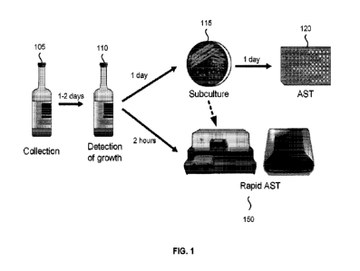
100391 FIG. 1 shows process flows for conventional AST and rapid AST
embodiments of the
present invention. A blood specimen is easy to collect. For example, a blood
specimen may be
obtained with a blood draw from a patient using a syringe. Specifically, a
blood specimen does
not require surgery or invasive procedures to obtain. However the number of
organisms is low
and may be on the order of <1-10 colony-forming units per milliliter of blood.
Therefore, when
we collect blood specimens from patients, we must first culture the blood to
increase the number
of organisms, in order to allow further analysis such as AST.
100401 Blood is collected into blood culture bottles for this purpose at the
patient bedside.
Bottle 105 is a bottle after blood collection. Generally, a blood culture draw
consists of a set of
bottles, an aerobic bottle and an anaerobic bottle. The bottles may be used
for different
pathogens. These bottles foster the growth of aerobic and facultative
anaerobic, and anaerobic
bacterial organisms, respectively. The bottles also may grow yeast. There is a
separate type of
bottle that will grow pathogenic fungi and mycobacteria. Blood cultures are a
very high-volume
process: for example, at BlEIMC, we process approximately 40,000 blood culture
sets per year
from patients with suspected bloodstream infections, approximately 5-10% of
which flag as
positive on our commercial blood culture system. We use the Becton Dickinson
BACTEC FX
blood culture system. There are several competing systems used in clinical
labs, all of which
may operate on similar principles: detection of organisms' metabolism.
9
CA 03153347 2022-3-31
WO 2021/067170
PCT/US2020/053030
100411 Bottle 110 shows a bottle after detection of growth. When a blood
culture bottle 110
flags positive, the system alerts technologists to undertake further steps in
order to guide
clinicians in use of appropriate therapy. Detection of growth may occur 1-2
days after collection.
A positive culture typically has ¨109 organisms per ml, reflecting logarithmic
growth of
organisms initially inoculated from the sample into the bottle. From the
standard aerobic and
anaerobic blood cultures sets, technologists perform a Gram stain and plate
the blood-culture
broth to isolate colonies. Dish 115 shows a plate after the Gram stain and
isolating colonies.
Isolating colonies may take 1 day or more after detection of growth. Isolated
colonies will then
be used for AST 120. AST may take an additional day after subculture.
Therefore, from the time
a blood culture bottle becomes positive there is a two-or-more day delay in
obtaining AST
results that allow a switch from empiric to appropriate/effective narrower
spectrum therapy.
I. OVERVIEW
100421 The technology described in this disclosure reduces the time from
positive blood
culture to AST results from two-or-more days to less than 3 hours, with our
preliminary results
indicating accurate results in 1.5 hours. With further refinement we expect to
reduce this time
even further, including down to 30 minutes. Consequently, we expect this
invention will have
significant impact on patient care.
A. Current systems
100431 Embodiments of the present invention provide faster AST results at a
lower cost than
recently commercially available technologies. One example of a commercial AST
system is the
FDA-cleared Accelerate Diagnostics Phenosense system. This system takes
positive blood-
culture broth, electrophoretically immobilizes organisms onto a solid surface,
and then
microscopically follows growth of organisms into colonies. However, the
Phenosense will only
provide AST results after 7 hours. The technology extrapolates the AST readout
from testing a
single concentration of each antibiotic. In other systems, such as the Vitek,
which extrapolates
AST from a minimum of 2-3 concentrations. Such extrapolations may often be
associated with
an unacceptable rate of error, especially for multidrug-resistant organisms
for which correct AST
results are critical.
CA 03153347 2022-3-31
WO 2021/067170
PCT/US2020/053030
100441 A major limitation of the Accelerate Phenosense system is that it can
test only one
pathogen on one system at a time. Another limitation is that each assay
cartridge costs more than
$200. A third is that each assay system platform, the machine on which the
assays are run, costs
¨$100,000. A typical reasonably sized hospital system would require several
such systems to
address the many positive blood cultures each day, as the lab could not wait
for 7 hours until
starting each successive positive blood culture test (which would defeat the
purpose of a "rapid"
AST system). So a laboratory may need a capital investment of >$300,000 to
employ such as
system, with a very high per-test reagent cost. Furthermore, the Accelerate
system at present is
only approved for positive blood culture broth detection. Potential future use
for higher volume
microbiology laboratory testing at more than $200 per sample with low
throughout is extremely
problematic.
B. AST in less than 3 hours
100451 In contrast, embodiments of the present invention will allow us to test
any antimicrobial
agent at any desired concentration, for many agents and concentrations, at
will. FIG. 1 shows
that Rapid AST 150 embodiments can be done in two hours, without the
subculture_
Nevertheless, Rapid AST 150 could still be used after subculture from dish
115. Currently, we
are testing true two-fold serial dilutions (also called doubling dilutions) of
antimicrobials, as is
performed in the reference AST method, to allow us to accurately determine the
minimal
inhibitory concentration, or MIC MIC is lowest doubling-dilution concentration
of antibiotic
that inhibits growth. The MIC is an important and discriminatory data from
AST. It is a
phenotypic measure that predicts patient response to therapy. In reference AST
format, it is
performed by testing the effects of doubling dilutions of antibiotics on the
growth of a pathogen
inoculated into a standard growth medium called cation adjusted Mueller-Hinton
broth. In
current standard of care, growth inhibition is interpreted typically after 16-
20 hours of incubation
at 35 C.
100461 Embodiments of the present invention, based on the foundation of the
doubling dilution
NIX method, will require significantly less extrapolation than methods like
Accelerate or Vitek.
Specifically, the AST plates in embodiments of the present invention, if
incubated for the time
performed in reference methods would approximate the reference method.
However,
11
CA 03153347 2022-3-31
WO 2021/067170
PCT/US2020/053030
embodiments of the present invention allow reference broth susceptibility
testing panels to be
read much more quickly: less than 3 hours currently, with anticipated
improvements.
100471 FIG. 2 shows a diagram of the dispensing technology. The dispensing
technology may
use well plate 205, which may be a standard 96-, 384-, or 1536-well microtiter
plate. Well plate
205 contains doubling dilutions of antimicrobials. Any antimicrobials can be
used. Antibiotics
are mentioned as an example of an antimicrobial, but any antimicrobial or
other treatment agent
(e.g. antifungal, antiviral, anti-parasitic) can be used unless context
clearly dictates otherwise.
The doubling dilution series can be prepared at the time of use, or plates
with doubling dilutions
can be pre-made and stored with lyophilized antibiotics or with antibiotics in
broth that are
frozen and thawed before use. In embodiments, 50 ptl of broth are added to
wells in well plate
205 and desired two-fold dilutions of antibiotics are added to wells using
inkjet printing
technology 210 as described in WO 2017/218202 Al, the entire contents of which
are
incorporated by reference for all purposes. In other embodiments, antibiotics
can be added either
manually or with any of several suitable liquid handling devices. Positive
blood culture broth is
then added to the microtiter wells. The positive blood culture broth or
dilutions of the positive
blood culture broth can be added to the wells using any of several liquid
handling devices or
manually or through use of an inkjet printer 210 which we determined can
quantitatively print
out positive blood culture broth.
100481 Typically positive blood-culture broth has organisms at 5x10⁹ colony-
forming units per
mL. In our experiments, we determined that this could be diluted 5-fold in
sterile water and 0.3%
tween-20 and this dilution dispensed at 250 nl per well to create a starting
inoculum of
approximately 2.5 x 10⁷ per rnL in wells in a 384 well plate containing a
volume of 10 pl.
Surfactant such as Tween-20 can be added to facilitate inkjet printing to
rapidly distribute
bacteria to each well of a 384-well plate or other liquid handling devices
known to the field can
be similarly used. The final inoculum can be varied in the range of 103 per ml
to 108 per ml to
optimize performance of rapid AST depending on the application, organism, and
incubation
temperature.
100491 FIG. 3 illustrates operations after the well plate is prepared. The
microtiter plate is then
placed in an incubator. The temperature of the incubator can be set at any
temperature from 15 C
to 72 C to optimize growth of organisms for different applications. Typically
temperatures for
12
CA 03153347 2022-3-31
WO 2021/067170
PCT/US2020/053030
bacterial pathogen testing will be in the range of 35 C to 37 C. Block 305
shows that the well
plate is incubated at 37 C. Either continually or at specific time points, the
plate is scanned at
block 310 using a scanning technology such is found in a flatbed scanner 312.
Scanner 312 may
consist of a linear charge-coupled device (CCD) sensor, contact image sensor
(CIS), or
photomultiplier tube sensor that is at least the diameter of the plate if the
sensor is in linear
format and the sensor scans across the plate or the size of the plate if a
single image of the plate
is captured simultaneously. Scanning results in an image file output 315.
100501 FIG. 4A shows an image of a well with microorganism growth. FIG. 4B
shows an
image of a well without microorganism growth. Portion 405 and portion 410 of
the images align
with the center portion of a well. Portion 405 is visually darker than portion
410. While this
difference can be noticed by the naked eye, most images of wells with growth
are not visually
discernible from a well without growth by a person and may need to be analyzed
by a computer.
Portion 405 and portion 410 exclude pixels from outside the center portion of
the well. The areas
outside portion 405 and portion 410 include the sidewalls of the well, which
do not contain the
testing sample with the microorganism, broth, and antimicrobial. Areas within
the well itself are
obscured by shadowing and other distortions from the imaging process.
Moreover, image
distortions may vary depending on the location of the well in the plate.
100511 Custom placement of biological replicates of doubling dilutions on a
given plate¨a
custom platemap¨are used to further control for artifacts and positional
variance. Raw images
of the plates in standard imaging formats such as TIFF or PNG are processed to
correct for per-
timepoint, per-axis-per-plate, and per-scan variability and then compared to
detect differences in
pixel intensities that indicate growth of the bacteria, resulting in data that
can be represented in
two-dimensional plots of doubling-dilution concentration (x-axis) vs. growth
(y-axis).
100521 We have made use of a CCD flatbed scanner to scan plates. hi our
example, the plates
are incubated in a separate incubator and placed on a plate bed scanner at
different time points
for reading. Using a flatbed scanner, we have determined that accurate AST
results can be
obtained with incubation times of 1.5 hours or less. We have shown that the
combination of
appropriately designed platemaps and the above image processing steps
substantially decrease
the time to determination of MICs, specifically accurately determining MilCs
at 3 hours, with
13
CA 03153347 2022-3-31
WO 2021/067170
PCT/US2020/053030
preliminary data showing MICs in many cases down to 1.5 hours, and machine
learning on each
time series and higher CCD resolution suggesting still further improvement&
C. Features
100531 Embodiments of the present invention include key advantages over other
AST
technologies.
100541 A first advantage is speed. The current clinical standard is AST in 16-
20 hours.
Embodiments of the present invention can perform AST at 3 hours, with
preliminary data down
to 1.5 hours. This short time needed for AST was unexpected, particularly in
view of the
inexpensive hardware used for AST.
100551 A second advantage is using inexpensive standard technologies.
Embodiments of the
present invention can use a standard mid-2000s-era flatbed scanner, which can
be obtained at a
cost of ¨$200 or less. Complex robotics and moving parts are not needed for
imaging. As a
result, manufacturing costs may be low.
100561 A third advantage is having inexpensive consumables. Our technology
uses the 96-,
384-, and 1,536-well microtiter plates that are standard across biological
research and clinical
practice and available inexpensively from multiple manufacturers (Corning,
Eppendorf, Fisher,
Nunc, etc.). Our broth may be the clinical standard. Expensive reagent
supplements are not
needed. For example, the broth does not require lanthanide-series metals or
moieties as tags to
identify organism surface area or mass.
100571 A fourth advantage is versatility. Embodiments of the present invention
may allow for
continuous measurement. Embodiments do not require addition of a reagent and
are not limited
to being an end-point assay (once the assay is performed, measurements cannot
be continued).
Embodiments of the present invention work well from direct-from-colony AST as
well as direct-
from-blood-culture AST.
14
CA 03153347 2022-3-31
WO 2021/067170
PCT/US2020/053030
EXAMPLE RESULTS
100581 The pixel intensity from imaged wells can be used in determining the
relative growth of
a microorganism. We conducted experiments to demonstrate that MIC can be
determined with
the dispensing, imaging, and analysis methods and systems described herein.
A. Results using gentamicin
100591 FIG. 5 shows graphs resulting from data from imaging using a flatbed
scanner well
plates. A microorganism (K coh) was added to dilutions of gentamicin in a well
plate. The well
plate was incubated at 37 C and scanned at various times. The well plate was
physically moved
from the incubator and then placed back in the incubator at several times.
Because of this
movement, the well plate was not in the exact same position on the scanner at
each scan.
100601 The graphs in FIG. 5 plot relative growth, as determined using pixel
intensity, versus
concentration of gentamicin in pg/pl. The largest concentration of gentamicin
is shown on the
left-most side of each graph, and the smallest concentration of gentamicin is
shown on the right-
side of each graph.
100611 Graph 510 shows results after 1.5 hours of incubation. Graph 520 shows
results after 2
hours of incubation. Graph 530 shows results after 3 hours of incubation.
Graph 540 shows
results after 4 hours of incubation.
100621 Graph 510 shows a graph that has an increase in the relative growth of
the
microorganism at gentamicin concentrations at or below 0.5 p.g/itl. The
relative growth is greater
than at concentrations at or above 1.0 ps4t1. The relative growth at 1.0
gigh.t1 is between the
relative growth at 0.5 p.g/gl and 1.0 g/til. The MIC could be determined to
be either 0.5 ug/g1
or 1.0 pg/ 1 from graph 510. Plus or minus one doubling dilution is considered
within the
allowable variance of the reference method. Arrow 512 indicates an MIC of 1.0
pg/pl. Arrows
522, 532, and 542 indicate the 1.0 pg/ul concentration for different periods
of incubation. Graphs
510, 520, 530, and 540 all show similar responses. Graph 540 at 4 hours shows
a clear increase
in relative growth at concentrations at or below 0.5 figitutl. The MIC in
graphs 520, 530, and 540
may also be determined to be 1.0 pa/1.11.
CA 03153347 2022-3-31
WO 2021/067170
PCT/US2020/053030
B. Results using cephalosporins
100631 The imaging and analysis techniques may also be sensitive enough to
capture unusual
patterns of growth of microorganisms in the presence of antimicrobials. For
example,
cephalosporins, including cephalexin and cefepime, may cause cell
filamentation_ These
filaments can be on the order of tens of microns in length, which is on the
order of the resolution
of the scanner used in these experiments.
100641 FIG. 6 shows graphs of resulting from data from imaging using a flatbed
scanner to
scan well plates with dilutions of cefepime. Graph 610 shows results after 1.5
hours of
incubation. Graph 620 shows results after 2 hours of incubation. Graph 630
shows results after 3
hours of incubation. Graph 640 shows results after 4 hours of incubation.
Graph 610 and graph
620 show relative growth that undergoes a decrease after an initial increase
as concentrations of
cefepime are reduced. This pattern may be capturing cell filamentation caused
by the cefepime,
which results in increased pixel intensity. Graph 630 and graph 640 do not
show the decrease
after the increase with lower cefepime concentrations. The MIC in graph 630
and graph 640 can
be determined to be 0.031 pg/pl.
100651 These cefepime results show that the scanning method can detect
different growth
patterns resulting from antimicrobials. Additionally, even with different
growth patterns, the
imaging method described herein may still determine the MIC in 3 hours.
C. Additional results
100661 Further experiments are run using the scanning image method using
different
microorganisms and different antimicrobials. Enterobacteriaceae, Pseudornonas,
and
Acinetobacter bacteria are tested with dilutions of meropenem, gentamicin,
ciprofoxacin, and
cefepime. Methicillin-sensitive Staphylococcus aureus (MSSA) is tested with
dilutions of
vanomycin, linezolid, daptomycin, and oxacillin. Enterococcus is tested with
dilutions of
vanomycin, linezolid, daptomycin, and ampicillin. MICs are determined at
various incubation
times. Machine learning using time series data may be applied to determine the
MIC from an
incubation time of under 1.5 hours, when the MIC is not readily apparent from
data at 1.5 hours.
16
CA 03153347 2022-3-31
WO 2021/067170
PCT/US2020/053030
D. Improvements in time for MIC
100671 AST results are expected to be achieved in times less than the
demonstrated 1.5 hours.
Improvements may be seen from using a higher resolution sensor. Previous
experiments used a
4800 dpi scanner. Scanners with 9600 dpi are commercially available and not
cost prohibitive. A
higher resolution scanner will allow for more pixels to be analyzed per well.
A higher quality
scanner may also reduce the distortions and shadowing in each well. More
pixels may result in a
better signal for growth. Machine learning models to filter out the pixels for
analysis and
machine learning models for determining MIC from image data may also reduce
the duration of
incubation needed. Additionally, additives may be added to the well that may
aid image analysis.
Additives may include a signal amplifier, a pH indicator, or a dye indicative
of metabolism. A
signal amplifier may include any of several systems in which detection of a
change (e.g., pH
change) sets off a cascade, chain reaction, or positive feedback loop of
chemical reactions. For
example, a fall in pH causes an inhibitor molecule to dissociate from an
enzyme that produces a
pigment.
III. EXAMPLE SYSTEM
100681 FIG. 7 shows a system 700 for rapid AST. System 700 may include a
subsystem 705,
which includes a dispensing unit 710, an incubation unit 720, an imaging unit
730, and a data
storage and processing unit 740.
100691 Dispensing unit 710 may be configured for automated dispensing of a
microorganism
or a plurality of concentrations of an antimicrobial to a plurality of
locations on a well plate. The
well plate may include 96 or more wells, including 384, 1,536, 3,456, or 9,600
wells. Each well
may have a volume from 10 nl to 2 ml, including 0.1 to 0.3 ml or 0.03 to 0.1
ml. The wells may
include polystyrene, polypropylene, or polycarbonate. The well plates may have
a flat or
substantially flat bottom. The well plates may not include lenses or other
optical components that
improve the imaging of a well. In some embodiments, a lens may be added to
each well to
improve imaging of the well. The plurality of concentrations may include
serial dilutions of the
antimicrobial. The plurality of locations on the well plate map to the wells
of the well plate. In
some embodiments, the well plate may be supplied with dilutions of an
antimicrobial. The
antimicrobials may be lyophilized or frozen until they are thawed for testing.
17
CA 03153347 2022-3-31
WO 2021/067170
PCT/US2020/053030
100701 Dispensing unit 710 may be a dispensing unit described in WO
2017/218202 Al. The
dispensing unit may dispense liquid using techniques and hardware used by an
inkjet printer to
dispense ink. Dispensing unit 710 may be configured for automated dispensing
of a single
concentration of microorganism to a plurality of wells in the well plate.
100711 Incubation unit 720 may be configured to receive the well plate and to
maintain a
temperature set point. Incubation unit 720 may include a heater, a temperature
sensor, a
programmable controller, and insulated walls. Incubation unit 720 may be set
at any temperature
from 15 C to 72 C, including 37 C or any temperature described herein.
100721 Imaging unit 730 may include a light source and a sensor. The imaging
unit may be
configured to measure light from the light source reflected by the well plate
and to generate an
image data set from the measured light. The sensor may have a resolution
greater than or equal to
600 dpi, including from 600 to 1200 dpi, from 1200 to 4800 dpi, from 4800 to
9600 dpi, or
greater than 9600 dpi. The sensor may be a charge-coupled device (CCD),
contact image sensor
(CIS), or photomultiplier tube sensor. The sensor may have bit depths of 24 or
more, including
30 or more, 36 or more, or 48 or more.
100731 The light source may include a fluorescent lamp or a xenon lamp. The
light source may
include a cold cathode fluorescent lamp. In some embodiments, the light source
may be light
emitting diodes (LEDs), and the sensor may be a CIS.
100741 The light source may be configured to move, and imaging unit 730 may
include a
mirror or mirrors to reflect light to the sensor during movement of the light
source. Light may
pass through filters (e.g., for red, green, or blue) before the sensor so that
a color image may be
produced. The sensor, mirrors, and filter may be part of a scan head. The scan
head may be
moved by a motor (e.g., a stepper motor), so that all components of the scan
head move
simultaneously. The scan head may be attached to a stabilizer bar, and the
scan head may be
moved by a belt in communication with the motor. The scan head may move in one
dimension
only. In some embodiments, the scan head may move in two dimensions. The scan
head may
make only one pass across the well plate. In some embodiments, the scan head
may make three
passes across the well plate and may use a different color filter for each
pass. An example of
18
CA 03153347 2022-3-31
WO 2021/067170
PCT/US2020/053030
imaging unit 730 is a flatbed scanner. For example, the imaging unit may be
Canon CanoScan
9000F MK II, which scans at 4800 dpi.
100751 Imaging unit 730 may include a glass plate, on which the well plate
sits during
imaging. The well plate may be oriented such that the bottom of the well plate
is closest to the
sensor. In some embodiments, the well plate may be oriented so that the top of
the well plate,
and therefore the opening of each well, is closest to the sensor.
100761 Imaging unit 730 may include a well plate holder to immobilize the well
plate. The well
plate holder may be a clamp, recessed portion of a surface to be imaged, a
hinge, or other
suitable device.
100771 Data storage and processing unit 740 may include a processor configured
to execute a
plurality of instructions. Data storage and processing unit may include logic
system 2130,
external memory 2140, and/or storage device 2145 in FIG. 10 or computer 10 in
FIG. 11, all of
which are described later. The processor may be configured to execute a
plurality of instructions.
The instructions may include analyzing the image data set to determine a first
value of an image
characteristic for a first subset of wells of the well plate, the first subset
of wells having a first
concentration of the microbial, analyzing the image data set to determine a
second value of an
image characteristic for a second subset of wells of the well plate, the first
subset of wells having
a second concentration of the microbial, the second concentration being
greater than the first
concentration, and determining a classification of the resistance of the
microorganism to the
antimicrobial using the first value and the second value. The plurality of
instructions may include
any method described herein.
100781 Dispensing unit 710, incubation unit 720, imaging unit 730, and data
storage processing
unit 740 may be considered part of a subsystem 705. Subsystem 705 may include
units after
growth of the microorganism is detected in a biological sample.
100791 System 700 may include a sample collection unit 750. Sample collection
unit 750 may
be configured to obtain a biological sample from a patient. The biological
sample may be any
described herein (e.g., a blood, urine, cerebral spinal, pleural fluid,
pericardial fluid,
bronchoalveolar lavage fluid, and sputum after liquifaction with N-
acetylcysteine). In some
19
CA 03153347 2022-3-31
WO 2021/067170
PCT/US2020/053030
embodiments, the sample is obtained by a medical practitioner with a syringe
or other suitable
device. The biological sample may be stored in a container, such as a bottle
or vial.
100801 System 700 may further include a sample growth unit 760. Sample growth
unit 760
may be a blood culture system, including, for example, Becton Dickinson BACTEC
FX blood
culture system. With certain biological samples (e.g., urine, cerebral spinal
fluid, pleural fluid,
pericardial fluid, bronchoalveolar lavage fluid, sputum after liquifaction
with N-acetylcysteine),
direct testing of the sample is possible where the biological sample is
dispensed into wells with a
growth medium, and sample growth unit 760 may be optional.
100811 System 700 may optionally include a subculture unit 770 Subculture unit
770 may be
configured to perform a Gram stain and plate the blood culture broth to
isolate colonies.
Microorganisms from isolated colonies may be added to a well plate by
dispensing unit 710. As
explained above, embodiments of the present invention may exclude subculture
unit 770.
IV. EXAMPLE METHODS
100821 Embodiments of the present invention may include methods to test for
antimicrobial
susceptibility. Embodiments include using imaging techniques similar to those
used by a
commercially available flatbed scanner. The methods described herein can
determine a
susceptibility for a microorganism for a certain concentration of an
antimicrobial. Methods may
determine the MIC for a microorganism and antimicrobial. The MIC may be
determined in 3
hours or less, including 1.5 hours or less, 1 hour or less, or 0.5 hours or
less. Time to determine
MIC may be improved with machine learning.
A. Determining antimicrobial susceptibility
100831 FIG. 8 may include a method 800 for testing antimicrobial
susceptibility. The method
may include using any system described herein, including system 700 and
subsystem 705.
100841 At block 810, an image data set generated from imaging a plurality of
wells may be
received by a computer system. The plurality of well may include a first
subset of wells
containing a microorganism and a first initial concentration of an
antimicrobial. The
microorganism may be any microorganism described herein, including any
pathogen. The
plurality of wells may include a second subset of wells containing the
microorganism and a
CA 03153347 2022-3-31
WO 2021/067170
PCT/US2020/053030
second initial concentration of the antimicrobial. The microorganism
concentration may be equal
in the two subsets of wells. The concentration of the microorganism may be
from 100 to 1010 per
ml, including from 104 to 108 per ml. Each well may further include signal
amplifier, a pH
indicator, or a dye indicative of metabolism. A subset of wells may include
only one well or may
include multiple wells. The subset of wells may include from 5 to 15 wells,
including from 5 to
wells or from 10 to 15 wells.
100851 The second initial concentration may be greater than the first initial
concentration. The
first initial concentration may be a doubling dilution of the second initial
concentration. The
second initial concentration may be equal to the first initial concentration
multiplied by 211, where
10 n is a non-zero integer.
100861 The plurality of wells may further include a third subset of wells
containing the
microorganism and a third initial concentration of the antimicrobial. The
third initial
concentration may be equal to the first initial concentration multiplied by
2m, where ni is a non-
zero integer and in does not equal n. Additional subsets of wells and
additional initial
concentrations of the antimicrobial may be included in the plurality of wells
to correspond to
multiple doubling dilutions. For example, there may be 10 to 16 initial
concentrations of the
antimicrobial in the plurality of wells.
100871 The plurality of wells may include a well or wells containing the
microorganism and
excluding the antimicrobial. These wells may be a positive control to confirm
growth in the
microorganism. The plurality of wells may include a well or wells containing
the antimicrobial
and excluding the microorganism, which may be negative controls. The positive
controls and/or
the negative controls may be used for normalizing pixel intensities of other
wells.
100881 The image data set may be generated by measuring light from a light
source reflected
by each well of the plurality of wells. The image data set may include a value
for pixel intensity
for each pixel of a plurality of pixels. The pixel size may be less than or
equal to 20 times the
microorganism size, including less than or equal to 10 times, 5 times, or 2
times the
microorganism size. The microorganism size may be the longest dimension of the
microorganism or the diameter of the microorganism if the microorganism
occupied a circle with
the same area as the non-circular microorganism.
21
CA 03153347 2022-3-31
WO 2021/067170
PCT/US2020/053030
100891 Measuring the light form the light source may include using a charge-
coupled device, a
contact image sensor, or photomultiplier tube. Measuring light from the light
source may include
using the charge-coupled device, and the image data set may be generated by
moving the light
source and the charge-coupled device relative the plurality of wells. The
image data set may be
generated using imaging unit 730, including a flatbed scanner.
100901 In some embodiments, method 800 may include adding a microorganism to
each well
of the plurality of wells. The microorganism may be from any biological sample
described
herein. The microorganism may not have been isolated in a subculture. For
example, the
microorganism may have undergone growth in sample growth unit 760 but then
underwent
subculture in subculture unit 770 to isolate colonies. The microorganism may
be added with a
cell or cells. For example, if the microorganism is a virus, the virus may be
added with a cell or
within a cell.
100911 Method 800 may include adding, by the automated dispenser, the first
initial
concentration of the antimicrobial to the first subset of wells. Method 800
may also include
adding, by the automated dispenser, the second initial concentration of the
antimicrobial to the
second subset of wells. Method 800 may further include incubating the
plurality of wells for a
duration. Incubating the wells may be from 30 minutes to 4 hours, including
from 30 minutes to
1 hour, from 1 to 1.5 hours, from 1.5 to 2 hours, from 2 to 3 hours, or from 3
to 4 hours.
100921 At block 820, the image data set may be analyzed by the computer system
to determine
a first value of an image characteristic for the first subset of wells. The
image characteristic may
be a statistical measure of pixel intensities corresponding to a subset of
wells. The statistical
measure may be an average (e.g., mean, median, mode) or percentile pixel
intensity. The image
characteristic may be of pixel intensities of pixels corresponding to a
central portion of each well
of the subset of wells. The pixels in the central portion may exclude pixels
that correspond to
sidewalls of wells, shadowing of wells, or distortions/artifacts (e.g.
parallax, lens flare). A model
may determine the pixels corresponding to the central portion of the
respective well.
100931 The first value of the image characteristic may be adjusted for non-
uniformities of the
reflected light based on a location of each well in the first subset of wells.
In some embodiments,
22
CA 03153347 2022-3-31
WO 2021/067170
PCT/US2020/053030
the first value of the image characteristic may be normalized or adjusted
based on a control or
based on image data from previously imaging the plurality of wells.
100941 At block 830, the image data set may be analyzed by the computer system
to determine
a second value of the image characteristic for the second subset of wells. The
second value may
be similar to the first value, but determined for the second subset of wells.
100951 At block 840, a classification of the resistance of the microorganism
to the
antimicrobial may be determined using at least one of the first value or the
second value. The
classification of the resistance of the microorganism to the antimicrobial may
include an MIC, a
likelihood of resistance, or a determination of resistant, not resistant
(i.e., susceptible), or
intermediately resistant.
100961 The first value may be compared to the second value to determine a
separation value.
The separation value may be a difference or ratio of the first value and the
second value. The
separation value may be compared to a cutoff value. Determining the
classification of the
resistance may include determining that the microorganism is resistant to the
first initial
concentration of the antimicrobial when the separation value exceeds the
cutoff value. In some
cases, determining the microorganism is resistant to the first initial
concentration includes
determining that the first value is greater than a threshold value.
100971 The second initial concentration may be the MIC when the second initial
concentration
is less than or equal to two times the first initial concentration when the
separation value exceeds
the cutoff value. The second value may be less than a certain threshold value.
The MIC may be
determined after incubating for 90 minutes or less, or any duration described
herein.
100981 The first value may be compared to a threshold value. When the first
value exceeds the
threshold value, the microorganism may be determined to be resistant to the
first initial
concentration of the antimicrobial. In some embodiments, the separation value
does not need to
be determined to determine the classification of the resistance. For example,
a higher value may
reflect a higher and darker pixel intensity, which may result from growth of
the microorganism.
100991 Based on the determined classification of the resistance, a patient
having the
microorganism may be treated with a dose of the antimicrobial based on at
least one of the first
23
CA 03153347 2022-3-31
WO 2021/067170
PCT/US2020/053030
initial concentration or the second initial concentration. If the
microorganism was determined to
be susceptible to either the first initial concentration or the second initial
concentration, the
patient may receive a treatment with the antimicrobial. The dose may be
related to the initial
concentration by a linear or non-linear equation. The initial concentration
may be compared to a
table to determine whether the microorganism is clinically resistant to the
antimicrobial. For
example, there are standards organizations (e.g., CLSI) that determine the
relationship between
MIC and clinical resistance and publish conversion tables for looking up the
microorganism and
antimicrobial and seeing if an MIC is susceptible, resistant, or intermediate.
In some cases if the
microorganism is determined to be resistant to the antimicrobial, the patient
may be treated be
treated with an alternative antimicrobial to which the microorganism is
susceptible.
B. Determining MIC using a model
101001 FIG. 9 shows a method 900 for testing antimicrobial susceptibility
using a model,
including a machine learning model.
101011 At block 910, an input data structure may be received. The input data
structure may
include an input image data set comprising a value for pixel intensity for
each pixel of a sample
plurality of pixels. The image data set may be generated from imaging a sample
plurality of
wells. The image data set may be generated by any method and using any system
described
herein. The sample plurality of wells may contain a sample microorganism and a
plurality of
initial concentrations of a sample antimicrobial. The plurality of initial
concentrations may be
concentrations resulting from doubling dilutions of the sample antimicrobial.
In some
embodiments, the input data structure may further include a sample duration of
incubating the
sample microorganism.
101021 At block 920, the input data structure may be inputted into a model.
Blocks 930, 940,
and 950 include elements of the training.
101031 At block 930, a first plurality of first data structures may be
received. Each first data
structure of the first plurality of first data structures may include a first
image data set comprising
a value for pixel intensity for each pixel of a first plurality of pixels. The
first image data set may
be generated from imaging a first plurality of wells. The first plurality of
wells may contain a
24
CA 03153347 2022-3-31
WO 2021/067170
PCT/US2020/053030
first microorganism and the plurality of initial concentrations of a first
antimicrobial. The first
microorganism may have a known minimum inhibitory concentration to the first
antimicrobial.
101041 The first antimicrobial may or may not be the same antimicrobial as the
sample
antimicrobial. In some embodiments, a certain antimicrobial may be used to
predict
susceptibility for other antimicrobials. For example, cefazolin may be used to
predict
susceptibility to other cephalosporins for Enterobacteriaceae. The first
microorganism may be
the same or different as the sample microorganism. For example, all species of
Enterobactericeae may behave similarly in response to an antibiotic, and
therefore, training on
one species may apply to other species.
101051 Each first data structure may also include a first map representing the
first plurality of
wells with values indicating the initial concentration of the first
antimicrobial for each well. The
map may be a matrix or 2D array. In some embodiments, the map may not be
rectangular. For
example, the well plate may not be rectangular or not every well in well plate
is measured.
101061 In some embodiments, each first data structure may include a first
duration of
incubating the first microorganism. These first durations may allow for the
model to determine
the MIC in an image from a shorter duration when the MIC is more clearly
determined from
longer durations (e.g., FIG! 6).
101071 At block 940, a plurality of first training samples is stored. Each
first training sample
may include one of the first plurality of first data structures and a first
label indicating the known
minimum inhibitory concentration of the first microorganism to the first
antimicrobial.
101081 At block 950, parameters of the model may be optimized, using the
plurality of first
training samples, based on outputs of the model matching or not matching
corresponding labels
of the first labels when the first plurality of first data structures is input
to the model. The output
of the model specifies the MIC of the first microorganism to the first
antimicrobial for a given
first data structure.
101091 At block 960, the MC of the sample microorganism to the sample
antimicrobial may
be determined using the model. A patient may be treated based on the MEC. The
patient may be
given a dose of the sample antimicrobial linearly or non-linearly related to
the MIC.
CA 03153347 2022-3-31
WO 2021/067170
PCT/US2020/053030
101101 The model may include a convolutional neural network (CNN). The CNN may
include
a set of convolutional filters configured to filter the first plurality of
data structures and
optionally the second plurality of data structures. The filter may be any
filter described herein.
The number of filters for each layer may be from 10 to 20, 20 to 30, 30 to 40,
40 to 50, 50 to 60,
60 to 70, 70 to 80, 80 to 90, 90 to 100, 100 to 150, 150 to 200, or more. The
kernel size for the
filters can be 2, 3,4, 5,6, 7, 8, 9, 10, 11, 12, 13, 14, 15, from 15 to 20,
from 20 to 30, from 30 to
40, or more. The CNN may include an input layer configured to receive the
filtered first plurality
of data structures and optionally the filtered second plurality of data
structures. The CNN may
also include a plurality of hidden layers including a plurality of nodes The
first layer of the
plurality of hidden layers coupled to the input layer. The CNN may further
include an output
layer coupled to a last layer of the plurality of hidden layers and configured
to output an output
data structure. The output data structure may include the properties.
101111 The model may include a supervised learning model. Supervised learning
models may
include different approaches and algorithms including analytical learning,
artificial neural
network, backpropagation, boosting (meta-algorithm), Bayesian statistics, case-
based reasoning,
decision tree learning, inductive logic programming, Gaussian process
regression, genetic
programming, group method of data handling, kernel estimators, learning
automata, learning
classifier systems, minimum message length (decision trees, decision graphs,
etc.), multilinear
subspace learning, naive Bayes classifier, maximum entropy classifier,
conditional random field,
Nearest Neighbor Algorithm, probably approximately correct learning (PAC)
learning, ripple
down rules, a knowledge acquisition methodology, symbolic machine learning
algorithms,
subsymbolic machine learning algorithms, support vector machines, Minimum
Complexity
Machines (MCM), random forests, ensembles of classifiers, ordinal
classification, data pre-
processing, handling imbalanced datasets, statistical relational learning, or
Proaftn, a
multicriteria classification algorithm The model may include linear
regression, logistic
regression, deep recurrent neural network, Bayes classifier, hidden Markov
model (HIMM),
linear discriminant analysis (LDA), k-means clustering, Density-based spatial
clustering of
applications with noise (DBSCAN), random forest algorithm, support vector
machine (SV1V1), or
any model described herein.
26
CA 03153347 2022-3-31
WO 2021/067170
PCT/US2020/053030
101121 As part of training a machine learning model, the parameters of the
machine learning
model (such as weights, thresholds, e.g., as may be used for activation
functions in neural
networks, etc.) can be optimized based on the training samples (training set)
to provide an
optimized accuracy in classifying MIC, and/or susceptibility or resistance to
one or many
antimicrobials. Various form of optimization may be performed, e.g.,
backpropagation, empirical
risk minimization, and structural risk minimization. A validation set of
samples (data structure
and label) can be used to validate the accuracy of the model. Cross-validation
may be performed
using various portions of the training set for training and validation. The
model can comprise a
plurality of submodels, thereby providing an ensemble model. The submodels may
be weaker
models that once combined provide a more accurate final model.
V. EXAMPLE LOGIC AND COMPUTER SYSTEMS
101131 FIG. 10 illustrates a system 2100 according to an embodiment of the
present invention.
The system as shown includes a sample 2105, such as a microorganism within a
sample holder
2110, where sample 2105 can be contacted with an assay 2108 to provide a
signal of a light
characteristic 2115. An example of a sample holder can be a well in a well
plate_ Light
characteristic 2115 (e.g., an intensity, a wavelength), from the sample is
detected by detector
2120. Detector 2120 can take a measurement at intervals (e.g., periodic
intervals) to obtain data
points that make up a data signal. Detector 2120 may be any sensor described
herein. Sample
holder 2110 and detector 2120 can form an assay device, e.g., an imaging unit
according to
embodiments described herein. A data signal 2125 is sent from detector 2120 to
logic system
2130. Data signal 2125 may be stored in a local memory 2135, an external
memory 2140, or a
storage device 2145.
101141 Logic system 2130 may be, or may include, a computer system, ASIC,
microprocessor,
etc. It may also include or be coupled with a display (e.g., monitor, LED
display, etc.) and a user
input device (e.g., mouse, keyboard, buttons, etc.). Logic system 2130 and the
other components
may be part of a stand-alone or network connected computer system, or they may
be directly
attached to or incorporated in a device (e.g., an imaging device) that
includes detector 2120
and/or sample holder 2110. Logic system 2130 may also include software that
executes in a
processor 2150. Logic system 2130 may include a computer readable medium
storing
27
CA 03153347 2022-3-31
WO 2021/067170
PCT/US2020/053030
instructions for controlling system 2100 to perform any of the methods
described herein_ For
example, logic system 2130 can provide commands to a system that includes
sample holder 2110
such that dispensing, imaging, or other physical operations are performed Such
physical
operations can be performed in a particular order, e.g., with reagents being
added and removed in
a particular order. Such physical operations may be performed by a robotics
system, e.g.,
including a robotic arm, as may be used to obtain a sample and perform an
assay.
101151 Any of the computer systems mentioned herein may utilize any suitable
number of
subsystems. Examples of such subsystems are shown in FIG. 11 in computer
system 10. In some
embodiments, a computer system includes a single computer apparatus, where the
subsystems
can be the components of the computer apparatus. In other embodiments, a
computer system can
include multiple computer apparatuses, each being a subsystem, with internal
components. A
computer system can include desktop and laptop computers, tablets, mobile
phones, other mobile
devices, and cloud-based systems.
101161 The subsystems shown in FIG. 11 are interconnected via a system bus 75.
Additional
subsystems such as a printer 74, keyboard 78, storage device(s) 79, monitor 76
(e.g., a display
screen, such as an LED), which is coupled to display adapter 82, and others
are shown.
Peripherals and input/output (I/0) devices, which couple to I/0 controller 71,
can be connected
to the computer system by any number of means known in the art such as
input/output (I/0) port
77 (e.g., USB). For example, I/O port 77 or external interface 81 (e.g.
Ethernet, Wi-Fi,
Bluetooth, etc.) can be used to connect computer system 10 to a wide area
network such as the
Internet, a mouse input device, or a scanner. The interconnection via system
bus 75 allows the
central processor 73 to communicate with each subsystem and to control the
execution of a
plurality of instructions from system memory 72 or the storage device(s) 79
(e.g., a fixed disk,
such as a hard drive, or optical disk), as well as the exchange of information
between
subsystems. The system memory 72 and/or the storage device(s) 79 may embody a
computer
readable medium. Another subsystem is a data collection device 85, such as a
camera,
microphone, accelerometer, and the like. Any of the data mentioned herein can
be output from
one component to another component and can be output to the user.
101171 A computer system can include a plurality of the same components or
subsystems, e.g.,
connected together by external interface 81, by an internal interface, or via
removable storage
28
CA 03153347 2022-3-31
WO 2021/067170
PCT/US2020/053030
devices that can be connected and removed from one component to another
component. In some
embodiments, computer systems, subsystem, or apparatuses can communicate over
a network. In
such instances, one computer can be considered a client and another computer a
server, where
each can be part of a same computer system. A client and a server can each
include multiple
systems, subsystems, or components.
[0118] Aspects of embodiments can be implemented in the form of control logic
using
hardware circuitry (e.g. an application specific integrated circuit or field
programmable gate
array) and/or using computer software with a generally programmable processor
in a modular or
integrated manner. As used herein, a processor can include a single-core
processor, multi-core
processor on a same integrated chip, or multiple processing units on a single
circuit board or
networked, as well as dedicated hardware. Based on the disclosure and
teachings provided
herein, a person of ordinary skill in the art will know and appreciate other
ways and/or methods
to implement embodiments of the present invention using hardware and a
combination of
hardware and software.
[0119] Any of the software components or functions described in this
application may be
implemented as software code to be executed by a processor using any suitable
computer
language such as, for example, Java, C, C++, C#, Objective-C, Swift, or
scripting language such
as Perl or Python using, for example, conventional or object-oriented
techniques. The software
code may be stored as a series of instructions or commands on a computer
readable medium for
storage and/or transmission. A suitable non-transitory computer readable
medium can include
random access memory (RAM), a read only memory (ROM), a magnetic medium such
as a hard-
drive or a floppy disk, or an optical medium such as a compact disk (CD) or
DVD (digital
versatile disk) or Blu-ray disk, flash memory, and the like. The computer
readable medium may
be any combination of such storage or transmission devices.
[0120] Such programs may also be encoded and transmitted using carrier signals
adapted for
transmission via wired, optical, and/or wireless networks conforming to a
variety of protocols,
including the Internet. As such, a computer readable medium may be created
using a data signal
encoded with such programs. Computer readable media encoded with the program
code may be
packaged with a compatible device or provided separately from other devices
(e.g., via Internet
download). Any such computer readable medium may reside on or within a single
computer
29
CA 03153347 2022-3-31
WO 2021/067170
PCT/US2020/053030
product (e.g. a hard drive, a CD, or an entire computer system), and may be
present on or within
different computer products within a system or network. A computer system may
include a
monitor, printer, or other suitable display for providing any of the results
mentioned herein to a
user.
101211 Any of the methods described herein may be totally or partially
performed with a
computer system including one or more processors, which can be configured to
perform the
steps. Thus, embodiments can be directed to computer systems configured to
perform the steps
of any of the methods described herein, potentially with different components
performing a
respective step or a respective group of steps. Although presented as numbered
steps, steps of
methods herein can be performed at a same time or at different times or in a
different order.
Additionally, portions of these steps may be used with portions of other steps
from other
methods. Also, all or portions of a step may be optional. Additionally, any of
the steps of any of
the methods can be performed with modules, units, circuits, or other means of
a system for
performing these steps.
101221 The specific details of particular embodiments may be combined in any
suitable
manner without departing from the spirit and scope of embodiments of the
invention. However,
other embodiments of the invention may be directed to specific embodiments
relating to each
individual aspect, or specific combinations of these individual aspects.
101231 The above description of example embodiments of the present disclosure
has been
presented for the purposes of illustration and description. It is not intended
to be exhaustive or to
limit the disclosure to the precise form described, and many modifications and
variations are
possible in light of the teaching above
101241 A recitation of "a", "an", or "the" is intended to mean "one or more"
unless specifically
indicated to the contrary. The use of "or" is intended to mean an "inclusive
or," and not an
"exclusive or unless specifically indicated to the contrary. Reference to a
"first" component
does not necessarily require that a second component be provided. Moreover,
reference to a
"first" or a "second" component does not limit the referenced component to a
particular location
unless expressly stated. The term "based on" is intended to mean "based at
least in part on."
CA 03153347 2022-3-31
WO 2021/067170
PCT/US2020/053030
1101251 All patents, patent applications, publications, and descriptions
mentioned herein are
incorporated by reference in their entirety for all purposes. None is admitted
to be prior art.
31
CA 03153347 2022-3-31