Note: Descriptions are shown in the official language in which they were submitted.
CA 03154198 2022-03-10
1
STENT
TECHNICAL FIELD
The present invention relates to a stent to be
implanted in a lumen structure of a biological body
.5 to expand a lumen_
BACKGROUND ART
In a case where stenosis occurs in a biological
organ having a lumen structure, such as a blood
vessel, trachea, or intestine, a net-shaped
cylindrical stent is used for ensuring the patency of
a lesion area by expansion of an inner cavity at the
location where the stenosis occurs. In many cases,
the above-described biological organ partially has a
bent or tapered structure (i.e., a tubular structure
in which an inner cavity sectional diameter varies
according to location in an axial direction). There
has been a demand for a stent with a high
conformability, i.e., a stent flexibly applicable to
such a complicated blood vessel structure. In recent
years, a stent has been applied to brain blood vessel
treatment. The cerebrovascular system has a
complicated structure among hollow organs. Inc
cerebrovascular system has many bent areas and many
areas with tapered structures. For this reason, a
stent needs to have a particularly high
conformability.
Date Recue/Date Received 2022-03-10
CA 03154198 2022-03-10
2
A stent structure is generally roughly
classified into two types, namely, an open cell type
and a closed cell type. A stent with the open cell
sLrucCure exhibits ext_remely-flexible mechanical
.5 properties in a longitudinal axis direction, and
therefore, has been considered as having a high
conformability and an effecUive stent sLrucLure for
placing the stent in a bent hollow organ. However,
there is a probability that in such a stent with the
open cell structure, some struts of the stent project
in a flare shape to the outside of the stent in a
radial direction upon bending, and for this reason,
there is a risk that tissue of a hollow organ of a
biological body, such as a blood vessel, may become
damaged when the stent is implanted therein. On the
other hand, there are stents with the closed cell
structure that allow partial intraoperative stent
reimplantation or complete intraoperative stent
reimplantation which is difficult to achieve with
stents with the open cell structure.
Although the stents with the closed cell
structure described above do not have the same risk
of thc stcnt struts protruding outwards in thc radial
direction as the stents with the open cell structure,
the stents with the closed cell structure tend to
lack conformability due to their structure. To solve
these problems, a spiral_ stent has been proposed as a
Date Recue/Date Received 2022-03-10
CA 03154198 2022-03-10
3
technique relating to a stent having a closed cell
structure and exhibiting high flexibility (see, e.g.,
Patent Document I). The stent of Patent Document 1
includes, in an open sLaLe, spiral annular bodies
having a corrugated pattern and coil-shaped elements
connecting the annular bodies adjacent to each other.
PaLenL Document 1: Japanese Unexamined PaLenL
Application (Translation of PCT Application),
Publication No. 2010-535075
DISCLOSURE OF THE INVENTION
Problems to be Solved by the Invention
Two types of mechanical flexibility in an axial
direction (an axis direction, a center axis
direction) and a radial direction (a direction
perpendicular to the axial direction) of a stent are
important for achieving a stent with high
conformability. Flexibility in the axial direction
means stiffness against bending along the axial
direction or the easiness of bending, and is a
property necessary for flexibly bending a stent along
the axial direction in accordance with a bent area of
a hollow organ of a biological body. On the other
hand, flexibility in the radial direction means
stiffness against expansion/contraction in the
direction perpendicular to the axial direction or the
easiness of expansion/contraction, and is a property
Date Recue/Date Received 2022-03-10
CA 03154198 2022-03-10
4
necessary for flexibly changing the radius of a stent
along the shape of an outer wall of a lumen structure
of a hollow organ of a biological body such that the
stent closely contacts the outer wall of the lumen
structure.
The stent including the spiral annular bodies
having the corrugated paLLern and Lhe coil-shaped
elements connecting these annular bodies as in Patent
Document l above has a higher conformability than
that of a typical closed cell-type stent. However,
in the cell structure of the stent of Patent Document
1, when the bending radius decreases to some extent,
a phenomenon called "kink" occurs. The kink means
that a twist/bend occurs in a section of the stent
and the stent section deforms to a substantially oval
shape. If a kink occurs in a stent implanted in a
bent hollow organ, there is a possibility that a gap
between an inner wail of the hollow organ and the
stent becomes clogged with a blood clot and flow of
liquid such as blood in the hollow organ becomes
obstructed. For this reason, not only the
conformability but also retention of a circular
sectional shape upon bending have been demanded for a
stent. In description below, the degree of retention
of the circular sectional shape upon bending of the
stent is referred to as "patency".
An object of the present invention is to provide
Date Recue/Date Received 2022-03-10
CA 03154198 2022-03-10
a stent having a high patency against bending.
Means for Solving the Problems
The present invention relates to a stent to be
inserted into a catheter while being compressed
radially, the stent including a plurality of
corrugated pattern bodies having a corrugated pattern
and arranged next to each cLher in an axial
direction, and a plurality of connection elements
arranged in a direction about an axis and connecting
the corrugated pattern bodies adjacent to each other.
The corrugated pattern is formed of a plurality of
corrugated units, each corrugated unit including a
first stem, a second stem, a third stem, a first top
portion coupling a first end portion of the first
stem on one side (first side) and a first end portion
of the second stem on one side (first side), and a
second top portion coupling a second end portion of
the second stem on the other side (second side) and a
first end portion of the third stem on one side
(first side). A second end portion of the third stem
on the other side (second side) is connected to a
second end portion of the first stem on the other
side (second side) in another one of the corrugated
units adjacent to each corrugated unit in the
direction about the axis. A first end portion of
each connection element on one side (first side) is
connected to the first top portion of one of adjacent
Date Recue/Date Received 2022-03-10
CA 03154198 2022-03-10
6
ones of the corrugated units in the axial direction,
and a second end portion of each connection element
is connected to the second end portion of the first
stem of Lhe other one of the adjacent ones of the
.5 corrugated units in the axial direction.
In the above-described aspect of the invention,
the second top porLion of each corruyaLed uniL may be
formed to protrude toward a distal side in the
direction of insertion of the stent into the
catheter.
In the above-described aspect of the invention,
the third stem of one of the corrugated units and the
first stem of another one of the corrugated units
adjacent to the one of the corrugated units in the
direction about the axis may be, at end portions
thereof, coupled to each other to form a slit
therebetween.
In the above-described aspect of the invention,
when viewed in a radial direction perpendicular to
the axial direction, an annular direction of the
corrugated pattern of each corrugated pattern body
may be inclined with respect to the radial direction.
In the above-described aspect of the invention,
the sum of the length of the first stem and the
length of the second stem may he longer than the
length of the third stem.
Date Recue/Date Received 2022-03-10
CA 03154198 2022-03-10
7
In the above-described aspect of the invention,
the sum of the length of the first stem and the
length of the second stem may be shorter than the
length of the third stem.
In the above-described aspect of the invention,
the length of each connection element may be shorter
Lhan the length of Lhe second sLein, and when viewed
in the radial direction perpendicular to the axial
direction, the annular direction of the corrugated
pattern of each corrugated pattern body may be
substantially coincident with the radial direction.
Effects of the Invention
According to the present invention, a stent with
a high patency against bending can be provided.
BRIEF DESCRIPTION OF THE DRAWINGS
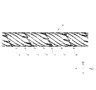
FIG. 1 is a side view showing the configuration
of a stent 10 of a first embodiment;
FIG. 2 is a development view showing a state in which
the stent 10 is virtually opened in a planar shape;
FIG. 3 is a partially-enlarged view of the stent 10;
FIG. 4A is a side view showing a state in which the
diameter of the stent 10 is expanded;
FIG. 48 is a side view of the stent 10 provided with
markers 100;
FIG. 4C is a sectional view of the marker 100;
FIG. 5A is a view for describing the direction of
Date Recue/Date Received 2022-03-10
CA 03154198 2022-03-10
protrusion of a second top portion 15 in the stent
10;
FIG. SB is a view for describing the direction of
protrusion of the second top portion 19 in the stent_
10;
FIG. 5C is a view for describing the direction of
prouru.sic_)n of the second top portion 19 in the stent
10;
FIG. 6 is a view showing the shape of each portion in
a case where the diameter-expanded stent 10 is bent
in a substantially U-shape;
FIG. 7 is a schematic view showing the state of cells
40 in each region of the stent 10 when the stent 10
is virtually opened in the planar shape;
FIG. 8 is a schematic view showing the state of
continuous cells 40 in a region Si of the bent stent
10 when the stent 10 is virtually opened in the
planar shape;
FIG. 9 is a development view showing a state in which
a stent 20 of Comparative Example 1 is virtually
opened in a planar shape;
FIG. 10 is a partially-enlarged view of the stent 20;
FIG. 11 is a schematic view showing the state of
continuous cells 40 in a back-side region of the bent
stent 20 when the stent 20 is virtually opened in the
planar shape;
FIG. 12 is a development view showing a state in
Date Recue/Date Received 2022-03-10
CA 03154198 2022-03-10
9
which a stent 30 of Comparative Example 2 is
virtually opened in a planar shape;
FIG. 13 is a partially-enlarged view of the stent 30;
FIG. 14 is a schematic view showing the state of
.5 continuous cells 40 in a back-side region of the bent
stent 30 when the stent 30 is virtually opened in the
planar shape;
FIG. 15A is a view for describing the patency of the
stent 30 of Comparative Example 1;
FIG. 15B is a view for describing the patency of the
stent 30 of Comparative Example 2;
FIG. 15C is a view for describing the patency of the
stent 30 of the embodiment;
FIG. 16 is a development view showing a state in
which a stent 10A of a second embodiment is virtually
opened in a planar shape;
FIG. 17 is a development view showing a state in
which the diameter of the stent 10A is narrowed;
FIG. 18 is a view showing a shape in a case where the
diameter-expanded stent 10A is bent in a
substantially 7-shape;
FIG. 19 is a development view showing a state in
which a stont 10A of a first variation is virtually
opened in a planar shape;
FIG. 20A is a partially-enlarged view of a corrugated
unit 14 of a second variation;
FIG. 20B is a partially-enlarged view of the
Date Recue/Date Received 2022-03-10
CA 03154198 2022-03-10
corrugated unit 14 of the second variation;
FIG. 20C is a partially-enlarged view of the
corrugated unit 14 of the second variation;
FIG. 21 is a development view showing a firsL
5 configuration of a stent 10C of a third variation;
FIG. 22 is a development view showing a second
configuraTiou of Lhe stent 10C of The third
variation;
FIG. 23 is a development view showing a state in
10 which a stent 100 of a fourth variation is virtually
opened in a planar shape;
FIG. 24 is a development view showing a state in
which a stent 10E of a fifth variation is virtually
opened in a planar shape; and
FIG. 25 is a development view showing a state in
which a stent 1OF of a sixth variation is virtually
opened in a planar shape.
PREFERRED MODE FOR CARRYING OUT THE INVENTION
Hereinafter, an embodiment of a stent according
to the present invention will be described. Note
that any of the drawings attached to the present
specification shows a schematic view and the shape,
scale, longitudinal-lateral dimensional ratio, etc.
of each portion are changed or exaggerated as
compared to actual shape, scale, longitudinal-lateral
dimensional ratio, etc. for the sake of easy
Date Recue/Date Received 2022-03-10
CA 03154198 2022-03-10
understanding of the drawings. Moreover, in the
drawings, hatching showing the cross-section of
members has been omitted where appropriate. In the
present specificaLion, terms specifying shapes,
geometric conditions, and the degrees thereof, such
as "parallel" and "direction", include not only exact
meanings of these terms, but also ranges Laken as
being substantially parallel and being substantially
in a direction.
(First Embodiment)
FIG. 1 is a side view showing the configuration
of a stent 10 of a first embodiment. FIG. 2 is a
development view showing a state in which the stent
10 shown in FIG. 1 is virtually opened in a planar
shape. FIG. 3 is a partially-enlarged view of the
stent 10 shown in FIG. 2. FIG. 4A is a side view
showing a state in which the diameter of the stent 10
shown in FIG. I is expanded. FIG. 4B is a side view
of the stent 10 provided with markers 100. FIG. 40
is a sectional view of the marker 100. FIGS. 5A to
5C are views for describing a protruding direction of
a second top portion 19 of the stent 10.
As shown in FIG. 1, tho stont 10 is in a
substantially cylindrical shape. A peripheral wall
of the stent 10 has such a mesh pattern structure
that a plurality of cells surrounded by a wire-like
material and having the same shape spreads in a
Date Recue/Date Received 2022-03-10
CA 03154198 2022-03-10
12
circumferential direction. For the sake of easy
understanding of the structure of the stent 10, FIG.
2 shows the state in which the stent 10 is opened in
Lhe planar shape. Moreover, FIG. 2 virtually shows,
for the sake of simplicity in the mesh pattern, such
a shape that the mesh pattern is repeated as compared
Lo an actual open sLaLe. In the present
specification, the "peripheral wall" of the stent 10
means a portion dividing the inside and outside of a
cylinder of a substantially cylindrical structure of
the stent 10 from each other. The "cell" is also
called an opening or a compartment, and means a
portion surrounded by the wire-like material forming
the mesh pattern of the stent 10. A "strut" means
each of stems 15 to 17, a connection element 12
(described later), etc. formed of the above-described
wire-like material.
As the material of the stent 10, a material
itself having a high stiffness and a high biological
compatibility is preferred. Examples of such a
material include titanium, nickel, stainless steel,
platinum, gold, silver, copper, iron, chromium,
cobalt, aluminum, molybdenum, manganese, tantalum,
tungsten, niobium, magnesium, calcium, and alloy
containing these materials. Particularly, the stent
10 is preferably made of a material having
superelastic properties, such as nickel titanium (Ni-
Date Recue/Date Received 2022-03-10
CA 03154198 2022-03-10
13
Ti) alloy. The stent 10 shown in FIG. 1 may be
produced in such a manner that a substantially-
cylindrical thin tube made of the above-described
maLerial is processed wiLh a laser.
As the material of the stent 10, synthetic resin
materials such as polyolefin including PE and PP,
polyamide, polyvinyl chloride, polyphenylene sulfide,
polycarbonate, polyether, and polymethylmethacrylate
may be also used. Further, biodegradable resins
(biodegradable polymers) such as polylactate (PLA),
polyhydroxybutyrate (PHD), poiyglycolic acid (PGA),
and poly(E-caprelactone) may be also used. Of these
materials, titanium, nickel, stainless steel,
platinum, gold, silver, copper, magnesium, or alloy
containing these materials are preferred. Examples
of such alloy include Ni-Ti alloy, Cu-Mn alloy, Cu-Cd
alloy, Co-Cr alloy, Cu-Al-Mn alloy, Au-CO-Ag alloy,
Ti-Ai-V alloy, and alloy of magnesium and Zr, Y, Ti,
Ta, Nd, Nb, Zn, Ca, Al, Li, Mn, or the like. In
addition to the materials described above, non-
biodegradable resins may be used as the material of
the stent 10. As described above, any material may
be used to form thc stent 10 as long as such a
material has a biological compatibility.
The stent 10 may contain a medical agent. The
stent 10 containing the medical agent as described
herein means that the stent 10 releasably carries the
Date Recue/Date Received 2022-03-10
CA 03154198 2022-03-10
14
medical agent so as to dissolve out the medical
agent. Although the medical agent is not limited, a
physiologically active substance may be used, for
example. Examples of the physiologically active
substance include a medical agent for inhibiting
intima thickening, a carcinostatic, an
immunosuppressanL, an antibiotic, an anLirheumatic,
an antithrombotic, an HMG-CcA reductase inhibitor, an
ACE inhibitor, a calcium channel blacker, an
antilipemic, an anti-inflammatory, an integrin
inhibitor, an antiallergic, an antioxidant, a
GPIibil-Ia antagonist, retinoid, fiavonoid,
carotenoid, a lipid improver, a DNA synthesis
inhibitor, a tyrosine kinase inhibitor, an
antipiatelet, a vascular smooth muscle growth
inhibitor, an anti-inflammatory agent, and
interferon, and these medical agents may be used in
combination.
Particularly, the medical agent for inhibiting
intima thickening for preventing restenosis is
preferred, and includes, for example, a medical agent
having intima thickening inhibitory action not
blocking endothelial cell growth. Examplcs of such a
medical agent include argatroban H2R,4R)-4-methyl-1-
[112-((RS)-3-methy1-1,2,3,4-tetrahydro-8-
quinolinesulfony1)-L-arginy1]-2-piperidinecarboxylic
acid (Japanese Unexamined Patent Application,
Date Recue/Date Received 2022-03-10
CA 03154198 2022-03-10
Publication No. 2001-190687; PUT International
Publication No. WO 2007/058190)), ximelagatran,
melagatoran, dabigatran, dabigatran etexilate,
rapamycln, everolimus, biolimus A9, zoLarolimus,
tacrolimus, paclitaxel, and statin,
For forming the stent 10 containing the medical
agent, Lhe surface of Lhe stent 10 may be coated with
the medical agent, for example. In this case, the
surface of the stent 10 may be directly coated with
10 the medical agent, or may be coated with a polymer
containing the medical agent. Alternatively, e.g., a
groove or a hole for storing the medical agent may be
provided as a reservoir at the stent 10, and the
medical agent or the mixture of the medical agent and
15 polymer may be stored in such a reservoir. The
reservoir for storage is, for example, described in
Japanese Unexamined Patent Application, Publication
(Translation of PCT Application) No. 2009-524501.
Polymer to be used in this case includes, for
example, soft polymer whose glass-transition
temperature (Tg) is -100 C to 50 C, such as silicone
rubber, urethane rubber, fluorine resin, polybutyl
acrylato, polybutyl methacrylate, acrylic rubber,
natural rubber, ethylene-vinyl acetate copolymer,
styrene-butadiene block copolymer, styrene-isoprene
block copolymer, and styrene-isobutylene block
copolymer; and biodegradable polymer such as
Date Recue/Date Received 2022-03-10
CA 03154198 2022-03-10
16
polylactate, poly(lactic acid-glycolic acid),
polyglycolic acid, poly(lactic acid-s-caprolactone),
poly (glycolic acid-trimethylene carbonate), and poly-
13-hydroxybuLyric acid. For example, polymer and the
medical agent may be mixed in such a manner that the
medical agent is dispersed in polymer according to
descripLion in PCT International Publication No. WO
2009/031295. The medical agent contained in the
stent 10 is delivered to an affected area through the
stent 10, and in such an area, the stent 10
sustained-releases the medical agent. The surface of
the stent 10 may be coated with a carbon-based
material such as diamond-like carbon (DLC, F-DLC).
In a case where the stent 10 shown in FIG. 1 is
produced from, e.g., a superelastic alloy tube, a
tube having a diameter of about 2 to 3 mm is
processed with a laser, and thereafter, is stretched
in a radial direction until the diameter reaches
about 5 mm. FIG. 2 shows the state in which the
stent 10 not stretched yet after a tube with a
diameter of 2 mm has been processed with a laser is
virtually opened in the planar shape. Moreover, FIG.
4A shows the state in which the diameter of the stent
10 shown in FIG. 1 is expanded to 5 mm. The diameter
of the stent 10 is narrowed in the radial direction
from the state shown in FIG. 4A, and thereafter, the
stent 10 is housed (inserted) in an inner cavity of a
Date Recue/Date Received 2022-03-10
CA 03154198 2022-03-10
17
catheter (not shown). The shape shown in FIG. 4A is
recovered in such a manner that the stent 10 housed
in the catheter is pushed out. The stent 10 is made
of an elastic material such as superelastic alloy or
shape-memory alloy so that the above-described shape
recovery function can be obtained. Note that
producLion of the stenl, 10 is not limited Lo
processing with a laser, and for example, the stent
may be also produced by other methods such as
10 cutting.
The markers 100 may be provided on both end
sides of the stent 10 in an axial direction LD. FIG.
43 shows such a configuration that the markers 100
are provided on both end sides of the diameter-
expanded stent 10 of FIG. 42 in the axial direction
LD. The marker 100 is a member serving as a mark for
checking the position of the stent 10 in a hollow
organ such as a blood vessel, and is made of a
radiopaque material. As shown in FIG. 4C, the marker
100 includes a tip end portion 110 of the stent 10
and a coil-shaped spring 120 provided outside the tip
end portion 110. A tip end of the tip end portion
110 of the stent 10 protrudes from the coil-shaped
spring 120. The coil-shaped spring 120 is preferably
made of a material through which radiation such as an
X-ray cannot pass and which can be formed in a coil
shape. Examples of the material of the coil-shaped
Date Recue/Date Received 2022-03-10
CA 03154198 2022-03-10
18
spring 120 include platinum-iridium (Pt-Ir).
The method for joining the coil-shaped spring
120 and the tip end portion 110 of the stent 10 to
each other is not particularly limited as long as
.5 such a method is used for medical equipment joint
such as welding, bonding with UV, or silver brazing.
The welding method includes, for example, a merhod in
which the coil-shaped spring 120 and the tip end
portion 110 of the stent 10 are melted by welding to
bond and fix the coil-shaped spring 120 and the tip
end portion 110 to each other, and a method in which
the region of the tip end portion 110 of the stent 10
protruding from the coil-shaped spring 120 is melted
to restrict movement of the coil-shaped spring 120.
In the case of bonding with UV, the coil-shaped
spring 120 is fixed to the tip end portion 110 of the
stent 10 by means of medical-grade radiation curable
polymer. The steps of such a method are as follows:
the tip end portion 110 of the stent 10 is coated
with a curable polymer solution, the coil-shaped
spring 120 is placed thereon, and thereafter, these
portions are irradiated with radiation to cure the
curable polymer solution to fix the coil-shaped
spring 120 to the tip end portion 110 of the stent
10. In the case of silver brazing, the coil-shaped
spring 120 is made of a material different from that
of the stent 10, and the coil-shaped spring 120 is
Date Recue/Date Received 2022-03-10
CA 03154198 2022-03-10
19
fixed to the tip end portion 110 of the stent 10 in
such a manner that, e.g., silver solder soaks into
the coil-shaped spring 120 from above.
As shown in FIGS. 1 to 3, the sLent 10 of the
first embodiment includes a plurality of annular
bodies (corrugated pattern bodies) 11 arranged next
each o1her in the axial direction (a longitudinal
axis direction, a center axis direction) LD and a
plurality of connection elements 12 connecting the
10 annular bodies 11 adjacent to each other in the axial
direction LD. As described later, when the stent 10
is viewed in a radial direction RD perpendicular to
the axial direction ID, an annular direction CD of
the annular body 11 is inclined with respect to the
radial direction RD. The angle +la of inclination of
the annular direction CD of the annular body 11 with
respect to the radial direction RD is 30 to 60
degrees, for example.
As shown in FIG. 2, the annular body 11 has a
corrugated pattern formed of a plurality of
corrugated units 14. In the annular body 11, the
plurality of corrugated units 14 is connected along
the annular direction CD. As shown in FIG. 3, the
corrugated unit 14 includes a first stem 15, a second
stem 16, a third stem 17, a first top portion 18, and
the second top portion 19. The first stem 15 is a
stem arranged substantially parallel with the axial
Date Recue/Date Received 2022-03-10
CA 03154198 2022-03-10
direction LD. The second stem 16 is a stem arranged
substantially parallel with the annular direction CD.
The stent 10 of the first embodiment is configured
such that the annular direcLion CD of She annular
.5 body 11 is inclined with respect to the radial
direction RD by the angle +0 when the stent 10 is
viewed in Lhe radial direcLion RD perpendicular Lo
the axial direction LD. In the form in which the
annular body 11 is inclined with respect to the
10 radial direction RD by the angle -re, the SUM of the
length Li of the first stem 15 of the corrugated unit
14 and the length L2 of the second stem 16 of the
corrugated unit 14 is longer than the length L3 of
the third stem 17.
15 As shown in FIG. 3, a first end portion 15a of
the first stem 15 on one side (first side) and a
first end portion 16a of the second stem 16 on one
side (first side) are coupled to each other through
the first top portion 18. A second end portion 16b
20 of the second stem 16 on the other side (second side)
and a first end portion 17a of the third stem 17 on
one side (first side) are coupled to each other
through the second top portion 19. A second end
portion 17b of the third stem 17 on the other side
(second side) is connected to a second end portion
15b of the first stem 15 on the other side (second
side) in the corrugated unit 14 adjacent to such a
Date Recue/Date Received 2022-03-10
CA 03154198 2022-03-10
21
second end portion 17b in the annular direction CD (a
direction about an axis).
In a certain corrugated unit 14, a second top
portion 19 coupling a second stem 16 and a third stem
17 to each other is not coupled to any of corrugated
units 14 adjacent to the certain corrugated unit 14
in the annular direction CD. A Lhird sLeni 17 of a
certain corrugated unit 14 and a first stem 15 of a
corrugated unit 14 adjacent to the certain corrugated
unit 14 in the direction about the axis are, at end
portions (a second end portion 17b and a second end
portion 15b) thereof, coupled to each other to form a
slit S therebetween. As shown in FIG. 3, the stent
10 of the first embodiment is configured such that
adjacent two of the corrugated units 14 in the axial
direction LD and two of the connection elements 12
(described later) connecting these two corrugated
units 14 in the axial direction ID form the cell.
This cell basically has a closed cell structure, but
in each corrugated unit 14, the second top portion 19
is a substantially V-shaped free end. Thus, the
stent 10 of the first embodiment is formed such that
the closed cell structure partially has an pan coil
structure. As described later, when the diameter of
the stent 10 is expanded, the second stem 16 and the
third stem 17 deform in a separation direction about
the second top portion 19 as the free end.
Date Recue/Date Received 2022-03-10
CA 03154198 2022-03-10
22
As shown in FIG. 2, the plurality of connection
elements 12 is arranged at equal intervals along the
annular direction CD of the annular body 11. Each
connection element 12 extends in a spiral shape about
.5 the center axis_ As shown in FIG. 3, a first end
portion 12a of a certain connection element 12 on one
side (first side) is connected La a first Lop porLion
18 of one corrugated unit 14 adjacent to the certain
connection element 12 in the axial direction LD.
That is, the first end portion 12a of the connection
element 12 is, at a first top portion 18 of the
corrugated unit 14a, connected to a first end portion
15a of a first stem 15 and a first end portion 16a of
a second stem 16. Moreover, a second end portion 12b
of the certain connection element 12 on the other
side (second side) is connected to a second end
portion 17b of a third stem 17 of the other
corrugated unit 14b adjacent to the certain
connection element 12 in the axial direction LD and a
second end portion 15b of a first stem 15 of a
corrugated unit 14c adjacent to the corrugated unit
14b in the direction about the axis. Note that in
FIG. 3, reforcnco numerals "14a", "14b", and "14c"
are assigned to some of the corrugated units 14 for
the sake of description above.
A direction in which the second top portion 19
of the corrugated unit 14 protrudes in the stent 10
Date Recue/Date Received 2022-03-10
CA 03154198 2022-03-10
23
of the first embodiment will be described herein.
FIG. 5A is the view virtually showing the entirety of
the stent 10 opened in the planar shape. in FIG. 5A,
when Lhe steni_ 10 is viewed from a practitioner
.5 operating the catheter (not shown) housing the stent
10, a side close to the practitioner in the axial
ciireuLion LD of Lhe sLent 10 is Laken as Et proximal
side LD1 and a side distant from the practitioner is
taken as a distal side LD2. Moreover, in FIG. 5A,
the annular bodies 11 and the connection elements 12
are drawn in a simple manner.
The stent 10 is implanted in the hollow organ
such as a blood vessel, but in some cases, may be
reimplanted elsewhere. In this case, the stent 10 is
housed again in the catheter. In FIG. 5A, a
direction in which the stent 10 is housed again is a
direction from the distal side I.D2 toward the
proximal side LD1. FIG. 53 is an enlarged view of a
portion from the center to an end portion on the
proximal side LD1 in the axial direction LD of the
stent 10. Moreover, FIG. 5C is an enlarged view of a
portion from the center to an end portion on the
distal side 1D2 in the axial direction LD of the
stent 10.
As shown in FIGS. 5B and 5C, in any of the
corrugated units 14 forming the annular body 11 of
the stent 10, the second top portion 19 is formed to
Date Recue/Date Received 2022-03-10
CA 03154198 2022-03-10
24
protrude to the distal side LD2 in the direction
(from LD2 toward LD1) of insertion of the stent 10
into the catheter. According to the above-described
configuration, when the stent_ 10 is housed again in
.5 the catheter, the substantially V-shaped protruding
end of the second top portion 19 as the free end does
noL face an insertion port of the catheter, and
therefore, the stent 10 can be easily housed again in
the catheter.
Next, patency when the stent 10 of the first
embodiment is bent will be described. FIG. 6 is a
view showing the shape of each portion in a case
where the diameter-expanded stent 10 (see FIG. 4A) is
bent in a substantially U-shape. FIG. 7 is a
schematic view showing the state of a cell 40 in each
region of the stent 10 shown in FIG. 6 when the stent
10 is virtually opened in the planar shape. FIG. 7
shows, at the center thereof, the cell 40 in a no-
load state (the state of FIG. 4A) in which any of
tensile force and compression force does not act on
the cell 40. FIG. 8 is a schematic view showing the
state of continuous cells 40 in a region Si of the
bent stent 10 shown in FIG. 6 when the stent 10 is
virtually opened in the planar shape. FIG. 8
schematically shows, on an upper side therein, the
section of a strut 50 by a circle. This circle is
drawn for describing stress acting on one strut 50,
Date Recue/Date Received 2022-03-10
CA 03154198 2022-03-10
and is different from an actual strut section.
As shown in FIG. 6, when the diameter-expanded
stent 10 is bent in the substantially U-shape, the
cells 40 are pulled in the region Si on a back side
5 (outside) of the bent portion. In this state, stress
acting on the region SI is, as shown on an upper side
in FIG. 7, in the directions of arrows 51, 52 at each
connection point a between the struts SO forming the
cell 40. Thus, as shown in FIG. 8, the continuous
10 cells 40 in the region Si deform so as to be pulled
in the directions of arrows 53. That is, in FIG. 8,
the struts 50 of the cells 40 in the no-load state as
indicated by dotted lines deform (move) so as to be
pulled in the directions of the arrows 53 as
15 indicated by solid lines. In this state, when the
strut 50 is viewed in section, the strut 50 deforms
so as to rotate in two directions indicated by arrows
54, as shown on the upper side in FIG. 8. The
directions indicated by the arrows 54 on the upper
20 side in FIG. 8 correspond to the directions of the
arrows 53 on a lower side in FIG. 8.
On the other hand, in FIG. 6, the cells 40 are
compressed in a region S2 on a stomach side (inside)
of the bent portion. In this state, stress acting on
25 the region 52 is, as shown on a lower side in FIG. 7,
in the directions of arrows 55 to 57 at each
connection point a between the struts 50 forming the
Date Recue/Date Received 2022-03-10
CA 03154198 2022-03-10
26
cell 40. Thus, although not shown in the figure, the
continuous cells 40 in the region S2 deform so as to
be pulled in a direction in which an interval between
the st_LuLs 50 is narrowed.
Next, deformation in response to stress on each
of the stents of Comparative Example 1, Comparative
Example 2, and Lhe firsL embodimenL will be
described. FIG. 9 is a development view showing a
state in which a stent 20 of Comparative Example 1 is
virtually opened in a planar shape. FIG. 10 is a
partially-enlarged view of the stent 20 shown in FIG.
9. FIG. 11 is a schematic view showing the state of
continuous cells 40 in a back-side region of the bent
stent 20 when the stent 20 is virtually opened in the
planar shape. FIG. 11 is the schematic view showing
the state of the continuous cells 40 in the back-side
region of the bent stent 20 of Comparative Example 1
when the stent 20 is virtually opened in the planar
shape.
As shown in FIG. 9, the stent 20 of Comparative
Example 1 includes a plurality of annular bodies 21
arranged next to each other in the axial direction ID
and connection elements 22 connecting the annular
bodies 21 adjacent to each other in the axial
direction LD. When the stent 20 of Comparative
Example 1 is viewed in the radial direction RD
perpendicular to the axial direction ID, an annular
Date Recue/Date Received 2022-03-10
CA 03154198 2022-03-10
27
direction CD of the annular body 21 is substantially
coincident with the radial direction RD.
As shown in FIG. 10, the stent 20 of Comparative
Example 1 has a corrugated paLLern formed such that a
.5 plurality of substantially V-shaped elements 23 is
connected in the circumferential direction. The V-
shaped elemenL 23 is formed such that_ Lwo stems 24
are coupled to each other at a top portion 25. The
V-shaped elements 23 are configured such that the top
portions 25 thereof face in the same direction in the
axial direction LD, and the stems 24 of adjacent ones
of the V-shaped elements 23 in the circumferential
direction are connected to each other to form the
corrugated pattern.
Two end portions 22a, 22b of each connection
element 22 in a longitudinal direction thereof are
each connected to adjacent two of the V-shaped
elements 23 in the axial direction LD. The end
portion 22a of the connection element 22 on one side
(first side) is connected in the axial direction LD
to the stems 24 of adjacent two of the V-shaped
elements 23 in a direction along the corrugated
pattern. Moreover, the end portion 22b of the
connection element 22 on the other side (second side)
is connected to the top portion 25 of the V-shaped
element 23 adjacent to the above-described two V-
shaped elements 23 in the axial direction LD. As
Date Recue/Date Received 2022-03-10
CA 03154198 2022-03-10
28
described above, in the stent 20 of Comparative
Example 1, all of the top portions 25 are connected
to the connection elements 22. Thus, the stent 20 of
Comparative Example I has a closed cell structure
.5 with no free end.
The diameter of the stent 20 of Comparative
Example 1 is expanded as in the stent 10 (see FIG. 4)
of the embodiment. When the stent 20 is bent in a
substantially U--shape, the cells 40 are pulled in the
back-side region. In this state, the continuous
cells 40 in the back-side region deform diagonally in
the direction of an arrow 55 as shown in FIG. 11.
That is, in FIG. 11, struts 50 of the cells 40 in a
no-load state as indicated by dotted lines deform
(move) as indicated by solid lines. In this state,
when the strut 50 is viewed in section, the strut 50
deforms so as to rotate in one direction indicated by
an arrow 56, as shown on an upper side in FIG. 11.
The direction indicated by the arrow 56 on the
upper side in FIG. 11 corresponds to the direction
indicated by the arrow 55 on a lower side in FIG. 11.
As described above, in the stent 20 of Comparative
Example 1, deformation of the cell 40 is small in the
back-side region of the bent portion, and the
direction of deformation when the strut 50 is viewed
in section is only one direction. Thus, in the stent
20 of Comparative Example 1, the amount of
Date Recue/Date Received 2022-03-10
CA 03154198 2022-03-10
29
deformation for absorbing stress acting on the back-
side region is smaller than that in the stent 10 of
the embodiment. That is, in the stent 20 of
Comparative Example 1, a twist/bend easily occurs in
the back-side region due to stress acting on the bent
portion. The same also applies to a stomach-side
region of L.he bent porLion in the stent 20 of
Comparative Example 1, and the stent 20 of
Comparative Example I has such a structure that a
twist/bend easily occurs due to the stress acting on
the bent portion.
FIG. 12 is a development view showing a state in
which a stent 30 of Comparative Example 2 is
virtually opened in a planar shape. FIG. 13 is a
partially-enlarged view of the stent 30 shown in FIG.
12. FIG. 14 is a schematic view showing the state of
continuous cells 40 in a back-side region of the bent
stent 30 when the stent 30 is virtually opened in the
planar shape.
As shown in FIG. 12, the stent 30 of Comparative
Example 2 includes a plurality of annular bodies 31
arranged next to each other in the axial direction Lb
and connection elements 32 connecting the annular
bodies 31 adjacent to each other in the axial
direction Lb. When the stent 30 of Comparative
Example 2 is viewed in the radial direction RD
perpendicular to the axial direction LD, the annular
Date Recue/Date Received 2022-03-10
CA 03154198 2022-03-10
direction CD of the annular body 31 is inclined with
respect to the radial direction RD.
As shown in FIG. 13, the stent 30 of Comparative
Example 2 has a corrugated pattern formed such that a
5 plurality of substantially V-shaped elements 33 is
connected in the annular direction CD. The V-shaped
element_ 33 is formed such that Leo stems 34 are
coupled to each other at a top portion 35. The V-
shaped elements 33 are configured such that the top
10 portions 35 thereof face in the same direction in the
axial direction LD, and the stems 34 of adjacent ones
of the V-shaped elements 33 in the annular direction
CD are connected to each other to form the corrugated
pattern.
15 Two end portions 32a, 32b of each connection
element 32 in a longitudinal direction thereof are
each connected to adjacent two of the V-shaped
elements 33 in the axial direction LD. The end
portion 32a of the connection element 32 on one side
20 (first side) is connected in the axial direction LD
to the stems 34 of adjacent two of the V-shaped
elements 33 in a direction along the corrugated
pattern extending along the annular direction CD.
Moreover, the end portion 32b of the connection
25 element 32 on the other side (second side) is
connected to the top portion 35 of the V-shaped
element 33 adjacent to the above-described two V-
Date Recue/Date Received 2022-03-10
CA 03154198 2022-03-10
31
shaped elements 33 in the axial direction LD. As
described above, in the stent 30 of Comparative
Example 2, all of the top portions 35 are connected
Lc Lhe conneoLion elemenLs 32. Thus, the stent 30 of
Comparative Example 2 has a closed cell structure
with no free end.
The diameLer of the stent 30 of Comparative
Example 2 is expanded as in the stent 10 (see FIG.
4A) of the embodiment. When the stent 30 is bent in
a substantially U-shape, the cells are pulled in the
back-side region. In this state, the continuous
cells in the back-side region deform diagonally in
the direction of an arrow 57 as shown in FIG. 14.
That is, in FIG. 14, struts 50 of the cells in a no-
load state as indicated by dotted lines deform (move)
as indicated by solid lines. In this state, when the
strut SO is viewed in section, the strut 50 deforms
so as to rotate in one direction indicated by an
arrow 58, as shown on an upper side in FIG. 14.
The direction indicated by the arrow 58 on the
upper side in FIG. 14 corresponds to the direction
indicated by the arrow 57 on a lower side in FIG. 14.
As described above, in the stent 30 of Comparative
Example 2, deformation of the cell is small in the
back-side region of the bent portion, and the
direction of deformation when the strut 50 is viewed
in section is only one direction. Thus, in the stent
Date Recue/Date Received 2022-03-10
CA 03154198 2022-03-10
32
30 of Comparative Example 2, the amount of
deformation for absorbing stress acting on the back-
side region is smaller than that in the stent 10 of
Lhe embodiment. Thai_ is, in the sLent 30 of
.5 Comparative Example 2, a twist/bend easily occurs in
the back-side region due to stress acting on the bent
porLion. The same also applies to a stomach-side
region of the bent portion in the stent 30 of
Comparative Example 2, and the stent 30 of
Comparative Example 2 has such a structure that a
twist/bend easily occurs due to the stress acting on
the bent portion.
Next, the patency of each of the stents of
Comparative Example 1, Comparative Example 2, and the
embodiment will be described. FIGS. 15A to 15C are
views for describing the patency of each of the
stents of Comparative Example 1, Comparative Example
2, and the embodiment. FIGS. 15A to 15C show
sectional shapes when the diameters of the stents of
Comparative Example 1, Comparative Example 2, and the
embodiment are expanded to the same diameter and
these stents are bent in a substantially U-shape. On
an upper side in FIGS. 15A to 15C, the sectional
shape at a center portion of a bend indicated by a
dashed line is shown. On a lower side in FIGS. 15A
to 15C, an appearance when the stent is bent in the
substantially U-shape is shown.
Date Recue/Date Received 2022-03-10
CA 03154198 2022-03-10
33
It has been found that in each of the stent 20
of Comparative Example 1 as shown in FIG. 15A and the
stent 30 of Comparative Example 2 as shown in FIG.
15B, a kink leading to the twist/bend of the section
in a substantially oval shape occurs and the patency
against bending is low. This is because each cell
deforms only in one direcLion in response Lo the
stress caused by bending in the stent 20 of
Comparative Example 1 and the stent 30 of Comparative
Example 2. On the other hand, it has been found that
in the stent 10 of the embodiment as shown in FIG.
15C, the twist/bend of the section is less likely to
occur and the patency against bending is high. This
is because each cell deforms in two directions in
response to the stress caused by bending in the stent
10 of the embodiment.
As described above, the stent 10 of the first
embodiment includes the tree ends (the second top
portions 19) in the plurality of corrugated units 14
forming the corrugated pattern. Thus, two stems
connected to the free end move in the separation
direction when the stent 10 is bent, and therefore,
the cells can be entirely deformed in two directions.
Thus, the stent 10 of the embodiment has a high
patency against bending.
The stent 10 of the first embodiment is formed
such that the second top portions 19 as the free ends
Date Recue/Date Received 2022-03-10
CA 03154198 2022-03-10
34
protrude to the distal side in the direction of
insertion into the catheter. According to the
present configuration, the substantially V-shaped
proLruding ends of Lhe second Lop porLions 19 as Lhe
free ends do not face the insertion port of the
catheter when the stent 10 is housed in the catheter
again, and therefore, Lhe sLent 10 can be easily
housed in the catheter again.
In the stent 10 of the first embodiment, a third
stem 17 of a certain corrugated unit 14 and a first
stem 15 of a corrugated unit 14 adjacent to the
certain corrugated unit 14 in the direction about the
axis are, at end portions thereof, coupled to each
other to form a slit S therebetween. Thus, in the
stent 10 of the first embodiment, the third stem 17
coupled to the first stem 15 and the second stem 16
coupled to such a third stem 17 at the second top
portion 19 can be more greatly deformed.
(Second Embodiment)
Next, a stent 102\ of a second embodiment will be
described. In description and drawings for the
second embodiment, the same reference numerals as
those of the first embodiment are used to represent
members etc. equivalent to those of the first
embodiment, and overlapping description thereof will
be omitted.
FIG. 16 is a development view showing a state in
Date Recue/Date Received 2022-03-10
CA 03154198 2022-03-10
which the stent 10A of the second embodiment is
virtually opened in a planar shape. FIG. 17 is a
development view showing a state in which the
diameLer of the stenL 10A is narrowed. FIG. 18 is a
.5 view showing a shape in a case where the diameter-
expanded stent 10A is bent in a substantially U-
shape.
As shown in FIG. 16, in the stent 10A of the
second embodiment, the length L4 of a connection
10 element 12 is set shorter than the length L2 of a
second stem 16. Specifically, the length L4 of the
connection element 12 is set to, e.g., about 0.7 to
0.9 in terms of the value of L4/1,2. The length L4 of
the connection element 12 and the length L2 of the
15 second stem 16 are measured in terms of the shortest
distance (a straight-line distance).
In the stent 10A of the second embodiment, a
plurality of corrugated units 14 is connected along a
radial direction RD. That is, when the stent 10A of
20 the second embodiment is viewed in the radial
direction RD perpendicular to an axial direction LD,
an annular direction CD of an annular body 11 is
substantially coincident with the radial direction
RD.
25 In the stent 10A of the second embodiment, the
length L4 of the connection element 12 is set shorter
than the length L2 of the second stem 16. According
Date Recue/Date Received 2022-03-10
CA 03154198 2022-03-10
36
to the present configuration, an interval between
adjacent ones of the corrugated units 14 in the axial
direction LD is short, and therefore, the number of
corrugated units 14 per unit length in the axial
.5 direction LD can be increased. As the number of
corrugated units 14 increases as described above, a
surface area per unit lenghh in the axial direction
LD increases. Thus, blood vessel holding performance
of the stent 10A can be improved.
In the stent 10A of the second embodiment, the
plurality of corrugated units 14 is connected along
the radial direction RD. Thus, upon processing of
the stent, stress acting on the inside of a strut is
uniformly transmitted in the radial direction RD at
the step of expanding the diameter of a laser-
processed thin tube to a finishing diameter. In a
case where the stress acting on the inside of the
strut is uniformly transmitted in the radial
direction RD as described above, e.g., the twist of
the strut due to non-uniform local stress is less
likely to occur, and therefore, a more-uniform
expanded shape can be obtained in a circumferential
direction. Moreover, in the stent 10A of the second
embodiment, the plurality of corrugated units 14 can
be patterned along the radial direction RD, leading
to excellent workability.
In the stent 10A of the second embodiment, a
Date Recue/Date Received 2022-03-10
CA 03154198 2022-03-10
37
basic structure of the corrugated unit 14 is the same
as that of the first embodiment. That is, as shown
in FIG. 17, any of the corrugated units 14 is formed
such LhaL a substantially V-shaped protruding end of
.5 a second top portion 19 protrudes to a distal side
LD2 in the direction (from LD2 toward LD1) of
insertion of the stent 10A into a caLheter (not
shown). Thus, the stent 10A of the second embodiment
can be also easily housed in the catheter again, as
in the first embodiment. Moreover, in the stent 10A
of the second embodiment, the second top portion 19
is less likely to overlap with the connection element
12 while being compressed radially as shown in FIG.
17, and therefore, upon diameter expansion, the stent
10A can be more uniformly deployed.
Note that two stems 16, 17 connected to the
second top portion 19 as a free end move in a
separation direction when the stent 10A of the second
embodiment is bent in the substantially U-shape as
shown in FIG. 18, and therefore, cells can be
entirely deformed in two directions. Thus, the stent
10A has a high patency against bending.
The embodiments of the stent according to the
present invention have been described above, but the
present disclosure is not limited to the above-
described embodiments. Various modifications and
changes as in later-described variations can be made,
Date Recue/Date Received 2022-03-10
CA 03154198 2022-03-10
38
and are also included in the technical scope of the
present disclosure. Moreover, most preferred
advantageous effects of the present disclosure have
been merely described as the advantageous effects of
.5 the embodiments, and the present disclosure is not
limited to those described in the embodiments. Note
that_ the above-described embodimenLs and Lne later-
described variations may be used in combination as
necessary, but detailed description thereof will be
omitted.
FIG. 19 is a development view showing a state in
which a stent 10B of a first variation is virtually
opened in a planar shape. The stent 105 of the first
variation is different from the stent 10 of the first
embodiment in the direction of inclination of an
annular direction CD of an annular body 11 with
respect to a radial direction RD. Specifically, when
the stent 10B of the first variation is viewed in the
radial direction RD perpendicular to an axial
direction LD, the annular direction CD of the annular
body 11 is inclined with respect to the radial
direction RD by an angle -O. In the form in which
the annular body 11 is inclined with respect to the
radial direction RD by the angle -e as shown in FT.
19, the sum of the length Ll of a first stem 15 of a
corrugated unit 14 and the length L2 of a second stem
16 of the corrugated unit 14 is shorter than the
Date Recue/Date Received 2022-03-10
CA 03154198 2022-03-10
39
length L3 of a third stem 17. As shown in FIG. 19,
the direction of inclination of the annular direction
CD of the annular body 11 with respect to the radial
direction RD may be opposite to LhaL of the stent 10
(see FIG_ 2) of the first embodiment. In the present
configuration, advantageous effects similar to those
of the sLeht 10 of the firsL embodiment_ can be
obtained. Note that the configuration of the first
variation can be also applied to the stent 10A of the
second embodiment.
FIGS. 20A to 200 are partially-enlarged views of
a corrugated unit 14 of a second variation. In the
first and second embodiments, connection shapes as
shown in FIGS. 20A to 200 can be applied to the
connection portion between the connection element 12
and the corrugated unit 14. FIGS. 20A to 20C show
the shapes applicable to the connection portion in
any of a region Al or a region A2 of the corrugated
unit 14 shown in FIG. 2. Hereinafter, the connection
portion in the region Al of FIG. 2 will be described
as an example. The region Al is a portion at which
the first end portion 12a of the connection element
12 is connected to the second end portion 15b of the
first stem 15 of the corrugated unit 14 and the
second end portion 17b of the third stem 17 of the
corrugated unit 14.
In the connection shape shown in FIG. 20A, the
Date Recue/Date Received 2022-03-10
CA 03154198 2022-03-10
first end portion 12a of the connection element 12 is
connected to a side close to the second end portion
17b of the third stem 17. In the connection shape
shown in FIG. 20B, Lhe firsL end portion 12a of the
.5 connection element 12 is connected to a side close to
the second end portion 15b of the first stem 15. In
Lhe connection shape shown in FIG. 200, Lhe first end
portion 12a of the connection element 12 is connected
to between the second end portion 15b of the first
10 stem 15 and the second end portion 17b of the third
stem 17. The connection shape shown in each figure
for the second variation can be selected as necessary
according to transmission of force upon bending of
the stent and the state of stress acting on the
15 inside and surface of the stent, for example.
FIGS. 21 and 22 are development views showing a
state in which a stent 100 of a third variation is
virtually opened in a planar shape. FIG. 21 is the
development view showing a first configuration of the
20 stent 100 of the third variation. As shown in FIG.
21, in the first configuration of the stent 10C of
the third variation, a connection element 12
connecting adjacent ones of annular bodies 11 in an
axial direction Lb is formed in a substantially S-
25 shaped corrugated pattern. FIG. 22 is the
development view showing a second configuration of
the stent 100 of the third variation. As shown in
Date Recue/Date Received 2022-03-10
CA 03154198 2022-03-10
41
FIG. 22, in the second configuration of the stent 10C
of the third variation, the connection element 12
connecting adjacent ones of the annular bodies 11 in
the axial direction LD is formed such that a
.5 substantially S-shaped corrugated pattern is repeated
twice. In the connection element 12 of the second
conflguraLloh, the substantially S-shaped corruyated
pattern may be repeated three times or more. The
shape of the connection element 12 shown in each
figure for the third variation can be selected as
necessary according to transmission of force upon
bending of the stent and the state of stress acting
on the inside and surface of the stent, for example.
FIG. 23 is a development view showing a state in
which a stent 105 of a fourth variation is virtually
opened in a planar shape. As shown in FIG. 23, a
first stem 15, a second stem 16, and a third stem 17
forming a corrugated unit 14 may be different from a
connection element 12 connecting annular bodies 11 to
each other in a strut thickness (e.g., the maximum
diameter). The stent 10D of FIG. 23 is an example
where the thickness of the connection element 12 is
thinner than the thicknesses of the first stem 15,
the second stem 16, and the third stem 17 for further
enhancing flexibility. The strut thickness in the
first stem 15, the second stem 16, the third stem 17,
and the connection element 12 can be selected as
Date Recue/Date Received 2022-03-10
CA 03154198 2022-03-10
42
necessary according to transmission of force upon
bending of the stent and the state of stress acting
on the inside and surface of the stent, for example.
Note that the example where the connection element 12
is formed thinner in the stent 100 of the fourth
variation has been described, but the strut thickness
may he changed for any one or more of the first sLein
15, the second stem 16, the third stem 17, and the
connection element 12.
FIG. 24 is a development view showing a state in
which a stent 10E of a fifth variation is virtually
opened in a planar shape. As shown in FIG. 24, in
the stent 10E of the fifth embodiment, two connection
element bands L12 are provided between two connection
points (cross marks) in a radial direction RD. In
FIG. 24, these two connection points (the cross
marks) indicate virtual connection positions in a
circumferential direction of the substantially
cylindrical stent 10D. The connection element band
112 indicates the line of a plurality of connection
elements 12 arranged along an annular direction CD.
In the stent 10E of the fifth variation, the two
connection element bands 112 are provided between the
two connection points (the cross marks) in the radial
direction RD. Thus, as compared to a configuration
(see, e.g., FIG. 2) in which a single connection
element band 112 is provided between two connection
Date Recue/Date Received 2022-03-10
CA 03154198 2022-03-10
43
points (cross marks) in the radial direction RD, the
surface area and cell density of the stent 10E of the
fifth variation can be increased. Note that in the
stent 10E shown in FIG. 24, three or more connection
element bands L12 may be provided between the two
connection points (the cross marks) in the radial
dlrecLion RD.
FIG. 25 is a development view showing a state in
which a stent 10F of a sixth variation is virtually
cypnd in a planar shape. FIG. 25 shows an area of
the stent 1OF from the substantially center to an end
portion on a proximal side LD1 in an axial direction
LD. As shown in FIG. 25, the stent 1OF includes
marker holding portions 13 at the end portion on the
proximal side LD1. The marker holding portion 13 is
a portion for holding a marker 130 (described later.
Note that FIG. 25 shows an example where three marker
holding portions 13 are provided at the end portion
of the stent 1OF on the proximal side LD1, but the
number of marker holding portions 13 is not limited
to that in the example of FIG. 25.
The marker holding portion 13 is configured such
that a slit 13a is formed along a center portion of
the marker holding portion 13 in a longitudinal
direction thereof. The slit 13a is a portion to be
fastened to a substantially center portion of the
marker 130 by swaging. Note that a left one of the
Date Recue/Date Received 2022-03-10
CA 03154198 2022-03-10
44
three marker holding portions 13 shown in FIG. 25
shows a state before fastening of the marker 130.
The marker 130 used for the stent 1OF of the
sixth variation is formed in a subsLantlally
cylindrical shape_ Of the marker 133, one end
portion is formed with a substantially semicircular
head porLion 131, and the Lher end portion is formed
with an opening 132. In the marker 130 held on the
proximal side LD1 of the stent 10F, the head portion
131 is positioned in an insertion direction (from L32
toward LD1) when the stent 1OF is housed in a
catheter (not shown) again. As in the marker 100
(see FIG. 43) described in the first embodiment, the
marker 130 is made of a radiopaque material.
As shown in FIG. 25, the marker 130 is inserted
onto the marker holding portion 13 of the stent 10F
from an opening 132 side, and by swaging, the marker
holding portion 13 of the stent 1OF and the marker
130 can be fastened to each other. Although not
shown in the figure, an area of the stent 10F from
the substantially center to an end portion on a
distal side LD2 in the axial direction Lb is also
configured similarly to FIG. 25.
According to the configuration of the sixth
variation, the substantially semicircular head
portion 131 is, when the stent 10F is housed in the
catheter again, positioned on the side from which the
Date Recue/Date Received 2022-03-10
CA 03154198 2022-03-10
marker 130 is inserted, and therefore, the stent 10F
can be more easily housed in the catheter again.
EXPLANATION OF REFERENCE NUMERALS
10, 10A, 103, 10C, 10D, 10E, 10F Stent
11 Annular Body (Corrugated Pattern Body)
12 ConneuLion ElemenL
I2a First End Portion
12b Second End Portion
10 14 Corrugated Unit
15 First Stem
15a First End Portion
15b Second End Portion
16 Second Stem
15 16a First End Portion
16b Second End Portion
17 Third Stem
17a First End Portion
17b Second End Portion
20 18 First Top Portion
19 Second Top Portion
100 Marker
Date Recue/Date Received 2022-03-10