Note: Descriptions are shown in the official language in which they were submitted.
1
CONTAINER LEAKAGE DETECTION USING THERMAL IMAGING
TECHNICAL FIELD
[0001] The technical field generally relates to inspection techniques for
detecting leakage in
containers, for example, flexible containers such as intravenous fluid bags,
and, more particularly, to
such techniques using thermal or infrared imaging.
BACKGROUND
[0002] Intravenous (IV) fluid bags are used as containers in various medical
applications. IV fluid
bags are typically packaged in a plastic overwrap, which protects the
integrity of the bag by providing
an additional barrier against contamination and damage. Leakage of IV fluid
bags may sometimes
occur if the bag becomes compromised, for example, due to a tear, a puncture,
a sealing defect, or
another issue. For overwrapped IV fluid bags, leaked fluid may accumulate in
the interstitial volume
between the bag and its overwrap. Defects in IV fluid bags can arise due to a
number of causes
including, to name a few, faulty manufacturing and damage incurred during
transportation, storage,
or handling. Leaking IV fluid bags may result in potential contamination,
compromised sterility, patient
and medical personnel exposure to hazardous materials contained in the IV
solution, and inadequate
IV solution concentration or dosage. Leakage of IV fluid bag is undesirable in
the medical field and
should be detected early on to mitigate its impact.
[0003] The discovery of a leaking IV fluid bag downstream in the supply chain
may lead to the
reinspection of the entire product lot or inventory to which the defective bag
belongs. This process
may entail significant costs in terms of time and resources. A conventional
way of inspecting an IV
fluid bag for leakage is to squeeze or push on the bag and visually assess
whether some fluid
accumulates in the volume between the bag and the overwrap. However, typical
IV fluid bag materials
and IV fluids tend to exhibit very similar transparencies in the visible
region, making visual leakage
detection challenging, especially for small leaks. Furthermore, some fluids
are packaged in an
opaque overwrap, which makes challenging the human or machine vision-based
observation of leaks
within the overwrap. Therefore, there remains a need for techniques enabling
better detection of
leaks in IV fluid bags and other types of flexible fluid containers.
SUMMARY
[0004] The present description generally relates to techniques for detecting
container leakage using
thermal imaging, for example, infrared imaging.
[0005] In accordance with an aspect, there is provided a method of detecting
leakage from a liquid-
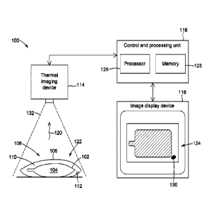
holding container enclosed in an overwrap, the method including:
Date Recue/Date Received 2022-05-16
2
sensing thermal radiation emanating from a scene encompassing the liquid-
holding container;
generating a thermal image of the scene based on the sensed thermal radiation;
and
analyzing the thermal image, said analyzing including assessing whether the
thermal image
includes a thermal feature indicative of a presence of leaked liquid in an
interstitial volume defined
between the liquid-holding container and the overwrap, and, if the thermal
image includes such a
thermal feature, determining that a leak exists in the liquid-holding
container.
[0006] In accordance with another aspect, there is provided an inspection
system for detecting
leakage from a liquid-holding container enclosed in an overwrap, the
inspection system including:
a thermal imaging device configured to sense thermal radiation emanating from
a scene
encompassing the liquid-holding container, and to generate a thermal image of
the scene based
on the sensed thermal radiation; and
a control and processing unit operatively connected to the thermal imaging
device and configured
to analyze the thermal image by assessing whether the thermal image includes a
thermal feature
indicative of a presence of leaked liquid in an interstitial volume defined
between the liquid-holding
container and the overwrap to provide a determination that a leak exists in
the liquid-holding
container.
[0007] In accordance with an aspect, there is provided a method of detecting
leakage from a liquid-
holding container, the method including:
sensing thermal radiation emanating from a scene encompassing the liquid-
holding container;
generating a thermal image of the scene based on the sensed thermal radiation;
and
analyzing the thermal image, the analyzing including assessing whether the
thermal image
includes a thermal feature indicative of a presence of leaked liquid outside
the liquid-holding
container, and, if the thermal image includes such a thermal feature,
determining that a leak
exists in the liquid-holding container.
[0008] In some embodiments, the sensed thermal radiation includes infrared
radiation. For example,
the infrared radiation may include long-wavelength infrared (LWIR) radiation,
with a wavelength
ranging from about 8 pm to about 15 pm.
[0009] In some embodiments, the method may include determining a location of
the leak in the liquid-
holding container based on a location of the identified thermal feature in the
thermal image.
[0010] In some embodiments, the method may include displaying the thermal
image on an image
display device, and performing the assessing based on the displayed thermal
image.
Date Recue/Date Received 2022-05-16
3
[0011] In some embodiments, the liquid-holding container may be a compressible
or flexible
container. In such embodiments, the method may include applying pressure to
the liquid-holding
container before, during, or both before and during the sensing of thermal
radiation emanating from
the liquid-holding container. It is appreciated that if the liquid-holding
container has a leak therein,
the application of sufficient pressure to the container may cause some liquid
to be expelled from the
container, which may facilitate its detection as a thermal feature in the
thermal image.
[0012] In some embodiments, the liquid-holding container is a flexible bag.
For example, in some
embodiments, the liquid-holding container is an IV fluid bag holding IV fluid.
[0013] In some embodiments, the liquid-holding container is enclosed in an
overwrap. The overwrap
may serve to seal or protect the bag from contamination and damage. In such
embodiments, the
assessed thermal feature may be indicative of liquid being present in an
interstitial volume defined
between an outer surface of the liquid-holding container and an inner surface
of the overwrap. For
example, the assessed thermal feature may be indicative of a temperature or an
emissivity difference,
or both, between the leaked liquid and the scene.
[0014] Depending on the application, the assessment of whether the thermal
image includes a
thermal feature indicative of the presence of liquid outside the liquid-
holding container to provide a
determination of leakage includes a human assessment, a computer assessment,
or a combination
of human assessment and computer assessment. When the assessment is made at
least partly by
a human operator, the method may include a step of displaying the thermal
image on an image
display device. The displayed thermal image may include different colors,
different intensities, or both
different colors and intensities, to represent different temperatures in
different regions of the scene.
For example, the displayed thermal image may provide a false-color or pseudo-
color representation
of the sensed thermal radiation within the scene.
[0015] In some embodiments, the thermal feature assessed in the thermal image
is indicative of a
temperature difference between the leaked liquid and liquid still inside the
liquid-holding container.
For example, in some embodiments, this temperature difference may result from
the leaked liquid
gradually evaporating, thus causing the temperature of the remaining,
nonevaporated leaked liquid
to drop relative to the temperature of the liquid still inside the liquid-
holding container.
[0016] In some embodiments, the method may include applying a thermal
stimulation to the liquid-
holding container before, during, or both before and during the sensing of
thermal radiation, and the
assessed thermal feature may be indicative of a thermal response of the leaked
liquid to the applied
thermal stimulation. Depending on the application, the thermal stimulation may
involve heat transfer
Date Recue/Date Received 2022-05-16
4
by thermal conduction, thermal convection, thermal radiation, or any
combination thereof. For
example, in one embodiment, the applied thermal stimulation may include pulsed
thermal radiation,
which may be applied by a flash lamp or another suitable source of thermal
radiation. Depending on
the application, the thermal stimulation may have a variety of spatio-temporal
heating profiles.
[0017] In some embodiments, the method may include exposing the liquid-holding
container to a
heating or cooling source and subsequently acquiring a thermal image. In such
embodiments, the
thermal feature assessed in the image may be representative of a modified
thermal inertia profile
associated with the presence of leaked liquid in the overwrap. In some
embodiments, the liquid-
holding container may move sequentially past a heating or cooling source, and
then a thermal
imaging device. The thermal image could be a single image, or an analysis
could be performed on a
sequence of images to extract time-varying behavior that could provide
additional information about
the observed thermal features.
[0018] In some embodiments, the thermal stimulation may be applied by a
background heat source,
for example, a heating device located behind the liquid-holding container and
maintained at a
controlled temperature relative to the temperature of the liquid-holding
container. In such
embodiments, the thermal feature assessed in the thermal image may be
representative of thermal
radiation emanating from the background heat source that interacted with
leaked liquid before being
sensed.
[0019] In some embodiments, the method may include suspending or hanging the
liquid-holding
container, and allowing the leaked liquid to flow away from the liquid-holding
container toward a
bottom region of the interstitial volume prior to sensing the thermal
radiation.
[0020] In accordance with another aspect, there is provided a method of
detecting leakage in an
interstitial volume defined between a liquid-holding container and an overwrap
enclosing the liquid-
holding container, the method including:
sensing thermal radiation emanating from a scene encompassing the liquid-
holding container and
the overwrap;
generating a thermal image of the scene based on the sensed thermal radiation;
and
analyzing the thermal image by assessing whether the thermal image includes a
thermal feature
indicative of liquid being present in the interstitial volume, and, if the
thermal image includes
such a thermal feature, determining that a leak exists in the liquid-holding
container.
[0021] In some embodiments, the liquid-holding container may be an IV fluid
bag, and the sensed
thermal radiation may include infrared radiation, for example, LWIR radiation.
In some embodiments,
Date Recue/Date Received 2022-05-16
5
the IV fluid bag and its overwrap may each be made of a plastic polymer
material having a relatively
high LWIR transmittance, while the IV fluid contained in, and potentially
leaking from, the IV fluid bag
may be composed of an aqueous solution having a relatively high LWIR
absorptance/emissivity. In
such embodiments, LWIR radiation may pass through the IV fluid bag and the
overwrap substantially
unaffected, but be more strongly attenuated by the IV fluid. As a result, the
IV fluid¨whether inside
or outside the bag¨will appear darker or lighter in the thermal image. In
other embodiments, the
overwrap may include or be made of a thermally opaque material (e.g., a
metallic coating or foil) and
the exterior of the overwrap may be exposed to thermal stimulation (e.g.,
heating or cooling) from a
heat source. In such embodiments, the degree of heating or cooling of the
opaque overwrap depends
on the thermal inertia of its exposed surface, so that regions of the overwrap
in contact with leaked
liquid may appear darker or lighter in the thermal image upon thermal
diffusion of the absorbed heat
into the contacted leaked liquid.
[0022] In accordance with another aspect, there is provided an inspection
system for detecting
leakage from a liquid-holding container. The inspection system may include a
thermal imaging device
configured to sense thermal radiation emanating from a scene encompassing the
liquid-holding
container, and to generate a thermal image of the scene based on the sensed
thermal radiation. The
inspection system may also include a control and processing unit operatively
connected to the
thermal imaging device and configured to analyze the thermal image by
assessing whether the
thermal image includes a thermal feature indicative of a presence of leaked
liquid outside the liquid-
holding container to provide a determination that a leak exists in the liquid-
holding container.
Alternatively, or additionally, the control and processing unit may be
configured to supply the thermal
image generated by the thermal imaging device to an image display device for
leakage assessment
by an operator.
[0023] In some embodiments, the thermal imaging device includes an infrared
camera.
[0024] In some embodiments, the control and processing unit may be configured
to assess whether
the thermal feature is indicative of the leaked liquid being present in an
interstitial volume defined
between the liquid-holding container and an overwrap enclosing the liquid-
holding container.
[0025] In some embodiments, the inspection system may include an image display
device configured
to display the thermal image generated by the thermal imaging device.
[0026] In some embodiments, the inspection system may include a heat source,
for example, a
thermal radiation source, configured to apply a thermal stimulation to the
liquid-holding container
before, during, or both before and during the sensing of thermal radiation by
the thermal imaging
Date Recue/Date Received 2022-05-16
6
device. In such embodiments, the control and processing unit may be configured
to assess whether
the thermal feature is indicative of a thermal response of the leaked liquid
to the applied thermal
stimulation. In some embodiments, the liquid-holding container may be enclosed
in a thermally
opaque overwrap, and the heat source may be configured to apply the thermal
stimulation to an
exterior of the thermally opaque overwrap. The thermal imaging device may be
configured to
generate the thermal image as a representation of a temperature distribution
at the exterior of the
thermally opaque overwrap. The control and processing unit is configured to
assess whether the
thermal feature is representative of a thermal spot in the temperature
distribution indicative of the
leaked liquid being present in an interstitial volume defined between the
liquid-holding container and
the thermally opaque overwrap.
[0027] In some embodiments, the inspection system includes a holding fixture
configured to suspend
the liquid-holding container for allowing leaked liquid to flow away from the
liquid-holding container
toward a bottom region of the interstitial volume.
[0028] In accordance with another aspect, there is provided a non-transitory
computer readable
storage medium having computer executable instructions stored thereon that,
when executed by a
processor, cause the processor to perform various steps of the methods
described herein. Such
steps may include making an assessment as to whether a thermal image includes
a thermal feature
indicative of liquid being present outside a liquid-holding container to
provide a determination of the
existence of a leak in the liquid-holding container.
[0029] In accordance with another aspect, there is provided a computer device
for use with or within
an inspection system such as described herein. The computer device may include
a processor and
a non-transitory computer readable storage medium operatively coupled to the
processor and having
computer readable instructions stored thereon that, when executed by a
processor, cause the
processor to perform various steps of the methods described herein, such as
noted above.
[0030] It is appreciated that other method and process steps may be performed
prior to, during, or
after the steps described herein. The order of one or more steps may also
differ, and some of the
steps may be omitted, repeated, and/or combined, depending on the application.
It is also
appreciated that some steps may be performed using various image analysis and
processing
techniques, which may be implemented in hardware, software, firmware, or any
combination thereof.
[0031] Other objects, features, and advantages of the present description will
become more apparent
upon reading of the following non-restrictive description of specific
embodiments thereof, given by
way of example only with reference to the appended drawings. Although specific
features described
Date Recue/Date Received 2022-05-16
7
in the above summary and in the detailed description below may be described
with respect to specific
embodiments or aspects, it should be noted that these specific features can be
combined with one
another unless stated otherwise.
BRIEF DESCRIPTION OF THE DRAWINGS
[0032] Fig. 1 is a schematic representation of an inspection system for
detecting leakage from a
liquid-holding container, in accordance with a possible embodiment.
[0033] Fig. 2 is a schematic representation of an inspection system for
detecting leakage from a
liquid-holding container, in accordance with another possible embodiment.
[0034] Fig. 3 is a schematic representation of an inspection system for
detecting leakage from a
liquid-holding container, in accordance with another possible embodiment.
[0035] Fig. 4 is a schematic representation of an inspection system for
detecting leakage from a
liquid-holding container, in accordance with another possible embodiment.
[0036] Fig. 5 is a schematic representation of an inspection system for
detecting leakage from a
liquid-holding container, in accordance with another possible embodiment.
[0037] Figs. 6A and 6B are schematic representations of two operation phases
of an inspection
system for detecting leakage from a liquid-holding container, in accordance
with another possible
embodiment.
[0038] Figs. 7A and 7B are a visible-light image (Fig. 7A) and a LWIR thermal
image (Fig. 7B) of an
overwrapped IV fluid bag. Leaking IV fluid is not visible in Fig. 7A but is
detected in Fig. 7B, as
evidenced by the presence of dark spots at the bottom of the overwrap.
[0039] Figs. 8A and 8B are LWIR thermal images of an IV fluid bag with (Fig.
8A) and without
(Fig. 8B) leakage. The thermal image in Fig. 8A contains darker spots
indicative of local temperature
differences between leaked IV fluid and the IV fluid still inside the bag, the
temperature differences
being induced by evaporative cooling undergone by the leaked IV fluid.
[0040] Figs. 9A to 9C are a sequence of three LWIR thermal images of a leaking
IV fluid bag
enclosed in a thermally opaque overwrap. The images were acquired at different
times following the
application of a thermal stimulation to the overwrap.
DETAILED DESCRIPTION
[0041] In the present description, similar features in the drawings have been
given similar reference
numerals. To avoid cluttering certain figures, some elements may not be
indicated if they were
Date Recue/Date Received 2022-05-16
8
already identified in a preceding figure. It should also be understood that
the elements of the drawings
are not necessarily depicted to scale, since emphasis is placed on clearly
illustrating the elements
and structures of the present embodiments. Furthermore, positional descriptors
indicating the
location and/or orientation of one element with respect to another element are
used herein for ease
and clarity of description. Unless otherwise indicated, these positional
descriptors should be taken in
the context of the figures and should not be considered limiting. It is
appreciated that such spatially
relative terms are intended to encompass different orientations in the use or
operation of the present
embodiments, in addition to the orientations exemplified in the figures.
[0042] The terms "a", "an", and "one" are defined herein to mean "at least
one", that is, these terms
do not exclude a plural number of elements, unless stated otherwise.
[0043] Terms such as "substantially", "generally", and "about", which modify a
value, condition, or
characteristic of a feature of an exemplary embodiment, should be understood
to mean that the value,
condition, or characteristic is defined within tolerances that are acceptable
for the proper operation
of this exemplary embodiment for its intended application or that fall within
an acceptable range of
experimental error. In particular, the term "about" generally refers to a
range of numbers that one
skilled in the art would consider equivalent to the stated value (e.g., having
the same or an equivalent
function or result). In some instances, the term "about" means a variation of
10% of the stated value.
It is noted that all numeric values used herein are assumed to be modified by
the term "about", unless
stated otherwise.
[0044] The term "based on" as used herein is intended to mean "based at least
in part on", whether
directly or indirectly, and to encompass both "based solely on" and "based
partly on". In particular,
the term "based on" could also be understood as meaning "depending on",
"representative of',
"indicative of', "associated with", and the like.
[0045] The terms "match", "matching", and "matched" refer herein to a
condition in which two
elements are either the same or within some predetermined tolerance of each
other. That is, these
terms are meant to encompass not only "exactly" or "identically" matching the
two elements but also
"substantially", "approximately", or "subjectively" matching the two elements,
as well as providing a
higher or best match among a plurality of matching possibilities.
[0046] The terms "connected" and "coupled", and derivatives and variants
thereof, refer herein to
any connection or coupling, either direct or indirect, between two or more
elements, unless stated
otherwise. For example, the connection or coupling between the elements may be
mechanical,
Date Recue/Date Received 2022-05-16
9
optical, electrical, magnetic, thermal, chemical, logical, fluidic,
operational, or any combination
thereof.
[0047] The present description generally relates to a method for detecting
container leakage using
thermal or infrared imaging, notably liquid container leakage, and to an
inspection system capable of
implementing the method. The present techniques may be used or implemented in
various inspection
fields and applications, for example, medical applications, pharmaceutical
applications, and
applications in food industry, that require or may benefit from improved
leakage monitoring and
detection capabilities.
[0048] One embodiment of the leakage detection method may include a step of
sensing thermal
radiation emanating from a scene encompassing a liquid-holding container or a
portion thereof, and
a step of generating a thermal image of the scene based on the sensed thermal
radiation. The method
may also include a step of assessing whether the thermal image includes one or
more thermal
features indicative of liquid being present outside the liquid-holding
container to provide a
determination that a leak exists in the liquid-holding container. In some
embodiments, the sensed
thermal radiation includes infrared radiation, for example, long-wavelength
infrared (LWIR) radiation.
The liquid-holding container may be a flexible bag, for example, an IV fluid
bag. In some
embodiments, the liquid-holding container is packaged or enclosed in an
overwrap to protect the bag
from contamination and damage. In such embodiments, the assessed thermal
feature may be
indicative of the presence of liquid in an interstitial volume defined between
the outer surface of the
liquid-holding container and the inner surface of the overwrap.
[0049] The terms "light" and "optical", and variants and derivatives thereof,
refer herein to radiation
in any appropriate region of the electromagnetic spectrum, and are not limited
to visible light. By way
of example, in some embodiments, the terms "light" and "optical" may encompass
electromagnetic
radiation with a wavelength ranging from about 0.7 to 1000 pm. Infrared
radiation is commonly
divided into various regions. One common division scheme defines the near-
infrared (NI R) region for
wavelengths ranging from 0.7 and 1.4 pm; the short-wavelength infrared (SWIR)
region for
wavelengths ranging from 1.4 to 3 pm; the mid-wavelength infrared (MWIR)
region for wavelengths
ranging from 3 to 8 pm; the long-wavelength infrared (LWIR) region for
wavelengths ranging from 8
to 15 pm; and the far-infrared (FIR) region for wavelengths ranging from 15 to
1000 pm. It is
appreciated that the definitions of different infrared regions in terms of
spectral ranges, as well as
their limits, may vary depending on the technical field under consideration,
and are not meant to limit
the scope of application of the present techniques. For example, the LWIR
region is sometimes
defined as encompassing wavelengths ranging from 7 to 14 pm. It is also
appreciated that although
some embodiments of the present techniques may be useful in applications
involving infrared
Date Recue/Date Received 2022-05-16
10
radiation, other embodiments may additionally or alternatively operate in
other regions of the
electromagnetic spectrum, for example, in the terahertz region.
[0050] The term "thermal imaging", and variants and derivatives thereof,
refers herein to an imaging
technique in which electromagnetic radiation emanating from objects in a
viewed scene is detected
and processed to output a thermal image representative of a spatial
temperature distribution within
the scene. Thermal imaging typically operates in the infrared portion of the
electromagnetic spectrum,
notably in the MWIR and LWIR regions, where the radiation emitted from an
object is a function of
the object's temperature and emissivity. The term "thermal imaging device",
and variants and
derivatives thereof, such as "thermal imager" and "thermal camera", refer
herein to non-contact
imaging devices configured to sense thermal radiation emitted from objects
present in a scene. These
imaging devices can use a single thermal detector or multiple thermal
detectors, for example,
arranged in a linear or matrix array. The thermal detectors are configured to
convert the sensed
thermal radiation into electrical signals on a per-pixel basis and output a
thermal image of the scene
that can be put in the form of an array of pixels. Each pixel is associated
with a corresponding thermal
detector and has a pixel value representative of the amount of thermal energy
emitted, transmitted,
and reflected by a corresponding region of the scene. A thermal image can
provide a temperature
map of the scene within a particular spectral band. Thermal images can be
displayed as single
images, sequences of images, or video streams.
[0051] Depending on the application, the present techniques can rely on
various types of thermal
imaging devices, which can be either cooled or uncooled and use either passive
or active
thermography. In some embodiments, the thermal imaging device can be a passive
thermal camera
including an array of uncooled thermal detectors. Non-limiting examples of
uncooled thermal
detectors include, to name a few, bolometers and microbolometers, thermopiles,
thermocouples,
Golay cells, pyroelectric detectors, and ferroelectric detectors. In other
embodiments, the thermal
imaging device can include a single thermal detector paired with a one-
dimensional or two-
dimensional scanning device to create a thermal image. It is appreciated that
the use of the term
"thermal" refers herein to the fact that the operation of thermal imaging
devices such as disclosed
herein involves the conversion of electromagnetic radiation into heat. In
particular, the term "thermal"
does not mean that the thermal radiation detectors disclosed herein are
limited to detecting "thermal
radiation", which is a term whose scope is sometimes limited to infrared
radiation. That is, the terms
"thermal imaging" and "infrared imaging" may, but not always, be used
interchangeably.
[0052] The terms "leak" and "leakage", and variants and derivatives thereof,
refer herein to an
unwanted or unintended escape or release of fluid or liquid out of its
container. The occurrence of
Date Recue/Date Received 2022-05-16
11
leaks may have various causes, such as aging, wear, damage, deterioration,
faulty manufacturing or
handling, exposure to adverse environmental conditions, or a combination
thereof.
[0053] The term "liquid" refers herein to a substance having a definite volume
and the ability to flow
and to conform to the shape of its container. It is appreciated that the term
"liquid" is meant to
encompass fluid substances of various viscosities. Depending on the
application, the term "liquid"
can refer to a pure substance (e.g., water), a homogeneous solution containing
one or more solutes
dissolved in a solvent, a heterogeneous suspension, dispersion, emulsion, or
multi-phase mixture, a
cream, a gel, a paste, and the like. In some instances, the terms "liquid" and
"fluid" may be used
interchangeably. It is appreciated that the present techniques are not
necessarily limited to the
detection of liquid leakage. For example, it is envisioned that the present
techniques may be used to
detect the presence of leaked gases (e.g., detected using multispectral
infrared imaging) or leaked
solids (e.g., powder materials) in the interstitial volume between the
container and its overwrap. In
some implementations, the leaking liquid to be detected can be an IV fluid.
The term "IV fluid" refers
herein to any fluid that can be infused, transfused, or otherwise injected
into a human or animal body.
Non-limiting examples of IV fluids include, to name a few, volume expanders
such as crystalloids
(e.g., normal saline solutions, Ringer's solutions, IV sugar solutions) and
colloids; blood and blood-
based products; blood substitutes; medications and drugs; buffer solutions;
parenteral nutrition fluids;
and various other artificial solutions and additives that can be injected into
the circulatory system of
a patient during medical treatment.
[0054] The terms "fluid container" and "liquid container", and variants and
derivatives thereof, are
intended to refer to any vessel or recipient configured to hold, store, and/or
transport a fluid or liquid,
and which can be inspected for leaks according to the present techniques.
Depending on the
application, the container may be made of various materials, for example,
plastic materials, and have
various shapes, sizes, colors, optical properties, and configurations. In some
implementations, the
container can be a flexible container, such as a bag or a pouch. Common
examples of flexible
containers are IV fluid bags for use in medical applications. IV fluid bags
are typically made of a
plastic polymer material, for example, a polyvinyl polymer, which can be
susceptible to tearing,
puncture, breakage, or other forms of degradation. To protect their physical
integrity, IV fluid bags
can be contained in an overwrap, for example, made of a stronger plastic
polymer, such as high-
density polyethylene (HDPE). For overwrapped IV fluid bags, leaking fluid will
accumulate in the
interstitial volume between the bag and its overwrap. As noted above, the
inspection of overwrapped
IV fluid bags for leaks by assessing for the presence or absence of leaked
fluid in the interstitial
volume can be difficult and time-consuming using conventional methods.
Date Recue/Date Received 2022-05-16
12
[0055] Various implementations of the present techniques are described below
with reference to the
figures.
[0056] Referring to Fig. 1, there is illustrated an embodiment of an
inspection system 100 for
detecting leakage from a liquid-holding container using thermal imaging, for
example, LWIR imaging.
In the illustrated embodiment, the liquid-holding container is a sealed,
flexible IV fluid bag 102
containing IV fluid 104. The IV fluid bag 102 is wrapped and sealed in a
protective overwrap 106 to
form an IV fluid bag assembly 108. The region between the outer surface of the
IV fluid bag 102 and
the inner surface of the overwrap 106 defines an interstitial volume 110,
where leaked IV fluid 112
will tend to accumulate if a leak exists in the IV fluid bag 102. Leakage of
IV fluid bags may be the
result of tear, puncture, breakage, rupture, sealing defects, or other forms
of damage to the physical
integrity of the IV fluid bag 102 incurred during manufacturing,
transportation, storage, or handling.
[0057] In some embodiments, the IV fluid bag 102 and the overwrap 106 may each
be made of a
plastic polymer material having a relatively high LWIR transmittance, while
the IV fluid 104 may be
made of a material having a relatively high LWIR absorptance/emissivity. For
example, the IV fluid
bag 102 may be made of polyvinyl chloride (PVC), while the overwrap 106 may be
made of high-
density polyethylene (HDPE). The IV fluid 104 may be composed of various types
of aqueous
solutions, non-limiting examples of which include crystalloids, colloids,
blood and blood-based
products, and blood substitutes. However, it is appreciated that, depending on
the application, the IV
fluid bag 102, the IV fluid 104, and the overwrap 106 in Fig. 1 can each have
various compositions,
resulting in various thermal radiative properties.
[0058] While the embodiment of Fig. 1 relates to leakage detection in IV fluid
bags used in medical
applications, the present techniques may be used in other medical and non-
medical applications for
detecting leaks in various types of liquid-holding containers filled with
various types of liquids, with or
without protective overwraps, and using both LWIR and non-LWIR radiation.
Furthermore, depending
on the application, the present techniques may be used in various inspection
scenarios, non-limiting
examples of which include in-line inspection operations (e.g., during
production, packaging, and/or
shipping) and reinspection operations (e.g., reinspection of entire lots of
products after a previous
inspection discovered leakage in one product). In another example, the
inspection techniques
disclosed herein have potential use in automated pharmacy systems, where IV
fluid bags are filled
and processed using robotic handing devices.
[0059] The inspection system 100 of Fig. 1 generally includes a thermal
imaging device 114, a
control and processing unit 116, and an image display device 118. More details
regarding the
Date Recue/Date Received 2022-05-16
13
structure and operation of these and other possible components of the
inspection system 100 are
provided below.
[0060] The thermal imaging device 114 is configured to sense thermal radiation
120 emanating from
a scene 122 encompassing the IV fluid bag 102 and the overwrap 106, and to
generate a thermal
image 124 of the scene 122 based on the sensed thermal radiation 120. It is
appreciated that the
use of the term "encompassing" in this context is intended to mean that the IV
fluid bag 102 and the
overwrap 106 may be either fully or partially contained in the field of view
of the thermal imaging
device 114. The term "thermal image" as used herein may refer to a single
thermal image, a plurality
of thermal images, or a combined thermal image obtained by combining at least
two thermal images.
Various types of thermal imaging devices can be used to implement the present
techniques. For
example, the thermal imaging device 114 may be a thermal or infrared camera
including a focal plane
array (FPA) of uncooled thermal detectors, such as microbolometer detectors.
Conventional
uncooled microbolometer-based thermal cameras can include FPAs having hundreds
of thousands
to millions of pixels, with a pixel pitch of the order of 10 to 50 pm. It is
appreciated that the general
principles underlying the construction, operation, and applications of thermal
imaging devices are
known in the art and need not be described in greater detail herein.
[0061] The control and processing unit 116 generally includes a processor 126
and a memory 128.
The control and processing unit 116 is operatively connected to the thermal
imaging device 114 and
the image display device 118 to control and coordinate, at least partly, their
operation. The control
and processing unit 116 may be configured to process and analyze the thermal
image 124 generated
by the thermal imaging device 114. The control and processing unit 116 may
also be configured to
supply the thermal image 124 to the image display device 118 in a suitable
format for viewing by an
operator. For example, the image display device 118 may display the thermal
image 124 generated
by the thermal imaging device 114 as a spatially resolved temperature map
representative of the
thermal radiation 120 sensed by the thermal imaging device 114. As can be
appreciated, various
types of image display devices (e.g., standalone monitors, laptop and desktop
computers, televisions,
smartphones, tablet computers) and display technologies (e.g., liquid crystal
display, light-emitting
diode, organic light-emitted diode, plasma) may be used depending on the
application.
[0062] Depending on the application, the assessment of whether the thermal
image 124 includes a
thermal feature 130 indicative of the presence of IV fluid 112 outside the IV
fluid bag 102 to provide
a determination of leakage is a human assessment, a computer assessment, or a
combination of
human assessment and computer assessment.
Date Recue/Date Received 2022-05-16
14
[0063] When leakage assessment is made at least partly by a computer, the
control and processing
unit 116 may be configured to receive the thermal image 124 generated by the
thermal imaging
device 114 and analyze the thermal image 124 to assess whether it contains a
thermal feature 130
indicative of IV fluid leakage. It is appreciated that various computer-
implemented and software-
based image analysis tools and techniques may be employed to identify a
thermal feature indicative
of fluid leakage in a thermal image. Such tools and techniques may use
contrast enhancement and
matching algorithms based on feature extraction and pattern recognition, and
may rely on machine
learning and/or artificial intelligence. When leakage assessment is made
solely by the control and
processing unit 116, the thermal image 124 may not be displayed to a human
operator and the image
display device 118 may be omitted.
[0064] In some embodiments, the control and processing unit 116 may be
configured to take an
action following the determination of whether a leak exists in the IV fluid
bag 102 under inspection.
For example, when the determination indicates a leak, the action taken may
include sending an alert
to notify the operator of the existence of the leak and/or generating commands
to have the leaking
IV fluid bag 102 discarded.
[0065] When leakage assessment is made at least partly by a human operator,
the image display
device 118 is configured to display the thermal image 124 in a format suitable
for viewing and
analysis by the human operator.
[0066] The control and processing unit 116 may be provided within one or more
general purpose
computers and/or within any other suitable computing devices implemented in
hardware, software,
firmware, or any combination thereof, and connected to other components of the
inspection
system 100 via appropriate wired and/or wireless communication links and
ports. As the case may
be, the control and processing unit 116 may be fully or partly integrated with
or physically separate
from the other components of the inspection system 100. The processor 126 may
implement
operating systems, and may be able to execute computer programs, also
generally known as
commands, instructions, functions, processes, software codes, executables,
applications, and the
like. The term "processor" should not be construed as being limited to a
single processor, and
accordingly, any known processor architecture may be used. Depending on the
application, the
processor 126 may include a single processing entity or a plurality of
processing entities. Such
processing entities may be physically located within the same device, or the
processor 126 may
represent the processing functionalities of a plurality of devices operating
in coordination.
Accordingly, the processor 126 may include or be part of: a computer; a
microprocessor; a
microcontroller; a coprocessor; a central processing unit (CPU); an image
signal processor (ISP); a
digital signal processor (DSP) running on a system on a chip (SoC); a single-
board computer (SBC);
Date Recue/Date Received 2022-05-16
15
a dedicated graphics processing unit (GPU); a special-purpose programmable
logic device embodied
in hardware device, such as, for example, a field-programmable gate array
(FPGA) or an application-
specific integrated circuit (ASIC); a digital processor; an analog processor;
a digital circuit designed
to process information; an analog circuit designed to process information; a
state machine; and/or
other mechanisms configured to electronically process information and operate
collectively as a
processor.
[0067] The memory 128, which can also be referred to as a "computer readable
storage medium" is
capable of storing computer programs and other data to be retrieved by the
processor 126. The terms
"computer readable storage medium" and "computer readable memory" are intended
to refer herein
to a non-transitory and tangible computer product that can store and
communicate executable
instructions for the implementation of various steps of the methods disclosed
herein. The computer
readable memory may be any computer data storage device or assembly of such
devices, including
a random-access memory (RAM); a dynamic RAM; a read-only memory (ROM); an
erasable
programmable ROM (EPROM); a magnetic storage device, such as a hard disk
drive, a solid-state
drive, a floppy disk, and a magnetic tape; an optical storage device, such as
a compact disc (e.g., a
CD or CDROM), a digital video disc (DVD), and a Blu-RayTM disc; a flash drive
memory; and/or other
non-transitory memory technologies. A plurality of such storage devices may be
provided, as can be
appreciated by those skilled in the art. The computer readable memory may be
associated with,
coupled to, or included in a computer or processor configured to execute
instructions contained in a
computer program stored in the computer readable memory and relating to
various functions
associated with the computer or processor.
[0068] It is appreciated that while the embodiment of Fig. 1 depicts the
thermal imaging device 114,
the control and processing unit 116, and the image display device 118 as three
standalone
components, this need not be the case in other embodiments. For example, in
some embodiments,
two or all of these three components may be provided in an integrated device.
[0069] In operation, the inspection method may include a step of placing the
IV fluid bag
assembly 108 in the field of view 132 of the thermal imaging device 114, a
step of using the thermal
imaging device 114 to sense thermal radiation 120 emanating from within the
field of view 132
(including thermal radiation emanating from the IV fluid bag assembly 108),
and a step of generating
a thermal image 124 based on the sensed thermal radiation 120. In some
embodiments, the IV fluid
bag assembly 108 may be placed on a conveyor, a table, a bin, or another type
of mobile or stationary
inspection unit. In other embodiments, a holding or supporting structure or
fixture may be provided
to hold the IV fluid bag assembly 108 to allow IV fluid 112 leaking from the
IV fluid bag 102 to drain
by gravity and accumulate at or near the bottom of the overwrap 106 prior to
acquiring the thermal
Date Recue/Date Received 2022-05-16
16
image 124 (see, e.g., Figs. 4 and 5 below). In yet other embodiments, the IV
fluid bag assembly 108
may be held by a human operator during the inspection method.
[0070] The inspection method may also include a step of applying pressure to
the IV fluid bag
assembly 108 before, during, or both before and during, the acquisition of the
thermal image 124. It
is appreciated that if the IV fluid bag 102 has a leak therein, pressing on
the IV fluid bag 102 may
cause IV fluid 104 contained in the IV fluid bag 102 to be released as leaked
IV fluid 112 into the
overwrap 106, where it can be detected in the thermal image 124 to provide a
determination of the
existence of the leak. It is also appreciated that pressing on the IV fluid
bag 102 during the inspection
method may help in assessing the location of the leak in the IV fluid bag 102
by increasing the
likelihood that the leaked IV fluid 112 detected outside the IV fluid bag 102
be located close to the
leak in the IV fluid bag 102.
[0071] The inspection method may further include a step of analyzing the
thermal image 124. The
analysis of the thermal image 124 may include assessing whether the thermal
image 124 includes a
thermal feature 130 indicative of the presence of IV fluid 112 outside the IV
fluid bag 102, in the
interstitial volume 110 between the IV fluid bag 102 and the overwrap 106. If
the assessment is
positive, that is, if a thermal feature 130 is found in the thermal image 124,
the method may include
a step of determining an existence of a leak in the IV fluid bag 102.
Conversely, if the assessment is
negative, that is, if a thermal feature 130 is not found in the thermal image
124, the method may
include a step of determining an absence of a leak in the IV fluid bag 102. It
is appreciated that
depending on the application, the thermal image 124 may be analyzed for leaks
upon being
generated, in real-time or near real-time, or be saved in memory for later
analysis.
[0072] The thermal feature 130 to be identified in the thermal image 124 may
be representative of
differences in radiative properties between the leaked IV fluid 112 and its
environment, notably the
IV fluid bag 102, the overwrap 106, and the IV fluid 104 still inside the bag
102. As noted above,
typical IV fluid materials exhibit a relatively high LWIR
absorptance/emissivity, while typical IV fluid
bags and overwrap materials exhibit a relatively high LWIR transmittance. Due
to these different
radiative properties, typical IV fluids may be distinguished from typical IV
fluid bags and overwrap
materials in LWIR thermal images, the former typically appearing darker or
lighter than the latter,
depending on the relative temperatures of the fluids and background. This is
illustrated in the thermal
image 124 depicted in Fig. 1, where the leaked IV fluid 112 appears as a dark
spot 130 against a
lighter background. Furthermore, in some embodiments, the method may be used
not only to detect
the presence of a leak in the IV fluid bag 102, but also to convey information
about the location of the
leak in the IV fluid bag 102, based on the location of the identified thermal
feature 130 in the thermal
image 124.
Date Recue/Date Received 2022-05-16
17
[0073] In some embodiments, the IV fluid 112 that leaks from the IV fluid bag
102 will undergo some
evaporation, which will cause the temperature of the remaining, nonevaporated
leaked IV fluid 112
to drop relative to the temperature of the IV fluid 104 still inside the IV
fluid bag 102. Thus, the leaked
IV fluid 112 that accumulates in the interstitial volume 110 may appear darker
in the thermal
image 124 than the IV fluid 104 contained in the IV fluid bag 102, thus
facilitating leakage detection.
It is appreciated that this evaporative cooling process may be useful in
distinguishing leaked IV
fluid 112 from the IV fluid 104 in the IV fluid bag 102 in scenarios where
their contributions are
superimposed in the thermal image 124. For example, referring to Fig. 2, there
is illustrated another
embodiment of an inspection system 100 for detecting leakage from an IV fluid
bag 102 using thermal
imaging.
[0074] The embodiment of Fig. 2 shares several features with the embodiment of
Fig. 1, which will
not be described again other than to highlight differences between them. The
inspection system 100
of Fig. 2 includes a thermal imaging device 114 disposed to image the IV fluid
bag 102 from above.
In some scenarios, the IV fluid bag 102 may have a leak in its top surface,
resulting in leaked IV
fluid 112 accumulating in the interstitial volume 110 defined between the
outer top surface 134 of the
IV fluid bag 102 and the inner top surface 136 of the overwrap 106. In such
scenarios, the IV fluid 112
having leaked out of the IV fluid bag 102 and the IV fluid 104 that remains in
the IV fluid bag 102 are
superimposed in the thermal image 124 acquired by the thermal imaging device
114. Thus, any
leakage-indicative feature 130 found in the thermal image 124 may be
representative, entirely or
mainly, of an evaporative cooling process having induced a local temperature
difference between the
leaked IV fluid 112 and the IV fluid 104 still inside the IV fluid bag 102. It
is noted that local
temperature differences as low as 50 millikelvins may be detectable in thermal
images acquired by
existing thermal cameras. It is also noted that besides evaporative cooling,
other endothermic and/or
exothermic processes resulting in a leakage-indicative feature in the thermal
image may be used to
detect leaks according to the present techniques. For example, in one
scenario, the leaked liquid
could react chemically with gas or material (e.g., water) present in the
interstitial volume to produce
a detectable temperature contrast in the thermal image 124. In another
scenario, the Joule-Thomson
effect may be exploited to produce compression-based heating and/or expansion-
based cooling
resulting in a leakage-indicative feature in the thermal image 124.
[0075] Referring to Fig. 3, there is illustrated another embodiment of an
inspection system 100 for
detecting leakage from an IV fluid bag 102 using thermal imaging. The
embodiment of Fig. 3 shares
several features with the embodiments of Figs. 1 and 2, which will not be
described again other than
to highlight differences between them. The embodiment of Fig. 3 is used to
inspect an IV fluid bag 102
that is not contained in an overwrap. However, as in Fig. 2, the presence of
leaked IV fluid 112 on
Date Recue/Date Received 2022-05-16
18
the outer top surface 134 of the IV fluid bag 102 may be detected as an
evaporative-cooling-induced,
leakage-indicative feature 130 in the thermal image 124 acquired by the
thermal imaging device 114.
Referring to Fig. 4, there is illustrated another embodiment of an inspection
system 100 for detecting
leakage from an IV fluid bag 102 using thermal imaging. Again, this embodiment
shares several
.. features with previously described embodiments, which will not be described
again other than to
highlight differences between them. In Fig. 4, the inspection system 100
includes a holding
fixture 138 configured to suspend or hang the IV fluid bag assembly 108 for
allowing leaked IV
fluid 112 to flow downward away from the IV fluid bag 102 and accumulate in
the bottom region 140
of the overwrap 106, in a space-apart relationship from the IV fluid bag 102
and the IV fluid 104
contained therein. The inspection configuration depicted in Fig. 4 may reduce
or help reduce the
likelihood that the response of the leaked IV fluid 112 and the response of
the IV fluid 104 still inside
the IV fluid bag 102 be superimposed on the thermal image 124. Thus, the
identification of a leakage-
indicative feature 130 in the thermal image 124 may be made against a lighter
or darker background
(e.g., the substantially LWIR-transparent overwrap material), rather than
being based solely or mainly
on the finding of an evaporative-cooling-induced temperature difference
between the leaked IV
fluid 112 and the IV fluid 104 still inside the IV fluid bag 102. For example,
the leakage-indicative
feature 130 may be indicative of a temperature or an emissivity difference, or
both, between the
leaked liquid 112 and the scene 122.
[0076] In some embodiments, the present techniques may use active
thermography. In active
thermography, an external stimulation source is used to apply a thermal
stimulation (i.e., heating or
cooling) to a test specimen in order to generate heat flow through the
specimen. The heat flow may
be affected by local variations in thermal conductivity and heat capacity
inside the specimen which
may be indicative of the presence of internal defects or other anomalies, such
as leakage. These
internal local variations may give rise to thermal spots (i.e., hot or cold
spots) in the surface
temperature distribution of the specimen, which may be observed in a thermal
image of the test
specimen during or following the application of the thermal stimulation.
Depending on the application,
active thermography can employ a variety of external stimulation sources
(e.g., infrared and other
thermal radiation sources, such as flash lamps, electrical heaters, and
lasers) and stimulation
techniques (e.g., pulsed thermography, lock-in thermography, step heating
thermography, frequency
modulated thermography, laser-spot thermography). It is appreciated that the
general principles
underlying the techniques and applications of active thermography are known in
the art and need not
be described in greater detail herein.
[0077] In embodiments using active thermography, the inspection method may
include a step of
applying a thermal stimulation to the liquid-holding container before, during,
or both before and
Date Recue/Date Received 2022-05-16
19
during, the acquisition of the thermal image, and the assessed thermal feature
may be indicative of
a thermal response of the leaked liquid resulting from the applied thermal
stimulation. It is appreciated
that the thermal stimulation is applied indirectly or directly to the liquid-
holding container, depending
on whether or not the liquid-holding container is enclosed in an overwrap. In
some embodiments, the
thermal stimulation need not be applied by a dedicated heat source, but could
be the result of a
change of environment producing a change in ambient temperature. For example,
a change in
ambient temperature amounting to a thermal stimulation may arise when the
liquid-holding container
is moved from a sterilization chamber maintained at an elevated temperature to
an environment at
room temperature.
[0078] Referring to Fig. 5, there is illustrated an embodiment of an
inspection system 100 for
detecting leakage from an IV fluid bag 102 using active thermography. This
embodiment shares
several features with previously described embodiments, which will not be
described again other than
to highlight differences between them. The inspection system 100 in Fig. 5
includes a heat
source 142 located behind the IV fluid bag 102 in the field of view 132 of the
thermal imaging
device 114 and having a controlled temperature relative to the temperature of
the IV fluid bag
assembly 108 and its surroundings, which may be the same as the ambient
temperature. The heat
source 142 may include a heating device, such as a heating plate or surface.
For example, in one
variant, the background heat source 142 may be embodied by a black- or dark-
painted sheet made
of aluminum or another metal and filling partly or completely the field of
view 132 of the thermal
imaging device 114. To improve contrast, the sheet may be maintained at a
controlled temperature
cooler or warmer (e.g., by one or a few degrees Celsius) relative to ambient
temperature, for example,
using heating strip and thermocouples. In the embodiment of Fig. 5, the
thermal feature 130
assessed in the thermal image 124 may be representative of thermal radiation
emitted by the
background heat source 142 that interacted with the leaked IV fluid 112 (e.g.,
by processes of
transmission, reflection, and absorption/reemission) prior to being sensed by
the thermal imaging
device 114. To this end, the temperature at which the background heat source
142 is maintained
and/or its separation from the IV fluid bag 102 under inspection may be
selected to enhance a
contrast between thermal features indicative of IV fluid leakage and other
thermal features in the
thermal image 124. It is also appreciated that the background heat source 142
may, but need not,
be maintained at a same temperature over its surface area. In particular, the
background heat source
may be used to produce spatially-varying temperature fields or gradients to
further enhance the
contrast of leakage-indicative features in thermal images. It is also
appreciated that, depending on
the application, the temperature of the background heat source 142 may be
constant or may vary as
a function of time.
Date Recue/Date Received 2022-05-16
20
[0079] Referring to Figs. 6A and 6B, there is illustrated another embodiment
of an inspection
system 100 for detecting leakage from an IV fluid bag 102 using active
thermography. This
embodiment shares several features with previously described embodiments,
which will not be
described again other than to highlight differences between them. The
inspection system 100 of
Figs. 6A and 6B generally includes a heat source 142, a thermal imaging device
114, an image
display device 118, a control processing unit 116, and a conveyor 144. The IV
fluid bag 102 is
enclosed in an overwrap 106 to form an IV fluid bag assembly 108. The IV fluid
bag assembly 108 is
transported on the conveyor 144 to be sequentially excited by the heat source
142 (Fig. 6A) and
imaged by the thermal imaging device 114 (Fig. 6B).
[0080] In Fig. 6A, the heat source 142 is configured to apply a thermal
stimulation 146 to the IV fluid
bag assembly 108 within a field of illumination 148. The thermal stimulation
146 may be a heating or
a cooling stimulation, and the field of illumination 148 of the heat source
142 may have various sizes
and shapes. Furthermore, it is appreciated that a variety of heat sources and
thermal stimulation
techniques are available for active thermography and may be used to implement
the present
techniques. In addition to those mentioned above, non-limiting examples of
heat sources can include,
to name a few, heated or cooled darkened metallic plates and quartz tungsten
halogen (QTH) lamps.
In some embodiments, the thermal stimulation 46 may be applied for a time
duration of the order of
tens of milliseconds to a few seconds. In Fig. 6B, the thermal imaging device
114 is configured to
sense thermal radiation 120 emanating from the scene 122 encompassing the IV
fluid bag 102 and
generate a thermal image 124 of the scene 122 based on the sensed thermal
radiation 120. The
thermal image 124 may be displayed on the image display device 118. Depending
on the application,
an appropriate delay may be controlled between the application of the thermal
stimulation 146 and
the acquisition of the thermal image 124 to allow heat transfer to the leaked
liquid 112 before imaging.
The thermal image 124 may be analyzed by human and/or computer assessment to
determine
whether it includes a thermal feature 130 indicative of a thermal response of
leaked liquid 112
resulting from the applied thermal stimulation 146.
[0081] In some embodiments, the overwrap 106 may be made of a thermally opaque
material, for
example, a metallic coating or foil. The term "thermally opaque" is used
herein to refer to a material
that prevents or substantially prevents transmission of thermal radiation
(e.g., infrared radiation,
notably in the LWI R region) therethrough. In such embodiments, the thermal
stimulation 146 may be
applied to an exterior of the thermally opaque overwrap 106 and the thermal
image generated by the
thermal imaging device 114 may be representative of a temperature distribution
at the exterior of the
overwrap 106. The assessed thermal feature 130 may be representative of a
thermal spot in the
temperature distribution that is indicative of leaked liquid 112 being present
in the interstitial
Date Recue/Date Received 2022-05-16
21
volume 110 defined between the IV fluid bag 102 and the overwrap 106. In this
case, the degree of
heating or cooling of the thermally opaque overwrap 106 may depend on the
thermal inertia of its
exposed surface, so that regions of the overwrap 106 in contact with leaked
liquid 112 may appear
darker or lighter in the thermal image 124. Such embodiments are based on the
principle that heat
flow generated through the overwrap 106 following the application of the
thermal stimulation 146 by
the heat source 142 may be affected by the presence of leaked liquid 112
inside the overwrap 106.
These changes in heat flow may cause localized differences in the temperature
map of the
overwrap 106, which may be observed as thermal features 130 (e.g., hot or cold
spots) in the thermal
image 124 to provide a determination of the presence and location of liquid
leakage. In some
implementations, a sequence of thermal images may be acquired, for example,
over a time interval
of the order of tens of milliseconds to a few seconds, to study the time
evolution of the surface
temperature of the overwrap 106 in response to the applied the thermal
stimulation 146, which may
convey further information indicative of container leakage. In some cases, the
sequence of thermal
images may include one or more images acquired before thermal stimulation, one
or more images
acquired during thermal stimulation, one or more images acquired after thermal
stimulation, or any
combination thereof. These images may be compared to convey information about
the presence of
container leakage.
[0082] In accordance with another aspect of the present description, there is
provided a non-
transitory computer readable storage medium having computer executable
instructions stored
thereon that, when executed by a processor, cause the processor to perform
various steps of the
methods described herein.
[0083] In accordance with another aspect of the present description, there is
provided a computer
device for use with or in an leak inspection system such as described herein.
The computer device
may include a processor and a non-transitory computer readable storage medium
such as described
herein. The non-transitory computer readable storage medium may be operatively
coupled to the
processor and may have computer readable instructions stored thereon that,
when executed by a
processor, cause the processor to perform various steps for controlling the
inspection system and/or
various steps of methods described herein.
[0084] The following description reports work conducted to study and
investigate some aspects of
the present techniques. It is appreciated that the container leakage detection
techniques described
herein may have a number of optional features, variations, and applications.
In particular, the
following description is provided to further illustrate some aspects and
capabilities of the disclosed
techniques, but should not be construed as in any way limiting their scope.
Date Recue/Date Received 2022-05-16
22
[0085] Figs. 7A and 7B illustrate that leaking IV fluid that cannot or can
hardly be detected in a visible-
light image (Fig. 7A) of an overwrapped IV fluid bag may readily be detected
in a LWIR thermal
image 124 (Fig. 7B), as evidenced by the presence of dark spots 130 in a
bottom region of the
overwrap. No such dark spots are observed in Fig. 7A.
[0086] Figs. 8A and 8B are LWIR thermal images 124 of an IV fluid bag with
(Fig. 8A) and without
(Fig. 8B) leakage. Fig. 8A illustrates the effect on the thermal image 124 of
an evaporative cooling
process undergone by leaked IV fluid and inducing local temperature
differences between the leaked
IV fluid and the IV fluid having remained inside the IV fluid bag. The local
temperature differences
appear as darker thermal features 130 in the thermal image 124 that facilitate
leakage detection. It
is appreciated that such dark features are not observed in the thermal image
124 of Fig. 8B,
corresponding to the IV fluid bag without leakage.
[0087] Figs. 9A to 9C are a sequence of three LWIR thermal images 124 of a
leaking IV fluid bag
enclosed in a thermally opaque overwrap. The three images 124 were acquired at
three different
times following the application of a thermal stimulation to the overwrap in
order to study the time
evolution of its surface temperature in response to the thermal stimulation.
The thermal stimulation
was applied by a QTH lamp for about five seconds. The image acquisition began
about three seconds
after the end of exposure, and the three images 124 were acquired about three
seconds apart. It is
appreciated, as noted above, that in practical applications, the duration of
the exposure to the thermal
stimulation and the image acquisition process will generally be significantly
shorter that in this
example. As noted above, the presence of leaked liquid in the overwrap may
change the heat flow
generated through the overwrap by the thermal stimulation and, in turn, create
local contrasts in the
temperature profile of the overwrap which may be observed by thermal imaging.
Each of the three
images 124 features a darker region 130 indicative of leaked liquid having
accumulated at the bottom
of the overwrap. It is noted that the average surface temperature of the
overwrap decreases from
Fig. 9A to Fig. 9C.
[0088] Numerous modifications could be made to the embodiments described above
without
departing from the scope of the appended claims.
Date Recue/Date Received 2022-05-16