Note: Descriptions are shown in the official language in which they were submitted.
- 1 ¨
MODIFIED NATURAL KILLER CELLS AND NATURAL KILLER CELL LINES
HAVING INCREASED CYTOTOXICITY
This is a divisional application of co-pending Canadian Application No.
2,993,796,
.. which entered the national phase in Canada on January 25, 2018 from
International
Application No. EP2016/068001, having an international filing date of July 28,
2016.
Introduction
The present invention relates to the modification of natural killer (NK) cells
and NK
cell lines to produce derivatives thereof with a more cytotoxic phenotype.
Furthermore, the present invention relates to methods of producing modified NK
cells
and NK cell lines, compositions containing the cells and cell lines and uses
of said
compositions in the treatment of cancer.
Background to the Invention
Typically, immune cells require a target cell to present antigen via major
histocompatibility complex (MHC) before triggering an immune response
resulting in
the death of the target cell. This allows cancer cells not presenting MHC
class I to
evade the majority of immune responses.
NK cells are able, however, to recognize cancer cells in the absence of MHC
class I
expression. Hence they perform a critical role in the body's defence against
cancer.
On the other hand, in certain circumstances, cancer cells demonstrate an
ability to
dampen the cytotoxic activity of NK cells, through expression of ligands that
bind
inhibitory receptors on the NK cell membrane. Resistance to cancer can involve
a
balance between these and other factors.
Cytotoxicity, in this context, refers to the ability of immune effector cells,
e.g. NK cells,
.. to induce cancer cell death, e.g. by releasing cytolytic compounds or by
binding
Date Recue/Date Received 2022-04-14
- 2 ¨
receptors on cancer cell membranes and inducing apoptosis of said cancer
cells.
Cytotoxicity is affected not only by signals that induce release of cytolytic
compounds
but also by signals that inhibit their release. An increase in cytotoxicity
will therefore
lead to more efficient killing of cancer cells, with less chance of the cancer
cell
dampening the cytotoxic activity of the NK, as mentioned above.
Genetic modification to remove inhibitory receptor function on NK cells has
been
suggested as a method for increasing the cytotoxicity of NK cells against
cancer cells
that lack MHC class I expression but are able to dampen NK cytotoxicity
(Bodduluru
et al. 2012). NKG2A has been established as an inhibitory receptor worth
silencing
under these circumstances, as certain cancer cells are known to express MICA
which
binds NKG2A and inhibits NK cell cytotoxicity in the absence of MHC class I
expression (Shook et al. 2011; WO 2006/023148).
Another method of downregulating NKG2A expression has been shown in NK-92
cells, in which transfection with a gene encoding IL-15 was shown to be
associated
with a reduction in NKG2A expression (Zhang et al. 2004). However, despite an
observed increase in the cytotoxicity of the NK cells, the increase was likely
a result
of a concomitant increase in expression of the activating receptor NKG2D. This
is
supported by the observation that blocking NKG2A receptors on NK-92 cells was
not
associated with an increase in cytotoxicity against multiple myeloma cells
(Heidenreich et al. 2012). Nevertheless, it is worth noting that the NK-92
cell line is a
highly cytotoxic cell line with very low expression of inhibitory receptors.
Therefore,
any increase in cytotoxicity associated with decreased NKG2A expression might
have been too trivial to detect.
Similar studies have been carried out in mice. For example, mice express a
receptor
called Ly49 on NK cells, which is analogous to human inhibitory KIR receptors.
It has
been shown that by blocking the Ly49 receptor with antibody fragments, NK
cells are
Date Recue/Date Received 2022-04-14
- 3 ¨
more cytotoxic and capable of killing murine leukemia cells in vitro and in
vivo (Koh et
al. 2001).
It is a consequence of reducing inhibitory receptor function, however, that
'normal'
cells in the body also become more susceptible to attack by modified NK cells,
as the
modified NK cells become less capable of distinguishing between 'normal' cells
and
cancer cells. This is a significant disadvantage of reducing 'classical'
inhibitory
receptor function.
Another way in which NK cells are known to kill cancer cells is by expressing
TRAIL
on their surface. TRAIL ligand is able to bind TRAIL receptors on cancer cells
and
induce apoptosis of said cancer cells. One speculative approach describes
overexpressing TRAIL on NK cells, in order to take advantage of this anti-
cancer
mechanism (EP1621550). Furthermore, IL-12 has been reported to upregulate
TRAIL
expression on NK cells (Smyth et al. 2001).
Nevertheless, cancer cells have developed evasive and protective mechanisms
for
dealing with NK cells expressing TRAIL. Decoy TRAIL receptors are often
expressed
on cancer cell membranes, and binding of TRAIL to these decoy receptors is
unable
to induce apoptosis; methods of overcoming such mechanisms have not yet been
pursued.
Acute myeloid leukemia (AML) is a hematopoietic malignancy involving precursor
cells committed to myeloid development, and accounts for a significant
proportion of
acute leukemias in both adults (90%) and children (15-20%) (Hurwitz, Mounce et
al.
1995; Lowenberg, Downing et al. 1999). Despite 80% of patients achieving
remission
with standard chemotherapy (Hurwitz, Mounce et al. 1995; Ribeiro, Razzouk et
al.
2005), survival remains unsatisfactory because of high relapse rates from
minimal
residual disease (MRD). The five-year survival is age-dependent; 60% in
children
(Rubnitz 2012), 40% in adults under 65 (Lowenberg, Downing et al. 1999) and
10%
Date Recue/Date Received 2022-04-14
- 4 ¨
in adults over 65 (Ferrara and Schiffer 2013). These outcomes can be improved
if
patients have a suitable hematopoietic cell donor, but many do not,
highlighting the
need for an alternative approach to treatment.
Natural killer (NK) cells are cytotoxic lymphocytes, with distinct phenotypes
and
effector functions that differ from e.g. natural killer T (NK-T) cells. For
example, while
NK-T cells express both CD3 and T cell antigen receptors (TCRs), NK cells do
not.
NK cells are generally found to express the markers CD16 and CD56, wherein
CD16
functions as an Fc receptor and mediates antibody dependent cell-mediated
cytotoxicity (ADCC) which is discussed below. KHYG-1 is a notable exception in
this
regard. Despite NK cells being naturally cytotoxic, NK cell lines with
increased
cytotoxicity have been developed. NK-92 and KHYG-1 represent two NK cell lines
that have been researched extensively and show promise in cancer therapeutics
(Swift et al. 2011; Swift et al. 2012).
Adoptive cellular immunotherapy for use in cancer treatment commonly involves
administration of natural and modified T cells to a patient. T cells can be
modified in
various ways, e.g. genetically, so as to express receptors and/or ligands that
bind
specifically to certain target cancer cells. Transfection of T cells with high-
affinity T
cell receptors (TCRs) and chimeric antigen receptors (CARs), specific for
cancer cell
antigens, can give rise to highly reactive cancer-specific T cell responses. A
major
limitation of this immunotherapeutic approach is that T cells must either be
obtained
from the patient for autologous ex vivo expansion or MHC-matched T cells must
be
used to avoid immunological eradication immediately following transfer of the
cells to
the patient or, in some cases, the onset of graft-vs-host disease (GVHD).
.. Additionally, successfully transferred T cells often survive for prolonged
periods of
time in the circulation, making it difficult to control persistent side-
effects resulting
from treatment.
In haplotype transplantation, the graft-versus-leukemia effect is believed to
be
mediated by NK cells when there is a KIR inhibitory receptor-ligand mismatch,
which
Date Recue/Date Received 2022-04-14
- 5 ¨
can lead to improved survival in the treatment of AML (Ruggeri, Capanni et al.
2002;
Ruggeri, Mancusi et al. 2005). Furthermore, rapid NK recovery is associated
with
better outcome and a stronger graft-vs-leukemia (GVL) effect in patients
undergoing
haplotype T-depleted hematopoietic cell transplantation (HCT) in AML (Savani,
Mielke et al. 2007). Other trials have used haploidentical NK cells expanded
ex vivo
to treat AML in adults (Miller, Soignier et al. 2005) and children (Rubnitz,
Inaba et al.
2010).
Several permanent NK cell lines have been established, and the most notable is
NK-
92, derived from a patient with non-Hodgkin's lymphoma expressing typical NK
cell
markers, with the exception of CD16 (Fc gamma receptor III). NK-92 has
undergone
extensive preclinical testing and exhibits superior lysis against a broad
range of
tumours compared with activated NK cells and lymphokine-activated killer (LAK)
cells
(Gong, Maki et al. 1994). Cytotoxicity of NK-92 cells against primary AML has
been
established (Yan, Steinherz et al. 1998).
Another NK cell line, KHYG-1, has been identified as a potential contender for
clinical
use (Suck et al. 2005) but has reduced cytotoxicity so has received less
attention
than NK-92. KHYG-1 cells are known to be pre-activated. Unlike endogenous NK
cells, KHYG-1 cells are polarized at all times, increasing their cytotoxicity
and making
them quicker to respond to external stimuli. NK-92 cells have a higher
baseline
cytotoxicity than KHYG-1 cells.
It is therefore clear that current adoptive immunotherapy protocols are
affected by
donor variability in the quantity and quality of effector cells, variables
that could be
eliminated if effective cell lines were available to provide more standardized
therapy.
A considerable amount of research into NK cell cytotoxicity has been performed
using mouse models. One example is the finding that perforin and granzyme B
mRNA are constitutively transcribed in mouse NK cells, but minimal levels of
protein
Date Recue/Date Received 2022-04-14
- 6 ¨
are detected until stimulation or activation of the NK cells (Fehniger et al,
2007).
Although this work and other work using mouse NK cells is of interest, it
cannot be
relied upon as conclusive evidence for NK cell cytotoxicity in humans. In
contrast to
the above example, human NK cells express high levels of perforin and granzyme
B
protein prior to stimulation (Leong et al, 2011). The result being that when
either
mouse or human NK cells are freshly isolated in culture, the mouse NK cells
have
weak cytolytic activity, whereas the human NK cells exhibit strong cytolytic
capabilities.
Mouse and human NK cells also vary greatly in their expression markers,
signalling
cascades and tissue distribution. For example, CD56 is used as a marker for
human
NK cells, whereas mouse NK cells do not express this marker at all.
Furthermore, a
well-established mechanism for regulating NK cell cytotoxicity is via ligand
binding
NK activation and inhibitory receptors. Two of the most prominent human NK
activation receptors are known to be NKp30 and NKp44, neither of which are
expressed on mouse NK cells. With regards to NK inhibitory receptors, whilst
human
NK cells express KIRs that recognise MHC class I and dampen cytotoxic
activity,
mouse NK cells do not express KIRs at all but, instead, express Ly49s
(Trowsdale et
al, 2001). All in all, despite mouse NK cells achieving the same function as
human
NK cells in their natural physiological environment, the mechanisms that
fulfil this role
vary significantly between species.
Thus there exists a need for alternative and preferably improved human NK
cells and
human NK cell lines, e.g. with a more cytotoxic profile.
An object of the invention is to provide NK cells and NK cell lines with a
more
cytotoxic phenotype. A further object is to provide methods for producing
modified NK
cells and NK cell lines, compositions containing the cells or cell lines and
uses of said
compositions in the treatment of cancers. More particular embodiments aim to
provide treatments for identified cancers, e.g. blood cancers, such as
leukemias.
Date Recue/Date Received 2022-04-14
- 7 ¨
Specific embodiments aim at combining two or more modifications of NK cells
and
NK cell lines to further enhance the cytotoxicity of the modified cells.
Summary of the Invention
There are provided herein modified NK cells and NK cell lines with a more
cytotoxic
phenotype, and methods of making the cells and cell lines. Also provided are
compositions of modified NK cells and NK cell lines, and uses of said
compositions
for treating cancer.
The invention provides methods of modifying NK cells and NK cell lines using,
for
example, genetic engineering to knock out genes encoding inhibitory receptors,
express genes encoding TRAIL ligands and variants, and express genes encoding
chimeric antigen receptors (CARs) and/or Fc receptors.
Furthermore, compositions of the invention include NK cells and NK cell lines
in
which two or more modifications are provided, wherein multiple modifications
further
enhance the cytotoxic activity of the composition.
According to the invention, there are further provided methods of treating
cancer, e.g.
blood cancer, using modified NK cell lines, e.g. derivatives of KHYG-1 cells,
wherein
the modified NK cell lines are engineered to lack expression of checkpoint
inhibitory
receptors, express TRAIL ligand variants and/or express CARs and/or Fc
receptors.
Diseases particularly treatable according to the invention include cancers,
blood
cancers, leukemias and specifically acute myeloid leukemia. Tumours and
cancers in
humans in particular can be treated. References to tumours herein include
references to neoplasms.
Details of the Invention
Date Recue/Date Received 2022-04-14
- 8 ¨
Accordingly, the present invention provides a natural killer (NK) cell or NK
cell line
that has been genetically modified to increase its cytotoxicity.
As described in detail below in examples, NK cells and NK cell lines have been
genetically modified so as to increase their cytotoxic activity against
cancer.
Together, the NK cells and NK cell lines of the invention will be referred to
as the NK
cells (unless the context requires otherwise).
In certain embodiments of the invention NK cells are provided having reduced
or
absent checkpoint inhibitory receptor function. Thus in examples below, NK
cells are
produced that have one or more checkpoint inhibitory receptor genes knocked
out.
Preferably, these receptors are specific checkpoint inhibitory receptors.
Preferably
still, these checkpoint inhibitory receptors are one or more or all of CD96
(TACTILE),
CD152 (CTLA4), CD223 (LAG-3), CD279 (PD-1), CD328 (SIGLEC7), SIGLEC9,
TIGIT and/or TIM-3.
In other embodiments, NK cells are provided in which one or more inhibitory
receptor
signaling pathways are knocked out or exhibit reduced function ¨ the result
again
being reduced or absent inhibitory receptor function. For example, signaling
pathways mediated by SHP-1, SHP-2 and/or SHIP are knocked out by genetic
modification of the cells.
The resulting NK cells exhibit improved cytotoxicity and are of greater use
therefore
in cancer therapy, especially blood cancer therapy, in particular treatment of
leukemias and multiple myeloma.
In an embodiment, the genetic modification occurs before the cell has
differentiated
into an NK cell. For example, pluripotent stem cells (e.g. iPSCs) can be
genetically
modified to lose the capacity to express one or more checkpoint inhibitory
receptors.
Date Recue/Date Received 2022-04-14
- 9 ¨
The modified iPSCs are then differentiated to produce genetically modified NK
cells
with increased cytotoxicity.
It is preferred to reduce function of checkpoint inhibitory receptors over
other
inhibitory receptors, due to the expression of the former following NK cell
activation.
The normal or 'classical' inhibitory receptors, such as the majority of the
KIR family,
NKG2A and LIR-2, bind MHC class I and are therefore primarily involved in
reducing
the problem of self-targeting. Preferably, therefore, checkpoint inhibitory
receptors
are knocked out. Reduced or absent function of these receptors according to
the
invention prevents cancer cells from suppressing immune effector function
(which
might otherwise occur if the receptors were fully functional). Thus a key
advantage of
these embodiments of the invention lies in NK cells that are less susceptible
to
suppression of their cytotoxic activities by cancer cells; as a result they
are useful in
cancer treatment.
As used herein, references to inhibitory receptors generally refer to a
receptor
expressed on the plasma membrane of an immune effector cell, e.g. a NK cell,
whereupon binding its complementary ligand resulting intracellular signals are
responsible for reducing the cytotoxicity of said immune effector cell. These
inhibitory
receptors are expressed during both 'resting' and 'activated' states of the
immune
effector cell and are often associated with providing the immune system with a
'self-
tolerance' mechanism that inhibits cytotoxic responses against cells and
tissues of
the body. An example is the inhibitory receptor family 'KIR' which are
expressed on
NK cells and recognize MHC class I expressed on healthy cells of the body.
Also as used herein, checkpoint inhibitory receptors are usually regarded as a
subset
of the inhibitory receptors above. Unlike other inhibitory receptors, however,
checkpoint inhibitory receptors are expressed at higher levels during
prolonged
activation and cytotoxicity of an immune effector cell, e.g. a NK cell. This
phenomenon is useful for dampening chronic cytotoxicity at, for example, sites
of
Date Recue/Date Received 2022-04-14
- 10 ¨
inflammation. Examples include the checkpoint inhibitory receptors PD-1, CTLA-
4
and CD96, all of which are expressed on NK cells.
The invention hence also provides a NK cell lacking a gene encoding a
checkpoint
inhibitory receptor selected from CD96 (TACTILE), CD152 (CTLA4), CD223 (LAG-
3),
CD279 (PD-1), CD328 (SIGLEC7), SIGLEC9, TIGIT and TIM-3.
A NK cell lacking a gene can refer to either a full or partial deletion,
mutation or
otherwise that results in no functional gene product being expressed. In
embodiments, the NK cell lacks genes encoding two or more of the inhibitory
receptors.
More specific embodiments comprise a NK cell lacking a gene encoding a
checkpoint
inhibitory receptor selected from CD96 (TACTILE), CD152 (CTLA4) and CD279 (PD-
1). Preferred embodiments comprise a NK cell being a derivative of KHYG-1.
In examples described below, the inventors have reliably shown the cytotoxic
effects
of using siRNA to knock down expression of the checkpoint inhibitory receptor
CD96
in KHYG-1 cells. CD96 knockdown (KD) KHYG-1 cells demonstrated enhanced
cytotoxicity against leukemia cells at a variety of effector:target (E:T)
ratios.
In other embodiments of the invention NK cells are provided that express a
TRAIL
ligand or, preferably, a mutant (variant) TRAIL ligand. As further described
in
examples below, cytotoxicity-enhancing modifications of NK cells hence also
include
increased expression of both TRAIL ligand and/or mutated TRAIL ligand
variants.
The resulting NK cells exhibit increased binding to TRAIL receptors and, as a
result,
increased cytotoxicity against cancers, especially blood cancers, in
particular
leukemias.
Date Recue/Date Received 2022-04-14
- 11 ¨
The mutants / variants preferably have lower affinity (or in effect no
affinity) for
'decoy' receptors, compared with the binding of wild type TRAIL to decoy
receptors.
Such decoy receptors represent a class of TRAIL receptors that bind TRAIL
ligand
but do not have the capacity to initiate cell death and, in some cases, act to
antagonize the death signaling pathway. Mutant / variant TRAIL ligands may be
prepared according to WO 2009/077857.
The mutants / variants may separately have increased affinity for TRAIL
receptors,
e.g. DR4 and DR5. Wildtype TRAIL is typically known to have a KD of >2 nM for
DR4, >5 nM for DR5 and >20 nM for the decoy receptor DcR1 (WO 2009/077857;
measured by surface plasmon resonance), or around 50 to 100 nM for DR4, 1 to
10
nM for DR5 and 175 to 225 nM for DcR1 (Truneh, A. et al. 2000; measured by
isothermal titration calorimetry and ELISA). Therefore, an increased affinity
for DR4 is
suitably defined as a KD of <2 nM or <50 nM, respectively, whereas an
increased
affinity for DRS is suitably defined as a KD of <5 nM or <1 nM, respectively.
A
reduced affinity for decoy receptor DcR1 is suitably defined as a KD of >50 nM
or
>225 nM, respectively. In any case, an increase or decrease in affinity
exhibited by
the TRAIL variant/mutant is relative to a baseline affinity exhibited by
wildtype TRAIL.
The affinity is preferably increased at least 10%, more preferably at least
25%,
compared with that exhibited by wildtype TRAIL.
The TRAIL variant preferably has an increased affinity for DRS as compared
with its
affinity for DR4, DcR1 and DcR2. Preferably, the affinity is at least 1.5-
fold, 2-fold, 5-
fold, 10-fold, 100-fold, or even 1,000-fold or greater for DRS than for one or
more of
DR4, DcR1 and DcR2. More preferably, the affinity is at least 1.5-fold, 2-
fold, 5-fold,
10-fold, 100-fold, or even 1,000-fold or greater for DRS than for at least
two, and
preferably all, of DR4, DcR1 and DcR2.
A key advantage of these embodiments of the invention lies in NK cells that
have
greater potency in killing cancer cells.
Date Recue/Date Received 2022-04-14
- 12 ¨
Further specific embodiments comprise a NK cell expressing a mutant TRAIL
ligand
that has reduced or no affinity for TRAIL decoy receptors. Preferably, this NK
cell is a
derivative of KHYG-1. Further specific embodiments comprise a NK cell
expressing a
mutant TRAIL ligand that has reduced or no affinity for TRAIL decoy receptors
and
increased affinity for DR4 and/or DR5.
In examples of the invention, described in more detail below, NK cells were
genetically modified to express a mutant TRAIL. Modified KHYG-1 cells
expressed
mutant TRAIL, and NK-92 expressed a mutant TRAIL. The modified KHYG-1 cells
exhibited improved cytotoxicity against cancer cell lines in vitro. KHYG-1
cells
express TRAIL receptors (e.g. DR4 and DR5), but at low levels. Other preferred
embodiments of the modified NK cells express no or substantially no TRAIL
receptors, or do so only at a low level ¨ sufficiently low that viability of
the modified
NK cells is not adversely affected by expression of the mutant TRAIL.
In an optional embodiment, treatment of a cancer using modified NK cells
expressing
TRAIL or a TRAIL variant is enhanced by administering to a patient an agent
capable
of upregulating expression of TRAIL death receptors on cancer cells. This
agent may
be administered prior to, in combination with or subsequently to
administration of the
modified NK cells. It is preferable, however, that the agent is administered
prior to
administering the modified NK cells.
In a preferred embodiment the agent upregulates expression of DR5 on cancer
cells.
The agent may optionally be a chemotherapeutic medication, e.g. Bortezomib,
and
administered in a low dose capable of upregulating DR5 expression on the
cancer.
The invention is not limited to any particular agents capable of upregulating
DR5
expression, but examples of DR5-inducing agents include Bortezomib, Gefitinib,
Piperlongumine, Doxorubicin, Alpha-tocopheryl succinate and HDAC inhibitors.
Date Recue/Date Received 2022-04-14
- 13 ¨
According to a preferred embodiment of the invention, the mutant / variant
TRAIL
ligand is linked to one or more NK cell costimulatory domains, e.g. 41BB /
CD137,
CD3zeta / CD247, DAP12 or DAP10. Binding of the variant to its receptor on a
target
cell thus promotes apoptotic signals within the target cell, as well as
stimulating
cytotoxic signals in the NK cell.
According to further preferred embodiments of the invention, NK cells are
provided
that both have reduced checkpoint inhibitory receptor function and also
express a
mutant TRAIL ligand, as described in more detail above in relation to these
respective NK cell modifications. In even more preferred embodiments, a NK
cell
expressing a mutant TRAIL ligand that has reduced or no affinity for TRAIL
decoy
receptors and may be a derivative of KHYG-1, further lacks a gene encoding a
checkpoint inhibitory receptor selected from CD96 (TACTILE), CD152 (CTLA4),
CD223 (LAG-3), CD279 (PD-1), CD328 (SIGLEC7), SIGLEC9, TIGIT and TIM-3.
The present invention also provides NK cells and NK cell lines, preferably
KHYG-1
cells and derivatives thereof, modified to express one or more CARs.
Suitably for cancer therapy uses, the CARs specifically bind to one or more
ligands
on cancer cells, e.g. CS1 (SLAMF7) on myeloma cells. For use in treating
specific
cancers, e.g. multiple myeloma, the CAR may bind CD38. For example, the CAR
may include the binding properties of e.g. variable regions derived from,
similar to, or
identical with those from the known monoclonal antibody daratumumab. Such NK
cells may be used in cancer therapy in combination with an agent that inhibits
angiogenesis, e.g. lenalidomide. For use in therapy of cancers, especially
leukemias
and AML in particular, the CAR may bind to CLL-1.
The CAR-NKs may be bispecific, wherein their affinity is for two distinct
ligands /
antigens. Bispecific CAR-NKs can be used either for increasing the number of
Date Recue/Date Received 2022-04-14
- 14 ¨
potential binding sites on cancer cells or, alternatively, for localizing
cancer cells to
other immune effector cells which express ligands specific to the NK-CAR. For
use in
cancer therapy, a bispecific CAR may bind to a target tumour cell and to an
effector
cell, e.g. a T cell, NK cell or macrophage. Thus, for example, in the case of
multiple
myeloma, a bispecific CAR may bind a T cell antigen (e.g. CD3, etc.) and a
tumour
cell marker (e.g. CD38, etc.). A bispecific CAR may alternatively bind to two
separate
tumour cell markers, increasing the overall binding affinity of the NK cell
for the target
tumour cell. This may reduce the risk of cancer cells developing resistance by
downregulating one of the target antigens. An example in this case, in
multiple
myeloma, would be a CAR binding to both CD38 and CS-1/SLAMF7. Another
tumour cell marker suitably targeted by the CAR is a "don't eat me" type
marker on
tumours, exemplified by CD47.
Optional features of the invention include providing further modifications to
the NK
cells and NK cell lines described above, wherein, for example, a Fc receptor
(which
can be CD16, CD32 or CD64, including subtypes and derivatives) is expressed on
the surface of the cell. In use, these cells can show increased recognition of
antibody-coated cancer cells and improve activation of the cytotoxic response.
Further optional features of the invention include adapting the modified NK
cells and
NK cell lines to better home to specific target regions of the body. NK cells
of the
invention may be targeted to specific cancer cell locations. In preferred
embodiments
for treatment of blood cancers, NK effectors of the invention are adapted to
home to
bone marrow. Specific NK cells are modified by fucosylation and/or sialylation
to
home to bone marrow. This may be achieved by genetically modifying the NK
cells to
express the appropriate fucosyltransferase and/or sialyltransferase,
respectively.
Increased homing of NK effector cells to tumour sites may also be made
possible by
disruption of the tumour vasculature, e.g. by metronomic chemotherapy, or by
using
drugs targeting angiogenesis (Melero et al, 2014) to normalize NK cell
infiltration via
cancer blood vessels.
Date Recue/Date Received 2022-04-14
- 15 ¨
Yet another optional feature of the invention is to provide modified NK cells
and NK
cell lines with an increased intrinsic capacity for rapid growth and
proliferation in
culture. This can be achieved, for example, by transfecting the cells to
overexpress
growth-inducing cytokines IL-2 and IL-15. Moreover, this optional alteration
provides
a cost-effective alternative to replenishing the growth medium with cytokines
on a
continuous basis.
The invention further provides a method of making a modified NK cell or NK
cell line,
comprising genetically modifying the cell or cell line as described herein so
as to
increase its cytotoxicity. This genetic modification can be a stable knockout
of a
gene, e.g. by CRISPR, or a transient knockdown of a gene, e.g. by siRNA.
In a preferred embodiment, a stable genetic modification technique is used,
e.g.
CRISPR, in order to provide a new NK cell line with increased cytotoxicity,
e.g. a
derivative of KHYG-1 cells.
In embodiments, the method is for making a NK cell or NK cell line that has
been
modified so as to reduce inhibitory receptor function. Preferably, these
inhibitory
receptors are checkpoint inhibitory receptors.
More specific embodiments comprise a method for making a NK cell or NK cell
line
with reduced inhibitory receptor function, wherein the checkpoint inhibitory
receptors
are selected from CD96 (TACTILE), CD152 (CTLA4), CD223 (LAG-3), CD279 (PD-
1), CD328 (SIGLEC7), SIGLEC9, TIGIT and TIM-3.
In preferred embodiments, the method comprises modifying the NK cells to
reduce
function of two or more of the inhibitory receptors.
Date Recue/Date Received 2022-04-14
- 16 ¨
The invention still further provides a method of making a modified NK cell or
NK cell
line comprising genetically modifying the cell or cell line to express TRAIL
ligand or
mutant TRAIL (variant) ligand.
In embodiments, the method comprises modifying a NK cell or NK cell line to
express
mutant TRAIL ligand that has an increased affinity for TRAIL receptors.
Preferably,
the TRAIL receptors are DR4 and/or DR5. Preferred embodiments provide a method
of modifying the NK cells or NK cell lines to express a mutant TRAIL ligand
that has a
reduced affinity for decoy TRAIL receptors.
In further preferred embodiments, the method comprises modifying a NK cell or
NK
cell line to remove function of a checkpoint inhibitory receptor and also to
express a
mutant TRAIL ligand with reduced or no binding affinity for decoy TRAIL
receptors.
Further typical embodiments provide a method for making a NK cell or NK cell
line, in
which function of one or more checkpoint inhibitory receptors has been removed
and/or a mutant TRAIL ligand is expressed, which has reduced or no binding
affinity
for decoy TRAIL receptors, and the cell is further modified to express a CAR
or
bispecific CAR. The properties of the CAR are optionally as described above.
In embodiments, the method comprises making a NK cell or NK cell line, in
which
function of one or more checkpoint inhibitory receptors has been removed
and/or a
mutant TRAIL ligand is expressed, which has reduced or no binding affinity for
decoy
TRAIL receptors, and the cell is optionally modified to express a CAR or
bispecific
CAR, and the cell is further modified to express one or more Fc receptors.
Suitable
Fc receptors are selected from CD16 (FcRIII), CD32 (FcRII) and CD64 (FcRI).
Preferred embodiments of all the above comprise a method of making NK cells
and
NK cell lines being a derivative of KHYG-1.
Date Recue/Date Received 2022-04-14
- 17 ¨
As per the objects of the invention, the modified NK cell, NK cell line or
composition
thereof with increased cytotoxicity are for use in treating cancer in a
patient,
especially blood cancer.
In preferred embodiments, the modified NK cell, NK cell line or composition is
for use
in treating blood cancers including acute lymphocytic leukemia (ALL), acute
myeloid
leukemia (AML), chronic lymphocytic leukemia (CLL), chronic myeloid leukemia
(CML), Hodgkin's lymphoma, non-Hodgkin's lymphoma, including T-cell lymphomas
and B-cell lymphomas, asymptomatic myeloma, smoldering multiple myeloma
(SMM), active myeloma or light chain myeloma.
In even more preferred embodiments, the invention is a NK cell line obtained
as a
derivative of KYHG-1 by reducing checkpoint inhibitory receptor function in a
KHYG-1
cell or expressing a mutant TRAIL ligand in a KHYG-1 cell, or both, for use in
treating
blood cancer.
Modified NK cells, NK cell lines and compositions thereof described herein,
above
and below, are suitable for treatment of cancer, in particular cancer in
humans, e.g.
for treatment of cancers of blood cells or solid cancers. The NK cells and
derivatives
are preferably human NK cells. For human therapy, human NK cells are
preferably
used.
Various routes of administration will be known to the skilled person to
deliver active
agents and combinations thereof to a patient in need. Embodiments of the
invention
are for blood cancer treatment. Administration of the modified NK cells and/or
NK cell
lines can be systemic or localized, such as for example via the
intraperitoneal route.
In other embodiments, active agent is administered more directly. Thus
administration can be directly intratumoural, suitable especially for solid
tumours.
Date Recue/Date Received 2022-04-14
- 18 ¨
NK cells in general are believed suitable for the methods, uses and
compositions of
the invention. As per cells used in certain examples herein, the NK cell can
be a NK
cell obtained from a cancer cell line. Advantageously, a NK cell, preferably
treated to
reduce its tumourigenicity, for example by rendering it mortal and/or
incapable of
dividing, can be obtained from a blood cancer cell line and used in methods of
the
invention to treat blood cancer.
To render a cancer-derived cell more acceptable for therapeutic use, it is
generally
treated or pre-treated in some way to reduce or remove its propensity to form
tumours in the patient. Specific modified NK cell lines used in examples are
safe
because they have been rendered incapable of division; they are irradiated and
retain
their killing ability but die within about 3-4 days. Specific cells and cell
lines are hence
incapable of proliferation, e.g. as a result of irradiation. Treatments of
potential NK
cells for use in the methods herein include irradiation to prevent them from
dividing
and forming a tumour in vivo and genetic modification to reduce
tumourigenicity, e.g.
to insert a sequence encoding a suicide gene that can be activated to prevent
the
cells from dividing and forming a tumour in vivo. Suicide genes can be turned
on by
exogenous, e.g. circulating, agents that then cause cell death in those cells
expressing the gene. A further alternative is the use of monoclonal antibodies
targeting specific NK cells of the therapy. CD52, for example, is expressed on
KHYG-
1 cells and binding of monoclonal antibodies to this marker can result in
antibody-
dependent cell-mediated cytotoxicity (ADCC) and KHYG-1 cell death.
As discussed in an article published by Suck et al, 2006, cancer-derived NK
cells and
cell lines are easily irradiated using irradiators such as the Gammacell 3000
Elan. A
source of Cesium-137 is used to control the dosing of radiation and a dose-
response
curve between, for example, 1 Gy and 50 Gy can be used to determine the
optimal
dose for eliminating the proliferative capacity of the cells, whilst
maintaining the
benefits of increased cytotoxicity. This is achieved by assaying the cells for
cytotoxicity after each dose of radiation has been administered.
Date Recue/Date Received 2022-04-14
- 19 ¨
There are significant benefits of using an irradiated NK cell line for
adoptive cellular
immunotherapy over the well-established autologous or MHC-matched T cell
approach. Firstly, the use of a NK cell line with a highly proliferative
nature means
expansion of modified NK cell lines can be achieved more easily and on a
commercial level. Irradiation of the modified NK cell line can then be carried
out prior
to administration of the cells to the patient. These irradiated cells, which
retain their
useful cytotoxicity, have a limited life span and, unlike modified T cells,
will not
circulate for long periods of time causing persistent side-effects.
Additionally, the use of allogeneic modified NK cells and NK cell lines means
that
MHC class I expressing cells in the patient are unable to inhibit NK cytotoxic
responses in the same way as they can to autologous NK cytotoxic responses.
The
use of allogeneic NK cells and cell lines for cancer cell killing benefits
from the
previously mentioned GVL effect and, unlike for T cells, allogeneic NK cells
and cell
lines do not stimulate the onset of GVHD, making them a much preferred option
for
the treatment of cancer via adoptive cellular immunotherapy.
As set out in the claims and elsewhere herein, the invention provides the
following
embodiments:
1. A natural killer (NK) cell or NK cell line that has been genetically
modified to
increase its cytotoxicity.
2. A NK cell or NK cell line according to embodiment 1, modified to have
reduced
function of one or more inhibitory receptors.
3. A NK cell or NK cell line according to embodiment 2, wherein the
inhibitory
receptors are checkpoint inhibitory receptors.
Date Recue/Date Received 2022-04-14
-20-
4. A NK cell or NK cell line according to embodiment 3, wherein the
checkpoint
inhibitory receptors are selected fromCD96 (TACTILE), CD152 (CTLA4), CD223
(LAG-3), CD279 (PD-1), CD328 (SIGLEC7), SIGLEC9, TIGIT and TIM-3.
5. A NK cell or NK cell line according to any of embodiments 2 to 4,
modified to
have reduced function of two or more inhibitory receptors.
6. A NK cell or NK cell line according to any of embodiments 1 to 5,
modified to
express TRAIL ligand.
7. A NK cell or NK cell line according to embodiment 6, wherein the TRAIL
ligand
is a mutant TRAIL ligand.
8. A NK cell or NK cell line according to embodiment 7, wherein the mutant
TRAIL ligand has an increased affinity for TRAIL receptors, e.g. DR4 and/or
DRS.
9. A NK cell or NK cell line according to any of embodiments 7 to 8,
wherein the
mutant TRAIL ligand has reduced affinity for decoy TRAIL receptors.
10. A NK cell or NK cell line according to any preceding embodiment,
modified to
remove function of a checkpoint inhibitory receptor and also modified to
express a
mutant TRAIL ligand with reduced or no binding affinity for decoy TRAIL
receptors.
11. A NK cell or NK cell line according to any preceding embodiment,
expressing
a chimeric antigen receptor (CAR).
12. A NK cell or NK cell line according to embodiment 11, wherein the CAR
is a
bispecific CAR.
Date Recue/Date Received 2022-04-14
-21-
13. A NK cell or NK cell line according to embodiment 12, wherein the
bispecific
CAR binds two ligands on one cell type.
14. A NK cell or NK cell line according to embodiment 12, wherein the
bispecific
CAR binds one ligand on each of two distinct cell types.
15. A NK cell or NK cell line according to embodiments 11 and 12, wherein
the
ligand(s) for the CAR or bispecific CAR are expressed on a cancer cell.
16. A NK cell or NK cell line according to embodiment 13, wherein the
ligands for
the bispecific CAR are both expressed on a cancer cell.
17. A NK cell or NK cell line according to embodiment 14, wherein the
ligands for
the bispecific CAR are expressed on a cancer cell and an immune effector cell.
18. A NK cell or NK cell line according to any preceding embodiment,
modified to
express one or more Fc receptors.
19. A NK cell or NK cell line according to embodiment 18, wherein the Fc
receptors are selected from CD16 (FcRIII), CD32 (FcRII) and CD64 (FcRI).
20. A NK cell or NK cell line according to any preceding embodiment,
wherein the
cell line is a derivative of the KHYG-1 cell line.
21. A NK cell lacking a gene encoding a checkpoint inhibitory receptor
selected
from CD96 (TACTILE), CD152 (CTLA4), CD223 (LAG-3), CD279 (PD-1), CD328
(SIGLEC7), SIGLEC9, TIGIT and TIM-3.
Date Recue/Date Received 2022-04-14
- 22 ¨
22. A NK cell according to embodiment 21, lacking genes encoding two or
more
checkpoint inhibitory receptors selected from CD96 (TACTILE), CD152 (CTLA4),
CD223 (LAG-3), CD279 (PD-1), CD328 (SIGLEC7), SIGLEC9, TIGIT and TIM-3.
23. A NK cell according to embodiment 21 or 22, wherein the checkpoint
inhibitory
receptor is selected from CD96 (TACTILE), CD152 (CTLA4) and CD279 (PD-1).
24. A NK cell according to any of embodiments 21 to 23, being a derivative
of
KHYG-1.
25. A NK cell expressing a mutant TRAIL ligand that has reduced or no
affinity for
TRAIL decoy receptors.
26. A NK cell according to embodiment 25, being a derivative of KHYG-1.
27. A NK cell according to embodiment 25 or 26, lacking a gene encoding a
checkpoint inhibitory receptor selected from CD96 (TACTILE), CD152 (CTLA4),
CD223 (LAG-3), CD279 (PD-1), CD328 (SIGLEC7), SIGLEC9, TIGIT and TIM-3.
28. A NK cell or cell line according to any preceding embodiment, incapable
of
proliferation, e.g. as a result of irradiation.
29. A method of making a modified NK cell or NK cell line, comprising
genetically
modifying the cell or cell line so as to increase its cytotoxicity.
30. A method according to embodiment 29, wherein the NK cell or NK cell
line is
modified so as to reduce inhibitory receptor function.
31. A method according to embodiment 30, wherein the inhibitory receptors
are
__ checkpoint inhibitory receptors.
Date Recue/Date Received 2022-04-14
-23-
32. A method according to embodiment 31, wherein the checkpoint inhibitory
receptors are selected from CD96 (TACTILE), CD152 (CTLA4), CD223 (LAG-3),
CD279 (PD-1), CD328 (SIGLEC7), SIGLEC9, TIGIT and TIM-3.
33. A method according to any of embodiments 29 to 32, comprising modifying
the
NK cells to reduce function of two or more of the inhibitory receptors.
34. A method according to any of embodiments 29 to 33, comprising modifying
the
NK cell or NK cell line to express TRAIL ligand or mutant TRAIL ligand.
35. A method according to embodiment 34, wherein the mutant TRAIL ligand
has
an increased affinity for TRAIL receptors.
36. A method according to embodiment 35, wherein the TRAIL receptors are
DR4
and/or DR5.
37. A method according to any of embodiments 34 to 36, wherein the mutant
TRAIL ligand has a reduced affinity for decoy TRAIL receptors.
38. A method according to any of embodiments 29 to 37, wherein the NK cell or
NK cell line is modified to remove function of a checkpoint inhibitory
receptor and
also modified to express a mutant TRAIL ligand with reduced or no binding
affinity for
decoy TRAIL receptors.
39. A method according to embodiment 38, wherein the NK cell or NK cell
line is
modified to express a CAR or bispecific CAR.
40. A method according to embodiment 39, wherein the bispecific CAR
binds two
ligands on one cell type.
Date Recue/Date Received 2022-04-14
-24-
41. A method according to embodiment 39, wherein the bispecific CAR binds
one
ligand on each of two distinct cell types.
42. A method according to embodiment 39, wherein the ligand(s) for the CAR
or
.. bispecific CAR are expressed on a cancer cell.
43. A method according to embodiment 40, wherein the ligands for the
bispecific
CAR are both expressed on a cancer cell.
44. A method according to embodiment 41, wherein the ligands for the
bispecific
CAR are expressed on a cancer cell and an immune effector cell.
45. A method according to any of embodiments 29 to 44, wherein the NK cell
or
NK cell line is modified to express one or more Fc receptors.
46. A method according to embodiment 45, wherein the Fc receptors are
selected
from CD16 (FcRIII), CD32 (FcRII) and CD64 (FcRI).
47. A method according to any of embodiments 29 to 46, wherein the cell
line is a
derivative of the KHYG-1 cell line.
48. A NK cell or NK cell line obtained by a method according to any of
embodiments 29 to 47.
49. A KHYG-1 derivative obtained by a method according to any of embodiments
29 to 48.
50. A modified NK cell, NK cell line or composition thereof with
increased
cytotoxicity for use in treating cancer in a patient.
Date Recue/Date Received 2022-04-14
-25-
51. A NK cell or NK cell line according to any of embodiments 1 to 28, or
obtained
according to any of embodiments 29 to 49, for use according to embodiment 50.
52. A modified NK cell, NK cell line or composition for use according to
embodiment 50 or 51, wherein the cancer is a blood cancer.
53. A modified NK cell, NK cell line or composition for use according to
embodiment 52, wherein the blood cancer is acute lymphocytic leukemia (ALL),
acute myeloid leukemia (AML), chronic lymphocytic leukemia (CLL), chronic
myeloid
leukemia (CML), Hodgkin's lymphoma, non-Hodgkin's lymphoma, including T-cell
lymphomas and B-cell lymphomas, asymptomatic myeloma, smoldering multiple
myeloma (SMM), active myeloma or light chain myeloma.
54. A NK cell line obtained as a derivative of KYHG-1 by reducing
checkpoint
inhibitory receptor function in a KHYG-1 cell or expressing a mutant TRAIL
ligand in a
KHYG-1 cell, or both, for use in treating blood cancer.
Examples
The present invention is now described in more and specific details in
relation to the
production of NK cell line KHYG-1 derivatives, modified to exhibit more
cytotoxic
activity and hence ability to cause leukemia cell death in humans.
The invention is now illustrated in specific embodiments with reference to the
accompanying drawings in which:
Fig. 1 shows the DNA sequence of the LIR2 gene target region and marks the
gRNA flanking regions;
Fig. 2 shows the DNA sequence of the CTLA4 gene target region and marks
the gRNA flanking regions;
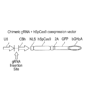
Fig. 3 shows the gRNA construct (expression vector) used for transfection;
Date Recue/Date Received 2022-04-14
- 26 ¨
Fig. 4 shows gel electrophoresis bands for parental and mutated LIR2 DNA,
before and after transfection;
Fig. 5 shows gel electrophoresis bands for parental and mutated CTLA4 DNA,
before and after transfection;
Fig. 6A is a FACS plot showing successful CD96 knockdown using
electroporation;
Fig. 6B is a FACS plot showing successful CD96 knockdown using
electroporation;
Fig. 7 is a bar chart showing increased cytotoxicity of CD96 knockdown
KHYG-1 cells against K562 cells at various E:T ratios;
Fig. 8 shows knockdown of CD328 (Siglec-7) in NK-92 cells;
Fig. 9 shows enhanced cytotoxicity of NK Cells with the CD328 (Siglec-7)
knockdown;
Fig. 10 shows a FACS plot of the baseline expression of TRAIL on KHYG-1
cells;
Fig. 11 shows a FACS plot of the expression of TRAIL and TRAIL variant after
transfection of KHYG-1 cells;
Fig. 12 shows a FACS plot of the expression of CD107a after transfection of
KHYG-1 cells;
Fig. 13 shows the effects of transfecting KHYG-1 cells with TRAIL and TRAIL
variant on cell viability;
Fig. 14 shows a FACS plot of the baseline expression of DR4, DRS, DcR1 and
DcR2 on both KHYG-1 cells and NK-92 cells;
Fig.s 15, 16 and 17 show the effects of expressing TRAIL or TRAIL variant in
KHYG-1 cells on apoptosis of three target cell populations: K562, RPMI8226
and MM1.S, respectively;
Fig. 18 shows two FACS plots of DRS expression on RPMI8226 cells and
MM1.S cells, respectively, wherein the effects of Bortezomib treatment on
DRS expression are shown;
Date Recue/Date Received 2022-04-14
- 27 ¨
Fig. 19 shows FACS plots of apoptosis in Bortezomib-pretreated/untreated
MM1.S cells co-cultured with KHYG-1 cells with/without the TRAIL variant;
Fig. 20 shows a FACS plot of perforin expression levels in KHYG-1 cells
treated with 100 nM CMA for 2 hours;
Fig. 21 shows FACS plots of KHYG-1 cell viability after treatment with 100 nM
CMA or vehicle;
Fig. 22 shows FACS plots of apoptosis in MM1.S cells co-cultured with KHYG-
1 cells with/without the TRAIL variant and pretreated with/without CMA;
Fig. 23 shows FACS plots of apoptosis in K562 cells co-cultured with KHYG-1
cells with CD96-siRNA and/or TRAIL variant expression; and
Fig. 24 shows FACS plots of apoptosis in MM1.S cells co-cultured with KHYG-
1 cells with CD96-siRNA and/or TRAIL variant expression.
DNA, RNA and amino acid sequences are referred to below, in which:
SEQ ID NO: 1 is the full LIR2 DNA sequence;
SEQ ID NO: 2 is the LIR2 amino acid sequence;
SEQ ID NO: 3 is the LIR2 g9 gRNA sequence;
SEQ ID NO: 4 is the LIR2 g18 gRNA sequence;
SEQ ID NO: 5 is the LIR2 forward primer sequence;
SEQ ID NO: 6 is the LIR2 reverse primer sequence;
SEQ ID NO: 7 is the full CTLA4 DNA sequence;
SEQ ID NO: 8 is the CTLA4 amino acid sequence;
SEQ ID NO: 9 is the CTLA4 g7 gRNA sequence;
SEQ ID NO: 10 is the CTLA4 g15 gRNA sequence;
SEQ ID NO: 11 is the CTLA4 forward primer sequence; and
SEQ ID NO: 12 is the CTLA4 reverse primer sequence.
Example 1 ¨ Knockout of Inhibitory Receptor Function
CRISPR/Cas9
Date Recue/Date Received 2022-04-14
- 28 ¨
Cells were prepared as follows, having inhibitory receptor function removed.
gRNA
constructs were designed and prepared to target genes encoding the 'classical'
inhibitory receptor LIR2 and the 'checkpoint' inhibitory receptor CTLA4 in the
human
genome of NK cells. CRISPR/Cas9 genome editing was then used to knock out the
LIR2 and CTLA4 target genes.
Two gRNA candidates were selected for each target gene and their cleavage
efficacies in K562 cells determined. The sequences of the gRNA candidates are
shown in Table 1 and the Protospacer Adjacent Motif (PAM) relates to the last
3
bases of the sequence. The flanking regions of the gRNA sequences on the LIR2
gene (SEQ ID NO: 1) and the CTLA4 gene (SEQ ID NO: 7) are shown in Figures 1
and 2, respectively.
Gene Plasmid Name Sequence
GAGTCACAGGTGGCATTTGGCGG
5M682.LIR2.g9
(SEQ ID NO: 3)
h LI R2
CGAATCGCAGGTGGTCGCACAGG
5M682.LIR2.g18
(SEQ ID NO: 4)
CACTCACCTTTGCAGAAGACAGG
5M683.CTLA4.g7 (SEQ ID NO: 9)
hCTLA4
CCTTGTGCCGCTGAAATCCAAGG
5M683.CTLA4.g 15 (SEQ ID NO: 10)
Table 1. gRNA candidates and sequences
K562 cells were transfected with the prepared gRNA constructs (Figure 3) and
subsequently harvested for PCR amplification. The presence of GFP expression
was
used to report successful incorporation of the gRNA construct into the K562
cells.
This confirmed expression of the Cas9 gene and therefore the ability to knock
out
expression of the LIR2 and CTLA4 genes.
Date Recue/Date Received 2022-04-14
- 29 ¨
The cleavage activity of the gRNA constructs was determined using an in vitro
mismatch detection assay. T7E1 endonuclease I recognises and cleaves non-
perfectly matched DNA, allowing the parental LIR2 and CTLA4 genes to be
compared to the mutated genes following CRISPR/Cas9 transfection and non-
homologous end joining (NHEJ).
Figure 4 shows the resulting bands following agarose gel electrophoresis after
knockout of the LIR2 gene with the g9 and g18 gRNA sequences. The three bands
corresponding to each mutation relate to the parental gene and the two
resulting
strands following detection of a mismatch in the DNA sequence after
transfection.
The g9 gRNA sequence resulted in an 11% success rate of transfection, whereas
the
g18 gRNA resulted in 10%.
Figure 5 shows the resulting bands following agarose gel electrophoresis after
knockout of the CTLA4 gene with the g7 and g15 gRNA sequences. The g7 gRNA
sequence resulted in a 32% success rate of transfection, whereas the g15 gRNA
resulted in 26%.
Following the successful knockout of LIR2 and CTLA4 in K562 cells, KHYG-1
cells
were transfected with gRNA constructs.
KHYG-1 derivative clones having homozygous deletions were selected. A Cas9 /
puromycin acetyltransferase (PAC) expression vector was used for this purpose.
Successfully transfected cells were selected, based on their resistance to the
antibiotic puromycin.
Cas9 RNP
Another protocol used for knockout of checkpoint inhibitory receptors in NK
cells was
that of Cas9 RNP transfection. An advantage of using this protocol was that
similar
Date Recue/Date Received 2022-04-14
- 30 ¨
transfection efficiencies were achievable but with significantly lower
toxicity compared
to using the DNA plasmids of the CRISPR/Cas9 protocol.
1x10⁶ KHYG1 cells were harvested for each transfection experiment. The cells
were
washed with PBS and spun down in a centrifuge. The supernatant was then
discarded. The CRISPR RNP (RNA binding protein) materials were then prepared
as
follows:
(1) a 20pM solution of the required synthesized crRNA and tRNA
(purchased from
Dharmacon) was prepared.
(2) 4p1 of crRNA (20pM) and 4p1 of tRNA (20pM) were mixed together.
(3) The mixture was then added to 2p1Cas9 protein (5pg/p1).
(4) All of the components were mixed and incubated at room temperature for
10
minutes.
Following the Neon Transfection System, the cells were mixed with Cas9 RNP
and
electroporation was performed using the following parameters:
Voltage: 1450v
Pulse width: 30ms
Pulse number: 1
The cells were then transferred to one well of a 12-well plate containing
growth
medium (inc. IL-2 and IL-15).
The cells were harvested after 48-72 hours to confirm gene editing efficiency
by T7
endonuclease assay and/or Sanger sequencing. The presence of indels were
confirmed, indicating successful knockout of CTLA4, PD1 and CD96 in KHYG1
cells.
Site-specific nucleases
Date Recue/Date Received 2022-04-14
- 31 ¨
Another protocol used for knockout of checkpoint inhibitory receptors in NK
cells was
that of XTN TALEN transfection. An advantage of using this protocol was that a
particularly high level of specificity was achievable compared to wildtype
CRISPR.
Step 1: Preparation of Reagents
KHYG-1 cells were assayed for certain attributes including transfection
efficiency,
single cell cloning efficiency and karyotype/copy number. The cells were then
cultured in accordance with the supplier's recommendations.
Depending on the checkpoint inhibitory receptor being knockout out, nucleases
were
prepared by custom-design of at least 2 pairs of XTN TALENs. The step of
custom-
design includes evaluation of gene locus, copy number and functional
assessment
(i.e. homologs, off-target evaluation).
Step 2: Cell Line Engineering
The cells were transfected with the nucleases of Step 1; this step was
repeated up to
3 times in order to obtain high levels of cutting and cultures were split and
intermediate cultures maintained prior to each transfection.
Initial screening occurred several days after each transfection; the pools of
cells were
tested for cutting efficiency via the Cel-1 assay. Following the level of
cutting
reaching acceptable levels or plateaus after repeated transfections, the cells
were
deemed ready for single cell cloning.
The pooled cells were sorted to one cell per well in a 96-well plate; the
number of
plates for each pool was dependent on the single cell cloning efficiency
determined in
Step 1. Plates were left to incubate for 3-4 weeks.
Date Recue/Date Received 2022-04-14
- 32 ¨
Step 3¨ Screening and Expansion
Once the cells were confluent in the 96-well plates, cultures were
consolidated and
split into triplicate 96-well plates; one plate was frozen as a backup, one
plate was re-
plated to continue the expansion of the clones and the final plate was used
for
genotype confirmation.
Each clone in the genotype plate was analyzed for loss of qPCR signal,
indicating all
alleles had been modified. Negative clones were PCR amplified and cloned to
determine the nature of the indels and lack of any wildtype or in-frame
indels.
Clones with the confirmed knockout were consolidated into no more than one 24-
well
plate and further expanded; typically 5-10 frozen cryovials containing 1x10⁶
cells per
vial for up to 5 individual clones were produced per knockout.
Step 4¨ Validation
Cells were banked under aseptic conditions.
Basic release criteria for all banked cells included viable cell number (pre-
freeze and
post-thaw), confirmation of identity via STR, basic sterility assurance and
mycoplasma testing; other release criteria were applied when necessary
(karyotype,
surface marker expression, high level sterility, knockout evaluation of
transcript or
protein, etc).
Example 2 ¨ Knockdown of Checkpoint Inhibitory Receptor CD96 Function via
RNAi
siRNA knockdown of CD96 in KHYG-1 cells was performed by electroporation. The
Nucleofection Kit T was used, in conjunction with the Amaxa Nucleofector II,
from
Date Recue/Date Received 2022-04-14
- 33 ¨
Lonza, as it is appropriate for use with cell lines and can successfully
transfect both
dividing and non-dividing cells and achieves transfection efficiencies of up
to 90%..
Control siRNA (catalog number: se-37007) and CD96 siRNA (catalog number: se-
45460) were obtained from Santa Cruz Biotechnology. Antibiotic-free RPMI-1640
containing 10% FBS, 2mM L-glutamine was used for post-Nucleofection culture.
Mouse anti-human CD96-APC (catalog number: 338409) was obtained from
Biolegend for staining.
A 20pM of siRNA stock solution was prepared. The lyophilized siRNA duplex was
resuspended in 33p1 of the RNAse-free water (siRNA dilution buffer: se-29527)
to
FITC-control/control-siRNA, in 165p1 of the RNAse-free water for the target
gene
siRNA (siRNA CD96). The tube was heated to 90 C for 1 minute and then
incubated
at 37 C for 60 minutes. The siRNA stock was then stored at -20 C until needed.
The KHYG-1 cells were passaged one to two days before Nucleofection, as the
cells
must be in logarithmic growth phase.
The Nucleofector solution was warmed to room temperature (100u1 per sample).
An aliquot of culture medium containing serum and supplements was also pre-
warmed at 37 C in a 50m1 tube. 6-well plates were prepared by adding 1.5m1 of
culture medium containing serum and supplements. The plates were pre-incubated
in
a humidified 37 C / 5% CO2 incubator.
2x10⁶ cells in 100p1 Nucleofection solution was mixed gently with 4p1 20pM
siRNA
solution (1.5pg siRNA). Air bubbles were avoided during mixing. The mixture
was
transferred into Amaxa certified cuvettes and placed into the Nucleofector
cuvette
holder and program U-001 selected.
Date Recue/Date Received 2022-04-14
- 34 ¨
The program was allowed to finish, and the samples in the cuvettes were
removed
immediately. 500p1 pre-equilibrated culture medium was then added to each
cuvette.
The sample in each cuvette was then gently transferred to a corresponding well
of
the prepared 6-well plate, in order to establish a final volume of 2m1 per
well.
The cells were then incubated in a humidified 37 C / 5% CO2 incubator until
transfection analysis was performed. Flow cytometry analysis was performed 16-
24
hours after electroporation, in order to measure CD96 expression levels. This
electroporation protocol was carried out multiple times and found to reliably
result in
CD96 knockdown in KHYG-1 cells (see e.g. Figures 6A and 6B).
Example 3¨ Enhanced Cytotoxicity of NK Cells with a CD96 Knockdown
KHYG-1 cells with and without the CD96 knockdown were co-cultured with K562
cells at different effector:target (E:T) ratios.
Cytotoxicity was measured 4 hours after co-culture, using the DELFIA EuTDA
Cytotoxicity Kit from PerkinElmer (Catalog number: AD0116).
Target cells K562 were cultivated in RPMI-1640 medium containing 10% FBS, 2mM
L-glutamine and antibiotics. 96-well V-bottom plates (catalog number: 83.3926)
were
bought from SARSTEDT. An Eppendorf centrifuge 581OR (with plate rotor) was
used
to spin down the plate. A VARIOSKAN FLASH (with ScanIt software 2.4.3) was
used
to measure the fluorescence signal produced by lysed K562 cells.
K562 cells were washed with culture medium and the number of cells adjusted to
1x10⁶ cells/mL with culture medium. 2-4mL of cells was added to 51j1 of BATDA
reagent and incubated for 10 minutes at 37 C. Within the cell, the ester bonds
are
hydrolysed to form a hydrophilic ligand, which no longer passes through the
membrane. The cells were centrifuged at 1500RPM for 5 mins to wash the loaded
Date Recue/Date Received 2022-04-14
- 35 ¨
K562 cells. This was repeated 3-5 times with medium containing 1mM Probenecid
(Sigma P8761). After the final wash the cell pellet was resuspended in culture
medium and adjusted to about 5 x10⁴ cells/mL.
Wells were set up for detection of background, spontaneous release and maximum
release. 100pL of loaded target cells (5,000 cells) were transferred to wells
in a V-
bottom plate and 100pL of effector cells (KHYG-1 cells) were added at varying
cell
concentrations, in order to produce effector to target ratios ranging from 1:1
to 20:1.
The plate was centrifuged at 100xg for 1 minute and incubated for 4 hours in a
humidified 5% CO2 atmosphere at 37 C. For maximum release wells 10pL of lysis
buffer was added to each well 15 minutes before harvesting the medium. The
plate
was centrifuged at 500xg for 5 minutes.
20pL of supernatant was transferred to a flat-bottom 96 well plate 200pL of
pre-
warmed Europium solution added. This was incubated at room temperature for 15
mins using a plate shaker. As K562 cells are lysed by the KHYG-1 cells, they
release
ligand into the medium. This ligand then reacts with the Europium solution to
form a
fluorescent chelate that directly correlates with the amount of lysed cells.
The fluorescence was then measured in a time-resolved fluorometer by using
VARIOSKAN FLASH. The specific release was calculated using the following
formula:
% specific release = Experiment release ¨ Spontaneous release / Maximum
release ¨ Spontaneous release
Statistical analysis was performed using Graphpad Prism 6.04 software. A
paired t
test was used to compare the difference between siRNA CD96 knockdown KHYG-1
cells and control groups (n=3).
Date Recue/Date Received 2022-04-14
- 36 ¨
The specific release was found to be significantly increased in co-cultures
containing
the CD96 knockdown KHYG-1 cells. This was the case at all E:T ratios (see
Figure
7).
As fluorescence directly correlates with cell lysis, it was confirmed that
knocking
down CD96 expression in KHYG-1 cells resulted in an increase in their ability
to kill
K562 cancer target cells.
Example 4 ¨ Enhanced Cytotoxicity of NK Cells with a CD328 (Siglec-7)
Knockdown
SiRNA-mediated knock-down of CD328 in NK-92 cells
Materials, reagents and instruments
Control siRNA (catalog number: se-37007) and CD328 siRNA (catalog number: se-
106757) were bought from Santa Cruz Biotechnology. To achieve transfection
efficiencies of up to 90% with high cell viability (>75%) in NK-92 cells with
the
NucleofectorTM Device (Nucleofector II, Lonza), a NucleofectorTM Kit T from
Lonza
was used. RPMI-1640 containing 10% FBS, 2mM L-glutamine, antibiotics free, was
used for post-Nucleofection culture. Mouse anti-human CD328-APC (catalog
number: 339206) was bought from Biolegend.
Protocol
To make 10pM of siRNA stock solution
= Resuspend lyophilized siRNA duplex in 66p1 of the RNAse-free water (siRNA
dilution buffer: se-29527) to FITC-control/control-siRNA, in 330p1 of the
RNAse-free water for the target gene siRNA (siRNA CD328).
= Heat the tube to 90 C for 1 minute.
= Incubate at 37 C for 60 minutes.
= Store siRNA stock at -20 C if not used directly.
Date Recue/Date Received 2022-04-14
- 37 ¨
= One Nucleofection sample contains (for 100p1 standard cuvette)
= Cell number: 2x10⁶ cells
= siRNA: 4p1 of 10pM stock
= Nucleofector solution: 100p1
Nucleofection
= Cultivate the required number of cells. (Passage one or two day before
Nucleofection, cells must be in logarithmic growth phase).
= Prepare siRNA for each sample.
= Pre-warm the Nucleofector solution to room temperature (100p1 per sample).
= Pre-warm an aliquot of culture medium containing serum and supplements at
37 C in a 50m1 tube. Prepare 6-well plates by filling with 1.5m1 of cullture
medium containing serum and supplements and pre-incubate plates in a
humidified 37 C /5% CO2 incubator.
= Take an aliquot of cell culture and count the cells to determine the cell
density.
= Centrifuge the required number of cells at 1500rpm for 5 min. Discard
supernatant completely so that no residual medium covers the cell pellet.
= Resuspend the cell pellet in room temperature Nucleofector Solution to a
final
concentration of 2x10⁶ cells/100p1. Avoid storing the cell suspension longer
than 15-20 min in Nucleofector Solution, as this reduces cell viability and
gene
transfer efficiency.
= Mix 100plof cell suspension with siRNA.
= Transfer the sample into an amaxa certified cuvette. Make sure that the
sample covers the bottom of the cuvette, avoid air bubbles while pipetting.
Close the cuvette with the blue cap.
= Select the appropriate Nucleofector program (A-024 for NK-92 cells).
Insert
the cuvette into the cuvette holder (Nucleofector II: rotate the carousel
clockwise to the final position) and press the "x" button to start the
program.
= To avoid damage to the cells, remove the samples from the cuvette
immediately after the program has finished (display showing "OK"). Add 500p1
Date Recue/Date Received 2022-04-14
- 38 ¨
of the pre-warmed culture medium into the cuvette and transfer the sample
into the prepared 6-well plate.
= Incubate cells in a humidified 37 C/5% CO2 incubator. Perform flow
cytometric
analysis and cytotoxicity assay after 16-24 hours.
Results: we followed the above protocol and performed flow cytometry analysis
of
CD328 expression level in NK-92 cells. The results of one representative
experiment
is shown in Fig. 8, confirming successful knockdown.
Knocking down CD328 enhances cytotoxicity
Materials, reagents and instruments
DELFIA EuTDA cytotoxicity kit based on fluorescence enhancing ligand (Catalog
nmber: AD0116) was bought from PerkinElmer. Target cells K562 were cultivated
in
RPMI-1640 medium containing 10% FBS, 2mM L-glutamine and antibiotics. 96-well
V-bottom plates (catalog number: 83.3926) were bought from SARSTEDT.
Eppendrof centrifuge 5810R (with plate rotor) was used to spin down the plate.
VARIOSKAN FLASH (with ScanIt software 2.4.3) was used to measure the
fluorescence signal produced by lysed K562 cells.
Protocol
= Load target K562 cells with the fluorescence enhancing ligand DELFIA
BATDA
reagent
= Wash K562 cells with medium, adjust the number of cells to 1x10⁶ cells/mL
with culture medium. Add 2-4 mL of cells to 5 pl of BATDA reagent, incubate
for 10 minutes at 37 C.
= Spin down at 1500RPM for 5minutes to wash the loaded K562 cells for 3-5
times with medium containing 1mM Probenecid (Sigma P8761).
= After the final wash resuspend the cell pellet in culture medium and
adjust to
about 5 x10⁴ cells/mL.
Date Recue/Date Received 2022-04-14
- 39 ¨
Cytotoxicity assay
= Set up wells for detection of background, spontaneously release and
maximum release.
= Pipette 100pL of loaded target cells (5,000 cells) to a V-bottom plate.
= Add 100pL of effector cells (NK-92) of varying cell concentrations.
Effector to
target ratio ranges from 1:1 to 20:1.
= Spin down the plate at 100xg of RCF for 1 minute.
= Incubate for 2 hours in a humidified 5% CO2 atmosphere at 37 C. For
maximum release wells, add 10 pL of lysis buffer to each well 15 minutes
before harvesting the medium.
= Spin down the plate at 500xg for 5 minutes.
= Transfer 20 pL of supernatant to a flat-bottom 96 well plate, add 200 pL
of pre-
warmed Europium solution, incubate at room temperature for 15 minutes using
plateshaker.
= Measure the fluorescence in a time-resolved fluorometer by using
VARIOSKAN FLASH. The specific release was calculated using the following
formula:
= % specific release = Experiment release ¨ Spontaneous release / Maximum
release - Spontaneous release
Results: we followed the above to determine the effect on cytotoxicity of the
CD328
knockdown. The results of one representative experiment are shown in figure 9.
As
seen, cytotoxicity against target cells was increased in cells with the CD328
knockdown.
Example 5 ¨ Protocol for Blood Cancer Therapy by Knockdown / Knockout of
Checkpoint Inhibitory Receptors
Date Recue/Date Received 2022-04-14
- 40 ¨
As demonstrated in the above Examples, checkpoint inhibitory receptor function
can
be knocked down or knocked out in a variety of ways. The following protocol
was
developed for use in treating patients with blood cancer:
Following diagnosis of a patient with a cancer suitably treated with the
invention, an
aliquot of modified NK cells can be thawed and cultured prior to
administration to the
patient.
Alternatively, a transient mutation can be prepared using e.g. siRNA within a
day or
two, as described above. The MaxCyte Flow Electroporation platform offers a
suitable solution for achieving fast large-scale transfections in the clinic.
The removal of certain checkpoint inhibitory receptors may be more beneficial
than
others. This is likely to depend on the patient and the cancer. For this
reason, the
cancer is optionally biopsied and the cancer cells are grown in culture ex
vivo. A
range of NK cells with different checkpoint inhibitory receptor modifications
can thus
be tested for cytotoxicity against the specific cancer. This step can be used
to select
the most appropriate NK cell or derivative thereof for therapy.
Following successful modification, the cells are resuspended in a suitable
carrier (e.g.
saline) for intravenous and/or intratumoural injection into the patient.
Example 6¨ KHYG-1 Knock-in of TRAIL / TRAIL variant
KHYG-1 cells were transfected with both TRAIL and TRAIL variant, in order to
assess their viability and ability to kill cancer cells following
transfection.
The TRAIL variant used is that described in WO 2009/077857. It is encoded by
the
wildtype TRAIL gene containing the D269H/E195R mutation. This mutation
Date Recue/Date Received 2022-04-14
- 41 ¨
significantly increases the affinity of the TRAIL variant for DR5, whilst
reducing the
affinity for both decoy receptors (DcR1 and DcR2).
Baseline TRAIL Expression
Baseline TRAIL (CD253) expression in KHYG-1 cells was assayed using flow
cytometry.
Mouse anti-human CD253-APC (Biolegend catalog number: 308210) and isotype
control (Biolegend catalog number: 400122) were used to stain cell samples and
were analyzed on a BD FACS Canto ll flow cytometer.
KHYG-1 cells were cultured in RPM! 1640 medium containing 10% FBS, 2mM L-
glutamine, penicillin (100 U/mL)/streptomycin (100 mg/mL) and IL-2 (10ng/mL).
0.5-
1.0 x 10⁶ cells/test were collected by centrifugation (1500rpm x 5 minutes)
and the
supernatant was aspirated. The cells (single cell suspension) were washed with
4 m L
ice cold FACS Buffer (PBS, 0.5-1% BSA, 0.1% NaN3 sodium azide). The cells were
re-suspended in 100 pL ice cold FACS Buffer, add 5uL antibody was added to
each
tube and incubated for 30 minutes on ice. The cells were washed 3 times by
centrifugation at 1500 rpm for 5 minutes. The cells were then re-suspended in
500 pL
ice cold FACS Buffer and temporarily kept in the dark on ice.
The cells were subsequently analyzed on the flow cytometer (BD FACS Canto II)
and
the generated data were processed using FlowJo 7.6.2 software.
As can be seen in Fig. 10, FACS analysis showed weak baseline expression of
TRAIL on the KHYG-1 cell surface.
TRAIL / TRAIL variant Knock-in by Electroporation
Wildtype TRAIL mRNA and TRAIL variant (D269H/195R) mRNA was synthesized by
TriLink BioTechnologies, aliquoted and stored as -80 C. Mouse anti-human CD253-
Date Recue/Date Received 2022-04-14
-42 ¨
APC (Biolegend catalog number: 308210) and isotype control (Biolegend catalog
number: 400122), and Mouse anti-human CD107a-PE (eBioscience catalog number:
12-1079-42) and isotype control (eBioscience catalog number: 12-4714)
antibodies
were used to stain cell samples and were analyzed on a BD FACS Canto II flow
cytometer. DNA dye SYTOX-Green (Life Technologies catalog number: S7020; 5 mM
Solution in DMSO) was used. To achieve transfection efficiencies of up to 90%
with
high cell viability in KHYG-1 cells with the NucleofectorTM Device
(Nucleofector II,
Lonza), a NucleofectorTM Kit T from Lanza was used. Antibiotics-free RPM! 1640
containing 10% FBS, L-glutamine (2mM) and IL-2 (10ng/mL) was used for post-
Nucleofection culture.
KHYG-1 and NK-92 cells were passaged one or two days before Nucleofection, as
the cells must be in the logarithmic growth phase. The Nucleofector solution
was pre-
warmed to room temperature (100 pl per sample), along with an aliquot of
culture
medium containing serum and supplements at 37 C in a 50 mL tube. 6-well plates
were prepared by filling with 1.5 mL culture medium containing serum and
supplements and pre-incubated in a humidified 37 C / 5% CO2 incubator. An
aliquot
of cell culture was prepared and the cells counted to determine the cell
density. The
required number of cells was centrifuged at 1500rpm for 5 min, before
discarding the
supernatant completely. The cell pellet was re-suspended in room temperature
Nucleofector Solution to a final concentration of 2x10⁶ cells/100p1 (maximum
time in
suspension = 20 minutes). 100 pl cell suspension was mixed with 10 pg mRNA
(volume of RNA < 10 pL). The sample was transferred into an Amaxa-certified
cuvette (making sure the sample covered the bottom of the cuvette and avoiding
air
.. bubbles). The appropriate Nucleofector program was selected (i.e. U-001 for
KHYG-1
cells). The cuvettes were then inserted into the cuvette holder. 500 pl pre-
warmed
culture medium was added to the cuvette and the sample transferred into a
prepared
6-well plate immediately after the program had finished, in order to avoid
damage to
the cells. The cells were incubated in a humidified 37 C / 5% CO2 incubator.
Flow
Date Recue/Date Received 2022-04-14
- 43 ¨
cytometric analysis and cytotoxicity assays were performed 12-16 hours after
electroporation. Flow cytometry staining was carried out as above.
As can be seen in Fig.s 11 and 12, expression of TRAIL / TRAIL variant and
CD107a
(NK activation marker) increased post-transfection, confirming the successful
knock-
in of the TRAIL genes into KHYG-1 cells.
Fig. 13 provides evidence of KHYG-1 cell viability before and after
transfection via
electroporation. It can be seen that no statistically significant differences
in cell
viability are observed following transfection of the cells with TRAIL / TRAIL
variant,
confirming that the expression of wildtype or variant TRAIL is not toxic to
the cells.
This observation contradicts corresponding findings in NK-92 cells, which
suggest the
TRAIL variant gene knock-in is toxic to the cells (data not shown).
Nevertheless, this
is likely explained by the relatively high expression levels of TRAIL
receptors DR4
and DR5 on the NK-92 cell surface (see Fig. 14).
Effects of TRAIL / TRAIL variant on KHYG-1 Cell Cytotoxicity
Mouse anti-human CD2-APC antibody (BD Pharmingen catalog number: 560642)
was used. Annexin V-FITC antibody (ImmunoTools catalog number: 31490013) was
used. DNA dye SYTOX-Green (Life Technologies catalog number: S7020) was used.
A 24-well cell culture plate (SARSTEDT AG catalog number: 83.3922) was used.
Myelogenous leukemia cell line K562, multiple myeloma cell line RPMI8226 and
MM1.S were used as target cells. K562, RPMI8226, MM1.S were cultured in RPM!
1640 medium containing 10% FBS, 2mM L-glutamine and penicillin (100
U/mL)/streptomycin (100 mg/mL).
As explained above, KHYG-1 cells were transfected with TRAIL / TRAIL variant.
The target cells were washed and pelleted via centrifugation at 1500rpm for 5
minutes. Transfected KHYG-1 cells were diluted to 0.5x10⁶/mL. The target cell
Date Recue/Date Received 2022-04-14
- 44 ¨
density was then adjusted in pre-warmed RPM! 1640 medium, in order to produce
effector:target (E:T) ratios of 1:1.
0.5 mL KHYG-1 cells and 0.5 mL target cells were then mixed in a 24-well
culture
plate and placed in a humidified 37 C / 5% CO2 incubator for 12 hours. Flow
cytometric analysis was then used to assay KHYG-1 cell cytotoxicity; co-
cultured
cells (at different time points) were washed and then stained with CD2-APC
antibody
(5 pL/test), Annexin V-FITC (5 pUtest) and SYTOX-Green (5 pL/test) using
Annexin
V binding buffer.
Data were further analyzed using FlowJo 7.6.2 software. CD2-positive and CD2-
negative gates were set, which represent KHYG-1 cell and target cell
populations,
respectively. The Annexin V-FITC and SYTOX-Green positive cells in the CD2-
negative population were then analyzed for TRAIL-induced apoptosis.
Fig.s 15, 16 and 17 show the effects of both KHYG-1 cells expressing TRAIL or
TRAIL variant on apoptosis for the three target cell lines: K562, RPMI8226 and
MM1.S, respectively. It is apparent for all target cell populations that TRAIL
expression on KHYG-1 cells increased the level of apoptosis, when compared to
normal KHYG-1 cells (not transfected with TRAIL). Moreover, TRAIL variant
expression on KHYG-1 cells further increased apoptosis in all target cell
lines, when
compared to KHYG-1 cells transfected with wildtype TRAIL.
Cells of the invention, expressing the TRAIL variant, offer a significant
advantage in
cancer therapy, due to exhibiting higher affinities for the death receptor
DRS. When
challenged by these cells of the invention, cancer cells are prevented from
developing defensive strategies to circumvent death via a certain pathway.
Thus
cancers cannot effectively circumvent TRAIL-induced cell death by upregulating
TRAIL decoy receptors, as cells of the invention are modified so that they
remain
cytotoxic in those circumstances.
Date Recue/Date Received 2022-04-14
- 45 ¨
Example 7 ¨ Protocol for Blood Cancer Therapy using NK Cells with TRAIL
Variants Knocked-in
KHYG-1 cells were transfected with TRAIL variant, as described above in
Example 6.
The following protocol was developed for use in treating patients with blood
cancer:
Following diagnosis of a patient with a cancer suitably treated with the
invention, a
DR5-inducing agent, e.g. Bortezomib, is administered, prior to administration
of the
modified NK cells, and hence is used at low doses to upregulate expression of
DR5
on the cancer, making modified NK cell therapy more effective.
An aliquot of modified NK cells is then thawed, cultured and administered to
the
patient.
Since the TRAIL variant expressed by the NK cells used in therapy has a lower
affinity for decoy receptors than wildtype TRAIL, there is increased binding
of death
receptors on the cancer cell surface, and hence more cancer cell apoptosis as
a
result.
Another option, prior to implementation of the above protocol, is to biopsy
the cancer
and culture cancer cells ex vivo. This step can be used to identify those
cancers
expressing particularly high levels of decoy receptors, and/or low levels of
death
receptors, in order to help determine whether a DR5-inducing agent is
appropriate for
a given patient. This step may also be carried out during therapy with the
above
protocol, as a given cancer might be capable of adapting to e.g. reduce its
expression of DR5, and hence it may become suitable to treat with a DR5-
inducing
agent part-way through therapy.
Example 8 ¨ Low Dose Bortezomib Sensitizes Cancer Cells to NK Cells
Expressing TRAIL Variant
Date Recue/Date Received 2022-04-14
- 46 ¨
Bortezomib (Bt) is a proteasome inhibitor (chemotherapy-like drug) useful in
the
treatment of Multiple Myeloma (MM). Bortezomib is known to upregulate DR5
expression on several different types of cancer cells, including MM cells.
KHYG-1 cells were transfected with TRAIL variant, as described above in
Example 6,
before being used to target MM cells with or without exposure to Bortezomib.
Bortezomib-induced DR5 expression
Bortezomib was bought from Millennium Pharmaceuticals. Mouse anti-human DR5-
AF647 (catalog number: 565498) was bought from BD Pharmingen. The stained cell
samples were analyzed on BD FACS Canto II.
(1) MM cell lines RPMI8226 and MM1.S were grown in RPMI1640 medium
(Sigma, St Louis, MO, USA) supplemented with 2 mM L-glutamine, 10 mM HEPES,
24 mM sodium bicarbonate, 0.01% of antibiotics and 10% fetal bovine serum
(Sigma,
St Louis, MO, USA), in 5% CO2 atmosphere at 37 C.
(2) MM cells were seeded in 6-well plates at 1x10⁶/mL, 2mL/well.
(3) MM cells were then treated with different doses of Bortezomib for 24
hours.
(4) DRS expression in Bortezomib treated/untreated MM cells was then
analyzed
by flow cytometry (Fig. 18).
Low dose Bortezomib treatment was found to increase DRS expression in both MM
cell lines (Fig. 18). DRS upregulation was associated with a minor induction
of
apoptosis (data not shown). It was found, however, that DRS expression could
not be
upregulated by high doses of Bortezomib, due to high toxicity resulting in
most of the
MM cells dying.
Bortezomib-induced sensitization of cancer cells
Date Recue/Date Received 2022-04-14
- 47 ¨
KHYG-1 cells were transfected with the TRAIL variant (TRAIL D269H/E195R), as
described above in Example 6.
(1) Bortezomib treated/untreated MM1.S cells were used as target cells. MM1.S
cells
were treated with 2.5nM of Bortzeomib or vehicle (control) for 24 hours.
(2) 6 hours after electroporation of TRAIL variant mRNA, KHYG-1 cells were
then
cultured with MM cells in 12- well plate. After washing, cell concentrations
were
adjusted to 1x10⁶/mL, before mixing KHYG-1 and MM1.S cells at 1:1 ratio to
culture
for 12 hours.
(3) Flow cytometric analysis of the cytotoxicity of KHYG-1 cells was carried
out. The
co-cultured cells were collected, washed and then stained with CD2-APC
antibody (5
uL/test), AnnexinV-FITC (5 uL/test) and SYTOX-Green (5 uL/test) using AnnexinV
binding buffer.
(4) Data were further analyzed using FlowJo 7.6.2 software. CD2-negative
population
represents MM1.S cells. KHYG-1 cells are strongly positive for CD2. Finally,
the
AnnexinV-FITC and SYTOX-Green positive cells in the CD2-negative population
were analyzed.
Flow cytometric analysis of apoptosis was performed in Bo rtezom ib-
pretreated/untreated MM1.S cells co-cultured with KHYG-1 cells electroporated
with/without TRAIL variant (Fig. 19).
It was found that Bortezomib induced sensitivity of MM cells to KHYG-1 cells
expressing the TRAIL variant. The data therefore indicated that an agent that
induced
DRS expression was effective in the model in increasing cytotoxicity against
cancer
cells, and hence may be useful in enhancing the cancer therapy of the present
invention.
Example 9¨ Confirmation of Induced Apoptosis by the TRAIL Variant
Date Recue/Date Received 2022-04-14
- 48 ¨
Despite the conclusive evidence of increased NK cell cytotoxicity resulting
from
TRAIL variant expression in the previous Examples, we wished to confirm
whether
the increased cytotoxicity resulted from inducing cancer cell apoptosis (most
likely) or
by inadvertently activating the NK cells to exhibit a more cytotoxic phenotype
and
hence kill cancer cells via perforin secretion.
Concanamycin A (CMA) has been demonstrated to inhibit perforin-mediated
cytotoxic
activity of NK cells, mostly due to accelerated degradation of perforin by an
increase
in the pH of lytic granules. We investigated whether the cytotoxicity of KHYG-
1 cells
expressing the TRAIL variant could be highlighted when perforin-mediated
cytotoxicity was partially abolished with CMA.
CMA-induced reduction of perform n expression
Mouse anti-human perforin-AF647 (catalog number: 563576) was bought from BD
pharmingen. Concanamycin A (catalog number: SC-202111) was bought from Santa
Cruz Biotechnology. The stained cell samples were analyzed using a BD FACS
Canto II.
(1) KHYG-1 cells were cultured in RPMI1640 medium containing 10%FBS (fetal
bovine serum), 2mM L-glutamine, penicillin (100 U/mL)/streptomycin (100
mg/mL),
and IL-2 (10ng/mL).
(2) KHYG-1 cells (6 hours after electroporation, cultured in
penicillin/streptomycin
free RPMI1640 medium) were further treated with 100nM CMA or equal volume of
vehicle (DMSO) for 2 hours.
(3) The cells were collected (1x10⁶ cells/test) by centrifugation (1500rpm
x 5
minutes) and the supernatant was aspirated.
(4) The cells were fixed in 4% paraformaldehyde in PBS solution at room
temperature for 15 minutes.
(5) The cells were washed with 4 mL of FACS Buffer (PBS, 0.5-1% BSA, 0.1%
sodium azide) twice.
Date Recue/Date Received 2022-04-14
- 49 ¨
(6) The cells were permeabilized with 1mL of PBS/0.1% saponin buffer for 30
minutes at room temperature.
(7) The cells were washed with 4 m L of PBS/0.1% saponin buffer.
(8) The cells were re-suspended in 100 uL of PBS/0.1% saponin buffer,
before
adding 5uL of the antibody to each tube and incubating for 30 minutes on ice.
(9) The cells were washed with PBS/0.1% saponin buffer 3 times by
centrifugation
at 1500 rpm for 5 minutes.
(10) The cells were re-suspended in 500 uL of ice cold FACS Buffer and kept in
the dark on ice or at 4 C in a fridge briefly until analysis.
(11) The cells were analyzed on the flow cytometer (BD FACS Canto II). The
data
were processed using FlowJo 7.6.2 software.
CMA treatment significantly decreased the perforin expression level in KHYG-1
cells
(Fig. 20) and had no negative effects on the viability of KHYG-1 cells (Fig.
21).
Cytotoxicity of NK cell TRAIL variants in the presence of CMA
KHYG-1 cells were transfected with the TRAIL variant (TRAIL D269H/E195R), as
described above in Example 6.
(1) MM1.S cells were used as target cells.
(2) 6 hours after electroporation of TRAIL mRNA, KHYG-1 cells were treated
with
100mM CMA or an equal volume of vehicle for 2 hours.
(3) The KHYG-1 cells were washed with RPMI1640 medium by centrifugation, and
re-suspended in RPMI1640 medium containing IL-2, adjusting cell concentrations
to
1x10⁶/mL.
(4) The MM1.S cells were re-suspended in RPMI1640 medium containing IL-2
adjusting cell concentrations to 1x10⁶/m L.
(5) The KHYG-1 and MM1.S cells were mixed at 1:1 ratio and co-cultured for 12
hours.
Date Recue/Date Received 2022-04-14
- 50 ¨
(6) Flow cytometric analysis of the cytotoxicity of KHYG-1 cells was carried
out. The
co-cultured cells were washed and stained with CD2-APC antibody (5 uL/test).
(7) After washing, further staining was performed with AnnexinV-FITC (5
uUtest) and
SYTOX-Green (5 uL/test) using AnnexinV binding buffer.
(8) Data were further analyzed using FlowJo 7.6.2 software. CD2-negative
population
represents MM1.S cells. KHYG-1 cells are strongly positive for CD2. The
AnnexinV-
FITC and SYTOX-Green positive cells in CD2-negative population were then
analyzed.
It was again shown that NK cells expressing the TRAIL variant show higher
cytotoxicity than control cells lacking expression of the TRAIL variant (Fig.
22). In this
Example, however, it was further shown that CMA was unable to significantly
diminish the cytotoxic activity of NK cells expressing TRAIL variant, in
contrast to the
finding for control NK cells treated with CMA.
NK cells without the TRAIL variant (control or mock NK cells) were shown to
induce
48% cancer cell death in the absence CMA and 35.9% cancer cell death in the
presence of CMA (Fig. 22). NK cells expressing the TRAIL variant were able to
induce more cancer cell death than control NK cells both in the presence and
absence of CMA. In fact, even with CMA present, NK cells expressing TRAIL
variant
induced more cancer cell death than control NK cells in the absence of CMA.
This data thus shows the importance of the TRAIL variant in increasing NK cell
cytotoxicity against cancer cells via a mechanism less susceptible to perforin-
related
downregulation. Since perforin is used commonly by NK cells to kill target
cells, and
many cancer cells have developed mechanisms for reducing NK cell perforin
expression, in order to evade cytotoxic attack, the NK cells of the invention
represent
a powerful alternative less susceptible to attenuation by cancer cells.
Date Recue/Date Received 2022-04-14
- 51 ¨
Example 10 ¨ Combined Expression of Mutant TRAIL Variant and Knockdown
of Checkpoint Inhibitory Receptor CD96 in KHYG-1 Cells
Increases in NK cell cytotoxicity were observed when knocking down checkpoint
inhibitory receptor CD96 expression and also when expressing TRAIL variant. We
also tested combining the two genetic modifications to provoke a synergistic
effect on
N K cell cytotoxicity.
CD96 expression was knocked down in KHYG-1 cells, as described in Example 2.
KHYG-1 cells were transfected with the TRAIL variant (TRAIL D269H/E195R), as
described above in Example 6.
(1) 12 hours after electroporation KHYG-1 cells were co-cultured with
target cells
(K562 or MM1.S) at a concentration of 1x10⁶/mL in 12-well plates (2mL/well)
for 12
hours. The E:T ratio was 1:1.
(2) 12 hours after co-culture, the cells were collected, washed, stained
with CD2-
APC, washed again and further stained with AnnexinV-FITC (5 uL/test) and SYTOX-
Green (5 uL/test) using AnnexinV binding buffer.
(3) Cell samples were analyzed using a BD FACS canto ll flow cytometer.
Data
were further analyzed using FlowJo 7.6.2 software. CD2-negative population
represents MM1.S cells. KHYG-1 cells are strongly positive for CD2. The
AnnexinV-
FITC and SYTOX-Green positive cells in the CD2-negative population were then
analyzed.
Simultaneously knocking down CD96 expression and expressing TRAIL variant in
KHYG-1 cells was found to synergistically enhance the cells' cytotoxicity
against both
K562 target cells (Fig. 23) and MM1.S target cells (Fig. 24). This was
indicated by the
fact that in both target cell groups, more cell death resulted from the
simultaneous
genetic modification than resulted from the individual modifications in
isolation.
Date Recue/Date Received 2022-04-14
- 52 ¨
At the same time, further evidence showing knockdown of CD96 increases NK cell
cytotoxicity was obtained (Fig. 23 & 24), in addition to further evidence
showing
expression of the TRAIL mutant/variant increases NK cell cytotoxicity (Fig. 23
& 24).
The invention thus provides NK cells and cell lines, and production thereof,
for use in
blood cancer therapy.
Date Recue/Date Received 2022-04-14