Note: Descriptions are shown in the official language in which they were submitted.
WO 2021/081676
PCT/CA2020/051487
HARNESSING THE POWER OF MICROBIOTA AND METABOLITES FOR THE
TREATMENT OF CANCER
RELATED APPLICATION DISCLOSURE
[0001] The present application claims the benefit
of U.S. Provisional Application
Ser. No. 62/929,340 filed November 1, 2019, the disclosure of which is hereby
incorporated by reference in its entirety.
SEQUENCE DISCLOSURE
[0002] This application includes as part of its
disclosure a Biological Sequence
Listing which has been submitted in ASCII format via EFS-Web and is hereby
incorporated by reference in its entirety. Said ASCII copy, created on October
30, 2020, is
named "115958300000011xt" and is 6,261 bytes in size.
FIELD
[0003] The present disclosure relates generally
to methods and compositions for
treating cancer, and in a specific aspect colorectal cancer. In another aspect
the
disclosure relates to novel strains of bacteria, as well as compositions and
uses thereof.
BACKGROUND
[0004] Colorectal cancer (CRC) is the second and
third most common malignancy
in Western countries in women and men, respectively (Ferlay et al., 2015). In
addition to
genetic aberrations, which are essential for the development of CRC, other
disease-
contributing factors have been identified. These include the microbiota and
inflammation,
whereby inflammation can drive or inhibit CRC development. Interferon (IFN)-y
producing
T helper type 1 (Thl) cells are known to be protective (Mager e al., 2016;
Mlecnik et al.,
2016; Wang et al., 2015), whereas interleukin (IL)-17-producing Th17 cells
promote CRC
development (Galon et al., 2006; Grivennikov et al., 2012; Le Gouvello e at,
2008). In
fact, the impact of the immune system is so potent that immune cell
infiltration in the
tumor is a superior prognostic factor compared to the classical tumor-lymph
nodes-
metastasis (TNM) system in CRC (Anitei et al., 2014; Mlecnik et al., 2016).
Similarly, the
microbiota also impacts on CRC progression (Arthur et al., 2012; Dejea et al.,
2018) and
may even alter the efficacy of chemotherapeutics (lida et al., 2013; Viaud et
al., 2013).
- 1 -
CA 03156203 2022-4-26
WO 2021/081676
PCT/CA2020/051487
[0005] Immune checkpoint blockade (ICB) therapy
is an efficient anti-cancer
strategy that utilizes the therapeutic potential of the immune system. Most
notably, ICB
inhibitors targeting cytotoxic T-lymphocyte-associated antigen 4 (CTLA-4),
programmed
cell death protein 1 (PD-1), or its ligand (PD-L1) have shown great success in
the
treatment of various cancers, including melanoma, renal cell carcinoma, and
non-small
cell lung cancer (Brahmer et al., 2012; Hodi et al., 2010). More recently,
seminal work has
shown that the efficacy of ICB therapy is dependent on the presence of certain
ICB-
promoting gut bacteria (Routy et al., 2018; Sivan et al., 2015; Vetizou et
al., 2015).
[0006] Despite these exciting advances, ICB
therapy efficacy in CRC has been
disappointing (Brahmer et al., 2012), with only 5-10% of all CRC patients
responding (Le
et al., 2017). Moreover, the detailed molecular mechanisms through which
bacteria
enhance the efficacy of ICB therapies remains unclear. Here, we identified
three bacterial
species that promote ICB efficacy in CRC and identified inosine as a critical
bacterial
metabolite that promoted differentiation of Th1-mediated anti-tumor immunity.
SUMMARY
[0007] In one aspect there is provided a method of treating a subject having a
cancer or
suspected of having a cancer, comprising or consisting of, administering an
immune
checkpoint inhibitor and one or more bacterium selected from Bifidobacterium
pseudolongum, Lactobacillus johnsonii, Olsenella profuse, Olsenella umbonata,
or
Olsenella all or a combination thereof
[0008] In one aspect there is provided a method of treating a subject having a
cancer or
suspected of having a cancer, comprising or consisting of, administering an
immune
checkpoint inhibitor and one or more bacterium selected from Bifidobacterium
pseudolongum, Lactobacillus johnsonii, or Olsenella sp. or a combination
thereof.
[0009] In an exemplary embodiment the bacterium is selected from the
Bifidobacterium
pseudolongum strain deposited as IDAC Deposit No. 231020-01, Lactobacillus
johnsonii
strain deposited as IDAC Deposit No. 231020-02, or Olsenella sp. strain
deposited as
IDAC Deposit No. 231020-03, or a combination thereof.
[0010] In one aspect there is provided a method of treating a subject having
or suspected
of having colorectal cancer (CRC), comprising or consisting of, administering
an immune
checkpoint inhibitor and one or more bacterium selected from Bifidobacterium
pseudolongum, Lactobacillus Johnson!!, Olsenella profuse, Olsenella umbonata,
or
Olsenella all, such as the Bifidobacterium pseudolongum strain deposited as
IDAC
- 2 -
CA 03156203 2022-4-26
WO 2021/081676
PCT/CA2020/051487
Deposit No. 231020-01, the Lactobacillus johnsonii strain deposited as IDAC
Deposit No.
231020-02, or the Olsenella sp. strain deposited as IDAC Deposit No. 231020-
03, or a
combination thereof.
[0011] In one aspect there is provided a method of treating a subject having
or suspected
of having colorectal cancer (CRC), comprising or consisting of, administering
an immune
checkpoint inhibitor and one or more bacteria selected from Bifidobacterium
pseudolongum (B.p.), Lactobacillus johnsonii (LI), or Olsenella sp. (0.sp.),
such as the
Bifidobacterium pseudolongum strain deposited as IDAC Deposit No. 231020-011
the
Lactobacillus johnsonii strain deposited as IDAC Deposit No. 231020-02, or the
Olsenella
sp. strain deposited as IDAC Deposit No. 231020-03, or a combination thereof.
[0012] In one aspect there is provided a method of treating a subject having
or suspected
of having colorectal cancer (CRC), comprising or consisting of, administering
an immune
checkpoint inhibitor and one or more bacteria selected from Bifidobacterium
sp. (B.sp.),
Lactobacillus sp. (L.sp.), or Olsenefia sp. (0.sp.), such as the
Bifidobacterium
pseudolongum strain deposited as IDAC Deposit No. 231020-01, the Lactobacillus
johnsonii strain deposited as IDAC Deposit No. 231020-02, or the Ofsenella sp.
strain
deposited as IDAC Deposit No. 231020-03, or a combination thereof.
[0013] In one aspect there is provided a use of an immune checkpoint inhibitor
and one
or more bacterium selected from Bifidobacterium pseudolongum, Lactobacillus
johnsonii,
Olsenella profuse, Olsenella umbonata, or Olsenella uli, or a combination
thereof, for
treating a subject having a cancer or suspected of having a cancer. Said
bacterium may
comprise Bifidobacterium pseudolongum strain deposited as IDAC Deposit No.
231020-
01, the Lactobacillus Johnson!! strain deposited as IDAC Deposit No. 231020-
02, or the
Olsenella sp. strain deposited as IDAC Deposit No. 231020-03, or a combination
thereof.
[0014] In one aspect there is provided a use of an immune checkpoint inhibitor
and one
or more bacterium selected from Bifidobacterium pseudolongum, Lactobacillus
johnsonii,
or Olsenella sp., or a combination thereof, for treating a subject having a
cancer or
suspected of having a cancer. Said bacterium may comprise Bificlobacterium
pseudolongum strain deposited as IDAC Deposit No. 231020-01, the Lactobacillus
Johnson!! strain deposited as IDAC Deposit No. 231020-02, or the Olsenella sp.
strain
deposited as IDAC Deposit No. 231020-03, or a combination thereof.
[0015] In one aspect there is provided a use of an immune checkpoint inhibitor
and one
or more bacterium selected from Bifidobacterium pseudolongum, Lactobadfius
Johnson!!,
Olsenella profuse, Olsenella umbonata, or Olsenella uli, or a combination
thereof, for
- 3 -
CA 03156203 2022-4-26
WO 2021/081676
PCT/CA2020/051487
treating a subject having or suspected of having colorectal cancer (CRC). Said
bacterium
may comprise Bffidobacterium pseudolongum strain deposited as IDAC Deposit No.
231020-01, the Lactobacillus johnsonii strain deposited as IDAC Deposit No.
231020-02,
or the Olsenella sp. strain deposited as IDAC Deposit No. 231020-03, or a
combination
thereof.
[0016] In one aspect there is provided a use of an immune checkpoint inhibitor
and one
or more bacteria selected from Bifidobacterium pseudolongum (B.p.),
Lactobacillus
johnsonii (L.j), or Olsenella sp. (0.sp.), or a combination thereof, for
treating a subject
having or suspected of having colorectal cancer (CRC). Said bacterium may
comprise
Bffidobacterium pseudolongum strain deposited as IDAC Deposit No. 231020-01,
the
Lactobacillus Johnson!! strain deposited as IDAC Deposit No. 231020-02, or the
Olsenella
sp_ strain deposited as IDAC Deposit No. 231020-03, or a combination thereof.
[0017] In one aspect there is provided a use of an immune checkpoint inhibitor
and one
or more bacteria selected from Bffidobacterium sp. (B.sp.), Lactobacillus sp.
(L.sp.), or
Olsenella sp. (asp.), or a combination thereof, for treating a subject having
or suspected
of having colorectal cancer (CRC). Said bacterium may comprise
Bfficlobacterium
pseudolongum strain deposited as IDAC Deposit No. 231020-01, the Lactobacillus
johnsonii strain deposited as IDAC Deposit No. 231020-02, or the Olsenella sp.
strain
deposited as IDAC Deposit No. 231020-03, or a combination thereof.
[0018] I n one aspect there is provided a kit for treating a subject having a
cancer or
suspected of having a cancer, comprising or consisting of, an immune
checkpoint
inhibitor and one or more bacterium selected from Bffidobacterium
pseudolongum,
Lactobacillus johnsonii, Olsenella profuse, Olsenella umbonata, or Olsenella
tut or a
combination thereof and optionally a container. In an exemplary embodiment the
bacterium is selected from the Bffidobacterium pseurfolongum strain deposited
as IDAC
Deposit No. 231020-01, Lactobacillus johnsonii strain deposited as IDAC
Deposit No.
231020-02, or Olsenella sp. strain deposited as IDAC Deposit No. 231020-03, or
a
combination thereof.
[0019] I n one aspect there is provided a kit for treating a subject having a
cancer or
suspected of having a cancer, comprising or consisting of an immune checkpoint
inhibitor
and one or more bacterium selected from Bifidobacterium pseudolongum,
Lactobacillus
johnsonii, or Olsenella sp., or a combination thereof, and optionally a
container. In an
exemplary embodiment the bacterium is selected from the Bffidobacterium
pseudolongum
strain deposited as IDAC Deposit No. 231020-01, Lactobacillus johnsonii strain
deposited
- 4 -
CA 03156203 2022-4-26
WO 2021/081676
PCT/CA2020/051487
as IDAC Deposit No. 231020-02, or Olsenella sp. strain deposited as IDAC
Deposit No.
231020-03, or a combination thereof.
[0020] In one aspect there is provided a kit for treating a subject having or
suspected of
having colorectal cancer (CRC), comprising or consisting of, an immune
checkpoint
inhibitor and one or more bacterium selected from Bifidobacterium
pseudolongum,
Lactobacillus Johnson!!, Olsenella profuse, Olsenella umbonata, or Olsenella
or a
combination thereof and optionally a container. In an exemplary embodiment the
bacterium is selected from the Bifidobacterium pseudolongum strain deposited
as IDAC
Deposit No. 231020-01, Lactobacillus johnsonii strain deposited as IDAC
Deposit No.
231020-02, or Olsenella sp. strain deposited as IDAC Deposit No. 231020-03, or
a
combination thereof.
[0021] I n one aspect there is provided a kit for treating a subject having or
suspected of
having colorectal cancer (CRC), comprising or consisting of, administering an
immune
checkpoint inhibitor and one or more bacteria selected from Bifidobacterium
pseudolongum (B.p.), Lactobacillus johnsonii (L.j), or Olsenella sp. (0.sp.)
and optionally
a container. In an exemplary embodiment the bacterium is selected from the
Bifidobacterium pseudolongum strain deposited as IDAC Deposit No. 231020-011
Lactobacillus johnsonii strain deposited as IDAC Deposit No. 231020-02, or
Olsenefia sp.
strain deposited as IDAC Deposit No. 231020-03, or a combination thereof.
[0022] In one aspect there is provided a kit for treating a subject having or
suspected of
having colorectal cancer (CRC), comprising or consisting of, administering an
immune
checkpoint inhibitor and one or more bacteria selected from Bifidobacterium
sp. (B.sp.).
Lactobacillus sp. (L.sp.), or Olsenella sp. (0.sp.) and optionally a
container. In an
exemplary embodiment the bacterium is selected from the Bifidobacterium
pseudolongum
strain deposited as IDAC Deposit No. 231020-01, Lactobacillus johnsonii strain
deposited
as IDAC Deposit No. 231020-02, or Olsenella sp. strain deposited as IDAC
Deposit No.
231020-03, or a combination thereof.
[0023] I n one aspect there is provided a method of treating a subject having
a cancer or
suspected of having a cancer, comprising or consisting of, administering: an
immune
checkpoint inhibitor; inosine, a derivative of inosine, functional derivative
of inosine, a
prodrug of inosine, or a physiologically functional derivative of inosine; and
a co-stimulant.
[0024] In one aspect there is provided a method of treating a subject having
or suspected
of having colorectal cancer (CRC), comprising or consisting of, administering:
an immune
- 5 -
CA 03156203 2022-4-26
WO 2021/081676
PCT/CA2020/051487
checkpoint inhibitor; inosine, a derivative of inosine, functional derivative
of inosine, a
prodrug of inosine, or a physiologically functional derivative of inosine; and
a co-stimulant.
[0025] In one aspect there is provided a use of an immune checkpoint
inhibitor, inosine, a
derivative of inosine, functional derivative of inosine, a prodrug of inosine,
or a
physiologically functional derivative of inosine; and a co-stimulant, for
treating a subject
having a cancer or suspected of having a cancer.
[0026] In one aspect there is provided a use of an immune checkpoint inhibitor
inosine, a
derivative of inosine, functional derivative of inosine, a prodrug of inosine,
or a
physiologically functional derivative of inosine; and a co-stimulant, for
treating a subject
having a cancer or suspected of having a cancer.
[0027] In one aspect there is provided a kit for treating a subject having a
cancer or
suspected of having a cancer, comprising or consisting of an immune checkpoint
inhibitor inosine, a derivative of inosine, functional derivative of inosine,
a prodrug of
inosine, or a physiologically functional derivative of inosine; and a co-
stimulant, and
optionally a container.
[0028] In one example, the cancer is colorectal cancer (CRC), lung cancer,
melanoma,
bladder cancer, kidney cancer, breast cancer, prostate cancer, stomach cancer,
liver
cancer, esophageal cancer, pancreatic cancer, brain cancer, cervical cancer,
ovarian
cancer, thyroid cancer, lip cancer, oral cancer, larynx cancer, nasopharynx
cancer, or
uterine cancer.
[0029] In an exemplary embodiment the cancer is a solid cancer. In an
exemplary
embodiment the cancer is a blood cancer (e.g., a leukemia or a lymphoma).
[0030] In another example, the cancer is selected from non-small cell lung
cancer, small
cell lung cancer, gastric carcinoma, testicular cancer, mesothelioma, head and
neck
cancers, glioblastoma, thymic carcinoma, or Merkel cell cancer. In another
example, the
cancer is selected from leukemias, myeloproliferative neoplasms (MPN),
myelodysplastic
syndromes (MDS), chronic lymphocytic leukemia (CLL), chronic myelocytic
leukemia
(CML), acute lyrnphoblastic leukemia (ALL), acute myeloid leukemia (ALL),
myelodysplastic syndrome (MDS), Hodgkin lymphoma (HL), Non-Hodgkin lymphoma
(NHL), multiple myeloma (MM), polycythemia vera (PV), essential
thronnbocythemia (ED,
primary myelofibrosis (PMF), chronic eosinophilic leukemia, or mycosis
fungoides.
[0031] In one example the cancer is mismatch repair deficient, such as an MMRD
colorectal cancer, gastrointestinal cancer, endometrial cancer, breast cancer,
prostate
cancer, bladder cancer, or thyroid cancer, and/or in a subject having Lynch
syndrome. In
- 6 -
CA 03156203 2022-4-26
WO 2021/081676
PCT/CA2020/051487
one example, the cancer is a CRC that is mismatch repair deficient (MMRD) CRC
or inflammation-associated CRC. In exemplary embodiments the MMRD is
determined
based on a lack of functional expression of one or more mismatch repair
proteins, e.g.,
MLH1, MSH2, MSH6 and PMS2 gene. MMRD may result from a loss of function in or
decreased expression of at least one of mismatch repair protein, such as due
to gene
methylation, e.g., in the MLH1 gene. MMRD deficiency can be determined by
immunohistochemical analysis of mismatch repair proteins. Said MMRD may be
determined based on cancer histological features, e.g., increased tumor
infiltrating
lymphocytes, medullary or micro-glandular morphology, and/or mucinous or
signet ring
cell morphology in 50% or more of the tumor. MMRD may also be identified by
the
presence of microsatellite instability (MSI).
[0032] In one example, said ICB inhibitor is an anti-CTLA4 antibody, or an
anti-PD-L1
antibody, or an anti-PD-1 antibody.
[0033] In one example said ICB inhibitor is an antagonist of CTLA-4, PD-1, PD-
L1, PD-
L2, LAG-3, VISTA, 100, ID01 ID02, TIGIT, BTLA, HVEM, CD226 (DNAM-1), C096
(Tactile), TIM-3, LAIR1, C0160 (BY55), CD244 (2B4), VTCN1 (B7-H4), KIR, A2AR,
or
B7-H3.
[0034] In one example said ICB inhibitor is a small molecule antagonist of
CTLA-4, P0-1,
PD-L1, PD-12, LAG-3, VISTA, IDO, 1001 1002, TIGIT, BTLA, HVEM, CD226 (DNAM-1),
C096 (Tactile), TIM-3, LAIR1, CD160 (BY55), CO244 (2134), VTCN1 (B7-H4), KIR,
A2AR,
or B7-H3.
[0035] In one example said ICB inhibitor comprises an antagonist antibody that
specifically binds to CTLA-4, PD-1, PD-L1, PD-L2, LAG-3, VISTA, 100, ID01
1002,
TIGIT, BTLA, HVEM, CD226 (DNAM-1), CD96 (Tactile), TIM-3, LAIR1, CD160 (BY55),
CO244 (264), VTCN1 (B7-H4), KIR, A2AR, or B7-H3.
[0036] In one example said ICB inhibitor comprises a fragment of CTLA-4, PD-1,
PD-L1,
PD-L2, LAG-3, VISTA, 100, 1001 1002, TIGIT, BTLA, HVEM, CD226 (DNAM-1), C096
(Tactile), TIM-3, LAIR1, CD160 (6Y55), CD244 (264), VTCN1 (67-H4), KIR, A2AR,
or
B7-H3, or comprises a fragment of a binding partner (e.g., receptor or ligand)
of any of
the foregoing.
[0037] In exemplary embodiments, said ICB inhibitor comprises an antibody,
small
molecule, or fusion protein, or a combination thereof. In exemplary
embodiments, said
ICB inhibitor is selected from ipilimumab (YERVOYS, anti-CDLA-4 antibody,
Bristol-
Myers Squibb), nivolumab (OPDIVO 0, anti-PD-1 antibody, Bristol-Myers Squibb),
- 7 -
CA 03156203 2022-4-26
WO 2021/081676
PCT/CA2020/051487
pembrolizumab (KEYTRUDA , anti-PD-1 antibody, Merck), atezolizumab
(TECENTRIQ , anti-PD-L1 antibody, Roche), avelumab (BAVENCI00, anti-PD-L1
antibody, Merck KGaA/Pfizer), durvalumab (IMFINZIO, anti-PD-L1 antibody,
Medimmune/AstraZeneca), cemiplimab (LIBTAY00, anti-PD-1 antibody,
Regeneron/Sanofi), lambrolizumab (anti-PD-1 antibody, Merck), pidilizumab
(anti-PD-1
and anti-DLL antibody, Medivation), BMS-936559 (anti-PD-L1, Bristol-Myers
Squibb),
MEDI-0680 (anti-PD-1 antibody; AMP-514; AstraZeneca), REGN2810 (anti-PD-1
antibody, Regeneron), CA-170 (small molecule PD-1 and PD-L1 inhibitor; Curls),
BMS-
1166 (small molecule PD-L1 inhibitor, Bristol-Myers Squibb), AMP-224 (anti-PD-
1 fusion
protein. Medimmune), spartalizumab (anti-PD-1 antibody, Novartis), ST1-A1110
(anti-
PD1 antibody, Sorrento/Servier), Dostarlimab (anti-PD-1 antibody, TSR-042,
Tesaro),
RG-7446 (anti-PD-L1 antibody, Roche), AUR-012 (peptide antagonist of PD1,
Aurigene),
STI-Al 010 (anti-PD-Ll antibody, Sorrento), or a combination thereof.
[0038] In one example, the Bifidobacterium sp. is presented in Figure 22.
[0039] In one example, the Lactobacillus sp. is presented in Figure 23.
[0040] In one example, the Olsenella sp. is presented in Figure 24.
[0041] In one example, the Bifidobacterium sp. comprises a 16S rDNA sequence
having
at least 85%, such as at least 90%, at least 95%, at least 96%, at least 97%,
at least
98%, at least 99%, at least 99.5%, or having 100% identity to SEQ ID NO: 1.
[0042] In one example, the Lactobacillus sp. comprises a 16S rDNA sequence
having at
least 85%, such as at least 90%, at least 95%, at least 96%, at least 97%, at
least 98%,
at least 99%, at least 99.5%, or having 100% identity to SEQ ID NO: 2.
[0043] In one example, the Olsen&la sp. comprises a 16S rDNA sequence having
at
least 85%, such as at least 90%, at least 95%, at least 96%, at least 97%, at
least 98%,
at least 99%, at least 99.5%, or having 100% identity to SEQ ID NO: 3.
[0044] In one example, the method or use or kit or use of a kit further
comprises
administration of a chemotherapeutic agent, an immunotherapeutic agent, or a
radiotherapy, or a combination thereof.
[0045] In one example, said subject is a human. Said human subject may be of
any age,
e.g., infant, child, adolescent, adult, or elderly.
[0046] In one example, said subject is a non-human animal, such as a non-human
primate, a companion animal (e.g., a mammalian animal such as a dog, cat,
ferret, horse,
rabbit, guinea pig, gerbil, hamster, chinchilla, rat, mouse, or other small
mammal; a bird; a
reptile; a fish; an amphibian; an arthropod) or a livestock animal (e.g., a
mammalian
- 8 -
CA 03156203 2022-4-26
WO 2021/081676
PCT/CA2020/051487
livestock animal such as a cow, pig, sheep, goat, alpaca, donkey, camel, water
buffalo, or
mink; or a chicken).
[0047] In exemplary embodiments, said bacteria may be a strain that raises the
level of
inosine, xanthine, hypoxanthine and/or xanthine monophosphate, preferably
inosine or
hypoxanthine, in vivo or in an in vitro secretion assay.
[0048] In exemplary embodiments, said bacteria may be administered in an
effective
amount to raise the level of inosine, xanthine, hypoxanthine and/or xanthine
monophosphate in said subject.
[0049] In exemplary embodiments, said bacteria may be administered in an
effective
amount to sensitize said cancer to treatment with said immune checkpoint
inhibitor.
[0050] In one example, the CRC is mismatch repair deficient (MMRD) CRC
or inflammation-associated CRC. In exemplary embodiments the MMRD is
determined
based on a lack of functional expression of one or more mismatch repair
proteins, e.g.,
MLH1, MSH2, MSH6 and PMS2 gene. MMRD may result from a loss of function in or
decreased expression of at least one of mismatch repair protein, such as due
to gene
methylation, e.g., in the MLH1 gene. MMRD deficiency can be determined by
immunohistochemical analysis of mismatch repair proteins. Said MMRD may be
determined based on cancer histological features, e.g., increased tumor
infiltrating
lymphocytes, medullary or micro-glandular morphology, and/or mucinous or
signet ring
cell morphology in 50% or more of the tumor. MMRD may also be identified by
the
presence of microsatellite instability (MSI).
[0051] In one example, said co-stimulant is Toll like receptor (TLR) signals,
CpG, LPS,
Flagellin, Nucleotide-binding oligomerization domain-like receptors (NLRs),
meso-
diaminopimelic acid, muramyl dipeptide, ATP, extracellular glucose, crystals
of
monosodium urate, calcium pyrophosphate dihydrate, alum, cholesterol or
environmental
irritants; silica; asbestos; UV irradiation and skin irritants. RIG-I-like
receptors (retinoic
acid-inducible gene-1-like receptors), single- or double-stranded RNA (e.g.,
from viruses),
C-type lectin receptors (CLR), repeated mannose units, C-type lectin domain,
Cytokine
receptor signalling, IL-12, IL-18, IL-33, IFN-g, Stimulation provided through
antigen
presenting cells or their counterpart on T-cells, CD8O-0O28, CD86-CD28,
CD40CD4OL,
OX-40L-0X40, -cGAS-STING pathway, for example, cytosolic DNA.
[0052] In another aspect, the disclosure provides an isolated bacterium
comprising a 16S
rDNA sequence having at least 85%, at least 90%, at least 95%, at least 96%,
at least 97%,
- 9 -
CA 03156203 2022-4-26
WO 2021/081676
PCT/CA2020/051487
at least 98%, at least 99%, at least 99.5%, or having 100% identity to SEQ ID
NO: 1,
preferably having at least 99.5%, or having 100% identity to SEQ ID NO: 1.
[0053] In another aspect, the disclosure provides an isolated bacterium
comprising a 168
rDNA sequence having at least 85%, at least 90%, at least 95%, at least 96%,
at least 97%,
at least 98%, at least 99%, at least 99.5%, or having 100% identity to SEQ ID
NO: 2,
preferably having at least 99.5%, or having 100% identity to SEQ ID NO: 2.
[0054] In another aspect, the disclosure provides an isolated bacterium
comprising a 168
rDNA sequence having at least 85%, such as at least 90%, at least 95%, at
least 96%, at
least 97%, at least 98%, at least 99%, at least 99.5%, or having 100% identity
to SEQ ID
NO: 3, preferably having at least 95%, at least 96%, at least 97%, at least
98%, at least
99%, at least 99.5%, or having 100% identity to SEQ ID NO: 3.
[0055] In another aspect, the disclosure provides an isolated bacterium of the
Bificiobacterium pseudolongum strain deposited as IDAC Deposit No. 231020-01.
[0056] In another aspect, the disclosure provides an isolated bacterium of the
Lactobacillus
johnsonii strain deposited as IDAC Deposit No. 231020-02.
[0057] In another aspect, the disclosure provides an isolated bacterium of the
Olsenella
sp. strain deposited as IDAC Deposit No. 231020-03.
[0058] In another aspect, the disclosure provides a composition comprising a
bacterium of
any of the aforementioned bacteria and a pharmaceutically acceptable carrier.
[0059] In another aspect, the disclosure provides a composition comprising an
effective
amount of any of the aforementioned bacteria for the treatment of a cancer and
optionally
further comprising a pharmaceutically acceptable carrier.
[0060] In another aspect, the disclosure provides a composition comprising a
mixture of
two or more of the aforementioned strains of bacteria and optionally further
comprising a
pharmaceutically acceptable carrier.
[0061] In another aspect, the disclosure provides a composition comprising an
effective
amount of a mixture of two or more of the aforementioned strains of bacteria
for the
treatment of a cancer and optionally further comprising a pharmaceutically
acceptable
carrier.
[0062] In another aspect, the disclosure provides a food, beverage, food
supplement,
probiotic, or nutraceutical comprising a bacterium of any of the
aforementioned bacteria,
which preferably is formulated for ingestion.
- 10 -
CA 03156203 2022-4-26
WO 2021/081676
PCT/CA2020/051487
[0063] In exemplary embodiments, said bacteria produce elevated levels of
inosine,
xanthine, hypoxanthine, and/or inosine monophosphate, preferably inosine, in
an in vitro
or in vivo assay.
[0064] In exemplary embodiments, said bacterium or composition is lyophilized.
[0065] In exemplary embodiments, said bacterium or composition is adapted for
administration to a subject, preferably a human subject. Said human subject
may be of any
age, e.g., infant, child, adolescent, adult, or elderly. Said subject may be a
non-human
animal, such as a non-human primate, a companion animal (e.g., a mammalian
animal
such as a dog, cat, ferret, horse, rabbit, guinea pig, gerbil, hamster,
chinchilla, rat, mouse,
or other small mammal; a bird; a reptile; a fish; an amphibian; an arthropod)
or a livestock
animal (e.g., a mammalian livestock animal such as a cow, pig, sheep, goat,
alpaca,
donkey, camel, water buffalo, or mink; or a chicken).
[0066] In exemplary embodiments, said bacterium or composition is adapted for
use in any
of the methods disclosed herein, e.g., methods of treating cancer as described
above.
[0067] In exemplary embodiments, said bacterium or composition contains an
effective
amount of said bacteria for treating a subject having a cancer or suspected of
having a
cancer according to the method disclosed herein.
BRIEF DESCRIPTION OF SEVERAL VIEWS OF THE DRAWINGS
[0068] Embodiments of the present disclosure
will now be described, by way of
example only, with reference to the attached Figures.
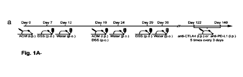
[0069] Figure 1A-1J: Immune cell and microbial
dynamics upon ICB therapy
in AOPNDSS tumors. (a) Overview of the experimental setup of AOWDSS-induced
CRC
and ICB treatment. (b) Tumor weight, (c) number of tumors, (d) Ep-CARELGR5+
cancer
stem cells and (e) tumor-infiltrating leukocytes (TILs) 140 days post
induction in animals
treated with isotype-, anti-PD-L1 or anti-CTLA-4 antibodies. (f) CD8+ T cell
frequencies in
the tumor draining lymph node at day 140. Splenic IFN-y+ production in (g)
CD4+ or (h)
CDS+ T cells. (i) 168 rRNA gene V4 region amplicon sequencing to identify
bacteria in
tumor tissue. Bacteria enriched or reduced in tumors of anti-PD-L1/anti-CTLA-4
compared to isotype treated animals are shown in green or red, respectively.
(j) Bacteria
cultured from homogenized tumors under anaerobe conditions from anti-PD-
L1/anti-
CTLA-4 (ICB groups) or isotype (lsotype group) treated animals. Bacteria
depicted as
green or red could only be cultured in ICB groups or lsotype group,
respectively. Bacteria
depicted as brown were present in both groups. Data are (b-h) mean SEM or
(i) mean
-11 -
CA 03156203 2022-4-26
WO 2021/081676
PCT/CA2020/051487
IfcSE (logfoldchangeStandard Error) and pooled from three individual
experiments. (b-f)
n =16-20 mice/group, (g and h) n = 4-5 mice/group. *, P< 0.05; **, Pc 0.01;
***, P
0.001; ****, P< 0.0001.
[0070] Figure 2A-2I: Individual bacterial
species boost ICB therapy. (a)
Schematic of the experimental setup to assess the effect of individual
bacteria on anti-
CTLA-4 therapy efficacy. (b) Tumor growth, (c) tumor weight, (d) live tumor
cells and (e)
representative pictures of tumors are shown at day 18. Scale bars: 1cm. (f)
representative
plots and (g) quantification of IFN-y* and Ki-674C084 T cells at day 18 in the
tumor tissue.
(h and i) same as (f and 9), but for CD4+ T cells. Data are mean SEM and
pooled from
three individual experiments (n = 8-15 mice/group). *, P< 0.05; *it, Pc 0.01;
***, P <
0.001; ****, Pc 0.0001.
[0071] Figure 3A4N: Effect of B.p., anti-CTLA-4
and B.p. conditioned serum
on T cell differentiation and activation. (a, h and k) Schematic of the
experimental
setups. (b) representative plots and (c) quantification of T-bet and T-
bet+IFN-yi events of
CD3c+CD4+ cells in the small intestine (SI) in the presence of indicated
bacteria at day 28.
(d and e) same as band c, but in the mesenteric lymph node (MLN). (f and g)
same as b
and c, but in the spleen. (i) representative plots and (j) quantification of T-
bet+ and T-
bet-FIFN-yi events of CD36+CD4+ T cells in the spleen in the presence of
indicated
bacteria and anti-CTLA-4 treatment at day 32. (I) Tumor growth and weight are
shown 32
days after MC38 tumor challenge and subsequent serum transfer as well as anti-
CTLA-4
treatment. (m) representative plots and (n) quantification of intratumoral IFN-
y or Ki-
67+C08+ T cells. Data are mean SEM and pooled from two individual
experiments (a-j)
n = 10-11 mice/group. (k-n) n = 5-7 mice/group. *, Pc 0.05; **, Pc 0.01; ***,
Pc 0.001;
****, Pc 0.0001.
[0072] Figure 4A-4K: Effect of inosine on T cell
differentiation and
dependency on classical dendritic cells of ICB therapy efficacy. (a) Scatter
plot of
untargeted metabolomics data in the serum of anti-CTLA-4 treated, tumor-
bearing B.p.
monocolonized compared to C.sp. monocolonized and germ-free (GF) mice. Grey
circles
or dotted grey circles depict inosine or inosine fragments/adducts,
respectively. Inset
shows an extracted ion chromatogram of inosine (b) Intensity of inosine (AUC:
area under
the curve) in sera shown in panel (a) of this figure. (c) Naïve CD4+ T cells
were co-
cultured with bone marrow derived dendritic cells and IFN-y. Quantification of
T-
bet+CD3c+CD4* T cells 48 hours after co-culture in the presence or absence of
inosine,
A2A receptor inhibitor (ZM241385), cell permeable cAMP (db-cAMP) and protein
kinase A
- 12 -
CA 03156203 2022-4-26
WO 2021/081676
PCT/CA2020/051487
inhibitor (RP-8-CPT-cAMPS). (d) Same as (c) without IFN-y. (e) Representative
plot and
quantification (left and right panel) of phospho-CREB (Seri 33) levels in
naive CD4+ T
cells cultured with anti-CD3/anti-0O28 coated beads for 1 hour in the presence
or
absence of inosine. (f) Schematic overview of experimental setup to deplete
classical
dendritic cells during MC38 tumor challenge and anti-CTLA-4 treatment.
Intratunnoral (g)
frequency of CD8+ T cells (h) IFN-y+CD8+ T cells. (i and j) same as (g and h),
but CD4+ T
cells and (k) tumor weight at day 95. Data are mean SEM and pooled from two
individual experiments (a-b) n = 5-8 samples/group (c-e) 10-15 biological
replicates/group
(f-k) 10 mice/group. *, Pc 0.05;", P< 0.01; ***, Pc 0.001;"", Pc 0.0001
[0073] Figure 5A-5L: Inosine promotes Thl
activation and anti-tumor
immunity. (a) Schematic overview of experimental setup to assess the effect of
inosine
on Th1 activation in vivo. (b, c) Representative dot plots and quantification
(left and right
panel respectively) of T-bet+IFN-y+CD3E+ (b) CD8+ or (c) CD4+ T cells in the
MLN. (d)
Schematic overview of experimental setup to assess the effect of inosine on
anti-tumor
immunity. Upon palpable tumors, mice were treated with 100pg anti-CTLA-4 i.p.
(5 times
every 72 hours) and in some groups 20pg CpG i.p. (5 times every 72 hours). In
addition,
inosine (300mg/KG/BW) or PBS was given daily orally (0), through gavage or
systemically (5) through i.p. injection. (e) Tumor weight and quantification
of IFN-y+ cells
amongst (f) CD4+ or (g) CD8+ cells are shown. (h). Schematic overview to
assess the
requirement of A2AR signaling for inosine-induced anti-tumor immunity. lx106
MC38 cells
(s.c.) and WT or A2AR-deficient 1x10⁷ T cells (i.v. 6x10⁶ CD4+ and 4x10⁶ CD8+
cells)
were injected. Upon palpable tumors, mice were treated with 100pg anti-CTLA-4,
20pg
CpG (4 times every 72 hours, both i.p.) and inosine (daily, 300mg/KG/BW,
through
gavage). (i) Pictures of tumors are shown at day 20. scale bars: 1 cm. (j)
Tumor weight
and quantification of IFN-y+ in (k) CD8+ or (I) CD4+ cells in the tumor are
shown. Data are
mean I SEM and pooled from two individual experiments (a-I) 10-11 mice/group.
*, P
0.05; **, Pc 0.01; ***, P< 0.001.
[0074] Figure 6A-6J: ICB therapy efficacy is CRC
subtype dependent. (a, c, e
and h) Schematic overview of the experimental setups to assess the effect of
ICB-
promoting bacteria in different subtypes of CRC. (b and d) Survival curve of
isotype, anti-
CTLA-4 or ICB-promoting (B.p. L.j. 0.sp.) or control (C.sp. P.sp.) and anti-
CTLA-4 co-
treated Apc2I'Dx 1 41+ ,-Krasisi--G12D1+;Fabpi-Cre animals. (f) Representative
plots and
quantification of intratumoral IFN-y+Ki-67+ CD8+ T cells and (g) tumor weight
of
Msh21-0xPA-0xPVillin-Cre mice. (i and j) same as (f and g), but for bacteria
anti-CTLA-4 co-
- 13 -
CA 03156203 2022-4-26
WO 2021/081676
PCT/CA2020/051487
treated and anti-1L-12p75 co-treated mice as indicated. Data are mean SEM
and pooled
from (b and f) five or (d and i) three individual experiments. (b) n = 8-9,
(d) n = 6-7, (f and
g) n = 10, (i and j) n = 7-9 mice/group. *, Pc 0.05; **, P< 0.01.
[0075] Figure 7A-7T: Bacteria are required for
ICB therapy efficacy. (a)
Schematic overview of the experimental setup to determine if bacteria modulate
ICB
therapy efficacy. (b) Tumor weight at day 28. Intratumoral (c) representative
plots and
quantification of (d) IFN-y+ and (e) Ki-67+ CD4+ T cells at day 28. (f-g) same
as (c-e), but
for CD8+ T cells. Splenic (i) representative plots and quantification of (j)
IFN-y* and (k) Ki-
67+ CD4+ T cells at day 28. (1-n) same as (i-k), but for CD8+ T cells. Data
are pooled from
two individual experiments (n = 5-10 mice/group). (o) SPF mice were injected
with 1x10⁶
MC38 s.c. and seven days later upon palpable tumors, mice were treated with
100pg
anti-CTLA-4 i.p. (5 times every 72 hours). Mice in the antibiotics (ABX) group
received a
mix of antibiotics (Ampicillin 1mg/ml, Colistin 1mg/m1 and Streptomycin
5mg/m1) orally
through the drinking water, staffing seven days prior to MC38 cell injection
until the end of
the experiment, whereas mice in the water group received regular water. Tumors
were
analyzed three days after the last anti-CTLA-4 injection. (p) Tumor weight,
quantification
of (q) IFN-y+ and (r) Ki-67+ in CD4+ T cells at day 25 in the tumor tissue. (s
and t) same as
(q and r) but CD8+ cells. (n = 9-10 mice/group). Data are mean SEM. *, Pc
0.05; **, P
< 0.01; ***, Pc 0.001; ****, P< 0.0001.
[0076] Figure 8A-8C: Microbiota dynamics in ICB
treated animals and
enrichment in fecal samples following ICB treatment. (a) Weighted UniFrac PCoA
analysis of 165 rRNA gene V4 region amplicon sequencing in tumors of anti-PD-
L1/anti-
CTLA-4 (ICB) compared to isotype treated animals. (b) same as (a) but for
fecal samples.
(c) 16S rRNA gene V4 region amplicon sequencing to identify bacteria in fecal
samples of
mice treated with ICB or control therapies. Bacteria enriched or reduced in
fecal samples
of anti-PD-L1/anti-CTLA-4 (ICB) compared to isotype treated animals are shown
in green
or red respectively. Data are mean IfcSE (logfoldchangeStandard Error) (n =
7-14
mice/group). Statistics: (a) and (b) PERMANOVA, (c) Benjamini-Hochberg-
[00771 Figure 9A-9F: B.p. enhances anti-PD-L1
therapy efficacy. (a) Germ-
free mice were monocolonized with B. p. or asp. Seven days later, 1x10⁶ MC38
cells
were injected s.c. and seven days later upon palpable tumors mice were treated
with
100F.ig anti-PD-L1 i.p. (5 times every 72 hours). Tumors were analyzed three
days after
the last anti-PD-L1 injection. (b) Tumor weight, quantification of (c) IFN-y+
and (d) Ki-67+
in CD4+ cells are shown at day 18 in the tumor tissue. (e and f) same as (c
and d) but
- 14 -
CA 03156203 2022-4-26
WO 2021/081676
PCT/CA2020/051487
CD8+ cells. Data are mean SEM (n = 7 mice/group). *, Pc 0_05; **, Pc 0.01;
***, P
0.001.
[0078] Figure 10A-10J: Bacteria alone to not
impact on tumor development.
(a) Overview of the experimental setup to determine whether Bp. alone have
anti-tumor
properties. (b) Tumor growth, (c) tumor weight and (d) representative pictures
of tumors
are shown, scale bars: 1cm. Intratumoral IFN-y+ (e) CD8+ or (f) CD4+ T cells
at the end of
the experiment. Splenic (g) IFN-y+ or (h) Ki-67+ CD8+ or (i) IFN-y+ or (j) Ki-
67+ CD4+ T
cells at the end of the experiment. Data are mean SEM. (a-j) n =7
mice/group.
[0079] Figure 11A-11B: Bacteria do not
translocate into tumor tissue. (a)
Representative pictures of SYTOX green nucleic acid stain of feces and tumor
tissue of
indicated colonized mice 18 days after initiation of anti-CTLA-4 therapy_ (b)
Agarose gel
of full length 16SrRNA amplicons of feces and tumor tissue of indicated
colonized mice.
[0080] Figure 12A-12R: Effect of B.p. on T cell
differentiation and activation.
GE animals were monocolonized with either B.p. C.sp. or left GE for 28 days
before
analysis. Small intestinal CD3e+CD4+ T cells expressing (a) RORyt and IL-17a,
(b)
RORyt, (c) Foxp3 or (d) naïve T cells (defined as RORyt-GATA3-Foxp3-T-bet-).
(e) Small
intestinal CD3e+CD8+ T cells expressing T-bet. (f-fl same as (a-e), but MLN.
(k-o) same
as (a-e), but spleen. (p-q) GE animals were colonized as indicated and after
14 days of
colonization treated with anti-CTLA-4 (5 times every 72 hours). Quantification
of, (p) T-
bet+IFN-y'CD3e+CD8 or (q) naive (defined as RORyt-GATA3-Foxp3-T-bet) CD4+
splenic
T cells. (r) Correlation of CD8+IFN-y+ and CD441FN-y+ T cells in tumors of
anti-CTLA-4
treated, differently colonized mice. Data are mean SEM and pooled from two
individual
experiments (a-q) n = 5-11 mice/group. (p) n = 46 mice. *, P< 0.05; **, P c
0.01.
[0081] Figure 13A-13D. Reduced barrier integrity
upon anti-CTLA-4
treatment. Serum from monocolonized mice treated with or without anti-CTLA-4
was
collected and binding against commensal bacteria was assessed. (a) Systemic
IgG2b
and IgG1 antibody response upon anti-CTLA-4 treatment in monocolonized mice.
(b)
Jejunum of B.p. or C.sp. monocolonized mice treated with or without anti-CTLA-
4 was
collected and barrier integrity was assessed through transepithelial
electrical resistance
measured in Ussing chambers. (c) Histological inflammation score of small
intestinal
intestine of B.p. or asp. monocolonized mice treated with or without anti-CTLA-
4. Blinded
scoring by a board-certified pathologist revealed no inflammation (Scale bar
=100pm). (d)
Levels of proinflammatory cytokines in the serum of lap. or asp. monocolonized
mice
with or without anti-CTLA-4 treatment (100pg i.p. five times every 72 hours)
were
- 15 -
CA 03156203 2022-4-26
WO 2021/081676
PCT/CA2020/051487
measured. Serum from DSS-treated SPF mice (2% DSS for 5 days) was used as a
positive control for systemic inflammatory cytokines. Data are mean SEM and
pooled
from two individual experiments. (a) n = 9-13 mice/group. (b) n = 6-11
mice/group (c) n =
4 mice/group (d) n = 5 mice/group (pos. ctrl. n = 2 mice). *, Pc 0.05; **, Pc
0.01; ***, Pc
0.0011 -***, Pc 0.0001
[0082] Figure 14A-14F: Systemic anti-tumor
immunity upon serum transfer
and anti-CTLA-4 treatment. GE animals were challenged with MC38 tumor cells.
Ten
days later mice received serum (i.v.) of anti-CTLA-4 treated tumor-bearing
animals. Mice
were then additionally treated with anti-CTLA-4 (3 times every 72 hours).
Serum donors
were colonized with B.p., C.sp. or remained GE, as indicated. Intratumoral (a)
IFN-ye or
(b) ki-67+CD4+ T cells. Splenic (c) IFN-r or (d) Ki-67+CD8+ T cells. (e and f)
same as (c
and d) but CD4* T cells. Data are mean SEM. (a-f) n = 5-8 mice /group. *, P<
0.05; 0,
PC 0.01; ***, Pc 0.001; ****, Pc 0.0001.
[0083] Figure 15A-15F: Inosine levels in vitro
and in vivo. (a) Scatter plot of
untargeted metabolomics data in the serum of anti-CTLA-4 treated, tumor-
bearing B.p.
monocolonized compared to C.sp. monocolonized mice. Grey circle identifies the
inosine
signal. (b) Scatter plot of untargeted metabolomics data in the serum of anti-
CTLA-4
treated, tumor-bearing B.ji monocolonized compared to GF mice. Grey circle
identifies
the inosine signal. (c) Intensity of inosine (AUC: area under the curve) in
culture
supernatant of indicated bacteria or BHI medium. (d) Parallel reaction
monitoring analysis
(FICD set at 50eV) of inosine comparing observed fragmentation patterns in BM
medium
spiked with and without 50uM inosine as well as B.p. cultured in BHI medium.
Extracted
ion chromatograms of each respective sample are shown in the right panel. (e)
Inosine
concentrations in duodenal, jejunal or cecal content of B.p. monocolonized
mice and in
the serum of B.p. or C.sp monocolonized and anti-CTLA-4 treated mice. (f)
Inosine
concentrations in the serum of untreated (SPF) tumor-bearing, anti-CTLA-4 i.p.
(SPF+
anti-CTLA-4) or anti-CTLA-4 plus antibiotic (SPF+ ABX + anti-CTLA-4) treated
SPF
colonized mice. Anti-CTLA4 treatment: (100pg 5 times every 72 hours).
Antibiotics:
Ampicillin lmg/ml, Colistin img/m1 and Streptomycin 5mg/m1 orally through the
drinking
water for 32 days. Data are mean SEM and pooled from two individual
experiments. (c)
n = 5 biological replicates /group. (e) n = 8-11 mice per group. (f) n = 6
samples/group. 0,
P< 0.01; =-**, Pc 0.001, ****, Pc 0.0001.
[0084] Figure 16A-16J: Context dependent effect
of inosine on 'ffil T cell
differentiation. (a) Naïve CD4+ T cells were co-cultured with bone marrow
derived
- 16 -
CA 03156203 2022-4-26
WO 2021/081676
PCT/CA2020/051487
dendritic cells without IFN-y. Quantification of T-bet*CD3e CD4 T cells 48
hours after co-
culture in the presence or absence of inosine and anti-CTLA-4, as indicated.
(b) Naïve
CDC T cells were cultured anti-CD3/anti-CD28 coated beads at a ratio of 1:1
without IFN-
y for 48 hours. Representative plot and quantification of 11_12R132 surface
expression on
CDC T cells in the presence or absence of inosine (left and right panel). (c)
Quantification
of T-bet+CD3e CD4+ T cells 48 hours after co-culture in the presence or
absence of
inosine, db-cAMP and anti-CTLA-4 as indicated. (d) NaIve, A2AR-deficient CDC T
cells
were cultured with anti-CD3/anti-CD28 coated beads at a ratio of 1:1 without
IFN-y.
Quantification of T-berCD3e+CD4+ T cells 48 hours after co-culture in the
presence or
absence of inosine or db-cAMP (e) Representative plot and quantification (left
and right
panel) of pCREB expression of anti-CD3/anti-0O28 bead co-cultured CDC T cells
in the
presence or absence of inosine or db-cAMP lhour after stimulation. Analysis
through flow
cytometry. (f and g) Naïve, wild type CDC T cells were cultured with anti-
CD3/anti-0O28
coated beads at a ratio of 1:1. 1112rb2 and Ifng gene transcripts (normalized
to Gapdh)
were evaluated 24 and 48 hours following inosine (1mM) stimulation_ Expression
was
normalized to cells treated with medium. Analysis through quantitative PCR
assay (h)
Naïve CDC T cells were cultured with anti-CD3/anti-CD28 coated beads at a
ratio of 1:1
without IFN-y for 24 hours. Then inosine or vehicle was added at the indicated
concentrations for another 48 hours before T cell differentiation and
activation was
assessed. (i) Adenosine concentrations in the duodenal-, jejunal- or cecal
content of B.p.
monocolonized mice and in the serum of B.p. or C.sp. monocolonized anti-CTLA-4
treated mice. 0) Naïve CDC T cells were cultured with anti-CD3/anti-CD28
coated beads
at a ratio of 1:1 without IFN-y for 24 hours. Adenosine or was added then in
the indicated
concentrations for another 48 hours followed by T cell differentiation and
activation was
assessed through flow cytometry. Data are mean SEM and show pooled data of 2
individual experiments (a and b) n = 10-16, (c) n = 5-10 biological
replicates/group, (d and
e) n = 6 biological replicates/group, (f and g) n=5 biological
replicates/group, (h) n = 8
biological replicates/group (i) n = 8 mice/group (j) n = 6 biological
replicates/group. *, P <
0.05; *4, Pc 0.01; ****, P< 0.0001.
[0085] Figure 17A-17B. lnosine does not directly
impact tumor cell viability
or condition tumor cells for T cell-mediated killing. (a) MC38 tumor cells
were treated
with the indicated doses of inosine in vitro for 72 hours. Cell death and
survival was
assessed through flow cytometry. (b) MC38 tumor cells expressing full length
ovalbumin
(MC38-OVA) were treated with the indicated doses of inosine in vitro for 72
hours. In
- 17 -
CA 03156203 2022-4-26
WO 2021/081676
PCT/CA2020/051487
parallel, OVA-specific naïve CD4 and CD8 T cells from spleens of OT-Il and OT-
1 mice,
respectively, were activated with anti-CD3/anti-0O28 beads and rmIL-2
(201U/m1) for 72
hours. lnosine was then washed away from conditioned MC38-OVA cells and fresh
medium together with activated T cells were added (100,000 MC38-OVA cells +
25,000
CD4 cells + 25,000 CD8 cells). 72 hours later, cell death and survival of MC38-
OVA cells
was assessed through flow cytometiy. Grey and black dash-dotted lines indicate
MC38-
OVA cell viability and death when co-cultured with naïve OVA-specific CD4 and
COB T
cells. Data are mean SEM and pooled from two individual experiments. n = 6
biological
replicates/condition. #, & Pc 0.05, **, P< 0.01; (** = 0.0001 vs 10mM, #0.001
vs 10mM
and & 1 vs 10mM)
[0086] Figure 18A-18H: Classical dendritic cells
are required for bacteria
dependent effect of ICB. (a) 1L-12p70 expression in classical dendritic cells
(M1-1C1I+CD11C1B220-CD64) and macrophages (MHCII+CD11c+B220-, CD64+) (b)
Quantification of IL-12p70 expression in macrophages and cDCs. Classical
dendritic cells
were depleted with diphtheria toxin (DT) in bone marrow chimeric mice after
MC38 tumor
challenge, followed by anti-CTLA-4 treatment (see Fig. 4h for experimental
setup).
Quantification of splenic (c) IFN-y-CD84- or (d) Ki-67+CD8+ T cells. (e and ft
same as (c
and d) but for CD4 + T cells. (g and h) 1x10⁶ MC38 cells (s.c.) were injected
in GF mice.
Seven days later upon palpable tumors, mice were treated with 100pg anti-CTLA-
4 i.p. (5
times every 72 hours) and in some groups 20pg CpG i.p. (5 times every 72
hours). In
addition, inosine (300mg/KG/BW) or PBS was given daily orally (0), through
gavage or
systemically (S) through i.p. injection. Quantification of Ki-6T cells is
shown. Data are
mean SEM and show pooled data of 2 individual experiments. (a - 0 n = 10
mice/group.
(g - h) n = 6-7 mice/group. *, Pc 0.05 ***, Pc 0.001; *"*, Pc 0.0001.
[0087] Figure 19A-19F: Bacteria-dependent
enhancement of ICB therapy
efficacy in Msh2"xim- xPVII/in-Cre mice. Msh21-0P4-0I'Vi1Iin-Cre mice were
treated with
ant-CTLA-4, ICB-promoting or control-bacteria and/or anti-1L-12p75 (see Fig.
5e and h for
a detailed experimental setup) (a) Representative plots and quantification of
intratumoral
IFN-y+Ki-674CD4+ T cells. (b) same as (a), but for bacteria anti-CTLA-4 co-
treated and
anti-IL-12p75 co-treated animals as indicated. (c) Representative plots and
quantification
of intratumoral CRC stem cells (defined as Ep-CAM+LGR5+). (d) same as (c), but
for
bacteria anti-CTLA-4 co-treated and anti-IL-12p75 co-treated animals as
indicated. (e)
Representative plots and quantification of tumor infiltrating leukocytes
(TILs). (f) same as
(e), but for bacteria anti-CTLA-4 co-treated and anti-1L-12p75 co-treated
animals as
- 18 -
CA 03156203 2022-4-26
WO 2021/081676
PCT/CA2020/051487
indicated. Data are mean SEM and pooled from (a, c and e) five or (b, d and
0 three
individual experiments. (a, c and e) n = 10 (b, d and 0 n = 7-9 mice/group. -.
P< 0.05;
P< 0.01; nit P< 0.001.
[0088] Figure 20A-20D: Oxaliplatin, anti-PD-L1
co-therapy is enhanced by
ICB-promoting bacteria. (a) Schematic overview of the experimental setup to
assess
the effect of ICB-promoting bacteria in Msh2thxwim
mice. 319 days post birth
antibiotics were given orally through the drinking water for seven days
(Ampicillin lmg/ml,
Colistin 1mg/m1 and Streptomycin 5mg/m1). Then Msh21-0aPithrP Villin-Cre mice
were
treated with Oxaliplatin, anti-PD-L1 and ICB promoting (B.p., 14. and 0.sp.)
or control
bacteria (C.sp. and P.sp.). Bacteria were given 5 times 72 hours apart through
gavage,
100 pg anti-PD-L1 was given 5 times 72 hours apart, i.p. Oxaliplatin
2.5mg/KG/BW was
given three times 7 days apart, LI:). (b) Tumor weight of Msh2LaYPVfflin-Cre
mice. (c)
Representative pictures of dissected tumors. (scale bar: 1 cm) (d)
Quantification of tumor-
infiltrating leukocytes (TILs). Data are mean SEM. n = 5-7 mice/group 4*, Pc
0.01.
[0089] Figure 21: Mechanism of bacteria-induced
ICB efficacy enhancement.
ICB-promoting bacteria increase inosine levels systemically, which is linked
to an ICB-
dependent reduction in gut barrier integrity. lnosine-mediated A2A receptor
engagement
leads to increased intracellular CAMP, protein kinase A activation and finally
phosphorylation of the transcription factor CREB. Together with TCR
stimulation, which is
further enabled through anti-CTLA-4 treatment, this leads to increased
expression of 1L12
receptor on T cells. Classical dendritic cells (cDC) sample antigens and are
the major
cellular source of IL-12. IL-12 produced by cDCs induces Thl differentiation,
through
induction of T-bet (Tbx21) expression and activation of T cells. cDCs are
required for
microbe-anti-CTLA-4 induced IFN-y (ffng) production by Th1 T cells, which are
protective
in cancer.
[0090] Figures 22-24: The lists of
Bifidobacterium sp. (Esp.), Lactobacillus
sp. (L.sp.) and Olsenella sp. (0.sp.). Tables show the sequence ID of
Bifidobacterium
sp. (asp.), Lactobacillus sp. (L.sp.) and Olseneffa sp. (0.sp.) with more than
84%-95%
identity to the sequences identified in examples (Figs. 22, 23, and 24,
respectively) based
on full length 165 sequence.
[0091] Figures 25-27. 168 rDNA sequence of the
administered strains,
Bifidobacterium pseudolongum strain deposited as IDAC Deposit No. 231020-011
Lactobacillus johnsonii strain deposited as IDAC Deposit No. 231020-02, and
Olseneffa
sp. strain deposited as IDAC Deposit No. 231020-03, respectively. SEQ ID NO: 1
has
- 19 -
CA 03156203 2022-4-26
WO 2021/081676
PCT/CA2020/051487
99% identity to the 165 rRNA sequence of Bifidobacterium pseudolongum subsp.
globosum strain RU 224. SEQ ID NO: 2 has 99% identity to the 16S rRNA sequence
of
Lactobacillus johnsonii strain CIP 103620. SEQ ID NO: 3 has 94% Olsenella
profuse
strain DSM 13989, 94% identity to Oisenella umbonata strain lac31, and 94%
identity to
Olsenella all strain DSM 7084.
[0092] Figure 28. Comparison of levels of
selected metabolites in transferred
serum samples in the serum of mice monocolonized with B.p. compared to C.sp.
or GE
mice. The purine metabolite inosine was significantly more abundant (8 to 9-
fold) in sera
from B.p. monocolonized mice compared to sera from C.sp. monocolonized or GE
mice.
Of note, xanthine and hypoxanthine, degradation products of inosine, were also
elevated
in the sera of B.p. monocolonized mice.
[0093] Figure 29A-29F. (A) Schematic of the
experimental setup, (B) Tumors and
tumor weight at the end of the experiment in MC38 tumor bearing and anti-CTLA-
4
treated (5 times, 72 hours apart) monocolonized mice. (C) lnosine
concentration
measured in the serum of mice shown in (B). (D) Hypoxanthine production of
indicated
bacteria in vitro in BHI media. (E) Tumors and tumor weight at the end of the
experiment
in MC38 tumor bearing and anti-PD-1 treated (5 times, 72 hours apart)
monocolonized
mice. (F) Tumors and tumor weight at the end of the experiment in MB49 tumor
bearing
and anti-CTLA-4 treated (4 times, 72 hours apart) monocolonized mice_ Data are
mean
SEM. n = 4-5 mice/group. (B) One-way ANOVA with Bonferroni post-test. (E and
F)
students t test. *, Pc 0.05; ", Pc 0.01; ***, Pc 0.001; ****, Pc 0.0001. The
strain
labeled B.pseudolongum+Ctrl in Figs. 30B-30D and as B. pseudolongum in Figs.
30E-
30F are the strain deposited as IDAC Deposit No. 231020-01.
[0094] Figure 30A4013. (A) Weighted UniFrac PCoA analysis of 168 rRNA gene V4
region amplicon sequencing in feces of anti-PD-Li anti-CTLA-4 (ICB) compared
to
isotype treated animals. (B) 168 rRNA gene V4 region amplicon sequencing to
identify
bacteria in fecal samples of mice treated with ICB or control therapies.
Bacteria enriched
or reduced in fecal samples of anti-PD-Li/anti-CTLA-4 (ICB) compared to
isotype treated
animals are shown in green or red, respectively. (C) same as (B) but for tumor
samples.
Bacteria enriched or reduced in tumors of (D) anti-CTLA-4 and (E) anti-PD-Ll
compared
to isotype treated animals are shown in green or red, respectively. Panels D
and E are
the same data as in Fig. II but separated by treatment. Data are (A) mean +1-
IfcSE
(logfoldchangeStandard Error). (A-C) a = 5-14 mice/group. *, Pc 0.05; **, Pc
0.01; ***, P
< 0.001; ****, Pc 0.0001.
- 20 -
CA 03156203 2022-4-26
WO 2021/081676
PCT/CA2020/051487
[0095] Figure 31A-31E. Bacteria alone do not impact on tumor development. (A)
Schematic of the experimental setup, (B) Tumor growth, (C) tumor weight, and
quantification of intratu moral IFN-y+ (D) CD8+ and (E) CD4+ T cells are shown
in germ-
free (GE) or monocolonized (B. pseudolongum. Colidextribacter species, L.
johnsonii, or
Olsen&Ha species) MC38 tumor-bearing mice. Data are mean SEM (B-E) n = 5
mice/group. *, P < 0.05; **, P < 0.01; ****, P < 0.0001.
[0096] Figure 32. lnosine levels in vitro. Fold induction compared to media of
inosine in
culture supernatant of indicated bacteria.
[0097] Figure 33A-33F. B cells and their responses are not required for B.
pseudolongum enhanced ICB therapy efficacy. (A) Germ-free (GE) wild type or
Igh-l-
mice were colonized with B. pseudolongum or left GE. Seven days later, lx106
MC38
cells were injected s.c. and seven days later upon palpable tumors, mice were
treated
with 100pg anti-CTLA-4 i.p. (5 times every 72 hours). Tumors were analysed
three days
after the last anti-CTLA-4 injection. (B) Tumor weight and quantification of
IFN-y+ in (C)
CD4+ and (D) CD8+ cells are shown at day 18 in the tumour tissue. (E and F)
same as
(C and D) but Ki-67+ cells. Data are mean +/- SEM. n = 4-7 mice/group. *, Pc
0.05; **, P
<0.01; Th Pc 0.001; Pc 0.0001.
[0098] Figure 34A-34H. Akkermansia muciniphila and Lactobacillus johnsonii
promote anti-CTLA-4 efficacy and is dependent on T cell expression of A2AR.
(A)
Schematic overview to assess the requirement of A2AR signaling for
Aldcerrnansia
muciniphila-induced anti-tumor immunity. Germ-free RAG-1-deficient mice were
gavaged
with Akkennansia muciniphila and seven days later 1x10⁶ MC38 cells (s.c.) and
WT or
A2AR-deficient 1x10⁷ T cells (i.v. 6x10⁶ CD4+ and 4x10⁶CD8+ cells) were
injected. Upon
palpable tumors, mice were treated with 100pg anti-CTLA-4 (4 times every 72
hours). (B)
Pictures of tumors (scale bars: 1 cm) and (C) tumor weight are shown at day
27. (D)
Quantification of IFN-y+ in CD8+ or CD4+ cells in the tumor are shown. (E-H)
same as
(A-D) but in Lactobacillus johnsonii monocolonized mice. Data are mean +/- SEM
and (A-
H) n = 7 mice/group. Pc 0.05; **, Pc 0.01; ***, pc 0.001.
[0099] Figure 35A-35H. Inosine and live B. pseudolongum improve anti-CTLA-4
therapy efficacy in moderately diverse and complex microbiomes. A) Schematic
overview of experimental setup to assess the effect of inosine on anti-tumor
immunity in
gnotobiotic mice (Oligo-MM12) stably colonized with 12 bacterial species. Upon
palpable
tumors, Oligo-MM12 colonized mice were treated with 100pg anti-CTLA-4 i.p. or
lsotype
antibody (5 times every 72 hours). In addition, inosine (300mg/KG/BW) or PBS
was given
- 21 -
CA 03156203 2022-4-26
WO 2021/081676
PCT/CA2020/051487
daily orally through gavage. (B) Pictures of tumors and (C) tumor weight are
shown at day
20. scale bars: 1 cm. (D) Quantification of intratumoral IFN-y+ cells amongst
CD8+ or
CD4+ T cells are shown. (E) Schematic overview of experimental setup to assess
the
effect of inosine and B. pseudolongum on anti-tumor immunity in SPF mice.
Following
MC38 injection some mice received antibiotics (ABX), specifically Ampicillin
1mg/rnl,
Colistin 1mg/m1 and Streptomycin 5mg/mlfor 7 days in the drinking water. Upon
palpable
tumors, antibiotics were removed and 100pg anti-CTLA-4 (5 times every 72
hours)
started. Mice concomitantly received either PBS, inosine (300mg/KG/BW daily),
B.
pseudolongum or heatinactivated (H.i.) B. pseudolongum orally through gavage
(5 times
every 72 hours). (F) Pictures of tumors and (G) tumor weight are shown at day
20. scale
bars: 1 cm. (H) Quantification of intratumoral IFN-y+ cells amongst 0D8+ or
CD4+ cells
are shown. Data are mean +/- SEM and (A-H) n = 7 mice/group. P < 0_05; **, Pc
0.01,
***, Pc 0.001.
[00100] Figure 36A-3613. Enrichment of
Bifidobacteria in tumors of Msh2LoxP/
LoxPVillin-Cre mice. SPF Msh2LoxP/LoxPVillin-Cre were treated with 100pg
isotype
antibody, anti-CTLA-4 or anti-PD-L1 (5 times every 72 hours) 10 months after
birth. Three
days following the last treatment tumor tissues were collected. (A) 16S and
(B)
Bifidobacteria DNA copy numbers were assessed (normalized to all 16S copy
numbers)
in the tumor tissue. Analysis through quantitative PCR assay. n = 7-11
tumors/group
(tumors collected from 4 individual mice in the isotype group and 8 individual
mice in the
ICB-therapy group). *, P < 0.05.
[00101] Figure 37A-376. Bifidobacteria abundance
in responders compared
to non responding cancer patients. (A) Abundance of B. pseudolongum in fecal
samples of non¨small cell lung cancer and renal cell carcinoma patients
receiving
checkpoint blockade therapy (n = 37 nonresponders and 44 responders) (8). B.
pseudolongum abundance was normalized to nonresponders. (B) Abundance of
Bifidobacteria in fecal samples of melanoma patients receiving checkpoint
blockade
therapy (n = 24 nonresponders and 13 responders) (9). Bifidobacteria abundance
was
normalized to nonresponders.
[00102] Figure 38. Inosine levels in vivo.
lnosine concentrations in duodenal,
jejunal or cecal content of B. pseudolongum monocolonized mice and in the
serum of B.
pseudolongum or Colidextribacter sp. monocolonized mice treated with anti-CTLA-
4 or
anti-PD-L1, as indicated. N = 8-11 mice per group. ***, Pc 0.001, ****, P<
0.0001.
- 22 -
CA 03156203 2022-4-26
WO 2021/081676
PCT/CA2020/051487
[00103] Figure 39. Reduced barrier integrity upon
anti-CTLA-4 treatment.
Serum from monocolonized mice treated with or without anti-CTLA-4 was
collected and
binding against commensal bacteria was assessed. N = 6-11 mice/group. *, P c
0.05; **,
P <0.01; Pc 0.001, ****, Pc 0.0001.
DETAILED DESCRIPTION
[00104] Generally, the present disclosure
provides a compound(s) and/or a
compositions for use in treating a subject having cancer, or suspected of
having cancer.
[00105] In some examples, the cancer may be
colorectal cancer (CRC), lung
cancer, melanoma, bladder cancer, or kidney cancer. In other examples, the
cancer
may be breast cancer, prostate cancer, stomach cancer, liver cancer,
esophageal cancer,
pancreatic cancer, brain cancer, cervical cancer, ovarian cancer, thyroid
cancer, lip
cancer, oral cancer, larynx cancer, nasopharynx cancer, uterine cancer, or
other cancer
as disclosed herein.
[00106] In a specific aspect, the present
disclosure provides a compound(s) and/or
a compositions for use in treating a subject having Colorectal cancer (CRC),
or suspected
of having CRC.
[00107] In one aspect, there is described a
method of treating a subject having a
cancer, or suspect of having a cancer, comprising or consisting of:
administering an ICB
inhibitor and one or more bacteria selected from Bifidobacterium pseudolongum,
Lactobacillus Johnson!!, or Olsenella species.
[00108] In one aspect, there is described a
method of treating a subject having a
cancer, or suspect of having a cancer, comprising or consisting of:
administering an ICB
inhibitor and one or more bacteria selected from Bifidobacterium pseudolongum,
Lactobacillus johnsonii, Olsenella profuse, Olsenella umbonata, or Olsenelia
uli
[00109] In a specific example the cancer may be
colorectal cancer (CRC), lung
cancer, melanoma, bladder cancer, or kidney cancer. In other examples, the
cancer may
be breast cancer, prostate cancer, stomach cancer, liver cancer, esophageal
cancer,
pancreatic cancer, brain cancer, cervical cancer, ovarian cancer, thyroid
cancer, lip
cancer, oral cancer, larynx cancer, nasopharynx cancer, uterine cancer.
[00110] In one aspect, there is described a
method of treating a subject having
CRC, or suspected of having CRC, comprising or consisting of: administering an
ICB
inhibitor and one or more bacteria selected from Bifidobacterium pseudolongum,
Lactobacillus johnsonii, Olsenella profuse, Olsenella umbonata, or Olsenella
all.
- 23 -
CA 03156203 2022-4-26
WO 2021/081676
PCT/CA2020/051487
[00111] In one aspect, there is described a
method of treating a subject having
CRC, or suspected of having CRC, comprising or consisting of: administering an
ICB
inhibitor and one or more bacterium selected from Bffidobacterium pseudolongum
(B.p),
Lactobacillus johnsonii (L.D, or Ofsenella sp. (0.sp.).
[00112] In one aspect, there is described a
method of treating a subject having
CRC, or suspected of having CRC, comprising or consisting of: administering an
ICB
inhibitor and one or more bacterium selected from Bffidobacterium sp_ (ifsp.)
listed in
Figure 22, Lactobacillus sp. (L. sp.) listed in Figure 23, or Olsen ella sp.
(0.sp.) listed in
Figure 24.
[00113] In one aspect, there is described a
method of treating a subject having a
cancer, or suspected of having a cancer, comprising or consisting of:
administering an
ICB inhibitor and inosine, a derivative of inosine, functional derivative of
inosine, or a
physiologically functional derivative of inosine.
[00114] In a specific example the cancer may be
colorectal cancer (CRC), lung
cancer, melanoma, bladder cancer, or kidney cancer. In other examples, the
cancer may
be breast cancer, prostate cancer, stomach cancer, liver cancer, esophageal
cancer,
pancreatic cancer, brain cancer, cervical cancer, ovarian cancer, thyroid
cancer, lip
cancer, oral cancer, larynx cancer, nasopharynx cancer, uterine cancer.
[00115] In one aspect, there is described a
method of treating a subject having
CRC, or suspected of having CRC, comprising or consisting of: administering an
ICB
inhibitor and inosine, a derivative of inosine, functional derivative of
inosine, or a
physiologically functional derivative of inosine.
[00116] As used herein, the terms "immune
checkpoint," "checkpoint pathway,"
and "immune checkpoint pathway" refer to a pathway by which the binding of an
immune
checkpoint ligand to an immune checkpoint receptor modulates the amplitude and
quality
of the activation of immune cells.
[00117] Immune checkpoint proteins include, but
are not limited to, cytotoxic T
lymphocyte-associated antigen 4 (CTLA-4), also known as CD152, programmed cell
death protein 1 (PD-1), also known as CD279, PD-1 ligands (PD-L1 or CD274, PD-
L2 or
CO274), lymphocyte-activation gene 3 (LAG-3), also known as CD223, B7-H3
(CO276),
V-domain 19 suppressor of T cell activation (VISTA), therapies targeting
indoleamine 2'3'
dioxygenase (IDO, ID01 and ID02), TIGIT (also called T cell immunoreceptor
with Ig and
ITIM domains), B and T Lymphocyte Attenuator (BTLA), Herpes virus entry
mediator
(HVEM), CD226 (DNAM-1) and CD96 (Tactile), T cell immunoglobulin mucin (TIM-
3),
- 24 -
CA 03156203 2022-4-26
WO 2021/081676
PCT/CA2020/051487
also known as HAVcr2, LAIR1 (Leukocyte Associated lmmunoglobulin Like Receptor
1;
C0305), CD160 (6Y55), CD244 (264), VTCN1 (67-H4), KIR, A2AR, or 67-H3.
[00118] The term "immune checkpoint blockade" or
"ICB," as used herein, refers to
the administration of one or more inhibitors of one or more immune checkpoint
proteins or
their ligand(s). Thus, the term "immune checkpoint blockade" refers to the
inhibition of an
immune checkpoint pathway by the administration or expression of a "blockade
agent" or
"inhibitor. Typically, the "blockade agent" prevents the interaction of the
immune
checkpoint receptor and ligand, thereby inhibiting the checkpoint pathway. A
blockade
agent may be a small molecule, peptide, antibody or fragment thereof, etc.
that binds to
an immune checkpoint ligand or immune checkpoint receptor and inhibits the
formation of
the ICPJICL complex. A blockade agent may also function by preventing
signaling by the
ICPJICL complex. Exemplary ICB agents include antibodies, fusion proteins, and
small
molecules, such as ipilimumab (YERVOY , anti-CDLA-4 antibody, Bristol-Myers
Squibb), nivolumab (OPDIVO 0, anti-PD-1 antibody, Bristol-Myers Squibb),
pembrolizumab (KEYTRUDA , anti-PD-1 antibody, Merck), atezolizumab
(TECENTRIQ , anti-PD-L1 antibody, Roche), avelumab (BAVENCI00, anti-PD-L1
antibody, Merck KGaA/Pirzer), durvalumab (IMFINZIO, anti-PD-L1 antibody,
Medimmune/AstraZeneca), cemiplimab (LIBTAY00, anti-PD-1 antibody,
Regeneron/Sanofi), lambrolizumab (anti-PD-1 antibody, Merck), pidilizumab
(anti-PD-1
and anti-DLL antibody, Medivation), BMS-936559 (anti-PD-L1, Bristol-Myers
Squibb),
MEDI-0680 (anti-PD-1 antibody; AMP-514; AstraZeneca), REGN2610 (anti-PD-1
antibody, Regeneron), CA-170 (small molecule PD-1 and PD-L1 inhibitor; Curis),
BMS-
1166 (small molecule PD-L1 inhibitor, Bristol-Myers Squibb), AMP-224 (anti-PD-
1 fusion
protein, Medimmune), and spartalizumab (anti-PD-1 antibody, Novartis).
[00119] As used herein, the term "immune
checkpoint inhibitor refers to molecules
that totally or partially reduce, inhibit, interfere with or modulate one or
more checkpoint
proteins. Checkpoint proteins regulate T-cell activation or function_ These
proteins are
responsible for co stimulatory or inhibitory interactions of T-cell responses.
Immune
checkpoint proteins regulate and maintain self-tolerance and the duration and
amplitude
of physiological immune responses. In some embodiments, the subject can be
administered an additional agent that can enhance or boost the immune
response, e.g.,
immune response effected by the binding molecules (e.g., BCMA-binding
molecules),
recombinant receptors, cells and/or compositions provided herein, against a
disease or
condition, e.g., a cancer, such as any described herein.
- 25 -
CA 03156203 2022-4-26
WO 2021/081676
PCT/CA2020/051487
[00120] Immune checkpoint inhibitors include any
agent that blocks or inhibits in a
statistically significant manner, the inhibitory pathways of the immune
system. Such
inhibitors may include small molecule inhibitors or may include antibodies, or
antigen
binding fragments thereof, that bind to and block or inhibit immune checkpoint
receptors,
ligands and/or receptor- ligand interaction. In some embodiments, modulation,
enhancement and/or stimulation of particular receptors can overcome immune
checkpoint
pathway components.
[00121] The terms "inhibit," "block," and
"suppress" are used interchangeably and
refer to any statistically significant decrease in biological activity,
including full blocking of
the activity.
[00122] An "inhibitor" is an active agent that
inhibits, blocks, or suppresses
biological activity in vitro or in vivo_
[00123] Inhibitors include but are not limited to
small molecule compounds; nucleic
acids, such as siRNA and shRNA; polypeptides, such as antibodies or antigen-
binding
fragments thereof, dominant-negative polypeptides, inhibitory peptides, and
fusion
proteins; and oligonucleotide or peptide aptamers.
[00124] In a specific example, the ICB inhibitor
is an anti-CTLA4 antibody, or an
anti-PD-Ll antibody, or an anti-PD-1 antibody.
[00125] Non-limiting examples of co-stimulants
include: Toll like receptor (TLR)
signals, for example CpG, LPS, Flagellin; Nucleotide-binding oligomerization
domain-like
receptors (NLRs), for example, meso-diaminopimelic acid, muramyl dipeptide,
ATP,
extracellular glucose, crystals of monosodium urate, calcium pyrophosphate
dihydrate,
alum, cholesterol or environmental irritants; silica; asbestos; UV irradiation
and skin
irritants; RIG-I-like receptors (retinoic acid-inducible gene-1-like
receptors), for example,
single- or double-stranded RNA (e.g., from viruses); C-type lectin receptors
(CLR), for
example, repeated mannose units. C-type lectin domain; Cytokine receptor
signalling, for
example, IL-12, IL-18, IL-33, IFN-g; Stimulation provided through antigen
presenting cells
or their counterpart on T-cells, for example, CD8O-CD28, CD86-CD28, CD40CD4OL,
OX-
40L-0X40; -cGAS-STING pathway; for example, cytosolic DNA.
[00126] A "standard dose" of ICB therapy is known
by a person of skill in the art for
each medication, and may be the dose that is indicated in the prescribing
information
and/or the dose that is most frequently administered under particular clinical
circumstances (for example for the particular PD-1 inhibitor and/or CTLA-4
inhibitor being
- 26 -
CA 03156203 2022-4-26
WO 2021/081676
PCT/CA2020/051487
used, the particular route of administration being used, the particular stage
of the CRC
being treated, the age, weight, and/or sex of the particular patient, etc.).
[00127] The term "subject", as used herein, refers
to an animal, and can include,
for example, domesticated animals, such as cats, dogs, etc., livestock (e.g.,
cattle,
horses, pigs, sheep, goats, etc.), laboratory animals (e.g., mouse, rabbit,
rat, guinea pig,
etc.), mammals, non-human mammals, primates, non-human primates, rodents,
birds,
reptiles, amphibians, fish, and any other animal. Additional examples of
domesticated
animals include a ferret, horse, rabbit, guinea pig, gerbil, hamster,
chinchilla, rat, mouse,
or other small mammal; a bird; a reptile; a fish; an amphibian; an arthropod
such as a
tarantula or hermit crab. Additional livestock animals include donkey, alpaca,
camel,
water buffalo, mink, or chicken.
[00128] In a specific example, the subject is a
human.
[00129] The terms "colorectal cancer" or "CRC",
used interchangeably herein, are
used in the broadest sense and refer to (1) all stages and all forms of cancer
arising from
epithelial cells of the intestinal tract below the small intestine (i.e., the
large intestine
(colon), including the cecum, ascending colon, transverse colon, descending
colon, and
sigmoid colon, and rectum), and/or (2) all stages and all forms of cancer
affecting the
lining of the large intestine and/or rectum.
[00130] In some examples. CRC is mismatch repair
deficient (MMRD) CRC or
inflammation-associated CRC.
[00131] Typically, in the staging systems used for
classification of colorectal
cancer, the colon and rectum are treated as one organ.
[00132] Additionally, as used herein, the term
"colorectal cancer" also includes
medical conditions which are characterized by cancer of cells of the duodenum
and small
intestine (jejunum and ileum).
[00133] The staging of CRC is known.
[00134] In some examples, CRC may be staged
according to the Dukes system,
the Astler-Coller system or the TNM system (tumors/nodes/metastases), whereby
the
latter is most commonly used. The TNM system of the American Joint Committee
of
Cancer (AJCC) describes the size of the primary tumor (T), the degree of lymph
node
involvement (N) and whether the cancer has already formed distant metastasis
(M), i.e.,
spread to other parts of the body. Here, stages 0, IA, IB, IIA, IIB, Ill and
IV are defined
based on the determined T-, N- and M-values. A corresponding staging scheme
can be
derived from the Cancer Staging Manual of the AJCC. Another system for staging
of
- 27 -
CA 03156203 2022-4-26
WO 2021/081676
PCT/CA2020/051487
colorectal cancer is the Dukes system, defining cancer stages A, B, C and D.
This system
was adapted by Astler and Caller, who further subdivided stages B and C
("modified
Astler-Coller classification").
[00135] As used herein, a CRC patient includes
patients staged according to any
staging system used and irrespective of the stage diagnosed.
[00136] As use herein "a patient suffering from
colorectal cancer" or "a subject
suffering from colorectal cancer" refers to any mammalian, in particular
human, patient
having developed atypical and/or malignant cells in the lining and/or the
epithelium of the
large intestine and/or rectum. This includes CRC patients independent of the
stage and
form of the CRC.
[00137] Patients suffering from colorectal cancer
also include patients which are
recurrent with colorectal cancer, i.e., patients wherein after surgical
treatment the tumor
could no longer be detected for a certain time span, but wherein the cancer
has returned
in the same or different part of the large intestine, and/or rectum and/or
wherein
metastases have developed at different sites of the patient's body such as in
the liver,
lung, peritoneum, lymph nodes, brain and/or bones.
[00138] In another example, the patient suffering
from CRC is a patient wherein
the initial tumor has already been treated surgically and the CRC is non-
metastatic.
[00139] In some examples, a derivative of inosine,
functional derivative of inosine,
a prodrug of inosine, or a physiologically functional derivative of inosine,
may be used.
[00140] The term "derivative", "functional
derivative" and "physiologically functional
derivative" as used herein means an active compound with equivalent or near
equivalent
physiological functionality to the named active compound when used and/or
administered
as described herein. As used herein, the term "physiologically functional
derivative"
includes any pharmaceutically acceptable salts, solvates, esters, prodrugs
derivatives,
enantiomers, or polymorphs.
[00141] The term "prodrug" used herein refers to
compounds which are not
pharmaceutically active themselves but which are transformed into their
pharmaceutical
active form in vivo, for example in the subject to which the compound is
administered.
[00142] The term "therapeutically effective
amount" or "effective amount", as used
herein, refers to an amount effective, at dosages and for periods of time
necessary to
achieve the desired result. Effective amounts may vary according to factors
such as the
disease state, age, sex and/or weight of the subject. The amount of a given
compound or
composition that will correspond to such an amount will vary depending upon
various
- 28 -
CA 03156203 2022-4-26
WO 2021/081676
PCT/CA2020/051487
factors, such as the given drug or compound, the pharmaceutical formulation,
the route of
administration, the identity of the subject being treated, and the like, but
can nevertheless
be routinely determined by one skilled in the art.
[00143] The term "treatment" or "treat" as used
herein, refers to obtaining
beneficial or desired results, including clinical results. Beneficial or
desired clinical results
can include, but are not limited to, alleviation or amelioration of one or
more symptoms or
conditions, diminishment of extent of disease, stabilized (i.e. not worsening)
state of
disease, preventing spread of disease, delay or slowing of disease
progression,
amelioration or palliation of the disease state, diminishment of the
reoccurrence of
disease, and remission (whether partial or total), whether detectable or
undetectable.
"Treating" and "Treatment" can also mean prolonging survival as compared to
expected
survival if not receiving treatment. 'Treating" and "treatment" as used herein
also include
prophylactic treatment. For example, a subject in the early stage of disease
can be
treated to prevent progression or alternatively a subject in remission can be
treated with a
compound or composition described herein to prevent progression.
[00144] "Prevent" or "prevention" refers to
prophylactic or preventative measures
that prevent and/or slow the development of a targeted pathologic condition or
disorder.
Thus, those in need of prevention include those at risk of or susceptible to
developing the
disorder. In certain embodiments, a disease or disorder is successfully
prevented
according to the methods provided herein if the patient develops, transiently
or
permanently, e.g., fewer or less severe symptoms associated with the disease
or
disorder, or a later onset of symptoms associated with the disease or
disorder, than a
patient who has not been subject to the methods of the invention_
[00145] In some examples, treatment results in
prevention or delay of onset or
amelioration of symptoms of a disease in a subject or an attainment of a
desired
biological outcome.
[00146] In some examples, treatment methods
comprise administering to a subject
a therapeutically effective amount of a compound or composition described
herein and
optionally consists of a single administration or application, or
alternatively comprises a
series of administrations or applications.
[00147] The term "pharmaceutically effective
amount" as used herein refers to the
amount of a compound, composition, drug or pharmaceutical agent that will
elicit the
biological or medical response of a tissue, system, animal or human that is
being sought
- 29 -
CA 03156203 2022-4-26
WO 2021/081676
PCT/CA2020/051487
by a researcher or clinician, for example, the treatment of colorectal cancer.
This amount
can be a therapeutically effective amount.
[00148] The compounds and compositions may be
provided in a pharmaceutically
acceptable form.
[00149] The term "pharmaceutically acceptable" as
used herein includes
compounds, materials, compositions, and/or dosage forms (such as unit dosages)
which
are suitable for use in contact with the tissues of a subject without
excessive toxicity,
irritation, allergic response, or other problem or complication, commensurate
with a
reasonable benefit/risk ratio. Each carrier, excipient, etc. is also
"acceptable" in the sense
of being compatible with the other ingredients of the formulation.
[00150] The term "functional derivative" and
"physiologically functional derivative"
as used herein means an active compound with equivalent or near equivalent
physiological functionality to the named active compound when used and/or
administered
as described herein. As used herein, the term "physiologically functional
derivative"
includes any pharmaceutically acceptable salts, solvates, esters, prodrugs
derivatives,
enantiomers, or polymorphs.
[00151] In some examples the compounds are
prodrugs.
[00152] The formulation(s) may conveniently be
presented in unit dosage form and
may be prepared by any methods well known in the art of pharmacy. Such methods
include the step of bringing the active compound into association with a
carrier, which
may constitute one or more accessory ingredients. In general, the formulations
are
prepared by uniformly and intimately bringing into association the active
compound with
liquid carriers or finely divided solid carriers or both, and then if
necessary shaping the
product.
[00153] The compounds and compositions may be
administered to a subject by
any convenient route of administration, whether systemically/peripherally or
at the site of
desired action, including but not limited to, oral (e.g. by ingestion);
topical (including e.g.
transdermal, intranasal, ocular, buccal, and sublingual); pulmonary (e.g. by
inhalation or
insufflation therapy using, e.g. an aerosol, e.g. through mouth or nose);
rectal; vaginal;
parenteral, for example, by injection, including subcutaneous, intratunnoral,
intradermal,
intramuscular, intravenous, intraarterial, intracardiac, intrathecal,
intraspinal,
intracapsular, subcapsular, intraorbital, intraperitoneal, intratracheal,
subcuticular,
intraarticular, subarachnoid, and intrastemal; by implant of a depot / for
example,
- 30 -
CA 03156203 2022-4-26
WO 2021/081676
PCT/CA2020/051487
subcutaneously or intramuscularly. Preferably compositions comprising bacteria
are
delivered to the gastrointestinal system, eig., by oral (such as ingestion) or
rectal route.
[00154] Formulations suitable for oral
administration (e.g., by ingestion) may be
presented as discrete units such as capsules, cachets or tablets, each
containing a
predetermined amount of the active compound; as a powder or granules; as a
solution or
suspension in an aqueous or non-aqueous liquid; or as an oil-in- water liquid
emulsion or
a water- in-oil liquid emulsion; as a bolus; as an electuary; or as a paste_
[00155] Formulations suitable for parenteral
administration (e.g., by injection,
including cutaneous, subcutaneous, intramuscular, intravenous and
intradermal), include
aqueous and non-aqueous isotonic, pyrogen-free, sterile injection solutions
which may
contain anti-oxidants, buffers, preservatives, stabilisers, bacteriostats, and
solutes which
render the formulation isotonic with the blood of the intended recipient; and
aqueous and
non- aqueous sterile suspensions which may include suspending agents and
thickening
agents, and liposomes or other microparticulate systems which are designed to
target the
compound to blood components or one or more organs. Examples of suitable
isotonic
vehicles for use in such formulations include Sodium Chloride Injection,
Ringers Solution,
or Lactated Ringers Injection.
[00156] The formulations may be presented in unit-
dose or multi-dose sealed
containers, for example, ampoules and vials, and may be stored in a freeze-
dried
(lyophilized) condition requiring only the addition of the sterile liquid
carrier, for example
water for injections, immediately prior to use. Extemporaneous injection
solutions and
suspensions may be prepared from sterile powders, granules, and tablets.
Formulations
may be in the form of liposomes or other microparticulate systems which are
designed to
target the active compound to blood components or one or more organs.
[00157] The compounds and/or compositions
described herein may be
administered either simultaneously (or substantially simultaneously) or
sequentially,
dependent upon the condition to be treated, and may be administered in
combination with
other treatment(s). The other treatment(s) may be administered either
simultaneously (or
substantially simultaneously) or sequentially.
[00158] As used herein the term 'reduces at least
one symptom of CRC' refers to a
qualitative or quantitative reduction in detectable symptoms, including but
not limited to a
detectable impact on the rate of recovery from disease or the rate of disease
progression
or severity.
- 31 -
CA 03156203 2022-4-26
WO 2021/081676
PCT/CA2020/051487
[00159] As used herein, the term 'at risk of
developing CRC" in reference to a
subject is understood as referring to a subject predisposed to the development
of CRC by
virtue of the subject's medical status.
[00160] In some example, a subject having CRC' is
a subject having been
"diagnosed with CRC'".
[00161] The term 'diagnosed with CRC" refers to a
subject demonstrating one or
more symptoms of CRC'. Methods of diagnosing CRC', are known in the art.
[00162] The pharmaceutical compositions can be
administered in a variety of unit
dosage forms depending upon the method of administration. For example, unit
dosage
forms suitable for oral administration include powder, tablets, pills,
capsules and
lozenges.
[00163] The pharmaceutical compositions described
herein may be useful for
parenteral administration, such as intravenous administration, intraperitoneal
administration, or administration into a body cavity or lumen of an organ or
joint.
[00164] The term "pharmaceutical composition"
refers to a preparation that is in
such form as to permit the biological activity of the active ingredient to be
effective, and
which contains no additional components that are unacceptably toxic to a
subject to
which the composition would be administered. Pharmaceutical compositions can
be
administered in any of numerous dosage forms, for example, tablet, capsule,
liquid,
solution, softgel, suspension, emulsion, syrup, elixir, tincture, film,
powder, hydrogel,
ointment, paste, cream, lotion, gel, mousse, foam, lacquer, spray, aerosol,
inhaler,
nebulizer, ophthalmic drops, patch, suppository, and/or enema. Pharmaceutical
compositions typically comprise a pharmaceutically acceptable carrier, and can
comprise
one or more of a buffer (e.g. acetate, phosphate or citrate buffer), a
surfactant (e.g.
polysorbate), a stabilizing agent (e.g. human albumin), a preservative (e.g.
benzyl
alcohol), a penetration enhancer, an absorption promoter to enhance
bioavailability
and/or other conventional solubilizing or dispersing agents. Choice of dosage
form and
excipients depends upon the active agent to be delivered and the disease or
disorder to
be treated or prevented, and is routine to one of ordinary skill in the art.
[00165] In some examples, the compositions for
administration will commonly
comprise a solution of the binding agent of the present disclosure dissolved
in a
pharmaceutically acceptable carrier, for example an aqueous carrier. A variety
of
aqueous carriers can be used, e.g., buffered saline and the like. The
compositions may
contain pharmaceutically acceptable auxiliary substances as required to
approximate
- 32 -
CA 03156203 2022-4-26
WO 2021/081676
PCT/CA2020/051487
physiological conditions such as pH adjusting and buffering agents, toxicity
adjusting
agents and the like, for example, sodium acetate, sodium chloride, potassium
chloride,
calcium chloride, sodium lactate and the like. The concentration of binding
agents of the
present disclosure in these formulations can vary widely, and will be selected
primarily
based on fluid volumes, viscosities, body weight and the like in accordance
with the
particular mode of administration selected and the patient's needs. Exemplary
carriers
include water, saline, Ringers solution, dextrose solution, and 5% human serum
albumin.
[00166] Optionally the treatment is combined with
another moiety useful for
treating CRC.
[00167] According to at least some embodiments of
the present invention, there is
provided use of a combination of the therapeutic agents and/or a
pharmaceutical
composition comprising same, as recited herein, that can be combined with
standard of
care or novel treatments for CRC.
[00168] For example, treatment methods for a
patient suffering from colorectal
cancer, in particular after removal of the primary tumor, may include
chemotherapy,
radiotherapy, targeted therapy, and immunotherapy.
[00169] As used herein, the term "chemotherapy"
relates to treatment of a subject
with an antineoplastic drug.
[00170] The terms "radiation therapy" and
"radiotherapy" relate to the use of
ionizing radiation to treat or control a cancer such as CRC.
[00171] The term "targeted therapy", as used
herein, relates to application to a
patient a chemical substance known to block growth of cancer cells by
interfering with
specific molecules known to be necessary for tumorigenesis or cancer or cancer
cell
growth
[00172] The term "immunotherapy" as used herein
relates to the treatment of
cancer by modulation of the immune response of a subject. Said modulation may
be
inducing, enhancing, or suppressing said immune response, e.g. by
administration of at
least one cytokine, and/or of at least one antibody specifically recognizing
cancer cells.
The term "cell-based immunotherapy" relates to a cancer therapy comprising
application
of immune cells, e.g. T- cells, preferably tumor-specific NK cells, to a
subject.
[00173] Whether a patient or a tumor is
"responsive," as used herein with respect
to a clinical response to treatment, can be assessed using any endpoint
indicating a
benefit to the patient, including, without limitation, (1) inhibition, to some
extent, of tumor
growth, including slowing down and complete growth arrest; (2) reduction in
the number
- 33 -
CA 03156203 2022-4-26
WO 2021/081676
PCT/CA2020/051487
of tumor cells; (3) reduction or shrinkage in tumor size; (4) inhibition
(i.e., reduction,
slowing down or complete stopping) of tumor cell infiltration into adjacent
peripheral
organs and/or tissues; (5) inhibition of metastasis; (6) enhancement of anti-
tumor immune
response, possibly resulting in regression or rejection of the tumor; (7)
relief, to some
extent, of one or more symptoms associated with the tumor; (8) increase in the
length of
survival following treatment; and/or (9) decreased modality at a given point
of time
following treatment. Responsiveness may also be expressed in terms of various
measures of clinical outcome. Positive clinical outcome can also be considered
in the
context of an individual's outcome relative to an outcome of a population of
patients
having a comparable clinical diagnosis. In one example, an increase in the
likelihood of
positive clinical response corresponds to a decrease in the likelihood of
cancer
recurrence.
[00174] In another example, clinical response to
treatment can be measured based
on disease control (DC), wherein tumors displaying disease control include
tumors whose
response to treatment is a complete response (CR), partial response (PR) or
stable
disease (SD). In one example, tumors displaying disease control do not include
tumors in
a progressive disease (PD) state.
[00175] In another example, clinical response to
treatment can be measured based
on an objective tumor response, e.g., tumor shrinkage, wherein tumors
undergoing an
objective tumor response include tumors undergoing either a complete response
(CR) or
a partial response (PR). In one embodiment, tumors undergoing an objective
tumor
response do not include tumors that display stable disease (SD) or tumors in a
progressive disease (PD) state.
[00176] Unless specifically stated or obvious
from context, as used herein, the
term "about" is understood as within a range of normal tolerance in the art,
for example
within 2 standard deviations of the mean. About can be understood as within
10%, 9%,
8%, 7%, 6%, 5%, 4%, 3%, 2%, 1%, 0_5%, 0.1%, 0.05%, or 0.01% of the stated
value.
Unless otherwise clear from context, all numerical values provided herein can
be modified
by the term about.
(00171 The recitation of a listing of chemical
group(s) in any definition of a variable
herein includes definitions of that variable as any single group or
combination of listed
groups. The recitation of an embodiment for a variable or aspect herein
includes that
embodiment as any single embodiment or in combination with any other
embodiments or
portions thereof.
- 34 -
CA 03156203 2022-4-26
WO 2021/081676
PCT/CA2020/051487
[00178] Any compositions or methods provided
herein can be combined with one
or more of any of the other compositions and methods provided herein.
[00179] Ranges provided herein are understood to
be shorthand for all of the
values within the range. For example, a range of 1 to 50 is understood to
include any
number, combination of numbers, or sub-range from the group consisting 1, 2,
3, 4, 5, 6,
7, 8, 9, 10, 11, 12, 13, 14, 15, 16, 17, 18, 19, 20, 21, 22, 23, 24, 25, 26,
27, 28, 29, 30,
31, 32, 33, 34, 35, 36, 37,38, 39, 40, 41, 42, 43, 44, 45, 46, 47, 48, 49, or
50.
[00180] As used herein, "one or more" is
understood as each value 1, 2, 3, 4, 5, 6,
7, 8, 9, 10, and any value greater than 10.
[00181] Method of the invention are conveniently
practiced by providing the
compounds and/or compositions used in such method in the form of a kit. Such
kit
preferably contains the composition. Such a kit preferably contains
instructions for the
use thereof.
[00182] To gain a better understanding of the
invention described herein, the
following examples are set forth. It should be understood that these examples
are for
illustrative purposes only. Therefore, they should not limit the scope of this
invention in
anyway.
[00183] EXAMPLES
[00184] Summary
[00185] Cancer is a leading cause of death
globally. Immune checkpoint blockade
therapies offer a promising new therapeutic strategy for many cancers, but
have been
relatively ineffective for colorectal cancers. Previous studies have shown
that the efficacy
of immune checkpoint therapies is modulated by the microbiota. Consequently,
we
hypothesized that specific gut bacteria could be harnessed to promote
immunotherapy for
colorectal cancer. Herein, we identify three commensal bacterial species and a
microbial
metabolite, inosine, that enhance the efficacy of immune checkpoint blockade
therapy in
colorectal cancer. We show that inosine interacts with the adenosine A2A
receptor on T
cells resulting in intestinal Thl cell differentiation. Decreased gut barrier
function induced
by immune checkpoint blockade increased the translocation of bacterial
metabolites and
promoted cancer protective Thl cell activation. This novel microbial-
metabolite-immune
circuit may provide a mechanism for a new class of bacteria-elicited
checkpoint blockade
therapies. We show that the efficacy of this mechanism differs among the
subtypes of
colorectal cancer and highlight the strengths and potential limitations of
this novel
bacterial co-therapy for cancer.
- 35 -
CA 03156203 2022-4-26
WO 2021/081676
PCT/CA2020/051487
[00186] Methods
[00187] Microbiota composition by 16S amplicon
sequencing
[00188] DNA extraction and purification from
feces and cancer epithelial cells was
performed using QIAamp Fast DNA Stool extraction kit (Qiagen). The V4 region
of the
16S rRNA gene was amplified with barcoded prinners(Kozich et al., 2013)using
KARA
HiFi polymerase (Roche) under the following cycling conditions: initial
denaturation 98 C
for 2 min, 25 cycles of 98 C for 30 sec, 55 C for 30 sec, 72 C for 20 sec and
final
elongation at 72 C for 7 min. NucleoMag NGS (Macherey-Nagel) was used for PCR
clean-up and size selection followed by PCR product normalization with the
SequalPrerm Normalization Plate Kit (ThermoFisher) according to the
manufacturers
protocols. Individual PCR libraries were pooled, then qualitatively and
quantitatively
assessed on a High Sensitivity D1000 ScreenTape station (Agilent) and on a
Qubit
fiuorometer (ThermoFisher). 16S rRNA v4 gene amplicon sequencing was performed
using a V2-500 cycle cartridge (IIlumina) on the MiSeq platform (IIlumina).
Sequences
were demultiplexed and processed using the dada2 pipeline(Callahan et al.,
2016) within
R. Forward and reverse reads were trimmed to 230 and 210 base pairs,
respectively.
Dereplicated sequences were merged, and chimeras were identified and removed
using
the removeBimeraDenovo function_ Taxonomy was assigned using the Greengenes
database(DeSantis et al., 2006). Differentially abundant taxa were identified
using
DEseq2(Love et al., 2014), fit to the mean (base mean intensity threshold 20)
and with
Benjamini-Hochberg correction applied to calculate adjusted p-values. For
weighted
UniFrac analysis, PERMANOVA was used (9999 permutations).
[00189] In vitro culture of bacteria and full
16SrRNA gene sequencing
[00190] AOM/DSS tumors were homogenized in
sterile culture media (see below)
using a steel bead and a TissueLyser 11 (Qiagen). Homogenized lysate was
streaked on
brain heart infusion agar (BHI) and Fastidious Anaerobic Agar (FAA), both
supplemented
with hemin (5pg/m1), menadione (0.5pg/m1), mucin (250pg/m1), cysteine-HCL
(250pg/m1)
and Sodium sulfide nonahydrate (250pg/m1) (all reagents from SIGMA) in
anaerobe
conditions (Whitley, A95 Workstation). Single colonies were picked 48 hours
later and
cultivated in BHA or FAA medium containing the same supplements as the agar
plates.
48 hours after liquid culture, bacteria were lysed and full-length 165 rRNA
PCR was
performed followed by standard Sanger sequencing. Bacteria were identified by
using
BLAST. 16S rRNA full length PCR was performed with the following primers:
forward:
- 36 -
CA 03156203 2022-4-26
WO 2021/081676
PCT/CA2020/051487
AGA GTT TGA TCC TGG CTC AG, and a mix of both reversel: AGA GTT TGA TCA
TGG CTC AG, reverse2: ACG GTT ACC TTG TTA CGA CTT.
[00191] Animal experiments
[00192] C576U6 j and 136(Cg)-Zbtb4el(HBE3F)AAnz
/J (cDC-DTR)(Meredith et al.,
2012) and C;129S-Adora2ain/J mice were obtained from Jackson and then bred and
maintained in house. Apeox14/xras1SL-G12D4
Fabpl-Cre and Msh2taPVillin-Cre were
kindly provided by Dr. Kevin Haigis and Dr. Winfried Edelman. C57BU6-
Tg(TcraTcrb)1100Mjb/J(Hogquist et al., 1994) (OT-1) mice were bred in house.
B6.Cg-
Tg(TcraTcrb)4250bn/J(Barnden et al., 1998) (0T-11) mice were kindly supplied
by Dr.
Markus Geuking. Global B.6-Adora2atinwickl(Allard et al., 2019) were obtained
from Dr.
John Stagg(Barnden et al., 1998). All animals were kept in a 12-hour light-
dark cycle on
standard 4% fat chow. Offsprings of different SPF (specific pathogen free)
breeding pairs
were housed together after weaning to minimize cage effects. Germ free
C57BLJ6J and
Rag-1-6 mice were bred and maintained in flexible film isolators at the IMC,
University of
Calgary, Canada. Germ-free status was routinely monitored by culture-dependent
and -
independent methods and all mice were independently confirmed to be pathogen-
free.
For experiments, germ-free and monocolonized mice were housed in HEPA filtered
lsocages (Tecniplast). Male and female mice between 7-12 weeks were used for
experiments. In each experiment mice were age and sex matched and randomly
assigned to the different experimental groups. All experiments were performed
in
accordance with the ethical laws of Alberta and with protocols approved by the
Health
Sciences Animal Care Committee (AC17-0090 and AC17-0011) following the
guidelines
set forth by the Canadian Council for Animal Care.
[00193] Single cell preparation and flow cytometry
[00194] Single cells were isolated from spleen,
small intestine, mesenteric-, colon
draining- and inguinal lymph nodes. Spleen and lymph nodes were cleaned of fat
and
connective tissue, minced and digested for 20 minutes at 37 C in a shaking
incubator
(220rpm) in RPM1-1640 supplemented with Collagenase type IA (Sigma) 1mg/m1 and
DNase I (Roche) 1011..1/ml. Tissues was then filtered through a 40 pm cell
strainer
(Thermo Fisher) and resuspended in PBS with 2% heat-inactivated fetal bovine
serum
(FBS) and 2mM EDTA. Fat, connective tissue and Peyer's patches were removed
from
small intestines, which was then cut into 0.5-1 cm small pieces. Tissue pieces
were
washed in pre-warmed calcium- and magnesium-free HBSS (Sigma) containing 5 mM
EDTA (Sigma) at 37 C in a shaking incubator (220rpm) for 20 min, twice.
Supernatant
- 37 -
CA 03156203 2022- 4- 26
WO 2021/081676
PCT/CA2020/051487
containing intestinal epithelial cells and intraepithelial lymphocytes was
discarded. The
remaining tissue pieces were then resuspended in in pre-warmed calcium-and
magnesium-free HBSS containing Collagenase type VIII (Sigma) 1mg/m1 and
digested for
20-25 min at 37 C in a shaking incubator (220rpm). Supernatant was filtered
first through
a 100 pm and then 40 pm cell strainer (Thermo Fisher). For intracellular
staining, cells
were plated in a 96-well U-bottom plate (Greiner Bio-One) in 200pIIMDM
supplemented
with 10% FBS, 0.05mM 2-Mercaptoethanol, 50ng/m1Phorbol 12-Myristate 13-Acetate
(PMA), 750ng/mllonomycin and 10pg/m1Brefeldin-A (all Sigma) and incubated at
37 C,
5% CO2 for four hours.Cells were then incubated in Fcy receptor blocking
antibody (BD
Biosciences) for 10 min at 4 C followed by surface staining for 25 min at 4 C.
For
intracellular staining, cells were fixed and permeabilized using the
eBioscience TM Foxp3
Fixation/Permeabilization kit (eBioscienc.e) according to the manufacturer's
protocol. Cells
were then stained with intracellular markers overnight at 4 C. Prior to
acquisition, cells
were washed and flow cytometry was performed on a FACSCanto (BD Biosciences).
Data was analyzed using Flowjo v10.5.3 (Treestar). The antibodies used are
tabulated
below:
[00195] Table 1.
IgG1 A85-1 BD 560089
APC RRID:AB 1645625
IgG2b R12-3 BD 743179 BV786
RRID:AB_2741330
phospho- 87G3 Cell 9198
PE RRID:AB_2561044
CREB Signaling
(Se r133)
IL-12 R beta 305719 R&D FAB1959P PE
RRID:AB_2124048
B220 RA3-662 BD 561101
PerCP/Cy5. 5 RRID:AB_10565970
CD64 X54- BD 558539
Alexa Fluor RRID:AB_647120
5/7.1
647
[00196] Colorectal cancer models and treatments
[00197] AOM/DSS tumors were induced as previously
described in C57BU6J
mice(Mager et al., 2017; Mertz et al., 2016). In brief, AOM (10mg/kg/BW)
(Sigma) was
injected twice at day 0 and 19. A 1% DSS (mpbio) in water solution was given
to the
animals 3 times at day 7, 19 and 29 for 5 days followed by regular water.
Isotype, anti-
CTLA-4 or anti-PD-L1 antibodies (all Bio X Cell) were injected 5 times every
72 hours
- 38 -
CA 03156203 2022-4-26
WO 2021/081676
PCT/CA2020/051487
(100pg/injection) intraperitoneally (i.p.), staffing at day 122. Ape14/+;Krasm-
-612a4;Fabpl-
Cre mice have been described previously(Haigis et al., 2008). In short,
animals have a
median survival of 70 days after birth. lsotype or anti-CTLA-4 antibodies were
injected 5
times every 72 hours (l 00pg /injection) i.p., starting at day 47. In case of
microbial
transfer co-therapy, antibiotics (annpicillin lmg/nnl (Sigma). Colistin
1mg/nnl (Cayman
Chemical) and streptomycin 5mg/m1(Sigma), were mixed with water and given ad
libidum
for 7 days starting at day 40 post birth. Bacteria were given through oral and
rectal
gavage 5 times every 72 hours starting at day 47. Tumor development in
Msh21-rixPA c'xPVillin-Cre mice has been described before(Kucherlapati et al.,
2010). The
median survival of Msh2f-a xPliillin-Cre animals is 365 days after birth.
Therefore, we
started treatment with isotype, anti-CTLA-4 or anti-IL12p40 (500 pg, Bio X
Cell)
antibodies 319 days after birth 5 times every 72 hours. In case of microbial
transfer co-
therapy antibiotics, same as described above, were given for 7 days starting
at day 312
and bacteria were supplied orally through gavage 5 times every 72 hours
starting on day
319. For heterotopic cancer models, 1x10⁶ cancer cells were injected
subcutaneously
(S.C.) in the flank of germ-free, monocolonized or SPF mice. Once tumors were
palpable
(7-10 days post injection) isotype or anti-CTLA-4 antibodies were injected 5
times every
72 hours (l 00pg /injection i.p.). For serum transfer experiments, germ-free
mice received
pooled serum from animals shown in Fig. 2. Serum was transferred 3 times
(200p1 serum
each time) every 72 hour intravenously (i.v.). Concomitantly to serum
transfer, mice
received anti-CTLA-4 three times i.p Tumors were measured every 72 hours using
a
caliper (length x width x height x Tr / 6). All tumors were weighed on a fine
scale (Mettler
Toledo).
[00198] Den dritic cell depletion experiment
[00199] For DC depletion experiments, chimeric
mouse generation was adapted
from previous reports(Mager et al., 2015; Meredith et al., 2012). In short,
C57BU6J mice
were lethally irradiated with 1100cGy, split into two sessions of 550cGy each
4 hours
apart in a Gamma Cell Exactor 40 (Nordion). Mice were then injected i.v. with
lx107
whole bone marrow from cDC-DTR mice, followed by two weeks of antibiotic
treatment in
the drinking water (ampicillin lmg/ml, Colistin 1mg/m1 and streptomycin
5mg/m1). Mice
then received normal drinking water and were gavaged with a mixture of ICES-
promoting
bacteria (B.p., W., 0.sp.). 1x10⁶ cancer cells were injected s.c. in the flank
8 week and
DC depletion was initiated with diphtheria toxin (10Ong every 48 hours i.p.,
Sigma) 9
weeks after irradiation and bone marrow reconstitution). Anti-CTLA-4 was
started one day
- 39 -
CA 03156203 2022-4-26
WO 2021/081676
PCT/CA2020/051487
after the first diphtheria toxin injection and given 5 times every 72 hours.
Tumors were
measured every 72 hours using a caliper (length x width x height x -rr /6) and
weighed at
the end of the experiment on a fine scale (Mettler Toledo).
[00200] Cell culture
[00201] MC38 parental strain, M038-EGFP and MC38-
OVA colorectal cancer cells
were kindly provided by Dr. Charles Drake. Cells were tested for contamination
(Charles
River) initially and thereafter screened for absence of mycoplasma every 6-8
weeks (PCR
Mycoplasma detection kit, Thermo Scientific). MC38 cell were maintained at 37
C under
5% CO2 in IMDM supplemented with 10% heat-inactivated FBS (Sigma), 100
units/m1
penicillin, 100pg/mIstreptomycin sulfate, 2mM L-glutamine, 1mM sodium pyruvate
and
non-essential amino acids (all Thermo Fisher). MB49 and B16F10 cells were
cultured in
IMDM supplemented with 10% FBS (Sigma), 100 units/ml penicillin.
[00202] Bone marrow derived dendritic cells
(BMDCs) were generated from
flushed bone marrow cells, maintained in RPMI-1640 (Sigma) supplemented with
10%
FBS, 50pm 2-Mercaptoethanol (Sigma) 100 units/int penicillin, 100 pg/ml
streptomycin
sulfate and 20ng/mIrm GM-CSF (R&D). Medium was exchanged after 48 hours and 72
hours. 5 days after culture, a magnetic cell sorting step was performed to
enrich for
CD11c+ cells (Miltenyi Biotec). CD1le cells were seeded in 96 flat bottom
wells and
pulsed with 20ng/m1OVA323_33,9 and 100ng/mILPS (both Sigma) for 18 hours. In
some
conditions BMOCs were also cultured with lOng/mIrmIFN-y (R&D).
[00203] Negative selection magnetic cell sorting
(Miltenyi Biotec) was used to
enrich naïve OT-II CD4+ T cells. Naïve OT-II CD4+ T cells were then co-
cultured with
BMDCs at a ratio of 2:1 or stimulated with anti-CD3/anti-CD28 T cell
activation beads
(Thermofisher) at a ratio of 1:1 for 48 hours prior to restimulation with
PMA/Ionomycin in
the presence of Brefeldin-A and analysis (see Single cell preparation and flow
cytometry
for details). In some conditions cells were additionally cultured with various
combinations
of 2pg/mlanti-CTLA-4, 100pM db-cAMP (Sigma), 5pM ZM 241385 (Sigma), 300pM Rp-8-
CPT-CAMPS (Cayman Chemical) or 2mM inosine (Sigma) as described previously(He
et
al., 2017; Yao et al., 2013).
[00204] Quantitative PCR
[00205] Naïve CD4 T-cells were MACS-purified
(Miltenyi) according to the
manufacturer's protocol. RNA was purified using TRI-reagent (Sigma-Aldrich).
RNA was
transcribed into cDNA using iScriptTm (BioRad). PerfeCTa SYBR Green (Quanta
Bio) was
used to detect the target genes 11-12rb1, lfng, and Gapdh (QIAGEN)..
Expression levels of
- 40 -
CA 03156203 2022-4-26
WO 2021/081676
PCT/CA2020/051487
genes were normalized to Gapdh mRNA, and medium versus inosine stimulated
groups
were compared applying the 2-ascT method.
[00206] Evaluation of intestinal barrier function
[00207] Ussing chamber measurements were performed
as previously described
(Mager et al., 2017). In brief, one intestinal section (approximately 3crn
long) per mouse
was collected from the middle of the small intestine, taking care to exclude
Peyer's
patches. Electrical resistance was measured in 37% oxygenized HBSS after
approximately 10 to 15 min of equilibration time.
[00208] Anti-commensal serum antibodies were
measured as described
before(Mager et al., 2017). Briefly, Sp. or C.sp. were cultured in anaerobe
conditions and
then diluted to an 0.D.600 of 0.07. Bacteria were then inactivated using
sodium-azide.
Serum from germ-free, B.p. or C.sp. monocolonized mice, treated with or
without anti-
CTLA-4 was heat-inactivated and incubated with bacterial pellets. Fluorescent
secondary
antibodies against IgG1 and IgG2b were then used to detect systemic antibodies
against
pure cultured bacteria. Serum cytokines were by Multiplexing LASER Bead
Technology
(Eve Technologies).
[00209] Metabolomic profile assessment
[00210] Metabolites in serum or bacterial cultures
were extracted in 50% methanol,
centrifuged, and the resulting supernatants were diluted into a linear range
for mass
spectrometry analysis (1:20 final dilution for microbial cultures and 1:50
total dilution for
serum). Ultra-high performance liquid chromatography mass spectrometry (UHPLC-
MS)
data were then acquired on a Q Exactivem HF Mass Spectrometer (Thermo
Scientific) in
negative ion full scan mode (50-750m/z) at 240,000 resolution. Metabolites
were
separated via UHPLC using a binary solvent mixture of 20mM ammonium formate at
pH3.0 in LC-MS grade water (Solvent A) and 0.1% formic add (%v/v) in LC-MS
grade
acetonitrile (Solvent B) in conjunction with a SyncronisTm column (Thermo
Fisher
Scientific 97502-102130). Samples were analyzed using a flow rate of 600uUmin
using
the following gradient: 0-2 mins, 100 %B; 2-7 mins, 100-80 %B; 7-10 mins, 80-5
%B; 10-
12 mins, 5% B; 12-13 mins, 5-100 %B; 13-15 mins, 100 %B. For all runs the
sample
injection volume was 2uL. Metabolite data were analyzed using the XCMS (Gowda
et al.,
2014; Tautenhahn et al., 2012) and MAVEN software packages (Clasquin et al.,
2012;
Melamud et al., 2010). Metabolites were identified by matching observed m/z
signals (+1-
10ppm) and chromatographic retention times to those observed from commercial
metabolite standards (Sigma). lnosine assignments, a key metabolite for this
study, were
- 41 -
CA 03156203 2022-4-26
WO 2021/081676
PCT/CA2020/051487
confirmed via MS/MS fragmentation patterns using parallel reaction monitoring.
These
assignments were further validated by spiking inosine standards into microbial
extracts to
demonstrate co-retention and matching fragmentation patterns between the
observed
biomarker and a 50 pM inosine standard.
[00211] Effect of inosine in vivo
[00212] To evaluate the effect of inosine on Thl
activation, mice received 30pg
CpG, 100mg EndoFit Ovalbumin (both Invivogen) and 2pg OVA323-339 (Sigma) i.p.
and 24
hours later mice received 300mg/kg/BW inosine or PBS as a control through i.p.
injection.
T cell differentiation was assessed 48 hours later. To assess the impact of
inosine on
tumor development during ICB therapy lx106 cancer cells were injected
subcutaneously
(s.c.) in the flank of germ-free mice_ Once tumors were palpable (7-10 days
post injection)
100pg anti-CTLA-4 antibodies and 20 pg CpG were injected 5 times every 72
hours
injection i.p.. 24 hours following the first anti-CTLA-4 / CpG treatment mice
received
300mg/kg/BW inosine daily orally (gavage) or systemically (200pli.p. and 50 pl
s.c.) until
the end of the experiment. WT or A2A deficient cells were isolated from
spleens using
CD4/CD8 (TIL) MicroBeads (Miltenyi). T-cell purity was >95% and lx107 T-cells
were
transferred i.v.
[00213] Bacterial detection
[00214] SYTOX green nucleic acid stain (Thermo
Fisher) was performed according
to the manufacturer's instructions. In brief, homogenized feces or tumor
tissue was fixed
in a 4% paraformaldehyde solution (Sigma) for 30 minutes. Subsequently,
samples were
diluted in PBS at a ratio 1:5 and stained with SYTOX green nucleic acid stain
for 60
minutes prior to picture acquisition (Leica DM2500). 16SrRNA full length PCR
was
performed as described in above ("In vitro culture of bacteria and full
16SrRNA gene
sequencing").
[00215] Statistical analysis
[00216] GraphPad Prism v.5.04 for Windows was
used. If variance between
groups was similar parametric tests were used, such as standard student t test
or one-
way ANOVA with Bonferroni post-test, in case of significantly different
variance between
groups non-parametric tests, such as Mann Whitney U test or Kruskal Wallis
with Dunn's
post-test, were applied. Two-way ANOVA with Bonferroni post-test was used for
tumor
growth curves. Survival was analyzed using the Mantel-Cox Log-rank test. Other
tests are
denoted in the corresponding figure or table legend. Only statistically
significant
differences are indicated in the figures. For all statistical analyses: t Pc
0.05; Pc
- 42 -
CA 03156203 2022-4-26
WO 2021/081676
PCT/CA2020/051487
0.01; ***, Pc 0.001; ****, Pc 0.0001. Exact P values and statistical tests
used for each
panel are reported in the source data.
[00217] Results
[00218] /CB therapy efficacy depends on the gut
microbiota
[00219] We first questioned whether ICB therapy
efficacy in CRC is dependent on
the microbiota. Heterotopic MC38 colorectal cancers were implanted into germ-
free (OF)
and specific pathogen free (SPF) mice and, upon palpable tumor development,
ICB
therapy was initiated, which led to smaller tumors in SPF animals (Fig. 7a and
7b).
Moreover, intratumoral and splenic CD4+ and CD&T cell activation and
proliferation were
markedly increased in SPF animals (Fig. 7c-7n). To ensure this was not merely
a
reflection of the immature immune system of germ-free mice, we also assessed
the effect
and ICB therapy in antibiotic-treated SPF mice (Fig. 7o). Compared to control
treatment,
broad spectrum antibiotics also reduced ICB therapy efficacy in tumor-bearing
SPF mice
(Fig. 7p-7t). These results indicated that ICB efficacy is enhanced in the
presence of
microbes, corroborating previous reports with other tumor types(Vetizou et
al., 2015).
[00220] Identification of ICB-promoting bacteria
in CRC
[00221] Clinically, ICB therapies are notoriously
ineffective in most CRC cases (Le
et al., 2015) and heterotopic tumors may not adequately model the spatially
close
interactions between the gut microbiota and local immunity in intestinal
tumors. We
therefore employed a more physiological model of CRC to investigate the
interactions
between the microbiota and immunity in the context of ICB therapy. Intestinal
tumors
were induced using azoxymethane (ACM) and dextran sulfate sodium (DSS) in SPF
animals. Following tumor development, we evaluated the ability of ICB therapy
to induce
anti-tumor immunity (Fig. 1a). Notably, ICB therapy led to smaller and fewer
tumors (Fig.
lb and c), reduced cancer stem cell numbers (Fig. 1d), increased immune cell
infiltration
into the tumors (Fig. le), and increased CDS+ T cell frequencies in the tumor
draining
lymph node together with increased splenic C04+ and CD8+ T cell activation
(Fig. 1f-h). In
this model, anti-CTLA-4 tumoricidal effects were greater than those induced by
anti-PD-
L1 treatment when using the same antibody dose. In order to identify
potentially beneficial
tumor-associated bacteria, we performed 16S rRNA gene V4 region amplicon
sequencing
of genomic DNA isolated from homogenized tumors as well as anaerobic culture
of
homogenized tumor tissue. Microbial sequencing revealed that the tumor-
associated
bacterial community composition of ICB-treated tumors differed from that of
control-
treated tumors (Fig. 8a and Fig. li). Furthermore, we were able to culture
twenty-one
- 43 -
CA 03156203 2022-4-26
WO 2021/081676
PCT/CA2020/051487
different bacterial species from tumor tissues. Notably, seven of these
cultured bacteria
were found only in the ICB-treated group, whereas four were found only in the
control
group (Fig. 1j).
[00222] Although no significant changes were
observed in the overall fecal
bacterial composition (13-diversity) between ICB-treated and control mice
(Fig. 30A), a few
bacterial families were differentially abundant (Fig. 8C). In contrast,
sequencing of tumor-
associated bacterial communities revealed differences in 8-diversity (Fig.
30B) and
additional bacterial genera were differentially abundant in the ICB-treated
tumors (Fig.
30C and 30D).
[00223] Interestingly, Akkennansia muciniphila,
which was recently identified to
enhance the efficacy of anti-PD-Ll and anti-PD-1 treatments in lung and kidney
cancers
(Ratty et al., 2018), was one of the seven bacteria cultured only from ICB-
treated tumors.
We also performed 16S rRNA gene V4 amplicon sequencing of fecal samples from
control and ICB groups but found no significant differences in microbiota
composition
(Fig. 8b-8c), indicating that the tumor-associated bacterial communities
provided a better
source for identification of ICB-promoting bacteria in CRC.
[00224] To address whether the bacteria that were
found to be enriched in the ICB-
treated tumors were able to boost the efficacy of ICB therapy, we selected
five of the
isolated culturable bacterial species for monocolonization of GF mice.
MonacoIonized or
GF mice were injected with MC38 tumor cells, treated with anti-CTLA-4 upon
palpable
tumor development and assessed for effects on tumor growth and anti-tumor
immunity (Fig.
2a). We chose the heterotopic model of CRC for this approach as the
development of
orthotopic CRC is severely reduced in animals with a limited
microbiota(Schwabe and
Jobin, 2013). Of the five bacteria tested, monocolonization with
Bifidobacterium
pseudolongum (B.p.), Lactobacillus johnsonii (L.j.), and Olsenelfa sp. (asp.)
significantly
enhanced the efficacy of anti-CTLA-4 treatment compared to GF mice or mice
monocolonized with Colidextribacter sp. (tsp.) or Prevotella sp. (P.sp.) (Fig.
2b-e). The
rDNA sequence of the administered Bifidobacterium pseutiolongum strain,
Lactobacillus
johnsonii strain, and Olsenella sp. strain are shown in Figures 25-27. Said
strains were
deposited with the International Depositary Authority of Canada (IDAC, located
at National
Microbiology Laboratory, Public Health Agency of Canada, 1015 Arlington
Street,
Winnipeg, Manitoba, Canada R3E 3R2) as follows: Bifidobacterium pseudolongum,
IDAC
deposit no. 231020-01, deposited on October 23, 2020; Lactobacillus johnsonii,
IDAC
- 44 -
CA 03156203 2022-4-26
WO 2021/081676
PCT/CA2020/051487
deposit no. 231020-02, deposited on October 23, 2020; and olsenella sp., IDAC
deposit
no. 231020-03, deposited on October 23, 2020..
[00225] In addition, CD4+ and CD8+ T cell
activation and proliferation were
substantially increased in the tumors of B.p., 14, and 0.sp. monocolonized
animals (Fig.
2f-i). The isolated ICB-promoting B.p. strain also improved the efficacy of
anti-PD-Ll
treatment in the MC38 heterotopic tumor model compared to the C.sp. control
strain (Fig.
9), albeit to a lower extent than observed for anti-CTLA-4 treatment (at the
same dose),
which is similar to our observations in the AOWDSS model. Due to its greater
observed
efficacy, we performed all subsequent mechanistic studies using anti-CTLA-4
treatment.
Of note, anti-tumor immunity was dependent on anti-CTLA-4 or anti-PD-L1 co-
therapy as
monocolonization with ap. alone was not able to reduce tumor growth (Fig. 10a-
10d and
Fig_ 31A-31C) or induce anti-tumor immunity (Fig. 10e-10] and Fig. 310-31E),
similar to
previous studies with other ICB-promoting bacteria (Routy et al., 2018;
Vetizou et al.,
2015).
[00226] Induction of Thl immunity through 1CB
promoting bacteria
[00227] To investigate the mechanism by which the
identified bacteria enhanced
ICB therapy we selected B.p. as a representative of the beneficial bacteria
since it
appeared to have the strongest ICB-promoting effect. GF or asp. monocolonized
served
as negative controls. Previous studies revealed the ability of some bacteria
to accumulate
in the tumor environment where they locally stimulate the immune system and
kill tumor
cells through toxic metabolites(Zheng et al., 2018). Although bacteria were
abundantly
present in the feces of ap. and C.sp. monocolonized mice, we were unable to
detect
bacteria or amplify 16S rDNA from heterotopic tumors of these mice (Fig. 11),
indicating
that the beneficial effect of bacteria in this model does not require bacteria
to reside within
the tumor itself. Compared to GF or asp_ monocolonized mice, B.p.
monocolonization
induced a significant increase in expression of the Thl master transcription
regulator T-
bet in small intestinal lamina propria CD4+ T cells. Similarly, albeit to a
lower extent, B.p.
induced T-bet expression in CDC T cells in the mesenteric lymph nodes (MLN),
but not in
the spleen (Fig. 3a-g). Intriguingly, B.p. did not activate the effector
function of Th1 cells
as T-ber-IFN-y+ double positive cells did not differ between ap, asp. or GF
groups in
any of the tissues assessed. Taken together, in the absence of tumors and ICB
therapy,
B.p. alone promoted Th1 transcriptional differentiation without increasing
effector function
locally in the gut and draining lymph nodes but not systemically. While ap.
had no effect
on other CD4+ T cell subsets in the small intestine, it also increased CD8+T-
bet+ T cells
- 45 -
CA 03156203 2022-4-26
WO 2021/081676
PCT/CA2020/051487
(Fig. 122-12e. Moreover, lap. had minimal impact on Th17 and Treg cells in the
MLN and
spleen (Fig. 121-12o).
[00228] Systemic effect of ICB promoting bacteria
[00229] Since B.p. alone promoted only local and
not systemic Thl differentiation
during homeostasis, we next asked whether the combination of Sp.
nnonocolonization
and anti-CTLA-4 therapy (in the absence of a tumor) would induce systemic Th1
activation_ Indeed, when combined with anti-CTLA-4, B.p. was able to
significantly
enhance splenic Th1 cell activation and effector function as evidenced by IFN-
y
production compared to C.sp. monocolonized or OF animals (Fig. 3h-j, Fig. 12p
and 12q).
We concluded that B.p. induces Thl differentiation and, together with anti-
CTLA-4,
activation of Th1 T cells. Interestingly, a recently defined consortium of
eleven bacteria
was found to induce IFN-y production preferentially in CD8+ T cells and
promote anti-
tumor immunity in the absence of immunotherapy (Tanoue et al., 2019). In
contrast, lap.-
induced IFN-y production in both CD4 and CD8+ T cells (Fig. 12r), and ICB
treatment
was required for tumoricidal function.
[00230] We were intrigued by the ability of B.p.
to induce Thl transcriptional
differentiation during homeostasis versus activation of effector function
following ICB
treatment. Gastrointestinal inflammation is a common immune-related adverse
effect of
anti-CTLA-4 treatment(Hodi et al., 2010) and we reasoned that this may be due
to
alterations in gut barrier integrity. Indeed, animals treated with anti-CTLA-4
had increased
systemic serum anti-commensal antibodies, particularly Th1-associated IgG2b
(Germann
et al., 1995), and reduced small intestinal transepithelial electrical
resistance compared to
controls (Fig. 13a and 13b; Fig. 39). Although anti-CTLA-4 treatment caused
impairment
of intestinal barrier integrity it did not induce local or systemic
inflammation (Fig. 13c and
13d). The induction of systemic anti-bacterial antibodies following ICB
therapy was not
required for the ICB-promoting effect as anti-CTLA-4 treatment was also
effective in B.
pseudolongum monocolonized mice deficient in B cells and antibodies (Fig. 33).
Since
bacteria did not accumulate in the (heterotopic) tumors and anti-CTLA-4
reduced the
integrity of the gut barrier, we hypothesized that increased systemic
translocation of
metabolites may be responsible for the systemic effect of B.p. during ICB
therapy. To
address this, we collected serum from tumor-bearing OF, B.p. or asp.
monocolonized
mice treated with anti-CTLA-4 (see Fig. 2a) and transferred it concomitantly
with anti-
CTLA-4 into GF MC38 tumor-bearing mice. Remarkably, serum from CTLA-4-treated
monocolonized mice, but not from OF or asp. monocolonized mice, was sufficient
to
- 46 -
CA 03156203 2022-4-26
WO 2021/081676
PCT/CA2020/051487
reduce tumor growth and elicit strong anti-tumor immunity in the tumor and
spleen of GE
mice (Fig. 3k-n, Fig. 14a-14f). In sum, these data show that soluble factors
derived from
or induced by B.p. were responsible for the observed ICB-promoting effects.
[00231] Molecular mechanism underlying Tiff immune
cell differentiation
[00232] In order to identify putative metabolites
that might be responsible for the
anti-tumor effects of the transferred serum, we determined the metabolomic
profile of the
transferred serum samples and identified metabolites that were increased in
the serum of
mice monocolonized with B.p. compared to C.sp. or GE mice. Untargeted
metabolomics
analysis revealed increased levels of several metabolites in sera from B.p.
compared to
C.sp. monocolonized and GE mice (Fig. 4a and Fig. 15a, 15b). Notably, the
purine
metabolite inosine was the only metabolite that was significantly more
abundant (8 to 9-
fold) in sera from B.p. monocolonized mice compared to sera from C.sp.
monocolonized
or GE mice (Fig. 4b). Of note, xanthine and hypoxanthine, degradation products
of
inosine, were also elevated in the sera of B.p. monocolonized mice (Fig. 28).
Analysis of
bacterial culture supernatant revealed that B.p. produced approximately ten-
fold higher
amounts of inosine than C.sp. cultured under the same culture conditions,
revealing that
inosine is a bacterial metabolite produced by B.p. (Fig. 15c; Fig. 32). The
identity of
inosine was confirmed by fragmentation analysis (Fig. 15d). To determine
physiological
inosine levels in vivo we next measured inosine concentrations in duodenal,
jejunal and
meal contents of B.p. monocolonized mice. Inosine concentrations were highest
in the
duodenum and gradually decreased along the gastrointestinal tract (duodenum
66.13
14.23 pM >jejunum 29.26 9.38pM >cecum 0.5 0.05pM; Fig. 15e; Fig. 38). We
also
quantified inosine concentrations in the serum of B.p. (26.16 3.32pM) and
C.sp. (3.26
1.01pM) monocolonized mice (Fig. 15e), verifying our previous results.
Furthermore,
inosine levels in the serum of SPF mice (4.08 1.12pM) increased
significantly following
anti-CTLA-4 treatment (11.65 2.09pM) and this was greatly diminished in
antibiotic-
treated SPF mice (2.03 0.86pM) (Fig. 15f). These data indicated that
bacterial
production in the upper gastrointestinal tract is likely to be the predominant
source of
systemic inosine in B.p. monocolonized mice.
[00233] We next investigated whether inosine could
directly enhance anti-tumor
Thl cell differentiation. To test this, we co-cultured activated OVA323.339
peptide-pulsed
bone marrow derived dendritic cells (BMOCs) with naive 0VA323.339-specific OT-
II CD4+ T
cells in the presence or absence of inosine. Intriguingly, inosine led to
context-dependent
induction or inhibition of CD4+ T cell differentiation. Specifically, in the
presence of
- 47 -
CA 03156203 2022-4-26
WO 2021/081676
PCT/CA2020/051487
exogenous IFN-y, inosine strongly boosted Thl differentiation of naïve T cells
(Fig. 4c)
whereas in the absence of IFN-y, inosine reduced Th1 induction (Fig. 4d and
Fig. 16a).
We then dissected the molecular mechanism through which inosine enhanced Th1
differentiation and found that addition of ZM241385, a pharmacological
inhibitor of
adenosine A2,6, receptor (A2AR) signaling, completely abrogated the effect of
inosine (Fig.
4c). Moreover, addition of cell permeable cyclic AMP (db-cAMP), a signaling
molecule
downstream of A2AR, restored Thl differentiation and bypassed the need for
inosine. In
addition, inhibition of protein kinase A (PICA), a downstream effector
molecule of cAMP,
similarly negated inosine-driven Th1 differentiation (Fig. 4c). Lastly, the
inosine-A2AR-
cAMP-PKA signaling cascade led to phosphorylation of the transcription factor
cAMP
response element-binding protein (CREB) (Fig. 4e), a known transcriptional
enhancer of
key Th1 differentiation factors such as IL-12 receptor and IFN-y (Samten et
al., 2005;
Samten et al., 2008; Yao et al., 2013). Indeed, inosine-dependent upregulation
of
IL12R(32 was also observed (Fig. 16b). The effect of inosine was T cell
intrinsic and was
not mediated indirectly through DCs because the addition of inosine to naïve T
cells that
had been activated with anti-CD3/anti-CD28-coated beads also enhanced Thl
differentiation, even in the absence of IFN-y (Fig. 16c). Furthermore,
induction of Th1
differentiation and phosphorylation of CREB was absent when A2AR-deficient T
cells
were stimulated with inosine (Fig. 16d, 16e). In contrast, bypassing the need
for A2AR
signaling by using db-cAMP increased Th1 differentiation and phosphorylation
of CREB
in A2AR-deficient T cells, confirming that the Th1 promoting effect of inosine
is depended
on A2AR signaling (Fig. 16d, 16e). In addition, since pCREB is known to bind
to key Thl
target genes, we also confirmed that inosine stimulation led to a sustained
upregulation of
II12rb2 and Ifng gene transcription in CD4 T cells (Fig. 16f, 16g).
Importantly, inosine
dose response experiments revealed that the physiological concentrations of
inosine
observed in sera of B.p. but not C.sp. monocolonized mice were sufficient to
induce Thl
activation (Fig. 16h). Since adenosine also binds to the A2AR we also measured
adenosine levels and found extremely low levels in intestinal contents and,
importantly,
no differences in serum levels between B.p. and C.sp. monocolonized mice (Fig.
16i),
indicating that adenosine could not be mediating the ICB-promoting effects of
B.p.
Furthermore, adenosine dose response experiments revealed that the levels of
adenosine in the serum were insufficient to promote Thl activation and
effector function
(Fig. 16j).
- 48 -
CA 03156203 2022-4-26
WO 2021/081676
PCT/CA2020/051487
[00234] We next wondered if inosine could directly
affect tumor cell survival or
susceptibility to T cell-mediated killing. Direct exposure of MC38 tumor cells
to inosine in
vitro did not exert any effects on tumor cell viability (Fig. 17a). In
addition, pretreatment of
MC38 tumor cells prior to co-culture with activated tumor-specific T cells did
not promote
or inhibit T cell-mediated killing of tumor cells (Fig. 17b). These data
indicate that the anti-
tumor effect of inosine is mediated through T cells.
[00235] Combined, these data suggest that the
effect of inosine on T cells required
sufficient co-stimulation (likely by DCs), IL-12 receptor engagement for Thl
differentiation
and IFN-y production for efficient anti-tumor immunity. Classical dendritic
cells (cDCs)
were found to be the primary source of IL-12 compared to macrophages (Fig. 18a
and
18b). Thus, we evaluated the impact of cDCs during cancer and ICB-bacteria co-
therapy.
To do so, bone marrow (BM) cells from cDC-DTR mice were transferred into
lethally y-
irradiated recipient (SPF) mice to allow for inducible, conditional depletion
of cDCs.
Following BM reconstitution, mice were treated with antibiotics and then
gavaged with a
mixture of the three previously identified ICB-promoting bacteria, B.p., 14,
and 0.sp. Ten
weeks after y-irradiation, mice were subcutaneously injected with MC38 CRC
cells and
when palpable tumors were established, cDCs were depleted by injection of
diphtheria
toxin followed one day later by anti-CTLA-4 treatment (Fig. 4f). Depletion of
cDCs led to a
significant reduction in intratumoral CD8+ and CD4+ T cell frequencies and IFN-
y
production (Fig. 4g-j), which resulted in larger tumors (Fig. 4k). Similarly,
IFN-y production
and proliferation of splenic CD8+ and CD44- T cells were also markedly reduced
in cDC-
depleted animals (Fig. 18c-18f). Therefore, depletion of cDC strongly reduced
the efficacy
of bacteria-elicited ICB to reduce established tumors, which indicates the
requirement for
continuous antigen presentation, IL-12 production and T cell co-stimulation by
cDCs for
efficient ICB therapy.
[00236] Inosine promotes ml immunity and
tumoricidal effects in vivo
[00237] To confirm whether the inosine-mediated
Thl promoting effect in vitro also
applied to in vivo conditions, CF mice were immunized with ovalbumin in
combination
with CpG as a co-stimulus. One day later mice received inosine or vehicle only
intraperitoneally. lnosine increased the proportions of T-ber, IFN-y+ CD8+ and
CD4+ T
cells in the MLN (Fig. 5a-c), validating our in vitro results. In order to
assess whether the
effect of inosine also promoted anti-tumor immunity, we challenged CF mice
with MC38
tumor cells. Upon palpable tumors, inosine or PBS was given orally or
systemically in
combination with anti-CTLA-4 treatment and CpG as indicated (Fig. 5 d).
Compared to
- 49 -
CA 03156203 2022-4-26
WO 2021/081676
PCT/CA2020/051487
PBS, inosine led to a reduction of tumor weight and increased anti-tumor
immunity
irrespective of oral or systemic application routes when given together with
anti-CTLA-4
and CpG (Fig. 5e-g and Fig. 18g, 18h). In contrast, in the absence of CpG as a
co-
stimulus, inosine increased tumor weight and reduced anti-tumor immunity
(Figure 5 e.g
and Fig. 18g, 18h). These results validate our previous in vitro findings
demonstrating that
the effect of inosine is context dependent based on the amount of co-
stimulation present.
[00238] We then confirmed that inosine-induced
anti-tumor immunity was
dependent on A2AR signaling in vivo. Germ-free Rag1-deficient animals were
challenged
with MC38 tumor cells and simultaneously received WT or A2AR-deficient T
cells. Seven
days later, inosine was given orally in combination with anti-CTLA-4 and CpG
(Fig. 5h).
lnosine enhanced anti-CTLA-4/CpG-mediated anti-tumor immunity in animals that
received WT but not A2AR-deficient T cells (Fig. 51-0. Specifically, in the
presence of
inosine, WT but not A2AR-deficient T cells displayed increased IFN-y
production within
tumors and subsequently only GF mice receiving WT T cells showed reduced tumor
weights (Figure 5i-l). This demonstrated a dependency on A2AR signaling
specifically in
T cells for the anti-tumor effect of inosine-ICB co-therapy.
[00239] Since we detected A muciniphila in ICB-
treated tumors, that was
previously shown to increase ICB therapy efficacy and to produce inosine in
vitro, we
further investigated whether A. muciniphila also relies on A2AR signaling to
enhance ICB-
therapy efficacy. We found that monocolonization with A muciniphila in
combination with
anti-CTLA-4 led to smaller tumors and increased anti-tumor immunity and this
was
dependent on T cell expression of A2AR (Fig. 34A-34D). Although
monocolonization with
L. johnsonii was able promote the anti-tumor effects of anti-CTLA-4,
hypoxanthine
(another ligand of the A2AR), and not inosine, was elevated in in vitro
cultures. Despite
this, the ICB-promoting effect of L. johnsonii, although less potent than that
of B.
pseudolongum and A. muciniphila, was also partially dependent on T cell
expression of
A2AR (Fig. 34E-34H).
[00240] We next tested whether inosine could also
promote the efficacy of anti-
CTLA-4 therapy in the presence of a complex microbiota. We first utilized a
gnotobiotic
model where mice are stably colonized with a defined microbiota consisting of
12
bacterial species, referred to as Oligo-Mouse-Microbiota-12 (Oligo-MM12),
which lacks B.
pseudolongum. We found that inosine was able to promote the anti-tumor effects
of anti-
CTLA-4 with reduced tumor size and increased intra- tumoral IFN-y+CD8+ and IFN-
y+CD4+ T cells even in gnotobiotic Oligo-MM12 mice (Fig. 35A-350). We also
found that
- 50 -
CA 03156203 2022-4-26
WO 2021/081676
PCT/CA2020/051487
inosine could promote the efficacy of anti-CTLA-4 in SPF mice, that contain a
highly
diverse microbiota (fig. Fig. 35E-35H). We then examined whether B.
pseudolongum
needed to be viable to enhance anti-CTLA-4 efficacy. While gavage of live B.
pseudolongum, with or without antibiotic pretreatment, enhanced anti-CTLA-4
effects in
SPF mice, heat-killed B. pseudolongum was unable to boost the effects of ICB
therapy,
likely due to the inability to produce inosine (Fig. 35E to 35H).
[00241] Taken together, inosine¨A2AR signaling
drives or inhibits anti-tumor
immunity in vivo, depending on the amount of co-stimulation present
[00242] Differential effect of ICB-promoting
bacteria on CRC subtypes
[00243] Lastly, we examined the effect of the
identified ICB-promoting bacteria in
two distinct models of CRC that mimic different subtypes of human CRC. First,
we tested
the ICB-promoting effect of B.p., LI, and 0.sp. in Apemit,-Krast81--
12DA;Fabpl-
Cre(Haigis et at, 2008) SPF mice, which have conditional Ape deficiency and
activation
of &as specifically in colonocytes. In this model of CRC, anti-CTLA-4
treatment alone did
not improve survival compared to isotype-treated animals (Fig. 6a, b). To test
if the
addition of ICB-promoting bacteria could switch a non-responsive to a
responsive effect in
this model, SPF mice were treated with a mixture of broad-spectrum antibiotics
for 7 days
to overcome colonization resistance(Lee et al., 2013), followed by bacterial
transfer and
treatment with anti-CTLA-4. Although the ICB-promoting bacteria colonized the
intestine,
this combined approach did not enhance survival (Fig. 6c, d), revealing a
limitation of
bacterial co-therapy in this model. Next, we examined the effect of ICB-
promoting
bacteria in SPF Msh2thx/mc'xPVillin-Cre(Kucheliapati et at, 2010) animals that
have
conditional inactivation of Msh2 in intestinal epithelial cells. In this
model, anti-CTLA-4
treatment alone (without the addition of ICB-promoting bacteria) led to
reduced tumor
weight and cancer stem cells and increased T cell activation and immune cell
infiltration
in the tumor (Fig. 6e-g, Fig. 19a, 19c, and 19e). Remarkably, co-treatment
with ICB-
promoting bacteria boosted the effect of anti-CTLA-4, leading to a further
marked
reduction of tumor weight and cancer stem cell numbers together with
drastically
enhanced T cell activation and immune cell infiltration in the tumor compared
to control
bacteria (Fig. 6h-j, Fig. 19b, 19d, and 190. In support of a critical role for
inosine-
dependent upregulation of IL12R132 on T cells and cDC IL-12 production and
function,
anti-IL-12p75 treatment almost completely abrogated the effect of ICB-
promoting, anti-
CTLA-4 co-therapy in Msh21-"PL0PVitlin-Cre tumors, which corroborates similar
findings
upon simultaneous depletion of IL-12 and IL-23, using anti-IL-12p40 treatment
(Routy et
- 51 -
CA 03156203 2022-4-26
WO 2021/081676
PCT/CA2020/051487
al., 2018; Vetizou et al., 2015). Finally, since oxaliplatin-anti-PD-L1 co-
treatment is a
more commonly used therapy in the clinics, we confirmed that ICB-promoting
bacteria
also promoted the efficacy of oxaliplatin-anti-PD-L1 treatment in SPF
flisti2fm"xPVillin-
Cre (Kucheriapati et at, 2010) animals (Fig. 20).
[00244] As B. pseudolongum was enriched in AOM/DSS
tumors of ICB-treated
animals and Bifidobacteria were previously associated with improved ICB-
therapy
efficacy in cancer patients, we wondered whether Bffidobacteria were also
enriched in
Msh21-0APA-0xPVinin-Cre tumors of ICB-treated mice. While the total amount of
tumor-
associated bacteria did not change with anti-CTLA-4 or anti-PD-L1 treatment
(Fig. 36A),
ICB treatment led to specific enrichment of tumor-associated Bffidobacteria
(Fig. 36B).
[00245] In summary, our results reveal a novel
bacterial-inosine-immune pathway
that boosts a cDC-dependent Th1 T cell circuit to greatly enhance the effect
of ICB
therapies in CRC (Fig. 21).
[00246] Effects of Additional Bifidobacterium
species on ICB Therapy
[00247] Next, we investigated whether ICB-
promoting properties were a conserved
feature among multiple Bifidobacterium species. To test this, we monocolonized
GF mice
with different Bifidobacterium species, challenged these mice with MC38
colorectal cancer
cells and treated them with anti-CTLA-4. Intriguingly, some but not all
Bifidobacterium
species tested improved ICB therapy efficacy (Fig. 29 A and B). All three
bacterial strains
belonging to Bffidobacterium pseudolongum species had the greater ICB-
promoting effect
among other species of Bffidobacterium tested. These results show that the ICB-
promoting
effects were species dependent. Consistent with our findings, ICB-improving
Bifidobacterium species had elevated serum inosine levels (Fig. 29C) and the
corresponding bacteria produced increased amounts of the A2A ligand
hypoxanthine
(degradation product of inosine) in vitro, whereas Bffidobactene that did not
improve ICB-
therapy did not produce hypoxanthine (Fig. 29 D).
[00248] Moreover, we also tested lithe
Bifidobacterium pseudolongum strain
isolated by us improved the efficacy of another ICB, anti-PD-1. Indeed,
compared to our
control bacterium (Colidextribacter sp.), B. pseudolongum together with anti-
PD-1
improved anti-tumor immunity against MC38 tumor cells (Fig. 29 E). Lastly, B.
pseudolongum compared to Colidextribacter sp. also increased the efficacy of
anti-CTLA-
4 against a bladder cancer cell line (MB49) (Fig. 29F). This indicates that B.
pseudolongum
increased ICB-efficacy in different tumor types.
[00249] Discussion
- 52 -
CA 03156203 2022-4-26
WO 2021/081676
PCT/CA2020/051487
[00250] ICB therapy has yielded rather
disappointing results in CRC, with an
objective response only in 40% of patients with the mismatch repair deficient
(MMRD)
sub-type of CRC, which amounts to only 4% of all CRC (Le et al., 2015). We
have now
identified a novel microbial-metabolite-immune circuit that enhances ICB
therapy in two
mouse models of CRC. These data indicate that modification of the nnicrobiota
may
provide a promising adjuvant therapy to ICB in CRC. Of note, compared to anti-
PD-L1,
anti-CTLA-4 induced stronger anti-tumor effects in the AOM/DSS and heterotopic
tumor
models when both antibodies were administered at the same dose. At this point
it is
difficult to know if this is due to differences in the biological effects of
blocking CTLA-4
versus PD-L1 in these models, but it should be noted that other experimental
studies
routinely use anti-PD-1 mAb at much higher does than anti-CTLA4 mAb (Routy et
al.,
2018).
[00251] By isolating tumor-associated bacteria we
have identified several bacterial
species that were found to be associated exclusively with tumors following
treatment with
ICB, with three of these bacteria able to significantly enhance the efficacy
of ICB therapy
in CRC. This suggests that the isolation of bacterial species from intestinal
tumor biopsies
rather than from feces may be a better approach in a clinical setting for
defining ICB-
promoting bacteria in CRC. Although isolated from mice, all three ICB-
promoting bacteria
are also found in humans, indicating their potential for clinical translation
(Dewhirst et al.,
2001: Pridmore et al., 2008: Turroni et al., 2009). Furthermore, we analyzed
published
human fecal microbiome metagenornic datasets and found a trend, although not
significant, where B. pseudolongum was enriched [up to 2.4-fold] in responders
compared
to nonresponding cancer patients (Fig. 37A). At the genus level,
Bifidobacteria were also
enriched (albeit non-significantly) in responders versus nonresponders [5.9-
fold; Fig.
376], with the species B. longum and B_ adolescentis significantly enriched.
Although
Bifidobacterium species, such as B. breve and B. Ion gum, have previously been
associated with anti-tumor immunity (Sivan et al., 2015), other
Bifidobacterium species
have been reported to provide protection from anti-CTLA-4-induced
enterocolitis with no
effect on tumor growth (Wang et al., 2018). Bifidobacterium pseudolongum
species are
widely distributed in the mammalian gut with many different strains displaying
genomic
diversity and differential metabolic capacities (Lugli et al., 2019),
suggesting strain-
dependent functions and a need for a precision approach to microbial therapy.
Lactobacillus johnsonii has not previously been associated with anti-tumor
immunity, in
- 53 -
CA 03156203 2022-4-26
WO 2021/081676
PCT/CA2020/051487
contrast, it has been shown to have anti-inflammatory effects (Bereswill et
al., 2017).
Much less is known about the functions of Olsenella species.
[00252] Our findings demonstrate a critical role
for the bacterial metabolite inosine
in setting a baseline Th1 level in local mucosa! tissues. Initially, this was
surprising
because previous reports have demonstrated an inhibitory effect of inosine,
and AR
engagement in general, on Thl differentiation in vitro and anti-tumor immunity
in vivo
(Csoka et al., 2008; Hasko et al., 2000; He et al., 2017; Ohta et al., 2006).
Indeed, the
wealth of data supporting an immunosuppressive role for adenosine and A2AR
signaling
has led to the development of novel immune checkpoint inhibitor targets, such
as mAb
targeting CD73, CD39 and CD38, and pharmacological antagonists of A2AR, many
of
which are currently in clinical trials (reviewed in(Vigano et al., 2019)).
However, a small
body of literature has demonstrated that inosine can be pro-inflammatory and
A2AR
signaling can sustain Thl/anti-tumor immunity in mice (Cekic and Linden, 2014;
Lasek et
al., 2015; Lioux et al., 2016). Our findings reconcile these contrasting
observations by
revealing a context-dependent effect of inosine-A2A receptor signaling based
on the
amount of co-stimulation. Mechanistically, inosine engages the A2A receptor
and
activates the transcription factor CREB, through cAMP. CREB, together with co-
factors
and the formation of heterodimers with ATF-2 and/or c-Jun, modulates the
transcription of
key Thl genes, including I112r1)2 and ling (Samten et al., 2008). It is worth
noting that in
addition to CAMP signaling, inosine (compared to adenosine) has a distinct
A2AR-
dependent signaling bias, with a 3.3-fold preference for ERK1/2
phosphorylation. In light
of our findings, blockade of inosine-A2A receptor signaling in cancer
immunotherapy
could negate a positive effect provided by beneficial microbes. We suggest
that A2A
receptor signaling is likely an integral anti-tumor pathway for bacterial-ICB
co-therapies.
Indeed, Tanoue et al recently identified a consortium of eleven bacteria that
improve ICB
therapies (Tanoue et al., 2019), which are not related to the bacteria
identified in this
work. Remarkably though, two of the most elevated metabolites in the cecum and
serum
of mice colonized with the consortium of 11 bacteria were inosine
monophosphate and
hypoxanthine, a substrate and product of inosine respectively, which are both
A2A
receptor agonists like inosine (Welihinda et al., 2016). The identification of
this context-
dependent effect of inosine-A2A receptor signaling is particularly relevant as
inosine is
currently used as an intervention in clinical trials in various Th1-associated
diseases
(Clinical.trials.gov), including multiple sclerosis, amyotrophic lateral
sclerosis and
- 54 -
CA 03156203 2022-4-26
WO 2021/081676
PCT/CA2020/051487
Parkinson's disease (BetteIli et al., 2004; Kustrimovic et al., 2018; Lovett-
Racke et al.,
2004; Saresella et al., 2013).
[00253] We identified cDCs and their production of
IL-12 as essential components
for efficient induction of anti-tumor T cell immunity elicited by ICB therapy
in the presence
of beneficial bacteria. The critical involvement of cDC and IL-12 has also
been recently
reported upon anti-PD-1 treatment (Garris et al., 2018).
[00254] Seminal work by Guinney et al revealed
four molecular consensus
subtypes of CRC (Guinney et al., 2015); MMRD, canonical, metabolic and
mesenchymal.
In line with the positive results of ICB in MMRD patients in the clinical
setting(Le et al.,
2015), in our animal model of MMRD (Ms/7210P/10xPVillin-Cre) we indeed
observed some
efficacy of anti-CTLA-4 single therapy. However, co-therapy with ICB-promoting
bacteria
strongly enhanced the tumoricidal effect of anti-CTLA-4. Thus, bacterial co-
therapy may
optimize treatment regimens in MMRD CRC patients. Secondly, ICB therapy was
efficacious and was associated with B. pseudolon gum, L. johnsonii, and
Olsenella sp. in
the AOWDSS model of CRC. AOM/DSS tumors have been used to model inflammation-
associated CRC. AOWDSS tumors also display characteristics of epithelial to
mesenchymal transition (Lin et al., 2015), such as reduced E-Cadherin,
increased N-
Cadherin, Vimentin and SNAIL expression as well as inflammation and increased
TGF-I3
expression (Becker et al., 2004; Mager et al., 2017), which are hallmarks of
the
mesenchymal consensus molecular CRC subtype (Guinney et al., 2015). Thus, our
results indicate a benefit of bacterial co-therapy also in this subtype.
Canonical and
metabolic CRC subtypes are both characterized by inactivation of Ape,
canonical
additionally by Wnt pathway and metabolic by KRAS activation (Guinney et al.,
2015).
These hallmarks are well represented in the A
pelox14/+ KrasLSL-G120/
*; Fabpl-Cre animal
model and intriguingly bacterial co-therapy did not improve anti-CTLA-4
treatment. The
divergent effect of ICB-promoting bacteria in the Msh2I-0x/wl-0xPViffin-Cre-
compared to the
Apc2oK14/+; KrasLSL G121/4; Fabpl-Cre model is intriguing and at this stage we
can only
speculate about the underlying reason(s). The mutational load and associated
number of
neoantigens, which is likely higher in Msh2I"Pn"PViiiin-Cre tumors, certainly
impacts on
the efficacy of ICB therapies(Havel et al., 2019). Moreover, anti-CTLA-4 had
no effect on
its own in the A
pc2 xio HA; KrasLSL-Gi2D1+; Fabpl-Cre model and bacteria alone did not impact
on heterotopic tumor development. We also showed that B.p. increased the Thl
cell pool
and their anti-tumor effect was unleashed followed by effective ICB therapy.
Thus, we
reason that the discovery of novel checkpoint blockade targets or other
therapies that
- 55 -
CA 03156203 2022- 4- 26
WO 2021/081676
PCT/CA2020/051487
have an effect of their own in the A
; KraswL-G12DA Fatipl-Cre model are required
to enable efficacious bacterial co-therapy to treat similar subtypes in CRC
patients.
[00255] Together, this work paves the way for new
approaches to treatment of
cancers including CRC.
REFERENCES
Allard, B., Cousineau, I., Allard, D., Buisseret, L., Pommey, S., Chrobak, P.,
and Stagg, J.
(2019). Adenosine A2a receptor promotes lymphangiogenesis and lymph node
metastasis.
Oncoinununology 8, 1601481.
Anitei, M.G., Zeitoun, if, Mlecnik, B., Marliot, F., Haicheur, N., Todosi,
A.M.,
Kirilovslcy, A., Lagorce, C., Bindea, G, Ferariu, D., et at (2014), Prognostic
and
predictive values of the immunoscore in patients with rectal cancer. din
Cancer Res 20,
1891-1899.
Arthur, J.C., Perez-Chanona, E., Muhlbauer, M., Tomkovich, S., Uronis, J.M.,
Fan, T.J.,
Campbell, B.J., Abujamel, T., Dogan, B., Rogers, A.B., et at (2012).
Intestinal
inflammation targets cancer-inducing activity of the microbiota. Science 338,
120-123.
Barndenõ M.J., Allison,!, Heath, W.R., and Carbone, F.R. (1998). Defective TCR
expression in transgenic mice constructed using cDNA-based alpha- and beta-
chain genes
under the control of heterologous regulatory elements, Immunol Cell Biol 76,
34-40,
Becker, C., Fantini, M.C., Schramm, C., Lehr, H.A., Whiz, S., Nikolaev, A.,
Burg, J.,
Strand, S., Kiesslich, R., Huber, S., et at (2004). TGF-beta suppresses tumor
progression
in colon cancer by inhibition of IL-6 trans-signaling. Immunity 21,491-501.
Bereswill, S., Ekmekciu, I., Escher, U., Fiebiger, U., Sting,l, K, and
Heimesaat, M.M.
(2017). Lactobacillus johnsonii ameliorates intestinal, extra-intestinal and
systemic pro-
inflammatory immune responses following murine Campylobacter jejuni infection.
Sci
Rep 7,2138.
Bettelli, E., Sullivan, B., Szabo, SAL, Sobel, RA., Glimcher, L.H., and
Kuchroo,
(2004). Loss of T-bet, but not STAT1, prevents the development of experimental
autoimmune encephalomyelitis. J Exp Med 200,79-87.
Brahmer, J.1t, Tykodi, S.S., Chow, L.Q., Hwu, W.J., Topalian, S.L., Hwu, P.,
Drake,
C.G., Carnacho, L.H., Kauh, J., Odunsi, K., et at (2012). Safety and activity
of anti-PD-
Li antibody in patients with advanced cancer. N Engl J Med 366,2455-2465.
- 56 -
CA 03156203 2022-4-26
WO 2021/081676
PCT/CA2020/051487
Callahan, B.J., McMurdie, P.J., Rosen, KJ., Han, _kW., Johnson, AI, and
Holmes, SP.
(2016). DADA2: High-resolution sample inference from Illumina amplicon data.
Nat
Methods 13, 581-583.
Cekic, C., and Linden, J. (2014). Adenosine A2A receptors intrinsically
regulate CD8+ T
cells in the tumor microenvironment. Cancer Res 74, 7239-7249.
Clasquin, M.F., Melamud, E., and Rabinowitz, ID. (2012). LC-MS data processing
with
MAVEN: a metabolomic analysis and visualization engine. Cuff Protoc
Bioinformatics
Chapter 14, Unit14 11.
Clinical.trials.gov at.
https://clinicaltrialsgovict2/results?intr=inosine&Search=Apply&age_v=&gndr=&ty
pe=&
rslt=.
Csoka, B., Himer, L., Selmeczy, Z., Vizi, E.S., Pacher, P., Ledent, C.,
Deitch, E.A.,
Spolarics, Z., Nemeth, Z.H., and Hasko, G. (2008). Adenosine A2A receptor
activation
inhibits T helper 1 and T helper 2 cell development and effector function.
FASEB J 22,
3491-3499.
Dejea, CM., Fathi, P., Craig, J.M., Boleij, A., Taddese, R., Geis, A.L., Wu,
X., DeStefano
Shields, CE., Hechenbleikner, E.M., Huso, DL., et al. (2018). Patients with
familial
adenomatous polyposis harbor colonic biofilms containing tumorigenic bacteria.
Science
359, 592-597.
DeSantis, T.Z., Hugenholtz, P., Larsen, N., Rojas, M., Brodie, E.L., Keller,
IC, Huber, T.,
Dalevi, D., Hu, P., and Andersen, Gt. (2006). Greengenes, a chimera-checked
16S rRNA
gene database and workbench compatible with ARB. Appl Environ Microbiol 72,
5069-
5072.
Dewhirst, FE., Paster, B.J., Tzellas, N., Coleman, B., Downes, J., Sprat,
D.A., and Wade,
W.G. (2001). Characterization of novel human oral isolates and cloned 165 rDNA
sequences that fall in the family Coriobacteriaceae: description of olsenella
gen. nov.,
reclassification of Lactobacillus uli as Olsenella uli comb. nov. and
description of
Olsenella profusa sp. nov. hit J Syst Evol Microbiol 51, 1797-1804.
Ferlay, J., Soeijomataram, I., Dikshit, R., Eser, S., Mathers, C., Rebel , M.,
Parkin, D.M.,
Forman, D., and Bray, F. (2015). Cancer incidence and mortality worldwide:
sources,
methods and major patterns in GLOBOCAN 2012. hit J Cancer 136, E359-386.
- 57 -
CA 03156203 2022-4-26
WO 2021/081676
PCT/CA2020/051487
Galon, J., Costes, A., Sanchez-Cabo, F., Kirilovslcy, A., Mlecnik, B., Lagorce-
Pages, C.,
Tosolini, M., Camus, M., Berger, A., Wind, P., et al. (2006). Type, density,
and location
of immune cells within human colorectal tumors predict clinical outcome.
Science 313,
1960-1964.
Oaths, CS., Arlauckas, SP., Kohler, R.H., Trefny, M.P., Garren, S., Plot, C.,
Engblom,
C., Pfirschke, C., Siwicki, M., Gungabeesoon, J., et at. (2018). Successful
Anti-PD-1
Cancer Immunotherapy Requires T Cell-Dendritic Cell Crosstalk Involving the
Cytokines
IFN-gamma and IL-12. Immunity 49, 1148-1161 e1147.
Germann, T., Bongartz, M., Dlugonska, H., Hess, H., Schmitt, E., Kolbe, L.,
Kolsch, E.,
Podlaski, RI, Gately, M.K., and Rude, E. (1995). Interleukin-12 profoundly up-
regulates
the synthesis of antigen-specific complement-fixing IgG2a, IgG2b and IgG3
antibody
subclasses in vivo. Eur J Immunol 25, 823-829.
Gowda, H., Ivanisevic, J., Johnson, CH., Kurczy, M.E., Benton, H.P., Rinehart,
D.,
Nguyen, T., Ray, J., Kuehl, J., Arevalo, B., n at (2014). Interactive XCMS
Online:
simplifying advanced metabolomic data processing and subsequent statistical
analyses.
Anal Chem 86, 6931-6939.
Grivennikov, S.I., Wang, K., Mucida, D., Stewart, C.A., Schnabl, B., Jauch,
D.,
Taniguchi, K,, Yu, G.Y., Osterreicher, C.H., Hung, K,E., et aL (2012). Adenoma-
linked
bather defects and microbial products drive IL-2311L-17-mediated tumour
growth. Nature
491, 254-258.
Guinney, J., Dienstmann, R., Wang, X., de Reynies, A., Schlicker, A., Soneson,
C.,
Marisa, L., Roepman, P., Nyamundanda, G., Angelino, P., et al (2015). The
consensus
molecular subtypes of colorectal cancer Nat Med 21, 1350-1356.
Haigis, K.M., Kendall, K.R., Wang, Y., Cheung, A., Haigis, M.C., Glickman,
IN., Niwa-
Kawakita, M., Sweet-Cordero, A., Sebolt-Leopold, J., Shannon, K.M., et ad.
(2008).
Differential effects of oncogenic K-Ras and N-Ras on proliferation,
differentiation and
tumor progression in the colon. Nat Genet 40, 600-608.
Hasko, G., Kuhel, DO., Nemeth, Z.H., Mabley, JO., Stachlewitz, R.F., Virag,
L.,
Lohinai, Z., Southan, G.J., Salzman, A.L., and Szabo, C. (2000). Inosine
inhibits
inflammatory cytokine production by a posttranscriptional mechanism and
protects against
endotoxin-induced shock. J Immunol 164, 1013-1019.
- 58 -
CA 03156203 2022-4-26
WO 2021/081676
PCT/CA2020/051487
Havel, J.J., Chowell, D., and Chan, T.A. (2019). The evolving landscape of
biomarkers for
checkpoint inhibitor immunotherapy. Nat Rev Cancer 19, 133-150,
He, B., Hoang, T.K., Wang, T., Ferris, M., Taylor, CM., Tian, X., Luo, M.,
Tran, D.Q.,
Thou, J., Tatevian, N., et al. (2017). Resetting microbiota by Lactobacillus
reuteri inhibits
T reg deficiency-induced autoimmunity via adenosine A2A receptors. J Exp Med
214,
107-123.
Hodi, F.S., O'Day, S.J., McDermott, D.F., Weber, R.W., Sosman, J. A., Haanen,
LB.,
Gonzalez, R., Robert, C., Schadendorf, D., Hassel, J.C., et al. (2010).
Improved survival
with ipilimurnab in patients with metastatic melanoma. N Engl J Med 363, 711-
723.
Hogquist, K.A., Jameson, S.C., Heath, W.R., Howard, IL., Bevan, M.J., and
Carbone,
F.R. (1994). T cell receptor antagonist peptides induce positive selection.
Cell 76, 17-27.
Iida, N., Dzutsev, A., Stewart, C.A., Smith, L., Bouladoux, N., Weingarten,
R.A., Molina,
D.A., Salcedo, R., Back, T., Cramer, S., et at (2013). Commensal bacteria
control cancer
response to therapy by modulating the tumor microenvironrnent. Science 342,
967-970.
Kozich, ii., Westcott, St., Baxter, N.T., Highlander, S.K., and Schloss, P.D.
(2013).
Development of a dual-index sequencing strategy and curation pipeline for
analyzing
amplicon sequence data on the MiSeq Illumina sequencing platform. Appl Environ
Microbiol 79, 5112-5120.
Kucherlapati, M.H., Lee, K., Nguyen, A.A., Clark, A.B., Hott, H., Jr.,
Rosulek, A., Li, H.,
Yang, K., Fan, K., Lipkin, M., et aL (2010). An Msh2 conditional knockout
mouse for
studying intestinal cancer and testing anticancer agents. Gastroenterology
138,993-1002
el001.
Kustrimovic, N., Conti, C., Magistrelli, L., Rasini, E., Legnaro, M.,
Bombetti, R., Aletcsic,
L, Blandini, F., Minafra, B., Riboldazzi, G., et aL (2018). Parkinson's
disease patients have
a complex phenotypic and functional Thl bias: cross-sectional studies of CD4+
Th1/Th2/T17 and Treg in drug-naive and dais-treated patients. J
Neuroinflammation 15,
205.
Lasek, W., Janyst, M., Wolny, R., Zapata, L., Bocian, K., and Drela, N.
(2015).
Immunomodulatory effects of inosine pranobex on cytokine production by human
lymphocytes. Ada Phartn 65, 171-180.
- 59 -
CA 03156203 2022-4-26
WO 2021/081676
PCT/CA2020/051487
Le, 13.T., Durham, IN., Smith, KN., Wang, H., Bartlett, KR., Aulaldi,
Lu, S.,
Kemberling, H., Wilt, C., Luber, B. S., et al. (2017). Mismatch repair
deficiency predicts
response of solid tumors to PD-1 blockade. Science 357, 409-413.
Le, D.T., Uram, J.N., Wang, H., Bartlett, B.R., Kemberling, H., Eyring, A.D.,
Skora,
A.D., Luber, B.S., Azad, N.S., Laheru, D., et al (2015). PD-1 Blockade in
Tumors with
Mismatch-Repair Deficiency. N Engl J Med 372, 2509-2520.
Le Gouvello, S., Bastuji-Garin, S., Aloulou, N., Mansour, H., Chaumette, MT.,
Berrehar,
F., Seikour, A., Charachon, A., Karoui, M., Leroy, K., et al (2008). High
prevalence of
Foxp3 and IL17 in MMR-proficient colorectal carcinomas. Gut 57, 772-779.
Lee, S.M., Donaldson, G.P., Mikulski, Z., Boyapan, S., Ley, K, and Mazmanian,
SIC
(2013). Bacterial colonization factors control specificity and stability of
the gut
microbiota. Nature 501, 426-429.
Lin, L., Sun, Y., Wang, D., Zheng, S., Zhang, J., and Zheng, C. (2015).
Celastrol
Ameliorates Ulcerative Colitis-Related Colorectal Cancer in Mice via
Suppressing
Inflammatory Responses and Epithelial-Mesenchymal Transition. Front Pharmacol
6, 320.
Limn, T., Mauny, M.A., Lamoureux, A., Bascoul, N., Hays, M., Vemejoul, F.,
Baudru,
AS., Boularan, C., Lopes-Vicente, J., Qushair, G., et at (2016). Design,
Synthesis, and
Biological Evaluation of Novel Cyclic Adenosine-Inosine Monophosphate (cAIMP)
Analogs That Activate Stimulator of Interferon Genes (STING). J Med Chem 59,
10253-
10267.
Love, MI., Huber, W., and Anders, S. (2014). Moderated estimation of fold
change and
dispersion for RNA-seq data with DESeq2. Genome Biol 15, 550.
Lovett-Racke, A.E., Rocchini, A.E., Choy, J., Northrop, S.C., Hussain, R.Z.,
Rails, R.B.,
Sikder, D., and Racke, M.K. (2004). Silencing T-bet defines a critical role in
the
differentiation of autoreactive T lymphocytes. Immunity 21, 719-731.
Lugli, Duranti, S., Albert, K., Mancabelli, L.,
Napoli, S., Viappiani, A., Anzalone,
R., Longhi, G., Milani, C., Turroni, F., a at (2019). Unveiling genomic
diversity among
members of the Bifidobacterium pseudolongurn species, a widely distributed gut
commensal of the animal kingdom. Appl Environ Microbiol.
Mager, L.F., Koelzer, V.H., Stuber, R, Thoo, L., Keller, I., Koeck, I.,
Langenegger, M.,
Simillion, C., Pfister, SP., Faded, M., et al. (2017). The ESRP1-GPR137 axis
contributes
to intestinal pathogenesis. Elife 6.
- 60 -
CA 03156203 2022-4-26
WO 2021/081676
PCT/CA2020/051487
Mager, L.F., Riether, C., Schurch, CM., Banz, Y., Wasmer,
Stuber, R.,
Theocharides, A.P., Li, X., Xia, Y., Saito, H., a aL (2015). IL-33 signaling
contributes to
the pathogenesis of myeloproliferative neoplasms. J Clin Invest 125, 2579-
2591.
Mager, L.F., Wasmer, M.H., Rau, T.T., and Krebs, P. (2016). Cytokine-Induced
Modulation of Colorectal Cancer. Front Oncol 6,96.
Melamud, E., Vastag, L., and Rabinowitz, J.D. (2010). Metabolomic analysis and
visualization engine for LC-MS data Anal Chem 82, 9818-9826.
Meredith, M.M., Liu, K., Darrasse-Jeze, G., Kamphorst, A.O., Schreiber, FLA.,
Guerrnonprez, P., Idoyaga, J., Cheong, C., Yao, K.H., Niec, RE., et al (2012).
Expression
of the zinc finger transcription factor zDC (Zbtb46, Btbd4) defines the
classical dendritic
cell lineage. J Exp Med 209, 1153-1165.
Mertz, K. D., Mager, L. F., Wasmer, M.H., Thiesler, T., Koelzer, V.H.,
Ruzzante, G.,
Jolter, S., Murdoch, IR., Brummendorf, T., Genitsch, V., et at (2016). The IL-
33/ST2
pathway contributes to intestinal tunnorigenesis in humans and mice.
Oncoimrnunology 5,
e1062966.
Mlecnik, B., Bindea, G., Angell, H.K_, Maby, P., Angelova, M., Tougeron, D.,
Church,
SE., Lafontaine, L., Fischer, M., Fredriksen, T., et at (2016). Integrative
Analyses of
Colorectal Cancer Show Immunoscore Is a Stronger Predictor of Patient Survival
Than
Microsatellite Instability. Immunity 44, 698-711.
Ohta, A., Gorelik, E., Prasad, S.J., Ronchese, F., Lukashev, D., Wong, M.K.,
Huang, X.,
Caldwell, S., Liu, K., Smith, P., a at (2006). A2A adenosine receptor protects
tumors
from antitumor T cells. Proc Nat! Acad Sci USA 103, 13132-13137.
Pridmore, RD., Pittet, AC., Praplan, F., and Cavadini, C. (2008). Hydrogen
peroxide
production by Lactobacillus johnsonii NCC 533 and its role in anti-Salmonella
activity.
FEMS Microbiol Lett 283, 210-215.
Routy, B., Le Chatelier, E., Derosa, L., Duong, C.P.M., Alou, M.T., Daillere,
R.,
Fluckiger, A., Messaoudene, M., Rauber, C., Roberti, M.P., et at (2018). Gut
microbiome
influences efficacy of PD-1-based immunotherapy against epithelial tumors.
Science 359,
91-97.
Samten, B., Howard, S.T., Weis, S.E., Wu, S., Shams, H., Townsend, J.C., Safi,
H., and
Barnes, P.F. (2005). Cyclic AMP response element-binding protein positively
regulates
- 61 -
CA 03156203 2022-4-26
WO 2021/081676
PCT/CA2020/051487
production of IFN-garnma by T cells in response to a microbial pathogen. J
Immunol /74,
6357-6363.
Samten, B., Townsend, J.C., Weis, S.F., Bhoutnik, A., Klucar, P., Shams, H.,
and Barnes,
P.F. (2008). CREB, ATF, and AP-1 transcription factors regulate IFN-gamma
secretion by
human T cells in response to mycobacterial antigen. J Immunol 181, 2056-2064.
Saresella, M., Piancone, F., Tortorella, P., Marventano, I., Gatti, A.,
Caputo, D., Lunetta,
C, Corbo, M., Rovaris, M., and Clerici, M. (2013). T helper-17 activation
dominates the
immunologic milieu of both amyotrophic lateral sclerosis and progressive
multiple
sclerosis. din Immunol 148, 79-88.
Schwabe, RT., and Jobin, C. (2013). The microbiome and cancer. Nat Rev Cancer
/3,
800-812.
Sivan, A., Corrales, L., Hubert, N., Williams, J.B., Aquino-Michaels, K.,
Earley, Z.M.,
Benyamin, F.W., Lei, Y.M., Jabri, B., Alegre, M.L., et at (2015). Commensal
Bifidobacterium promotes antitumor immunity and facilitates anti-PD-L1
efficacy.
Science 350, 1084-1089.
Tanoue, T., Morita, S., Plichta, D.R., Skelly, AN., Suda, W., Sugiura, Y.,
Narushima, S.,
Vlamakis, H., Motoo, I., Sugita, K., et at (2019). A defined commensal
consortium elicits
CD8 T cells and anti-cancer immunity. Nature 565,600-605.
Tautenhahn, R., Patti, G.J., Rinehart, D., and Siuzdalc, G. (2012). XCMS
Online: a web-
based platform to process untargeted metabolotnic data Anal Chem 84, 5035-
5039.
Turroni, F., Foroni, E., Pizzetti, P., Giubellini, V., Ribbera, A., Merusi,
P., Cagnasso, P.,
Bizzath, B., de'Angelis, (IL., Shanahan, F., et al. (2009). Exploring the
diversity of the
bifidobacterial population in the human intestinal tract. Appl Environ
Microbiol 75, 1534-
1545.
Vetizou, M., Pitt, J.M., Daillere, it, Lepage, P., Waldschmitt, N., Flament,
C.,
Rusakiewicz, S., Routy, B., Roberti, M.P., Duong, C.P., n al (2015).
Anticancer
immunotherapy by CTLA-4 blockade relies on the gut microbiota. Science 350,
1079-
1084.
Viaud, S., Saccheri, F., Mignot, G., Yamazaki, T., Daillere, R., Hannani, D.,
Enot, D.P.,
Pfirschke, C., Engblom, C., Pittet, M.J., et at (2013). The intestinal
microbiota modulates
the anticancer immune effects of cyclophosphamide. Science 342, 971-976.
- 62 -
CA 03156203 2022-4-26
WO 2021/081676
PCT/CA2020/051487
Vigano, S., Alatzoglou, D., Irving, M., Menetrier-Caux, C., Caux, C., Romero,
P., and
Coukos, G. (2019). Targeting Adenosine in Cancer Immunotherapy to Enhance T-
Cell
Function. Front Immunol 10, 925.
Wang, F., Yin, Q., Chen, L., and Davis, 101. (2018). Bifidobacterium can
mitigate
intestinal irrununopathology in the context of CTLA-4 blockade. Proc Natl Acad
Sci U S
A 115, 157-161.
Wang, L., Wang, Y., Song, Z., Chu, J., and Qu, X. (2015). Deficiency of
interferon-
gamma or its receptor promotes colorectal cancer development. J Interferon
Cytokine Res
35, 273-280.
Welihinda, A.A., ICaur, M., Greene, K, Thai, Y., and Amento, KR (2016). The
adenosine
metabolite inosine is a functional agonist of the adenosine A2A receptor with
a unique
signaling bias. Cell Signal 28, 552-560.
Yao, C., Hirata, T., Soontrapa, K, Ma, X, Takemori, H., and Narumiya, S.
(2013).
Prostaglandin E(2) promotes Tin differentiation via synergistic amplification
of IL-12
signalling by cAMP and P13-kinase. Nat Commun 4, 1685.
Zheng, D.W., Chen, Y., Li, Z.H., Xu, L., Li, C.X., Li, B., Fan, IX., Cheng,
S.X., and
Zhang, X.Z. (2018). Optically-controlled bacterial metabolite for cancer
therapy. Nat
Cotnmun 9, 1680.
[00256] The embodiments described herein are
intended to be examples only.
Alterations, modifications and variations can be effected to the particular
embodiments by
those of skill in the art. The scope of the claims should not be limited by
the particular
embodiments set forth herein, but should be construed in a manner consistent
with the
specification as a whole.
[00257] All publications, patents and patent
applications mentioned in this
Specification are indicative of the level of skill those skilled in the art to
which this
invention pertains and are herein incorporated by reference to the same extent
as if each
individual publication patent, or patent application was specifically and
individually
indicated to be incorporated by reference.
[00258] The invention being thus described, it
will be obvious that the same may
be varied in many ways. Such variations are not to be regarded as a departure
from the
spirit and scope of the invention, and all such modification as would be
obvious to one
skilled in the art are intended to be included within the scope of the
following claims.
- 63 -
CA 03156203 2022-4-26
WO 2021/081676
PCT/CA2020/051487
EXEMPLARY EMBODIMENTS OF THE INVENTION
[00259] Exemplary aspects of the invention are
specified by the following
embodiments.
El. A method of treating a subject having a cancer or suspected of having a
cancer,
comprising or consisting of, administering an immune checkpoint inhibitor and
one or
more bacterium selected from Bifidobacterium pseudolongum, Lactobacillus
johnsonii,
Olsenella profuse, Olsenella umbonata, or OlseneHa all.
E2. A method of treating a subject having a cancer or suspected of having a
cancer,
comprising or consisting of, administering an immune checkpoint inhibitor and
one or
more bacterium selected from Bifidobacterium pseudolongum, Lactobacillus
johnsonii, or
Olsenella sp.
E3. The method of embodiment El or E2, where the cancer is colorectal cancer
(CRC),
lung cancer, melanoma, bladder cancer, kidney cancer, breast cancer, prostate
cancer,
stomach cancer, liver cancer, esophageal cancer, pancreatic cancer, brain
cancer,
cervical cancer, ovarian cancer, thyroid cancer, lip cancer, oral cancer,
larynx cancer,
nasopharynx cancer, or uterine cancer.
E4. The method of embodiment E3, wherein the CRC is mismatch repair deficient
(MMRD) CRC or inflammation-associated CRC.
E5. A method of treating a subject having or suspected of having colorectal
cancer
(CRC), comprising or consisting of, administering an immune checkpoint
inhibitor and one
or more bacterium selected from Bifidobacterium pseudolongum, Lactobadllus
johnsonii,
Olsenella profuse, Olsenella umbonata, or Olsenella all.
E6. A method of treating a subject having or suspected of having colorectal
cancer
(CRC), comprising or consisting of, administering an immune checkpoint
inhibitor and one
or more bacteria selected from Bifidobacterium pseudolongum (B.p.),
Lactobacillus
Johnson!! (L.j), or Olsenella sp. (0.sp.).
- 64 -
CA 03156203 2022-4-26
WO 2021/081676
PCT/CA2020/051487
E7. A method of treating a subject having or suspected of having colorectal
cancer
(CRC), comprising or consisting of, administering an immune checkpoint
inhibitor and one
or more bacteria selected from Bifidobacterium sp. (B.sp.), Lactobacillus sp.
(L.sp.), or
Olsenella sp. (0.sp.).
Ell The method of any one of embodiments E5 to E7, wherein the CRC is mismatch
repair deficient (MMRD) CRC or inflammation-associated CRC.
E9. The method of any one of embodiments El to E8, wherein said ICB inhibitor
is an
anti-CTLA4 antibody, or an anti-PD-Li antibody, or an anti-PD-1 antibody.
El 0_ The method of any one of embodiments El to E9, wherein the
Bifidobacterium sp.is
presented in Figure 22.
Eli. The method of any one of embodiments El to E10, wherein the Lactobacillus
sp. is
presented in Figure 23.
E12. The method of any one of embodiments El to Ell, where the Olsenella sp.
is
presented in Figure 24.
E13. The method of any one of embodiments El to E12, further comprising
administration of a chemotherapeutic agent, an immunotherapeutic agent, or a
radiotherapy.
E14. The method of any one of embodiments El to E13, wherein said subject is a
human.
El 5. Use of an immune checkpoint inhibitor and one or more bacterium selected
from
Bifidobacterium pseuclolongum, Lactobacillus johnsonii, Olsenella profuse,
Olsenella
umbonata, or Olsenella ufi, for treating a subject having a cancer or
suspected of having
a cancer.
E16. Use of an immune checkpoint inhibitor and one or more bacterium selected
from
Bifidobacterium pseudoiongum, Lactobacillus johnsonii, or Olsenella sp. for
treating a
subject having a cancer or suspected of having a cancer.
- 65 -
CA 03156203 2022-4-26
WO 2021/081676
PCT/CA2020/051487
E17. The use of embodiment E15 or E16, where the cancer is colorectal cancer
(CRC),
lung cancer, melanoma, bladder cancer, kidney cancer, breast cancer, prostate
cancer,
stomach cancer, liver cancer, esophageal cancer, pancreatic cancer, brain
cancer,
cervical cancer, ovarian cancer, thyroid cancer, lip cancer, oral cancer,
larynx cancer,
nasopharynx cancer, or uterine cancer.
El 8. The use of embodiment E17, wherein the CRC is mismatch repair deficient
(MMRD) CRC or inflammation-associated CRC.
El 9. Use of an immune checkpoint inhibitor and one or more bacterium selected
from
Bifidobacterium pseuciolongum, Lactobacillus johnsonii, Olsenella profuse,
Olsenella
umbonata, or Olsenella vii for treating a subject having or suspected of
having colorectal
cancer (CRC).
E20. Use of an immune checkpoint inhibitor and one or more bacteria selected
from
Bifidobacterium pseudolongum (B.p.), Lactobacillus johnsonii (L.j), or
Olsenella sp.
(asp.) for treating a subject having or suspected of having colorectal cancer
(CRC).
E21. Use of an immune checkpoint inhibitor and one or more bacteria selected
from
Bifidobacterium sp. (B.sp.), Lactobacillus sp. (L.sp.), or Olsenella sp.
(0.sp.) for treating
a subject having or suspected of having colorectal cancer (CRC).
E22. The use of any one of embodiments E19 to E21, wherein the CRC is mismatch
repair deficient (MMRD) CRC or inflammation-associated CRC.
E23. The use of any one of embodiments El5 to E22, wherein said ICB inhibitor
is an
anti-CTLA4 antibody, or an anti-PD-L1 antibody, or an anti-PD-1 antibody.
E24. The use of any one of embodiments El5 to E23, wherein the Bifidobacterium
sp. is
presented in Figure 22.
E25. The use of any one of embodiments El5 to E24, wherein the Lactobacillus
sp. is
presented in Figure 23.
- 66 -
CA 03156203 2022-4-26
WO 2021/081676
PCT/CA2020/051487
E26. The use of any one of embodiments El5 to E25, where the Otsenefia sp. is
presented in Figure 24.
E27. The use of any one of embodiments E15 to E26, further comprising a use of
a
chemotherapeutic agent, an immunotherapeutic agent, or a radiotherapy.
E28. The use of any one of embodiments El5 to E27, wherein said subject is a
human.
E29. A kit for treating a subject having a cancer or suspected of having a
cancer,
comprising or consisting of, an immune checkpoint inhibitor and one or more
bacterium
selected from Bifidobacterium pseudolongum, Lactobacillus johnsonii, Oisenella
profuse,
Olsenella umbonata, or Olsenella ull, and optionally a container.
E30. A kit for treating a subject having a cancer or suspected of having a
cancer,
comprising or consisting of an immune checkpoint inhibitor and one or more
bacterium
selected from Bifidobacterium pseudolongum, Lactobacillus johnsonii, or
Olsenella sp.
and optionally a container.
E31. The kit of embodiment E29 or E30, where the cancer is colorectal cancer
(CRC),
lung cancer, melanoma, bladder cancer, kidney cancer, breast cancer, prostate
cancer,
stomach cancer, liver cancer, esophageal cancer, pancreatic cancer, brain
cancer,
cervical cancer, ovarian cancer, thyroid cancer, lip cancer, oral cancer,
larynx cancer,
nasopharynx cancer, or uterine cancer.
E32. The kit of embodiment E31, wherein the CRC is mismatch repair deficient
(MMRD)
CRC or inflammation-associated CRC.
E33. A kit for treating a subject having or suspected of having colorectal
cancer (CRC),
comprising or consisting of, administering an immune checkpoint inhibitor and
one or
more bacterium selected from Bifidobacterium pseudolongum, Lactobacillus
johnsonii,
Olsenella profuse, Olsenelia umbonata, or Ofsenella all and optionally a
container.
- 67 -
CA 03156203 2022-4-26
WO 2021/081676
PCT/CA2020/051487
E34. A kit for treating a subject having or suspected of having colorectal
cancer (CRC),
comprising or consisting of, administering an immune checkpoint inhibitor and
one or
more bacteria selected from Bifidobacterium pseudolongum (B.p.), Lactobacillus
johnsonii
(L.j), or Olsenella sp. (0.sp.) and optionally a container.
E35. A kit for treating a subject having or suspected of having colorectal
cancer (CRC),
comprising or consisting of, administering an immune checkpoint inhibitor and
one or
more bacteria selected from Bifidobacterium sp. (B.sp.), Lactobacillus sp.
(L.sp.), or
Olsenella sp. (0.sp.) and optionally a container.
E36. The kit of any one of embodiments E33 to E35, wherein the CRC is mismatch
repair deficient (MMRD) CRC or inflammation-associated CRC.
E37. The kit of any one of embodiments E29 to E36, wherein said ICB inhibitor
is an anti-
CTLA4 antibody, or an anti-PD-L1 antibody, or an anti-PD-1 antibody.
E38. The kit of any one of embodiments E29 to E37, wherein the
Etifidobacterium sp. is
presented in Figure 22.
E39. The kit of any one of embodiments E29 to E38, wherein the Lactobacillus
sp. is
presented in Figure 23.
E40. The kit of any one of embodiments E29 to E39, where the Olsen ella sp. is
presented in Figure 24.
E41. The kit of any one of embodiments E29 to E40, further comprising
administration of
a chemotherapeutic agent, an immunotherapeutic agent, or a radiotherapy.
E42. The kit of any one of embodiments E29 to E41, wherein said subject is a
human.
E42. A method of treating a subject having a cancer or suspected of having a
cancer,
comprising or consisting of, administering: an immune checkpoint inhibitor;
inosine, a
derivative of inosine, functional derivative of inosine, a prodrug of inosine,
or a
physiologically functional derivative of inosine; and a co-stimulant.
- 68 -
CA 03156203 2022-4-26
WO 2021/081676
PCT/CA2020/051487
E43. The method of embodiment E42, where the cancer is colorectal cancer
(CRC), lung
cancer, melanoma, bladder cancer, kidney cancer, breast cancer, prostate
cancer,
stomach cancer, liver cancer, esophageal cancer, pancreatic cancer, brain
cancer,
cervical cancer, ovarian cancer, thyroid cancer, lip cancer, oral cancer,
larynx cancer,
nasopharynx cancer, or uterine cancer.
E44. The method of embodiment E43, wherein the CRC is mismatch repair
deficient
(MMRD) CRC or inflammation-associated CRC.
E45. A method of treating a subject having or suspected of having colorectal
cancer
(CRC), comprising or consisting of, administering: an immune checkpoint
inhibitor;
inosine, a derivative of inosine, functional derivative of inosine, a prodrug
of inosine, or a
physiologically functional derivative of inosine; and a co-stimulant.
E46. The method of embodiment E45, wherein the CRC is mismatch repair
deficient
(MMRD) CRC or inflammation-associated CRC.
E47. The method of any one of embodiments E42 to E46, wherein said ICB
inhibitor is
an anti-CTLA4 antibody, or an anti-PD-Ll antibody, or an anti-PD-1 antibody.
E48. The method of any one of embodiments E42 to E47, further comprising
administration of a chemotherapeutic agent, an immunotherapeutic agent, or a
radiotherapy.
E49. The method of any one of embodiments E42 to E48, where said co-stimulant
is Toll
like receptor (TLR) signals, CpG, LPS, Flagellin, Nucleotide-binding
oligomerization
domain-like receptors (NLRs), meso-diaminopimelic acid, muramyl dipeptide,
ATP,
extracellular glucose, crystals of monosodium urate, calcium pyrophosphate
dihydrate,
alum, cholesterol or environmental irritants; silica; asbestos; UV irradiation
and skin
irritants. RIG-I-like receptors (retinoic acid-inducible gene-1-like
receptors), single- or
double-stranded RNA (e.g., from viruses), C-type lectin receptors (CLR),
repeated
mannose units, C-type lectin domain, Cytokine receptor signalling, IL-12, IL-
18, IL-33,
IFN-g, Stimulation provided through antigen presenting cells or their
counterpart on T-
- 69 -
CA 03156203 2022-4-26
WO 2021/081676
PCT/CA2020/051487
cells, CD8O-0O28, C086-0O28, CD40CD4OL, OX-40L-0X40, -WAS-STING pathway,
for example, cytosolic DNA.
E50. The method of any one of embodiments E42 to E48, wherein said subject is
a human.
E51. A Use of an immune checkpoint inhibitor; inosine, a derivative of
inosine, functional
derivative of inosine, a prodrug of inosine, or a physiologically functional
derivative of
inosine; and a co-stimulant, for treating a subject having a cancer or
suspected of having
a cancer.
E52. The use of embodiment E51, where the cancer is colorectal cancer (CRC),
lung
cancer, melanoma, bladder cancer, kidney cancer, breast cancer, prostate
cancer,
stomach cancer, liver cancer, esophageal cancer, pancreatic cancer, brain
cancer,
cervical cancer, ovarian cancer, thyroid cancer, lip cancer, oral cancer,
larynx cancer,
nasopharynx cancer, or uterine cancer.
E53. The use of embodiment E52, wherein the CRC is mismatch repair deficient
(MMRD) CRC or inflammation-associated CRC.
E54. A use of an immune checkpoint inhibitor; inosine, a derivative of
inosine, functional
derivative of inosine, a prodrug of inosine, or a physiologically functional
derivative of
inosine; and a co-stimulant, for treating a subject having a cancer or
suspected of having
a cancer_
E55. The use of any one of embodiments E51 to E54, wherein said ICB inhibitor
is an
anti-CTLA4 antibody, or an anti-PD-L1 antibody, or an anti-PD-1 antibody.
E56. The use of any one of embodiments E51 to E55, further comprising use of a
chemotherapeutic agent, an immunotherapeutic agent, or a radiotherapy.
E57. The use of any one of embodiments E51-E56, wherein the Toll like receptor
(TLR)
signals, CpG, LPS, Flagellin, Nucleotide-binding oligomerization domain-like
receptors
(NLRs), meso-diaminopimelic acid, muramyl dipeptide, ATP, extracellular
glucose,
crystals of monosodium urate, calcium pyrophosphate dihydrate, alum,
cholesterol or
- 70 -
CA 03156203 2022-4-26
WO 2021/081676
PCT/CA2020/051487
environmental irritants; silica; asbestos; UV irradiation and skin irritants.
RIG-I-like
receptors (retinoic acid-inducible gene-1-like receptors), single- or double-
stranded RNA
(e.g., from viruses), C-type lectin receptors (CLR), repeated mannose units, C-
type lectin
domain, Cytokine receptor signalling, IL-12, IL-18, IL-33, IFN-g, Stimulation
provided
through antigen presenting cells or their counterpart on T-cells, CD8O-CD28,
CD86-
0028, CD40CD4OL, OX-40L-0X40, -WAS-STING pathway, for example, cytosolic DNA.
E58. The use of any one of embodiments E51 to E57, wherein said subject is a
human.
E59. A kit for treating a subject having a cancer or suspected of having a
cancer,
comprising or consisting of an immune checkpoint inhibitor; inosine, a
derivative of
inosine, functional derivative of inosine, a prodrug of inosine, or a
physiologically
functional derivative of inosine; and a co-stimulant, and optionally a
container.
E60. The kit of embodiment E59, where the cancer is colorectal cancer (CRC),
lung
cancer, melanoma, bladder cancer, kidney cancer, breast cancer, prostate
cancer,
stomach cancer, liver cancer, esophageal cancer, pancreatic cancer, brain
cancer,
cervical cancer, ovarian cancer, thyroid cancer, lip cancer, oral cancer,
larynx cancer,
nasopharynx cancer, or uterine cancer.
E61. The method of embodiment E60, wherein the CRC is mismatch repair
deficient
(MMRD) CRC or inflammation-associated CRC.
E62. The kit of any one of embodiments E59 to E61, wherein said ICB inhibitor
is an anti-
CTLA4 antibody, or an anti-PD-L1 antibody, or an anti-PD-1 antibody.
E63. The kit of any one of embodiments E59 to E62, further comprising a
chemotherapeutic agent, an immunotherapeutic agent, or a radiotherapy.
E64. The kit of any one of embodiments E59 to E63, wherein the Toll like
receptor (TLR)
signals, CpG, LPS, Flagellin, Nucleotide-binding oligomeilzation domain-like
receptors
(NLRs), meso-diaminopimelic acid, muramyl dipeptide, ATP, extracellular
glucose,
crystals of monosodium urate, calcium pyrophosphate dihydrate, alum,
cholesterol or
environmental irritants; silica; asbestos; UV irradiation and skin irritants.
RIG-I-like
- 71 -
CA 03156203 2022-4-26
WO 2021/081676
PCT/CA2020/051487
receptors (retinoic acid-inducible gene-1-like receptors), single- or double-
stranded RNA
(e.g., from viruses). C-type lectin receptors (CLR), repeated mannose units. C-
type lectin
domain, Cytokine receptor signalling, IL-12, IL-18, IL-33, IFN-g, Stimulation
provided
through antigen presenting cells or their counterpart on T-cells, CD8O-CD28,
CD86-
0O28, CD40CD4OL, OX-40L-0X40, -cGAS-STING pathway, for example, cytosolic DNA.
E65. The kit of any one of embodiments E59 to E64, wherein said subject is a
human.
ADDITIONAL EXEMPLARY EMBODIMENTS OF THE INVENTION
[00260] Additional Exemplary aspects of the
invention are specified by the
following embodiments.
Al. Use of one or more bacteria selected from Bffidobacterium sp. (B.sp.),
Lactobacillus
sp. (L.sp.), Olsenella sp. (0.sp.), or a combination thereof and an immune
checkpoint
inhibitor for treating a subject having a cancer or suspected of having a
cancer.
A2. The use of embodiment Al, wherein the bacteria comprise one or more
Bffidobacterium sp. presented in Figure 22, Lactobacillus sp. presented in
Figure 23,
and/or Olsenella sp. presented in Figure 24, or a combination thereof.
A3. The use of any one of the foregoing embodiments, wherein said bacteria
comprise a
Bifidobacterium sp. comprising a 16S rDNA sequence having at least 85%, at
least 90%,
at least 95%, at least 96%, at least 97%, at least 98%, at least 99%, at least
99.5%, or
having 100% identity to SEQ ID NO: 1.
A4. The use of any one of the foregoing embodiments, wherein said bacteria
comprise a
Lactobacillus sp. comprising a 165 rDNA sequence having at least 85%, at least
90%, at
least 95%, at least 96%, at least 97%, at least 98%, at least 99%, at least
99.5%, or
having 100% identity to SEQ ID NO: 2.
A5. The use of any one of the foregoing embodiments, wherein said bacteria
comprise an
Olsenella sp. comprising a 16S rDNA sequence having at least 85%, such as at
least
- 72 -
CA 03156203 2022-4-26
WO 2021/081676
PCT/CA2020/051487
90%, at least 95%, at least 96%, at least 97%, at least 98%, at least 99%, at
least 99.5%,
or having 100% identity to SEQ ID NO: 3.
AS. The use of any one of the foregoing embodiments, wherein said bacteria
comprise
Bifidobacterium pseudolongum, Lactobacillus johnsonii, Olsenella sp. or a
combination
thereof.
A7. The use of any one of the foregoing embodiments, wherein said bacteria
comprise an
Olsenella sp. comprising Olsenella profuse, Olsenella umbonata, or Olsenella
all, or a
combination thereof.
AS. The use of any one of the foregoing embodiments, wherein said bacteria
include the
Bifidobacterium pseudolongum strain deposited as IDAC Deposit No. 231020-01.
A9. The use of any one of the foregoing embodiments, wherein said bacteria
include the
Lactobacillus johnsonii strain deposited as IDAC Deposit No. 231020-02.
A10. The use of any one of the foregoing embodiments, wherein said bacteria
include the
Olsenella sp. strain deposited as IDAC Deposit No. 231020-03.
All. The use of any one of the foregoing embodiments, wherein said bacteria
include at
least two of the Bifidobacterium pseudolongum strain deposited as IDAC Deposit
No.
231020-01, the Lactobacillus johnsonii strain deposited as IDAC Deposit No.
231020-02,
and the Olsenella sp. strain deposited as IDAC Deposit No. 231020-03.
Al2. The use of any one of the foregoing embodiments, wherein said bacteria
produce
elevated levels of inosine, xanthine, hypoxanthine, and/or inosine
monophosphate,
preferably inosine, in an in vitro or in vivo assay.
Al 3. The use of any one of the foregoing embodiments, wherein said bacteria
produce
elevated levels of inosine, xanthine, hypoxanthine, and/or inosine
monophosphate,
preferably inosine, when administered to said subject.
- 73 -
CA 03156203 2022-4-26
WO 2021/081676
PCT/CA2020/051487
Alt The use of any one of the foregoing embodiments, wherein said bacteria are
for
administration to the gastrointestinal tract of said subject, preferably
orally or rectally.
Al &Use of an immune checkpoint inhibitor and an additional agent selected
from inosine,
a derivative of inosine, functional derivative of inosine, a prodrug of
inosine, or a
physiologically functional derivative of inosine; and a co-stimulant for
treating a subject
having a cancer or suspected of having a cancer.
A16. The use of embodiment A15, wherein said additional agent comprises
inosine,
xanthine, hypoxanthine, inosine monophosphate, or a combination thereof.
Al 7_ The use of embodiment A15, wherein said additional agent comprises an
A2A
agonist.
Al 8_ The use of any one of embodiments Al5 to A17, wherein said additional
agent is for
administration in an amount effective to potentiate the therapeutic effects of
said ICB
inhibitor on said cancer.
Al 9_ The use of any one of embodiments Al5 to A18, where said co-stimulant
comprises
one or more Toll like receptor (TLR) signals, CpG, LPS, Flagellin, Nucleotide-
binding
oligomerization domain-like receptors (NLRs), meso-diaminopimelic acid,
muramyl
dipeptide, ATP, extracellular glucose, crystals of monosodium urate, calcium
pyrophosphate dihydrate, alum, cholesterol or environmental irritants; silica;
asbestos; UV
irradiation and skin irritants. RIG-I-like receptors (retinoic acid-inducible
gene-1-like
receptors), single- or double-stranded RNA (e.g., viral RNA), C-type lectin
receptors
(CLR), repeated mannose units. C-type lectin domain, cytokine receptor
signalling, IL-12,
IL-18, IL-33, IFN-g, stimulation provided through antigen presenting cells or
their
counterpart on T-cells, CD8O-CD28, CD86-CD28, CD40CD4OL, OX-40L-0X40, -cGAS-
STING pathway, or cytosolic DNA.
A20. The use of any one of the foregoing embodiments, where the cancer is
colorectal
cancer (CRC), lung cancer, melanoma, bladder cancer, kidney cancer, breast
cancer,
prostate cancer, stomach cancer, liver cancer, esophageal cancer, pancreatic
cancer,
- 74 -
CA 03156203 2022-4-26
WO 2021/081676
PCT/CA2020/051487
brain cancer, cervical cancer, ovarian cancer, thyroid cancer, lip cancer,
oral cancer,
larynx cancer, nasopharynx cancer, or uterine cancer, preferably CRC.
A21. The use of any one of embodiments A1-A19, wherein said cancer is selected
from
non-small cell lung cancer, small cell lung cancer, gastric carcinoma,
testicular cancer,
mesothelioma, head and neck cancers, glioblastoma, thymic carcinoma, or Merkel
cell
cancer. In another example, the cancer is selected from leukemias,
myeloproliferative
neoplasms (MPN), myelodysplastic syndromes (MDS), chronic lymphocytic leukemia
(CLL), chronic myelocytic leukemia (CML), acute lymphoblastic leukemia (ALL),
acute
myeloid leukemia (ALL), myelodysplastic syndrome (MDS), Hodgkin lymphoma (HL),
Non-Hodgkin lymphoma (NHL), multiple myeloma (MM), polycythemia vera (PV),
essential thrombocythemia (ET), primary myelofibrosis (PMF), chronic
eosinophilic
leukemia, or mycosis fungoides.
A22. The use of any one of the foregoing embodiments, wherein said cancer is a
mismatch repair deficient (MMRD) cancer or inflammation-associated cancer.
A23. The use of any one of the foregoing embodiments, wherein said cancer is a
mismatch repair deficient (MMRD) colorectal cancer, gastrointestinal cancer,
endometrial
cancer, breast cancer, prostate cancer, bladder cancer, or thyroid cancer.
A24. The use of any one of the foregoing embodiments, wherein said cancer is a
mismatch repair deficient (MMRD) cancer in a subject having a Lynch syndrome.
A25. The use of any one of the foregoing embodiments, wherein said cancer is
mismatch
repair deficient (MMRD) CRC or inflammation-associated CRC.
A26. The use of any one of embodiments A22 to A25, wherein said MMRD comprises
(1)
decreased or abolished expression of an MMRD protein selected from MLH 1 ,
MSH2,
MSH6 and PMS2; and/or (2) methylation of an MMRD gene selected from MLH1,
MSH2,
MSH6 and PMS2, preferably MLH1; and/or (3) microsatellite instability.
A27. The use of any one of embodiments A22 to A26, wherein said use further
comprises
detecting MMRD in said cancer by a method comprising: (1) measuring expression
of an
- 75 -
CA 03156203 2022-4-26
WO 2021/081676
PCT/CA2020/051487
MMRD protein selected from MLH1, MSH2, MSH6 and PMS2 in said cancer or a
sample
thereof, such as by immunohistochemical analysis; and/or (2) detecting
methylation of an
MMRD gene selected from MLH1, MSH2, MSH6 and PMS2, preferably MLH1 in said
cancer or a sample thereof; and/or (3) detecting microsatellite instability in
said cancer or
a sample thereof.
A28. The use of any one of the foregoing embodiments, wherein said ICB
inhibitor
comprises an antagonist of CTLA-4, PD-1, PD-L1, PD-L2, LAG-3, VISTA, 100, ID01
ID02, TIGIT, BTLA, HVEM, CD226 (DNAM-1), C096 (Tactile), TIM-3, LAIR1, C0160
(BY55), CD244 (264), VTCN1 (B7-H4), KIR, A2AR, B7-H3, or a combination
thereof.
A29. The use of any one of the foregoing embodiments, wherein said ICB
inhibitor
comprises ipilimumab (YERVOY , anti-CDLA-4 antibody, Bristol-Myers Squibb),
nivolumab (OPDIVO 0, anti-PD-1 antibody, Bristol-Myers Squibb), pembrolizumab
(KEYTRUDA , anti-PD-1 antibody, Merck), atezolizumab (TECENTRIQ , anti-PD-Ll
antibody, Roche), avelumab (BAVENCI00, anti-PD-L1 antibody, Merck
KGaA/Pfizer),
durvalumab (IMFINZIO, anti-PD-L1 antibody, Medimmune/AstraZeneca), cemiplimab
(LIBTAY00, anti-PD-1 antibody, Regeneron/Sanofi), lambrolizumab (anti-PD-1
antibody,
Merck), pidilizumab (anti-PD-1 and anti-DLL antibody, Medivation), BMS-936559
(anti-
PD-Ll , Bristol-Myers Squibb), MEDI-0680 (anti-PD-1 antibody; AMP-514;
AstraZeneca),
REGN2810 (anti-PD-1 antibody, Regeneron), CA-170 (small molecule P0-1 and PD-
L1
inhibitor Curls), BMS-1166 (small molecule PD-L1 inhibitor, Bristol-Myers
Squibb), AMP-
224 (anti-PD-1 fusion protein, Medimmune), spartalizumab (anti-PD-1 antibody,
Novartis),
STI-A1110 (anti-PD1 antibody, Sorrento/Servier), Dostarlimab (anti-PD-1
antibody, TSR-
042, Tesaro), RG-7446 (anti-PD-L1 antibody, Roche), AUR-012 (peptide
antgaonist of
PD1, Aurigene), STI-Al 010 (anti-PD-L1 antibody, Sorrento), or a combination
thereof.
A30. The use of any one of the foregoing embodiments, wherein said ICB
inhibitor is an
anti-CTLA4 antibody, or an anti-PD-L1 antibody, anti-PD-L2 antibody, or an
anti-PD-1
antibody_
A31. The use of any one of the foregoing embodiments, wherein said use further
comprises, prior to administration, measuring the level of inosine in the
serum of said
subject, wherein optionally said subject has reduced inosine levels prior to
administration.
- 76 -
CA 03156203 2022-4-26
WO 2021/081676
PCT/CA2020/051487
A32. The use of any one of the foregoing embodiments, wherein said use further
comprises, prior to administration, measuring the level of said bacteria in
the
gastrointestinal tract of said subject, wherein optionally said subject has
reduced or
absent levels of said bacteria prior to administration.
A33. There use of any one of the foregoing embodiments, wherein said subject
has
completed a single dose of antibiotics or a course of antibiotics prior to,
such as up to one
day, two days, three days, four days, five days, six days, one week, up to two
weeks, up
to three weeks, or up to four weeks, prior to said administration..
A34.. The use of any one of the foregoing embodiments, wherein said use
further
comprises administration of a chemotherapeutic agent, an immunotherapeutic
agent, or a
radiotherapy to said subject.
A35. The use of any one of the foregoing embodiments, wherein said subject is
a human.
A36. The use of any one of the foregoing embodiments, wherein said bacteria
are for
administration in an amount comprising between 108 and 1012 colony forming
units (CFU)
of said bacteria, such as between 107 and 1011 CFU of said bacteria, between
108 and 1011
CFU of said bacteria, between 109 and 1011 CFU of said bacteria, or between
109 and 1010
CFU of said bacteria.
- 77 -
CA 03156203 2022-4-26