Note: Descriptions are shown in the official language in which they were submitted.
WO 2021/087141
PCT/US2020/058003
METHODS FOR PREDICTING RESPONSIVENESS OF PROSTATE
CANCER PATIENTS TO PARP INHIBITORS
CROSS-REFERNCE TO RELATED APPLICATIONS
[0001] This application claims the benefit of and priority to U.S. Provisional
Patent
Application No. 62/928,286, filed October 30, 2019, the entire contents of
which are
incorporated herein by reference.
TECHNICAL FIELD
[0002] The present technology relates generally to methods for determining
whether a patient
diagnosed with or at risk for metastatic castration-resistant prostate cancer
will benefit from
or is predicted to be responsive to treatment with a PARP inhibitor. These
methods are based
on detecting a co-deletion in BRCA2 and R131 in a biological sample obtained
from a
prostate cancer patient. Kits for use in practicing the methods are also
provided.
STATEMENT OF GOVERNMENT INTEREST
[0003] This invention was made with government support under CA008748, awarded
by the
National Institutes of Health/National Cancer Institute, and CA228696-02,
awarded by the
National Cancer Institute. The government has certain rights in the invention.
BACKGROUND
[0004] The following description of the background of the present technology
is provided
simply as an aid in understanding the present technology and is not admitted
to describe or
constitute prior art to the present technology.
[0005] Approximately 11.6 percent of men will be diagnosed with prostate
cancer at some
point during their lifetime. Pathologic variants of DNA damage response (DDR)
genes are
prevalent in a subset of men with metastatic castration-resistant prostate
cancer (mCRPC).
DDR is an essential defense and cell survival mechanism. Inherited (germline)
or somatic
genetic abnormalities of DDR pathway components, primarily insertions or
deleterious
mutations resulting in protein truncations, occur in 20 4-25% of men with
mCRPC. Recent
1
CA 03156423 2022-4-27
WO 2021/087141
PCT/US2020/058003
observations have shown that alterations of BRCA2 are more prevalent than
previously
appreciated in men with prostate cancer and more frequent than alterations in
any other DDR
gene (Mandelker D et at, JAAIA 318(9):825-35 (2017)). In one study, BRCA2
alterations
were seen in 13.3% of men with metastatic prostate cancer, while another found
germline
BRCA2 mutations in 5.3% of men with advanced prostate cancer ( Pritchard CC
etal., N Engl
JMed.375(5):443-53 (2016), Robinson D etal., Cell 162(2):454 (2015)).
Importantly, in a
cohort of 1,302 men with localized and locally advanced prostate cancer, the
67 patients with
BRCA2 germline mutations experienced more rapid progression to mCRPC, with 5-
year
metastasis-free survival rates of approximately 50%-60%, suggesting a more
aggressive
phenotype (Castro E et at, Etc Urot 68(2):186-93 (2015)). Deep sequencing of
cell-free
DNA (cf-DNA) from 202 patients with mCRPC treated with abiraterone acetate or
enzalutamide after development of CRPC revealed that defects in BRCA2 and
ATA/1 were
strongly associated with poor clinical outcomes and resistance to these second-
generation
antiandrogens, independent of other prognostic factors (Annala M et al.,
Cancer Discov.
8(4):444-57 (2018)). The mechanisms by which loss of BRCA2 might promote
aggressive
prostate cancer and confer resistance to androgen deprivation therapy (ADT)
and androgen
signaling pathway inhibitors are not understood.
SUMMARY OF THE PRESENT 'TECHNOLOGY
100061 In one aspect, the present disclosure provides a method for selecting a
prostate cancer
patient for treatment with a PARP inhibitor comprising: (a) detecting a co-
deletion in BRCA2
and RB1 in a biological sample obtained from a prostate cancer patient; and
(b) administering
a PARP inhibitor to the prostate cancer patient, optionally wherein the co-
deletion comprises
a frameshift mutation or a nonsense mutation in each of BRCA2 and RBI. In some
embodiments, the co-deletion results in the production of non-functional BRCA2
and RBI
polypeptides. The co-deletion in BRCA2 and RB1 may be homozygous or
heterozygous.
Examples of PARP inhibitors include, but are not limited to, olaparib,
rucaparib, niraparib,
talazoparib, veliparib, an inhibitory nucleic acid targeting PARP, and an anti-
PARP
neutralizing antibody. The inhibitory nucleic acid targeting PARP may be a
shRNA, a
siRNA, a sgRNA, a ribozyme, or an anti-sense oligonucleotide.
2
CA 03156423 2022-4-27
WO 2021/087141
PCT/US2020/058003
[0007] Additionally or alternatively, in some embodiments, the patient has not
previously
received an anti-cancer therapy. Examples of anti-cancer therapy include
chemotherapy,
radiation therapy, surgery or any combination thereof. In certain embodiments,
the prostate
cancer patient is diagnosed with or at risk for metastatic castration-
resistant prostate cancer.
The prostate cancer may be castration-resistant prostate cancer or primary
(localized) prostate
cancer. Additionally or alternatively, in some embodiments, the patient
harbors a mutation in
TP53 and/or ATM.
[0008] In any and all embodiments of the methods disclosed herein, the co-
deletion in
BRCA2 and RBI is detected via polymerase chain reaction (PCR), reverse
transcriptase
polymerase chain reaction (RT-PCR), next-generation sequencing, Northern
blotting,
Southern blotting, microarray, dot or slot blots, fluorescent in situ
hybridization (FISH),
electrophoresis, chromatography, or mass spectroscopy. In certain embodiments,
the
biological sample is blood, plasma, serum, or a prostate tissue sample.
[0009] In another aspect, the present disclosure provides a method for
treating or preventing
metastatic castration-resistant prostate cancer in a patient in need thereof
comprising
administering to the patient an effective amount of a PARP inhibitor, wherein
the patient
harbors a co-deletion in BRCA2 and RB1, and wherein the co-deletion comprises
a
frameshift mutation or a nonsense mutation in each of BRCA2 and RBI. In some
embodiments, the co-deletion results in the production of non-functional BRCA2
and RBI
polypeptides. The co-deletion in BRCA2 and RB1 may be homozygous or
heterozygous.
Examples of PARP inhibitors include, but are not limited to, olaparib,
rucaparib, niraparib,
talazoparib, veliparib, an inhibitory nucleic acid targeting PARP, and an anti-
PARP
neutralizing antibody. The inhibitory nucleic acid targeting PARP may be a
shRNA, a
siRNA, a sgRNA, or an anti-sense oligonucleotide.
[0010] Additionally or alternatively, in some embodiments, the patient has not
previously
received an anti-cancer therapy. Examples of anti-cancer therapy include
chemotherapy,
radiation therapy, surgery or any combination thereof In certain embodiments,
the prostate
cancer patient is diagnosed with or at risk for metastatic castration-
resistant prostate cancer.
The prostate cancer may be castration-resistant prostate cancer or primary
(localized) prostate
3
CA 03156423 2022-4-27
WO 2021/087141
PCT/US2020/058003
cancer. Additionally or alternatively, in some embodiments, the patient
harbors a mutation in
TP53 and/or ATM.
100111 In any and all embodiments of the methods disclosed herein, the co-
deletion in
BRCA2 and RB1 is detected via polymerase chain reaction (PCR), reverse
transcriptase
polymerase chain reaction (RT-PCR), next-generation sequencing, Northern
blotting,
Southern blotting, microarray, dot or slot blots, fluorescent in situ
hybridization (FISH),
electrophoresis, chromatography, or mass spectroscopy.
100121 Also disclosed herein are kits for selecting a prostate cancer patient
for treatment
with a PARP inhibitor disclosed herein. The kits comprise reagents for
detecting a co-
deletion in BRCA2 and RBI in a biological sample obtained from the patient. In
some
embodiments, the reagents for detecting a co-deletion in BRCA2 and RB1 include
primers or
probes that are complementary to a portion of the BRCA2 gene, along with
primers or probes
that are complementary to a portion of the RBI gene. Additionally or
alternatively, in some
embodiments, the primers or probes comprise one or more detectable labels
(e.g.,
fluorophores).
BRIEF DESCRIPTION OF THE DRAWINGS
100131 Figures 1A-1G demonstrate that the BRCA2 loss induces castration
resistance in
prostate cancer cells. Figure 1A shows western blots of protein in LNCaP cells
transduced
with three different guide RNAs (gRNAs) targeting BRCA2 (CRISPR-BRCA2). Cells
infected with scrambled (scr) gRNA were used as a negative control. Cas9 and
RHoGDI
served as loading controls. Figure 1B shows the immunofluorescence study of
phospho-
gamma H2AX (pyH2AX) and DNA-PKcs (52056) in BR
CR1SPR-edited LNCaP cells.
Nuclei were stained with DAN (blue). Figure 1C shows bar graphs of rryH2AX and
DNA-
PKcs (S2056) positive foci counted in high power field. P-values were
determined by
Student's t-test. ***P-<0.001. Figures 1D-1E shows a bar graph (Figure 1D) and
growth
curve (Figure 1E) of the proliferation of LNCaP BRCA2 CRISPR-edited and non-
targeting
control gRNA (scr) infected cells in charcoal-stripped medium (CSS) or
complete medium
supplemented with enzalutamide (ENZ; indicated concentration) for 7 days.
Equivalent
volume of DMS0 was used as placebo treatment. Cell growth was measured by BRDU
4
CA 03156423 2022-4-27
WO 2021/087141
PCT/US2020/058003
incorporation assay (see Example 1) (+ SD); P-values were determined by
Student's t-test.
***P<0.001. Figure IF shows the analysis of BRCA2 mRNA by qPCR. Parental LAPC4
cells were transiently transfected with BRCA2-specific SMARTpool siRNA for 96
hours.
Total RNA was isolated, and BRCA2 mRNA was analyzed by qPCR. Scrambled
SMARTpool siRNA-transfected cells were used as control (top). BRCA2- or
scrambled
SMARTpool siRNA-transfected LAPC4 cells were cultured in charcoal-stripped
medium
(CSS) or complete medium supplemented with enzalutamide (ENZ; 20 [iM) for 72
hours
after transfection (bottom). Equivalent volume of DMSO was used as placebo
treatment.
Cell growth was measured by MTT assay; SD, p-values were determined by
Student's t-test.
Figure 1G shows the effect of BRCA2 on 3D organoid growth. Control and CRISPR-
edited
LNCaP cells (103 cells/well) were mixed with Matigel, and 3D cell cultures
(organoids)
were grown for 7 days in androgen-depleted, growth factor¨enriched media. The
photographs show the picture of the 24-well plate at day 7 (top left) and the
40x
magnification images of representative 3D organoids (bottom left). The graph
(right) shows
the number of 3D organoids (>100 gM diameter, k SD); each point represents the
number of
organoids grown from 103 cells in each individual well, p-value was determined
by Student's
t-test.
100141 Figures 2A-2H demonstrate that co-loss of BRCA2 and RB1 induces
invasive
phenotype in LNCaP cells. Figure 2A shows western blots of indicated protein
levels in
LNCaP-BRCA2 CRISPR-edited (CRISPR gRNA 2) and non-targeting gRNA infected
control
(Scr-CRISPR) cells infected with lentiviral RB1 short hairpin RNA (shRNA). Scr-
CRISPR
and BRCA2-CRISPR2 cells were also transfected with non-targeting shRNA (scr-
Sh) for
control of shRNA. RHoGDI served as the loading control. Figure 2B shows the
cell growth
of indicated cells treated with 31.tM palbociclib (CDK4/6 inhibitor) for 3
days. Equivalent
volume of DMSO was used as placebo treatment. Cell growth was measured by MTT
assay;
SD, p-values were determined by Student's t-test. Figure 2C (Top row) shows
the phase
contrast bright field micrograph (200x magnification) of the morphology of
LNCaP cells
after infection with indicated CRISPRishRNA in stable lentiviral vector.
Figure 2C (211d and
3rd rows) show the immunofluorescence (400x magnification) of F-actin filament
stained with
phalloidin in indicated CRISPR/shRNA-infected LNCaP cells. Nuclei were stained
with
DAPI (blue). Note that LNCaP-BRCA2-RB1 cells exhibit cytoskeleton
rearrangement
CA 03156423 2022-4-27
WO 2021/087141
PCT/US2020/058003
compared to scrambled control LNCaP cells. Figure 2C (411' row) shows the
micrographs (in
40x magnification) of 24-hour wound migration of indicated cells (see Example
1). Figure
2C (bottom row) shows Matrigel invasion. 5 x 10³ indicated cells were plated
on the top of
Boyden chamber (see Example 1) in serum-free media; 10% serum in the bottom
chamber
was used as chemo-attractant. After 48 hours, cells in the lower side of the
chamber were
fixed, stained, and photographed (100x magnification). Figure 2D shows the
immunofluorescence images showing phospho-gamma H2AX (pyH2A.,X) and DNA-PKcs
(S2056) in indicated LNCaP cells. Nuclei were stained with DAN (blue). Figure
2E shows
the viability of the indicated cells treated with PARP inhibitors (olaparib 3
pM, talazoparib
0.005 pM) for indicated days. The graphs show cell growth measured by MTT
assay ( SD);
P-values determined by Student's t-test. Figure 2F shows the RNA sequencing
heat map
generated by RNA sequencing followed by hierarchical clustering of the genes
altered in
LNCaP cells stably infected with indicated CRISPR/shRNA (false discovery rate
[FDR]
0.1). RNA sequencing was analyzed by Partek. Figure 2G (top panel) shows the
volcano
plot showing the genes altered in LNCaP cells stably co-infected with BRCA2
CRISPR and
RB1 shRNA compared to scrambled gRNA- scrambled shRNA (scr) infected LNCaP
cells.
Figure 2G (bottom panel) shows the bar graph representing the disease-specific
pathway
analysis of the genes unregulated in BRCA2-RB1 knockout/knockdown LNCaP cells.
Pathway analyses were performed using ToppGene. Figure 213 shows the BR CA 2-
RBI
signature score (see Example 1) generated from the 10 most upregulated (top
panel) or
downregulated (bottom panel) genes in LNCaP-BRCA 2-RBI cells compared to
control
LNCaP cells from the RNA sequencing (Figure 2F) and converted into an tnRNA
score
using ssGSEA. Clinical significance of BRCA2-RBI score determined by
biochemical
recurrence¨free survival in Taylor primary prostate cancer cohort (n=131). Log-
rank test was
used to compare groups.
100151 Figures 3A-3J demonstrate that the induction of EMT phenotype resulted
in co-loss
of BRCA2 and RB1 phenotype in LNCaP cells, Figure 3A shows the western blots
showing
indicated protein levels in LNCaP-BRCA2 CRISPR-edited (CRISPR gRNA 2) and non-
targeting gRNA infected control (Scr-CRISPR) cells infected with lentiviral
RBI short
hairpin RNA (shRNA). Scr-CRISPR and BRCA2-CRISPR2 cells were also transfected
with
non-targeting shRNA (scr-Sh) for control of shRNA. GAPDH served as the loading
control.
6
CA 03156423 2022-4-27
WO 2021/087141
PCT/US2020/058003
Figure 3B shows the immunofluorescence (400x magnification) of E-cadherin,
vimentin and
I3-catenin on indicated CRISPR/shRNA knockdown and scrambled CRISPR control
LNCaP
cells. Nuclei were stained with DAPI (blue). Note that LNCaP-BRCA2-RB1 cells
exhibited
significant loss of cell surface E-cadherin and13-catenin but exhibited gain
of vimentin
compared to scrambled CRISPR control LNCaP cells. Figure 3C shows the levels
of
indicated protein in BRCA2 and/or RB1 transiently overexpressed in PC3M cells
as assayed
by western blots. Control cells were transfected with empty vector. Western
blot shows
expression of indicated proteins. GAPDH sewed as the loading control. Figure
3D shows
the western blots showing BRCA2 and RB1 levels in RWPE1-BRCA2 CRISPR-edited
(CRISPR gRNA 2) and non-targeting gRNA infected control cells. LNCaP cells
were used
as control. GAPDH served as the loading control. Note that RWPE1 cells exhibit
significantly depleted RBI protein compare to LNCaP cells. Figure 3E (top
panel) shows
the phase contrast bright field micrograph (200x magnification) showing the
morphology of
RWPE1 cells after infection with BRCA2 CRISPR. Figure 3E (middle panel) shows
the
immunofluorescence (400x magnification) of F-actin filament stained with
phalloidin in
indicated BRCA2 CRISPR-infected RWPE1 cells. Nuclei were stained with DAPI
(blue).
Note that RWPE1-BRCA2 cells exhibit cytoskeleton rearrangement compared to
control
RWPE1 cells. Figure 3E (bottom panel) shows the micrographs (in 40x
magnification) of
24-hour wound migration of indicated cells (see Example I). Figure 3F shows
the
immunofluorescence (400x magnification) of E-cadherin, vimentin and 13-catenin
on BRCA2
CRISPR-infected RWPE1 and CRISPR control RWPE1 cells. Note that RWPE1-BRCA2
cells exhibit significant loss of cell surface E-cadherin andj3-catenin but
exhibit gain of
vimentin compared to control RWPE1 cells. Figure 3G shows the BRCA2 CRISPR-
infected
RWPE1 and CRISPR control RWPE1 cells were treated with 3 i.tM and 10 p.M
olaparib for 7
days. Equivalent volume of DMSO was used as placebo treatment. Cell growth was
measured by MTT assay; SD, P-values determined by Student's t-test. Figure 3H
shows the
bar graph showing the changes in selected EMT and stem cell markers after co-
elimination of
BRCA2 and RB1 in LNCaP cells as determined by qPCR, compared to scrambled
control
cells. LNCaP-BRCA2-RB1 or control cells were incubated in charcoal-stripped
medium
(C 55) for 24 hours followed by treatment with 1 niv1 R1881 for another 48
hours (in CSS).
Figure 31 shows the bar graph showing the changes (via qPCR) of SLUG and PRRX1
in
7
CA 03156423 2022-4-27
WO 2021/087141
PCT/US2020/058003
treated and untreated cells. Expression of the indicated genes normalized with
untreated
control and GAPDH. Figure 3J shows the invasion in SLUG-, SNAIL- and PRRX1- or
SMARTpool siRNA-transfected LNCaP-BRCA 2-RBI cells. Scrambled-SMARTpool siRNA
transfected LNCaP cells were used as control. 2.5 x 10³ indicated cells (72
hours after
indicated siRNA transfection) were plated on the top of Boyden chamber in
serum-free
media; 10% serum in the bottom chamber was used as chemo-attractant. After 24
hours,
cells in the lower side of the chamber were fixed, stained, photographed in
100x
magnification (top panel), and counted and represented in the form of the bar
graph (bottom
panel). P-values were determined by Student's t-test.
100161 Figures 4A-4J demonstrate that concomitant deletion of BRCA2 and RB 1
represents
an aggressive variant of prostate cancer. Figure 4A shows the alteration
frequency of
various DNA damage response (DDR) components in the cohort from Armenia et at,
Nat
Genet 50:645-51 (2018); P-values calculated by Fisher's exact test. Figure 4B
shows the
significance of BRCA2 alteration (either homozygous or heterozygous deletion)
and
disease/progression-free survival (5 years) in TCGA provisional cohort
(primary prostate
cancer). Kaplan-Meier curves were calculated for BRCA2 wild-type (wt) (diploid
+
chromosomal gain) and BRCA2 homozygous or heterozygous deletion; the log-rank
test was
used to compare groups and to determine the significance. Figure 4C shows the
association
between BRCA2 protein expression (reverse-phase protein arrays [RPPA]) and
genomic
deletion in TCGA cohort; P-value ( SD) and P-trend determined by one-way
ANOVA.
Figure 4D (top panel) shows the co-deletion (homozygous or heterozygous) of
BRCA2 and
RB1 in TCGA provisional cohort. Note that BRCA2 is frequently deleted with
RBI. Figure
4D (bottom panel) shows the significance of co-deletion of BRCA2 and RB I as
determined by
disease/progression-free survival in primary prostate cancer patients in the
TCGA provisional
cohort. Kaplan-Meier curves for 60 months were defined for each group. Log-
rank test was
used to compare groups. Figure 4E shows the higher rates of co-deletion of
BRCA2 and RB1
and higher risk in primary tumors and advanced-stage disease. Gleason grade
and metastatic
status are shown by alteration status in the cohort from Armenia et al., Nat
Genet 50:645-51
(2018); P-value calculated by Fisher's exact test (Figure 19) Figure 4F shows
the fraction
of genome alteration (FGA) in prostate cancer patients with BRCA2 and/or RBI
deletion
analyzed from primary and metastatic cases in the prostate cancer cohort from
Armenia et al.,
8
CA 03156423 2022-4-27
WO 2021/087141
PCT/US2020/058003
Nat Genet 50:645-51(2018) SD); individual blue circles indicate individual
patients. Due
to the very low number of cases with BRCA2 deletion only, those patients are
not shown on
this graph. P-values determined by Student's Hest; P-trends determined by one-
way
ANOVA. Figure 4G shows the copy number (CN) segment analysis of BRCA2-RB1
region
of chromosome 13q in the cohort from Armenia et al., Nat Genet 50:645-
51(2018). Samples
are divided into primary and metastatic prostate cancer. Figure 411 shows the
copy number
(top panel) and mRNA expression (bottom panel) of the chromosome 13q genes in
TCGA
2015 cohort. Genes located in the region between BRCA2 and RB1 and outside
this region
are marked in different colors. Median expression of mRNA indicated by red
lines. Figure
41 shows the comparison between mean mRNA expression of the 13q genes in
prostate
cancer patients. The transcriptomic analyzed data from TCGA pan-cancer
prostate cohort.
Parents harboring BRCA2-RB1 co-deletion indicated as yellow and unaltered
indicated as
blue. The genes were divided in 3 groups on the basis of their chromosomal
position
(upstream from BRCA2 [n=69], in the region between BRCA2 and RBI [n= 631, or
downstream from the BRCA2-RB] region [n=150]) SD); P-values determined by
Student's
t-test. Each point represents a single gene. Figure 4J shows the heat map
(hierarchical
clustering) of the mRNA expression of 63 genes (BRCA2-RB1 region of chromosome
13q) in
primary and mCRPC samples in Grasso cohort. The heat map is generated in
Oncomine
suite. Genes are ranked on the basis of P-value and fold changes.
[0017] Figures 5A-5G demonstrate the concomitant heterozygous co-deletion of
BRCA2-
RB1 in prostate cancer cell lines. Figure SA shows the FISH analysis of
indicated human
prostate cancer cell lines using 3-color probes. The bar graph shows the
deletion of BRCA2
and/or RBI per 100 cells. Normal peripheral blood cells and RWPE1 cells were
used as
controls. Figure 5B shows the BRCA2 and RBI status in various prostate cancer
cell lines in
the Cancer Cell Line Encyclopedia. Figure 5C shows the micrographs of FISH
analysis of
indicated human prostate cancer cell lines using a 3-color probe (red: BRCA2;
orange: RB1;
green: 13q 12, internal control). Figure 5D shows a graph representing the
copies of
BRCA2, RB1 and 13q LNCaP cells analyzed by the 3-color FISH. Each point
represents a
single cell. A total of 100 individual cells from each cell line were counted
and represented
graphically. Figure 5E shows the BRCA2 and RB1 protein expression in various
prostate
cancer cell lines as analyzed by western blot. RHoGDI was used as loading
control. Figure
9
CA 03156423 2022-4-27
WO 2021/087141
PCT/US2020/058003
SF shows the expression of the androgen receptor, vimentin and E-cadherin in
human
prostate cancer cells analyzed by western blots. GAPDH was used as loading
control.
Figure 5G- shows the viability of LNCaP and LNCaP-Abl cells treated with a
PARP inhibitor
(PARPi) (rucaparib [500 nIVI] or talazoparib [5 nIvI]) or cisplatin (500 n/VI)
for 4 days.
DMSO was used as a control. The graph shows cell growth measured by MTT assay
( SD);
P-values determined by Student's t-test (*** P<0.001).
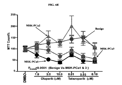
[0018] Figures 6A-6E demonstrate that the organoids derived from mCRPC
patients
represent an experimental model for BRCA2-RBI co-deletion. Figure SA shows the
FISH
analysis of indicated mCRPC-derived organoids (MSK-PCa 1-3) and benign
prostate
organoids using 3-color probes (see Example 1). Figure 6B shows a bar graph
showing the
deletion of BRCA2 and/or RBI per 100 cells. Near-diploid benign prostate
organoid is used
as a control. Figure 6C shows the copy number (CN) segment analysis of BRCA2-
RB1
region of chromosome 13q in mCRPC organoids. Figure 6D shows western blots
showing
indicated protein levels in human mCRPC organoids. GAPDH served as the loading
control.
Figure 6E shows the cell growth of organoids treated with PARPi (olaparib and
talazoparib)
in indicated concentrations for 3 days. The graphs show cell growth measured
by MTT assay
( SD); P-trends determined by 2-way ANOVA.
100191 Figures 7A-7G demonstrate the effect of BRCA2 deletion in prostate
cancer. Figure
7A shows a western blot (top panel) and qPCR (bottom panel) showing BRCA2
protein and
mRNA in LNCaP cells transduced with three different guide RNAs (gRNAs)
targeting
BRCA2 (CRISPR-BRCA2). Cells infected with scrambled (scr) gRNA were used as
control.
Equal amount of proteins was loaded in WedgeWell 6% gel and western blot was
performed.
¨400 l(Da BRCA2 band is indicated by arrow. GAPDH served as loading controls
for
western blot and qPCR. Figure 7B shows an ethidium bromide stained agarose gel
showing
BRCA2 genotype in BRCA2-CRISPR-edited LNCaP cell& Genotypes are detected by
PCR
amplification followed by being treated with T7 endonuclease. Wt and mutant
BRCA2 are
indicated by red and green arrows, respectively. Figure 7C shows the growth
curve of
LNCaP CRISPR-edited cells cultured in complete medium supplemented with
indicated
amount of various PARP inhibitors or cisplatin for 6 days. Equivalent volume
of DMSO was
used as placebo treatment. Cell growth was measured by MTT assay (see Example
1); SD,
I0
CA 03156423 2022-4-27
WO 2021/087141
PCT/US2020/058003
P-values determined by Student's t-test. *P<0.05, **P<0.01, ***P<0.001.
Figures 7D-7E
show the growth curve (Figure 7D) and a bar graph (Figure 7E) representing the
cell growth
of LNCaP BRCA2 CRISPR-edited and non-targeting control gRNA (scr) infected
cells in
complete medium supplemented with enzalutamide (ENZ; indicated concentration)
for 7 days
or charcoal-stripped medium (CSS) for 5 days, respectively. Equivalent volume
of DMSO
was used as placebo treatment. Cell growth was measured by crystal violet
staining assay
(see Example 1) (i SD); P-values determined by Student's t-test. Figure 7F
shows the
growth curve representing the growth of LNCaP BRCA2 CRISPR-edited and non-
targeting
control gRNA (scr) infected cells in charcoal-stripped medium (CSS) or
complete medium
supplemented with enzalutamide (ENZ; 20 M) for indicated days. Equivalent
volume of
DMSO was used as placebo treatment. Cell growth was measured by MTT assay (see
Example 1) ( SD); P-values determined by Student's t-test. Figure 7G (left
panel) shows
the expression of BRCA2 mRNA. Parental LNCaP cells were transiently
transfected with
BRCA2-specific SMARTpool siRNA for 96 hours. Total RNA was isolated, and BRCA2
mRNA was analyzed by qPCR. Scrambled SMARTpool siRNA¨transfected cells were
used
as control. Figure 7G (right panel) shows the cell growth of BRCA2- or
scrambled
SMARTpool siRNA¨transfected LNCaP cells cultured in charcoal-stripped medium
(CSS) or
complete medium supplemented with enzalutamide (ENZ; 20 pM) for 96 hours post
transfection. Equivalent volume of DMSO was used as placebo treatment. Cell
growth was
measured by MTT assay; SD, P-values determined by Student's t-test.
[0020] Figures SA-8J demonstrate that co-loss of BRCA2 and RB 1 induces
invasive
phenotype. Figure 8A shows the qPCR analysis showing BRCA2 and RBI mRNA
expression in LNCaP-BRCA2 CRISPR-edited (CRISPR gRNA 2) and scrambled control
cells
infected with lentiviral RBJ shRNA. Scr-CRISPR and BRCA2-CRISPR2 cells also
transfected with non-targeting shRNA (scr-Sh) for control of shRNA. mRNA
expression
normalized with internal control (GAPDH). Figure 8B shows the western blots
showing
RBI expression in LNCaP cells transduced with three different guide RNAs
(gRNAs)
targeting BRCA2 (CRISPR-BRCA2). Cells infected with scrambled (scr) gRNA were
used as
control. Cas9 and RHoGDI served as loading controls. Figure 8C shows the
western blot
showing RBI and BRCA2 expression in LNCaP cells transduced with two different
guide
RNAs (gRNAs) targeting RBI (CRISPR-RBI). Cells infected with scrambled (sot)
gRNA
11
CA 03156423 2022-4-27
WO 2021/087141
PCT/US2020/058003
were used as control. Cas9 and RHoGDI served as loading controls. Figure 8D
shows the
invasion assays. 5 x 10³ of indicated cells were plated on the top of Boyden
chamber (see
Example 1) in serum-free media; 10% serum in the bottom chamber was used as
chemo-
attractant. After 24 hours, cells in the lower side of the chamber were fixed
and stained, cells
were counted and represented in the form of the bar graph. P-values were
determined by
Student's 1-test. *P<0.05, **P<0.01, ***P<0.001µ Figure SE shows the bar
graphs showing
pyH2AX and DNA-PKcs (S2056) positive foci counted in high power field
(reference to
Figure 2D). P-values determined by Student's t-test. Figure SF shows the
western blot
showing R.I31 in 22RV1 cells transduced with three different guide RNAs
(gRNAs) targeting
R131 (CRISPR-RB/). Cells infected with scrambled (scr) gRNA were used as
control. Cas9
and RHoGDI served as loading controls. Figure SG shows the Matrigel invasion
assay. 2.5
x 103 of RB I CRISPR-edited or scrambled CRISPR control 22RV1 cells were
plated on the
top of Boyden chamber (see Example 1) in serum-free media; 10% serum in the
bottom
chamber was used as chemo-attractant. After 72 hours, cells in the lower side
of the chamber
were fixed, stained and photographed. Figure 811 shows the bar graph
representing the
pathways (gene ontology-biological pathways) that are positively enriched in
BRCA2-RBI
knockout/knockdown LNCaP cells. Pathway analyses were performed using gene set
enrichment analysis (GSEA). Figures 81-8J show the GSEA utilizing previously
published
RBI signatures. Mateo et at, N Engl flied. 373(18):1697-708 (2015); Pritchard
et at, N
Engl flied. 375(5):443-53 (2016). NES, normalized enrichment score.
100211 Figures 9A-9F demonstrate that the induction of EMT phenotype resulted
in co-loss
of BRCA2 and RB1. Figure 9A shows a bar graph representing the pathway
analysis of the
genes unregulated in BRCA2-RB1 knockout/knockdown LNCaP cells. Pathway
analyses
were performed using GSEA. Figure 9B shows the qPCR analysis showing vimentin
mRNA
expression in LNCaP-BRCA2 CRISPR-edited (CRISPR gRNA 2) and scrambled control
cells
infected with lentiviral RBI shRNA. mRNA expression normalized with internal
control
(GAPDH). Figure 9C shows the Venn diagram showing the common pathways from the
transcriptome of TCGA (BRCA2-RB1 co-deleted vs wild-type) and LNCaP (BRCA2-RBI
knockout/knockdown vs scrambled control cells). Figure 9D shows the migration
and
invasion assay. 1 x 10³ BRCA2 and RBI transiently co-overexpressed PC3M cells
(72 hours
post transfection) were plated on the top of a Boyden chamber or Matrigel
invasion chamber
12
CA 03156423 2022-4-27
WO 2021/087141
PCT/US2020/058003
in serum-free media; 10% serum in the bottom chamber was used as chemo-
attractant. After
24 hours, cells in the lower side of the chambers were fixed, stained with
crystal violet, and
photographed in 100x magnification. Figure 9E shows the relative mRNA
expression of
EMT and stem cell markers in LNCaP-BRCA2-RB1 cells. Figure 9F shows the Venn
diagram shows the common hallmark pathways that are in lethal compared to
indolent
prostate cancer (Setlur prostate cancer; Swedish watchful waiting cohort) and
LNCaP
(BRCA2-RB 1 knockout/knockdown vs control cells).
100221 Figures 10A-10J demonstrate that concomitant deletion of BRCA2 and RBI
represents an aggressive variant of prostate cancer. Figure 10A shows the pan-
cancer
analysis of BRCA2 alteration. Samples are customized on the basis of
alteration frequency
(>5% cases of BRCA2 alteration) and total number of cases in each group (>50
cases in each
group). Figure 10B shows the in-depth analysis of BRCA2 alterations
(homozygous or
heterozygous deletions and mutations) in multiple publicly available prostate
cancer cohorts
using cBioPortal. Cohorts are divided into primary (n=925) and metastatic
castration-
resistant (n=444) groups. Figure 10C shows the graph representing the BRCA2
mRNA
expression for wt and BRCA2-deleted (heterozygous or homozygous) patients in
TCGA
provisional cohort; P-trend determined by one-way ANOVA. Individual blue
circles indicate
individual patients. Figure 10D shows the Kaplan-Meier curves. Patients from
TCGA
provisional cohort were divided into 3 groups on the basis of BRCA2 protein
expression
(reverse phase protein array [RPPA]; >+1; +1 to -1; <-1), and Kaplan-Meier
curves described
disease/progression-free survival (5 years) in each group. Log-rank test was
performed to
examine significance_ Figure 10E (top panel) shows the alteration status of
BRCA2 and RB1
in MSK-IMPACT prostate cancer cohort. Co-occurrence of indicated genes, as
presented by
cBioPortal. Figure 10E (bottom panel) shows the pie charts showing the
percentage of
BRCA2 alteration (mutation and homozygous deletion) and co-occurrence with RBI
homozygous deletion in primary prostate cancer and mCRPC in MSK-IMPACT
prostate
cancer cohort. Figure 101? shows the concordance of BRCA2 and RB1 copy number
in
TCGA (primary prostate cancer) and Kumar (mCRPC) cohorts. Individual blue
circles
indicate individual patients. Figure 10G shows the graph representing the RB1
mRNA
expression in wt and Rill deleted (homozygous and heterozygous) patients in
TCGA and
Kumar cohorts; SD, P-trends determined by one-way ANOVA. Individual blue
circles
13
CA 03156423 2022-4-27
WO 2021/087141
PCT/US2020/058003
indicate individual patients. Figure 1011 shows the concomitant deletion of
BRCA2 and
RB1; significance was determined by disease/progression-free survival in
Taylor et al.
primary prostate cancer cohort (n=131). Kaplan-Meier curves for indicated
months were
defined for each group. Log-rank test was used to calculate P-value. Figure
101 shows a
volcano plot showing the genes altered in Gleason 6 patients (BRCA2-RBI co-
deleted vs wt)
in TCGA cohort. Individual circles indicate individual genes. Heat map showed
the
expression of genes that are upregulated in BRCA2-RBI co-deleted prostate
cancer patients
compared to wt prostate cancer (in TCGA cohort) analyzed in primary and mCRPC
samples
in Taylor cohort. The heat map was generated in Oncomine Suite. Genes are
ranked on the
basis of P-value and fold changes. Figure 10J shows the heat map (hierarchical
clustering)
of the mRNA expression of 63 genes (BRCA2-RB1 region of chromosome 13q) in
primary
and mCRPC samples in Taylor cohort. The heat map is generated in Oncomine
Suite. Genes
are ranked on the basis of P-value and fold changes.
100231 Figures 11A-11D demonstrate the concomitant heterozygous co-deletion of
BRCA2-
RBI in prostate cancer cell lines. Figure 11A shows the micrographs of FISH
analysis of
indicated human prostate cancer cell lines using a 3-color probe (see Example
1). The ploidy
of each cell line is indicated. Figure 11B shows the BRCA2 and RBI rnRNA
expression in
various prostate cancer cell lines was analyzed by qPCR. Figure 11C shows the
profile of 12
major DDR genes in prostate cancer cell lines in The Cancer Cell Line
Encyclopedia. Figure
11D shows the graphs representing the growth of 22RV1 (BRCA2-mutated and RBI-
wt; near
diploid) and PC3M (BRCA2-RB1 co-deleted; triploid) cultured in or complete
medium
supplemented with olaparib (2.5 pM), veliparib (5 pM), niraparib (1 pM),
rucaparib (500
nM), talazoparib (5 nM), and cisplatin (500 nM) for 6 days. Equivalent volume
of DMSO
was used as placebo treatment. Cell growth was measured by MTT assay (see
Example 1);
SD, each point indicates the MTT count of each well of 96-well plate, P-values
determined
by Student's t-test. **P<0.01, ****P<0.0001. Note that talazoparib (5 nM)
exhibited more
growth inhibition in PC3M cells compared to olaparib (2.5 AM), P<0.000 1
100241 Figures 12A-12D demonstrate that organoids derived from human mCRPC
patients
harbor co-heterozygous deletion of BRCA2 and RBI. Figure 12A shows a graph
representing the copies of BRCA2, RB1 and 13q in human mCRPC organoids
analyzed by
14
CA 03156423 2022-4-27
WO 2021/087141
PCT/US2020/058003
the 3-color FISH. Each point represents a single cell. A total of 100
individual cells from
each cell line were counted and represented graphically. Figure 12B shows the
mutation
profile of 12 major DDR genes in mCRPC organoids analyzed in cBioPortal.
Figure 12C
shows the SLUG, SNAIL and PRRX1 mRNA in the organoids analyzed by qPCR. Figure
12D shows the concomitant deletion of BRCA2 and R131; significance was
determined by
overall survival in TCGA pan-cancer cohort (excluding the prostate cancer
cases; n=10,830).
Kaplan-Meier curves for indicated months were defined for each group. Log-rank
test was
used to calculate P-value.
[0025] Figure 13 shows the list of antibodies and reagents used herein.
[0026] Figure 14 shows the list of top 10 upregulated and downregulated genes
BRCA2-
RB1 Vs SCR obtained from RNA sequencing of LNCaP cells infected with BRCA2
CRISPR
and/or RBI shRNA in a stable lentiviral vector.
[0027] Figures 15A-15B show Toppgene (Figure 15A) and GSEA (Figure 1518)
pathway
analysis of the genes upregulated upon co-deletion of BRCA and RBI in LNCaP
cells.
[0028] Figure 16 shows the hallmark pathway analysis of the genes upregulated
upon co-
deletion of BRCA and RB/ in LNCaP cells.
[0029] Figure 17 shows the hallmark pathway analysis in TCGA provisional
cohort
(BRCA2-R131 deleted vs unaltered patients).
[0030] Figure 18 shows the hallmark pathway analysis in Setlur prostate cancer
cohort (5-
year lethal vs indolent patients).
[0031] Figure 19 shows the analysis of BRCA2 and Rill co-deletion in Armenia
et al. cohort
and calculated P-values between different groups
[0032] Figure 20 shows the changes of mRNA expression in BRCA2-RB I co-deleted
vs
unaltered patients with Gleason 6 disease in TCGA provisional cohort (FDR
0.25). Changes
of mRNA expression calculated in cBioPortal.
CA 03156423 2022-4-27
WO 2021/087141
PCT/US2020/058003
[0033] Figure 21 shows the BRCA2-RB1 deletion status in matched prostate
cancer
(localized and metastatic) samples in Kumar et at mCRPC cohort.
[0034] Figure 22 shows the list of chromosome 13q genes. Genes located inside
the
BRCA2-RB1 region denoted as inside (without highlight), all the rest denoted
as outside
(highlighted in gray).
[0035] Figure 23 shows the FISH of BRCA2 and RBI in patient-derived human
organoids.
DETAILED DESCRIPTION
[0036] It is to be appreciated that certain aspects, modes, embodiments,
variations and
features of the present methods are described below in various levels of
detail in order to
provide a substantial understanding of the present technology.
[0037] In practicing the present methods, many conventional techniques in
molecular
biology, protein biochemistry, cell biology, microbiology and recombinant DNA
are used.
See, e.g., Sambrook and Russell eds. (2001) Molecular Cloning: A Laboratory
Manual, 3rd
edition; the series Ausubel et al., eds. (2007) Current Protocols in Molecular
Biology; the
series Methods in Enzymology (Academic Press, Inc., N.Y.); MacPherson et al.,
(1991) PCR
1: A Practical Approach (IRL Press at Oxford University Press); MacPherson et
at, (1995)
PCR 2: A Practical Approach; Harlow and Lane eds. (1999) Antibodies, A
Laboratory
Manual; Freshney (2005) Culture of Animal Cells: A Manual of Basic Technique,
5th edition;
Gait ed. (1984) Oligonucleotide Synthesis; U.S. Patent No. 4,683,195; Hames
and Higgins
eds. (1984) Nucleic Acid Hybridization; Anderson (1999) Nucleic Acid
Hybridization; Haines
and Higgins eds. (1984) Transcription and Translation; Immobilized Cells and
Enzymes ORL
Press (1986)); Perbal (1984)A Practical Guide to Molecular Cloning, Miller and
Cabs eds.
(1987) Gene Transfer Vectors for Mammalian Cells (Cold Spring Harbor
Laboratory);
Makrides ed. (2003) Gene Transfer and Expression in Mammalian Cells; Mayer and
Walker
eds. (1987) Immunochemical Methods in Cell and Molecular Biology (Academic
Press,
London); and Herzenberg et al., eds (1996) Weir's Handbook of Experimental
Immunology.
[0038] The present disclosure identifies a previously uncharacterized prostate
cancer subset
characterized by concomitant deletions (homozygous and heterozygous) of BRCA2
and RBI.
16
CA 03156423 2022-4-27
WO 2021/087141
PCT/US2020/058003
Further, the cell line¨based models of the present disclosure demonstrate that
even single
copy loss of both BRCA2 and RB1 is sufficient to induce an aggressive
phenotype in prostate
cancer.
100391 Previous case studies reported mCRPC progression in a patient with
germline BRCA2
mutation and a newly emerged RB1 single copy number loss following treatment
with PARP
inhibitor olaparib. Ma et al., BA1C Med Genet. 19: 185 (2018). Previous papers
have
suggested that Retinoblastoma (RBI.) tumor suppressor gene loss drives
transformation of
prostate adenocarcinoma (PADC) to neuroendocrine prostate cancer variants
(NEPC)
resistant to antiandrogen therapy (AAT) (Wadosky K et al., Molecular &
Cellular Oncology
4(2):e1291397 (2017)), which may also be one of the mechanisms of PARP
inhibitors
resistance. As shown in the Examples described herein, PARP inhibition
unexpectedly and
significantly attenuated growth of prostate cancer cell lines and organoids
derived from
human mCRPC that harbor not only homozygous but also heterozygous co-deletion
of
BRCA2 and RB1. Accordingly, the present disclosure demonstrates that co-
deletion of
BRCA2 and RBI in a subset of prostate cancer patients is an independent
genomic driver of
therapy-resistant aggressive prostate cancer rather than the consequence of
exposure to
therapy, and that co-loss of BRCA2 and RBI may induce an epithelial-to-
mesenchymal
transition (EMT) mediated by induction of the transcription factors SLUG or
SNAIL or
transcriptional co-activator PRRX1. Thus, the methods disclosed herein permit
the early
recognition and intervention using PARP inhibitor-based therapy in prostate
cancer cases
identified as having a BRCA2-RB1 co-deletion.
Definitions
100391 Unless defined otherwise, all technical and
scientific terms used herein generally
have the same meaning as commonly understood by one of ordinary skill in the
art to which
this technology belongs. As used in this specification and the appended
claims, the singular
forms "a", "an" and "the" include plural referents unless the content clearly
dictates
otherwise. For example, reference to "a cell" includes a combination of two or
more cells,
and the like. Generally, the nomenclature used herein and the laboratory
procedures in cell
culture, molecular genetics, organic chemistry, analytical chemistry and
nucleic acid
17
CA 03156423 2022-4-27
WO 2021/087141
PCT/US2020/058003
chemistry and hybridization described below are those well-known and commonly
employed
in the art.
100401 As used herein, the term "about" in reference to a
number is generally taken to
include numbers that fall within a range of 1%, 5%, or 10% in either direction
(greater than
or less than) of the number unless otherwise stated or otherwise evident from
the context
(except where such number would be less than 0% or exceed 100 4 of a possible
value).
100411 As used herein, the "administration" of an agent
or drug to a subject includes any
route of introducing or delivering to a subject a compound to perform its
intended function.
Administration can be carried out by any suitable route, including orally,
intranasally,
parenterally (intravenously, intramuscularly, intraperitoneally, or
subcutaneously), or
topically. Administration includes self-administration and the administration
by another.
100421 The terms "complementary" or "complementarity" as
used herein with reference
to polynucleotides (Le., a sequence of nucleotides such as an oligonucleotide
or a target
nucleic acid) refer to the base-pairing rules. The complement of a nucleic
acid sequence as
used herein refers to an oligonucleotide which, when aligned with the nucleic
acid sequence
such that the 5' end of one sequence is paired with the 3' end of the other,
is in "antiparallel
association." For example, the sequence "5'-A-G-T-31" is complementary to the
sequence
"3'-T-C-A-5." Certain bases not commonly found in naturally-occurring nucleic
acids may
be included in the nucleic acids described herein. These include, for example,
inosine, 7-
deazaguanine, Locked Nucleic Acids (LNA), and Peptide Nucleic Acids (PNA).
Complementarity need not be perfect stable duplexes may contain mismatched
base pairs,
degenerative, or unmatched bases. Those skilled in the art of nucleic acid
technology can
determine duplex stability empirically considering a number of variables
including, for
example, the length of the oligonucleotide, base composition and sequence of
the
oligonucleotide, ionic strength and incidence of mismatched base pairs. A
complementary
sequence can also be an RNA sequence complementary to the DNA sequence or its
complementary sequence, and can also be a cDNA.
100431 As used herein, a "control" is an alternative
sample used in an experiment for
comparison purpose. A control can be "positive" or "negative," For example,
where the
18
CA 03156423 2022-4-27
WO 2021/087141
PCT/US2020/058003
purpose of the experiment is to determine a correlation of the efficacy of a
therapeutic agent
for the treatment for a particular type of disease or condition, a positive
control (a compound
or composition known to exhibit the desired therapeutic effect) and a negative
control (a
subject or a sample that does not receive the therapy or receives a placebo)
are typically
employed.
[0044] As used herein, a "deletion" refers to a genetic
aberration in which at least a part
of a chromosome or a gene sequence is lost or missing. Deletion of a number of
nucleotides
that is not evenly divisible by three will lead to a frameshift mutation,
causing all of
the codons occurring after the deletion to be read incorrectly during
translation, thereby
producing a severely altered and potentially nonfunctional protein.
[0045] As used herein, the term "effective amount" refers
to a quantity sufficient to
achieve a desired therapeutic and/or prophylactic effect, e.g., an amount
which results in the
prevention of, or a decrease in a disease or condition described herein or one
or more signs or
symptoms associated with a disease or condition described herein. In the
context of
therapeutic or prophylactic applications, the amount of a composition
administered to the
subject will vary depending on the composition, the degree, type, and severity
of the disease
and on the characteristics of the individual, such as general health, age,
sex, body weight and
tolerance to drugs. The skilled artisan will be able to determine appropriate
dosages
depending on these and other factors. The compositions can also be
administered in
combination with one or more additional therapeutic compounds. In the methods
described
herein, the therapeutic compositions may be administered to a subject having
one or more
signs or symptoms of prostate cancer, such as castration-resistant prostate
cancer (e.g.,
mCRPC). As used herein, a "therapeutically effective amount" of a composition
refers to
composition levels in which the physiological effects of a disease or
condition are
ameliorated or eliminated. A therapeutically effective amount can be given in
one or more
administrations.
[0046] As used herein, "expression" includes one or more
of the following: transcription
of the gene into precursor mRNA; splicing and other processing of the
precursor mRNA to
produce mature mRNA; mRNA stability; translation of the mature mRNA into
protein
19
CA 03156423 2022-4-27
WO 2021/087141
PCT/US2020/058003
(including codon usage and tRNA availability); and glycosylation and/or other
modifications
of the translation product, if required for proper expression and function.
[0047] As used herein, the term "gene" means a segment of
DNA that contains all the
information for the regulated biosynthesis of an RNA product, including
promoters, exons,
introns, and other untranslated regions that control expression.
[0048] "Homology" or "identity" or "similarity" refers to
sequence similarity between
two peptides or between two nucleic acid molecules. Homology can be determined
by
comparing a position in each sequence which may be aligned for purposes of
comparison.
When a position in the compared sequence is occupied by the same nucleobase or
amino
acid, then the molecules are homologous at that position. A degree of homology
between
sequences is a function of the number of matching or homologous positions
shared by the
sequences. A polynucleotide or polynucleotide region (or a polypeptide or
polypeptide
region) has a certain percentage (for example, at least 60%, 65%, 70%, 75%,
80%, 85%,
90%, 95%, 98% or 99%) of "sequence identity" to another sequence means that,
when
aligned, that percentage of bases (or amino acids) are the same in comparing
the two
sequences. This alignment and the percent homology or sequence identity can be
determined
using software programs known in the art. In some embodiments, default
parameters are
used for alignment. One alignment program is BLAST, using default parameters.
In
particular, programs are BLASTN and BLASTP, using the following default
parameters:
Genetic code=standard; filter=none; strand=both; cutoff=60; expect=10;
Matrix=BLOSUIVI62; Descriptions=50 sequences; sort by =HIGH SCORE;
Databases=non-
redundant, GenBank+EMEL+DDBJ+PDB+GenBank CDS
translations-FSwissProtein-FSPupdate-FPIR. Details of these programs can be
found at the
National Center for Biotechnology Information_ Biologically equivalent
polynucleotides are
those having the specified percent homology and encoding a polypeptide having
the same or
similar biological activity. Two sequences are deemed "unrelated" or "non-
homologous" if
they share less than 40% identity, or less than 25% identity, with each other.
[0049] The term "hybridize" as used herein refers to a
process where two substantially
complementary nucleic acid strands (at least about 65% complementary over a
stretch of at
CA 03156423 2022-4-27
WO 2021/087141
PCT/US2020/058003
least 14 to 25 nucleotides, at least about 75%, or at least about 90%
complementary) anneal
to each other under appropriately stringent conditions to form a duplex or
heteroduplex
through formation of hydrogen bonds between complementary base pairs. Nucleic
acid
hybridization techniques are well known in the art. See, e.g., Sambrook,
etal., 1989,
Molecular Cloning: A Laboratory Manual, Second Edition, Cold Spring Harbor
Press,
Plainview, N.Y. Hybridization and the strength of hybridization (i.e., the
strength of the
association between the nucleic acids) is influenced by such factors as the
degree of
complementarity between the nucleic acids, stringency of the conditions
involved, and the
thermal melting point (Tin) of the formed hybrid. Those skilled in the art
understand how to
estimate and adjust the stringency of hybridization conditions such that
sequences having at
least a desired level of complementarity will stably hybridize, while those
having lower
complementarity will not. For examples of hybridization conditions and
parameters, see,
e.g., Sambrook, etal., 1989, Molecular Cloning: A Laboratory Manual, Second
Edition,
Cold Spring Harbor Press, Plainview, N.Y.; Ausubel, F. M. etal., 1994, Current
Protocols
in Molecular Biology, John Wiley & Sons, Secaucus, N.J. In some embodiments,
specific
hybridization occurs under stringent hybridization conditions. An
oligonucleotide or
polynucleotide (e.g., a probe or a primer) that is specific for a target
nucleic acid will
"hybridize" to the target nucleic acid under suitable conditions.
100501 As used herein, "oligonucleotide" refers to a
molecule that has a sequence of
nucleic acid bases on a backbone comprised mainly of identical monomer units
at defined
intervals. The bases are arranged on the backbone in such a way that they can
bind with a
nucleic acid having a sequence of bases that are complementary to the bases of
the
oligonucleotide. The most common oligonucleotides have a backbone of sugar
phosphate
units. A distinction may be made between oligodeoxyribonucleotides that do not
have a
hydroxyl group at the T position and oligoribonucleotides that have a hydroxyl
group at the 2'
position. Oligonucleotides may also include derivatives, in which the hydrogen
of the
hydroxyl group is replaced with organic groups, e.g., an allyl group. One or
more bases of
the oligonucleotide may also be modified to include a phosphorothioate bond
(e.g., one of the
two oxygen atoms in the phosphate backbone which is not involved in the
internucleotide
bridge, is replaced by a sulfur atom) to increase resistance to nuclease
degradation. The exact
size of the oligonucleotide will depend on many factors, which in turn depend
on the ultimate
21
CA 03156423 2022-4-27
WO 2021/087141
PCT/US2020/058003
function or use of the oligonucleotide. The oligonucleotide may be generated
in any manner,
including, for example, chemical synthesis, DNA replication, restriction
endonuclease
digestion of plasmids or phage DNA, reverse transcription, PCR, or a
combination thereof.
The oligonucleotide may be modified e.g., by addition of a methyl group, a
biotin or
digoxigenin moiety, a fluorescent tag or by using radioactive nucleotides.
100511 As used herein, the term "pharmaceutically-
acceptable carrier" is intended to
include any and all solvents, dispersion media, coatings, antibacterial and
antifimgal
compounds, isotonic and absorption delaying compounds, and the like,
compatible with
pharmaceutical administration. Pharmaceutically-acceptable carriers and their
formulations
are known to one skilled in the art and are described, for example, in
Remington's
Pharmaceutical Sciences (20th edition, ed. A. Gennaro, 2000, Lippincott,
Williams & Wilkins,
Philadelphia, Pa.).
100521 As used herein, the term "polynucleotide" or
"nucleic acid" means any RNA or
DNA, which may be unmodified or modified RNA or DNA. Polynucleotides include,
without limitation, single- and double-stranded DNA, DNA that is a mixture of
single- and
double-stranded regions, single- and double-stranded RNA, RNA that is mixture
of single-
and double-stranded regions, and hybrid molecules comprising DNA and RNA that
may be
single-stranded or, more typically, double-stranded or a mixture of single-
and double-
stranded regions. In addition, polynucleotide refers to triple-stranded
regions comprising
RNA or DNA or both RNA and DNA. The term polynucleotide also includes DNAs or
RNAs containing one or more modified bases and DNAs or RNAs with backbones
modified
for stability or for other reasons.
100531 As used herein, "prevention," "prevent," or
"preventing" of a disorder or
condition refers to one or more compounds that, in a statistical sample,
reduces the
occurrence of the disorder or condition in the treated sample relative to an
untreated control
sample, or delays the onset of one or more symptoms of the disorder or
condition relative to
the untreated control sample. As used herein, preventing prostate cancer such
as castration-
resistant prostate cancer (e.g., mCRPC), includes preventing or delaying the
initiation of
symptoms of prostate cancer such as castration-resistant prostate cancer
(e.g., mCRPC). As
22
CA 03156423 2022-4-27
WO 2021/087141
PCT/US2020/058003
used herein, prevention of prostate cancer such as castration-resistant
prostate cancer (e.g.,
mCRPC) also includes preventing a recurrence of one or more signs or symptoms
of prostate
cancer such as castration-resistant prostate cancer (e.g., mCRPC).
100541 As used herein, the term "primer" refers to an
oligonucleotide, which is capable of
acting as a point of initiation of nucleic acid sequence synthesis when placed
under
conditions in which synthesis of a primer extension product which is
complementary to a
target nucleic acid strand is induced, i.e., in the presence of different
nucleotide triphosphates
and a polymerase in an appropriate buffer ("buffer" includes pH, ionic
strength, cofactors
etc.) and at a suitable temperature. One or more of the nucleotides of the
primer can be
modified for instance by addition of a methyl group, a biotin or digoxigenin
moiety, a
fluorescent tag or by using radioactive nucleotides. A primer sequence need
not reflect the
exact sequence of the template. For example, a non-complementary nucleotide
fragment may
be attached to the 5' end of the primer, with the remainder of the primer
sequence being
substantially complementary to the strand. The term primer as used herein
includes all forms
of primers that may be synthesized including peptide nucleic acid primers,
locked nucleic
acid primers, phosphorothioate modified primers, labeled primers, and the
like. The term
"forward primer" as used herein means a primer that anneals to the anti-sense
strand of
double-stranded DNA (dsDNA). A "reverse primer" anneals to the sense-strand of
dsDNA.
100551 "Probe" as used herein refers to a nucleic acid
that interacts with a target nucleic
acid via hybridization. A probe may be fully complementary to a target nucleic
acid
sequence or partially complementary. The level of complementarity will depend
on many
factors based, in general, on the function of the probe. Probes can be labeled
or unlabeled, or
modified in any of a number of ways well known in the art. A probe may
specifically
hybridize to a target nucleic acid. Probes may be DNA, RNA or a RNA/DNA
hybrid.
Probes may be oligonucleotides, artificial chromosomes, fragmented artificial
chromosome,
genomic nucleic acid, fragmented genomic nucleic acid, RNA, recombinant
nucleic acid,
fragmented recombinant nucleic acid, peptide nucleic acid (PNA), locked
nucleic acid,
oligomer of cyclic heterocycles, or conjugates of nucleic acid. Probes may
comprise
modified nucleobases, modified sugar moieties, and modified intemucleotide
linkages. A
probe may be used to detect the presence or absence of a methylated target
nucleic acid.
23
CA 03156423 2022-4-27
WO 2021/087141
PCT/US2020/058003
Probes are typically at least about 10, 15, 20, 25, 30, 35, 40, 50, 60, 75,
100 nucleotides or
more in length.
100561 As used herein, the term "sample" refers to
clinical samples obtained from a
subject. Biological samples may include tissues, cells, protein or membrane
extracts of cells,
mucus, sputum, bone marrow, bronchial alveolar lavage (BAL), bronchial wash
(BW), and
biological fluids (e.g., ascites fluid or cerebrospinal fluid (CSF)) isolated
from a subject, as
well as tissues, cells and fluids (blood, plasma, saliva, urine, serum etc.)
present within a
subject.
100571 As used herein, the term "separate" therapeutic
use refers to an administration of
at least two active ingredients at the same time or at substantially the same
time by different
routes.
100581 As used herein, the term "sequential" therapeutic
use refers to administration of at
least two active ingredients at different times, the administration route
being identical or
different. More particularly, sequential use refers to the whole
administration of one of the
active ingredients before administration of the other or others commences. It
is thus possible
to administer one of the active ingredients over several minutes, hours, or
days before
administering the other active ingredient or ingredients. There is no
simultaneous treatment
in this case.
100591 As used herein, the term "simultaneous"
therapeutic use refers to the
administration of at least two active ingredients by the same route and at the
same time or at
substantially the same time.
100601 As used herein, the terms "subject," "individual,"
or "patient" are used
interchangeably and refer to an individual organism, a vertebrate, a mammal,
or a human. In
certain embodiments, the individual, patient or subject is a human.
100611 As used herein, the terms "target sequence" and
"target nucleic acid sequence"
refer to a specific nucleic acid sequence to be detected, or quantified in the
sample to be
analyzed. Alternatively, the terms "target sequence" and "target nucleic acid
sequence" refer
to a specific nucleic acid sequence to be modulated (e.g., inhibited or
downregulated).
24
CA 03156423 2022-4-27
WO 2021/087141
PCT/US2020/058003
[0062] The term "PARP inhibitor" as used herein refers to
an agent that inhibits gene
expression and/or biological activity of PARR Examples of PARP biological
activity
include, but are not limited to, enzymatic activity, substrate binding
activity, homo- or hetero-
dimerization activity, and binding to a cellular structure. Examples of PARP
inhibitors
include, but are not limited to, olaparib, rucapatib, niraparib, talazoparib,
veliparib, inhibitory
nucleic acids targeting PARP (e.g., shRNAs, siRNAs or anti-sense
oligonucleotides), and
anti-PARP neutralizing antibodies.
[0063] "Treating", "treat", or "treatment" as used herein
covers the treatment of a disease
or disorder described herein, in a subject, such as a human, and includes: (i)
inhibiting a
disease or disorder, i.e., arresting its development; (ii) relieving a disease
or disorder, i.e.,
causing regression of the disorder; (iii) slowing progression of the disorder;
and/or (iv)
inhibiting, relieving, or slowing progression of one or more symptoms of the
disease or
disorder. In some embodiments, treatment means that the symptoms associated
with the
disease are, e.g., alleviated, reduced, cured, or placed in a state of
remission.
[0064] It is also to be appreciated that the various
modes of treatment or prevention of
medical diseases and conditions as described are intended to mean
"substantial," which
includes total but also less than total treatment or prevention, and wherein
some biologically
or medically relevant result is achieved. The treatment may be a continuous
prolonged
treatment for a chronic disease or a single, or few time administrations for
the treatment of an
acute condition.
PARP
100651 Poly (ADP-ribose) polymerase (PARP) is a family of
proteins involved in a
number of cellular processes such as DNA repair, genomic stability, and
programmed cell
death. DNA damage may be caused by normal cell actions, UV light, some
anticancer drugs,
and radiation. The main role of PARP, which is found in the nucleus, is to
detect and initiate
an immediate cellular response to metabolic, chemical, or radiation-induced
single-strand
DNA breaks (SSB) by signaling the enzymatic machinery involved in the SSB
repair. Once
PARP detects a SSB, it binds to the DNA, undergoes a structural change, and
begins the
synthesis of a polymeric adenosine diphosphate ribose (poly (ADP-ribose) or
PAR) chain,
CA 03156423 2022-4-27
WO 2021/087141
PCT/US2020/058003
which acts as a signal for the other DNA-repairing enzymes. Target enzymes
include DNA
ligase III (LigIII), DNA polymerase beta (poli3), and scaffolding proteins
such as X-ray cross-
complementing gene 1 (XRCC1). Upon completion of the repair process, the PAR
chains are
degraded via Poly(ADP-ribose) glycohydrolase (PAR.G). NAD+ is required as a
substrate for
generating ADP-ribose monomers. It is believed that overactivation of PARP may
deplete
the stores of cellular NADA- and induce progressive ATP depletion and necrotic
cell death,
since glucose oxidation is inhibited. PARP is inactivated by caspase-3
cleavage during
programmed cell death.
PARP Inhibitors
100661 In one aspect, the present disclosure provides
inhibitory nucleic acids (e.g.,
sgR_NAs, antisense RNAs, ribozymes, or shRNAs) that inhibit PARP expression
and/or
activity. The mammalian nucleic acid sequences of PARP are known in the art
(e.g., NCBI
Gene ID. 142). The inhibitory nucleic acids of the present technology may
comprise a
nucleic acid molecule that is complementary to a portion of a PARP nucleic
acid sequence.
In some embodiments, the inhibitory nucleic acids (e.g., sgRNAs, antisense
RNAs,
ribozymes, or shRNAs) target at least one exon and/or intron of PARP. An
exemplary
nucleic acid sequence of Homo sapiens PARP1 is provided below:
1 agcaatctat cagggaacgg cggtggccgg tgcggcgtgt tcggtggcgg ctctggccgc
61 tcaggcgcct gcggctgggt gagcgcacgc gaggcggcga ggcggcagcg tgtttctagg
121 tcgtggcgtc gggcttccgg agctttggcg gcagctaggg gaggatggcg gagtcttcgg
181 ataagctcta tcgagtcgag tacgccaaga gcgggcgcgc ctcttgcaag aaatgcagcg
241 agagcatccc caaggactcg ctccggatgg ccatcatggt gcagtcgccc atgtttgatg
301 gaaaagtccc acactggtac cacttctcct gcttctggaa ggtgggccac tccatccggc
361 accctgacgt tgaggtggat gggttctctg agcttcggtg ggatgaccag cagaaagtca
421 agaagacagc ggaagctgga ggagtgacag gcaaaggcca ggatggaatt ggtagcaagg
481 cagagaagac tctgggtgac tttgcagcag agtatgccaa gtccaacaga agtacgtgca
541 aggggtgtat ggagaagata gaaaagggcc aggtgcgcct gtccaagaag atggtggacc
601 cggagaagcc acagctaggc atgattgacc gctggtacca tccaggctgc tttgtcaaga
661 acagggagga gctgggtttc cggcccgagt acagtgcgag tcagctcaag ggcttcagcc
721 tccttgctac agaggataaa gaagccctga agaagcagct cccaggagtc aagagtgaag
781 gaaagagaaa aggcgatgag gtggatggag tggatgaagt ggcgaagaag aaatctaaaa
841 aagaaaaaga caaggatagt aagcttgaaa aagccctaaa ggctcagaac gacctgatct
901 ggaacatcaa ggacgagcta aagaaagtgt gttcaactaa tgacctgaag gagctactca
961 tcttcaacaa gcagcaagtg cottctgggg agtoggcgat cttggaccga gtagctgatg
1021 gcatggtgtt cggtgccctc cttccctgcg aggaatgctc gggtcagctg gtcttcaaga
1081 gcgatgccta ttactgcact ggggacgtca ctgcctggac caagtgtatg gtcaagacac
1141 agacacccaa ccggaaggag tgggtaaccc caaaggaatt ccgagaaatc tcttacctca
1201 agaaattgaa ggttaaaaaa caggaccgta tattcccccc agaaaccagc gcctccgtgg
1261 cggccacgcc tccgccctcc acagcctcgg ctcctgctgc tgtgaactcc tctgcttcag
1321 cagataagcc attatccaac atgaagatcc tgactctcgg gaagctgtcc cggaacaagg
26
CA 03156423 2022-4-27
WO 2021/087141
PCT/US2020/058003
1381 atgaagtgaa ggccatgatt gagaaactcg gggggaagtt gacggggacg gccaacaagg
1441 cttccctgtg catcagcacc aaaaaggagg tggaaaagat gaataagaag atggaggaag
1501 taaaggaagc caacatccga gttgtgtctg aggacttcct ccaggacgtc tccgcctcca
1561 ccaagagcct tcaggagttg ttcttagcgc acatcttgtc cccttggggg gcagaggtga
1621 aggcagagcc tgttgaagtt gtggccccaa gagggaagtc aggggctgcg ctctccaaaa
1681 aaagcaaggg ccaggtcaag gaggaaggta tcaacaaatc tgaaaagaga atgaaattaa
1741 ctcttaaagg aggagcagct gtggatcctg attctggact ggaacactct gcgcatgtcc
1801 tggagaaagg tgggaaggtc ttcagtgcca cccttggcct ggtggacatc gttaaaggaa
1861 ccaactccta ctacaagctg cagcttctgg aggacgacaa ggaaaacagg tattggatat
1921 tcaggtcctg gggccgtgtg ggtacggtga tcggtagcaa caaactggaa cagatgccgt
1981 ccaaggagga tgccattgag cacttcatga aattatatga agaaaaaacc gggaacgctt
2041 ggcactccaa aaatttcacg aagtatccca aaaagttcta cccoctggag attgactatg
2101 gccaggatga agaggcagtg aagaagctga cagtaaatcc tggcaccaag tccaagctcc
2161 ccaagccagt tcaggacctc atcaagatga tctttgatgt ggaaagtatg aagaaagcca
2221 tggtggagta tgagatcgac cttcagaaga tgcccttggg gaagctgagc aaaaggcaga
2281 tccaggccgc atactccatc ctcagtgagg tccagcaggc ggtgtctcag ggcagcagcg
2341 actctcagat cctggatctc tcaaatcgct tttacaccct gatcccccac gactttggga
2401 tgaagaagcc tccgctcctg aacaatgcag acagtgtgca ggccaaggtg gaaatgcttg
2461 acaacctgct ggacatcgag gtggcctaca gtctgctcag gggagggtct gatgatagca
2521 gcaaggatcc catcgatgtc aactatgaga agctcaaaac tgacattaag gtggttgaca
2581 gagattctga agaagccgag atcatcagga agtatgttaa gaacactcat gcaaccacac
2641 acaatgcgta tgacttggaa gtcatcgata tctttaagat agagcgtgaa ggcgaatgcc
2701 agcgttacaa gccctttaag cagcttcata accgaagatt gctgtggcac gggtccagga
2761 ccaccaactt tgctgggatc ctgtcccagg gtcttcggat agccccgcct gaagcgcccg
2821 tgacaggcta catgtttggt aaagggatct atttcgctga catggtctcc aagagtgcca
2881 actactgcca tacgtctcag ggagacccaa taggcttaat cctgttggga gaagttgccc
2941 ttggaaacat gtatgaactg aagcacgctt cacatatcag caagttaccc aagggcaagc
3001 acagtgtcaa aggtttgggc aaaactaccc ctgatccttc agctaacatt agtctggatg
3061 gtgtagacgt tcctcttggg accgggattt catctggtgt gaatgacacc tctctactat
3121 ataacgagta cattgtctat gatattgctc aggtaaatct gaagtatctg ctgaaactga
3181 aattcaattt taagacctcc ctgtggtaat tgggagaggt agccgagtca cacccggtgg
3241 ctctggtatg aattcacccg aagcgcttct gcaccaactc acctggccgc taagttgctg
3301 atgggtagta cctgtactaa accacctcag aaaggatttt acagaaacgt gttaaaggtt
3361 ttctctaact tctcaagtcc cttgttttgt gttgtgtctg tggggagggg ttgttttggg
3421 gttgtttttg ttttttcttg ccaggtagat aaaactgaca tagagaaaag gctggagaga
3481 gattctgttg catagactag tcctatggaa aaaaccaagc ttcgttagaa tgtctgcctt
3541 actggtttcc ccagggaagg aaaaatacac ttccaccctt ttttctaagt gttcgtcttt
3601 agttttgatt ttggaaagat gttaagcatt tatttttagt taaaaataaa aactaatttc
3661 atactattta gattttcttt tttatcttgc acttattgtc ccctttttag ttttttttgt
3721 ttgcctcttg tggtgagggg tgtgggaaga ccaaaggaag gaacgctaac aatttctcat
3781 acttagaaac aaaaagagct ttccttctcc aggaatactg aacatgggag ctcttgaaat
3841 atgtagtatt aaaagttgca tttgaaattc ttgactttct tatgggcact tttgtcttcc
3901 aaattaaaac tctaccacaa atatacttac ccaagggcta atagtaatac tcgattaaaa
3961 atgcagatgc cttctcta
(SEQ 11) NO: 13)
[0067] The present disclosure also provides an antisense
nucleic acid comprising a
nucleic acid sequence that is complementary to and specifically hybridizes
with a portion of a
PARP inRNA. The antisense nucleic acid may be antisense RNA, or antisense DNA.
27
CA 03156423 2022-4-27
WO 2021/087141
PCT/US2020/058003
Antisense nucleic acids based on the known nucleic acid sequences of PARP can
be readily
designed and engineered using methods known in the an.
100681 Antisense nucleic acids are molecules which are
complementary to a sense nucleic
acid strand, e.g., complementary to the coding strand of a double-stranded DNA
molecule (or
cDNA) or complementary to an mRNA sequence. Accordingly, an antisense nucleic
acid can
form hydrogen bonds with a sense nucleic acid. The antisense nucleic acid can
be
complementary to an entire PARP coding strand, or to a portion thereof, e.g.,
all or part of the
protein coding region (or open reading frame). In some embodiments, the
antisense nucleic
acid is an oligonucleotide which is complementary to only a portion of the
coding region of
PARP mRNA. In certain embodiments, an antisense nucleic acid molecule can be
complementary to a noncoding region of the PARP coding strand. In some
embodiments, the
noncoding region refers to the 5' and 3' untranslated regions that flank the
coding region and
are not translated into amino acids. For example, the antisense
oligonucleotide can be
complementary to the region surrounding the translation start site of PARP. An
antisense
oligonucleotide can be, for example, about 5, 10, 15, 20, 25, 30, 35, 40, 45
or 50 nucleotides
in length.
100691 An antisense nucleic acid can be constructed using
chemical synthesis and
enzymatic ligation reactions using procedures known in the art_ For example,
an antisense
nucleic acid (e.g., an antisense oligonucleotide) can be chemically
synthesized using naturally
occurring nucleotides or modified nucleotides designed to increase the
biological stability of
the molecules or to increase the physical stability of the duplex formed
between the antisense
and sense nucleic acids, e.g., phosphorothioate derivatives and acridine
substituted
nucleotides. Examples of modified nucleotides which can be used to generate
the antisense
nucleic acid include 5-fluorouracil, 5-bromouracil, 5-chlorouracil, 5-
hodouracil,
hypoxanthine, xanthine, 4-acetylcytosine, 5-(carboxyhydroxylmethyl)uraci1, 5-
carboxymethylaminomethyl-2-thouridine, 5-carboxymethylaminometh-yluracil,
dihydrouracil, beta-D-galactosylqueosine, inosine, N6-isopentenyladenine, 1-
methylguanine,
1-methylinosine, 2,2-dimethylguanine, 2-methyladenine, 2-methylguanine, 3-
methylcytosine,
5-metnylcytosine, N6-adenine, 7-methylguanine, 5-methylaminomethyluracil, 5-
methoxyaminomethy1-2-thiouracil, beta-D-mannosylqueosine, 5'-
28
CA 03156423 2022-4-27
WO 2021/087141
PCT/US2020/058003
methoxycarboxymethyluracil, 5-methoxyuracil, 2-methylthio-N6-isopenten-
yladenine, uracil-
5-oxyacetic acid (v), wybutosine, pseudouracil, queosine, 2-thiocytosine, 5-
methyl-2-
thiouracil, 2-thlouracil, 4-thiouracil, 5-methyluracil, uracil-5-oxyacetic
acid methylester,
uracil-5-cxyacetic acid (v), 5-methyl-2-thiouracil, 3-(3-amino-3-N-2-
carboxypropyl) uracil,
(acp3)w, and 2,6-diaminopurine. Alternatively, the antisense nucleic acid can
be produced
biologically using an expression vector into which a nucleic acid has been
subcloned in an
antisense orientation (i.e., RNA transcribed from the inserted nucleic acid
will be of an
antisense orientation to a target nucleic acid of interest).
100701 The antisense nucleic acid molecules may be
administered to a subject or
generated in situ such that they hybridize with or bind to cellular mRNA
and/or genomic
DNA encoding the protein of interest to thereby inhibit expression of the
protein, e.g., by
inhibiting transcription and/or translation. The hybridization can occur via
Watson-Crick
base pairing to form a stable duplex, or in the case of an antisense nucleic
acid molecule
which binds to DNA duplexes, through specific interactions in the major groove
of the
double helix.
[0071] In some embodiments, the antisense nucleic acid
molecules are modified such that
they specifically bind to receptors or antigens expressed on a selected cell
surface, e.g., by
linking the antisense nucleic acid molecules to peptides or antibodies which
bind to cell
surface receptors or antigens. In some embodiments, the antisense nucleic acid
molecule is
an alpha-anomeric nucleic acid molecule. An alpha-anomeric nucleic acid
molecule forms
specific double-stranded hybrids with complementary RNA in which, contrary to
the usual [3-
units, the strands run parallel to each other (Gaultier etal., Nucleic Acids.
Res. 15:6625-
6641(1987)). The antisense nucleic acid molecule can also comprise a 2'-0 -
methylribonucleotide (Inoue et al., Nucleic Acids Res. 15:6131-6148 (1987)) or
a chimeric
RNA-DNA analogue (Inoue clot, FEBS Lett 215:327-330 (1987)).
100721 The present disclosure also provides a short
hairpin RNA (shRNA) or small
interfering RNA (siRNA) comprising a nucleic acid sequence that is
complementary to and
specifically hybridizes with a portion of a PARP mRNA, thereby reducing or
inhibiting
PARP expression. In some embodiments, the shRNA or siRNA is about 18, 19, 20,
21, 22,
29
CA 03156423 2022-4-27
WO 2021/087141
PCT/US2020/058003
23, 24, 25, 26, 27, 28 or 29 base pairs in length. Double-stranded RNA (dsRNA)
can induce
sequence-specific post-transcriptional gene silencing (e.g., RNA interference
(RNAi)) in
many organisms such as C. elegans, Drosophila, plants, mammals, oocytes and
early
embryos. RNAi is a process that interferes with or significantly reduces the
number of
protein copies made by an mRNA. For example, a double-stranded siRNA or shRNA
molecule is engineered to complement and hybridize to an mRNA of a target
gene.
Following intracellular delivery, the siRNA or shRNA molecule associates with
an RNA-
induced silencing complex (RISC), which then binds and degrades a
complementary target
mRNA (such as PARP mRNA).
100731 The present disclosure also provides a ribozyme
comprising a nucleic acid
sequence that is complementary to and specifically hybridizes with a portion
of a PARP
mRNA, thereby reducing or inhibiting PARP expression. Ribozymes are catalytic
RNA
molecules with ribonuclease activity which are capable of cleaving a
complementary single-
stranded nucleic acid, such as an mRNA. Thus, ribozymes (e.g., hammerhead
tibozymes
(described in Haselhoff and Gerlach, Nature 334:585-591 (1988))) can be used
to
catalytically cleave PARP transcripts, thereby inhibiting translation of PARP.
100741 A ribozyme having specificity for a PARP-encoding
nucleic acid can be designed
based upon a PARP nucleic acid sequence. For example, a derivative of a
Tetrahymena L-19
IVS RNA can be constructed in which the nucleotide sequence of the active site
is
complementary to the nucleotide sequence to be cleaved in a PARP-encoding
mRNA. See,
e.g., U.S. Pat. No 4,987,071 and U.S. Pat. No. 5,116,742. Alternatively, PARP
mRNA can
be used to select a catalytic RNA having a specific ribonuclease activity from
a pool of RNA
molecules. See, e.g., Bartel and Szostak (1993) Science 261:1411-1418,
incorporated herein
by reference.
100751 The present disclosure also provides a synthetic
guide RNA (sgRNA) comprising
a nucleic acid sequence that is complementary to and specifically hybridizes
with a portion of
a PARP nucleic acid sequence. Guide RNAs for use in CRISPR-Cas systems are
typically
generated as a single guide RNA comprising a crRNA segment and a tracrRNA
segment.
The crRNA segment and a tracrRNA segment can also be generated as separate RNA
CA 03156423 2022-4-27
WO 2021/087141
PCT/US2020/058003
molecules. The crRNA segment comprises the targeting sequence that binds to a
portion of a
PARP nucleic acid sequence, and a stem portion that hybridizes to a tracrRNA.
The
tracrRNA segment comprises a nucleotide sequence that is partially or
completely
complementary to the stem sequence of the crRNA and a nucleotide sequence that
binds to
the CRISPR enzyme. In some embodiments, the crRNA segment and the tracrRNA
segment
are provided as a single guide RNA. In some embodiments, the crRNA segment and
the
tracrRNA segment are provided as separate RNAs. The combination of the CRISPR
enzyme
with the crRNA and tracrRNA make up a functional CRISPR-Cas system. Exemplary
CRISPR-Cas systems for targeting nucleic acids, are described, for example, in
W02015/089465.
[0076] In some embodiments, a synthetic guide RNA is a
single RNA represented as
comprising the following elements: 5'-X1-X2-Y-Z-3'
where X1 and X2 represent the crRNA segment, where X1 is the targeting
sequence that
binds to a portion of a PARP nucleic acid sequence, X2 is a stem sequence that
hybridizes to
a tracrRNA, Z represents a tracrRNA segment comprising a nucleotide sequence
that is
partially or completely complementary to X2, and Y represents a linker
sequence. In some
embodiments, the linker sequence comprises two or more nucleotides and links
the crRNA
and tracrRNA segments. In some embodiments, the linker sequence comprises 2,
3, 4, 5, 6,
7, 8, 9, 10 or more nucleotides. In some embodiments, the linker is the loop
of the hairpin
structure formed when the stem sequence hybridized with the tracrRNA.
[0077] In some embodiments, a synthetic guide RNA is
provided as two separate RNAs
where one RNA represents a crRNA segment: 5'-XI-X2-3` where XI is the
targeting
sequence that binds to a portion of a PARP nucleic acid sequence, X2 is a stem
sequence that
hybridizes to a tracrRNA, and one RNA represents a tracrRNA segment, Z, that
is a separate
RNA from the crRNA segment and comprises a nucleotide sequence that is
partially or
completely complementary to X2 of the crRNA.
[0078] Exemplary crRNA stem sequences and tracrRNA
sequences are provided, for
example, in WO/2015/089465, which is incorporated by reference herein. In
general, a stem
sequence includes any sequence that has sufficient complementarity with a
complementary
31
CA 03156423 2022-4-27
WO 2021/087141
PCT/US2020/058003
sequence in the tracrRNA to promote formation of a CRISPR complex at a target
sequence,
wherein the CRISPR complex comprises the stem sequence hybridized to the
tracrRNA. In
general, degree of complementarity is with reference to the optimal alignment
of the stem and
complementary sequence in the tracrRNA, along the length of the shorter of the
two
sequences. Optimal alignment may be determined by any suitable alignment
algorithm, and
may further account for secondary structures, such as self-complementarity
within either the
stem sequence or the complementary sequence in the tracrRNA. In some
embodiments, the
degree of complementarity between the stem sequence and the complementary
sequence in
the tracrRNA along the length of the shorter of the two when optimally aligned
is about or
more than about 25%, 30%, 40%, 50%, 60%, 70%, 80%, 90%, 95%, 97.5%, 99%, or
higher.
In some embodiments, the stem sequence is about or more than about 5, 6, 7, 8,
9, 10, 11, 12,
13, 14, 15, 16, 17, 18, 19, 20, 25, 30,40, 50, or more nucleotides in length.
In some
embodiments, the stem sequence and complementary sequence in the tracrRNA are
contained
within a single RNA, such that hybridization between the two produces a
transcript having a
secondary structure, such as a hairpin. In some embodiments, the tracrRNA has
additional
complementary sequences that form hairpins. In some embodiments, the tracrRNA
has at
least two or more hairpins. In some embodiments, the tracrRNA has two, three,
four or five
hairpins. In some embodiments, the tracrRNA has at most five hairpins.
[0079] In a hairpin structure, the portion of the
sequence 5' of the final "N" and upstream
of the loop corresponds to the crRNA stem sequence, and the portion of the
sequence 3' of
the loop corresponds to the tracrRNA sequence. Further non-limiting examples
of single
polynucleotides comprising a guide sequence, a stem sequence, and a tracr
sequence are as
follows (listed 5' to 3'), where "N" represents a base of a guide sequence
(e.g a modified
oligonucleotide provided herein), the first block of lower case letters
represent stem
sequence, and the second block of lower case letters represent the tracrRNA
sequence, and
the final poly-T sequence represents the transcription terminator: (a)
NNNNNNNNNNNNNNNNNNNNgtttLtgtactctcaagatttaGAAAtaaatcttgcagaagctacaaagataa
ggettcatgccgaaatcaacaccatgtcattttatggcagggtgattcgttatttaaTTTTTT (SEQ ID NO:
1); (b)
NTNNNTNNNNNNNNNNNNNTNNNgtttttgtactctcaGAAAtgcagaagctacaaagataaggcttcatgccg
aaatcaacaccctgtcattttatggcagggtguttcgttatttaaTTTITT (SEQ )NO: 2); (c)
NNNNNNNlNNNTNNNNNNNNNNgttttlgtactctcaGAAAtgcagaagctacaaagataaggcttcatgccg
32
CA 03156423 2022-4-27
WO 2021/087141
PCT/US2020/058003
aaatcaacaccctgtcattttatggcagggtgtTTTTTT (SEQ ID NO. 3); (d)
NNNNNNNlNlNlNlNNNNNNNNNNgttttagagctaGAAAtagcaagttaaaataaggctagtccgttatcaactt
gaaaaagtggcaccgagtcggtgcTTTTTT (SEQ ID NO: 4); (e)
NNNNNNNNNNNNNNNNNNNNgttttagagctaGAAATAGcaagttaaaataaggctagtccgttatcaac
ttgaaaaagtgTTTTTTT (SEQ ID NO: 5); and (f)
NNNNNNNNFThThlNNNNNNNNNgttttagagctagAAATAGcaagtta.aaataaggctagtccgttatcafl
TTTTTT (SEQ ID NO: 6).
100801 Selection of suitable oligonucleotides for use as
a targeting sequence in a CRISPR
Cas system depends on several factors including the particular CRISPR enzyme
to be used
and the presence of corresponding proto-spacer adjacent motifs (PA.Ms)
downstream of the
target sequence in the target nucleic acid. The PAM sequences direct the
cleavage of the
target nucleic acid by the CRISPR enzyme. In some embodiments, a suitable PAM
is 5'-
NRG or 5'-NNGRR (where N is any Nucleotide) for SpCas9 or SaCas9 enzymes (or
derived
enzymes), respectively. Generally, the PAM sequences should be present between
about 1 to
about 10 nucleotides of the target sequence to generate efficient cleavage of
the target nucleic
acid. Thus, when the guide RNA forms a complex with the CRISPR enzyme, the
complex
locates the target and PAM sequence, unwinds the DNA duplex, and the guide RNA
anneals
to the complementary sequence on the opposite strand. This enables the Cas9
nuclease to
create a double-strand break.
100811 A variety of CRISPR enzymes are available for use
in conjunction with the
disclosed guide RNAs of the present disclosure. In some embodiments, the
CRISPR enzyme
is a Type II CRISPR enzyme. In some embodiments, the CRISPR enzyme catalyzes
DNA
cleavage. In some embodiments, the CRISPR enzyme catalyzes RNA cleavage. In
some
embodiments, the CRISPR enzyme is any Cas9 protein, for instance any naturally-
occurring
bacterial Cas9 as well as any chimeras, mutants, homologs or orthologs. Non-
limiting
examples of Cas proteins include Casl, Cas1B, Cas2, Cas3, Cas4, Cas5, Cas6,
Cas7, Cas8,
Cas9 (also known as Csnl and Csx12), Cast , Csyl, Csy2, Csy3, Csel, Cse2,
Cscl, Csc2,
Csa5, Csn2, Csm2, Csm3, Csm4, Csm5, Csm6, Cmrl, Cmr3, Cmr4, Cmr5, Cmr6, Csbl,
Csb2, Csb3, Csx17, Csx14, Csx10, Csx16, CsaX, Csx3, Csxl, Csx15, Csfl, Csf2,
Csf3, Csf4,
homologues thereof, or modified variants thereof. In some embodiments, the
CRISPR
33
CA 03156423 2022-4-27
WO 2021/087141
PCT/US2020/058003
enzyme cleaves both strands of the target nucleic acid at the Protospacer
Adjacent Motif
(PAM) site. In some embodiments, the CRISPR enzyme is a nickase, which cleaves
only one
strand of the target nucleic acid.
100821 In another aspect, the present disclosure provides
pharmacological inhibitors of
PARP including, but not limited to olaparib, rucaparib, niraparib,
talazoparib, and veliparib.
Anti-PARP neutralizing antibodies may also be employed in the methods
disclosed herein.
Pharmaceutical Compositions Includine the PARP Inhibitors of the Present
TechnoloEv
100831 The pharmaceutical compositions of the present
technology can be manufactured
by methods well known in the art such as conventional granulating, mixing,
dissolving,
encapsulating, lyophilizing, or emulsifying processes, among others.
Compositions may be
produced in various forms, including granules, precipitates, or particulates,
powders,
including freeze dried, rotary dried or spray dried powders, amorphous
powders, tablets,
capsules, syrup, suppositories, injections, emulsions, elixirs, suspensions or
solutions
Formulations may optionally contain solvents, diluents, and other liquid
vehicles, dispersion
or suspension aids, surface active agents, pH modifiers, isotonic agents,
thickening or
emulsifying agents, stabilizers and preservatives, solid binders, lubricants
and the like, as
suited to the particular dosage form desired. In certain embodiments, the
compositions
disclosed herein are formulated for administration to a mammal, such as a
human.
100841 Liquid dosage forms for oral administration
include, but are not limited to,
pharmaceutically acceptable emulsions, microemulsions, solutions, suspensions,
syrups and
elixirs. In addition to the active compounds, the liquid dosage forms may
contain inert
diluents commonly used in the art such as, for example, water or other
solvents, solubilizing
agents and emulsifiers such as ethyl alcohol, isopropyl alcohol, ethyl
carbonate, ethyl acetate,
benzyl alcohol, benzyl benzoate, propylene glycol, 1,3-butylene glycol,
cyclodextrins,
dimethylformamide, oils (in particular, cottonseed, groundnut, corn, germ,
olive, castor, and
sesame oils), glycerol, tetrahydrofurfitryl alcohol, polyethylene glycols and
fatty acid esters
of sorbitan, and mixtures thereof Besides inert diluents, the oral
compositions can also
include adjuvants such as wetting agents, emulsifying and suspending agents,
sweetening,
flavoring, and perfuming agents.
34
CA 03156423 2022-4-27
WO 2021/087141
PCT/US2020/058003
[0085] Injectable preparations, for example, sterile
injectable aqueous or oleaginous
suspensions may be formulated according to the known art using suitable
dispersing or
wetting agents and suspending agents. The sterile injectable preparation may
also be a sterile
injectable solution, suspension or emulsion in a nontoxic parenterally
acceptable diluent or
solvent, for example, as a solution in 1,3-butanediol. Among the acceptable
vehicles and
solvents that may be employed are water, Ringer's solution, U.S.P. and
isotonic sodium
chloride solution. In addition, sterile, fixed oils are conventionally
employed as a solvent or
suspending medium. For this purpose any bland fixed oil can be employed
including
synthetic mono- or diglycerides. In addition, fatty acids such as oleic acid
are used in the
preparation of injectables. The injectable formulations can be sterilized, for
example, by
filtration through a bacterial-retaining filter, or by incorporating
sterilizing agents in the form
of sterile solid compositions which can be dissolved or dispersed in sterile
water or other
sterile injectable medium prior to use. Compositions formulated for parenteral
administration
may be injected by bolus injection or by timed push, or may be administered by
continuous
infusion.
[0086] In order to prolong the effect of a compound of
the present disclosure, it is often
desirable to slow the absorption of the compound from subcutaneous or
intramuscular
injection. This may be accomplished by the use of a liquid suspension of
crystalline or
amorphous material with poor water solubility. The rate of absorption of the
compound then
depends upon its rate of dissolution that, in turn, may depend upon crystal
size and crystalline
form. Alternatively, delayed absorption of a parenterally administered
compound form is
accomplished by dissolving or suspending the compound in an oil vehicle.
Injectable depot
forms are made by forming microencapsule matrices of the compound in
biodegradable
polymers such as polylactide-polyglycolide. Depending upon the ratio of
compound to
polymer and the nature of the particular polymer employed, the rate of
compound release can
be controlled, Examples of other biodegradable polymers include
poly(orthoesters) and
poly(anhydrides). Depot injectable formulations are also prepared by
entrapping the
compound in liposomes or microemulsions that are compatible with body tissues.
[0087] Solid dosage forms for oral administration include
capsules, tablets, pills,
powders, and granules. In such solid dosage forms, the active compound is
mixed with at
least one inert, pharmaceutically acceptable excipient or carrier such as
sodium citrate or
CA 03156423 2022-4-27
WO 2021/087141
PCT/US2020/058003
dicalcium phosphate and/or a) fillers or extenders such as starches, lactose,
sucrose, glucose,
mannitol, and silicic acid, b) binders such as, for example,
carboxymethylcellulose, alginates,
gelatin, polyvinylpyrrolidinone, sucrose, and acacia, c) humectants such as
glycerol, d)
disintegrating agents such as agar, calcium carbonate, potato or tapioca
starch, alginic acid,
certain silicates, and sodium carbonate, e) solution retarding agents such as
paraffin, f)
absorption accelerators such as quaternary ammonium compounds, g) wetting
agents such as,
for example, cetyl alcohol and glycerol monostearate, h) absorbents such as
kaolin and
bentonite clay, and i) lubricants such as talc, calcium stearate, magnesium
stearate, solid
polyethylene glycols, sodium lauryl sulfate, and mixtures thereof. In the case
of capsules,
tablets and pills, the dosage form may also comprise buffering agents such as
phosphates or
carbonates.
100881 Solid compositions of a similar type may also be
employed as fillers in soft and
hard-filled gelatin capsules using such excipients as lactose or milk sugar as
well as high
molecular weight polyethylene glycols and the like The solid dosage forms of
tablets,
dragees, capsules, pills, and granules can be prepared with coatings and
shells such as enteric
coatings, release controlling coatings and other coatings well known in the
pharmaceutical
formulating art. They may optionally contain opacifying agents and can also be
of a
composition that they release the active ingredient(s) only, or in a certain
part of the intestinal
tract, optionally, in a delayed manner. Examples of embedding compositions
that can be
used include polymeric substances and waxes.
100891 The active compounds can also be in micro-
encapsulated form with one or more
excipients as noted above. In such solid dosage forms the active compound may
be admixed
with at least one inert diluent such as sucrose, lactose or starch. Such
dosage forms may also
comprise, as is normal practice, additional substances other than inert
diluents, e.g., tableting
lubricants and other tableting aids such a magnesium stearate and
microcrystalline cellulose.
In the case of capsules, tablets and pills, the dosage forms may also comprise
buffering
agents. They may optionally contain pacifying agents and can also be of a
composition that
they release the active ingredient(s) only, or in a certain part of the
intestinal tract, optionally,
in a delayed manner, Examples of embedding compositions that can be used
include
polymeric substances and waxes
36
CA 03156423 2022-4-27
WO 2021/087141
PCT/US2020/058003
[0090] Polynucleotides containing gene sequence
alterations (e.g., deletions) may be
detected by a variety of methods known in the art. Non-limiting examples of
detection
methods are described below. The detection assays in the methods of the
present technology
may include purified or isolated DNA (genomic or cDNA), RNA or protein or the
detection
step may be performed directly from a biological sample without the need for
further DNA,
RNA or protein purification/isolation.
Nucleic Acid Amplification and/or Detection
[0091] Polynucleotides containing deletions in BRCA2 and
RB1 can be detected by the
use of nucleic acid amplification techniques that are well known in the art.
The starting
material may be genomic DNA, cDNA, RNA or mRNA. Nucleic acid amplification can
be
linear or exponential. Specific mutations (e.g., deletions) may be detected by
the use of
amplification methods with the aid of oligonucleotide primers or probes
designed to interact
with or hybridize to a particular target sequence in a specific manner, thus
amplifying the
target sequence.
[0092] Non-limiting examples of nucleic acid
amplification techniques include
polymerase chain reaction (PCR), reverse transcriptase polymerase chain
reaction (RT-PCR),
nested PCP,, ligase chain reaction (see Abravaya, K. etal., Nucleic Acids its.
(1995),
23:675-682), branched DNA signal amplification (see Urdea, M. S. etal., AIDS
(1993),
7(suppl 2):S11- S14), amplifiable RNA reporters, Q-beta replication,
transcription-based
amplification, boomerang DNA amplification, strand displacement activation,
cycling probe
technology, isothermal nucleic acid sequence based amplification (NASBA) (see
Kievits, T.
etal., J Virological Methods (1991), 35:273-286), Invader Technology, next-
generation
sequencing technology or other sequence replication assays or signal
amplification assays.
[0093] Primers: Oligonucleotide primers for use in
amplification methods can be
designed according to general guidance well known in the art as described
herein, as well as
with specific requirements as described herein for each step of the particular
methods
described. In some embodiments, oligonucleotide primers for cDNA synthesis and
PCR are
to 100 nucleotides in length, preferably between about 15 and about 60
nucleotides in
length, more preferably 25 and about 50 nucleotides in length, and most
preferably between
about 25 and about 40 nucleotides in length.
37
CA 03156423 2022-4-27
WO 2021/087141
PCT/US2020/058003
[0094] Tm of a polynucleotide affects its hybridization
to another polynucleotide (e.g., the
annealing of an oligonucleotide primer to a template polynucleotide). In
certain
embodiments of the disclosed methods, the oligonucleotide primer used in
various steps
selectively hybridizes to a target template or polynucleotides derived from
the target template
(i.e., first and second strand cDNAs and amplified products). Typically,
selective
hybridization occurs when two polynucleotide sequences are substantially
complementary (at
least about 65% complementary over a stretch of at least 14 to 25 nucleotides,
preferably at
least about 75%, more preferably at least about 90% complementary). See
Kanehisa, M.,
Polynucleotides Res. (1984), 12:203, incorporated herein by reference. As a
result, it is
expected that a certain degree of mismatch at the priming site is tolerated.
Such mismatch
may be small, such as a mono-, di- or tri-nucleotide. In certain embodiments,
100%
complementarity exists.
[0095] Probes: Probes are capable of hybridizing to at
least a portion of the nucleic acid
of interest or a reference nucleic acid (i.e., wild-type sequence). Probes may
be an
oligonucleotide, artificial chromosome, fragmented artificial chromosome,
genomic nucleic
acid, fragmented genomic nucleic acid, RNA, recombinant nucleic acid,
fragmented
recombinant nucleic acid, peptide nucleic acid (PNA), locked nucleic acid,
oligomer of cyclic
heterocycles, or conjugates of nucleic acid. Probes may be used for detecting
and/or
capturing/purifying a nucleic acid of interest.
[0096] Typically, probes can be about 10 nucleotides,
about 20 nucleotides, about 25
nucleotides, about 30 nucleotides, about 35 nucleotides, about 40 nucleotides,
about 50
nucleotides, about 60 nucleotides, about 75 nucleotides, or about 100
nucleotides long.
However, longer probes are possible. Longer probes can be about 200
nucleotides, about 300
nucleotides, about 400 nucleotides, about 500 nucleotides, about 750
nucleotides, about 1,000
nucleotides, about 1,500 nucleotides, about 2,000 nucleotides, about 2,500
nucleotides, about
3,000 nucleotides, about 3,500 nucleotides, about 4,000 nucleotides, about
5,000 nucleotides,
about 7,500 nucleotides, or about 10,000 nucleotides long.
[0097] Probes may also include a detectable label or a
plurality of detectable labels. The
detectable label associated with the probe can generate a detectable signal
directly.
Additionally, the detectable label associated with the probe can be detected
indirectly using a
38
CA 03156423 2022-4-27
WO 2021/087141
PCT/US2020/058003
reagent, wherein the reagent includes a detectable label, and binds to the
label associated with
the probe.
100981 In some embodiments, detectably labeled probes can
be used in hybridization
assays including, but not limited to Northern blots, Southern blots,
microarray, dot or slot
blots, and in situ hybridization assays such as fluorescent in situ
hybridization (FISH) to
detect a target nucleic acid sequence within a biological sample. Certain
embodiments may
employ hybridization methods for measuring expression of a polynucleotide gene
product,
such as mRNA. Methods for conducting polynucleotide hybridization assays have
been well
developed in the art Hybridization assay procedures and conditions will vary
depending on
the application and are selected in accordance with the general binding
methods known
including those referred to in: Maniatis et al. Molecular Cloning: A
Laboratory Manual (2nd
Ed. Cold Spring Harbor, N.Y., 1989); Berger and Kimmel Methods in Enzymology,
Vol. 152,
Guide to Molecular Cloning Techniques (Academic Press, Inc., San Diego, Calif,
1987);
Young and Davis, PNAS. 80: 1194 (1983).
100991 Detectably labeled probes can also be used to
monitor the amplification of a target
nucleic acid sequence. In some embodiments, detectably labeled probes present
in an
amplification reaction are suitable for monitoring the amount of amplicon(s)
produced as a
function of time. Examples of such probes include, but are not limited to, the
5'- exonuclease
assay (TAQMANO probes described herein (see also U.S. Pat. No. 5,538,848)
various stem-
loop molecular beacons (see for example, U.S. Pat. Nos. 6,103,476 and
5,925,517 and Tyagi
and Kramer, 1996, Nature Biotechnology 14:303- 308), stemless or linear
beacons (see, e.g.,
WO 99/21881), PNA Molecular BeaconsTM (see, e.g., U.S. Pat Nos. 6,355,421 and
6,593,091), linear PNA beacons (see, for example, Kubista et al., 2001, SPIE
4264:53-58),
non-FRET probes (see, for example, U.S. Pat. No. 6,150,097), Sunrise
/AmplifluorTm
probes (U.S. Pat. No. 6,548,250), stem-loop and duplex Scorpion probes
(Solinas et al., 2001,
Nucleic Acids Research 29:E96 and U.S. Pat. No. 6,589,743), bulge loop probes
(U.S. Pat.
No. 6,590,091), pseudo knot probes (U.S. Pat. No. 6,589,250), cyclicons (U.S.
Pat. No.
6,383,752), MGB Eclipsem probe (Epoch Biosciences), hairpin probes (U.S. Pat.
No.
6,596,490), peptide nucleic acid (PNA) light-up probes, self-assembled
nanoparticle probes,
and ferrocene-modified probes described, for example, in U.S. Pat. No.
6,485,901 ; Mhlanga
et aL, 2001, Methods 25:463-471; Whitcombe et al., 1999, Nature Biotechnology.
17:804-
39
CA 03156423 2022-4-27
WO 2021/087141
PCT/US2020/058003
807; Isacsson et al., 2000, Molecular Cell Probes. 14:321-328; Svanvik et al.,
2000, Anal
Bloc/tern. 281 :26-35; Wolffs et al., 2001, Blotechniques 766:769-771 ;
Tsourkas et aL, 2002,
Nucleic Acids Research. 30:4208-4215; Riccelli et al., 2002, Nucleic Acids
Research
30:4088-4093; Zhang et aL, 2002 Shanghai. 34:329-332; Maxwell et aL, 2002, .I.
Am. Chern.
Soc. 124:9606-9612; Broude et aL, 2002, Trends Biotechnol. 20:249-56; Huang et
at., 2002,
Chem. Res. Totted. 15:118- 126; and Yu et al., 2001,1 Am. Chem. Sac 14:11155-
11161.
1001001 In some embodiments, the detectable label is a fluorophore. Suitable
fluorescent
moieties include but are not limited to the following fluorophores working
individually or in
combination: 4-acetamido-4'-isothiocyanatostilbene- 2,2'disulfonic acid;
acridine and
derivatives: acridine, acridine isothiocyanate; Alexa Fluors: Alexa Fluor
350, Alexa Fluor
488, Alexa Fluor 546, Alexa Fluor 555, Alexa Fluor 568, Alexa Fluor 594,
Alexa
Fluor 647 (Molecular Probes); 5-(2- aminoethyDaminonaphthalene4 -sulfonic
acid
(EDANS);
vinylsulfonyl)phenynnaphthalimide-3,5 disulfonate (Lucifer
Yellow VS); N-(4-anilino-l- naphthyl)maleimide; anthranilamide; Black Hole
QuencherTM
(BHQTM) dyes (biosearch Technologies); BODIPY dyes: BODIPY R-6G, BOP1PY
530/550, BODIPY FL; Brilliant Yellow; coumarin and derivatives: coumatin, 7-
amino-4-
methylcoumarin (AMC, Coumarin 120),7-amino-4-tfifluoromethylcouluarin
(Coumarin 151);
Cy20, Cy3 , Cy3.50, Cy50, Cy5.50; cyanosine; 4',6-diaminidino-2-phenylindole
(DAPI);
5', 5"-dibromopyrogallol- sulfonephthalein (Bromopyrogallol Red); 7-
diethylamino-3-(4'-
isothiocyanatopheny1)-4- methylcoumarin; diethylenetri amine pentaacetate;
4,4'-
diisothiocyanatodihydro-stilbene-2,2'- disulfonic acid; 4,41-
diisothiocyanatostilbene-2,21-
disulfonic acid; 5- [dimethylamino]naphthalene4 -sulfonyl chloride (DNS,
dansyl chloride);
4-(4'- dimethylaminophenylazo)benzoic acid (DABCYL); 4-
dimethylaminophenylazophenyl-
41- isothiocyanate (DABITC); Eclipse' (Epoch Biosciences Inc); eosin and
derivatives:
eosin, eosin isothiocyanate; erythrosin and derivatives: erythrosin B,
erythrosin
isothiocyanate; ethidium; fluorescein and derivatives: 5-carboxyfluorescein
(PAM),
dichlorotriazin-2- yl)amino fluorescein (DTAF), 2',7'-dimethoxy-4'5'-dichloro-
6-
carboxyfluorescein (JOE), fluorescein, fluorescein isothiocyanate (FITC),
hexachloro-6-
carboxyfluorescein (HEX), QFITC (XRITC), tetrachlorofluorescem (TET);
fiuorescamine;
1R144; IR1446; lanthamide phosphors; Malachite Green isothiocyanate; 4-
methylumbelliferone; ortho cresolphthalein; nitrotyrosine; pararosaniline;
Phenol Red; B-
4.0
CA 03156423 2022-4-27
WO 2021/087141
PCT/US2020/058003
phycoerythrin, R-phycoerythrin; allophycocyanin; o-phthaldialdehyde; Oregon
Greene);
propidium iodide; pyrene and derivatives: pyrene, pyrene butyrate,
succinimidyl 1 -pyrene
butyrate; QSY0 7; QSY0 9; QSY0 21; ()SY 35 (Molecular Probes); Reactive Red 4
(CibacroneBrilliant Red 3B-A); rhodamine and derivatives: 6-carboxy-X-
rhodamine (ROX),
6-carboxyrhodamine (R6G), lissamine rhodamine B sulfonyl chloride, rhodamine
(Rhod),
rhodamine B, rhodamine 123, rhodamine green, rhodamine X isothiocyanate,
riboflavin,
rosolic acid, sulforhodamine B, sulforhodamine 101, sulfonyl chloride
derivative of
sulforhodamine 101 (Texas Red); terbium chelate derivatives; N,N,N,N-
tetramethy1-6-
carboxyrhodamine (TAMRA); tetramethyl rhodamine; tetramethyl rhodamine
isothiocyanate
(TRITC); and VICO. Detector probes can also comprise sulfonate derivatives of
fluorescenin
dyes with S03 instead of the carboxylate group, phosphoramidite forms of
fluorescein,
phosphoramidite forms of CI' 5 (commercially available for example from
Amersham),
1001011 Detectably labeled probes can also include quenchers, including
without limitation
black hole quenchers (Biosearch), Iowa Black (1DT), QSY quencher (Molecular
Probes), and
Dabsyl and Dabcel sulfonate/carboxylate Quenchers (Epoch).
1001021 Detectably labeled probes can also include two probes, wherein for
example a
fluorophore is on one probe, and a quencher is on the other probe, wherein
hybridization of
the two probes together on a target quenches the signal, or wherein
hybridization on the target
alters the signal signature via a change in fluorescence.
1001031 In some embodiments, interchelating labels such as ethidium bromide,
SYBRO
Green I (Molecular Probes), and PicoGreenO (Molecular Probes) are used,
thereby allowing
visualization in real-time, or at the end point, of an amplification product
in the absence of a
detector probe. In some embodiments, real-time visualization may involve the
use of both an
intercalating detector probe and a sequence-based detector probe. In some
embodiments, the
detector probe is at least partially quenched when not hybridized to a
complementary
sequence in the amplification reaction, and is at least partially unquenched
when hybridized
to a complementary sequence in the amplification reaction.
1001041 In some embodiments, the amount of probe that gives a fluorescent
signal in
response to an excited light typically relates to the amount of nucleic acid
produced in the
amplification reaction. Thus, in some embodiments, the amount of fluorescent
signal is
41
CA 03156423 2022-4-27
WO 2021/087141
PCT/US2020/058003
related to the amount of product created in the amplification reaction. In
such embodiments,
one can therefore measure the amount of amplification product by measuring the
intensity of
the fluorescent signal from the fluorescent indicator.
1001051 Primers or probes can be designed so that they hybridize under
stringent
conditions to BRCA2 and/or RB1 target nucleic acid sequences in humans. In
some
embodiments, detection can occur through any of a variety of mobility
dependent analytical
techniques based on the differential rates of migration between different
nucleic acid
sequences. Exemplary mobility-dependent analysis techniques include
electrophoresis,
chromatography, mass spectroscopy, sedimentation, for example, gradient
centrifugation,
field-flow fractionation, multi-stage extraction techniques, and the like. In
some
embodiments, mobility probes can be hybridized to amplification products, and
the identity
of the target nucleic acid sequence determined via a mobility dependent
analysis technique of
the eluted mobility probes, as described in Published PCT Applications
W004/46344 and
W001/92579. In some embodiments, detection can be achieved by various
microarrays and
related software such as the Applied Biosystems Array System with the Applied
Biosystems
1700 Chemiluminescent Microarray Analyzer and other commercially available
array
systems available from Affymetrix, Agilent, Illumina, and Amersham
Biosciences, among
others (see also Gerry et aL, J MoL Biol. 292:251-62, 1999; De Bellis et aL,
Minerva Biotee
14:247-52, 2002; and Stears et aL, Nat Med. 9:14045, including supplements,
2003).
1001061 It is also understood that detection can comprise reporter groups that
are
incorporated into the reaction products, either as part of labeled primers or
due to the
incorporation of labeled dNTPs during an amplification, or attached to
reaction products, for
example but not limited to, via hybridization tag complements comprising
reporter groups or
via linker arms that are integral or attached to reaction products. In some
embodiments,
unlabeled reaction products may be detected using mass spectrometry.
1001071 NGS Platforms. Polynucleotides containing human-specific SNPs
associated with
cancer susceptibility can be detected using high throughput, massively
parallel sequencing
(a.k.a., next generation sequencing). In some embodiments, high throughput,
massively
parallel sequencing employs sequencing-by-synthesis with reversible dye
tertninators. In
certain embodiments, sequencing is performed via sequencing-by-ligation. In
other
42
CA 03156423 2022-4-27
WO 2021/087141
PCT/US2020/058003
embodiments, sequencing is single molecule sequencing. Examples of Next
Generation
Sequencing techniques include, but are not limited to pyrosequencing,
Reversible dye-
terminator sequencing, SOLiD sequencing, Ion semiconductor sequencing,
Helioscope single
molecule sequencing etc.
1001081 The Ion Torrentrm (Life Technologies, Carlsbad, CA) ampl icon
sequencing
system employs a flow-based approach that detects pH changes caused by the
release of
hydrogen ions during incorporation of unmodified nucleotides in DNA
replication. For use
with this system, a sequencing library is initially produced by generating DNA
fragments
flanked by sequencing adapters. In some embodiments, these fragments can be
clonally
amplified on particles by emulsion PCR. The particles with the amplified
template are then
placed in a silicon semiconductor sequencing chip. During replication, the
chip is flooded
with one nucleotide after another, and if a nucleotide complements the DNA
molecule in a
particular microwell of the chip, then it will be incorporated. A proton is
naturally released
when a nucleotide is incorporated by the polymerase in the DNA molecule,
resulting in a
detectable local change of pH. The pH of the solution then changes in that
well and is
detected by the ion sensor. If homopolymer repeats are present in the template
sequence,
multiple nucleotides will be incorporated in a single cycle. This leads to a
corresponding
number of released hydrogens and a proportionally higher electronic signal.
1001091 The 454TM GS FLX Th'i sequencing system (Roche, Germany), employs a
light-
based detection methodology in a large-scale parallel pyrosequencing system.
Pyrosequencing uses DNA polymerization, adding one nucleotide species at a
time and
detecting and quantifying the number of nucleotides added to a given location
through the
light emitted by the release of attached pyrophosphates. For use with the
454Th system,
adapter-ligated DNA fragments are fixed to small DNA-capture beads in a water-
in-oil
emulsion and amplified by PCR (emulsion PCR). Each DNA-bound bead is placed
into a
well on a picotiter plate and sequencing reagents are delivered across the
wells of the plate.
The four DNA nucleotides are added sequentially in a fixed order across the
picotiter plate
device during a sequencing run. During the nucleotide flow, millions of copies
of DNA
bound to each of the beads are sequenced in parallel. When a nucleotide
complementary to
the template strand is added to a well, the nucleotide is incorporated onto
the existing DNA
strand, generating a light signal that is recorded by a CCD camera in the
instrument.
43
CA 03156423 2022-4-27
WO 2021/087141
PCT/US2020/058003
1001101 Sequencing technology based on reversible dye-terminators: DNA
molecules are
first attached to primers on a slide and amplified so that local clonal
colonies are formed.
Four types of reversible terminator bases (RT-bases) are added, and non-
incorporated
nucleotides are washed away. Unlike pyrosequencing, the DNA can only be
extended one
nucleotide at a time. A camera takes images of the fluorescently labeled
nucleotides, then the
dye along with the terminal 3' blocker is chemically removed from the DNA,
allowing the
next cycle.
1001111 Helicos's single-molecule sequencing uses DNA fragments with added
polyA tail
adapters, which are attached to the flow cell surface At each cycle, DNA
polymerase and a
single species of fluorescently labeled nucleotide are added, resulting in
template-dependent
extension of the surface-immobilized primer-template duplexes. The reads are
performed by
the Helioscope sequencer. After acquisition of images tiling the full array,
chemical cleavage
and release of the fluorescent label permits the subsequent cycle of extension
and imaging.
1001121 Sequencing by synthesis (SBS), like the "old style" dye-termination
electrophoretic sequencing, relies on incorporation of nucleotides by a DNA
polymerase to
determine the base sequence. A DNA library with affixed adapters is denatured
into single
strands and grafted to a flow cell, followed by bridge amplification to form a
high-density
array of spots onto a glass chip. Reversible terminator methods use reversible
versions of
dye-terminators, adding one nucleotide at a time, detecting fluorescence at
each position by
repeated removal of the blocking group to allow polymerization of another
nucleotide. The
signal of nucleotide incorporation can vary with fluorescently labeled
nucleotides, phosphate-
driven light reactions and hydrogen ion sensing having all been used. Examples
of SBS
platforms include Illumina GA and HiSeq 2000. The MiSeq personal sequencing
system
(lllumina, Inc.) also employs sequencing by synthesis with reversible
terminator chemistry.
1001131 In contrast to the sequencing by synthesis method, the sequencing by
ligation
method uses a DNA ligase to determine the target sequence. This sequencing
method relies
on enzymatic ligation of oligonucleotides that are adjacent through local
complementarity on
a template DNA strand. This technology employs a partition of all possible
oligonucleotides
of a fixed length, labeled according to the sequenced position.
Oligonucleotides are annealed
and ligated and the preferential ligation by DNA ligase for matching sequences
results in a
44
CA 03156423 2022-4-27
WO 2021/087141
PCT/US2020/058003
dinucleotide encoded color space signal at that position (through the release
of a fluorescently
labeled probe that corresponds to a known nucleotide at a known position along
the oligo).
This method is primarily used by Life Technologies' SOLiDi'm sequencers.
Before
sequencing, the DNA is amplified by emulsion PCR. The resulting beads, each
containing
only copies of the same DNA molecule, are deposited on a solid planar
substrate.
1001141 SMRTTm sequencing is based on the sequencing by synthesis approach.
The DNA
is synthesized in zero-mode wave-guides (ZMWs)-small well-like containers with
the
capturing tools located at the bottom of the well. The sequencing is performed
with use of
unmodified polymerase (attached to the ZMW bottom) and fluorescently labeled
nucleotides
flowing freely in the solution. The wells are constructed in a way that only
the fluorescence
occurring at the bottom of the well is detected. The fluorescent label is
detached from the
nucleotide at its incorporation into the DNA strand, leaving an unmodified DNA
strand.
Theranostic Methods of the Present Technology
1001151 In one aspect, the present disclosure provides a method for selecting
a prostate
cancer patient for treatment with a PARP inhibitor comprising: (a) detecting a
co-deletion in
BRCA2 and RB1 in a biological sample obtained from a prostate cancer patient;
and (b)
administering a PARP inhibitor to the prostate cancer patient. In some
embodiments, the co-
deletion comprises a frameshift mutation or a nonsense mutation in each of
BRCA2 and RB1.
In some embodiments, the co-deletion results in the production of non-
functional BRCA2
and RB1 polypeptides. The co-deletion in BRCA2 and RBI may be homozygous or
heterozygous. Examples of PARP inhibitors include, but are not limited to,
olaparib,
rucaparib, niraparib, talazoparib, veliparib, an inhibitory nucleic acid
targeting PARP, and an
anti-PARP neutralizing antibody. The inhibitory nucleic acid targeting PARP
may be a
shRNA, a siRNA, a sgRNA, a ribozyme, or an anti-sense oligonucleotide.
Additionally or
alternatively, in some embodiments, the prostate cancer patient is human.
1001161 Additionally or alternatively, in some embodiments, the patient has
not previously
received an anti-cancer therapy. Examples of anti-cancer therapy include
chemotherapy,
radiation therapy, surgery or any combination thereof In certain embodiments,
the prostate
cancer patient is diagnosed with or at risk for metastatic castration-
resistant prostate cancer.
CA 03156423 2022-4-27
WO 2021/087141
PCT/US2020/058003
The prostate cancer may be castration-resistant prostate cancer or primary
(localized) prostate
cancer. Additionally or alternatively, in some embodiments, the patient
harbors a mutation in
TP53 and/or ATM.
1001171 In any and all embodiments of the methods disclosed herein, the co-
deletion in
BRCA2 and RB1 is detected via polymerase chain reaction (PCR), reverse
transcriptase
polymerase chain reaction (RT-PCR), next-generation sequencing, Northern
blotting,
Southern blotting, microarray, dot or slot blots, fluorescent in situ
hybridization (FISH),
electrophoresis, chromatography, or mass spectroscopy. In certain embodiments,
the
biological sample is blood, plasma, serum, or a prostate tissue sample.
1001181 In another aspect, the present disclosure provides a method for
treating or
preventing metastatic castration-resistant prostate cancer in a patient in
need thereof
comprising administering to the patient an effective amount of a PARP
inhibitor, wherein the
patient harbors a co-deletion in BRCA2 and RBI. In some embodiments, the co-
deletion
comprises a frameshift mutation or a nonsense mutation in each of BRCA2 and
RB1. In
some embodiments, the co-deletion results in the production of non-functional
BRCA2 and
RB1 polypeptides. The co-deletion in BRCA2 and RB1 may be homozygous or
heterozygous. Examples of PARP inhibitors include, but are not limited to,
olaparib,
rucaparib, niraparib, talazoparib, veliparib, an inhibitory nucleic acid
targeting PARP, and an
anti-PARP neutralizing antibody. The inhibitory nucleic acid targeting PARP
may be a
shRNA, a siRNA, a sgRNA, or an anti-sense oligonucleotide_ Additionally or
alternatively,
in some embodiments, the prostate cancer patient is human
1001191 Additionally or alternatively, in some embodiments, the patient has
not previously
received an anti-cancer therapy. Examples of anti-cancer therapy include
chemotherapy,
radiation therapy, surgery or any combination thereof. In certain embodiments,
the prostate
cancer patient is diagnosed with or at risk for metastatic castration-
resistant prostate cancer.
The prostate cancer may be castration-resistant prostate cancer or primary
(localized) prostate
cancer. Additionally or alternatively, in some embodiments, the patient
harbors a mutation in
TP53 and/or ATM.
46
CA 03156423 2022-4-27
WO 2021/087141
PCT/US2020/058003
[00120] In any and all embodiments of the methods disclosed herein, the co-
deletion in
BRCA2 and RB1 is detected via polymerase chain reaction (PCR), reverse
transcriptase
polymerase chain reaction (RT-PCR), next-generation sequencing, Northern
blotting,
Southern blotting, microarray, dot or slot blots, fluorescent in situ
hybridization (FISH),
electrophoresis, chromatography, or mass spectroscopy.
[00121] In therapeutic applications, compositions or medicaments comprising a
PARP
inhibitor disclosed herein are administered to a subject suspected of, or
already suffering
from such a disease or condition (such as a subject diagnosed with castration-
resistant
prostate cancer (e.g., mCRPC) and/or a subject diagnosed with prostate
cancer), in an amount
sufficient to cure, or at least partially arrest, the symptoms of the disease,
including its
complications and intermediate pathological phenotypes in development of the
disease.
[00122] Subjects diagnosed with prostate cancer such as castration-resistant
prostate
cancer (e.g., mCRPC) can be identified by any or a combination of diagnostic
or prognostic
assays known in the art.
[00123] In some embodiments, subjects suffering from prostate cancer, such as
castration-
resistant prostate cancer (e.g., mCRPC), that are treated with the PARP
inhibitor will show
amelioration or elimination of one or more of the following symptoms: frequent
urination,
weak or interrupted urine flow or the need to strain to empty the bladder, the
urge to urinate
frequently at night, blood in the urine, blood in the seminal fluid, new onset
of erectile
dysfunction, pain or burning during urination, discomfort or pain when
sitting, caused by an
enlarged prostate.
[00124] In certain embodiments, subjects suffering from castration-resistant
prostate
cancer (e.g., mCRPC), and/or subjects suffering from prostate cancer that are
treated with the
PARP inhibitor will show reduced levels of EMT, metastasis or invasive
phenotype and/or
reduced PARP activity levels compared to untreated subjects suffering from
castration-
resistant prostate cancer (e.g., mCRPC)
[00125] In one aspect, the present technology provides a method for preventing
or
delaying the onset of prostate cancer, such as castration-resistant prostate
cancer (e.g.,
47
CA 03156423 2022-4-27
WO 2021/087141
PCT/US2020/058003
mCRPC). Subjects at risk or susceptible to prostate cancer, or castration-
resistant prostate
cancer (e.g, mCRPC), include those that exhibit one or more mutations in BRCA2
and RR,
increased levels of EMT, metastasis, or invasive phenotype. Such subjects can
be identified
by, e.g., any or a combination of diagnostic or prognostic assays known in the
art.
1001261 In prophylactic applications, pharmaceutical compositions or
medicaments
comprising a PARP inhibitor disclosed herein are administered to a subject
susceptible to, or
otherwise at risk of prostate cancer or castration-resistant prostate cancer
(e.g., mCRPC), in
an amount sufficient to eliminate or reduce the risk, or delay the onset of
the disease,
including biochemical, histologic and/or behavioral symptoms of the disease,
its
complications and intermediate pathological phenotypes presenting during
development of
the disease. Administration of a prophylactic PARP inhibitor can occur prior
to the
manifestation of symptoms characteristic of the disease or disorder, such that
the disease or
disorder is prevented or, alternatively, delayed in its progression.
1001271 In some embodiments, treatment with the PARP inhibitor will prevent or
delay the
onset of one or more of the following symptoms: frequent urination, weak or
interrupted
urine flow or the need to strain to empty the bladder, the urge to urinate
frequently at night,
blood in the urine, blood in the seminal fluid, new onset of erectile
dysfunction, pain or
burning during urination, discomfort or pain when sitting, caused by an
enlarged prostate.
1001281 For therapeutic and/or prophylactic applications, a composition
comprising a
PARP inhibitor disclosed herein, is administered to the subject. In some
embodiments, the
PARP inhibitor is administered one, two, three, four, or five times per day.
In some
embodiments, the PARP inhibitor is administered more than five times per day.
Additionally
or alternatively, in some embodiments, the PARP inhibitor is administered
every day, every
other day, every third day, every fourth day, every fifth day, or every sixth
day. In some
embodiments, the PARP inhibitor is administered weeldy, bi-weekly, tri-weekly,
or monthly.
In some embodiments, the PARP inhibitor is administered for a period of one,
two, three,
four, or five weeks. In some embodiments, the PARP inhibitor is administered
for six weeks
or more. In some embodiments, the PARP inhibitor is administered for twelve
weeks or
more. In some embodiments, the PARP inhibitor is administered for a period of
less than one
48
CA 03156423 2022-4-27
WO 2021/087141
PCT/US2020/058003
year. In some embodiments, the PARP inhibitor is administered for a period of
more than
one year. In some embodiments, the PARP inhibitor is administered throughout
the subject's
life.
1001291 In some embodiments of the methods of the present technology, the PARP
inhibitor is administered daily for 1 week or more. In some embodiments of the
methods of
the present technology, the PARP inhibitor is administered daily for 2 weeks
or more. In
some embodiments of the methods of the present technology, the PARP inhibitor
is
administered daily for 3 weeks or more. In some embodiments of the methods of
the present
technology, the PARP inhibitor is administered daily for 4 weeks or more. In
some
embodiments of the methods of the present technology, the PARP inhibitor is
administered
daily for 6 weeks or more. In some embodiments of the methods of the present
technology,
the PARP inhibitor is administered daily for 12 weeks or more. In some
embodiments, the
PARP inhibitor is administered daily throughout the subject's life.
Modes of Administration and Effective Dosages
1001301 Any method known to those in the art for contacting a cell, organ or
tissue with
one or more PARP inhibitors disclosed herein may be employed. Suitable methods
include
in vitro, ex vivo, or in vivo methods. In vivo methods typically include the
administration of
one or more PARP inhibitors to a mammal, suitably a human. When used in vivo
for therapy,
the one or more PARP inhibitors described herein are administered to the
subject in effective
amounts (i.e., amounts that have desired therapeutic effect). The dose and
dosage regimen
will depend upon the degree of the disease state of the subject, the
characteristics of the
particular PARP inhibitor used, e.g., its therapeutic index, and the subject's
history.
1001311 The effective amount may be determined during pre-clinical trials and
clinical
trials by methods familiar to physicians and clinicians. An effective amount
of one or more
PARP inhibitors useful in the methods may be administered to a mammal in need
thereof by
any of a number of well-known methods for administering pharmaceutical
compounds. The
PARP inhibitor may be administered systemically or locally.
49
CA 03156423 2022-4-27
WO 2021/087141
PCT/US2020/058003
1001321 The one or more PARP inhibitors described herein can be incorporated
into
pharmaceutical compositions for administration, singly or in combination, to a
subject for the
treatment or prevention of prostate cancer such as castration-resistant
prostate cancer (e.g.,
mCRPC). Such compositions typically include the active agent and a
pharmaceutically
acceptable carrier. As used herein the term "pharmaceutically acceptable
carrier" includes
saline, solvents, dispersion media, coatings, antibacterial and antifungal
agents, isotonic and
absorption delaying agents, and the like, compatible with pharmaceutical
administration.
Supplementary active compounds can also be incorporated into the compositions.
1001331 Pharmaceutical compositions are typically formulated to be compatible
with its
intended route of administration. Examples of routes of administration include
parenteral
(e.g., intravenous, intradermal, intraperitoneal or subcutaneous), oral,
inhalation, transdermal
(topical), intraocular, iontophoretic, and transmucosal administration.
Solutions or
suspensions used for parenteral, intradermal, or subcutaneous application can
include the
following components: a sterile diluent such as water for injection, saline
solution, fixed oils,
polyethylene glycols, glycerine, propylene glycol or other synthetic solvents;
antibacterial
agents such as benzyl alcohol or methyl parabens; antioxidants such as
ascorbic acid or
sodium bisulfite; chelating agents such as ethylenediaminetetraacetic acid;
buffers such as
acetates, citrates or phosphates and agents for the adjustment of tonicity
such as sodium
chloride or dextrose. pH can be adjusted with acids or bases, such as
hydrochloric acid or
sodium hydroxide. The parenteral preparation can be enclosed in ampoules,
disposable
syringes or multiple dose vials made of glass or plastic. For convenience of
the patient or
treating physician, the dosing formulation can be provided in a kit containing
all necessary
equipment (e.g, vials of drug, vials of diluent, syringes and needles) for a
treatment course
(e.g., 7 days of treatment).
1001341 Pharmaceutical compositions suitable for injectable use can include
sterile
aqueous solutions (where water soluble) or dispersions and sterile powders for
the
extemporaneous preparation of sterile injectable solutions or dispersion. For
intravenous
administration, suitable carriers include physiological saline, bacteriostatic
water,
CREMOPHOR ELTM (BASF, Parsippany, N.J.) or phosphate buffered saline (PBS). In
all
cases, a composition for parenteral administration must be sterile and should
be fluid to the
CA 03156423 2022-4-27
WO 2021/087141
PCT/US2020/058003
extent that easy syringability exists. It should be stable under the
conditions of manufacture
and storage and must be preserved against the contaminating action of
microorganisms such
as bacteria and fungi.
1001351 The pharmaceutical compositions having one or more PARP inhibitors
disclosed
herein can include a carrier, which can be a solvent or dispersion medium
containing, for
example, water, ethanol, polyol (e.g., glycerol, propylene glycol, and liquid
polyethylene
glycol, and the like), and suitable mixtures thereof The proper fluidity can
be maintained,
for example, by the use of a coating such as lecithin, by the maintenance of
the required
particle size in the case of dispersion and by the use of surfactants.
Prevention of the action
of microorganisms can be achieved by various antibacterial and antifungal
agents, for
example, parabens, chlorobutanol, phenol, ascorbic acid, thiomerasol, and the
like.
Glutathione and other antioxidants can be included to prevent oxidation. In
many cases, it
will be advantageous to include isotonic agents, for example, sugars,
polyalcohols such as
mannitol, sorbitol, or sodium chloride in the composition. Prolonged
absorption of the
injectable compositions can be brought about by including in the composition
an agent that
delays absorption, for example, aluminum monostearate or gelatin.
1001361 Sterile injectable solutions can be prepared by incorporating the
active compound
in the required amount in an appropriate solvent with one or a combination of
ingredients
enumerated above, as required, followed by filtered sterilization. Generally,
dispersions are
prepared by incorporating the active compound into a sterile vehicle, which
contains a basic
dispersion medium and the required other ingredients from those enumerated
above. In the
case of sterile powders for the preparation of sterile injectable solutions,
typical methods of
preparation include vacuum drying and freeze drying, which can yield a powder
of the active
ingredient plus any additional desired ingredient from a previously sterile-
filtered solution
thereof
1001371 Oral compositions generally include an inert diluent or an edible
carrier. For the
purpose of oral therapeutic administration, the active compound can be
incorporated with
excipients and used in the form of tablets, troches, or capsules, e.g.,
gelatin capsules. Oral
compositions can also be prepared using a fluid carrier for use as a
mouthwash.
51
CA 03156423 2022-4-27
WO 2021/087141
PCT/US2020/058003
Pharmaceutically compatible binding agents, and/or adjuvant materials can be
included as
part of the composition. The tablets, pills, capsules, troches and the like
can contain any of
the following ingredients, or compounds of a similar nature: a binder such as
microcrystalline
cellulose, gum tragacanth or gelatin; an excipient such as starch or lactose,
a disintegrating
agent such as alginic acid, Primogel, or corn starch; a lubricant such as
magnesium stearate or
Sterotes; a glidant such as colloidal silicon dioxide; a sweetening agent such
as sucrose or
saccharin; or a flavoring agent such as peppermint, methyl salicyl ate, or
orange flavoring.
1001381 For administration by inhalation, the compounds can be delivered in
the form of
an aerosol spray from a pressurized container or dispenser, which contains a
suitable
propellant, e.g., a gas such as carbon dioxide, or a nebulizer. Such methods
include those
described in U.S. Pat. No. 6,468,798.
1001391 Systemic administration of a therapeutic compound as described herein
can also
be by transmucosal or transdermal means. For transmucosal or transdermal
administration,
penetrants appropriate to the barrier to be permeated are used in the
formulation. Such
penetrants are generally known in the art, and include, for example, for
transmucosal
administration, detergents, bile salts, and fusidic acid derivatives.
Transmucosal
administration can be accomplished through the use of nasal sprays. For
transdermal
administration, the active compounds are formulated into ointments, salves,
gels, or creams
as generally known in the art. In one embodiment, transdermal administration
may be
performed by iontophoresis.
1001401 A therapeutic agent can be formulated in a carrier system. The carrier
can be a
colloidal system. The colloidal system can be a liposome, a phospholipid
bilayer vehicle. In
one embodiment, the therapeutic agent is encapsulated in a liposome while
maintaining the
agent's structural integrity. One skilled in the art would appreciate that
there are a variety of
methods to prepare liposomes. (See Lichtenberg, et al., Methods Biochem.
Anal., 33:337-462
(1988); Anselem, et cii, Liposome Technology, CRC Press (1993)). Liposomal
formulations
can delay clearance and increase cellular uptake (See Reddy, Ann. Pharmacother
34(7-
8):915-923 (2000)). An active agent can also be loaded into a particle
prepared from
pharmaceutically acceptable ingredients including, but not limited to,
soluble, insoluble,
52
CA 03156423 2022-4-27
WO 2021/087141
PCT/US2020/058003
permeable, impermeable, biodegradable or gastroretentive polymers or
liposomes. Such
particles include, but are not limited to, nanopatticles, biodegradable
nanoparticles,
microparticles, biodegradable microparticles, nanospheres, biodegradable
nanospheres,
microspheres, biodegradable microspheres, capsules, emulsions, liposomes,
micelles and
viral vector systems.
1001411 The carrier can also be a polymer, e.g., a biodegradable,
biocompatible polymer
matrix. In one embodiment, the therapeutic agent can be embedded in the
polymer matrix,
while maintaining the agent's structural integrity. The polymer may be
natural, such as
polypeptides, proteins or polysaccharides, or synthetic, such as poly a-
hydroxy acids.
Examples include carriers made of, e.g., collagen, fibronectin, elastin,
cellulose acetate,
cellulose nitrate, polysaccharide, fibrin, gelatin, and combinations thereof
In one
embodiment, the polymer is poly-lactic acid (PLA) or copoly lactic/glycolic
acid (PGLA).
The polymeric matrices can be prepared and isolated in a variety of forms and
sizes,
including microspheres and nanospheres. Polymer formulations can lead to
prolonged
duration of therapeutic effect. (See Reddy, Ann. Pharmacother., 34(7-8):915-
923 (2000)). A
polymer formulation for human growth hormone (hGH) has been used in clinical
trials. (See
Kozarich and Rich, Chemical Biology, 2:548-552 (1998)).
1001421 Examples of polymer microsphere sustained release formulations are
described in
PCT publication WO 99/15154 (Tracy, et al.), U.S. Pat. Nos. 5,674,534 and
5,716,644 (both
to Zale, et al.), PCT publication WO 96/40073 (Zale, et al.), and PCT
publication WO
00/38651 (Shah, flat). U.S. Pat. Nos. 5,674,534 and 5,716,644 and PCT
publication WO
96/40073 describe a polymeric matrix containing particles of erythropoietin
that are
stabilized against aggregation with a salt.
1001431 In some embodiments, the therapeutic compounds are prepared with
carriers that
will protect the therapeutic compounds against rapid elimination from the
body, such as a
controlled release formulation, including implants and microencapsulated
delivery systems.
Biodegradable, biocompatible polymers can be used, such as ethylene vinyl
acetate,
polyanhydrides, polyglycolic acid, collagen, polyorthoesters, and polylactic
acid. Such
formulations can be prepared using known techniques. The materials can also be
obtained
53
CA 03156423 2022-4-27
WO 2021/087141
PCT/US2020/058003
commercially, e.g., from Alza Corporation and Nova Pharmaceuticals, Inc.
Liposomal
suspensions (including liposomes targeted to specific cells with monoclonal
antibodies to
cell-specific antigens) can also be used as pharmaceutically acceptable
carriers. These can be
prepared according to methods known to those skilled in the art, for example,
as described in
U.S. Pat. No. 4,522,811.
1001441 The therapeutic compounds can also be formulated to enhance
intracellular
delivery. For example, liposomal delivery systems are known in the art, see,
e.g., Chonn and
Cullis, "Recent Advances in Liposome Drug Delivery Systems," Current Opinion
in
Biotechnology 6:698-708 (1995); Weiner, "Liposomes for Protein Delivery:
Selecting
Manufacture and Development Processes," Immunomethods, 4(3):201-9 (1994); and
Gregoriadis, "Engineering Liposomes for Drug Delivery: Progress and Problems,"
Trends
Biotechnol., 13(12):527-37 (1995). Mizguchi, et al., Cancer Lett., 100:63-69
(1996),
describes the use of fusogenic liposomes to deliver a protein to cells both in
vivo and in vitro.
1001451 Dosage, toxicity and therapeutic efficacy of any therapeutic agent can
be
determined by standard pharmaceutical procedures in cell cultures or
experimental animals,
e.g., for determining the LD50 (the dose lethal to 50% of the population) and
the ED50 (the
dose therapeutically effective in 50% of the population). The dose ratio
between toxic and
therapeutic effects is the therapeutic index and it can be expressed as the
ratio LD50/ED50.
Compounds that exhibit high therapeutic indices are advantageous. While
compounds that
exhibit toxic side effects may be used, care should be taken to design a
delivery system that
targets such compounds to the site of affected tissue in order to minimize
potential damage to
uninfected cells and, thereby, reduce side effects.
1001461 The data obtained from the cell culture assays and animal studies can
be used in
formulating a range of dosage for use in humans. The dosage of such compounds
may be
within a range of circulating concentrations that include the ED50 with little
or no toxicity.
The dosage may vary within this range depending upon the dosage form employed
and the
route of administration utilized. For any compound used in the methods, the
therapeutically
effective dose can be estimated initially from cell culture assays. A dose can
be formulated
in animal models to achieve a circulating plasma concentration range that
includes the IC50
54
CA 03156423 2022-4-27
WO 2021/087141
PCT/US2020/058003
(i.e., the concentration of the test compound which achieves a half-maximal
inhibition of
symptoms) as determined in cell culture. Such information can be used to
determine useful
doses in humans accurately. Levels in plasma may be measured, for example, by
high
performance liquid chromatography.
1001471 Typically, an effective amount of the one or more PARP inhibitors
disclosed
herein sufficient for achieving a therapeutic or prophylactic effect, range
from about
0.000001 mg per kilogram body weight per day to about 10,000 mg per kilogram
body
weight per day. Suitably, the dosage ranges are from about 0.0001 mg per
kilogram body
weight per day to about 100 mg per kilogram body weight per day. For example,
dosages
can be 1 mg/kg body weight or 10 mg/kg body weight every day, every two days
or every
three days or within the range of 1-10 mg/kg every week, every two weeks or
every three
weeks. In one embodiment, a single dosage of the therapeutic compound ranges
from 0.001-
10,000 micrograms per kg body weight In one embodiment, one or more PARP
inhibitor
concentrations in a carrier range from 0.2 to 2000 micrograms per delivered
milliliter. An
exemplary treatment regime entails administration once per day or once a week.
In
therapeutic applications, a relatively high dosage at relatively short
intervals is sometimes
required until progression of the disease is reduced or terminated, or until
the subject shows
partial or complete amelioration of symptoms of disease. Thereafter, the
patient can be
administered a prophylactic regime.
1001481 In some embodiments, a therapeutically effective amount of one or more
PARP
inhibitors may be defined as a concentration of inhibitor at the target tissue
of 10' to 10'
molar, e.g., approximately 10 molar. This concentration may be delivered by
systemic
doses of 0.001 to 100 mg/kg or equivalent dose by body surface area. The
schedule of doses
would be optimized to maintain the therapeutic concentration at the target
tissue, such as by
single daily or weekly administration, but also including continuous
administration (e.g.,
parenteral infusion or transdermal application).
1001491 The skilled artisan will appreciate that certain factors may influence
the dosage
and timing required to effectively treat a subject, including but not limited
to, the severity of
the disease or disorder, previous treatments, the general health and/or age of
the subject, and
CA 03156423 2022-4-27
WO 2021/087141
PCT/US2020/058003
other diseases present. Moreover, treatment of a subject with a
therapeutically effective
amount of the therapeutic compositions described herein can include a single
treatment or a
series of treatments.
1001501 The mammal treated in accordance with the present methods can be any
mammal,
including, for example, farm animals, such as sheep, pigs, cows, and horses;
pet animals,
such as dogs and cats; laboratory animals, such as rats, mice and rabbits. In
some
embodiments, the mammal is a human.
Combination Therapy
[00151] In some embodiments, one or more of the PARP inhibitors disclosed
herein may
be combined with one or more additional therapies for the prevention or
treatment of prostate
cancer such as castration-resistant prostate cancer (e.g., mCRPC). Additional
therapeutic
agents include, but are not limited to, Abiraterone Acetate, Apalutamide,
Bicalutamide,
Cabazitaxel, Darolutamide, Degarelix, Docetaxel, Leuprolide Acetate,
Enzalutamide,
Flutamide, Goserelin Acetate, Mitoxantrone Hydrochloride, Nilutamide,
Darolutamide,
Sipuleucel-T, Radium 223 Dichloride, surgery, radiation, or a combination
thereof.
[00152] In some embodiments, the one or more PARP inhibitors disclosed herein
may be
separately, sequentially or simultaneously administered with at least one
additional
therapeutic agent selected from the group consisting of alkylating agents,
topoisomerase
inhibitors, endoplasmic reticulum stress inducing agents, antimetabolites,
mitotic inhibitors,
nitrogen mustards, nitrosoureas, alkylsulfonates, platinum agents, taxanes,
vinca agents, anti-
estrogen drugs, aromatase inhibitors, ovarian suppression agents, VEGF/VEGFR
inhibitors,
EGF/EGFR inhibitors, cytostatic alkaloids, cytotoxic antibiotics,
antimetabolites,
endocrine/hormonal agents, bisphosphonate therapy agents, phenphormin and
targeted
biological therapy agents (e.g., therapeutic peptides described in US 6306832,
WO
2012007137, WO 2005000889, WO 2010096603 etc.). In some embodiments, the at
least
one additional therapeutic agent is a chemotherapeutic agent
[00153] Specific chemotherapeutic agents include, but are not limited to,
cyclophosphamide, fluorouracil (or 5-fluorouracil or 5-FU), methotrexate,
edatrexate (10-
56
CA 03156423 2022-4-27
WO 2021/087141
PCT/US2020/058003
ethyl-10-deaza-aminopterin), thiotepa, carboplatin, cisplatin, taxanes,
paclitaxel, protein-
bound paclitaxel, docetaxel, vinorelbine, tamoxifen, raloxifene, toremifene,
fulvestrant,
gemcitabine, irinotecan, ixabepilone, temozolmide, topotecan, vincristine,
vinblastine,
eribulin, mutamycin, capecitabine, anastrozole, exemestane, letrozole,
leuprolide, abarelix,
buserlin, goserelin, megestrol acetate, risedronate, pamidronate, ibandronate,
alendronate,
denosumab, zoledronate, trastuz-umab, tykerb, anthracyclines (e.g.,
daunorubicin and
doxorubicin), cladribine, midostaurin, bevacizumab, oxaliplatin, melphalan,
etoposide,
mechlorethamine, bleomycin, microtubule poisons, annonaceous acetogenins,
chlorambucil,
ifosfamide, streptozocin, carmustine, lomustine, busulfan, dacarbazine,
temozolomide,
altretamine, 6-mercaptopurine (6-MP), cytarabine, floxuridine, fludarabine,
hydroxyurea,
pemetrexed, epirubicin, idarubicin, SN-38, ARC, NPC, campothecin, 9-
nitrocamptothecin, 9-
aminocamptothecin, rubifen, gimatecan, diflomotecan, BN80927, DX-8951f, MAG-
CPT,
amsacnne, etoposide phosphate, teniposide, azacitidine (Vidaza), decitabine,
accatin IH, 10-
deacetyltaxol, 7-xylosy1-10-deacetyltaxol, cephalomannine, 10-deacety1-7-
epitaxol,
epitaxol, 10-deacetylbaccatin III, 10-deacetyl cephalomannine, streptozotocin,
nimustine,
ranimustine, bendamustine, uramustine, estramustine, mannosulfan,
camptothecin, exatecan,
lurtotecan, lamellarin D9-aminocamptothecin, amsacrine, ellipticines,
aurintricarboxylic acid,
HU-331, or combinations thereof
1001541 Examples of antimetabolites include 5-fluorouracil (5-FU), 6-
mercaptopurine (6-
MP), capecitabine, cytarabine, floxuridine, fludarabine, gemcitabine,
hydroxyurea,
methotrexate, pemetrexed, and mixtures thereof.
001551 Examples of taxanes include accatin III, 10-deacetyltaxol, 7-xylosy1-10-
deacetyltaxol, cephalomannine, 10-deacety1-7-epitaxol, 7-epitaxol, 10-
deacetylbaccatin III,
10-deacetyl cephalomannine, and mixtures thereof.
1001561 Examples of DNA alkylating agents include cyclophosphamide,
chlorambucil,
melphalan, bendamustine, uramustine, estramustine, carmustine, lomustine,
nimustine,
ranimustine, streptozotocin; busulfan, mannosulfan, and mixtures thereof.
1001571 Examples of topoisomerase I inhibitor include SN-38, ARC, NPC,
camptothecin,
topotecan, 9-nitrocamptothecin, exatecan, lurtotecan, lamellarin D9-
aminocamptothecin,
57
CA 03156423 2022-4-27
WO 2021/087141
PCT/US2020/058003
rubifen, gimatecan, diflomotecan, BN80927, DX-8951f, MAG-CPT, and mixtures
thereof.
Examples of topoisomerase II inhibitors include amsacrine, etoposide,
etoposide phosphate,
teniposide, daunorubicin, mitoxantrone, amsacrine, ellipticines,
aurintricarboxylic acid,
doxorubicin, and HU-331 and combinations thereof
1001581 In any case, the multiple therapeutic agents may be administered in
any order or
even simultaneously. If simultaneously, the multiple therapeutic agents may be
provided in a
single, unified form, or in multiple forms (by way of example only, either as
a single pill or
as two separate pills). One of the therapeutic agents may be given in multiple
doses, or both
may be given as multiple doses. If not simultaneous, the timing between the
multiple doses
may vary from more than zero weeks to less than four weeks. In addition, the
combination
methods, compositions and formulations are not to be limited to the use of
only two agents.
Kits
1001591 The present disclosure also provides kits for selecting a prostate
cancer patient for
treatment with a PARP inhibitor disclosed herein. The kits comprise reagents
for detecting a
co-deletion in BRCA2 and REt1 in a biological sample obtained from the
patient. In some
embodiments, the reagents for detecting a co-deletion in BRCA2 and RB1 include
primers or
probes that are complementary to a portion of the BRCA2 gene, along with
primers or probes
that are complementary to a portion of the RBI gene. Additionally or
alternatively, in some
embodiments, the primers or probes comprise one or more detectable labels
(e.g.,
fluorophores). Optionally, the above described components of the kits of the
present
technology are packed in suitable containers and labeled for selecting a
prostate cancer
patient for treatment with a PARP inhibitor disclosed herein.
1001601 The kits are useful for selecting a prostate cancer patient for
treatment with one or
more PARP inhibitors disclosed herein based on the detection of a co-deletion
in BRCA2 and
RB1 in a biological sample, e.g., any body fluid including, but not limited
to, e.g., serum,
plasma, lymph, cystic fluid, urine, stool, cerebrospinal fluid, ascitic fluid
or blood and
including prostate tissue samples. The biological sample may be Formalin-Fixed
Paraffin-
Embedded (FFPE) tissue samples, fresh tissue samples or frozen tissue samples.
For
example, the kit can comprise primers or probes that are complementary to a
portion of the
58
CA 03156423 2022-4-27
WO 2021/087141
PCT/US2020/058003
BRCA2 gene, along with primers or probes that are complementary to a portion
of the RB1
gene. One or more of the primers or probes may be labeled. The kit components,
(e.g.,
reagents) can be packaged in a suitable container. The kit can further
comprise instructions
for using the kit to select a prostate cancer patient based on the detection
of a co-deletion in
BRCA2 and RB 1.
1001611 The present disclosure also provides kits for the prevention and/or
treatment of
castration-resistant prostate cancer (e.g., mCRPC), comprising a) reagents for
detecting a co-
deletion in BRCA2 and RBI in a biological sample; and b) one or more PARP
inhibitors
disclosed herein.
1001621 The above-mentioned components may be stored in unit or multi-dose
containers,
for example, sealed ampoules, vials, bottles, syringes, and test tubes, as an
aqueous,
preferably sterile, solution or as a lyophilized, preferably sterile,
formulation for
reconstitution. The kit may further comprise a second container which holds a
diluent
suitable for diluting the pharmaceutical composition towards a higher volume.
Suitable
diluents include, but are not limited to, the pharmaceutically acceptable
excipient of the
pharmaceutical composition and a saline solution. Furthermore, the kit may
comprise
instructions for diluting the pharmaceutical composition and/or instructions
for administering
the pharmaceutical composition, whether diluted or not. The containers may be
formed from
a variety of materials such as glass or plastic and may have a sterile access
port (for example,
the container may be an intravenous solution bag or a vial having a stopper
which may be
pierced by a hypodermic injection needle). The kit may further comprise more
containers
comprising a pharmaceutically acceptable buffer, such as phosphate-buffered
saline, Ringer's
solution and dextrose solution. It may further include other materials
desirable from a
commercial and user standpoint, including other buffers, diluents, filters,
needles, syringes,
and the like. The kits may optionally include instructions customarily
included in
commercial packages of therapeutic or diagnostic products, that contain
information about,
for example, the indications, usage, dosage, manufacture, administration,
contraindications
and/or warnings concerning the use of such therapeutic or diagnostic products.
59
CA 03156423 2022-4-27
WO 2021/087141
PCT/US2020/058003
[00163] The kit can also comprise, e.g., a buffering agent, a preservative or
a stabilizing
agent. The kit can also contain a control sample or a series of control
samples, which can be
assayed and compared to the test sample. Each component of the kit can be
enclosed within
an individual container and all of the various containers can be within a
single package, along
with instructions for interpreting the results of the assays performed using
the kit. The kits of
the present technology may contain a written product on or in the kit
container. The written
product describes how to use the reagents contained in the kit. In certain
embodiments, the
use of the reagents can be according to the methods of the present technology.
EXAMPLES
[00164] The present technology is further illustrated by the following
Examples, which
should not be construed as limiting in any way. The examples herein are
provided to
illustrate advantages of the present technology and to further assist a person
of ordinary skill
in the art with preparing or using the compositions and methods of the present
technology.
The examples can include or incorporate any of the variations, aspects, or
embodiments of
the present technology described above.
Example 1: Experimental Materials and Methods
[00165] Cell Culture. Human prostate cancer cells LNCaP, 22RV1, DU145, PC3,
and
VCaP were obtained from ATCC (Manassas, VA). LNCaP-C42 cells were obtained
from
VitroMed (Burlington, NC). The LNCaP-Abl cell line, E006AA-T cells, PC3M LAPC4
cell
line were obtained. These cells were maintained in 10% FBS (LNCaP, LNCaP-C42,
LAPC4,
VCaP, 22RV1, DU145, PC3, PC3M, and E006AA) or 10% charcoal-stripped serum
(LNCaP-
Abl) supplemented with 2 mM of L-glutamine and lx antibiotic/antimycotic
(Gemini Bio-
Products, Sacramento, CA) at 37 C in 5% CO2, Human prostate epithelial cell
RWPE1 was
obtained from ATCC and cultured in keratinocyte serum-free medium (Thermo
Fisher
Scientific, Waltham, MA) at 37 C in 5% CO2. Cells were authenticated by human
short
tandem repeat profiling at the MSK Integrated Genomics Operation Core. Patient-
derived
human prostate cancer organoids were cultured as described. Gao et at., Cell
159:176-87
(2014).
CA 03156423 2022-4-27
WO 2021/087141
PCT/US2020/058003
[00166] CRISPR, Gene Expression, and Gene Silencing. Lentivira1 vectors
encoding
CRISPR or short hairpin RNA (shRNA) were generated as previously described
(Komura et
at, Proc Nat! Acad Sci US A113:6259-64 (2016)) and transfected to LNCaP cells
using
LentiBlast (OZ Biosciences, Marseille, France). Stable cells were generated
using puromycin
and/or hygromycin selection. Three separate guide RNAs (gRNA) were designed
for human
BRCA2 and human R131 (Figure 13) and cloned the gRNAs into a LentiCRISPRv2-
puromycin or hygromycin backbone respectively; a third generation lentiviral
backbone that
constitutively expresses Cas9. Nontargeting scrambled gRNA (scr gRNA) was used
as
control. A similar strategy was used for generating 22RV1-RB/ cells and LNCaP-
RB/ cells.
[00167] To generate BRCA2 knockout RWPEI cells, BRCA2 gRNA2 was cloned into
LentiCRISPRv2-GFP backbone which constitutively expresses Cas9 and GFP.
Lenfiviral
infected cells were selected by FACS sorting for GFP positive cells (twice)
and analyzed by
western blot. To generate BRCA2 knockout LNCaP cells by CRISPRJCAS9 methods,
LNCaP cells were infected parental with BRCA2 scr gRNA lentivirus, followed by
5 itg/m1
puromycin for 5 days. Loss of BRCA2 in the pooled populations of LNCaP cells
was
analyzed by western blot using BRCA 2-specific antibodies and this pooled
population of cells
was used for the following experiments. For generation of single cell¨derived
clones, BRCA2
pooled population cells were plated in very low density (500 cells in each 150-
mm tissue
culture plate in 20 ml of puromycin-supplemented media). After 4 weeks, single
cell¨derived
clones were isolated using PYREXTm cloning cylinders (Fisher Scientific # 99-
552-21). To
determine the genome targeting efficiency of BRCA2 scr gRNA in the pooled
population as
well as in single cell¨derived clones, T7 endonuclease assay was performed
using EnGen
Mutation detection kit according to manufacturer's protocol (NEB, Ipswich,
MA). The
primers corresponding to specific gRNA that were used for PCR amplification
are listed in
Figure 13. The T7 assay demonstrated a mixed heterozygous population of cells
containing
wild-type (wt) and mutant BRCA2 DNA (Figure 7B).
[00168] To generate BRCA2-RB1 knockout-knockdown LNCaP cells, parental LNCaP
cells were first infected with lentivirus containing BRCA2 gRNA or scr gRNA.
Pooled
population of the stable cells were established by puromycin selection and
analyzed by
western blot and gPCR. BRCA2-knockout or scr LNCaP cells were infected with
lentivirus
61
CA 03156423 2022-4-27
WO 2021/087141
PCT/US2020/058003
containing RB1 shRNA followed by hygromycin selection. BRCA2-knockout or scr
(gRNA)
LNCaP cells also infected with lentiviral non-targeting shRNA (scr-shRNA) were
used as
control. Cells within 4-10 passages after stable selection were used for the
following
experiments.
1001691 siRNA or cDNA constructs were transiently transfected in indicated
cells using
the TransIT-X2 system (Mints, Madison, WI). A list of CRISPR, cDNA, shRNA, and
SMARTpool siRNA constructs is provided in Figure 13. Efficiency of knockdown
and
overexpression was verified by qPCR and western blot.
1001701 Bioinfortnatic Analysis of Clinical Cohorts. Bioinformatic analysis of
publicly
available genomics data from various clinical cohorts was performed using data
obtained
from cBioPortal (Cerami et at, Cancer Discov 2:401-4 (2012); Gao etal., Sci
Signal 6:01
(2013)) and Oncomine. Rhodes etal., Neoplasia 6:1-6 (2004). The graphs and
Kaplan-
Meier survival curves were plotted using GraphPad Prism (version 7, La Jolla,
CA). Also
used in this study were the cohorts described in the following sources:
Armenia et al., Nat
Genet 50:645-51 (2018); Baca et al., Cell 153:666-77 (2013); Barbieri et al.,
Nat Genet
44:685-9 (2012); Beltran et at, Nat Med 22:298-305 (2016); Grasso etal.,
Nature 487:239-
43 (2012); Hieronymus et at, Proc Nall Acad Sc! USA 111:11139-44 (2014); Kumar
et at,
Nat Med 22:369-78 (2016); Robinson etal., Cell 162:454 (2015); Setlur etal., J
Nail Cancer
Inst 100:815-25 (2008); Taylor et at, Cancer Cell 18:11-22 (2010); Cancer
Genome Atlas
Research Network, Cell 163:1011-25 (2015); TCGA provisional and pan-cancer
prostate,
TCGA provisional pan-cancer (unpublished data in cBioPortal); and Zehir etal.,
Nat Med
23:703-13 (2017).
1001711 Western Blot. Cells were washed with HBSS and lysed in
radioimmunoprecipitation assay (RIPA) buffer unless otherwise noted (50 mM
TRIS-HCI pH
7.4, 150 mM NaCl, 1 mM EDTA, 1% Triton X-100, 1% sodium deoxycholate, and 0.1%
SDS) supplemented with protease and phosphatase inhibitors (ThermoFisher
Scientific).
Protein concentrations were measured using the Bradford protein assay. Western
blot was
performed using specific antibodies (Figure 13). For BRCA2 western blot d
Novex Tris-
Glycine Mini Gels, WedgeWellm format (6% or 4-20%, ThermoFisher Scientific)
were use.
62
CA 03156423 2022-4-27
WO 2021/087141
PCT/US2020/058003
1001721 RNA Extraction and qPCR Total RNA was extracted using the Direct-zol
RNA
Kit (Zymo Research, Irvine, CA) and reverse transcribed with qScript cDNA
SuperMix
(Quantabio, Beverly, MA). cDNA corresponding to approximately 10 ng of
starting RNA
was used for one reaction. qPCR was performed with Taqman Gene Expression
Assay
(Applied Biosystems, Waltham, MA). All quantifications were normalized to
endogenous
GAPDEL Probes used for qPCR are listed in Figure 13.
1001731 RNA Sequencing and Pathway Analysis. Total RNA from indicated cells
and
control LNCaP cells were isolated and analyzed by RNA sequencing by 50 million
2 x 50bp
reads in the MSK Integrated Genomics Operation Core Facility. RNA sequencing
data were
analyzed at Partek (St. Louis, MO). Heat maps and volcano plots were developed
using
Partek manufacturer's instructions. Pathway analysis from RNA sequencing data
was
performed using gene set enrichment analysis (GSEA) and ToppGene. Chen et al.,
Nucleic
Acids Its 37:W305-11 (2009). The Molecular Signatures Database (MSigDB) is a
useful
tool to analyze gene set enrichment from the transcriptomic data. Liberzon et
al.,
Bioinformatics 27:1739-40 (2011). Liberzon et al. developed a collection of
"hallmarks"
gene sets as a part of MSigDB which summarize and represent specific well-
defined
biological states or processes and display coherent expression. Liberzon et
al., Cell Syst
1:417-25 (2015). These "hallmark pathways" summarize information across
multiple gene
sets and therefore provide more defined biological space for GSEA analysis.
Liberzon et al.,
Cell Syst 1:417-25 (2015). This hallmark signature was used to analyze the RNA
sequencing
and clinical cohort transcriptome data. Sequencing data are deposited to GEO
repository
under accession number GSE114155.
1001741 For the generation of survival curves using 10-gene (upregulated or
downregulated from RNA sequencing) signatures, the Z score for each gene in 10-
gene
signatures was generated based on the mRNA expression data from the Taylor
cohort by
using only the subset of primary prostate cancer samples. Taylor et al.,
Cancer Cell 18:11-22
(2010). mRNA signature score was obtained by summing the Z scores. This
generated a
unique value for each sample in the cohort; this score was then divided into
low and high
based on the median. These mRNA scores were then correlated to clinical
outcomes in the
63
CA 03156423 2022-4-27
WO 2021/087141
PCT/US2020/058003
Taylor cohort. The Kaplan-Meier survival curves were generated and compared
using the
log-rank test.
1001751 3D Matrigel Organoid Assays. 3D organoid assays were performed as
previously
described. Gao et al., Cell 166:47-62 (2016). Cells were detached using
Accutase
(Innovative Cell Technologies, San Diego, CA), collected using 70-pm cell
strainers, counted
(1 x 10³ cell/well), and re-suspended in serum-free PrEGM BulletKit (Lonza,
Morristown,
NJ, catalog # CC-3165 & CC-4177) supplemented with 1:50 B-27 supplement
(Thermo
Fisher Scientific catalog # 17504044) and mixed with Matrigel Membrane Matrix
(Fisher
Scientific CB-40234C) in a 1:1 ratio. The cell and Matrigel mixture were
plated on ultra-low
attachment plates and allowed to grow for 2 weeks in serum-free PrEGM
BulletKit
supplemented with 1:50 8-27 medium. Organoids were counted and photographed
using
GelCount colony counter (Oxford Optronix, Abingdon, England). Organoid
diameters more
than 100 pm were counted.
1001761 Immunofluorescence Study. Cells were plated on cover slips and allowed
to grow
for 48 hours. Cells were washed with HBSS and fixed in 4% paraformaldehyde for
10
minutes. Cells were permeabilized in 0.2% triton X100 for 20 minutes in room
temperature
and blocked in blocking solution (2.5% BSA, 2.5% goat and 2.5% donkey serum in
HBSS)
for 1 hour at room temperature followed by incubation with indicated primary
antibody in
blocking solution in 4 C overnight and then secondary antibody for 1 hour at
room
temperature. For Phalloidin staining, fixed cells were incubated in 1 x Alexa
FluorTm 594
Phalloidin (Thermo Fisher Scientific) at 40 C overnight. Cells were mounted in
mounting
media containing DAPI and visualized and photographed under a fluorescent
microscope.
1001771 Cell Proliferation Assay by MIT. BrdU and Crystal Violet. For MTT
assay, cells
were plated at 2.5 x 10³ per well in 96-well plates in complete media (10%
FBS) or media
supplemented with 10% charcoal-stripped serum. Cells were either treated with
DMSO or
with indicated inhibitors. After indicated times, cells were incubated in 0.5
mg/mL MTT
(Invitrogen) for 1 hour at 37 C. MTT crystals were dissolved in isopropanol
and absorbance
was measured in a BioTek plate reader at 570 n.M and represented graphically.
64
CA 03156423 2022-4-27
WO 2021/087141
PCT/US2020/058003
1001781 The BrdU assay was performed by BRDU cell proliferation assay kit
according to
manufacturer's instructions (BrdU cell proliferation assay kit, Cell
Signalling if 6813). Cells
were plated at 2.5 x 10³ per well in 96-well plates in complete media (10%
FBS) or media
supplemented with 10% charcoal-stripped serum. Cells were either treated with
DMSO or
with indicated inhibitors. BrdU incorporation in the proliferating cells was
measured in
BioTek plate reader at 450 nM and represented graphically. For the Crystal
Violet cell
proliferation assay, cells (in 96-well plate, treated with indicated drugs or
cultured in FBS or
CSS supplemented medium) were fixed in chilled 100% methanol for 10 minutes
followed
by staining with crystal violet (MilliporeSigma) for 2 hours and then washed
with water.
Crystal violet was dissolved in 1% SDS and absorbance was measured in BioTek
plate reader
at 595 nM and represented graphically.
1001791 Wound Scratch Assay. Control and indicated LNCaP or RWPE1 cells were
seeded at a density of 0.5 x 10⁵cells per 24-well cell culture plate in
complete medium. After
48 hours, a scratch was made with a 10 Rif pipette tip in a confluent area of
the cell culture
dish. Photographs of a selected area of each scratch were taken 48 hours after
scratching.
1001801 Mairigel Invasion and Boyden Chamber Migration Assay. Matrigel
invasion and
Boyden chamber migration assays were performed as described earlier.
Chakraborty et at,
PLoS One 7:e33633 (2012). Briefly, cells in serum-free media (2.5 x 10³
cells/well for
control LNCaP and variants; 1 x 10³ for PC3M and variants) were added in the
top of the
Matrigel invasion chamber (Fisher Scientific catalog if 08-774-122) or Corning
migration
chamber (Fisher Scientific catalog #07-200-174). 10% FBS in the lower chamber
was used
as chemo-attractant. After indicated times, cells in the bottom chamber were
fixed in
methanol and stained with crystal violet, photographed, and counted under
phase-contrast
microscopy.
1001811 FISH Analysis. All cell lines were harvested and fixed in methanol:
acetic acid
(3:1). FISH analysis was performed on fixed cells and was based on TCGA data
(see, e.g.,
Figure 9D). Cancer Genome Atlas Research Network, Cell 163:1011-25 (2015). A 3-
color
probe was designed to detect loss of BRCA2 (red) and RBI (orange). Region
13q12 (green)
served as the control. The bacterial artificial chromosome (BAC) clones used
in the probe-
CA 03156423 2022-4-27
WO 2021/087141
PCT/US2020/058003
mix were as follows: BRCA2 (RP11-281G19; labelled with red dUTP), RBI (RP11-
305D15;
labelled with orange dUTP), and 13q12 (RP11-867N8 and RP11-1031D16; labelled
with
green dUTP). All RP11 clones were purchased from the Roswell Park Cancer
Institute
Genomics Shared Resource (Buffalo, NY). Probe labelling, hybridization, post-
hybridization
washing, and fluorescence detection were performed according to standard
laboratory
procedures. Prior to hybridization on cell lines, the probe was hybridized on
normal
peripheral blood (male) and locus specificity was confirmed. Slides were
scanned using a
Zeiss Axioplan 2i epifluorescence microscope (Carl Zeiss Microscopy, Thomwood,
NY)
equipped with a 1.4-megapixel CCD camera (CV-M4+CL, JAI, Copenhagen, Denmark)
controlled by Isis 5.5.9 imaging software (MetaSystems Group Inc, Waltham,
MA).
1001821 The entire hybridized area was scanned through a 63x or 100x objective
lens to
assess quality of hybridization and signal pattern. Following initial scan,
for each cell line, a
minimum of 100 nuclei were scored and representative cells/regions imaged. A
minimum of
25 metaphases were also analyzed and chromosomes counted to infer ploidy. The
call for
loss was in relation to ploidy; for example, in a near-tetraploid (-4n) cell
line, copy number
<3 was considered as loss. Three normal lymphoblastoid cell lines (GM06875A,
GM07535B, and GM21677), obtained from Corielle Institute (Camden, NJ), were
also
analyzed and for each cell line, a minimum of 100 nuclei scored to derive the
cut-off values
(false-positive). The cut-off value for each gene/locus was calculated as the
mean of false-
positive plus three times the standard deviation and set at 5% for loss (<2
copies) and
applicable to diploid cell lines.
1001831 Statistical Analysis. Results are reported as mean SD or SEM,
unless
otherwise noted. Comparisons between groups were performed using an unpaired
two-sided
Student's / test (P <0.05 was considered significant), unless noted. P-trends
were analyzed
by one-way ANOVA. Bar graphs were generated using GraphPad Prism software
(version
7.0 GraphPad Software, Inc, La Jolla, CA).
66
CA 03156423 2022-4-27
WO 2021/087141
PCT/US2020/058003
Example 2: Elimination of BRCA2 Leads to Therapy Resistance in Prostate Cancer
Cell
Lines
101841 The consequences of BRCA2 deletion were investigated via lentiviral
CRISP1R/Cas9-
mediated stable elimination of BRCA2 in LNCaP cells, a hormone-dependent human
prostate
cancer cell line_ All three gRNAs used herein successfully diminished BRCA2
transcript and
protein levels in LNCaP cells (Figures 1A, 7A top and bottom panels).
Furthermore, the T7
endonuclease assay revealed that all 3 gRNAs induced heterozygous loss of
BRCA2 in
LNCaP cells (Figure 7B). As shown in Figure 7C, BRCA2-null LNCaP cells
exhibited
enhanced sensitivity to various PARPi and cisplatin. However, the data also
showed that
BRCA2 knockout LNCaP cells exhibited more sensitivity towards talazoparib (BMN
673)
and rucaparib compared to control gRNA (scr) infected cells (Figure 7C).
Higher expression
of FOLK I was detected in BRCA2 knockout LNCaP cells compared to control cells
(Figure
1A). It was observed that elimination of BRCA2 increased phosphorylation of
yH2AX in
LNCaP cells (Figures 1B top panel, 1C top panel), a biomarker for defective
repair of
double-strand breaks, indicating that CRISPR-mediated elimination of BRCA2 may
induce a
defect in homologous recombination repair in LNCaP cells An increase in S2056
autophosphorylation of DNA-PKcs was also observed in BRCA2 knockout LNCaP
cells,
indicating hyperactivation of DNA-PKcs (Figures 1B bottom panel, 1C bottom
panel).
Furthermore, as shown in Figures 1D, 7E and 7F, BRCA2-null LNCaP cells
exhibited
androgen-independent growth, as evidenced by enhanced 213 growth in androgen-
deprived
charcoal-stripped medium compared to control LNCaP. Also, the BRCA2-null LNCaP
cells
exhibited relative resistance to enzalutamide (Figures 1E, 7D and 7F),
indicating that these
cells became castration-resistant Similarly, RNAi-mediated transient silencing
of BRCA2 in
LNCaP and LAPC4 (another androgen-dependent human prostate cancer cell line)
cells also
exhibited resistance to androgen depletion, as evidenced by growth in charcoal-
stripped
medium or complete media supplemented with enzalutamide (Figures 7G and 1F).
As
shown in Figure 1G, BRCA 2-null LNCaP cells also exhibited enhanced
prostatosphere
formation in 3D Matrigel cultures (organoids) in the androgen-independent
condition,
indicating that BRCA 2-null LNCaP cells are more tumorigenic compared to
control LNCaP
cells.
67
CA 03156423 2022-4-27
WO 2021/087141
PCT/US2020/058003
[0185] These results demonstrate that BRCA2-mutant prostate cancer cells show
defective
double-strand break repair, castration-resistance, and an invasive phenotype.
Example 3: Concomitant Elimination of BR
and RB1 Induces an Invasive
Phenotype in
Human Prostate Cancer Cells.
[0186] To investigate the direct effect of the BRCA2-RB1 co-deletion on human
prostate
cancer cells, a shRNA against Rill (shRB1; in a lentiviral stable expression
vector) was
introduced into BRCA2-null LNCaP cells, generating BRCA2-RB1 knockdown LNCaP
cells
(hereafter "LNCaP-BRCA2-R131"). As shown in Figures 2A and 8A, downregulation
of
BRCA2 protein and mRNA was observed in RB1 knockdown LNCaP cells.
Interestingly, it
was also observed that loss of BRCA2 attenuated RB1 protein expression in all
BRCA2
knockout LNCaP cells (Figures 2A and 8B). Similarly, as shown in Figure 8C,
CRISPR-
mediated knockout of RB1 also inhibited BRCA2 expression in LNCaP cells,
indicating a
possible feed-forward loop between BRCA2 and RB1 expression in prostate cancer
cells.
Induction of E2F-1 was observed in RB1 and/or BRCA2 knockdown/knockout cells
(Figure
2A). Furthermore, BRCA2-RB1 knockout/knockdown LNCaP cells exhibited relative
resistance to the CDK4/6 inhibitor palbociclib as determined by MTT assay, as
shown in
Figure 2B. These data suggested that depletion of RB1 and/or BRCA2 in LNCaP
cells is
sufficient to induce canonical downstream pathway suppression by RB1.
[0187] As shown in Figure 2C, LNCaP-BRCA2-RB1 cells exhibited elongated
morphology.
Immunofluorescence staining using phalloidin showed the remodeling of actin
filaments in
LNCaP-BRCA2-RB1 cells, further supporting the changes of cellular morphology
upon co-
loss of BRCA2 and RB1 (Figure 2C). LNCaP-BRCA2-RB1 cells also exhibited
enhanced
wound migration and invasion through Matrigel, as shown in Figures 2C and 8D.
Knockdown/knockout of either RB1 or BRCA2 alone induced an intermediate
invasive
phenotype (Figures 2C and 8D).
[0188] As shown in Figures 2D and 8E, increased phosphorylation of 7H24X was
observed
in LNCaP-BRCA2-RB1 cells compared to BRCA2 or RB1 knockout/knockdown LNCaP
cells. Furthermore, a very modest increase of S2056 autophosphorylation of DNA-
PKes was
observed in LNCaP-BRCA2-RB1 cells compared to BRCA2 knockout LNCaP cells
(Figures
68
CA 03156423 2022-4-27
WO 2021/087141
PCT/US2020/058003
2D and 8F). RB1 loss alone only caused a modest increase of phosphorylation of
7H2A-X but
not S2056 autophosphotylation of DNA-PKcs compared to control LNCaP cells, as
shown in
Figures 2D and 8F. As shown in Figure 2E, treatment with the PARPi olaparib
and
talazoparib caused more cell growth inhibition in LNCaP-BRCA2-RB1 cells than
on
BRCA2-null LNCaP cells. Any inhibitory effect of olaparib or talazoparib on
RB1
knockdown cells could not be detected compared to control LNCaP cells. These
data
suggested that co-loss of BRCA2 and R131 increases sensitivity to PARPi in
prostate cancer
cells compared to BRCA2 loss alone. In contrast, R131 loss alone was not
associated with
sensitivity of prostate cancer cells to PARPi (Figure 2E).
[0189] To further confirm the effect of co-loss of BRCA2 and RB1 on the
invasive
phenotype of prostate cancer cells, RBI was knocked out in 22RV1 cells which
harbor
oncogenic mutation of BRCA2 (T3033Nfs*11; Figure 5B). As shown in Figure 8G,
RB1
knockout 22RV1 cells exhibited higher Matrigel invasion compared to control
22RV1 cells.
[0190] To understand the molecular consequence of BRCA2-RB1 loss, RNA
sequencing
from the LNCaP-BRCA2-RB1 cells was performed. Interestingly, a gradation of
changes in
gene expression was observed in these cells compared to knockdown of either
BRCA2 or
RB1 alone, which provided further evidence of an additive effect of BRCA2-RB1
co-loss in
LNCaP cells (Figure 2F). As shown in Figure 2G (top and bottom panels),
pathway
analysis of upregulated genes in LNCaP-BRCA2-RB1 cells showed that the gene
signature
was prostate cancer¨specific (Figures 15A-15B). Using single-sample GSEA
(ssGSEA), a
10-gene signature was developed from the 10 mRNAs most upregulated and most
downregulated by co-loss of BRCA2 and RB1 in LNCaP cells, as shown in Figure
14. Both
10-gene signatures strongly predicted early relapse in localized prostate
cancer in the Taylor
cohort (Figure 211). In addition, GSEA was performed on the upregulated
transcriptome of
LNCaP-BRCA2-RB1 cells (Figures 811 and 15A-15B) and induction of several
essential
molecular pathways, including regulation of cell differentiation and
transcription, were
observed to be enriched upon co-loss of BRCA2 and RBI in LNCaP cells. However,
any
correlation between previously published RB1 signatures (McNair et at, Clan
Invest.
128(1):341-58 (2018); Chen et at, Cancer Research 25(14):4290-9 (2019)) and
the LNCaP
cell¨derived BRCA2-RB1 signature could not be to detected (Figures 81 and 8.1)
69
CA 03156423 2022-4-27
WO 2021/087141
PCT/US2020/058003
101911 These results demonstrate that BRCA2-RB double mutant prostate cancer
cells show
an invasive phenotype. These results also show that BRCA2-R13 double mutant
cancer cells
are more sensitive to the PARP inhibitors of the present technology than BRCA2
or RB single
mutant. Accordingly, the methods disclosed herein are useful for selecting a
prostate cancer
patient for treatment with a PARP inhibitor, and/or treating or preventing
metastatic
castration-resistant prostate cancer in a patient in need thereof.
Example 4: Co-Elimination of BRCA2 and RB1 Leads to EMT.
101921 These observations prompted investigation of the molecular mechanism by
which the
invasive phenotype resulting from co-loss of BRCA2 and RBI in LNCaP cells
occurs. The
"hallmark pathways" analysis was performed using GSEA in the upregulated
transcriptome
of LNCaP-BRCA2-RB1 cells (Figure 2G top panel). As shown in Figures 9A and 16,
an
increased expression of several EMT and de-differentiation¨related signaling
pathways
(mTORC1, Hedgehog, TNFa-NFKB, TFG1)) was observed, including enrichment of the
hallmark EMT signaling pathway. Decreased expression of E-cadherin and
increased
expression of the mesenchymal marker vimentin (both translational and
transcriptional) was
observed in the double knocked down cells compared to control LNCaP cells
(Figures 3A
and 9B). As shown in Figure 3B, immunofluorescence staining also showed loss
of cell
membrane E-cadherin and13-catenin and gain of vimentin in the LNCaP-BRCA2-RB1
cells.
These observations are consistent with the elongated morphology and actin
cytoskeleton
remodeling of LNCaP-BRCA2-RB1 cells shown in Figure 2C. Moreover, these
findings
further supported the observation that LNCaP-BRCA2-RB1 cells undergo an EMT-
like
transformation, while knockdown of BRCA2 or RBI alone induce a partial EMT-
like
phenotype (Figures 3A-3B). However, any changes in expression of AR or the
neuroendocrine marker NSE were not detected in double knockout/knockdown LNCaP
cells
compared to control cells (Figure 3A).
101931 BRCA2 and RBI was overexpressed in highly aggressive mesenchymal-like
PC3M
cells which exhibit low endogenous BRCA2 and RB1. As shown in Figure 3C,
overexpression of BRCA2 and RB1 inhibited vimentin and N-cadherin expression
in PC3M
cells; however, NSE expression remains unchanged. Interestingly, it was also
observed that
CA 03156423 2022-4-27
WO 2021/087141
PCT/US2020/058003
overexpression of either of the genes (BRCA2 or RBI) auto-regulated the
expression of the
other in PC3M cells (Figure 3C), further indicating the feed-forward loop
between BRCA2
and RB1 in prostate cancer. BRCA2 and RB1 also exhibited diminished Boyden
chamber
migration and Matrigel invasion in overexpressed PC3M cells compared to
control cells, as
shown in Figure 9D.
101941 To further validate whether loss of BRCA2 and RB1 is sufficient to
induce EMT in
prostate cancer cells, the immortalized benign human prostate cells RWPE1 were
used.
RWPE1 cells express significantly lower RB1 protein compared to parental LNCaP
cells due
to their expression of a single copy of human papilloma virus 18 (HPV 18)
(Figure 3D).
CRISPR was used to knockout BRCA2 from RWPE1 cells (Figure 3D) and BRCA2
knockout RWPE1 cells exhibited elongated morphology and remodeling of actin
filament
(Figure 3E). As shown in Figure 3E, enhanced wound migration was also observed
in
BRCA2 knockout RWPE1 cells. Immunofluorescence staining also showed loss of
cell
membrane E-cadherin and I3-catenin and gain of vimentin in the BRCA2-knockout
RWPE1
cells as shown in Figure 3F. As shown in Figure 3G, BRCA2-null RWPE1 cells
also
exhibited enhanced sensitivity to PARPi olaparib.
[0195] The transcriptome that is enriched in the BRCA2-RB1 co-deleted TCGA
provisional
prostate cancer cohort was analyzed and GSEA hallmark pathway analyses were
performed
(Figure 17 and data not shown). As shown in Figures 9C and 17, EMT was
observed to be
one of the common pathways enriched in the BRCA2-RB1-null cell line and TCGA
cohort.
More importantly, the analysis of the Setlur prostate cancer cohort (lethal vs
indolent) using
Oncomine suite and GSEA also demonstrated enrichment (P=0.015, q [adjusted P-
value
based on false discovery rate (FDR)]=0.039, normalized enrichment score
[NES]=1.764) of
the EMT pathway (Figures 9F and 18), indicating the clinical significance of
EMT in lethal
prostate cancer.
101961 To determine which transcriptional factors were involved in EMT
transformation, the
expression of previously demonstrated EMT-related transcription factors was
analyzed by
qPCR. As shown in Figure 311, upregulation of EMT transcription factors SLUG
(SNAI2)
and SNAIL (SNAI1) and transcriptional co activator PRRX1 was observed in LNCaP-
71
CA 03156423 2022-4-27
WO 2021/087141
PCT/US2020/058003
BRCA2-RBI compared to LNCaP cells. Relative SLUG expression was significantly
(>100-
fold) higher compared to other EMT transcription factors in LNCaP-BRCA2-RBI
cells
(Figure 31). Previously SLUG had been demonstrated as an androgen-regulated
transcription
factor which facilitates castration resistance in prostate cancer. Wu etal.,
Mol Endocrinol.
26(9):1496-507 (2012). Accordingly, it observed that treatment with androgen
(R1881)
significantly increased SLUG, but not SNAIL or PRRX1 mRNA in LNCaP-BRCA2-RB1
cells as shown in Figure 31. As shown in Figure 3J, siRNA-mediated knockdown
of SLUG,
SNAIL or PRRX1 showed inhibition of invasiveness compared to control siRNA-
transfected
LNCaP-BRCA2-RB1 cells or control (scr) LNCaP cells.
101971 These results demonstrate that BRCA2-RB mutant prostate cancer cells
show an
upregulation of EMT transcription factors.
Example 5: Frequent Deletion of BRCA2 in Prostate Cancer.
101981 BRCA2 status was analyzed in a pan-cancer dataset derived from
cBioPortal for
Cancer Genomics, where BRCA2 is frequently altered (BRCA2 alteration frequency
>5% of
cases; number of cases >50). As shown in Figure 10A, more frequent homozygous
deletions
of BRCA2 were observed in prostate cancer (localized and mCRPC) than in other
cancers
(whereas other cancers exhibited frequent mutational events). In the Armenia
et al, prostate
cancer dataset, which contains both primary (localized) and mCRPC cases
(Armenia et al,
Nat Genet. 50(5):645-51 (2018)), BRCA2 alterations were observed in ¨10% of
mCRPC
cases compared to only ¨2.5% in primary cases; P=2.91e-06; (data not shown).
BRCA2
alterations were more common than other major DDR pathway components, and were
enriched in mCRPC relative to localized disease, suggesting it is associated
with, if not a
driver of, aggressive disease (Figure 4A and data not shown). Note that the
Armenia cohort
was not designed to determine germline mutations of DDR pathway components.
101991 Further in-depth analysis of the BRCA2 status in multiple independent
publicly
available and published prostate cancer datasets (from cBioPortal) revealed
that a significant
fraction of localized as well as metastatic cases exhibit deletion (homozygous
and
heterozygous) of BRCA2, which had not been previously described (Figure 10B),
This
analysis also revealed that BRCA2 alterations (homozygous or heterozygous
deletions, as
72
CA 03156423 2022-4-27
WO 2021/087141
PCT/US2020/058003
well as mutations, denoted as BRCA2 alterations throughout this study) were
significantly
enriched (P=0.0216) in this combined mCRPC dataset (n=444) compared to a
primary
(localized) dataset (n=925) (Figure 10B). While the TCGA provisional cohort
was not
designed to look at clinical outcomes (overall survival), in the available
data, BRCA2
deletion is significantly associated with shorter disease/progression-free
survival (5 years;
Ptrend = 0.0059), as shown in Figure 4B. Interestingly, any difference in
disease progression
could not be detected between patients with homozygous and heterozygous BRCA2
deletions
(Figure 4B). These observations suggests that even heterozygous loss of BRCA2
may be
associated with a more aggressive form of prostate cancer.
102001 As shown in Figure 4C, homozygous and even heterozygous deletion of
BRCA2
significantly reduced BRCA2 protein levels as determined by reverse phase
protein array
(RPPA) (Ptrend = 0.0083). Any difference in BRCA2 protein expression between
heterozygous and homozygous cases could not be detected (Figure 4C). However,
in the
same TCGA prostate cancer cohort, a relationship between BRCA2 deletion
(either
homozygous or heterozygous) and BRCA2 mRNA expression was not detected (Figure
10C). Heterozygous deletion of BRCA2 is sufficient to reduce protein level but
not mRNA
level, indicating that single copy loss may lead to haploinsufficiency of
BRCA2 protein
expression. As shown in Figure 10D, decreased BRCA2 protein expression is
significantly
correlated with shorter disease-free survival. Taken together, for the first
time it was
demonstrated the potential clinical significance of heterozygous deletion of
BRCA2 in
primary prostate cancer through loss of BRCA2 protein expression.
102011 These results demonstrate that homozygous or heterozygous deletion of
BRCA2 plays
a significant role in more aggressive form of prostate cancer. These results
also suggest that
more aggressive form of prostate cancer harboring homozygous or heterozygous
deletion of
BRCA2 are sensitive to the PARP inhibitors of the present technology.
Accordingly, the
methods disclosed herein are useful for selecting a prostate cancer patient
for treatment with a
PARP inhibitor, and/or treating or preventing metastatic castration-resistant
prostate cancer in
a patient in need thereof.
73
CA 03156423 2022-4-27
WO 2021/087141
PCT/US2020/058003
Example 6: BRCA2 is Frequently Co-Deleted with RBI in Aggressive Prostate
Cancer.
[0202] A prior sequencing study revealed that co-deletion (heterozygous and
homozygous) of
RB1 and BRCA2 is present in a significant fraction of primary prostate cancers
(¨ 25% in
TCGA provisional cohort (Figure 4D, top panel). Cancer Genome Atlas Research
Network.
Cell 163(4):1011-25 (2015). Interestingly, in the MSK-IMPACT prostate cancer
cohort (36),
BRCA2 homozygous deletion, not mutation, was observed to be enriched in
metastatic cases
and co-occurs with homozygous RB1 deletion (Figure 10E). In the TCGA and
Taylor
prostate cancer datasets, patients with primary prostate cancer who have BRCA2-
RB1 co-
deletion have significantly shorter disease/progression-free survival compared
to patients
with deletion of neither or of RB1 alone (Figures 4D (bottom panel) and 1011
(bottom panel),
while deletion of BRCA2 without RBI is rare (Figures 4D (top panel) and 1011
(top panel).
Also, as shown in Figure 10F, BRCA2 copy number and RB1 copy number are
correlated in
both primary prostate cancer (TCGA) and mCRPC (Kumar) cohorts. However, note
that
unlike BRCA2, RBI mRNA expression was significantly associated with RBI
genomic
deletion (heterozygous and homozygous) in primary (TCGA) and mCRPC (Kumar)
cohorts,
as shown in Figure 10G.
[0203] Co-deletion of BRCA2-RB1 was significantly enriched in high Gleason
grade
prostate cancer as well as in metastases (Figures 4E and 19). However, as
shown in Figure
4E, deletion of RB1 alone is not significantly associated with stage or
progression to
metastasis. The details of the co-deletion and P-values of each stage are
summarized in
Figure 19. It was also observed that ¨10% of low-grade (Gleason 6) cases
harbor genomic
co-deletion of BRCA2 and RB1 (Figure 10I). The mRNA expression of the genes
that are
upregulated due to co-deletion of BRCA2 and RB1 in Gleason 6 disease in TCGA
provisional prostate cancer cohort was established (Figures 101 and 20). To
further assess
the importance of the BRCA2-RB1 co-deletion in low grade primary prostate
cancer, the
BRCA2-RB1 loss Gleason 6 gene signature from TCGA was compared to the
metastatic
prostate cancer signature using Oncomine suite. Rhodes et al., Neoplasia.
6(I):1-6 (2004).
In the Taylor cohort, was observed enrichment of this BRCA2-RBI loss Gleason 6
gene
signature in metastatic prostate cancer (p=2.00E-20, odds 3.7), as shown in
Figure 101.
74
CA 03156423 2022-4-27
WO 2021/087141
PCT/US2020/058003
[0204] This study was extended to match (localized and metastatic) prostate
cancer samples
in the Kumar et al. cohort to further assess the direct association between co-
deletion of both
genes and metastatic progression. Figure 21 displays the 12 mCRPC patients in
the Kumar
etal. cohort that had matched localized and metastatic samples. As shown in
Figure 21, all 8
patients (66.7%) who had co-deletion of BRCA2 and RBI in their localized
tumors retained
their BRCA2-RB1 co-deletion in all of their metastatic tumors, indicating that
this co-
deletion may be critical to metastatic progression. Interestingly, for the one
patient (06-081)
who had an RB1 deletion alone in his localized prostate tumor, the RB1
deletion was not seen
in all his metastatic tumors. These data suggest that co-deletion of BRCA2 and
RB1 in
primary disease is likely a driver to mCRPC.
[0205] In an analysis of the Armenia et al. dataset, which contains both
primary and mCRPC
cases, it was found that BRCA2-RB1 co-loss in early prostate cancer appeared
to be
significantly associated with increased fraction of genome altered, as shown
in Figure 4F.
Fraction of genome altered is a biomarker associated with genomic instability
and also
appeared to be associated with prostate tumor aggressiveness, suggesting that
BRCA2-RB1-
null tumors are likely aggressive in nature.
[0206] These results demonstrate that BRCA2-RB1 co-loss in prostate cancer is
likely a
driver to metastatic castration-resistant prostate cancer (mCRPC).
Accordingly, the methods
disclosed herein are useful for selecting a prostate cancer patient for
treatment with a PARP
inhibitor, and/or treating or preventing metastatic castration-resistant
prostate cancer in a
patient in need thereof.
Example 7: Deletion of BRCA2-R/3] Region of Chromosome 13q in Prostate Cancer.
[0207] As shown in Figure 4G, copy number segment analysis of primary and
mCRPC
samples from the Armenia et at dataset indicated frequent deletion of the
BRCA2-RB1
region of chromosome 13q. Copy number loss of other genes located in the BRCA2-
RB1
region was also observed in patients who harbored the co-deletion of BRCA2 and
RB1
(Figure 411, top panel). To further assess the nature of this deletion the
mRNA of all the
protein coding genes on chromosome 13q was analyzed (Figures 41E1 (bottom
panel) and 22).
As shown in Figure 4H, the mRNA expression of chromosome 13q genes between
BRCA2
CA 03156423 2022-4-27
WO 2021/087141
PCT/US2020/058003
and RB1 was lower in BRCA2-RB1 deleted patients compared to wild-type patients
in the
TCGA 2015 cohort. More in-depth analysis in the TCGA pan-cancer prostate
cohort
(extended TCGA 2015 cohort) showed that the mRNA expression of genes located
downstream of BRCA2 was significantly lower than for genes located upstream of
BRCA2 in
patients who harbored a co-deletion of BRCA2 and RBI (Figure 41). These data
indicate an
interstitial deletion of the BRCA2-RB1 region in prostate cancer rather than
deletion of the
entire chromosome 13q arm.
[0208] An association between the loss of mRNA expression of BRCA2-RB1 region
genes in
the mCRPC cohorts compared to primary (localized) prostate cancer was
observed. Loss of
expression of these genes was seen (to a greater degree) in mCRPC compared to
primary
cases in the Grasso (p= 2.12E-6, OR 4.4) and Taylor (p= 2.47E-20, OR 12.2)
cohorts
(Grasso: primary n= 59, mCRPC n= 35; Taylor primary n= 131, mCRPC n= 19;
Figures 4J
and 10J). Note that in the Grasso cohort, the mCRPC specimens were isolated by
rapid
autopsy from metastatic sites. Grasso et al., Nature. 487(7406):239-43 (2012).
[0209] Taken together, these data suggest that an interstitial deletion of the
BRCA2-RB1
region of chromosome 13q may be associated with castration resistance and
metastasis.
Accordingly, the methods disclosed herein are useful for selecting a prostate
cancer patient
for treatment with a PARP inhibitor, and/or treating or preventing metastatic
castration-
resistant prostate cancer in a patient in need thereof.
Example 8: Castration-Resistant Aggressive Human Prostate Cancer Cells Exhibit
Genomic
Co-Deletion of BRCA2 and RB.1.
[0210] To further confirm that in prostate cancer BRCA2 is frequently deleted
with RB1
rather than alone, a 3-color FISH probe was developed to apply to human cells.
The probes
were validated on human peripheral blood and immortalized prostate cells (RWPE-
1), in
which almost every cell exhibits 2 copies of BRCA2 and RB1 (Figures 5A, 5C,
11A, and
data not shown).. As shown in Figures 5A and 11A, human CRPC cell lines
E006AA,
DU145, PC3, and PC3M exhibited uniform heterozygous co-deletion of BRCA2 and
RBI (
additional data not shown). Heterozygous co-deletion of BRCA2 and RBI is
associated with
high fraction of genome altered in PC3 and DU145 cells but not in 22RV1 and
MDA PC2B
76
CA 03156423 2022-4-27
WO 2021/087141
PCT/US2020/058003
cells (absence of co-deletion) or in LNCaP cells (partial co-deletion) in The
Cancer Cell Line
Encyclopedia, as shown in Figure 5B. Barretina et at, Nature 483(7391):603-7
(2012). The
detailed analysis of the BRCA2-RB1 copy number and ploidy of individual
prostate cancer
cell lines was also performed (data not shown). Most importantly, heterozygous
co-deletion
of BRCA2 and RB1 was detected in VCaP cells (not noted in sequencing study),
which also
display a high fraction of genome altered (Figures 5A, 5B and 11A).
[0211] As shown in Figures 5A-5C, ¨60% of parental LNCaP cells were found to
harbor
loss of one or more copies of RBI, including ¨10% with co-deletion of BRCA2
(additional
data not shown). Heterogeneity in chromosome number (ploidy, 2-10 copies of
chromosome/cell) was observed in LNCaP cells indicating the heterogeneous
nature of the
parental LNCaP cell line (Figure 5D and data not shown). Previous studies have
identified a
castration-resistant low-PSA subpopulation among parental LNCaP cells. Qin et
at, Cell
Stem Cell 10(5):556-69 (2012). This is consistent with the current observation
and suggests
the clonal expansion of a subpopulation of LNCaP cells in the castrate
environment as
demonstrated previously. Qin et at, Cell Stem Cell 10(5):556-69 (2012).
Interestingly, the
LNCaP-derived hormone-independent LNCaP-Abl cell line (able to grow in
androgen-
independent culture condition) exhibits uniform co-loss of 1 of 4 copies of
BRCA2 and RB1,
further indicating this co-deletion is directly associated with ADT resistance
and also may
indicate a clonal expansion of castration-resistant BRCA2-RB1-deleted
population from
parental LNCaP cells, as shown in Figures 5A, 5C, and 5D (additional data not
shown).
[0212] As shown in Figures 5E and 11B, the protein and niRNAs of both genes
were
consistently decreased in these cell lines. Although the castration-resistant
LNCaP subclone
C42 exhibited uniform heterozygous deletion of RB1 only, attenuation of BRCA2
protein
and mRNA was observed as well. This indicates that an additional mechanism of
loss of
BRCA2 in RB1-deleted cells may lead to the castration-resistant phenotype
(Figures 5A, 5E
and 11B).
[0213] The immunoblot analysis showed that the human CRPC cell lines DU145,
PC3, and
the PC3 derivative PC3M which exhibited uniform heterozygous co-deletion of
BRCA2 and
RB1 as shown in Figures 5A and 11A, and also exhibited the EMT-like phenotype,
including
77
CA 03156423 2022-4-27
WO 2021/087141
PCT/US2020/058003
upregulation of vimentin and loss of E-cadherin expression (Figure 5F).
However, LAPC4,
22RV1 (mutant BRCA2 but wild-type RB1), and LNCaP (RBI partial deletion but
wild-type
BRCA2) exhibited more epithelial-like characteristics as shown in Figure 5F.
Co-deletion of
BRCA2-RB1 in LNCaP-Abl cells was also associated with upregulation of vimentin
protein
expression, which is consistent with the current observations (Figure 5F).
102141 As shown in Figure 5G, co-deletion of BRCA2-RB1 in the LNCaP-Abl cell
line was
consistently associated with sensitivity to various PARPi (rucaparib and
talazoparib) and
platinum drugs compared to parental LNCaP cells Note that although parental
LNCaP cells
harbor several defects in various DDR genes (Figure 11C), the LNCaP subline
LNCaP-Abl
exhibited more PARPi-mediated cell growth inhibition compared to parental
LNCaP cells
(Figure 5G). Although the COSMIC cancer cell line dataset showed that LNCaP
cells
harbor a deletion-frameshift mutation of BRCA2 (p.D946fs*14), sequencing
studies from
The Cancer Cell Line Encyclopedia (Figure 5B) and the Taylor prostate dataset
were unable
to detect such BRCA2 mutation in parental LNCaP cells. A prior study also
showed that
LNCaP cells express a wild-type BRCA2 transcript These data suggest that
heterozygous
co-deletion of BRCA2 and RB1 in LNCaP-Abl cells is sufficient to reduce the
mRNA
expression of both genes and therefore induce sensitivity to PARPi (Figure
SG). Similarly,
PC3M cells, which also harbor genomic co-deletion of BRCA2 and RB1, show
sensitivity to
various PARPi or platinum drugs (Figure 11D, bottom). In contrast, it was
observed that the
22RV1 cell line, which harbors a T3033Nfs*11 mutation in BRCA2, showed
sensitivity to
cisplatin and modest sensitivity to talazoparib but not to other PARPi (Figure
11D, top
panel).
102151 Taken together, these results indicate that co-loss of BRCA2-RB1 is a
cell line¨
independent event and is frequently associated with castration resistance and
leads to
heightened sensitivity to PARP inhibitors. Accordingly, the methods disclosed
herein are
useful for selecting a prostate cancer patient for treatment with a PARP
inhibitor, and/or
treating or preventing metastatic castration-resistant prostate cancer in a
patient in need
thereof.
78
CA 03156423 2022-4-27
WO 2021/087141
PCT/US2020/058003
Example 9: Organoids Derived from Human mCRPC Patients Harbor Co-Heterozygous
Deletion of BRCA2 and RBI.
102161 3D organoid cultures of human cancers have shown extreme promise in
cancer
research. Organoids can potentially be used as avatars for human cancer to
study the
molecular mechanisms of candidate genes and the effect of drugs. Earlier
prostate organoids
(MSK-PCa 1-7) were successfully developed from patients with CRPC. These
organoids
successfully retained the genetic characteristics of patients and grew in
vitro as well as in
immunodeficient mice. The BRCA2-RB1 status was tested by 3-color FISH in three
mCRPC
organoids which were originally isolated from metastatic sites from castration-
resistant
tumors. As a control, a benign prostate organoid was also analyzed by FISH. It
was observed
that organoid MSK-PCal and MSK-PCa3 exhibited heterozygous co-deletion (-100%
of
cells) of BRCA2 and RB 1; however, MSK-PCa2 largely (94%) exhibited
heterozygous
deletion of RB1 only (Figures 6A, 6B, 12A, and 23). As shown in Figure 6C, the
copy
number segment analysis of the prostate cancer organoids matched the FISH
analysis,
showing co-deletion of BRCA2 and RBI in MSK-PCa I and MSK-PCa3, and deletion
of RB I
only in MSK-PCa2. Heterozygous deletion of BRCA2 and RB1 was consistent with
loss of
their protein expression as identified in the previous observation in TCGA
prostate cancer
cohort (Figure 4C). Upregulation of BRCA2 protein expression was observed in
the MSK-
PCa2 organoid, which may be due to an extra copy of chromosome 13 (Figures 12A
and 23)
rather than due to transcriptional activity of BRCA2. As shown in Figure 6D,
MSK-PCal
and MSK-PCa3 also showed upregulation of mesenchymal markers N-cadherin and
vimentin
(the latter only in MSK-PCa1), indicating the EMT-like phenotype of these
cells. However,
MSK-PCa2 exhibited more epithelial morphology (Figure 6D). Higher SNAIL and
PRRX I
mRNA expression was also observed in the MSK-PCal organoid (Figure 12C)
[0217] As shown in Figure 6E, growth reduction of MSK-PCal and MSK-PCa3 was
observed compared to the benign organoid when treated with the PARPi olaparib
and
talazoparib. However, PARPi did not have an inhibitory effect on the growth of
the MSK-
PCa2 organoid. Interestingly, none of the organoids harbored any other known
mutation in
DDR genes (Figure 12B), indicating that co-heterozygous deletion of BRCA2 and
RBI is
sufficient to sensitize cells to PARPi treatment¨mediated growth inhibition.
79
CA 03156423 2022-4-27
WO 2021/087141
PCT/US2020/058003
102181 BRCA2-RB1 deletion (heterozygous and homozygous) was observed in ---30%
of all
cancer types determined from TCGA pan-cancer cohort (without prostate cancer
n= 10,820)
(Figure 12D (top panel) and data not shown). Deletion of either BRCA2 or RB1
or co-
deletion was associated with shorter overall survival (ptrend<0.0001) (Figure
12D, bottom
panel), indicating that loss of BRCA2 or RB 1 alone may also play an important
role in
disease progression in the pan-cancer scenario.
102191 These results show that prostate cancer patients harboring a co-
deletion in BRCA2-
and RBI are sensitive to treatment with PARP inhibitors. Accordingly, the
methods
disclosed herein are useful for selecting a prostate cancer patient for
treatment with a PARP
inhibitor, and/or treating or preventing metastatic castration-resistant
prostate cancer in a
patient in need thereof
EQUIVALENTS
102201 The present technology is not to be limited in terms of the particular
embodiments
described in this application, which are intended as single illustrations of
individual aspects
of the present technology. Many modifications and variations of this present
technology can
be made without departing from its spirit and scope, as will be apparent to
those skilled in the
art. Functionally equivalent methods and apparatuses within the scope of the
present
technology, in addition to those enumerated herein, will be apparent to those
skilled in the art
from the foregoing descriptions. Such modifications and variations are
intended to fall within
the scope of the present technology. It is to be understood that this present
technology is not
limited to particular methods, reagents, compounds compositions or biological
systems,
which can, of course, vary. It is also to be understood that the terminology
used herein is for
the purpose of describing particular embodiments only, and is not intended to
be limiting.
102211 In addition, where features or aspects of the disclosure are described
in terms of
Markush groups, those skilled in the art will recognize that the disclosure is
also thereby
described in terms of any individual member or subgroup of members of the
Markush group.
102221 As will be understood by one skilled in the art, for any and all
purposes, particularly
in terms of providing a written description, all ranges disclosed herein also
encompass any
CA 03156423 2022-4-27
WO 2021/087141
PCT/US2020/058003
and all possible subranges and combinations of subranges thereof Any listed
range can be
easily recognized as sufficiently describing and enabling the same range being
broken down
into at least equal halves, thirds, quarters, fifths, tenths, etc. As a non-
limiting example, each
range discussed herein can be readily broken down into a lower third, middle
third and upper
third, etc. As will also be understood by one skilled in the art all language
such as "up to,"
"at least," "greater than," "less than," and the like, include the number
recited and refer to
ranges which can be subsequently broken down into subranges as discussed
above. Finally,
as will be understood by one skilled in the art, a range includes each
individual member.
Thus, for example, a group having 1-3 cells refers to groups having 1, 2, or 3
cells. Similarly,
a group having 1-5 cells refers to groups having 1, 2, 3, 4, or 5 cells, and
so forth.
[0223] All patents, patent applications, provisional applications, and
publications referred to
or cited herein are incorporated by reference in their entirety, including all
figures and tables,
to the extent they are not inconsistent with the explicit teachings of this
specification.
81
CA 03156423 2022-4-27