Note: Descriptions are shown in the official language in which they were submitted.
WO 2021/092442
PCT/US2020/059485
APPARATUS AND METHODS FOR LASER-BASED SINGLE CELL
RECOVERY FROM MICROCAPILLARY ARRAYS
BACKGROUND OF THE INVENTION
100011 The analysis of biological samples, including the
identification, characterization, and le-
engineering of proteins, nucleic acids, carbohydrates, and other important
biomolecules, has
benefited greatly from the scaling up of sample numbers and the scaling down
of sample size& For
instance, the two-dimensional microarrays of biological materials, such as DNA
microarrays. have
enabled the development of high-throughput screening methods involving
multiplexed approaches for
processing samples and detecting results.
100021 While such techniques provide analytical information about a
particular sample, for
instance the presence and potentially the amount of a particular biornolecule
in a solution or the
sequence of a particular nucleic acid or polypeptide, they typically do not
allow for the recovery of a
biological sample identified by the assay without inactivating or otherwise
damaging the sample of
interest. Moreover, methods that allow for retrieval are often based on the
use of fluorescent or other
tags.
100031 Fluorescence and other methods that have been
employed in the context of microarray
assay technologies have their limitations_ Cells and/or molecules must
fluoresce so that they are
capable of detection using such fluorescence methods. As such, these methods
require labeling,
adding extra time and effort for assay set-up and development. ill the context
of high throughput
technologies, such extra time and effort can be significant, in particular
when working with hundreds
of thousands or even millions of sample&
100041 There is therefore a continuing need to develop
improved rnicroscale screening and
analysis methods, systems and devices with high throughput capabilities, and
particularly methods
and systems that enable analysis and recovery of samples without the need to
pre-tag or pre-label the
samples being analyzed. Such methods can find use in many applications,
including enzyme
engineering, ELISA assays, stability assays, and cell growth measurements.
100051 While various groups have tried other methods for
sample retrieval, there remains a need
for more efficient and better methods (see, for instance, U.S. Patent
Publication No.: 2017/0028376,
U.S. Patent Publication No.: 2015/0072897, U.S. Patent Publication No.:
201710028376, and U.S.
Patent No.: 8,105,554, all of which are incorporated by reference herein in
their entireties).
100061 Microcapillary arrays have recently been employed
in approaches for high-throughput
analysis and protein engineering with large numbers of biological samples, for
instance in an
approach that has been termed "rnicrocapillary single-cell analysis and laser
extraction" or "uSCALE.r
See, Chen etal. (2016) Nature Chem. Slot 12:76-8. This approach relies on the
spatial segregation
of single cells within a microcapillary array, and thus enables repeated
imaging, cell growth, and
1
CA 03156776 2022-4-29
WO 2021/092442
PCT/US2020/059485
protein expression of the separate samples within each microcapillary of the
microcapillary array.
Accordingly, the technique enables massively parallel, quantitative
biochemical and biophysical
measurements on millions or multi-millions of samples within a microcapillary
array, for instance, in
the analysis of millions or multi-millions of protein variants expressed from
yeast bacteria, or other
suitable cells distributed throughout the array. Advantageously, the approach
has allowed the
simultaneous time-resolved kinetic analysis of the multiplexed samples, as
well as the sorting of those
cells based on targeted phenotypic features.
[00071 The development of pSCALE methods and apparatus
for the quantitative biochemical
and biophysical analysis of populations of biological variants has also been
reported in U.S. Patent
Application Publication No: 2016/0244749 Al, which is incorporated by
reference herein in its
entirety. Extraction of the contents of a desired microcapillary according to
the pSCALE approach
requires, however, the inclusion of a radiation-absorbing material in each
sample and the directing of
electromagnetic radiation from a pulsed laser into this material, thus adding
complexity to the
extraction methods. In addition, earlier methods of screening of biological
variants in arrays of
rnicrocavities relied on the addition of rnicroparticles to the arrayed
samples to partially or completely
inhibit the transmission of electromagnetic radiation into and out of the
sample in order to minimize
signal emitted from microcavities lacking a desired binding activity. See,
U.S. Patent Application
Publication No.: U.S. 2014/0011690 Al.
[0008] Furthermore, while such electromagnetic radiation
transmitting methods typically allow for
the recovery of a biological sample identified by the assay without
inactivating or otherwise damaging
the identified sample, such methods require that the cell be living after
recovery. Once the live cell is
retrieved from the rnicrocapillary, the live cell is cultured for a prolonged
period (e.g, days) and then
sequenced, which further consumes significant time and effort. Accordingly,
these methods require
that the live cell be recovered in a wet environment (e.g., a well
accommodating a lysis mix) to
prevent decay of the cell.
[0009] The information disclosed in this background
section is only for enhancement of
understanding of the general background of the invention and should not be
taken as an
acknowledgement or any form of suggestion that this information forms the
prior art already known to
a person skilled in the ad.
BRIEF SUMMARY OF THE INVENTION
[00101 Advantageously, the systems and methods detailed
in the present disclosure address the
shortcomings in the prior art detailed above. Systems and methods for laser-
based single cell content
recovery from a microcapillary array are provided. A laser is positioned to
target a first microcapillary
well in a plurality of microcapillary wells of the microcapillary array. The
laser pulses at least one time
towards the first microcapillary well The content from the first
microcapillary well is extracted, which
recovers, or allows for the recovering of, the content of the first
microcapillary well.
2
CA 03156776 2022-4-29
WO 2021/092442
PCT/U52020/059485
(00111 In one aspect, the present disclosure provides a
method for recovering content of a
sample from a microcapillary array comprising a plurality of microcapillary
wells, wherein the method
comprises: (A) positioning a laser to target a first microcapillary well in
the plurality of microcapillary
wells; (B) pulsing the laser at least one time towards the first
microcapillary well; and (C) extracting
the content from the first microcapillary well, thereby recovering the content
of the first microcapillary
well.
0012] In some embodiments, the method further comprises,
prior to the positioning the laser
(A), identifying the first microcapillary well. In some embodiments, the
pulsing the laser (B) further
comprises pulsing the laser towards one or more subsections of the first
microcapillary well. In some
embodiments, the pulsing the laser (B) further comprises pulsing the laser
more than one time and
pulsing the laser in more than one subsection of the first microcapillary
well.
100131 In some embodiments, the content comprises one or
more intact cells. In some
embodiments, the one or more intact cells comprise mammalian cells, fungal
cells, bacterial cells,
insect cells, or plant cells. In some embodiments, the one or more intact
cells is no longer capable of
cellular growth.
(0014] In some embodiments, the extracting and recovering
(C) further comprise imaging the
content during the recovery of the content.
100151 In some embodiments, the positioning the laser (A)
is performed using a laser guiding
system. In some embodiments, the laser guiding system comprises the laser, a
laser scanning
assembly, a scan lens system, and a tube lens. In some embodiments, the laser
guiding system is a
galvanometer system. In some embodiments, the laser guiding system is a
ScannerMAX Compact-
506RE system. In some embodiments, the laser scanning assembly is a
galvanometer mirror.
100161 In some embodiments, a wavelength of light emitted
from the laser is in a range of from
213 nanometers (rim) to 1380 nm. In some embodiments, the wavelength of light
emitted from the
laser is 355 nm, 514 nm, 532 nm, or 1064 nm.
(00171 In some embodiments, the microcapillary array is
coupled to a sample stage. In some
embodiments, the sample stage moves at a slower rate than the laser guiding
system during the
positioning of the laser (A).
100181 In some embodiments, the laser pulses in a range
of from 100 pulses per second to 1000
pulses per second, including for example 20,000 to 120,000 Hz and 10-1000
pulses total. In some
embodiments, the laser pulses at 20,000 to 120.000 Hz and 10-1000 pulses
total. In some
embodiments, the laser pulses 500 pulses per second.
100191 In some embodiments, the positioning the laser (A)
further comprises imaging the content
of the first microcapillary well using a laser guiding system.
100201 In some embodiments, the laser pulses a plurality of subsections
of the first microcapillary
well in a range of from 2 subsections to 10 subsections. In some embodiments,
the laser pulses 5
3
CA 03156776 2022-4-29
WO 2021/092442
PCT/US2020/059485
subsections of the first microcapillary well in some embodiments, the laser
pulses in a range of from
pulses to 15 pulses per subsection of the first microcapillary well. In some
embodiments, the laser
pulses 10 pulses per subsection of the first microcapillary well. In some
embodiments, each laser
pulse has a duration in a range of from 5 nanoseconds (ns) to 20 ns. In some
embodiments, each
5 laser pulse has a duration of 15 ns. In some embodiments, the laser
pulses 5 subsections of the first
microcapillary well, wherein the laser pulses 10 pulses per subsection of the
first microcapillary well,
and wherein each laser pulse has a duration of 15 ns.
100211 In some embodiments, the laser guiding system
further comprises one or more spatial
light modulators to alter a shape of a beam emitted from the laser. In some
embodiments, the laser
guiding system further comprises a Digital MiCrOnliffOr Device (DMD) to alter
a shape of a beam
emitted from the laser. In some embodiments, the content of the extracting and
recovering (C) is
disposed onto a collection slide.
10022] In some embodiments, the collection slide
comprises one or more collection slides
containing a lysis buffer, wherein the lysis buffer is added to the one or
more collection wells prior to
the recovering the content (C). In some embodiments, the collection slide
comprises one or more
collection wells which do not contain a lysis buffer, wherein the lysis buffer
is not added to the one or
more collection wells prior to the recovering the content (C). in some
embodiments, the method
further comprises, following the extracting and the recovering (C), disposing
the content onto a
collection slide and freezing the collection slide. In some embodiments, the
collection slide is
subsequently thawed. in some embodiments, the thawed collection slide is
subjected to treatment to
denature RNA in the cell. In some embodiments, the thawed collection slide
comprising the denatured
RNA is subjected to RT-PCR amplification. In some embodiments, the RT-PCR
amplification product
is quantified. In some embodiments, the RT-PCR amplification product is
sequenced.
10023] In some embodiments, the method further comprises,
following the extracting and the
recovering (C), disposing the content onto a collection slide and transferring
the content of the
collection slide to a PCR plate and freezing the PCR plate. In some
embodiments, the PCR plate is
subsequently thawed. in some embodiments, the thawed the PCR plate is
subjected to treatment to
denature the RNA. In some etnboclirnents, the thawed the PCR plate comprising
the denatured RNA
is subjected to RT-PCR amplification_
10024] In some embodiments, the content comprises genetic material and
wherein the sample
comprises one or more intact cells with a desired phenotype. In some
embodiments, the one or more
cells are B cells. In some embodiments, the genetic material comprises an
antibody sequence. In
some embodiments, the antibody sequence comprises a heavy chain and a light
chain. In some
embodiments, the genetic material comprises inRNA.
10025] In some embodiments, reverse transcription is performed on the
mRNA. In some
embodiments, the RT-PCR amplification of the heavy chain and the light chain
is performed in
separate reactions. in some embodiments, the RT-PCR amplification of the heavy
chain and the light
chain is performed in a single reaction vessel.
4
CA 03156776 2022-4-29
WO 2021/092442
PCT/US2020/059485
(00261 In some embodiments, the content comprises genetic
material and wherein the sample
comprises one or more intact cells with a desired phenotype, and wherein
single-cell NGS sequencing
is employed to determine the genetic phenotype. In some embodiments, the RT-
PCR amplification
further comprises one or more single cell specific DNA level barcodes. In some
embodiments, the RT-
PCR amplification further comprises one or more single cell specific DNA level
barcodes, wherein the
same barcode is added to the heavy chain and the light chain of an antibody
sequence being
amplified.
[00271 The methods and apparatuses of the present
disclosure have other features and
advantages which will be apparent from or are set forth in more detail in the
accompanying drawings,
which are incorporated herein, and the following Detailed Description, which
together serve to explain
certain principles of the invention.
BRIEF DESCRIPTION OF THE DRAWINGS
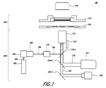
[0028] Figure 1 illustrates an exemplary system topology
for recovering a content of a sample
from a microcapillary array, in accordance with an exemplary embodiment of the
present disclosure.
[0029] Figure 2 provides a flow chart of processes and features of a
system for recovering a
content of a sample, in accordance with an exemplary embodiment of the present
disclosure.
[0030] Figure 3 illustrates a chart of various
coordinating components of a system with respect
to time, in accordance with an exemplary embodiment of the present disclosure;
[0031] Figure 4 illustrates a first view of a
microcapillary array and positioning and pulsing of a
laser towards the microcapillary array with respect to time, in accordance
with an exemplary
embodiment of the present disclosure;
[0032] Figure 5 illustrates a second view of a
microcapillary array and positioning and pulsing of
a laser towards the microcapillary array with respect to time, in accordance
with an exemplary
embodiment of the present disclosure.
[0033] Figure 6A-6B. Figure 6A illustrates a view of positioning and
pulsing of a laser towards
an internal portion of each microcapillary well in a subset of microcapillary
wells, in accordance with
an exemplary embodiment of the present disclosure. Figure 6B is an enlarged
fragmentary view of
Figure 6A.
[0034] Figures 7A, 7B, 7C, 7D, 7E, IF, 7G, 7H, 71, 7J,
7K, and 71_ collectively illustrate a variety
of positioning configurations of a laser towards an internal portion of a
microcapillary, in accordance
with an exemplary embodiment of the present disclosure.
100351 Figure 8A4B. Figure 8A illustrates a view of
positioning and pulsing of a laser towards
an a boundary portion of each microcapillary well in a subset of
microcapillary wells, in accordance
with an exemplary embodiment of the present disclosure. Figure 8B is an
enlarged fragmentary view
of Figure 8A.
5
CA 03156776 2022-4-29
WO 2021/092442
PCT/US2020/059485
[0036] Figure 9A-913. Figure 9A illustrates a progressive
series of positioning and pulsing
configurations of a laser and a boundary portion of a target, in accordance
with an exemplary
embodiment of the present disclosure. Figure 9B is an enlarged fragmentary
view of Figure 9A.
[00371 Figure 10 illustrates a graph of an amount of a
content recovery from a sample with
respect to a number of pulses of a laser towards the sample, in accordance
with an exemplary
embodiment of the present disclosure.
[0038] Figures 11A-1113 illustrate a first cross section
and a second cross section of a beam of
a laser respectively, in accordance with an exemplary embodiment of the
present disclosure,
[0039] Figure 12 provides a flow chart of a workflow for
sequencing a sample, in accordance
with an exemplary embodiment of the present disclosure.
[0040] Figures 13A-13C illustrate transferring of one or
more samples from a collection slide to
a PCR plate.
[0041] Figure 14 shows a method to recover a cell with
desired functional activity via a laser as
discussed in Example 2.
/0042] Figure 15 shows reverse transcription (PT) and polymerase chain
reaction (PCR)
preparation for Next-Generation Sequencing (NGS).
[0043] Figure 16 shows the binding analysis of the
xPloration B assay discussed in Example 2.
[0044] Figure 17 provides the results from the
quantification and sorting steps of the xPloration
B cell assay discussed in Example 2.
/0045] Figure 18 provides a heat map of the pairwise distance between
each cells concatenated
HCDR3-LCDR3 measured in the xPloration B cell assay discussed in Example 2.
[0046] Figure 19 shows clonotype clustering results of
the )(Placation B cell assay as discussed
in Example 2.
[0047] Figure 20 shows distinct subdomains of
progranulin, with broad coverage of the target
subdomains (A, B, G, P) as discussed in Example 2,
100481 Figure 21 shows the rarefaction curve of CDR3.
[0049] Figure 22 shows rarefaction curves of L3,
VII, and VK.
[0050] It should be understood that the appended drawings
are not necessarily to scale,
presenting a somewhat simplified representation of various features
illustrative of the basic principles
of the invention. The specific design features of the present invention as
disclosed herein, including,
for example, specific dimensions, orientations, locations, and shapes will be
determined in part by the
particular intended application and use environment_
6
CA 03156776 2022-4-29
WO 2021/092442
PCT/US2020/059485
(00511 In the figures, reference numbers refer to the
same or equivalent parts of the present
invention throughout the several figures of the drawing.
DETAILED DESCRIPTION OF THE INVENTION
[0052] The systems and methods of the present disclosure
provide laser-based single cell
recovery from microcapillary arrays for use in high-throughput analyses. The
present disclosure
meets an unmet need by providing systems and methods for laser-based single
cell recovery from
microcapillary arrays. The systems and methods of the present disclosure
provide a positional laser
that allows for a rapid recovery of a large number of samples from
microcapillary arrays. The
positional laser pulses a number of times towards the sample or a boundary of
the sample and the
microcapillary array to extract the sample to ensure optimal recovery of the
sample. Furthermore, the
positional laser transmits through an objective lens prior to illuminating the
sample, allowing for
simultaneous imaging and recovery of the sample. Additionally, the systems and
methods of the
present disclosure improve recovery of cells by allowing recovery an intact
cell, and therefore for
recovery and sequencing of a deceased cell into a dry environment, instead of
requiring that the cell
maintain cellular growth for extended culturing_ In some aspects of the
invention, the screening
methods do not rely on the recovery of a live cell, and therefore do not rely
on recovering in a wet
environment, thus significantly reducing the consumed time and improving the
efficiency of the
screening techniques.
[0053] Reference will now be made in detail to various
embodiments of the present disclosure,
examples of which are illustrated in the accompanying drawing and described
below. While the
disclosure will be described in conjunction with exemplary embodiments, it
will be understood that the
present description is not intended to limit the invention(s) to those
exemplary embodiments. On the
contrary, the invention(s) is/are intended to cover not only the exemplary
embodiments, but also
various alternatives, modifications, equivalents and other embodiments, which
may be included within
the spirit and scope of the present invention as defined by the appended
claims.
(0054] It will also be understood that, although the
terms first, second, etc. may be used herein
to describe various elements, these elements should not be limited by these
terms. These terms are
only used to distinguish one element from another. For instance, a first
microcapillary well could be
termed a second microcapillary well, and, similarly, a second microcapillary
well could be termed a
First microcapillary well, without departing from the scope of the present
disclosure. The first
microcapillary well and the second mictocapillary well are both microcapillary
wells, but they are not
the same microcapillary well.
10055] The terminology used in the present disclosure is
for the purpose of describing particular
embodiments only and is not intended to be limiting of the invention_ As used
in the description of the
invention and the appended claims, the singular forms "a," "an," and the" are
intended to include the
plural forms as well, unless the context clearly indicates otherwise. It will
also be understood that the
term "and/or" as used herein refers to and encompasses any and all possible
combinations of one or
7
CA 03156776 2022-4-29
WO 2021/092442
PCT/US2020/059485
more of the associated listed items. It will be further understood that the
terms -comprises" and or
-comprising," when used in this specification, specify the presence of stated
features, integers, steps,
operations, elements, and or components. but do not preclude the presence or
addition of one or
more other features, integers, steps, operations, elements, components, and/or
groups thereof_
[0056] As used herein, the term -11" may be construed to mean "when" or -
upon" or "in response
to determining" or "in response to detecting," depending on the context.
Similarly, the phrase "if it is
determined" or "if [a stated condition or event] is detected" may be construed
to mean "upon
determining" or min response to determining" or -upon detecting [the stated
condition or eventy or -in
response to detecting [the stated condition or event]," depending on the
context.
[0057] Furthermore, when a reference number is given an -Oh" denotation,
the reference number
refers to a generic component, set. Of embodiment. For instance, a
microcapillary well termed
"microcapillary well in refers to the P microcapillary well in a plurality of
microcapillary wells (e.g., a
rnicrocapillary well 500-i in a plurality of microcapillary wells 500).
[0058] In some embodiments, microcapillary wells are
long, through-holes with diameter small
enough such that the surface tension holds a liquid in place. In some
embodiments, the microcapillary
wells have one entry from the top where the liquid is loaded. In some
embodiments, the liquid in the
microcapillary wells is held via surface tension with no bottom. In some
embodiments, the opposite
end of the through-hole is where the sample is recovered from.
[0059] The foregoing description included example
systems, methods, techniques, instruction
sequences, and computing machine program products that embody illustrative
implementations_ For
purposes of explanation, numerous specific details are set forth in order to
provide an understanding
of various implementations of the inventive subject matter. It will be
evident, however, to those skilled
in the art that implementations of the inventive subject matter may be
practiced without these specific
details. In general, well-known instruction instances, protocols, structures
and techniques have not
been shown in detail.
(00601 The foregoing description, for purpose of
explanation, has been described with reference
to specific implementations. However, the illustrative discussions below are
not intended to be
exhaustive or to limit the implementations to the precise forms disclosed.
Many modifications and
variations are possible in view of the above teachings. The implementations
are chosen and
described in order to best explain the principles and their practical
applications, to thereby enable
others skilled in the art to best utilize the implementations and various
implementations with various
modifications as are suited to the particular use contemplated.
[0061] In the interest of clarity, not all of the routine
features of the implementations described
herein are shown and described. It will be appreciated that, in the
development of any such actual
implementation, numerous implementation-specific decisions are made in order
to achieve the
designers specific goals, such as compliance with use case- and business-
related constraints, and
that these specific goals will vary from one implementation to another and
from one designer to
another. Moreover, it will be appreciated that such a design effort might be
complex and time-
8
CA 03156776 2022-4-29
WO 2021/092442
PCT/US2020/059485
consuming, but nevertheless be a routine undertaking of engineering for those
of ordering skill in the
art having the benefit of the present disclosure.
100621 Terms used in the claims and specification are
defined as set forth below unless
otherwise specified. In the case of direct conflict with a term used in a
parent provisional patent
application, the term used in the instant specification shall control. "Amino
acid" refers to naturally
occurring and synthetic amino acids, as well as amino add analogs and amino
acid rnimetics that
function in a manner similar to the naturally occurring amino acids. Naturally
occurring amino acids
are those encoded by the genetic code, as well as those amino acids that are
later modified. e.g.,
hydroxyproline, y-carboxyglutamate, and 0-phosphoserine. Amino acid analogs
refer to compounds
that have the same basic chemical structure as a naturally occurring amino
acid, i.e., an a carbon that
is bound to a hydrogen, a carboxyl group, an amino group, and an R group,
e.g., homoserine,
nodeucine, methionine sulfoxide, methionine methyl sulfortium. Such analogs
have modified R
groups (e.g., norleucine) or modified peptide backbones, but retain the same
basic chemical structure
as a naturally occurring amino acid. Amino acid mimetics refer to chemical
compounds that have a
structure that is different from the general chemical structure of an amino
acid, but that function in a
manner similar to a naturally occurring amino acid. Amino acids can be
referred to herein by either
their commonly known three letter symbols or by the one-letter symbols
recommended by the IUPAC-
IUB Biochemical Nomenclature Commission. Nucleotides, likewise, can be
referred to by their
commonly accepted single-letter codes.
100631 An "amino acid substitutions' refers to the replacement of at
least one existing amino add
residue in a predetermined amino acid sequence (an amino acid sequence of a
starting polypeptide)
with a second, different "replacement" amino add residue. An "amino acid
insertion" refers to the
incorporation of at least one additional amino acid into a predetermined amino
acid sequence. While
the insertion will usually consist of the insertion of one or two amino acid
residues, the present larger
"peptide insertions," can be made, e.g., insertion of about three to about
five or even up to about ten,
fifteen, or twenty amino acid residues. The inserted residue(s) may be
naturally occurring or non-
naturally occurring as disclosed above. An "amino acid deletion" refers to the
removal of at least one
amino acid residue from a predetermined amino add sequence.
[0064] "Polypeptide." "peptide," and "protein" are used
interchangeably herein to refer to a
polymer of amino acid residues. The terms apply to amino acid polymers in
which one or more amino
acid residue is an artificial chemical mimetic of a corresponding naturally
occurring amino acid, as well
as to naturally occurring amino acid polymers and non-naturally occurring
amino add polymer.
[0065] The term "protein." as used herein, refers both to
full-length proteins or polypeptide
sequences and to fragments thereof. Such fragments may include fragments that
retain a functional
activity, such as, for instance, a binding activity. The terms "protein" and
"polypeptide" are used
interchangeably throughout the disclosure and include chains of amino acids
covalently linked through
peptide bonds, where each amino acid in the polypeptide may be referred to as
an "amino acid
residue." Use of the terms "protein" or "polypeptide" should not be considered
limited to any particular
9
CA 03156776 2022-4-29
WO 2021/092442
PCT/US2020/059485
length of polypeptide, e.g.., any particular number of amino acid residues.
The subject proteins may
include proteins having non-peptidic modifications, such as post-translational
modifications, including
glycosylation, acetylation, phosphorylation, sulfation, or the like, or other
chemical modifications, such
as alkylation, acetylation, esterification, PEGylation, or the like.
Additional modifications, such as the
inclusion of non-natural amino acids within a polypeptide sequence or non-
peptide bonds between
amino acid residues should also be considered within the scope of the
definition of the term "protein"
or "polypeptide."
(00661 In some embodiments, the population of variant
proteins is a population of proteins
having minor variations, for instance a population of proteins where each
protein has a slightly
different amino acid sequence era different post-translational modification.
In some embodiments,
the variant proteins can differ by 1, 2, 3, 4, 5, 6, 7, 8, 9, or 10 or more
amino adds. In some
embodiments, the variants differ by at least 1 amino acid. The screening
assays can, therefore.
identify variant protein sequences having desirable properties. Because the
screens can be
performed in such large numbers at microscopic scale, huge numbers of variant
proteins can be
assayed in relatively short times.
100671 "Nucleic acid" refers to deoxyribonudeotides or
ribonucleotides and polymers thereof in
either single- or double- stranded form. Unless specifically limited, the term
encompasses nucleic
acids containing known analogues of natural nucleotides that have similar
binding properties as the
reference nucleic acid and are metabolized in a manner similar to naturally
occurring nucleotides.
Unless otherwise indicated, a particular nucleic acid sequence also implicitly
encompasses
conservatively modified variants thereof (e.g., degenerate codon
substitutions) and complementary
sequences and as well as the sequence explicitly indicated. Specifically,
degenerate codon
substitutions can be achieved by generating sequences in which the third
position of one or more
selected (or all) codons is substituted with mixed-base and/or deoxyinosirte
residues (Batzer et al,
Nucleic Acid Res. 19:5081, 1991; Ohtsuka et at, Biol. Chem. 260:2605-2608,
1985; and Caw)l et at ,
1992; Rossolini etal., Mot Cell Probes 8:91-98, 1994). For arginine and
leucine, modifications at the
second base can also be conservative. The term nucleic acid is used
interchangeably with gene,
cDNA, and mRNA encoded by a gene. Polynucleotides used herein can be composed
of any
polyribonucleotide or polydeoxribonucleotide, which can be unmodified RNA or
DNA or modified RNA
or DNA. For instance, polynudeotides can be composed of single- and double-
stranded DNA, DNA
that is a mixture of single- and double- stranded regions, single- and double-
stranded RNA, and RNA
that is mixture of single- and double- stranded regions. hybrid molecules
comprising DNA and RNA
that can be single- stranded or, more typically, double- stranded or a mixture
of single- and double-
stranded regions. In addition, the polynucleotide can be composed of triple-
stranded regions
comprising RNA or DNA or both RNA and DNA. A polynucleotide can also contain
one or more
modified bases or DNA or RNA backbones modified for stability or for other
reasons. "Modified"
bases include, for instance, tritylated bases and unusual bases such as
inosine. A variety of
modifications can be made to DNA and RNA; thus, "polynucleotide" embraces
chemically.
enzymatically. or metabolically modified forms.
CA 03156776 2022-4-29
WO 2021/092442
PCT/US2020/059485
[0068] The term "barcode" as used herein is a label or
tag used for identification purposes. In
some embodiments, a barcode may be a sequence of nucleotides. In some
embodiments, a barcode
may be a single cell specific DNA level barcode. In some embodiments, one or
more single cell
specific DNA level barcodes may be used to identify heavy chains and/or light
chains of an antibody
sequence. In some embodiments, the same cell specific DNA level barcode is
added to the heavy
chain and the light chain of an antibody sequence being amplified.
00691 "Microcavity" and variations thereof refer to a
microcavity array comprising a plurality of
rnicrocavities, each microcavity comprising a sample component, including but
not limited to proteins,
polypeptides, nucleic acids, small molecules, and/or cells. The term
microcavity includes
microc,apillaries and/or microwells.
(0070] Additionally, the terms light guiding system,"
laser guiding system," and Imaging guiding
system" are used interchangeably herein unless expressly stated otherwise.
Further, the terms light"
and "beam" are used interchangeably herein unless expressly stated otherwise.
(00711 The term, lens," as used herein, includes a single
lens or an assembly of lenses unless
expressly stated otherwise.
(0072] Further, the term "target," as used herein, means
a feature pulsed by a beam of a light
source a number of times. A respective target can be subject to a single pulse
or a plurality of pulses_
(0073] An aspect of the present disclosure is directed to
providing a service for a recovery of a
content of a sample from a microcapillary array. Systems and methods for
recovering a content of a
sample from a microcapillary array are provided. The microcapillary array
includes a plurality of
rnicrocapillary wells. which are formed in a compact order. Each
microcapillary well in a subset of the
plurality of microcapillary wells accommodates a corresponding sample
including a content for
selective recovery. A laser positions to target a first microcapillary well of
the plurality of
microcapillary wells. The laser pulses at least one time towards the first
microcapillary well,
illuminating a portion of the first microcapillary well. The content from the
first microcapillary well is
extracted, recovering the content of the first microcapillary well.
100741 Figures, illustrates an exemplary topography of a
system 10 for recovering a content of a
sample. The system 10 includes a light guiding system 100 (e.g., a laser
guiding system) that
includes one or more light sources (e.g., a first light source 102 and a
second light source 116). Each
light source emits electromagnetic radiation (e.g., light) towards a
designated target. The illuminating
of a designated target provides imaging of the designated target and/or
recovery of a content of the
designated target depending on the respective light source. Accordingly, the
guidance system 100
directs the light emitted from sources the light sources towards a designated
target and controls one
or more operations of the light source 102.
100751 In some embodiments, the designated targeted includes a
microcavity array 132
comprising a plurality of microcavities. Each microcavity in the microcavity
array 132 accommodates
a corresponding sample. Accordingly, the laser guiding system 100 illuminates
a microcavity in the
11
CA 03156776 2022-4-29
WO 2021/092442
PCT/US2020/059485
microcavity array 132 to recover the respective sample from the corresponding
microcavity. In some
embodiments, the microcavity array 132 is a component of a sample stage 130,
which further includes
a collection slide 134 for receiving the content of the sample once extracted.
100761 In general, the microcavity array 132 includes an
array of a plurality of chambers (e.g.,
microcapillaries 500 of Figure 5). Each chamber accommodates a respective
sample having vadous
content. Further, each chamber of the rnicrocavity array 132 allows for a
transmission of light through
the chamber. This transmission of light is enabled be either a shape of the
chamber of and/or a
material of the chamber, which will be described in more detail infra.
100771 In some embodiments, the microcavity array 132
includes a microcapillary well array
having a plurality of microcapillary wells (e.g., a first microcapillary well
500-1, a second microcapillary
well 500-2, ..., an P microcapillary well 500-1, eta). Further, each
microcapillary well 500
accommodates a respective sample (e.g., a first microcapillary well 500-1
accommodates a first
sample 800-1, a second microcapillary well 500-2 accommodates a second sample
600-2, etc.). In
some embodiments, the microcapillary array 132 includes a plurality of
longitudinally fused capillaries
500. for instance fused silica capillaries. However, in some embodiments
another suitable material is
utilized for the microcapillary array 132. See, e.g., the arrays described
U.S. Application No.:
621433,210. filed December 12,2016; U.S. Application No.: 15/376,588, filed on
December 12, 2016:
PCT International Patent Publication Nos.: WO 2012/007537 and WO 20141008058,
each of which is
hereby incorporated by reference in its entirety.
[0078] In some embodiments, the microcapillary array 132 is fabricated,
for instance, by a
method including bundling millions or billions of silica capillaries and
fusing them together through a
thermal process. However, as described supra, in some embodiments, another
suitable material is
utilized for the microcapillary array 132. In some embodiments, the fusing
process includes, for
instance, heating a capillary single draw glass that is drawn under tension
into a single clad fiber. A
capillary multi draw single capillary is formed by bundling, heating, and
drawing the single draw glass.
Accordingly, a multi-draw multi-capillary is formed by additional bundling,
heating, and drawing the
multi-draw single capillary. A block assembly of drawn glass is further formed
by stacking the multi-
multi-draw multi-capillary in a press block. A block pressed block is formed
through by treating the
press block from the block assembly with heat and pressure. A block forming
block is then formed by
cutting the block pressing block at a predetermined length (e.g.. 1 millimeter
(ram)).
100791 In some embodiments, the fabrication method
further includes slicing (e.g., cutting) the
silica capillaries. This slicing forms a glass microcapillary array 132 with a
relatively high density of
microcapillaries per unit area. In some embodiments, the microcapillary array
132 is sliced a height of
approximately 1 mm. in some embodiments, the microcapillary array 132 includes
a plurality of
microcapillary wells 500, each having a height in a range of from I microns
(pm) to 25 arm, from 5 pm
to 20 mm, from 5 pm to 15 mm, from 10 pm to 15 mm, or from 10 pm to 10 mm.
However, the
present disclosure is not limited thereto. For instance, in some embodiments,
a height of the plurality
12
CA 03156776 2022-4-29
WO 2021/092442
PCT/US2020/059485
of microcapillary wells 500 is contemplated that is shorter than or longer
than the heights described
above.
100801 In some embodiments, each microcapillary well 500
in the microcapillary array 132 has a
uniform height. This uniform height of the microcapillary array 132 allows for
a surface (e.g.. an upper
surface andfor a lower surface) of each respective microcapillary well 500 to
be coplanar, or
substantially coplanar (e.g., coplanar within an acceptable tolerance known to
one skilled in the art),
with the plurality of microcapillary wells 500.
109811 Suth processes form a high-density microcapillary
array 132 that is suitable for use in the
present disclosure. In some embodiments, each microcapillary well 500 is
formed in a cylindrical
shape or a substantially cylindrical shape (e.g, a hemi-cylindrical shape, a
shape of a polygon with n-
skies of uniform length such as a hexagon, eta) with an internal diameter. In
such embodiments, an
equivalent characteristic dimension including a hydraulic diameter of non-
circular forms is used to
determine an internal diameter of a substantially cylindrical microcapillary
well 500. In some
embodiments, an internal diameter of each microcapillary well 500 in the
microcapillary array 132 is in
a range of from 0.5 pm to 500 pm, from 1 pm to 500 pm, from 1 pm to 300 pm.
from 25 pm to 250
pm, from 50 pm to 250 pm, from 50 pm to 200 pm, from 75 pm to 150 pm, from 75
pm to 125 pm,
from 75 pm to 110 pm, from 80 pm to 110 pm, from 1 pm to 100 pm, from 1 pm to
75 pm, from 1 pm
and 50 pm, from 5 pm to 50 pm, or from 1 pm to 10 pm. In some embodiments, the
internal diameter
of each respective microcapillary well 500 is 80 pm, 90 pm, 100 pm, 110 pm, or
a combination
thereof In some embodiments, the internal diameter of each respective
microcapillary well SOO is 1
pm, 5 pm, 10 pm, or a combination thereof In some embodiments, the internal
diameter of each
respective microcapillary well 500 is constant. For instance, in some
embodiments, the internal
diameter of each respective microcapillary well 500 in the microcapillary
array is 5 pm, 10 pm, or 100
pm. Furthermore, in some embodiments, the internal diameter of each respective
microcapiliary well
500 is constant. In some embodiments, the internal diameter transitions from a
first diameter at a first
end portion to a second diameter at a second end portion of the corresponding
microcapiliary well
500. In some embodiments, the internal diameter of the microcapillary well 500
includes a constant
portion and an inconstant portion.
[0082] In some embodiments, each respective
microcapillary well 500 includes an open region
representing a lumen of the microcapillary well 500. In some embodiments, a
proportion of one or
more microcapillary wells 500 in the microcapillary array 132 that is open is
in a range of from 40% to
95%, from 45% to 95%, from 50% to 90%. from 50% to 85%, from 55% to 80%, from
60% to 75%,
from 65% to 70%, or from 66% to 68% of microcapillary wells 500. In some
embodiments, the
proportion of one or more microcapillary wells 500 in the microcapillary array
132 that is open is 67%
of microcapillary wells 500. In some embodiments, the proportion of one or
more microcapillary wells
500 in the microcapillary array 132 is that as provided by a commercially
available microcapillary array
132, such as that of Hamamatsu Photonics K. K. (Japan).
13
CA 03156776 2022-4-29
WO 2021/092442
PCT/US2020/059485
(00831 In some embodiments, a collective open region
including each open region of each
rnicrocapillary well 500 in the microcapillary array 132 includes 90% of an
open area of the
microcapillary array 132 so that, if the internal diameter of each
microcapillary well 500 varies in a
range of from 1 pm to 500 pm, a number of microcapillary wells 500 per
centimeter squared (cm2) of
the microcapillary array 132 similarly varies in a range of from 460
microcapillary wells 500 to 1.1*106
microcapillary wells 500 or more. In some embodiments, the collective open
area of the
microcapillary array 132 includes approximately 67% of the open area, so that,
if a pore size (e.g.,
open area) varies between 1 pm and 500 pm, a number of microcapillaries wells
500 per cm2 of the
microcapillary array 132 varies in a range of from approximately 340 to over
800,000 microcapillaries
wells SOO. In some embodiments, the number of microcapillary wells 500 per cm2
of the
rnicrocapillary array 132 is in a range of from 500 microcapillary wells 500
to 1=107microcapillary wells
500.
100841 In some embodiments, the microcapillary array 132
includes a surface area of 10
centimeters (cm) by 10 cm across (e.g., a surface area of an upper surface
and/or lower surface of
the microcapillary array 132). Further, each microcapillary well 500 in the
microcapillary array 132
has an internal diameter of 5 pm and an open region of 66%. Accordingly, the
microcapillary array
132 includes approximately 3.3=10 microcapillary wells 500 (e.g.. a first
microcapillary well 500-1, a
second microcapillary well 500-2, ,.., a (3.3.108)th microcapillary well 500-
(3.3.108), eta). In some
microcapillary arrays, the open area of the array comprises up to 90% of the
open area (OA), so that,
when the pore diameter varies between 1 pm and 500 pm, the number of
microcapillaries per cm of
the array varies between approximately 460 and over 11 million. In some
microcapillary arrays, the
open area of the array comprises about 67% of the open area, so That, when the
pore size varies
between 1 pm and 500 pm, the number of microcapillaries per square cm of the
array vanes between
approximately 340 and over 800,000. In some embodiments, the pore size is 1
pm, 5 pm, 10 pm 50
pm, 100 pm, 250 pm 350 or 500 pm. In some embodiments, the pore size is
between 5 pm and 500
pm. In some embodiments, the pore size is between 10 pm and 450 pm. In some
embodiments, the
pore size is between 50 pm and 500 pm. In some embodiments, the pore size is
between 100 pm
and 500 pm. In some embodiments, the pore size is between 250 pm and 500 pm.
In some
embodiments, the pore size is between 350 pm and 500 pm. In some embodiments,
the pore size is
between 100 pm and 450 pm. In some embodiments, the pore size is between 250
pm and 450 pm.
In some embodiments, the number of microcapillaries per square cm of the array
is approximately
400; 500; 1000; 2,000; 3,000; 4,000; 5,000: 6,000; 7,000; 8,000; 9,000;
10,000; 20.000; 501000,
100,000; 200,000: 300,000; 400,000; 500,000: 600, 000; 700,000; or 800,000. In
some
embodiments, the number of micmcapillaiies per square cm of the array varies
between
approximately 500 and 800,000. In some embodiments, the number of
microcapillaries per square
cm of the array varies between approximately 1000 and 700,000. In some
embodiments, the number
of microcapillaries per square cm of the array varies between approximately
2000 and 600.000. In
some embodiments, the number of microcapillaries per square cm of the array
varies between
approximately 10.000 and 800,000. In some embodiments, the number of
microcapillaries per square
cm of the array varies between approximately 101000 and 700,000. In some
embodiments, the
14
CA 03156776 2022-4-29
WO 2021/092442
PCT/US2020/059485
number of microcapillaries per square cm of the array varies between
approximately 50,000 and
800,000. in some embodiments, the number of microcapillaries per square cm of
the array varies
between approximately 50,000 and 700,000. In some embodiments, the number of
microcapillaries
per square cm of the array varies between approximately 100,000 and 700,000.
In some
embodiments, the number of microcapillaries per square cm of the array varies
between
approximately 100,000 and 600,000. In some embodiments, the number of
microcapillaries per
square cm of the array varies between approximately 100,000 and 500,000_ In
some embodiments,
the number of microcapillaries per square cm of the array varies between
approximately 500,000 and
800,000.
100851 In some embodiments, the microcapillary array 132 is fabricated by
bending a plurality of
silica capillaries (e.g., 1=109capillaries) before fusing the silica
capillaries together through a thermal
fabrication process. The fused silica capillaries are sliced to a height of
approximately greater than or
equal to 0.5 mm, forming a glass microcapillary array 132 with a relatively
high aspect ratio (e.g.,
greater than or equal to 10, greater than or equal to 25. ... , greater than
or equal to 10,000, eta).
The aspect ratio of a microcapillary array 132 is a ratio of a height with
respect to an internal diameter
of a microcapillary well 500 of the microcapillary array 500. In some
embodiments, the aspect ratio of
the microcapillary array 132 is in a range of from 0.002 (e.g.. a
microcapillary well 500 having a 1 pm
height and a 500 prn internal diameter) to 50.000 (e.g., a microcapillary well
500 having a 25 mm
height and a 0.5 pm internal diameter), from 2 to 50,000, from 5 to 50,000,
from 5 to 25,000, from 10
to 25,000, from 10 to 15,0001 from 20 to 15,000, or from 25 to 10,000. In some
embodiments, the
inicrocapillary array 132 is a commercially available microcapillary array,
such as a microcapillary
array available from Hamamatsu 1photortics K. K. (Japan); Incom. Inc.
(Massachusetts); Photonis
Technologies, S.A.S. (France) Inc.: and others.
[0086] The microcapillary array 132 of the present
disclosure is not limited to a specific number
of microcapillary wells 500. In some embodiments, the number of microcapillary
wells 500 of the
microcapillary array 132 is determined in view of a size of a variant protein
library to be screened. For
instance, in some embodiments, the sample stage 130 of the system 10 can
accommodate a number
of different microcapillary arrays 132. In some embodiments, the
microcapillary array 132 includes a
number of wells in a range of from 1'104 microcapillary wells 500 to 5-1
microcapillary wells 500 or
greater. However, the present disclosure is not limited thereto_
100871 In some embodiments, the microcapillary array 132
has a thickness in a range of from
100 pm to 3,000 pm, from 150 pm to 2,500 pm, from 200 pm to 2,000 pm, from 500
pm to 2,500 pm,
from 500 pm to 2,000 pm, from 750 pm to 1,750 pm, or from 11000 pm to 1,500
pm. In some
embodiments, the thickness of the microcapillary array 132 corresponds to a
height of the
microcapillary wells 500. For instance, if the height of the microcapillary
wells 500 is 1 mm, the
corresponding thickness of the microcapillary array 132 is approximately 1 mm
(e.g., the thickness of
the microcapillary array 132 is 1 mm excluding an additional thickness
provided by a mounting
mechanism of sample stage 130, such as a bracket).
CA 03156776 2022-4-29
WO 2021/092442
PCT/US2020/059485
(00881 In some embodiments, the microcavity array 132 has
a thickness 01 1.5 mm and each
respective microcapillary well 500 has an internal diameter of 150 pm. In some
embodiments, the
microcavity array 132 has a thickness of 2 mm and each microcapillary well 500
has an internal
diameter of 200 pm. In some embodiments, the microcavity array 132 has a
thickness of 1 mm and
each microcapillary well 500 has an internal diameter of 100 pm. In some
embodiments, the
microcavity array 132 has a thickness of 1 mm and each microcapillary well 500
has an internal
diameter of 10 pm.
NM] In some embodiments, a volume provided by each
respective microcapillary well 500
(e.g., a volume of the sample for each respective microcapillary well 500) is
in a range of nanoliters
(nL) (e.g., 1 nL to 1,000 nL), in a range of picoliters (pL) (e.g., 1 pL to
1,000 pL), or in a range of
femtoliters (fL) (e.g.. 1 fl to 11000 fL). In some embodiments. a volume of
the sample accommodated
by each respective microcapillary well 500 is in a range of from 1 nanoliters
(nL) to 600 nL, from 5 nL
1o500 nL, from 5 nL to 450 nL, from 5 nL to 400 nL, from 5 nL to 350 nL, from
5 nL to 300 nL, from 5
nL to 250 nio, from 5 nL to 200 nL, from 5 nL to 150 nL, from 5 rite 100 nL,
from 5 nL to 90 iii., from
5 nL to 80 nL, from 5 nL to 70 nL, from 5 nL to 60 nL, frorn 5 nL to 50 ni,
from 5 nL to 40 nL, from 5
nL to 30 nL, from 5 rIL to 20 nL, from 5 it to 10 !IL, from 5 nL to 8 nL, or
from 7 nL to 8 nt., In some
embodiments, the volume of the sample accommodated by each respective
microcapillary well 500 is
7.8 nL Furthermore, in some embodiments, the volume of the sample accommodated
by each
respective microcapillary well 500 is in a range of from 50 pL to 150 pL, from
65 pL to 110 pt., from 70
pL to 100 pL, from 70 pi_ to 90 pL, or from 70 pL to 80 pL. In some
embodiments, the volume of the
sample accommodated by each respective microcapillary well 500 is 78.5 nL.
Additionally, in some
embodiments, the volume of the sample accommodated by each respective
microcapillary well 500 is
in a range of from 100 IL to 1000 fie from 150 IL to 1000 E.. from 200 to 1000
fmrn 250 IL to
1000 fL, from 300 IL to 1000 fL, from 350 ft_ to 1000 fL, from 350 IL to 950
IL, from 350 cloth 900 fL,
from 400 IL to 900 IL, from 450 IL to 900 IL, from 500 fL to 950 IL, from 500
it to 800 IL, from 100 IL
to 250 fL, from 150 fl.., to 250 IL. from 150 fL to 200 fL. or from 125 flo to
175 it. In some
embodiments, the volume of the sample accommodated by each respective
microcapillary well 500 is
157 fL.
100901 In some embodiments, each microcapillary well 500
in the microcapillary array 132 further
includes one or more agents disposed within the respective microcapillary well
500. The one or more
agents improve a viability of the cellular expression system if a cellular
expression assay is utilized
with the systems and methods of the present disclosure. In some embodiments,
the one or more
agents prevent cell damage while recovering the content of a microcapillary
well 500. For instance. in
some embodiments, the recovery of the content includes emitting a pulse of
light from a laser (e.g.,
laser 102 of Figure 1), and the one or more agents prevent the sample (ago
sample 6024 of Figure
6B) from being damaged by the laser 102.
100911 In some embodiments, the agent is methylcellulose
(for instance in a range of from 0.001
wt % to 10 wt %), dextran (for instance in a range of from 0.5 wt % to 10 wt
%), pluronic F-68 (for
instance in a range of from 0.01 wt % to 10 wt %), polyethylene glycol ("PEG')
(for instance in a range
16
CA 03156776 2022-4-29
WO 2021/092442
PCT/US2020/059485
of from 0.01 wt % to 10 wt %), polyvinyl alcohol CPVA") (for instance in a
range of from 0.01 wt % to
wt %), or the like.
100921 Alternatively, or in addition, in some
embodiments, each microcapillary well 500 in the
microcapillary array 132 further includes a growth additive, such as, for
instance, 50% conditioned
5 growth media, 25% standard growth media: or 25% serum. In some
embodiments, the conditioned
growth media is conditioned for approximately 24 hours. In some embodiments,
the added agent
includes insulin, transferrin, ethanolamine, selenium, an insulin-like growth
factor, or a combination of
thereof, or any of the agents recited above.
100931 It should also be understood that the
concentrations of each component of the screening
10 assay within a microcapillary well 500 can be modulated as desired in an
assay in order to achieve an
optimal outcome. In particular, it may be desirable to modulate the
concentration of proteins,
polypeptides, nucleic adds, small molecules, and/or cells to achieve a desired
level of association
between these components. The level of association will also depend on a
particular affinity between
these components, wherein a higher affinity results in a higher level of
association for a given
concentration of the components, and a lower affinity results in a lower level
of association of the
components for a given concentration. Concentration of various components may
likewise be
modulated in order to achieve optimum levels of signal output, as would be
understood by those of
ordinary skill in the art.
(00941 In some embodiments, each microcapillary well 500
includes to an open planar surface at
a first end portion and a second end portion of the microcapillary well 500.
Further, in some
embodiments, each corresponding open planar surface of the microcapillary
array is coplanar to the
corresponding open planar surfaces. In some embodiments, each microcapillary
well 500 includes a
through hole from a first planar surface to a second planar surface of the
microcapillary well 500.
However, the present disclosure is not limited thereto. In some embodiments,
the microcapillary array
132 includes a solid substrate coupled an end portion (e.g., a surface or a
portion of the surface) of
the microcapillary array 132, which forms a closed end portion of one or more
microcapillary wells 500
of the microcapillary array 132.
10095] In some embodiments, a respective sample (e.g.,
sample 602-i of Figure 6B) is
accommodated in the microcapillary wells 500 by surface tension. For instance,
in some
embodiments the rnicrocapillary wells 500 include an open surface at the first
end portion and the
second end portion of the rnicrocapillary well 500, such that the sample 602
is accommodated by
surface tension alone in microcapillary 500 with an expose first and second
end portions. In some
embodiments, the surface tension is the only force holding the sample in the
respective microcapillary
well 500. Accordingly, in such embodiments, pulsing of a beam of a laser
(e.g., beam 104 of laser
102 of Figure 1) towards the sample 602 disrupts the surface tension of the
respective microcapillary
well 500 and extracting the sample 602.
[0096] Libraries that can be screened according to the
present disclosure include any library
including a plurality of molecules as well as mixtures and/or combinations
thereof. In some
17
CA 03156776 2022-4-29
WO 2021/092442
PCT/US2020/059485
embodiments, the library includes samples including biological material In
some embodiments, the
library includes samples including a plurality of one or more molecules and/or
cells as well as mixtures
and/or combinations thereof In some embodiments, the library includes samples
including a plurality
of one or more proteins, polypeptides, nucleic acids, small molecules, dyes,
and/or cells as well as
mixtures and/or combinations thereof. In some embodiments, the small molecules
include any
molecule. In some embodiments, the molecules include proteins, polypepticles,
nucleic acids, small
molecules, and/or dyes as well as mixtures and/or combinations thereof In some
embodiments, the
library includes samples including biological materials that include
polypeptides, nucleic acids, small
molecules, and/or cells as well as mixtures and/or combinations thereof. In
some embodiments, the
library includes a plurality of samples. In some embodiments, the samples
include biological
materials that include polypeptides, nucleic acids, small molecules, dyes,
and/or cells as well as
mixtures and/or combinations thereof. In some embodiments, the samples contain
a least one
molecule and/or cell to be screened. In some embodiments, the samples contain
a least one to ten
molecules and/or cells to be screened, as well as mixtures and/or combinations
thereof. In some
embodiments, the samples contain a plurality of molecules and/or cells to be
screened, as well as
mixtures and/or combinations thereof. In some embodiments, the molecule to be
screened is termed
a target molecule. In some embodiments, the cell to be screened is termed a
target cell.
100971 The microcapillary array 132 provided herein
allows for screening of a library including
proteins, polypeptides, nucleic acid, small molecules, dyes, and/or cells, as
well as mixtures and/or
combinations thereof. In some embodiments, the target molecule to be screened
is a protein,
polypeptide, nucleic acid, small molecule, dye, carbohydrate, lipid, or a
combination of thereof In
some embodiments, the proteins and/or polypeptides are selected from the group
consisting of
enzymes, ligands, and receptors. For instance, in some embodiments, the target
molecule includes a
lipid-modified or glycosylated protein. In some embodiments, the target
molecule includes a native
protein.
[0098] As described above, in embodiments, each
microcapillary well 500 in the microcapillary
array 132 provided by the present disclosure accommodates a respective sample
having a content
that differs from the content of a sample of another microcapillary well 500
in the microcapillary array
132 (e,g, a first microcapillary well 500-1 accommodates a first sample 600-1,
a second
microcapillary well 500-2 accommodates a second sample 600-2, a third
microcapillary well 5004
accommodates a third sample 600-3. eta). Similarly, in embodiments, one or
more microcapillary
wells 500 in the microcapillary array 132 provided by the present disclosure
accommodates a
respective sample having a content that differs from the content of a sample
of another microcapillary
well 500 in the microcapillary array 132 (e.g., a first microcapillary well
500-1 accommodates a first
sample 600-1, a second microcapillary well 500-2 accommodates a second sample
600-2, a third
microcapillary well 500-3 accommodates the first sample 600-11 eta). In some
embodiments, the
content of the sample includes proteins. polypeptides, nucleic acids, small
molecules, dyes, and/or
cells (i.e., target molecules and/or target cells). as well as mixtures and/or
combinations thereof. In
some embodiments, the library for screening includes a variant protein, a
variant poiypeptide, a
18
CA 03156776 2022-4-29
WO 2021/092442
PCT/US2020/059485
variant nucleic acid, a variant small molecule, a variant dye, and/or one or
more variant cells
exhibiting distinguishing characteristics. In some embodiments, the variant
protein, the variant
polypeptide, the variant nucleic add, the variant small molecule, the variant
dye, and/or the one or
more variant cells exhibit distinguishing characteristics. These exhibited
distinguishing characteristics
allow each microcapillary well 500 to include a respective sample including a
different target molecule
anchor target cell from a corresponding sample accommodated by each of the
other microcapillary
wells 500 within the microcapillary array 132. In some embodiments, one or
more microcapillary wells
500 within the microcapillary array 132 includes a respective sample including
the same target
molecule and/or target cell as another sample accommodated by at least one
other microcapillary well
130 within the microcapillary array 132 (e.g., a respective sample is at least
duplicated for
comparison).
(00991 In some embodiments, the proteins and/or
polypeptides in the library to be screened in
the microcapillary array 132 include one or more variant proteins and/or
polypeptides. Variant
proteins include proteins and polypeptides that are distinguishable from one
another based on at least
one characteristic or feature. In some embodiments, the variant proteins
and/or polypeptides exhibit
different amino acid sequences, exhibit different amino acid sequence lengths,
are
produced/generated by different methods, exhibit different activities, exhibit
different chemical
modifications, exhibit different post-translational modifications, or a
combination thereof. In some
embodiments, the variant protein includes a population of variant proteins
andfor polypeptides that is
being subjected to screening and analysis utilizing the microcapillary array
132 of the present
disclosure. In some embodiments, the population of variant proteins anchor
polypeptides include any
population of proteins that is suitably distributable within the
microcapillary array 132.
1001001 In some embodiments, the nucleic acids in the
library to be screened in the microcavity
array includes one or more variant nucleic acids. Variant nucleic acids
include nucleic acids that are
distinguishable from one another based on at least one characteristic or
feature. In some
embodiments, the variant nucleic acids include different nucleotide sequences,
different nucleotide
sequence lengths, different rnethylation patterns, different chemical
modifications, are
produced/generated by different methods, exhibit other distinguishing
modifications, or a combination
thereof. In some embodiments, the nucleic add is of a population of variant
nucleic acids that is being
subjected to screening and analysis utilizing the microcapillary array 132 of
the present disclosure. In
some embodiments, the population of variant nucleic acids includes any
population of nucleic acids
that is suitably distributable within the microcapillary array 132.
1001011 In some embodiments, the small molecules in the
library to be screened in the
microcapillary array 132 includes variant and/or different small molecules.
Variant small molecules
include small molecules that are distinguishable from one another based on at
least one characteristic
or feature. In some embodiments, the variant small molecules include different
structures, have been
produced/generated by different methods, have different chemical
modifications, exhibit other
distinguishing different features, or a combination thereof. In some
embodiments, the small
molecules are derivatives of one another. In some embodiments, the small
molecule is of a
19
CA 03156776 2022-4-29
WO 2021/092442
PCT/US2020/059485
population of small molecules that is being subjected to screening and
analysis utilizing the
microcapillary array 132 of the present disclosure. In some embodiments, the
population of small
molecules includes any population of small molecules that is suitably
distributable within the
microcapillary array 132.
[001021 In some embodiments, the cells in the library to be screened in
the microcavity array
include variant cells and/or cells of varying types. Variant cells include
cells that are distinguishable
from one another based on at least one characteristic or feature. In some
embodiments, the cells are
derived from different samples, are derived from different patients, are
derived from different
diseases, have different chemical modifications, have been genetically
modified, or a combination
thereof. In some embodiments, the cells include eukaryotic and/or prokaryotic
cells. In some
embodiments, the cells include mammalian cells (e.g., human cells, rodent
cells such as mice cells
and rat cells, avian cells such as chicken cells, eta), bacterial cells,
fungal cells including yeast cells,
insect cells, or plant cells. In some embodiments, the mammalian cells include
blood cells,
lymphocyte cells, splenocyte cells, lymph node cells, bone marrow cells, or a
combination thereof. In
some embodiments, the cell is of a population of cells that is being subjected
to screening and
analysis utilizing the microcapillary array 132 of the present disclosure. In
some embodiments, the
population of cells include any population of cells that is suitably
distributable within the microcapillary
array 132.
1001031 In some embodiments, the population of proteins,
polypeptides, nucleic acids, and/or cells
is distributed in the microcapillary array 132 allowing each microcapillary
well 500 to accommodate a
small number of different variant proteins, variant polypeptides, variant
nucleic acid, and/or cells. In
some embodiments, each respective microcapillary well 500 includes a single
different variant protein,
variant polypeptide, variant nucleic acid, and/or cell. In some embodiments,
each respective
microcapillary well 500 includes a single different variant protein. In some
embodiments, each
respective microcapillary well 500 includes a single different variant
polypeptide. In some
embodiments, each respective microcapillary well 500 includes a single
different variant nucleic acid
per microcavity. In some embodiments, each respective microcapillary well 500
includes a single
different cell per microcavity. The population of variant proteins, variant
polypeptides, variant nucleic
acid, and/or cells is chosen in combination with other components within the
sample.
1001041 In some embodiments, each microcapillary well 500 in the
microcapillary array 132
includes different variant proteins, variant polypeptides, variant nucleic
acids, and/or cells from the
population of variant proteins in a range of from 0 different variants to 10
different variants, from 0
different variants to 7 different variants, from 0 different variants to 5
different variants, from 0 different
variants to 4 different variants, from 0 different variants to 3 different
variants, from 0 different variants
to 2 different variants, or from 0 different variants to 1 different variant.
1001051 Accordingly, in some embodiments, the variant
proteins include soluble proteins, for
instance, soluble proteins that are secreted by a cellular expression system.
Exemplary soluble
variant proteins include antibodies and antibody fragments, alternative
protein scaffolds such as
CA 03156776 2022-4-29
WO 2021/092442
PCT/US2020/059485
disulfide-bonded peptide scaffolds, extracellular domains of cell-surface
receptor proteins, receptor
ligands such as G-protein coupled receptor ligands, other peptide hormones,
tectins, and the like. In
some embodiments, the vadant proteins screened using the systems and methods
of the present
disclosure do not need to be covalently attached to the cell or virus that
expresses the respective
variant protein in order to be identified following a screening assay.
Recovery of the content of a
respective microcapillant well 500, followed by propagation of the cell or
virus clone responsible for
expression of the desired variant protein, enables the identification and
characterization of the
respective variant protein. Unlike screening assays in which a variant protein
is displayed by fusion of
the protein to a molecule on a surface of a cell or virus particle, the
variant proteins identified in
present disclosure need not be altered in any way either before or after their
identification. The
observed activities of the variant proteins in the screens are thus more
likely to represent the actual
activities of those proteins in their subsequent applications. Not needing to
alter variant proteins or
polypeptides prior to screening also allows for more efficient screening,
saving costs and time for
library preparation.
[00106] In some embodiments, the variant proteins to be screened include
membrane-associated
proteins, for instance, proteins typically associated with a surface of a cell
or a viral particle in an
expression system. In some embodiments, screening of cell-associated variant
proteins is desirable if
the variant protein and its target molecule mediate interactions between two
cells within a biological
tissue_ In some embodiments, the ability to screen cell-associated variant
proteins is desirable in
screening for interactions with traditionally "non-druggable protein targets
such as, for instance. G-
protein coupled receptors or ion channels. Again, not needing to after variant
proteins or polypeptides
prior to screening also allows for more efficient screening, which saves
resources during library
preparation.
1001071 In some embodiments, the variant nucleic acids to
be screened include any nucleic acid
or polynudeotide, including nucleic acids or polynucleotides that bind to or
interact with proteins.
Again, not needing to alter the nucleic acids or polynucleotides prior to
screening also allows for more
efficient screening, saving costs and time for library preparation.
1001081 in some embodiments, the protein to be screened is
an antibody, antibody fragment, such
as an Fe. or an antibody fusion. including, for instance, Fe fusions. in some
embodiments, the
antibody or antibody fragment can be labeled.
1001091 In some embodiments, the method employs the use of an antibody to bind
to the target
molecule to be screened. In some embodiments, the antibody is a labeled
primary antibody or a
labeled secondary antibody as is used to bind to the target molecules. A
primary antibody is typically
considered to be an antibody that binds directly to an antigen of interest,
whereas a secondary
antibody is typically considered to be an antibody that binds to a constant
region on a primary
antibody for purposes of labeling the primary antibody. Accordingly. secondary
antibodies are
frequently labeled with fiuorophores or other detectable labels Of are labeled
with enzymes that are
capable of generating detectable signals. The secondary antibodies are
generally specific for a
21
CA 03156776 2022-4-29
WO 2021/092442
PCT/US2020/059485
primary antibody from a different species. For instance, in some embodiments,
a goat or other animal
species is used to generate secondary antibodies against a mouse, chicken,
rabbit, or nearly any
primary antibody other than an antibody from that animal species, as is
understood by those of
ordinary skill in the art. In some embodiments, the labeled antibody is a
primary or secondary
antibody. In some embodiments, the labeled antibody is a fluorescent antibody
or an enzyme-linked
antibody.
1001101 As would be understood by those of ordinary skill
in the art, if a fluorescent antibody, for
example, is used in the systems and methods of the present disclosure, a
signal emitted by any
excess reporter element remaining free in solution (i..e., either not bound to
a variant protein or bound
to a variant protein that is not bound to a target molecule) within a
respective microcapillary well 500
should not be so high that it overwhelms a signal of reporter elements
associated with a target
molecule via a variant protein (see, e.g., the unassociated fluorescent
antibodies). Such background
signals can be minimized, however, by limiting the concentration of labeled
antibody or other reporter
element within the microcapillary solution. In addition, in some embodiments,
if signals from the
screening methods are measured using a fluorescent microscope, a microscope is
configured to
image a relatively narrow depth of field bracketing the location of the target
molecules (e.g., a lower
end portion of the microcapillary wells 500 if target cells have settled at
the lower end portion by
gravitational sedimentation) to minimize a background signal from reporter
elements not associated
with the target molecule.
1001111 In some embodiments, a number of rnicrocapillary wells 500 within
the microcapillary
array 132 is determined in view of a size of the library to be screened. In
some embodiments, a size
of the library is in a range of from 1 -104to 5-108 proteins. polypeptides,
nucleic acids, small molecules,
and/or cells, as well as mixtures and/or combinations thereof. However, the
present disclosure is not
limited thereto.
1001121 It would be understood by one of skill in the art that each
respective microcapillary well
500 will typically include a plurality of copies of the same protein,
polypeptide, nucleic acid, small
molecule, andior cell, depending on a source and an expression level of the
particular protein,
polypeptide, nucleic acid, small molecule, and/or cell, as well as mixtures
and/or combinations
thereof. In some embodiments, each respective microcapillary well 500 includes
a number of
molecules of a particular protein, polypeptide, nucleic acid, small molecule,
and/or cell in a range of
from 5-102to 1,10!0, depending on how the protein, polypeptide. nucleic acid.
small molecule, and/or
cell is delivered to or expressed within the respective microcapillary well
500 as well as mixtures
and/or combinations thereot in some embodiments, a number of types of
proteins, polypeptides,
nucleic acids, small molecules, and/or cells in a sample accommodated by a
respective roicrocapillary
well 500 is in a range of from 1 type to 10 types, from 1 type to 5 types, or
from 1 type to 4 types.
[001131 The population of proteins, polypeptides, nucleic
acids, and/or small molecules, as well as
mixtures and/or combinations thereof, is typically generated using a genetic
library in a biological
expression system, for example, in an in vitro (e.g., cell-free) expression
system or in an in vivo or
22
CA 03156776 2022-4-29
WO 2021/092442
PCT/US2020/059485
cellular expression system. in some embodiments, the population of proteins,
polypeptides, nucleic
acids, and/or small molecules, as well as mixtures and/or combinations
thereof, is generated via any
known synthesis methods. Exemplary cellular expression systems include, for
example, animal
systems (e.g., mammalian systems), fungal systems (e_g, yeast systems),
bacterial systems, insect
systems, or plant systems. In some embodiments, the expression system is a
mammalian system or
a yeast system. The expression system, whether cellular or cell-free,
typically includes a library of
genetic material encoding the population of variant proteins. Cellular
expression systems offer the
advantage that cells with a desirable phenotype, for example, cells that
express a particular variant
protein of interest, such as a variant protein capable of associating with an
immobilized target
molecule with high affinity, can be grown and multiplied, thus facilitating
and simplifying the
identification and characterization of the proteins of interest expressed by
the cells.
(001141 Genetic libraries encoding large populations of
proteins, polypeptides, nucleic acids,
and/or small molecules, as well as mixtures and/or combinations thereof, are
well known in the art of
bioengineering. Such libraries are often utilized in systems relying on the
process of directed
evolution to identify proteins with advantageous properties, such as high-
affinity binding to target
molecules, stability, high expression, or particular spectroscopic, e.g,
fluorescence, or enzymatic
activities. Often the libraries include genetic fusions with sequences from
the host expression system,
for example, fragments of proteins directing subcellular localization, where
the expressed population
of variant fusion proteins are directed by the targeting fragment to a
particular location of the cell or
virus particle for purposes of activity screening of the variant protein
population. Large numbers of
variant proteins, polypeptides, nucleic acids, small molecules, and/or cells
(e.g., 106 variants. 103
variants, 1010 variants, 1012 variants, or even more variants), as well as
mixtures andlor combinations
thereof, can be generated using routine bioengineering techniques, as is well
known in the art. In
some embodiments, the library is purchased from a commercial source.
1001151 However, the present disclosure is not limited thereto. In some
embodiments. the sample
includes inorganic material or a combination of organic and inorganic
material, such as an
environmental source (e.g., soil, water, vegetation, eta). n some embodiments,
the microscope
stage mounting allows for recovery of said microcapillary array contents with
a laser. In some
embodiments, the recovered contents comprise cells. In some embodiments. the
recovered contents
include live cells and/or deceased (e.g., dead) cells. In some embodiments,
the recovered cells are
further analyzed for the presence of one or more nucleic acids. In some
embodiments, the recovered
cells are further analyzed for the presence of one or more amino acids.
[001-146.1 Now that details of a microcapillary array 132 and
a rnicrocapillary well 500 have been
disclosed, details regarding a light guiding system 100 of the sampling system
10, in accordance with
an embodiment of the present disclosure, are disclosed.
1001171 Referring to Figure 1, the system 10 includes a
light guiding system 100 that facilitates
recovery and/or imaging of the content of the microcapillary array 132. The
system 10 further
includes a sampling stage 130 that accommodates the microcapillary array 132
and, in some
23
CA 03156776 2022-4-29
WO 2021/092442
PCT/US2020/059485
embodiments, aids in facilitating recovery and/or imaging of the content of
the microcapillary array
132.
1001181 In some embodiments, the system 10 includes one or more computers
(e.g, a computer
system) having a memory and processor. The memory stores one or more programs
for execution by
the one or more processors. The one or more programs singularly or
collectively include instructions
for utilizing the light guiding system 100 and/or the sampling stage 130. In
some embodiments, the
one or more instructions include positioning instructions for a light source
of the light guiding system
100 (e.g., positioning instructions for positioning a component on an optical
assembly of the light
guiding system 100, eta), operational instructions for the light source (e.g.,
activation and deactivation
instructions, intensity instructions, pulse parameter instructions, and the
like), and similar instructions
with respect to the sample stage 130 (e.g., positioning instructions).
1001191 The guiding system 100 includes one or more light sources that direct
electromagnetic
radiation towards a target to illuminate a portion of the target (e.g., the
microcapillary array 132). In
some embodiments, a first subset of the one or more light sources facilities
extracting and recovering
the content from a sample of a microcapillary well 500 (e.g., cell 604-1 of
500-i of Figure 66), while a
second subset of the one or more light sources facilitates imaging of the
sample. In some
embodiments, the first and second subset of light sources operate in concert,
allowing for real time
imagining of the microcapillary array 132 during recovery of the sample. In
some embodiments, the
light guiding system 100 includes a corresponding guiding system for one or
more light dependent
components of the system 10. For instance, in some embodiments the guiding
system 100 includes a
laser guiding system 100 for use at least in operating a laser 102 and
positioning a beam 104 of the
laser 102. In some embodiments, the guiding system 100 includes an imaging
guiding system for
use at least in operating one or more cameras 120.
1001201 Due to a relatively small size of diameter for
each microcapillary well 500 (e.g., a diameter
of less than 1 cm), the light guiding system 100 utilizes a focused beam of
light 104 to accurately and
precisely target a respective microcapillary well 500 within the
microcapillary array 132 during the
recovery of the content of the microcapillary array 132. Accordingly, in some
embodiments, the light
source includes a laser 102 that emits a coherent beam 104 of electromagnetic
radiation. In some
embodiments, the laser 102 is a laser diode_ In some embodiments. the laser
102 is a continuous
wave laser or, preferably. a pulse laser.
1001211 The laser 102 includes a pulse period defining a
frequency of pulses (e.g., 1 pulse per
second yields 1 Hertz (Hz), eta). In some embodiments, the pulse period of the
laser is in a range of
from 1 Hz to 60,000 Hz (e.g., 60 kilo-Hertz (kHz)), from 100 Hz to 60 kHz,
from 500 Hz to 60 kHz,
from 500 Hz to 50 kHz, from 500 Hz to 40 kHz, from I kHz to 40 kHz, from 1 kHz
to 30 kHz, from 1
kHz to 27.5 kHz, from 1 kHz to 25 kHz, from 1.5 kHz to 26 kHz. from 2 kHz to
25 kHz, from 1 kHz to
20 kHz, from 2 kHz to 20 kHz, from 2.5 kHz to 20 kHz, from 5 kHz to 25 kHz.
from 5 kHz to 20 kHz,
from 10 kHz to 40 kHz, or from 15 kHz to 25 kHz. In some embodiments, the
pulse period of the laser
102 is 20 kHz.
24
CA 03156776 2022-4-29
WO 20211092442
PCT/US2020/059485
[001221 in some embodiments, the laser 102 emits light 104
in the ultra-violet spectrum (e.g., a
UV laser) or in the visible spectrum (e.g., a visible spectrum laser).. hi
some embodiments, the beam
104 emitted from the laser 102 is a range of from 213 nanometers (nm) to 1380
mn. In some
embodiments, the beam 104 emitted from the laser 102 is selected from the
group consisting of 355
nm, 375 nm, 404 nm, 405 nm, 406 nm, 450 nm, 462 nm, 473 nm, 488 nm, 514 nm,
520 nm, 532 nm,
633 rim, 635 nm, 637 nm, 638 nm, 639 rim, 640 nm, 642 rim, 650 mil, 658 Elm,
660 rim, 670 run, 685
nm, 690 nm, and 1064 nm. In some embodiments, the wavelength of the beam 104
is selected in
accordance with a target of laser 102.
1001231 In some embodiments, the laser 102 is a
commercially available laser. In some
embodiments, the commercial laser 102 includes, for example, those available
from Thoriabs (see, for
example, those listed on the World Wide Web at
Thorlabs.cominewgrouppage9.cfm?objectgroup_id=7); Spectra-Physics (see, for
example. those
listed on the Word Vtide Web at spectra-physics.corniproductslq-switched-
lasersiexplorer-one);
andfor Integrated Optics (see, for example, those listed at
integratedoptics.comfproductsfrianosecond-
lasers).
1001241 In some embodiments, the guiding system 100 of system 10 includes an
objective lens
112 that intercepts a beam from the one or more light sources (e.g., beam 104
of the laser 102, beam
118 of a fluorescent light source 116, eta), and/or a field of view of the one
or more cameras 120.
For instance, as illustrated in Figure 1, in some embodiments the objective
lens 112 interposes
between the sample stage 130 and the one or more light sources, allowing the
objective lens 112 to
collect light from each of the one or more light sources. In some embodiments,
the objective lens 112
is based on a modified microscope. In some embodiments, the microscope
provides front-end image
collection and optical zoom with high light collection efficiency. In some
embodiments, the objective
lens 10 provides an optical magnification in a range of from Ito 100 optical
magnification, from 2 to
100 optical magnification, from 4 to 100 optical magnification, from 4 to 80
optical magnification, or
from 4 to 40 optical magnification. In some embodiments, the optical
magnification of the objective
lens 112 is 10.
[00125] In some embodiments, the objective lens 112 is
fixed, such that the sample stage 130
positions relative to the objective lens 112. In some embodiments, the
objective lens 112 is coupled
to a motor 114 that allows for a positioning of the objective lens 112. In
some embodiments, The
motor 114 provides one degree of freedom to the objection lens 112. in some
embodiments, the one
degree of freedom provided by the motor 114 is different than a degree of
freedom provided by a
galvanometer system 106 (e.g, the galvanometer system 106 provides two
translational degrees of
freedom in X and Y axes and the motor 114 provides one translational degree of
freedom in a Z axis
for redirecting the beam 104). Accordingly, in some embodiments, the objective
lens 112 is fixed on a
plane parallel to a corresponding plane of the sample stage 130 such that the
beam 104, after
passing through the objective lens 112, is directed towards the microcapillary
array 132 with an angle
of incidence of 0 degrees (1, or approximately about 00. This limited angle of
incidence allows the
objective lens 112 to provide a single field of view of the microcapillary
array 132. In such
CA 03156776 2022-4-29
WO 2021/092442
PCT/US2020/059485
embodiments, the single field of view allows the laser 102 to position from a
first target to a second
target without having to refocus the beam 104 or otherwise adjust the laser
102. Accordingly, in some
embodiments, the guiding system 100 and the sample stage 130 traverse relative
to one another,
such that the movement of the sample stage 130 provides a new field of view
for the objective lens.
Furthermore, this configuration with the limited angle of incidence allows for
imaging of the
microcapillary array during (e.g., simultaneous to) an extracting of the
content from a microcapillary
well 500.
(001261 In some embodiments, the one or more light sources
includes a fluorescent light source
116 that emits a corresponding beam 118 of light. In some embodiments, the
fluorescent light source
116 emits a beam 118 of light in the UV wavelength spectrum (e.g., in a range
of from 10 nm to 400
nm).
[001271 In some embodiments, the one or more light sources
include a bright-field light source
126. The bright-field light source 126 is disposed at an opposing end portion
of the microcapillary
array 132 with respect to the fluorescent light source 116, such that the
microcapillary array 132
interposes between the beam 118 of the fluorescent light source 116 and the
bright-field light source
126. In some embodiments, emitted light from the one or more light sources is
clireded from the
sample of a designated microcapillary well 500 to a detection unit (e.g., the
bright-field light source
126).
[00128] hi some embodiments, the laser guiding system 100
and the microcapillary array 132 are
each disposed at a fixed position, such that an optical train (e.g., a laser
scanning assembly)
facilitates guiding the beam 104 of the laser towards a designated target
(e.g., a first microcapillary
well 500-1, a portion of the -first microcapillary well 500-1, etc.). For
instance, in some embodiments,
the sampling stage 130 accommodates the microcapillary 132 in a fixed
position, preventing the
microcapillary array 132 from moving positions during recovery and/or imaging
of the content and
physical disruption to the content of the microcapillary array 132.
1001291 In some embodiments, the sampling stage 130
provides the microcapillary array 132 with
a plurality of degrees of freedom, yielding the microcapillary array 132 the
ability to position (e.g.,
traverse and/or rotate) accordingly. For instance, in some embodiments, the
microcapillary array 132
positions with six degrees of freedom, allowing the microcapillary array 132
to traverse and/or rotate
about three orthogonal axis (e.g., traverse about a three-dimensional
Cartesian coordinate system
and rotate with variable pitch, roll and yaw). In some embodiments, the
rnicrocapillary array 132
positions with only one or more translational degree of freedom. Accordingly,
in embodiments in
which the content of the microcapillary array 130 is fluid the content remains
at a substantially
horizontal free surface (e.g.. within an acceptable tolerance, excluding a
meniscus, eta). This
includes embodiments in comprising substantially fluid content (e.g., a
heterogeneous mixture
including solid and fluid matter) or the like.
[NUN Referring to Figure 3. in some embodiments, the laser guiding system 100
and the
microcapillary array 132 are movable relative to one another (e.g., both or
one of the laser guiding
26
CA 03156776 2022-4-29
WO 2021/092442
PCT/US2020/059485
system 100 and the microcapillary array 132 is movable), allowing the beam 104
of the laser 102 to
be guided in accordance with a relative movement of the system 10. In some
embodiments, the
guiding of the beam 104 is provided by at least a galvanometer 106. In some
embodiments, the
positioning of two or more components of the system 10 is conducted
sequentially, such that a first
component of the system 10 is positioned from a first position to a second
position. Once the first
component positions to the second position, the system 10 conducts positioning
of a second
component of the section from a third position to a fourth position_ For
instance, in some
embodiments, the system 10 conducts a first positioning of the sampling stage
130 and, upon
completion of the first positioning, conducts a second positioning of the beam
104 (e.g., via the
galvanometer 106). Furthermore, in some embodiments, the system 10 conducts a
pulsing of the
laser 102 upon completion of the second positioning of the beam 104. In some
embodiments,
conducting the positioning of the sample stage prior to the positioning of the
beam 104 allows for a
greater period of time to elapse between the completion of the positioning of
the sample stage 130
and the pulsing of the laser 102, further allowing the content of the
microcapillary array 132 to settle
from a transient disturbance occurring during the positioning of the sample
stage 130.
1001311 In some embodiments, the system 10 conducts a
plurality of instances of the positioning
of the beam 104 and, upon completion of the positioning of the beam 104,
pulsing of the laser 102
towards the designated target before repositioning the sample stage 130. In
some embodiments, the
plurality of instances of the positioning and the pulsing towards the
designated target is in a range of
from 1 instance to 100 instances, from 1 instance to 50 instances, from 1
instance to 25, from 1
instance to 15 instances, from 2 instances to 15 instances, from 1 instance to
12 instances, from 2
instances to 12 instances, from 1 instance to 10 instances, from 2 instances
to 10 instances, or from 4
instances to 10 instances. In some embodiments, the plurality of instances of
the positioning and the
pulsing towards the designated target is 5 instances.
1001321 Moreover. in some embodiments, pulsing of the beam 104 towards a
designated target
(e.g.. a first rnicrocapillary well 500-1) more than once is desirable for
recovery of the content of the
designated target. Accordingly, upon completion of the positioning of the
sample stage 130, the
system 10 conducts two or more instances of the positioning of the beam 104
and the pulsing of the
laser 102 towards the designated target. For instance, a coordination of the
positioning of the sample
stage 130, the positioning of the galvanometer 106, and the pulsing of the
laser 102 is depicted in
Figure 3. in the coordination of Figure 3, the system 10 conducts a first
positioning of the sample
stage 130 from a respective initial position to a first position, which occurs
over a period of time of
approximately 100 milliseconds (ms). Upon completion of the first positioning
of the sample stage
132, the system 10 conducts a first positioning of the beam 104 through the
galvanometer 106 from a
respective initial position towards the first position. This first positioning
of the beam 104, and
depending on a distance between respective positions, each respective instance
of the positioning of
the beam 104, occurs over a period of time of approximately 300 microseconds
(ps). Accordingly,
upon completion of the first positioning of the beam 104, the system 10
conducts a first pulsing of the
laser 102 towards the first position_ Each respective instance of the pulsing
the laser 102, occurs over
27
CA 03156776 2022-4-29
WO 2021/092442
PCT/US2020/059485
a period of time of approximately 20 nanoseconds (ns) (e.g., a pulse duration
of 20 ns). In some
embodiments the pulse duration is in a range of from ,001 its to 100 ns, from
.01 ns to 100 ns, from .1
ns to 100 ns, from less than 1 ns to 50 ns, from 1 INS to 50 its. from 0.1 ns
to 30 ns. from 0.1 ns to 25
ns, from 0.1 ns to 20 ns, or from 5 ns to 20 ns. Upon completion of the first
pulsing of the laser 102
towards the first position, the system 10 conducts a second positioning of the
beam 104 from the first
position to a second position. Upon completion of the second positioning of
the beam 104, the
system 10 conducts a second pulsing of the laser 102 towards the second
position. This series of the
positioning the beam 104 and the pulsing the laser 102 is repeated for a
predetermined number of
instances for one or more positioning of the sample stage 130. in the
coordination of Figure 3, the
positioning the beam 104 and the pulsing the laser 102 is repeated seven times
for the first position of
the sample stage 130_ However, the present disclosure is not limited thereto.
In some embodiments,
prior upon completion of each instant of the pulsing of the laser 102, the
system 10 is at least partially
at rest (e.g., not actively recovering the content from the microcapillary
array 132) for a period of time.
In some embodiments, this period of time of partial rest is utilized to
conduct imaging of the
microcapillary array 132.
100133] In some embodiments, the positioning of the sample
stage 130 is conducted at a rate in a
range of from 0.1 millimeters per second (minis) to 20 mm/s. from 0.1 mmis to
10 minis, from 0.5
mmis to 20 minis, from 0_5 MINS to 15 minis, from 1 minis to 15 minis, from 1
mints to 10 minis, from
5 minis to 15 rninis, from 5 minis to 10 minis, from 7.5 minis to 15 minis,
from or 7.5 minis to 12.5
minis, or from 9 ininis to 11 mm/s. In some embodiments, the positioning of
the sample stage 130 is
conducted at a rate of less than 10 or at a rate of
less than or equal to 10 mints. Furthermore,
in some embodiments, the positioning of the beam 104 (e.g., through the
guiding system 100) is
conducted at a rate in a range of from 10 minis to 1,500 mrn/s, 50 mints to
1,500 minis, from 50 minis
to 1,250 mints. from 100 minis to 1,250 minis. or from 100 minis to 1,000
mm/s. Moreover, in some
embodiments, the rate of the positioning of the sample stage 130 is conducted
at a slower rate than
the beam 104, and therefore at a slower rate than the guiding system 100. For
instance, in some
embodiments, a ratio of the rate of positioning of the sample stage 130 to the
rate of positioning of the
beam 104 via the guiding system 100 is in a range of from 1:5 to 1:1,500, from
1:5 to 1:1,250, from
1:10 to 1:1,500, from 1:1010 1,250, or from 1:10 to 1:1,000. In some
embodiments, the above
described rates of the positioning are an instantaneous velocity or an average
velocity taken over a
period of time of the positioning.
1001341 In some embodiments, the laser guiding system 100
includes an optical train, also
referred to as an optical assembly. The optical train includes an arrangement
of one or more light
manipulation instruments including one or more lenses (e.g., an objective lens
112 of Figure 1), one
or more filters, one or more mirrors (e.g.. mirror 1241 of Figure 1), or a
combination thereof employed
as part of the imaging system to, at least, guide the beam 104 emitted from
the laser 102. In some
embodiments, a position and/or an angle of each lens and/or mirror 124 is
adjusted to guide a beam
of a light source (e.g.. the beam 104 of the laser 104) towards a designated
target, and such
28
CA 03156776 2022-4-29
WO 2021/092442
PCT/US2020/059485
adjustments would be within the level of skill of one of skill in the art to
adjust as needed for an
imaging system_
1001351 In some embodiments, the optical train for the
instrument is based on a modified
microscope. In some embodiments, the microscope provides front-end image
collection and optical
zoom with high light collection efficiency. In some embodiments, the imaging
system includes a color
camera. In some embodiments, the imaging system includes a black and white
camera. In some
embodiments, the imaging system includes a color camera and a black and white
camera. In some
embodiments, the optical train is coupled to one or more emission filters
optimized for a particular
wavelength, fluorophore, and/or ratiornetric dye (e.g., mirror 124-1, mirror
124-2, and mirror 124-3 of
Figure 1).
1001361 In some embodiments, the optical train of the
laser guiding system 100 includes a laser
scanning assembly that further guides the beam 104 emitted from the laser 102
towards a designated
targeted, such as a first microcapillary well 500-1_ In some embodiments, the
laser scanning
assembly includes one or more mirrors 124 that redirect a course of the beam
104 of the laser 102.
For instance, in some embodiments the one or more mirrors 124 includes a first
mirror 124-1, a
second mirror 124-2, and a third mirror 124-3. In some embodiments, one or
more of the mirrors 124
is a filter. Further, in some embodiments, one or more of the mirrors 124 is a
dichroic. For instance,
in some embodiments, one or more of the mirrors 124 includes a material such
as a dichroic mirror or
a dichroic fitter. In some embodiments, the first mirror 124-1 is a reflective
mirror configured to
manipulate a field of view for one or more cameras 120 and direct fight (e.g.,
beam 122) towards the
one or more cameras 120. In some embodiments, the second mirror 124-2 includes
a dichroic
material that interacts with the electromagnetic radiation 118 emitted from
the light source 116. For
instance, in some embodiments, the second mintor 124-2 is an epifluoresceric.e
dichroic mirror.
Accordingly, in some embodiments, the second mirror 124-2 allows for a
transmission (e.g.. beam
122) of a first spectrum of light (e.g., visible light) captured by the one or
more cameras 120 to pass
through the second mirror 124-2, while reflecting transmission (e.g., beam
118) of a second spectrum
of light (e.g.. UV light) emitted from a fluorescent light source 116.
Furthermore, in some
embodiments, the third mirror 124-3 is a laser dichroic mirror. Accordingly,
in some embodiments, the
third mirror 124-3 allows for a transmission (e.g. beam 104) of a fourth
spectrum of light while
reflecting transmission (e.g., beam 104) of a third spectrum of light (e.g., a
wavelength of the beam
104). In some embodiments, the fourth spectrum of light includes the first and
second spectrums of
light.
[00131 In some embodiments, the optical assembly of the
light guiding system 100 includes a
galvanometer system 106_ The galvanometer system 106 includes a plurality of
mirrors and a
galvanometer that controls a positioning of each mirror in the plurality of
mirrors of the galvanometer
system 106. The beam 104 of the laser 102 is directed towards the galvanometer
system 106, arid in
accordance with a positioning of the plurality of mirrors, the beam 1004 is
directed towards either the
rnicrocapillary array 132 or a further component of the optical assembly of
the guiding system 100
(e.g., a scan lens 108, a tube lens 110, dichroic mirror 124-3, an objective
lens 112, or a combination
29
CA 03156776 2022-4-29
WO 2021/092442
PCT/US2020/059485
thereof). in some embodiments, the galvanometer system 106 allows for a
positioning of the beam
104 in one or more axis, such as two axis (e.g., an X-axis and a Y-axis of a
Cartesian system). in
some embodiments, the galvanometer system 106 includes a commercial
galvanometer system, such
as a Thorlabs Galvo Mirror Assembly (GVSM002) or a Scan nerMAX Cornpact-506RE.
As described
supra, in some embodiments the galvanometer system 106 positions at a rate
substantially greater
than a positioning rate of the sample stage 130 (e.g., by a factor of 10, a
factor of 100, a factor of
1000, etc.), allowing the laser 102 to emit pulses of the beam 104 towards
different targets in rapid
succession with a high level of accuracy and precision.
1001381 Referring to Figure 4, in some embodiments, the galvanometer system
106 directs the
beam 104 emitted from the laser 102 towards one or more defined coordinates at
high speed. For
instance, in some embodiments a surface of the microcapillary array 132 (e.g..
a surface facing the
objective lens 112), is mapped to a coordinate system. Accordingly, in some
embodiments, the
computer system of the system 10 provides instructions to the galvanometer
system 106 to direct the
beam 104 towards a predetermined series of defined coordinates. In some
embodiments, the laser
102 pulses the beam 104 an equal number of pulses at each defined coordinate
in the series. As
illustrated in Figure 4, the beam 104 of the laser 102 interacts with the
surface of an object with each
pulse of the laser. In the illustrated embodiment of Figure 4, the beam 104
first targets a first
coordinate and progresses through the series of defined coordinates in a
linear manner over a period
of time, which is occurs at a rate of approximately 500 beam pulses 104 per
second. However. the
present disclosure is not limited thereto. In some embodiments, instead of
progressing by rows or
columns of coordinates, the galvanometer system 106 directs the beam 104
emitted from the laser
102 towards a target in a cluster of targets. In some embodiments, the laser
pulses in a range of from
100 pulses per second to 1000 pulses per second. In some embodiments, the
laser pulses about 100
pulses per second, about 200 pulses per second, about 300 pulses per second,
about 400 pulses per
second, about 500 pulses per second, about 600 pulses per second, about 700
pulses per second.
about 800 pulses per second, about 900 pulses per second, or about 1000 pulses
per second. In
some embodiments, the laser pulses 500 pulses per second.
1001391 Referring to Figure 5, in some embodiments, the galvanometer system
106 directs the
beam 104 emitted from the laser 102 towards one or more microcapillary wells
500 (e.g, identified
targets) at high speed. For instance, in some embodiments each respective
defined coordinate
represents a position of a corresponding microcapillary well 500 in the
microcapillary array 132.
Accordingly, the galvanometer system 106 directs the beam 104 towards a first
defined coordinate of
a first microcapillary well 500-1, and the laser 102 is pulsed to extract the
content from the first
microcapillary well 500-1. The galvanometer system 106 further directs the
beam 104 towards a
second defined coordinate of a second microcapillary well 500-2, and the laser
102 is pulsed to
extract the content from the second microcapillary well 500-1. In Figure 5, a
relatively darker colored
microcapillary well 500 depicts a previously targeted microcapillary well 50.
[001401 In some embodiments, the galvanometer system 106 directs the beam 104
emitted from
the laser 102 towards a plurality of specific parts of each microcapillary
well 500 to improve sample
CA 03156776 2022-4-29
WO 2021/092442
PCT/US2020/059485
recovery. For instance, referring to Figures 6A and 66, in some embodiments,
the galvanometer
system 106 directs the beam 104 emitted from the laser 102 towards a plurality
of internal portions of
the microcapillary well 500. In the Figures, targets 600 (e.g., target 600-1
of Figure 6B, target 600-5
of Figure 7E, eta) represents a plurality of pulses of the laser 102 towards
the designated target
pulsed by the laser 102 (e.g., each of target 600-1 through target 600-5
represent approximately 10 to
20 individual pulses of the laser 102 at a rate of 20 kHz per target 600).
(001411 In some embodiments, the sample (e.g., sample 6024
including cell 604-1 of Figure 66)
of a microcapillary well 500 includes one or more opaque beads or a similar
absorbing material.
Accordingly, in some embodiments. illuminating the one or more opaque beads
with the beam 104 of
the laser 102 is desired to recovery the content from the microcapillary well
500. In such
embodiments, the beam 104 illuminates a portion of a visible surface area of
each of the one or more
opaque beads in order to maximize energy transfer to the absorbing material.
In some embodiments:
the beam 104 of the laser 102 is directed towards a plurality of targets that
collectively consume a
signification portion of a surface area of the microcapillary well 500 (e.g, a
portion in a range of from
30% to 100%). For instance, Figure 6A illustrates a plurality of
microcapillary 500-1 through 500-i,
each of which includes a plurality of targets 600 formed in a cross (e.g., *1-
1-") shape.
1001421 Referring to Figures 7A through IL, in some embodiments, a shape of
the plurality of
targets 600 pulsed by the laser 102 within a single microcapillary well 500 is
determined in
accordance with a cross section of the beam 104 and a shape of the
microcapillary well. In the
present embodiment, both the cross section of the beam 104 and the shape of
the microcapillary well
500 are assumed circular (e.g., a circular beam target 600-1 as illustrated in
Figure 11A). However,
the present disclosure is not limited thereto. In some embodiments, the cross
section of the beam 104
is of a non-circular shape (e.g., a crescent, or blade-like, shape target 6004
as illustrated in Figure
116). In some embodiments, the beam 104 is directed towards the target as a
point laser (e.g.,
without substantially altering a cross section of the beam. due to the
coherent nature of lasers). and
the target is illuminated in a corresponding circular shape. The
mathematically optimal solution for
filling a larger exterior circle (e.g, a microcapillary well 500) with a
plurality of uniform interior whole
(e.g., no concentric circles) circles (e.g., cross section of beam 104) is
described by a packing
efficiency of the above shapes and forms. This packing efficiency is a ratio
of an area of the Niger
circle with respect to an area collectively consumed by the plurality of
uniform smaller. See, Kravitz,
S., 1967, Packing Cylinders into Cylindrical Containers," Math. Mag., 40, pg.
65; Friedman. E.,
"Circles in Circles," Stetson University, print, each of which is hereby
incorporated by reference in its
entirety. In Figures TA through 7L, "r represents a radius of a smallest known
external circle (e.g.,
microcapillary well 500) with an increasing number of internal unit circles.
[001431 Referring to Figures BA through Figure 10, in some embodiments, the
sample is free of
absorbing material. Accordingly, extracting the content from the
microcapillary array 132 includes
directing (e.g., using the galvanometer system 106) the beam 104 of the laser
102 towards an
interface between the content of the sample and the microcapillary well 500
(e.g., an internal wall of
the microcapillary well 500). In some embodiments, the directing the beam 104
of the laser 102
31
CA 03156776 2022-4-29
WO 2021/092442
PCT/US2020/059485
towards the interface includes pulsing the laser towards a plurality of
targets. In some embodiments,
the plurality of targets 600 of the pulsing towards the interface is formed as
a drcle with each
respective target being at equally spaced interval around the circle. For
instance. referring to Figure
9A, a spacing internal for the plurality of targets for a number of targets in
a range of from 1 to 14
targets is depicted. While the present disclosure is not limited to a
particular number of targets in the
plurality of targets, Figure 10 illustrates an efficiency of recovery with
respect to the number of targets
As illustrated in Figure 10, in some embodiments an optimal number of targets
per microcapillary well
500 was determined to be greater than or equal to 5. In some embodiments an
optimal number of
targets per microcapillary well 500 was determined to be greater than or equal
to 6. In some
embodiments an optimal number of targets per microcapillary well 500 was
determined to be greater
than or equal to 8. In some embodiments an optimal number of targets per
microcapillary well 500
was determined by recovery percentage, wherein the recovery percentage was
equal to or greater
than 80%. In some embodiments an optimal number of targets per microcapillary
well 500 was
determined by recovery percentage, wherein the recovery percentage was equal
to or greater than
85%. in some embodiments an optimal number of targets per microcapillary well
SOO was determined
by recovery percentage, wherein the recovery percentage was equal to or
greater than 90%. In some
embodiments an optimal number of targets per microcapillary well 500 was
determined by recovery
percentage. wherein the recovery percentage was equal to or greater than 95%.
In some
embodiments an optimal number of targets per microcapillary well 500 was
determined by recovery
percentage. wherein the recovery percentage was equal to or greater than 96%.
In some
embodiments an optimal number of targets per microcapillary well 500 was
determined by recovery
percentage, wherein the recovery percentage was equal to or greater than 97%.
In some
embodiments an optimal number of targets per microcapillary well 500 was
determined by recovery
percentage, wherein the recovery percentage was equal to or greater than 98%.
In some
embodiments an optimal number of targets per microcapillary well 500 was
determined by recovery
percentage, wherein the recovery percentage was equal to or greater than 99%.
In some
embodiments an optimal number of targets per microcapillary well 500 was
determined by recovery
percentage, wherein the recovery percentage was equal to 100%. In some
embodiments an optimal
number of targets per microcapillary well 500 was determined to be greater
than or equal to 10.
However, the present disclosure is not limited thereto as the optimal number
can depend on a variety
of factors. Referring to Figure 9B, an order of firing the plurality of
targets 600 is illustrated for three
total targets. In the illustrated embodiment, the beam 104 of the laser 102 is
directed towards a first
target 600-1. and then positions to direct the beam 104 towards a second
target 600-1, and finally a
third target 600-3. While Figure 9B depicts the positioning and directing in
an orderly clockwise
manner, the present disclosure is not limited thereto. For instance, in some
embodiments, an order of
pulsing the plurality of targets 600 is determined to minimize a distanced
collectively traversed by the
beam 104 during transitions between respective targets.
1001441 In some embodiments, the galvanometer system 106 includes a
galvanometer-resonant
scanner. Utilizing a galvanometer-resonant scanner provides improved
positioning capabilities of the
guiding system 100. For instance, the galvanometer-resonant scanner improves a
rate of positioning
32
CA 03156776 2022-4-29
WO 2021/092442
PCT/US2020/059485
the laser 102 from a first microcapillary well 500-1 to a second
microcapillary well 500-2, or the like,
and therefore improves an elapsed time to extract and recovery the content
from the rnicrocapillary
array 132. For instance, in some embodiments. the galvanometer-resonant
scanner increases a
speed of recovering the content from the microcapillary tube in a range of
from a factor of 8 to a factor
of 12 (e.g., a ten-fold increase in throughput of recovery from an hour to six
minutes for a given
number of extractions and recoveries). In some embodiments, the galvanometer-
resonant scanner is
a commercially available galvanometer-resonant scanner, such as a Thorlabs LSK-
GR08.
1001451 Referring to Figures I IA and 11B, in some embodiments, the guiding
system 100 of the
system 10 includes one or more spatial light modulators that distort a shape
(e.g., cross section) of
the beam 102. allowing the beam 104 to pulse a target with a non-circular
shape. The one or more
spatial light modulators utilize a liquid crystal on silicon (LeoS) device
disposed between a first
transparent thin-film transistor (TFT) and a silicon semiconductor. The one or
more spatial light
modulators produce high-resolution, high-speed reflective phase modulation
with individually
addressable pixels, allowing each addressable pixel to direct a portion of the
beam 102
independently, and therefore, manipulate a cross section of the beam 104.
Furthermore, in such
embodiments in which the system includes the one or more spatial light
modulators, more than one
target can be pulsed with a single pulse of the laser 102 or, similarly, a
larger sized target can be
pulsed by the laser 102 within a single field of view, further decreasing an
elapsed time to extract and
recovery the sample from the rnicrocapillary array. For instance, Figure 11A
illustrates a cross
section of the beam 104 at a first target 600-1. which is typically associated
with a point laser. On the
other hand, a second target 600-2 of Figure 118 has a cross section that is
shaped in a crescent, or
blade-like, shape due to the add individually addressable pixels of the one or
more spatial light
modulators produce high-resolution. One skilled in the art will know of other
plausible cross sections
of the beam 104 that are not expressly contemplated herein. In some
embodiments. the one or more
spatial light modulators include a commercial spatial light modulator, such as
a Holoeye Pluto-2 or a
Thor Exulus-HD1.
1001461 In some embodiments, the system 10 utilizes a
digital micromirror device to direct the
beam 104 of the laser 102 towards a target in with a configurable cross-
section (e.g., as described
surpa). in some embodiments, the digital micromirror device is utilized in
substitute for the spatial
light modulator. The digital micromirror device includes a plurality of
mirrors disposed on a surface of
the device. In some embodiments, the plurality of mirrors are in a range of
from 1 -103 to mirrors 1-106
mirrors, from 1-104to mirrors 1-106 mirrors, from 1-104to mirrors 9-
105mirrors, from 1-104to mirrors
7=10 mirrors, or 5-104to mirrors 51O mirrors. In some embodiments, the digital
micromirror device
is a commercial digital microminror device, such as a Mirrorcle Integrated
MEMS Mirrors (A5M24.1-
2400AL).
1001471 In some embodiments, the optical train of the
laser guiding system 100 includes a scan
lens 108_ The scan lens 108 provides a flat image plane with minimal optical
aberration across the
plane and, in some embodiments, a relatively large field of view. In some
embodiments, the large
field of view is particularly useful for the present disclosure to the single
field of view of the objective
33
CA 03156776 2022-4-29
WO 2021/092442
PCT/US2020/059485
lens 112. In some embodiments, the scan lens 108 is a commercial scan lens
such as a Thorlabs
Large Field of View Scan Lens or a Thoriabs Scan Lens (1.3M03-VIS).
1001481 In some embodiments, the optical train includes a
tube lens 110. In some embodiments,
the tube lens 110 includes a telecentric tube lens. In some embodiments, the
tube lens 110 is a
commercially available tube lens, such as a Thoriabs Standard Tube Lens (T1L-
200) or a Thodabs
Laser Scanning Tube Lens (T1_200-CLS2).
1001491 In some embodiments, the laser guiding system 100
includes one or more high-resolution
cameras 120. In some embodiments, the one or more cameras 120 includes a black
and white
camera. In some embodiments, the one or more cameras 120 includes a color
camera. In some
embodiments, the laser guiding system 100 includes one real-time, high-
resolution camera and one
color camera. In some embodiments, the imaging system consists of one color
camera and one
monochrome camera, in order to expand the range of detection. However, the
present disclosure is
not limited thereto. For instance, in some embodiments the one or more cameras
120 includes a
thermal graphic camera (e_g., an infrared camera) aiding in the recovery of
the content of the
microcapillary array.
1001501 In some embodiments, while the two cameras see exactly the same field
of view. they
capture different information. For instance, the color camera captures RGB
light while the
monochrome (e.g, black and white) camera captures transmitted light in the
same field. In some
embodiments, to capture two different images one can employ a high-speed
pulsed light source that
is synchronized with the image capturing process. In some embodiments, to
capture two different
images one can employ a high-speed pulsed light source (e.g,, laser 102) in
combination with two
cameras.
1001511 In some embodiments, the guiding system 100
includes an imaging guiding system for
use imaging the rnicrocapillary array 132 using the one or more cameras 120.
In some embodiments,
the imaging guiding system facilitates control of the control or cameras 120,
such as control of a
capture sequence, image capture settings, and the like. In some embodiments,
the imaging guiding
system includes one or more mirrors 124-1 that provide a new field of view for
the one or more
cameras. In some embodiments, the one or more mirrors 124-1 includes a
corresponding subset of
mirrors for each camera 120 in the one or more mirrors, allowing each camera
to have a separately
configurable field of view. Further, in some embodiments, the one or mirrors
124-1 of the imaging
system are either fixed or provided one or more degrees of freedom.
1001521 In some embodiments, the optical train is coupled
to one or more emission filters
optimized for a particular wavelength, fiuorophom, and/or ratiometric dye.
1001531 Now that details of a system 10 for recovering a
content of a sample from a microcapillary
array 132 have been disclosed, details regarding a flow chart of processes and
features for
implementing a method 200 of the system 10, in accordance with an embodiment
of the present
disclosure, are disclosed with reference to Figure 2 through Figure 118.
34
CA 03156776 2022-4-29
WO 2021/092442
PCT/US2020/059485
(001541 Block 202 Referring to block 202 of Figure 2, a system (e.g., the
system 10 of Figure 1)
for recovering the content of a sample (e.g.. sample 602-1 of Figure GB) is
provided. The system 10
includes a microcapillary array 132 that further includes a plurality of
microcapillary wells 500. In
some embodiments, each respective microcapillary well 500 accommodates a
unique sample or a
replicate of a sample of a different microcapillary well 500. Accordingly,
each microcapillary well 500
is configured to accommodate a respective sample 602 of biological material.
As described supra,
the biological material, in some embodiments, includes one or more cell. These
one or more cells
include mammalian cells, fungal cells, bacterial cells, insect cells, plant
cells, or a combination thereof.
In some embodiments, the one or more cells is intact and no longer capable of
cellular growth.
1001551 In some embodiments, the microcapillary array is coupled to a
sample stage (e.g., sample
stage 130 of Figure 1), that assists in facilitating recovery of the sample.
In some embodiments, the
sample stage 130 includes a collection slide 134 that receives an extracted
sample 602.
(001561 Block 204. Referring to block 204, the method further includes
positioning the laser 102
to target a first microcapillary well 500-1 in the plurality of microcapillary
wells 500 of the
microcapillary array 132.
(001571 In some embodiments, the positioning the laser 102
utilizes a guiding system (e.g., a
laser guiding system 100 of Figure 1), which allows the laser 102 to remain
stationary while other
components of the system 10 (e.g., the objective lens 112 of Figure 1, the
sample stage 130 of
Figure 1, the galvanometer system 106 of Figure 1, eta) move relative to the
laser 102. In some
embodiments, the laser guiding system 100 includes the laser 102, a laser
scanning assembly (e,g., a
galvanometer system and/or mirror), a galvanometer resonance scanner, etc.), a
scan lens (e.g.. scan
lens 108 of Figure 1) system: and a tube lens (e.g,. tube lens 106 of Figure
1), In some
embodiments, the laser guiding system is a commercial scanner system, such as
a ScannerMAX
Compact-506RE system. Furthermore, in some embodiments a rate of positioning
of the sample
stage 130 is less than a rate of positioning of the laser guiding system 100.
In some embodiments,
the laser scanning assembly includes a device that alters a shape of the beam
from the laser 102. In
some embodiments, the device includes the galvanometer system and/or mirror
106, a digital
rnicrornirror device, one or more spatial light modulator, or a combination
thereof.
1001581 In some embodiments, the laser 102 emits a beam
(e.g., beam 104 of Figure 1) at a
discrete wavelength in a range of from 213 nm to 1380. For instance, in some
embodiments the
wavelength of the beam 104 from the laser 102 is 355 nrn, 514 nm. 532 nm, or
1064 nm.
[001591 In some embodiments, the first microcapillary well
500-1 is identified from the plurality of
microcapillary wells 500. For instance, in some embodiments, one or more
cameras (e.g., one or
more cameras 120 of Figure 1) and/or a fluorescence light source 116 image
some or the entire
microcapillary array 132 during the positioning of the laser 102, In some
embodiments, the one or
more cameras 120 image some or the entire microcapillary array 132 during an
extracting of the
content from the microcapillary array. In some embodiments, the microcapillary
well 500 for recovery
CA 03156776 2022-4-29
WO 2021/092442
PCT/US2020/059485
is identified through expression of an antibody from a cell accommodated in
the microcapillary well
500.
1001601 Block 206. Referring to block 206, the method includes pulsing the
laser 102 towards the
first microcapillary well 500-1_ The pulsing disrupts the surface tension of
the sample within the
microcapillary well, allowing for extraction of the sample. In some
embodiments, the laser 102 pulses
a plurality of subsections (e.g.. targets 600) of the first rnicrocapillary.
well 500-1. In some
embodiments, the plurality of subsections is disposed within an internal
portion of the microcapillary
well 500 (e.g., target 600-5 of Figure 6B), disposed at an interface between
an inner surface of the
microcapillary well 500 and the content of the microcapillary well 500 (e.g..
target 600-2 of Figure 8B),
or a combination thereof. In some embodiments, the laser 102 pulses in a range
of from 2
subsections to 20 subsections, from 2 subsections to 14 subsections, from 2
subsections to 14
subsections, or from 2 to 10 subsections. In some embodiments, the laser 102
pulses a plurality of
times at each respective subsection. For instance, in some embodiments, the
laser 102 pulses in a
range of from 1 time to 30 times at a subsection, 1 time to 25 times at the
subsection, 1 time to 20
times at the subsection, or greater than or equal to 5 times at the
subsection. Furthermore, in some
embodiments, the laser 102 pulses at a frequency in a range of from 1 kHz to
60 kHz, from 1 kHz to
40 kHz, from 5 kHz to 40 kHz, from 15 kHz to 25 kHz. or from 5 kHz to 10 kHz
(e.g., 5,000 to 10,000
laser pulses per second). Additionally, in some embodiments, each pulse of the
laser 102 has a
duration in a range of from 0.05 ns to 100 ns, from 0.1 in to 50 ns, from 0_1
in to 40 ns, from 0.1 F1S to
25 11S, from 0.1 ns to 20 in, or form 5 ns to 20 ns. In some embodiments, each
pulse of the laser 102
has a duration in a range of from 5 nanoseconds (ns) to 20 ns. In some
embodiments, each pulse of
the laser 102 has a duration in a range of from 5 nanoseconds (ns) to 15 ns.
In some embodiments,
each pulse of the laser 102 has a duration in a range of from 8 nanoseconds
(in) to 18 ns. In some
embodiments, each pulse of the laser 102 has a duration in a range of from 10
nanoseconds (ns) to
18 EIS. In some embodiments, each pulse of the laser 102 has a duration in a
range of from 10
nanoseconds (ns) to 15 ns. In some embodiments, each pulse of the laser 102
has a duration in a
range of 15 ns. In some embodiments, the duration of the pulse is
approximately about 15 ns. As an
example, in some embodiments, the laser 102 pulses 5 subsections (e.g.,
targets 600) of each
respective microcapillary well 500_ Each pulsing of a respective subsection of
each respective
rnicrocapillary well 500 emits 10 laser pulses. Further, each of the 10 laser
pulses has a duration of
15 in.
1001611 Block 206. Referring to block 208, the method
includes extracting the content of the
sample from the first microcapillary well 500-1 to recover the content. In
some embodiments, the
content from the microcapillary array is recovered in a collection slide
(e.g., collection slide 134 of
Figure 1), such that each sample recovered from a respective microcapillary
well 500 is received in a
corresponding well of the collection slide. In some embodiments, the wells of
the collection slide are
dry upon recovery of the sample (e.g., the well does not include a fluid). In
some embodiments, a
lysis buffer is added to the wefts of the collection slide after recovery of
the sample from microcapillary
array 130.
36
CA 03156776 2022-4-29
WO 2021/092442
PCT/US2020/059485
1001621 Now that details of a method 200 for recovering a content of a sample
from a
rnicrocapillary array 132 have been disclosed, details regarding workflow for
implementing a method
200 of the system 10, in accordance with an embodiment of the present
disclosure, are disclosed with
reference to Figure 12.
1001631 Now that details of workflow 1200 for recovering a content of a sample
from a
microcapillary array 132 have been disclosed, details regarding an example of
extracting, recovering,
and synthesizing the content, in accordance with an embodiment of the present
disclosure, are
disclosed.
1001641 In some embodiments of the workflow 1200, a step
(C) of extracting the content from the
first microcapillary well, thereby recovering the content of the first
microcapiliary well. In some
embodiments of the workflow 1200, the content of the extracting and recovering
extracting (e.g., cell
extraction 1210) is disposed onto a collection slide. In some embodiments, the
collection slide
comprises one or more collection wells containing a lysis buffer, wherein the
lysis buffer is added to
the one or more collection wells prior to the recovering the content. In some
embodiments, the
collection slide comprises one OF more collection wells which do not contain a
lysis buffer, wherein the
lysis buffer is not added to the one or more collection wells prior to the
recovering the content (C). In
some embodiments, the method further comprises, following the extracting and
the recovering (C),
disposing the content onto a collection slide and freezing the collection
slide. In some embodiments,
the collection slide is subsequently thawed. In some embodiments, the thawed
collection slide is
subjected to treatment to denature the RNA. in some embodiments, the thawed
collection slide
comprising the denatured RNA is subjected to RT-PCR amplification. In some
embodiments, the RT-
PCR amplification product is quantified. In some embodiments, the RT-PCR
amplification product is
sequenced. In some embodiments, the method further comprises, following the
extracting and the
recovering (C). disposing the content onto a collection slide and transferring
the content of the
collection slide to a PCR plate and freezing the PCR plate. In some
embodiments, the PCR plate is
subsequently thawed. in some embodiments, the thawed the PCR plate is
subjected to treatment to
denature the RNA. In some embodiments, the thawed the PCR plate comprising the
denatured RNA
is subjected to RT-PCR amplification.
1001651 In some embodiments, the sample being recovered comprises a population
of variant
proteins and/or a population of nucleic acids encoding the variant proteins,
which can in some
embodiments be generated using a genetic library in a biological expression
system, for example in
an in vitro (La, cell-free) expression system or in an in vivo or cellular
expression system. Exemplary
cellular expression systems include, for example, animal systems (e.g.,
mammalian systems), fungal
systems (e_g, yeast systems), bacterial systems, insect systems, or plant
systems. In specific
embodiments, the expression system is a mammalian system or a yeast system. In
spedfic
embodiments, the expression system is an avian system (for example, a chicken
system). The
expression system, whether cellular or cell-free, typically comprises a
library of genetic material
encoding the population of variant proteins. Cellular expression systems offer
the advantage that
cells with a desirable phenotype, for example cells that express a particular
variant protein of interest,
37
CA 03156776 2022-4-29
WO 2021/092442
PCT/US2020/059485
such as a variant protein capable of associating with an immobilized target
molecule with high affinity,
can be grown and multiplied, thus facilitating and simplifying the
identification and characterization of
the proteins of interest expressed by the cells. In some embodiments, the
biological expression
system comprises a mammalian cell line. In some embodiments, the mammalian
cell line is selected
from the group consisting of CHO-K1, CHO-S, HEK293T, and/or any derivatives of
these cell types.
In some embodiments, the mammalian cell line is CHO-Kl. In some embodiments,
the mammalian
cell line is CHO-S. In some embodiments, the mammalian cell line is HEK293T.
In some
embodiments, the mammalian cell line is selected from the group consisting of
human, mouse, and/or
rat hybridoma cell lines. In some embodiments, the mammalian cell line is a
human hybricloma cell
line. In some embodiments, the mammalian cell line is a mouse hybddoma cell
line. In some
embodiments, the mammalian cell line is a rat hybridorna cell line. In some
embodiments, the
mammalian cell line is a B cell line, hi some embodiments, the mammalian cells
comprise B cells.
Genetic libraries encoding large populations of variant proteins are well
known in the art of
bioengineering. Such libraries are often utilized in systems relying on the
process of directed
evolution to identify proteins with advantageous properties, such as high-
affinity binding to target
molecules, stability, high expression, or particular spectroscopic, e.g.,
fluorescence, or enzymatic
activities. Often the libraries include genetic fusions with sequences from
the host expression system,
for example fragments of proteins directing subcellular localization, where
the expressed population of
variant fusion proteins are directed by the targeting fragment to a particular
location of the cell or virus
particle for purposes of activity screening of the variant protein population.
Large numbers of variant
proteins (e.g., 108 variants, 108 variants, 1018 variants. 1012 variants, or
even more variants) can be
generated using routine bioengineering techniques, as is well known in the
art. Such libraries can
include any of the variant proteins described herein, including antibodies,
antibody fragments, single
chain variable fragments, or natural protein ligands. In some embodiments, the
system of the present
invention allows for accurate pairing of VH/1,./L (variable heavy chains and
variable light chains).
1001661 In some embodiments, the variant proteins (or
variant proteins encoded by nucleic acids)
are soluble proteins, for example, soluble proteins that are secreted by a
cellular expression system.
Exemplary soluble variant proteins include antibodies and antibody fragments,
alternative protein
scaffolds, such as disulfide-bonded peptide scaffolds, eidracellular domains
of cell-surface receptor
proteins, receptor ligands, such as, for example, G-protein coupled receptor
ligands. other peptide
hormones, lectins, and the like. In other embodiments, however, it may be
desirable for the variant
proteins to be membrane-associated proteins, for example, proteins remaining
associated with the
surface of a cell or a viral particle in an expression system. Isolation of
the contents of the desired
microcapillary according to workflow 1200 thereby enables the identification
and characterization of
contents.
1001671 In some embodiments, the content in the
microcapillary wells comprises genetic material.
In some embodiments, the sample comprises the genetic material of one or more
intact cells with a
desired phenotype. In some embodiments, the phenotype of the one or more
intact cells identifies B
cells. In some embodiments, the phenotype of the one or more intact cells
identifies subgroups of B
38
CA 03156776 2022-4-29
WO 2021/092442
PCT/US2020/059485
cells. In some embodiments, the genetic material comprises an antibody
sequence. In further
embodiments, the antibody sequence comprises a heavy chain and a light chain.
In some
embodiment the genetic material comprises rnRNA. hi further embodiments,
reverse transcription is
performed on the mRNA. In further embodiments, amplification of the heavy
chain and the light chain
is performed in separate reactions.
EXAMPLE 1: CELL EXTRACTION & LYSIS
1001681 Referring to Figure 12, a framework of a workflow 1200 for recovering
a content of a
sample from a microcapillary array 132 is provided.
[001691 Step 1210. Within the microcapillary array 132 a target sample is
identified, such as a
target anti-body of interest, for recovery and sequencing. Accordingly, a
laser (e.g., laser 102 of
Figure 1) extracts one or more cells from the microcapillary array 132 (e.g..
method 200 of Figure 2)
expressing the anti-body of interest. Upon extraction, each respective cell
(e.g., cell 604-1 of Figure
6B) from the microcapillary well 500 (e.g., microcapillary well 500-3 of
Figure 6B) is recovered in a
corresponding well of a collection slide (e.g., collection slide 134 of Figure
1). As described supra, an
aspect of the present disclosure is directed to providing recovery methods
that do not require the cell
to be capable of cellular growth upon extraction, but instead require the cell
to be intact. Accordingly,
the systems and methods of the present disclosure allow for extracting and
recovering of the cell into
a dry environment (e.g., an empty well).
1001701 Step 1220. A lysis buffer is prepared. The lysis buffer includes
water, a recombinant
RNAse inhibitor, triton (e.g., triton 100-X), a plurality of RT primers, and
dNTP mix. In some
embodiments, the lysis buffer contains water, recombinant RNAse inhibitor,
triton, RT primers, and
dNTP combination. Furthermore, in some embodiments, the RT primers are either
gene specific or
not gene specific. In some embodiments, the preparing the lysis buffer of step
1220 is conducted
prior to the extracting (Step 1210) of the workflow 1200 for timing
considerations.
1001711 The lysis buffer is supplied to each well of the
collection slide 134 (e_g_, collection slide
134 of Figure 1). in some embodiments, each well of the collection slide 134
includes at least 8 pl of
the lysis buffer. In some embodiments, each well of the collection slide 134
includes a minimum
volume of the lysis buffer that is required to submerge a lower end portion of
the well. In some
embodiments, the collection slide 134 includes 18 wells slide_ In some
embodiments, the workflow
1200 utilizes a plurality of collection slides simultaneously. In some
embodiments, the lysis buffer is
supplied to a subset of wells of the collection slide 134. In some
embodiments, the preparing and the
supplying the lysis buffer is conducted prior to the extracting (Step 1210),
allowing for recoveiy of the
cell into a wet environment. In some embodiments, upon supplying the lysis
buffer to each well of the
collection slide 134, an agitation is applied to the collection slide 13410
ensure that each cell is coated
with or submerged by the lysis buffer.
39
CA 03156776 2022-4-29
WO 2021/092442
PCT/US2020/059485
(001721 The samples of the collection slide 134 are transferred to a PCR plate
(e.g., PCR plate
1302 of Figure 13) for RNA denaturation and further analysis. As shown in
Figure 13A through
Figure 13B, the collection slide 134 is accommodated in a tray 1300 that
includes a first portion and a
second portion that removeably couple. In transferring the samples, each the
collection slide 134 has
an upward facing opening exposed to a surrounding environment (e_g., each well
of the collection
slide 134 is not capped) and accommodated in the first portion of the tray
1300, while one or more
PCR strip caps 1304 are accommodated in the second portion of the tray 1300.
In some
embodiments, the number of the one or more PCR strip caps 1304 corresponds to
a number of wells
of the collection slide 134 and/or the number of wells of the PCR plate 1302.
The PCR plate 1302 is
inverted, such that an opening of each well is facing towards the opening of
the collection slide, as
illustrated in Figure 13A. The second portion of the tray 1300 including the
PCR strip caps 1304 is
coupled to the first portion of the tray 1300. In some embodiments, the first
portion of the tray 1300
including the collection slide 134 is not reoriented until the second portion
of the tray 1300 is coupled
to the first portion (e.g, a lower end portion of the first portion of the
tray 1300 remains stationary and
fadng downwardly in the above-described portions of the transferring as
illustrated in Figures 134
and 13B). In some embodiments, once the tray 1300 is coupled and the
collection slide 134, the PCR
plate 1302, and the PCR cap strips 1304 are fixed within the tray 1300, the
tray 1300 is reoriented
such that the lower end portion of the first portion of the tray 1300 is now
facing upwards. as
illustrated in Figure 13C. In some embodiments, the coupled tray 1300
including the collection slide
134, the PCR plate 1302, and the PCR cap strips 1304 is accommodated by a
centrifuge, forcing the
cell lysate from the wells of the collection slide 134 into a lower end
portion the wells of the PCR plate
132 with little to no residual liquid in the wells of the collection slide 134
while also compacting the cell
lysate. In some embodiments, the centrifuging of the tray 1300 is conducted
for a period of time in a
range of from 0 to 180 s, from 30 s to 90 s, from 45 s to 75 s. In some
embodiments, the centrifuging
the tray 1300 is conducted for a period of time of approximately about 60
seconds. In some
embodiments, the centrifuging the tray 1300 is conducted at a relative
centrifugal force (e.g.. G force)
of in a range of from 2,000 G to 3,500 G, from 2250 G to 3,500 C. from 2,500 G
to 3,500 G. or from
2,750 G to 3,250 G. In some embodiments, the centrifuging the tray 1300 is
conducted at a relative
centrifugal force of approximately about 3,000 G.
[001731 In some embodiments, the initial volume of the cell lysate is
retrieved from the collection
slide 134. Accordingly, the initial volume is supplied to a first and a second
well of the PCR plate
wells, with allows for a cell lysate from one cell to be supplied to more than
one well. In some
embodiments, the initial volume is 8 micro-liters (pl.), allowing for an equal
distribution of 4 pi_ of the
cell lysate per well of each respective PCR plate from same cell. For
instance, in some embodiments
the first well is utilized for light chain sequencing and the second well for
heavy chain sequencing.
1001741 Once the one or more cells is extracted, recovered, and transferred to
the PCR plate, the
PCR plate is exposed to a rapid negative temperature differential (e.g., snap
freeze) to cool the cells
and solidify the content of the collection slide 134. This cooling of the
cells completes the lysis of the
cell and preserving the genornic content of the cell. In some embodiments, the
rapid cooling of the
CA 03156776 2022-4-29
WO 2021/092442
PCT/US2020/059485
cells is conducted by exposing the cells to a temperature in a range of from -
90 cC to -70 C, -88 C to
-72 C, -86 C to -74 C, -84 C to -76 C, or from -82 C to -78 C. In some
embodiments, the rapid
cooling of the cells is conducted by exposing the cells to a temperature of
approximately about -80 C.
In some embodiments, the rapid cooling of the cells is conducted by
accommodating, and thereby
exposing, the cells to solid carbon dioxide, also known as dry ice. In some
embodiments, the rapid
cooling of the cells is conducted for a period of time in a range of from 2.5
minutes to 7.5 minutes,
from 3 minutes to 7 minutes, from 3.5 minutes to 6.5 minutes, from 4 minutes
to 6 minutes, or from
4.5 minutes to 5.5 minutes. In some embodiments, the rapid cooling of the
cells is conducted for a
period of time of approximately about 5 minutes. Accordingly, the collection
slide 134, is thawed
bringing the contents to a fluid phase. In some embodiments, the thawing is
conducted in by
exposing the cells to a temperature in a range of from 0 C to 10 C, from 0
C to 8 C, or 2 CC toe
C. In some embodiments, the thawing is conducted in by exposing the cells to a
temperature of
approximately about 4 C. In some embodiments, the thawing is conducted on a
bed of ice. In some
embodiments, the freezing and thawing is conducting while the cell lysate is
accommodated within the
collection slide 1341 such that the cell lysate is transferred to the PCR
plate 1302 after thawing within
the collection slide 134.
1001751 Step 1230. In some embodiments, such as that in
which the collection slide 134 is frozen
and thawed, the PCR plate 1302 is rotated at high speed, forcing the cell
lysate to a lower end portion
of the respective well In some embodiments, the rotating the PCR plate 1302 is
as described supra
with respect to the centrifuging the tray 1300. Once exposed to the high
rotational speed, the PCR
plate 1302 is accommodated in a thermal cycler at a first predetermined
temperature for a first
predetermined period of time. In some embodiments, the first temperature of
the thermal cycler is in a
range of from 78 C to 66 *C, from 76 C 1o68 C, or from 74 C 1o70 C. In
some embodiments, the
first temperature of the thermal cycler is approximately about 72 C. In some
embodiments, the first
predetermined period of time of exposure to the thermal cycler is in a range
of from 2 minutes to 5
minutes, from 2 minutes to 4 minutes, or from 2.5 minutes to 3.5 minutes. in
some embodiments, the
first predetermined period of time of exposure to the thermal cycler is
approximately about 3 minutes.
Furthermore, in some embodiments, the PCR plate 1304 is maintained at a
predetermined
temperature in-between the steps of rotating the PCR plate 1302 at a high
speed and accommodating
the PCR plate 1302 within the thermal cycler. In some embodiments, the
predetermined temperature
is in a range of from 0 C to 10 C, from 0 C to 8 C. or 2 C to 6 C. In
some ernbodiments, the
predetermined temperature is approximately about 4 C. In some embodiments,
the maintaining at
the predetermined temperature is conducted on a bed of ice_ Accordingly, the
collection slide 134 is
subjected to treatment to denature the RNA.
1001761 Step 1240. A reverse transcription (RD formulation is prepared. In
some embodiments,
the reverse transcription formulation is prepared prior to each use (e.g. each
instance of the workflow
1200). The reverse transcription formulation includes a 10x RT buffer, water,
a recombinant RNAse
inhibitor, and a reverse transcriptase. A volume of the RT formulation is
supplied to each well of the
PCR plate 1302 that includes a cell lysate. In some embodiments, the volume of
the RT formulation
41
CA 03156776 2022-4-29
WO 2021/092442
PCT/US2020/059485
is in a range of from 2 pL to 10 It or from 4 pia to 8 pie In some
embodiments, the volume of the RT
Formulation is approximately about 6 pie Accordingly, the PCR plate 1302
including the cell lysates
and RT formulation is accommodated in the thermal cycler at a second
predetermined temperature for
a second predeteimined period of time. In some embodiments, the second
temperature of the
thermal cycler is in a range of from 48 C to 36 C, from 46 C to 38 C, or
from 44 C 10 40 C. In
some embodiments, the second temperature of the thermal cycler is
approximately about 32 C. in
some embodiments, the second predetermined period of time of exposure to the
thermal cycler is in a
range of from 80 minutes to 100 minutes, from 82 minutes to 98 minutes, from
84 minutes to 96
minutes, from 86 minutes to 94 minutes, or from 88 minutes to 92 minutes. In
some embodiments,
the second predetermined period of time of exposure to the thermal cycler is
approximately about 90
minutes. This second accommodating in the thermal cycler provides reserve
transcription.
(001771 Once reverse transcription is complete, the PCR plate 1302 is further
accommodated in
the thermal cycler at a third predetermined temperature for a third
predetermined period of time. In
some embodiments, the third temperature of the thermal cycler is in a range of
from 78 C to 62 C,
from 76 C to 64 C, from 74 C to 66 C, or from 72 C to 68 C. In some
embodiments, the third
temperature of the thermal cycler is approximately about 70 C. In some
embodiments, the third
predetermined period of time of exposure to the thermal cycler is in a range
of from 10 minutes to 20
minutes, from 12 minutes to 18 minutes, or from 14 minutes to 16 minutes. In
some embodiments,
the third predetermined period of time of exposure to the thermal cycler is
approximately about 15
minutes. This third accommodating in the thermal cycler provides reserve
transcriptase.
Furthermore, in some embodiments, the PCR plate 1304 is maintained at a
predetermined
temperature in-between each step in a subset of steps of the above-described
reserves transcription
of the workflow 2100. In some embodiments, the predetermined temperature is in
a range of from 0
C to 10 C. from 0 C to 8 C, or 2 C to 6 C. In some embodiments, the
predetermined
temperature is approximately about 4 C. In some embodiments, the maintaining
at the
predetermined temperature is conducted on a bed of ice.
1001781
Step 1250. A PCR formulation is
preparing including a PCR master formulation, water,
and gene-specific forward and/or reverse primers. A volume of the PCR
formulation is supplied to
each reaction well of the PCR plate 1304. In some embodiments, the volume of
the PCR formulation
is in a range of from 10 pL to 20 pL, from 12 pi_ to 18 pL, or from 14 pL to
16 pL. In some
embodiments, the volume of the PCR formulation is approximately about 15 pL.
After stippling the
PCR formulation. the PCR plate 1304 is accommodated in the thermal cycler for
a thermal cycle. In
some embodiments, the thermal cycle includes exposing the PCR plate 1304 to a
fourth
predetermined temperature for a fourth predetermined period of time, exposing
the PCR plate 1304 to
a fifth predetermined temperature for a fifth predetermined period of time,
and exposing the PCR plate
1304 to a further sub-thermal cycle of the thermal cycle. In some embodiments,
the fourth
predetermined temperature is approximately about 37 'C and the fourth
predetermined period of time
is about approximately 30 minutes. In some embodiments, the filth
predetermined temperature is
approximately about 95 C and the fifth predetermined period of time is about
approximately 3
42
CA 03156776 2022-4-29
WO 2021/092442
PCT/US2020/059485
minutes. In some embodiments, the sub-thermal cycle of the thermal cycle
includes a first condition
exposing the PCR plate 1304 to a sixth predetermined temperature for a sixth
predetermined period
of time, a second condition exposing the PCR plate 1304 to a seventh
predetermined temperature for
a seventh predetermined period of time, and a condition exposing the PCR plate
1304 to an eighth
predetermined temperature for an eighth predetermined period of time. In some
embodiments, the
sixth predetermined temperature is approximately about 98 QC and the sixth
predetermined period of
time is about approximately 20 s. In some embodiments, the seventh
predetermined temperature is
approximately about 65 C and the seventh predetermined period of time is
about approximately 15 s_
In some embodiments, the eighth predetermined temperature is approximately
about 72 C and the
eighth predetermined period of time is about approximately 1 minute. In some
embodiments, the sub-
thermal cycle of the thermal cycle is repeated for a number of repetitions
before proceeding with the
workflow 1200. In some embodiments, the number of repetitions of the sub-
thermal cycle in a range
of from 40 repetitions to 50 repetitions, from 42 repetitions to 48
repetitions, or 44 repetitions to 46
repetitions. In some embodiments, the number of repetitions of the sub-thermal
cycle is 45
repetitions. In some embodiments, the third condition of a final repetition of
the sub-thermal cycle is
maintained for an additional period of time (e.g., 6 minutes for the final
repetition instead of 1 minute
for each subsequent repetition). In some embodiments, the additional period of
time is in a range of
from 1 minutes to 10 minutes. or from 1 minutes to 9 minutes, from 3 minutes
to 7 minutes, or from 4
minutes to 6 minutes. In some embodiments, the additional period of time is 5
minutes. Keep at 4 C
(or on ice). Furthermore, in some embodiments, the PCR plate 1304 is
maintained at a
predetermined temperature in-between each step in a subset of steps of the
above-described
reserves transcription of the workflow 2100. In some embodiments, the
predetermined temperature is
in a range of from 0 C to 10 QC, from 0 QC to 8 C, or 2 C to 6 QC. In some
embodiments, the
predetermined temperature is approximately about 4 C. In some embodiments,
the maintaining at
the predetermined temperature is conducted on a bed of ice. Accordingly, the
PCR plate 1302
subjected to RT-PCR amplification. In some embodiments, a period of time
[001791 Step 1260. Conduct sequencing to recover genome of the RT-PCR
amplification product.
In some embodiments, the RT-PCR amplification product is quantified.
1001801 Accordingly, the present disclosure provides improved systems and
methods for
extracting and recovering a single cell from a microcapillary array. The
extracting of the cell is
conducted with a high level of precision and accuracy, allowing a laser to
target one or more portions
of a microcapillary array to optimize the extraction and recovery. Moreover,
the systems and methods
of the present disclosure yield a high throughput (e.g., greater than or equal
to approximately about
11 0 extractions per hour. This combination of the high throughout with the
high level of precision
and accuracy in targeting yields vastly improves a quality of extraction and
efficiency of recovery.
Further, the systems and methods of the present disclosure to not require the
inclusion of additional
sample components or manipulations, such as radiation absorbing materials,
thus simplifying and
improving the efficiency of the screening techniques_ Moreover, the systems
and methods of the
present disclosure allow for recovery an intact cell, that is not required to
be capable of cellular growth
43
CA 03156776 2022-4-29
WO 2021/092442
PCT/US2020/059485
(e.g., alive). This recovery of an intact, but not growing, cell allows for
the recovery of the cell into a
dry environment, as compared to requiring a wet environment to sustain the
cell. Additionally,
recovery of an intact, but not growing, cell according to the systems and
methods of the present
disclosure allows a designer of the present disclosure to omit culturing the
cell over a period of days,
greatly reducing an elapsed period of time from an initiation of the
extracting of the cell to an initiation
of the sequencing of the cell.
EXAMPLE 2: XFLORATION NGS WORKFLOW
[001811 In another example, the present disclosure further
comprises methods of recovering
desired genetic material from cells with desired phenotype in
microcapillaries. A high level overview is
presented below arid in Figures 14 and 15:
[001821 Step 1: In this step, cells with desired
functional activity are recovered via a laser based
on the methods comprised in the present disclosure.
1001831 Step 2: Next , the cells undergo RT and PCR preparation for NGS based
on the
methods comprised in the present disclosure. These steps include lysis,
reverse transcription, single
cell barcoding, plate-wide barcoding, and result in NOS ready sequences.
1001841 xPloration B cell assay: First, mAb secretor (OmniChicken splenocytes
(-1 cellipPore))
and target beads (progranulin-biotin on streptavidin beads (2.8prn)) were
combined. Next, cells were
loaded with the detection antibody (goat anti-Chicken IgY (H-FL) Alexa Fluor
488) into the array,
creating a homogenous assay. The cells were incubated for three hours, and
then they were imaged
and the data was quantified. The binding analysis is shown in Figure 16 and
the quantification and
sorting is shown in Figure 17.
[001851 Expanded NOS Panel: Post screen, the cells were
isolated in a 96 well plate. Single cell
reverse transcription was performed, followed by separate amplification of the
variable heavy chain
and variable light chain. The total number of cells with paired heavy and
light chains that were
recovered was 569 (75%). The total unique 113/1.3 clonotypes was 280. The
total number of unique
sequences was 485. Paiiwise distance between each cell's concatenated HCDR3-
LCDR3 is shown in
Figure 18.
/001861 Results: The results of the experiments show that
deeper characterization identifies new
clonotype families (See Figure 19). The xPloration screen identified the
majority of clonotypes
identified by the Gel Encapsulated Microenvimnment (GEM) assay (as described
in lzquiendo et al.,
High-efficiency antibody discovery achieved with multiplexed microscopy,
Microscopy, 2016, 65(4):
341-352, herein incorporated by reference for assay procedures), and multiple
new clonotypes were
also identified with xPloration. The NGS sequencing added support to new
clusters and revealed even
more diversity. Antigen specific clones were show to have high affinity and
broad epitopic coverage.
Ligand expressed subsets of discovered clones as sc-Fv. Characterization via
earterra LSA (affinity
and epitope binning data). Clusters mapped to distinct subdomains of
progranulin, with broad
44
CA 03156776 2022-4-29
WO 2021/092442
PCT/US2020/059485
coverage of the target subdomains (A, B, G, P) (See Figure 20). Deeper
characterization also allows
for estimates of antibody diversity (See Figure 21). Using the equation S=
naray./(n+B), where S is the
number of unique sequences, n is the total number of sequences, Smax is the
maximum number of
unique sequences, and B is a fitting constant, rarefaction curves were fit to
the data using bootstrap
sampling. The estimated total diversity (% captured by NGS set) for 1-13 was
201 (62%), for L3 was 95
(65%), for VH was 700 (32%), for VK was 1450 (22%) as shown in Figure 22.
(001871 While embodiments and applications of the present invention have been
described in
some detail by way of illustration and example, it would be apparent to those
of skill in the art that
many additional modifications would be possible without departing from the
inventive concepts
contained herein. All references cited herein are hereby incorporated in their
entirety.
CA 03156776 2022-4-29