Note: Descriptions are shown in the official language in which they were submitted.
CA 03156815 2022-04-01
WO 2021/064663
PCT/IB2020/059250
1
"High-content analysis method"
* * ** ** * * * * * * *
DESCRIPTION
Background art
Functional test-based precision medicine is an emerging field with
rapidly evolving technologies.
To date, personalization through predictive functional tests is mainly
linked to in-vitro analysis of the behavior of specific cell populations,
such as tumor cells, analyzing parameters such as viability,
immunophenotype and variation thereof following in-vitro stimulation
with drugs. The cells may be cells from samples obtained from patients.
The need to obtain a datum with high predictive accuracy, necessary
for the purposes of personalized medicine applications, creates the
need to reproduce in-vitro conditions which allow mimicking cell
interactions and the microenvironment which surrounds tumor cells in
the body.
When used to assess cell death in the presence and/or absence of a
therapeutic agent, flow cytometry does not allow for the collection of
information on the microenvironment and cell-cell interactions.
Therefore, the need to obtain more predictive data leads to the need
to build in-vitro models which best represent said microenvironment and
said cell-cell interactions. This may be achieved by selecting the most
suitable in-vitro context which leads to maximizing the in-vitro/in-vivo
correlation, and to this end it is necessary to evaluate properties which
in addition to cell death include, for example, the expression of
biomarkers, in order to take into account the interaction between
different cell populations and the microenvironment in which the cell is
found. These properties cannot be evaluated except by means of high-
content technologies.
The need is all the more felt with the entry on the market of
CA 03156815 2022-04-01
WO 2021/064663
PCT/IB2020/059250
2
increasingly more personalized therapies which are very expensive and
the administration of which must thus be as targeted as possible.
Furthermore, in many diseases, especially cancer, the rapid
progression and side effects of anticancer therapies require that the
therapeutic approach be the best right from the start.
US2017356911 discloses an in-vitro system which isolates PMBC
(Peripheral Blood Mononuclear Cells) from a patient's blood sample
and plates them in multi-well plates, preferably in 384-well plates,
obtaining an experimental model which interprets the physiological
context well.
Experimental works carried out over the years in multi-well plates
show how, in each of the wells in which, for example, an equal volume
of the same cell suspension has been seeded, different and highly
complex relationships occur between the cells. Not necessarily, in each
of said wells and in all the cells belonging to said wells, the optimal
context is created so that the well is representative of the physiological
context.
The exclusion of deviant data where the deviation is non-specific,
i.e., not correlated with the analysis in progress and frequently due to
the sample's non-representativeness of the physiological context, is not
currently feasible with the high-content analysis platforms.
Therefore, the need to have a method which allows obtaining, on a
sample of cells, an efficient, accurate and precise analysis for obtaining
useful indications in clinical practice is strongly felt.
Description
The present invention relates to a method for the high-content analysis
of a biological sample, where said method is based on an analytical
process of selection of sets or subsets of cells and/or microwells which
allows defining an in-vitro model where the correlation between the
CA 03156815 2022-04-01
WO 2021/064663
PCT/IB2020/059250
3
measurements made by said model and the actual biological in-vivo
behavior is maximized. In one embodiment, said method allows
observing for example an interaction between an agent and a biological
sample, eliminating interferences not associated with the action of said
agent. In a further embodiment, said method allows selecting the most
suitable in-vitro context so as to maximize the in-vitro / in-vivo
correlation. By way of example, the method according to the present
invention, providing an objective method for the removal of those data
which represent effects of alteration of the cellular functionality not due
to a treatment but to conditions of the in-vitro microenvironment which
differ from the conditions to which the cells are subjected in-vivo, allows
maximizing the correlation between the efficacy results of a treatment
obtained in-vitro on a sample of patient cells and the actual clinical
response to the same treatment by the same patient.
Said method is large-scale and thus allows focusing the analysis on
the most suitable samples for the specific analysis to be carried out,
excluding the samples which would lead to aberrant data for reasons
not related to the analysis in progress but due to the sample's non-
representativeness of the physiological context, while maintaining a
number of data such as to ensure a statistical robustness of the result.
In particular, the method according to the present invention offers the
possibility of focusing the analysis on a set of microwells, each
characterized by a different microenvironment due to the different
relationships which are created in each of said microwells between cells
and/or agents contained therein.
Definitions
Unless otherwise defined, all the technical and scientific terms used
herein have the same meaning as that commonly understood by those
skilled in the art to which the present invention refers.
CA 03156815 2022-04-01
WO 2021/064663
PCT/IB2020/059250
4
The term "about" of "approximately" as used herein indicates a
variability within 10%, more preferably within 5%, of a given value or
range.
"Microwell," as used herein, means a receptacle which is micrometric
in size (less than 1000 micrometers), including height, cross-sectional
area, for example diameter where the microwell is tubular, and volume.
The term "high-content" refers to a phenotypic analysis method
conducted in cells which involves the analysis of whole cells or cell
components with simultaneous reading of different parameters, typically
performed by acquiring images under a microscope, under phase
contrast and/or fluorescence, and analyzing them.
The term "cell-cell interactions" as used herein refers to direct
interactions between cell surfaces which may be stable, such as those
made through cellular or transient or temporary junctions, such as those
between cells of the immune system or interactions involved in the
inflammation of tissues. Said interactions may also be indirect, where
said cells are not in contact but are sufficiently close for the secretion of
molecules, for example proteins, by a first cell to cause functional
effects on a second, close cell. By way of example, following a
treatment with an agent or manipulation, as in the case of CAR-Ts, T
cells release cytokines which cause the death of a target which is
sufficiently close.
"Treatment" refers to the therapeutic treatment of in-vitro cells or of a
subject in which the goal is to improve or slow (reduce) the target
disease condition or disorder, or one or more symptoms associated
therewith. Said therapeutic treatment may consist of drugs or
therapeutic agents.
"Response" or "responsive" refers to a cell or subject which exhibits
at least one altered feature after the treatment. Similarly, "responsive
to" or "responds" and similar terms refer to indications that the target
CA 03156815 2022-04-01
WO 2021/064663
PCT/IB2020/059250
disease condition, or one or more symptoms associated therewith, is
prevented, improved or decreased in the in-vitro cell or in the subject.
By way of example, the reduction in the number of tumor cells or a
tumor mass rather than the hematological response, defined according
5 to criteria known to those skilled in the art, are considered responses.
"Therapeutic agents" or "agent" according to the invention are a type
of treatment consisting of molecules which include, without limitation,
polypeptides, peptides, glycoproteins, nucleic acids, drugs of synthetic
or natural origin, peptides, polyenes, macrocytes, glycosides, terpenes,
terpenoids, aliphatic and aromatic compounds, and the derivatives
thereof. In a preferred embodiment, the therapeutic agent is a chemical
compound such as a synthetic and natural drug. In another preferred
embodiment, the therapeutic agent causes the improvement and/or
cure of a disease, disorder, pathology and/or symptoms associated
therewith.
Suitable therapeutic agents include, without limitation, those
presented in The Pharmacological Basis of Therapeutics by Goodman
and Gilman or The Merck Index. The types of therapeutic agents
include, without limitation, drugs which affect inflammatory responses,
drugs which affect the composition of body fluids, drugs which affect the
metabolism of electrolytes, chemotherapeutic agents (e.g., for
hyperproliferative diseases, in particular cancer, for parasitic infections
and for microbial diseases), antineoplastic agents, immunosuppressive
agents, drugs which affect the blood and blood-forming organs,
hormones and hormone antagonists, vitamins and nutrients, vaccines,
oligonucleotides, and gene and cell therapies. It will be understood that
compositions comprising combinations, e.g., mixtures or mixtures of two
or more active agents, such as two drugs, are also included in the
invention.
CA 03156815 2022-04-01
WO 2021/064663
PCT/IB2020/059250
6
In one embodiment, the therapeutic agent may be a drug or prodrug,
antibody, vaccine, or cell. The method of the invention may be used to
predict whether administering a therapeutic agent to a patient will
trigger a response to the therapeutic agent or to monitor a patient's
response to an ongoing therapy. In a further application, said method
may be used to test the efficacy of an agent on a target of potential
pharmacological interest.
The nature of the therapeutic agent in no way limits the scope of the
invention. In non-limiting embodiments, the method of the invention may
be used to evaluate the response to small synthetic molecules, naturally
occurring substances, naturally occurring biological agents or synthetic
products, or any combination of two or more of the above, optionally in
combination with excipients, vectors or carriers.
The term "diagnosis" refers to the identification of a molecular or
pathological state, disease or condition, such as the identification of
cancer, or refers to the identification of a cancer patient who may
benefit from a particular therapeutic regimen.
The term "prognosis" refers to the prediction of the probability of
observing or not observing a change in the state of the disease, for
example a progression or regression, or the onset of certain clinical
events, regardless of the specific treatment or therapeutic agent
administered to a subject affected by a specific pathology.
The term "prediction" is used here to indicate the likelihood that a
patient will respond favorably or unfavorably to a particular therapeutic
agent. In one embodiment, the prediction relates to if and/or the
likelihood of a patient surviving or improving after treatment, e.g.,
treatment with a particular therapeutic agent, and for a certain period of
time without the progression of the disease.
The general methods and techniques described herein may be
performed according to conventional methods well known in the art and
CA 03156815 2022-04-01
WO 2021/064663
PCT/IB2020/059250
7
as described in various general and more specific references which are
cited and discussed throughout these specifications, unless otherwise
indicated. See, for example, Sambrook et al., Molecular Cloning: A
Laboratory Manual, 2d ed., Cold Spring Harbor Laboratory Press, Cold
Spring Harbor, N.Y. (1989); Ausubel et al., Current Protocols in
Molecular Biology, Greene Publishing Associates (1992); Harlow and
Lane Antibodies: A Laboratory Manual, Cold Spring Harbor Laboratory
Press, Cold Spring Harbor, N.Y. (1990).
A "dye" or "marker" means a molecule, compound or substance
which may provide an optically detectable signal, such as a colorimetric,
luminescent, bioluminescent, chemiluminescent, phosphorescent, or
fluorescent signal. In a preferred embodiment of the invention, the dye
is a fluorescent dye. Non-limiting examples of dyes include CF dyes
(Biotium, Inc.), Alexa Fluor dyes (Invitrogen), DyLight dyes (Thermo
Fisher), Cy dyes (GE Healthscience), IRDyes (Li-Cor Biosciences, Inc.)
and HiLyte dyes (Anaspec, Inc.). In some embodiments, the excitation
and/or emission wavelengths of the dye are between 350 nm and 900
nm, or between 400 nm and 700 nm or between 450-650 nm. In one
embodiment, a marker is an antibody used to characterize the
immunophenotype, a marker of viability, apoptosis, an antibody
showing protein phosphorylation and pathway activation.
The term "time-lapse imaging" herein means the acquisition of
multiple images of the same field carried out at successive times.
Description of the drawings
FIGURE 1: graph showing the cell mortality data (output parameter =
cell death, property = % cell death) obtained by subjecting a plurality of
microwells to a cell viability assay, then selected based on the
cumulative derived properties "cell density in the microwell" and the
CA 03156815 2022-04-01
WO 2021/064663
PCT/IB2020/059250
8
relationship "average distance of each cell from the cells belonging to
the same microwell."
FIGURE 2: graph showing a dose/response measurement of FLAI-5
therapy, where a plurality of microwells were subjected to a cell viability
assay and the output parameter, i.e., cell death, was extrapolated from
the "cell death" property measured in the areas of interest, calculating
the percentage of areas of interest which have cell death marker
intensity above a certain threshold and which belong to the selected set
based on the cumulative derived property "number of cells per well,"
i.e., said output parameter was extrapolated from the count of the
fraction of the areas of interest with cell death marker intensity above a
certain threshold and which belong to the set of microwells selected on
the basis of the cumulative derived property "number of cells per well,"
where said areas of interest are a multiplicity which encompasses all
the areas of interest included in microwells containing the same number
of cells per well.
FIGURE 3: theoretical graph (A) and comparison between theoretical
and experimental value (B) related to the co-localization obtained by
sequentially seeding two homogeneous cell populations.
FIGURE 4: co-localization frequency observed as the number c of
cells per well varies as R1 varies
FIGURE 5: co-localization frequency observed as the number of cells
per well varies, with the variation of R1, for two values of R2.
FIGURE 6: illustrative diagram of the steps included in the image
acquisition and processing process: identification of the areas
corresponding to the microwells (A); detection of a plurality of areas of
interest (B); measurement of properties (columns B-G in table C) of said
areas of interest (column A in table C); obtaining output parameter
(column F in table D) from a set of areas of interest selected based on
two of said measured properties (columns C, G in table D).
CA 03156815 2022-04-01
WO 2021/064663
PCT/IB2020/059250
9
FIGURE 7: block diagram of the method according to the present
invention (A) and of two embodiments thereof (B, C) (pw = microwell).
FIGURE 8: table showing the properties measured in step c) of the
method according to the present invention.
FIGURE 9: effect of an anti-CD38 agent on cell death ("-" indicates
untreated microwells; "+" indicates treated microwells).
FIGURE 10: diagrammatic representation of the system according to
the present invention.
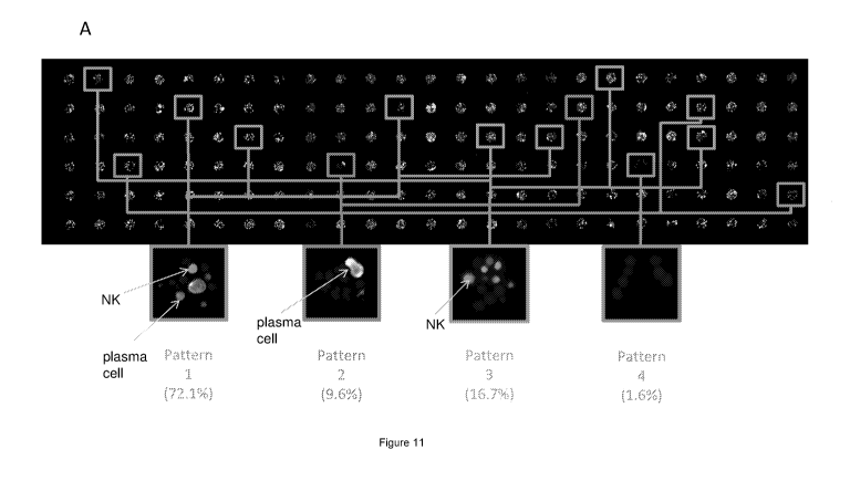
FIGURE 11: (A) Analysis based on ICNP with selection of four
subsets of microwells, where each subset satisfies one of the four
inclusion criteria (patterns) described herein below. Pattern 1: subset of
microwells selected to comprise at least one area of interest which
satisfies the direct property "NK cell immunophenotype" and at least
one area of interest which satisfies the direct property "plasma cell
immunophenotype" (E / T co-localization); pattern 2: subset of
microwells selected to comprise at least one area of interest which
satisfies the direct property "plasma cell immunophenotype" and no
area of interest which satisfies the direct property "NK cell
immunophenotype"; pattern 3: subset of microwells selected to
comprise at least one area of interest which satisfies the direct property
"NK cell immunophenotype" and no area of interest which satisfies the
direct property "plasma cell immunophenotype"; pattern 4: subset of
microwells selected to not comprise any area of interest which satisfies
the direct property "plasma cell immunophenotype" and any area of
interest which satisfies the direct property "NK cell immunophenotype";
(B) Measurement of the distance d between an NK cell and a plasma
cell, diagrammatically represented and in an original image. (C) Plasma
cell mortality assessed for each of the 20 patterns identified based on
the number of E (NK cells) and T (plasma cells) numbers in the same
microwell. The % of wells is shown, indicating the pattern in
CA 03156815 2022-04-01
WO 2021/064663
PCT/IB2020/059250
parentheses. (D) Example of time lapse images analyzed at the single
cell level. (E) Measurement of the death of target cells (plasma cells)
located inside a microwell at a distance between zero (contact) and
(pm) from an NK cell. The method allows estimating the fraction of
5 active NK
cells by comparing the frequency of death events where the
NK cell is in contact with a target cell, with the spontaneous cell death
of the target cells measured in a control represented by microwells in
which E (NK cells) is null (no NK). (F) Viability of target cells expressed
in % measured in the experiment in time lapse at 1, 3, 4, 5 and 6 h. The
10 tables show
the results obtained in the selected patterns, in relation to
the number of NK cells and the number of plasma cells present in the
microwell. A clear correlation emerges from the data, where plasma cell
death increases as NK cells increase, i.e., cell mortality is higher in the
lower right boxes of the graphs. The effect is already clear at an early
stage (3 h). (G) (comparative) Results obtained with standard Cr51
release assay.
Detailed description of the invention
The present invention relates to, with reference to the block diagram
in figure 7A, a method for subjecting a plurality of microwells containing
cells to a high-content assay, said method comprising:
a) Acquiring at least one image of said plurality of microwells;
b) In said image, detecting a plurality of areas of interest,
each area of interest corresponding to a single cell;
c) Measuring at least one property, direct or derived, of said
areas of interest;
d) Selecting a set of areas of interest based on one or more
of said properties, where said one or more properties are
defined as selection properties;
e) Extrapolating an output parameter from a property
CA 03156815 2022-04-01
WO 2021/064663
PCT/IB2020/059250
11
measured in the set of areas of interest selected, where said
property is defined as output property, said output property
being distinct from said selection properties.
In a preferred form, in said step c), at least one derived property is
measured and, optionally, at least one direct property of said areas of
interest, where said one or more properties is a selection property.
In a preferred form, in said step d) a subset of said plurality of
microwells is selected, where said microwells belonging to said subset
contain areas of interest selected based on said at least one selection
property and in said step e) an output parameter is extrapolated from a
property measured in the set of areas of interest selected, where said
property is defined as output property, said output property being
distinct from said selection properties, where said output parameter is
the processing of an output property measured in said set of areas of
interest.
In a preferred form, said microwells are embedded in a plate
comprising at least 15,000, or at least 18,000, preferably 19,200
microwells.
Said selection is made by imposing inclusion criteria, where said
inclusion criteria comprise:
identifying, from said direct or derived properties
measured, one or more selection properties;
imposing, for each of said selection properties, the
threshold value, or the range of values, within which said selection
property must fall.
The term "pattern" herein defines the inclusion criterion to be adopted
for the selection of said set of areas of interest for a specific output
parameter.
In a preferred embodiment, where a subset of said plurality of
microwells is selected, said microwells belonging to the subset were
CA 03156815 2022-04-01
WO 2021/064663 PCT/IB2020/059250
12
selected because they contain areas of interest in which said at least
one selection property satisfies said inclusion criterion.
In the present description and in the claims, the expression "property,
direct or derived, of said areas of interest" has the meaning indicated
below.
A direct property is a property associated with a single area of
interest, i.e., a property which can be measured by assessing the single
area of interest (for example, immunophenotype, cell viability, cell
morphology, signaling activity).
A derived property is a property associated with a multiplicity of areas
of interest, i.e., a property which, in order to be measured, requires the
assessment of two or more areas of interest included in the same
microwell, for example:
A property of relationship between two or more areas of
interest included in the same microwell (for example, cell-cell
distance); or
A property of coexistence of areas of interest, for example
one or more types of immunophenotype; or
A cumulative property of all the areas of interest included
in the same microwell (for example, number of cells in the microwell
to which an area of interest belongs for which said cumulative
derived property is calculated, average distance between the cells
contained in the microwell).
Said direct properties are directly obtained from the image analysis.
Said derived properties are obtained by processing direct properties. In
one embodiment, where the inclusion criterion is the immunophenotype,
said selection property is a direct property, the immunophenotype, and
a set of areas of interest which satisfy the inclusion criterion is selected.
Still maintaining the immunophenotype as inclusion criterion, in an
embodiment, where at least a subset of said plurality of microwells is
CA 03156815 2022-04-01
WO 2021/064663
PCT/IB2020/059250
13
selected, said selection property is a derived property, where said
derived property is a coexistence property, i.e., a property generated by
evaluating the direct immunophenotype property in each area of interest
included in a single microwell, and by processing the direct properties of
each of the areas of interest contained in a microwell, by extrapolating
the derived property which is the peculiar immunophenotypic pattern of
the microwell which, based on this pattern, will be attributed to a subset
of said plurality of microwells.
As a further example, the cell-cell distance is a derived property,
obtained by processing the direct "position" properties associated with
two areas of interest included in the same microwell. From a multiplicity
of said "cell-cell distance" derived properties, a further derived property
is obtained which is a further relationship property, i.e., the average
distance between the cells contained in a given microwell surrounding a
selected cell, from said selected cell. A further derived property is also
derived which is a cumulative property of all the areas of interest
included in the same microwell to which a given area of interest
belongs, i.e., the average distance between the cells contained in a
given microwell. It is also possible to determine further properties
derived from the combination of direct properties with relationship
properties. For example, the derived property "distance of an immune
cell from a tumor cell" requires combining the direct property
"immunophenotype" with the relationship property "cell-cell distance."
Is should be noted that the derived properties are also properties of
the areas of interest. Some cells belonging to the same microwell have
the same relationship-derived property value. All the cells belonging to
the same microwell have the same cumulative derived property value.
For example, two cells contained within the same microwell have the
same derived property "cell-cell distance" value when this is calculated
between said two cells. Furthermore, the relationship property "average
CA 03156815 2022-04-01
WO 2021/064663
PCT/IB2020/059250
14
distance between cells contained in a given microwell surrounding a cell
selected by said selected cell" takes on a different value for each
selected cell, since, for each selected cell, the distance from the other
cells in the same microwell surrounding it will be different. Again, all the
cells belonging to the same microwell have the same cumulative
derived property "number of cells per microwell" value, meaning that
this property of the context in which each cell is placed (the microwell)
is made its own by each cell, i.e., by each area of interest, belonging to
the microwell itself. In this case, or when a cumulative property is
discussed, equal for each area of interest embedded in the same
microwell, this property may be considered a property of the microwell,
meaning that this property applies to all areas of interest embedded
within said microwell.
Said set of areas of interest comprises:
- a subset of two or more areas of interest not embedded in the same
well; and/or
- a subset of two or more areas of interest included in the same
microwell; and/or
- a subset of all the areas of interest included in the same microwell.
In a preferred form, said set of areas of interest consists of a subset
of all areas of interest included in the same microwell, i.e., said set of
areas of interest corresponds to a subset of microwells.
In one embodiment, at least one of said selection properties is a
coexistence property.
In one embodiment, at least one of said selection properties is a
cumulative property of all the areas of interest included in the same
microwell.
In the present description and in the claims, the expression "output
CA 03156815 2022-04-01
WO 2021/064663
PCT/IB2020/059250
parameter from a property measured in the selected set of areas of
interest" will indicate the result of any statistical processing of the output
property measured in each area of interest belonging to said selected
set. "Statistical processing" means, for example, the mean value, the
5 median, the mean square value, etc.
In the embodiment where one or more of said selection properties is
a cumulative property of all the areas of interest included in the same
microwell, said set of areas of interest corresponds to a subset of said
plurality of microwells and said output parameter is the processing of an
10 output property measured in said subset of said plurality of microwells.
It is understood that said selection, in one embodiment, comprises a
selection of a first set of areas of interest based on a first selection
property. This is followed by a selection, within said first set of areas of
interest, of a subset of areas of interest based on a second selection
15 property. Said first and second selection properties are independently
direct or derived properties. In a preferred form, said set of areas of
interest and/or said subset of areas of interest corresponds to a subset
of the plurality of microwells.
In a further embodiment, said process comprises a first selection, a
second selection and a third or further selections.
Said at least one image is acquired with an image acquisition device
configured to acquire at least one image of said plurality of microwells.
In one embodiment, the image analysis and processing process
comprises the following steps, with reference, where appropriate and
purely for explanatory purposes and not in any way limiting the scope of
the invention, to Figure 6:
In an image containing a plurality of microwells, the zones
corresponding to the microwells are identified (figure 6, panel A);
Within the zones corresponding to the microwells, a
plurality of areas of interest are detected, each area of interest
CA 03156815 2022-04-01
WO 2021/064663
PCT/IB2020/059250
16
corresponding to one of said cells contained in said plurality of
microwells (figure 6, panel B);
At least one property of said areas of interest is measured
(figure 6, panel C; column A: areas of interest; columns B, C, D:
direct properties; columns E, F, G: derived properties);
A set of areas of interest is selected based on one or more
of said properties (figure 6, panel D; the columns of the selection
properties are highlighted in gray, the set of areas of interest
selected is highlighted in dark gray);
An output parameter is extrapolated from properties
measured in said set (figure 6, panel D; the output properties are
surrounded).
With reference to figure 6, panel D, the inclusion criterion is: property
C = Y and property G = Z. The output parameter is extrapolated from
property F. Being enclosed in a circle, the properties F related to the set
of selected areas of interest are thus highlighted. The result of a
statistical processing of said output property F measured in said set of
areas of interest is the output parameter provided by the method
according to the present invention, representative of the output property
F in analysis of the sample under examination.
It should be noted here that, for simplicity, the diagram in figures 6C
and 6D comprises a limited number of areas of interest, where in the
implementation of said method the areas of interest are advantageously
very numerous. By way of example, where said plurality of microwells
corresponds to a plate of 19,200 microwells, assuming an average of
about 20 cells/microwell, 384,000 areas of interest are available.
The acquired images are analyzed by a computer, with the aid of
suitable software products for image processing. Such software
products are for example ImageJ, BiolmageXD (Kankaanpaa P et al.
CA 03156815 2022-04-01
WO 2021/064663
PCT/IB2020/059250
17
Nature Methods. 2012), Icy (De Chaumont F et al. Nature Methods.
2012), Fiji ( Schindelin J et al. Nature Methods. 2012), Vaa3D (Peng H
et al. Nat Biotechnol. 2010), CellProfiler (Carpenter AE et al. Genome
Biol. 2006), 3D Slicer, Image Slicer, Reconstruct (Fiala JC. J Microsc.
2005), FluoRender, ImageSurfer, OsiriX (Rosset A et al. J Digit
Imaging. 2004), IMOD (Kremer JR et al. J Struct Biol [Internet]. 1996)
among others (Eliceiri KW et al. Nature Methods. 2012).
Those skilled in the art can easily understand that the above software
products are exemplary only and that the method may be carried out
using approaches not explicitly mentioned here, providing the same
type of result.
In a preferred form, said plurality of microwells is embedded in a
microfluidic device where each microwell is in fluid communication with
one or a plurality of microchannels for the delivery of fluids and/or
particles and/or molecules to the wells.
In one embodiment, the microwells are inverted open microwells, i.e.,
they are microwells with both an upper end and a lower end open,
preferably said ends being open on one or more microchannels in
which a fluid is present, a fluid comprising cells or particles or
molecules, or air or other gases.
The microwell has a vertical axis, such as a central axis, which
extends between the top and bottom of the microwell. In one
embodiment, said microwell is open at the upper end on a
microchannel, called upper microchannel, which comprises a fluid and,
at the lower end, on a microchannel in which air or other gases is
present. In this embodiment, the fluid inserted into the microchannel fills
the microwell through capillary action, while the surface tension holds
the fluid inside the open microwell, forming a meniscus at the air/fluid
interface.
CA 03156815 2022-04-01
WO 2021/064663
PCT/IB2020/059250
18
In one embodiment, said microwells are sized so as to have a height
equal to or greater than the diameter thereof.
In an even more preferred form, said microwells are microwells of the
type described in the application W02012072822.
Said cells are seeded in said microwells according to methods known
to those skilled in the art, and are a homogeneous cell population, i.e.,
they have the same immunophenotype, or heterogeneous, i.e., with a
different immunophenotype.
In a preferred form, said cells are seeded according to the method
described in W02017216739.
Said cells are seeded in a single step, or in sequence. By way of
example, using inverted open microwells it is possible to load
populations which are different from each other in sequence and each
of which contains cells which are homogeneous to each other, creating
heterogeneous populations in the volumes in which the cells are
deposited.
By way of example, using microwells with a diameter of 70 pm, up to
20, up to 30 or up to 50 cells/microwell are seeded.
In one embodiment, a heterogeneous cell population is seeded on a
subset of microwells in a single step. In a further embodiment, several
seeding processes are carried out in sequence. By way of example, a
first seeding of a population 1 which is at a concentration c1 and a
second seeding with a population 2 at a concentration c2 are
performed. Where said concentrations c1 and c2 are equal, seeding
equal volumes will result in a heterogeneous population in the set of
microwells belonging to the subset where on average the number of
type 1 cells is equal to the number of type 2 cells. The cells will instead
be present inside the microwells according to a distribution, typically a
Poisson distribution, which sees a variable number of type 1 and type 2
cells. Some wells will contain only type 1 or 2 cells, others will contain
CA 03156815 2022-04-01
WO 2021/064663
PCT/IB2020/059250
19
both types, and still others may be empty. By seeding a double volume
of the type 1 population, a heterogeneous population will be obtained in
the set of microwells belonging to the subset where on average the
number of type 1 cells is double compared to the number of type 2
cells. The distribution of type 1 cells in the microwells, compared to the
previous case, will see a doubled average value.
In one embodiment, said method is carried out on the same plurality
of microwells at successive and repeated times. That is, in this
embodiment, images are acquired, a multiplicity of areas of interest
detected and the at least one property measured at time to and,
subsequently, at time _1t , t _2, ... tn. In this embodiment, the assay is
defined as dynamic, i.e., multiple images of the same field are acquired
at successive times (time-lapse imaging) and the measurement of said
at least one property, at time to and, subsequently, at time _1t , t _2, ...
tn,
returns an analysis which reflects variations over time.
Said property at to is to be understood as distinct from said property
at t1. I.e., assuming to measure the property C (Pc), Pcto and Pcti are
clearly to be intended distinctly.
As a result, within the execution of the same assay, said output
parameter may be derived from the output property Pcti in the set of
areas of interest selected, where a selection property was Pcto.
In a further embodiment, a derived property is a variation (e.g., the
difference, or ratio) between the property measured at to and the
property measured at t1, or vice versa.
In one embodiment, said cells, while they are kept in said plurality of
microwells, are exposed to one or more agents which promote or inhibit
an objective effect of the analysis, i.e., which impact the output
parameter. The dynamic method according to the present invention
allows determining the effect of said agent over time.
CA 03156815 2022-04-01
WO 2021/064663
PCT/IB2020/059250
By way of example, and with reference to the table in figure 8, for
each area of interest corresponding to a cell (column A), the direct
properties "DAPI signal intensity," "FITC signal intensity," "Cy5 signal
intensity," "TRITC signal intensity," "cell position on the X axis and Y
5 axis"
(columns B-E, G, H) at to (lines 2 to 20) and the same properties
at t1 (lines 21 to 42) were measured. The microwell to which each cell
belongs is also reported (column F). Combining said direct properties
referred to in columns B-E, G, H with the information related to the
microwell to which each cell belongs, it is possible to calculate derived
10 properties,
for example for each cell it is possible to determine the
average distance from other cells contained in the same microwell from
said cell.
Properties
15 In the
following paragraphs, some categories of properties are listed,
providing some technical-experimental details which allow measuring
them. Downstream of each of said procedures, it is understood that an
image acquisition and processing step by means of the above
computational approaches is included, which is capable of returning the
20 information
related to the specific property which is typically information
of a numerical type.
The following list is illustrative and must in no way be construed as
limiting the technical-experimental approaches described for each
property. Given a property, those skilled in the art know the most
suitable experimental approach to give evidence thereof. Furthermore,
this list must not be construed as limiting the possible properties. Those
skilled in the art know how to extend the list with further direct or
derived properties to be effectively measured in the method according
to the present invention.
CA 03156815 2022-04-01
WO 2021/064663
PCT/IB2020/059250
21
It is understood that said properties may independently constitute
selection properties or output properties.
Immunophenotype (direct property)
It may be determined and/or verified using methods known in the art.
For example, using detectable markers/dyes. Such markers/dyes may
be specific for one or more subpopulations embedded in the microwells.
Where specific markers/dyes are used, these may be selected to
highlight cell populations which play a role in various diseases. For
example, because they are responsible for a tumor, for example a blood
cancer, or because they are responsible for an inflammatory and/or
immune response.
The staining may comprise the use of multiple detectable markers,
for example, cells may be stained with a primary antibody which binds
to a specific target antigen and a secondary antibody which binds the
primary antibody or a molecule coupled to the primary antibody may be
coupled to a detectable marker. The use of indirect coupling may
improve the signal-to-noise ratio, for example by reducing the
background binding and/or by providing signal amplification.
The staining may also comprise a primary or secondary antibody
directly or indirectly coupled to a fluorescent marker. By way of non-
exhaustive example, the fluorescent marker may be selected from the
group consisting of: Alexa Fluor 350, Alexa Fluor 405, Alexa Fluor 430,
Alexa Fluor 488, Alexa Fluor 514, Alexa Fluor 532, Alexa Fluor 546,
Alexa Fluor 555, Alexa Fluor 568, Alexa Fluor 568 594, Alexa Fluor
610, Alexa Fluor 633, Alexa Fluor 635, Alexa Fluor 647, Alexa Fluor
660, Alexa Fluor 680, Alexa Fluor 700, Alexa Fluor 750 and Alexa Fluor
790, fluorescein isothiocyanate (FITC), Texas Red, SYBR Green, Fluidi
DyLight, green fluorescent protein (GFP), TRIT (tetramethyl rhodamine
isothiol), NBD (7-nitrobenz-2-oxa-1,3-diazole), Texas red dye, phthalic
acid, terephthalic acid, isophthalic acid, fast cresyl violet, cresyl blue
CA 03156815 2022-04-01
WO 2021/064663
PCT/IB2020/059250
22
violet, brilliant cresyl blue, para-aminobenzoic acid, erythrosine, biotin,
digoxygenin, 5-carboxy-4', 5'-dichloro-2', 7'-dimethoxy fluorescein,
phthalocyanines, azomethines, cyanines (e.g., Cy3, Cy3.5, Cy5),
xanthines, succinyl fluorescein, N, N-diethyl-4-(5'-azobenzotriazolyI)-
phenylamine, aminoacridine, brilliant Violet 421, phycoerythrin (PE).
Number of cells/microwell (derived property, cumulative of all areas
of interest included in the same microwell)
Before seeding or when seeded in the microwells, the cells are
stained with a dye such as the fluorescent cell localization marker 7-
amino-4-chloromethylcoumarin.
Cell-to-cell distance (direct property associated with derived,
relationship property)
Before or after seeding, the cells are stained, possibly with a staining
which differentiates them according to the immunophenotype, and
through the above image processing approaches, a direct property is
obtained for each cell which is the position of said cell in space.
Combining said direct property "position" associated with an area of
interest with said direct property "position" associated with a different
area of interest, the derived property of the desired relationship is
obtained, i.e., the cell-cell distance.
Cell viability (direct property)
Known markers/dyes are used which specifically recognize the cells
which are at a particular stage of the cell cycle. These include, by way
of example, selective markers for cells with non-intact membranes or
selective markers for cells in an advanced stage of cell death or early
apoptosis. For example, it is possible to use antibodies against
cytochrome C, causing DNA turnover, or dyes which cause cell
viability/death such as propidium iodide (P1) and calcein, or dyes which
cause cell proliferation, or apoptosis markers such as Annexin V, or
dyes which cause apoptosis by means of the measurement of the
CA 03156815 2022-04-01
WO 2021/064663
PCT/IB2020/059250
23
signaling and release activity of certain proteins and enzymes, such as
caspases. Preferably, said markers/dyes are added to the cells in the
m icrowel I.
Signaling activity (direct property)
Preferably already in the microwell, the cells are labeled with markers
such as to highlight cellular signaling, such as antibodies capable of
highlighting the phosphorylation of proteins or the release of calcium
ions in the cytoplasm. In one embodiment, the "signal intensity"
property is determined by time-lapse imaging at to and "signal strength"
at _1t 7 t _27 ... tn associated with the marker used and cells are selected
having the variation of said "signal intensity" property over time beyond
a certain threshold value.
Cell morphology (direct property)
The image of the cells, possibly stained according to one of the
methods described and known in the state of the art, is acquired and
processed through the computational approaches mentioned above,
returning the information about the cell morphology.
In a preferred embodiment, said selection is made based on at least
2 selection properties, or at least 3, or at least 4, or at least 5 selection
properties.
One or more of said selection properties lead to selecting a set of
areas of interest which, in a preferred form, correspond to a subset of
microwells from which the output parameter will be derived.
A well-defined pattern allows optimizing the assay result.
Those skilled in the art know how to establish the pattern best suited
to the output parameter of interest.
By way of example, where the assay is conducted to measure cell
death in a sample, those skilled in the art, knowing that cell viability is
negatively affected by being in an isolated microenvironment and not
CA 03156815 2022-04-01
WO 2021/064663
PCT/IB2020/059250
24
with other neighboring cells, establishes that at least one of said
selection properties is the number of cells/microwell, imposing a
minimum threshold value X for this property. Therefore, the pattern will
be: microwell cell number > X. The result will derive from extrapolating,
from the set of microwells which satisfy the established pattern, the
output parameter.
In one embodiment, said patterns are advantageously established
using the method according to the present invention, so as to make
them optimal for the specific sample on which the assay is conducted.
As an example, in an assay, control subset(s) are used in which an
output parameter is optimized and subsequently these control values
are also used for the classification of the subgroups exposed to
treatment. For example, in a plurality of microwells containing cells not
exposed to any agent, the minimum number of cells for each microwell
is determined, which allows obtaining a minimum mortality at 24 h
(number of cells at to). This threshold value of the selection property
"number of cells" at to is used to select the set of areas of interest
exposed to a drug, and therefore the subset of microwells exposed to a
drug, in which the output property will be read and then the output
parameter which is the mortality at 24 h (number of cells at t24h) will be
extrapolated. I.e., the output parameter "number of cells" at t24h will be
the result of the statistical processing, in the specific case the average
value, of the output property "number of cells" at t24h measured in each
of the microwells belonging to the subset of microwells exposed to the
selected drug because they satisfied the pattern, i.e., showed, at to, a
number of cells above the threshold value as defined above. Thereby
the pattern is optimized based on the biological features of a specific
sample.
In a further aspect, with reference to Figure 10, a system (1) for
CA 03156815 2022-04-01
WO 2021/064663
PCT/IB2020/059250
subjecting a plurality of microwells containing cells to a high-content
assay is claimed, said system comprising:
- an image acquisition device (2) configured to acquire at
least one image of said plurality of microwells (3); and
5 - a data processing unit (4) configured to:
In said image, detecting a plurality of areas of
interest, each area of interest corresponding to a single cell;
Measuring at least one property, direct or derived,
of said areas of interest;
10 Select a set
of areas of interest based on one or
more of said properties, where said one or more properties
are defined as selection properties;
-Extrapolating an output parameter from a property
measured in the set of areas of interest selected, where said
15 property is
defined as output property, said output property
being distinct from said selection properties.
In a preferred form, said processing unit is configured to measure at
least one derived property, and, optionally, at least one direct property,
of said areas of interest, where said one or more properties is a
20 selection property;
In a preferred form, said processing unit is configured to select a
subset of said plurality of microwells, where said microwells belonging
to the subset contain areas of interest selected based on said at least
one selection property.
25 In a
preferred form, said processing unit is configured to extrapolate
an output parameter from a property measured in the set of areas of
interest selected, where said property is defined as output property,
said output property being distinct from said selection properties, where
said output parameter is the processing of an output property measured
in said set of areas of interest.
CA 03156815 2022-04-01
WO 2021/064663
PCT/IB2020/059250
26
In a further aspect, a computer program is claimed for subjecting a
plurality of microwells containing cells to a high-content assay, said
computer program comprising instructions which, when the program is
executed by a data processing unit, cause the processing unit to
perform the following steps:
In at least one image of said plurality of microwells,
detecting a plurality of areas of interest, each area of interest
corresponding to a single cell;
Measuring at least one property of said areas of
interest;
Selecting a set of areas of interest based on one or
more of said properties, where said one or more properties
are defined as selection properties;
-Extrapolating an output parameter from a property
measured in the set of areas of interest selected, where said
property is defined as output property, said output property
being distinct from said selection properties.
In a preferred form, said computer program comprises instructions
which, when the program is executed by a data processing unit, cause
the processing unit to perform the following steps:
Measuring at least one derived property, and,
optionally, at least one direct property of said areas of
interest, where said one or more properties is a selection
property;
Selecting a subset of said plurality of microwells,
where said microwells belonging to the subset contain areas
of interest selected based on said at least one selection
property;
Extrapolating an output parameter from a property
CA 03156815 2022-04-01
WO 2021/064663
PCT/IB2020/059250
27
measured in the set of areas of interest selected, where said
property is defined as output property, said output property
being distinct from said selection properties where said output
parameter is the processing of an output property measured
in said set of areas of interest.
Embodiments
In an embodiment, with reference to the block diagram in figure 7B,
said selection is carried out based on the selection property "number of
cells contained in a microwell" to and said output parameter is
extrapolated from the "cell viability" output property at t1 measured in
the set of areas of interest which corresponds to the subset of
microwells in which said selection property is greater than a threshold
value at to. In this embodiment, said output property is measured at a
time t1 later than the time to for measuring said selection property, after
having exposed the cells to an agent which influences cell viability.
Assuming that the optimal condition for the growth of said cells requires
having at least 10 cells in a microwell, since having less than 10 cells
leads to non-negligible cell death in the microwell, the subset of
microwells to which those microwells comprising more than 10 cells
belong will be selected. Said output parameter is extrapolated from said
subset. The cell viability datum thus obtained is a "clean" datum, i.e.,
not affected by the readings in those wells containing less than 10 cells
which are to be considered outlier readings, since they carry therewith a
high cell death independent from the agent to which the cells were
exposed but linked to the experimental condition thereof.
In a further embodiment, with reference to the block diagram in figure
7C, said selection is made in three steps.
In the first step, a first set of areas of interest is selected based on a
direct property "immunophenotype CT" of the areas of interest at to. Said
CA 03156815 2022-04-01
WO 2021/064663
PCT/IB2020/059250
28
first set of areas of interest corresponds to the subset of the plurality of
microwells comprising those microwells in which there is an area of
interest which satisfies said selection property, or in which there is at
least one cell with immunophenotype CT at to.
In the second step, in said subset of the plurality of microwells, a
second subset is selected based on a direct property
"Immunophenotype CE" of the areas of interest at to, said second
subset will thus comprise those microwells which have at least one cell
with immunophenotype CT and at least one cell with immunophenotype
CE at to.
In the third step, in said second subset a third subset is selected
based on the direct property "immunophenotype CT" of the areas of
interest at to, said third subset will thus comprise cells with
immunophenotype CT which are found in microwells which also
comprise cells with immunophenotype CE.
The output parameter is then extrapolated from the property "cell
viability" at t1 measured in said third subset of selected areas of interest.
That is, said output parameter is extrapolated in relation exclusively
to cells with immunophenotype CT contained in microwells which see
the simultaneous presence at to of cells with immunophenotype CE. In
this embodiment, said output parameter is provided at a time t1 later
than the time to for measuring said selection properties, after having
exposed the cells to an agent which influences the viability of the cells
CT, the activity of said agent being mediated by the cells CE.
This embodiment is particularly advantageous in carrying out an
assay which measures the efficacy of an agent which is an
immunotherapy, i.e., which acts on a target by promoting the activity of
the immune system cells towards said target. The method according to
the present invention advantageously allows excluding from the result
the microwells which, not comprising cells of the immune system, would
CA 03156815 2022-04-01
WO 2021/064663
PCT/IB2020/059250
29
inevitably return a negative datum, i.e., a lack of response to the
immunotherapeutic agent, where said lack of response would not be
linked to an ineffectiveness of the compound under analysis but to the
sample which is not suitable for the analysis itself, i.e., a datum which if
it were positive would be linked to a mechanism of direct action of the
drug against the target and not mediated by the cells of the immune
system.
In a further embodiment, said output parameter is extrapolated in
relation exclusively to cells with immunophenotype CT contained in
microwells which see the simultaneous presence at to of cells with
immunophenotype CE and the distance of which from cells with
immunophenotype CT is less than a predetermined threshold value.
This embodiment is particularly advantageous when the agent for which
the efficacy is to be evaluated involves a contact or a high proximity
between cells with immunophenotype CT and CE so that the agent may
exercise the action thereof.
Where each of the analyzed cells has a potential agonist or
antagonist role with respect to the effect of the assay, advantageously
said selection property is a relationship property, for example cell-cell
distance, signaling activity. By way of example, where cells of the
immune system have a potential antagonistic effect with respect to the
viability of tumor cells, the assay is effectively conducted on a set of
areas of interest identified according to the method of the present
invention after a selection based on derived selection properties, of
coexistence, "tumor immunophenotype" and "immune system cell
immunophenotype" so as to comprise cells of the immune system and
tumor cells, and a derived selection property "cell-cell distance", with a
pattern thus imposing that tumor cells and immune system cells are at a
distance such as to allow an interaction therebetween. In one
embodiment, the pattern imposes that the aforementioned distance be
CA 03156815 2022-04-01
WO 2021/064663
PCT/IB2020/059250
such as to produce contact between an immune cell, for example a
natural killer cell (NK), and a target cell, for example a tumor cell. In
another embodiment, the pattern imposes that the aforesaid distance is
equal to or greater than the distance which allows contact between the
5 immune cell and a target cell since the functional effect is generated by
secretion products, for example cytokines produced by T lymphocytes,
which exert an effect on the target cell even in the absence of contact,
as long as the distance between the two types of cells is sufficient to
ensure that the concentration of the products secreted by the immune
10 cell is significant to produce the desired effect.
In one embodiment, the immune cells are modified before the
analysis by means of known processes, being for example CAR-T cells,
NK cells destined for an autologous transplant, and the analysis
described herein aims to verify the effective ability of the modified cells
15 to produce a desired effect on target cells.
Again, the cell-to-cell distance, assessed at to and at t1, before and
after the addition of one or more agents in said plurality of microwells,
allows verifying the changes of the cell-cell interactions due to the one
or more agents.
20 For example,
in a further embodiment the plurality of microwells is
first divided into homogeneous subgroups, for example 2, or 3, or 4, or
16, or 32, or 64, or 96, or 128, or 384 subgroups, and on each of said
subgroups a different treatment is tested, where each treatment is
defined by a specific agent at a specific dosage. The microwells
25 belonging to each of the subgroups are selected for a direct selection
property "immunophenotype" at t1 and the output parameter is
extrapolated from the property "cell viability" measured in the set of
areas of interest selected. The method according to the present
invention, being capable of being implemented on plates containing
30 19,200 microwells, and allowing the automated analysis, allows a
CA 03156815 2022-04-01
WO 2021/064663
PCT/IB2020/059250
31
multiplicity of different conditions to be tested in each experimental
plate, for example up to 16, or up to 32 different experimental
conditions, where hundreds or thousands of microwells are dedicated to
each experimental condition. In one embodiment, the plates contain
1,200 wells for each condition and the plurality of microwells are
exposed to 2 or 3 or 4 or 16 or 32 or 64 or 96 or 128 or 384 different
conditions. The data obtained in each microwell belonging to the same
subset are processed with a statistical analysis so as to return the result
of the analysis. By way of example, where the agents tested were
tested for the ability to cause cell death in tumor cells, an output
parameter is extrapolated from the property "cell viability" measured in
each subset of microwells and the subset in which the greatest degree
of cell death is indicative of the most suitable agent, where the most
suitable agent means the agent which may be most effective in causing
the in-vivo cell death of tumor cells in the patient from whom said cells
were taken or, more in general, the agent which causes the desired
effect on the biological sample tested, having excluded causes other
than the action of the drug itself which could cause a variation of the
output parameter from which the desired effect is deduced. The number
of microwells for each of the experimental conditions allows maintaining
a high statistical significance even if, following the selection made
according to the aforementioned selection properties, the number of
wells actually subjected to the analysis is significantly reduced. The
availability of a large number of microwells thus represents a
fundamental requirement for supporting the method discussed herein,
where the actual number of wells is strictly connected to the type of
analysis. In order to ensure statistical significance, the output
parameter(s) must be read on a sufficient number of samples. Typically,
a sufficient number of samples is at least 30, or 100 or 300.
CA 03156815 2022-04-01
WO 2021/064663
PCT/IB2020/059250
32
The selection of a subset of microwells advantageously allows
testing an effect in a subset of microwells, where said selection has
been carried out based on a pattern, i.e., homogeneous features of the
selection properties considered.
In one embodiment, the pattern is determined in a control subset not
exposed to any agent, in order to ensure optimal functional features in
the control sample itself. Subsequently, said pattern is also imposed on
the subsets subjected to different in-vitro treatments, or treated with
different therapeutic agents possibly at different dosages. Said optimal
functional features are obtained, for example, through the maximization
of the cell viability, the maximization of the cell proliferation rate,
obtaining a cell proliferation rate similar to the expected proliferation
rate in the body from which the cells under analysis were extracted,
obtaining a cellular composition, i.e., the related ratio between cells
having different immunophenotype, or belonging to different cell
populations, similar to that observed in said organism.
In a further embodiment, where it is desired to determine as a
selection parameter the signaling in response to an agent, the intensity
of the signal associated with a marker is observed at subsequent times
through time-lapse imaging. Once a threshold value has been defined,
the subset of microwells is selected where one or more effectors have
produced a functional effect in the presence or absence of a certain
agent.
Advantages
The method of the present invention is carried out in microwells and,
with the data acquisition and processing method described herein,
conveniently allows observing and processing all the information related
to each of the cells contained in each microwell. This means having all
the information of a niche, where a niche herein means the
CA 03156815 2022-04-01
WO 2021/064663
PCT/IB2020/059250
33
microenvironment occupied by the cell population. Advantageously, this
information allows defining a pattern, and therefore the output
parameter is assessed in the context in which the assay is conducted.
The method advantageously allows carrying out assays on a sample
purged of data which would introduce deviations with respect to the
measurement of the analysis or which would introduce additional
factors in the analysis, thus increasing the variability of the result.
Therefore, the method according to the present invention allows
excluding from the assay those microwells and possibly those cells
which, for reasons independent of the assay to be conducted, are
identified as outliers. Since said selection is made thanks to a pattern
which is optimal for what is defined above, said selection made on the
sample is absolutely controlled and objective and maximizes the in-
vitro/in-vivo correlation.
Optionally, once the microwells of interest have been selected, the
method allows for a further selection at the cellular level, thus excluding
cells which behave as outliers inside microwells, thus allowing further
refinement of the analysis.
Assays conducted on subsets of microwells selected according to the
method of the present invention, ensuring a sufficient parallelism of the
analysis by performing it on a sufficiently large number of microwells,
lead to results with a high level of statistical significance despite the
application of selection criteria which reduce the number of data
actually considered in the analysis. For example, where the assay
involves the assessment of an agent which causes death in tumor cells,
carrying out the assay in microwells comprising a few cells, distant from
each other, would in some cases inevitably lead to the reading of an
effect on cell viability, where said effect is not at all indicative of the
activity of the tested agent but is related to the experimental in-vitro
conditions to which the specific sample under examination is exposed
CA 03156815 2022-04-01
WO 2021/064663
PCT/IB2020/059250
34
and which introduce artificial effects of toxicity towards the sample
which are not due to the drug. Such artificial effects, if not eliminated
from the analysis, would lead to an erroneous conclusion with respect
to the measurement of the actual efficacy of the drug.
Furthermore, the method according to the present invention allows
measuring and processing said properties in an automated manner,
processing the acquired images and processing the data obtained by a
computer.
The combination of these features ensures that the number of
samples tested is such as to ensure a statistically significant datum.
Therefore, the present invention provides a method which allows the
use of physiologically relevant, multi-population cell samples in studies
which allow defining, by way of example, the biological effects of drug-
based therapies on cellular samples, based on accurate analyses at the
single cell level, thus allowing the prediction, with a quick and accurate
ex-vivo analysis, of the drug which will prove to be the most effective in
the subject under analysis.
The following examples have the sole purpose of illustrating the
invention, and do not in any way limit it, the scope of which is defined by
the claims.
Example 1: Cell death control
Cells of the HL-60 cell line are plated in culture medium in inverted
open microwells of a microfluidic device with 19,200 microwells. At to
the cells are labeled with a cell death marker (propidium iodide, PI) kept
in the culture medium for the entire duration of the experiment and with
a fluorescent cell localization marker (7-amino-
4-
chloromethylcoumarin). Images are then acquired after a 24-hour
incubation (t24) and a range of properties are measured in the areas of
interest.
CA 03156815 2022-04-01
WO 2021/064663
PCT/IB2020/059250
The selection properties used in this example were:
- cumulative derived property: number of cells contained in
each microwell;
- derived relationship property: average distance of each cell
5 from the other cells belonging to the same microwell.
The extrapolated output parameter is cell mortality (expressed as %
of dead cells, i.e., cells for which the intensity of the fluorescence signal
emitted by the PI marker exceeds a certain threshold).
With reference to figure 1, classes are identified for the selection
10 property
"number of cells per well", in particular 7 classes are
determined for values equal to 2-4, 5-6, 7-8, 9-10, 11- 12, 13-17, 15-17
cells/microwell, datum reported on the x-axis of the graph in figure 1.
Within each well, a classification is then performed for the relationship-
derived selection property "average distance of each cell from the cells
15 of the same
well," obtained from the average of the distances between
each cell and the cells present in the same well. The plurality of
microwells is thus classified into subsets which include cells in contact,
in which the average distance of the cells of the same microwell is
between 0 and 2 D, where D means the average diameter of the cell
20 under
analysis, and with cells not in contact and which see cells of the
same microwell gradually more and more distant, in which the average
distance is between 2 and 2.5 D, between 2.5 and 2.7 D, between 2.7
and 3 D, and greater than 3D, datum reported on the y-axis of the graph
in figure 1.
25 The output
parameter, i.e., cell mortality, is extrapolated in each of
the above subsets. Said output parameter is indicated with the gray
scale in figure 1.
Surprisingly, cell mortality is observed to have a gradient behavior
with respect to the two imposed selection properties. In fact, an
30 increased
cell death (darker color in the graph) is observed in the set of
CA 03156815 2022-04-01
WO 2021/064663
PCT/IB2020/059250
36
areas of interest which correspond to the subset of microwells
containing fewer cells and/or in the set of areas of interest for which the
average distance from the cells of the same microwell is higher. With
the same number of cells contained, cell death is in fact greater for
those cells which are further away from other cells.
By defining a maximum mortality which is accepted as tolerated as
an artificial effect, the assay in this example allows defining, afterwards
and for the purposes of subsequent analysis, the optimal pattern,
establishing the threshold value for the selection property "number of
cells/microwell" and the threshold value for the property "average cell-
cell distance," where said threshold values are those which allow
keeping mortality within the tolerated limits.
By way of example, assuming that the tolerance limit is a maximum
mortality of 10%, the subsets of microwells which meet this criterion are
those highlighted with the symbol (x) in Figure 1A. The pattern which
identifies the subsets of microwells of interest is thus defined by the
following relationship:
(N 9 and P 3D) or (N Sand N 8 and P 2.7D) or (P 2D)
having indicated with N the property "number of cells per microwell"
and with P the property "average cell-cell distance."
As established above, the pattern is conveniently applied in the
execution of a response assay to an agent which impacts cell viability,
as in example 2 below. In a dose-response analysis, a reference
analysis is thus normally carried out on a control, for example the
sample kept in optimal conditions to ensure maximum viability and in
the absence of agents, from which said pattern is determined. The
analysis is also conducted in other conditions which see the
administration of an agent at one or more dosages, where the analysis
of the drug's efficacy is carried out on the subset of areas of interest
CA 03156815 2022-04-01
WO 2021/064663
PCT/IB2020/059250
37
identified based on the pattern defined by said reference analysis on a
control.
Example 2: efficacy analysis of a pharmacological agent
Cells of the HL-60 cell line are plated in inverted open microwells of a
microfluidic device with 19,200 microwells in culture medium and
exposed to treatment with FLAI-5: Fludarabine (FL) + Ara-C (A) +
ldarubicine (I) at 3 different concentrations (low, medium, and high). As
a positive control (Ctrl +) hydrogen peroxide (H202) 10mM is added, an
agent which is certainly capable of causing high cell death in HL-60
cells. At to the cells are labeled with a cell death marker (P1) kept in the
culture medium for the entire duration of the experiment and with a
fluorescent cell localization marker (7-amino-4-chloromethylcoumarin).
Properties are measured at to and at t24.
The property used as selection property in this example was:
- derived property: number of cells contained in each microwell
at to.
The output parameter is cell mortality at t24h, expressed as % of dead
cells, i.e., cells the intensity for which the fluorescence signal emitted by
the PI marker exceeds a certain threshold.
With reference to figure 2, classes are selected for the selection
property "number of cells per well" at to, in particular, classes are
selected for values equal to 1, 2, 3, 4, 5, 6, 7, 8, 9, 10, 11, 12 or more
than 12 cells per microwell. The data is shown on the x-axis in the
graph in figure 2.
The output parameter, i.e., cell mortality, is extrapolated into the
subsets of microwells classified as above.
It should be noted that, in the control samples, i.e., not exposed to
the agent, cell death above a threshold value is measured exclusively in
those subsets in which the number of cells per microwell is less than or
CA 03156815 2022-04-01
WO 2021/064663
PCT/IB2020/059250
38
equal to 8, as expressed by the gray scale on the Ctrl ¨ line in the graph
in figure 2.
The data shown in figure 2 indicate that, by selecting exclusively the
subsets of microwells with a low basic mortality, i.e., those microwells
selected to have a cell content per microwell greater than 8, the efficacy
percentage of the drug is approximately equal to 80%, measured as the
ratio of the percentage of dead cells in the treated sample to the control.
In the subset of microwells with a content of up to 7 cells/well, i.e., those
excluded from the assay due to the method according to the present
invention, the percentage of efficacy would have instead been equal to
about 50%, since part of the drug effect would have been masked by
the presence of a higher base mortality.
The result is indicative of how the method according to the present
invention allows obtaining a robust datum, excluding from the
processing the subsets of microwells which would have returned an
artificial datum, affected by external or environmental agents but in any
case not correlated with the analysis in progress.
Example 3: immunotherapy efficacy analysis
Blood samples from individuals with multiple myeloma are made
available. These samples are seeded in microwells. The selection
properties used in this assay are:
- direct properties: "CD38 immunophenotype" = tumor cells,
"CD16-CD56 immunophenotype" = immune cells;
- derived property of coexistence: co-localization of immune
cells and tumor cells in the same microwell.
A subset of microwells was selected where immune system cells (NK
cells) are in close proximity to CD38+ tumor cells.
The plurality of microwells was exposed to an anti-CD38 agent and
the output parameter was extrapolated which is the mortality evoked by
CA 03156815 2022-04-01
WO 2021/064663
PCT/IB2020/059250
39
said agent measured in the selected set of areas of interest, i.e., in the
tumor cells found in microwells which have co-localization with NK cells.
This approach allows performing an ADCC assay (Antibody-Dependent
Cellular Cytotoxicity) with high precision, i.e., limited to microwells
where there is a co-localization of the two types of cells of interest.
The data obtained and reported in Figure 9 show that the activity of
an anti-CD38 agent is greater in that subset of microwells which
comprise immune system cells which co-localize with tumor cells
(column D) compared to the average response obtained on the overall
cell population (column C).
Also in this case, the removal of deviant data or which introduce
noise effects into the measurement, such as wells without co-
localization of the two cell types, allows achieving a more accurate
measurement of the effective efficacy of the therapy and the level of
activity or fitness of the patient-specific immune system cells. In the
specific case, it is observed that 70% of the patient's NK cells, once
stimulated with the drug, have the ability to cause cell death of the
target cells placed in contact.
Further analyses and assessments on the efficacy of the drug may
be conducted in the same experimental system. For example, the
selection of the subset of microwells which do not comprise NK cells but
only CD38+ cells, in the presence of the anti-CD38 drug, allows
assessing as an output parameter the direct cytotoxic effect caused by
the drug on target cells and not mediated by NK cells (column B).
By selecting subgroups of microwells which comprise CD38+ tumor
cells which co-localize with NK cells, in the absence of the anti-CD38
drug it is possible to measure the spontaneous activity of NK cells
towards tumor cells (column A).
Finally, further evaluations may be performed to highlight drug
activity as the distance between NK cells and tumor target varies,
CA 03156815 2022-04-01
WO 2021/064663
PCT/IB2020/059250
adding the derived property "tumor cell to NK cell distance" among the
selection properties.
It is worth noting that, having acquired the complete panel of
properties as per panel C in figure 6, the assessments described herein
5 and others which those skilled in the art will want to conduct may be
carried out by independently choosing selection properties and output
properties, processing the data available, as schematized in panel D in
figure 6.
10 Example 4: control of the co-localization of heterogeneous cell
populations in the microwells.
With the aim of maximizing the probability of arranging microwells
with the co-localization of at least one type A cell and one type B cell,
different approaches have been defined herein, detailed below.
15 For the
purpose of the following examples, the following definitions
are assumed:
R1 = the ratio of effector cells (e.g., immune system cells) to total
cells in the initial cell population.
R2 = the ratio of target cells (e.g., tumor cells) to total cells in the
20 initial cell population.
E:T = the ratio of effector cells to total cells.
c = the concentration of the co-culture.
Example 4A
On the sample isolated from the patient, divided into two tubes, a first
25 enrichment step is carried out, obtaining in a first tube an R1 equal to
about 100% and in a second tube an R2 equal to about 100%.
In doing so, it is possible to determine E:T which will be obtained by
mixing together known quantities of the contents of the two tubes and it
is thus possible to define c so as to obtain the desired average number
30 of cells per microwell.
CA 03156815 2022-04-01
WO 2021/064663
PCT/IB2020/059250
41
In theory, in the case of using 2 pure populations, i.e., with R1 and
R2 approximately equal to 100%, by sequentially seeding the 2
populations, a co-localization probability close to 100% is obtained if an
average of 10 cells/microwell is assumed (Figure 3A, theoretical graph).
The experimental data, obtained on NK cells and tumor cells
enriched as described above, confirm the expected trend with good
approximation (Figure 3B, experimental data).
Example 4B
The sample under analysis comprises effector cells in PBMCs
(peripheral blood mononuclear cells) isolated from the patient at varying
frequencies without any enrichment (e.g., R1 = 5-20% within 8 samples
analyzed).
The tumor cells are enriched or, alternatively, a cell line is used (R2 -
100%).
Optimal E:T is known from probability theory
The effector cells and target cells are seeded sequentially. Assuming
to have an average of 10 cells/well, the probability of effector/target cell
co-localization is between 30% and 70%. In particular, as shown in the
graph in figure 4, with R1 = 5 the probability of co-localization is 30%,
with R1 = 20% the probability rises to 70%. The graph also shows that
the ideal number of cells per microwell to obtain the maximum co-
localization is approximately 10 cells/microwell. Having 1,200
microwells for each condition, also considering a reduction of
microwells in the limit condition of 30%, a good statistical significance is
maintained thanks to the replication of the microwells.
Example 4C
For this test, NK effector cells are made available in PBMCs isolated
from the patient at varying frequencies (e.g., R1 = 5-20%).
The tumor cells are also variable-frequency in the same patient's
PBMCs.
CA 03156815 2022-04-01
WO 2021/064663
PCT/IB2020/059250
42
In this case, i.e., by using a single population withdrawn from a
patient containing a range between 5-20% of effector cells of interest
and a variable range of tumor cells, very different situations can be
obtained in terms of co-localization probability. Some examples show
that the values predicted by theoretical calculations are reached with a
good approximation. The minimum usable extremes of the frequency
ranges of the two cell populations depend on the number of microwells
available and the statistical power required.
By way of example, the graph in figure 5 shows the co-localization
frequency observed, as the number of cells/microwell varies, with R2 =
50% or with R2 = 100%.
In a real case, subject A showed R1 = 17.3 and R2 = 28.1. The
theoretical calculation led to estimate a co-localization in 57.2% of the
microwells. The experimental data led to observe a co-localization in
48.1% of the microwells. In a further experimental case, subject B
showed R1 = 14.2 and R2 = 10Ø The theoretical calculation led to
estimate a co-localization in 56% of the microwells. The experimental
data led to observe a co-localization in 56.2% of the microwells.
Example 5: assays on cells from patients with multiple myeloma
EDTA bone marrow samples were collected from 13 patients with
multiple myeloma (MM, 7 de novo and 6 relapses). 8 primary samples
were processed through density centrifugation (Ficoll-Pacque; Merck) in
order to obtain mononuclear cells while preserving the original
composition of the effector (E) and target (T) cells, i.e., NK and plasma
cells, respectively. 5 samples were processed with CD138 Antibody
coupled to magnetic beads (Miltenyi Biotec) to obtain a population of
white blood cells (WBC), a population which comprises NK cells and is
depleted of plasma cells.
CA 03156815 2022-04-01
WO 2021/064663
PCT/IB2020/059250
43
The resulting cells were co-cultured with U-266 or NCI-H929 cell
lines as target cells. The U-266 cells were kept growing at 37 C with
5% CO2 in 1640 RPM! medium (Sigma-Aldrich) supplemented with 10%
fetal bovine serum (Sigma-Aldrich), 1% L-glutamine (Sigma-Aldrich)
and 1% penicillin/streptomycin mixture (Sigma-Aldrich). The NCI-H929
cells were cultured at 37 C with 5% CO2 in 1640 RPM! medium
(Sigma-Aldrich) admixed with 20% fetal bovine serum (Sigma-Aldrich),
1 A L-glutamine (Sigma-Aldrich), 1 A penicillin/streptomycin mixture
(Sigma-Aldrich) and 1% sodium pyruvate (Merck).
The cells from the primary samples were stained with CMAC
(Thermo Fisher Scientific), used as a cell tracer. In co-culture
experiments, white blood cells and target cells (U-266 or NCIH929)
were stained with Calcein AM (Thermo Fisher Scientific) and CMAC,
respectively. NK cells (effector cells, E) and plasma cells (target cells,
T) were labeled using BV421 Mouse anti-Human CD16 / CD56 (BD
Biosciences) and AF647 Mouse anti-Human CD138 (BioRad)
fluorescent antibodies, respectively. Propidium iodide (PI, Thermo
Fisher Scientific) was used as a cytotoxicity marker.
Statistical model for cell co-localization
A statistical model was created to define the optimal experimental
setup which produces the maximum number of microwells containing
the desired effector/target co-localization pattern (derived selection
property, of co-existence), defined by an effector/target co-localization
factor E / TcF which is the ratio of E to T in the same microwell.
The model takes into account four parameters which influence the E /
TcF factor:
1) the initial effector/target mixing ratio (E:T);
2) the overall concentration of the cells (c);
CA 03156815 2022-04-01
WO 2021/064663
PCT/IB2020/059250
44
3) the ratio between the effector cells and the input cell population
(R1) and 4) the ratio between the target cells and the input cell
population (R2).
The parameters R1 and R2 depend only on the type of sample (e.g.,
cell line, patient primary sample), while E:T and c can generally be
modified by the user to maximize the frequency of specific models of
interest within the matrix of microwells. For experiments where both the
E cells and T cells are the patient's primary sample cells, E:T cannot be
modified and only c can be optimized.
Cell seeding and drug exposure
The cells from primary or co-culture samples were seeded in 96-well
plates, with a final concentration of 2x10⁵ cells / well, with variable E:T
ratios. In addition, conditions with E:T ratios of 1:0 (effector cells only)
and 0:1 (target cells only) were used as controls. Using a robotic
microfluidics system, the cells were loaded into the microfluidic device
and trapped in the microwells. The Daratumumab monoclonal antibody
(anti-CD38) was used in 3 doses, administered through different
microchannels (0.1 pg/mL, 1 pg/mL and 10 pg/mL), while a further
microchannel, without drug, was used as a control. The drug was
diluted in RPM! 1640 medium (Sigma-Aldrich) admixed with 10 or 20%
fetal bovine serum (Sigma-Aldrich), 1% L-glutamine (Sigma-Aldrich),
1`)/0 penicillin/streptomycin mixture (Sigma-Aldrich). Each experiment
was analyzed by time lapse in fluorescence microscopy for up to 12
hours.
ICNP image and data analysis
ICNP is an analytical method enabled by the availability of a large
number of said microwells, based on randomly creating a huge number
of heterogeneous cell clusters and then classifying and analyzing the
cells into specific groups of cell clusters which share analogous cell-cell
interaction patterns (Figure 11A). The large number of clusters which
CA 03156815 2022-04-01
WO 2021/064663
PCT/IB2020/059250
are obtainable by the method according to the present invention, 19,200
in this specific example, allows identifying even relatively rare patterns
or evaluating multiple interaction patterns in a single experiment while
maintaining good statistical significance. In this example, the ICNP
5 analysis was optimized to perform an ADCC assay (Antibody-
Dependent Cell Cytotoxicity) for the assessment of the potency of NK
cells against tumor cell lines and primary tumor cells, under stimulation
with anti-CD38 (Daratumumab).
The images for said plurality of microwells are then acquired and,
10 with a detection algorithm, the areas of interest, where each area
of
interest corresponds to a single cell, for each of said areas of interest,
properties which comprise localization, the intensity of certain markers
in each fluorescence channel, the cell area, the position of the center of
gravity and the morphology are then measured. The data related to
15 each of said properties are collected at different and subsequent
times,
in this case at T = 0 h, T= 1 h, T = 2 h, T = 4 h, T= 12 h and stored in a
database.
A subset of said plurality of microwells is then selected based on said
properties, where said selection is based on 4 specific co-localization
20 patterns, as shown in Figure 11A. Each of the patterns is
characterized
by E / TcF values, from a specific number of E cells and a specific
number of T cells. Consequently, for each channel of the microfluidic
device, and on the same cell pool, multiple E / T co-localization patterns
are assessed.
25 Furthermore, some microwells serve the function of internal control.
For example, wells containing only target cells in a microchannel
stimulated with a drug allow assessing the direct cytotoxicity caused by
the drug.
On the subset of said plurality of microwells selected, a second
30
classification is made, at the level of the area of interest, by evaluating
CA 03156815 2022-04-01
WO 2021/064663
PCT/IB2020/059250
46
the cell-cell interaction models within a specific subset of microwells,
based on immunophenotype, vitality and spatial information. In this
classification, a key step is the assessment of the distances and
contacts between the areas of interest included in the same microwell.
This information (Figure 11B) is derived from the coordinates (x, y) of
the center and radius r of each pair of areas of interest being assessed.
The radius refers to a circular object having the same area as the area
of interest, i.e., the single cell under analysis.
For the purpose of the method, a pair of cells is defined as "in
contact" if:
d dist '(x1 , y1), (x2, y2) 1 - r1 - r2 ¨ tol
where dist ((x1, y1), (x2, y2)) is the distance d between the two
centers, r1 and r2 are the radii of the two areas of interest and tol is a
tolerance value, set at 4 pm here. For example, the target cells are
classified based on the distance from the immune cells in the same
microwell, thus allowing the identification of those target cells which are
in contact with immune cells or those target cells which are located
within a certain distance from an effector cell.
The method allowed assessing how the potency of NK cells (i.e., the
cell-mediated cytotoxicity caused on the tumor cells) changes with the
distance from the CD138+ cells.
Specifically, the 4 selected patterns, shown in Figure 11A, were:
pattern 1) microwells comprising NK and plasma cells (72.1%), pattern
2) only plasma cells (9.6%), pattern 3) only NK cells (16.7%), pattern 4)
no cells of interest (1.6%).
The selection of said subset of microwells advantageously allowed a
targeted study of NK-mediated cytotoxicity, where the study was carried
out exclusively on the subset of microwells selected for pattern 1.
Furthermore, a key advantage of the method according to the present
CA 03156815 2022-04-01
WO 2021/064663
PCT/IB2020/059250
47
invention lies in the possibility of assessing, for a certain experiment,
specific co-localization patterns.
Figure 11C shows a heatmap resulting from the analysis of an
experiment in which 20 different co-localization patterns of NK and U-
266 cells were analyzed, each box of the heatmap is related to a
pattern. The cells were exposed to anti-CD38 antibody and each
pattern differs in the number of E (NK) cells and T cells (U-266 cells)
included in the same microwell, thus allowing the influence of the
effector : target ratio on the death of the target cells to be assessed.
The plasma cell death rate assessed in the microwells with the method
according to the present invention revealed that target cell death is
higher in the microwell subset with a higher E / TcF ratio. The datum can
be superimposed on the datum obtained with methods known in the art,
i.e., in culture plates, as shown by the comparative data obtained with
the Cr51 release assay (Figure 11G), with the key advantage of being
capable of measuring multiple patterns simultaneously and with a
resolution of a single area of interest.
After the classification of the microwell subsets, a detailed analysis at
the single cell level was performed on images acquired in time lapse.
The method according to the present invention allowed investigating the
effects of cellular "networking," grouping the data by homogeneous
interaction pattern. Figure 11D shows an example of images analyzed
to investigate the interaction between NK cells and plasma cells in
detail. Each line in the image corresponds to a different condition: direct
effect of the anti-CD38 on a target cell belonging to a microwell with
pattern 2, i.e., without effector cells (NK-); effect of the spontaneous
interaction between a target cell and the effector in microwells with
pattern 1, without anti-CD38 stimulation (CTRL), or with anti-CD38
stimulation with contact between NK and plasma cells (anti-CD38). The
samples with pattern 1 (anti-CD38) show that the interaction causes the
CA 03156815 2022-04-01
WO 2021/064663
PCT/IB2020/059250
48
death of plasma cells, as detected by the absorption of propidium iodide
and the consequent appearance, starting from 1 h, more evident at 2 h,
of the signal (indicated with the arrow in the image). The plasma cell, on
the other hand, does not die in the representative image shown for
pattern 2, i.e., in the absence of effector cells. The plasma cell death in
the pattern 1 microwells was assessed with respect to the distance from
an NK cell, with the aim of estimating the actual potency of the NK cell
which is responsible for the observed toxicity.
Figure 11E shows the data collected from 1,200 microwells in which
the cells were stimulated with Daratumumab at a dose of 10 pg/mL. The
method according to the present invention allowed observing that the
death is maximum for those plasma cells in contact with NK cells and
decreases as the plasma cell - NK cell distance increases. The plasma
cells not in direct contact but in the immediate vicinity of NK cells show
an increased mortality rate compared to the cells further away. These
data are indicative of the fact that the activation of an NK cell not only
impacts the cell with which it comes into direct contact but can also
affect the surrounding environment, i.e., the cells located at a minimum
distance from the NK cell which are likely to be subjected to death
caused by a contact with the NK cell which can occur in a different
instant of time than that corresponding to the observation or due to the
secretion of toxic substances such as perforin and granzymes following
the first activation of the NK cell from contact. Hence the value of the
method according to the present invention, which even during the
analysis on a single cell takes into consideration the environment in
which said cell is contained, thus allowing the selection of
representative subsets of the environment of interest.
In the experiment reported, the method allowed estimating that the
fraction of powerful NK cells, i.e., capable of killing the target when
contact is provided, is 12.82% of the total. This number was calculated
CA 03156815 2022-04-01
WO 2021/064663
PCT/IB2020/059250
49
as the difference between the mortality rate of plasma cells belonging to
pattern 1, therefore in contact with an NK cell (23.68%) and the death
rate of the plasma cells belonging to pattern 2 (10.86%), which is due to
spontaneous death or a direct effect of the anti-CD38 antibody. The
heatmap in Figure 11F shows the results of cell viability measured in
the different patterns over time.