Note: Descriptions are shown in the official language in which they were submitted.
CA 03156986 2022-04-05
WO 2021/071840 PCT/US2020/054407
IN-PLANE ROTATION CANNULA
TECHNICAL FIELD
[0001] The present disclosure relates generally to a cannula arrangement for a
surgical device
and more particularly to a cannula arrangement that may be selectively bent,
but still allows for
360 degrees of rotation within the same plane after the bend point.
BACKGROUND
[0002] Advances in surgical techniques and devices have allowed for access to
deep seated
tumors that were previous though to be inaccessible. More specifically, narrow
surgical
corridors allow for such access to the structures.
[0003] However, often it is difficult to differentiate between normal and
abnormal tissue, and
many known surgical devices have line of sight issues whereby the shaft and/or
the effector end
of the instrument is very difficult to observe during a procedure without also
occluding the
surgeon's view of the surgical site due to the narrow surgical corridor. As a
result, users are
often required to hold the instrument at awkward angles so as to maintain
visibility during the
procedure, which may lead to unintended movements by the operator causing harm
to normal
tissues.
[0004] Further, because of the size and angle at which the instrument must be
entered into the
surgical site, the actual use of the instrument may block the surgeon's view
of the surgical site
during its use and the surgeon effectively uses the instrument "blindly" and
removes the
instrument from the surgical field to inspect the outcome of the use of the
instrument. If
unsatisfactory, then the instrument is then repositioned in the surgical field
for additional work.
This procedure can often go on for multiple passes at the surgical site. Such
repeated action can
cause damage to the tissues which line the surgical corridor due to bumping of
the instrument as
they are passed in and out, as well as unintentional excision of healthy
tissue. Accordingly, this
results in procedural inefficiency as well as increased surgeon fatigue.
[0005] Manipulation of the surgical device is also difficult within the
surgical field. For
example, while a device may be configured to cut tissue or grasp different
structures, often there
1
CA 03156986 2022-04-05
WO 2021/071840 PCT/US2020/054407
is no mechanism to change angles of orientation of the effector end of the
device during the
cutting or grasping operation while it is in use within the surgical site.
Accordingly, the user has
traditionally been required to extract the device from the surgical field to
reposition the device
for further operation or remove the device and angle a lighting device to
provide sufficient
visibility. This action thus lengthens a procedure. Moreover, depending on the
set up in the
operating suite, in some instances the repositioned angle requires the user to
hold and operate the
device in an awkward manner, also leading to fatigue and an unintended adverse
impact to the
tissues within the surgical site.
[0006] In other arrangements, while the end effector may be rotated while
within the surgical
site, if those arrangements are bent to allow for effective line of sight
during use of the device,
rotation of the end effector results in a gross arc movement, moving a tissue
opening
dramatically. For operations being performed within the surgical access
corridor, such
movement is often prohibitive as the end effector swings into the walls of the
surgical access
corridor. Further such movement also results in requiring a surgeon to
physically move their
hand, wrist, or even arm to properly reposition the tissue opening to a
location where the
effective portion of the device is visible and can access the desired area.
[0007] Based on the foregoing, an improved device that allows for easy
manipulation for both
line of sight issues, as well as to permit flexibility of use during a
procedure is needed, while
minimizing gross movement of the device.
BRIEF SUMMARY OF THE INVENTION
[0008] A surgical device is disclosed that allows for in-plane rotation of a
distal end to avoid
gross movements during surgical procedures. In one exemplary arrangement, the
surgical device
comprises a housing, an outer cannula, and an inner cannula. The outer cannula
is mounted for
rotation to the handpiece. The inner cannula is mounted within the outer
cannula and mounted
for rotation with the outer cannula.
[0009] To allow for in-plane rotation, the outer cannula includes a plurality
of outer relieving
cuts disposed thereon. Similarly, the inner cannula includes a plurality of
inner relieving cuts
disposed thereon. The outer relieving cuts are offset from the inner relieving
cuts such that the
2
CA 03156986 2022-04-05
WO 2021/071840 PCT/US2020/054407
outer relieving cuts do not overlap with the inner relieving cuts when the
inner and outer
cannulas are in a first position.
[0010] In a further embodiment, the surgical device further comprises a vacuum
sleeve mounted
to the outer cannula, the vacuum sleeve being disposed over the outer
relieving cuts to maintain
vacuum within the surgical device.
[0011] In a further embodiment, the surgical device further comprises a
stiffening sleeve
disposed over the outer cannula.
BRIEF DESCRIPTION OF THE DRAWINGS
[0010] Exemplary embodiments of the present disclosure will now be described
in greater detail
with reference to the attached figures, in which
[0011] FIG. 1 is a perspective view of a surgical device with a stiffening
adapter mounted
thereto;
[0012] FIG. 2 is an enlarged view of the stiffening adapter disposed about an
outer cannula of a
surgical device;
[0013] FIG. 3 is a side elevational view of a prior art outer cannula of a
tissue cutting device;
[0014] FIG. 4 is a side elevational view of a prior art inner cannula of a
tissue cutting device;
[0015] FIG. 5A is a cross-sectional view of a prior art assembled inner and
outer cannula of a
tissue cutting device in a cutting position;
[0016] FIG. 5B is a cross-section view of the prior art assembled inner and
outer cannula of the
tissue cutting device in retracted position;
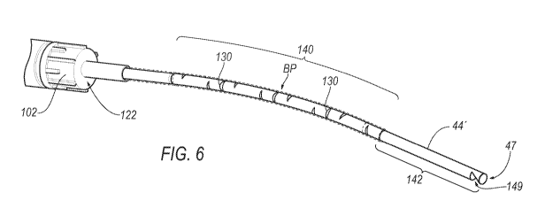
[0017] FIG. 6 is a partial perspective view of an inner and outer cannula
assembly for a tissue
cutting device;
[0018] FIG. 7 is a side elevational view of the inner and outer cannula
assembly of FIG. 6;
3
CA 03156986 2022-04-05
WO 2021/071840 PCT/US2020/054407
[0019] FIG. 8 is an enlarged cross-sectional view of a portion of the inner
and outer cannula
assembly of FIG. 6 when the surgical device is in a retracted position;
[0020] FIG. 9 is an enlarged cross-sectional view of a portion of the inner
and outer cannula
assembly when the surgical device is in a forward position; and
[0021] FIG. 10 is a plan view of an exemplary arrangement of a relieving cut.
DETAILED DESCRIPTION OF THE INVENTION
[0022] Referring now to the discussion that follows and also to the drawings,
illustrative
approaches to the disclosed assemblies and methods are shown in detail.
Although the drawings
represent some possible approaches, the drawings are not necessarily to scale
and certain features
may be exaggerated, removed, or partially sectioned to better illustrate and
explain the present
disclosure. Further, the descriptions set forth herein are not intended to be
exhaustive or
otherwise limit or restrict the claims to the precise forms and configurations
shown in the
drawings and disclosed in the following detailed description.
[0023] Described herein are surgical devices that are suited for neurosurgical
applications such
as the removal of spine and brain tissue. The components disclosed herein
provide surgeons with
an enhanced ability to minimize trauma to the patient, while providing
efficient improved
minimally invasive surgical techniques, such as, for example, during
intracranial surgical
techniques. The components disclosed herein may further be used for
application of targeted and
effective treatment regimens. Referring to FIG. 1, an exemplary surgical
device 40 is shown. In
one exemplary arrangement, the surgical device may be configured as a tissue
cutting device.
More specifically, surgical device 40 may be configured similarly to the shown
and described in
commonly owned U.S. Patent No. 9,387,010, the contents of which are
incorporated in its
entirety.
[0024] In one exemplary arrangement, surgical device 40 includes a handpiece
42 and an outer
cannula 44. A rotation dial 60 for selectively rotating the outer cannula 44
with respect to
handpiece 42 is mounted within a portion of handpiece 42.
4
CA 03156986 2022-04-05
WO 2021/071840 PCT/US2020/054407
[0025] As best seen in FIGS. 3-5B, prior art outer cannula 44 includes an open
proximal end 45,
a closed distal end 47, and a distal opening 49 proximate distal end 47.
Surgical device 40 further
comprises an inner cannula 50 which is partially disposed in an outer cannula
lumen 52. In one
exemplary arrangement, inner cannula 50 is configured to reciprocate within
outer cannula
lumen 52 and to cut tissue samples entering outer cannula 50 via outer cannula
distal opening 49.
Inner cannula 50 reciprocates between a proximal position and a distal
position. Referring to
FIG. 4, inner cannula 50 includes an open proximal end 54 and an open distal
end 56. Distal end
56 is configured to cut tissue, and in certain exemplary arrangements is
configured for cutting
neurological system tissues such as those from the brain or spine. In another
exemplary
arrangement, inner cannula 50 is used to selectively deliver vacuum to a
surgical site such that
movement of the inner cannula 50 to a distal position reduces and/or prevents
vacuum from
being delivered to the tissue opening 49.
[0026] Outer cannula 44 is not translatable, and its position with respect to
handpiece 42 along
the direction of the longitudinal axis of handpiece 42 remains fixed. A motor
(not shown) is
disposed in a section of handpiece 42 and is operably connected to inner
cannula 50 to drive the
reciprocation of inner cannula 50 within outer cannula lumen 52.
[0027] Outer cannula 44 includes an opening 49 for receiving tissue into outer
cannula lumen
52. Opening 49 may be defined by a cutting edge 51 that is configured to sever
tissue and a non-
cutting edge 53 that is not configured to sever tissue. In one embodiment,
inner cannula distal
end 56 is preferably configured to cut tissue. As tissue is received in outer
cannula opening 49, it
is compressed between inner cannula distal end 56 and outer cannula cutting
edge 51, causing
the received tissue to be severed from the surrounding tissue.
[0028] Inner cannula may include a hinge 58. Hinge 58 is located between an
inner cannula body
section 60 which is located on the proximal side of hinge 58 and inner cannula
cutting section 62
which is located on the distal side of hinge 58. Hinge 58 allows cutting
section 62 to pivot about
hinge 58 as inner cannula 50 reciprocates within outer cannula 44. As inner
cannula 50 translates
in the distal direction, it contacts tissue received in outer cannula opening
49 and encounters
progressively increasing resistance from the tissue as the tissue is urged in
the distal direction.
As the resisting force of the tissue increases, cutting section 62 pivots
progressively more until a
zero annular clearance is obtained between inner cannula distal end 56 and
outer cannula
CA 03156986 2022-04-05
WO 2021/071840 PCT/US2020/054407
opening 49. The received tissue is severed and aspirated in the proximal
direction along inner
cannula lumen 64 and is received in a tissue collector (not shown). Thus,
inner cannula lumen 64
provides an aspiration path from the inner cannula distal end 56 to the inner
cannula proximal
end 54.
[0029] Surgical device 40 aspirates tissue samples received in inner cannula
lumen 64 to cause
the tissue samples to move in the proximal direction along the length of the
inner cannula 50. In
some exemplary embodiments, surgical device 40 preferably includes a tissue
collector (not
shown) into which aspirated tissue samples are deposited during a tissue
cutting procedure. In
some exemplary arrangements, the tissue collector may be located remotely from
handpiece 42
and outside the sterile field during a tissue cutting operation or may be
removably connected to
handpiece 40.
[0030] When device 40 is used to cut tissue, outer cannula opening 49 must be
aligned with the
target tissue of interest to receive it for cutting. In an exemplary
embodiment, device 40 includes
a selectively rotatable outer cannula 44. As best seen in FIG. 1, a rotation
dial 46 is provided and
is rotatably seated within the handpiece 42. Rotation dial 46 is configured
such that when it is
rotated, it causes outer cannula 44 to rotate about its longitudinal axis.
[0031] To ensure the correct operation of hinge 58 of inner cannula 50, the
circumferential
alignment of hinge 58 and outer cannula opening 49 should be maintained. Thus,
rotation dial 46
is preferably connected to inner cannula 50 such that when rotation dial 46 is
rotated, both outer
cannula 44 and inner cannula 50 rotate in a fixed angular orientation with
respect to one another
by an amount that directly corresponds to the amount by which rotation dial 46
is rotated.
Rotation dial 46 may be directly connected to inner cannula 50 or may use an
intervening
connecting device. However, rotation dial 46 should be configured to allow
inner cannula 50 to
reciprocate with respect to rotation dial 46.
[0032] Because rotation dial 46 is directly or indirectly connected to both
outer cannula 44 and
inner cannula 50, both cannulae rotate in direct correspondence to the
rotation of rotation dial 46,
thereby allowing the user to adjust the orientation of outer cannula opening
49 and inner cannula
hinge 58 in a circumferential direction with respect to handpiece 42. As a
result, surgeons need
not rotate the entire tissue cutting device 40 to obtain the desired angular
orientation.
6
CA 03156986 2022-04-05
WO 2021/071840 PCT/US2020/054407
[0033] Rotation dial 46, outer cannula 44, and inner cannula 50 are preferably
configured for
360 rotation. In addition, tactile indicators are preferably provided on
rotation dial 46 to allow a
user to reliably determine the extent to which dial 46 has been rotated from a
given starting
point. The tactile indication may comprise surface features defined on or in
the exterior surface
of rotation dial 46.
[0034] In one configuration, surgical device 40 is connected to a vacuum
source and configured
for variable aspiration, i.e., configured to supply variable levels of vacuum
to inner cannula
lumen 64.
[0035] Referring to FIGS. 1-2, a stiffening adapter 100 is illustrated that
may be selectively
attached to the surgical device 40. An exemplary stiffening adapter 100 is
described in co-
owned patent No. 10,383,680, the disclosure of which is incorporated by
reference in its entirety.
While tissue cutting deice 40 is depicted in FIG. 1, it is understood that
stiffening adapter 100
may be used with other embodiments of surgical devices.
[0036] In one exemplary arrangement, stiffening adapter 100 comprises cap
member 102 and a
stiffening sleeve 104. In one exemplary arrangement, the stiffening adapter
100 is configured to
be fixedly connected to the surgical device 40. Stiffening adapter 100 may be
fixedly connected
using glue, ultrasonic welding and/or a snap-fit arrangement, among other
suitable attachment
mechanisms. Once connected to the surgical device, the stiffening adapter 100
does not rotate.
[0037] Alternatively, the stiffening adapter 100 may be configured to be
selectively detachable.
For example, the cap member 102 may be threadingly engaged with the distal end
103 of the
surgical device 40. In such an example, the interior of the cap member 102, as
well as a portion
of an outer surface of the distal end 103 of the tissue cutting device 40
would need to include
corresponding threads. Other suitable methods of selectively attaching the
stiffening adapter 100
to the tissue cutting device 40 include using a cooperating keyed connection
or other mechanical
attachment mechanism.
[0038] The stiffening sleeve 104 is defined by a proximal end 122 and a distal
end 124. The
proximal end 122 is mounted to the cap member 102. The stiffening sleeve 104
includes a
channel 126 that extends between the proximal and distal ends 122 and 124. The
diameter of the
channel 126 is sized to be slightly larger than a cross-section dimension of
outer cannula 44.
7
CA 03156986 2022-04-05
WO 2021/071840 PCT/US2020/054407
The length of the stiffening sleeve 104 is sized to be shorter than the length
of the outer cannula
44 so as to allow visibility of and access to the tissue opening 49.
[0039] The stiffening sleeve 104 is constructed of a material that is more
rigid than the outer
cannula 44. One example of a suitable material is stainless steel, though it
is contemplated that
other suitable materials may also be used.
[0040] In one exemplary arrangement the material for the stiffening sleeve may
have a degree of
malleability that allows a user to impart a bending force to achieve a desired
bend in the
stiffening sleeve that facilitates a line of sight to a working end of the
surgical device 40, as
discussed below. Once bent, the stiffening sleeve 104 will retain the shape of
the bend once the
bending force is removed. Alternatively, the stiffening sleeve 104 may be
provided with a bend
preformed. Providing a bend to direct the distal end 47 of the tissue cutting
device 44 away from
a longitudinal axis of the tissue cutting device 44 advantageously improves a
line of sight for
using the surgical device. This is especially true if the tissue cutting
device is used in a delivery
cannula, such as that disclosed in commonly owned U.S. Patent Appl. Serial No.
13/444,713, the
contents of which are incorporated by reference in its entirety.
[0041] The stiffening sleeve 104 may also be provided with an anti-reflective
surface. In one
exemplary arrangement, the anti-reflective surface may be formed by texturing
all or part of the
outer surface of the stiffening sleeve 104. The anti-reflective surface serves
to prevent glare
from illumination sources, thereby reducing eye fatigue during use of the
surgical device 40.
[0042] Referring to FIGS. 6-10, details of the inner cannula 50' and outer
cannula 44' that allow
for in-plane rotation will now be described. In FIGS. 6-9, the stiffening
adapter 100 is hidden
for clarity.
[0043] As explained above, when the inner and outer cannula 50', 44' are
rotated, it is important
to minimize gross movement within the operating field. This is particularly
true for devices that
include a non ¨liner axis (i.e., a bend B) as the operational end (i.e., the
distal end) will swing
about the axis proximal of the bend point when rotated, such that the
operation end swings out of
plane along a large arc. To minimize such movement and allow the operational
end to be rotated
360 degrees in plane after a bend point (i.e., about the axis distally of the
bend point), the inner
cannula 50' and the outer cannula 44' are provided with one or more relieving
cuts 130 (outer
8
CA 03156986 2022-04-05
WO 2021/071840 PCT/US2020/054407
cannula 44', best see in FIGS. 6-7) and 132 (inner cannula 50', best seen in
FIGS. 8-9).
Relieving cuts 130, 132 prevent interference between the inner and outer
cannulas 50', 44'
during rotation at the bend.
[0044] Referring to FIG. 10, in one exemplary arrangement, relieving cuts
130/132 FIGS. 6-9,
are formed with a diamond shape when viewed in plan. In one exemplary
arrangement, the
relieving cuts 130 are formed by removing a portion of a sidewall that forms
the outer cannula
44'. A center line CL is disposed through the relieving cut 130/132, bisecting
the relieving cut
130/132. A first side 134 of the relieving cut 130/132 is angled away from the
center line CL
until reaching a central axis A-A at which point first side 134 angles back
inward toward center
line CL. Similarly, a second side 136 of the relieving cut 130/132 is angled
away from the center
line CL until reaching central axis A-A, and then angles back inward toward
the center line CL
In one exemplary arrangement, where the first and second sides 134/136 join
together, a land
area 138a, 138b may be formed.
[0045] The first and second sides 134/136 angles between 5-20 degrees from the
centerline CL.
In one exemplary arrangement, the first and second sides 134/136 extend 8
degrees from the
centerline CL. The relieving cuts 130/132 serve to prevent interference at the
bend point BP of
the outer cannula 44' and inner cannula 50'.
[0046] Referring to FIGS. 6-7, outer cannula 44' is shown. Outer cannula 44'
is generally
constructed similar to the outer cannula 44 described above, but further
includes a relieved
section 140 and an unrelieved section 142. The tissue opening 49 is positioned
in the unrelieved
section 142. In one exemplary arrangement, the relieved section 140 has a
length that is slightly
shorter than a length of the stiffening sleeve 104 (hidden in FIGS. 6 and 7).
In a further
exemplary arrangement, the relieved section 140 has a length that is greater
than half the length
of the outer cannula 44'. The relieving cuts 130 are disposed within the
relieved section 140.
The adjacent relieving cuts 130 are disposed approximately 90 degrees from one
another, but are
spaced along the length of the outer cannula 44', as is explained in further
detail below.
[0047] Similar to outer cannula 44', inner cannula 50' is generally
constructed similar to the
inner cannula 50 described above, but further includes a relieved section (a
portion of which is
visible in FIGS. 8-9) 144 and an unrelieved section (not shown). The hinge 58,
if provided, is
9
CA 03156986 2022-04-05
WO 2021/071840 PCT/US2020/054407
positioned in the unrelieved section. In one exemplary arrangement, the
relieved section 144 has
a length that is slightly shorter than a length of the stiffening sleeve 104
(hidden in FIGS. 6 and
7). In a further exemplary arrangement, the relieved section 144 has a length
that is greater than
half the length of the inner cannula 50'. In yet a further exemplary
arrangement, the relieved
section 144 has a length that is the same as the length of the relieved
section 140 of the outer
cannula 44'. The relieving cuts 132 are disposed within the relieved section
144. The adjacent
relieving cuts 132 are disposed 90 degrees from one another, but are spaced
along the length of
the inner cannula 50' so as to be offset from the relieving cuts 130 of the
outer cannula 40'.
More specifically, the relieving cuts 130 and 132 are offset from one another
by approximately
half the expected operational stroke length of the inner cannula 50'.
[0048] In operation, the inner cannula 50' is inserted into the outer cannula
44' similar to the
arrangement discussed above in connection with FIGs. 5A-5B. The inner cannula
50' and the
outer cannula 44' are mounted to the handpiece 40 such that the both rotate
together to keep the
tissue opening 49 aligned with the hinge 58. The inner cannula 50' and the
outer cannula 44' are
malleable such that they may be bent at a bend point BP to allow for ease of
line of sight. To
insure that the distal end 47 rotates the tissue opening 49 in plane, the
relieving cuts 130/132 of
the inner cannula 50' and the outer cannula 44' are offset from one another as
discussed above.
When the inner cannula 50' is in the cutting position (as shown in FIG. 8),
the relieving cuts
130/132 opposite of each other and offset such that the relieving cuts 130/132
do not overlap
with one another when the inner cannula 50' is in a reciprocating motion and
to prevent binding
from occurring when the inner cannula 50' and outer cannula 44' are rotated.
When the inner
cannula is in the retracted position (as shown in FIG. 9), the relieving cuts
130/132 are
positioned 90 degrees from each other and also offset to allow for rotation
without binding.
[0049] Relieving cuts 130/132 allow for vacuum to escape from the inner
cannula 50' through
the outer cannula 44'. To maintain vacuum through the device 40, a vacuum
tubing 150 (FIG. 7)
may be provided that seals off the relieving cuts 130 from communicating with
the atmosphere.
In one exemplary arrangement, the vacuum tubing 150 is formed as a heat
shrink. In one
exemplary arrangement, the vacuum tubing 150 has a length that extends
distally past the
relieving section 140, but proximally from the tissue opening 49. A distal end
152 seals against
CA 03156986 2022-04-05
WO 2021/071840 PCT/US2020/054407
a portion of the unrelieved section 142 of the outer cannula 44'. In one
exemplary arrangement,
the vacuum tubing 150 may extend past the stiffening sleeve 104.
[0050] It is intended that the scope of the present methods and apparatuses be
defined by the
following claims. However, it must be understood that this disclosure may be
practiced
otherwise than is specifically explained and illustrated without departing
from its spirit or scope.
It should be understood by those skilled in the art that various alternatives
to the embodiments
described herein may be employed in practicing the claims without departing
from the spirit and
scope as defined in the following claims. The scope of the disclosure should
be determined, not
with reference to the above description, but should instead be determined with
reference to the
appended claims, along with the full scope of equivalents to which such claims
are entitled. It is
anticipated and intended that future developments will occur in the arts
discussed herein, and that
the disclosed systems and methods will be incorporated into such future
examples. Furthermore,
all terms used in the claims are intended to be given their broadest
reasonable constructions and
their ordinary meanings as understood by those skilled in the art unless an
explicit indication to
the contrary is made herein. In particular, use of the singular articles such
as "a," "the," "said,"
etc. should be read to recite one or more of the indicated elements unless a
claim recites an
explicit limitation to the contrary. It is intended that the following claims
define the scope of the
invention and that the method and apparatus within the scope of these claims
and their
equivalents be covered thereby. In sum, it should be understood that the
invention is capable of
modification and variation and is limited only by the following claims.
11