Note: Descriptions are shown in the official language in which they were submitted.
CA 03157255 2022-04-06
WO 2021/069806 1 PCT/F12020/050673
An oncolytic virus vector coding for variant interleukin-2 (vIL-2) polypeptide
FIELD OF THE INVENTION
The present invention relates to the fields of life sciences and medicine.
Specifically, the invention relates to cancer therapies of humans. More
specifically, the
present invention relates to an oncolytic viral vector comprising a nucleic
acid
sequence encoding a variant interleukin-2 (vIL-2) polypeptide.
BACKGROUND OF THE INVENTION
The immunostimulatory cytokine interleukin-2 (IL-2) belongs to the family
lo of y-chain cytokines. It is a growth factor of leukocytes, such as T
cells and natural
killer (NK) cells. IL-2 is produced primarily by activated CD4+ and CD8+ T
lymphocytes
and has various immunological effects, such as inducing T cell proliferation
and
activation, potentiating B cell growth and activating monocytes and natural
killer cells.
IL-2 has been investigated as a therapeutic agent for a wide range of immune
disorders, but adverse effects related to systemic administration of high IL-2
doses
have limited its clinical application. IL-2 signals by binding to its
receptor, which consist
of three subunits: IL-2Ry (or CD132), IL-2R r3 (or CD122) and IL-2Ra (or
CD25). Both
CD8+ and CD4+ T cells, including regulatory T cells (CD4+Foxp3+; Tregs),
express
the trimeric form constitutively. The dimeric intermediate form of IL-2
receptor,
consisting of IL-2Ry and IL-2R13 subunits, is expressed on NK cells and
resting CD8+
and CD4+ T cells.
The ability of IL-2 to expand and activate CD8+ effector cells encouraged
its application in the treatment of renal cell carcinoma and melanoma. As a
downside,
IL-2 also plays a central role in the expansion and maintenance of
immunosuppressive
regulatory cells, mainly Tregs. Although IL-2 therapy has shown long-lasting
responses
in some patients, systemic delivery has demonstrated limitations in several
clinical
trials. High-dose IL-2 is needed for the effective treatment, causing liver,
heart, and
lung problems, while the antitumor efficacy is compromised through Treg
induction.
In the prior art, Levin et al., 2012, eliminated the functional requirement of
IL-2 for CD25 expression by engineering an IL-2 `superkine' (also called super-
2) with
increased binding affinity for IL-2R[3. Compared to IL-2, the IL-2 superkine
induced
superior expansion of cytotoxic T cells, leading to improved antitumour
responses in
vivo, and elicited proportionally less expansion of T regulatory cells and
reduced
pulmonary oedema.
CA 03157255 2022-04-06
WO 2021/069806 2 PCT/F12020/050673
US9428567 discloses human interleukin-2 (hIL-2) variants having an
equilibrium dissociation constant for the IL-2R6 subunit which is less than
that of wild-
type human IL-2. The variants may also exhibit reduced binding to the IL-2Ra
or IL-
2Ry subunit relative to wild type IL-2.
Given the ability of IL-2 to stimulate T-cells, a virus coding for IL-2 would
have potential for enhancing T-cell therapies (Itzhaki et al., 2013; Schwartz
et al.,
2002). T-cell therapies include tumor-infiltrating lymphocytes (TILs),
receptor-modified
T cells (TCR) and chimeric antigen receptor T-cells (CAR-T). T cells are
extracted from
patient's blood or tumor, activated and/or modified in laboratory, expanded,
and given
lo
back to the patient as a therapeutic regimen (Tahtinen et al., 2016). However,
because
the highly immunosuppressive tumor microenvironment renders adoptively
transferred
T cells hypofunctional, T-cell infusion requires pre- and postconditioning
with
chemotherapeutics and high-dose systemic IL-2, respectively, which both cause
severe toxicities (Schwartz, Stover et al. 2002, Itzhaki, Levy et al. 2013).
After years of development, the oncolytic viruses are currently starting to
be used as cancer therapeutics. Although there have been some discoveries
relating
to the mechanisms of action and factors that influence the efficacy of the
viruses, there
is still a need to identify pathways that determine the overall response to
virotherapy.
In clinical trials, oncolytic viruses have demonstrated a favorable safety
profile and
promising efficacy.
W02014170389 relates to oncolytic adenoviral vectors alone or together
with therapeutic compositions for therapeutic uses and therapeutic methods for
cancer.
For instance, a separate administration of adoptive cell therapeutic
composition and
oncolytic adenoviral vectors is disclosed. Adoptive cell therapies (ACT) are a
potent
approach for treating cancer but also for treating other diseases such as
infections and
graft versus host disease. Adoptive cell transfer is the passive transfer of
ex vivo grown
cells, most commonly immune-derived cells, into a host with the goal of
transferring
the immunologic functionality and characteristics of the transplant.
W02014170389
also discloses nucleic acid sequences of oncolytic adenoviral vectors.
W02016146894 discloses an oncolytic adenoviral vector encoding a
bispecific monoclonal antibody.
US2019062395 discloses a modified oncolytic vaccinia virus vector
comprising a transgene encoding an IL-2 variant.
There is still room for improvement in the responses to oncolytic viral
treatments, especially in patients with a significant metastasis burden.
Further
characterization of pathways related to the activity of oncolytic viruses
could reveal
CA 03157255 2022-04-06
WO 2021/069806 3 PCT/F12020/050673
potential targets for improving the efficacy of virotherapy. Therefore, the
efficacy of
oncolytic viral vectors, either alone or together with other therapies, can
still be
improved. The present invention provides efficient tools and methods for
cancer
therapeutics by utilizing specific viral vectors, e.g. with adoptive cell
therapies.
SUMMARY OF THE INVENTION
The aim of this invention is to overcome the limitations seen in the use of
IL-2 therapy, chiefly, the stimulation of immunosuppressive Tregs. We designed
an
oncolytic adenoviral vector expressing a variant IL-2 (vIL-2) polypeptide as a
io transgene. The vIL-2 gene has point mutations in the natural IL-2 gene
to abolish its
binding to 0D25 (receptor subunit a). The vIL2 thus expressed is therefore
unable to
stimulate Treg cells, resulting in a preferred expansion of cytotoxic T cells.
In this
construct, virus replication is restricted to cancer cells and transgene (vIL-
2)
expression is linked to the virus replication. Thus, vIL-2 is only expressed
where it is
needed: in the tumor microenvironment. Virus replication within the cancer
cells
causes danger signaling and spreading of tumor-associated antigens, which
facilitates
recognition of the cancer cells by the immune system for killing of the cells.
Moreover,
expression of immunostimulatory cytokine further boosts this effect.
Accordingly, an object of the present invention is to provide simple
methods and tools for overcoming the problems of inefficient, unsafe and
unpredict-
able cancer therapies. In one embodiment, the invention provides novel methods
and
means for cell therapy. The objects of the invention are achieved by specific
viral
vectors, methods and arrangements, which are characterized by what is stated
in the
independent claims. The specific embodiments of the invention are disclosed in
the
dependent claims.
Specifically, the present invention provides an oncolytic adenoviral vector
comprising a nucleic acid sequence encoding a variant interleukin 2 (vIL-2)
polypeptide
as a transgene. The present invention also provides a pharmaceutical
composition
comprising said oncolytic vector and at least one of the following:
physiologically
acceptable carriers, buffers, excipients, adjuvants, additives, antiseptics,
preservatives, filling, stabilising and/or thickening agents. A particular aim
of the
present invention is to provide said oncolytic viral vector or pharmaceutical
composition
for use in the treatment of cancer or tumor, preferably a solid tumor.
CA 03157255 2022-04-06
WO 2021/069806 4 PCT/F12020/050673
BRIEF DESCRIPTION OF THE DRAWINGS
Figure 1. Variant IL-2 (vIL-2) has more beneficial effects on immune
cell population proliferation than the conventional IL-2. Recombinant human
(rh)
vIL-2 induces considerable increase in A) CD3+ CD8+T cell and B) CD3-T CD56+
NK
cell populations as compared with conventional rhIL-2. C) rhIL-2 induces the
proliferation of CD3+T CD4+T cell population more than (rh)vIL-2. After 3
days, the
(rh)vIL-2 was more potent in inducing the proliferation of CD8+ effector T
cells and NK
cells than rhIL-2, whereas the levels of CD4+ T cells (including Tregs)
remained lower
with the variant. Data are presented as mean + standard error of means (SEM).
lo "p<0.01.
Figure 2. The effects of vIL-2 on Effector and Exhausted T Cells. A)
CD8/CD4+ T cell ratio of CD3+ T cell parent population that was pre-activated
and
cultured with rhIL-2, (rh)vIL-2, or media only. Alternatively, the pre-
activated T cells
were cultured with rhIL-2, (rh)vIL-2, or media only in the presence of non-
infected (B)
or infected (C) cancer cells. rhIL-2 and (rh)vIL-2 had similar effects on
CD8/CD4 cell
ratios in the presence and absence of cancer cells (A and B). However, when
the
cancer cells were infected with an oncolytic virus, rhvIL-2 induced a trend
towards
CD8+CD27-CD62L-CD45R0+ cell dominance over CD4+ cells (C). Mean is shown.
Virus: Ad5/3-E2F-d24; CC: Cancer cells.
Figure 3. The effects of vIL-2 on Central Memory T Cells. A)
CD8/CD4+ T cell ratio of CD3+ T cell parent population was pre-activated and
cultured
with rhIL-2, (rh)vIL-2, or media only. Alternatively, the pre-activated T
cells were
cultured with rhIL-2, (rh)vIL-2, or media only in the presence of non-infected
(B) or
infected (C) cancer cells. We did not find any difference in CD8/CD4 Tcm
ratios
between rhIL-2 and (rh)vIL-2 in the absence of cancer cells (A). In the
presence of
tumor cells, we first observed a decrease in the ratio on day 2, followed by
an increase
on day 4. Again, the (rh)vIL-2 induced higher CD8 to CD4 ratio in the Tcm
population
than the conventional rhIL-2 (B). In the presence of backbone virus, CD8/CD4
Tcm
cells were higher in the group with (rh)vIL-2 on day 2, and remains steady
till day 4,
the ratio of these cells in other groups increased as compared to the (rh)vIL-
2 group
(C). This indicates that (rh)vIL-2 influence Tcm compartment in a similar way
as rhIL-
2. Mean is shown. Virus: Ad5/3-E2F-d24, CC: Cancer cells.
Figure 4. The effects of vIL-2 on Effector Memory T Cells. A)
CD8/CD4+ T cell ratio of CD3+ T cell parent population was pre-activated and
cultured
with rhIL-2, (rh)vIL-2, or media only. Alternatively, the pre-activated T
cells were
cultured with rhIL-2, (rh)vIL-2, or media only in the presence of non-infected
(B) or
infected (C) cancer cells. We did not observe differences in CD8/CD4 Tem ratio
CA 03157255 2022-04-06
WO 2021/069806 5 PCT/F12020/050673
between rhIL-2 and (rh)vIL-2, if cancer cells were not present (A). With
cancer cells,
rh IL-2 and (rh)vIL-2 induce high ratio of CD8/CD4 Tern by day 4 (B). When the
cancer
cells were infected, we observed a trend towards high CD8/CD4 Tern ratio in
(rh)vIL-2
group on day 4 (C). Mean is shown. Virus: Ad5/3-E2F-d24, CC: Cancer cells.
Figure 5. The constructed virus is oncolytic and has a backbone from
a common cold virus, adenovirus serotype 5. A) A schematic presentation of
chimeric 5/3 oncolytic adenovirus with E2F promoter; 24-base-pair deletion in
E1A;
disabling deletion of ElB; human vIL-2 transgene inserted in the E3 region;
and an
Ad3 serotype knob in the Ad5 fiber. B) The virus has oncolytic potency ex
vivo. BB
lo indicates the backbone virus without transgenes C) Virus-infected cells
secrete
transgene products into the growth medium. There were no major differences
between
viruses' cell killing ability between Ad5/3-E2F-d24-IL-2 and Ad5/3-E2F-d24-vIL-
2, thus
indicating that presence of vIL-2 transgene does not reduce the oncolytic
potency of
the virus (B). Additionally, cells infected with Ad5/3-E2F-d24-vIL-2 were able
to secrete
the cytokine (C).
Figure 6. Oncolytic adenovirus Ad5/3-E2F-d24-vIL-2 induces CD8+
effector cell dominance and does not induce Treg differentiation. A) Unlike
the
virus coding for conventional IL-2, Ad5/3-E2F-d24-vIL-2 induces the presence
of
activated effector T cells (CD3+ CD8+ CD25+ CD69+) over the activated CD4+ T
cells.
B) The virus coding for conventional IL-2 stimulates the differentiation of
immunosuppressive Tregs, unlike Ad5/3-E2F-d24-vIL-2. The CD8/CD4 ratio of
CD25+
CD69+ activated effector T cells was significantly higher in the group treated
with
Ad5/3-E2F-d24-vIL-2 on day 3 and day 6, than when treated with the virus
expressing
conventional IL-2 (A). Ad5/3-E2F-d24-vIL-2 did not induce Treg differentiation
like
Ad5/3-E2F-d24-IL-2 (B). Data are presented as mean + SEM. ****p<0.0001;
"p=0.01.
Ad5/3-vIL-2: Ad5/3-E2F-d24-vIL2; Ad5/3-IL-2: Ad5/3-E2F-d24-1L2; Ad5/3: Ad5/3-
E2F-
d24; PBMCs: Human peripheral blood mononuclear cells.
Figure 7. Ad5/3-E2F-d24-vIL2 enhanced antitumor efficacy and
overall survival in hamsters: 2*106 HapT1 tumors were implanted subcutaneously
in Syrian hamsters. (A-D) Individual tumor growth till day 16 for hamsters
treated with
1*109 VP of Ad5/3-E2F-d24-IL-2, Ad5/3-E2F-d24-vIL-2 or unarmed control virus
Ad5/3-E2F-d24 and mock received PBS on day 1, 4, 8 and 13. Ad5/3-E2F-d24-vIL-2
showed better tumor control as compared with other groups. (E) Hamsters with
established HapT1 tumors were treated with different adenoviruses on days 1,
4, 8,
13. Starting from day 18, groups received six additional rounds of treatment
every 5
days. Ad5/3-E2F-d24-vIL2 significantly reduced tumor growth as compared with
other
groups, including Ad5/3-E2F-d24-IL-2. Hamsters were considered as cured when
CA 03157255 2022-04-06
WO 2021/069806 6 PCT/F12020/050673
tumors were no longer visible. Normalized median tumor volume and SEM. ***, P
<
0.001; *, P < 0.05. (F) Percentage of cured tumors by day 30. (G) Overall
survival and
statistical significances.
Figure 8: Induction of Tumor-Specific immunological memory after
treatment with cytokine armed adenovirus: All hamsters, which were cured of
HapT1 tumors were rechallenged with (A) HapT1 (the same tumor) and with (B)
DDT1-
MF2 (a different tumor), implanted on the upper back of hamsters. Previous
treatment
with cytokine-armed adenovirus appeared to reduce growth of HapT1 cells
following
rechallenge. Most importantly, vIL-2 armed adenovirus was able to induce
complete
io tumor rejection in 40% (2 out of 5) of the animals following HapT1
rechallenge.
Figure 9. Variant IL-2-virus treatment achieves substantial tumor
reduction and moderate infiltration levels of CD4+ and CD8+ at the tumor
microenvironment. HapT1-bearing hamsters were treated 4 times with
intratumoral
injections of PBS (Mock), or Ad5/3-E2F-d24, or Ad5/3-E2F-d24-IL-2, or Ad5/3-
E2F-
d24-vIL-2. Tumors were collected from hamsters at day 16 (after 4 treatments)
for
detection of immune cells through flow cytometry. (A) Tumor volumes at day 0
and 16.
(B) Frequency of CD4+ cells, (C) Frequency of CD8+ cells. Data is presented as
mean+SEM. *p<0.05
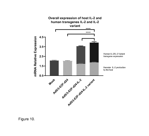
Figure 10. Variant IL-2-virus treatment achieves high-levels of IL-2 at
the tumor microenvironment. Tumors were collected from hamsters at day 16
(after
4 treatments) for relative mRNA quantification through RT-qPCR. Intratumoral
relative
mRNA expression levels of hamster IL-2 (lower bars), human IL-2 and IL-2
variant
(upper bars). Data is presented as mean+SEM. *p<0.05,****p<0.0001
Figure 11. Overall mRNA expression profile of virus-treated animals.
Tumors were collected from hamsters at day 16 and mRNA expression profile was
determined through Nanostring. (A) mRNA expression profile of Ad5/3-E2F-d24,
(B)
mRNA expression profile of Ad5/3-E2F-d24-IL-2 (C) mRNA expression profile of
Ad5/3-E2F-d24-IL-2 variant. Names are indicated for genes which were
statistically
significant different (adjusted p value < 0.05) expression compared to
reference group
(-1 > 10g2 fold change > 1).
Figure 12. mRNA expression levels of T-cell receptor signaling and
cytotoxic compounds in tumors treated with oncolytic adenovirus coding for
wild-type human IL-2 or variant IL-2. Tumors were collected from hamsters at
day
16 and mRNA expression levels were determined through Nanostring. (A) mRNA
count for genes related to T-cell receptor (TCR)-complex and signaling, (B)
mRNA
count for genes related to cytotoxic compounds, (C) Pearson's correlation
between
CA 03157255 2022-04-06
WO 2021/069806 7 PCT/F12020/050673
variant IL-2 mRNA relative expression and GZMK or SAP1 mRNA counts, or between
both the latter genes. Data is presented as mean+SEM from genes which were
statistically different from the reference group (Mock). ns ¨ non-significant
Figure 13. mRNA expression levels of anti-inflammatory and pro-
inflammatory signal genes in tumors treated with oncolytic adenovirus coding
for human IL-2 or variant IL-2. Tumors were collected from hamsters at day 16
and
mRNA expression levels were determined through Nanostring. (A) mRNA count for
genes which are related with co-stimulatory and co-inhibitory molecules. (B)
mRNA
count for genes which are related with antigen-presenting cells and
suppressive
io myeloid cells. (C) mRNA count for genes which are related with signals
associated with
anti-inflammatory and pro-inflammatory signals. Data is presented as mean+SEM
from
genes which were statistically different from the reference group (Mock).
*p<0.05,"p<0.01
DETAILED DESCRIPTION OF THE INVENTION
Interleukin 2 (IL-2) and variants thereof
As used herein, "IL-2" means wild-type IL-2, whether native or
recombinant. Mature human IL-2 occurs as a 133 amino acid sequence (without
the
signal peptide, consisting of an additional 20 N-terminal amino acids). The
amino acid
sequence of human IL-2 (SEQ ID NO: 1) is found in Genbank under accession
number
NP000577.2. The amino acid sequence of mature human IL-2 is depicted in SEQ ID
NO: 2.
As used herein, "IL-2 variant", "variant IL-2", "vIL2" or "vIL-2" means a
polypeptide or a nucleic acid (i.e. a gene) encoding said polypeptide, wherein
specific
substitutions to the interleukin-2 polypeptide have been made. The term
"polypeptide"
refers herein to any chain of amino acid residues, regardless of its length or
post-
translational modification (e.g., glycosylation or phosphorylation). The
variant IL-2
polypeptides can also be characterized by amino acid insertions, deletions,
substitutions and modifications at one or more sites in or at the other
residues of the
native IL-2 polypeptide chain. In accordance with this disclosure any such
insertions,
deletions, substitutions and modifications result in a variant IL-2 that
preferably exhibits
reduced binding to receptor subunit IL-2a but retains or improves the IL-2R6
binding
activity. Exemplary variants can include substitutions of 1, 2, 3, 4, 5, 6, 7,
8, 9, 10 or
more amino acids. Variants may also include conservative modifications and
substitutions at other positions of IL-2 (i.e., those that have a minimal
effect on the
activity or secondary or tertiary structure of the variant).
CA 03157255 2022-04-06
WO 2021/069806 8 PCT/F12020/050673
An exemplary variant IL-2 polypeptide includes an amino acid sequence
that is at least about 80% identical to SEQ ID NO:2 which binds the IL-2Ra
with an
affinity that is lower than the affinity with which the polypeptide
represented by SEQ ID
NO: 2 binds the IL-2Ra. Exemplary variant IL-2 polypeptides can be at least
about
50%, at least about 65%, at least about 70%, at least about 80%, at least
about 85%,
at least about 87%, at least about 90%, at least about 95%, at least about
97%, at least
about 98%, or at least about 99% identical to wild-type IL-2. The variant
polypeptide
can comprise a change in the number or content of amino acid residues. For
example,
the variant IL-2 can have a greater or a lesser number of amino acid residues
than
io wild-type IL-2. Alternatively, or in addition, an exemplary variant
polypeptide can
contain a substitution of one or more amino acid residues that are present in
the wild-
type IL-2. In various embodiments, the variant IL-2 polypeptide can differ
from wild-
type IL-2 by the addition, deletion, or substitution of a single amino acid
residue, for
example, a substitution of the residue at position 80 of SEQ ID NO:2.
Similarly,
exemplary variant polypeptides can differ from wild-type by a substitution of
two or
more amino acid residues, for example, the residues at positions 24, 45, 65,
72, 74,
80, 81, 85, 86, 89, 92, 93, 109 and 117 of SEQ ID NO:2. For example, the
mutation
can be selected from the group of consisting of: I24V, Y45A P65H, L72G, Q74R,
Q74H,
Q74N, Q745, L80F, L80V, R81I, R81T, R81D, L85V, I86V, I89V, I92F, V93I, D109L,
F117A. Preferably, the variant polypeptide comprises the substitutions L80F,
R81D,
L85V, I86V and I92F.
In another embodiment, variant IL-2 polypeptides can also be prepared as
fusion or chimeric polypeptides that include a variant IL-2 polypeptide and
another
heterologous polypeptide. A chimeric polypeptide including a variant IL-2 and
an
antibody or antigen-binding portion thereof can be generated. The antibody or
antigen-
binding component of the chimeric protein can serve as a targeting moiety. For
example, it can be used to localize the chimeric protein to a particular
subset of cells
or target molecule.
The present invention is particularly directed to a design of an oncolytic
viral vector comprising nucleic acid sequence encoding any of the above-
mentioned
variant IL-2 polypeptides as a transgene.
Viral vectors
Oncolytic viral vectors are therapeutically useful anticancer viruses that can
selectively infect and destroy cancer cells. Most current oncolytic viruses
are adapted
or engineered for tumour selectivity, although there are viruses, such as
reovirus and
Mumps virus, having natural preference for cancer cells. Many engineered
oncolytic
viral vectors take advantage of tumor-specific promoter elements making them
CA 03157255 2022-04-06
WO 2021/069806 9 PCT/F12020/050673
replication competent only in cancer cells. Surface markers expressed
selectively by
cancer cells can also be targeted by using them as receptors for virus entry.
A number
of viruses including adenovirus, reovirus, measles, herpes simplex, Newcastle
disease
virus and vaccinia have now been clinically tested as oncolytic agents.
Preferably, the oncolytic vector used in the present invention is an
adenoviral vector suitable for treating a human or animal. As used herein "an
oncolytic
adenoviral vector" refers to an adenoviral vector capable of infecting and
killing cancer
cells by selective replication in tumor versus normal cells.
In one embodiment of the invention, the adenoviral vectors are vectors of
lo human viruses. In one embodiment the adenoviral vectors are selected
from the group
consisting of Ad5, Ad3 and Ad5/3 vectors. As used herein, expression
"adenovirus
serotype 5 (Ad5) nucleic acid backbone" refers to the genome of Ad5.
Similarly,
"adenovirus serotype 3 (Ad3) nucleic acid backbone" refers to the genome of
Ad3.
"Ad5/3 vector" refers to a chimeric vector comprising or having parts of both
Ad5 and
Ad3 vectors. In a specific embodiment a backbone of the adenoviral vector is
an
adenovirus serotype 5 (Ad5) or serotype 3 (Ad3) nucleic acid backbone with
specific
mutations. E.g. fiber areas of the vector can be modified. In one embodiment
the
backbone is Ad5 nucleic acid backbone further comprising an Ad3 fiber knob. In
other
words the construct has the fiber knob from Ad3 while the remainder or the
most of the
remainder of the genome is from Ad5 (see, e.g., W02014170389).
The adenoviral vectors may be modified in any way known in the art, e.g.
by deleting, inserting, mutating or modifying any viral areas. The vectors are
made
tumor specific with regard to replication. For example, the adenoviral vector
may
comprise modifications in El, E3 and/or E4 such as insertion of tumor specific
promoters (e.g. to drive El), deletions of areas (e.g. the constant region 2
of El as
used in "A24", E3/gpl 9k, E3/6.7k) and insertion of a transgene or transgenes.
In a specific embodiment, the El B 19K gene (SEQ ID NO:3), generally known to
support replication of adenoviral vectors, has a disabling deletion dEl B 19K
(SEQ
ID NO:4) in the present vectors. Deletion of El B 19K is known to sensitize
cancer
cells to TNFalpha and thus it promotes apoptosis (White et al.,1992).
The sequence for wild-type E1B 19K gene is the following (the deletable region
is
underlined):
atggaggctt gggagtgttt ggaagatttt tctgctgtgc gtaacttgct
ggaacagagc tctaacagta cctcttggtt ttggaggttt ctgtggggct
catcccaggc aaagttagtc tgcagaatta aggaggatta caagtgggaa
tttgaagagc ttttgaaatc ctgtggtgag ctgtttgatt ctttgaatct
gggtcaccag gcgcttttcc aagagaaggt catcaagact ttggattttt
CA 03157255 2022-04-06
WO 2021/069806 10 PCT/F12020/050673
ccacaccggg gcgcgctgcg gctgctgttg cttttttgag ttttataaag
gataaatgga gcgaagaaac ccatctgagc ggggggtacc tgctggattt
tctggccatg catctgtgga gagcggttgt gagacacaag aatcgcctgc
tactgttgtc ttccgtccgc ccggcgataa taccgacgga ggagcagcag
cagcagcagg aggaagccag gcggcggcgg caggagcaga gcccatggaa
cccgagagcc ggcctggacc ctcgggaatg a (SEQ ID NO:3)
Accordingly, in an embodiment, the sequence for dE1B 19K in the present viral
vectors is
atggaggctt gggagtgttt ggaagatttt tctgctgtgc gtaacttgct
ggaacagctg ggtcaccagg cgcttttcca agagaaggtc atcaagactt
tggatttttc cacaccgggg cgcgctgcgg ctgctgttgc ttttttgagt
tttataaagg ataaatggag cgaagaaacc catctgagcg gggggtacct
gctggatttt ctggccatgc atctgtggag agcggttgtg agacacaaga
atcgcctgct actgttgtct tccgtccgcc cggcgataat accgacggag
gagcagcagc agcagcagga ggaagccagg cggcggcggc aggagcagag
cccatggaac ccgagagccg gcctggaccc tcgggaatga (SEQ ID NO:4)
One approach for generation of a tumor specific oncolytic adenovirus is
engineering a 24 base pair (bp) deletion ("A,24" or "d24") affecting the
constant region
2 (CR2) of El. In wild type adenovirus CR2 is responsible for binding the
cellular Rb
tumor suppressor/cell cycle regulator protein for induction of the synthesis
(S) phase
i.e. DNA synthesis or replication phase. The interaction between pRb and El A
requires
amino acids 121 to 127 of the El A protein conserved region. The vector may
comprise
a deletion of nucleotides corresponding to amino acids 122-129 of the vector
according
to Heise C. et al. (2000, Nature Med 6, 1134-1139) and Fueyo J. et al. (2000,
Oncogene 19(1):2-12). Viruses with the A24 are known to have a reduced ability
to
overcome the G1 -S checkpoint and replicate efficiently only in cells where
this
interaction is not necessary, e.g. in tumor cells defective in the Rb-pl 6
pathway, which
includes most if not all human tumors. In one embodiment of the invention the
vector
comprises a 24 bp deletion ("A,24" or "d24") in the Rb binding constant region
2 of
adenoviral El (See figure 5).
It is also possible to replace El A endogenous viral promoter for example by
a tumor specific promoter. For instance, E2F1 (e.g. in Ad5 based vector) or
hTERT
(e.g. in Ad3 based vector) promoter can be utilized in the place of El A
endogenous
viral promoter. The vector may comprise E2F1 promoter for tumor specific
expression
of El A.
CA 03157255 2022-04-06
WO 2021/069806 11 PCT/F12020/050673
The E3 region is nonessential for viral replication ex vivo, but the E3
proteins
have an important role in the regulation of host immune response i.e. in the
inhibition
of both innate and specific immune responses. In one embodiment of the
invention the
deletion of a nucleic acid sequence in the E3 region of the oncolytic
adenoviral vector
is a deletion of viral gpl 9k and 6.7k reading frames. The gpl 9k/6.7K
deletion in E3
refers to a deletion of 965 base pairs from the adenoviral E3A region. In a
resulting
adenoviral construct, both gpl 9k and 6.7K genes are deleted (Kanerva A et al.
2005,
Gene Therapy 12, 87-94). The gpl 9k gene product is known to bind and
sequester
major histocompatibility complex I (MHC1, known as HLA1 in humans) molecules
in
io the endoplasmic reticulum, and to prevent the recognition of infected
cells by cytotoxic
T-lymphocytes. Since many tumors are deficient in HLA1/MHC1, deletion of gpl
9k
increases tumor selectivity of viruses (virus is cleared faster than wild type
virus from
normal cells but there is no difference in tumor cells). 6.7K proteins are
expressed on
cellular surfaces and they take part in downregulating TNF-related apoptosis
inducing
ligand (TRAIL) receptor 2.
In one embodiment of the invention, the transgene, i.e. a gene encoding
variant interleu kin 2 (vIL2), is placed into a gpl 9k/6.7k deleted E3 region,
under the E3
promoter. This restricts transgene expression to tumor cells that allow
replication of
the virus and subsequent activation of the E3 promoter. In a specific
embodiment a
nucleic acid sequence encoding variant interleukin 2 is inserted into the
place of the
deleted nucleic acid sequence of viral gpl9k and 6.7k reading frames. In
another
embodiment of the invention E3 gpl 9k/6.7k is kept in the vector but one or
many other
E3 areas have been deleted (e.g. E3 9-kDa, E3 10.2 kDa, E3 15.2 kDa and/or E3
15.3
kDa).
E3 promoter may be any exogenous (e.g. CMV or E2F promoter) or
endogenous promoter known in the art, specifically the endogenous E3 promoter.
Although the E3 promoter is chiefly activated by replication, some expression
occurs
when El is expressed. As the selectivity of A24 type viruses occurs post El
expression
(when El is unable to bind Rb), these viruses do express El also in transduced
normal
cells. Thus, it is of critical importance to regulate also El expression to
restrict E3
promoter mediated transgene expression to tumor cells.
Specific embodiments of the invention include oncolytic adenoviral vectors
(e.g. Ad5 or Ad3 vectors) whose replication is restricted to the p16/Rb
pathway by dual
selectivity devices: an E2F (e.g. E2F1) tumor specific promoter placed in
front of the
adenoviral El A gene which has been mutated in constant region 2, so that the
resulting
El A protein is unable to bind Rb in cells. Furthermore, the fiber is modified
by 5/3
chimerism to allow efficient entry into tumor cell.
CA 03157255 2022-04-06
WO 2021/069806 12 PCT/F12020/050673
In a specific embodiment of the invention the oncolytic adenoviral vector
comprises:
1) a 24 bp deletion (A24) in the Rb binding constant region 2 of adenoviral
El;
2) a nucleic acid sequence deletion of viral gp19k and 6.7k reading frames;
and
3) a nucleic acid sequence encoding a variant interleukin 2 (vIL2) transgene
in the place of the deleted nucleic acid sequence as defined in point 2).
In the Experimental Section below, we constructed and characterized an
lo oncolytic adenovirus based on Ad5/3-E2F-d24 backbone and armed it with
vIL2. The
virus has an E2F promoter and a 24-base pair deletion in the El A constant
region 2
("D24") to enable its replication only in retinoblastoma/p16 pathway-defective
cells,
which is one of the common features for all cancer cells. E1B region is
deleted to
induce cancer cell apoptosis (dE1B 19K). Moreover, to improve its ability to
transduce
cancer cells and enhance its antitumor efficacy, the virus features fiber knob
from
serotype 3, while the rest of the genome derives from serotype 5. Most
importantly,
Ad5/3 viruses have good safety profile in humans. Preferably, oncolytic virus
armed
with vIL-2 is used with concomitant T-cell therapy or checkpoint inhibitors,
as a
potential platform to safely and effectively treat currently incurable solid
tumors. In
particular, tumor types where Tregs play an important role are preferably
treated.
In an embodiment, the present invention is directed to an oncolytic viral
vector, preferably an oncolytic adenoviral vector, comprising a nucleic acid
sequence
encoding a variant interleukin 2 (vIL2) transgene.
In a preferred embodiment, the backbone of the oncolytic adenoviral vector
is an adenovirus serotype 5 (Ad5) or serotype 3 (Ad3) nucleic acid backbone.
In a more preferred embodiment, said nucleic acid sequence encoding a
variant interleukin 2 (vIL2) transgene is in the place of a deleted nucleic
acid sequence
in the E3 region of said oncolytic adenoviral vector. Most preferably, the
deletion of a
nucleic acid sequence in the E3 region is a deletion of viral gp19k and 6.7k
reading
frames.
In another preferred embodiment, the vector also comprises a 24 bp
deletion (A24) in the adenoviral El sequence of said oncolytic adenoviral
vector.
In another preferred embodiment, the vector also comprises a disabling
deletion of E1B ((dE1B 19K).
In another preferred embodiment, the vector also comprises an Ad5/3 fiber
knob.
In another preferred embodiment, the vector comprises nucleic acid
sequence encoding a further transgene. More preferably, the further transgene
is
CA 03157255 2022-04-06
WO 2021/069806 13 PCT/F12020/050673
encoding a cytokine. In an embodiment, the cytokine is selected from the list
consisting of: TNFalpha, interferon alpha, interferon beta, interferon gamma,
complement C5a, CD4OL, IL12, IL-23, IL15, IL17, CCL1, CCL11, CCL12, CCL13,
CCL14-1, CCL14-2, CCL14-3, CCL15-1, CCL15-2, CCL16, CCL17, CCL18, CCL19,
CCL2, CCL20, CCL21, CCL22, CCL23-1, CCL23-2, CCL24, CCL25-1, CCL25-2,
CCL26, CCL27, CCL28, CCL3, CCL3L1, CCL4, CCL4L1, CCL5 (=RANTES), CCL6,
CCL7, CCL8, CCL9, CCR10, CCR2, CCR5, CCR6, CCR7, CCR8, CCRL1, CCRL2,
CX3CL1, CX3CR, CXCL1, CXCL10, CXCL11, CXCL12, CXCL13, CXCL14, CXCL15,
0X0L16, CXCL2, CXCL3, CXCL4, CXCL5, CXCL6, CXCL7, CXCL8, CXCL9,
lo CXCR1, CXCR2, CXCR4, CXCR5, CXCR6, CXCR7 and XCL2.
In a more preferred embodiment, the cytokine is TNFalpha.
The viral vectors utilized in the present inventions may also comprise other
modifications than described above. Any additional components or modifications
may
optionally be used but are not obligatory for the present invention.
Insertion of exogenous elements may enhance effects of vectors in target
cells. The use of exogenous tissue or tumor-specific promoters is common in
recombinant vectors and they can also be utilized in the present invention.
Adoptive cell therapy
One approach of the present invention is the development of a treatment for
patients with cancer using the transfer of immune lymphocytes that are capable
of
reacting with and destroying the cancer. Isolated tumor-infiltrating
lymphocytes are
grown in culture to large numbers and infused into the patient. In the present
invention
oncolytic vectors encoding a variant interleukin 2 (vIL2) transgene may be
utilized for
increasing the effect of lymphocytes. As used herein "increasing the efficacy
of adoptive
cell therapy" refers to a situation, wherein the oncolytic vector of the
invention is able to
cause a stronger therapeutic effect in a subject when used together with an
adoptive cell
therapeutic composition compared to the therapeutic effect of the adoptive
cell
therapeutic composition alone. A specific embodiment of the invention is a
method of
treating cancer in a subject, wherein the method comprises administration of
an
oncolytic vector of the invention to a subject, said method further comprising
administration of adoptive cell therapeutic composition to the subject.
Adoptive cell
therapeutic composition and the vectors of the invention are administered
separately.
Separate administrations of an adoptive cell therapeutic composition and
adenoviral
vectors may be preceded by myeloablating or non-myeloablating preconditioning
chemotherapy and/or radiation. The adoptive cell therapy treatment is intended
to
reduce or eliminate cancer in the patient.
CA 03157255 2022-04-06
WO 2021/069806 14 PCT/F12020/050673
A specific embodiment of the invention relates to therapies with adenoviral
vectors and an adoptive cell therapeutic composition, e.g. tumor-infiltrating
lymphocytes, TCR modified lymphocytes or CAR modified lymphocytes. T-cell
therapies in particular, but also any other adoptive therapies such as NK cell
therapies
or other cell therapies may be utilized in the present invention. Indeed,
according to
the present invention the adoptive cell therapeutic composition may comprise
unmodified cells such as in TIL therapy or genetically modified cells. There
are two
common ways to achieve genetic targeting of T-cells to tumor specific targets.
One is
transfer of a T-cell receptor (TCR) with known specificity and with matched
human
io leukocyte antigen (HLA, known as major histocompatibility complex in
rodents) type.
The other is modification of cells with artificial molecules such as chimeric
antigen
receptors (CAR). This approach is not dependent on HLA and is more flexible
with
regard to targeting molecules. For example, single chain antibodies can be
used and
CARs can also incorporate costimulatory domains. However, the targets of CAR
cells
need to be on the membrane of target cells, while TCR modifications can
utilize
intracellular targets.
As used herein "adoptive cell therapeutic composition" refers to any
composition comprising cells suitable for adoptive cell transfer. In one
embodiment of
the invention the adoptive cell therapeutic composition comprises a cell type
selected
from a group consisting of a tumor-infiltrating lymphocyte (TIL), TCR (i.e.
heterologous
T-cell receptor) modified lymphocytes and CAR (i.e. chimeric antigen receptor)
modified lymphocytes. In another embodiment of the invention, the adoptive
cell
therapeutic composition comprises a cell type selected from a group consisting
of T-
cells, CD8+ cells, CD4+ cells, NK-cells, dendritic cells, delta-gamma T-cells,
regulatory
T-cells and peripheral blood mononuclear cells. In another embodiment, TILs, T-
cells,
CD8+ cells, CD4+ cells, NK-cells, delta-gamma T-cells, regulatory T-cells or
peripheral
blood mononuclear cells form the adoptive cell therapeutic composition. In one
specific
embodiment of the invention the adoptive cell therapeutic composition
comprises T cells.
As used herein "tumor-infiltrating lymphocytes" or TILs refer to white blood
cells that
have left the bloodstream and migrated into a tumor. Lymphocytes can be
divided into
three groups including B cells, T cells and natural killer cells. In another
specific
embodiment of the invention the adoptive cell therapeutic composition
comprises T-cells
which have been modified with target-specific chimeric antigen receptors or
specifically
selected T-cell receptors. As used herein "T-cells" refers to CD3+ cells,
including CD4+
helper cells, CD8+ cytotoxic T-cells and yb T cells.
In addition to suitable cells, adoptive cell therapeutic composition used in
the present invention may comprise any other agents such as pharmaceutically
acceptable carriers, buffers, excipients, adjuvants, additives, antiseptics,
filling,
CA 03157255 2022-04-06
WO 2021/069806 15 PCT/F12020/050673
stabilising and/or thickening agents, and/or any components normally found in
corresponding products. Selection of suitable ingredients and appropriate
manufacturing methods for formulating the compositions belongs to general
knowledge of a person skilled in the art.
The adoptive cell therapeutic composition may be in any form, such as solid,
semisolid or liquid form, suitable for administration. A formulation can be
selected from
a group consisting of, but not limited to, solutions, emulsions, suspensions,
tablets,
pellets and capsules. The compositions are not limited to a certain
formulation; instead
the composition can be formulated into any known pharmaceutically acceptable
formulation. The pharmaceutical compositions may be produced by any
conventional
processes known in the art.
A combination of an oncolytic adenoviral vector of the invention and an
adoptive cell therapeutic composition refers to use of an oncolytic adenoviral
vector and
an adoptive cell therapeutic composition together but as separate
compositions. It is
clear to a person skilled in the art that an oncolytic adenoviral vector of
the present
invention and an adoptive cell therapeutic composition are not used as one
composition.
Indeed, adenoviral vectors are not used for modifying the adoptive cells but
for modifying
the target tumor, so that the tumor is more amenable to the desired effects of
the cellular
transplant. In particular, the present invention enhances recruitment of the
adoptive
transplant to the tumor, and increases its activity there. In a specific
embodiment of the
invention oncolytic adenoviral vectors and an adoptive cell therapeutic
composition of a
combination are for simultaneous or sequential, in any order, administration
to a
subject.
Checkpoint inhibitor
Immune checkpoint proteins interact with specific ligands which send a signal
into T cells that inhibits T-cell function. Cancer cells exploit this by
driving high level
expression of checkpoint proteins on their surface thereby suppressing the
anti-cancer
immune response.
A checkpoint inhibitor (also referred to as a CPI) as described herein is any
compound capable of inhibiting the function of an immune checkpoint protein.
Inhibition
includes reduction of function as well as full blockade. In particular, the
immune
checkpoint protein is a human checkpoint protein. Thus, the immune checkpoint
inhibitor
is preferably an inhibitor of a human immune checkpoint.
Checkpoint proteins include, without limitation, CTLA-4, PD-1 (and its ligands
PD-L1 and PD-L2), B7-H3, B7-H4, HVEM, TIM3, GAL9, LAG3, VISTA, KIR, BTLA,
TIGIT and/or IDO. The pathways involving LAG3, BTLA, B7-H3, B7-H4, TIM3 and
KIR
are recognized in the art to constitute immune checkpoint pathways similar to
the CTLA-
4 and PD-1 dependent pathways. The immune checkpoint inhibitor can be an
inhibitor
CA 03157255 2022-04-06
WO 2021/069806 16 PCT/F12020/050673
of CTLA-4, PD-1 (and its ligands PD-L1 and PD-L2), B7-H3, B7- H4, HVEM, TIM3,
GAL9, LAG3, VISTA, KIR, BTLA, TIGIT and/or IDO. In some embodiments, the
immune
checkpoint inhibitor is an inhibitor of PD-L1. Preferably, the immune
checkpoint inhibitor
is a monoclonal antibody that selectively binds to PD-L1, more preferably
selected from
the group consisting of: BMS-936559, LY3300054, atezolizumab, durvalumab and
avelumab.
In some embodiments, the checkpoint inhibitor of the combination is an
antibody. The term "antibody" as used herein encompasses naturally occurring
and
engineered antibodies as well as full length antibodies or functional
fragments or analogs
lo thereof that are capable of binding e.g. the target immune checkpoint or
epitope (e.g.
retaining the antigen-binding portion). The antibody for use according to the
methods
described herein may be from any origin including, without limitation, human,
humanized, animal or chimeric and may be of any isotype with a preference for
an IgG1
or IgG4 isotype and further may be glycosylated or non-glycosylated. The term
antibody
also includes bispecific or multispecific antibodies so long as the
antibody(s) exhibit the
binding specificity herein described.
Cancer
The recombinant vectors of the present invention are replication competent
in tumor cells. In one embodiment of the invention the vectors are replication
competent
in cells, which have defects in the Rb-pathway, specifically Rb-p16 pathway.
These
defective cells include all tumor cells in animals and humans. As used herein
"defects in
the Rb-pathway" refers to mutations and/or epigenetic changes in any genes or
proteins
of the pathway. Due to these defects, tumor cells overexpress E2F and thus,
binding of
Rb by E1A CR2, that is normally needed for effective replication, is
unnecessary. Further
selectivity is mediated by the E2F promoter, which only activates in the
presence of free
E2F, as seen in Rb/p16 pathway defective cells. In the absence of free E2F, no
transcription of E1A occurs and the virus does not replicate. Inclusion of the
E2F
promoter is important to prevent expression of E1A in normal tissues, which
can cause
toxicity both directly and indirectly through allowing transgene expression
from the E3
promoter.
The present invention relates to approaches for treating cancer in a subject.
In one embodiment of the invention, the subject is a human or a mammal,
specifically
a mammal or human patient, more specifically a human or a mammal suffering
from
cancer.
The approach can be used to treat any cancers or tumors, including both
malignant and benign tumors, both primary tumors and metastases may be targets
of
the approach. In one embodiment of the invention the cancer features tumor-
infiltrating
lymphocytes. The tools of the present invention are particularly appealing for
treatment
CA 03157255 2022-04-06
WO 2021/069806 17 PCT/F12020/050673
of metastatic solid tumors featuring tumor-infiltrating lymphocytes. In
another
embodiment the T-cell graft has been modified by a tumor or tissue specific T-
cell
receptor of chimeric antigen receptor.
As used herein, the term "treatment" or "treating" refers to administration of
at least oncolytic adenoviral vectors to a subject, preferably a mammal or
human
subject, for purposes which include not only complete cure but also
prophylaxis,
amelioration, or alleviation of disorders or symptoms related to a cancer or
tumor.
Therapeutic effect may be assessed by monitoring the symptoms of a patient,
tumor
markers in blood, or for example a size of a tumor or the length of survival
of the patient
io In another embodiment of the invention the cancer or tumor is
selected from
a group consisting of nasopharyngeal cancer, synovial cancer, hepatocellular
cancer,
renal cancer, cancer of connective tissues, melanoma, lung cancer, bowel
cancer,
colon cancer, rectal cancer, colorectal cancer, brain cancer, throat cancer,
oral cancer,
liver cancer, bone cancer, pancreatic cancer, choriocarcinoma, gastrinoma,
pheochromocytoma, prolactinoma, T-cell leukemia/lymphoma, neuroma, von Hippel-
Lindau disease, Zollinger-Ellison syndrome, adrenal cancer, anal cancer, bile
duct
cancer, bladder cancer, ureter cancer, brain cancer, oligodendroglioma,
neuroblastoma, meningioma, spinal cord tumor, bone cancer, osteochondroma,
chondrosarcoma, Ewing's sarcoma, cancer of unknown primary site, carcinoid,
carcinoid of gastrointestinal tract, fibrosarcoma, breast cancer, Paget's
disease,
cervical cancer, esophagus cancer, gall bladder cancer, head and neck cancer,
eye
cancer, kidney cancer, Wilms' tumor, Kaposi's sarcoma, prostate cancer,
testicular
cancer, Hodgkin's disease, non-Hodgkin's lymphoma, oral cancer, skin cancer,
mesothelioma, multiple myeloma, ovarian cancer, endocrine pancreatic cancer,
glucagonoma, parathyroid cancer, penis cancer, pituitary cancer, soft tissue
sarcoma,
retinoblastoma, small intestine cancer, stomach cancer, thymus cancer, thyroid
cancer, trophoblastic cancer, hydatidiform mole, uterine cancer, endometrial
cancer,
vagina cancer, vulva cancer, acoustic neuroma, mycosis fungoides, insulinoma,
carcinoid syndrome, somatostatinoma, gum cancer, heart cancer, lip cancer,
meninges cancer, mouth cancer, nerve cancer, palate cancer, parotid gland
cancer,
peritoneum cancer, pharynx cancer, pleural cancer, salivary gland cancer,
tongue
cancer and tonsil cancer. Preferably, the cancer or tumor treated is selected
from the
group consisting of renal cancer, ovarian cancer, bladder cancer, prostate
cancer,
breast cancer, colorectal cancer, lung cancer (such as small-cell lung
carcinoma, non-
.. small-cell lung carcinoma and squamous non-small-cell lung carcinoma),
gastric
cancer, classical Hodgkin lymphoma, mesothelioma, and liver cancer. In a more
preferred embodiment, the cancer or tumor type is head and neck cancer, most
preferably human head and neck cancer.
CA 03157255 2022-04-06
WO 2021/069806 18 PCT/F12020/050673
Before classifying a human or animal patient as suitable for the therapy of
the present invention, the clinician may examine a patient. Based on the
results
deviating from the normal and revealing a tumor or cancer, the clinician may
suggest
treatment of the present invention for a patient.
Pharmaceutical composition
A pharmaceutical composition of the invention comprises at least one type
of viral vector of the invention. Preferably, the present invention provides a
pharmaceutical composition containing (a) an oncolytic virus as such or in
combination
with (b) adoptive cell composition or (c) a checkpoint inhibitor. The present
invention
lo also provides said pharmaceutical combination for use in the treatment
of cancer.
Furthermore, the composition may comprise at least two, three or four
different vectors.
In addition to the vector and adoptive cell composition or checkpoint
inhibitor, a
pharmaceutical composition may also comprise other therapeutically effective
agents,
any other agents such as pharmaceutically acceptable carriers, buffers,
excipients,
adjuvants, additives, preservatives, antiseptics, filling, stabilising and/or
thickening
agents, and/or any components normally found in corresponding products.
Selection
of suitable ingredients and appropriate manufacturing methods for formulating
the
compositions belongs to general knowledge of a man skilled in the art.
The pharmaceutical composition may be in any form, such as solid,
semisolid or liquid form, suitable for administration. A formulation can be
selected from
a group consisting of, but not limited to, solutions, emulsions, suspensions,
tablets,
pellets and capsules. The compositions of the current invention are not
limited to a
certain formulation, instead the composition can be formulated into any known
pharmaceutically acceptable formulation. The pharmaceutical compositions may
be
produced by any conventional processes known in the art.
A pharmaceutical kit of the present invention comprises an oncolytic
adenoviral vector encoding a variant IL-2 as a transgene and one or more
immune
checkpoint inhibitors. The oncolytic adenoviral vector encoding a variant IL-2
as a
transgene is formulated in a first formulation and said one or more immune
checkpoint
inhibitors are formulated in a second formulation. Alternatively, the
pharmaceutical kit
of the present invention comprises an oncolytic adenoviral vector encoding a
variant
IL-2 as a transgene in the first formulation and an adoptive cell composition
in the
second formulation. In another embodiment of the invention the first and the
second
formulations are for simultaneous or sequential, in any order, administration
to a
subject. In another embodiment, said kit is for use in the treatment of cancer
or tumor.
CA 03157255 2022-04-06
WO 2021/069806 19 PCT/F12020/050673
Administration
The vector or pharmaceutical composition of the invention may be
administered to any mammal subject. In a specific embodiment of the invention,
the
subject is a human. A mammal may be selected from a group consisting of pets,
domestic animals and production animals.
Any conventional method may be used for administration of the vector or
composition to a subject. The route of administration depends on the
formulation or
form of the composition, the disease, location of tumors, the patient,
comorbidities and
other factors. Accordingly, the dose amount and dosing frequency of each
therapeutic
io agent in the combination depends in part on the particular therapeutic
agent, the
severity of the cancer being treated, and patient characteristics. Preferably,
a dosage
regimen maximizes the amount of each therapeutic agent delivered to the
patient
consistent with an acceptable level of side effects.
The effective dose of vectors depends on at least the subject in need of the
treatment, tumor type and location of the tumor and stage of the tumor. The
dose may
vary for example from about 1x10⁸ viral particles (VP) to about 1x10¹⁴ VP,
specifically
from about 5x10⁹ VP to about 1x10¹³ VP and more specifically from about 3x10⁹
VP to
about 2x10¹² VP. In one embodiment oncolytic adenoviral vectors coding for a
variant
IL-2 are administered in an amount of 1x10¹⁰- 1x10¹⁴ virus particles. In
another
embodiment of the invention the dose is in the range of about 5x101 - 5x10¹¹
VP.
In one embodiment of the invention, the administration of oncolytic virus is
conducted through an intratumoral, intra-arterial, intravenous, intrapleural,
intravesicular, intracavitary, intranodal or peritoneal injection, or an oral
administration.
Any combination of administrations is also possible. The approach can give
systemic
efficacy despite local injection.
In one embodiment of the invention, the separate administration(s) of (a) an
oncolytic adenoviral vector encoding a variant IL-2 as a transgene and (b) one
or more
immune checkpoint inhibitors to a subject is (are) conducted simultaneously or
consecutively, in any order. This means that (a) and (b) may be provided in a
single
unit dosage form for being taken together or as separate entities (e.g. in
separate
containers) to be administered simultaneously or with a certain time
difference. This
time difference may be between 1 hour and 2 weeks, preferably between 12 hours
and
3 days, more preferably up to 24 or 48 hours. In a preferred embodiment, the
first
administration of the adenoviral vector is conducted before the first
administration of
the checkpoint inhibitor. In addition, it is possible to administer the virus
via another
administration way than the checkpoint inhibitor. In this regard, it may be
advantageous
to administer either the virus or checkpoint inhibitor intratumorally and the
other
systemically or orally. In a particular preferred embodiment, the virus is
administered
CA 03157255 2022-04-06
WO 2021/069806 20 PCT/F12020/050673
intratumorally and the checkpoint inhibitor intravenously. Preferably, the
virus and the
checkpoint inhibitor are administered as separate compounds. Concomitant
treatment
with the two agents is also possible.
In a preferred embodiment, the checkpoint inhibitor is administered in an
amount from about 2 mg/kg to 50 mg/kg, more preferably about 2 mg/kg to 25
mg/kg.
As used herein "separate administration" or "separate" refers to a situation,
wherein (a) an oncolytic adenoviral vector encoding a variant IL-2 as a
transgene and
(b) one or more immune checkpoint inhibitors are two different products or
compositions
distinct from each other.
io Any other treatment or combination of treatments may be used in
addition
to the therapies of the present invention. In a specific embodiment the method
or use
of the invention further comprises administration of concurrent or sequential
radiotherapy, chemotherapy, antiangiogenic agents or targeted therapies, such
as
alkylating agents, nucleoside analogs, cytoskeleton modifiers, cytostatic
agents,
monoclonal antibodies, kinase inhibitors or other anti-cancer drugs or
interventions
(including surgery) to a subject.
The terms "treat" or "increase", as well as words stemming therefrom, as
used herein, do not necessarily imply 100% or complete treatment or increase.
Rather,
there are varying degrees of which one of ordinary skill in the art recognizes
as having
a potential benefit or therapeutic effect.
It will be obvious to a person skilled in the art that, as the technology
advances, the inventive concept can be implemented in various ways. The
invention
and its embodiments are not limited to the examples described above but may
vary
within the scope of the claims.
EXPERIMENTAL SECTION
Materials and methods
Cell lines
Human lung adenocarcinoma A549, human melanoma SK-MEL-28 and
hamster leiomyosarcoma DDT1-MF2 cell lines were maintained in DMEM and hamster
pancreatic cancer HapT1 was maintained in RPMI. Both DMEM or RPM! were
supplemented with 10% fetal bovine serum (FBS), 100 U/mL penicillin, 100 mg/mL
streptomycin, and 2 mM L-glutamine (all from Sigma-Aldrich). Both cell lines
were
cultured at +37 C and 5% CO2.
CA 03157255 2022-04-06
WO 2021/069806 21 PCT/F12020/050673
Recombinant human cytokine
Recombinant human (rh) IL-2 (Peprotech) and rh vIL-2 (Adipogen)
cytokines were used as positive controls in the ex vivo experiments in
concentrations
of 0.1-100 U/mL.
Virus and vIL-2 transgene construction
All the viruses used in this study have the backbone of Ad5/3-E2F-d24. The
construction of this and Ad5/3-E2F-d24-IL-2 has been explained previously
(Havunen
et al., 2017).
The vIL-2 transgene was constructed by making five point mutations in IL-2
sequence
at positions 80 L->F, 81 R->D, 85 L->V, 86 I->V and 92 I->F. Ad5/3-E2F-d24-vIL-
2
virus was generated with bacterial artificial chromosome (BAC)-recombineering
strategy, which used galk selection (Warming et al., 2005; Muck-Hausl et al.,
2015).
The transgene vIL-2 was inserted in E3 region by homologous recombination. PCR-
amplified vIL-2 was electroporated into SW102 bacteria containing BAC-Ad5/3-
E2F-
A24-GalK/amp and the positive clones with vIL-2 transgene were identified with
deoxyglucose selection. The sequence was verified by restriction enzyme
analysis.
The virus genome was released from BACs with Pad l restriction enzyme (Thermo
Scientific) and transfected into A549 cells with Lipofectamine 2000 reagent
(Invitrogen). The vIL-2-armed Ad5/3 virus was then purified twice with cesium
chloride
gradient centrifugation. Optical density and tissue culture infectious dose
(TCID50)
assay was used to determine viral particle (VP) concentration and infectious
units,
respectively.
Cytokine expression by virus ex vivo
A549 cells were infected with either Ad5/3-E2F-d24-IL-2, Ad5/3-E2F-d24-
vIL-2, or left uninfected for 48 hr. Supernatant was collected and filtered
(Amicon
ultra 100K), and then analyzed with IL-2 human ELISA kit (Abcam) according to
the
manufacturer's instructions to determine the amount of virally-produced
cytokines.
Lytic potency assay
10,000 A549 cells/well were plated in 100u1 of 2% DMEM assay media
into 96-well plate. Cells were infected with Ad5/3-E2F-d24, Ad5/3-E2F-d24-IL-
2, or
Ad5/3-E2F-d24-vIL-2 at 0-1000 VP/cell in triplicates. After 3 days, cell
viability was
determined with MTS cytotoxicity assay according to manufacturer's
instructions (cell
titer 96 Aqueous One Solution Cell Proliferation Assay, Promega, Madison, WI).
CA 03157255 2022-04-06
WO 2021/069806 22 PCT/F12020/050673
Cell proliferation assay
Peripheral blood mononuclear cells (PBMCs) were obtained from healthy
donors and isolated through density gradient centrifugation using Lymphoprep
(StemCell technologies). The PBMCs were incubated with rh vIL-2 and rh IL-2 at
different concentrations (0.1 U, 1 U, 10 U, and 100 U) for three days and
analyzed for
CD4+ T cells, CD8+ T cells, and NK cells through flow cytometry. To measure
the
relative cell expansion, we compared the percentage of positive cells on day 3
to the
corresponding numbers on day 0.
T-cell isolation, and stimulation
io T cells were enriched from freshly isolated PBMCs through CD3+ T
cell
isolation kit (Miltenyi Biotec). Sorted T lymphocytes were activated with
CD3/0D28
beads (Invitrogen) in a 1:5 bead/T-cell ratio and then cultured for 4 days
either with:
(1) rh IL-2 at 100 U/mL; (2) rh vIL-2 at 100 U/mL, or (3) without any
cytokine, but with
complete media as a control. These three conditions were studied in three
groups: in
group one, activated T cells only; in group two, tumor cells in addition to
activated T
cells; and in group three, activated T cells and tumor cells with unarmed
virus Ad5/3-
E2F-d24. Cytokines and half of the assay medium were replaced on day 2. Cells
were
analyzed on days 0, 2, and 4 by flow cytometry with Sony SH800Z (Sony, Tokyo,
Japan).
Immune subset analysis after virus infection ex vivo
Tumor cells were infected with either unarmed Ad5/3-E2F-d24, Ad5/3-
E2F-d24-1L-2, or Ad5/3-E2F-d24-vIL-2 viruses at 100 VP/cell or left
uninfected. PBMCs
isolated from healthy donor were added on top of infected cancer cells 24
hours post-
infection. PBMCs alone were used as mock control. Cells were stained
immunofluorescently with anti-CD3, anti-CD8, anti-CD4, anti-CD25, anti-CD69,
anti-
CD127, and anti-CD56 and analyzed on days 0, 3, and 6 through BD Accuri C6
flow
cytometer. Next, the effects of specific immune cell populations, namely T
cells and
NK cells, were studied more in detail in a similar set up.
Animal experiment
To study treatment-induced changes in tumors, 2*106 HapT1 cells per
animal were implanted on the lower back subcutaneously in 5 week-old
immunocompetent Syrian hamsters. Animals were randomized into groups of four
(n=13), when the average tumor diameter reached 0.5 cm. Viruses Ad5/3-E2F-d24,
Ad5/3-E2F-d24-IL-2, and Ad5/3-E2F-d24-vIL-2 were administered intratumorally
at
1 *1 09 VPs and mock received PBS only. Virus were injected on days 1, 4, 8,
and 13.
CA 03157255 2022-04-06
WO 2021/069806 23 PCT/F12020/050673
Five animals were euthanized from each group on day 16 and tumors and
selected organs were collected to evaluate histopathological characteristics
and
immune cell subsets present. The rest of the animals were monitored for
survival.
These animals received 6 additional rounds of virus treatment after every 5
days,
starting at day 18. Tumors were measured with digital caliper in all even days
till day
30. End point criteria included 20.0 mm tumor size limit and skin ulcerations.
Cured animals were re-challenged on their upper back with either the
same HapT1 tumor (2*106 cells/tumor) or with a different tumor DDT-MF2
(1.5*105
cells/tumor) after the observation period of 160 days. Naïve animals (n=3)
that had not
lo been exposed to any cancer cell or treatment before were included as
mock group.
Tumor growth was followed for 21 days until DDT1-MF2 tumors reached the
maximum
tolerated diameter. Of note, two out of three Ad5/3-E2F-d24 therapeutic
animals were
not re-challenged because of the presence of visible tumors, i.e. the tumors
had not
been cured with unarmed virus.
Histopathology
For pathological evaluation, hamster organs such as liver, spleen, lung,
kidney, heart, and tumor samples were collected on day 16 from five hamsters
of each
group. Collected samples were first fixed in 10 % formalin, after 48 hr
transferred to 70
% ethanol and embedded in paraffin. For microscopic evaluation, tissue
sections with
5 pm thickness were stained with hematoxylin and eosin. A pathologist
evaluated the
histological changes in stained tissue samples.
Statistical Analysis
Evaluation of tumor growth was performed with Linear mixed model with
the log-transformed tumor volumes with SPSS version 25 Statistics (IBM). Two-
way
ANOVA and Log-rank (Mantel-Cox) tests were used to analyse the group variation
in
the re-challenged and survival curve, respectively. GraphPad Prism (version
8Ø0.)
was used to present individual and grouped tumor growth data and to plot
survival
curve. P value was considered significant when p<0.05.
Example 1. Effector cells proliferate more in the presence of vIL-2 than with
conventional IL-2 ex vivo
We compared rh vIL-2 and rh IL-2 with regard to their ability to stimulate
immune cells, such as CD8+ T cells, NK cells, and CD4+ T cells. We cultured
PBMCs
either with or without recombinant human (rh) vIL-2 or rh IL-2 at different
concentrations (0.1 ¨100 U/ml). After 3 days, the rh vIL-2 was more potent in
inducing
the proliferation of CD8+ effector T cells and NK cells than IL-2, whereas the
levels of
CA 03157255 2022-04-06
WO 2021/069806 24 PCT/F12020/050673
CD4+ T cells (including Tregs) remained lower with the variant (Figure 1).
These
results indicated that the vIL-2 has preferred effects on T cells and NK cells
over the
conventional IL-2. It should be noted that as activated T-cells produce IL-2,
there will
be IL-2 present in cultures even in the vIL-2 groups. This would be expected
to dilute
the (lack of) effect of vIL-2 on Tregs, for example.
Example 2. The effects of rh vIL-2 on different T cell subsets provides
rationale
for constructing a virus coding for the cytokine
To investigate the effect of rh vIL-2 on T cells in the presence of
adenovirus,
io we isolated T cells with CD3/0D28 beads and activated them for 4 days with
either
100 U/ml of rh vIL-2 or rh IL-2 and infected/non-infected cancer cells. IL-2
and vIL-2
had similar effects on CD8/CD4 cell ratios in the presence and absence of
cancer cells
(Figure 2A and B). However, when the cancer cells were infected with an
oncolytic
virus, vIL-2 induced a trend towards CD8+0D27-CD62L-CD45R0+ cell dominance
over CD4+ cells (Figure 20).
The hallmark of acquired immunity is a memory response, which is the
consequence of antigen-specific lymphocytes' clonal expansion and
differentiation that
persists for a lifetime (Sallusto et al., 2004). We evaluated the percentage
change of
central memory T cells (Tcm; CD45R0+, CD62L+, 0D27+) in the presence of rh IL-
2
or rh vIL-2 with infected or non-infected cancer cells. Tcm mediate reactive
memory
responses and differentiate to effector cells upon antigen stimulation. We did
not find
any difference in 0D8/0D4 Tcm ratios between IL-2 and vIL-2 in the absence of
cancer
cells (Figure 3A). In the presence of tumor cells, we first observed a
decrease in the
ratio on day 2, followed by an increase on day 4. Again, the vIL-2 induced
higher 0D8
to 0D4 ratio in the Tcm population than the conventional IL-2 (Figure 3B).
In addition to Tcm, we also evaluated effector memory T cells (Tem;
0D45R0+, 0D62L-, 0D27-F) in the same conditions as Tcm cells. Tem provide
protective memory and are characterized by prompt effector function. We did
not
observe differences in 0D8/0D4 Tem ratio between IL-2 and vIL-2, if cancer
cells were
not present (Figure 4A). With cancer cells, IL-2 and vIL-2 induce high ratio
of 0D8/0D4
Tem by day 4 (Figure 4B). When the cancer cells were infected, we observed a
trend
towards high 0D8/0D4 Tem ratio in rh vIL-2 group on day 4 (Figure 40). To
conclude,
we did not see significant difference between rh IL-2 and rh vIL-2 with regard
to their
effect on pro-inflammatory T cell compartments. These results provided us
solid
grounds to construct a vIL-2 armed oncolytic adenovirus.
CA 03157255 2022-04-06
WO 2021/069806 25 PCT/F12020/050673
Example 3. Ad5/3-E2F-d24-vIL-2 virus expresses vIL-2 and kills tumor cells
efficiently ex vivo
Adenovirus 5/3 features the backbone of adenovirus serotype 5 and fiber
knob of adenovirus serotype 3, to enhance tumor transduction, as its receptor
is highly
expressed in advanced tumors (Wang et al., 2011). To restrict virus
replication to
tumor cells, a mutation in constant region 2 of the El A gene and introduction
of a
heterologous tumor-specific E2F promoter were performed. To enhance apoptosis
enabling deletion of El B 19K gene region was made. The variant IL-2 transgene
was
placed into the E3 region under the E3 promoter, to link the expression to
virus
io
replication (Figure 5A). The transgene cassette replaces the open reading
frames for
gpl 9k and 6.7k.
To investigate the oncolytic potency of the constructed virus, cytotoxicity
assay was performed using human lung cancer A549 cells. There were no major
differences between viruses' cell killing ability between Ad5/3-E2F-d24-IL-2
and Ad5/3-
E2F-d24-vIL-2, thus indicating that presence of vIL-2 transgene does not
reduce the
oncolytic potency of the virus (Figure 5B). Additionally, cells infected with
Ad5/3-E2F-
d24-v1L-2 were able to secrete the cytokine (Figure 5C).
Example 4. Ad5/3-E2F-d24-vIL-2 stimulates effector cells but not Tregs ex vivo
Human cancer cells A549 were infected with either Ad5/3-E2F-d24,
Ad5/3-E2F-d24-IL-2, or Ad5/3- E2F-d24-vIL-2, or left uninfected. After 24h,
cancer
cells were incubated with PBMCs. The CD8/CD4 ratio of CD25-F0D69+ activated
effector T cells was significantly higher in the group treated with Ad5/3-E2F-
d24-vIL-2
on day 3 and day 6, than when treated with the virus expressing conventional
IL-2
(Figure 6A). Actually, we did not see any significant difference in CD8/CD4
ratio of
activated effector T cells between the control viruses. Thus, vIL-2-armed
Ad5/3 virus
is a potent stimulator for effector cells.
To investigate effects on Tregs, we analyzed CD25+CD12710w expressing
cells of the CD4+ CD3+ parent population. Ad5/3-E2F-d24-vIL-2 did not induce
Treg
differentiation like Ad5/3-E2F-d24-IL-2 (Figure 6B). Thus, virally produced
vIL-2 seems
to retain the key attractive feature of recombinant vIL-2: preferential
stimulation of
effector cells over Tregs.
Example 5. Variant IL-2 armed oncolytic adenovirus significantly enhances
antitumor efficacy and survival in hamsters
Following the promising ex vivo results, variant IL-2 armed adenovirus was
then studied in immunocomptent Syrian hamsters. Since human adenoviruses are
able
to replicate in hamsters (unlike in mice) and some human cytokines such as
human IL-
CA 03157255 2022-04-06
WO 2021/069806 26 PCT/F12020/050673
2 are bioactive in hamsters (Havunen et al., 2017; Gowen et al., 2008), it is
the optimal
model for studying armed oncolytic adenoviruses (Havunen et al., 2017).
Animals treated with backbone Ad5/3-E2F-d24 or IL-2 armed virus,
(Ad5/3-E2F-d24-IL-2) showed a trend for tumor control as compared to mock
(difference not significant). Impressively, we got best tumor control in the
group treated
with Ad5/3-E2F-d24-vIL-2 and this result was statistically significant in
comparison to
all other groups by day 30. This underlines the utility of vIL-2 as a
stimulator of anti-
tumor effector T cells without the unwanted immunosuppressive effects on Treg.
Thus,
Ad5/3-E2F-d24-vIL-2 appears to be a potent modulator of the tumor
microenvironment
lo towards a direction compatible with complete tumor eradication.
In order to investigate the mechanism of action of the therapy, we treated
hamsters with either backbone Ad5/3-E2F-d24, Ad5/3-E2F-d24-IL-2, Ad5/3-E2F-d24-
vIL-2 or PBS on days 1, 4, 8, and 13. On day 16, hamsters were euthanized,
tumors
were collected to deeply analyze tumor microenvironment through flow cytometry
and
Nanostring assessments. To study treatment-related changes, tumors and
selected
organs were collected for histopathological evaluation. Pathological results
revealed
no difference between mock and oncolytic adenovirus treated groups thus, our
virus
didn't cause any systemic toxic effects.
Survival data shows that the group treated with backbone virus was able
to cure one hamster. Additionally, two hamsters with stable tumors survived to
the end
of the experiment. Ad5/3-E2F-d24-IL-2 cured three hamsters. Ad5/3-E2F-d24-vIL-
2
was able to cure 60% of treated hamsters and this difference was significant
over
mock (Figure 7).
Example 6. Treatment with cytokine armed adenovirus induces tumor-specific
immunological memory
In order to study if oncolytic virus treatment had induced tumor-specific
immunological memory, all cured hamsters were re-challenged with the same
HapT1
cancer cells and with different DDT1-MF2 cancer cells. Both cancer cell types
were
implanted on the upper back of cured hamsters. In this re-challenge
experiment, the
number of animals per group differed because different viruses had cured a
different
number of hamsters. There was no hindrance of HapT1 (Figure 8A) or DDT1-MF2
(Figure 8B) tumor rechallenge in naïve or unarmed virus treated animals. In
contrast,
prior cure of HapT1 with Ad5/3-E2F-d24-IL-2 or Ad5/3-E2F-d24-vIL-2 appeared to
offer
protection against HapT1 rechallenge. Of note, 40% of animals treated with
Ad5/3-
E2F-d24-vIL-2 remained HapT1 tumor free after rechallenged. However, DDT-MF2
tumors grew normally in these animals underlining the epitope-specific
immunological
CA 03157255 2022-04-06
WO 2021/069806 27 PCT/F12020/050673
memory induced by the armed viruses. These findings are in line with previous
data
indicating the importance of cytokines such as IL-2 in establishing systemic
and tumor-
specific antitumor immunity (Havunen et al., 2017).
We have shown that variant IL-2 armed adenovirus Ad5/3-E2F-d24-vIL-2
appears safe and effective in immunocompetent Syrian hamsters. Ad5/3-E2F-d24-
vIL-
2 exhibited potent antitumor efficacy and expression of variant IL-2 within
the tumor
microenvironment. Studies on human lymphocytes indicated induction of
antitumor
immunological effects. Specifically, vIL-2 seemed to preferentially activate
effector T-
cells over suppressive Tregs.
Example 7. Variant IL-2 coding oncolytic adenovirus induces substantial tumor
reduction with moderate infiltration of immune cells and the highest
intratumoral
expression of IL-2
Cell lines
Hamster pancreatic cancer HapT1 was maintained in RPM! supplemented with
10% fetal bovine serum (FBS), 100 U/mL penicillin, 100 mg/mL streptomycin, and
2 mM L-glutamine (all from Sigma-Aldrich). Both cell lines were cultured at
+370 and
5% 002.
Virus and vIL-2 transgene construction
All the viruses used in this study have the backbone of Ad5/3-E2F-d24. The
construction of the latter and Ad5/3-E2F-d24-IL-2 has been explained
previously
in Havunen et al., 2017. The vIL-2 transgene was constructed by making five
point mutations in IL-2 sequence at positions 80 L->F, 81 R->D, 85 L->V, 86 1-
>V
and 92 1->F. Ad5/3-E2F-d24-vIL-2 virus was generated with bacterial artificial
chromosome (BAC)-recombineering strategy, which used galk selection (Warming
et al., 2005; Muck-Hausl et al., 2015). The transgene vIL-2 was inserted in E3
region by homologous recombination. PCR-amplified vIL-2 was electroporated
into 5W102 bacteria containing BAC-Ad5/3-E2F-A24-GalK/amp and the positive
clones with vIL-2 transgene were identified with deoxyglucose
selection. The
sequence was verified by restriction enzyme analysis. The virus genome was
released from BACs with Pad l restriction enzyme (Thermo Scientific) and
transfected into A549 cells with Lipofectamine 2000 reagent (Invitrogen). The
vIL-2-armed Ad5/3 virus was then purified twice with cesium chloride gradient
centrifugation. Optical density and tissue culture infectious dose (TCID50)
assay
was used to determine viral particle (VP) concentration and infectious units,
respectively.
CA 03157255 2022-04-06
WO 2021/069806 28 PCT/F12020/050673
Animal experiment
To study treatment-induced changes in tumors, 2x10⁶ HapT1 cells per animal
were implanted on the lower back subcutaneously in 5 week-old
immunocompetent Syrian hamsters. Animals were randomized into groups of
four (n=13), when the average tumor diameter reached 0.5 cm. Viruses Ad5/3-
E2F-d24, Ad5/3-E2F-d24-IL-2, and Ad5/3-E2F-d24-vIL-2 were administered
intratumorally at 1x10⁹VPs and mock received PBS only. Viruses were injected
on
days 1, 4, 8, and 13.
Five animals were euthanized from each group on day 16 and tumors were
collected to evaluate immunological changes and mRNA expression levels.
Flow cytometty
Hamster tumour samples collected on day 16, were processed as single cell
suspensions and further analyzed following previously established protocols
(Havunen
et al., 2017; Siurala et al., 2016) These samples were then stained with
antibodies for
CD8+ (PE, 12-0080-82), CD4+ (PE-Cyanine 7, 25-0041-82), and MHC II+ cells
(FITC,
11-5980-82) cells. NK+ cells were labelled with the polyclonal antibody anti-
Asialo-
GM1 (Alexa Fluor-488, 53-6507-80), and macrophages+ cells with anti-Galectin
(PE,
12-5301-82) as described before (Havunen et al., 2017). Cell fluorescence was
detected using Sony SH800Z cytometer (Sony, Tokyo, Japan) upon the acquisition
of
the 100.000 events per sample. Cell data processing and gating were performed
with
FlowJo v.10.6.1 (BD , New Jersey, USA).
Gene-expression analysis
Fragments of animal tumour samples harvested on day 16 were preserved in
RNAlater (R0901; Sigma-Aldrich, St. Louis, USA), and stored in -20 C until
further
processing. RNA from these samples were then purified with RNAeasy Mini Kit
(74104; QIAGEN, Hilden, Germany) following the manufacturer's instructions.
The
final RNA yield was measured with the Thermo Scientific NanoDrop TM 1000
Spectrophotometer (Thermo Fisher Scientific, Massachusetts, USA), and the RNA
concentration of the samples were adjusted to 2Ong/pl.
Reverse transcriptase (RT)-quantitative Polym erase Chain Reaction (qPCR)
RNA purified from Day 16 tumours, were used to synthetize cDNA using
Quantitect
Reverse Transcription Kit (205313, QIAGEN, Hilden, Germany) to be used for the
relative quantification of the viruses transgene expression as well as for
hamster IL-2
relative expression. The reverse transcription real-time PCR (RT-qPCR) was
performed as previously described by Santos et al., 2017. Wild-type IL-2 virus
CA 03157255 2022-04-06
WO 2021/069806 29 PCT/F12020/050673
transgene was detected with the primers and probe designed for human IL-2. For
IL-
2 variant virus transgene, primers and probe designed for human IL-2v were
used.
Normalization of hamster IL-2 and viruses transgenes were performed using
hamster
GAPDH gene (Siurala et al., 2015).
Statistical analysis
GraphPad Prism (version 8Ø0.) was used to present tumor volume data,
relative
and absolute mRNA expression levels. Unpaired t-test with Welch's correction
was
performed to assess differences between different therapeutic groups.
Pearson's
correlation coefficient was calculated to determine correlation between
granzyme
io production with SAP gene or variant IL-2 transgene production. P value
was
considered significant when p<0.05.
Results
To gain further insight on the biological events induced by the oncolytic
adenovirus
treatments, we collected tumors 16 days after the experiment started.
Comparison of
tumor volumes from day 0 and 16 showed that treatment with Ad5/3-E2F-D24 had a
minimum effect in controlling the growth of hamster tumors (Figure 9A). On the
other
hand, human IL-2 and variant IL-2-coding oncolytic adenovirus treatments
substantially delayed tumor growth of hamster pancreatic tumors. Such effect
was
more prominent in hamsters treated with variant IL-2-coding oncolytic
adenoviruses
considering that, by day 16, this group demonstrated a trend towards the
lowest
tumor burden (Figure 9A).
Further analysis of the immunological compartment revealed that the frequency
of
CD4+ and CD8+ cells in tumors was the highest in the wild-type human IL-2
oncolytic
adenovirus treated hamsters (Figure 9B-C). In contrast, tumors from hamsters
undergoing variant IL-2-virus therapy had surprisingly moderate levels of
infiltration of
these cells (Figure 9B-C). Considering that no significant difference in the
tumor
volumes on day 16 was observed between the cytokine-armed viruses, this
suggests
that the virus-expressing variant IL-2 induces qualitatively superior
antitumor
response relative to the wild-type counterpart.
In addition, key limitations of wild-type human IL-2 are its pharmacokinetics
(short
half-life) and, consequently, its minimal accumulation at the target lesions
(Arenas-
Ramirez et al., 2015). The results in Figure 10 demonstrate that the above-
mentioned problems can be ablated with the Ad5/3-E2F-D24-vIL-2 oncolytic
adenovirus. For example, a significant difference can be observed in the
production
of IL-2 between tumors from hamsters treated with wild-type IL-2 or variant IL-
2
oncolytic adenoviruses (Figure 10). In fact, Ad5/3-E2F-D24-vIL-2 treated group
CA 03157255 2022-04-06
WO 2021/069806 30 PCT/F12020/050673
presented the highest global cytokine expression (host IL-2 and IL-2 variant
transgene) compared to all other groups. Moreover, the levels of IL-2 variant
were
high even after 3 days of the last virus-treatment (Figure 10). It is
important to note
that these results are unforeseen for proteins of similar nature, which is
especially
remarkable, considering that the effort to develop recombinant variant IL-2
proteins,
focuses on improving safety and intratumorally relevant high-levels of variant
IL-2
proteins (Arenas-Ramirez et al., 2015).
Example 8. Variant IL-2 coding oncolytic adenovirus therapy causes immune
reconfiguration for increased effector T-cell function and low
immunosuppression
Cell lines
Hamster pancreatic cancer HapT1 was maintained in RPM! supplemented with
10% fetal bovine serum (FBS), 100 U/mL penicillin, 100 mg/mL streptomycin, and
2 mM L-glutamine (all from Sigma-Aldrich). Both cell lines were cultured at
+370 and
5% 002.
Virus and vIL-2 transgene construction
All the viruses used in this study have the backbone of Ad5/3-E2F-d24. The
construction of the latter and Ad5/3-E2F-d24-IL-2 has been explained
previously
in Havunen et al., 2017. The vIL-2 transgene was constructed by making five
point mutations in IL-2 sequence at positions 80 L->F, 81 R->D, 85 L->V, 86 1-
>V
and 92 1->F. Ad5/3-E2F-d24-vIL-2 virus was generated with bacterial artificial
chromosome (BAC)-recombineering strategy, which used galk selection (Warming
et al., 2005; Muck-Hausl et al., 2015). The transgene vIL-2 was inserted in E3
region by homologous recombination. PCR-amplified vIL-2 was electroporated
into 5W102 bacteria containing BAC-Ad5/3-E2F-A24-GalK/amp and the positive
clones with vIL-2 transgene were identified with deoxyglucose
selection. The
sequence was verified by restriction enzyme analysis. The virus genome was
released from BACs with Pad l restriction enzyme (Thermo Scientific) and
transfected into A549 cells with Lipofectamine 2000 reagent (Invitrogen). The
vIL-2-armed Ad5/3 virus was then purified twice with cesium chloride gradient
centrifugation. Optical density and tissue culture infectious dose (TCID50)
assay
was used to determine viral particle (VP) concentration and infectious units,
respectively.
CA 03157255 2022-04-06
WO 2021/069806 31 PCT/F12020/050673
Animal experiment
To study treatment-induced changes in tumors, 2x10⁶ HapT1 cells per animal
were implanted on the lower back subcutaneously in 5 week-old
immunocompetent Syrian hamsters. Animals were randomized into groups of
four (n=13), when the average tumor diameter reached 0.5 cm. Viruses Ad5/3-
E2F-d24, Ad5/3-E2F-d24-IL-2, and Ad5/3-E2F-d24-vIL-2 were administered
intratumorally at 1x10⁹VPs and mock received PBS only. Viruses were injected
on
days 1, 4, 8, and 13.
Five animals were euthanized from each group on day 16 and tumors were
collected to evaluate immunological changes and mRNA expression levels.
Gene-expression analysis
Fragments of animal tumour samples harvested on day 16 were preserved in
RNAlater
(R0901; Sigma-Aldrich, St. Louis, USA), and stored in -20 C until further
processing.
RNA from these samples were then purified with RNAeasy Mini Kit (74104;
QIAGEN,
.. Hilden, Germany) following the manufacturer's instructions. The final RNA
yield was
measured with the Thermo Scientific NanoDropTM 1000 Spectrophotometer (Thermo
Fisher Scientific, Massachusetts, USA), and the RNA concentration of the
samples
were adjusted to 2Ong/pl.
NanoString nCounter gene expression analysis was performed on the RNA samples
from all hamster tumours utilizing the nCounter0 Digital Analyzer (NanoString
Technologies, Seattle, USA). Gene expression was assessed with a custom-panel
designed for hamster cells containing 101 genes analysed by nSolver software
4.0
(NanoString Technologies, Seattle, USA). Differential expression is displayed
as the
values for each genes' gene's -10g10 (p-value) and 10g2 fold change in the
volcano
plots. Likewise, differential expression as RNA counts (Log2) are displayed in
the bars
graphs. The expression level of each gene in the treatment groups was
normalized to
their corresponding genes in the control (mock) group.
Statistical analysis
GraphPad Prism (version 8Ø0.) was used to present absolute mRNA expression
.. levels. Unpaired t-test with Welch's correction was performed to assess
differences
between different therapeutic groups. Pearson's correlation coefficient was
calculated
to determine correlation between granzyme production with SAP gene or variant
IL-2
transgene production. P value was considered significant when p<0.05.
CA 03157255 2022-04-06
WO 2021/069806 32 PCT/F12020/050673
Results
Additional characterization was performed by evaluating the transcriptome of
tumors
undergoing virus therapy. Compared to Ad5/3-E2F-D24 virus treatments, tumors
treated with oncolytic viruses encoding wild-type IL-2 or variant IL-2
demonstrated a
.. seemingly similar number of upregulated genes. When evaluating the
downregulation
profile in both groups, wild-type IL-2 had approximately 34.5% more genes
downregulated than variant IL-2 group (Figure 11). It is important to note
that the wild-
type IL-2 group displayed higher values for almost all genes, being up- or
downregulated, compared to IL-2 variant, which is compatible with IL-2 potent
io .. biological effects previously related in the literature (Figure 11)
(Jiang et al., 2016).
Here, however, elevated values did not necessarily translate into better
overall
response, since Ad5/3-E2F-D24-vIL-2 virus was the most successful group in
promoting tumor shrinkage and overall survival.
Detailed view of the differential expression analysis, however, demonstrated
that
Ad5/3-E2F-D24-vIL-2 virus preferentially stimulates the expression of known
genes
associated to the T-cell receptor complex and downstream signaling genes, over
immunosuppressive associated genes (Figures 12 and 13). Importantly, treatment
with
variant IL-2-coding oncolytic adenoviruses induces the highest expression of
TCR-
complex genes (CD3E, CD3D) (Figure 12A), which are responsible for harboring
the
.. TCR upon MHC binding and initiating TCR induced signaling in humans
(Ngoenkam
et al., 2018). Additionally, variant IL-2-coding oncolytic adenovirus
treatment
upregulated known key downstream signaling genes (LCK, ITK, ZAP70) compared
with the virus coding for wild-type IL-2 (Figure 12A). Surprisingly, treatment
with IL-2
variant-coding virus stimulated the upregulation of some critical genes for
TCR
.. expression, anchoring, and signaling (CD3G, SAP), while their levels for
wild-type IL-
2 virus group remained unaltered (Figure 12A). Even though not statistically
significant,
these data suggest an unexpected increased activity of the TCR function of T-
cells
upon treatment with variant IL-2 coding oncolytic adenoviruses, which to our
knowledge, was not documented in current art. In fact, only the variant IL-2
virus was
.. capable of inducing high expression of granzymes and perforin (GZMK, GZMM,
PRF1)
(Figure 12B). When secreted by effector cytotoxic cells (T-cells and Natural
killer cells),
granzymes and perforin induce apoptosis of tumor cells (Voskoboinik et al.,
2015). In
fact, the degree of correlation between variant IL-2 transgene mRNA relative
expression and both GZMK or SAP1 genes mRNA counts is high and positive, while
.. an additional positive correlation can be seen between GZMK and SAP1 genes
(Figure
12C). Particularly, expression of Granzyme K and Granzime M is not known from
the
current art to be induced by neither oncolytic adenoviruses nor variant IL-2
proteins.
CA 03157255 2022-04-06
WO 2021/069806 33 PCT/F12020/050673
The highest expression of 77M-3 and CTLA-4 genes was seen in tumors treated
with
wild-type IL-2-coding virus (Figure 13A). These genes are generally known to
inhibit
T-cell function in humans (Ngoenkam et al., 2018), therefore they can be
potentially
associated with an increased immunosuppressive environment. Another gene, PD-
L1,
remained unaltered upon treatment with variant IL-2 virus (Figure 13A). This
may have
contributed to the possible low immunosuppression in tumors belonging to the
variant
IL-2 virus-treated animals, considering the role of PD-L1 in immunosuppression
in mice
and humans. Even though the highest expression levels of CD137 (activation
marker
of human tumor-reactive TILs) was seen in tumors from animals treated with
wild type-
io IL-2-coding virus (Figure 13A), it may have been insufficient to counter
the
immunosuppression in these tumors. In contrast, expression of 0D27 (a co-
stimulatory
marker) was increased in the variant-IL-2-virus groups whilst unaltered in the
wild-type
counterpart (Figure 13A). Longer telomeres have been previously reported in
TILs
expressing 0D27, thus hinting that variant IL-2 potentially increases the
presence of
TILs which are less terminally differentiated. On the other hand, 0D27 is
known to be
highly expressed in memory T-cells, which have been deemed to produce a
multifunctional response. This suggests that variant IL-2 oncolytic virus
enables
superior quality response in tumors.
On the other hand, virus-derived secretion of wild-type IL-2 caused the
highest
expression of known antigen-presenting cells in humans and mice (CD80, 0D86,
CD40) (Figure 13B). Yet, surprisingly, wild-type IL-2-coding virus treatment
rendered
inferior antitumor efficacy in hamsters compared to its variant counterpart.
The
presence and activity (CD11b, CD206, Arg1) of immune suppressive cells, such
as
myeloid cells, was also reduced or unaltered in the variant IL-2 coding virus
compared
with the wild-type IL-2 virus (Figure 13B). Contrary to common art, such
effect is
unexpected considering that certain mutations in the variant IL-2 protein aim
at mainly
preventing unwanted targeting of regulatory T-cells instead of other
immunosuppressive cells.
Further analysis revealed an overall decreased gene expression of IL-6, TGFb,
IL-10
.. (Figure 13C) in tumors from hamsters treated with either wild-type IL-2 or
variant IL-2-
virus therapy (Figure 13C). Interestingly, while the former had caused an
increased
expression of CCL3, TNF, IL-1b (Figure 13C) (genes that encode proinflammatory
cytokines in humans and mice), wild-type IL-2-virus treated tumors remained
larger
than the variant IL-2 virus-treated tumors. A possible explanation for this is
the known
ambiguous role of TNF in antitumor immunity, which in chronic levels, can
contribute
for the tumor progression in humans.
CA 03157255 2022-04-06
WO 2021/069806 34 PCT/F12020/050673
In summary, variant IL-2-virus promotes the ability of T-cells to harbor TCRs,
signaling,
lower T-cell inhibition and immunosuppression from the myeloid cells
compartment.
These effects may have contributed to improved cytotoxic function of effector
cells,
which render variant IL-2-coding oncolytic adenovirus therapy capable of
providing the
.. best survival and antitumor efficacy, compared to other therapeutic groups.
REFERENCES
Arenas-Ramirez, N., Woytschak, J. & Boyman, 0. Interleukin-2: Biology, Design
and
Application. Trends Immunol. 36, 763-777 (2015).
Gowen BB, Judge JW, Wong MH, Jung KH, Aylsworth OF, Melby PC, Rosenberg B,
Morrey
JD: lmmunoprophylaxis of Punta Toro virus (Phlebovirus, Bunyaviridae)
infection in hamsters
with recombinant Eimeria profilin-like antigen. Int Immunopharmacol 2008,
8:1089-1094.
Havunen R, Siurala M, Sorsa S, Gronberg-Vaha-Koskela S, Behr M, Tahtinen S,
Santos JM,
Karell P, Rusanen J, Nettelbeck DM, et al.: Oncolytic Adenoviruses Armed with
Tumor
Necrosis Factor Alpha and Interleukin-2 Enable Successful Adoptive Cell
Therapy. Mol Ther
Oncolytics 2017, 4:77-86.
ltzhaki 0, Levy D, Zikich D, Treves AJ, Markel G, Schachter J, Besser MJ:
Adoptive T-cell
transfer in melanoma. lmmunotherapy 2013, 5:79-90.
Jiang, T., Zhou, C. & Ren, S. Role of IL-2 in cancer immunotherapy.
Oncoimmunology5,
e1163462¨e1163462 (2016).
Klein C, Waldhauer I, Nicolini VG, Freimoser-Grundschober A, Nayak T, Vugts
DJ, Dunn C,
Bolijn M, Benz J, Stihle M, Lang M, Roemmele M, Hofer T, van Puijenbroek E,
Wittig D,
Moser S, Ast 0, Brunker P, Gorr IH, Neumann S, Mudry MCV, Hinton H, CramericF,
Saro J,
Evers S, Gerdes C, Bacac M, van Dongen G, Moessner E & Umafia P. (2017)
Cergutuzumab amunaleukin (CEA-IL2v), a CEA-targeted IL-2 variant-based
immunocytokine
for combination cancer immunotherapy: Overcoming limitations of aldesleukin
and
conventional IL-2-based immunocytokines, Oncolmmunology, 6:3, DOI:
10.1080/2162402X.2016.1277306
Levin, Aron M., Darren L. Bates, Aaron M. Ring, Carsten Krieg, Jack T. Lin,
Leon Su, Ignacio
Moraga, Miro E. Raeber, Gregory R. Bowman, Paul Novick, Vijay S. Pande, C.
Garrison
Fathman, Onur Boyman and K. Christopher Garcia. Exploiting a natural
conformational
switch to engineer an interleukin-2 `superkine'. Nature vol. 484, pages 529-
533 (2012).
CA 03157255 2022-04-06
WO 2021/069806 35 PCT/F12020/050673
Muck-Hausl M, Solanki M, Zhang W, Ruzsics Z, Ehrhardt A: Ad 2.0: a novel
recombineering
platform for high-throughput generation of tailored adenoviruses. Nucleic
Acids Res 2015,
43:e50.
Ngoenkam, J., Schamel, W. W. & Pongcharoen, S. Selected signalling proteins
recruited to
the T-cell receptor-CD3 complex. Immunology 153, 42-50 (2018).
Sallusto F, Geginat J, Lanzavecchia A: Central memory and effector memory T
cell subsets:
function, generation, and maintenance. Annu Rev Immunol 2004, 22:745-763.
Santos, J. M. et al. Adenoviral production of interleukin-2 at the tumor site
removes the need
for systemic postconditioning in adoptive cell therapy. Int. J. Cancer 141,
(2017).
Schwartz RN, Stover L, Dutcher JP: Managing toxicities of high-dose
interleukin-2. Oncology
(Williston Park) 2002, 16:11-20.
Siurala, M. et al. Oncolytic adenovirus and doxorubicin-based chemotherapy
results in
synergistic antitumor activity against soft-tissue sarcoma. Int. J. Cancer
136, 945-954
(2015).
Siurala, M. et al. Syngeneic syrian hamster tumors feature tumor-infiltrating
lymphocytes
allowing adoptive cell therapy enhanced by oncolytic adenovirus in a
replication permissive
setting. Oncoimmunology 5, e1136046¨e1136046 (2016).
Sockolosky JT, Trotta E, Parisi G, Picton L, Su LL, Le AC, Chhabra A, Silveria
SL, George
BM, King IC, Mr T, Jude K, Sibener LV, Baker D, Shizuru JA, Ribas A, Bluestone
JA, Garcia
.. KC. (2018) Selective targeting of engineered T cells using orthogonal IL-2
cytokine-receptor
complexes, Science, 359(6379): 1037, DOI: 10.1126/science.aar3246.
Tahtinen S, Blattner C, Vaha-Koskela M, Saha D, Siurala M, Parviainen S,
Utikal J, Kanerva
A, Umansky V, Hemminki A: T-Cell Therapy Enabling Adenoviruses Coding for IL2
and
TNFalpha Induce Systemic lmmunomodulation in Mice With Spontaneous Melanoma. J
.. lmmunother 2016, 39:343-354.
Voskoboinik, I., Whisstock, J. C. & Trapani, J. A. Perforin and granzymes:
function,
dysfunction and human pathology. Nat. Rev. Immunol. 15, 388-400 (2015).
Wang H, Li ZY, Liu Y, Persson J, Beyer I, Moller T, Koyuncu D, Drescher MR,
Strauss R,
Zhang XB, et al.: Desmoglein 2 is a receptor for adenovirus serotypes 3, 7, 11
and 14. Nat
Med 2011, 17:96-104.
Warming S, Costantino N, Court DL, Jenkins NA, Copeland NG: Simple and highly
efficient
BAC recombineering using galK selection. Nucleic Acids Res 2005, 33:e36.
CA 03157255 2022-04-06
WO 2021/069806 36 PCT/F12020/050673
White, E., Sabbatini, P., Debbas, M., Wold, W.S.M., Kusher, D.I., and Gooding,
L. (1992).
The 19-kilodalton adenovirus E1B transforming protein inhibits programmed cell
death and
prevents cytolysis by tumor necrosis factor alpha. Mol. Cell. Biol. 1992; 12:
2570-2580.).
Cited patent publications:
US9428567
US2019062395
W02014170389
W02016146894