Note: Descriptions are shown in the official language in which they were submitted.
WO 2021/089558 1
PCT/EP2020/080836
CELL CULTURE MONITORING SYSTEM
The present invention relates to a system for monitoring the culture of cells
in a liquid
medium.
The emergence of cell therapies and cell based product is leading to an
increased need for
accurate and timely control of cell cultures. Cell cultures may also be used
for bioproduction
for instance of antibodies and vaccines. Many steps of conventional culture
processes need
human intervention, in particular for cell counting and cell viability
measurement. Each
intervention increases the risk of contamination and the final cost of the
therapy. The loss of
a therapy batch due to error or contamination has dramatic consequences for
the patient.
In view of the foregoing, an object of the invention is to provide a cell
culture monitoring
system that allows for accurate control of cell growth and reduces the risk of
contamination in
an economical manner.
It is advantageous to provide a cell culture monitoring system that is
reliable.
It is advantageous to provide a cell culture monitoring system that allows
continuous or
frequent analyses of the state of cells during culture in an economical and
sterile manner.
Continuous measurements of viability would allow to detect cell culture
disease at an early
stage.
Objects of the invention have been achieved by providing a cell culture
monitoring system
according to claim 1.
Disclosed herein is a cell culture monitoring system comprising a monitoring
apparatus for
coupling to a culture tank containing a cell culture medium therein, and a
fluid circulation
system for fluidic coupling to the cell culture tank, the fluid circulation
system comprising a
dielectrophoresis cartridge for connection to the cell culture tank via supply
and return
conduits. The dielectrophoresis cartridge comprises a base and an electrode
support having
electrodes in or on the electrode support, the electrodes configured for
travelling wave
dielectrophoresis and comprising a measurement zone arranged above a measuring
chamber formed between the electrode support and a floor of the base forming a
measuring
chamber therebetween, whereby cells in a liquid medium flowing through the
measuring
chamber are subject to a travelling wave dielectrophoresis force orthogonal to
a direction of
flow of said liquid through said measuring chamber. The monitoring apparatus
comprises a
CA 03157471 2022-5-5
WO 2021/089558 2
PCT/EP2020/080836
computing unit, an image capture system connected to the computing unit, and a
cartridge
holder portion for receiving said dielectrophoresis cartridge such that the
image capture
system may detect cells flowing through said measuring chamber.
In an advantageous embodiment, at least the base of the dielectrophoresis
cartridge is made
of a polymer, preferably a transparent polymer.
In an advantageous embodiment, the electrode support is made of a transparent
polymer or
glass.
In an advantageous embodiment, the dielectrophoresis cartridge comprises an
outlet and an
inlet configured for coupling to tubes of a supple polymer forming said supply
and return
conduits.
In an advantageous embodiment, the electrodes are formed on an inner surface
of the
electrode support bounding the measuring chamber and having contact portions
extending to
an electrode connection window formed in the base for plugging contact to
complementary
spring contacts of the monitoring apparatus, the electrode connection window
being sealingly
separated from the measuring chamber.
In an advantageous embodiment, the measuring chamber comprises a raised floor
and
lateral guides defining a gap between the floor and electrode support.
In an advantageous embodiment, said electrodes comprise a measurement zone
formed by
one or more spiraling conductive tracks.
In an advantageous embodiment, said electrodes consist of four to ten
electrodes, preferably
four to eight electrodes.
In an advantageous embodiment, the electrodes are arranged in the measurement
zone in
two sets in mirror image symmetry.
In an advantageous embodiment, the cartridge holder portion of the monitoring
apparatus
comprises a cartridge holder slot configured for slidable insertion of the
dielectrophoresis
cartridge therein.
CA 03157471 2022-5-5
WO 2021/089558 3
PCT/EP2020/080836
In an advantageous embodiment, the cartridge holder portion comprises locating
elements
engaging in complementary locating elements in the dielectrophoresis cartridge
for
positioning and securing the dielectrophoresis cartridge in a measurement
position.
In an advantageous embodiment, the locating elements comprise spring
protuberances or
spring resist portions on either the cartridge holder portion or the
dielectrophoresis cartridge.
In an advantageous embodiment, the image capture system comprises a microscope
connected to an image processing circuit of the computing unit configured for
digital analysis
of the trajectory of the cells captured by the image capture system.
In an advantageous embodiment, the computing unit comprises a signal generator
connected via the connector to the electrodes of the dielectrophoresis
cartridge configured to
generate a travelling wave dielectrophoresis signal in the measurement zone of
the
electrodes.
In an advantageous embodiment, the measuring chamber between electrode and
floor is in
the range of 10 to 200pm.
In an advantageous embodiment, the cell culture tank is separate from the
monitoring
apparatus and comprises a fluidic connector for connection to supply and
return conduits
connected to the dielectrophoresis cartridge.
Further objects and advantageous features of the invention will be apparent
from the claims,
from the detailed description, and annexed drawings, in which:
Figure 1 is a schematic representation of a cell culture monitoring system
according to an
embodiment of the invention;
Figure 2a is a perspective view of a cell culture monitoring system according
to an
embodiment of the invention;
Figure 2b is a perspective view of a portion of the cell culture monitoring
system of figure 2a
with a cover removed and certain internal components removed;
Figure 3a and 3b are schematic views of a tube inserted in a cell culture tank
2 of a cell
culture monitoring system according to an embodiment of the invention;
Figure 4 is a cross-sectional view of a fluidic connector of the cell culture
tank;
Figure 5a is a perspective view of a cartridge holder portion of a monitoring
apparatus of a
cell culture system according to an embodiment of the invention;
CA 03157471 2022-5-5
WO 2021/089558 4
PCT/EP2020/080836
Figure 5b is a view similar to figure 5a with a cartridge of the cell culture
monitoring system
according to an embodiment of the invention, inserted in the holder;
Figure 5d is a perspective partial cross-sectional simplified view of the
cartridge and holder of
figure 5b;
Figure Sc is an exploded view of the elements of figure 5b;
Figures 6a and 6b are perspective views of the cartridge according to an
embodiment of the
invention;
Figure 6c is an exploded perspective view of the cartridge according to an
embodiment of the
invention;
Figure 6d is a plan view of a base of the cartridge according to an embodiment
of the
invention;
Figure 6e is a cross-sectional view through the cartridge according to an
embodiment of the
invention;
Figure 7 is a view of electrodes of a dielectrophoresis cartridge according to
an embodiment
of the invention;
Figure 8 is a schematic simplified representation of the trajectory of cells
relative to the
electrodes when subject to dielectrophoresis;
Figure 9 is a schematic representation of a dielectrophoresis cartridge
according to a variant.
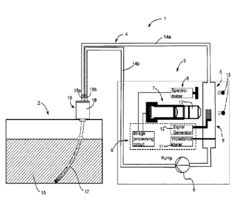
Referring to the figures, a cell culture monitoring system 1 according to
embodiments of the
invention comprises a monitoring apparatus 3, a cell culture tank 2, and a
fluid circulation
system 4 for transporting a cell culture medium containing cells to be
observed between the
cell culture tank and the monitoring apparatus.
The monitoring apparatus 3 comprises an image capture system 7, a spectrometer
8, a
computing unit 9, and a cartridge holder portion 28 for receiving a
dielectrophoresis cartridge
5 of the fluid circulation system 4.
The fluid circulation system 4 comprises the dielectrophoresis cartridge 5 and
conduits 14a,
14b interconnecting the dielectrophoresis cartridge 5 to the cell cartridge
tank 2. The fluid
circulation system 4 further comprises a pump 6 that may be mounted or formed
part of the
monitoring apparatus 3 (as illustrated) or that may in other variants be
mounted on the cell
cartridge tank and electrically connected to the monitoring apparatus for
control of the pump.
In a preferred embodiment, the pump is mounted on the monitoring apparatus and
may
advantageously be in a form of a peristaltic pump. At least a portion of the
supply conduit 14a
comprises a flexible section of tube mounted in the peristaltic pump for
pumping of the cell
culture medium in a sterile manner.
CA 03157471 2022-5-5
WO 2021/089558 5
PCT/EP2020/080836
The supply conduit 14a and return conduit 14b connected to the
dielectrophoresis cartridge 5
and cell culture tank 2 of the fluid circulation system advantageously forms a
closed circuit
enabling fluid of a cell culture medium 15 contained in the cell culture tank
2 to be circulated
to the dielectrophoresis cartridge 5 and back to the cell culture tank in a
closed circuit. In a
variant, the fluid circulation system may further comprise an exit conduit
coupled to a waste
channel 23 for the removal of dead (i.e. apoptotic) cells separated from live
cells, or for
separating different cell pheonotypes, due to their different trajectories in
the
dielectrophoresis cartridge. The fluidic connector 18 may be connected to the
cell culture
tank via a luer lock type of connection as per se well known in the art of
fluidic connections,
or may be interconnected by other means. The fluidic connector 18 allows
flexible tubes, in
particular the tank supply and return tubes, to be coupled to the connector.
The supply conduit 14a may further comprise a perforated tube 17 immersed in
the cell
culture medium 15, and preferably that extends to the bottom of the cell
culture tank. The
perforations in the tube 17 may be arranged such that there are a larger
number of
perforations towards the bottom of the tank and a progressively decreasing
number of
perforations towards the top of the tank, such that the inlet resistance
decreases towards the
bottom of the tank. This ensures that the sucking pressure is substantially
evenly distributed
in order to ensure that cell culture medium throughout the height of the cell
culture tank is
drawn into the supply tube for a uniform sampling over the height. A weight at
the bottom of
the perforated tube and a float at the top of the tube may be provided to
ensure all the holes
are under liquid. Other tube holding and positioning means may however be
provided.
Moreover, the perforated tube may comprise various shapes, for instance a
"corkscrew"
shape to increase uniformity of horizontal sampling. The cell culture
container may further
comprise mixing system, for instance rotating blades or a magnetic bar stirrer
(not shown) to
homogenize the cell distribution in the culture medium.
A valve may be provided in the fluidic connector 18 allowing re-circulation of
cell culture
medium within the supply return conduits to circulate in a closed circuit
without passing
through the culture tank, or to change the valve setting such that new cell
culture medium
drawn from the cell culture tank is pumped into the supply conduit. The
functioning of the
valve may depend on the analysis to be performed, for instance if the supply
and return
conduits are connected together, multiple recirculation of the sample medium
may be passed
through the dielectrophoresis cartridge for measurement, for instance for
increasing the
sensitivity of measurement, or new cell culture medium may be pumped into the
supply
CA 03157471 2022-5-5
WO 2021/089558 6
PCT/EP2020/080836
conduit and return to the cell culture tank for a single pass through the
dielectrophoresis
cartridge.
A valve may also be provided to switch the return conduit to a waste container
(not shown) in
certain instances where the sample being measured is discarded and is not
returned to the
cell culture medium.
The dielectrophoresis cartridge 5 according to an advantageous embodiment of
the invention
comprises a base 20 and an electrode support 19. The base 20 may
advantageously be
made of a polymer material which in certain embodiments may advantageously be
a
transparent polymer material such as ABS (Acrylnitril-Butadien-Styrol-
Copolymer). The base
may advantageously be molded, for instance injection molded, or made by
additive
manufacturing techniques (such as 3D printing).
The electrode support 19 may be made of a polymer material, but is preferably
made of
glass, and comprises conductive electrodes on the glass that may be made by
various per se
known deposition and patterning techniques, such as chemical vapor deposition,
lithography,
printing, and other known metallic layer deposition techniques. In
advantageous
embodiments, the electrode support 19 is a part separately formed from the
base and
assembled to the base, for instance by adhesive bonding, ultrasound bonding,
or welding.
However it is also possible by way of additive manufacturing techniques to
form the base,
support and electrodes as a single part.
The base 20 comprises a fluidic connector portion 24 comprising an inlet 24a
and an outlet
24b, and a nnicrofluidic circuit formed within the base having channels
interconnecting the
inlet 24a to the outlet 24b. The base further comprises an electrode
connection window 22
that allows access to contact portions 21b of the electrodes 21.
The microfluidic circuit 23 comprises an inlet channel 23a connected to the
inlet 24a, flowing
into a measuring chamber 23b, a return channel 23c flowing out from the
measuring
chamber 23b to the outlet 24b. The measurement chamber 23b may advantageously
comprise a raised floor 26 that defines a channel height between the base 20
and the
electrode support 19. This ensures that a very well defined gap for the fluid
flowing through
the measuring chamber is provided under a measuring zone 21a of the electrodes
21
positioned over the measuring chamber. The height in the measuring chamber 23
between
electrode and floor 26 is preferably in the range of 10 to 200pm.
CA 03157471 2022-5-5
WO 2021/089558 7
PCT/EP2020/080836
Cells in the liquid flowing through the measuring chamber 23b are subject to a
travelling
wave dielectrophoresis force depending on the state of the cells_ Use of
dielectrophoresis
electrodes to determine the state of a cell is per se a well-known concept. In
conventional
systems, typically cells within a liquid medium are displaced by
dielectrophoresis, such
displacement being indicative of the state of the cells. Dead cells are
displaced less or are
not subject to a travelling wave dielectrophoresis force whereby living cells
are subject to the
dielectrophoresis force and translate across the electrodes. In the present
invention, the cells
in the measuring chamber 23b are subject to a fluid flow such that they
exhibit a component
in the liquid flow direction LF through the measuring chamber, from the inlet
towards the
outlet as well as a translational movement T laterally due to the travelling
wave
dielectrophoresis force FtwoEp. The direction of movement of the cells is
captured by the
image capture system 7 and analysed by the computing unit 9.
An important advantage of the simultaneous fluid flow and translational
movement by
dielectrophoresis is that the vectorial component allows for very accurate and
easy
measurement of the state of the cells, to discriminate between healthy and
dead cells as well
as the state of the cells affecting the dielectrophoresis force.
Electroporation is a technique used to improve cell transfer. According to
another aspect of
the invention the dielectrophoresis zone in the measuring chamber may be used
for this
purpose. The generated electric field (amplitude dependent) increases the
permeability of
cell membranes and promotes the integration of vectors (e.g. viruses) into
cells. Being able
to move microorganisms of different sizes (e.g. viruses and cells) at
different speeds through
dielectrophoresis would amplify the integrations of viruses since collisions
would occur. The
traveling wave dielectrophoresis forces generated in the measurement chamber
can
therefore be used to move the microorganisms laterally in both directions and
create multiple
collisions.
In another embodiment, as illustrated schematically in figure 9, it is
possible to have two
outlet channels, a first one corresponding to the return channel 23c and
another one
corresponding to a waste channel 23d in which non-viable cells are removed
from the fluid
stream, the viable cells returning to the cell culture medium.
The dielectrophoresis cartridge 5 allowing continuous or semi continuous
analysis of cell
viability, in combination with the closed circuit connections from the cell
culture tank to back
to the cell culture tank, using a peristaltic pump or shuttle pump (or other
pump type that
does not have actuators that contact the liquid medium), ensures on the one
hand a sterile
CA 03157471 2022-5-5
WO 2021/089558 8
PCT/EP2020/080836
liquid circuit while at the same time allowing economical automated analysis
of the state of
cells in the culture medium. The dielectrophoresis cartridge and cell culture
tank are
moreover sterilely separated from the monitoring apparatus 3 and they can be
economically
and easily disposed of while reusing the monitoring apparatus without
requirement for special
treatment.
The dielectrophoresis cartridge 5 may be coupled to supple tubes forming the
supply and
return conduits 40a, 40b and removably inserted into a slot of a cartridge
holder portion 28 of
the monitoring apparatus 3. While in position within the cartridge holder
portion 28, the
dielectrophoresis cartridge 5 is positioned such that the image capture system
7 and
spectrometer 8 are positioned over the measuring chamber 23b, able to capture
the
movement of cells flowing in the measuring chamber and detect properties of
the fluid. The
cartridge is provided with a transparent window formed at least over the
measuring chamber
in the measuring chamber. The transparent window may be formed by the
electrode support
19, for instance in a form of a layer of glass, but may also be viewable
through a transparent
polymer window of the base 20.
In certain variants, light sources 13 may be positioned on an opposite side of
the cartridge
holder portion with respect to the image capture system 7.
The spectrometer 8 may be used to capture properties of the fluid whereas the
image
capture system may be used to detect the cells within the liquid to capture
the movement of
the cells through the measuring chamber.
The computing unit 9 connected to the spectrometer 8 and image capture system
7 is
configured with algorithms to count cells and to analyze the trajectory of the
cells and
determine therefrom the viability of the cells. The computing unit comprises a
signal
generator 12 connected to the electrodes 21 for generating the travelling wave
dielectrophoresis signal. An impedance meter 11 may further be connected to
the computing
unit 9, the impedance meter measuring the electrical impedance of liquid
flowing through the
measuring chamber. The impedance meter may comprise two spaced apart
electrodes
immersed in the culture medium flowing through the cartridge 5.
As best seen in figure 7, according to an advantageous embodiment, the
multiple electrodes
may form a pair of mirror image spirals. In the illustrated embodiment, there
are eight
electrodes, four on each spiral. The spirals in the illustrated embodiment
have a substantially
CA 03157471 2022-5-5
WO 2021/089558
PCT/EP2020/080836
9
rectangular form, but could have oval or rounded forms. In advantageous
embodiments,
there may be less electrodes, for instance six or four electrode&
In an embodiment (not shown), there may however be only a single spiral of the
plurality of
electrodes.
This spiral shaped measurement portion of the electrodes advantageously allows
to reduce
the number of electrodes while providing a sufficiently large width
application of the travelling
wave dielectrophoresis signal, causing easily measurable translation of viable
cells.
Reducing the number of electrodes advantageously allows a reduction number of
electrodes
to be contacted, the contact portions 21b extending and spreading outwardly
and increasing
in width to provide sufficient contact surface areas for complementary
terminals 31a of an
electrical connector 31 in the cartridge holder portion 28 of the monitoring
apparatus. As best
seen in figures 5d and Sc, the connector 31 comprises spring mounted contacts
that
elastically press against the metallized pads of the electrode contact
portions 21b when the
dielectrophoresis cartridge 5 is fully plugged into the cartridge holder
portion 28.
The cartridge holder portion 28 comprises a cartridge holder slot 29 within
which the
dielectrophoresis cartridge may be inserted fully into the measurement
position, whereby
locating elements 30, for instance in a form of protuberances 30a received in
corresponding
recesses 30b in the base 20 of the dielectrophoresis cartridge, to hold and
locate the
dielectrophoresis cartridge within the cartridge holder slot 29. The locating
elements 30b may
be spring mounted in the cartridge holder portion 28, or may be rigid whereby
the elastic
compliance is provided by the material of the dielectrophoresis cartridge 5,
and optionally by
providing the dielectrophoresis cartridge with elastic guides and recesses
that engage the
protuberances on the cartridge holder portion 28.
The monitoring apparatus may be provided with a manually or electrically
actuated ejector 33
comprising a pusher mechanism (only schematically represented) to eject or
assist ejection
of the cartridge out of the cartridge holder slot 29.
The image capture system 7 may comprise an optical microscope 12 coupled to a
digital
image capture system that allows digital processing of the optical images. In
variants it is
however possible to employ other image capture systems such as:
- phase contrast imaging using a phase contrast microscope as imaging system
to increase
the contrast of the image and improve the quality of cell recognition.
CA 03157471 2022-5-5
WO 2021/089558 10
PCT/EP2020/080836
- a confocal microscope as imaging system to increase the resolution of the
image, whereby
confocal imaging allows to reconstruct a 3D model of cells that improve the
quality of cell
characterization.
- Light sheet microscopy could be used for creating 30 images of the
channel inside_ It would
provide more information about the cell morphology.
In the measuring chamber 23b, lateral guides 27 may be provided either lateral
side of the
measurement chamber portion in order to determine the precise height of the
measuring
chamber, i.e. the gap between the electrode support 19 and the floor of the
measuring
chamber.
The electrode support 19 may be mounted within a recess 25 of the base 20,
providing
protection for the electrode support 19.
The dielectrophoresis cartridge 5 can thus be easily plugged into the
cartridge holder slot 29
and firmly and accurately located within the cartridge holder slot while at
the same time
establishing contact by the spring contacts 31a of the connector 31 that press
against the
electrode contact portion 21b through the electrode connection window 22 of
the base 20.
The dielectrophoresis cartridge may thus be connected to the supply and return
conduits to
the culture tank which can be separately prepared and then easily coupled to
the monitoring
apparatus for a semi-continuous or continuous analysis of cells during a
culture period for
instance during a two week period during growth of the cells in the culture
medium.
The closed circuit configuration and sterile separation of the fluid
circulation system from the
the monitoring apparatus, allowing automated analysis of the cells by the
image capture
system connected to the computing unit, without requiring manual intervention,
allows for a
particularly safe, sterile and economical growth of cells in a culture medium.
One of the main applications of the present invention is to monitor a cell
culture in an aseptic
way during an expansion phase (e.g. - 2 weeks). The invention provides a
sterile single use
disposable kit which may be connected to a monitoring device, the disposable
kit thrown
away after first use. Using a disposable kit that is connected to the
monitoring apparatus in a
closed loop, allows the system to perform continuous or semi-continuous
analyses of the cell
culture during the full time of culture. The measured data may be made
available through a
communications network to follow remotely the state of the cell culture in
real time.
CA 03157471 2022-5-5
WO 2021/089558 11
PCT/EP2020/080836
Other phases than the expansion phase may also be interesting to monitor, for
instance for
bioproduction, these phases for example including a Log Phase, a Stationary
Phase, and a
Death Phase. Dielectrophoresis can detect cells in early apoptotic state.
Therefore the
transition to death phase can be anticipated.
Operation of the system may comprise the following aspects. A sample is
extracted from the
cell culture tank and flows through the dielectrophoresis cartridge. The image
capture system
with a magnification records the cells passing through the measuring
(observation) zone
observed through a transparent window of the cartridge. In the observation
zone, traveling-
wave dielectrophoresis is used to manipulate the cells. Different cell
populations can be
discriminated and also sorted.
Optical and impedance spectroscopy of the medium will allow monitoring of
further
parameters such as metabolites content The data generated by these
measurements may
be analyzed to provide information about the cell culture status.
Cell density may be measured with the image capture system and subsequent
image
analysis in the computing unit. The volume which corresponds to the observed
zone is
known. Two dimensions (x and y) can be calculated with the projection model of
the optical
microscope. The measurement chamber height is known from the mechanical design
and
counting may thus be done automatically with image recognition algorithms.
Cell viability may be measured with traveling wave dielectrophoresis, by
analyzing the
trajectories of the cells with the image capture system. Depending on its
trajectory, the
viability of each cell can be assessed. By correlating this with image
analysis, a precise
viability percentage of each cell type can be determined.
Cells phenotypes can be discriminated based on their trajectories generated by
dielectrophoretic forces. The size, membrane and dielectric properties of the
cells play a role
in the dielectrophoretic force. The optical properties (shape, absorption) may
also be
extracted from image processing algorithms executed in the signal processing
unit and
increase the confidence for cell discrimination. Different cell types can be
clustered along the
electrodes by applying different signal patterns. Different signal
configurations (phase,
amplitude, time) may be run and with the feedback of the image capture system
and/or with a
method of reinforcement learning, the same cells types may be regrouped
together. A similar
methodology can also be used for sorting.
CA 03157471 2022-5-5
WO 2021/089558 12
PCT/EP2020/080836
The ability of discriminating the cells allows to observe if certain
populations of cells grow
faster than others or grow to the detriment of the needed cells. The culture
condition
(nutriments, temperature, diluted gazes, pH, metabolite content ...) for the
needed cells can
be improved with the data collected and their analysis. Unwanted cells and
other particles
(bacteria, viruses ...) can also be sorted during the monitoring.
The data provided by the spectrometer and impedance meter coupled with other
data
provided by the system (viability, cell populations...) and data from other
devices stored in a
communications network may be used in addition to provide information on the
state of the
culture. Patterns can be found with algorithms (e.g. machine learning) and
prediction can be
done on current cultures. The data of a plurality of monitoring recordings can
be collected
and analyzed in the cloud or in the distributed devices.
CA 03157471 2022-5-5
WO 2021/089558 13
PCT/EP2020/080836
List of references used
cell culture monitoring system 1
monitoring apparatus 3
image capture system 7
microscope 12
light 13
spectrometer 8
Computing unit 9
signal generator 10
impedance meter 11
cartridge holder portion 28
cartridge holder slot 29
locating elements 30
spring protuberances 30a
connector 31
electrical terminals 31a
ejector 33
fluid circulation system 4
dielectrophoresis cartridge 5
base 20
electrode connection window 22
microfluidic circuit 23
inlet channel 23a
measuring chamber 23b
raised floor 26
lateral guides 27
return channel 23c
waste channel 23d
supplementary inlet channel 23e
outlet (return) 24b
inlet (supply) 24a
locating recess 30b
support mounting recess 25
electrode support 19
electrodes 21
measurement zone 21a
CA 03157471 2022-5-5
WO 2021/089558 14
PCT/EP2020/080836
contacts 21b
supply conduit 14a
exit / return conduit 14b
tank supply /return fluidic connections 16
perforated tube 17
fluidic connector 18
supply connection 18a
return connection 18b
pump 6
cell culture tank 2
cell culture medium 15
CA 03157471 2022-5-5