Note: Descriptions are shown in the official language in which they were submitted.
ANTI-HUMAN PROGRAMMED CELL DEATH LIGAND-1 (PD-L1) ANTIBODY AND
USE THEREOF
100011 This application claims the priority of Chinese Patent Application No.
201911088643.5,
filed with the China National Intellectual Property Administration on November
08, 2019, and
titled with "ANTI-HUMAN PROGRAMMED CELL DEATH LIGAND-1 (PD-L1) ANTIBODY
AND USE THEREOF", which is hereby incorporated by reference in entirety.
FIELD
109021 The present invention relates to antibodies against human programmed
death ligand-1
(PD-L1) or antigen-binding fragments thereof, nucleic acids encoding the same,
their expression
vectors and expression cells, preparation methods, pharmaceutical
compositions, and their use for
enhancing the function of T cells, upregulating T cell-mediated immune
responses and for the
treatment of diseases related to abnormal expression of PD-Li and abnormal
function of T cells,
such as tumors.
BACKGROUND
100031 Immunotherapy has become one of the most rapidly developing and
promising research
fields in tumor therapy. The use of immune checkpoint inhibitors, such as PD-
1/PD-L1 monoclonal
antibody and CTLA-4 monoclonal antibody, is a revolutionary treatment method
for tumor
immunotherapy, which has greatly improved the survival time of patients with
malignant tumor.
100041 The immune response mediated by T cells is strictly regulated by co-
stimulation and co-
inhibition mechanisms and maintains an optimal balance between antigen immune
response and
maintenance of self-tolerance. This balance is engaged by a variety of
activating and inhibitory
proteins. Inhibitory proteins, also known as immune checkpoint proteins,
regulate the activation
and effector function of cytotoxic T lymphocytes (CTLs) to maintain self-
tolerance. Immune
checkpoint inhibitor proteins play a key role in tumor regulatory pathways.
One of the important
19020038.2
34273/115
- 1 -
CA 03157516 2022-5-6
immune checkpoint proteins, PD-1, upon binding to its ligand PD-L1, can
transmit
immunosuppressive signals and reduce the activity of T cells. Tumor cells can
also express PD-Li
on the cell surface to inhibit the activation and proliferation of T cells ,
thus escaping from the
attack and killing by CTLs. Using PD-I or PD-Li monoclonal antibody to prevent
the binding and
interaction of PD-1/PD-L1 can partially restore the function of T cells, thus
enhancing the ability
to kill tumor cells.
[0005] In 2011, ipilimumab, an anti-CTLA-4 monoclonal antibody and the first
immune check
point inhibitor, became a successful tumor immunotherapy for the treatment of
melanoma. Up to
now, many patients treated by this immunotherapy have obtained a longer 5-year
survival time than
traditional treatment methods. Since then, FDA has successively approved 3 PD-
1 monoclonal
antibodies and 3 PD-Li monoclonal antibodies, which have been successfully
used in
immunotherapy for more than a dozen tumors in addition to melanoma and have
become the first-
line treatment for various cancers, such as non-small cell lung cancer
(NSCLC), renal cell
carcinoma (RCC) and bladder or urothelial carcinoma. China has so far approved
the listing of 2
imported PD-1 antibodies and 3 domestic PD-1 antibodies, but no PD-Li antibody
has been
approved. In view of the differences in the therapeutic mechanism between PD-
Li antibody and
PD-1 antibody, as well as in the combination drugs for current clinical trials
and in applicable
indications, the development of new PD-Li monoclonal antibodies and PD-Li-
based double
antibodies still has great social and economic significance.
SUMMARY
[0006] The present invention provides an antibody and antigen-binding fragment
thereof that
specifically binds to human programmed death ligand-1 (PD-L1), a nucleic acid
encoding the
antibody and antigen-binding fragment thereof, a pharmaceutical composition
and kit comprising
the antibody and antigen-binding fragment thereof, and use for enhancing T
cell function,
upregulating T cell-mediated immune responses and for the treatment of a
disease associated with
abnormal PD-Li expression and abnormal T cell function, such as tumor. The
antibody can not
only bind to human and cynomolgus monkey PD-L1 protein, but also block the
interaction between
19020038.2
34273/115
- 2 -
CA 03157516 2022-5-6
human PD-Li and human PD-1.
100071 In some embodiments, an isolated antibody or antigen-binding fragment
thereof
specifically binding to human programmed death ligand-1 (PD-L1) is provided,
comprising a
combination of heavy chain CDRs and a combination of light chain CDRs:
100081 (1) the combination of heavy chain CDRs comprises CDR1-VH, CDR2-VH and
CDR3-
VH; the CDR1-VH, CDR2-VH and CDR3-VH have sequences selected from the group
consisting
of the following combinations or sequences with insertion, deletion and/or
substitution of 1, 2, 3 or
more amino acids compared to the following combinations:
SEQ ID NO:
Combination No.
CDR1-VH CDR2-
VH CDR3-VH
VH1 SEQ ID NO: 19 SEQ ID
NO: 20 SEQ ID NO: 21
VH2 SEQ ID NO: 25 SEQ ID
NO: 26 SEQ ID NO: 27
VH3 SEQ ID NO: 31 SEQ ID
NO: 32 SEQ ID NO: 33
VH4 SEQ ID NO: 37 SEQ ID
NO: 38 SEQ ID NO: 39
VHS SEQ ID NO: 43 SEQ ID
NO: 44 SEQ ID NO: 45
VH6 SEQ ID NO: 49 SEQ ID
NO: 50 SEQ ID NO: Si
VH7 SEQ ID NO: 55 SEQ ID
NO: 56 SEQ ID NO: 57
VHS SEQ ID NO: 61 SEQ ID
NO: 62 SEQ ID NO: 63
VH9 SEQ ID NO: 67 SEQ ID
NO: 68 SEQ ID NO: 69
VH10 SEQ ID NO: 73 SEQ ID
NO: 74 SEQ ID NO: 75
VH11 SEQ ID NO: 79 SEQ ID
NO: 80 SEQ ID NO: 81
VH12 SEQ ID NO: 85 SEQ ID
NO: 86 SEQ ID NO: 87
VH13 SEQ ID NO: 91 SEQ ID
NO: 92 SEQ ID NO: 93
VH14 SEQ ID NO: 97 SEQ ID
NO: 98 SEQ ID NO: 99
VH15 SEQ ID NO: 103 SEQ ID NO: 104
SEQ ID NO: 105
VH16 SEQ ID NO: 109 SEQ ID NO: 110
SEQ ID NO: 111
VH17 SEQ ID NO: 115 SEQ ID NO: 116
SEQ ID NO: 117
19020038.2
34273/115
- 3 -
CA 03157516 2022-5-6
VH18 SEQ ID NO: 121 SEQ ID NO: 122
SEQ ID NO: 123
100091 and
100101 (2) the combination of light chain CDRs comprises CDR1-VL, CDR2-VL and
CDR3-VL;
the CDR1-VL, CDR2-VL and CDR3-VL have sequences selected from the group
consisting of the
following combinations or sequences with insertion, deletion and/or
substitution of 1, 2, 3 or more
amino acids compared to the following combinations:
SEQ ID NO:
Combination No.
CDR1-VL CDR2-
VL CDR3-VL
VL1 SEQ ID NO: 22 SEQ
ID NO: 23 SEQ ID NO: 24
VL2 SEQ ID NO: 28 SEQ
ID NO: 29 SEQ ID NO: 30
VL3 SEQ ID NO: 34 SEQ
ID NO: 35 SEQ ID NO: 36
VL4 SEQ ID NO: 40 SEQ
ID NO: 41 SEQ ID NO: 42
VL5 SEQ ID NO: 46 SEQ
ID NO: 47 SEQ ID NO: 48
VL6 SEQ ID NO: 52 SEQ
ID NO: 53 SEQ ID NO: 54
VL7 SEQ ID NO: 58 SEQ
ID NO: 59 SEQ ID NO: 60
VL8 SEQ ID NO: 64 SEQ
ID NO: 65 SEQ ID NO: 66
VL9 SEQ ID NO: 70 SEQ
ID NO: 71 SEQ ID NO: 72
VL10 SEQ ID NO: 76 SEQ
ID NO: 77 SEQ ID NO: 78
VL11 SEQ ID NO: 82 SEQ
ID NO: 83 SEQ ID NO: 84
VL12 SEQ ID NO: 88 SEQ
ID NO: 89 SEQ ID NO: 90
VL13 SEQ ID NO: 94 SEQ
ID NO: 95 SEQ ID NO: 96
VL14 SEQ ID NO: 100 SEQ ID NO: 101
SEQ ID NO: 102
VL15 SEQ ID NO: 106 SEQ ID NO: 107
SEQ ID NO: 108
VL16 SEQ ID NO: 112 SEQ ID NO: 113
SEQ ID NO: 114
VL17 SEQ ID NO: 118 SEQ ID NO: 119
SEQ ID NO: 120
VL18 SEQ ID NO: 124 SEQ ID NO: 125
SEQ ID NO: 126
100111 each of CDR1-VH, CDR2-VH, CDR3-VH, CDR1-VL, CDR2-VL and CDR3-VL is
defined by a common analysis method of KABAT, Chothia or IMGT numbering.
19020038.2
34273/115
- 4 -
CA 03157516 2022-5-6
[0012] In some embodiments, an isolated humanized antibody or antigen-binding
fragment
thereof specifically binding to human programmed death ligand-1 (PD-L1) is
provided, comprising
a combination of heavy chain CDRs and a combination of light chain CDRs:
[0013] (1) the combination of heavy chain CDRs comprises CDR1-VH, CDR2-VH and
CDR3-
VH; the CDR1-VH, CDR2-VH and CDR3-VH have sequences selected from the group
consisting
of the following combinations or sequences with insertion, deletion and/or
substitution of 1, 2, 3 or
more amino acids compared to the following combinations:
SEQ ID NO:
Combination No.
CDR1-VH
CDR2-VH CDR3 -VH
VH19 SEQ ID NO: 135 SEQ ID NO: 136
SEQ ID NO: 137
VH20 SEQ ID NO: 141 SEQ ID NO: 142
SEQ ID NO: 143
VH21 SEQ ID NO: 147 SEQ ID NO: 148
SEQ ID NO: 149
VH22 SEQ ID NO: 153 SEQ ID NO: 154
SEQ ID NO: 155
VH23 SEQ ID NO: 159 SEQ ID NO: 160
SEQ ID NO: 161
VH24 SEQ ID NO: 165 SEQ ID NO: 166
SEQ ID NO: 167
VH25 SEQ ID NO: 171 SEQ ID NO: 172
SEQ ID NO: 173
VH26 SEQ ID NO: 177 SEQ ID NO: 178
SEQ ID NO: 179
[0014] and
[0015] (2) the combination of light chain CDRs comprises CDR1-VL, CDR2-VL and
CDR3-VL;
the CDR1-VL, CDR2-VL and CDR3-VL have sequences selected from the group
consisting of the
following combinations or sequences with insertion, deletion and/or
substitution of 1, 2, 3 or more
amino acids compared to the following combinations:
SEQ ID NO:
Combination No.
CDR1-VL
CDR2-VL CDR3-VL
VL19 SEQ ID NO: 138 SEQ ID NO: 139
SEQ ID NO: 140
VL20 SEQ ID NO: 144 SEQ ID NO: 145
SEQ ID NO: 146
VL21 SEQ ID NO: 150 SEQ ID NO: 151
SEQ ID NO: 152
19020038.2
34273/115
- 5 -
CA 03157516 2022-5-6
VL22 SEQ ID NO: 156 SEQ ID NO: 157
SEQ ID NO: 158
VL23 SEQ ID NO: 162 SEQ ID NO: 163
SEQ ID NO: 164
VL24 SEQ ID NO: 168 SEQ ID NO: 169
SEQ ID NO: 170
VL25 SEQ ID NO: 174 SEQ ID NO: 175
SEQ ID NO: 176
VL26 SEQ ID NO: 180 SEQ ID NO: 181
SEQ ID NO: 182
[0016] each of CDR1-VH, CDR2-VH, CDR3-VH, CDR1-VL, CDR2-VL and CDR3-VL is
defined by a common analysis method of KABAT, Chothia or IMGT numbering.
[0017] In particular, for example, the antibody or antigen-binding fragment
thereof of the present
invention comprises a combination of heavy chain CDRs and light chain CDRs,
and the
combination of heavy chain CDRs and light chain CDRs is selected from the
group consisting of
VH1+VL1, VH2+VL2, VH3+VL3, VH4+VL4, VH5+VL5, VH6+VL6, VH7+VL7, VH8+VL8,
VH9+VL9, VH1O+VL10, VH11+VL11, VH12+VL12, VH13+VL13, VH14+VL14, VH15+VL15,
VH16+VL16, VH17+VL17, VH18+VL18, VH19+VL19, VH2O+VL20, VH21+VL21,
VH22+VL22, V1123+VL23, VH24+VL24, VH25+VL25, VH26+VL26 and CDR combinations
having sequences with insertion, deletion and/or substitution of 1, 2, 3 or
more amino acids
compared to the sequences of combination of the heavy chain CDRs and light
chain CDRs.
[0018] In another specific embodiment, the present invention provides an
antibody or antigen-
binding fragment thereof comprises:
[0019] 1) a heavy chain variable region and a light chain variable region
having the sequences
set forth in SEQ ID NO: 1 and SEQ ID NO: 2, respectively, or sequences having
70%, 75%, 80%,
85%, 90%, 95%, 96%, 97%, 98%, 99% or higher identity to the aforementioned
sequences;
[0020] 2) a heavy chain variable region and a light chain variable region
having the sequences
set forth in SEQ ID NO: 3 and SEQ ID NO: 4, respectively, or sequences having
70%, 75%, 80%,
85%, 90%, 95%, 96%, 97%, 98%, 99% or higher identity to the aforementioned
sequences;
[0021] 3) a heavy chain variable region and a light chain variable region
having the sequences
set forth in SEQ ID NO: 5 and SEQ ID NO: 6, respectively, or sequences having
70%, 75%, 80%,
85%, 90%, 95%, 96%, 97%, 98%, 99% or higher identity to the aforementioned
sequences;
19020038.2
34273/115
- 6 -
CA 03157516 2022-5-6
[0022] 4) a heavy chain variable region and a light chain variable region
having the sequences
set forth in SEQ ID NO: 7 and SEQ ID NO: 8, respectively, or sequences having
70%, 75%, 80%,
85%, 90%, 95%, 96%, 97%, 98%, 99% or higher identity to the aforementioned
sequences;
100231 5) a heavy chain variable region and a light chain variable region
having the sequences
set forth in SEQ ID NO: 9 and SEQ ID NO: 10, respectively, or sequences having
70%, 75%, 80%,
85%, 90%, 95%, 96%, 97%, 98%, 99% or higher identity to the aforementioned
sequences;
[0024] 6) a heavy chain variable region and a light chain variable region
having the sequences
set forth in SEQ ID NO: 11 and SEQ ID NO: 12, respectively, or sequences
having 70%, 75%,
80%, 85%, 90%, 95%, 96%, 97%, 98%, 99% or higher identity to the
aforementioned sequences;
[0025] 7) a heavy chain variable region and a light chain variable region
having the sequences
set forth in SEQ ID NO: 13 and SEQ ID NO: 14, respectively, or sequences
having 70%, 75%,
80%, 85%, 90%, 95%, 96%, 97%, 98%, 99% or higher identity to the
aforementioned sequences;
100261 8) a heavy chain variable region and a light chain variable region
having the sequences
set forth in SEQ ID NO: 15 and SEQ ID NO: 16, respectively, or sequences
having 70%, 75%,
80%, 85%, 90%, 95%, 96%, 97%, 98%, 99% or higher identity to the
aforementioned sequences;
[0027] 9) a heavy chain variable region and a light chain variable region
having the sequences
set forth in SEQ ID NO: 17 and SEQ ID NO: 18, respectively, or sequences
having 70%, 75%,
80%, 85%, 90%, 95%, 96%, 97%, 98%, 99% or higher identity to the
aforementioned sequences;
[0028] 10) a heavy chain variable region and a light chain variable region
having the sequences
set forth in SEQ ID NO: 127 and SEQ ID NO: 128, respectively, or sequences
having 70%, 75%,
80%, 85%, 90%, 95%, 96%, 97%, 98%, 99% or higher identity to the
aforementioned sequences;
[0029] 11) a heavy chain variable region and a light chain variable region
having the sequences
set forth in SEQ ID NO: 129 and SEQ ID NO: 130, respectively, or sequences
having 70%, 75%,
80%, 85%, 90%, 95%, 96%, 97%, 98%, 99% or higher identity to the
aforementioned sequences;
[0030] 12) a heavy chain variable region and a light chain variable region
having the sequences
set forth in SEQ ID NO: 131 and SEQ ID NO: 132, respectively, or sequences
having 70%, 75%,
80%, 85%, 90%, 95%, 96%, 97%, 98%, 99% or higher identity to the
aforementioned sequences;
19020038.2
342731115
- 7 -
CA 03157516 2022-5-6
or
100311 13) a heavy chain variable region and a light chain variable region
having the sequences
set forth in SEQ ID NO: 133 and SEQ ID NO: 134, respectively, or sequences
having 70%, 75%,
80%, 85%, 90%, 95%, 96%, 97%, 98%, 99% or higher identity to the
aforementioned sequences.
100321 In a preferred embodiment, the antibody or antigen-binding fragment
thereof of the
present invention is chimeric or humanized or fully humanized.
[0033] In a preferred embodiment, the antibody or antigen-binding fragment
thereof of the
present invention has a dissociation constant (ICD) of no more than 10 nM for
binding to human
programmed death ligand-1 (PD-L1), and a dissociation constant (ICD) of no
more than 100 nM
for binding to cynomolgus monkey programmed death ligand-1 (PD-L1).
[0034] In a preferred embodiment, the antibody or antigen-binding fragment
thereof of the
present invention comprises a constant region selected from the group
consisting of human or
murine IgGl, IgG2, IgG3, IgG4, IgA, IgM, IgE and IgD; preferably a constant
region selected from
the group consisting of human or murine IgGl, IgG2, IgG3 and IgG4; or a
constant region selected
from the group consisting of mutated human or murine IgGl, IgG2, IgG3 or IgG4.
100351 In a preferred embodiment, the antigen-binding fragment thereof of the
present invention
is selected from the group consisting of F(ab)2, Fab', Fab, Fv, scFv,
bispecific antibody, nanobody,
a minimum recognition unit of an antibody and a combination thereof
100361 In a preferred embodiment, the antibody or the antigen-binding fragment
thereof of the
present invention can competitively bind to PD-L1 with an antibody selected
from the group
consisting of antibodies numbered 34, 50, 90, 130, 156, 370, 373, 413 and 794,
and has
characteristics of:
100371 1) specifically binding to a PD-Li recombinant protein and a cell
expressing PD-Li;
[0038] 2) blocking the binding of PD-L1 to PD-1 protein;
100391 3) suppressing the binding of PD-1 to PD-Li expressed on cell surface;
100401 4) enhancing the activity of T cells;
19020038.2
34273/115
- 8 -
CA 03157516 2022-5-6
[0041] 5) mediating antibody-dependent cell-mediated cytotoxicity (ADCC)
activity; or/and
[0042] 6) inhibiting tumor growth.
[0043] In some embodiments, the present invention provides an isolated nucleic
acid encoding
the antibody or antigen-binding fragment thereof, or any combination thereof
described above.
[0044] In some embodiments, the present invention provides an expression
vector comprising
the above-described isolated nucleic acid of the present invention.
[0045] In some embodiments, the present invention provides a host cell
comprising the isolated
nucleic acid or expression vector of the present invention described above.
[0046] In a preferred embodiment, the host cell is a eukaryotic cell or a
prokaryotic cell; more
preferably, the host cell is derived from mammalian cells, yeast cells, insect
cells, Escherichia coli
and/or Bacillus subtilis; more preferably, the host cell is Chinese Hamster
Ovary (CHO) cells.
[0047] In some embodiments, the present invention provides a method for
producing an antibody
or antigen-binding fragment thereof, comprising culturing the host cell of the
present invention
described above under a suitable condition and isolating the antibody or
antigen-binding fragment
thereof
[0048] In some embodiments, the present invention provides a pharmaceutical
composition,
which comprises the antibody or antigen-binding fragment thereof of the
invention described above,
the isolated nucleic acid of the invention described above, the expression
vector of the invention
described above, the cell of the invention described above, or the product
(e.g. antibody and
antigen-binding fragment thereof) produced by the method of the invention
described above, and a
pharmaceutically acceptable carrier.
[0049] In a preferred embodiment, the pharmaceutical composition further
comprises an
additional anti-tumor agent.
[0050] In some embodiments, the present invention provides a method for
preventing and/or
treating a disease associated with abnormal expression of PD-Li and/or
abnormal T cell function,
comprising administering to a subject in need thereof the antibody or antigen-
binding fragment
19020038.2
34273/115
- 9 -
CA 03157516 2022-5-6
thereof of the invention described above, the isolated nucleic acid of the
present invention, the
expression vector of the present invention, the cell of the present invention,
the product (e.g.,
antibody and antigen-binding fragment thereof) produced by the method of the
present invention,
or the pharmaceutical composition of the present invention; the disease is
preferably a tumor; and
the tumor is preferably colorectal cancer.
100511 In some embodiments, the present invention provides use of the antibody
or antigen-
binding fragment thereof described above, the isolated nucleic acid of the
present invention
described above, the expression vector of the present invention described
above, the cell of the
present invention described above, the product (e.g. antibody and antigen-
binding fragment thereof)
produced by the method of the present invention described above, or the
pharmaceutical
composition of the present invention described above in the manufacture of a
medicament for the
prevention and/or treatment of a disease associated with abnormal expression
of PD-Li, the disease
is preferably a tumor; and the tumor is preferably colorectal cancer.
100521 In some embodiments, the present invention provides a kit, which
comprises the antibody
or antigen-binding fragment thereof of the present invention, the isolated
nucleic acid of the present
invention, the expression vector of the present invention, the cell of the
present invention, or a
product (e.g., antibody and antigen-binding fragment thereof) produced by the
method of the
present invention, and an instruction for use.
100531 Terms and Definitions:
100541 Unless otherwise defined, terms used herein shall have the meanings
generally understood
by those of ordinary skill in the art. For terms explicitly defined herein,
the meaning of the term
shall be subject to the definition.
100551 As use herein, the term "antibody" (Ab) refers to an immunoglobulin
molecule that
specifically binds to the target antigen or has immunoreactivity, including
polyclonal, monoclonal,
genetically engineered and other modified forms of antibodies (including but
not limited to
chimeric antibodies, humanized antibodies, fully human-origin antibodies,
heterologous coupled
antibodies (such as bispecific, trispecific and tetraspecific antibodies,
diabodies, tribodies and
19020038.2
34273/115
- 10 -
CA 03157516 2022-5-6
tetrabodies), antibody conjugates) and antigen-binding fragments of antibodies
(including, for
example, Fab', F(abr)2, Fab, Fv, rIgG and scFv fragments). In addition, unless
otherwise defined,
the term "monoclonal antibody" (mAb) is intended to include intact antibody
molecules as well as
incomplete antibody fragments (e.g. Fab and F (ab52 fragments, which lack the
Fc fragment of the
intact antibody (cleansed more quickly from the animal circulation) and
therefore lack Fc-mediated
effector function) capable of specifically binding to a target protein (see
Wahl et al., J. Nucl. Med.
24: 316, 1983; the contents of which are incorporated herein by reference).
[0056] As use herein, the term "antigen-binding fragment" refers to one or
more antibody
fragments that retain the ability to specifically bind to a target antigen.
The antigen binding function
of an antibody can be performed by a fragment of the full-length antibody. The
antibody fragment
may be Fab, F(ab')2, scFv, SMIP, diabody, tribody, affibody, nanobody, aptamer
or single-domain
antibody Examples of binding fragments encompassing the term "antigen-binding
fragment" of an
antibody include, but are not limited to: (i) Fab fragment, a monovalent
fragment consisting of VL,
VH, CL and CHI domains; (ii) F(ab)2 fragment, a bivalent fragment comprising
two Fab fragments
linked by disulfide bonds in the hinge region; (iii) Fd fragment consisting of
VH and CHI domains;
(iv) Fv fragment consisting of VL and VH domains of the single arm of the
antibody; (V) dAb
fragment comprising the VH and VL domains; (vi) dAb fragment consisting of a
VH domain (Ward
et al., Nature 341: 544-546, 1989); (vii) dAb fragment consisting of a VH or
VL domain; (viii)
isolated complementarity determining region (CDR); and (ix) a combination of
two or more
separated CDRs, which may optionally be connected by a synthetic linker.
Furthermore, although
the two domains VL and VH of the Fv fragment are encoded by separate genes,
the two domains
can be conjugated using a recombinant method by a linker that enables them to
be made into a
single protein chain in which the VL and VH regions are paired to form a
monovalent molecule
(referred to as single-chain Fv (scFv); see, for example, Bird et al., Science
242: 423-426, 1988
and Huston et al., Proc. Natl. Acad. Sci. USA 85: 5879-5883, 1988). These
antibody fragments can
be obtained by conventional techniques known to those of skill in the art, and
these fragments are
screened for use in the same manner as intact antibodies. Antigen-binding
fragments may be
produced by recombinant DNA techniques, enzymatic or chemical cleavage of
intact
19020038.2
34273/115
- 11 -
CA 03157516 2022-5-6
immunoglobulin, or in some embodiments by chemical peptide synthesis
procedures known in the
art.
[0057] As use herein, the term "PD-Li" refers to programmed death ligand-1,
also known as
CD279 (differentiation cluster 279), which is an important immunosuppressive
molecule. The PD-
S Li is preferably human PD-Li.
[0058] As use herein, the term "anti-programmed death ligand-1 antibody",
"programmed death
ligand-1 antibody", "anti-PD-Li antibody", "PD-Li antibody", "anti-PD-L1
antibody moiety"
and/or "anti-PD-Li antibody fragment" and the like refer to any protein- or
peptide-containing
molecule comprising at least a portion of an immunoglobulin molecule capable
of specifically
binding to PD-Li (for example, but not limited to at least one complementarity
determining region
(CDR) of a heavy or light chain or a ligand binding portion thereof, a heavy
or light chain variable
region, a heavy or light chain constant region, a framework region or any
portion thereof). The PD-
L1 antibody also includes antibody-like protein scaffolds (such as the tenth
fibronectin type HI
domain (10Fn3)), which contain BC, DE and FG structural loops similar in
structure and solvent
accessibility to CDR of the antibody. The tertiary structure of the 10Fn3
domain is similar to the
tertiary structure of the heavy chain variable region of IgG, and by replacing
the residues of the BC,
DE and FG loops of 10Fn3 with residues of the CDR-H1, CDR-H2 or CDR-H3 region
from the
PD-Li monoclonal antibody, one skilled in the art can graft, for example, the
CDR of the PD-Li
monoclonal antibody onto the fibronectin scaffold.
[0059] As use herein, the term "bispecific antibody" refers to an antibody,
typically a human or
humanized antibody, which has monoclonal binding specificity for at least two
different antigens.
In the present invention, one of the binding specificities may be detected for
an epitope of PD-L1,
and the other may be detected for another epitope of PD-Li or any other
antigen, such as a cell
surface protein, a receptor, a receptor subunit, a tissue-specific antigen, a
virus-derived protein, a
virus-encoded envelope protein, a bacterial-derived protein or a bacterial
surface protein, etc.
[0060] As use herein, the term "chimeric" antibody refers to an antibody which
has a variable
region of an immunoglobulin derived from one source organism, such as a rat or
mouse, and a
constant region of an immunoglobulin derived from a different organism, such
as a human.
19020038.2
34273/115
- 12 -
CA 03157516 2022-5-6
Methods for producing chimeric antibodies are known in the art. See, for
example, Morrison, 1985,
Science 229 (4719): 1202-7; Oi et al., 1986, Bio Techniques 4: 214-221;
Gillies et al., 1985 J
Immunol Methods 125: 191-202; all of which are incorporated herein by
reference.
[NM] As use herein, the term "complementarity determining region" (CDR) refers
to a
hypervariable region found in both light and heavy chain variable domains. The
more conserved
part of the variable domain is called the framework region (FR). As understood
in the art, the amino
acid position representing the hypervariable region of an antibody may vary
depending on the
context and various definitions known in the art. Some positions within the
variable domain can be
considered heterozygous hypervariable positions because these positions can be
considered to be
within the hypervariable region under a set of standards (such as IMGT or
KABAT) and are
considered to be outside the hypervariable region under a different set of
standards (such as KABAT
or IMGT). One or more of these positions may also be found in the extended
hypervariable region.
The present invention includes antibodies that contain modifications in these
heterozygous
hypervariable positions. The variable domains of the natural heavy and light
chains each comprise
four framework domains predominantly in a lamellar configuration, which are
linked by three
CDRs (CDR1, CDR2 and CDR3). These three CDRs form loops connecting the
lamellar structure
and in some cases form part of the lamellar structure. The CDRs in each chain
are tightly held
together by the FR regions in the order FR1-CDR1-FR2-CDR2-FR3-CDR3-FR4, and
together with
CDRs from other antibody chains contribute to the formation of antigen binding
sites of antibodies
(see Kabat et al., Sequences of Protein of Immunological Interest, National
Institute of Health,
Bethesda, Md. 1987; incorporated herein by reference). For example, as herein,
CDR I-VH, CDR2-
VH and CDR3-VH refer to the first CDR, the second CDR and the third CDR of the
variable region
of the heavy chain (VH), respectively, and these three CDRs constitute the CDR
combination of
the heavy chain (or its variable region) (VHCDR combination); CDRI -VL, CDR2-
VL and CDR3-
VL refer to the first CDR, the second CDR and the third CDR of the variable
region of the light
chain (VL), respectively, and these three CDRs constitute the CDR combination
of the light chain
(or its variable region) (VLCDR combination).
19020038.2
34273/115
- 13 -
CA 03157516 2022-5-6
[0062] As use herein, the term "antibody conjugate" refers to a conjugate
formed by chemically
bonding an antibody molecule to another molecule directly or through a linker,
for example, an
antibody-drug conjugate (ADC), wherein the drug molecule is the other
molecule.
[0063] As use herein, the term "monoclonal antibody" refers to an antibody
derived from a single
clone (including any eukaryotic, prokaryotic, or bacteriophage clone), and is
not limited to the
method by which the antibody is produced.
[0064] As use herein, the term "VH" refers to the variable region of the
immunoglobulin heavy
chain of an antibody (including the heavy chain of Fv, scFv or Fab). The term
"VU' refers to the
variable region of the immunoglobulin light chain (including the light chain
of Fv, scFv, dsFy or
Fab).
[0065] As use herein, the term "percentage (%) sequence identity" refers to
the percentage of
amino acid (or nucleotide) residues of a candidate sequence that are identical
to those of a reference
sequence after alignment of sequences and introduction of gaps (if desired)
for maximum
percentage sequence identity (e.g., for optimal alignment, gaps may be
introduced in one or both
of the candidate and reference sequences, and non-homologous sequences may be
ignored for
comparison purposes). For the purpose of determining the percentage sequence
identity, the
alignment can be achieved in a variety of ways well known to those of skill in
the art, such as using
publicly available computer software, such as BLAST, ALIGN, or Megalign
(DNASTAIi) software.
Those of skill in the art can determine appropriate parameters for measuring
alignment, including
any algorithms that require maximum alignment over the full length of the
sequence being
compared. For example, the reference sequence aligned for comparison with the
candidate
sequence may show that the candidate sequence exhibits from 50% to 100%
sequence identity over
the full length of the candidate sequence or over selected portions of
successive amino acid (or
nucleotide) residues of the candidate sequence. The length of the candidate
sequences aligned for
comparison may be, for example, at least 30% (e.g., 30%, 40%, 50%, 60%, 70%,
80%, 90%, or
100%) of the length of the reference sequence. When the position in the
candidate sequence is
occupied by the same amino acid (or nucleotide) residue as the corresponding
position in the
reference sequence, these molecules are the same at that position.
19020038.2
34273/115
- 14 -
CA 03157516 2022-5-6
[0066] As use herein, that term "specific binding" refers to a binding
reaction that determines the
presence of an antigen in a heterogeneous population of proteins and other
biomolecules which
specifically recognized, for example, by antibodies or antigen-binding
fragments thereof
Antibodies or antigen-binding fragments thereof that specifically bind to an
antigen will bind to
the antigen with a ICD of less than 100nM. For example, an antibody or antigen-
binding fragment
thereof that specifically binds to an antigen will bind to the antigen with a
TCD of up to 100nM (e.g.,
between 1pM and 100nM). An antibody or antigen-binding fragment thereof that
does not show
specific binding to a particular antigen or epitope thereof will show a ICD of
greater than 100nM
(e.g., greater than 500nM, 1 [tM, 100pM, 500[tM or 1mM) for that particular
antigen or epitope
thereof A variety of immunoassay methods can be used to select antibodies that
perform specific
immune response with specific proteins or carbohydrates. For example, solid-
phase ELISA
immunoassay is routinely used to select antibodies that perform specific
immune response with
proteins or carbohydrates. See Harlow & Lane, Antibodies, ALabortory Manual,
Cold Spring
Harbor Press, NewYork (1988) and Harlow & Lane, Using Antibodies, A Laboratory
Manual, Cold
Spring Harbor Press, NewYork (1999), which describe immunoassay methods and
conditions that
can be used to determine specific immunoreactivity.
[0067] As use herein, the term "vector" includes a nucleic acid vector, such
as a DNA vector (e.g.,
a plasmid), an RNA vector, a virus, or other suitable replicon (e.g., a viral
vector). Various vectors
have been developed for delivering polynucleotides encoding foreign proteins
into prokaryotic or
eukaryotic cells. The expression vector of the present invention contains
polynucleotide sequences
and additional sequence elements, for example, for expressing proteins and/or
integrating these
polynucleotide sequences into the genome of mammalian cells. Certain vectors
that can be used to
express antibodies and antibody fragments of the present invention include
plasmids containing
regulatory sequences (such as promoter and enhancer regions) that direct gene
transcription. Other
useful vectors for expressing antibodies and antibody fragments contain
polynucleotide sequences
that enhance the translation rate of these genes or improve the stability or
nuclear output of mRNA
produced by gene transcription. These sequence elements include, for example,
5' and 3'
untranslated regions, internal ribosomal entry sites (IRES) and
polyadenylation signal sites to direct
effective transcription of genes carried on expression vectors. The expression
vector of the present
19020038.2
34273/115
- 15 -
CA 03157516 2022-5-6
invention may also contain polynucleotides encoding markers for selecting
cells containing such
vectors. Examples of suitable markers include genes encoding resistance to
antibiotics, such as
ampicillin, chloramphenicol, kanamycin or nourseothricin.
[0068] As use herein, the terms "subject" and "patient" refer to an organism
receiving treatment
for a particular disease or condition, such as cancer or infectious disease,
as described herein.
Examples of subjects and patients include mammals, such as humans, primates,
pigs, goats, rabbits,
hamsters, cats, dogs, guinea pigs, bovine family members (such as domestic
cattle, bison, buffalo,
elk, yak, etc.), cattle, sheep, horses, bison, etc., receiving treatment for
diseases or conditions, such
as cell proliferative disorders, for example, cancer or infectious diseases.
[0069] As use herein, that term "treatment" refers to surgical or therapeutic
treatment, the purpose
of which is to prevent, alleviate (reduce) the progression of undesirable
physiological changes or
lesions in the subject of treatment, such as the progression of cell
proliferative disorders (e.g.,
cancer or infectious diseases). Beneficial or desired clinical outcomes
include but not limited to,
alleviation of symptoms, reduction of disease severity, stabilization (i.e.,
no deterioration) of the
disease state, delay or amelioration of disease progression, improvement or
mitigation of the
disease state, and remission (whether partial or complete), whether detectable
or undetectable. The
subjects to be treated include those who already suffer from diseases or
conditions, and those who
are susceptible to diseases or conditions or intend to prevent diseases or
diseases. When referring
to terms such as alleviation, reduction, mitigation, amelioration and
remission, the meanings also
include elimination, disappearance and non-occurrence.
BRIEF DESCRIPTION OF DRAWINGS
[0070] The foregoing and other aspects of the present invention will be
clearly illustrated by the
following detailed description of the invention and the accompanying figures.
The accompany
figures are intend to illustrate some preferred embodiments of the invention;
however, it is to be
understood that the invention is not limited to the particular embodiments
disclosed.
100711 FIG. 1. The titer of mouse serum binding to human PD-L 1 -mFc (A) and
PD-Li-His (B)
19020038.2
34273/115
- 16 -
CA 03157516 2022-5-6
recombinant proteins determined after the last immunization.
[0072] FIG. 2. Fluorescence-activated Cell Sorting (FACS) and gating strategy
diagram of PD-
L1 specific B cells.
[0073] FIG. 3. Binding of PD-L1 protein to PD-1 protein blocked by anti-PD-L1
antibody as
determine by competitive ELISA.
[0074] FIG. 4. EC5 0 of anti-PD-L1 antibody bound to PD-L1 protein on cell
surface determined
by FAC S.
[0075] FIG. 5. Expression and activity of reporter genes increased by anti-PD-
L1 antibody in
Jurkat-PD-1 -CHO-PD-Ll -NFAT system.
[0076] FIG. 6. IFN-1 secretion promoted by anti-PD-Li antibody in mixed
lymphocyte reaction.
[0077] FIG. 7. Antibody-dependent cell-mediated cytotoxicity (ADCC) activity
of anti-PD-L1
antibody against A43 1 cells.
[0078] FIG. 8. Growth of MC3 8-hPD-L1 colon cancer tumor inhibited by murine
anti-PD-L1
antibody in human PD-1/PD-L1 transgenic mice.
[0079] FIG. 9. Growth of MC3 8-hPD-L1 colon cancer tumor inhibited by
humanized anti-PD-
L1 antibody in human PD-L1 transgenic mice.
DETAILED DESCRIPTION
[0080] The present invention is described in detail below in conjunction with
embodiments and
accompanying figures, which are intended to illustrate some preferred
embodiments of the
invention. However, it is to be understood that the invention is not limited
to the particular
embodiments disclosed or considered as limitations to the scope of the
invention. If no specific
conditions are specified in the examples, the conventional conditions or the
conditions suggested
by the manufacturer shall be followed. If the manufacturer is not indicated,
the reagents or
instruments used are conventional products that can be purchased commercially.
19020038.2
34273/115
- 17 -
CA 03157516 2022-5-6
Example 1 Production of PD-Li antibody by mouse immunization
[0081] Female SJL mice (purchased from Beijing Vital River Laboratory Animal
Technology
Co., Ltd.) or Balb/c mice (purchased from Shanghai SLAC Laboratory Animal Co.,
Ltd) aged 6-8
weeks were subjected to the first immunization with human PD-Li protein fused
with mouse Fe
(PD-L1-mFc, Novoprotein, Cat. CM06, or Sino Biological, Cat. 10084-H05H) or
human PD-L1-
His (Novoprotein, Cat. C315 or Sino Biological, Cat: 10084-H08H) and complete
Freund's
adjuvant (CFA, Sigma, Cat. F5881). Mice were then subjected to the last three
immunizations with
the above PD-L1-mFc or human PD-Li-His and an incomplete Freund's adjuvant
(IFA, Sigma, Cat.
F5506) and unmethylated cytosine guanine dinucleotide (CpGODN1826, synthesized
by Shanghai
Sangon Biotech), and injected with 50[1g of a uniform and stable emulsion
prepared by
emulsification operation per mouse each immunization. In particular, during
the first and second
immunizations, the antigen were injected into the hind foot pads and back, and
during the third and
fourth immunizations, the antigen were injected subcutaneously into back and
near the tail, in order
to obtain antiserum with high titer, high affinity and high specificity as
well as specific immune
cells. On the 5th-7th day after the last immunization (the fourth
immunization), the mice were
euthanized and the spleen was aseptically taken out. The spleen lymphocytes of
mice were
aseptically separated, extracted and aliquoted into cryopreservation tubes,
which were then frozen
in liquid nitrogen. Blood samples were collected 10 days after the second
immunization, 10 days
after the third immunization and the day of euthanasia, and the serum was
separated. Enzyme-
linked immunosorbent assay (ELISA) was used to determine the titer of anti-PD-
L1 specific
antibodies in the serum.
[0082] The results in FIG.1 show that after four immunizations, the titers of
the antibodies
binding to human PD-L 1-mFc and PD-Li-His in the serum of immunized mice were
very high. It
shows that this method to immunize mice can be utilized to make mice produce
high titer of anti-
PD-Li antibody.
19020038.2
34273/115
- 18 -
CA 03157516 2022-5-6
Example 2 Fluorescence-activated cell sorting (FACS) of PD-L1-specific single
B cells
100831 Spleen cells from mice immunized with PD-L1 protein were treated with
the antigen PD-
Li-His protein (Novoprotein, Cat. C315 or Sino Biological, Cat. 10084-H08H),
the indirect marker
antibody anti-His-APC (R&D Systems, Cat. IC050A) and the antibody against
specific markers on
the surface of mouse B cells (anti-mouse B220-Pacfic Blue, R&D Systems, Cat.
553089; Anti-
mouse IgD-PE, R&D Systems, Cat. 558597; Anti-mouse IgM-PE Cy7, R&D Systems,
Cat.
552867), and the dye 7-AAD (R&D Systems, Cat. 51-68981E) distinguishing dead
from living
cells was added before sorting. PD-Li-specific single B cells (7AAD-B2201gD-
IgM-PDL1-His )
were sorted into PCR wells containing cell lysate and RNase inhibitors by
using AriaIII (BD
Company) flow cytometry with one cell collected in each well. The results show
(FIG. 2) that no
significant PD-L1+ antigen-specific B cell subsets were detected in the spleen
(A) of blank control
mice without immunization or the spleen (B) of mice immunized with PD-Li
stained with the
His-tagged unrelated protein CREG-His, while PD-L1+ B cells, about 114 PD-L1+
B cells per 106
spleen cells were detected in the spleen (C) of mice immunized with PD-Li
labeled with PD-L1-
His.
Example 3 Amplification and high throughput expression of monoclonal antibody
100841 The mRNA of single cells was reverse transcribed into cDNA using the
method of
Example 1 in Chinese Patent Application No. 201811618134.4, "Combined primers
for nested
amplification and application of combined primer". Then cDNA was used as
template for nested
PCR to amplify the heavy chain and light chain of the antibody, respectively.
The heavy chain
variable region and light chain variable region of the antibody were amplified
and cloned into the
expression vector for heavy chain and the expression vector for light chain by
homologous
recombination. The constant regions in the expression vector for heavy chain
and the expression
vector for light chain were both from human IgGl. The sequence for complete
heavy chain
expression was signal peptide-VH-CH1-hinge region-CH2-CH3, and the sequence
for complete
light chain expression was signal peptide-Vic-Cx. The cloning and expression
of single B cell
antibodies described above all achieve rapid identification and discovery of
antibodies in 96-well
19020038.2
34273/115
- 19 -
CA 03157516 2022-5-6
plates in a high throughput manner. After a series of physicochemical and
functional screening of
the heavy chain and light chain of 324 cloned antibodies, a total of 9
candidate murine antibodies
with physicochemical and functional activities equivalent to or better than
those of the marketed
PD-Li antibody Avelumab or Atezolizumab were obtained, and the CDRs of their
sequences were
analyzed by IMGT and KABAT software, respectively The corresponding sequence
information
is shown in Table 1 to 2 below, wherein Table 1 shows the VH and VL sequences
of the murine
antibodies, and Table 2 shows the results of IMGT and KABAT analysis of the
murine antibodies.
Table 1. Specific sequence information of heavy chain variable region and
light chain variable
region of murine anti-PD-Li antibody
Antibody
Sequence No. Sequence of
heavy chain variable region (VH)
No.
EVQLQESGGDLVICPGGSLICLSCAASGFTENTYGMSWVR
QTPDICRLEWVATLSSGGSYTYYSESVKGRFTISRDNAKN
34 SEQ ID NO: 1
TLYLQMNSLKSEDTAIYYCARPTADWHLDVWGTGTTVT
VSS
EVQLQESGGDLVKPGGSLICLSCAASGFTFNTYGMSWVR
QTPDKRLEWVATLSSGGSYTYYSESVKGRFTISRDNAKN
50 SEQ ID NO: 3
TLYLQMTSLKSEDTAIYYCARPTADWHLDVWGTGTTVT
VSS
EVQLQESGGDLVKPGGSLKLSCTASGFTFSTYGMSWVR
QTPDKRLEWVATVSSGGSYTYYPDSVKGRFTISRDNAK
90 SEQ ID NO: 5
NTLYLQMSSLKSEDTAIYYCARPTADWHLDVWGTGTTV
TVS S
EVQLQESGPGLAKPSQTLSLTCSVTGYSITSDYWNWIRK
FPGNKLEYVGYISYTGSTYYNPSLRSRISITRDTSKNQYY
130 SEQ ID NO: 7
LQLNSVTAEDTATYYCARCPGWLNAMDYWGQGTTVTV
SS
19020038.2
34273/115
- 20 -
CA 03157516 2022-5-6
EVQLQESGPELVICPGASVKISCKASGYTFTDYYMNWVR
156 SEQ ID NO: 9
QSHGKSLEWIGDINPNNGDTSYNQICFKGKATLTVDKSSS
TAYMDLRSLTSEDSAVYYCASSVMDYWGQGTTVTVSS
EVQLQESGPGLAICPSQTLSLICSVTGYSITSDYWNWIRK
FPGNICLEYMGYISYTGSTYYNPSLKSRISIARDTSRNQYY
370 SEQ ID NO: 11
LQLNSVTTEDSATYYCARAGGWLLPFAVWGTGTTVTVS
EVQLQESGGGLVKPGGSLKLSCAASGFTFSDYGMHWVR
QAPEKGLEWVAYISSGSSTIYYADTVKGRFTISRDNAICNT
373 SEQ ID NO: 13
LFLQMTSLRSEDTAMYYCARRNFGS SYDYWGQGTTVTV
SS
EVQLQESGPGLAICPSQTLSLICSVTGYSITSDYWNWIRK
FPGNICLEYMGYISYTGSTYYNPSLKSRISIARDTSRNQYY
413 SEQ ID NO: 15
LQLKSVTTEDTATYYCARAGGWLLPFAVWGTGTTVTVS
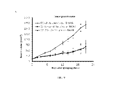
EVQLQESGPSLVKPSQTLSLICSVTGDSITSGYWNWIRKF
PGNKLEYMGYISYSGSTYYNPFLKSRISITRDTSKNQYYL
794 SEQ ID NO: 17
QLNSVTTEDTATYYCAICMGDWLAWFAYWGQGTIVTV
SS
Antibody
Sequence No. Sequence of
light chain variable region (VL)
No.
DILMTQSPS SL SASLGGKVTITCNASQDINKYIAWYQHICP
34 SEQ ID NO: 2
GKGPSLLIHYTSTLQPGIPSRFSGSGSGRDYSFSISNLEPED
IATYYCLQHDNLLFTEGSGTICLEIK
DIQMTQ SP S SL SA SLGGKVTITCKASQDINKYIAWYQHKP
50 SEQ ID NO: 4
GKGPRLLIHYTSTLQPGIPSRFSGSGSGRDYSFSISNLEPE
DIATYYCLQHDNLLFTEGSGTICLEIK
19020038.2
34273/115
- 21 -
CA 03157516 2022-5-6
DIQMTQTPSSLSASLGGKVTITCKASQDINRYIAWYQHICP
90 SEQ ID NO: 6
GKGPSLLIHYTSTLQPGIPSRFSGSGSGRDYSFSISNLEPED
IATYYCLQHDNLLFTFGSGTICLEIK
DILMTQSPSSLAVSVGEKVTLSCKSSQSLLYSNNQKNSL
130 SEQ ID NO: 8
AWYQQKPGQSPKLLIYWASTGESGVPDRFTGSGSGTDF
TLTISSVKAEDLAVYYCQQYYGYPFTFGAGTKLEIK
DIVLTQSPALMSASPGEKVTMTCSASSSVNYVYWYQQK
156 SEQ ID NO: 10
PRSSPICPWIYLTFNLASGVPARFSGSGSGTSYSLTISSMEA
EDAATYYCQQWSSNPLIFGAGTICLEIK
DIVITQSPSSLAVSVGEKVTMSCKSSQSLLYSSNQQNSLA
370 SEQ ID NO: 12
WYQQICPGQSPILLIYWASTRESGVPDRFTGGGSGTDFTL
TISSVRAEDLAVYYCQQYYNYPWTFGGGTICLEIK
EIVMTQSHICFMSTSVGDRVSITCKASQDVDTPVAWYQQ
373 SEQ ID NO: 14
ICPGQSPRLLIYWASIRHTGVPDRFTGSGSGTDFTLTISNV
QSEDLADYFCQQYSSYPLTFGSGTICLEIK
DIVMTQSPSSLAVSVGEKVTMSCKSSQSLLYSSNQQNSL
413 SEQ ID NO: 16
AWYQQICPGQSPILLIYWASTRESGVPDRFTGSGSGTDFTL
TISSVRAEDLAVYYCQQYYSYPWTFGGGTICLEIK
EIVMTQSPSSLAVSVGEKVILSCKSSQSLLYSSNQICNSLA
794 SEQ ID NO: 18
WYQQICPGQSPICLLIYWASTRESGVPDRFTGSGSGTDFTL
TISSVKAEDLAVYYCQQYYGYPYTFGGGTICLEIK
[0085] The CDRs of each antibody were analyzed by KABAT and IMGT software,
respectively.
The specific sequence information is shown in Table 2 below:
Table 2. Specific sequence information of CDRs of murine antibodies by
analysis of KABAT and
IMGT software
KABAT analysis
Antibody Sequence Sequence
Sequence
CDR1-HC
CDR2-HC CDR3-11C
No. No. No.
No.
19020038.2
34273/115
- 22 -
CA 03157516 2022-5-6
TLSSGGSY
SEQ ID SEQ ID
SEQ ID PTADWHL
34 TYGMS
TYYSESV
NO: 19 NO: 20
NO: 21 DV
KG
TLSSGGSY
SEQ ID SEQ ID
SEQ ID PTADWHL
50 TYGMS
TYYSESV
NO: 25 NO: 26
NO: 27 DV
KG
TVSSGGSY
SEQ ID SEQ ID
SEQ ID PTADWHL
90 TYGMS
TYYPDSV
NO: 31 NO: 32
NO: 33 DV
KG
SEQ ID SEQ ID
YISYTGST SEQ ID CPGWLNA
130 SDYWN
NO: 37 NO: 38
YYNPSLRS NO: 39 MDY
DINPNNG
SEQ ID SEQ ID
SEQ ID
156 DYYMN
DTSYNQK SSVMDY
NO: 43 NO: 44
NO: 45
FKG
SEQ ID SEQ ID
YISYTGST SEQ ID AGGWLLP
370 SDYWN
NO: 49 NO: 50
YYNPSLKS NO: 51 FAV
YISSGSSTI
SEQ ID SEQ ID
SEQ ID RNFGSSYD
373 DYGMH
YYADTVK
NO: 55 NO: 56
NO: 57 Y
G
SEQ ID SEQ ID
YISYTGST SEQ ID AGGWLLP
413 SDYWN
NO: 61 NO: 62
YYNPSLKS NO: 63 FAV
SEQ ID SEQ ID
YISYSGST SEQ ID MGDWLA
794 SGYWN
NO: 67 NO: 68
YYNPFLKS NO: 69 WFAY
Antibody Sequence Sequence
Sequence
CDR1-LC
CDR2-LC CDR3-LC
No. No. No.
No.
SEQ ID NASQDIN SEQ ID
SEQ ID LQHDNLLF
34
YTSTLQP
NO: 22 KYIA NO: 23
NO: 24 T
19020038.2
34273/115
- 23 -
CA 03157516 2022-5-6
SEQ ID KASQDIN SEQ ID
SEQ ID LQHDNLLF
50
YTSTLQP
NO: 28 KYIA NO: 29
NO: 30 T
SEQ ID KASQDIN SEQ ID
SEQ ID LQHDNLLF
90
YTSTLQP
NO: 34 RYIA NO: 35
NO: 36 T
KSSQSLL
SEQ ID SEQ ID
SEQ ID QQYYGYP
130 YSNNQK
WASTGES
NO: 40 NO: 41 NO: 42 FT
NSLA
SEQ ID SASSSVN SEQ ID
SEQ ID QQWSSNPL
156
LTFNLAS
NO: 46 YVY NO: 47
NO: 48 T
KSSQSLL
SEQ ID SEQ ID
SEQ ID QQYYNYP
370 YSSNQQN
WASTRES
NO: 52 NO: 53 NO: 54 WT
SLA
SEQ ID KASQDV SEQ ID
SEQ ID QQYSSYPL
373
WA SIRHT
NO: 58 DTPVA NO: 59
NO: 60 T
KSSQSLL
SEQ ID SEQ ID
SEQ ID QQYYSYP
413 YSSNQQN
WASTRES
NO: 64 NO: 65 NO: 66 WT
SLA
KSSQSLL
SEQ ID SEQ ID
SEQ ID QQYYGYP
794 YSSNQKN
WASTRES
NO: 70 NO: 71 NO: 72 YT
SLA
IMGT analysis
Antibody Sequence Sequence
Sequence
CDR1-HC
CDR2-HC CDR3-HC
No. No. No.
No.
SEQ ID GFTFNTY SEQ ID
SEQ ID ARPTADW
34
LSSGGSYT
NO: 73 G NO: 74
NO: 75 HLDV
SEQ ID GFTFNTY SEQ ID
SEQ ID ARPTADW
50
LSSGGSYT
NO: 79 G NO: 80
NO: 81 HLDV
19020038.2
34273/115
- 24 -
CA 03157516 2022-5-6
SEQ ID GFTF STY SEQ
ID SEQ ID ARPTADW
90
VSSGGSYT
NO: 85 G NO:
86 NO: 87 HLDV
SEQ ID GYSITSD SEQ
ID SEQ ID ARCPGWL
130
ISYTGST
NO: 91 Y NO:
92 NO: 93 NAMDY
SEQ ID GYTFTDY SEQ ID SEQ ID
156
INPNNGDT ASS VMDY
NO: 97 Y NO:
98 NO: 99
SEQ ID GYSITSD SEQ
ID SEQ ID ARAGGWL
370
ISYTGST
NO: 103 Y NO:
104 NO: 105 LPFAV
SEQ ID GFTFSDY SEQ
ID SEQ ID ARRNFGSS
373
ISSGSSTI
NO: 109 G NO:
110 NO: 111 YDY
SEQ ID GYSITSD SEQ
ID SEQ ID ARAGGWL
413
ISYTGST
NO: 115 Y NO:
116 NO: 117 LPFAV
SEQ ID GDSITSG SEQ
ID SEQ ID AKMGDWL
794
ISYSGST
NO: 121 Y NO:
122 NO: 123 AWFAY
Antibody Sequence Sequence
Sequence
CDR1-LC
CDR2-LC CDR3-LC
No. No. No.
No.
SEQ ID SEQ
ID SEQ ID LQHDNLLF
34 QDINKY
YTS
NO: 76 NO:
77 NO: 78 T
SEQ ID SEQ
ID SEQ ID LQHDNLLF
50 QDINKY
YTS
NO: 82 NO:
83 NO: 84 T
SEQ ID SEQ
ID SEQ ID LQHDNLLF
90 QDINRY
YTS
NO: 88 NO:
89 NO: 90 T
SEQ ID QSLLYSN SEQ
ID SEQ ID QQYYGYP
130
WAS
NO: 94 NQKNS NO:
95 NO: 96 FT
SEQ ID SEQ
ID SEQ ID QQWSSNPL
156 SSVNY
LTF
NO: 100 NO:
101 NO: 102 T
370
SEQ ID QSLLYSS SEQ ID WAS
SEQ ID QQYYNYP
NO: 106 NQQNS NO:
107 NO: 108 WT
19020038.2
34273/115
- 25 -
CA 03157516 2022-5-6
SEQ ID SEQ
ID SEQ ID QQYSSYPL
373 QDVDTP
WAS
NO: 112 NO:
113 NO: 114 T
SEQ ID Q SLLY SS SEQ
ID SEQ ID QQYYSYP
413
WAS
NO: 118 NQQNS NO:
119 NO: 120 WT
SEQ ID Q SLLY SS SEQ
ID SEQ ID QQYYGYP
794
WAS
NO: 124 NQICNS NO:
125 NO: 126 YT
Example 4 Humanization of antibody
100861 Firstly, the classical "CDRs transplantation" method was used to
humanize the antibody,
specifically, the humanized antibody with the highest homology was selected
based on sequence to
provide the framework regions (FRs) of an antibody, and the antigen-binding
fragment
complementarity determination regions (CDRs) of the target antibody based on
Kabat numbering
were transplanted into FRs to form a humanized antibody. Secondly, in order to
better maintain the
activity and affinity of the antibody, based on the antibody structure
modeling analysis (MOE
software): 1). the amino acid residues such as the antibody framework regions
located at the VH-
VL interface, close to or directly interacting with CDRs were selected for
reverse mutation, and
such amino acid residues were important for maintaining the conformation of
CDRs; 2) considering
the immunogenicity, the amino acid embedded in the protein was selected as far
as possible for
reverse mutation; 3) considering antibody stability and expression level, the
mutation with
molecular energy reduction was given priority; and 4). in the process of
humanization, the affinity
of humanized antibody was further improved by site-directed mutagenesis of
amino acids in CDRs.
By testing the affinity of humanized antibodies containing different mutations
with human PD-Li
and the binding to cells expressing PD-Li on the surface, humanized antibodies
with the affinity,
antibody characterization and activity function equivalent to or better than
those of murine PD-Li
antibodies were screened.
100871 Among them, CDRs of the preferred candidate antibodies after
humanization of the
PDL1-156 antibody were analyzed by IMGT and KABAT software as follows, and the
corresponding sequence information is shown in Table 3 and Table 4 below,
wherein Table 3 shows
19020038.2
34273/115
- 26 -
CA 03157516 2022-5-6
the VH and VL sequences of the humanized antibodies, and Table 4 shows the
IMGT and KABAT
analysis results of the humanized antibodies.
Table 3. Specific sequence information of heavy chain variable region and
light chain variable
region of humanized anti-PD-L1 antibodies
Antibody
Sequence No. Sequence of heavy
chain variable region (VH)
No.
QVQLVQSGPELKKPGASVKISCKASGYTFTDYYMNWV
SEQ ID NO: RQAPGQSLEWIGDIWPNNGDTWYNQICKGRVTLTRDT
156-1H
127
STSTVYMELRSLRSEDTAVYYCARSVMDYWGQGTLVT
VSS
QVQLVQSGPELKKPGASVKISCKASGYTFTDYYMNWV
156 7H
SEQ ID NO:
RQAPGQSLEWIGDIWPNNGDTSYNQICKGRVTLTRDTS
- 129
TSTVYMELRSLRSEDTAVYYCARSVMDTWGQGTLVTV
SS
QVQLVQSGPELICKPGASVKISCKASGYTFTDYYMNWV
SEQ ID NO: RQAPGQSLEWIGDIWPNNGDTSYNQICKGRVTLTRDTS
156-10H
131
TSTVYMELRSLRSEDTAVYYCARSVMSDWGQGTLVTV
SS
QVQLVQSGPELICKPGASVKISCKASGYTFTDYYMNWV
SEQ ID NO: RQAPGQSLEWIGDIWPNNGDTSYNQICKGRVTLTRDTS
156-11H
133
TSTVYMELRSLRSEDTAVYYCARSVMDYWGQGTLVTV
SS
Antibody
Sequence No. Sequence of
light chain variable region (VL)
No.
EIVLTQSPALLSLSPGERVTLSCSASSSVNYVYWYQQKP
SEQ ID NO:
156-1H
GQAPRPLIYLTFNLASGIPARFSGSGSGTDFTLTISSLEPE
128
DFAVYYCQQWSVNPLTFGGGTKVEIK
19020038.2
34273/115
- 27 -
CA 03157516 2022-5-6
EIVLTQSPALLSLSPGERVTLSCSASSSVNYVYWYQQICP
SEQ ID NO:
156-7H
GQAPRPLIYLTFNLASGIPARFSGSGSGTDFTLTISSLEPE
130
DFAVYYCQQWSVNPLTFGGGTKVEIK
EIVLTQSPALLSLSPGERVTLSCSASSSVNYVYWYQQICP
SEQ ID NO:
156-10H
GQAPRPLIYLTFNLASGIPARFSGSGSGTDFTLTISSLEPE
132
DFAVYYCQQWSSNPLTFGGGTKVEIK
EIVLTQSPALLSLSPGERVTLSCSASSSVNYVYWYQQICP
SEQ ID NO:
156-11H
GQAPRPLIYLTFNLASGIPARFSGSGSGTDFTLTISSLEPE
134
DFAVYYCQQWSVNPLTFGGGTKVEIK
[0088] The CDRs of each humanized antibody were analyzed by KABAT and IMGT
software,
respectively. The specific sequence information is as follows:
Table 4. Specific sequence information of CDRs of each humanized PD-L1
antibody by analysis
of KABAT and IMGT software
KABAT analysis
Antibody Sequence Sequence
Sequence
CDR1-HC
CD42-HC CDR3-HC
No. No. No.
No.
SEQ ID DYYMN SEQ ID
DIWPNNGD SEQ ID SVMDY
156-1H NO: 135 NO: 136
TWYNQKFK NO: 137
SEQ ID DYYMN SEQ ID
DIWPNNGD SEQ ID SVMDT
156-7H NO: 141 NO: 142
TSYNQKFK NO: 143
SEQ ID DYYMN SEQ ID
DIWPNNGD SEQ ID SVMSD
156-10H NO: 147 NO: 148
TSYNQKFK NO: 149
SEQ ID DYYMN SEQ ID
DIWPNNGD SEQ ID SVMDY
156-11H NO: 153 NO: 154
TSYNQKFK NO: 155
19020038.2
34273/115
- 28 -
CA 03157516 2022-5-6
Antibody Sequence Sequence
Sequence
CDR1-LC
C1M42-LC CDR3-LC
No. No. No.
No.
SEQ ID SASSSVN SEQ ID LTFNLAS SEQ ID QQWSVNP
156-1H
NO: 138 YVY NO: 139
NO: 140 LT
SEQ ID SASSSVN SEQ ID LTFNLAS SEQ ID QQWSVNP
156-7H
NO: 144 YVY NO: 145
NO: 146 LT
SEQ ID SASSSVN SEQ ID LTFNLAS SEQ ID QQWSSNPL
156-10H
NO: 150 YVY NO: 151
NO: 152 T
SEQ ID SASSSVN SEQ ID LTFNLAS SEQ ID QQWSVNP
156-11H
NO: 156 YVY NO: 157
NO: 158 LT
IMGT analysis
Antibody Sequence Sequence
Sequence
CDR1-HC
CDR2-HC CDR3-HC
No. No. No.
No.
SEQ ID GYTFTDY SEQ ID IWPNNGDT SEQ ID ARSVMDY
156-1H
NO: 159 Y NO: 160
NO: 161
SEQ ID GYTFTDY SEQ ID IWPNNGDT SEQ ID ARSVMDT
156-7H
NO: 165 Y NO: 166
NO: 167
SEQ ID GYTFTDY SEQ ID IWPNNGDT SEQ ID ARSVMSD
156-10H
NO: 171 Y NO: 172
NO: 173
SEQ ID GYTFTDY SEQ ID IWPNNGDT SEQ ID ARSVMDY
156-11H
NO: 177 Y NO: 178
NO: 179
Antibody Sequence Sequence
Sequence
CDR1-LC
CDR2-LC CDR3-LC
No. No. No.
No.
SEQ ID SSVNY SEQ ID LTF
SEQ ID QQWSVNP
156-1H
NO: 162 NO: 163
NO: 164 LT
SEQ ID SSVNY SEQ ID LTF
SEQ ID QQWSVNP
156-7H
NO: 168 NO: 169
NO: 170 LT
19020038.2
34273/115
- 29 -
CA 03157516 2022-5-6
156 10H SEQ ID SSVNY SEQ ID LTF SEQ ID QQWSSNPL
- NO: 174 NO: 175
NO: 176 T
SEQ ID SSVNY SEQ ID LTF SEQ ID QQWSVNP
156-11H
NO: 180 NO: 181
NO: 182 LT
Example 5 Determination of antibody purity by molecular exclusion
chromatography
100891 The purity of antibody was determined by molecular exclusion
chromatography using
TSKgel G3000SWXL column (TOSOH, 0008541) and pre-column TSKgel guard column
SWXL
(TOSOH, Cat. 0008543). The mobile phase was phosphate buffer solution (NaH2PO4-
Na2HPO4),
which was prepared by mixing 8.88g of NaH2PO4-2H20 and 33.33g of
Na2HPO4.12H20. The
mobile phase was used to balance the chromatographic column, and the flow rate
was lmL/min.
After the baseline was flattened, the sample was injected at a volume of 10pL.
The ultraviolet
detection wavelength was 280nm, the bandwidth was 16nm, and the reference
wavelength was off.
The determination results are shown in Table S.
Table 5. Purity of humanized anti-PD-Li antibody
Main
High Degradation
peak
molecular (A)
Sample Number
(%)
weight peak
(%)
PDL1-156 95.03
1.33 2.88
156-1H 96.16
3.00 0.84
156-7H 81.80
3.56 14.64
156-10H 86.05
3.5 10.45
156-11H 93.17
6.83
Example 6 KD determination of antibody binding to human and cynomolgus monkey
PD-L1
recombinant protein
19020038.2
34273/115
- 30 -
CA 03157516 2022-5-6
[0090] Biacore T200 (GE Healthcare) was used to determine binding affinity of
the PD-Li
antibody to the human and cynomolgus monkey PD-Li-His proteins. Anti-human IgG
Fc (Genway,
Cat. GWB-20A705) was immobilized on CMS chip (GE Healthcare, Cat. BR-1005-30)
at 25 C.
Anti-human Fc (Genway, Cat. GWB-20A705) was diluted to 20gg/m1 with Acetate
pH5.0 (GE
Healthcare, BR-1003-51). The Amine method in the Immobilization method was
used to
immobilize. Alternatively, a commercially available Protein A (GE Healthcare,
Cat. 29127556)
chip was used for detection. The affinity between antibody and antigen was
determined by multi-
cycle kinetic method at 25 C. In each cycle, the antibody to be tested was
captured on the fixed
CMS chip, then injected with recombinant human PD-Li-His (Novoprotein, Cat.
315) and
cynomolgus monkey PD-Li-His protein (Sino Biological, Cat. 90251-CO8H), and
finally
regenerated with Glycine pH1.5. The mobile phase was HBS-EP + Buffer (GE
Healthcare, Cat.
BR-1006-69), the flow rate was 304/min, and the binding time was 300 seconds.
The regeneration
flow rate was 30[EL/min and the time was 30 seconds. Using Biacore T200
evaluation software
(version 3.0), a 1:1 binding model was used to analyze the experimental data,
fit the equilibrium
dissociation constant KD of the antibody and antigen, and determine the
binding rate constant Ka
and dissociation rate constant Kd.
[0091] From the results, it can be seen that the binding of the tested PD-L1
antibodies to the
human PD-Li recombinant protein all showed an affinity of nM or better, and
the affinity for the
cynomolgus monkey PD-Li recombinant protein was between 62.5 nM and 0.375 nM.
See Table
6 below for details.
Table 6. Determination result of Biacore binding affinity ICD of murine PD-Li
antibody (M)
Antibody No. Human PD-Li
Cynomolgus monkey PD-Li
PDL1-156 2.91 E-10
3.75 E-10
PDL1-794 1.25 E-09
8.83 E-10
PDL1-130 2.18 E-09
1.95 E-09
PDLI-34 4.58 E-10
5.43 E-09
PDLI-50 6.99 E-10
3.31 E-08
PDLI-90 8.70 E-10
6.25 E-08
19020038.2
34273/115
- 31 -
CA 03157516 2022-5-6
PDL1-370 2.42E-09
2.12E-09
PDL1-373 2.45 E-09
3.64 E-09
PDL1-413 2.09E-09
2.76E-09
109921 Table 7 shows that the binding of humanized antibodies 156-1H, 156-7H,
156-10H and
156-11H derived from murine antibody PDLI-156 to human PDLI protein exhibited
an affinity
equivalent to PDLI-156.
Table 7. Determination result of Biacore binding affinity of humanized PD-L1
antibody
Equilibrium
Antibody Binding rate constant Dissociation
rate
dissociation constant
No. ka (1/Ms) constant kd
(1/s)
KD (M)
PDL 1-156 8.02E+05 2.33E-
04 2.91E-1O
156-1H 4.940 E+05
2.265 E-04 4.584 E-10
156-7H 5.14E+05
1.805 E-04 3.481 E-10
156-10H 5.954E+05
1.333 E-04 2.239 E-10
156-11H 5.742E+05
1.928 E-04 3.359 E-10
Example 7 IC50 determination of antibody blocking the interaction between PD-
Li and PD-
1
100931 The ICso of anti-PD-Li antibody blocking the binding of PD-Li protein
to PD-1 protein
was determined by competitive ELISA.
[00941 The recombinant human PD-Li protein (Sino Biological, Cat. 10084-H05H)
was diluted
with carbonate buffer and added to a 96-well plate at a final concentration of
flag/mi. After blocking
with PBS solution containing 3% BSA, serially diluted anti-PD-Li antibody
(6000ng/m1-2ng/m1)
and human PD-1-His recombinant protein (Sino Biological, Cat. 103 77-BOSH)
were added for co-
incubation. Then HRP-labeled anti-His tag antibody (MBL, Cat. D291-7) was
added, and color
development was performed with TMB. After termination with IM sulfuric acid,
OD values (dual
19020038.2
34273/115
- 32 -
CA 03157516 2022-5-6
wavelength 450nm-630nm) were read. The competitive binding curve of the test
antibody can be
drawn by matching the antibody concentration with the OD value to calculate
the IC50 value. FIG.
3 shows the competitive binding curve of anti-PD-L1 antibody to human PD-Li
recombinant
protein. The results show that the 9 murine anti-PD-Li antibodies (A) and 4
humanized antibodies
(B) tested could effectively block the interaction between human PD-Li protein
and human PD-1
protein compared with the antibody isotype negative control anti-Hel (prepared
by Biointron)
without any blocking effect, and the humanized antibodies had a comparable
inhibitory activity to
the murine PDL1-156 before humanization (B), with ICso of 197.0 ng/mL (156-
1H), 230.5 ng/mL
(156-7H), 250.1 ng/mL (156-10E1) and 207.2 ng/mL (156-11E), respectively. The
ICso of murine
PD-L1-156 was 221.3 ng/ml, the positive control Atezolizumab (prepared by
Biointron) was 446.4
ng/ml, and Avelumab (Pfizer, lot AU020322) was 190.3 ng/ml.
Example 8 EC59 determination of PD-Li antibody binding to PD-L1 on cell
surface by FACS
100951 A gradient concentration of antibodies to be detected (final
concentration of antibody:
10000ng/m1-0.1ng/ml, 10-foldserial dilution) were incubated with CHO-PD-Li
cells (Nanjing
Yongshan Biotechnology Co., Ltd., 105 cells/well) with high expression of PD-
Li on the cell
surface at 4 C t for 30min. After incubation, 1:250 diluted anti-human IgG PE
fluorescent
antibody (eBioscience, Cat. 12-4998-8) was added, and incubated at 4 C for
30min. The
fluorescent antibody specifically bound to the Fc fragment of the antibody to
be detected. The
fluorescent intensity of PE was detected by FACS, and the ability of the
antibody to be detected to
bind to PD-L1 protein highly expressed on the cell surface was analyzed. As
shown in FIG. 4, the
ECso of the murine PD-Li antibody to be test was similar to that of the
positive controls Avelumab
(ECso of 58.2 ng/ml) and Atezolizumab (¨ 99.73 ng/ml) in this experiment,
among which the ECso
of PDL1-156 and PDL1-370 were the lowest, and the ECso of the detected
humanized antibodies
156-1H, 156-7H, 156-10E1 and 156-11H were 49.42 ng/ml, 78.37 ng/ml, 63.5 ng/ml
and 49.97
ng/ml, respectively, similar to that of the positive control Avelumab (ECso of
56.88 ng/ml) in this
experiment. This determination quantitatively confirmed the ability of each PD-
Li antibody to bind
to PD-Li targets on the cell surface in a dose-dependent manner. MFI fold =
MFI value of the
19020038.2
34273/115
- 33 -
CA 03157516 2022-5-6
experimental group/MFI value of the control group without drugs.
Example 9 Determination of anti-PD-Li antibody inhibiting PD-1: PD-Li binding
and signal
transduction by PD-1/PD-L1-NFAT reporter gene assay
100961 The antagonistic effects of PD-Li antibody on PD-1/PD-L1 protein
interaction and its
signaling pathway were compared by using Jurkat cell line (GenScript,
Cat.00612) stably
transfected with PD-1 and CHO cell line (GenScript, Cat. M00613) stably
transfected with PD-Li.
When the inhibitory signaling pathway was inhibited, the expression of NFAT-
controlled
luminescence reporter gene increased, and the luminescence signal value
increased. The blocking
effect of antibody on PD-L1 was reflected by the intensity of relative
luminescence units (RLUs).
100971 The CHO cells stably expressing PD-Li were seeded on a 96-well white
bottom plate
with 40,000 cells/well and 100pL/well, and placed back into an incubator
overnight. On the next
day, the well plate was taken out, the culture medium was removed, and then
the cells stably
expressing PD-1 and the PD-L1 antibody to be tested were added for co-
incubation. The PD-1 cells
were added at 16,000 cells/well, while the antibody was serially diluted,
added with each dose in
triplicate at a volume of 100 L/well, and incubated for 6 hours. After the
incubation, the well plate
was taken out and added with luminescence detection reagent in the same volume
(100pL). Then
the values were read. According to the values, a regression curve was plotted
by Graphpad with the
4-parameter analysis, and the ECso values of each antibody were obtained. The
results are detailed
in FIG.5A. The ECso values of the tested murine PD-Li antibody were all
similar to those of the
positive control antibodies Avelumab and Atezolizumab (222.9 ng/ml and 321.6
ng/ml). FIGs.5B
and 5C show that the ECso of the four humanized PD-Li antibodies 156-1H, 156-
7H and 156-10H,
156-11H were: 342ng/ml, 313.7 ng/ml, 357.1 ng/ml and 282.2 ng/ml,
respectively, which were
similar to the ECso of the positive control Avelumab. The determination
quantitatively confirmed
that murine and humanized anti-PD-Li antibodies showed a dose-dependent
suppression of T cell
activity inhibition caused by the interaction of PD-1: PD-L1 on the cell
surface, thus dose-
dependently enhancing the activity of reporter gene in Jurkat cells.
19020038.2
34273/115
- 34 -
CA 03157516 2022-5-6
Example 10 ELISA detection of IFN-y secreted by T cell in mixed lymphocyte
reaction
100981 The activity of T cells enhanced by PD-Li monoclonal antibody was
determined by
mixed lymphocyte reaction (MLR). CD14 monocyte was isolated from peripheral
blood
mononuclear cells (PBMCs) of healthy human donor 1, and then induced in vitro
to differentiate
into dendritic cells (DCs) with recombinant human granulocyte-macrophage
colony stimulating
factor (GM-CSF, Peprotech, Cat. 300-03) and recombinant human interleukin-4
(rhIL-4, Peprotech,
Cat. 200-04). On the 6th day of culture, LPS (Sigma, Cat: L4516) was added to
stimulate DCs
maturation. On the 7th day, DCs from donor 1 were mixed and co-cultured with
CD4+ T cells
enriched from PBMCs of healthy donor 2 at a ratio of DCs: CD4 T cells being
1:10. The antibody
to be tested or positive control antibody Avelumab or Atezolizumab (antibody
concentration of
1000ng/ml, 10Ong/ml, 10 ng/ml, lng/m1) was added to incubate for 4 days. After
4 days, the cell
culture supernatant was collected, and the concentration of IFN-1 in the
supernatant was determined
by ELISA. As shown in FIG.6, all the tested murine antibodies and positive
control antibodies
Avelumab and Atezolizumab can significantly enhance the ability of CD4 T cells
to secrete IFN-
y in the MLR experiment compared with anti-HEL monoclonal antibody and no
treatment negative
control group, and as the concentration of PD-L1 antibody decreased, the
activity of increasing
IFN-secretion also decreased. The results indicated that PD-Li antibody could
enhance the function
of T cells in a dose-dependent manner. T-test, * P < 0.05, ** P < 0.01, *** P
< 0.001, **** P <
0.0001.
Example 11 Determination of Antibody Dependent Cell-mediated Cytotoxicity
(ADCC)
Activity of Anti-PD-Li Antibody
100991 The ADCC activity mediated by the anti-PD-Li monoclonal antibody was
determined by
the cell killing experiment of natural killer (NK) cells from isolated and
purified PBMCs of normal
human on A431 cells with high expression of human PD-Li.
101001 Preparation of effector cells: Cryopreserved human PBMC cells
(StemExpress) were
19020038.2
34273/115
- 35 -
CA 03157516 2022-5-6
thawed and incubated overnight in RPMII640 medium (Invitrogen, Cat. 11835030)
supplemented
with 100IU/m1 recombinant human interleukin 2 (rhIL-2, Peprotech, Cat. 200-
02). After that, the
cells were collected and the living cells were counted. NK cells were then
purified from PBMCs
using the NK cell magnetic bead separation kit (Stemcell, Cat. 17955),
resuspended in DMEM
(Invitrogen, Cat. 11965084) supplemented with 10% inactivated fetal bovine
serum and counted
for use as effector cells.
[0101] Preparation of target cells: The target cells A431 were cultured in
DMEM medium
containing 10% fetal bovine serum. The day before the ADCC experiment, IFN-y
(Peprotech, Cat.
300-02) with the final concentration of 500IU was added to stimulate cells
overnight.
[0102] The anti-PD-L1 antibody was diluted with DMEM medium containing 10%
fetal bovine
serum. The diluted antibody was added into white wall 96-well plates (Corning,
Cat. 3610) at 25
and target cells were added to the wells (25 [EL/well, 10000 cells/well). The
plates were
incubated at an incubator at 37 C and 5% CO2 for 30 minutes. After that, the
effector cells were
added to the wells (25 4/well, 40,000 cells/well, i.e., the effector-target
ratio NK: A431 = 4:1),
with a total volume of 100 [EL, and the plates were incubated at an incubator
at 37 C and 5% CO2
for 4 hours.
[0103] It is necessary to set the medium background control well (adding 100
pt of medium to
the well), the target cell spontaneous death release well (only target cells
in the well), the effector
cell spontaneous death release well (only NK cells in the well), and the
target cell maximum death
release well (target cells plus the lysis buffer provided in the CytoTox-
Glorrm Cytotoxicity kit
(Promega, Cat. G9291)). Finally, the volume of all control wells was
supplemented to 100 [EL with
medium. After incubation for 4 hours, the plates were taken out, 504 of
CytoTox-GloTm
CytoToxicity Assay Reagent was added to each well, mixed evenly on a shaking
table, and left at
room temperature for 15 minutes before reading chemiluminescence value (RLU).
[0104] The cell lysis rate caused by ADCC activity was calculated according to
the following
procedure: first, the average reading value of the medium background control
well was subtracted
from the reading value of all wells, and then the lysis rate% was calculated
by: lysis rate%= (reading
value of experimental well - reading value of target cell spontaneous death
release well - reading
19020038.2
34273/115
- 36 -
CA 03157516 2022-5-6
value of effector cell spontaneous death release well) / (reading value of
target cell maximum death
release well - reading value of target cell spontaneous death release well) *
100%
[0105] The results are shown in FIG.7. The experimental results show that the
three murine PD-
Li antibodies tested and the FDA-approved positive control anti-PD-L1 drug
Avelumab all showed
antibody concentration-dependent lytic killing activity and similar EC50
values against the target
cells A431. Also, the negative isotype control antibody hIgGl, K (Abdserotec,
PHP010) showed
no ADCC activity even at the highest concentration.
Example 12 In vivo pharmacodynamics detection of murine PD-Li antibody PDL1-
156
[0106] B-hPD-1/hPD-L1 transgenic mice (Beijing Biocytogen Gene Biotechnology
Co., Ltd.)
were used to establish an animal model of MC38-hPD-L1 colon cancer, and the
pharmacodynamics of PD-Li antibody was then evaluated. 5 / 105 MC38-hPD-L1
colon cancer
cells were inoculated subcutaneously on the right side of mice. When the tumor
volume reached
about 110 mm, mice with moderate individual tumor volume were selected into
the group, and
the animals were assigned to three experimental groups according to the tumor
volume by
random grouping software: anti-Hel hIgG isotype control group, anti-PDL1-156
antibody group,
and positive drug Avelumab group (Pfizer, lot AU020322), with 8 mice in each
group.
Administration was started on the day of grouping (defined as the 0th day of
the study). Each
group was given 10 mg/kg by intraperitoneal injection twice a week for a total
of 7 times. The
tumor volume and body weight of mice were measured twice a week, and the
measured values
were recorded. The tumor volume (long diameter )< short diameter2/2) and the
tumor growth
inhibition rate (TGIry (%) = [1-(Ti-TO)/(Vi-V0)] / 100%; Ti: the mean value of
tumor volume in
the treatment group on the day i of administration, TO: the mean value of
tumor volume in the
treatment group on the 0th day of administration; Vi: the mean value of tumor
volume in the
solvent control group on the day i of administration, VU: the mean value of
tumor volume in the
solvent control group on the 0th day of administration) were calculated. Also,
the tumor volume
was statistically analyzed, and P < 0.05 was considered as significant
difference.
19020038.2
34273/115
- 37 -
CA 03157516 2022-5-6
[0107] On the 21st day of grouping and administration, compared with the
control group, the
positive drug Avelumab group and PDL1-156 murine anti-PD-L1 antibody group had
significant
and similar inhibitory effects on tumor volume, and there was a statistical
difference (P < 0.05).
See FIGs. 8A, B, C, and Table 8.
Table 8. Effect of test substance on volume of MC38-hPD-L1 tumor from B-hPD-
1/hPD-L1
humanized mouse
Tumor volume (mm3) a
Test
Day 21 of
Group Before
TGITy Ph
substance
grouping and
administration
CYO
administration
G1 hIgG 114 3
2675 470
G2 Avelumab 114 3
797 176 73.3 **0.001
G3 PDL1-156 114 3
1173 291 58.6 *0.014
Note: a: mean standard error; B: the tumor volume of the administration
group and the tumor
volume of the hIgG control on the 21st day after the grouping and
administration were statistically
analyzed, two-way ANOVA test, * p < 0.05, ** p < 0.01, *** p < 0.001, **** p <
0.0001.
[0108] In addition, during the experiment, one mouse in hIgG control group
died abnormally in
advance. Except for that, the rest of the experimental animals were in good
activity and eating state
during the administration, and their weight increased to a certain extent. The
results showed that
the antibody was safe, as shown in FIG. 8D and Table 9.
Table 9. Effect of test substance on body weight of B-hPD-1/hPD-L1 humanized
mice
transplanted with MC38-hPD-L1 tumor cells
Group Test substance Weight
(g) a
19020038.2
34273/115
- 38 -
CA 03157516 2022-5-6
Weight
change (g)
Before
Day 21 of
after 21
administrat grouping and
p b
days of
ion
administration
administrat
ion
G 1 hIgG 18.1 0.3
21.3 0.5 +3.2
G2 Avelumab 18.4 0.4
21.1 0.4 0.765 +2.7
G3 Sim-anti-PD-L1 12.4 0. 3
21.5 0. 5 0.747 +3.1
Note: a: mean standard error; B: the body weight of the administration group
and the body
weight of the hIgG control were statistically compared on the 21st day of
grouping and
administration, T-test.
Example 13 In vivo pharmacodynamics detection of humanized anti-PD-Li antibody
101091 MC38-hPD-L1 cells were inoculated subcutaneously at a concentration of
5< 105/0.1 mL
on the right side of female B-hPD-L1 transgenic mice aged 6-8 weeks
(Biocytogen Jiangsu Gene
Biotechnology Co., Ltd.). When the tumor grew to about 102 mm3, 24 mice were
selected
according to the tumor volume and randomly divided into 3 groups with 8 in
each group, a total of
3 groups, namely: saline, Avelumab (5 mg/kg), 156-10H (5 mg/kg). All groups
were given
intraperitoneal injection twice a week for 6 times, and the experiment ended 5
days after the last
administration. The body weight and tumor volume of mice were measured three
times a week
during administration and observation, and the measured values were recorded.
The tumor volume
(long diameter x short diameter2/2) and the growth inhibition rate (TGITv (%))
were calculated. On
the 21st day of grouping and administration, the positive drug Avelumab group
and the PD-Li
antibody 156-10H group had significant and similar inhibitory effects on tumor
volume compared
with the saline control group, with statistical differences (P < 0.05). See
FIGs. 9A, B, C, and Table
19020038.2
34273/115
- 39 -
CA 03157516 2022-5-6
10.
Table 10. Effect of test substance on volume of MC38-hPD-L1 tumor from B-hPD-
L1 humanized
mouse
Tumor volume (mm3)
Day 21 of
grouping
Before
Group Test substance
and TGITy p h
ad minis
administr
(%)
tration
ation
1682
G1 Normal saline 108 4
177
G2 Avelumab 108 5 672
301 64.2 *0.012
G3 156-10H 108 5 593
223 69.2 **0.002
Note: a: mean standard error; B: The tumor volume of the administration
group and the tumor
volume of the normal saline control group were statistically compared on the
21st day of grouping
and administration, T-test analysis, * P < 0.05, * * P <0.01.
[0110] The experimental animals were in good activity and eating state during
administration,
and their weight increased to a certain extent, as shown in FIG.9D and Table
11.
Table 11 Effect of test substance on body weight of B-hPD-L1 humanized mouse
transplanted
with MC38-hPD-L1 cells
Group Weight (g) 3
19020038.2
34273/115
- 40 -
CA 03157516 2022-5-6
Weight change
Day 21 of
Test Before
(g) after 21 day
grouping and
ph
substance administration
of
administration
administration
Normal
GI 19.5 0.7 22.5 0.9 +3.0
saline
G2 Avelumab 19.5 0.6
22.8 0.6 0.762 +3.3
G3 156-10H 19.5 0.3
22.4 0.5 0.972 +2.9
Note: a: mean standard error; B: The weight of the administration group and
the weight of the
vehicle control group were statistically compared on the 21st day of grouping
and administration,
and T-test analysis was performed.
101111 The above results indicate that humanized PD-Li antibody 156-10H had a
significant
inhibitory effect on the growth of subcutaneous transplanted tumor MC38-hPD-LI
and showed
high safety As compare with positive control antibody Avelumab, the TGI level
of the two
antibodies was comparable, and 156-10H showed a more homogeneous anti-tumor
effect.
101121 The antibody against human programmed death ligand-1 (PD-L1) and use
thereof
provided by the present invention have been illustrated in detail above. The
principles and
implementations of the present invention are described herein by using
specific embodiments, and
the descriptions of the above embodiments are only used to help understand the
method and the
core idea of the present invention. It should be pointed out that for those
skilled in the art, several
improvements and modifications can be made to the present invention without
departing from the
principle of the present invention, and these improvements and modifications
also fall within the
protection scope of the claims of the present invention.
19020038.2
34273/115
- 41 -
CA 03157516 2022-5-6