Note: Descriptions are shown in the official language in which they were submitted.
CA 03157532 2022-04-08
WO 2021/072390 PCT/US2020/055286
USE OF ENTPD3 FOR IDENTIFICATION, ISOLATION, AND ENHANCING MATURE STEM
CELL DERIVED INSULIN-PRODUCING CELLS
FIELD
[0001] The disclosed processes, methods, and systems are directed to cell
therapy
treatments for diabetes.
CROSS-REFERENCE TO RELATED APPLICATIONS
[0002] This application claims benefit of priority pursuant to 35 U.S.C.
119(e) of U.S.
provisional patent application No. 62/913,544, filed on 10 October 2019, which
is hereby
incorporated by reference in its entirety.
GOVERNMENT RIGHTS
[0003] This invention was made with government support under grant number
RO1DK120444 awarded by the National Institutes of Health/National Institute of
Diabetes and
Digestive and Kidney Diseases. The government has certain rights in the
invention.
SUMMARY
[0004] Disclosed herein are methods, systems, and compositions for
enhancing the
effectiveness of 6-cell (Beta-cell)-based therapies. Also disclosed herein are
methods, systems,
and compositions related to identifying, sorting and separating heterogeneous
populations of
stem cell-derived pancreatic 13-cells (sBCs) into more useful and functionally
homogeneous cell
populations. In many embodiments, the most mature and functional of the sBCs
are identified
and live-sorted using the cell surface protein Ectonucleoside Triphosphate
Diphosphohydrolase-
3 (ENTPD3), which is also referred to as CD39L3. The presently disclosed
methods, systems,
and compositions are useful for cell therapies, for example replacement
therapy. In many
embodiments the disclosed systems, methods, and compositions are useful in
treatments for
diabetes. In some embodiments, the disclosed methods, systems, and
compositions may be
useful in treating, preventing, and/or curing diabetes, for example type-1
diabetes.
BRIEF DESCRIPTION OF THE DRAWINGS
[0005] Figure 1. Stem cell-derived beta-like cells (sBC) self-enrich to
form insulin+ enriched
islet-like caps. Panel A, schematic representation of step-wise
differentiation of hPSC clusters
towards beta-like cells in suspension. Panel B, representative live image of
green fluorescent
1
CA 03157532 2022-04-08
WO 2021/072390 PCT/US2020/055286
protein driven by the endogenous insulin promoter (pINSGFP) of clusters during
immature
(imBC, day - 23) and self-enriched (seBC, day - 30) beta-like cell
differentiation stages (scale
bars indicate 200 pm). Panel C, flow cytometric quantification of GFP
expression in imBC and
seBC clusters (n = 7 independent differentiation experiments). Panel D,
quantification of GFP
intensity in imBC and seBC clusters (n = 6 independent differentiation
experiments). Panel E &
Panel F, immunofluorescence analysis of sections from imBC and seBC clusters
respectively
for endocrine and 6 cell markers. Panel G, immunofluorescence analysis of
sections from imBC
clusters, seBC clusters and human islets for mitochondria specific mtFA
protein. Panel H,
quantification of mtFA fluorescence intensity in imBC and seBC cells (n = 5
independent
differentiation experiments with 10 clusters analyzed per experiment). Panel
I, schematic
representation of pINSGFP+ cell sorting from imBC and seBC clusters. Panel J,
bulk RNA-seq
analysis of pINSGFP+ sorted imBC versus seBC (un-curated, top 30 genes
significantly up and
down regulated) adjusted p-value < 0.05 (n= 3 independent differentiation
experiments). Panel
K, gene ontology of differentially regulated genes. Panel L, quantitative PCR
analysis of mtDNA
normalized to gDNA in pINSGFP+ sorted cells (n = 3 independent differentiation
experiments
with 3 x 500 cells collected for analysis from each). Panel M, global levels
of 5-
hydroxymethylcytosine in pINSGFP+ sorted cells (n = 3 independent
differentiation
experiments). Panel N & Panel 0, total insulin content (Panel N) and
proinsulin to insulin
content ratios (Panel 0) per 1,000 pINSGFP+ sorted cells (n = 3 independent
differentiation
experiments with 3 x 1,000 cells collected per experiment). *p<0.05 "p<0.01
***p<0.001. Error
bars are representative of the mean +1- the standard deviation. Scale bars
represent 20 pm
unless otherwise indicated.
[0006] Figure 2 shows data demonstrating seBC functional maturity. Panel A,
representative perifusion analysis of imBC, seBC and primary human islets
(hlslets), 20 - 25
clusters were analyzed per sample and data is presented as % of total insulin
in cluster pellet
recovered. Panel B, total insulin content of pellet recovered after perifusion
and Panel C, Panel
D, & Panel E, relative insulin secretion during perifusion of imBC (n= 3
independent
differentiation experiments), seBC (n= 5 independent differentiation
experiments) and hlslet (n =
3 independent human islets prep) respectively (data normalized to basal (0.5
mM Glucose)
secretion). Panel F, representative images of imBC (left) and seBC clusters
(right) displaying
Ca2+ indicator (Rhod-2) labelling, pINSGFP, and map (below) showing magnitude
and extent of
Ca2+ elevations at 2 mM and 11 mM glucose, scale bar is 10 pm and white arrows
point to cells
chosen for time courses in Panel G. Panel G, representative time courses of
individual cells
from the imBC and seBC clusters displayed in (F) at 2 mM and 11 mM glucose,
scale bar is
2
CA 03157532 2022-04-08
WO 2021/072390 PCT/US2020/055286
50% change from mean. Panel H, fraction of area within intact cluster showing
elevations in
Ca2+ activity at 2 mM and 11 mM glucose (n = 3 independent differentiation
experiments with >
clusters measured per condition). Human islet data quantified in the same
manner is
included for reference Westacott, M. J. et al. (Age-dependent decline in the
coordinated [Ca2+]
and insulin secretory dynamics in human pancreatic islets. Diabetes 66, 2436-
2445 (2017)).
Panel I, fold change in Ca2+ activity when glucose is elevated from 2 mM to 11
mM glucose (n =
3 independent differentiation experiments with > 10 clusters measured per
condition). *p<0.05
"p<0.01 ***p<0.001. Panel B - Panel E, error bars are representative of the
mean +/- the
standard deviation. Panel H and Panel I, error bars are representative of the
mean +/- SEM.
[0007] Figure 3 presents single cell RNA-seq profiling of beta cell
differentiation identify
distinct subpopulations and defines temporal dynamics of beta cell maturation.
Panel A,
schematic representation of seBC production and sorting. Panel B, tSNE
projection of 4,143
seBC. Cells are colored by inferred cell type based on marker gene expression.
Panel C,
heatmap showing scaled abundance of the top ten marker genes for each cell
type identified by
single cell RNA-seq analysis. Panel D, tSNE projection with RNA velocity
vector estimates over-
layed. Panel E & Panel F, differentiation start-point (Panel E) and end-point
(Panel F) modeled
using a markov diffusion process on RNA velocity transmission probabilities.
Start and end
points were sampled from a uniform 100 x 100 grid, then imputed for all cells
using K = 10 K-
nearest neighbor pooling. Values range from 0 (yellow) to 1 (dark blue). Panel
G, trajectory
inference (m0n0c1e2) analysis with cells colored by cell type. Panel H,
heatmap of scaled gene
expression of genes with varying expression across pseudotime. Genes were
clustered into two
clusters with k-means clustering and expression values were smoothed using
cubic-spline
interpolation. Panel I, go-term enrichment analysis of marker genes of most
mature seBC
population identified by RNA velocity end-point analysis (>0.8 end-point
density).
[0008] Figure 4 presents data showing that Ectonucleoside Triphosphate
Diphosphohydrolase 3 marks mature beta-like cells. Panel A, tSNE projection of
RNA velocity
endpoints, pINSGFP transgene and ENTPD3 (Gene ID: 956; see
ncbi.nlm.nig.gov/956)
expression in 4,143 seBC. Panel B is relative ENTPD3 gene expression in
pINSGFP+ imBC
and seBC (n = 3 independent differentiation experiments). Panel C,
immunofluorescence
staining for c-peptide (CPEP; a byproduct of processing of endogenous insulin,
INS, Gene ID:
3630) and ENTPD3 in sections of imBC and seBC clusters (scale bar represents
20 pm). Panel
D & Panel E, representative sorting gates for pINSGFP+ENTPD3+/- cells in
unstained negative
control cells, (Panel D) and with direct conjugated ENTPD3 antibody (Panel E).
Panel F,
quantification of the percentage of ENTPD3+ cells within total pINSGFP+
population by FACS
3
CA 03157532 2022-04-08
WO 2021/072390 PCT/US2020/055286
(n= 4 independent differentiation experiments). Panel G, schematic
representation of
pINSGFP+ENTPD3+/- sorting from seBC. Panel H, bulk RNA-seq analysis of
INS+ENTPD3+ vs
INS+ENTPD3- cells sorted from seBC clusters (un-curated list of top 30 genes
significantly up
and down regulated as per adjusted p-value < 0.05, n= 4 independent
differentiation
experiments). Panel I, go-term enrichment analysis of differentially expressed
genes identified in
bulk RNA seq. Panel J. volcano plot of differential expression (DE) analysis
of INS+ENTPD3+
vs INS+ENTPD3-. Panel K, insulin content per 1,000 INS+ENTPD3+/- sorted cells
from seBC
and human islets (n= 3 independent differentiation experiments or human islets
preps, with 3 x
1,000 cells analyzed per experiment). Panel L, proinsulin to insulin content
ratio of
INS+ENTPD3+/- sorted cells from seBC and human islets (n= 3 independent
differentiation
experiments or human islets preps, with 3 x 1,000 cells analyzed per
experiment). Panel M,
quantitative PCR analysis of mtDNA normalized to gDNA in INS+ENTPD3+ vs
INS+ENTPD3-
sorted cells from seBC and human islets n= 3 independent differentiation
experiments or human
islets preps, with 3 x 500 cells analyzed per experiment). Panel N, global
levels of 5-
hydroxymethylcytosine in INS+ENTPD3+ vs INS+ENTPD3- sorted cells from seBC day
30 and
human islets (n = 4 independent differentiation experiments, with 1 x 500
cells analyzed per
experiment). *p<0.05 "p<0.01 ***p<0.001 error bars are representative of the
mean +/- the
standard deviation.
[0009] Figure 5 shows that INS+ENTPD3+ cells display improved function and
are present
in patient derived seBC. Panel A, schematic representation of pINS+ENTPD3+/-
cells sorted
from seBC clusters reaggregated in the presence of support cells, human
umbilical vein
endothelial cells (HUVEC) and mesenchymal stem cells (MSC), for 48 h. Panel B
shows
perifusion analysis of intact INS+ENTPD3- and INS+ENTPD3+ clusters, 20 ¨ 25
clusters were
analyzed per condition and data is presented as % of total insulin in cluster
pellet recovered (n=
2 independent differentiation experiments). Panel C, total insulin content of
clusters recovered
following perifusion analysis (n= 2 independent differentiation experiments).
Panel D relative
insulin secretion during perifusion of INS+ENTPD3- and INS+ENTPD3+ clusters
(n= 2
independent experiments) (data normalized to basal (0.5 mM Glucose)
secretion). Panel E,
iPSC derived from a patient with type-1 diabetes (T1D-iPSC) were
differentiated to seBC using
an improved protocol, schematic representation. Panel F, representative flow
cytometry analysis
of iPSC differentiation at definitive endoderm (DE), pancreatic endoderm (PE),
imBC and seBC
for specific lineage markers (n= 2 independent differentiation experiments).
Panel G,
immunofluorescence staining of T1D-iPSC derived seBC clusters (scale bar
represents 20 pm).
Error bars are representative of the mean +/- the standard deviation, n=2
biological replicates.
4
CA 03157532 2022-04-08
WO 2021/072390 PCT/US2020/055286
[0010] Figure 7 shows pINSGFP+ caps form spontaneously and independently of
maturation media. Panel A, imBC clusters were cultured for 10 days in the
presence of
endocrine differentiation media (EN DO), maturation media (containing ALK5i
and thyroid
hormone (13)) (MAT) and minimal maturation media (lacking ALK5i and T3 (MIN)),
schematic
representation. Panel B, pINSGFP images of clusters in different media at day
20 and day 30
(scale bars represent 200 pm). Panel C, quantitative PCR analysis of insulin
gene expression in
clusters at day 30 of differentiation. Error bars are representative of the
mean +/- the standard
deviation, n = 2 independent differentiation experiments.
[0011] Figure 8 shows Ca2+ analysis of imBC and seBC clusters. Panel A
fraction of area
within intact cluster showing elevations in Ca2+ activity of individual imBC
and seBC clusters at 2
mM and 11 mM glucose (n = 3 independent differentiation experiments with > 10
clusters
measured per condition). Panel B, fraction of area within intact cluster
exhibiting coordinated
Ca2+ activity (n = 3 independent differentiation experiments with > 10
clusters measured per
condition). *p<0.05 "p<0.01 ***p<0.001 error bars are representative of the
mean +/- SEM.
[0012] Figure 9 shows analysis of alternative beta cell differentiation
trajectories identified
by trajectory inference. Panel A, Branchpoint Expression Analysis Modeling
(BEAM)
demonstrating top 200 genes differentially expressed across branches indicated
by arrows. egfp
shown in cluster 2 corresponds to the expression of the pINSGFP transgene.
Panel B, Same as
panel A, but performed for the branch indicated by arrows.
[0013] Figure 10. Single cell RNA-seq profiling of eBC differentiation.
Panel A, schematic
representation of eBC differentiation and sorting. Panel B, tSNE projection of
4,178 eBC labeled
by inferred cell types. Panel C, heatmap showing scaled abundance of the top
ten marker
genes for each cell type identified by single cell RNA-seq. Panel D, tSNE
projection with RNA
velocity vector estimates overlayed. Panel E & Panel F, differentiation start-
point (Panel E) and
end-points (Panel F) modeled using a markov diffusion process on RNA velocity
transmission
probabilities. Start and end points were sampled from a uniform 100 x 100
grid, then imputed for
all cells using K = 10 K-nearest neighbor pooling. Values range from 0
(yellow) to 1 (dark blue).
Panel G, RNA velocity endpoints (left) and INS-eGFP transgene expression
(right) overlayed on
tSNE projection. Panel H, tSNE embedding of both seBC and eBC single cell RNA-
seq datasets
colored by respective dataset. Datasets were aligned using Seurat v2
integration methods.
Panel I, tSNE projections colored by the expression (log- normalized) of key
genes related to
beta-cell differentiation.
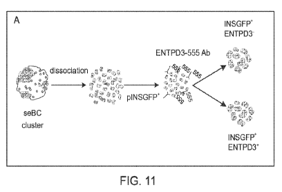
[0014] Figure 11 depicts ENTPD3 sorting strategy in pINSGFP reporter cell
line. Panel A,
seBC were dissociated and sequentially incubated with anti-ENTPD3 (mouse)
antibody and
CA 03157532 2022-04-08
WO 2021/072390 PCT/US2020/055286
anti-mouse 555 secondary antibody then sorted first based on pINSGFP
expression and second
based on +/- ENTPD3-555. Panel B. Cells were plotted on FSC vs SSC linear axes
and gated
to remove cell debris. Remaining cells were plotted by FSC area vs FSC height
and gated to
excluded non-single cells. Single cells were then plotted against DAPI stain
and gated to
remove dead cells. Live cells were plotted against pINSGFP reporter and those
positive were
then plotted for ENTPD3-555 in unstained, secondary antibody only and ENTPD3
conditions.
[0015] Figure 12 shows human islet sorting strategy. Panel A,
representative image of
immunofluorescence staining of intact human islet sections with ENTPD3, c-PEP
and NKX6.1
(Gene ID: 4825). Panel B, representative gating strategy for ENTPD3+/- cells.
Panel C,
quantification of immunofluorescence analysis of pancreatic hormone markers to
verify presort
and sorted populations (ENTPD3+/-) by single cell cytospin and counting using
Image J
analysis. Panel D, insulin content per 1,000 pINSGFP+ENTPD3+ sorted cells (n =
3 separate
human islet preps, with 3 x 1,000 cell analyzed per prep). Panel E, Proinsulin
to insulin content
molar ratio (n = 3 separate human islet preps, with 3 x 1,000 cell analyzed
per prep). Panel F,
quantitative PCR analysis of mtDNA normalized to gDNA in pINSGFP+ENTPD3+
sorted cells (n
= 3 separate human islet preps, with 3 x 500 cell analyzed per prep). Panel G,
global levels of
5-hydoxmethylcytosine in pINSGFP+ENTPD3+ sorted cells (n = 3 separate human
islet preps,
with 1 x 500 cell analyzed per prep). Error bars are representative of the
mean +/- the standard
deviation.
[0016] Figure 13 shows ENTPD3+ caps form continuously after removal of
already formed
pINSGFP+ENTPD3+ cells. Panel A, seBC sorted for ENTPD3+/- and the ENTPD3+
cells
discarded, the remaining pINSGFP+ and pINSGFP- cells reaggregated for 7 days
in maturation
media. Panel B, immunofluorescence staining of reaggregated intact clusters
collected after 1
and 7 days of culture (scale bar represents 20 pm).
[0017] Figure 14 shows T1D-iPSC established from patient-specific PBMC.
Panel A,
schematic of type-1 diabetic induced pluripotent stem cell (T1D-iPSC)
generation from patient-
derived peripheral blood mononuclear cells (PBMC). PBMC were isolated from a
blood drawn
from a T1D patient and reprogrammed using episomal OKITA factor nucleofection
to generate
patient specific hiPSC. Panel B, micrograph images of isolated Ti D- PBMC and
T1D-iPSC
colony generated after reprograming (scale bar representation of 200 pm).
Panel C,
representative karyotype of established T1D-iPSC line. Panel D, quantitative
PCR analysis for
episomal vector expression in T1D-iPSC after 4 passages (positive control,
PBMC 3 days after
electroporation with episomal vector). Panel E, quantitative PCR for key
pluripotency factors in
6
CA 03157532 2022-04-08
WO 2021/072390 PCT/US2020/055286
T1D-iPSC (expression normalized to GAPDH). Panel F, immunofluorescence
staining for key
pluripotency transcription factors in T1D-iPSC (scale bar is representative of
50 pm).
[0018]
[0019] Figure 16 shows the beta cell surface marker ITGA1 displays wide
spread
expression across all maturity levels of pINSGFP+ seBC. Panel A, tSNE
projection of insulin
transgene (pINSGFP), ENTPD3 and ITGA1 expression in 4,143 seBC. Panel B,
relative ITGA1
gene expression in pINSGFP+ imBC and seBC, (bulk RNA-seq experiment described
in Fig. 1).
Panel C, relative ITGA1 gene expression in pINSGFP+ENTPD3- and pINSGFP+ENTPD3+
cells
(bulk RNA-seq experiment described in Fig. 4). Error bars are representative
of the mean +/-
the standard deviation.
DETAILED DESCRIPTION
[0020] Stem cell derived insulin producing beta- like cells (sBCs) have
emerged as an
excellent research tool to study human pancreas/beta cell biology and show
great promise for
cell therapy treatments of patients in the clinic. Specifically, cell
replacement therapy represents
a potential cure for patients suffering from diabetes, including both type I
and II. However, as
yet, in vitro differentiation of 6-like cells from human pluripotent stem
cells (sBCs) results in cells
that, albeit glucose responsive, phenotypically and functionally resemble
human fetal 13-cells
rather than mature adult 13-cells. This is not ideal, because unlike fully
mature 13-cells that
release very little to no insulin at low glucose levels (from about 2.0 mM to
about 5.6mM) and
exhibit a large response in insulin secretion in the presence of higher
glucose levels (from about
5.6 to about 20 mM), fetal (and fetal-like) 13-cells secret higher levels of
insulin constitutively at
low glucose levels and exhibit a blunted or undetectable increase in insulin
secretion upon
exposure to high glucose levels. Thus, mature 13-cells represent a more
desirable population for
cell replacement therapy compared to immature 13-cells due to their superior
function and
improved safety profile.
[0021] Typically, sBCs are generated by a step wise differentiation
protocol that guides the
cells through subsequent developmental steps, including pancreatic endoderm
(PE), which is
predominantly compromised of pancreatic progenitor cells. As noted above,
previous studies
have shown that transplantation of PE into preclinical animal models results
in the generation of
glucose responsive cells after several months. Indeed, first clinical trials
are currently on the
way to evaluating the potential of PE cells for cell replacement therapy to
treat diabetes.
However, due to the long time required for PE cells to differentiate into sBCs
in vivo, and the
7
CA 03157532 2022-04-08
WO 2021/072390 PCT/US2020/055286
relative heterogenous cell population of PE, a more defined and differentiated
cell population is
desirable for cell therapy approaches.
[0022] A pure, fully mature sBC population, that is functionally comparable
to bona fide beta
cells (such as those found in adult healthy individuals) is sought after for
commercial purposes.
As noted above, glucose responsive sBCs can be generated in vitro by
optimization of
differentiation conditions, but while the disclosed sBC respond to increases
in glucose
concentrations by secreting elevated levels of insulin (thus showing the cells
to be functional),
careful characterization of the cells reveals them to be a beta cell phenotype
akin to fetal,
immature beta cells, and thus not fully matured beta cells as found in healthy
adults. While
different approaches have been used to improve the sBC maturation state,
success has been
limited. These different approaches include artificial re-aggregation in
enriched sBC clusters
(eBCs), circadian entrainment, and/or further optimization of differentiation
conditions.
[0023] Using an insulin promoter driven transgenic fluorescence reporter
gene, Applicants
show that sBCs can be sorted and reaggregated into enhanced beta-like clusters
(eBCs, as
described in Nair et al. "Recapitulating endocrine cell clustering in culture
promotes maturation
of human stem-cell-derived 13 cells" 2019, Nat. Cell Biol. 21, 263-274). eBCs
exhibit further
maturation into cells that are very closely matched to bona fide, adult human
13-cells from donor
tissues. Applicants note that cell therapy approaches using purified sBCs
cells are desirable
due to their enhanced functionality. In addition, this would allow a reduction
in the total number
of cells needed for transplantation by removing unwanted, not completely
differentiated sBCs.
Disclosed herein are methods, systems, and compositions that achieve these
goals, without the
need for expression of an exogenous reporter gene linked to insulin
expression.
[0024] Disclosed herein are cell culture conditions that allow sBCs to
actively self-sort and
aggregate into distinct gaps within cell clusters. Characterization of seBC,
by RNAseq, Ca2+
signaling, transmission electron microscopy (TEM), hormone content,
mitochondrial analysis
and global methylation pattern, shows that they are phenotypically more mature
than sBC and,
similarly to eBCs, resemble bona fide beta cells. Specifically, proper Eph-
ephrin signaling is
required for attaining mature functionality in seBCs by lowering basal insulin
secretion.
[0025] Using scRNAseq to investigate seBCs and eBCs Applicants surprisingly
find that
neither of these cell populations represent, as previously believed,
homogenous populations.
Rather, the both seBCs and eBCs can be clustered into different
subpopulations, of which one
cluster represents the most mature sBCs, as defined by insulin responsiveness
(see above) and
key gene marker expression (Fig.3 C, H, Fig. 10 I) compared to all other
clusters.
8
CA 03157532 2022-04-08
WO 2021/072390 PCT/US2020/055286
[0026] To be able to specifically sort for these most mature sBCs,
Applicants have
identified surface markers that specifically mark these cells and can be used
to facilitate live cell
sorting and isolation. Specfiically, Applicants show that the surface marker
ENTPD3 fulfills this
criteria - allowing the separation and isolation of these 13-cells from other
cells. As disclosed
herein, Applicants identify a novel cell surface marker that can be used to
specifically label the
most mature sBCs, which can be generated from either human embryonic stem
cells or induced
pluripotent stem cells. We anticipate that these results will have significant
implications for
current and future cell therapy strategies.
B-cell enrichment
[0027] The disclosed compounds, methods, and systems may aid in enriching
for mature,
functional 13-cells. In many embodiments, the cells may be enriched from a
population of cells
that may include immature 13-cells and/or a-cells. In some embodiments, the
disclosed cells
may be enriched from a population comprising less than about 50% mature 13-
cells, and the
enriched population may be greater than about 90% mature 13-cells. In many
embodiments,
mature ENTPD3 expressing cells may represent less than about 60%, 55%, 50%,
45%, 40%,
35 /0, 30 /0, 25 /0, 2.4 /0, 23 /0, 22 /0, 210/0, 20 /0, 19 /0, 180/0, 17 /0,
16%, 15 /0, 1.4%, 13 /0, 120/0,
11 cY0, 10`)/0, 5% or 1% and greater than about 1%, 5%, 10%,11%, 12%, 13%,
14%, 15%,16%,
170/0, 180/0, 19 /0, 20 /0, 210/0, 220/0, 23 /0, 2.4 /0, 25%, 30%, 35%, 40%,
.45%, 50%, 55 /0, or 60 /0
of a stem cell population before sorting/isolation. In many embodiments, after
sorting/isolation
these cells may represent greater than about 20%, 25%, 30%, 35%, 40%, 45%,
50%, 55%,
65%, 70%, 80%, 90%, or 95% and less that 100%, 95%, 90%, 80%, 70%, 65%, 60%,
55%,
50%, 45%, 40%, 35%, 30%, 25%, or 20% after sorting/isolation. In many
embodiments, the
remaining cells may include one or more of other hormone producing cells and
support cells, for
example mesenchymal, endothelial, pericytes, and nerve cells. In most
embodiments, the
presently disclosed mature 13-cells express one or more of ENPTD3, INS, at
levels that are
greater than 2X, 3X, 4X, 5X, 10X, or 20X higher than the remaining population
of cells after the
enriched 13-cells are removed.
ENTPD3-binding compounds
[0028] Populations of 13- and 6-like cells (for example more than about 10,
100, 1000,
1x10^6, 1x10^9 cells, or more) may be contacted by a compound having binding
affinity for
ENTPD3. In many embodiments, the disclosed compound with binding affinity for
ENPTD3 may
be an antibody, for example a monoclonal or polyclonal antibody. In one
embodiment, the
compound may be an antibody with affinity for human NTPDase3. In other
embodiments, the
9
CA 03157532 2022-04-08
WO 2021/072390 PCT/US2020/055286
disclosed compound may be selected from various single and multiple molecules
including
proteins, peptides, nucleopeptides, aptamers, and other compounds having
affinity for ENPTD3.
In many embodiments, the compound may be conjugated/connected to one or more
detectable
markers, for example a fluorescent marker that may aid in sorting cells non-
covalently bound by
the compound. The compounds with binding affinity for ENTPD3 may bind with a
Kd of greater
than 1 micromolar, for example 1 nanomolar higher, for example 1 picomolar or
more, with little
or no affinity for non ENTPD3 proteins, for affinity for non-ENPTD3 proteins
may be greater than
about 100X less, 1000X less, 10000X less, 1000000X less or more than affinity
for ENTPD3.
[0029] Applicants have shown that ENTPD3 is enriched on the most mature
sBCs. Isolation
and characterization of these ENTPD3+ sBCs indicates that inclusion and
enrichment of these
cells for cell therapy treatments may help treat and/or cure diabetes. Cell
replacement therapy
represents a potential cure for type-1 diabetes; present methods for in vitro
differentiation of 13-
like cells from human pluripotent stem cells results in production of cells
that phenotypically and
functionally resemble human fetal p cells.
[0030] Antibody may be immunoglobulin-based molecules that recognizes and
specifically
binds a target, such as a cell, protein, polypeptide, peptide, carbohydrate,
polynucleotide, lipid,
or combinations of the foregoing. Antibodies may be full-length monoclonal or
polyclonal
antibodies, as well as antibody fragments, such as Fab, Fab', F(ab')2, and Fv
fragments, single
chain Fv (scFv) mutants and Fc fusion proteins, including multi-specific and
bispecific
antibodies.
Treatment with enriched 13-cells.
[0031] Use of the disclosed methods, systems, and compositions may result
in more
effective cell therapy treatments. In many embodiments, the cells may be
mammalian, as may
be the patients administered the cells. In many embodiments, the mammal may be
selected
from humans, dogs, and cats. As noted above, sBCs self-enrich into discrete,
islets like
structures within differentiated clusters (referred to as seBCs), in a process
that improves cell
maturation. Within seBCs the most mature sBCs can be identified by the mature
beta cell
marker ENTPD3. While, ENTPD3 does not appear to have a significant effect on
maturation
signaling, compounds with affinity for ENTPD3 can affect the maturation
process.
[0032] A population of the disclosed ENTPD3-enriched 13-cells may be
administered to a
subject in need thereof. In many embodiments, about 100x10^6 to about 600x10^6
ENTPD3-
enriched 13-cells may be administered, wherein about 30x10^6 to about
3001101'6, or more, are
mature ENTPD3 expressing seBCs, for example greater than about 20x10^6 (20M),
30M, 40M,
CA 03157532 2022-04-08
WO 2021/072390 PCT/US2020/055286
50M, 60M, 70M, 80M, 90M, 100M, 110M, 120M, 130M, 140M, 150M, 160M, 170M, 180M,
190M, 200M, 250M, or 300M, and less than about 400M, 350M, 300M, 250M, 200M,
190M,
180M, 170M, 160M, 150M, 140M, 130M, 120M, 110M, 100M, or 500M. In many
embodiments,
a population of enriched 13-cells may be administered to the subject by
several methods
including, injection, transplantation, implantation. In some embodiments, the
disclosed
population of ENTPD3-enriched 13-cells may be administered to a patient in
need thereof with or
without a coating, capsule, or device to reduce or prevent rejection by the
patient's immune
system. In many embodiments, implantation of a population of cells may include
a macro or
micro immune-protective device, capsule, or coating. In some embodiment, cells
are loaded into
devices ex vivo or in vivo. In many embodiments, the site of injection may be
one or more of
intraperitoneal and hepatic portal vein, while transplantation may be at or
near the omentum,
liver lobes, intra peritoneal and sub-cutaneously. In some embodiments, the
disclosed
compositions and treatments may be contained in a pharmaceutical formulation.
In most cases,
a pharmaceutical formulation is a preparation that permits appropriate
biological activity of the
active ingredient (molecule, compound, cell, etc.), such that the active
ingredient retains a
biological effect. The formulation may include additional components, such as
pharmaceutically
acceptable excipients, buffers, pH stabilizers, salts, etc., and thus able to
be administered to a
mammalian subject.
Insulin deficiency disorders
[0033] The disclosed compositions, cells, methods, and systems may be
useful in treating
subjects with various disorders, diseases, conditions. In some embodiments,
the disclosed
disorders may be selected from diabetes, pancreatitis, trauma to the pancreas,
infection of the
pancreas, pancreatectomy, and pancreatic carcinoma.
[0034] Over time, immature stem cell derived 3-like cells (SBC) self-
aggregate in 3D culture
forming insulin + 'caps' or self-enriched beta-like cells (seBC).
Characterization of seBC, by
RNAseq, Ca2+ signaling, transmission electron microscopy (TEM), hormone
content,
mitochondrial analysis, global methylation pattern and responds profile to
stimuli in dynamic
secretion assays, shows that they are phenotypically more mature than SBC.
[0035] Disclosed herein are results, from single cell RNAseq, demonstrating
that seBC are
heterogenous and comprise populations of cells with varying maturity. Use of
the disclosed
methods and systems provide for a developmental trajectory towards mature p
cell phenotypes
under cell culture conditions described below. Analysis of the mature p cell
subset has allowed
11
CA 03157532 2022-04-08
WO 2021/072390 PCT/US2020/055286
identification of a novel mature p cell marker that can be used to
specifically sort out the most
mature cells from these heterogeneous cell populations.
[0036] Establishing these different models of p cell maturation has allowed
us to begin
elucidating the complex mechanisms that drive maturation of human p cells
enabling better
recapitulation of the process in vitro.
[0037] Finally, taking all of this together, we show that sorting and
reaggregation of mature
p cells from iPSC-derived from type-1 diabetic patients allows production of 3-
like cells that
closely resemble mature human p cells. The disclosed methods, systems, and
compositions,
therefore, allow for producing clinically relevant cells for transplantation
therapy.
Generation of stem cell derived beta-like cells from human embryonic stem
cells
[0038] B-like cells may be generated from various sources. In one
embodiment, the
disclosed cells may be generated from undifferentiated human embryonic stem
cells (hESC). In
some embodiments, the cells may be MEL1 cells, that may contain an INSGFP/W
reporter. In
some embodiments, the cells may be maintained on hESC qualified Matrigel
(Corning #354277)
in mTESR1 or mTeSR+ media (STEMCELL Technologies #05826).
[0039] Differentiation to stem cell-derived beta-like cells (sBCs) may be
carried out by
various methods. In one embodiment, the cells are grown in suspension-based,
low attachment
suspension culture plates. In other embodiments, the cells may be grown in a
bioreactor, with a
magnetic stirring system (Reprocell #ABBWVS03A-6, #ABBWVDW-1013, #ABBWBP03NOS-
6).
Briefly, hESC cultures may be dissociated to create single cell suspensions.
In some
embodiments, confluent hESC cells may be collected and dissociated into single-
cell
suspension by incubation with TrypLE (Gibco #12-604-021) for about 6 min at
about 37 C, and
then quenched with mTESR media.
[0040] hESCs may be prepared at about 0.5 x 10⁶ per ml in mTeSR media,
wherein the
media is supplemented with about 101..1M ROCK inhibitor (Y-27632, R&D Systems
#1254-50)
(cluster media). Sphere formation may be induced by growing the cells in
bioreactors for about
48 h, wherein the bioreactors may be stirred at about 60 RPM at 5 % CO2. To
induce definitive
endoderm differentiation, spheres were collected in a 50 mL Falcon tube,
allowed to settle by
gravity, washed once with RPM! (Gibco #11-875-093) + 0.2% FBS, and re-
suspended in d 0
media (RPM! containing 0.2 % FBS, 1:5,000 ITS (Gibco #41400-045), containing
100 ng/mL
Activin-A (R&D Systems #338-AC-01M), and 31..1M CHIR (STEMCELL Technologies
#72054)).
Culture media was then changed daily by letting spheres settle by gravity for
3-10 min.
supernatant (-80%) was removed by aspiration, and fresh media was added.
12
CA 03157532 2022-04-08
WO 2021/072390 PCT/US2020/055286
[0041] sBC differentiation has been described by Russ, H. A. et al.
(Controlled induction of
human pancreatic progenitors produces functional beta-like cells in vitro .
EMBO J. 34, 1759-
1772 (2015)) with modifications as outlined below. Differentiation medias are
as follows: d 1 and
2, RPM! containing 0.2 % FBS, 1:2,000 ITS, and 100 ng/LmL Activin A; d 3 and
4, RPM!
containing 2% FBS, 1:1,000 ITS, and 25 ng/LmL KGF (Peprotech #100-19-1MG); );
d 5, DMEM
with 4.5 g/L D-glucose (Gibco #11960-044) containing 1:100 SM1 (STEMCELL
Technologies
#5711), 1:100 NEAA (Gibco #11140-050), 1 mM Sodium Pyruvate (Gibco #11360-
070), 1:100
GlutaMAX (Gibco #35050-061), 3 nM TTNPB, (R&D Systems #0761), 250 nM Sant-1
(R&D
Systems #1974), 250 nM LDN (STEMCELL Technologies #72149), 30 nM PMA (Sigma
Aldrich
#P1585-1MG), 50 g/mL 2-phospho-L-ascorbic acid trisodium salt (VitC) (Sigma
#49752-10G);
d6, DMEM with 4.5 g/L D-glucose containing 1:100 SM1, 1:100 NEAA, 1 mM Sodium
Pyruvate,
1:100 GlutaMAX, 3 nM TTNPB and 50 g/mL VitC; d 7, addition of 100 ng/mL EGF
(R&D
Systems #236-EG-01M) and 50 g/mL VitC to existing media; d 8 and 9, DMEM
containing
1:100 SM1, 1:100 NEAA, 1 mM Sodium Pyruvate, 1:100 GlutaMAX, 100 ng/mL EGF, 25
ng/mL
KGF, and 50 g/mL VitC; d 10- 16 DMEM containing 2% fraction V BSA, 1:100
NEAA, 1 mM
Sodium Pyruvate, 1:100 GlutaMAX, 1:100 ITS, 10 g/ml Heparin (Sigma #H3149-
250KU), 2
mM N-Acetyl-L-cysteine (Cysteine) (Sigma #A9165-25G), 10 M Zinc sulfate
heptahydrate
(Zinc) (Sigma #Z0251-100g), lx BME, 10 M Alk5i II RepSox (R&D Systems
#3742/50), 1 M
3,3',5-Triiodo-L-thyronine sodium salt (T3) (Sigma #T6397), 0.5 M LDN, 1 M
Gamma
Secretase Inhibitor XX (XXi) (AsisChem #ASIS-0149) and 1:250 1 M NaOH to
adjust pH to
-7.4; d 17 and up, CMRL (Gibco #11530-037) containing 1% BSA, 1:100 NEAA, 1 mM
Sodium
Pyruvate, 1:100 GlutaMAX, 10 g/mL Heparin, 2 mM Cysteine, 10 M Zinc, lx BME,
10 M
Alk5i II RepSox, 1 M T3, 50 g/mL VitC, and 1:250 NaOH to adjust pH to -7.4.
All media
contained lx PenStrep (Gibco #15140-122). At d11, all media was changed every
other day.
Generation of stem cell-derived beta-like cells from induced pluripotent stem
cells
[0042] Induced pluripotent stem cells (iPSC) were derived from PBMC
isolated from a type-
1 diabetes patient (T1D-iPSC) and reprogrammed as described by Hudish, et al.
(Modeling
Hypoxia-Induced Neuropathies Using a Fast and Scalable Human Motor Neuron
Differentiation
System. Stem Cell Reports 14, 1033-1043 (2020))(Fig 14) . iPSC were maintained
on hESC
qualified Matrigel in mTeSR+ media in 6 well plates. For differentiations 70 -
80 % confluent
cultures were washed with PBS and incubated in TrypLE for 8 min at 37 C
followed by
quenching with mTeSR+. 0.5 x 10⁶ cells/mL in mTeSR media supplemented with 10
M ROCK
inhibitor were seeded and differentiated as per hESC bioreactor
differentiation protocol above,
13
CA 03157532 2022-04-08
WO 2021/072390 PCT/US2020/055286
with the following modifications: d 4 and 5, 50 ng/mL KGF instead of 25 ng/mL;
d 7, DMEM
containing 1:100 SM1, 1:100 NEAA, 1 mM Sodium Pyruvate, 1:100 GlutaMAX, 3 nM
TTNPB
and 50 pg/mL VitC; d8 and d9, DMEM containing 1:100 SM1, 1:100 NEAA, 1 mM
Sodium
Pyruvate, 1:100 GlutaMAX, 200ng/m1EGF and 50 ng/mL KGF; d 10-16, DMEM
containing 2%
fraction V BSA, 1:100 NEAA, 1 mM Sodium Pyruvate, 1:100 GlutaMAX, 1:100 ITS,
10 g/m1
Heparin, 2 mM Cysteine, 10 M Zinc, lx BME, 10 M Alk5i 11 RepSox, 1 M 13,
0.5 M LDN,
M RI, 1 M Xxi and 1:250 1 M NaOH to adjust pH to -7.4; d 17 and up, CMRL
(Gibco
#11530-037) containing 1% BSA, 1:100 NEAA, 1 mM Sodium Pyruvate, 1:100
GlutaMAX, 10
pg/mL Heparin, 2 mM Cysteine, 10 M Zinc, lx BME, 10 M Alk5i 11 RepSox, 1 M
13, 50
pg/mL VitC, and 1:250 NaOH to adjust pH to -7.4 (also referred to as
maturation media). All
media contained lx PenStrep. Media was changed every other day starting d11.
[0043] The disclosed sorted seBCs may be obtained from stem cells as is
known in the art.
In many embodiments, the disclosed seBCs may be derived from embryonic or
induced
pluripotent stem cells from a donor's stem, progenitor, or adult cells, in
most cases the cells are
selected from blood or skin cells, for example peripheral blood mononuclear
cells (PBMCs).
One embodiment may include the method of Hudish, et al. as described in
"Modeling Hypoxia-
Induced Neuropathies Using a Fast and Scalable Human Motor Neuron
Differentiation System"
Stem Cell Reports 14, 1033-1043 (2020).
[0044] The iPSCs for generation of the presently disclosed stem cell-
derived 8-like cells
may be used for autologous and/or allogenic therapies and uses. In some
embodiments,
allogenic cells for use with the described therapies, may include one or more
engineered
genomic changes directed to one or more immune genes/molecules, for example
one or more
of MHCs, HLA, and immune check point genes. In various embodiments, for
example where
autologous cell therapies are used, the cells may include one or more genes or
mutations to
correct one or more diseases, conditions, or characteristics of the patient's
cells. In most
embodiments, the presently disclosed stem cell derived 8-like cells may
include one or more
copies of exogenous genes selected from 00T4, 50X2, NANOG and MYC.
Disaciareciation/Reaciareciation
[0045] Human Umbilical Vein Endothelial Cells (HUVEC) (Lonza #C2519A) human
mesenchymal stem cells (hMSC) (Lonza #PT-2501) were grown as per manufactures
instruction. For reaggregation experiments a total of 1,000 sBC were sorted
and reaggregated
with 100 hMSC and 400 HUVEC cells for 2 days in round bottom plates in a 50:50
mixture of
maturation and HUVEC culture media.
14
CA 03157532 2022-04-08
WO 2021/072390 PCT/US2020/055286
seBC exhibit enhanced ENTPD3 gene expression
[0046] The disclosed sorted seBCs may exhibit enhanced expression of
various genes. In
many embodiments, the increase in expression may be greater than about 1.1X,
1.2X, 1.3X,
1.4X, 1.5X, 1.6X, 1.7X, 1.8X, 1.9X, 2X, 3X, 4X, 5X, 10X, or 20X and less than
about 25X, 20X,
15X, 10X, 5X, 3X, 2X, 1.9X, 1.8X, 1.7X, 1.6X, 1.5X, 1.4X, 1.3X, 1.2X, or 1.1X
compared to
immature sBCs (imBCs). In many embodiments, the genes are selected from one or
more of
insulin, CPEP, and ENTPD3.
[0047] The disclosed sorted seBCs may exhibit significantly reduced or no
expression of
various hormones and genes, for example genes and hormones that are expressed
in immature
imBCs. In many embodiments, genes that are expressed at significantly reduced
levels or are
not expressed may be selected from one or more of SST, GCG, TPH1, and FEV. In
many
embodiments, hormones that are not expressed or expressed at significantly
reduced levels
may include one or more of Glucagon, Somatostatin, Pancreatic poly peptide,
and ghrelin. In
some embodiment, gene transcription may be expressed as RPKM or rpkm. RPKM, as
is
known in the art, describes reads per kilobase of transcript, per Million
mapped reads. RPKM is
a normalized unit of transcript expression, and is scaled by transcript
length, such that it
compensates for the fact that most RNA-sequencing protocols generate more
sequencing reads
from longer RNA molecules. In most embodiments, a gene that is not expressed,
or expressed
at significantly reduced levels may have a RPKM of about 150, for example less
than 500, 450,
400, 350, 300, 250, 240, 230, 220, 210, 200, 190, 180, 170, 160, 150, 140,
130, 120, 110, 100,
90, 80, 70, 60, or 50, and more than about 10, 20, 40, 60, 80, 100, 150, 175,
200, 250, 300,
350, 400, 450, 500 or more. In many embodiment, a hormone may be said to be
unexpressed
or expressed at significantly reduced levels when its concentration is less
than about 1X, 0.1X
(one tenth the number of molecules), 0.01X, 0.001X, 0.0001X or less compared
to expression of
insulin.
Insulin response
[0048] The disclosed sorted seBCs may possess enhanced insulin content and
responsiveness that is better than imBCs, and is more similar to islet cells.
In many
embodiments, insulin content of a population of seBCs may be greater than a
population of
imBCs, for example by about 1X or more, for example 1.1X, 1.2X, 1.3X, 1.4X,
1.5X, 1.6X, 1.7X,
1.8X, 1.9X, 2X, 3X, 4X, 5X, 10X or more and less than about 20X, 10X, 5X, 3X,
2X, 1.9X, 1.8X,
1.7X, 1.6X, 1.5X, 1.4X, 1.3X, 1.2X, or 1.1X. In response to glucose, the
insulin secretion by a
population of seBCs may be greater than insulin secretion by a population of
imBC, and may
CA 03157532 2022-04-08
WO 2021/072390 PCT/US2020/055286
exhibit a spike in insulin secretion in response to 16.7 mM glucose of between
2 and 10%, for
example greater than 2%, 3%, 4%, 5%, 6%, 7%, 8%, 9%, or 10%, and less than
about 15%,
1CP/0, 9`)/0, 80/0, 70/0, 6`)/0, 5`)/0, .4`)/0, 3O/0, or 20/0.
Mitochondria! content
[0049] The disclosed sorted seBCs may possess a greater amount of
mitochondria than
imBCs. In some embodiments, the number of mitochondria may be measured by
comparing
mitochondria! DNA of intensity of mitochondrial staining in a cell
preparation. In many
embodiments, the number of mitochondria in a population of seBC may be greater
than about
1.1X, 1.2X, 1.3X, 1.4X, 1.5X, 1.6X, 1.7X, 1.8X, 1.9X, 2X , 3X, 4X, or 5X, and
less than about
5X, 3X, 2X, 1.9X, 1.8X, 1.7X, 1.6X, 1.5X, 1.4X, 1.3X, 1.2X, or 1.1X that of a
population of imBC.
Global methylation pattern
[0050] The disclosed sorted seBCs may possess enhanced DNA methylation
content
compared to imBCs. In many embodiments, the % methylation of a population of
seBC may be
greater than about 1.1X, 1.2X, 1.3X, 1.4X, 1.5X, 1.6X, 1.7X, 1.8X, 1.9X, 2X ,
3X, 4X, or 5X, and
less than about 5X, 3X, 2X, 1.9X, 1.8X, 1.7X, 1.6X, 1.5X, 1.4X, 1.3X, 1.2X, or
1.1X that of a
population of imBC.
[0051] The presently disclosed sorted enhanced mature stem-cell derived 13-
cells typically
react to glucose with a biphasic insulin release that is distinguishable from
immature 13-cells. In
most cases, mature seBCs exhibit a clear first phase of insulin release,
indicated by a brief
spike of insulin secretion in response to glucose, for example 16.7 mM
glucose, or greater than
about 5 mM and less than about 20 mM. In addition, seBCs exhibit a sustained
second phase of
insulin secretion that is rapidly reverted when glucose levels are reduced,
for example below
5mM. In most embodiments, the presently disclosed cells may not release
significant levels of
insulin in response to glucose concentrations less than about 5 mM compared to
imBCs. In
most embodiments, immature 13-cells, such as unsorted stem cell derived 13- or
13-like cells may
secrete insulin in response to glucose concentrations of less than about 5 mM
and may not
show a first phase response to elevated glucose levels, for example 16.7 mM
glucose. In most
cases, a spike may be an increase of insulin secretion of between about 2 to
10 to 100 fold over
basal secretion levels, for example from 1% to about 5% to 8 % insulin
secreted from total
cellular insulin content, and occur between about 0 and 10 minutes after
exposure to glucose
greater than about 5.6 mM, for example about 16.7 mM. In most embodiments, a
spike may
occur more than 1, 2, 3, 4, 5, 6, 7, 8, 9, or 10 min. after glucose exposure
and less than about
11, 10, 9, 8, 7, 6, 5, 4, 3, or 2 min. after exposure. In most cases, a second
phase of insulin
16
CA 03157532 2022-04-08
WO 2021/072390 PCT/US2020/055286
release may include a gradual reduction in insulin secretion that is less than
the spike amount
and may continue for about 30 minutes or more.
EXAMPLES
Example 1 ¨ Immature stem cell derived beta-like cells spontaneously self-
organize to
form caps within cell clusters that contain matured self-enriched BC (seBC).
[0052] A human embryonic stem cell line that contains a green fluorescent
protein (GFP)
reporter gene under the control of the endogenous insulin promoter (herein
referred to as
pINSGFP) was used in the following experiments. These cells underwent a
suspension culture-
based direct differentiation protocol to generate glucose responsiveness, but
remained largely
immature sBC after approximately 23 days (imBC) (Fig. 1 Panel A). Use of GFP
expression to
visualize individual imBC revealed a heterogeneous distribution of insulin
expression throughout
individual clusters (Fig. 1 Panel B). Intriguingly, extending the culture
period of sBC clusters by
one-week resulted in spontaneous self-aggregation of imBC into discrete self-
enriched beta-like
cell (seBC) caps (Fig. 1 Panel B). seBC cap formation was not dependent on
TGFbeta inhibition
or the presence of T3 thyroid hormone, as imBC rearrangement was also observed
in a minimal
culture media without factors that could potentially exhibit confounding
effects. However, sBC
cultured in minimal media showed reduced levels of insulin expression,
indicating optimal insulin
expression is dependent on addition of factors at this culture stage (Fig. 7).
The percentage of
pINSGFP+ cells remained constant during the self-aggregation process (Fig. 1
Panel C)
suggesting that cap formation is not due to de novo production of sBC, but
rather a result of
active rearrangement of existing cells within each cluster. The intensity of
pINSGFP
fluorescence, which correlates with insulin expression, was significantly
higher in seBC when
compared to imBC (Fig. 1 Panel D). Analysis of common endocrine and beta cell
markers and
hormones by immunofluorescence staining showed no obvious differences in
expression
intensity or pattern in imBC and seBC clusters (Fig. 1 Panel E and Panel F).
[0053] Since mitochondrial number is known to increase with beta cell
maturation, sBC
mitochondria were stained for mtFA and quantified intensity quantified in imBC
and seBC
clusters. This analysis demonstrated significantly stronger mtFA staining
intensity in seBC
compared to the dispersed imBC cells indicating there was an increased number
of
mitochondria (Fig. 1 Panel G and H).
[0054] Using the pINSGFP reporter line, imBC and seBC were FAC sorted at
day 23 and
day 30, respectively (Fig. 1 Panel l), for global transcriptomic analysis.
Overall, 158 and 53
genes were found to be significantly up- or down-regulated, respectively, in
seBC compared to
17
CA 03157532 2022-04-08
WO 2021/072390 PCT/US2020/055286
imBC (adjusted p-value < 0.05) (Fig. 1 Panel J). However, in accordance with
immunofluorescence analysis, seBC exhibited no differences in common markers
of beta cell
identity.
[0055] Gene Ontology (GO) analysis of differentially expressed genes
indicated significant
enrichment of genes associated with cell morphogenesis and differentiation in
seBC (Fig. 1
Panel K). Analysis of mtDNA in sorted imBC and seBC showed a significant
increase in seBC
(Fig. 1 Panel L) further supporting the observed increase in mitochondria!
staining. Global levels
of 5-hydroxymethylcytosine (5-hmc) has recently been suggested to increase
with beta cell
maturation; quantification of global 5-hmc in DNA isolated from sorted imBC
and seBC by
ELISA demonstrated a three-fold increase in the percentage of 5-hmc levels in
seBC (Fig. 1
Panel M). Finally, aliquots of 1,000 pINSGFP+ cells from imBC and seBC were
FAC sorted to
quantify total insulin and proinsulin. seBC were found to contain twice as
much insulin as imBC
(Fig. 1 Panel N) and the proinsulin/insulin molar ratio was found to be
significantly lower in seBC
than imBC (Fig 1 Panel 0) indicating a profile of more mature insulin
processing and storage in
seBC. Taken together, these data demonstrate a more mature phenotype for self-
enriched beta-
like cells at the protein, RNA, DNA and mitochondrial level compared to imBC.
[0056] To more directly investigate the functional maturation state of
seBC, dynamic
glucose stimulated insulin secretion (dGSIS) assays were performed via islet
perifusion. 20 - 30
clusters of imBC, seBC, or human islets were subjected to a sequence of
different glucose
concentrations (0.5 mM, 16.7 mM), 10 nM exendin-4, and 30 mM KCI challenges
(Fig. 2 Panel
A). As expected, human islets exhibited a characteristic first and second
phase insulin secretion
in response to a 16.7 mM glucose challenge that was efficiently diminished by
subsequent
exposure to 0.5 mM glucose. Membrane depolarization with 30 mM KCI resulted in
a maximal
secretion that was similar to the observed peak at first phase secretion in
response to 16.7 mM
glucose alone. imBC clusters exhibited minimal elevated insulin secretion in
response to
increased glucose levels and showed exaggerated insulin secretion in response
to KCI
membrane depolarization. In contrast, seBC displayed low insulin secretion at
0.5 mM glucose
and a significant increase in secretion in response to stimulation with 16.7
mM glucose; with a
typical first and second phase profile. seBC clusters efficiently and rapidly
reduced insulin
secretion upon return to 0.5 mM glucose levels and membrane depolarization
resulted in insulin
secretion comparable to the first phase peak, thus exhibiting a dGSIS profile
similar to human
islets. As with the sorted seBC cells analyzed in Figure 1, seBC clusters
recovered after dGSIS
had higher insulin content than imBC clusters; while, human islets exhibited
levels comparable
to seBC (Fig. 2 Panel B). The fold change in insulin secretion from additional
perifusion
18
CA 03157532 2022-04-08
WO 2021/072390 PCT/US2020/055286
experiments was calculated (Fig. 2 Panel C - Panel E); seBC and human islets
showed a
significant increase in insulin secretion in response to high glucose that was
comparable to
membrane depolarization with KCI, while imBC showed a significant increase in
insulin
secretion upon KCL exposure but not to high glucose.
[0057] Highly sensitive Ca2+ imaging has been used to accurately assay beta
cell function
from both mice and humans. Intact imBC and seBC clusters were incubated with
Rhod2 AM
calcium binding dye and then exposed to 2 mM and 11 mM glucose concentrations;
uptake of
Ca2+ into individual cells was recorded by fluorescence imaging (Fig. 2 Panel
F) and oscillations
in Ca2+ uptake quantified over time (Fig. 2 Panel G). Both imBC and seBC
clusters were found
to exhibit robust beta cell function, evidenced by a significant increase in
the Ca2+ active area
upon exposure to elevated glucose (Fig. 2 Panel H - Panel I & Fig 8 Panel A).
However, seBC
displayed a significantly larger response compared to imBC. Interestingly,
seBC clusters also
present with significantly lower basal Ca2+ active areas than imBC clusters
indicating reduced
insulin secretion; a feature specific to mature beta cells (Fig. 2 Panel H).
Coordination of Ca2+
dynamics of whole clusters was not changed between imBC and seBC, but was
within the
range of what has been previously reported for human islets (Fig. 8 Panel B).
[0058] These data demonstrate that sBC generated after approximately 3
weeks in vitro are
immature, but self-enrich and mature during extended culture into seBC that
are both
phenotypically and functionally akin to cadaveric human islets.
Example 2 - Self-enriched beta-like cells are heterogeneous and comprise
subpopulations of cells with varying maturity expression profiles
[0059] To molecularly characterize this novel population of in vitro
differentiated cells,
pINSGFP+ seBC were FAC sorted and profiled via scRNA-seq using the 10x
Genomics
platform (Fig. 3 Panel A). A total of 4,143 cells were assigned to seven
distinct subpopulations
based on marker gene expression (Fig. 3 Panel B); seBC subpopulations were
distinguished by
INS and FEV expression, among other genes, into mature and immature
subpopulations,
respectively. Two polyhormonal subpopulations expressing transcripts for SST
or GCG along
with INS were identified. Expression of IGF2 or CD9 identified two additional
subpopulations of
seBC. Finally, a small proliferative (Ki67+) subpopulation was also found
(Fig. 3 Panel C, Supp.
Table 1). RNA velocity analysis identified a differentiation trajectory from
the immature
subpopulations towards the most mature subpopulation of seBC (Fig. 3 Panel D),
while Markov
diffusion modeling of the RNA velocity allowed estimation of the probable
differentiation start-
point (Fig. 3 Panel E) as the polyhormonal and proliferative seBC
subpopulations, and end-point
(Fig. 3 Panel F) as the mature seBC subpopulation. Inferred trajectory of
differentiation through
19
CA 03157532 2022-04-08
WO 2021/072390 PCT/US2020/055286
the various subpopulations shows a drift from polyhormonal seBC towards mature
seBC with
two key branch points along the predicted trajectory (Fig. 3 Panel G). In
depth analysis of the
branch points and their gene expression demonstrates that the first branch
point is primarily
composed of the proliferative cell subpopulation (Fig. 9 Pane A). However, the
second branch
is enriched for cells from the immature seBC FEV+ and CD9+ beta cell
subpopulations,
suggesting that the CD9+ beta cell subpopulation may be generated through a
trajectory distinct
from the dominant mature seBC population (Fig. 9 Panel B). Analysis of gene
expression
dynamics across pseudotime demonstrated increasing expression of INS, IAPP,
and LMO1
along the differentiation axis, concomitant with decreasing expression of SST,
GCG,
AP0A1/03, known markers of the less differentiated poly-hormonal
subpopulations (Fig. 3
Panel H). Finally, GO analysis of the mature seBC population revealed
significant enrichment of
genes associated with insulin processing, beta cell development, hormone
activity and K+
channel activity; further strengthening the identity of the subpopulation.
[0060] Artificial re-aggregation of quasi-pure, FAC sorted imBC into
enhanced beta-like cell
(eBC) clusters results in improved maturation. To compare eBC and seBC, sBC
sorted and
reaggregated for 4 days were profiled by scRNA-seq (Fig. 10 Panel A). 4,178
cells were
assigned to seven different subpopulations based on marker gene expression
(Fig 10 Panel B
and C). RNA velocity analysis identified a trajectory from immature
polyhormonal
subpopulations to the most mature beta cell populations, similar to the
trajectory observed in the
seBC (Fig 10 Panel D - Panel G). Alignment of the seBC and eBC scRNA-seq
datasets into the
same tSNE projection revealed that similar subpopulations are generated by
both protocols,
with the exception of a minor unknown cell population found in the eBC dataset
(Fig. 10 Panel H
and Panel l). Taken together, these data indicate that phenotypically and
functionally mature
seBC, present as distinct subpopulations with different maturation levels. Our
analysis further
suggests that under the culture conditions employed seBC exhibit a trajectory
towards the most
mature subpopulation.
Example 3 - Ectonucleoside Triphosphate Diphosphohydrolase 3 marks most mature
beta-like cells
[0061] Detailed analysis of the most mature seBC subpopulation revealed
significant
enrichment of the cell-surface marker ectonucleoside triphosphate
diphosphohydrolase 3
(ENTPD3) recently described as a marker of mature human beta cells in vivo
(Fig. 4 Panel A).
ENTPD3 transcripts are significantly increased in seBC compared to imBC and
while
undetectable at the protein level in imBC, ENTPD3 is readily expressed in
CPEP+ cells within
seBC clusters; strongly marking CPEP+ caps (Fig. 4 Panel B and Panel C). While
pINSGFP
CA 03157532 2022-04-08
WO 2021/072390 PCT/US2020/055286
based cell sorting allows for collection of all seBC, addition of an ENTPD3
specific antibody
directly conjugated to Alexa Fluor-555 allows the specific isolation of a most
mature
INS+ENTPD3+ seBC subpopulation, equaling around 30% of the total pINSGFP+ seBC
population (Fig. 4 Panel D - Panel F and Fig. 10 Panel A - Panel B). For in-
depth analysis, seBC
were sorted into 'mature' INS+ENTPD3+ and 'immature' INS+ENTPD3- seBC
subpopulations
(Fig. 4 Panel G). Differential analysis of RNA collected for bulk RNA
sequencing allowed
compilation and identification of novel maturation-associated genes showing up-
and down-
regulation (Fig. 4 Panel H - Panel J). GO analysis of differentially expressed
genes revealed
significant enrichment for genes encoding cell membrane proteins, in
particular those involved
in ion channel activity in mature INS+ENTPD3+ seBC (Fig. 4 Panel l).
[0062] To further characterize INS+ENTPD3+ and INS+ENTPD3- seBC, 1,000
cells from
each subpopulation were FAC sorted and analyzed by ELISA for insulin and
proinsulin content.
To allow direct comparison to human beta cells, ENTPD3+ cells from human islet
preps, were
sorted to an average purity of 90% insulin expressing cells (Fig. 12).
INS+ENTPD3+ seBC have
significantly higher insulin content than INS+ENTPD3- cells (Fig. 4 Panel K),
however, levels
are lower when compared to FAC sorted, ENTPD3+ cadaveric beta cells. The
proinsulin to
insulin molar ratio of INS+ENTPD3- cells is significantly higher compared to
INS+ENTPD3+
suggesting more efficient insulin bioprocessing in the INS+ENTPD3+ seBC
subpopulation (Fig.
4 Panel L). The observed proinsulin to insulin ratio of INS+ENTPD3+ seBC is
comparable to
ENTPD3+ cadaveric beta cells further strengthening the idea that INS+ENTPD3+
seBC
represent a mature beta cell subpopulation. mtDNA copy number is also
significantly increased
in INS+ENTPD3+ seBC compared to immature seBCs and within the range of h Islet
ENTPD3+
cells (Fig. 4 Panel M). No significant difference was detected in global 5-hmc
levels across the
three cell types.
[0063] To test the functionality of INS+ENTPD3+ seBC directly, immature
INS+ENTPD3-
and mature INS+ENTPD3+ cells were sorted and reaggregated in the presence of
endothelial
and mesenchymal support cells for 48 h followed by dGSIS assay (Fig. 5 Panel
A). Of note, we
found that reaggregated INS+ENTPD3- clusters started to co-express the
maturation marker
ENTPD3 after longer culture periods, indicating that seBC maturation is a
dynamic and
potentially continuous process within differentiation cultures (Fig. 13). This
observation
prevented functional analysis of clusters cultured for longer periods of time
and necessitated the
use of support cells to stabilize clusters. Reaggregated immature INS+ENTPD3-
clusters were
found to be non-glucose responsive but responded to membrane depolarization
with KCI (Fig. 5
Panel B and Panel D). In contrast, mature INS+ENTPD3+ clusters readily
responded to 16.7
21
CA 03157532 2022-04-08
WO 2021/072390 PCT/US2020/055286
mM glucose, exendin-4, and KCI, and regulated insulin secretion dynamically
(Fig. 5 Panel B
and Panel D). Clusters from each condition were recovered after dGSIS and
tested for total
insulin content; mature INS+ENTPD3+ clusters were found to contain more
insulin than the
immature clusters (Fig. 5 Panel C), consistent with data described above (Fig.
4).
[0064] While the transgenic pINSGFP reporter line is an excellent research
tool, its use for
clinical applications may be limited. Thus, iPSCs were established from a
donor with type 1
diabetes (T1D-iPSC) through episomal reprogramming of peripheral blood
mononuclear cells
(PBMC) as reported (Fig 14). T1D-iPSC were differentiated for 30 days using a
differentiation
protocol (described below) and protein expression of specific lineage markers
at key
differentiation stages was quantified by flow cytometry (Fig. 5 Panel E and
Panel F). Typically,
by day 23 around 50% of cells were CPEP+NKX6.1+ indicating efficient
production of sBC.
After an additional seven days in culture, approximately 30% CPEP+ENTPD3+
cells could be
readily identified (Fig. 5 Panel F). lmmunofluorescence staining of the T1D-
iPSC derived seBC
revealed formation of INS+ENTPD3+ caps within clusters (Fig. 4 Panel G). Taken
together,
these data show that the surface protein ENTPD3 can be used as a marker to
identify and FAC
sort the most mature beta cell subpopulation of sBC; as characterized by gene
expression,
insulin storage, insulin bioprocessing, mtDNA copy number and beta cell
function. In fact,
INS+ENTPD3+ seBC are comparable to ENTPD3+ cadaveric beta cells sorted from
human
islets by a number of different assay parameters.
Example 5¨ Materials and Methods
Generation of stem cell derived beta-like cells from human embryonic stem
cells
[0065] Undifferentiated MEL1 human embryonic stem cells (hESC) containing
the
INSGFP/W reporter 18 and sub-clones thereof 19,31 were maintained on hESC
qualified
Matrigel (Corning #354277) in mTESR1 or mTeSR+ media (STEMCELL Technologies
#05826).
Differentiation to stem cell-derived beta-like cells (sBCs) was carried out in
suspension-based,
low attachment suspension culture plates as described 19 or in a bioreactor
magnetic stirring
system (Reprocell #ABBWVS03A-6, #ABBWVDW-1013, #ABBWBP03NOS-6) as follows.
Confluent hESC cultures were dissociated into single-cell suspension by
incubation with TrypLE
(Gibco #12-604-021) for 6 min at 37 C. Detached cells were quenched with
mTESR media.
Live cells were counted using a MoxiGo II cell counter (Orflow), followed by
seeding 0.5 x 10⁶
cells per ml in mTeSR media supplemented with 101..1M ROCK inhibitor (Y-27632,
R&D
Systems #1254-50) (cluster media). Bioreactors were placed on a magnetic
stirring system set
at 60 RPM in a cell culture incubator at 5 % CO2 to induce sphere formation
for 48 h. To induce
22
CA 03157532 2022-04-08
WO 2021/072390 PCT/US2020/055286
definitive endoderm differentiation, spheres were collected in a 50 mL Falcon
tube, allowed to
settle by gravity, washed once with RPM! (Gibco #11-875-093) + 0.2 % FBS, and
re-suspended
in d 0 media (RPM! containing 0.2% FBS, 1:5,000 ITS (Gibco #41400-045), 100
ng/mL Activin-
A (R&D Systems #338-AC-01M), and 3 M CHIR (STEMCELL Technologies #72054)).
Differentiation media was changed daily by letting spheres settle by gravity
for 3-10 min. -80 %
of spent supernatant was removed by aspiration; fresh media was added, and
bioreactors were
placed back on stirrer system. sBC differentiation was based on published
protocol (Russ, H. A.
et al. Controlled induction of human pancreatic progenitors produces
functional beta-like cells in
vitro . EMBO J. 34, 1759-1772 (2015)) with modifications as outlined below.
Differentiation
medias are as: d 1 and 2, RPM! containing 0.2 % FBS, 1:2,000 ITS, and 100
ng/LmL Activin A;
d 3 and 4, RPM! containing 2% FBS, 1:1,000 ITS, and 25 ng/LmL KGF (Peprotech
#100-19-
1MG); d 5, DMEM with 4.5 g/L D-glucose (Gibco #11960-044) containing 1:100 SM1
(STEMCELL Technologies #5711), 1:100 NEAA (Gibco #11140-050), 1 mM Sodium
Pyruvate
(Gibco #11360-070), 1:100 GlutaMAX (Gibco #35050-061), 3 nM TTNPB, (R&D
Systems
#0761), 250 nM Sant-1 (R&D Systems #1974), 250 nM LDN (STEMCELL Technologies
#72149), 30 nM PMA (Sigma Aldrich #P1585-1MG), 50 pg/mL 2-phospho-L-ascorbic
acid
trisodium salt (VitC) (Sigma #49752-10G); d6, DMEM with 4.5 g/L D-glucose
containing 1:100
SM1, 1:100 NEAA, 1 mM Sodium Pyruvate, 1:100 GlutaMAX, 3 nM TTNPB and 50 pg/mL
VitC;
d 7, addition of 100 ng/mL EGF (R&D Systems #236-EG-01M) and 50 pg/mL VitC to
existing
media; d 8 and 9, DMEM containing 1:100 SM1, 1:100 NEAA, 1 mM Sodium Pyruvate,
1:100
GlutaMAX, 100 ng/mL EGF, 25 ng/mL KGF, and 50 pg/mL VitC; d 10- 16 DMEM
containing 2%
fraction V BSA, 1:100 NEAA, 1 mM Sodium Pyruvate, 1:100 GlutaMAX, 1:100 ITS,
10 g/m1
Heparin (Sigma #H3149-250KU), 2 mM N-Acetyl-L-cysteine (Cysteine) (Sigma
#A9165-25G),
M Zinc sulfate heptahydrate (Zinc) (Sigma #Z0251-100g), lx BME, 10 M Alk5i II
RepSox
(R&D Systems #3742/50), 1 M 3,3',5-Triiodo-L-thyronine sodium salt (T3)
(Sigma #T6397), 0.5
M LDN, 1 M Gamma Secretase Inhibitor XX (XXi) (AsisChem #ASIS-0149) and 1:250
1 M
NaOH to adjust pH to -7.4; d 17 and up, CMRL (Gibco #11530-037) containing 1%
BSA, 1:100
NEAA, 1 mM Sodium Pyruvate, 1:100 GlutaMAX, 10 pg/mL Heparin, 2 mM Cysteine,
10 M
Zinc, lx BME, 10 M Alk5i II RepSox, 1 M T3, 50 pg/mL VitC, and 1:250 NaOH to
adjust pH to
-7.4. All media contained lx PenStrep (Gibco #15140-122). Media was changed
every other
day starting d11.
23
CA 03157532 2022-04-08
WO 2021/072390 PCT/US2020/055286
Generation of stem cell-derived beta-like cells from induced pluripotent stem
cells
[0066] Induced pluripotent stem cells (iPSC) were derived from PBMC
isolated from a type-
1 diabetes patient (T1D-iPSC) and reprogrammed as described 24 (Fig 14). iPSC
were
maintained on hESC qualified Matrigel in mTeSR+ media in 6 well plates. For
differentiations 70
- 80 % confluent cultures were washed with PBS and incubated in TrypLE for 8
min at 37 C
followed by quenching with mTeSR+. 0.5 x 10⁶ cells/mL in mTeSR media
supplemented with 10
M ROCK inhibitor were seeded and differentiated as per hESC bioreactor
differentiation
protocol above, with the following modifications: d 4 and 5, 50 ng/mL KGF
instead of 25 ng/mL;
d 7, DMEM containing 1:100 SM1, 1:100 NEAA, 1 mM Sodium Pyruvate, 1:100
GlutaMAX, 3
nM TTNPB and 50 g/mL VitC; d8 and d9, DMEM containing 1:100 SM1, 1:100 NEAA,
1 mM
Sodium Pyruvate, 1:100 GlutaMAX, 200ng/m1EGF and 50 ng/mL KGF; d 10-16, DMEM
containing 2 % fraction V BSA, 1:100 NEAA, 1 mM Sodium Pyruvate, 1:100
GlutaMAX, 1:100
ITS, 10 g/ml Heparin, 2 mM Cysteine, 10 M Zinc, lx BME, 10 M Alk5i 11
RepSox, 1 M 13,
0.5 M LDN, 10 M RI, 1 i.iM Xxi and 1:250 1 M NaOH to adjust pH to -7.4; d 17
and up, CMRL
(Gibco #11530-037) containing 1% BSA, 1:100 NEAA, 1 mM Sodium Pyruvate, 1:100
GlutaMAX, 10 g/mL Heparin, 2 mM Cysteine, 10 M Zinc, lx BME, 10 M Alk5i 11
RepSox, 1
M 13, 50 g/mL VitC, and 1:250 NaOH to adjust pH to -7.4 (also referred to as
maturation
media). All media contained lx PenStrep. Media was changed every other day
starting d11.
Human islet culture - Two sources of human islets (hlslet) were used in this
study:
[0067] Human islets for research were provided by the Alberta Diabetes
Institute Islet Core
at the University of Alberta in Edmonton (at website bcell.org/isletcore) with
the assistance of
the Human Organ Procurement and Exchange (HOPE) program, Trillium Gift of Life
Network
(TGLN) and other Canadian organ procurement organizations. Islet isolation was
approved by
the Human Research Ethics Board at the University of Alberta (Pr000013094)
32,33.
[0068] Human pancreatic islets were provided by the NIDDK-funded Integrated
Islet
Distribution Program (IIDP) (RRID:SCR 014387) at City of Hope, NIH Grant #
2UC4DK098085.
[0069] All donors' families gave informed consent for the use of pancreatic
tissue in
research (details of individual preps outlined in Methods Table 1). hlslet
were cultured for up to
24 h in hlslet media (CMRL containing 1X Pen/Strep, 10% FBS, 100 g/mL
Gentamicin (Sigma
#G1914), 1X BME) before analysis.
24
CA 03157532 2022-04-08
WO 2021/072390 PCT/US2020/055286
Tablel
RRID:SAMN The Scharp-Lacy
HI033 90 95 ND 32.8 50
11476721 Research Institute
RRID:SAMN The Scharp-Lacy
HI034 95 95 ND 33.4 35
11523048 Research Institute
RRID:SAMN Southern California
HI035 75 95 ND 38.7 44
11578544 Islet Cell Resources
R341 HI043 95 ND 30 42 University of Alberta
2296 HI049 90 89 ND 29.2 52 University of Alberta
2301 HI050 30 82 ND 29.9 49 University of Alberta
Reaqqreqation
[0070] Human Umbilical Vein Endothelial Cells (HUVEC) (Lonza #C2519A) human
mesenchymal stem cells (hMSC) (Lonza #PT-2501) were grown as per manufactures
instruction. For reaggregation experiments a total of 1,000 sBC were sorted
and reaggregated
with 100 hMSC and 400 HUVEC cells for 2 days in round bottom plates in a 50:50
mixture of
maturation and HUVEC culture media as described previously 34.
[0071]
Fluorescence associated cell sorting (FACS)
pINSGFP/ENTPD3 sorting
[0072] pINSGFP clusters were collected in an Eppendorf tube, allowed to
settle by gravity,
the supernatant removed and then washed twice with PBS containing 2 mM EDTA
(KD Medical
#RGF-3130). Clusters were dissociated in 0.05 A, trypsin/EDTA (Lonza #cc3232)
in 37 C bead
bath (Thermo Scientific) for 15 min. After 15 min cluster/trypsin solution was
vortexed for 1 min,
fresh trypsin added and then incubated for a further 5 min at 37 C. Finally,
the suspension was
pipetted up and down using a p1000 pipette until all clusters were fully
dissociated. Cells were
quenched immediately with ice cold culture media and spun down. Supernatant
was removed
and cells resuspend in FACS buffer (PBS containing 2 A, FBS and 2 mM EDTA).
Cells were
filtered through a 40 pm cell strainer into FACS tubes (Falcon #352235) for
staining. For
pINSGFP sorting, cells were incubated for 20 min on ice with DAPI (1:1000)
then analyzed on
BioRad 53e Cell Sorter; gating for live cells using DAPI and then pINSGFP on
488/FITC
channel as per Micallef, et al. (INSGFP/w human embryonic stem cells
facilitate isolation of in
vitro derived insulin-producing cells. Diabetologia 55, 694-706 (2012)). For
ENTPD3 sorting,
cells were incubated for 20 min on ice with DAPI (1:1000) and in house
conjugated ENTPD3-
Alexa555 antibody. ENTPD3 antibody has been described by Saunders et al.
(Ectonucleoside
CA 03157532 2022-04-08
WO 2021/072390 PCT/US2020/055286
Triphosphate Diphosphohydrolase-3 Antibody Targets Adult Human Pancreatic 8
Cells for In
Vitro and In Vivo Analysis. Cell Metab. 29, 745-754.e4 (2019)). Conjugation of
ENTPD AB was
done as per manufacture protocol (Thermo Fisher #A20187). Cells were gated for
live cells,
then pINSGFP expression and then ENTPD3 as outlined in Fig. 4 and Fig 10.
Antibodies were
used at concentrations indicated in Methods Table 2 with secondary antibodies
of Table 3.
Table 2
Primary Antibodies
Antigen Species Conjugate Supplier Cat no. Dilution
Application
PDX1 goat R&D AF2419 1:200 IF
NKX6.1 mouse Hybridoma/DSHB F55A10 1:200 IF
cPEP rat Hybridoma/DSHB GN-ID4 1:1000 IF
NGN3 sheep R&D AF3444 1:300 IF
NKX2.2 mouse PE BD 564730 1:100 IF
MAFA rabbit Cell Signaling D2Z6N 1:1000 IF
NEUROD1 mouse 647 BD 563566 1:50 IF
GCG mouse Sigma 62654 1:1000 IF
SST rabbit Phenoix Pharam H-060-03 1:200
IF
INS guinea pig Dako A0564 1:500 IF
mtTFA (F6) mouse Santa Cruz sc-166965 1:100 IF
ENTPD3 mouse Universite Laval hN3-B3s 1:50 IF
ENTPD3 mouse alexa 555 made in house 1:50 FACS
ENTPD3 mouse alexa 488 made in house 1:30 FACS
HPI1 (HICO-49F) mouse biotin Novus Biologicals NBP1-
18872B 1:200 FACS
HIC3-2D12 PE OHSU OHSU 1873-A1 1:100 FACS
FOXA2 mouse PE BD Bioscience 561589 1:200 FC
50X17 mouse alexa 488 BD Bioscience 562205 1:200 FC
NKX6.1 mouse Alexa 647 BD Bioscience 563338 1:50 FC
PDX1 mouse PE BD Bioscience 562161 1:25 FC
cPEP mouse alexa 488 made in house 1:100 FC
ENTPD3 mouse alexa 555 made in house 1:50 FC
26
CA 03157532 2022-04-08
WO 2021/072390 PCT/US2020/055286
Table 3
Secondary Antibodies
Antigen Conjugate Supplier Cat no. Dilution
Application
anti-goat a I exa 647 Thermo A21447 1:1000 IF
anti-mouse a I exa 555 Thermo A31570 1:1000 IF
anti-rat a I exa 488 Thermo A21208 1:1000 IF
anti-sheep a I exa 647 Thermo A21448 1:1000 IF
anti-mouse a I exa 555 Thermo A31570 1:1000 IF
anti-rabbit a I exa 555 Thermo A31572 1:1000 IF
anti-mouse a I exa 647 Thermo A31571 1:1000 IF
anti-mouse a I exa 555 Thermo A31570 1:1000 IF
anti-rabbit a I exa 647 Thermo A31573 1:1000 IF
anti-guinea pig a I exa 488 Thermo A11073 1:1000 IF
anti-mouse a I exa 555 Thermo A31570 1:1000 IF
anti-mouse a I exa 555 Thermo A31570 1:1000 IF
Streptavidin PE-Cy7 biolegend 405206 1:200 FACS
Human islet sorting
[0073] Human islets were collected in an Eppendorf tube, allowed to settle
by gravity, the
supernatant removed and then washed twice with PBS. The islets were
dissociated in 5004 of
warm 0.05 A, trypsin for 15 min in a 37 C bead bath ¨ islets were pipetted
up and down every 3
min using a p1000 pipette to aid dissociation. Single cells were quenched with
culture media
and resuspended in FACS buffer, filtered through a 40 pm cell strainer into a
FACS tube. Cells
were first incubated with biotin labelled HPi1(HIC0-49F) antibody 17 for 20
min on ice, then
washed with FACS buffer. Cells were then incubated for 20 min on ice with
Streptavidin-PECy7,
HIC3-2D1D-PE 17, ENTPD3-4888 antibodies and DAPI (1:1000). After incubation,
cells were
washed with FACS Buffer and resuspended in FACS Buffer. Populations were gated
and sorted
on BioRad 53e Cell Sorter as per Fig. 11. Antibodies were used at
concentrations indicated in
Methods Table. 2.
Cell characterization
Flow cytometry
[0074] hESC and iPSC clusters were collected and dissociated as outlined
above. Single
cells were filtered through cell strainer into FACS tubes and incubated for 30
min on ice (or
overnight at 4 C) in conjugated antibody diluted in FACS buffer. After
incubation the cells were
27
CA 03157532 2022-04-08
WO 2021/072390 PCT/US2020/055286
washed and strained again through cell strainer and resuspended in FACS buffer
for analyses
on CYTEK Aurora.
Content analysis
[0075] Total insulin and proinsulin content analyses were carried out on
aliquots of 1,000
sorted pINSGFP+ cells lysed in acid ethanol using commercially available ELISA
kits (insulin:
Alpco 80-INSHU-E01.1 and proinsulin: 80-PINHUT-CH01).
Global 5-hmc analysis
[0076] 500 cells were sorted into Eppendorf tubes and lysed by flash
freezing pellets at -80
C. DNA was extracted using PicoPure DNA Extraction Kit (Thermo Fisher
#KIT0103) and
global 5-hmc percentage determined using Quest 5-hmc DNA ELISA kit (Zymo
research
#D5425) as per manufacturer's instructions.
lmmunofluorescence
[0077] sBC and human islet clusters were fixed for 20 min at room
temperature with 4 %
paraformaldehyde then washed twice with PBS. Fixed clusters were then prepped
for (i) whole
mount staining or (ii) embedding and cryo-sectioning. (i) whole mount staining
was performed in
suspension by blocking for 30 min in CAS-block (Thermo Fisher #008102) with
0.2 % Triton X-
100 (Thermo Fisher #85111) then incubation in primary antibody solution
(antibody diluted in
CAS-block, 0.2 % Triton X-100) overnight at 4 C. On the following day, the
clusters were
washed three times for 5 min in PBS containing 0.1 % Tween-20 (PBS-T) (Sigma
#P4417) and
incubated in appropriate secondary antibody solution (antibody diluted in PBS-
T and DAPI
(1:1000)) for 2 h at room temperature. Clusters were then washed 2 times for 5
min in PBST
and 1 time for 5 min in PBS and mounted with Vectashield (Vector #H2000) on
glass slides. (ii)
fixed clusters for cryo-sectioning were incubated overnight in 30 % sucrose
(Sigma #S0389)
before embedding in tissue-tek OCT (Sakura #4583) and storing at -80 C for
minimum 2 h.
OCT-blocks containing fixed clusters were cryo-sectioned (10 pm thickness) and
transferred to
glass slides. Blocking and staining of cryo-sections proceeded as per whole
mount staining
protocol above. Antibody dilutions were prepared as indicated in Table 2.
Images were acquired
using confocal microscopy (Carl Zeiss LSM 800) using 10, 20 and 40 X
objectives. Where
appropriate, mean fluorescence intensity of individual clusters was calculated
using Image J.
mtDNA copy number
[0078] 500 cell were sorted into Eppendorf tubes and lysed by flash
freezing pellets at -80
C. DNA was extracted using PicoPure DNA Extraction Kit (Thermo Fisher
#KIT0103) and
28
CA 03157532 2022-04-08
WO 2021/072390 PCT/US2020/055286
Human Mitochondria! DNA (mtDNA) Monitoring Primer Set (Takara #7246) used to
quantify the
relative number of copies of human mtDNA by real-time PCR, using genomic DNA
(gDNA) as
standard for normalization.
RT-qPCR
[0079] Total RNA was isolated using micro RNeasy kit (Qiagen #74104) and
reverse
transcribed using the iSCRIPT cDNA kit (BioRad #1708891) as per manufacturer's
instructions.
qPCR analysis was performed on BioRad CFX96 Real Time System using TaqMan
probes
(Thermo Fisher #4331182: Insulin Hs00355773 m1, ENTPD3 Hs00154325 m1 and GAPDH
BioRad #10031285).
Single cell RNA-seq
[0080] Single cell RNA-seq libraries were generated using the 10x Genomics
3' end
platform. Sequencing reads were processed using Cell Ranger (version 2.2.0)
with the GRCh38
genome assembly to generate unique molecular identifier (UMI) gene count
matrices per
sample. The genome reference was supplemented with the eGFP coding sequence to
enable
detection of the pINS-eGFP transgene (GenBank U55761.1). Matrices were next
processed
using Seurat (version 2.3.0-3.0) to perform quality control filtering,
normalization, tSNE
projection, and clustering 35. Cells were removed if the UMI count or number
of genes detected
was less than 250, greater than 75,000, or if the proportion of UMIs mapped to
mitochondrial
genes was greater than 20%. Genes were excluded if they were detectable in
fewer than 5
cells. Following filtering, the UMI counts were normalized to library size
(total number of UMIs
detected), scaled by 10,000, and log-transformed. Principal component analysis
was performed
on the Z scores of the normalized expression values, and the top 20 dimensions
were selected
for tSNE projection using a perplexity of 30. Graph-based clustering was
performed using the
top 20 principal components, with the 30 nearest neighbors, and a resolution
of 0.5. Genes
differentially expressed in each cluster compared with other clusters in each
tested comparison
were determined using a wilcox rank sum test and corrected for multiple
hypothesis testing
using Bonferroni correction (Seurat FindAllMarkers function). Cells were
ordered in pseudotime
using Monocle2 with the DDRTree method for dimensionality reduction (v2.10.0)
36. RNA
velocity estimates were computed using the velocyto Python package 37.
Canonical correlation
analysis was performed using the RunCCA and AlignSubspace Seurat commands.
Bulk RNA seq
[0081] Total RNA was isolated from cell cultures using RNeasy kits from
Qiagen.
Sequencing libraries were generated using the NEBNext Ultra II Directional RNA
Library kit with
29
CA 03157532 2022-04-08
WO 2021/072390 PCT/US2020/055286
NEBNext rRNA depletion. Paired-end sequencing reads were trimmed using
cutadapt (v1.16
38, aligned using STAR (v 2.5.2a 39), and exonic read counts quantified using
featureCounts
from the subread package (v1.6.2 40). Differentially expressed genes were
identified using
DESeq2 (v1.24.0 41). Heatmaps were generated using ComplexHeatmap and ordered
using
hierarchical clustering of Euclidean distances with the complete meth0d42.
GO Enrichment
[0082] Gene Ontology enrichment for single cell and bulk RNA-seq was
conducted using
gProfiler (43) using an ordered query with genes ranked by adjusted p-values.
Data and Resource Availability
[0083] The datasets generated during and/or analyzed during the current
study are
available in the NCBI's Gene Expression Omnibus database (GSE142290). Reviewer
access
token: wIalmegavnsIpmv. Analysis scripts and an interactive UCSC cell browser
are provided at
a GitHub repository (github.com/rnabioco/sebeta).
Functional characterization
Calcium Imaging
[0084] Isolated clusters were loaded with 21..1M Rhod-2 AM (lnvitrogen) for
35 min at 37oC in
imaging medium (125 mM NaCI, 5.7 mM KCI, 2.5 mM CaCl2, 1.2 mM MgCl2, 10 mM
HEPES, 2
mM glucose, and 0.1 A, BSA, pH 7.4) and were imaged in 35 mm glass bottom
dishes
maintained at 37 C. Rhod-2 fluorescence was imaged on a confocal microscope
(Carl Zeiss
LSM 800) with a 20x 0.8 NA Plan Apochromat objective, 561 nm diode laser for
excitation, and
band pass emission filter of 568-700 nm. GFP fluorescence was imaged on the
same
microscope with a 488 nm diode laser for excitation with a band pass filter of
500-560 nm.
Calcium images were acquired at -1.5-3.5 sec/frame for 3 min at 2 mM glucose
and for 10 min
at 11 mM glucose after 20 min of glucose stimulation. Microscope settings
(integration time,
scan time, gain, laser power) were constant for all images collected within
the same day.
Image Analysis
[0085] All images were analyzed similarly to previously published methods
21 with custom
Matlab (Mathworks) scripts.
Activity Analysis
[0086] Images were smoothed using a 5 x 5 pixel averaging filter. Areas
without significant
Rhod-2 fluorescence were removed. Saturated areas were also removed by
limiting the area to
intensity below the maximum value. Photobleaching was adjusted for by removing
any linear
CA 03157532 2022-04-08
WO 2021/072390 PCT/US2020/055286
trend. Any islets with significant motion artifacts were removed (displacement
of > 0.5 cell
width). For the time course of each 5 x 5 pixel region in the image with
significant fluorescence,
a peak detection algorithm was used to determine if the areas had peak
amplitudes significantly
above background. A region was considered 'active' if the corresponding time
course for each
pixel region had a peak amplitude >2.4x background. The fraction of active
area was calculated
as the number of pixels detected as 'active' normalized to the total number of
pixels that showed
significant fluorescence that were not saturated. Activity maps in Figure 2
display peak
amplitude normalized to average value of each 5 x 5 pixel region over time.
This is only shown
for areas of the islet determined as active. Fold change was determined by
calculating the ratio
of activity at 11 mM glucose to activity at 2 mM glucose.
Coordinated Area Analysis
[0087] Coordinated area was only calculated for active area at 11 mM
glucose.
Coordination was determined based on coincident timing of identified peaks,
where areas were
segmented by identified peaks occurring at similar time points. The cross
correlation of the time
courses for two 5 x 5 pixel sub-regions were taken. If the Pearson's
correlation coefficient was >
0.75, then the two sub-regions were considered highly coordinated and merged
into a larger
region. The coordinated area was calculated as the number of pixels in the
largest area of
coordination across the islet normalized to the total number of pixels that
showed significant
fluorescence that were not saturated.
Statistical Analysis
[0088] All statistical analysis was performed in Prism (Graphpad) or
Matlab. First an F-test
was used to determine if variances were equal then a student's t-test or welch
t-test (for unequal
variance) were utilized for assessing differences in activity, fold change in
activity and
coordination. A paired t-test was performed for activity when detecting
differences between 2
and 11 mM glucose for the same islet. IQR outlier analysis was performed on 2
mM data for the
imBC and seBC groups and outliers were removed from all data sets. Outliers
were identified as
any data point outside of [01 - 1.5 x IQR, 03 + 1.5 x IQR] for each group,
where 01 and 03 are
the first and third quartiles.
Perifusion assay
[0089] Dynamic insulin secretion was measured using a BioRep Technologies
perifusion
machine (PERI4-02-0230-FA-ORB). 20-30 sBC clusters or human islets were placed
on a filter
in the perifusion chamber and various solutions were perifused through the
system at 100
plimin by a peristaltic pump; cells and solutions were kept at 37 C. The
perifusion program
31
CA 03157532 2022-04-08
WO 2021/072390 PCT/US2020/055286
consisted of a 1.5 h preincubation step with KRB buffer containing 0.5 mM
Glucose followed by
alternating high (16.7 mM) glucose, low (0.5 mM) glucose, exendin-4 (10 nM or
10 mM), and
KCI (30 mM) solutions. Perifusion flow-through was collected in 96 well plates
and stored at 4
C for future analysis. Cell pellets were recovered from the chamber after
perifusion and lysed
with acid/ethanol solution over night at 4 C.
Ratio calculation
[0090] Response to low glucose was calculated as the average insulin
secretion read out
for the initial 10 min low glucose incubation. The high glucose response was
taken as the
highest insulin secretion reached during the 20 min high glucose incubation.
The KCI response
was taken as the highest insulin secretion reached during the 5 min KCI
incubation.
[0091] While multiple embodiments are disclosed, still other embodiments of
the present
invention will become apparent to those skilled in the art from the following
detailed description.
As will be apparent, the invention is capable of modifications in various
obvious aspects, all
without departing from the spirit and scope of the present invention.
Accordingly, the detailed
description is to be regarded as illustrative in nature and not restrictive.
[0092] All references disclosed herein, whether patent or non-patent, are
hereby
incorporated by reference as if each was included at its citation, in its
entirety. In case of
conflict between reference and specification, the present specification,
including definitions, will
control.
[0093] Although the present disclosure has been described with a certain
degree of
particularity, it is understood the disclosure has been made by way of
example, and changes in
detail or structure may be made without departing from the spirit of the
disclosure as defined in
the appended claims.
32