Note: Descriptions are shown in the official language in which they were submitted.
CA 03157667 2022-04-11
WO 2021/074648 PCT/GB2020/052620
1
VECTOR FOR CANCER TREATMENT
Field of Invention
The present invention relates to vectors which are capable of eliciting an
inflating memory CD8+
T cell response. These vectors which elicit an inflating memory CD8+ T cell
response are
suitable for use in the treatment of cancer. The present invention also
relates to methods for
making the vectors and methods for inducing an inflating memory CD8+ T cell
response.
Introduction
Anti-cancer strategies aiming to activate the CD8 T cell arm of immunity have
shown remarkable
efficacy. There is considerable overlap between the requirements for a good
CD8 T cell
response against a chronic infection with that against cancer ¨ they have to
be durable,
functional, sustained and able to home to the correct site and resist
exhaustion owing to
prolonged TCR stimulation.
Epitope based cancer vaccines are one strategy that has been used to activate
a T cell response
to specific tumour associated antigens. Initially, peptide-based single
epitope vaccines were
used, however these provided poor clinical responses as they did not
adequately active the
innate immune system. To enhance the immune activation multi-peptide vaccines
were
developed, wherein multiple epitopes were administered together.
This approach of administering multiple epitopes has also been performed using
adenoviral
vectors. By using an adenoviral vector, which has the capacity to encode large
transgenes,
multiple epitopes can be encoded and delivered as a concatemer (Bei and
Scardino., J Biomed
Biotechnol 2010;2010:102758). Alternatively, full length antigens can be
encoded and
delivered. However, there is still a need to improve immune activation against
cancer cells.
Summary of the Invention
The present invention arises from the surprising finding that a vector
encoding a single cancer
specific CD8+ and/or CD4+ T cell epitope, referred to herein as a minigene
vector, can induce
an inflating memory CD8+ T cell response. Memory inflation describes the
longitudinal
development of stable, expanded CD8+ T cell memory pools, wherein the cells
have distinct
phenotype and function. This inflating memory response results in a long-lived
pool of epitope
specific T cells which remain abundant and functional even beyond the acute
phase of infection
(Klernerman., Immunol Rev 2018 283(1):99-11). It is believed that the features
of inflating
memory cells, may result in an enhanced anti-tumour response.
The present inventors have developed a vaccine platform based on the
replication-deficient
AdHu5 adenoviral vector backbone in which only the CD8+ T cell epitope of
interest is inserted.
CA 03157667 2022-04-11
WO 2021/074648 PCT/GB2020/052620
2
In this manner the antigen processing requirements are bypassed, which allows
inflating
responses against otherwise non-inflationary epitopes to develop. It has been
demonstrated
herein that a single priming injection of the vector resulted in a large
epitope specific CD8+ T
cell response, wherein the T cells presented inflating memory phenotype.
Surprisingly, the
responses raised were long-lived, being able to control tumours even >50-90
days after
immunization in prophylactic immunization experiments, and when administered
into mice
already bearing tumours. These responses were detectable for long term, low in
PD-1 and also
low in checkpoint inhibitors, Lag-3 and Tim-3. In comparison, administration
of a vector
encoding the full-length protein antigen did not result in a CD8+ T cell
response of the same
magnitude nor of the same phenotype.
Adenoviral vectors generally provide the advantage of large transgene
packaging capacity, due
to the removal of one or more viral genes. As such, previous approaches for
epitope-based
vaccines using adenoviral vectors, have encoded multiple T cell epitopes as a
concatemer.
However, the present approach has found that a long and durable immune
response can be
produced by an adenoviral vector comprising a relatively small insert of
approx. 70bp and
minimal enhancer elements (referred to herein as a minigene vector).
Surprisingly, it has been
shown that the short nucleic acid sequence is transcribed in vivo and
successfully presented on
the MHC molecule, generating peptide specific CD8+ T cells.
Additionally, the magnitude and durability of the CD8+ T cell response
generated by the
minigene is of a much higher magnitude at the later stages post-delivery (more
than 50 days)
than previously observed in responses induced using adenoviral vectors
containing multiple
CD8+ T cell epitopes. By providing an adenovirus or adeno-associated virus
encoding a short
epitope peptide sequence, it is believed that the encoded peptide circumvents
the normal
antigen processing requirements for presentation on an MHC molecule. This
results in a T cell
response which is easier to predict, more reliable, and broader, as well as
more robust and
effective.
These minigene vectors provide a number of advantages over traditional peptide-
based
vaccines and DNA vaccines. Firstly, adenoviral vector minigenes are able to
induce
appropriate priming responses (co-stimulation) within the infected cell. This
leads to the
generation of potent antigen-specific CD8+ T cell responses. DNA and peptide
vaccines are
not able to induce priming responses unless combined with an adjuvant.
Secondly, adenoviral
vector minigenes are able to persistently infect a cell. This characteristic
may allow the vector
to serve as a long-term source of the antigen, thereby maintaining the size of
antigen-specific T
cell pool. Thirdly, peptide and DNA vaccines are not able to generate long-
lived antigen specific
CD8+ T cell responses unless given in multiple prime boost dosing regimens and
usually in
combination with an adjuvant. By contrast large pools of long-lived antigen-
specific CD8+ T
CA 03157667 2022-04-11
WO 2021/074648 PCT/GB2020/052620
3
cells are generated from a single injection of the minigene. These long-lived
tumour specific
CD8+ T cell responses are found in the blood and so are present systemically.
Therefore, they
may play an important role in suppressing micrometastasis after primary tumour
control. Finally,
the adenoviral vector minigenes also have the advantage of being easy to
design and produce,
due to the simplicity of the vector and encoded sequence.
As such, the invention relates to an adenoviral vector comprising a nucleotide
sequence
encoding a single cancer specific CD8+ and/or CD4+ T cell epitope, wherein the
vector is
capable of inducing an inflating memory CD8+ T cell response.
In an embodiment the invention relates to an adenoviral vector or an adeno-
associated virus
(AAV) vector comprising a nucleotide sequence encoding a single cancer
specific CD8+ T cell
epitope, wherein the vector is capable of inducing an inflating memory CD8+ T
cell response. In
an embodiment the vector is capable of inducing production of CD8+ T cells
characterised by
markers selected from the group comprising CX3CR1+, KLRG-1+, 0D44+, CD62L-. In
an
embodiment the vector is capable of inducing production of CD8+ T cells
characterised by
markers selected from the group comprising CX3CR1+, KLRG-1+, 0D44+, CD62L-,
0D27(low),
0D127(low). In an embodiment the nucleotide sequence encoding the cancer
specific CD8+ or
CD4+ T cell epitope comprises from 12 to 45 nucleotide base pairs. In an
embodiment the
nucleotide sequence encoding the cancer specific CD8+ and/or CD4+ T cell
epitope comprises
from 24 to 45 nucleotide base pairs. In an embodiment the cancer specific CD8+
and/or CD4+
T cell epitope is derived from a tumour associated antigen. In an embodiment
the cancer specific
CD8+ and/ or CD4+ T cell epitope is mutated in a cancer cell. In an embodiment
the cancer
specific CD8+ and/or CD4+ T cell epitope is overexpressed in a cancer cell. In
an embodiment
the cancer specific CD8+ and/or CD4+ T cell epitope is derived from a tumour
associated
antigen selected from the group consisting of TRP-1, CEA, TAG-72, 9D7, Ep-CAM,
EphA3,
telomerase, mesothelin, SAP-1 Melan-A/MART-1, tyrosinase, CLPP, cyclin-Al ,
cyclin-B1
MAGE-Al , MAGE-C1, MAGE-02, 55X2, XAGE1b/GAGED2a, 0D45, glypican-3, IGF2B3,
kallikrein-4, KIF20A, lengsin, meloe, MUC5AC, survivin, PRAME, SSX-2, NY-ES0-
1/LAGE1,
gp70, MOIR, TRP-1/-2, 13-catenin, BRCA1/2, CDK4, foetal protein SIMI . In an
embodiment the
cancer specific CD8+ or CD4+ T cell epitope comprises SEQ ID NO:1 (SPSYVYHQF)
or SEQ
ID NO:2 (SLLMWITQC). In an embodiment the cancer specific CD8+ and/or CD4+ T
cell epitope
is specific for colorectal cancer, prostate cancer, oesophageal cancer, liver
cancer, renal cancer,
lung cancer, breast cancer, breast cancer, pancreatic cancer, brain cancer,
hepatocellular
cancer, lymphoma, leukaemia, gastric cancer, cervical cancer, ovarian cancer,
thyroid cancer,
melanoma, carcinoma, head and neck cancer, skin cancer, nasopharyngeal cancer,
Epstein
Barr driven cancers, Human Papilloma virus driven cancers and soft tissue
sarcoma. In an
embodiment the vector is human serotype 5 (AdHu5). In an embodiment the vector
comprises
a CMV promoter. In an embodiment the vector comprises a TATA box. In an
embodiment the
CA 03157667 2022-04-11
WO 2021/074648 PCT/GB2020/052620
4
vector lacks the El and E3 proteins. In an embodiment the vector does not
comprise any
additional nucleotide sequence encoding a cancer specific CD8+ and/or CD4+ T
cell epitope.
Thus, the vector has a nucleotide sequence encoding a single cancer specific
CD8+ T cell
epitope and may comprise other vector elements necessary for the transcription
of the nucleic
acid, but it does not include a nucleic acid sequence that encodes a cancer
specific epitope that
is not a CD8+ T cell epitope, e.g. a CD4+ T cell epitope. Moreover, it does
not include more than
one cancer specific CD8+ or CD4+ T cell epitope. Thus, the presence of
multiple anti-cancer T
cell epitopes in the vector is excluded. This excludes multiple copies of the
same anti-cancer T
cell epitope or copies of different anti-cancer T cell epitopes. The vector
does not have a
concatemer, that is a long continuous DNA molecule that contains multiple
copies of the same
cancer specific T cell epitope linked in series.
In an aspect the invention relates to an immunogenic composition, comprising
the vector
according to the invention.
In an aspect the invention relates to an immunogenic composition or vaccine
composition
comprising at least 1, 2, 3, 4, 5, 6, 7, 8, 9 or 10, up to 20, 30, 40 or 50
vectors according to the
invention.
In an aspect the invention relates to a host cell, comprising the vector
according to the invention,
or the immunogenic composition according to the invention.
In an aspect the invention related to the vector or composition according to
the invention, for use
in therapy.
In an aspect the invention relates to a method of treating or preventing a
cancer, comprising
administering a therapeutically effective amount of the vector or composition
according to the
invention.
In an aspect the invention relates to a method of inducing an inflating memory
CD8+ T cell
response, comprising the step of; administering a therapeutically effective
amount of the vector
or composition according to the invention, to a subject in need thereof,
wherein the CD8+ T cells
are characterised by markers selected from the group comprising CX3CR1+, KLRG-
1+, 0D44+
and CD62L-.
In an aspect the invention relates to a method of producing the vector are
described above,
comprising the steps of;
i) synthesising the nucleic acid sequence encoding the epitope, as a sense and
antisense
primer,
CA 03157667 2022-04-11
WO 2021/074648 PCT/GB2020/052620
ii) cloning the nucleic acid sequence encoding epitope sequence into a first
plasmid,
iii) cloning the sequence comprising the nucleic acid sequence encoding
epitope into a
second plasmid comprising the adenoviral DNA
5 In an aspect the invention relates to a kit comprising the vector
according to the invention, one
or more additional active ingredients, pharmaceutically acceptable carrier,
diluent, excipient or
adjuvant, and optionally instructions for use.
In an aspect the invention relates to a method for inducing a T cell immune
response in an animal
against a cancer specific CD8+ and/or CD4+ T cell epitope, comprising
contacting a cell with
the vector or composition according to the invention.
Figures
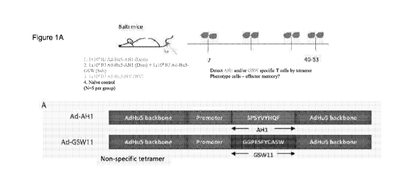
Figure 1. Immunization of Balb/c mice with an AdHu5 replication-deficient
vector encoding an
AH1 CD8+ T cell tumour epitope stimulates a durable CD8+ T response in the
periphery. (A)
Schematic representation of the constructs used for the production of AdHu5
vector expressing
a MHC-1 binding 0T26-specific cancer epitope. (B) FACs plots showing %CD8+ AH1
tetramer
+ (tet+) cells in the blood from AH1 (left) and Ad-I8V (right) vaccinated
mice. (C) AH1-tetramer-
specific CD8 + T cell responses in the blood at day 7 (left) and day 50
(right) from two
independent experiments. (D) FACS plots showing the presence of indicated
markers on CD8+
AH1 -tet+ (left) and AH1 -tet-(right) populations in the blood from the same
sample. (E) Phenotype
of AH1-tet+ CD8+ T cells compared to AH1-tet-CD8+ T cells from the same groups
at day 7
(left) and day 50 (right) from two independent experiments. Geo M Fl =
geometric mean
fluorescence intensity.
Figure 2. Memory inflationary AH1 -specific T cells demonstrate inhibition of
0T26 tumour growth
in Balb/c mice after both prophylactic and therapeutic vaccination with Ad-
AH1. (A)
Experimental setup for prophylactic vaccination (independently performed
twice, P1 and P2) and
therapeutic vaccination (Ti). A star indicates presence of palpable tumours.
(B-D) Tumour
growth curves for the different groups (N =5 per group) in a prophylactic (P1
and P2)(B and C)
and therapeutic vaccination setting (Ti) (D). In Ti, the arrow indicates the
time-point of
vaccination. Mice vaccinated with Ad-AH1 (1 x 10⁸ IU ) are shown in green, Ad-
A H 1 Low (1 X
107 IU) in orange, Ad-AH1 (1 x 10⁸ IU) + Ad-GSW11 (1 xi 08 IU) in red, Ad-
GSW11 (1 X 10⁸
IU ) in lilac, Ad-I8V (1 x 1 081U) in grey, and naïve mice in black. TF =
tumour free. (E, G, L)
Statistically significant differences in tumour sizes between groups at day 18
post-challenge.
Dots indicate individual mice. (F,H,J) Graphs show the slope of the tumour
growth curves
determined by linear regression from the day tumours show clear tumour growth
(day 7 post-
challenge for controls and day 18 post-challenge for Ad-AH1 vaccinated mice).
(K) The tumour
growth rate was recalculated to determine the specific growth rate. The tumor
growth rate
between implantation and humane endpoint was quantified using the parameter of
specific
CA 03157667 2022-04-11
WO 2021/074648 PCT/GB2020/052620
6
growth rate (SGR, %/day) calculated using the following equation:[15] SGR = In
(V2N1)/(t2 ¨
t1), where V1 and V2 are the tumor volumes at one day post implantation (V1
was fixed at
0.01 mm) (ti =Day 0) and endpoint (t2), respectively.
Figure 3. AH1-specific CD8+ T cells differ between tumour and spleen in both
abundance and
phenotype. (A) Representative FACS plots showing %CD8+ AH1-tet+ cells in the
tumour (upper
panel) and spleen (lower panel) from Ad-AH1 vaccinated mice. For negative
controls, tumour
and spleen samples were stained with the full range of fluorochrome-conjugated
antibodies and
an irrelevant H2-Ld-binding tetramer (pp89) for tumour samples or no tetramer
for spleen
samples (no tet). (B) Graphs show %CD8+ AH1-tet+ cells in the tumour (upper
panel) and
spleen (lower panel) from prophylactic (left panel) and therapeutic (right
panel) vaccinated mice.
(C) Heatmap showing phenotype of AH1-specific CD8+ T cells in tumour and
spleen from
prophylactic vaccinated (Ad-AH1) and control mice (Ad-I8V and naïve). Values
in cells indicate
mean of two independent experiments (N=5-10). Markers quantified by geometric
MFI have
been normalized to a 0 ¨ 100% scale.
Figure 4. (A) In tumours from Ad-AH1 vaccinated mice, presence of regulatory T
cells (CD4+
FoxP3+ cells) appear to be lower compared to control mice after both
prophylactic (left) and
therapeutic vaccination (right). Data from Ad-AH1 and Ad-AH1 + Ad-GSW11
vaccinated mice
was grouped likewise to Ad -I8V vaccinated and naïve mice (indicated by
vaccinated and
controls, respectively). (B) AdHu5-AH1-MG immunization increases the
percentage of Trm tet+
cells in the tumour. Mice immunized with AdHu5-AH1-MG, either singly or in
combination, show
increased percentages of AH-1 specific CD8 T cells in the tumour (TIL) with a
resident-memory
phenotype compared to control mice (naive or immunized with irrelevant AdHu5
constructs
(AdHu5-I8V-MG or AdHu5-GSW11)).
Figure 5. AdHu5-AH1-MG immunization induces AH-1+ CD8 T cells in the spleen
that remain
functional during tumour growth. Splenocytes and TILs were stimulated with AH-
1 peptide to
measure their cytotoxic potential based on IFN-gamma secretion. AH-1- peptide
specific
splenocytes from immunized animals are able to respond to peptide stimulation
(A and B). In
contrast, CD8 T cells in the TIL did not respond to peptide stimulation (C and
D); however the
levels of IFN-g secreted in response to PMA-ionomycin was also low indicating
a general state
of CD8 T cell downregulation in the tumour.
Figure 6. (A) The figures show the correlation between the slope of the tumour
growth curve for
each animal (indicated with a dot) and its percentage of CD8+ AH-1-specific T
cells in the blood
(left), spleen (middle) and absolute number of CD8+ AH1 tet+ cells in the
tumour (right). The
data is shown for two independent prophylactic (P1 and P2) and a single
therapeutic (Ti)
experiment. A lower tumour growth rate correlates with increased levels of AH1-
specific CD8+
T cells in spleen and blood post-tumour challenge but weakly correlates with
absolute numbers
of AH-1 specific CD8 T cells in the tumour. (B) shows the comparison of
antigen-specific cells
from various compartments with the specific growth rates.
CA 03157667 2022-04-11
WO 2021/074648 PCT/GB2020/052620
7
Figure 7. (A) Therapeutic immunization with an AdHu5 vector encoding full-
length gp90
(AdHu5-gp90FL) does not confer similar level of tumour control. Specific
growth rate of tumors
in each group was compared using Mann Whitney tests.*p<0.05, "p<0.005 (B) Mice
who
cleared the tumour by therapeutic and prophylactic immunization continue to
bear AH-1 specific
cells in circulation. The blood of mice who completely cleared tumours 6 month
previously were
sampled and stained for AH1+ CD8 T cells by tetramer staining.
Figure 8. HHD mice immunized with AdHu5- NY-ESO-1 157-165 minigene construct
develop a
long-lived circulating population of NY-ESO-1 157-165 Tet+ CD8 T cells with an
inflating memory
phenotype. (A) Levels of NY-ESO-1 1 57-1 65 Tet+ cells were measured by
tetramer staining in
blood after groups of HHD mice (N=4-5 per group) were immunized with 1x11 08
IU AdHu5-NY-
ESO-1 mini or 1x1 09 I.U. AdHu5-NY-ES0-1- FL. The schematic representation of
the constructs
used is shown. These cells were phenotyped by surface staining with (B) 0D44
and CD62L to
determine memory subset, markers of inflating cells (C) 0X30R1 and (D) KLRG-1
and markers
of exhaustion, (E)PD-1, (F)Tim3 and (G)Lag-3. The results shown are from 4-5
mice per group
from 1 of 2 independent experiments.
Figure 9. Mice primed with a single dose of AdHu5- NY-ESO-1 1 57-165 minigene
develop higher
percentages of circulating NY-ESO-1 157-165 Tet+ CD8 T cells after tumour
challenge and exhibit
better control of tumour growth. (A) At 53 (solid line) or 99 (dashed line)
days post-immunization
with either 1x11 08 IU AdHu5-NY-ES0-1 mini or 1x1 09 I.U. AdHu5-NY-ES0-1-FL,
animals were
injected subcutaneously (s.c) with either 1 x1 06 (solid line)) or 5x1 06
(dashed line) HHD-NY-ESO-
1 sarcoma cells As negative controls, groups of mice were either immunized
with 1x1 08 I.0 of
an irrelevant AdHu5-minigene construct (N=5) or left naïve (N=1 0). The
tumours were measured
every 1-2 days using digital callipers. (B)Circulating levels of NY-ESO-1 157-
165 Tet+ cells were
measured by tetramer staining in blood taken 14 days after tumour challenge.
(C) The levels of
NY-ESO-1 157-165 Tet+ detected in blood before tumour challenge versus the
size of the tumours
measured at early (Day1 4) and (D) late, Day 27/28 are shown. Statistical
measurement was
performed by T-tests. The data shown are from two separate independent
experiments.
Figure 10. NY-ESO-1 157-165 Tet+ CD8 T cells from tumours (TIL) display
elevated levels of
markers of exhaustion and activation. Mice were sacrificed, spleens and
tumours were removed
and analysed when the humane endpoint was reached, either by unhealed
ulceration or when
they approached 1 300mm3 in size. Lymphocytes were isolated from both
compartments and
(A)the percentage of CD8 T cells were measured. (B) The percentages of NY-ESO-
1 157-165 Tet+
cells in were also determined and as were levels of the exhaustion markers (C)
PD-1, (D)Tim-3
and (E) Lag-3 along with apoptotic marker (F) FasL.
Figure 11. 0X30R1 is preferentially upregulated on NY-ESO-1 157-165 Tet+ CD8 T
cells in spleen
and TIL after AdHu5- NY-ESO-1 157-165 minigene immunization. Lymphocytes
isolated from TIL
or spleen when the humane endpoint was reached were stained with the tetramer
and the levels
of the following molecules on Tet+ cells were determined. The inflating marker
0X30R1 on (A)
spleen and (B) TIL. (C) Markers of an effector memory phenotype, 0D44 and
CD62L and (D)
CA 03157667 2022-04-11
WO 2021/074648 PCT/GB2020/052620
8
resident memory markers CD103 and 0D69. (E) The levels of CD4+ regulatory T
cells (Treg) in
both compartments were also determined by intracellular staining.
Figure 12. CX3CR1+ CD8 T cells are more resistant to oxidative stress and
contain higher levels
of healthy polarized mitochondria. (A) The levels of intracellular reactive
oxygen species (ROS)
in CX3CR1+/-gfp splenocytes from Ad-lacZ or MCMV infected mice at day >50 post-
infection
were detected by CelIROX Red assay. (N=2 independent experiments)
(B)Peripheral blood
lymphocytes from C57BL/6 mice infected >100 days previously with MCMV or an
AdHu5
recombinant adenovector (Ad-I8V) were stained with MitoTracker Green (detects
all
mitochondria) and MitoTracker DeepRed (detects only healthy, polarized
mitochondria), then
surface stained with anti-mouse CD8, anti-mouse CX3CR1, LiveDead nearIR
Fixable Marker
and then analysed on an LSRII and the data calculated on FlowJo. Antigen-
specific CX3CR1+
inflating cells contain healthier mitochondria and show enhanced redox
resilience. (D) and (E)
show that when incubated in serum-free media (i.e. stress), there was a marked
survival of the
CX3CR1+ population compared to CX3CR1 negative T cells (Fig D) in the bulk and
antigen-
specific populations (Fig E). (F) shows the levels of reactive oxygen species
(ROS) upon serum
starvation indicating that CX3CR1+ T cells (bulk and antigen-specific) possess
intrinsically lower
levels of reactive oxygen species and are more resistant to oxidative stress.
Figure 13. Preventative immunization with HPV16 E749-57 minigene vector
confers protection
against tumour challenge. E749-57 specific cells are able to traffic to site
of tumour implantation
and confer protection against tumour challenge.
Figure 14. Synergistic effect after immunization with a panel minigenes
encoding CD8 T cell
epitopes against MCMV at a suboptimal dose. A panel of 3 minigenes against
known MCMV-
specific CD8 T cell epitopes, namely M45 (985HGIRNA5FI993), M38
(316SSPPMFRV325) and
m139 (419TWYGFCLL426) were constructed. These were injected i.v. into C57BL/6
mice either
as individual minigenes or as a cocktail. The minigene encoding M38 and M139
were injected
at a suboptimal dose of 1x10⁷ infectious units (I.U) while the minigene
encoding M45 was
injected at the optimal dose of 1x10⁸ I.U. The levels of M38-specific cells in
the blood at Day 6
post-immunization was measured. Surprisingly, mice that received the
combination minigene
vaccine containing M38-minigene and m139-minigene vectors at suboptimal doses,
plus M45-
minigene at optimal dose, developed higher levels of M38-specific T cells
compared to the
groups injected with only a sub-optimal dose of M38-minigene vector alone.
This unexpected
result suggests that delivery of a cocktail of minigene vectors at suboptimal
doses may have
additive effect to enhance the magnitude of the antigen-specific T cell over
that observed in upon
immunization with the single vector alone.
Figure 15. CD8 T cells from the tumours of AdHu5-AH1-MG immunized mice express
higher
levels of granzyme-B. 15A shows levels of granzyme B in total CD8 T cells in
the tumours 23
days post-implantation, 16 days post immunization and tumour sizes at time of
analysis. 15B
shows levels of the transcription factors T-bet and Eomes in AH1-specific CD8
T cells in the
tumours 23 days post-implantation, 16 days post immunization.
CA 03157667 2022-04-11
WO 2021/074648 PC T/GB2020/052620
9
Figure 16. Testing the GP70423-431 (AH1) minigene as a therapeutic vaccine in
combination
with anti-PD-L1. 16A shows groups of mice immunized with the indicated
adenovectors 7 days
after tumour challenge were then treated with anti-PD-L1 or isotype control.
The tumour sizes of
the individual mice are shown. 16B shows survival curve of all groups of mice.
16C shows the
% of G P70423-431 (AH1) Tet+ cells in circulation 15 days after immunization
(22 days post-tumour
challenge). 16D shows the specific growth rate of tumors in each group was
compared using
Mann-Whitney test. *p<0.05, "p<0.005
Figure 17. 17 A, B, C, D Spleen- and tumour-derived single cells from
prophylactic (A, C), or
therapeutic (B, D) therapeutic vaccination were stimulated ex vivo with AH1-
peptide (4 g/m1) or
PMA-Ionomycin (10) for 7 hours and then stained for intracellular cytokine
production of IFNy.
For each sample, low-level background activation (media only) was subtracted.
17 E-H Spleen
and tumour-derived single cells from therapeutic vaccination combined with
anti-PD-L1 were
stimulated ex vivo with AH1-peptide (4 g/m1) or PMA-Ionomycin (10) for 7 hours
and then
stained for intracellular cytokine production of IFNy. For each sample, low-
level background
activation (media only) was subtracted. The CD8 T cell response in spleen
(17E) and tumour
(17G) and CD4 T cell response in spleen (17F) and tumour (17H) are shown.
Figure 18. Pilot experiment to determine if two minigenes encoding two tumour
antigens will
improve tumour control. A shows protocol used ¨ tumour implantation performed
on day 0,
vaccination with one of AdHu5-AH1 minigene (MG), AdHu5-e2F8-27mer MG, Combo
(both MG
.. - AdHu5-AH1 and AdHu5-e2F8-27mer), irrelevant AdHu5-MG, unvaccinated on day
7, N=6 per
treatment group. Half of each group was treated with the checkpoint inhibitor
anti-PD-1 and half
the group were treated with an isotype control at 12, 16 and19 days post-
implantation. Bleeds
were performed on days 13 and 20. Figures 18 B, C, D, E and F show the tumour
growth over
time.
Figure 19. Shows comparison of the combination minigene treatment plus ant-PD-
1 compared
to negative controls and vaccination with a single minigene.
Figure 20. Growth rates of tumours calculated by linear regression for the
combination minigene
treatment, single minigene treatment and negative control.
Figure 21. % CD8+ AH1-tet + cells and %CD8+ ef28-tet+ cells produced from
vaccination with
the combination minigene treatment and vaccination with the single minigenes
AdHu5-AH1 and
AdHu5-e2F8-27mer measured 6 days post-vaccination.
Figure 22. Simultaneous i.v. immunization with two minigene
constructs/vaccines (combo -
AdHu5-AH1 and AdHu5-e2F8) induces both antigen-specific populations at similar
magnitudes
and phenotype to single vaccine measured 11 days post-vaccination.
Figure 23. Shows immunization with two minigenes targeting CD8 T cell epitopes
(AdHu5-AH1
and AdHu5-e2F8) in a cancer cell controls tumor growth. The linear regression
data in Figure
20 has been recalculated as specific growth rate.
Figure 24. Transcriptional profiling of an unconventional subset of memory T
cells: inflating
memory T cells. (A) A PCA of Inflating/non-Inflating CD8 T cells. 3D PCA
showing distribution
CA 03157667 2022-04-11
WO 2021/074648 PCT/GB2020/052620
of transcription profiles of two independent models of Inflating samples (M38,
D8V ¨ later stages
i.e. inflating memory, circled in blue) and non-Inflating Samples (M45,I8V ¨
later stages, i.e.
central memory, circled in brown), at acute stages (days 7 or 21) and later
stages (days 50 or
100), and naive samples. (B) PCA of Exhausted/non-Exhausted CD8 T cells. 3D
PCA showing
5 distribution of transcription profiles of a model of Exhaustion
(CI13,Tetrahedrons ¨ day 30 are
circled in grey), with non-Exhaustive samples (Arm, spheres ¨ day 30 are
circled in blue) at
different stages, and naive samples. Stages: 6 days (yellow), 8 days (brown),
15 days (pink), 30
days (black), naive (green).
Figure 25. The inflating memory subset express a distinct gene module compared
to other T
10 cell memory subsets. (A) Weighted Gene Co-expression Network Analysis of
Inflating samples.
Gene co-expression network analysis detected 6 gene modules (merging distance
= 0.25, soft-
thresholding power 13 = 9); Blue module (highlighted) genes are enriched with
immune relevant
GO categories and contains relevant genes such as Tbx21, Eomes, Zeb2, and
E2f2. (B) PCA
of Inflating/Exhausted samples based on Blue module genes. PCA plot using the
first three
principal components and based on a gene set of 588 genes, detected as blue
module in Gene
co-expression network analysis of Inflating samples only. The plot shows
distribution of Naïve
(green), Non-inflating and Non-exhausting (blue), and Inflating and Exhausting
(red) samples
(spheres: Exhaustion study; tetrahedron: Inflation study) (The inflating
memory population are
red tetrahedrons circled in blue). (C) Hierarchical clustering of
Inflating/Exhausted samples
based on Blue module genes. Dendogram plot showing sample clustering analysis
(Euclidian
distance) on Inflating-Exhausted merged sets, based on a gene set of 469
genes, detected as
blue module in a repeated Gene co-expression network analysis of Inflating
samples after
removing outliers (Soft-thresholding power 13 = 20). The memory inflation
cluster is contained in
the rectangle.
Figure 26. (A) Shows the schematic of the AdHu5 adenovirus with minigene
immunogen
cassette and a close-up view of the minigene immunogen cassette. (B) Shows the
schematic of
the AAV ITR with minigene immunogen cassette and a close-up view of the
minigene
immunogen cassette.
Detailed Description
The present invention will now be further described. In the following
passages, different aspects
of the invention are defined in more detail. Each aspect so defined may be
combined with any
other aspect or aspects unless clearly indicated to the contrary. In
particular, any feature
indicated as being preferred or advantageous may be combined with any other
feature or
features indicated as being preferred or advantageous. The practice of the
present invention will
employ, unless otherwise indicated, conventional techniques of immunology,
molecular biology,
chemistry, biochemistry and recombinant DNA technology, which are within the
skill of the art.
Such techniques are explained fully in the literature, see, e.g., Green and
Sambrook et al.,
CA 03157667 2022-04-11
WO 2021/074648 PCT/GB2020/052620
11
Molecular Cloning: A Laboratory Manual, 4th ed., Cold Spring Harbor Laboratory
Press, Cold
Spring Harbor, N.Y. (2012).
The present invention is based on the surprising finding that an adenoviral
vector encoding a
single cancer specific epitope results in an inflating memory CD8+ T cell
response. The term
inflating memory response refers to a sustained, functional, durable CD8+ T
cell response. The
resulting pool of CD8+ T cells are able to resist exhaustion which can occur
due to prolonged
TCR stimulation. T cell exhaustion can be characterised by upregulation of
markers such as
PD-1, Tim-3 and Lag-3.
The inflating memory CD8+ T cells are characterised by a unique phenotype
compared to other
CD8+ memory subsets, including the expression of markers CX3CR1 and KLRG-1.
The cells
also demonstrate a distinct transcriptional profile from both central memory
and exhausted
memory T cell subsets. The cells also demonstrate features such as enhanced
redox resilience
which may be due to intrinsically lower levels of reactive oxygen species and
resilience to
oxidative stress. In particular, the transcriptional profile is driven by the
transcription factor
Tbx21 with minimal contribution from Eomes. This results in a CD8+ T cell
phenotype that is
long lived, and present in the peripheral organs in high numbers whilst
retaining effector function.
The antigen-specific inflating memory CD8+ T cells develop through a unique
set of processing,
presentation and co-stimulation conditions. The processing of the epitope
occurs independently
of the immunoproteasome and presentation by a non-haematopoietic
unconventional APC
during the later stages may help to preserve this phenotype. Without wishing
to be bound by
theory it is thought that by using a vector of the present invention, which
encodes a single epitope
of interest, the antigen processing requirements are bypassed thereby
resulting in an inflating
memory response.
In an embodiment the invention relates to an adenoviral vector comprising a
nucleotide
sequence encoding a single cancer specific CD8+ and/or CD4+ T cell epitope,
wherein the
vector is capable of inducing an inflating memory CD8+ T cell response. In an
embodiment the
adenoviral vector comprises a nucleotide sequence encoding a single cancer
specific CD8+
and/or CD4+ T cell epitope, e.g. a single cancer specific CD8+ T cell epitope,
and the vector
does not comprise any additional cancer specific CD8+ and/or CD4+ T cell
epitopes. As such
the vector of the present invention encodes a single cancer specific CD8+
and/or CD4+ T cell
epitope, e.g. a single cancer specific CD8+ T cell epitope. The present
invention does not extend
to adenoviral vectors encoding more than one or multiple cancer specific CD8+
and/or CD4+ T
cell epitopes.
In an embodiment the invention relates to an adenoviral vector comprising a
nucleotide
sequence encoding a single cancer specific CD8+ T cell epitope, wherein the
vector is capable
CA 03157667 2022-04-11
WO 2021/074648 PCT/GB2020/052620
12
of inducing an inflating memory CD8+ T cell response. In an embodiment the
adenoviral vector
comprises a nucleotide sequence encoding a single cancer specific CD8+ T cell
epitope, and
the vector does not comprise any additional cancer specific CD8+ T cell
epitopes. As such the
adenoviral vector of the present invention encodes a single cancer specific
CD8+ T cell epitope.
The present invention does not extend to adenoviral vectors encoding more than
one or multiple
cancer specific CD8+ T cell epitopes.
The vector of the present invention, which encodes a single cancer specific
CD8+ T cell epitope,
is able to generate a sustained, functional, durable CD8+ T cell response from
a single dose.
The resulting pool of CD8+ T cells are able to resist exhaustion which can
occur due to prolonged
TCR stimulation. The resulting pool of CD8+ T cells may also demonstrate
enhanced redox
resilience and low levels of reactive oxygen species.
As used herein the term "vector" refers to a nucleic acid sequence capable of
transporting into
a cell another nucleic acid to which the vector sequence has been linked. The
vectors of the
present invention are adenoviral and comprises the nucleotide sequence
encoding a single
cancer specific CD8+ or CD4+ T cell epitope containing a gene construct in a
form suitable for
expression by a cell (e.g., linked to a transcriptional control element).
As used herein the term "epitope" refers to a part of an antigen that is
recognised by the immune
system which may be a short protein sequence. A "cancer specific CD8+ and/or
CD4+ T cell
epitope" refers to an epitope that may be presented by an antigen presenting
cell bound to an
MHC molecule which are then recognised by the T-cell receptor (TCR). CD4+ T
cells express
the CD4 coreceptor, which binds to MHC II, and recognize peptides presented by
MHC II
molecules. CD8+ T cells express the CD8 coreceptor, which binds to MHC I, and
recognize
peptides presented by MHC I molecules.
Inflating memory T cells can be characterized by the presences of specific
markers and cell
surface markers. Methods to identify and quantify these markers are well known
in the art.
Examples of suitable methods include but are not limited to affinity-based
separation methods,
magnetic cell sorting techniques, fluorescence-based cell sorting techniques
such as FACS
(fluorescence activated cell sorting). The inflating memory CD8+ T cells can
be characterised
by the presence of a number of markers, examples include but are not limited
to CX3CR1,
KLRG-1, 0D44. The inflating memory CD8+ T cells can also be characterised by
the low
expression of a number of markers, example include but are not limited to
CD62L, 0D27,
0D127. The term "low expression" may refer to cells wherein there is no
expression of the
markers, it may also refer to cells wherein there is low expression of the
markers relative to other
cells in the sample.
CA 03157667 2022-04-11
WO 2021/074648 PCT/GB2020/052620
13
In an embodiment the inflating memory CD8+ T cells are characterised by
markers selected
from the group comprising CX3CR1+, KLRG-1+, 0D44+, CD62L-, wherein the
designation (+)
indicates the presence of the marker, and the designation (-) indicates low
expression or no
expression of the marker. Wherein the (-) designation means low expression
this may be further
indicated by "(low)". The inflating memory CD8+ T cells may be characterised
by markers
selected from the group comprising CX3CR1+, KLRG-1+, 0D44+, CD62L-, 0D27-
(low), CD127-
(low).
The inflating memory CD8+ T cells may be characterised by the phenotype
CX3CR1+, KLRG-
1+, 0D44+, CD62L-. The inflating memory CD8+ T cells may be characterised by
the phenotype
CX3CR1+, KLRG-1+, 0D44+, CD62L-, 0D27-(low), 0D127-(low).
The CD8+ T cells produced in an inflating memory response may have a number of
other
characteristics. For example, the cells comprise a transcriptional profile
driven by Tbx21 (also
referred to as T-bet). These cells show a sustained expression of Tbx21. The
cells may also
show a sustained expression of E2f2 a transcription factor generally involved
in cell growth and
proliferation. The cells may also lack expression or have low expression of
the transcription
factor Eomes.
The inflating memory CD8+ T cells may not demonstrate classical contraction
after exposure to
an antigen. During classical memory evolution after exposure to an antigen the
cells form a
contracted central memory pool which makes up <1% of total circulating CD8+ T
cells. However,
inflating memory cells are maintained as large pools of cells which circulate
in the blood. As
such, in an embodiment the resulting inflating memory CD8+ T cells form
approximately 2% to
approximately 20% of total CD8+ T cells, preferably approximately 8% to
approximately 20% of
total CD8+ T cells, more preferably approximately 12% to approximately 20% of
total CD8+ T
cells.
In an embodiment the large pools of inflating memory CD8+ T cells retain their
effector memory
phenotype. The resulting inflating memory CD8+ T cells may retain their memory
effector
phenotype for a prolonged period, wherein the effector phenotype is
characterised by 0D44+,
CD62L-. The inflating memory CD8+ T cells may retain their memory effector
phenotype for up
to 60 days post exposure to the vector of the present invention, up to 55 days
post exposure to
the vector of the present invention, up to 50 days post exposure to the vector
of the present
invention, up to 40 days post exposure to the vector of the present invention,
or up to 30 days
post exposure to the vector of the present invention.
The inflating memory CD8+ T cells may also lack markers of exhaustion. T cell
exhaustion can
occur from excessive TCR (T cell receptor) stimulation. Markers of T cell
exhaustion can include
CA 03157667 2022-04-11
WO 2021/074648 PCT/GB2020/052620
14
upregulation of markers such as PD-1, Tim-3, Lag-3. As such, in an embodiment
the inflating
memory CD8+ T cells may lack or demonstrate low expression of markers selected
from the
group consisting of PD-1, Tim-3, Lag-3.
.. The nucleotide sequence encoding a single cancer specific CD8+ and/or CD4+
T cell epitope
may comprise from approximately 12 to approximately 45 base pairs, in another
embodiment
the nucleotide sequence may comprise approximately 15 to approximately 45 base
pairs, in
another embodiment the nucleotide sequence may comprise approximately 18 to
approximately
45 base pairs, in another embodiment the nucleotide sequence may comprise
approximately 21
to approximately 45 base pairs, in a preferred embodiment the nucleotide
sequence may
comprise approximately 24 to approximately 45 base pairs. As such, the vector
encodes a single
cancer specific CD8+ and/or CD4+ T cell epitope comprising approximately 5 to
approximately
amino acids, in another embodiment the vector encodes an epitope comprising
approximately 6 to approximately 15 amino acids, in another embodiment the
vector encodes
15 an epitope comprising approximately 7 to approximately 15 amino acids,
in a preferred
embodiment the vector encodes an epitope comprising approximately 8 to
approximately 15
amino acids.
The single cancer specific CD8+ and/or CD4+ T cell epitope is an immunogenic
epitope, in that
it elicits an immune response. T cell epitopes bind to the major
histocompatibility complex in
order to initiate a subsequent immune response. As such in an embodiment the
epitope is
capable of binding and presenting on an MHC molecule. There are multiple
methods known in
the art to identify epitopes which bind the MHC and therefore produce an
immune response.
These methods include peptide-MHC binding prediction models of which there are
multiple
programs publicly available.
In an embodiment the single cancer specific CD8+ and/or CD4+ T cell epitope is
derived from a
tumour associated antigen (TAA). A TAA is an antigenic product produced by a
cancer and it
provides a biomarker for targeted identification of a tumour. TAAs can be
broadly categorized
into aberrantly expressed self-antigens, mutated self-antigens and tumour
specific antigens. As
such, the TAA may be upregulated or over-expressed in the cancer cell. The TAA
may be
mutated within the cancer cell. The TAA may specific for the cancer cell and
only expressed
within the cancer cell, this may also be referred to as a tumour specific
antigen.
In an embodiment the cancer specific CD8+ and/or CD4+ T cell epitope is
mutated in a cancer
cell. In an embodiment the cancer specific CD8+ and/or CD4+ T cell epitope is
overexpressed
in a cancer cell. In an embodiment the cancer specific CD8+ and/or CD4+ T cell
epitope is a
non-coding tumour specific epitope. As used herein the term "non-coding tumour
specific
epitope" refers to a peptide found on a cancer cell, wherein the peptide is
derived from a
CA 03157667 2022-04-11
WO 2021/074648 PCT/GB2020/052620
nucleotide sequence that is epigenetically supressed in healthy cells. These
peptide sequences
are aberrantly expressed within tumour cells.
In an embodiment the cancer specific CD8+ and/or CD4+ T cell epitope is not a
cryptic epitope.
5 As used herein a "cryptic epitope" refers to refers to an epitope which
is not immunogenic in
immunocompetent individuals.
In an embodiment the cancer specific CD8+ and/or CD4+ T cell epitope may be a
viral epitope
that is associated with a virally driven cancer. The virally driven cancer may
be HPV (human
10 papilloma virus), HTLV (human T-Iymphotropic virus), or EBV (Epstein
Barr virus).
In an embodiment the cancer specific CD8+ and/or CD4+ T cell epitope is
derived from a tumour
associated antigen selected from the group consisting of TRP-1, CEA, TAG-72,
9D7, Ep-CAM,
EphA3, telomerase, mesothelin, SAP-1 Melan-A/MART-1, tyrosinase, CLPP, cyclin-
A1, cyclin-
15 B1 MAGE-A1, MAGE-C1, MAGE-02, 55X2, XAGE1b/GAGED2a, 0D45, glypican-3,
IGF2B3,
kallikrein-4, KIF20A, lengsin, meloe, MUC5AC, survivin, PRAME, SSX-2, NY-ES0-
1/LAGE1,
gp70, MOIR, TRP-1/-2, 13-catenin, BRCA1/2, CDK4.
The cancer specific CD8+ and/or CD4+ T cell epitope may be a private epitope.
As used herein
the term "private epitope" refers to an epitope which is found exclusively on
a single antigen in
the cancer of a single person. The cancer specific CD8+ and/or CD4+ T cell
epitope may be a
public epitope. As used herein the term "public epitope" refers to an epitope
that is found on the
cancer of two or more people.
In an embodiment the cancer specific CD8+ and/or CD4+ T cell epitope may be a
neoepitope.
As used herein the term "neoepitope" refers to epitopes which have arisen
through mutations
within the tumour cells, in particular somatic or passenger mutations may lead
to the production
of a neoepitope. In an embodiment the cancer specific CD8+ and/or CD4+ T cell
epitope is not
a neoepitope.
In an embodiment the cancer specific CD8+ and/or CD4+ T cell epitope is
specific for colorectal
cancer, prostate cancer, oesophageal cancer, liver cancer, renal cancer, lung
cancer, breast
cancer, breast cancer, pancreatic cancer, brain cancer, hepatocellular cancer,
lymphoma,
leukaemia, gastric cancer, cervical cancer, ovarian cancer, thyroid cancer,
melanoma,
carcinoma, head and neck cancer, skin cancer, nasopharyngeal cancer, Epstein
Barr driven
cancers, Human Papilloma virus driven cancers and soft tissue sarcoma. The
term "cancer" as
used herein refers to diseases with abnormal cell growth, as used herein the
term refers to both
a primary tumour and metastasis of the primary tumour.
CA 03157667 2022-04-11
WO 2021/074648
PCT/GB2020/052620
16
In an embodiment the cancer specific CD8+ and/or CD4+ T cell epitope comprises
SEQ ID NO:1
(SPSYVYHQF) or SEQ ID NO:2 (SLLMWITQC) or SEQ ID NO:37 (SLLMWITQV). Where the
cancer specific CD8+ and/or CD4+ T cell epitope is a viral epitope that is
associated with a virally
driven cancer the epitope may comprise SEQ ID NO:7 (RAHYNIVTF). The virally
driven cancer
may be selected from EBV driven cancers, HTL driven cancers, and HPV driven
cancers. EBV
driven cancers may include Hodgkin Lymphoma (HL), Burkitt Lymphoma (BL),
Diffuse Large B
cell Lymphoma (DLBCL) and two rarer tumors associated with profound immune
impairment,
plasmablastic lymphoma (PBL) and primary effusion lymphoma (PEL), LPDs and
malignant
lymphomas of T or NK cells, nasopharyngeal carcinoma (NPC) and gastric
carcinoma of
epithelial origin, and leiomyosarcoma. HPV driven cancers may include
anogenital cancers,
oropharyngeal cancers, oral cavity cancer, head and neck squamous cell
carcinoma and
laryngeal cancer.
In an embodiment the cancer specific CD8+ and/or CD4+ T cell epitope comprises
one or more
of the epitopes in Table 1.
Table 1
HLA Target-epitope Amino acid SEQ ID NO Gene Target
cancer
type sequence
Human
A*0201 NY-ESO-1 157- SLLMWITQC SEQ ID NO:2 CTAG1B Cancers
165
expressing NY-
ESO-1
Mouse
H-2Ld MuLV env SPSYVYHQF SEQ ID NO:1 env gp70 CT26
gp70423-431
H-2Dd MuLV gp90147_ GGPESFYCA SEQ ID NO:3 env gp70 CT26
148 SW
H-2Kd E2f8509-535 VILPQAPSGP SEQ ID NO:4 e2f8
CT26
SYATYLQ PA
QAQMLTPP
H-Kd Mtch1361-370 KYLSVQSQLF SEQ ID NO:5 mtch1
CT26
H-2Kd Mtch1361-369 KYLSVQSQL SEQ ID NO:6 mtch1
CT26
and H-
2Ld
H-2Db HPV16 E746-57 RAHYNIVTF SEQ ID NO:7 Human
papillomavirus-
driven cancers
CA 03157667 2022-04-11
WO 2021/074648 PCT/GB2020/052620
17
Further, cancer specific CD8+ and/or CD4+ T cell epitopes may be determined
using techniques
know in the art such as proteomics approaches, mass spectrometry approaches,
genomic
approaches, transcriptome analysis, bioinformatics approaches and in silico
methods. It would
be possible for the skilled person to select an appropriate epitope to be
encoded within the vector
of the present invention.
The nucleic acid encoding the cancer specific CD8+ and/or CD4+ T cell epitope
may be codon
optimised for mammalian codon usage. Suitably the nucleic acid sequence may be
codon
optimised for human codon usage.
The vector may comprise adeno-associated virus (AAV). The vector may comprise
adenovirus.
The adenoviral vector or AAV vector may also have additional features such as
enhancer and
promoter regions. In an embodiment the vector may comprise a strong promoter
examples
include but are not limited to a CMV promoter, an RSV promoter, an EF1a
promoter. In a
preferred embodiment the vector comprises a CMV promoter, a suitable sequence
for a CMV
promotor is provided in SEQ ID NO:18. In an embodiment the vector may comprise
a TATA
box. In an embodiment the vector comprises a translation initiation sequence,
for example a
Kozak sequence. A Kozak sequence has the consensus sequence (gcc)gccRccAUGG, a
suitable Kozak sequence is provided in SEQ ID NO:19. In an embodiment the
vector comprises
a termination sequence and/or a polyadenylation sequence. A suitable
polyadenylation
sequence is provided in SEQ ID NO:34. The AAV vector may comprise inverted
terminal repeat
(ITR) sequences. A suitable ITR sequence is provided in SEQ ID NO:42.
In an embodiment the vector does not comprise additional cancer specific CD8+
and/or CD4+
T cell epitopes. The vector only encodes a single cancer specific CD8+ and/or
CD4+ T cell
epitope. In an embodiment the adenoviral vector consists of the vector back
bone, a promoter
region and a nucleotide sequence encoding a single cancer specific CD8+ T cell
epitope. The
adenoviral backbone may comprise additional features such as enhancer regions,
promoter
regions, TATA box, translation initiation sequence.
The AAV vector may be serotype 1, 2, 3, 4, 5, 6, 7, 8 or 9. In a preferred
embodiment the AAV
vector may be serotype 2 or 5. The AAV vector may comprise ITR sequences, in a
preferred
embodiment the ITR sequences flank the encoded cancer specific CD8+ and/or
CD4+ T cell
epitope. There may be an ITR sequence present 5' to the cancer specific
epitope and an ITR
sequence 3' to the cancer specific epitope. The 5' ITR sequence may comprise
SEQ ID NO:39.
The 3' ITR sequence may comprise SEQ ID NO: 42. The AAV vector may comprise
sequences
5' to the cancer specific epitope for example SEQ ID NO:38. The AAV vector may
comprise
sequences 3' to the cancer specific epitope for example SEQ ID NO:41. In order
to produce the
CA 03157667 2022-04-11
WO 2021/074648 PCT/GB2020/052620
18
AAV vector comprising the cancer specific CD8+ and/or CD4+ T cell epitope,
helper plasmids
may be used. A helper plasmid or plasmids may be used to provide genes
required for AAV
replication or packaging. In an embodiment helper plasmid encodes E2A, E4 and
VA adenoviral
proteins and or encodes the rep and cap genes of AAV.
The adenoviral vector may be a Species C serotype. Species C includes Adl , 2,
5 and 6
serotypes. In a preferred embodiment the adenoviral vector is a human serotype
5 (AdHu5). It
may be preferable for the adenoviral vector to be modified for example to
reduce the
immunogenicity and improve biosafety of the vector. As such, the adenoviral
vector may be
replication-incompetent. The adenoviral vector may lack the El and E3
proteins. The adenoviral
vector may comprise sequences 5' to the cancer specific epitope for example
SEQ ID NO:13.
The adenoviral vector may comprise sequences 3' to the cancer specific epitope
for example
SEQ ID NO:14.
Other adenoviral vectors may also be suitable for the vector for the present
invention. In an
embodiment the vector may be an animal derived adenoviral vector for example
canine, simian
in particular rhesus monkey and chimpanzee. In an embodiment the adenoviral
vector may be
a rare serotype vector derived from a non-human primate. Vectors derived from
chimpanzee
may be suitable for the vector for the present invention, examples include but
are not limited to
ChAd63, ChAd3, ChAdY25.
In an embodiment there is provided an immunogenic composition, comprising the
vector as
defined above. The immunogenic composition may further comprise one or more
additional
active ingredients, pharmaceutically acceptable carrier, diluent, excipient or
adjuvant.
The immunogenic composition comprising a vector according to the invention may
be used in
combination with at least one other immunogenic composition comprising a
vector according to
the invention, wherein each vector encodes a different cancer specific CD8+
and/or CD4+ T cell
epitope. The immunogenic composition comprising a first vector according to
the invention may
be administered separately, sequentially or simultaneously with an immunogenic
composition
comprising a second vector according to the invention.
In an embodiment the immunogenic composition may comprise at least two vectors
according
to the invention. It may be preferable for the at least two vectors to encode
different cancer
specific CD8+ and/or CD4+ T cell epitopes. Wherein further additional vectors
are present in
the composition the vector may encode different cancer specific CD8+ and/or
CD4+ T cell
epitopes. The immunogenic composition may further comprise one or more
additional active
ingredients, pharmaceutically acceptable carrier, diluent, excipient or
adjuvant. Without wishing
to be bound by theory, use of a cocktail of vectors encoding different
epitopes may result in a
CA 03157667 2022-04-11
WO 2021/074648 PCT/GB2020/052620
19
stronger immune response, further there may be a synergistic effect which
enhances the
immune response.
Wherein the composition of the present invention comprises at least two
vectors as described
herein, the vectors may be provided as separate medicaments for administration
at the same
time or at different times.
In an embodiment, wherein the composition comprises at least two vectors as
described herein,
the vectors may be provided as separate medicaments for administration at
different times.
When administered separately and at different times, either vector may be
administered first. In
some embodiments, both can be administered on the same day or on different
days, and they
can be administered using the same schedule or at different schedules during
the treatment
cycle.
Alternatively, wherein the composition comprises at least two vectors as
described herein, the
administration of the vectors may be performed simultaneously. Wherein
simultaneous
administration is used the vectors may be formulated as separate
pharmaceutical compositions.
In a preferred embodiment the at least two vectors may be formulated as a
single pharmaceutical
composition.
The composition of the invention can be in the form of a liquid, e.g., a
solution, emulsion or
suspension. The liquid compositions of the invention, whether they are
solutions, suspensions
or other like form, can also include one or more of the following: sterile
diluents such as water,
saline solution, preferably physiological saline, Ringer's solution, isotonic
sodium chloride, fixed
oils such as synthetic mono or digylcerides, polyethylene glycols, glycerin,
or other solvents;
antibacterial agents such as benzyl alcohol or methyl paraben; and agents for
the adjustment of
tonicity such as sodium chloride or dextrose. The composition can be enclosed
in an ampoule,
a disposable syringe or a multiple-dose vial made of glass, plastic or other
material.
An intravenous formulation of the vector or composition of the invention may
be in the form of a
sterile injectable aqueous or non-aqueous (e.g. oleaginous) solution or
suspension. The sterile
injectable preparation may also be in a sterile injectable solution or
suspension in a non-toxic
parenterally-acceptable diluent or solvent, for example, a solution in 1 ,3-
butanediol. Among the
acceptable vehicles and solvents that may be employed are water, phosphate
buffer solution,
Ringer's solution and isotonic sodium chloride solution. In addition, sterile,
fixed oils may be
employed as a solvent or suspending medium. For this purpose, any bland fixed
oil may be
employed, including synthetic mono- or diglycerides. In addition, fatty acids
such as oleic acid
may be used in the preparation of the intravenous formulation of the
invention.
CA 03157667 2022-04-11
WO 2021/074648 PCT/GB2020/052620
The immunogenic compositions can be prepared using methodology well known in
the
pharmaceutical art. For example, a composition intended to be administered by
injection can
be prepared by combining a vector of the present invention with water so as to
form a solution.
A surfactant can be added to facilitate the formation of a homogeneous
solution or suspension.
5 .. In an embodiment the invention relates to a host cell, comprising the
vector or the immunogenic
composition as described herein. The host cell may be mammalian for example
human or
mouse. The host cell may be transduced with the vector. The host cell may be
used to produce
an adenoviral stock.
10 In an embodiment the vector or immunogenic composition is for use in
therapy. In a preferred
embodiment the vector or immunogenic composition is for use in the treatment
or prevention of
cancer.
The term "treatment" refers to the medical management of a patient with the
intent to cure,
15 ameliorate, stabilize, or prevent a disease, pathological condition, or
disorder. This term includes
active treatment, that is, treatment directed specifically toward the
improvement of a disease,
pathological condition, or disorder, and also includes causal treatment, that
is, treatment directed
toward removal of the cause of the associated disease, pathological condition,
or disorder. In
addition, this term includes palliative treatment, that is, treatment designed
for the relief of
20 .. symptoms rather than the curing of the disease, pathological condition,
or disorder; preventative
treatment, that is, treatment directed to minimizing or partially or
completely inhibiting the
development of the associated disease, pathological condition, or disorder;
and supportive
treatment, that is, treatment employed to supplement another specific therapy
directed toward
the improvement of the associated disease, pathological condition, or disorder
The invention furthermore relates to a method of treating or preventing a
cancer, comprising
administering a therapeutically effective amount of the vector or composition
according to the
invention to a subject in need thereof.
In an embodiment the invention relates to the use of a vector or composition
described herein
in the manufacture of a medicament for the treatment or prevention of cancer.
In an embodiment
the invention relates to the use of a vector or composition described herein
in the treatment or
prevention of cancer.
.. As used herein, the term "therapeutically effective" refers to the amount
of the composition used
is of sufficient quantity to ameliorate one or more causes or symptoms of a
disease or disorder.
Such amelioration only requires a reduction or alteration, not necessarily
elimination.
CA 03157667 2022-04-11
WO 2021/074648 PCT/GB2020/052620
21
The present invention also provides a method of inducing an inflating memory
CD8+ T cell
response, comprising the step of; administering a therapeutically effective
amount of the vector
or composition according to the invention, to a subject in need thereof,
wherein the CD8+ T cells
are characterised by markers selected from the group comprising CX3CR1+, KLRG-
1+, CD44+
and CD62L-.
Preferably the CD8+ T cells are characterised by the phenotype CX3CR1+, KLRG-
1+, CD44+
and CD62L-. More preferably they are characterised by the phenotype CX3CR1+,
KLRG-1+,
CD44+, CD62L-, CD27(low), CD127(low).
The vector or immunogenic composition may be for use in the treatment or
prevention of
colorectal cancer, prostate cancer, oesophageal cancer, liver cancer, renal
cancer, lung cancer,
breast cancer, breast cancer, pancreatic cancer, brain cancer, hepatocellular
cancer,
lymphoma, leukaemia, gastric cancer, cervical cancer, ovarian cancer, thyroid
cancer,
melanoma, carcinoma, head and neck cancer, skin cancer and soft tissue
sarcoma.
The vector or composition as described herein may be administered by any
convenient route.
The vector or composition may be administered by any convenient route,
including but not
limited to oral, topical, parenteral, sublingual, rectal, vaginal, ocular,
intranasal, pulmonary,
intradermal, intravitreal, intramuscular, intraperitoneal, intravenous,
subcutaneous,
intracerebral, transdermal, transmucosal, by inhalation. Parenteral
administration includes, for
example, intravenous, intramuscular, intraarterial, intraperitoneal,
intranasal, rectal, intravesical,
intradermal, topical or subcutaneous administration. In
an embodiment the vector or
composition is administered intravenously or intramuscularly. Compositions can
take the form
of one or more dosage units.
In specific embodiments, it may be desirable to administer the vector or
composition of the
present invention locally to the area in need of treatment such at as the site
of a tumour. In
another embodiment it may be desirable to administer the vector or composition
by intravenous
injection or infusion. The amount of the vector of the present invention that
is effective/active in
the treatment of a particular disorder or condition will depend on the nature
of the disorder or
condition, and can be determined by standard clinical techniques. In addition,
in vitro or in vivo
assays can optionally be employed to help identify optimal dosage ranges. The
precise dose to
be employed in the compositions will also depend on the route of
administration, and the
seriousness of the disease or disorder, and should be decided according to the
judgment of the
practitioner and each patients circumstances.
The compositions comprise an effective amount of the vector according to the
present invention
such that a suitable dosage will be obtained. The correct dosage of the
compounds will vary
CA 03157667 2022-04-11
WO 2021/074648 PCT/GB2020/052620
22
according to the particular formulation, the mode of administration, and its
particular site, host
and the disease being treated. Other factors like age, body weight, sex, diet,
time of
administration, rate of excretion, condition of the host, drug combinations,
reaction sensitivities
and severity of the disease shall be taken into account. Administration can be
carried out
continuously or periodically.
In the therapy of cancer, the vector or immunogenic composition of the present
invention, can
be used in combination with existing therapies. In one embodiment, the vector
or composition is
used in combination with an existing therapy or therapeutic agent, for example
an anti-cancer
therapy. Thus, in another aspect, the invention also relates to a combination
therapy comprising
administration of the vector or composition of the invention and an anti-
cancer therapy. The
anti-cancer therapy may include a therapeutic agent or radiation therapy and
includes gene
therapy, viral therapy, RNA therapy bone marrow transplantation, nanotherapy,
targeted anti-
cancer therapies or oncolytic drugs. Examples of other therapeutic agents
include checkpoint
inhibitors, antineoplastic agents, immunogenic agents, attenuated cancerous
cells, tumour
antigens, antigen presenting cells such as dendritic cells pulsed with tumour-
derived antigen or
nucleic acids, immune stimulating cytokines (e.g., IL-2, IFNa2, GM-CSF),
targeted small
molecules and biological molecules (such as components of signal transduction
pathways, e.g.
modulators of tyrosine kinases and inhibitors of receptor tyrosine kinases,
and agents that bind
to tumour- specific antigens, including EGFR antagonists), an anti-
inflammatory agent, a
cytotoxic agent, a radiotoxic agent, or an immunosuppressive agent and cells
transfected with a
gene encoding an immune stimulating cytokine (e.g., GM-CSF), chemotherapy. In
one
embodiment, the vector or composition is used in combination with surgery. The
vector or
composition of the invention may be administered at the same time or at a
different time as the
other therapy, e.g., simultaneously, separately or sequentially.
In an embodiment the vector or composition is used in combination with an
immunomodulatory
agent. The immunomodulatory agent may be administered simultaneously,
sequentially or
separately with the immunomodulatory agent. In specific embodiments the
immunomodulatory
agent may be an immune checkpoint inhibitor, examples of immune checkpoint
inhibitors include
but are not limited to inhibitors of an immune checkpoint protein selected
from the group
consisting of CTLA-4, PD-1, PD-L1, PD-L2, TIM3, LAG -3, B7-H3, B7-H4, B7-H6,
A2aR, BTLA,
GAL9 and IDO.
Certain tumour types have previously been reported to be unresponsive to anti-
PD-1 and anti
PD-L1 monotherapies. It has surprisingly been shown herein that immunization
with a minigene
vector can result in enhanced tumour control when administered in combination
with a
checkpoint inhibitor such as an anti-PD-L1 therapy. This has been shown
effective in tumour
models which are known to be unresponsive to standard checkpoint inhibitor
therapy. As such,
CA 03157667 2022-04-11
WO 2021/074648 PCT/GB2020/052620
23
in an embodiment the present vector or composition may be used in combination
with a check
point inhibitor for the treatment of checkpoint inhibitor unresponsive
tumours.
The vector or composition of the present invention and the immunomodulatory
agent may be
provided as separate medicaments for administration at the same time or at
different times.
In an embodiment, the vector or composition of the present invention and the
immunomodulatory
agent are provided as separate medicaments for administration at different
times. When
administered separately and at different times, either the vector or the
immunomodulatory agent
may be administered first. In some embodiments, both can be administered on
the same day
or on different days, and they can be administered using the same schedule or
at different
schedules during the treatment cycle.
Alternatively, the administration of the immunomodulatory agent may be
performed
simultaneously with the administration of the vector or immunogenic
composition. Wherein
simultaneous administration is used the vector or immunogenic composition and
the
immunomodulatory agent may be formulated as separate pharmaceutical
compositions. The
vector or immunogenic composition and the immunomodulatory agent may be
formulated as a
single pharmaceutical composition.
The vector or composition of the present invention can be administered
prophylactically or
therapeutically. The term "prophylactically" refers to administration intended
to have a protective
effect against disease. The term "therapeutically" refers to administration
intended to have a
curative effect.
The vector or composition of the present invention may be administered as a
single dose. The
dose may be provided in a prophylactic setting or a therapeutic setting. In an
embodiment the
single dose may be provided as a single dose unit further comprising one or
more additional
active ingredients, pharmaceutically acceptable carrier, diluent, excipient or
adjuvant.
The vector or composition of the present invention may be administered as
multiple doses.
Wherein multiple doses are administered, one or more may be administered
prophylactically or
one or more may be administered therapeutically. Where multiple doses are
administered, one
or more may be administered prophylactically and one or more may be
administered
therapeutically. In an embodiment the vector may be administered as a "prime
boost" regimen,
wherein there is a first administration (a priming administration) of the
adenoviral vector, followed
by a second administration (a boosting administration).
CA 03157667 2022-04-11
WO 2021/074648 PCT/GB2020/052620
24
Dose delays and/or dose reductions and schedule adjustments are performed as
needed
depending on individual patient tolerance to treatments.
Wherein the immunogenic composition comprises at least two vectors and wherein
the vectors
encode different epitopes as described above, there may be synergy between the
vectors. As
such, each of the vectors may be administered at a sub-optimal dose. The term
"sub-optimal"
dose refers to a dose level that it is not intended to fully remove or
eradicate the tumour, but
nevertheless results in some tumour cells or tissue becoming necrotic. The
skilled person will
be able to determine an appropriate dose required in order to achieve this,
depending on factors
such as; age of the patient, status of the disease and size and location of
tumour or metastases
In an embodiment there is provided a method of producing the vector are
described above,
comprising the steps of;
i) synthesising the nucleic acid sequence encoding the epitope, as a sense and
antisense
primer,
ii) cloning the nucleic acid sequence encoding epitope sequence into a first
plasmid,
iii) cloning the sequence comprising the nucleic acid sequence encoding
epitope into a
second plasmid comprising the adenoviral DNA.
Suitable cloning methods are known within the art, examples of cloning methods
include but are
not limited to, restriction ligations methods, Gateway cloning, Gibson
assembly, ligation
independent cloning. The person skilled in the art will be able to determine a
suitable method
to clone the sequence into the plasmid. The cloning method to introduce the
nucleic acid
sequence encoding epitope sequence into the first plasmid may be the same or
different from
the cloning method used to. In an embodiment the cloning method to introduce
the nucleic acid
sequence encoding epitope sequence into the first plasmid is selected from
restriction ligations
methods, Gateway cloning, Gibson assembly, ligation independent cloning. In an
embodiment
the cloning method to introduce the nucleic acid sequence encoding epitope
into the second
plasmid comprising the adenoviral DNA is selected from restriction ligations
methods, Gateway
cloning, Gibson assembly, ligation independent cloning.
In an embodiment, step iii) comprises cloning the sequence comprising the
nucleic acid
sequence encoding the epitope into a second plasmid comprising the adenoviral
DNA, wherein
the sequence comprising the nucleic acid sequence encoding the epitope also
comprises
additional features selected from the group comprising a translation
initiation sequence, a
promotor, a termination sequence, a polyadenylation sequence.
In an embodiment the method of producing the vector comprises the steps of;
i) synthesising the nucleic acid encoding the epitope, as a sense and
antisense primer,
CA 03157667 2022-04-11
WO 2021/074648 PCT/GB2020/052620
ii) allowing the sense and antisense primers to anneal,
iii) digesting the annealed primers with appropriate restriction enzymes to
allow insertion
into a donor plasmid, and
iv) transferring the donor plasmid into a second plasmid comprising the
adenoviral DNA.
5
Appropriate restriction enzymes and sites will be known to the skilled person.
It is within the
capability of the skilled person to design appropriate restriction sites
within the sense and anti-
sense primers to allow insertion into the donor plasmid.
10 The epitope that is encoded is a cancer specific CD8+ and/or CD4+ T cell
epitopes. Multiple
cancer specific epitopes have been determined and are known in the art. It
would be possible
for the skilled person to select an appropriate epitope to be encoded within
the vector. Further
methods for identifying cancer specific epitopes are known in the art include
bioinformatics
approaches, transcriptome analysis and in silico methods.
The second plasmid which encodes the adenoviral vector may comprise any of the
following
features. The adenoviral vector may comprise enhancer and promoter regions for
example a
strong promoter such as a CMV promoter, an RSV promoter, an EFla promoter. In
a preferred
embodiment the vector comprises a CMV promoter. The vector may comprise a TATA
box. In
an embodiment the vector comprises a translation initiation sequence, for
example a Kozak
sequence. A Kozak sequence has the consensus sequence (gcc)gccRccAUGG. In an
embodiment the vector comprises a termination sequence and/or a
polyadenylation sequence.
The adenoviral vector may be a Species C serotype such as Adl , 2, 5 and 6
serotypes. In a
preferred embodiment the adenoviral vector is a human serotype 5 (AdHu5). It
may be
preferable for the adenoviral vector to be modified for example to reduce the
immunogenicity
and improve biosafety of the vector. As such, the adenoviral vector may be
replication-
incompetent. The adenoviral vector may lack the El and E3 proteins.
Transferring the donor plasmid into the second plasmid may be performed by any
method, for
example ligation methods.
In an embodiment of the present invention there is provided a kit comprising
the vector or
immunogenic composition as described herein, one or more additional active
ingredients,
pharmaceutically acceptable carrier, diluent, excipient or adjuvant, and
optionally instructions for
use.
The additional active agent may include checkpoint inhibitors, antineoplastic
agents,
immunogenic agents, attenuated cancerous cells, tumour antigens, antigen
presenting cells
such as dendritic cells pulsed with tumour-derived antigen or nucleic acids,
immune stimulating
CA 03157667 2022-04-11
WO 2021/074648 PCT/GB2020/052620
26
cytokines (e.g., IL-2, IFNa2, GM-CSF), targeted small molecules and biological
molecules (such
as components of signal transduction pathways, e.g. modulators of tyrosine
kinases and
inhibitors of receptor tyrosine kinases, and agents that bind to tumour-
specific antigens,
including EGFR antagonists), an anti-inflammatory agent, a cytotoxic agent, a
radiotoxic agent,
or an immunosuppressive agent and cells transfected with a gene encoding an
immune
stimulating cytokine (e.g., GM-CSF).
The pharmaceutical acceptable carrier, diluent, excipient or adjuvant may
include; sterile
diluents such as water, saline solution, preferably physiological saline,
Ringer's solution, isotonic
sodium chloride, fixed oils such as synthetic mono or digylcerides,
polyethylene glycols, glycerin,
or other solvents; antibacterial agents such as benzyl alcohol or methyl
paraben; and agents for
the adjustment of tonicity such as sodium chloride or dextrose.
In an embodiment the present invention relates to a method for inducing a T
cell immune
response in an animal against a cancer specific CD8+ and/or CD4+ T cell
epitope, comprising
contacting a cell with the vector or immunogenic composition as described
herein.
The cell may be contacted with the vector or composition in an in vitro
manner, in an ex vivo
manner, or in an in vivo manner. Wherein the cells are contacted with the
vector or composition
either in vitro or ex vivo, the cells may then be administered to a subject.
The T cell immune response may comprise an inflating memory CD8+ T cell
response.
In another aspect, the invention provides a vector as set out in the examples
and/or
accompanying figures.
Unless otherwise defined herein, scientific and technical terms used in
connection with the
present disclosure shall have the meanings that are commonly understood by
those of ordinary
skill in the art. While the foregoing disclosure provides a general
description of the subject matter
.. encompassed within the scope of the present invention, including methods,
as well as the best
mode thereof, of making and using this invention, the following examples are
provided to further
enable those skilled in the art to practice this invention and to provide a
complete written
description thereof. However, those skilled in the art will appreciate that
the specifics of these
examples should not be read as limiting on the invention, the scope of which
should be
apprehended from the claims and equivalents thereof appended to this
disclosure. Various
further aspects and embodiments of the present invention will be apparent to
those skilled in the
art in view of the present disclosure.
CA 03157667 2022-04-11
WO 2021/074648 PCT/GB2020/052620
27
All documents mentioned in this specification are incorporated herein by
reference in their
entirety.
The invention is further described in the non-limiting examples.
Examples
Example 1: Single AdHu5 construct encoding the dominant AH1 epitope, a CD8
epitope
identified in CT26 colorectal carcinoma is immunogenic
A series of experiments in a mouse cancer model with an endogenous neoantigen
was
performed to investigate whether minigene vaccination is able to raise a T
cell response against
an endogenous T cell epitope. We utilized the 0T26 murine colorectal carcinoma
model, where
the peptide sequence SPSYVYHQF (termed AH-1 SEQ ID NO:1) was derived from the
protein
of the murine leukaemia virus (MuLV env gp70423_432), an endogenous retrovirus
which is
recognized by Balbc mice in a H2-DL-restricted manner. A minigene encoding a
"cryptic" CD8
T cell epitope, GGPESFYCASW (from MuLV env gp90147-158, termed GSW11 SEQ ID
NO:3)
was also tested. This H-2Dd-restricted epitope does not induce a CD8 T cell
response in healthy
immunocompetent BALB/c mice - although it is also derived from MuLV it is
encoded in a
different open reading frame to AH-1. Furthermore, it does not stably bind the
Dd MHC molecule
as it does not conform to the canonical peptide motif and as such has a very
rapid half-life of
stabilization of 20min5 before it is lost from the cell surface. By contrast
AH-1 has a half-life of
60 mins; consequently, CD8 T cell-specific responses to GSW11 only develop
when regulatory
CD4 T (Treg) cells are systemically depleted (James et al., 2010 J. Immunol.
185: 5048-5055)
leading to a very high level of activation of antigen presenting cells
(Shevach., 2009 Immunity
30(5):636-45). This was included to examine whether responses against such
unstable "cryptic"
epitopes could be raised by minigene immunization in immunocompetent animals
without
requiring systemic Treg depletion. The two epitopes were constructed as
separate minigenes
on the AdHu5 backbone as previously described (Fig 1A).
Injection of BALB/c mice with minigene vector AdHu5-AH1 -MG, at a dose of 1 x
108 or 1 x 10⁷
Infectious Units (IU) for Ad-AH1 Low, induced AH1 -specific 0D8+ T cells in
the blood as detected
by AH1 -tetramer staining (Figure 1B) at approximately 25% of total 0D8+ T
cells at day 7 post-
vaccination (Figure 10, left). This level gradually decreased over time to -5%
at day 50 (Figure
10, right) and -2.5% at day 80 post-vaccination (data not shown). Similar
percentages are
observed following Ad-minigene vaccination in C57BL/6 mice, albeit plateau at
higher levels. No
AH1-specific responses could be detected in naïve mice nor mice vaccinated
with an AdHu5
minigene encoding an irrelevant epitope (I8V, an epitope derived from P-
galactosidase - a
bacterial enzyme) (Figure 1C left and right).
GSW11-specific responses could not be detected by GSW11-tetramer staining or
GSW11-
peptide stimulation indicating that such "cryptic", unstable epitopes are not
able to generate a
CA 03157667 2022-04-11
WO 2021/074648 PCT/GB2020/052620
28
CD8 T cell response in immunocompetent animals. Nevertheless, it is
interesting to note that
there was no difference in the magnitude of the AH-1+ tetramer response in
groups immunized
with only AdHu5-AH-1 versus animals immunized with both minigenes, AdHu5-AH-
1+AdHu5-
GSW11, (Fig 10, left), indicating that co-delivery of an AdHu5 minigene
construct encoding a
.. non-immunogenic epitope did not interfere with the induction of the AH-1
specific response.
While the magnitude of AH1-specific CD8+ T cells decreased over time, their
phenotype
remained stable ¨ similar to that observed in C57BL/6 mice15, have an effector
memory
phenotype (0D44+CD62L-, Fig 1D and 1 E top row), express lower levels of 0D27
(Fig 1D and
1E, middle row)and higher levels of PD-1 (Fig 1D and 1E, bottom row) compared
to the tetramer
negative (tet-) population in the blood from the same mice (Figure 1D and 1E).
In addition, high
levels of 0X30R1 and low levels of CD127 were also detected within this
population. By day 50,
a slight loss of the effector memory phenotype was observed, together with
some upregulation
of the activation marker 0D27 and PD-1 (Figure 1E, left panels, top, middle
and bottom
respectively).
Example 2: AdHu-5 minigene immunization delays CT26 tumour growth in
prophylactic
and therapeutic immunization models
To measure the protective efficacy of this prophylactic immunization regimen,
the immunized
animals were injected subcutaneously (s.c) with 0T26 tumour cells 5 days post-
Ad-AH1-
vaccination (Fig 2A, B-C). All animals immunized with Ad-AH1 significantly
suppressed tumour
growth, with complete remission in one of the animals challenged (1/15) (Fig
2B and C). As
expected from the immunogenicity data, no protection was seen in groups
immunized only with
Ad-GSW11 (Fig 2B-P1), irrelevant minigene or non-immunized (Fig 2B-P1).
Interestingly,
marginally better control was seen in the group vaccinated with a low dose of
Ad-AH1 (1 X 10⁷
IU)17 (Figure 20-P2) (although not statistically significant). Significant
difference in tumour sizes
was observed at day 18 post-tumour challenge, between Ad-AH1 vaccinated and
control mice
(Figure 2E and 2G). In addition, the rate at which tumours grow (determined by
the slope of the
linear regression line fitting the curve) is lower in the Ad-AH1 vaccinated
mice compared to
control mice from the day tumours start to grow (Figure 2F, 2H and 2K). In
conclusion, Ad-AH1
vaccination pre- and post- 0T26 tumour challenge delays tumour growth.
We next tested the minigene constructs in a therapeutic challenge model (Fig
2D). Groups of
mice were injected s.c with tumour cells, and then 6 day later injected i.v.
with Ad-AH1 (Figure
2A and Fig 2D). As before, immunization with Ad-AH-1 delayed tumour growth ¨
Ad-AH1-
immunized mice had significantly smaller tumours compared to naïve and
irrelevant AdHu5-
immunized animals at Day 18 post-immunization (Fig 21 and 2J) with an animal
remaining
tumour-free (1/10).
CA 03157667 2022-04-11
WO 2021/074648 PCT/GB2020/052620
29
Example 3: AdHu5-AH1 minigene immunization alters the phenotype of specific
CD8+ T
cells
The tumour-bearing mice were culled when the humane endpoint was reached. At
that point,
the magnitude of AH-1-specific CD8 T cells in tumours (TIL) and spleen was
determined. AH1-
tetramer staining (Figure 3A, upper panel), showed high levels of AH1-specific
CD8+ T cells in
tumours from vaccinated mice as well as in control mice (Figure 3B, upper row,
left and right).
Staining with the complete panel of fluorochrome-conjugated antibodies
together with a
different/irrelevant H2-Ld-binding tetramer (recognizing the MCMV pp89
epitope), did not show
any positive cells (Figure 3A), confirming that the high levels of AH-1
tetramer cells are not a
consequence of autofluorescence or non-specific binding of the H2-Ld tetramer.
It is worth noting
however that at this stage, loss of tumour control had occurred.
At the late stage of prophylactic immunization, a population of AH-1 specific
CD8 T cells were
detected in the spleen (Fig 3B, bottom row, left) in all groups, with little
difference between the
groups. By contrast only animals immunized with Ad-Hu5 AH-1 after tumour
challenge
developed elevated levels of AH-1 tetramer positive cells in the spleen (Fig
3B, bottom row,
right), suggesting that immunization may boost the levels of tetramer positive
cells in other
compartments.
Phenotypic analysis showed that in the spleen, these AH1-specific CD8+ T cells
from Ad-AH1
vaccinated mice are mostly effector memory cells (CD44+ CD62L-) which
upregulate CX3CR1,
CD127, Fas, and LFA-1, and downregulate CD27 and Trm cell markers (CD69+
CD103+)
(Figure 3C, right panel). By contrast, AH1-specific CD8+ T cells in the
tumours expressed a
different phenotype, becoming highly upregulated but exhausted, as evidenced
by high levels
of PD-1 (Figure 3C, left panel). PD-1 upregulation is likely due to extensive
TCR stimulation, as
the levels of PD-1 on the other CD8 T cells in the TIL was not as high (Figure
3C, middle panel).
All indicated markers are elevated except for CD127 which is downregulated,
and CD27 and
CD69+ CD103+ which remain the same in both tetramer positive and non-tetramer
positive TIL
(Figure 3C, left and middle panels). Therefore, immunization appears to alter
the level of
tetramer positive cells in lymphoid compartment and skew the phenotype towards
one of effector
memory in the TIL.
Example 4: The percentage of regulatory T cells appears to be lower in tumours
from Ad-
AH1 vaccinated mice compared to control mice while Trms are increased in TILs
after Ad-AH1 immunization.
To determine whether immunization results in other alterations in the tumour
microenvironment,
the levels of Treg and AH-1-specific resident memory T cells (Trm) were
measured. We found
that within the CD4 T cell compartment, the proportion of Tregs (CD4+ FoxP3+)
cells were lower
CA 03157667 2022-04-11
WO 2021/074648 PCT/GB2020/052620
in tumours from Ad-AH1 vaccinated compared to control groups (naïve and
irrelevant-Ad-
immunized) (Figure 4A).
Recently it has been reported that antigen -specific CD8 T cells expressing
Trm phenotype
5 exerted superior tumour control. In line with this, we found that
adenoviral vector minigene
immunization increases the percentage of AH1+ CD103+ 0D69+ Trm in the TIL.
This is
statistically significant when immunized groups are combined and compared
against negative
control (IrrAd and Naïve) groups. In the naïve group, despite the presence of
large populations
of AH1+ tetramers (Fig 8A), few displayed the Trm phenotype of CD103+ 0D69+
CD62L low,
10 CD44hi (Fig 4B). This increase was evident in both prophylactic and
therapeutic immunization
settings (Fig 4B). Taken together, in this cancer model, minigene
immunizations appears to alter
the tumour microenvironment towards favouring recognition and killing of
tumour cells.
Example 5: While antigen-specific CD8 T cells in the TIL are not responsive to
cognate
15 peptide, splenocytes from the cognate animals retain their functionality
The high percentage of AH1-specific CD8+ T cells detected in the tumour by AH1-
tetramer
staining (Figure 3A) indicates the number of cells that express the AH1-
specific TCR. However,
this does not prove whether TCR signalling and T cell activation occurs upon
interaction with
AHl-peptide. Therefore, we stimulated single cells from spleen and tumour with
(1) AHl-peptide
20 to measure effect of TCR signalling (2) PMA/I0 to measure non-specific
activation. The
production of the pro- inflammatory cytokine interferon gamma (IFNy) was used
as a read-out.
Splenocytes from the corresponding mice were also stimulated with cognate
peptide and
PMA/I0.
25 As shown in Figure 5A and 5B, CD8+ splenocytes from minigene-immunized
groups are able to
respond (i.e. produce IFNy) when stimulated with AH1-peptide ex vivo from both
prophylactic
and therapeutic vaccinated mice and very little responses were recorded from
the non-AH1-
immunized splenocytes. By contrast, there was very low/no cytokine production
observed in the
TILs of the corresponding immunized animals. PMA/I0 stimulation also induced
very little IFN-
30 gamma production from the immunized TILs, with even lower levels
observed in the non-AH-1
immunized animals. Taken together, the results suggest that at late
timepoints, antigen-specific
CD8 T cells in the tumour are dysfunctional although the similar antigen-
specific CD8 T cells in
other compartments maintain their functionality. Furthermore, the dysfunction
in the TILs is likely
intrinsic as the cells were also not able to respond to PMA/I0 stimulation,
which does not require
intact antigen presenting cells. Finally, these results also show that while
minigene
immunizations raises a population of IFN-gamma producing antigen-specific
cells in other
compartments, e.g. the spleen, this does not occur when antigen-specific cells
are raised in
response to the tumour cells.
CA 03157667 2022-04-11
WO 2021/074648 PCT/GB2020/052620
31
Example 6: Minigene immunization induces a population of AH1-specific CD8 T
cells in
the periphery that slows down tumour growth
An additional effect of immunization was uncovered when the growth rates of
the tumours were
calculated. Growth rates were determined by calculating the slope of the
linear regression line
fitting the curve taken from the day the tumours show clear tumour growth (day
7 post-
implantation for negative controls and day 18 post-implantation for AH1-
immunized animals)
(Fig 6A). Alternatively, specific growth rates were calculated using the same
raw data (Fig6B).
We found that even when tumour escape occurred, the tumours grew at a slower
rate compared
to controls, suggesting that the selection pressure exerted by the inflating
cells may have
resulted in outgrowth of less fit tumour population. Alternatively, the
tumours are actually dividing
at the same rate as controls but a proportion of them are always cleared by
the inflating anti-
AH1 CD8 T cells. When the growth rates of the tumours (from Fig 2D) were
plotted against the
percentage of tetramer positive cells in TIL or spleen after tumour challenge,
a strong inverse
correlation was observed between the tumour growth rate and the percentage of
AH1 tetramer+
splenocytes (Fig 3B) from both prophylactic and therapeutic challenge studies,
indicating that
higher levels of these cells result in better control of tumour growth
Example 7: Immunization with AdHu5-AH1-minigene construct may confer better
tumour
control compared to immunization with AdHu5-gp9OFL
The protection afforded by adenoviral constructs encoding the dominant CD8 T
cell epitope and
a similar construct encoding the full-length protein gp90, from which the
epitope is derived from,
was compared in the therapeutic immunization experiment. Here, minigene
constructs were
found to exert better control compared the AdHu5-gp90-FL as evidenced by
statistically
significant lower growth rates of the tumours (Fig 7A). The blood from mice
which cleared the
tumours (from Fig 2B) were sampled approx. 6 months post challenge and a
population of AH1
Tet+ cells continued to be detected in circulation, indicating a functional
CD8 T cell response is
present long-term (Fig 7B).
Example 8: Immunization with an AdHu5-NY-ES0-1(157-165) (SEQ ID NO:2), a
HLA:A2-
restricted CD8 T cell epitope leads to development of HLA:A2-restricted
inflating memory
response
A minigene construct expressing the dominant HLA-A2 restricted epitope from
the cancer testis
antigen NY-ESO-1 was generated (Figure 8A) by inserting the epitope under the
control of the
CMV promoter on a replication-deficient AduHu5 backbone with the El and E3
genes deleted.
A control adenovector containing the full-length NY-ESO-1 was also
constructed. These were
injected i.v. into transgenic HHD mice expressing the HLA-A2 antigen, on a
C57BL/6
background. In HHD mice, there is also a knock-out of H-2Db and the mouse beta-
2-
microglobulin(b2m) (as well as the HLA-A2 HHDb2m hybrid molecule). This
results in only HLA-
A2 as the MHC class 1. Their CD8 T cell responses to the epitope NY-ESO-1 was
followed in
CA 03157667 2022-04-11
WO 2021/074648 PCT/GB2020/052620
32
the blood by tetramer staining. As shown in Figure 8A, mice immunized
responded to either
constructs with a population of tetramer specific cells which were measurable
at Day 7. In the
majority of mice immunized with full-length NY-ESO-1 protein, the responses
diminished by day
21 and remained at low but detectable levels (approx. 2-5%) for the duration
of the experiment,
although there were some mice which displayed high levels (up to 20%) of this
response even
at the late timepoints. By contrast, the majority of mice immunized with the
minigene construct
displayed consistently elevated levels of the tetramer positive CD8 T cells at
subsequent
timepoints. This was consistent with previously reported kinetics after
immunization with
minigene vectors.
The tetramer positive cells were phenotyped, and were found to display
inflating cell phenotypes,
being predominantly effector memory (0D44+ CD62L-, Figure 80), terminally
differentiated,
expressing KLRG1-hi (Figure 8D), and CX3CR1+ (Figure 8E). These cells were
also PD-1 low
in the later stages and interestingly appeared to express lower levels of PD-1
compared to
tetramer positive cells which were generated by immunization with the full-
length construct
(Figure 8F). Levels of other exhaustion markers such as Tim-3 and Lag-3 were
also lower in
minigene-induced Tetramer+ CD8 T cells compared to their full-length induced
counterparts
(Figure 8G and H). Taken together the data show that CD8 T cell peptide
epitopes on minigene
constructs are able to be processed and loaded onto human HLA-A2 antigens,
which are then
able to prime and generate an inflating CD8 T cell response. Furthermore,
these responses are
large and durable with very low/no expression of checkpoint inhibitors even at
late timepoints
post immunization.
Example 9: Immunization with AdHu5-NY-ES0-1(157-165) controls tumour challenge
To determine whether these responses are able to control tumours, the mice
were
subcutaneously injected (s.c.) with a high number of sarcoma cells (0.5-1x10⁶
cells) derived from
HHD mice which were stably transfected with NY-ESO-1 protein. The tumour
growth was
tracked. The results indicate the mice immunized with the AdHu5-NY-ES0-1
minigene was able
to delay tumour growth at early and late timepoints (Fig 9A) with 2/10 animals
showing complete
clearance of the tumour. Additionally, this control was observed in both high
(solid lines) and
lower (dashed lines) dose challenge. By contrast, mice immunized with FL
vector were able to
control tumour growth at the lower challenge dose but failed to do so at the
higher cell
concentration (solid lines). It is worth noting that as the mice are only
transgenic for HLA-A2,
NY-ESO-1 being a human protein would likely be immunogenic and recognized by
naïve mouse
CD4 and CD8 T cells upon tumour challenge. Blood taken two weeks after tumour
challenge
was analysed for the presence of Tet+ cells. All groups developed a detectable
circulating tet+
response 14 days after tumour challenge, with the MG-immunized group
displaying the largest
magnitude (Fig 9B). Animals that had more than 2.5% of tet+ cells in
circulation prior to tumour
challenge exerted better control of tumour growth at the early and late
timepoints (Fig 90 and
CA 03157667 2022-04-11
WO 2021/074648 PCT/GB2020/052620
33
D). This correlation was not observed in animals immunized with FL vectors.
The data from this
part of the experiment indicates that a single priming immunization with
minigene vectors is able
to confer long-lived protection against tumour challenge.
Example 10: Immunization with AdHu5-NY-ES0-1(157-165) leads to a population of
antigen- specific T cells in the spleen
Animals were culled when they reached their endpoint which was either when the
tumour size
was approaching 1300mm3 or when ulcers developed which did not improve after
48 hours.
Tumours were removed between day 17-29 for naïve groups, days 28-29 for MG,
days 26-29
for FL and days 22-29 for irrelevant Ad groups. At that point, the lymphocytes
were isolated from
the tumours and spleens to investigate whether immunization altered the
composition of the
tumour immune microenvironment and functionality of the tumour- specific
cells. Splenocytes
and tumour-infiltrating lymphocytes (TILs) were isolated and TILS from all
groups were found to
contain similar levels of CD8 T cells (Fig 10A). The levels of splenic CD8 T
cells were slightly
elevated in the MG-immunized group, but this did not reach statistical
significance. NY-ESO-1
tet+ cells were detected in the TILS of all groups with no statistical
difference in the percentage
of Tet+ TILs between immunized and non-immunized groups (Fig 10B). A
difference in the
percentage of Tet+ splenocytes was observed however, with higher levels in MG
and FL-
immunized animals compared to unimmunized animals. The tumours were removed
because
they had reached their endpoint and at this timepoint, expression of the
checkpoint inhibitor PD-
1 was found to be elevated in tet+ TILs in all groups (Fig 100). Fas was
upregulated in
splenocytes and also TILs of all the groups (Fig 10D) indicating activation of
the Tet+ cells.
Interestingly, CD8 Tet+ splenocytes from MG and FL- immunized animals
expressed higher
levels of PD-1, with FL-immunized animals showing the highest expression of PD-
1 on
splenocytes. FL-immunization also resulted in higher levels of Lag-3 on the
splenocytes and
TILs. Lag-3 and Tim-3 was not upregulated on the Tet+ splenocytes of the other
conditions; but
was detected in Tet+ TILs at similar levels in all groups (Fig 10E and F).
Example 11: CX3CR1 is upregulated in the antigen-specific splenocytes after
Immunization with AdHu5-NY-ES0-1 (-)
TILs and splenocytes were further characterized with markers of inflating
memory. In the spleen,
only antigen-specific CD8 T cells from minigene-immunized mice showed a larger
population of
upregulated 0X30R1 expression (Fig 11A), as hypothesized but in the tumour,
antigen-specific
cells from all groups showed large percentages of 0X30R1hi cells (Fig 11B).
The majority of the
antigen- specific cells in the spleen and tumours of all groups were effector
memory (Fig 110).
To investigate if adenoviral immunization alters the tumour microenvironment,
the levels of Treg
in the tumour and spleen were measured ¨ the levels of Treg in the spleen was
slightly elevated
in the full-length immunized group, although the levels of Treg in the tumour
was not different
between groups (Fig 11D). Likewise, there was no difference in the level of
resident memory
CA 03157667 2022-04-11
WO 2021/074648 PCT/GB2020/052620
34
antigen-specific CD8 T cells in the tumour (Fig 11E) unlike what was observed
in the 0T26
tumour model.
Example 12: CX3CR1hi CD8 T cells are more resistant to oxidative stress
Inflating memory cells upregulate of a number of molecules involved in the
anti-apoptotic
pathway including Bcl-XL. CX3CR1 expression on human monocytes has been
reported to aid
cell survival by reducing anti-oxidative stress. We therefore investigated
whether CX3CR1
expression conferred a prosurvival effect on inflating memory cells. The
levels of intracellular
reactive oxygen species (ROS) in CX3CR1+/-gfp splenocytes from Ad-lacZ or MCMV
infected
mice at day >50 post-infection were detected by CelIROX Red assay. We found
that in the
steady state, CX3CR1hi CD8 T cells contained lower levels of ROS compared to
CX3CR1 neg
and int CD8 T cell populations (Fig 12A and 120), suggesting CX3CR1hi cells
possessed
intrinsically lower levels of ROS. Interestingly, CX3CR1hi cells from
CX3CR1gfp/gfp mice also
possessed lower levels of ROS compared to the CX3CR1 neg subset, indicating
that this effect
is not solely dependent on CX3CR1 signalling. Also, the levels of reactive
oxygen species (ROS)
in bulk CX3CR1+ CD8 T cell subset (Fig 12F, middle) and antigen-specific
CX3CR1+ T cells
(Fig 12F, right) remained lower compared to their CX3CR1neg counterpart upon
serum
starvation thus showing enhanced redox resilience. Additionally, when
incubated in serum-free
media (i.e. stress), there was a marked survival of the CX3CR1+ population
compared to
CX3CR1 negative T cells in the bulk (Fig 12D) and antigen-specific populations
(Fig 12E).
We next determined the percentage of depolarised mitochondria in these subsets
by staining
peripheral blood lymphocytes from mice persistently infected with MCMV or an
adenovector with
MitoTracker Green which is used as a marker of mitochondrial mass and
MitoTracker DeepRed
which stains only polarised, healthy mitochondria. Depolarised mitochondria
are positive for
MitoTracker Green but not for MitoTracker DeepRed and can be separated from
polarised
mitochondria by flow cytometry. CX3CR1hi CD8 T cells from wild-type C57BL/6
mice contain a
lower percentage of depolarised mitochondria compared to CX3CR1 neg CD8
T cells (Fig 12B). Taken together these results indicate that murine CX3CR1hi
CD8 T cells
possess a prosurvival advantage over their CX3CR1 neg CD8 T cell counterparts,
which may
promote their long-term persistence and accumulation in the host.
Crucially, cancer is associated with oxidative stress mediated mainly through
reactive oxygen
species (ROS) generated by malignant cells, granulocytes, TAM and MSDCs in the
tumour
microenvironment. Therefore, these properties may also protect and preserve
their cytotoxic
abilities once the cells are inside the tumour.
CA 03157667 2022-04-11
WO 2021/074648 PCT/GB2020/052620
Example 13: Immunization with AdHu5-R9F encoding the dominant E7 epitope in
HPV
protects levels against TC1-HPV E6/E7 cervical carcinoma challenge
The protection afforded by a minigene construct encoding the dominant CD8 T
cell epitope in
the E7 protein was compared against that conferred by a similar construct
encoding the full-
5 length E7 protein in a prophylactic immunization model. Mice immunized
with either construct
generated large epitope-specific responses (Fig 13A) which conferred complete
protection upon
tumour challenge (Fig13B) with no difference observed in the level of
protection afford by the
whole protein versus the epitope alone.
10 Example 14: Synergistic effect after immunization with a panel minigenes
encoding CD8
T cell epitopes against MCMV at a suboptimal dose
A panel of 3 minigenes against known MCMV-specific CD8 T cell epitopes, namely
M45
(985HGIRNASFI993 SEQ ID NO:10), M38 (318SSPPMFRV325 SEQ ID NO:11) and m139
(419TWYGFCLL428 SEQ ID NO:12) were constructed. These were injected i.v. into
C57BL/6 mice
15 either as individual minigenes or mixed together as a cocktail. The
minigene encoding M38 and
M139 were injected at a suboptimal dose of 1x10⁷ infectious units (I.U) while
the minigene
encoding M45 was injected at the optimal dose of 1x10⁸ I.U. The levels of M38-
specific cells in
the blood at Day 6 post-immunization was measured. Surprisingly, mice that
received the
combination minigene vaccine containing M38-minigene and m139-minigene vectors
at
20 suboptimal doses, plus M45-minigene at optimal dose, developed higher
levels of M38-specific
T cells compared to the groups injected with only a sub-optimal dose of M38-
minigene vector
alone. This unexpected result suggests that delivery of a mixture of minigene
vectors at
suboptimal doses may have additive effect to enhance the magnitude of the
antigen-specific T
cell over that observed upon immunization with a sub-optimal dose of the
single vector alone.
Example 15: Minigene immunization alters the tumour environment, resulting in
higher
levels of granzyme B.
Minigene immunization was performed followed by analysis of the levels of
granzyme B. Levels
of granzyme B in total CD8+ T cells in the tumours were assessed 23 days post
tumour
implantation, 16 days post immunization with minigene by intracellular
cytokine staining followed
by flow cytometry of the single cell suspensions prepared from the tumour, As
can be seen in
Figure 15 the level of granzyme B was significantly higher in the CD8+ T cells
immunized with
the minigene vector compared with when immunization was performed with the
full-length
epitope vector. The tumour sized was also assessed at 23 days post tumour
implantation, figure
15 demonstrates that minigene immunization significantly reduced the tumour
size compared to
controls.
Tetramer+ CD8 T cells were also assessed for the level of transcription
factors Eomes and Tbet.
Tetramer+ CD8 T cells taken from animals immunized with the minigenes vectors
expressed
CA 03157667 2022-04-11
WO 2021/074648 PCT/GB2020/052620
36
higher levels of Tbet and lower levels of Eomes compared to the tetramer+
cells isolated from
the other groups. This is in line with the memory inflation phenotype.
Example 16: Combination treatment with minigene immunization and anti-PD-L1
therapy
enhances tumour control.
CT26 tumours have been reported to be unresponsive to anti-PD-1 PD-L1
monotherapy (Selby
etc al., Preclinical Development of Ipilimumab and Nivolumab Combination
Immunotherapy:
Mouse Tumor Models, In Vitro Functional Studies, and Cynomolgus Macaque
Toxicology. PLoS
ONE. Public Library of Science; 2016 Sep 9;11 (9):e0161779-19). However, the
present data
demonstrates that combination therapy of minigene and anti-PD-L1 results in
enhances tumour
control and survival.
Groups of mice were immunized with the adenoviral vectors as indicated in
Figure 16. 7 days
after tumour challenge mice were then administered an anti-PD-L1 or isotype
control. Figure
16A shows that enhanced tumour control (i.e. reduction in tumour size) is
observed when the
minigene in administered in combination with the anti-PD-L1 therapy. The
combination therapy
also results in an increased time to humane endpoint of all the treated
animals by approx. 33%
compared to IrrAdHu5 immunized untreated subjects. Survival curves of all
groups of mice are
shown in Figure 16B. The % of GP70423-431 Tet+ cells in circulation 15 days
after immunization
(22 days post-tumour challenge) was assessed. Combination therapy increased
the levels of
tetramer+ cells in circulation compared to minigene-alone treatments (Fig 16C)
and significantly
reduced the growth rate of the tumors (Fig 16D).
Example 17. Analysis of IFNy production in tumour and spleen derived cells.
Spleen- and tumour-derived single cells were obtained from mice immunized both
prophylactically and therapeutically and were stimulated ex-vivo with with AHl-
peptide (4 g/m1)
or PMA-Ionomycin (10) for 7 hours and then stained for intracellular cytokine
production of IFNy.
IFNy -secreting cells were detected and elevated only in the spleens of
prophylactic (Fig 17A)
or therapeutic (Fig 17B) immunized groups, with low/no IFNy -secreting cells
detected in the
tumour (Fig 17C and Fig 17D). Therapeutic vaccination combined with anti-PD-L1
experiments
were stimulated ex vivo with AH1-peptide (4 g/m1) or PMA-Ionomycin (10) for 7
hours and then
stained for intracellular cytokine production of IFNy. IFNy -secreting CD8 T
cells in both spleen
and tumour were increase in the samples treated with the combination therapy
compared to
minigene-alone treatments (Fig 17E and Fig 17G). In the CD4 T cell
compartment, IFNy -
secreting CD4 T cells could be detected in the tumors (Fig 17H) of vaccinated
combined with
anti-PD-L1 group but not the spleen (Fig17F).
CA 03157667 2022-04-11
WO 2021/074648 PCT/GB2020/052620
37
Example 18. Immunization with a combination of two AdHu-5 minigenes (MG)
encoding
two different tumour antigens confers enhanced survival over immunization with
single
in a therapeutic immunization model.
Mice were s.c. implanted with 0T26 tumour cells (5x10^5 cells/mouse). 6 days
later mice were
vaccinated with single minigene vaccines each encoding a different 0T26 tumour
antigen,
AdHu5-AH1-MG or AdHu5-e2F8-27merMG at 1x10^8 IU, or both minigene vaccines
together
(Combo, both at 1x10^8IU). Half of each group was treated with the checkpoint
inhibitor anti-
PD-1 at 12, 16 and 19 days post-implantation and half the group were treated
with an isotype
control. Tumour growth was monitored until it approached 1.3 cm3.
Figure 18 B-F show vaccination with combination vaccines (Combo) slowed tumour
growth
compared to the negative controls (unvaccinated or vaccinated with AdHu5-MG
encoding an
irrelevant antigen). Figure 19A demonstrates combination vaccine treatment
plus anti-PD-1
enhanced survival over the negative control while treatment with combination
vaccines in
general increased the median survival compared to negative controls or groups
vaccinated with
a single minigene vaccine only as shown in Figure 19B.
The growth rate of the tumours were determined by simple linear regression
analysis of the
tumour sizes over time to calculate the slope of the curve (steeper = higher
growth rate) (Fig
20). Alternatively, the same data was used to calculate the growth rate as
specific growth rates
(Fig 23). The values of individual mice according to vaccination type are
shown. Combination
vaccination significantly slowed down tumour growth compared to the negative
control groups
(Figure 20 and Figure 23). Blood was sampled 6 days post-vaccination and
stained with surface
markers against CD8 and tetramers specific for AH-1 or e2f8 antigen (Figure
21). The % Tet+
in the live CD8 T cell compartment is shown. Vaccination with combination
vaccines increases
the magnitude of the AH-1 tet+ population compared to the group vaccinated
AdHu5-AH-1 MG
only (Figure 21). Figures 22 and 23 demonstrate that simultaneous i.v.
immunization with two
minigene constructs/vaccines (combo) induces both antigen-specific populations
at similar
magnitudes and phenotype to single vaccine and act to control tumour growth.
Methods
Animals
Mouse experiments were performed according to UK Home Office regulations
(project licence
numbers PBA43A2E4 and PPL 30/3293) and approved by the local ethical review
board at the
University of Oxford. Male and female mice were maintained in Specific
Pathogen Free (SPF)
conditions in individually ventilated cages and fed normal chow diet. Adult
HHD mice transgenic
for HLA-A2 were bred at the university's BSL2 facility and kindly provided by
Vincenzo
Cerundolo (HIU, University of Oxford, Oxford). Balbc mice aged 6-8 weeks were
obtained from
Charles River (Margate, UK).
CA 03157667 2022-04-11
WO 2021/074648 PCT/GB2020/052620
38
Adenoviral vectors
For the NY-ESO-1 studies, the full-length NY-ESO-1 gene or the dominant CD8 T
cell epitope
SLWTQC was cloned into the AdHu5 vector backbone. For the CT-26 studies, the
full-length
Murine Leukemia virus gene gp90 or the dominant CD8 T cells epitope SPSYVYHQF
(SEQ ID
NO:1) was inserted as above to generate the constructs AdHu5-FL and AdHu5- AH1-
MG. The
constructs were scaled up, purified and quantitated by the Viral Vector Core
Facility (Oxford,
UK) in 293A cells with purification by Caesium Chloride centrifugation and
stocks were stored at
-80 C in PBS. A second construct AdHu5-e2f8-27MG encoding an immunogenic
mutation from
CT26 tumour containing a predicted CD8 T cell epitope,
VILPQAPSGPSYATYLQPAQAQMLTPP (SEQ ID NO:4), was generated, scaled up in 293A
cells and purified by membrane purification (Sartorious).
For HPV 16 E7 studies, the full length HPV16 E7 gene or the dominant CD8 T
cell epitope
RAHYNIVTF (SEQ ID NO:7) was cloned into the AdHu5 vector backbone. Control
vectors
comprised of the CD8 T cell epitope ICPMYARV (SEQ ID NO:8) from the bacterial
enzyme 13-
galactosidase inserted into the AdHu5 vector backbone,
Mouse immunizations and tumour challenge and treatment with anti-PD-L1
antibody
Mice were immunized intravenously by tail vein injection with 1x10⁷-9
infectious units (IU) of virus
as indicated. The HHD-sarcoma cell line transgenic for NY-ESO-1 or CT26
colorectal cancer or
TC-1 (HPV 16 E7 expressing) cell lines, were injected s.c. in the flank at
between 0.1-1x10⁶
cells/200 I. Mycoplasma testing was performed on the cell lines prior to
injection and only
mycoplasma negative cells were used.
Mice were monitored post tumour challenge and when palpable, the tumour
diameters were
measured every 1-2 days using digital callipers and the volume calculated
using the modified
ellipses formula, Volume = (width)2 x length/2, to determine the rate of
tumour growth.
For therapeutic challenge studies, mice were first implanted with tumour cells
s.c. in the flank ¨
6-7 days later the animals were immunized intravenously via the tail vein with
the relevant
adenoviral vectors at 1x10⁷-9 IU and the tumours measured as before. In some
experiments,
mice were treated with 0.2mg of either anti-mouse PD-L1 (clone 10F.9G2,
Biolegend) or isotype
control by i.v. injection at days 14,17,20 and 22 post-tumour implantation.
Lymphocyte isolation from blood and tissues
Blood, spleen and tumour samples were processed using enzymatic and mechanical
digestion
to obtain lymphocyte populations with high viability. Tumours were excised and
then digested
with collagenase and DNAse for 45 mins at 37 C. The digested tumours were
passed through
a 100pm cell sieve, then washed with complete RPM! and pelleted by
centrifugation at 1500rpm
for 5 mins. The cell pellet was resuspended and then passed through a 4011m
cell sieve, before
CA 03157667 2022-04-11
WO 2021/074648 PCT/GB2020/052620
39
being washed and pelleted as before. The isolated tumour cells were then
resuspended and
counted.
Detection and analysis of tumour and vaccine-specific T cells
Details of tetramers and pentamers used to detect virus and vaccine-specific T
cells are shown
in Table 2.
Table 2
HLA type Target-epitope Amino acid Abbreviation/
Source
sequence SEQ ID NO
Human
A*0201 NY-ESO-1 157-165 SLLMWITQC NY-ESO-1 NIH
Tetramer
SEQ ID NO: 2
Facility
Mouse
H-2Ld MuLV env gp70423- SPSYVYHQF AH1 NIH
Tetramer
431 SEQ ID NO:1
Facility
H-2Dd MuLV gp90147-148 GGPESFYCASW GSW1 1 NIH
Tetramer
SEQ ID NO:3
Facility
H-2Kd Mtch1 361-369 KYLSVQSQL Mtch1 (9mer)
Immudex
SEQ ID NO:6
H-2Kd Mtch1 361-370 KYLSVQSQLF MTCH1 (10mer)
Immudex
SEQ ID NO:5
H-2Dd E2f8516-524 SGPSYATYL e2f8 Immudex
SEQ ID NO:38
H-2Kb 8ga1497-504 ICPMYARV I8V NIH Tetramer
SEQ ID NO:8
Facility
H-2Ld MCMV- YPHFMPTNL pp89 NIH Tetramer
m123/pp89168-176 SEQ ID NO:9
Facility
H-2Db MCMV-M45985-993 HGIRNASFI M45 NIH
Tetramer
SEQ ID NO:10
Facility
H-2Kb MCMV-M38316-324 SSPPMFRV M38 NIH
Tetramer
SEQ ID NO:11
Facility
H-2Kb MCMV-m 1 f-49 ¨419-426 TWYGFCLL m139 NIH
Tetramer
SEQ ID NO:12
Facility
H-2Db HPV1 6 E749-57 RAHYNIVTF E749-57 NIH Tetramer
SEQ ID NO:7
Facility
The reagents listed in Table 2 were synthesized as monomers and tetramerized
by addition of
streptavidin-PE (BD Bioscience) or streptavidin-APC (Invitrogen, Paisley, UK).
Peptides for
CA 03157667 2022-04-11
WO 2021/074648
PCT/GB2020/052620
construction of the monomers was obtained from Proimmune (Oxford, UK).
Aliquots of approx.
50p1 of whole blood were stained using 50p1 of a solution containing
tetrameric class 1 peptide
complexes at 37 C for 20 min followed by staining with mAbs and fixable NIR
LIVE/DEAD stain.
5 Antibody staining
Single cell suspensions were blocked with a FcR-blocking reagent (CD16/0D32,
eBiosciences)
(20 minutes at 4 C) to prevent nonspecific antibody binding. Subsequently,
cells were
immunostained with tetramer (as described above) and various fluorochrome-
conjugated
antibodies (20 minutes at 4 C). In all antibody panels fixable viability dye
(LIVE/DEADTM near-
10 IR dye (Invitrogen)) was added to exclude dead cells from analysis. The
following antibodies
were used for flow cytometry at a concentration of 1:100 with exceptions
marked in the list: CD4-
AF700 (RMA4-4, Biolegend), CD8 (53-6.7 eBiosciences or Biolegend),
CD11a/0D18/LFA-1
(H155-78, Biolegend), 0D25 (PC61.5, eBiosciences), 0D27 (LF.3A10, Biolegend),
0D44 (IM7,
eBiosciences), CD62L (MEL-14, Biolegend), 0D69 (H1.2F3, Biolegend, 1/200),
0D95/Fas (Jo2
15 BD), CD103 (2E7, Biolegend, 1/200), CD127 (SB/199, Biolegend), 0D279/PD-
1 (RMP1-30,
Biolegend), CX3CR1 (SA011F11, Biolegend), FoxP3 (FJK-16s, eBiosciences), IFN-y
(XMG1.2,
eBiosciences), IL-2 (JES6-5H4, eBiosciences), KLRG1 (2F1, abcam), TNF-a (MP6-
XT22,
eBiosciences). Prior to fixation and permeabilization of single cell samples
(using the
FoxP3/Transcription Factor staining buffer set, Invitrogen) required for
intracellular staining,
20 extracellular staining was performed. For intracellular cytokine
staining, tumour- or spleen-
derived single cells were stimulated ex vivo with peptide (4 pg/ml) alongside
positive (PMA at 2
pg/ml and 10 at 4.4 pg/ml) and negative (medium only) controls for 2,5 hours
after which cells
were incubated with Golgi Plug (BD, 1 p1/ml) for 4,5 hours at 37 C.
Antibodies used are listed in the table below. These were used at 1:100
dilution except where
25 indicated. Table 3:
Catalogue
Antibodies Supplier Clone number
Dilution
Anti-human/mouse
0D44 FITC eBioscience IM7 1929433
Anti-human/mouse
Granzyme-b PB Biolegend GB11 B243514
Anti-human/mouse T-bet
APC eBioscience eBio4B10 E12135-1631
Anti-mouse CD103
Pacific Blue/BV421 Biolegend 2E7 B227281 1/200
Anti-mouse
141011
CD11a/CD18/LFA-1 Biolegend H155-78
CA 03157667 2022-04-11
WO 2021/074648
PCT/GB2020/052620
41
121122
Anti-mouse CD127 APC Biolegend SB/199
Anti-mouse 0D223
(LAG-3) PerCP Cy5.5 Biolegend C9B7W B261545
Anti-mouse 0D25 AF700 Biolegend P061 B102024
Anti-mouse 0D25(IL-2R
a-chain) FITC BD Pharmingen M056210
Anti-mouse 0D27
124214
PerCP/Cy5.5 Biolegend LF.3A10
Anti-mouse 0D279 (PD-
1) PE-0y7 Biolegend RMP1-30 B228182
Anti-mouse 0D4 AF700 eBioscience GK1.5 4313129
Anti-mouse 0D4 BV650 Biolegend GK 1.5 B282965
Anti-mouse 0D4 Pacific
Blue eBioscience RM4-5 E08484-1634
Anti-mouse 0D44
BV605 Biolegend IM7 B230511
Anti-mouse 0D62L
AF700 Biolegend MEL-14 B268247
Anti-mouse 0D62L PE
Cy7 eBioscience MEL-14 E07577-1631
Anti-mouse 0D69
BV605 Biolegend Hi .2F3 B237998
Anti-mouse 0D69 PerCP
Cy5.5 Biolegend H1.2F3 B277659 1/200
Anti-mouse CD8a AF700 eBioscience 53-6.7 E08952-1632
Anti-mouse CD8a APC Biolegend 53-6.7 B244174
Anti-mouse CD8a
BV650 Biolegend 53-6.7 B253266
Anti-mouse CD8a FITC Biolegend 53-6.7 B277418
CA 03157667 2022-04-11
WO 2021/074648
PCT/GB2020/052620
42
Anti-mouse CD8a
PB/ef1uor450 eBioscience 53-6.7 E08488-1632
Anti-mouse CD8a PerCP
Cy5.5 Biolegend 53-6.7 B249622
554259
Anti-mouse 0D95/Fas BD Jo2
Anti-mouse CX3CR1
BV421 Biolegend SA011F11 B262849
Anti-mouse Fas FITC Pharmingen M076296
Anti-mouse IFN-y PE Invitrogen XMG1.2 2028218
Anti-mouse IL-2 APC Biolegend JES6-5H4 B248052
ab24867
Anti-mouse KLRG1 FITC Abcam 2F1
Anti-mouse Tim-3 APC Biolegend B8.2C12 B228873
Anti-mouse Tim-3
BV605 Biolegend RMT3-23 B262042
Anti-mouse Tim-3 PerCP
Cy5.5 Biolegend RMT3-23 B224464
Anti-mouse TNFa FITC Invitrogen MP6-XT22 1927452
Anti-mouse/human
0D44 PI/PE-Texas Red Biolegend IM7 B250780
Anti-mouse/Rat FoxP3
PE-Cy7 Invitrogen FJK-16s 4344418
eBioscience (San
Eomes PerCP Cy5.5 Diego, CA) Dan11mag E12115-1631
L/D (near IR fluorescent
reactive dye) APC Cy7 Invitrogen 1937144
Flow cytometry
CA 03157667 2022-04-11
WO 2021/074648 PCT/GB2020/052620
43
All immunostained samples were analysed by flow cytometry using a BD LSR II
Flow Cytometer.
Data analysis was conducted using the software FlowJo v10. Cells were gated on
lymphocytes,
single cells, live cells, and subsequent relevant markers for analysis.
CelIROX red assay
Single cell splenocytes were prepared from CX3CR1gfp/+ or gfp/gfp mice
infected >50
previously with MCMV or Ad-lacZ. The splenocytes were plated out into 96-well
plates and
cultured in complete media (RPMI+10%FCS) for 48 hours. The cells were spun
down and
washed with 200111 sterile DPBS (Life Technologies). The cells were then
treated with either
serum-free RPM! or RPM+10%FCS (added at 40111 per well). These were incubated
for 1-1.5
hours at 37C. CelIROX red reagent (Life Technologies) was diluted 1:50 with
serum-free media
and then 4111 of diluted reagent was added to each well and incubated for
40min5 at 37C. The
cells were then stained with appropriate surface antibodies (appropriate
tetramer -PE, CD8-
eFluor 450, CD62L-AlexaFluor 700, CD44-PerCP-Cy5.5 and Fixable Live Dead
marker) for 20
mins at 37C. Cells were washed with PBS and then resuspended in PBS and
analysed on an
LSRII and the geometric mean of CelIROX red on live CD8 T cells calculated on
FlowJo
software.
MitoTracker assay
PBL from C57BL/6 mice infected >100 days previously with MCMV or an AdHu5
recombinant
adenovector (Ad-I8V) were stained with anti-mouse CD8, anti-mouse CX3CR1,
LiveDead
nearIR Fixable Marker. Staining with 12.5 nm MitoTracker Green and 12.5 nm
MitoTracker
DeepRed (Fisher Scientific) for 30 min at 37 C was carried out prior to
surface staining and then
analysed on an LSRII and the data calculated on FlowJo.
Statistical analysis
Descriptive statistics (percent means, standard deviations, counts) were
calculated using
GraphPad PRISM (Graphpad software, Inc., La Jolla, CA). P-values for
comparison of means
was determined by T test, one-way and two-way ANOVA and corrected using Holm-
Sidak for
multiple comparisons. Statistical significance was defined as p<0.05.
Method for recombinant AAV-minigene production
Recombinant AAV encoding a minigene of interest will be generated by
transfecting HEK 293
cells with three plasmids: (1) AAV-ITR plasmid containing the minigene of
interest [AAV-ITR-
minigene], (2) an adenovirus helper plasmid that encodes the E2A, E4 and VA
adenoviral
proteins that are required for AAV replication and (3) a helper plasmid
encoding the rep and cap
genes of AAV, required for packaging the AAV-ITR-minigene within the AAV viral
particles.
CA 03157667 2022-04-11
WO 2021/074648 PCT/GB2020/052620
44
Vector sequences:
The following provides exemplary sequences that may be used in the vector of
the present
invention.
SEQ ID NO:13 AdHu5 adenovirus nucleotide sequence 5' to the minigene immunogen
cassette:
SEQ ID NO:14 AdHu5 adenovirus nucleotide sequence 3' to the minigene immunogen
cassette:
Minigene immunogen cassette:
SEQ ID NO:15 Minigene immunogen cassette nucleotide sequence 5' to the T cell
epitope
Minigene immunogen cassette nucleotide sequence 5' to the T cell epitope
sequence
SEQ ID NO:16 attR1 sequence
SEQ ID NO:17 attL1 sequence
SEQ ID NO:18 CMV promoter sequence
SEQ ID NO:19 Kozak sequence
SEQ ID NO:20 Start codon
T cell epitopes:
SEQ ID NO:2 NY-ESO-1 epitope
SEQ ID NO:21 Homo sapiens codon optimized NY-ESO-1 epitope nucleotide sequence
SEQ ID NO:1 AH1 epitope
SEQ ID NO:22 Mus Musculus codon optimized AH1 epitope nucleotide sequence
SEQ ID NO:3 GSW11 epitope
SEQ ID NO:23 Mus Musculus codon optimized GSW11 epitope nucleotide sequence
SEQ ID NO:4 e2f8 epitope
SEQ ID NO:24 Mus Musculus codon optimized e2f8 epitope nucleotide sequence
SEQ ID NO:5 Mtch1-10mer epitope
SEQ ID NO:25 Mus Musculus codon optimized Mtch1-10mer epitope nucleotide
sequence
SEQ ID NO:6 Mtch1-9mer epitope
SEQ ID NO:26 Mus Musculus codon optimized Mtch1-9mer epitope nucleotide
sequence
SEQ ID NO:8 I8V epitope
SEQ ID NO:27 Mus Musculus codon optimized I8V epitope nucleotide sequence
SEQ ID NO:9 pp89 epitope
SEQ ID NO:28 Mus Musculus codon optimized pp89 epitope nucleotide sequence
SEQ ID NO:10 M45 epitope
SEQ ID NO: 29 Mus Musculus codon optimized M45 epitope nucleotide sequence
SEQ ID NO:11 M38 epitope
SEQ ID NO:30 Mus Musculus codon optimized M38 epitope nucleotide sequence
CA 03157667 2022-04-11
WO 2021/074648 PCT/GB2020/052620
SEQ ID NO:12 m139 epitope
SEQ ID NO:31 Mus Musculus codon optimized m139 epitope nucleotide sequence
SEQ ID NO:7 HPV16 E749-57 epitope
SEQ ID NO:32 Mus Musculus codon optimized HPV16 E749-57 epitope nucleotide
sequence
5
SEQ ID NO:33 Minigene immunogen cassette nucleotide sequence 3' to the T cell
epitope
sequence
Minigene immunogen cassette nucleotide sequence 3' to the T cell epitope
sequence:
10 Stop codon
SEQ ID NO: 34 BGH poly A sequence
SEQ ID NO:35 attL2 sequence
SEQ ID NO:36 attR2 sequence
15 The minigene immunogen cassette described above may be used with an AAV
vector. For
example an AAV vector comprising inverted terminal repeats may be used. An
example sequence
is provided below.
SEQ ID NO:38 AAV nucleotide sequence 5' to the minigene immunogen cassette
Descriptions for AAV nucleotide sequence 5' to the minigene immunogen
cassette:
SEQ ID NO:39 5' ITR nucleotide sequence
SEQ ID NO:40 Extra sequences 5' to the minigene immunogen cassette
SEQ ID NO:41 AAV adenovirus nucleotide sequence 3' to the minigene immunogen
cassette
Descriptions for AAV adenovirus nucleotide sequence 3' to the minigene
immunogen
cassette:
SEQ ID NO:42 3' ITR nucleotide sequence
SEQ ID NO:43 5' Extra sequences 3' to the minigene immunogen cassette