Note: Descriptions are shown in the official language in which they were submitted.
WO 2021/095013
PCT/1132020/060723
MANUFACTURING PROCESS FOR MAKING T CELLS EXPRESSING
CHIMERIC ANTIGEN RECEPTORS
CROSS-REFERENCE TO RELATED APPLICATIONS
5 This application claims the benefit of priority to U.S.
Provisional Patent Application No.
62/934,991, filed November 13, 2019, which is hereby incorporated by reference
in its entirety.
BACKGROUND
Chimeric antigen receptor (CAR) T-cell therapy has shown promising therapeutic
effects
in treating hematologic cancer. Typically, CAR-T cells are generated by
genetic engineering of
10 either patient immune cells (autologous) or immune cells from unrelated
human donors
(allogenic). Production of high-quality, clinical grade CAR-T cells is a
prerequisite for the wide
application of this technology. It is therefore of great interest to develop
efficient manufacturing
processes for large-scale production of CAR-T cells.
15 SUMMARY OF THE INVENTION
The present disclosure is based, at least in part, on the development of
methods for
manufacturing genetically engineered T cells expressing a chimeric antigen
receptor (CAR) that
provide several improvements over conventional manufacturing methods. Such
improvements
include, but are not limited to, improvements in consistency and efficiency of
genetic
20 modifications described herein, which allows production of a robust
supply of clinically useful
CAR T-cell therapies.
Accordingly, one aspect of the present disclosure provides a method for
manufacturing
genetically engineered T cells, the method comprising: (i) providing a first
population of T cells;
(ii) incubating the first population of T cells in the presence of a T cell
activating agent in a cell
25 culture vessel to produce a second population of T cells, wherein the
second population of T
cells comprises activated T cells; (iii) introducing into the activated T
cells a first
ribonucleoprotein (RNP) complex comprising a first Cas9 enzyme and a first
guide RNA
(gRNA) targeting a T cell receptor alpha chain constant region (TRAC) gene,
and a second RNP
complex comprising a second Cas9 enzyme and a second gRNA targeting a /32M
gene to
30 produce a third population of T cells, wherein the third population of T
cells comprises T cells
having the TRAC gene disrupted and the n2M gene disrupted; (iv) incubating the
third population
of T cells with an adeno-associated viral (AAV) vector to produce a fourth
population of T cells,
wherein the fourth population of T cells comprises T cells expressing a
chimeric antigen receptor
(CAR), wherein the AAV vector comprises a nucleic acid sequence encoding the
CAR, and
1
CA 03158122 2022-5-11
WO 2021/095013
PCT/1112020/060723
wherein the CAR-encoding nucleic acid sequence is flanked by homologous
sequences to the
TRAC gene locus targeted by the first gRNA; (v) expanding the fourth
population of T cells; (vi)
removing TCRal3+ T cells from the expanded T cells to produce a population of
genetically
engineered T cells, wherein the population of genetically engineered T cells
comprises T cells
5 expressing the CAR and having the TRAC gene and the /3'2M gene disrupted;
and (vii) harvesting
the population of genetically engineered T cells.
In some embodiments, the first population of T cells is derived from
cryopreserved T
cells enriched from human blood cells. In some embodiments, the first
population of T cells is
prepared by a process comprising: (a) obtaining blood cells from a human
donor; and (b)
10 enriching CD4+ T cells and CD8+ T cells. In some embodiments, (b) is
performed using
magnetic beads conjugated with anti-CD4 and/or anti-CD8 antibodies. In some
embodiments,
the first population of T cells has a cell viability of at least 80% and/or a
purity of at least 80% of
CD4+ and CDS+ T cells. In some embodiments, methods further comprise (c)
cryopreserving the
enriched CD4+ T cells and CD8+ T cells produced in step (b).
15
In some embodiments, the T cell activating agent
comprises a CD3 agonist and a CD28
agonist attached to a nanomatrix particle. In some embodiments, step (ii) is
performed by
mixing the first population of T cells with the T cell activating agent in the
cell culture vessel at a
cell seeding density of about 2x10⁶/cm2 and a cell concentration of about
2x10⁶ cells/tnL; and
incubating the mixture thus formed for about 48 hours. In some embodiments,
the ratio of the T
20 cell activating agent to medium in the mixture is about 1:12.5 (v/v).
In some embodiments, a method disclosed herein may further comprise diluting
the T cell
activating agent in the second population of T cells after step (ii) to reduce
activation and to
allow cells to recover before step (iii).
In some embodiments, step (iii) is performed by electroporation. In some
embodiments,
25 step (iii) involves one electroporation event. In some embodiments, the
first RNP complex and
the second RNP complex are introduced into the activated T cells in the one
electroporation
event. In some embodiments, the amount of the fast Cas9 enzyme in the first
RNP complex is
the same as the amount of the second Cas9 enzyme in the second RNA complex. In
some
embodiments, the concentration of the first Cas9 enzyme is about 0.15 mg/tnL,
the concentration
30 of the second Cas9 enzyme is about 0.15 mg/mL, the concentration of the
first gRNA targeting
the TRAC gene is about 0.08 mg/mL, and the concentration of the second gRNA
targeting the
gm gene is about 0.2 mg/mL. In some embodiments, the cell concentration in
step (iii) is about
100x106 cellsimL to about 400x106 cells/mL. In some embodiments, the cell
concentration in
step (iii) is about 300 x 106 cells/mL. In other embodiments, the total cell
number in each vessel
2
CA 03158122 2022-5-11
WO 2021/095013
PCT/I112020/060723
used in step (iii) (e.g., electroporation) can be about 5x10⁸ to about 1x10⁹
cells, for example,
about 7x10⁸ cells. In some examples, multiple vessels may be used in step
(iii) (e.g.,
electroporation), for example, about 5-10 vessels. In specific examples, as
many as 7 vessels
may be used in step (iii), which may contain about 1.5x10⁹ to about 3x10⁹
cells (e.g., about
5 2.1x10⁹ cells or about 2.7x10⁹ cells), e.g., for electroporation.
In some embodiments, the AAV vector has a multiplicity of infection (MO!)
value of
about 10,000 to about 80,000. In some embodiments, the MOI of the AAV vector
is about
20,000. In some embodiments, the AAV vector is AAV serotype 6 (AAV6) vector.
In some embodiments, step (v) is performed by seeding the fourth population of
T cells in
10 a cell culture vessel at a seeding density of about 2x10⁵ cells/cm2 to
about 7x10⁵ cells/cm2, and
culturing the cells for about 6 days to about 12 days. In some embodiments,
the fourth
population of T cells may be seeded in a cell culture vessel at a seeding
density of about 150,000
cells/cm2 to about 600,000 cells/cm2. In some embodiments, step (v) is
performed by culturing
the fourth population of T cells in a cell culture vessel at a seeding density
of about 2x10⁵
15 cells/cm' to about 5x10⁵ cells/cm' for about 7 days to about 9 days. In
some embodiments, step
(v) is performed by seeding the fourth population of T cells in a cell culture
vessel at a seeding
density of about 3x10⁵ cells/cm2 to about 5x10⁵ cells/cm2. In some
embodiments, the cell culture
vessel is a static cell culture vessel (also referred interchangeably herein
as a static culture
vessel) allowing for cell expansion for about 10 days to about 12 days without
medium change.
20 In some embodiments, the cell culture vessel is a static cell culture
vessel allowing for cell
expansion for about 7 days to about 9 days without medium change
In some embodiments, step (vi) is performed by contacting the expanded cells
to beads
on which anti-TCRt43 antibodies are immobilized, and collecting unbound cells.
In some embodiments, the first Cas9 enzyme, the second Cas9 enzyme, or both
are
25 Streptococcus pyo genes Cas9 nuclease (spCas9). In some embodiments, the
first Cas9 enzyme
and the second Cas9 enzyme are the same. In some embodiments, the first Cas9
enzyme
comprises the amino acid sequence of SEQ ID NO: 1, and/or wherein the second
Cas9 enzyme
comprises the amino acid sequence of SEQ ID NO: 1. In some embodiments, the
first gRNA
targeting the TRAC gene comprises a spacer sequence of SEQ ID NO: 4_ In some
embodiments,
30 the first gRNA targeting the TRAC gene comprises the nucleotide sequence
of SEQ ID NO: 2. In
some embodiments, the second gRNA targeting the fi2A4 gene comprises a spacer
sequence of
SEQ ID NO: 8. In some embodiments, the second gRNA targeting the gal gene
comprises the
nucleotide sequence of SEQ ID NO: 6. In some embodiments, the first gRNA, the
second gRNA,
or both comprise one or more 7-0-methyl phosphorothioate modification.
3
CA 03158122 2022-5-11
WO 2021/095013
PCT/1112020/060723
In some embodiments, the CAR comprises an extracellular domain targeting a
cancer
antigen, a transmembrane domain, a co-stimulatory domain, and a CD3z
cytoplasmic signaling
domain. In some embodiments, the extracellular domain comprises a single-chain
variable
fragment (scFv), the transmembrane domain is derived from CD8a, and/or the co-
stimulatory
5 domain is derived from CD28 and/or 4-1BB. In some embodiments, the CAR
binds CD19. In
some embodiments, the CAR comprises the amino acid sequence of SEQ ID NO: 37.
In some
embodiments, the CAR binds BCMA. In some embodiments, the CAR comprises the
amino
acid sequence of SEQ ID NO: 61.
Aspects of the present disclosure provide a genetically engineered T cell
population,
10 which is produced by a method described herein_
The details of one or more embodiments of the invention are set forth in the
description
below. Other features or advantages of the present invention will be apparent
from the following
drawings and detailed description of several embodiments, and also from the
appended claims.
15 DETAILED DESCRIPTION OF THE DRAWINGS
FIGs. 1A-1B include diagrams showing activation and expansion of T cells under
various conditions. FIG. 1A: a graph showing T cell activation measured as
percent of cells
expressing CD25 and/or CD69. FIG. 1B: a graph showing that the expression
level of CD25 is
correlated to the cell expansion rate. The expression level of CD25 was
measured as the mean
20 florescent intensity (MN) of CD25.
FIGs. 2A-2D include diagrams showing editing efficiency and CAR expression in
T cells
prepared in a small scale manufacturing process in which T cells were
activated in a static
culture vessel using optimized conditions described herein. T cells were
manufactured in
parallel in a T-flask as a control. UT: untreated T cells; EP: mock
electroporated T cells; Flask:
25 T cells in T-flask; and Vessel: T cells in static culture vessel. FIG.
2A: a graph showing TCRal3
knockout efficiency in T cells. FIG. 2B: a graph showing 132M knockout
efficiency in T cells.
FIG. 2C: a graph showing double knockout (DKO) efficiency in T cells_ FIG. 2D:
a graph
showing CAR percent (CAR%) expression in T cells.
FIG. 3 is a diagram showing T cell expansion post editing of T cells prepared
in a small
30 scale manufacturing process. UT: untreated T cells; EP: mock
electroporated T cells; Flask: T
cells in T-flask; and Vessel: T cells in static culture vessel.
FIGs. 4A-4F include diagrams showing editing efficiency and CAR expression in
T cells
that were electroporated at different cell concentrations. UT: untreated T
cells; D3: editing
efficiency on day 3; D6: editing efficiency on day 6; D9: editing efficiency
on day 9; and D12:
4
CA 03158122 2022-5-11
WO 2021/095013
PCT/1112020/060723
editing efficiency on day 12. FIG. 4A: a graph showing p2M knockout efficiency
in T cells
electroporated at cell concentrations of 100x106 cells/mL to 300x106
cells/nth. FIG. 4B: a graph
showing TCRall knockout efficiency in T cells electroporated at cell
concentrations of 100x106
cells/mL to 300x106 cells/mL. FIG. 4C: a graph showing CAR percent (CAR%)
expression in T
5 cells electroporated at cell concentrations of 100x106 cells/mL to
300x106 cells/mL. FIG. 40: a
graph showing P2M knockout efficiency in T cells electroporated at cell
concentrations of
200x106 cells/nth to 400x106 cells/mL. MG. 4E: a graph showing TCRap knockout
efficiency
in T cells electroporated at cell concentrations of 200x106 cells/nth to
400x106 cells/mL. FIG.
4F: a graph showing CAR percent (CAR%) expression in T cells electroporated at
cell
10 concentrations of 200x106 cells/nth to 400x106
FIGs. 5A-5B include diagrams showing CAR F expression in T cells transduced
with
varying MO!. FIG. 5A: a graph showing CAR' expression in T cells transduced
with MOT
ranging from 1.25K to 80K. UT: untreated T cells; D3: CARP expression 3 days
after
transduction; D6: CAR+ expression 6 days after transduction; D10: CAR'
expression 10 days
15 after transduction; and D13: CARP expression 13 clays after
transduction. FIG. 5B: a graph
showing CAR' expression in T cells measured 11 days after transduced with MOT
ranging from
0.12K to 23K. P.C.: positive control; EP: electroporation only control; and
Iso Type: CAR
positive isotype replaced with goat IgG.
FIGs. 6A-6C include diagrams showing effects of cell seeding density on
expansion of
20 edited T cells. FIG. 6A: a graph showing cell number during expansion.
FIG. 6B: a graph
showing cell density during expansion. FIG. 6C: a graph showing fold expansion
during
expansion.
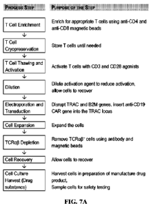
FIGs. 7A-7E include diagrams showing data from manufacturing of genetically
engineered T cells expressing an anti-CD19 directed chimeric T cell antigen
receptor (CTX110).
25 FIG. 7A includes a flow chart of an illustrative manufacturing process
for making T cells
expressing an anti-CD19 CAR, in accordance with some embodiments of the
technology
described herein. FIGs. 7B-7C include diagrams showing CARP expression in T
cells
transduced with varying MOI. FIG. 7B: a graph showing CAR+ expression in T
cells transduced
with rAAV-138 MOI ranging from OK to 80K. FIG. 7C: a graph showing CARP
expression in
30 T cells transduced with rAAV-138 MOI ranging from OK to 80K.
Transduction with rAAV-138
MOI of 20K was used as a positive control. FIGs. 7D-7E include diagrams
showing editing
efficiency in T cells electroporated with RNP complexes formed from different
concentrations of
sgRNA targeting TCR (TA-1 sgRNA) or sgRNA targeting B2M (B2M-1 sgRNA). TCRaP-:
percent of cells having TCRap edits; p2M-: percent of cells having 132M edits;
and double
CA 03158122 2022-5-11
WO 2021/095013
PCT/1112020/060723
knockout (DKO): percent of cells having TCRaff edits and (32M edits. FIG. 71):
a graph
showing knockout efficiency in T cells electroporated with RNP complexes
formed using 37.5
pg/mL to 300 pg/mL of TA-1. FIG. 7E: a graph showing knockout efficiency in T
cells
electroporated with RNP complexes formed using 37.5 pg/mL to 300 pg/mL of B2M-
1.
5 FIGs. 8A-8G include diagrams showing data from manufacturing of
genetically
engineered T cells expressing an anti-BCMA directed chimeric T cell antigen
receptor
(CTX120). FIG. 8A includes a flow chart of an illustrative manufacturing
process for making T
cells expressing an anti-BCMA CAR, in accordance with some embodiments of the
technology
described herein. FIG. 8B: a graph showing CARP expression in T cells
transduced with
10 increasing MOI. FIG. 8C: a graph showing levels of exhaustion markers
detected in CTX120.
FIG. 8D: a graph showing levels of memory markers detected in CDS' T cells of
CTX120.
FIG. 8E: a graph showing levels of memory markers detected in CD4+ T cells of
CTX120. FIG.
8F: a graph showing production of IFNy upon co-culture of CTX120 with BCMA+
tumor cells.
FIG. 8G: a graph showing tumor killing upon co-culture of CTX120 with BCMA+
tumor cells.
15 FIGs. 9A and 9B provide graphs of cell concentration per mL as a
function of days of
expansion post editing.
FIGs. 10A and 10B provide graphs of calculated cell number as a function of
days of
expansion post editing.
FIGs. 11A and 11B provide graphs of percentage cell viability as a function of
days of
20 expansion post editing.
FIGs. 12A-12C provide graphs of depicting editing efficiency including CAR1%
(FIG.
12A), TRAC-% (FIG. 12B) and I32M-% (FIG. 12C) assessed in the various
replating and low-
plating groups.
FIGs. 13A and 13B provide the ratio of CD4+ and CD8t cells in the various
replated cell
25 populations.
FIGs. 14A-14F provide bar graphs depicting the assessment of memory cell
subtype
markers in the replated populations. The cells in the replated populations
were assessed as naive
T cells, central memory (CM) T cells, effector memory (EM) T cells and
terminal effector (TE)
T cells.
30 FIGs. 154-15F provide bar graphs depicting the assessment of
exhaustion markers in the
replated populations of CARP, CD4+/CAR+, and CD8+/CAR+ cells. The three
exhaustion
markers assayed were PD1, LAG3 and TIM3.
6
CA 03158122 2022-5-11
WO 2021/095013
PCT/1112020/060723
FIGs. 164-16C provide graphs showing the ability of the CAR-T cells in
replated and
low-plating density groups to kill CD19 positive Raji target cells in vitro,
which was assessed
using a flow cytometry-based cytotoxicity assay.
FIGs. 17A-17D provide graphs showing the percentage of survival of tumor cells
as a
5 function of days post inoculation at three different doses of CAR cells
in viva
FIGs. 18A-18D provides graphs showing the tumor mass in mice as a function of
days
post inoculation at three dose of CAR cells in vivo.
FIG. 19 shows a flow chart illustrating one embodiment of the present
disclosure.
FIG. 20 shows an assay control FACS analysis by measuring CAR T-cell lysis.
The
10 CAR T-cells were CTX110 CAR T-cells. 81% of the T-cells were CAR'.
FIGs. 214-21C show the results of an assay control experiment measuring cell
lysis and
cytokine production in vitro. The assay used CTX110 CAR-T cells thawed from
frozen stock.
The T-cells were 80% CARS day 6 post HDR.
FIGs. 22A-22C show the results of an in vitro efficacy analysis showing that T-
cells
15 derived from each of the three donors had varying degrees of in vitro
efficacy among lx, 2x and
4x culture conditions.
FIGs. 23A-23C show the results of an analysis of cell lysis at different cell
concentrations, demonstrating that cells derived from donors 1 and 2 showed
similar responses
despite differing percentages of CARP cells.
20
FIGs. 24A-24B show the results of an analysis of
cell lysis from the three donors when
normalized for CARt cells. Donors 2 and 3 behaved similarly in the assay when
CAR cells were
normalized. The assay was repeated with 2x CAR-T cell number for donor 2 at
the same E:T
ratios.
FIGs. 25A-25C provide survival curves showing the percentage of survival of
mice as a
25 function of days post inoculation of CAR cells for all three donors and
expansion conditions in
viva
FIGs. 26A-26C provide graphs showing the tumor mass in mice as a function of
days
post inoculation of CAR cells from all three donors and expansion conditions
in viva
30 DETAILED DESCRIPTION OF THE INVENTION
The present disclosure is based, at least in part, on the development of
improved
manufacturing processes for producing CAR-T cells, particularly allogenic CAR-
T cells,
including improved conditions for one or more steps of the manufacturing
processes. The
7
CA 03158122 2022-5-11
WO 2021/095013
PCT/1B2020/060723
improved manufacturing processes disclosed herein led to at least the
following advantageous
outcomes:
(a) Improved T cell purity and improved T cell
viability resulting from the improved
T cell enrichment conditions provided herein.
5 (b) Improved consistency and improved efficiency for
producing CAR-expressing T
cells resulting from the improved T cell transduction conditions provided
herein.
(c) Improved consistency and improved
efficiency of TRAC gene and fl2M gene
disruptions in T cells resulting from the improved CRISPR-Cas9-mediated gene
editing
conditions provided herein.
10 (d) Increased supply of CAR T-cell therapy resulting from
decreased production
times and decreased production costs provided by the improved manufacturing
processes
described herein.
(e) Reduced variability of manufactured drug
product resulting from production of
uniform and high quality CAR T-therapies using the improved manufacturing
processes
15 described herein.
(0 Simplified AAV transduction condition
while maintaining high CAR expression
level in T cells.
Accordingly, provided herein are methods for manufacturing genetically
engineered T
cells expressing a CAR construct, such as a CAR construct targeting a cancer
antigen, for
20 example, CD19 or BCMA, and having ?RAC and fl2M gene knocked-out. The
genetically
engineered T cell populations produced by methods described herein, and
therapeutic uses
thereof are also within the scope of the present disclosure.
I. Manufacturing Genetically Engineered T Cells
25 Aspects of the present disclosure provide methods for
manufacturing genetically
engineered T cells comprising a disrupted beta-2-microglobulin (/32M) gene,
and a disrupted T
cell receptor alpha chain constant region (TRAC) gene, and an inserted nucleic
acid encoding a
chimeric antigen receptor (CAR).
Disruption of the I32M gene and the TRAC gene renders the genetically
engineered T cell
30 non-alloreactive and suitable for allogeneic transplantation. Insertion
of a nucleic acid encoding
a CAR enables the genetically engineered T cell to express the CAR on its
surface where it
targets the genetically engineered T cell to cancer cells.
Accordingly, methods for manufacturing genetically engineered T cells
disclosed herein,
in some embodiments, involve the use of CRISPR-Cas9 gene editing to disrupt
expression of
8
CA 03158122 2022-5-11
WO 2021/095013
PCT/1112020/060723
TRAC and I32M, and the use of adeno-associated virus (AAV) transduction to
insert a nucleic
acid encoding a CAR.
In general, the method for manufacturing CAR-T cells disclosed herein may
comprise: (i)
enriching CD4+/CD8+ T cells from a suitable human immune cell source, (ii)
activating the
5 enriched CD4+/CD8+ T cells, and (iii) genetically engineering the
activated T cells to produce
CAR-T cells having disrupted TRAC and B2M genes; and harvesting the
genetically engineered
T cells for therapeutic uses. When needed, the enriched CD4+/CD8+ T cells may
be stored via
cryopreservation for future use. Alternatively or in addition, the genetically
engineered T cells
may be expanded in vitro prior to harvesting. TCRal3+ T cells may be depleted
from the CAR-T
10 cell population thus produced.
(i) T Cell Enrichment
Any of the manufacturing methods disclosed herein may use human blood cells as
the
starting material. For example, T cells can be obtained from a unit of blood
collected from a
subject using techniques known to a skilled person, such as sedimentation,
e.g., FICOLLTM
15 separation. Alternatively, the T cells for use in making the genetically
engineered T cells may be
derived from stem cells (e.g., HSCs or iPSCs) via in vitro differentiation. In
some embodiments,
blood cells can be obtained from an individual human donor. In other
embodiments, blood cells
can be obtained from multiple human donors (e.g., 2, 3, 4, or 5 human donors).
In some examples, leukopak samples from a suitable human donor may be used. As
20 known in the art, a leukopak sample is an enriched leukapheresis product
collected from
peripheral blood. It typically contains a variety of blood cells including
monocytes,
lymphocytes, platelets, plasma, and red cells. The human donor preferably is a
healthy human
donor. For example, a human donor candidate may be subject to screening for
HBV, HCV, HIV,
HTLV, WNV, trypanosoma cruzi, and/or CMV. A human subject showing negative
results in
25 the screening may be used as a donor for blood cells.
The sources of T-cells that find use in the present methods is not
particularly limited. In
some embodiments, T cells from a T cell bank can be used as the starting
material in any of the
manufacturing methods disclosed herein. A T cell bank may comprise T cells
with genetic
editing of certain genes (e.g., genes involved in cell self renewal,
apoptosis, and/or T cell
30 exhaustion or replicative senescence) to improve T cell persistence in
cell culture. A T cell bank
may be produced from bonafide T cells, for example, non-transformed T cells,
terminally
differentiated T cells, T cells having stable genome, and/or T cells that
depend on cytokines and
growth factors for proliferation and expansion. Alternatively, such a T cell
bank may be
9
CA 03158122 2022-5-11
WO 2021/095013
PCT/1112020/060723
produced from precursor cells such as hematopoietic stem cells (e.g., iPSCs),
e.g., in vitro
culture. In some examples, the T cells in the T cell bank may comprise genetic
editing of one or
more genes involved in cell self-renewal, one or more genes involved in
apoptosis, and/or one or
more genes involved in T cell exhaustion, so as to disrupt or reduce
expression of such genes,
5 leading to improved persistence in culture. Examples of the edited genes
in a T cell bank include,
but are not limited to, Tet2, Fas, CD70, Regnase-1, or a combination thereof.
Compared with the
non-edited T counterpart, T cells in a T cell bank may have enhanced expansion
capacity in
culture, enhanced proliferation capacity, greater T cell activation, and/or
reduced apoptosis
levels.
10
Suitable T cells can be enriched from human
blood cells using conventional methods or
methods disclosed herein. T cells for use in making the genetically engineered
T cells may
express one or more of the T cell markers, including, but not limited to a
CD4+, CD8+, or a
combination thereof. In some embodiments, CD4+ T cells can be enriched from
human blood
cells. In other embodiments, CD8+ T cells can be enriched. In specific
examples, both CD4+
15 and CDS+ T cells are purified from human blood cells.
CD4+ T cells and/or CD8+ T cells can be isolated from a suitable blood cell
source, such
as those described herein, using any method known in the art or those
disclosed herein, for
example, using antibodies capable of binding to specific cell-surface
biomarkers for the target T
cells, e.g., antibodies specific to CD4 and/or antibodies specific to CD8. In
some embodiments,
20 enriching CD4+ T cells and CDS+ T cells can be performed using anti-CD4
and anti-CD8
antibodies conjugated to magnetic beads. A cell population comprising CD4+ and
CD81- T cells
can be incubated with such magnetic beads under suitable conditions for a
suitable period
allowing for binding of the target T cells to the magnetic beads via the
antibodies conjugated to
the beads. Non-bound cells can be washed and CD4* and CD8* T cells bound to
the beads can
25 be collected using routine methods.
The enriched T cells (e.g., CD4+ T cells and CD8+ T cells) may be evaluated
for
features such as cell viability and/or purity of the target T cells following
routine practice. In
some embodiments, the T cell population from the enrichment step disclosed
here may have a
cell viability of at least about 80% (e.g., at least about 85%, at least about
90%, at least about
30 95%, or above). Alternatively or in addition to, the enriched T cell
population may have a
purity of at least about 80% of the target T cells (e.g., CD4+ and/or CDS+ T
cells), for
example, at least about 85%, at least about 90%, at least about 95%, at least
about 97%, about
98% or higher. Alternatively or in addition to, the enriched T cell population
may have a
purity of at least about 70% of the target T cells (e.g., CD4F and/or CD8 T
cells), for
CA 03158122 2022-5-11
WO 2021/095013
PCT/1112020/060723
example, at least about 75%, at least about 80%, at least about 85%, at least
about 90%, at
least about 95%, at least about 97%, about 98% or higher.
The term "about" or "approximately" means within an acceptable error range for
the
particular value as determined by one of ordinary skill in the art, which will
depend in part on
5 how the value is measured or determined, La, the limitations of the
measurement system. For
example, "about" can mean within an acceptable standard deviation, per the
practice in the
art. Alternatively, "about" can mean a range of up to 20 %, preferably up to
10 %, mom
preferably up to 5 %, and more preferably still up to 1 % of a given
value. Alternatively,
particularly with respect to biological systems or processes, the term can
mean within an order
10 of magnitude, preferably within 2-fold, of a value. Where particular
values are described in
the application and claims, unless otherwise stated, the term "about" is
implicit and in this
context means within an acceptable error range for the particular value.
The enriched T cell population (which is also within the scope of the present
disclosure) may be used immediately for further processing as disclosed
herein.
15 Alternatively, the enriched T cell population may be stored under
suitable conditions for
future use, for example, via cryopreservation. Prior to further processing,
cryopreserved T
cells can be thawed following routine procedures. Cell viability of the thawed
cells can be
assessed to determine whether the thawed cells are suitable for further
processing.
(ii) T Cell Activation
20 The enriched T cells may be subject to T cell activation to allow
for proliferation and
expansion of the enriched CD4j/CD8+ T cells. The T cell activation step used
in any of the
methods disclosed herein may involve T cell activation conditions disclosed
herein that provide
high T cell activation efficiency. Further, the activated T cells obtained
therefrom would exhibit
high gene editing efficiencies and great rates of T cell expansion post
editing. See Examples
25 below.
In some embodiments, T cell activation can be achieved using a T cell
activating agent or
agents, for example, agents that stimulates a CD3/TCR-mediated signaling
pathway and/or a co-
stimulatory molecule (e.g., CD28) mediated signaling pathway. For example, a T
cell activating
agent may be a CD3 agonist (e.g., an agonistic anti-CD3 antibody) and
activates the CD3fICR-
30 mediated cell signaling pathway. Alternatively or in addition, a T cell
activating agent may be a
CD28 agonist (e.g., an anti-CD28 antibody) and activate the co-stimulatory
signaling pathway
mediated by CD28. Any of the T cell activating agents for use in the method
disclosed herein
may be conjugated to a support member, such as a nanomatrix particle. In
specific examples, the
T cell activating agent for use in the method disclosed herein may comprise an
anti-CD3
11
CA 03158122 2022-5-11
WO 2021/095013
PCT/1112020/060723
antibody and an anti-CD28 antibody, which may be conjugated to nanomatrix
particles. In some
embodiments, the T cell activating agent comprises a CD3 agonist and a CD28
agonist attached
to a nanomatrix particle. In some embodiments, the CD3 agonist and a CD28
agonist are
attached to the same nanomatrix particle. In some embodiments, the CD3 agonist
and a CD28
5 agonist are attached to different nanomatrix particles.
To achieve T cell activation, the enriched T cells as disclosed herein (e.g.,
CD41-/CD8+ T
cells) may be placed in a cell culture vessel at a suitable cell seeding
density and a suitable cell
concentration and incubated in the presence of any of the T cell activating
agents disclosed
herein for a suitable period to induce T cell activation.
10 In some instances, ratios of the T cell activating agent to the
cell culture medium in the
cell culture vessel may range from about 1:10 (v/v) to about 1:15 (v/v). In
some examples, the
ratio of the T cell activating agent to the cell culture medium in the cell
culture vessel may be
about 1:10 (v/v), about 1:10.5 (v/v), about 1:11 (v/v), about 1:11.5 (v/v),
about 1:12 (v/v), about
1:12.5 (v/v), about 1:13 (v/v), about 1:13.5 (v/v), about 1:14 (v/v), about
1:14.5 (v/v), or about
15 1:15 (v/v). In specific examples, the ratio of the T cell activating
agent to the culture medium in
the cell culture vessel is about 1:12.5 (v/v).
Alternatively or in addition, a suitable cell seeding density may be about 1.5
x 106 to 2.5
x 106 (e.g., 2x1(P/cm2) and a suitable cell concentration may be about 1.5 x
106 to 2.5 x 10⁶ (e.g.,
2x10⁶/m1). The cells may be incubated with the T cell activating agent for
about 42-54 hours, for
20 example, about 48 hours.
In some embodiments, the cell culture vessel may be a static culture vessel,
which would
allow for relatively large-scale production of the genetically engineered T
cells as disclosed
herein. Compared to conventional cell culture flasks, static cell culture
vessels allow T cells to
reside on a highly gas permeable membrane submerged under medium that supplies
oxygen and
25 nutrients to the T cells without mixing or shaking. Static culture
vessels allow T cell
manufacturing without medium change. Accordingly, in some embodiments, the T
cell
activation process in any of the methods disclosed herein may involve no
medium change.
When needed, the activating agent may be removed from the cell culture vessel
or diluted
prior to the follow-on gene editing events to minimize any potential impact
that the activating
30 agent may confer during gene editing. In some embodiments, the
activating agent can be
removed from the cell culture vessel using routine methods, e.g.,
centrifugation. Alternatively,
the activating agent may be diluted in the cell culture vessel prior to gene
editing, e.g., diluted by
addition of media to the cell culture vessel.
In some embodiments, the activated T cells derived from any of the T cell
activation
12
CA 03158122 2022-5-11
WO 2021/095013
PCT/1112020/060723
processes disclosed herein may be cultured overnight (e.g., about 16 hours) to
allow T cells to
recover prior to gene editing. In some instances, the activated T cell culture
may still contain the
T activating agent. In other instances, the activated T cells may have little
or no presence of the
T cell activating agent.
5 (iii) CRISPR-CA 59-Mediated Gene Editing of Activated T Celts
The activated T cells prepared by any of the procedures disclosed herein may
subject to
gene editing to knock out host response related genes, for example, the TRAC
gene and/or the
132111 gene, via, for example, CRISPR-Cas9 gene editing technology,
The TRAC gene encodes a component of the TCR complex. Disruption of the TRAC
10 gene leads to loss of function of the TCR and renders the engineered T
cell non-alloreactive and
suitable for allogeneic transplantation, minimizing the risk of graft versus
host disease. The PM
gene encodes a common (invariant) component of the major histocompatibility
complex (MHC)
I complexes. Disrupting the PM gene can prevent host versus therapeutic
allogeneic T cells
responses. Knocking out both the TRAC gene and the I32M gene would result in
production of
15 allogeneic T cells for use in cell therapy.
CR1SPR-Cas9-Mediated Gene Editing System
The CRISPR-Cas9 system is a naturally-occurring defense mechanism in
prokaryotes
that has been repurposed as an RNA-guided DNA-targeting platform used for gene
editing. It
relies on the DNA nuclease Cas9, and two noncoding RNAs, crisprRNA (crRNA) and
trans-
20 activating RNA (tracrRNA), to target the cleavage of DNA. CRISPR is an
acronym for
Clustered Regularly Interspaced Short Palindromic Repeats, a family of DNA
sequences found
in the genomes of bacteria and archaea that contain fragments of DNA (spacer
DNA) with
similarity to foreign DNA previously exposed to the cell, for example, by
viruses that have
infected or attacked the prokaryote. These fragments of DNA are used by the
prokaryote to
25 detect and destroy similar foreign DNA upon re-introduction, for
example, from similar viruses
during subsequent attacks. Transcription of the CRISPR locus results in the
formation of an
RNA molecule comprising the spacer sequence, which associates with and targets
Cas (CRISPR-
associated) proteins able to recognize and cut the foreign, exogenous DNA.
Numerous types and
classes of CRISPR/Cas systems have been described (see, e.g., Koonin et al.,
(2017) Curr Opin
30 Microbiol 37:67-78).
crRNA drives sequence recognition and specificity of the CRISPR-Cas9 complex
through Watson-Crick base pairing typically with a 20 nucleotide (nt) sequence
in the target
DNA. Changing the sequence of the 5' 20nt in the crRNA allows targeting of the
CRISPR-Cas9
13
CA 03158122 2022-5-11
WO 2021/095013
PCT/1112020/060723
complex to specific loci. The CRISPR-Cas9 complex only binds DNA sequences
that contain a
sequence match to the first 20 nt of the crRNA, if the target sequence is
followed by a specific
short DNA motif (with the sequence NGG) referred to as a protospacer adjacent
motif (PAM).
TracrRNA hybridizes with the 3+ end of crRNA to form an RNA-duplex structure
that is
5 bound by the Cas9 endonuclease to form the catalytically active CRISPR-
Cas9 complex, which
can then cleave the target DNA.
Once the CRISPR-Cas9 complex is bound to DNA at a target site, two independent
nuclease domains within the Cas9 enzyme each cleave one of the DNA strands
upstream of the
PAM site, leaving a double-strand break (DSB) where both strands of the DNA
terminate in a
10 base pair (a blunt end).
After binding of CRISPR-Cas9 complex to DNA at a specific target site and
formation of
the site-specific DSB, the next key step is repair of the DSB. Cells use two
main DNA repair
pathways to repair the DSB: non-homologous end joining (NHEJ) and homology-
directed repair
(HDR).
15 NHEJ is a robust repair mechanism that appears highly active in
the majority of cell
types, including non-dividing cells. NHEJ is error-prone and can often result
in the removal or
addition of between one and several hundred nucleotides at the site of the
DSB, though such
modifications are typically < 20 nt. The resulting insertions and deletions
(indels) can disrupt
coding or noncoding regions of genes. Alternatively, HDR uses a long stretch
of homologous
20 donor DNA, provided endogenously or exogenously, to repair the DSB with
high fidelity. MDR
is active only in dividing cells, and occurs at a relatively low frequency in
most cell types. In
many embodiments of the present disclosure, NHEJ is utilized as the repair
operant.
(i) Cas9
In some embodiments, the Cas9 (CRISPR associated protein 9) endonuclease is
used in a
25 CRISPR method for making the genetically engineered T cells as disclosed
herein. The Cas9
enzyme may be one from Streptococcus pyogenes, although other Cas9 homologs
may also be
used. It should be understood that wild-type Cas9 may be used or modified
versions of Cas9
may be used (e.g., evolved versions of Cas9, or Cas9 orthologues or variants),
as provided
herein. In some embodiments, Cas9 comprises a Streptococcus pyogenes-derived
Cas9 nuclease
30 protein that has been engineered to include C- and N-terminal SV40 large
T antigen nuclear
localization sequences (NLS). The resulting Cas9 nuclease (sNLS-spCas9-sNLS)
is a 162 kDa
protein that is produced by recombinant E. coil fermentation and purified by
chromatography.
The spCas9 amino acid sequence can be found as UniP'rot Accession No. Q99ZW2,
which is
14
CA 03158122 2022-5-11
WO 2021/095013
PCT/1112020/060723
provided herein as SEQ ID NO: 1.
(ii) Guide RNAs (gRNAs)
CRISPR-Cas9-mediated gene editing as described herein includes the use of a
guide
RNA or a gRNA. As used herein, a "gRNA" refers to a genome-targeting nucleic
acid that can
5 direct the Cas9 to a specific target sequence within a TRAC gene or a a
gene for gene editing
at the specific target sequence. A guide RNA comprises at least a spacer
sequence that
hybridizes to a target nucleic acid sequence within a target gene for editing,
and a CRISPR
repeat sequence.
An exemplary gRNA targeting a TRAC gene is provided in SEQ ID NO: 2. See also
10 International Application Na PCT/IB2018/001619, filed May 11, 2018,
which published as WO
2019/097305A2, the relevant disclosures of which are incorporated by reference
herein for the
subject matter and purpose referenced herein. Other gRNA sequences may be
designed using
the TRAC gene sequence located on chromosome 14 (GRCh38: chromosome 14:
22,547,506-
22,552,154; Ensembl; EN5G00000277734). In some embodiments, gRNAs targeting
the
15 TRAC genomic region and Cas9 create breaks in the TRAC genomic region
resulting Indels in
the TRAC gene disrupting expression of the mRNA or protein.
In some embodiments, gRNAs targeting the TRAC genomic region create Indels in
the
TRAC gene comprising at least one nucleotide sequence selected from the
sequences in Table 9.
In some embodiments, gRNA (SEQ ID NO: 2) targeting the TRAC genomic region
create Indels
20 in the TRAC gene comprising at least one nucleotide sequence selected
from the sequences in
Table 9.
An exemplary gRNA targeting a fl2M gene is provided in SEQ ID NO: 6. See also
International Application Na PCT/IB2018/001619, filed May 11, 2018, which
published as WO
2019/097305A2, the relevant disclosures of which are incorporated by reference
herein for the
25 subject matter and purpose referenced herein. Other gRNA sequences may
be designed using
the 132M gene sequence located on Chromosome 15 (GRCh38 coordinates:
Chromosome 15:
44,711,477-44,718,877 ; Ensembl: ENSG00000166710). In some embodiments, gRNAs
targeting the 32M genomic region and RNA-guided nuclease create breaks in the
132M genomic
region resulting in Indels in the fi2M gene disrupting expression of the mRNA
or protein.
30 In some embodiments, gRNAs targeting the /32M genomic region
create Indels in the
fl2M gene comprising at least one nucleotide sequence selected from the
sequences in Table 10.
In some embodiments, gRNA (SEQ ID NO: 6) targeting the fl2M genomic region
create Indels
in the fi2M gene comprising at least one nucleotide sequence selected from the
sequences in
CA 03158122 2022-5-11
WO 2021/095013
PCT/1112020/060723
Table 10.
In Type II systems, the gRNA also comprises a second RNA called the tracrRNA
sequence. In the Type II gRNA, the CRISPR repeat sequence and tracrRNA
sequence hybridize
to each other to form a duplex. In the Type V gRNA, the crRNA forms a duplex.
In both
5 systems, the duplex binds a site-directed polypeptide, such that the
guide RNA and site-direct
polypeptide form a complex. In some embodiments, the genome-targeting nucleic
acid provides
target specificity to the complex by virtue of its association with the site-
directed polypeptide.
The genome-targeting nucleic acid thus directs the activity of the site-
directed polypeptide.
As is understood by the person of ordinary skill in the art, each guide RNA is
designed to
10 include a spacer sequence complementary to its genoinic target sequence.
See Jinek et al.,
Science, 337, 816-821 (2012) and Deltcheva et al., Nature, 471, 602-607
(2011).
In some embodiments, the genome-targeting nucleic acid (a g., gRNA) is a
double-
molecule guide RNA. In some embodiments, the genome-targeting nucleic acid
(e.g., gRNA) is
a single-molecule guide RNA.
15 A double-molecule guide RNA comprises two strands of RNA
molecules. The first
strand comprises in the 5' to 3' direction, an optional spacer extension
sequence, a spacer
sequence and a minimum CRISPR repeat sequence. The second strand comprises a
minimum
tracrRNA sequence (complementary to the minimum CRISPR repeat sequence), a 3'
tracrRNA
sequence and an optional tracrRNA extension sequence.
20 A single-molecule guide RNA (referred to as a "sgRNA") in a Type
II system comprises,
in the 5' to 3' direction, an optional spacer extension sequence, a spacer
sequence, a minimum
CRISPR repeat sequence, a single-molecule guide linker, a minimum tracrRNA
sequence, a 3'
tracrRNA sequence and an optional tracrRNA extension sequence. The optional
tracrRNA
extension may comprise elements that contribute additional functionality
(e.g., stability) to the
25 guide RNA. The single-molecule guide linker links the minimum CRISPR
repeat and the
minimum tracrRNA sequence to form a hairpin structure. The optional tracrRNA
extension
comprises one or more hairpins. A single-molecule guide RNA in a Type V system
comprises,
in the 5' to 3' direction, a minimum CRISPR repeat sequence and a spacer
sequence.
The "target sequence" is in a target gene that is adjacent to a PAM sequence
and is the
30 sequence to be modified by Cas9. The "target sequence" is on the so-
called PAM-strand in a
"target nucleic acid," which is a double-stranded molecule containing the PAM-
strand and a
complementary non-PAM strand. One of skill in the art recognizes that the gRNA
spacer
sequence hybridizes to the complementary sequence located in the non-PAM
strand of the target
16
CA 03158122 2022-5-11
WO 2021/095013
PCT/1112020/060723
nucleic acid of interest. Thus, the gRNA spacer sequence is the RNA equivalent
of the target
sequence.
For example, if the TRAC target sequence is 5'-AGAGCAACAGTGCTGTGGCC-3'
(SEQ ID NO: 11), then the gRNA spacer sequence is 5"- AGAGCAACAGUGCUGUG-GCC-3'
5 (SEQ ID NO: 5). In another example, if the 132M target sequence is 5'-
GCTACTCTCTCITTCMGCC-3' (SEQ ID NO: 13), then the gRNA spacer sequence is 5'
-
GCUACUCUCUCUUUCUGGCC-3' (SEQ ID NO: 9). The spacer of a gRNA interacts with a
target nucleic acid of interest in a sequence-specific manner via
hybridization (i.e., base pairing).
The nucleotide sequence of the spacer thus varies depending on the target
sequence of the target
10 nucleic acid of interest.
In a CRISPFJCas system herein, the spacer sequence is designed to hybridize to
a region
of the target nucleic acid that is located 5' of a PAM recognizable by a Cas9
enzyme used in the
system. The spacer may perfectly match the target sequence or may have
mismatches. Each
Cas9 enzyme has a particular PAM sequence that it recognizes in a target DNA.
For example, S.
15 pyogenes recognizes in a target nucleic acid a PAM that comprises the
sequence 5'-NRG-3',
where R comprises either A or G, where N is any nucleotide and N is
immediately 3' of the
target nucleic acid sequence targeted by the spacer sequence.
In some embodiments, the target nucleic acid sequence has 20 nucleotides in
length. In
some embodiments, the target nucleic acid has less than 20 nucleotides in
length. In some
20 embodiments, the target nucleic acid has more than 20 nucleotides in
length. In some
embodiments, the target nucleic acid has at least: 5, 10, 15, 16, 17, 18, 19,
20, 21, 22, 23, 24, 25,
30 or more nucleotides in length. In some embodiments, the target nucleic acid
has at most: 5,
10, 15, 16, 17, 18, 19, 20, 21, 22, 23, 24, 25, 30 or more nucleotides in
length. In some
embodiments, the target nucleic acid sequence has 20 bases immediately 5' of
the first nucleotide
25 of the PAM. For example, in a sequence comprising 5'-
NNNNNNNNNNNNNNNNNNNNNRG-3', the target nucleic acid can be the sequence that
corresponds to the Ns, wherein N can be any nucleotide, and the underlined NRG
sequence is the
S. pyogenes PAM.
A spacer sequence in a gRNA is a sequence (e.g., a 20 nucleotide sequence)
that defines
30 the target sequence (e.g., a DNA target sequences, such as a genomic
target sequence) of a target
gene of interest. An exemplary spacer sequence of a gRNA targeting a 7RAC gene
is provided
in SEQ ID NO: 4. An exemplary spacer sequence of a gRNA targeting a/32M gene
is provided in
SEQ ID NO:8.
17
CA 03158122 2022-5-11
WO 2021/095013
PCT/1112020/060723
The guide RNA disclosed herein may target any sequence of interest via the
spacer
sequence in the crRNA. In some embodiments, the degree of complementarity
between the
spacer sequence of the guide RNA and the target sequence in the target gene
can be about 60%,
65%, 70%, 75%, 80%, 85%, 90%, 95%, 97%, 98%, 99%, or 100%. In some
embodiments, the
5 spacer sequence of the guide RNA and the target sequence in the target
gene is 100%
complementary. In other embodiments, the spacer sequence of the guide RNA and
the target
sequence in the target gene may contain up to 10 mismatches, e.g., up to 9, up
to 8, up to 7, up to
6, up to 5, up to 4, up to 3, up to 2, or up to 1 mismatch.
Non-limiting examples of gRNAs that may be used as provided herein are
provided in
10 International Application No. PCT/IB2018/001619, filed May 11, 2018,
which published as WO
2019/097305A2, and International Application No. PCT/IB2019/000500, filed May
10, 2019,
which published as WO/2019/215500. the relevant disclosures of each of the
prior applications
are herein incorporated by reference for the purposes and subject matter
referenced herein. For
any of the gRNA sequences provided herein, those that do not explicitly
indicate modifications
15 are meant to encompass both unmodified sequences and sequences having
any suitable
modifications.
The length of the spacer sequence in any of the gRNAs disclosed herein may
depend on
the CRISPWCas9 system and components used for editing any of the target genes
also disclosed
herein. For example, different Cas9 proteins from different bacterial species
have varying
20 optimal spacer sequence lengths. Accordingly, the spacer sequence may
have 5, 6, 7, 8, 9, 10,
11, 12, 13, 14, 15, 16, 17, 18, 19, 20, 21, 22, 23, 24, 25, 26, 27, 28, 29,
30, 35, 40, 45, 50, or
more than 50 nucleotides in length. In some embodiments, the spacer sequence
may have 18-24
nucleotides in length. In some embodiments, the targeting sequence may have 19-
21 nucleotides
in length. In some embodiments, the spacer sequence may comprise 20
nucleotides in length.
25 In some embodiments, the gRNA can be a sgRNA, which may comprise
a 20 nucleotide
spacer sequence at the 5' end of the sgRNA sequence. In some embodiments, the
sgRNA may
comprise a less than 20 nucleotide spacer sequence at the 5' end of the sgRNA
sequence. In
some embodiments, the sgRNA may comprise a more than 20 nucleotide spacer
sequence at the
5' end of the sgRNA sequence. In some embodiments, the sgRNA comprises a
variable length
30 spacer sequence with 17-30 nucleotides at the 5' end of the sgRNA
sequence. Examples are
provided in Table 8 in Example 7.
In some embodiments, the sgRNA comprises no uracil at the 3' end of the sgRNA
sequence. In other embodiments, the sgRNA may comprise one or more uracil at
the 3' end of
the sgRNA sequence. For example, the sgRNA can comprise 1-8 uracil residues,
at the 3' end of
18
CA 03158122 2022-5-11
WO 2021/095013
PCT/1112020/060723
the sgRNA sequence, e.g., 1, 2, 3, 4, 5, 6, 7, or 8 uracil residues at the 3'
end of the sgRNA
sequence.
Any of the gRNAs disclosed herein, including any of the sgRNAs, may be
unmodified.
Alternatively, it may contain one or more modified nucleotides and/or modified
backbones. For
5 example, a modified gRNA such as an sgRNA can comprise one or more 2'-0-
methyl
phosphorothioate nucleotides, which may be located at either the 5' end, the
3' end, or both.
In certain embodiments, more than one guide RNAs can be used with a CRISPR/Cas
nuclease system. Each guide RNA may contain a different targeting sequence,
such that the
CRISPR/Cas system cleaves more than one target nucleic acid. In some
embodiments, one or
10 more guide RNAs may have the same or differing properties such as
activity or stability within
the Cas9 RNP complex. Where more than one guide RNA is used, each guide RNA
can be
encoded on the same or on different vectors. The promoters used to drive
expression of the more
than one guide RNA is the same or different.
It should be understood that more than one suitable Cas9 and more than one
suitable
15 gRNA can be used in methods described herein, for example, those known
in the art or disclosed
herein. In some embodiments, methods comprise a Cas9 enzyme and/or a gRNA
known in the
art. Examples can be found in, e.g., International Application No.
PCT/IB2018/001619, filed
May 11, 2018, which published as WO 2019/097305A2, and International
Application No.
PCT/1132019/000500, filed May 10, 2019, which published as W0/2019/215500, the
relevant
20 disclosures of each of the prior applications are herein incorporated by
reference for the purposes
and subject matter referenced herein.
CRISPR-Cas9-Mediated Gene Editing of TRAC and B2M Genes
In some embodiments, the activated T cells as disclosed herein may subject to
gene
25 editing of both the TRAC gene and /32M gene via CRISPR-Cas9-mediated
gene editing under
conditions disclosed herein, which would result in higher and more consistent
gene editing
efficiencies compared to those provided by conventional conditions. Further,
the TRAC//32M- T
cells obtained from the gene editing process disclosed herein showed high
expression level of a
chimeric antigen receptor (CAR) when a viral vector coding for the CAR
construct is delivered
30 into the TRACW2M- T cells.
The Cas9 enzyme and the gRNAs targeting the TRAC gene and f32M gene may form
one
or more ribonucleoprotein (RNP) complexes, which can be delivered into the
activated T cells as
disclosed herein. RNPs are useful for gene editing, at least because they
minimize the risk of
promiscuous interactions in a nucleic acid-rich cellular environment and
protect the RNA from
19
CA 03158122 2022-5-11
WO 2021/095013
PCT/1B2020/060723
degradation. Methods for forming RNPs are known in the art.
The CRISPR-Cas9-mediated gene editing process may involve two
ribonueleoprotein
complexes. The first RNP complex comprises a first Cas9 enzyme and a guide RNA
(gRNA)
targeting a TRAC gene. The second RNP complex comprises a second Cas9 enzyme
and a
5 gRNA targeting al32/1/ gene. In some examples, the two RNP complexes may
comprise
different Cas9 enzymes. In other examples, the two RNP complexes comprise the
same Cas9
enzyme. In specific examples, the Cas9 enzyme of SEQ ID NO:1 can be used in
both the first
and second RNPs.
In some embodiments, the two RNP complexes may contain the same amount of the
10 Cas9 enzyme. For example, both RNP complexes may comprise about 0.1-0.3
mg/m1 (e.g.,
about 0.1-0.2 mg/ml) of the Cas9 enzyme (e.g., the Cas9 enzyme of SEQ ID
NO:1). In some
examples, each of the RNP complexes may comprise about 0.15 mg/ml of the Cas9
enzyme,
which may be the Cas9 enzyme of SEQ ID NO: 1.
In other embodiments, the two RNP complexes may contain different amounts of
the
15 Cas9 enzyme. In some examples, the RNP complex targeting the TRAC gene
may comprise a
higher amount of the Cas9 enzyme relative to the RNP complex targeting the
I32M gene.
Alternatively, the RNP complex targeting the /32M gene may comprise a higher
amount of the
Cas9 enzyme relative to the RNP complex targeting the TRAC gene.
The two RNP complexes may comprise the same amount of the gRNAs (one targeting
20 TRAC and the other targeting I32M). Alternatively, the two RNP complexes
may comprise
different amounts of the gRNAs. For example, the amount of the gRNA targeting
the TRAC
gene may range from about 0.035 mg/ml to about 0.8 mg/ml, for example, about
50 jug/m1 to
about 80 jig/mi. In specific examples, the amount of the gRNA targeting the
TRAC gene is about
0.08 mg/ml. Alternatively or in addition, the amount of the gRNA targeting the
p2m gene may
25 range from about 0.075 mg/ml to about 0.3 mg/ml, for example, about 0.1
mg/ml to about 0.3
mg/ml. In specific examples, the amount of the gRNA targeting the PM gene is
about 0.2
mg/ml.
In specific examples, the RNP complex targeting the TRAC gene may comprise
about
0.15 mg/ml Cas9 (e.g., the Cas9 of SEQ ID NO:1) and about 0.08 mg/m1 of a gRNA
targeting
30 the TRAC gene (e.g., the gRNA of TA-1). Alternatively or in addition,
the RNP complex
targeting the I32M gene may comprise about 0_15 mg/nil Cas9 (e.g., the Cas9 of
SEQ ID NO:1)
and about 0.2 mg/ml of a gRNA targeting the PM gene (e.g., the gRNA of B2M-1).
In some embodiments, the two RNPs may be introduced into the activated T cell
via
CA 03158122 2022-5-11
WO 2021/095013
PCT/1B2020/060723
electroporation sequentially, Le., via two electroporation event.
Alternatively, the two RNPs
may be introduced into the activated T cells simultaneously, La, via one
electroporation event.
In this case, the two RNPs may be combined to form a mixture prior to the
electroporation event.
Any of the RNPs disclosed herein may he introduced into the activated T cells
by mixing
5 the RNP(s) with a suitable amount of the activated T cells and the
mixture thus formed is subject
to electroporation under suitable conditions allowing for delivery of the RNPs
into the cells. In
some instances, the suitable amount of the activated T cells may range from
about 100x106
cells/mL to about 300x106 cells/mL. For example, suitable amount of the T
cells for the
electroporation step may range from about 200x106 cells/mL to about 300x106
cells/mL. In
10 some examples, the concentration of the activated T cells may he about
100x106 cells/mL. In
some embodiments, the concentration of activated T cells may be about 200x106
cells/mL. In
some embodiments, the concentration of activated T cells may be about 300x106
cells/mL.
In some embodiments, the suitable amount of the activated T cells may range
from about
1x10⁸ to about lx101 cells, e.g., about 5x10⁸ to about 8x10⁹ cells, about
1x10⁹ to about 5x10⁹
15 cells, or about lx109 to about 3x10⁹ cells.
The T cells for use in electroporation may be placed in multiple cell
cassettes, depending
upon the electroporation instrument used. Suitable electroporation instruments
are known to
those skilled in the art and could include static and flow electroporators,
including the Lonza
Nucleofector, Maxcyte UT, and MaxCyte GTx. In some instances, multiple cell
cassettes may
20 be used in an electroporation process. More details are provided in
Example 10 below.
In specific examples, the two RNPs disclosed above, comprising about 0.3 mg/m1
of the
Cas9 enzyme in total (e.g., the Cas9 enzyme of SEQ ID NO:1), about 0.08 mg/m1
of the gRNA
of TA-1, and about 0.2 mg/ml of the gRNA of B2M-1, may be mixed with the
activated T cells
in the amount of about 100x106 cells/mL to about 300x106 cells/mL (e.g., about
300x106
25 cells/mL). The mixture is then subject to electroporation for delivery
of the RNPs into the T
cells.
After electroporation, the cells may be cultured in a fresh medium or
electroporation
buffer for a suitable period for recovery. Gene editing efficiency may be
performed following
routine practice. The genetically edited T cells thus produced may be
subjected to viral vector
30 transduction for delivery of a nucleic acid configured for CAR
expression.
21
CA 03158122 2022-5-11
WO 2021/095013
PCT/I132020/060723
(iv) T Cell Transduction
The genetically edited T cells, having TRAC and I32M genes knocked out, may be
subject
to transduction with a viral vector such as an adeno-associated viral (AAV)
vector that comprises
a nucleic acid sequence encoding a chimeric antigen receptor (CAR) to produce
a population of
5 T cells expressing the CAR.
Chimeric Antigen Receptor (CAR)
A chimeric antigen receptor (CAR) refers to an artificial immune cell receptor
that is
engineered to recognize and bind to an antigen expressed by undesired cells,
for example,
disease cells such as cancer cells. A T cell that expresses a CAR polypeptide
is referred to as a
10 CAR T cell. CARs have the ability to redirect T-cell specificity and
reactivity toward a selected
target in a non-MHC-restricted manner. The non-MHC-restricted antigen
recognition gives
CAR-T cells the ability to recognize an antigen independent of antigen
processing, thus
bypassing a major mechanism of tumor escape. Moreover, when expressed on T-
cells, CARs
advantageously do not dimerize with endogenous T-cell receptor (TCR) alpha and
beta chains.
15 There are various generations of CARs, each of which contains
different components.
First generation CARs join an antibody-derived scFv to the CD3zeta (C or z)
intracellular
signaling domain of the T-cell receptor through hinge and transmembrane
domains. Second
generation CARs incorporate an additional co-stimulatory domain, e.g., CD28, 4-
1BB (41BB),
or ICOS, to supply a costimulatory signal. Third-generation CARs contain two
costimulatory
20 domains (e.g., a combination of CD27, CD28, 4-1BB, ICOS, or 0X40) fused
with the TCR
CD3C chain. Maude et al., Blood. 2015; 125(26):4017-4023; Kakarla and
Gottschalk, Cancer J.
2014; 20(2):151-155). Any of the various generations of CAR constructs is
within the scope of
the present disclosure.
Generally, a CAR is a fusion polypeptide comprising an extracellular domain
that
25 recognizes a target antigen (e.g., a single-chain variable fragment
(scFv) of an antibody or other
antibody fragment) and an intracellular domain comprising a signaling domain
of the T-cell
receptor (TCR) complex (e.g., CD3C) and, in most cases, a co-stimulatory
domain. (Enblad et al.,
Human Gene Therapy. 2015; 26(8):498-505). A CAR construct may further comprise
a hinge
and transmembrane domain between the extracellular domain and the
intracellular domain, as
30 well as a signal peptide at the N-terminus for surface expression.
Examples of signal peptides
include MLLLVTSLLLCELPHPAFLLIP (SEQ ID NO: 44) and MALPVTALLLPLALLLHAARP
(SEQ ID NO: 75). Other signal peptides may be used.
(a) Antigen Binding Extracellular Domain
22
CA 03158122 2022-5-11
WO 2021/095013
PCT/IB2020/060723
The antigen-binding extracellular domain is the region of a CAR polypeptide
that is
exposed to the extracellular fluid when the CAR is expressed on cell surface.
In some instances,
a signal peptide may be located at the N-terminus to facilitate cell surface
expression. In some
embodiments, the antigen binding domain can be a single-chain variable
fragment (scFv, which
5 may include an antibody heavy chain variable region (VII) and an antibody
light chain variable
region (VL) (in either orientation). In some instances, the Vu and VL fragment
may be linked via
a peptide linker. The linker, in some embodiments, includes hydrophilic
residues with stretches
of glycine and serine for flexibility as well as stretches of glutamate and
lysine for added
solubility. The scFv fragment retains the antigen-binding specificity of the
parent antibody, from
10 which the scFv fragment is derived. hi some embodiments, the scFv may
comprise humanized
VH and/or VL domains. In other embodiments, the Vii and/or VL domains of the
scFv are fully
human.
The antigen-binding extracellular domain may be specific to a target antigen
of interest,
for example, a pathologic antigen such as a tumor antigen. In some
embodiments, a tumor
15 antigen is a "tumor associated antigen," referring to an immunogenic
molecule, such as a protein,
that is generally expressed at a higher level in tumor cells than in non-tumor
cells, in which it
may not be expressed at all, or only at low levels. In some embodiments, tumor-
associated
structures, which are recognized by the immune system of the tumor-harboring
host, are referred
to as tumor-associated antigens. In some embodiments, a tumor-associated
antigen is a universal
20 tumor antigen, if it is broadly expressed by most types of tumors. In
some embodiments, tumor-
associated antigens are differentiation antigens, mutational antigens,
overexpressed cellular
antigens or viral antigens. In some embodiments, a tumor antigen is a "tumor
specific antigen"
or "TSA," referring to an immunogenic molecule, such as a protein, that is
unique to a tumor
cell. Tumor specific antigens are exclusively expressed in tumor cells, for
example, in a specific
25 type of tumor cells.
In some examples, the CAR constructs disclosed herein comprise a scFv
extracellular
domain capable of binding to CD19. In some examples, the CAR constructs
disclosed herein
comprise a scFv extracellular domain capable of binding to BCMA. Examples of
anti-CD19
CAR and anti-BCMA CAR are provided in Examples below.
(b) Transmembrane Domain
The CAR polypeptide disclosed herein may contain a transmembrane domain, which
can
be a hydrophobic alpha helix that spans the membrane. As used herein, a
"transmembrane
domain" refers to any protein structure that is thermodynamically stable in a
cell membrane,
23
CA 03158122 2022-5-11
WO 2021/095013
PCT/IB2020/060723
preferably a eukaryotic cell membrane_ The transmembrane domain can provide
stability of the
CAR containing such.
In some embodiments, the transmembrane domain of a CAR as provided herein can
be a
CD8 transmembrane domain. In other embodiments, the transmembrane domain can
be a CD28
5 transmembrane domain. In yet other embodiments, the transmembrane domain
is a chimera of a
CD8 and CD28 transmembrane domain. Other transmembrane domains may be used as
provided
herein. In some embodiments, the transmembrane domain is a CD8a transmembrane
domain
containing the sequence of:
FVFVFLPAKPTTTPAPRPPTPAPTIASOPLSLRPEACRPAAGGAVHTRGLDFACDIYIW
10 APLAGTCGVLLLSLVITLYCNHRNR (SEQ ID NO: 49); or
IYIWAPLAGTCGVLLLSLVITLY (SEQ ID NO: 31).
Other transmembrane domains may also be used.
(c) Hinge Domain
15
In some embodiments, a hinge domain may be
located between an extracellular domain
(comprising the antigen binding domain) and a transmembrane domain of a CAR,
or between a
cytoplasmic domain and a transmembrane domain of the CAR. A hinge domain can
be any
oligopeptide or polypeptide that functions to link the transmembrane domain to
the extracellular
domain and/or the cytoplasmic domain in the polypeptide chain_ A hinge domain
may function
20 to provide flexibility to the CAR, or domains thereof, or to prevent
steric hindrance of the CAR,
or domains thereof.
In some embodiments, a hinge domain may comprise up to 300 amino acids (e.g.,
10 to
100 amino acids, or 5 to 20 amino acids). In some embodiments, one or more
hinge domain(s)
may be included in other regions of a CAR. In some embodiments, the hinge
domain may be a
25 CD8 hinge domain. Other hinge domains may be used.
(d) Intracellular Signaling Domains
Any of the CAR constructs contain one or more intracellular signaling domains
(e.g.,
CD3C, and optionally one or more co-stimulatory domains), which are the
functional end of the
30 receptor. Following antigen recognition, receptors cluster and a signal
is transmitted to the cell.
CD3C is the cytoplasmic signaling domain of the T cell receptor complex. CD3C
contains three (3) immunoreceptor tyrosine-based activation motif (ITAM)s,
which transmit an
activation signal to the T cell after the T cell is engaged with a cognate
antigen. In many cases,
24
CA 03158122 2022-5-11
WO 2021/095013
PCT/I132020/060723
CD3C provides a primary T cell activation signal but not a fully competent
activation signal,
which requires a co-stimulatory signaling.
In some embodiments, the CAR polypeptides disclosed herein may further
comprise one
or more co-stimulatory signaling domains. For example, the co-stimulatory
domains of CD28
5 and/or 4-1BB may be used to transmit a full proliferative/survival
signal, together with the
primary signalling mediated by CD3C. In some examples, the CAR disclosed
herein comprises a
CD28 co-stimulatory molecule. In other examples, the CAR disclosed herein
comprises a 4-1BB
co-stimulatory molecule. In some embodiments, a CAR includes a CD3C signaling
domain and a
CD28 co-stimulatory domain. In other embodiments, a CAR includes a CD3C
signaling domain
10 and 4-1BB co-stimulatory domain. In still other embodiments, a CAR
includes a CD3C signaling
domain, a CD28 co-stimulatory domain, and a 4-1BB co-stimulatory domain.
It should be understood that methods described herein encompasses more than
one
suitable CAR that can be used to produce genetically engineered T cells
expressing the CAR, for
example, those known in the art or disclosed herein. Examples can be found in,
e.g.,
15 PCT/IB2018/001619, filed May 11, 2018, which published as WO
2019/097305A2, the relevant
disclosures of which are incorporated by reference herein for the purpose and
subject matter
referenced herein. In another example, the CAR binds CD19 (also known as a
"CD19 CAR" or
an "anti-CD19 CAR"). The amino acid sequence of an exemplary CAR that binds
CD19 is
provided in SEQ ID NO: 37 (see Example 7 below, Table 11). In yet another
example, the CAR
20 binds BCMA (also known as a "BCMA CAR" or an "anti-BCMA CAR"). The amino
acid
sequence of an exemplary CAR that binds to BCMA is provided in SEQ ID NO: 61
(see
Example 8 below, Tables 16 and 17).
AAV Vectors for Delivery of CAR Constructs to T Cells
25 A nucleic acid encoding a CAR construct can be delivered to a
cell using an adeno-
associated virus (AAV). AAVs are small viruses which integrate site-
specifically into the host
genome and can therefore deliver a transgene, such as CAR. Inverted terminal
repeats (ITRs)
are present flanking the AAV genome and/or the transgene of interest and serve
as origins of
replication. Also present in the AAV genome are rep and cap proteins which,
when transcribed,
30 form capsids which encapsulate the AAV genome for delivery into target
cells. Surface
receptors on these capsids which confer AAV serotype, which determines which
target organs
the capsids will primarily bind and thus what cells the AAV will most
efficiently infect. There
are twelve currently known human AAV serotypes. In some embodiments, the AAV
for use in
delivering the CAR-coding nucleic acid is AAV serotype 6 (AAV6).
CA 03158122 2022-5-11
WO 2021/095013
PCT/1B2020/060723
Adeno-associated viruses are among the most frequently used viruses for gene
therapy
for several reasons. First, AAVs do not provoke an immune response upon
administration to
mammals, including humans. Second, AAVs are effectively delivered to target
cells, particularly
when consideration is given to selecting the appropriate AAV serotype.
Finally, AAVs have the
5 ability to infect both dividing and non-dividing cells because the genome
can persist in the host
cell without integration. This trait makes them an ideal candidate for gene
therapy.
A nucleic acid encoding a CAR can be designed to insert into a genomic site of
interest in
the host T cells. In some embodiments, the target genomic site can be in a
safe harbor locus.
In some embodiments, a nucleic acid encoding a CAR (e.g., via a donor
template, which
10 can be carried by a viral vector such as an adeno-associated viral (AAV)
vector) can be designed
such that it can insert into a location within a TRAC gene to disrupt the TRAC
gene in the
genetically engineered T cells and express the CAR polypeptide. Disruption of
TRAC leads to
loss of function of the endogenous TCR. For example, a disruption in the TRAC
gene can be
created with an endonuclease such as those described herein and one or more
gRNAs targeting
15 one or more TRAC genomic regions. Any of the gRNAs specific to a TRAC
gene and the target
regions can be used for this purpose, e.g., those disclosed herein.
In some examples, a genomic deletion in the TRAC gene and replacement by a CAR
coding segment can be created by homology directed repair or HDR (e.g., using
a donor
template, which may be part of a viral vector such as an adeno-associated
viral (AAV) vector).
20 In some examples, the gRNA target sequence, or portion thereof, is
deleted (e.g., SEQ ID NO:
17). In some embodiments, a disruption in the TRAC gene can be created with an
endonuclease
as those disclosed herein and one or more gRNAs targeting one or more TRAC
genomic regions,
and inserting a CAR coding segment into the TRAC gene.
A donor template as disclosed herein can contain a coding sequence for a CAR.
In some
25 examples, the CAR-coding sequence may be flanked by two regions of
homology to allow for
efficient HDR at a genomic location of interest, for example, at a TRAC gene
using CRISPR-
Cas9 gene editing technology. In this case, both strands of the DNA at the
target locus can be
cut by a CRISPR Cas9 enzyme guided by gRNAs specific to the target locus. HDR
then occurs
to repair the double-strand break (DSB) and insert the donor DNA coding for
the CAR. For this
30 to occur correctly, the donor sequence is designed with flanking
residues which am
complementary to the sequence surrounding the DSB site in the target gene
(hereinafter
"homology arms"), such as the TRAC gene. These homology arms serve as the
template for
DSB repair and allow HDR to be an essentially error-free mechanism. The rate
of homology
directed repair (HDR) is a function of the distance between the mutation and
the cut site so
26
CA 03158122 2022-5-11
WO 2021/095013
PCT/1B2020/060723
choosing overlapping or nearby target sites is important. Templates can
include extra sequences
flanked by the homologous regions or can contain a sequence that differs from
the genomic
sequence, thus allowing sequence editing.
Alternatively, a donor template may have no regions of homology to the
targeted location
5 in the DNA and may be integrated by NHEJ-dependent end joining following
cleavage at the
target site.
A donor template can be DNA or RNA, single-stranded and/or double-stranded,
and can
be introduced into a cell in linear or circular form. If introduced in linear
form, the ends of the
donor sequence can be protected (e.g., from exonucleolytic degradation) by
methods known to
10 those of skill in the art. For example, one or more dideoxynucleotide
residues are added to the 3'
terminus of a linear molecule and/or self-complementary oligonucleotides are
hgated to one or
both ends. See, for example, Chang et al., (1987) Proc. Natl. Acad. Sci. USA
84:4959-4963;
Nehls et al., (1996) Science 272:886-889. Additional methods for protecting
exogenous
polynucleotides from degradation include, but are not limited to, addition of
terminal amino
15 group(s) and the use of modified internucleotide Linkages such as, for
example,
phosphorothioates, phosphorainidates, and 0-methyl ribose or deoxyribose
residues.
A donor template can be introduced into a cell as part of a vector molecule
having
additional sequences such as, for example, replication origins, promoters and
genes encoding
antibiotic resistance_ Moreover, a donor template can be introduced into a
cell as naked nucleic
20 acid, as nucleic acid complexed with an agent such as a Liposome or
poloxamer, or can be
delivered by viruses (e.g., adenovirus, AAV, herpesvirus, retrovirus,
lentivirus and integrase
defective lentivirus (IDLV)).
A donor template, in some embodiments, can be inserted at a site nearby an
endogenous
promoter (e.g., downstream or upstream) so that its expression can be driven
by the endogenous
25 promoter. In other embodiments, the donor template may comprise an
exogenous promoter
and/or enhancer, for example, a constitutive promoter, an inducible promoter,
or tissue-specific
promoter to control the expression of the CAR gene. In some embodiments, the
exogenous
promoter is an EFla promoter. Other promoters may be used.
Furthermore, exogenous sequences may also include transcriptional or
translational
30 regulatory sequences, for example, promoters, enhancers, insulators,
internal ribosome entry
sites, sequences encoding 2A peptides and/or polyadenylation signals.
T Cell Transduction
A suitable amount of any of the viral vectors such as an AAV vector, which
encodes a
27
CA 03158122 2022-5-11
WO 2021/095013
PCT/1B2020/060723
CAR construct disclosed herein (e.g., an anti-CD19 CAR or an anti-BCMA CAR)
may be
incubated with a suitable amount of T cells, such as the genetically edited T
cells disclosed
herein for a suitable period to allow for entry of the viral vector into the T
cells. For example,
the transduction process may involve the use of a range of optimized
multiplicity of infection
5 (MOD that increases percentages of CARP T cells_ In some instances, the
MOT of an AAV
vector in the transduction process may range from about 1,000 to about
150,000, such as from
about 10,000 to about 80,000. In some examples, the MOI of the AAV vector used
in the
transduction process may be about 1,000 to about 150,000, about 5,000 to about
100,000, about
10,000 to about 100,000, about 10,000 to about 90,000, about 10,000 to about
80,000, about
10 10,000 to about 70,000, about 10,000 to about 60,000, about 10,000 to
about 50,000, about
10,000 to about 40,000, about 10,000 to about 30,000, about 10,000 to about
20,000, about
20,000 to about 80,000, about 30,000 to about 80,000, about 40,000 to about
80,000, about
50,000 to about 80,000, about 60,000 to about 80,000, or about 70,000 to about
80,000. In some
examples, the MOI of the AAV vector used in the transduction process may be
about 1,000,
15 about 2,500, about 5,000, about 10,000, about 15,000, about 20,000,
about 25,000, about 30,000,
about 31,000, about 32,000, about 33,000, about 34000, about 35,000, about
40,000, about
50,000, about 60,000, about 70,000, about 80,000, about 90,000, about 100,000,
about 110,000,
about 120,000, about 130,000, about 140,000, or about 150,000.
In some embodiments, the AAV vector encodes an anti-CD19 CAR (e.g., as
disclosed in
20 Example 7 below) and the MOI of such an AAV vector for use in the
transduction process is
about 20,000. In other embodiments, the AAV vector encodes an anti-BCMA CAR
(e.g., as
disclosed in Example 8 below) and the MOI of such an AAV vector for use in the
transduction
process is about 20,000.
After transduction, the T cells may be cultured in a suitable cell culture
medium for a
25 suitable period for recovery. The genetically engineered T cells, having
TRAC and B2M genes
knocked-out and expressing the CAR, may be expanded in vitro as disclosed
below.
(v) T Cell Expansion
The genetically engineered T cells as disclosed herein may be expanded in
vitro under
suitable conditions to produce a population of genetically engineered T cells
to a clinically
30 relevant scale. Cell culture conditions used in this expansion step
intend to, at least in part,
achieve higher final cell densities in shorter incubation periods (thereby
reducing manufacturing
cost) and higher potent T cells for use in cell therapy. Potency may be
indicated by various T
cell functions, e.g., proliferation, target cell killing, cytokine production,
activation, migration,
28
CA 03158122 2022-5-11
WO 2021/095013
PCT/1B2020/060723
and combinations thereof.
In some embodiments, the T cell expansion step may be performed by seeding a
population of T cells (e.g., the genetically engineered T cells as disclosed
herein) in a cell culture
vessel at a seeding density of about 150,000 cells/cm2 to about 600,000
cells/cm2 in a cell vessel.
5 For example, the T cells may be seeded at about 300,000 cells/cm2 to
about 500,000 cells/cm2, in
a cell vessel. In some aspects, the T cell expansion is performed by seeding a
population of T
cells in a cell culture vessel at a seeding density of at least about 60,000
cells/cm2, at least about
62,500 cells/cm2, or at least about 83,000 cells/cm2. In some aspects, the T
cell expansion is
performed by seeding a population of T cells in a cell culture vessel at a
seeding density of at
10 least about 150,000 cells/cm2, or at least about 250,000 cells/cm2, or
at least about 300,000
cells/cm2, or at least about 400,000 cells/cm2, or at least about 500,000
cells/cm2, or at least
about 600,000 cells/cm2. In some aspects, the seeding density is about 250,000
cells/cm2. In
other aspects, the seeding density is about 500,000 cells/cm2. In other
aspects, the seeding
density is about 600,000 cells/cm2.
15 In some embodiments, the T cell expansion step may be performed
by seeding a
population of T cells (e.g., the genetically engineered T cells as disclosed
herein) in a cell culture
vessel at a seeding density of about 2x10 cells/cm2 to about 7x1 cells/cm2,
and culturing the
cells for about 6 days to about 12 days. In some examples, the T cell
expansion is performed by
seeding a population of T cells in a cell culture vessel at a seeding density
of about 2x10⁵
20 cells/cm2 to about 7x10⁵ cells/cm2, about 2x10⁵ cells/cm2 to about 5x10⁵
cells/cm2, about 2x10⁵
cells/cm2 to about 4x10⁵ cells/cm2, 2x10⁵ cells/cm2 to about 3x10⁵ cells/cm2,
3x10⁵ cells/cm2 to
about 5x10⁵ cells/cm2, or 4x10⁵ cells/cm2 to about 5x10⁵ cells/cm2, and
culturing the cells for
about 6 days to about 12 days, about 6 days to about 11 days, about 6 days to
about 10 days,
about 6 days to about 9 clays, about 6 days to about 8 days, about 6 days to
about 7 days, about 7
25 days to about 12 days, about 7 days to about 11 days, about 7 days to
about 10 days, about 7 days
to about 9 days, about 7 days to about 8 days, about 8 days to about 12 days,
about 8 days to
about 9 days, about 9 days to about 12 days, about 10 days to about 12 days,
or about 11 days to
about 12 days. In some embodiments, the T cell expansion is performed by
seeding a population
of T cells in a cell culture vessel at a seeding density of about 3x10⁵
cells/cm2 to about 5x10⁵
30 cells/cm2 and culturing the cells for about 7 days to about 9 days.
In some embodiments, the T cell expansion step may include replating the cell
culture
(i.e., splitting the cell culture into new culture vessels). In some
embodiments, the cell culture
can be replated at day 3, 4, 5, 6, or 7 post editing at a 1:4 ratio (1 vessel
split into 4 new vessels)
for further expansion.
29
CA 03158122 2022-5-11
WO 2021/095013
PCT/1B2020/060723
T cell expansion may be performed in a static culture vessel, which allows
expansion of
the T cells without medium change. For example, T cells can be expanded in a
static culture
vessel for at about 7 days to about 12 days, or at about 7 days to about 9
days without medium
change.
5 (vi) Depletion of TCRaft. 7' Cells
In some embodiments, TCRal3 T cells may be depleted from the expanded T cell
population disclosed herein to produce a population of allogenic T cells for
use in cell therapy.
As used herein, "TCRair T cell depletion" refers to depleting TCRal3+ T cells
from a population
of cells comprising such. Following TCRar T cell depletion, the resultant T
cell population
10 may have a substantially low level of TCRal3+ T cell (e.g., less than 3%
in the total cell
population, or less than 2%, less than 1% , or less than 0.5% in the total
cell population). In
some examples, the resultant T cell population may be free of TCRafr T cell,
i.e., presence of
TCRa131- T cell is not dateable via a conventional method (e.g., in an immune
assay using an
antibody binding to TCRafr or by flow cytometry).
15 TCRaf3+ T cell depletion may be performed using an agent that
recognizes TCRair T
cells to capture the TCRaf3+ T cells, thereby separating them from those
lacking TCRC43+, e.g., by
performing a magnetic cell separation. Such methods may be carried out by
contacting the
expanded T cells disclosed above to beads on which anti-TCRan antibodies are
immobilized,
and collecting unbound cells. Unbound cells (those lacking TCRapl) thus
collected may be
20 cultured to allow cell recovery prior, for example, unbound cells may be
cultured overnight to
allow cells to recover.
(vii) Harvest of Genetically Engineered 7' Cells
The genetically engineered T cells produced by any of the methods disclosed
herein can
then be harvested for therapeutic uses using conventional methods known in the
art. For
25 example, harvesting genetically engineered T cells may comprise
collecting cells from which
TCRa131- has been depleted. The harvested population of genetically engineered
T cells may be
used as the drug substance. As used herein, a "drug substance refers to a
population of
genetically engineered T cells that may be administered to patients. The drug
substance may be
formulated for therapeutic uses, e.g., formulated in storage media (e.g.,
CryoStor CS5) and
30 cryopreserved for future use.
Drug substance may be tested for one or more contaminants, e.g., mycoplasma,
human
viruses (e.g., HIV, HBV, HCV, CNN), and bacterial endotoxins. Alternatively,
or in addition to,
drug substance may be tested for sterility. Contamination free drug substance
may be aliquoted
CA 03158122 2022-5-11
WO 2021/095013
PCT/IB2020/060723
into individual patient doses. Alternatively, or in addition to, contamination
free drug substance
may be stored for therapeutic use.
Accordingly, aspects of the present disclosure provide a population of
genetically
engineered T cells (drug substance). The population of genetically engineered
T cells has a
5 disrupted TRAC gene, a disrupted /32M gene, and a nucleic acid encoding a
CAR, e.g., those
described herein. In some embodiments, the CAR binds an antigen expressed on a
pathological
cell. In some embodiments, the CAR binds CD19. In some embodiments, the CAR
binds
BCMA.
In some embodiments, at least 30%, at least 40%, at least 50%, at least 60%,
at least
10 70%, at least 80%,at least 90%, or at least 95% of the population of
genetically engineered T
cells produced by methods described herein express a CAR. In other aspects,
these cells that
express a CAR further do not express a detectable level of surface TCR and/or
a detectable level
of surface I32M.
In other embodiments, where at least 30% of the population of genetically
engineered T
15 cells produced by methods described herein express a CAR, that
population of cells comprises
not more than about 1.0%, not more than about 0.5%, not more than about 0.4%,
or not more
than about 0.15% T cells that express surface TCR (e.g., TCRo/fil- cells).
In other embodiments, where at least 30% of the population of genetically
engineered T
cells produced by methods described herein express a CAR, that population of
cells comprises
20 not more than about 50%, not more than about 40%, or not more than about
30%, T cells that
express surface I32M.
Also within the scope of the present disclosure is a genetically engineered T
cell
population produced by methods described herein comprising a Cas9 enzyme, a
gRNA targeting
a TRAC gene, a gRNA targeting a /32M gene, and an AAV vector comprising a
nucleic acid
25 sequence encoding a CAR (e.g., a CD19 CAR or a BCMA CAR).
II. Therapeutic Applications
A population of genetically engineered T cells produced by methods described
herein
may be administered to a subject for therapeutic purposes, for example,
treatment of a cancer
targeted by the CAR construct expressed by the population of genetically
engineered T cells.
30 A subject may be any subject for whom diagnosis, treatment, or
therapy is desired. In
some embodiments, the subject is a mammal. In some embodiments, the subject is
a human.
Non-limiting examples of cancers that may be treated using a genetically
engineered T
cell population produced by methods described herein include, but are not
limited to, multiple
myeloma, leukemia (e.g., T cell leukemia, B-cell acute lymphoblastic leukemia
(B-ALL), and/or
31
CA 03158122 2022-5-11
WO 2021/095013
PCT/1132020/060723
chronic lymphoeytic leukemia (C-CLL)), lymphoma (e.g., B-cell non-Hodgkin's
lymphoma (B-
NHL), Hodgkin's lymphoma, and/or T cell lymphoma), and/or clear cell renal
cell carcinoma
(ccRCC), pancreatic cancer, gastric cancer, ovarian cancer, cervical cancer,
breast cancer, renal
cancer, thyroid cancer, nasopharyngeal cancer, non-small cell lung (NSCLC),
glioblastoma,
5 and/or melanoma.
Administering may include placement (e.g., transplantation) of the genetically
engineered
T cell population into a subject by a method or route that results in at least
partial localization of
the genetically engineered T cell population at a desired site, such as a
tumor site, such that a
desired effect(s) can be produced. The genetically engineered T cell
population can be
10 administered by any appropriate route that results in delivery to a
desired location in the subject
where at least a portion of the implanted cells or components of the cells
remain viable_ The
period of viability of the cells after administration to a subject can be as
short as a few hours,
e.g., twenty-four hours, to a few days, to as long as several years, or even
the life time of the
subject, La, long-term engraftment. For example, in some aspects described
herein, an effective
15 amount of the genetically engineered T cell population can be
administered via a systemic route
of administration, such as an intraperitoneal or intravenous route.
In some embodiments, the genetically engineered T cell population is
administered
systemically, which refers to the administration of a population of cells
other than directly into a
target site, tissue, or organ, such that it enters, instead, the subject's
circulatory system and, thus,
20 is subject to metabolism and other like processes. Suitable modes of
administration include
injection, infusion, instillation, or ingestion. Injection includes, without
limitation, intravenous,
intramuscular, intra-arterial, intrathecal, intraventricular, intracapsular,
intraorbital, intracardiac,
intradermal, intraperitoneal, transtracheal, subcutaneous, subcuticular,
intraarticular, sub
capsular, subarachnoid, intraspinal, intracerebro spinal, and intrasternal
injection and infusion. In
25 some embodiments, the mute is intravenous_
An effective amount refers to the amount of a genetically engineered T cell
population
needed to prevent or alleviate at least one or more signs or symptoms of a
medical condition
(e.g., cancer), and relates to a sufficient amount of a genetically engineered
T cell population to
provide the desired effect, e.g., to treat a subject having a medical
condition. An effective
30 amount also includes an amount sufficient to prevent or delay the
development of a symptom of
the disease, alter the course of a symptom of the disease (for example but not
limited to, slow the
progression of a symptom of the disease), or reverse a symptom of the disease.
It is understood
that for any given case, an appropriate effective amount can be determined by
one of ordinary
skill in the art using routine experimentation.
32
CA 03158122 2022-5-11
WO 2021/095013
PCT/1B2020/060723
An effective amount of a genetically engineered T cell population may comprise
at least
102 cells, at least 5x10² cells, at least 10 cells, at least 5x10³ cells, at
least 104 cells, at least
5x10⁴ cells, at least 105 cells, at least 2x10⁵ cells, at least 3x10⁵ cells,
at least 4x10⁵ cells, at least
5x10⁵ cells, at least 6x10⁵ cells, at least 7x10⁵ cells, at least 8x10⁵ cells,
at least 9x10⁵ cells, at
5 least 1x10⁶ cells, at least 2x10⁶ cells, at least 3x10⁶ cells, at least
4x10⁶ cells, at least 5x10⁶ cells,
at least 6x10⁶ cells, at least 7x10⁶ cells, at least 8x10⁶ cells, at least
9x10⁶ cells, or multiples
thereof.
The efficacy of a treatment using the genetically engineered T cell population
manufactured as described herein can be determined by one of ordinary skill in
the art. A
10 treatment is considered "effective", if any one or all of the signs or
symptoms of, as but one
example, levels of functional target are altered in a beneficial manner (e.g.,
increased by at least
10%), or other clinically accepted symptoms or markers of disease (e.g.,
cancer) are improved or
ameliorated. Efficacy can also be measured by failure of a subject to worsen
as assessed by
hospitalization or need for medical interventions (e.g., progression of the
disease is halted or at
15 least slowed). Methods of measuring these indicators are known to those
of skill in the art and/or
described herein. Treatment includes any treatment of a disease in subject and
includes: (1)
inhibiting the disease, e.g., arresting, or slowing the progression of
symptoms; or (2) relieving
the disease, e.g., causing regression of symptoms; and (3) preventing or
reducing the likelihood
of the development of symptoms.
20 Genetically engineered T cell populations manufactured as
described herein may also be
used in combination therapies. For example, the genetically engineered T cell
population
manufactured as described herein may be co-used with other therapeutic agents,
for treating the
same indication, or for enhancing efficacy of the genetically engineered T
cell population and/or
reducing side effects of the genetically engineered T cell population.
General techniques
The practice of the present disclosure will employ, unless otherwise
indicated,
conventional techniques of molecular biology (including recombinant
techniques), microbiology,
cell biology, biochemistry, and immunology, which are within the skill of the
art. Such
30 techniques are explained fully in the literature, such as Molecular
Cloning: A Laboratory
Manual, second edition (Sambrook, et at., 1989) Cold Spring Harbor Press;
Oligonucleotide
Synthesis (M. J. Gait, ed. 1984); Methods in Molecular Biology, Humana Press;
Cell Biology: A
Laboratory Notebook (õ1. E. Cellis, ed., 1989) Academic Press; Animal Cell
Culture (R. I.
Freshney, ed. 1987); Introduction to Cell and Tissue Culture (J. P. Mather and
P. E. Roberts,
33
CA 03158122 2022-5-11
WO 2021/095013
PCT/1B2020/060723
1998) Plenum Press; Cell and Tissue Culture: Laboratory Procedures (A. Doyle,
J. B. Griffiths,
and D. G. Newell, eds. 1993-8) J. Wiley and Sons; Methods in Enzymology
(Academic Press,
Inc.); Handbook of Experimental Immunology (D. M. Weir and C. C. Blackwell,
eds.): Gene
Transfer Vectors for Mammalian Cells (J. M. Miller and M. P. Cabs, eds.,
1987); Current
5 Protocols in Molecular Biology (F. M. Ausubel, et at eds. 1987); PCR: The
Polymerase Chain
Reaction, (Mullis, et al., eds. 1994); Current Protocols in Immunology (J. E.
Coligan et al., eds.,
1991); Short Protocols in Molecular Biology (Wiley and Sons, 1999);
Immunobiology (C. A.
Janeway and P. Travers, 1997); Antibodies (P. Finch, 1997); Antibodies: a
practice approach (D.
Catty., ed., IRL Press, 1988-1989); Monoclonal antibodies: a practical
approach (P. Shepherd
10 and C. Dean, eds., Oxford University Press, 2000); Using antibodies: a
laboratory manual (E.
Harlow and D. Lane (Cold Spring Harbor Laboratory Press, 1999); The Antibodies
(M. Zanetti
and J. D. Capra, eds. Harwood Academic Publishers, 1995); DNA Cloning: A
practical
Approach, Volumes I and II (D.N. Glover ed. 1985); Nucleic Acid Hybridization
(B.D. Hames &
S.J. Higgins eds.(1985; Transcription and Translation (B.D. Hames & S.J.
Higgins, eds. (1984;
15 Animal Cell Culture (R.I. Freshney, ed. (1986; Immobilized Cells and
Enzymes (1RL Press,
(1986; and B. Perbal, A practical Guide To Molecular Cloning (1984); F.M.
Ausubel et at
(eds.).
Without further elaboration, it is believed that one skilled in the art can,
based on the
above description, utilize the present invention to its fullest extent. The
following specific
20 embodiments are, therefore, to be construed as merely illustrative, and
not limitative of the
remainder of the disclosure in any way whatsoever. All publications cited
herein are
incorporated by reference for the purposes or subject matter referenced
herein.
EXAMPLES
In order that the invention described may be more fully understood, the
following
25 examples are set forth. The examples described in this application are
offered to illustrate the
methods and compositions provided herein and are not to be construed in any
way as limiting
their scope.
EXAMPLE 1: Identification of Optimized Conditions for T Cell Enrichment.
30 This Example reports identification of optimized conditions for T
cell enrichment, using
an automated cell processing system to enrich CD4+ and CD8+ T cells from
leukopaks.
METHODS
Leukopak and Buffer Preparation
34
CA 03158122 2022-5-11
WO 2021/095013
PCT/162020/060723
Human leulcopaks were collected from HemaCare or Stem Express and processed
for T
cells enrichment. PBS/EDTA Buffer (phosphate buffered saline, pH 7.2,
supplemented with 1
mIVI EDTA) was supplemented with 0.5% Human Serum Albumin (HSA) and used for
processing, priming, washing, and elution during T cell selection.
5 The leukopak donors were screened for the following:
= Hepatitis B Surface Antigen (HEsAg EIA)
= Hepatitis C Virus Antibody (Anti-HCV EIA)
= Human Immunodeficiency Virus Antibody (HIV 1/2 plus 0)
= Human T-Lymphotropic Virus Antibody (HTLV-I/II)
10 = HIV-1/1-ICV/HBV Nucleic Acid Testing
= WNV Nucleic Acid Testing
= Trypanosoma Cruzi Antibody (Selective Chagas Disease Testing, a single
lifetime
test per donor)
= HIV/HBV/HCV
15 = CMV
Donors showing positive results of any of the above tests were excluded.
Demographic
information of the donors used in the Examples disclosed herein is shown in
Table 1.
20 Table 1. Donor demographic and hematology parameters. All donors
were male.
Donor Donor
Product
C
Batch Supplier source Age weight BMI Ethnicity ABC,/ volume WB Lymphocyte
Rh (109 519
ID (lb)
(mL) x )
1 HearnCare D327083 26 144 19
Hispanic/ 0-
.0
279 9.77 79
Latino
POS
A-
2 HennCare 141402 29 160 22S Caucasian
302 13.59 75.9
POS
0-
3 HewnCare 141121 26 154 24.8 Hispanic pos 250 8.75 74.7
A-
4 HemaCare 136723 20 130 20.9 Caucasian
305 12.81 70.1
POS
HennCare D64140 28 272 42.6 Hispanic/ A-
339
21.36 81.1
Latino
POS
Stem D001003
A-
6 33 176 24.0 Caucasian
140 8.14 70.9
Express 864
POS
0-
7 HemaCare 141722 20 135 19.9 Hispanic
308 13.24 78.5
POS
8 Hem
African B-
aCare D327737 36 200 26.4
310 14.57 81.3
American POS
9 HennCare D326737 31
225 29.7 African AB-
314
W99 77.9
American POS
Leukopak Hematology Analysis with Sysmex
CA 03158122 2022-5-11
WO 2021/095013
PCT/1B2020/060723
Samples from incoming leukopaks were processed for hematology analysis with
Sysmex
XP300 (Sysmex, Serial No: B0628) following manufacturer's instructions. White
blood cell
(WBC) count was used to calculate the total cell mass loaded into the
automated cell processing
system.
T cell Enrichment
Process buffer, leukopak, CD4 microbeads, and CD8 microbeads were loaded in
the
automated cell processing system prior to starting the run. Cells were washed
and labeled in the
chamber and directed to the magnet column for separation. CD4 + and CD8+ T
cells were
captured and further eluted into the target bag in processing buffer.
Cell Count and Viability
Cell count and viability assessment were perforrned with COUNTESS II (Life
Technologies, Cat: AMQAX1000) using a default profile. Cells (20 pL) were
mixed with
Trypan blue (20 pL) by pipetting up and down a few times without introducing
bubbles.
Cell/Trypan blue mixture (10 pL) was loaded into COUNTESS II cell counting
chamber slides.
Flow Cytometry
About 1x10⁶ total nuclei cells were blocked with 5 pa, of human TruStain FcXTM
in 95
pL of staining buffer (0.5% Bovine Serum Albumin (BSA)/DPBS)) at room
temperature (RT)
for 10 minutes. Cells were further incubated with Pacific blue-conjugated anti-
human CD45
antibody (1:50), BV510-conjugated anti-human CD3 antibody (1:50), APC-Cy7-
conjugated anti-
human CD4 antibody (1:50), PE-Cy7-conjugated anti-human CD8 antibody (1:50),
APC-
conjugated anti-human CD19 antibody (1:50), FITC-conjugated anti-human CD56
antibody
(1:50) and PE-conjugated anti-human CD33 antibody (1:50) at 4 C for 30
minutes. Then, 1 inL
of Ammonium-Chloride-Potassium (ACK) lysis buffer containing 5 p L 7-amino-
actinomycin D
(7-AAD) viability staining solution was applied to each sample. After
incubation with ACK
lysing buffer at RT for 10 minutes, cells were acquired with NovoCyte-3000
flow cytometer.
RESULTS
White Blood Cells (WBCs) in Leukopak Samples
WBC in the tested leukopaks ranged from 8.14x10⁹ to 21.36x10⁹ cells with
lymphocyte
number ranging from 5.77x10⁹ to 17.32x10⁹.
CD4 and CBS Enrichment ¨ Purity, Viability, Cell recovery, and Yield
36
CA 03158122 2022-5-11
WO 2021/095013
PCT/IB2020/060723
Among the 9 batches tested, four were evaluated with program A and five were
evaluated
with program B. All batches yielded T cells with >90% purity and with >90%
viability (Table
2). Cell recovery from program A was 31% whereas cell recovery from program B
was 55.69%.
Table 2. C04 and CD8 enrichment results
Target Cell
Leukopak Non-Target
Batch Program Cell
Number Viability Recovery
CD3% Cell CD3% CD3%
(x109)
(3) (%)
1 73.20 50.80
1.32 96.20 96.50 29.24
2 72.30 60.40
2.76 96.30 93.50 27.00
A
3 64.90 46.00
2.32 96.80 95.00 39.15
4 63.50 55.00
2.59 89.70 94.00 30.77
Avg (A) 68.48 53.05
2.25 94.75 94.75 31.54
70.30 15.70 6.00 94.50 93.00 39.75
6 56.00 3.17
2.14 92.80 96.00 47.10
7 B 69.00 16.80
4.68 96.60 93.00 49.10
8 59.40 15.20
6.82 92.60 96.00 75.87
9 55.50 11.20
3.88 93.60 98.00 61.65
Avg (B) 62.04 12.41
4.70 94.02 95.20 54.69
5
Taken together, these results demonstrate that T cells from healthy donor (HD)
leukopaks
were enriched with high purity (>90%) and high viability (>90%) for CD4+ and
CD8+ T cells.
EXAMPLE 2: Identification of Optimized Conditions for T Cell Activation.
This Example reports identification of optimized conditions for T cell
activation, using a
colloidal polymeric nanomatrix conjugated to recombinant humanized CD3 and
CD28 agonists.
Identification of Optimized Conditions for T Cell Activation in a Static
Culture Vessel
In brief, cryopreserved T cells from healthy donor leukopaks were thawed, and
activated
with recombinant humanized CD3 and CD28 agonists conjugated to a polymeric
nanomatrix for
48 hours in a T-flask as a control or a static culture vessel. T cell
activation was evaluated by
monitoring the surface expression of cell activation marker CD25 and CD69, and
by monitoring
cell proliferation. Various T cell activation conditions for activation in
static culture vessels
were tested including cell seeding density, medium volume, and recombinant
humanized CD3
and CD28 agonists conjugated to a polymeric nanomatrix concentration
("CD3/CD28 agonists")
(Table 3). T cell activation was evaluated by monitoring the surface
expression of cell
activation marker CD25 and CD69, and by monitoring cell proliferation. T cell
activation in a T-
flask was used as a positive control (PC) (Table 3).
37
CA 03158122 2022-5-11
WO 2021/095013
PCT/1B2020/060723
Table 3. T cell activation conditions tested
CD3/CD28 i
.
cell number Volume of Medium cell density per CD3/CD28 agonists agonists to
iCondition Vessel
per cue (nd.,)
ml.. (pl per lx10 cells) Medium
.
ratio
i
i 1 1.00x101 8
2.50x10⁶ 40 1:10
i 2 1.00x10⁷ 4
5.00x10⁶ 40 1:5 i
3 1.00x10⁷ 2
1.00x10⁷ 40 1:2.5
_______________________________________________________________________________
______________________________________________ i
i 4 Static 1.00x10⁷ 4
5.00x10⁶ 8 1:25
%
i 5 Vessel
1.00x10⁷ 2
1.00x10⁷ 4 1:25 1
_______________________________________________________________________________
______________________________________________ i
6 2.00x10⁶ 4
1.00x10⁶ 40 1:25 i
_______________________________________________________________________________
______________________________________________ i
7 2.00x10⁶ 2
2.00x10⁶ 40 1:12.5 i
I8 2.00x10⁶ 2
2.00x10⁶ 20 1:12.5
Positive
i Control T-Flask 10
1.00x10⁶ 40 1:25 1
i (PC)
As shown in FIG. 1A, the percentage of cells expressing CD25 and CD69 were
similar
among the conditions tested. A slightly higher (-10% higher) population of
CD69+ cells and
5 CD25-ECD69+ cells were observed in condition 3 (FIG. 1A).
The cell expansion rate, however, was not correlated with the expression level
of CD69
but rather, it was correlated with the expression level of CD25, as measured
by the mean
florescent intensity (MU) of CD25 (FIG. 1B)_ Among the conditions tested,
condition 7 had the
most similar correlation between CD25 MFI and cell expansion when compared to
that of the
10 positive control (FIG. 1B). CD25 and CD69 are T-cell activation markers,
where early
upregulation and late upregulation are correlated with activation status.
In sum, these results demonstrate that Condition 7 in Table 3 led to superior
T cell
activation effect (Condition 7: 2.00x10⁶ cells/cm2; 2.00(106 cells/mL; 40 L
of colloidal
polymeric nanomatrix conjugated to recombinant humanized CD3 and CD28
agonists/lx106
15 cells; and 1:12.5 colloidal polymeric nanomatrix conjugated to
recombinant humanized CD3 and
CD28 agonists to medium ratio) in a static culture vessel.
Validation of Optional T Cell Activation Conditions in a Small-Scale
Manufacturing Process
Next, the identified T cell activation conditions (Condition 7:
2.00x10⁶cells/cm2;
20 2.00x10⁶ cells/mL; 40 'IL of colloidal polymeric nanomatrix conjugated
to recombinant
humanized CD3 and CD28 agonists/lx106 cells; and 1:12.5 colloidal polymeric
nanomatrix
conjugated to recombinant humanized CD3 and CD28 agonists to medium ratio)
were tested in a
38
CA 03158122 2022-5-11
WO 2021/095013
PCT/1B2020/060723
small scale manufacturing process (in a static culture vessel) and the
activated T cells were
investigated for their gene editing efficiency with respect to expression of a
chimeric antigen
receptor (CAR), T cell receptor alpha chain constant region (TRAC) knock-out,
and/or beta-2-
microglobulin (I32M) knock-out. Activation and editing of T cells in a T-flask
(Flask) was
5 compared to that in a static cell culture vessel (Vessel). TRAC and PM
electroporated T cells
(EP) and untreated T cells (UT) were used as controls.
Small Scale Manufacturing Process
Cryovials were retrieved from liquid nitrogen storage and were thawed in a
water bath
until a small amount of frozen material remained. Cells were then added
dropwise to a lox
10 volume of full growth medium (XVIVOTM 15 (Lonza), 5% Human AB Serum,
100U/mL IL2,
100U/mL IL7), and pelleted by centrifugation at 300g for 10 minutes at room
temperature. Cells
were resuspended to a concentration of 1x10⁶ cells/mL and subjected to
colloidal polymeric
nanomatrix conjugated to recombinant humanized CD3 and CD28 agonists-mediated
activation,
which improved downstream modification. In brief, isolated T cells were
activated with
15 recombinant CD3 and CD28 covalently attached to a colloidal polymeric
nanomatrix. The
colloidal polymeric nanomatrix conjugated to recombinant humanized CD3 and
CD28 agonists
was applied to cells at a 1:25 ratio or 40 pL per lx106 cells in a nontreated
flask. Cells were
maintained in the colloidal polymeric nanomatrix conjugated to recombinant
humanized CD3
and CD28 agonists for 2 days in an incubator at 37 C, 5% CO2 for 48 hours.
Following
20 incubation, cells are centrifuged at 300g for 10 minutes at room
temperature. Cell pellets were
then resuspended in full growth media and cultured overnight at a
concentration of 1x10⁶
cells/mL prior to gene modification.
Following overnight culture in full media without the colloidal polymeric
nanomatrix
conjugated to recombinant humanized CD3 and CD28 agonists, total cell numbers
and cell
25 viability were quantified by addition of Trypan blue and counting on the
COUNTESS
cytometer. Then, cells were centrifuged at 300g for 10 minutes at room
temperature. Cell
pellets were washed in 10 inL of electroporation buffer and centrifuged again.
While cells were
being centrifuged, ribonucleoprotein (RNP) complexes were prepared. Two
separate RNP
complexes were formed. One RNP was formed containing B2M sgRNA and Cas9 at
30 concentrations of 150pg/mL and 150pg/mL, respectively. The other RNP was
formed
containing TCR sgRNA and Cas9 at concentrations of 150pg/mL and 150pg/mL
respectively.
RNP complexes containing sgRNAs and Cas9 were formed by incubation at room
temperature
for 10 minutes. One RNP complex was formed containing Cas9 (Cas9; SEQ ID NO:
1) and a
39
CA 03158122 2022-5-11
WO 2021/095013
PCT/IB2020/060723
gRNA targeting the I32M gene (B2M-1; SEQ ID NO: 6), and the other RNP complex
was formed
containing Cas9 (Cas9; SEQ ID NO: 1) and a gRNA targeting the TCR gene (TA-1;
SEQ ID
NO: 2). Following centrifugation, cell pellets were resuspended in
electroporation buffer to a
concentration of 400x106 cells/mt. Using the resulting cell suspension,
further dilutions were
5 generated bearing final cell concentrations of 300x106 cells/int, 200x106
cells/mL, 150x106
cells/mL, and 100x106 cells/mL. Separate RNP complexes were combined and
pipetted into
electroporation cuvettes. Cells at the varying concentrations were added to
the RNP complexes
and pipetted up and down 5 times.
Cells were electroporated using a transfection system based on flow
electroporation.
10 Once each individual cuvette was electroporated, the cell and RNP
solution was aliquoted into a
non-treated 12-well plate, with each well containing 500 pt of X-VIVOTm 15
media (without
Human AB serum, IL2 and IL7). Cells were allowed to rest for 20 minutes in the
incubator.
Total cell numbers and cell viability were quantified by addition of Trypan
blue and counting on
the COUNTESS c ytometer or NC-200_
15 Based on total cell numbers after testing, cells may need to be
further diluted with X-
VIVOTm 15 (without Human AB serum, IL2 or IL7) to reach the desired
concentration. Total
cell numbers are needed to calculate the volume of AAV needed to perform the
transduction.
pL of AAV needed =
(Total cell numbers)(desired MO! (Le., 20,000))l( virus vgc/mL (i.e.,
1.5x10¹³))
20 AAV and cell suspension was mixed and allowed to incubate in a
non-treated flask at
37 C and 5% CO2 for 1 hour. The entire volume, including AAV, was added to a
static culture
vessel containing 100 mL of full media. The static culture vessel was
incubated for 3 days to
allow cell expansion.
After electroporation, each well of a static culture vessel was filled with
100 nit of full
25 growth media. Gene modified cells were seeded at a concentration of
5x10⁵ cells/mL to I x106
cells/mL in full growth media. IL2 or IL7 were replenished every three to four
days to a final
working concentration of 100U/mL. Total cell numbers were quantified every
three to four days
by addition of Trypan blue and counting on the COUNTESS cytometer. Cells
were maintained
in culture for nine to twelve days after electroporation to achieve maximal
total cell numbers
30 based on a saturation concentration of 30x106 cells/mL. Once cells
reached this threshold,
depletion of any remaining unedited cells that expressed TCR alpha or beta was
performed to
remove these cellular impurities.
CA 03158122 2022-5-11
WO 2021/095013
PCT/162020/060723
During the expansion phase in a static culture vessel, cells may reach a
plateau phase,
thereby attaining a maximal number of cells in the static culture vessel. At
this stage, the total
cell population comprised 6% or less of TCR alpha and beta positive expressing
cells. TCR
alpha and beta positive cells may be depleted from the population because they
may contribute to
5 graft versus host response. Volume reduction was performed on the static
culture vessel to
remove 90% of the volume, with the remaining 10% of the volume containing
cells. Cells were
loaded into a transfer bag which was sterile welded to the tubing set used to
perform the
depletion. TCR alpha and beta positive cells were removed from the main
population using a
TCR alpha beta depletion kit comprising biotin anti-TCR alpha beta, which may
be captured by
10 anti-biotin beads. Cells depleted of TCR alpha beta positive were eluted
into the target hag and
are transferred back into a static culture vessel and cultured for an
additional day. Cells were
then cryopreserved in CS5 and stored at -145 C.
Cells fresh from culture or thawed from cryovials were washed in staining
buffer and
centrifuged at 1500 rpm for 5 minutes. As a negative control, lx106 cells were
incubated with
15 Fab-Biotin or IgG-Biotin antibodies. Cells were washed with staining
buffer and incubated with
mouse anti-IgG to capture excess primary antibodies. Cells were washed again
and incubated
with the full panel of secondary antibodies (CD45, CD5, CD4, CD8, B2M, TCR,
Streptavidin-
APC) and viability dye. Cells were washed a final time with staining buffer
and run on the flow
cytometer to capture various stained populations.
20 Flow cytometry was used to quantify the diverse populations
present in in-process
samples as well as cryopreservecl product. The gating strategy described
herein was used to
differentiate subpopulations. In brief, the strategy used is based on
initially gating the
lymphocyte population, selecting singlet cell populations, and gating CD45+ or
CD5+
populations. Editing efficiency was determined by visualizing B2M- and TRAC-
stained cells as
25 a proportion of the parental CD45+ or CDS+ population. Similarly, ratios
of CD4 and CD8
subpopulations were plotted as a proportion of the CD45+ or CD5+ population.
Isotype controls
were used to set the gate for CAR' expression in the APC channel.
Results
In the small-scale manufacturing process disclosed herein, T cells were
activated in a
30 static culture vessel and in a T-flask under the same activation
conditions (Condition 7) and the
resultant activated T cells were then electroporated in the presence of two
ribonucleoprotein
(RNP) complexes using a transfection system based on flow electroporation.
After
electroporation, the cells were transduced with a rAAV vector for expressing
an anti-CD19 CAR
41
CA 03158122 2022-5-11
WO 2021/095013
PCT/IB2020/060723
(anti-CD19 CAR; SEQ ID NO: 53) at multiplicity of infection (MO!) of 20,000
and expanded.
Knockout efficiency of TCRap and 132M, anti-CD19 CAR expression, and cell
expansion were
assessed during cell expansion. TCRall depletion was performed using the
automated cell
processing system. Process buffer, cell product, and a TCRa13 kit that
includes anti-TCRa/f3
5 monoclonal antibodies conjugated to biotin were loaded in the automated
cell processing system
prior to the run. Cells were washed and labeled in the chamber and directed to
the magnet
column for separation. Unbound cells (TCRa13-) were collected into the target
bag in processing
buffer.
The cells thus obtained were analyzed by flow cytometry to examine T cell
activation
10 efficiency (as represented by CD25 %, CD69+% and fluorescence intensity
or Mn), gene
editing efficiency (a13% and (12M%), TCDall depletion efficiency, and CAR
expression
efficiency. See Table 4 below.
Table 4. Flow panels for flow cytometry.
I Unconjugated
Panel Purpose
Unconjugated Antibody
1 Antibody
T cell activation status:
CD45-Pacific Blue; CD5-FITC;
T cell activation CD25%, CD25 mean
CD4-APC-Cy7; CD3-B V510;
panel fluorescence intensity i
CD4-APC-Cy7; CD8-Percp55;
(MN), CD69%
i
CD25-PE; CD69-APC; 7-AAD
Editing outcome and i
CD45-Pacific Blue; CD5-FITC;
CAR full panel TCRa43 depletion !
Biotin-Anti- CD4-APC-Cy7; CD8-Percp5.5:
(In-process) efficiency: ufr%,132M-% i Mouse
Fab' TCRab-PE; B2M-PE-Cy7; Live-
and CAW% 1
Dead-HV500
CAR reduced Editing outcome: CAR+% 1 Biotin-
Anti- CD45-Pacific Blue; Streptavidin-
panel (Post Thaw) Mouse
Fab' APC; Live-Dead-HV500
z=
z
CD45-Pacific Blue; CD5-FITC;
I
TCR panel Editing outcome:
CD4-APC-Cy7; CD8-Percp5.5;
(Post thaw) TCRa43-%, pm-% ,
z'
TCRab-PE; B2M-PE-Cy7; Live-
!
Dead-HV500
15
Briefly, a total 0.5x106 tol x106 cells were
incubated in primary un-conjugated antibody
for CAR full panel and CAR reduced panel at 4 C for 20 min. Unbound antibody
was removed
by washing with 1 mL of staining buffer (DPBS/0.5% BSA), and then cells were
incubated with
1 pg control mouse IgG in 100 L of staining buffer at room temperature (RT)
for 10 min.
Then, cells were stained with conjugated antibodies (all panels) including
LIVEIDEADTM
20 Fixable Dead Cell Stain (Thermo Fisher) (except for T cell activation
panel) at 4 C for 30 min
protected from light After incubation, cells were washed with staining buffer
and resuspended
in staining buffer except for T cell activation panel, which was resuspended
in staining buffer
42
CA 03158122 2022-5-11
WO 2021/095013
PCT/IB2020/060723
containing 7-AAD.
T cells activated in the static culture vessel showed comparable or higher
TCRccp and
(32M knockout efficiency, and CAR% expression as compared to T cells activated
in the T-flask
(FIGs. 2A-21)). Editing remained persistent over a 12-day time period in which
editing
5 efficiency was monitored (FIGs. 2A-2D). An elevated level of CAR%
expression in untreated T
cells (UT) on day 9 resulted from a technical issue during flow cytometry and
was inconsistent
with the CAR% expression measured on days 3, 6, and 12 (FIG. 21)).
T cells activated in the static culture vessel showed significantly higher
fold expansion
post editing compared to T cells activated in T-flasks (98.66 fold compared to
58.46 fold; FIG.
10 3). Fold expansion post editing of untreated T cells (84.61 fold) and
mock electroporated T cells
(71.77 fold) was higher than that observed for T cells activated in the T-
flask (58.46 fold), but
lower than that observed for T cells activated in the static culture vessel
(98.66 fold) (FIG. 3).
Taken together, these results demonstrate that the identified optimal
conditions for T cell
activation showed similar high T cell activation efficiency in a small scale
manufacturing
15 process (represented by a static culture vessel) as compared with a
control T-flask. Further, the
resultant activated T cells produced in the static culture vessel showed
comparable or higher
editing efficiency, CAR expression efficiency, and greater cell expansion post
editing compared
to activated T cells manufactured in T-flasks.
20 EXAMPLE 3: Identification of Optimized Conditions for T Cell
Electroporation.
This Example reports identification of optimized conditions for gene editing
of T cells
via electroporation, including the range of T cell concentrations for optimal
CRISPR-Cas9-
dependent gene editing at the TRAC and the I32M loci. In this Example, fixed
concentrations of
gsRNA and Cas9 were introduced into increasing T cell concentrations by
electroporation, and
25 editing efficiency was determined by flow cytometry.
Cell Concentrations of 100x106 cells/mL to 300x106 cells/mL Permits Efficient
Editing
Using a fixed concentration of B2M sgRNA (B2M-1; SEQ ID NO: 6), TRAC sgRNA
(TA-1, SEQ ID NO: 2) and CAS9 (SEQ ID NO: 1) at 150mg/mL, 150pg/InL and 300
g/mL
30 respectively, electroporation was performed with an increasing cell
concentration (100x106
cells/mL to 400x106 cells/mL). Editing efficiency was monitored every three
days after gene
editing using flow cytometry. Concentrations in each sample are summarized in
Table 5.
43
CA 03158122 2022-5-11
WO 2021/095013
PCT/IB2020/060723
Table 5. Cell concentrations for electroporation.
Cell Concentration B2M-1 256117 TA-I 256116 CAS9 E0417
Cassette Volume
(106/mL) (pg/mL) (pkg/naL)
(pg/mL) (pL)
100 150 150
150 100 pL volume --- 100
150 150 150
150 100 pL volume 100
200 150 150
150 100 pL volume 100
300 150 150
150 100 p L volume 100
200 150 150
150 100 pL volume 50
300 = 150 150
150 100 pL volume 50
400 150 150
150 100 p L volume 50
At cell concentrations ranging from 100x106 cells/mL to 300x106 cells/mL, B2M(-
) and
TCR(-) subpopulations in edited cells were >80% and >98%, respectively (FIGs.
4A-4B).
5 CART expression was >40% when cells were electroporated at concentrations
ranging from
100x106 cells/mL to 300x106 cells/m.L (FIG. 4C). For a cell concentration of
400x106 cells/mL,
B2M(-) and TCR(-) subpopulations were <80% and <87%, respectively (FIGs. 4D-
4E). CAW
expression was also slightly reduced in cells electroporated at a density of
400x106 cells/mL
(FIG. 4F).
10 In sum, these results demonstrate that a range of cell
concentration between 100x106
cells/mL to 300x106 cells/mL allows efficient editing at the endogenous p2M
and TCR loci.
EXAMPLE 4: Identification of Optimized Conditions for T Cell Transduction.
This Example reports identification of the range of MOI for optimal T cell
transduction
15 of an rAAV vector coding for a chimeric antigen receptor, leading to
CARS expression in T cells.
In this Example, T cells were transduced by the rAAV vector with increasing
MOI, and CARP
expression was quantified by flow eytometry.
In brief, cryopreserved T cells from healthy donor leukopak were thawed and
activated
for 48 hours. Cells were electroporated in bulk at a cell concentration of
1x10⁶ in the presence of
20 RNP complexes comprising Cas9 and sgRNA targeting TCR (TA-1; SEQ ID NO:
2/Cas9; SEQ
ID NO: 1), and Cas9 and sgRNA targeting P2M (B2M-1; SEQ ID NO: 6/Cas9; SEQ ID
NO: 1),
with 150 pg/mL of sgRNA and 150 pg/mL in each complex (Table 6). See also
Examples 1-3
above.
Following electroporation, cells were resuspended and allowed to rest in the
incubator for
25 20 minutes. Electroporated cells were then separated into various
aliquots and transduced with
44
CA 03158122 2022-5-11
WO 2021/095013
PCT/IB2020/060723
increasing MOI of rAAV for 1 hour at 37 C (Table 6). CAR+ expression was
determined by
flow cytometry after electroporation and transduction on days 3, 6, 10, and
13.
Table 6. T cell transduction conditions tested.
B2M-1 256162 TA-1 256161 CAS9 E0417
MO!
(mint) (pg/mL)
(pg/m Cuvette VolumeL) (vg/ce11)
1 150 150 150
400 gL volume 400 80K
2 150 150 150
400 pL volume 400 40K
3 150 150 150
400 pL volume 400 20K
4 150 150 150
400 pL volume 400 10K
150 150 150 400 pL volume 400
5K
6 150 150 150
400 pL volume 400 1.25K
7 Untreated Untreated
Untreated NA NA NA
5
As shown in FIG. 5A, a MO! of 20K was sufficient
to achieve CAR+ expression of at
least 50% over the time period tested. CAR' expression was saturated at MOIs
of 10K, 20K,
40K, and 80K (FIG. 5A). Varying MOI had no effect on cell viability and cell
expansion (data
not shown). Differences in CAR + expression were not due to inefficiencies in
gene editing, as
bulk electroporation was performed, and B2M and TRAC knockdown was consistent
across all
10
samples, with the exception of untreated cells
(data not shown). MOIs between 1.25K and 10K
appeared to correlate linearly with decreased CAR expression (FIG. 5A).
Additional
experiments in which T cells were incrementally transduced at MO! between OK
to 23K showed
a linear correlation between CARP expression and MOIs in the range of 0.12K to
4.7K (HG.
5B).
15
Taken together, these results demonstrate that
CAR* expression was saturated in T cells
transduced at an MOI between 10K and 80K, and that CAR' expression was
linearly correlated
to MO! in T cells transduced at an MO! between 0.12K and 4.7K.
EXAMPLE 5: Identification of Optimized Conditions for T Cell Expansion.
20
This Example reports identification of optional
cell seeding densities for superior T cell
expansion. In this Example, T cells were seeded at increasing densities and
cell expansion was
monitored over time.
In brief, cryopreserved T cells from healthy donor leukopak were thawed and
activated
for 48 hours. Cells were then electroporated in the presence of RNP complexes
comprising Cas9
25
and sgRNA targeting TCR (TA-1; SEQ ID NO: 2/Cas9;
SEQ ID NO: 1) and Cas9 and sgRNA
CA 03158122 2022-5-11
WO 2021/095013
PCT/IB2020/060723
targeting 132M (82M-1; SEQ ID NO: 6/Cas9; SEQ ID NO: 1), with 150 pg/mL of
sgRNA and
150 pg/mL in each complex. After electroporation, cells were transduced with
the rAAV at MOI
of 20,000, and then expanded in a static culture vessel. See Examples 1-4
above for details.
After editing, cells were seeded in a static culture vessel at 5x10⁴cells/cm2
(50,000),
5 1x10⁵cells/cm2 (100,000), 2x10⁵cells/cm2 (200,000), 3x10⁵cells/cm2
(300,000), and 5x10⁵
cellskm2 (500,000) for expansion of up to 12 days. Cell count and viability
were assessed every
3 days. Fold expansion was calculated as the ratio of ending cell number and
starting cell
number.
As shown in FIGs. 6A-6B, cells seeded at a density of 5x10⁵ cellskm2 reached a
growth
10 plateau in 9 days. By day 12, cells serried at a density of 3x10⁵km2
reached cell numbers
comparable to those reached by cells seeded at 5x10⁵cells/cm2 on day 9 (FIGs.
6A-6B). Cells
seeded at densities of 2x10⁵ cellsicm2 and lx105 cellskm2 showed modest
expansion without
reaching a growth plateau by day 12 (FIGs. 6A-6B). Among the tested seeding
densities, the
lowest levels of proliferation were observed for cells seeded at a density of
5x10⁴cellskm2 cells
15 (FIGs. 6A-6B). Cells seeded at a density of 1x10⁵ cellsicm2 showed more
robust fold expansion
rate (223.4 fold) compared to cells seeded at densities of 2x10⁵cellskm2
(185.5 fold) and 3x10⁵
cells/cm2 (164.6 fold) (FIG. 6C), although they resulted in smaller total
cells. Fold expansion
rate for cells seeded at either 5x10⁵ cellskin2 and 5x10⁴cellskne were around
100-fold (HG.
6C).
20 In sum, these results demonstrate that a range of cell seeding
densities between 3x10⁵
cellskm2 to 5x10⁵ cellsicm2 provided efficient T cell expansion post editing.
EXAMPLE 6: Identification of Optimized Conditions for TCRuf Depletion.
This Example reports identification of conditions for optimal depletion of
TCRal3+ cells
25 that remain after editing. CRISPR-Cas9-mediated gene-editing typically
leads to an ablation of
TCRal3 expression in >90% of T cells. To minimize the potential of graft
versus host disease
(GvHD), the remaining TCRalr T cells may be further reduced through a TCRa13+
depletion
process.
In brief, cells were incubated with biotin conjugated-TCRa13 antibody and anti-
biotin
30 microbeads. After removal of excess unbound antibody and microbeads,
cells were passed
through a magnet column, and labeled TCRair cells were captured on the column.
Unbound
TCRa13- cells are eluted into a target bag with 0.5% HSA in PBS/EDTA buffer.
Eluted cells
were cultured overnight to allow cell recovery, and then harvested for drug
product formulation.
46
CA 03158122 2022-5-11
WO 2021/095013
PCT/102020/060723
Four batches of CAR-expressing T cell product were processed for TCRal3
depletion.
Three batches were generated from a full scale process, and one batch (CTX110-
18-01) was
generated from a medium size process. Input cell number varied from 7.4x10⁹
cells to 32.0x10⁹
cells due to donor variation and expansion scale (Table 7). Post depletion
cell number recovery
5 ranged from 75% to 11333% (Table 7). Cell number recovery of 100% or 113%
may have
been caused by an under estimation of input cell number (Table 7). Viability
of input and output
cells were above 90%, except input cells from the CTX110-18-01 batch (84.5%)
(Table 7).
Average percent of TCRa131- in input cells and output cells was 2.06% and 0%,
respectively.
10 Table 7. TCRc43 Depletion of Four Batches of Drug
Product.
Input Cells
Output Cells
Cell
TCRaP+ Cell Cell TCRap+
Viability
Viability cell
Batch number cell number number
Recovery
( TCRaP+% 9
(% 'TCRap+% number x109) (x10 ) (To)
(x109)
CTX110- 7.40 84.50 1.98 (115
6.32 93.00 85.41 0.000 0.00
18-01 ....................
CTX110-
32.00 93.50 0.71 0.23 24.00 96.00 75.00
0.000 0.00
18-02
CTX110-
8.48 93.50 2.76 0.23 9.61 97.00 113.33
0.000 0.00
18-03
CTX110: 14.30 92.00 2.78 0.40
14.40 96.00 100.70 .. 0.003 .. 0.04)
18-04
Average 1-5-55 90-88 2.06 0.25
13_58 95.50 93.61 0_00 0.00
In sum, these results demonstrate efficient depletion of TCRal3 from CAR-
expressing T
cells in which the TRAC gene and the fl2M gene has been genetically disrupted.
15 EXAMPLE 7: Manufacturing Process Development for Making Genetically
Engineered T
Cells Expressing an Anti-CD19 CAR and Having Genetically Disrupted TRAC and
112M
Genes (CTX110).
Overview
20
CTX110 is a CD19-directed T cell immunotherapy
comprised of allogeneic T cells that
are genetically modified ex vivo using CRISPR/Cas9 (Clustered Regularly
Interspaced Short
Palindrornic Repeats/CRISPR associated protein 9) gene editing components
(sgRNA and Cas9
nuclease).
The modifications include targeted disruption of the TRAC and 162M genes. The
25
disruption of the TRAC locus results in loss of
expression of the T cell receptor (TCR) and is
intended to reduce the probability of Graft versus Host Disease (GvHD), while
the disruption of
the (32M locus results in lack of expression of the major histocompatibility
complex type I (MHC
47
CA 03158122 2022-5-11
WO 2021/095013
PCT/1B2020/060723
I) proteins and is intended to improve persistence by reducing the probability
of host rejection.
The addition of the anti-CD19 CAR directs the modified T cells towards CD19-
expressing tumor
cells.
The CAR is composed of an anti-CD19 scFv, the CD8 transmembrane domain, a CD28
5 co-stimulatory domain, and a CD31 signaling domain. Expression of the
CTX110 CAR is driven
by the EF-la promoter.
An exemplary manufacturing process for CTX110 is depicted in HG. 7A.
Evolution of Manufacturing Process
10 The CTX110 manufacturing process was performed at three production
scales including
research scale, development scale, and clinical scale. The Research Scale
Process was
performed at small scale, and the Research Scale Process was scaled up and
transferred for
Development Scale Process and Clinical Scale Process. Initial development
campaigns (4 lots)
were conducted using laboratory-grade starting materials for the drug
substance for feasibility
15 and adjustment of the operating parameters. Subsequently, use of GMP-
sourced starting
materials (sgRNAs, Cas9 and rAAV-138) and quantitative acceptance criteria
were implemented
for the Clinical Scale Process, which is operationally identical to the
Development Scale
Process.
20 Selection of the Starting Materials
The starting materials for production of CTX110 include:
- leukopalcs collected from healthy donors,
- bacterially-derived Cas9 nuclease,
- two single guide RNAs (sgRNA), TA-1 which
targets the TRAC locus and (32M-1
25 which targets the I32M locus, and
- the recombinant AAV-6 vector (rAAV-138),
which encodes the anti-CD19 CAR
gene.
Structure information for the components used in making the genetic
modifications of
30 CTX110, as well as edited TRAC and P2M gene loci, is provided below:
Amino acid sequence of Cas9 nuclease (SEQ ID NO:1):
MDKK YS I GLD I GTNSVGWAVI TDEYKVP SKKFKVL GNTDRHS IKKNL I GALLFDSGETAEATRL
KRTARRRYTRRKNR I CYLQE I FSNEMAKVDDSFF HRLEESFLVEEDKKHERHP IF GNIVDEVAY
HEKYP T I YHLRKKLVDS TDKADLRL I YLALAHMIKFRGHF L I EGDLNP DNSDVDKLF IQLVQTY
35 NQLFEENP I NA SGVDAKAI LSARL SKSRRLENL IAQLPGEKKHGLFGNL IAL SLOLTPNFKSHF
DLAEDAKLQ LS KDT YDDD LDNLLAQ I GDQYAD LF LAAKNLSD AI LLS D I LRVNTE I T KAP
LS AS
48
CA 03158122 2022-5-11
WO 2021/095013
PCT/1132020/060723
MIKRYDEHHQDLTLLKALVRQQLPEKYKEIFFDQSKNGYAGYIDGGASQEEFYKFIKPILEKMD
GTEELLVKLNREDLLRKQRTFDNGSIPHQIHLGELHAILRRQEDFYPFLKDNREKIEKILTFRI
PYYVGPLARGNSRFAWMTRKSEETITPWNFEEVVDKCASAQSFIERMTNFDKNLPNEKVLPKHS
LLYEYFTVYNELTKVKYVTEGMRKPAFLSGEQKKAIVDLLFKTNRKVTVKQLKEDYFKKIECFD
SVEISGVEDRFNASLGTYHDLLKIIKDKDFLDNEENEDILEDIVLTLTLFEDREMIEERLKTYA
HLFDDKVMKQLKRRRYTGWGRLSRKLINGIRDKQSGKTILDFLKSDGFANRNFMQLIHDDSLTF
KEDIQKAQVSGQGDSLHEHIANLAGSPAIKKGILQTVKVVDELVKVMGRHKPENIVIEMARENQ
TTQKGQKNSRERMKRIEEGIKELGSOILKEHPVENTQLONEKLYLYYLONGRDMYVDQELDINR
LSDYDVDHIVPQSFLKDDSIDNKVLTRSDKNRGKSDNVPSEEVVKKMKNYWRQLLNAKLITQRK
FDNLTKAERGGLSELDKAGFIKRQLVETRQITKHVAQILDSRMNIKYDENDKLIREVKVITLKS
KLVSDFRKDFQFYKVREINNYHHARDAYLNAVVGTALIKKYPKIESEFVYGDYKVYDVRKMIAK
SEQEIGKATAKYFFYSNIMNFFKTEITLANGEIRKRPLIETNGETGEIVWDKGRDFATVRKVLS
MPQVNIVKKTEVQTGGFSKESILPKRNSDKLIARKKDWDPKKYGGFDSPTVAYSVLVVAKVEKG
KSKKLKSVKELLGITIMERSSFEKNPIDFLEAKGYKEVKKDLIIKLPKYSLFELENGRKRMLAS
AGELQKGNELALPSKYVNFLYLASHYEKLKGSPEDNEQKQLFVEQHKHYLDEIIEQISEFSKRV
ILADANLDKVLSAYNKHRDKPIREQAENIIHLFTLTNLGAPAAFKYFDTTIDRKRYTSTKEVLD
ATLIHQSITGLYETRIDLSQLGGD
Table 8. sgRNA Sequences and Target Gene Sequences.
SEQ.
sgRNA Sequences
1130
Whi
Nkdified A*G*A*CCAACAGUGCUGUGGCCguuuuagagcua 2
gaaauagcaaguuaaaauaaggcuaguccguuauc
TRAC
aacuugaaaaaguggcaccgagucggugcU*U*U*
sg1001
(TA-1)
...............................................................................
.................................... %
Unmodified AGAGCAACAGUGCUGUGGCCguuuuagagcuagaa 3
auagcaaguuaaaauaaggcuaguccguuaucaac
uugaaaaaguggcaccgagucggugcUUUU
TRAC Modified
A*C*A*CCAACAGUGCUGUGGCC 4
metNIA Unmodified AGAGCAACACUGCUGUGGCC
5
spacer
Modified G*C*U*ACUCUCUCUUUCUGGCCguuuuagagcua 6
gaaauagcaaguuaaaauaaggcuaguccguuauc
/CM
aacuugaaaaaguggcaccgagucggugcU*U*U*
momei
(B2M-1)
Unmodified GCUACUCUCUCUUUCUGGCCguuuuagagcuagaa 7
auagcaaguuaaaauaaggcuaguccguuaucaac
%
uugaaaaaguggcaccgagucggugcUUUU
%
/PM Modified
G*C*U*ACUCUCUCUOUCUGGCC 8
sgleCk Unmodified GCUACUCUCUCUUUCUGGCC
9
mpac*r
Target Sequences (PAM)
MAC
AGAGCAACAGTGCTGTGGCC(TGG)
10
sgleCk
...............................................................................
........................................... %
MAC 11 i
AGAGCAACAGTGCTGTGGCC
mOUS1A
49
CA 03158122 2022-5-11
WO 2021/095013
PCT/1132020/060723
GCTACTCTCTCTTTCTGGCC (TGG)
12
sgRNA
fin/
13
GCTACTCTCTCTTTCTGGCC
sgRNA
Exemplary sgRNA Formulas
sgRNA nnnunnnnnnnnnnnnnnnnguuuuagagcuagaaauagcaag-uua 14
z
sequence aaauaaggcuaguceguuaucaa cuugaa aaa
guggcaccgagucg
..................................... g ug cuuuu
sgRNA
nnnnnnnnnnnnnnnnnnnnguuuuagagcuagaaauagcaaguua 15
sequence aaauaaggcuag-uccg-u-
uaucaacuugaaaaaguggcaccgag-ucg
gugc
sgRNA n 417_30)g
uutmagagcuagaaauagcaaguuaaaautaaggcuag uc 16
sequence cguuaucaacuug-
aaaaaguggcaccgagucggugcu(1-s) z
* indicates a nucleotide with a 2'-0-methyl phosphorothioate modification
"n" refers to the spacer sequence at the 5' end
Table 9. Edited TRAC Gene Sequence
Description Sequence (Deletions indicated by
dashes (-); insertions SEQ
indicated by bold)
ID
...............................................................................
..................................... NO:
TRAC gene edit Ezu -------------------------------
-------------------------------- GAGCAACAAATCTGACT 17
TRAC gene edit
I. AAGAGCAACAGTGCTGT¨GCCTGGAGCAACAAATCTGACT
18
TRAC gene edit AAGAGCAACAGTG -------------------
-------------------------------- CTGGAGCAACAAATC TGACT 19
TRAC gene edit AAGAGCAACAGT --------------------
-------------------------------- GCCTGGAGCAACAAATCTGACT 20
TRAC gene edit t AAGAGCAACAGTG ------------------
-------------------------------- CTGACT 21
TRAC gene edit
AAGAGCAACAGTGCTGTGGGCCTGGAGCAACAAATCTGACT
22
TRAC gene edit
AAGAGCAACAGTGC¨TGGCCTGGAGCAACAAATCTGACT
23
TRAC gene edit
AAGAGCAACAGTGCTGTGTGCCTGGAGCAACAAATCTGACT
24
Table 10. Edited I32M Gene Sequence.
Description Sequence (Deletions indicated by
dashes (-); insertions indicated SEQ
by bold)
ID
...............................................................................
..................................... NO:
132AI gene-edit
CGTGGCCTTAGCTGTGCTCGCGCTACTCTCTCTTICT¨
GCCTGGAGGCTATCCAGCGTGAGTCTCTCCTACCCTCCCGCT
25
/32A/ gene-edit
CGTGGCCTTAGCTGTGCTCGCGCTACTCTCTCTTTC--
26
GCCTGGAGGCTATCCAGCGTGAGTCTCTCCTACCCTCCCGCT
gm gene-edit CGTGGCCTTAGCTGTGCTCGCGCTACTCTCTCTTT
----------------------------
CTGGAGGCTATCCAGCGTGAGTCTCTCCTACCCTCCCGCT
27
/32M gene-edit
CGTGGCCTTAGCTGTGCTCGCGCTACTCTCTCTTTCTGGATAG
28
CCTGGAGGCTATCCAGCGTGAGTCTCTCCTACCCTCCCGCT
fl2M gene-edit CGTGGCCTTAGCTGTGCTCGC --------------
---------------------------------
29
---GCTATCCAGCGTGAGTCTCTCCTACCCTCCCGCT
CA 03158122 2022-5-11
WO 2021/095013
PCIAB2020/060723
fl2M gene-edit CGTGGCCTTAGCTGTGCTCGCGCTACTCTCTCTTTCTGTGGCC
TGGAGGCTATCCAGCGTGAGTCTCTCCTACCCTCCCGCT
Table 11. Sequences of Anti-CD19 CAR Construct Components
Name
SEQ
Sequence
CD8a IYIWAPLAGTCGVLLLSLVITLY---
31
transmembrane
z
domain
CD28 nucleotide
TCAAAGCGGAGTAGGTTGTTGCATTCCGATTACATGAATATGACT 1, 32
sequence
CCTCGCCGGCCTGGGCCGACAAGAAAACATTACCAACCCTATGCC
CCCCCACGAGACTTCGCTGCGTACAGGTCC
CD28 amino acid
SKRSRLLHSDYMNMTPRRPGPTRKHYQPYAPPRDFAAYRS
33
sequence
CD3-zeta nucleotide CGAGTGAAGTITICCCGAAGCGCAGACGCTCCGGCATATCAGCAA
34
sequence
GGACAGAATCAGCTGTATAACGAACTGAATTTGGGACGCCGCGA.G
GAGTATGACGTGCTTGATAAACGCCGGGGGAGAGAGCCGGAAATG
GGGGGTAAAC CC CGAAGAAAGAAT CCCCAAGAAGGACTC TACAAT
GAACTCCAGAAGGATAAGATGGCGGAGGCCTACTCAGAAATAGGT
ATGAAGGGCGAACGACGACGGGGAAAAGGTCACGATGGCCTCTA.0
CAAGGGTTGAGTACGGCAACCAAAGATACGTACGATGCACTGCAT
ATGCAGGCCCTGCCTCCCAGA
0D3-zeta amino
RVICESRSADAPAYQQGQNQLYNELNLGRREEYDVLDKRRGRDPEM
acid sequence GGKPRRKNPQEGLYNELQKDKMAEAYSE I
GMKGERRRGKGIMGLY
QGLSTATKDTYDALHMQALPPR
Anti-CD19 CAR
ATGCTTCTTTTGGTTACGTCTCTGTTGCTTTGCGAACTTCCTCAT
36
FMC63-28Z (FMC63- CCAGCGTTCTTGCTGATCCCCGATATTCAGATGACTCAGACCACC
CD8Ltrni-CD28[co-
AGTAGCTIGICTGCCTCACTGGGAGACCGAGTAACAATCTCCTGC
stimulatory domainl-
AGGGCAAGTCAAGACATTAGCAAATACCTCAATTGGTACCAGCAG
CD3z) Nucleic Acid
AAGCCCGACGGAACGGTAAAACTCCTCATCTATCATACGTCAAGG
TTGCATTCCGGAGTACCGTCACGATTTTCAGGTICIGGGAGCGGA
ACTGACTATTCCTTGACTATTICAAACCTCGAGCAGGAGGACATT
GCGACATATT TT TGTCAACAAGGTAATACCCTCCCTTACACTTTC
GGAGGAGGAACCAAACTCGAAATTACCGGGTCCACCAGTGGCTCT
GGGAAGCCTGGCAGTGGAGAAGGTTCCACTAAAGGCGAGGTGAAG
CTCCAGGAGAGCGGCCCCGGTCTCGTTGCCCCCAGICAAAGCCTC
TCTGTAACGTGCACAGTGAGTGGTGTATCATTGCCTGATTATGGC
GTCTCCTGGATAAGGCAGCCCCCGCGAAAGGGICTTGAATGGCTT
GGGGTAATATGGGGCTCAGAGACAACGTATTATAACTCCGCTCTC
AAAAGTCGCT TGACGATAATAAAAGATAACTCCAAGAGTCAAGTT
TTCCTTAAAATGAACAGTTTGCAGACTGACGATACCGCTATATAT
TATTGTGCTAAACATTATTACTACGGCGGTAGTTACGCGATGGAT
TATTGGGGGCAGGGGACTTCTGTCACAGICAGTAGTGCTGCTGCC
ITTGTCCCGGTATTICTCCCAGCCAAACCGACCACGACTCCCGCC
CCGCGCCCTCCGACACCCGCTCCCACCATCGCCTCTCAACCICIT
AGTCTTCGCCCCGAGGCATGCCGACCCGCCGCCGGGGGTGCTGTT
CATACGAGGGGCTTGGACTTCGCTTGTGATATTTACATTTGGGCT
CCGTTGGCGGGTACGTGCGGCGTCCTTTTGTTGTCACTCGTIATT
ACTTTGTATTGTAATCACAGGAATCGCTCAAAGCGGAGTAGGTTG
51
CA 03158122 2022-5-11
WO 2021/095013
PCT/D32020/060723
Name
T SEQ
Sequence
Description
ID NO:
TTGCATTCCGATTACATGAATATGACTCCTCGCCGGCCTGGGCCG
ACAAGAAAACATTACCAACCCTATGCCCCCCCACGAGACTTCGCT
GCGTACAGGTCCCGAGTGAAGITTTCCCGAAGCGCAGACGCTCCG
GCATATCAGCAAGGACAGAATCAGCTGTATAACGAACTGAATTTG
GGACGCCGCGAGCAGTATGACGTGCTTGATAAACGCCGGGGGAGA
GACCCGGAAATGGGGGGTAAACCCCGAAGAAAGAATCCCCAAGAA
GGACTCTACAATGAACTCCAGAAGGATAAGATGGCGGAGGCCTAC
TCAGAAATAGGTATGAAGGGCGAACGACGACGGGGAAAAGGTCAC
GATGGCCTCTACCAAGGGTTGAGTACGGCAACCAAAGATACGTAC
GATGCACTGCATATGCAGGCCCTGCCTCCCAGA
Anti-CD19 CAR MLLLVTSLLLCELPHPAFLL
IPDIQMTQTTSSLSASLGDRVTI SC 37
FMC63-28Z (FMC63- RASQDISKYLNWYQQKPDGTVICLIYHTSRLHSGVPSRFSGSGSG
CD8Ltmj-CD28Lco- TDYSLTI
SNLEQEDIATYFCQQGNTLPYTEGGGTKLE ITGSTSGS
stimulatory domain1-
GKPGSGEGSTKGEVKLQESGPGLVAP SQSLSVTCTVSGVSLPDYG
CD3z) Amino Acid
VSWIRQP PRKGLEWLGVIWGSETTYYNSALKSRLT I IKDNSKSQV
FLKMNSLQTDDTAIYYCAKHYYYGGSYAMDYWGQGTSVTVSSAAA
FVPVF LPAKP TT TPAPRPP TPAPT IASQPLSLRPEACRPAAGGAV
HTRGLDFACDIYIWAPLAGTCGVLLLSLVITLYCNHRNRSKRSRL
LHSDYMNMTPRRPGPTRKHYQPYAPPRDFAAYRSRVKFSRSADAP
AYQQGQNQLYNELNLGRREEYDVLDKRRGRDPEMGGKPRRKNPQE
GLYNELQKDKMAEAY SE IGMKGERRRGKGHDGLYQGLS TATKDTY
........................................... DALHMQALPPR
Left FUR (5' FUR)
CCTGCAGGCAGCTGCGCGCTCGCTCGCTCACTGAGGCCGCCCGGG
38
(alternate)
CGTCGGGCGACCTTTGGTCGCCCGGCCTCAGTGAGCGAGCGAGCG
CGCAGAGAGGGAGTGGCCAACTCCATCACTAGGGGTTCCT
Right ITR (3' ITR)
AGGAACCCCTAGTGATGGAGTTGGCCACTCCCTCTCTGCGCGCTC 139
(alternate)
GCTCGCTCACTGAGGCCGGGCGACCAAAGGTCGCCCGACGCCCGG
GCTTTGCCCGGGCGGCCTCAGTGAGCGAGCGAGCGCGCAGCTGCC
TGCAGG
TRAC-LHA (800hp) GAGATGTAAGGACCTGCTGTGACTTGCTCAAGGCCTTATATCGAG I 40
TAAACGGTAGTGCTGGGGCTTAGACGCAGGTGTTCTGATTTATAG
TTCAAAACCTCTATCAATGAGAGAGCAATCTCCTGGTAATGTGAT
AGATTTCCCAACTTAATGCCAACATACCATAAACCTCCCATTCTG
CTAATGCCCAGCCTAAGTTGGGGAGACCACTCCAGATTCCAAGAT
GTACAGTTTGCTTTGCTGGGCCTTTTTCCCATGCCTGCCTTTACT
CTGCCAGAGTTATATTGCTGGGGTTTTGAAGAAGATCCTATTAAA
TAAAAGAATAAGCAGTATTATTAAGTAGCCCTGCATTTCAGGTTT
CCTTGAGTGGCAGGCCAGGCCTGGCCGTGAACGTTCACTGAAATC
ATGGCCTCTTGGCCAAGATTGATAGCTTGTGCCTGTCCCTGAGTC
CCAGTCCATCACGAGCAGCTGGTTTCTAAGATGCTATTTCCCGTA
TAAAGCATGAGACCGTGACTTGCCAGCCCCACAGAGCCCCGCCCT
TGTCCATCACTGCCATCTGGACTCCAGCCTGGGTTGGGGCAAAGA
GGGAAATGAGAT CATGT CCTAACCCTGATCCTCTTGTCCCACAGA
TATCCAGAACCCTGACCCTGCCGTGTACCAGCTGAGAGACTCTAA
ATCCAGTGACAAGTCTGTCTGCCTATTCACCGATTTTGATTCTCA
AACAAATGTGTCACAAAGTAAGGATTCTGATGTGTATATCACAGA
CAAAACTGTGCTAGACATGAGGTCTATGGACTTCA
TRAC-RHA (800bp) TGGAGCAACAAATCTGACTTTGCATGTGCAAACGCCTTCAACAAC
41
AGCATTATTCCAGAAGACACCTTCTTCCCCAGCCCAGGTAAGGGC
52
CA 03158122 2022-5-11
WO 2021/095013
PCT/IB2020/060723
...............................................................................
....................................... , ----------
Name
i SEQ
Sequence
Description
1 ID NO:
AGCTTTGGTGCCTTCGCAGGCTGTTTCCTTGCTTCAGGAATGGCC i
AGGTTCTGCCCAGAGCTCTGGTCAATGATGTCTAAAACTCCTCTG i
ATTGGIGGTCTCGGCCITATCCATTGCCACCAAAACCCTCTTTTT i
ACTAAGAAACAGTGAGCCTTGTTCTGGCAGTCCAGAGAATGACAC 1
GGGAAAAAAGCAGATGAAGAGAAGGTGGCAGGAGAGGGCACGTGG i
CCCAGCCTCAGTCTCTCCAACTGAGTTCCTGCCTGCCTGCCTTTG 1
CTCAGACTGTTTGCCCCTTACTGCTCTTCTAGGCCTCATTCTAAG 1
CCCCTTCTCCAAGTTGCCTCTCCTTATTTCTCCCTGTCTGCCAAA i
AAATCTTTCCCAGCTCACTAAGTCAGTCTCACGCAGTCACTCATT
AACCCACCAATCACTGATTGTGCCGGCACATGAATGCACCAGGTG 1
TTGAAGTGGAGGAATTAAAAAGTCAGATGAGGGGTGTGCCCAGAG i
GAAGCACCAT TC TAGTT GGGGGAGCCCATCTGTCAGCTGGGAAAA 1
GTCCAAATAACTTCAGATTGGAATGTGTTTTAACTCAGGGTTGAG 1
AAAACAGCTACCTTCAGGACAAAAGTCAGGGAAGGGCTCTCTGAA i
GAAAT GC TAC TT GAAGATACCAGCCCTACCAAGGGCAGGGAGAGG
...........................................
ACCCTATAGAGGCCTGGGACAGGAGCTCAATGAGAAA.GG
i
...............................................................................
...................................................
EFla
GGCTCCGGTGCCCGTCAGTGGGCAGAGCGCACATCGCCCACAGTC 42
CCCGAGAAGTTGGGGGGAGGGGTCGGCAATTGAACCGGTGCCTAG i
AGAAGGTGGCGCGGGGTAAACTGGGAAAGTGATGTCGTGTACTGG 1
CTCCGCCTTTTTCCCGAGGGTGGGGGAGAACCGTATATAAGTGCA i
GTAGTCGCCGTGAACGTICTTITTCGCAACGGGITTGCCGCCAGA i
ACACAGGTAAGTGCCGTGTOTGOTTCCCGCGGGCCTGGCCTCTTT 1
ACGGGTTATGGCCCTTGCGTGCCTTGAATTACTTCCACTGGCTGC 1
AGTAC GT GAT TC T T GAT CCCGAGCTTCGGGTTGGAAGTGGGTGGG i
AGAGTTCGAGGCCTTGCGCTTAAGGAGCCCCTTCGCCTCGTGCTT i
GAGTT GAGGC CT GGCCT GGGCGCTGGGGCCGCCGCGTGCGAATCT i
GGTGGCACCTTCGCGCCTGTCTCGCTGCTTTCGATAAGTCTCTAG i
CCATTTAAAATTTTTGATGACCTGCTGCGACGCTTTTTTTCTGGC i
AAGATAGTCTTGTAAATGCGGGCCAAGATCTGCACACTGGTATTT 1
CGGTTTTTGGGGCCGCGGGCGGCGACGGGGCCCGTGCGTCCCAGC 1
GCACATGTTCGGCGAGGCGGGGCCTGCGAGCGCGGCCACCGAGAA i
TCGGACGOGGGTAGICICAAGCTGGCCGGCCTGCTCTGGTGCCTG i
GCCTCGCGCCGCCGTGTATCGCCCCGCCCTGGGCGGCAAGGCTGG i
CCCGGTCGGCACCAGTTGCGTGAGCGGAAAGATGGCCGCTTCCCG
GCCCTGCTGCAGGGAGCTCAAAATGGAGGACGCGGCGCTCGGGAG i
AGCGGGCGGGTGAGTCACCCACACAAAGGAAAAGGGCCTTTCCGT
CCTCAGCCGTCGCTTCATGTGACTCCACGGAGTACCGGGCGCCGT i
CCAGGCACCTCGATTAGTTCTCGAGCTTTTGGAGTACGTCGTCTT i
TAGGTTGGGGGGAGGGGTTTTATGCGATGGAGTTTCCCCACACTG 1
AGTGGGTGGAGACTGAAGTTAGGCCAGCTTGGCACTTGATGTAAT i
TCTCCTTGGAATTTGCCCTTTTTGAGTTTGGATCTTGGTTCATTC i
TCAAGCCTCAGACAGTGGTTCAAAGTTTTTTTCTTCCATTTCAGG 1
TGTCGTGA
.=
GM-CSF signal
ATGCTTCTTTTGGTTACGTCTCTGTTGCTTTGCGAACTTCCTCAT i 43
peptide CCAGCGTTCTTGCTGATCCCC
: ,
.=
GM-CSF signal MLLLVTSLLLCELPHPAFLL IP
i 44
peptide
Anti-CD19 scFv
GATATTCAGATGACTCAGACCACCAGTAGCTTGTCTGCCTCACTG
45
GGAGACCGAGTAACAATCTCCTGCAGGGCAAGTCAAGACATTAGC
,
53
CA 03158122 2022-5-11
WO 2021/095013
PCT/D32020/060723
Name
T SEQ
Sequence
Description
ID NO:
AAATACCTCAATTGGTACCAGCAGAAGCCCGACGGAACCGTAAAA
CTCCTCATCTATCATACGTCAAGGTTGCATTCCGGAGTACCGTCA
CGATTITCAGGTTCTGGGAGCGGAACTGACTATTCCTTGACTATT
TCAAACCTCGAGCAGGAGGACATTGCGACATATITTTGTCAACAA
GGTAATACCCTCCCTTACACTTTCGGAGGAGGAACCAAACTCGAA
ATTACCGGGTCCACCAGTGGCTCTGGGAAGCCTGGCAGTGGAGAA
GGTTCCACTAAAGGCGAGGTGAAGCTCCAGGAGAGCGGCCCCGGT
CTCGTTGCCCCCAGTCAAAGCCTCTCTGTAACGTGCACAGTGAGT
GGTGTATCATTGCCTGATTATGGCGTCTCCTGGATAAGGCAGCCC
CCGCGAAAGGGTCTTGAATGGCTTGGGGTAATATGGGGCTCAGAG
ACAACGTATTATAACTCCGCTCTCAAAAGTCGCTTGACGATAATA
AAAGATAACTCCAAGAGTCAAGTTTTCCTTAAAATGAACAGTTTG
CAGACTGACGATACCGCTATATA.TTATTGTGCTAAACATTATTAC
TACGGCGGTAGTTACGCGATGGATTATTGGGGGCAGGGGACTTCT
GTCACAGTCAGTAGT
CD19 scFv amino D I QMTQTTSS LSASLGDRVTISCRASQD
I SKYLNWYQQKPDGTVK 46
acid sequence LL IYHTSRLHSGVP SRF SGSGSGTDYSLT
ISNLEQED IATYFCQQ
Linker underlined
GNTLPYTFGGGTKLEITGSTSGSGKPGSGEGSTKGEVKLQESGPG
LVAPSQSLSVTCTVSGVSLPDYGVSWIRQPPRKGLEWLGVIWGSE
TTYYNSALKSRLT I IKDNSKSQVFLKMNSLQTDDTAIYYCAKHYY
YGGSYAMDYWGQGTSVTVSS
CD8a extracellular + GCTGC TGCCT TT GTCCCGGTATTTCTCCCAGCCAAACCGACCACG
47
CD8a transmembrane ACTCCCGCCCCGCGCCCTCCGACACCCGCTCCCACCATCGCCTCT
+5' Linker CAACC TCTTAGT
CTTCGCCCCGAGGCATGCCGACCCGCCGCCGGG
(underlined)
GGTGCTGTTCATACGAGGGGCTTGGACTTCGCTTGTGATATTTAC
ATTTGGGCTCCGTTGGCGGGTACGTGCGGCGTCCTITTGTTGTCA
CTCGT TATTACT TTGTATTGTAATCACAGGAATCGC
CD8a extracellular + TTTGTCCCGGTATTTCTCCCAGCCAAACCGACCACGACTCCCGCC
48
CD8a transmembrane CCGCGCCCTCCGACACCCGCTCCCACCATCGCCTCTCAACCTCTT
(without linker)
AGTCTTCGCCCCGAGGCATGCCGACCCGCCGCCGGGGGTGCTGTT
CATACGAGGGGCTTGGACTTCGCTTGTGATATTTACATTTGGGCT
CCGTTGGCGGGTACGTGCGGCGTCCTTTTGTTGTCACTCGTTATT
ACTTTGTATTGTAATCACAGGAATCGC
CD8a extracellular + FVPVF LP AKP TT TPAPRPP TPAPT I ASQPLSLRPEACRPAAGGAV
49
. CD8a . transmembrane ..... HTRGLDFACDIYIWAPLAGTCGVLLLSLVITLYCNIIRNR
... L..
....... ......
............... ....... 34
EWLGVIWGSE TT YYNSALKSRLTIIKDNSKSQVFLKMNSLQTDDT
AIYYCAKHYYYGGSYAMDYWGQGTSVTVSS
CD19 VL
DIQMTQTTSSLSASLGDRVTISCRA.SQDISKYLNWYQQKPDGTVK F 51
LLIYHTSRLHSGVPSRFSGSGSGTDYSLTISNLEQEDIATYFCQQ
GNTLPYTFGGGTKLE I T
. CD19 linker GSTSGSGKPGSGEGSTKG
... L52
riaV -
..
- .... ----------------------------
----------- ------------- ---------------------------------------- ------------
- ----- - ---------------------- -------- ---------
53
CGTCGGGCGACCTTTGGTCGCCCGGCCTCAGTGAGCGAGCGAGCG
CGCAGAGAGGGAGTGGCCAACTCCATCACTAGGGGTTCCTOCGGC
CGCACGCGTGAGATGTAAGGAGCTGCTGTGACTTGCTCAAGGCCT
TATATCGAGTAAACGGTAGTGCTGGGGCTTAGACGCAGGTGTTCT
GATTTATAGTTCAAAACCTCTATCAATGAGAGAGCAATCTCCTGG
TAATGTGATAGATTTCCCAACTTAATGCCAACATACCATAAACCT
54
CA 03158122 2022-5-11
WO 2021/095013
PCT/IB2020/060723
...............................................................................
....................................... , ----------
Name
i SEQ
Sequence
Description
1 ID NO:
CCCATTCTGCTAATGCCCAGCCTAAGTTGGGGAGACCACTCCAGA i
TTCCAAGATGTACAGTTTGCTTTGCTGGGCCTTTTTCCCATGCCT i
GCCTTTACTCTGCCAGAGTTATATTGCTGGGGTITTGAAGAAGAT i
CCTATTAAATAAAAGAATAAGCAGTATTATTAAGTAGCCCTGCAT 1
TTCAGGTTTCCTTGAGTGGCAGGCCAGGCCTGGCCGTGAACGTTC i
ACTGAAATCATGGCCTCTTGGCCAAGATTGATAGCTTGTGCCTGT 1
CCCTGAGTCCCAGTCCATCACGAGCAGCTGGTTTCTAAGATGCTA 1
TTTCCCGTATAAAGCATGAGACCGTGACTTGCCAGCCCCACAGAG i
CCCCGCCCTTGTCCATCACTGGCATCTGGACTCCAGCCTGGGTTG
GGGCAAAGAGGGAAATGAGATCATGTCCTAACCCTGATCCTCTTG 1
TCCCACAGATATCCAGAACCCTGACCCTGCCGTGTACCAGCTGAG i
AGACTCTAAATCCAGTGACAAGTCTGTCTGCCTATTCACCGATTT 1
TGATTCTCAAA.CAAATGTGTCACAAAGTAAGGATTCTGA.TGTGTA 1
TATCACAGACAAAACTGTGCTAGACATGAGGTCTATGGACTTCAG i
GCTCCGGTGCCCGTCAGTGGGCAGAGCGCACATCGCCCACAGTCC i
CCGAGAAGTTGGGGGGAGGGGTCGGCAATTGAACCGGTGCCTAGA 1
GAAGGTGGCGCGGGGTAAACTGGGAAAGTGATGTCGTGTACTGGC 1
TCCGCCTTTTTCCCGAGGGTGGGGGAGAACCGTATATAAGTGCAG i
TAGTCGCCGTGAACGTTCTTTTTCGCAACGGGTTTGCCGCCAGAA i
CACAGGTAAGTGCCGTGTGTGGTTCCCGCGGGCCTGGCCTCTTTA i
CGGGTTATGGCCCTTGCGTGCCTTGAATTACTTCCACTGGCTGCA i
GTACGTGATTCTTGATCCCGAGCTTCGGGTTGGAAGTGGGTGGGA 1
GAGTTCGAGGCCTTGCGCTTAAGGAGCCCCTTCGCCTCGTGCTTG 1
AGTTGAGGCCTGGCCTGGGCGCTGGGGCCGCCGCGTGCGAATCTG i
GTGGCACCTTCGCGCCTGTCTCGCTGCTTTCGATAAGTCTCTAGC i
CATTTAAAATTTTTGATGACCTGCTGCGACGCTTTTTTTCTGGCA i
AGATAGTCTTGTAAATGCGGGCCAAGATCTGCACACTGGTATTTC i
GGTTTTTGGGGCCGCGGGCGGCGACGGGGCCCGTGCGTCCCAGCG i
CACATGTTCGGCGAGGCGGGGCCTGCGAGCGCGGCCACCGAGAAT 1
CGGACGGGGGTAGTCTCAAGCTGGCCGGCCTGCTCTGGTGCCTGG 1
CCTCGCGCCGCCGTGTATCGCCCCGCCCTGGGCGGCAAGGCTGGC i
CCGGTCGGCACCAGTTGCGTGAGCGGAAAGATGGCCGCTTCCCGG i
CCCTGCTGCAGGGAGCTCAAAATGGAGGACGCGGCGCTCGCGAGA i
GCGGGCGCGTGAGTCACCCACACAAAGGAAAAGGGCCTTTCCGTC i
CTCAGCCGTCGCTTCATGTGACTCCACGGAGTACCGGGCGCCGTC i
CAGGCACCTCGATTAGTTCTCCAGCTTTTGGAGTACGTCGTCTTT i
AGGTTGGCGCGAGGGGTTTTATGCGATGGAGTTTCCCCACACTGA i
GTGGGTGGAGACTGAAGTTAGGCCAGCTTGGCACTTGATGTAATT 1
CTCCTTGGAATTTGCCCTTTTTGAGTTTGGATCTTGGTTCATTCT 1
CAAGCCTCAGACAGTGGTTCAAAGTTTTTTTCTTCCATTTCAGGT i
GTCGTGACCACCATGCTTCTTTTGGTTACGTCTCTGTTGCTTTGC i
GAACTTCCTCATCCAGCGTTCTTGCTGATCCCCGATATTCAGATG 1
ACTCAGACCACCAGTAGCTTGTCTGCCTCACTGGGAGACCGAGTA 1
ACAATCTCCTGCAGGGCAAGTCAAGACATTAGCAAATACCTCAAT 1
TGGTACCAGCAGAAGCCCGACGGAACGGTAAAACTCCTCATCTAT 1
CATACGTCAAGGTTGCATTCCGGAGTACCGTCACGATTTTCAGGT i
TCTGGGAGCGGAACTGACTATTCCTTGACTATTICAAACCTCGAG
CAGGAGGACATTGCGACATATTTTTGTCAACAAGGTAATACCCTC 1
CCTTACACTTTCGGAGGAGGAACCAAACTCGAAATTACCGGGTCC 1
CA 03158122 2022-5-11
va
iT4 z
ct e
P.
0
VJOHHUHHH UUUH 0004 UUEDCDOU4HH004 C-)00(JUHHUHU404 fcCUFIK4
040000UHH1000< rIOL)OHU<<r) 400H0000000U HIFI()
c::. ()HUH H CD 4 4 < 0 CD 4 H 0 (..) 01 PC
4 0 < 4H 000
0<00H4 HH4 HW 0 0000
es H C)<CDH OUHU)CDUUH 0 HU<HH 040.00000F
40H CJ00 4 CDOULDOC_D<CDCDH
0
0014 4 401-10HU4HUOUIDFC 4 CD-04 000(30(11-1H U<ULDULD<IDE-1<<L)<FIC1130
ea 4 ON H<OCDCDU4CDOHHUUUH 04 < UOUH
.RUC.) UOUCDUU,0004, <40CDHEDU
OH< UHHU4 4 UUHHUHOULDH<H140HHr <UHUHOOPCH<HOHHUPCHUUUU
UH HOU< CDOC-.)< UCDOHOUUUH Hr-D< 0000 OH WC-)(D<H1 HOU< <UHLDC-)4 ULDEDH
H
FILD0010¶Drz1041 4CDHL)04C-DHUOC-)0040HOUF H 4 <UCJOHH UCDFIHUL)< HI UCDU
V
H0H0r4 < OUOUU<HUHCHHOWUCD<HUOCDUU4 UUOUHHU<H4 H4 4000CDC
a4
o
H w o o < < <HI UUCDUH<H<H<HL)< PCOHHHOULDULDOH HIHH 0 4 ULDUHH10004
O0004,4 VH000000,4 4HHOUL)<O4UUCDHEIHOLDEIHU<DUCDUHHUUH Or< 0
el.HHICDOPCOUHFC HCDUCJOHUOHU4C-DUOUHHHHI 4 HHOOHIH<0 400000(DHOO
A.COLDCD<HH4C-)00CDHWO<C-)<< 4 <K4 OHO< rz1C-)H<HULDFC OHUOULDH<U4 000(-)
O0<U04 HHH (JOHN 04 <UPCPC OPCUCDHOOUHUU4HPCODUHUH4 4 PC CDCDH HUO
4 0004 4 HHH UL)40000,<WOHH000000HF HUH< H HU00004 4 4)0UUU
OUH<UHCD<UHUU<CD<H<HK4 <WHI -404HHououuuouH<H C3CDPCUr< UNDO
O00UH 4 4H<U000H04000H0040(,)<HUOOHE-14 4 gut< 0 OUouuUu04
Hogluout)HuE-14c.DOWHOL)4< C-D CD< CD<O0H40H 4F1O<HEIHH WH4 4 < <UHO
ge 00000044CDHL)<H04 4 <CD04 LD<H<H<EDU<UHU< CJI-100H/ UHUHCDUL)CDU
404r.100C<U0H 40H<<HH000004U4 4 4 4uHHu4 ouu4 4 H<0400H00
41 UOU<OC.DOrD <000HHHHC-)04 00H HUH 00000000H UOUOHH 04 00Hig
2 000HUHH 4 h uuu ou4 4 cipC4H4 044 uHri4uuugE-IouHHHHu4o4E-104 u
54 (34 H4HH
ouuuuuHuuHou4 4 uHuHu4F 4 L.DucH uHuu4K4 pH 4 ouu u
ge H0004 0 HCDCDHCDOCDHU r-DriU<C-)400 < HOF WC-DHOOLDOOK4 <HHH 000000
Ct U<UCDHLD UCDUULICU<L) r-D<U<<HHHCA<U4U(D<HHI< PC CiEDUHUHU< DU<
/
U04H rel 0 000UU4OHH 4000H1U00 UUUH<UOHI<UH OH UUH000<0
O0roCi U<HHHCDUUHULDUH UH Ur-DU 04H UCLUUUCD<HUU<P4H<K4U<UHUH
<4 HUH WHDOH EDHUHH 4(514,<DUE-1 OU<UHHUH
(_)(3()U4 CD< 4H 40UHUU
PCULDHO<CDHHILDUCD<HHU<HHOL)0000H<HUWO
HHHHUOUHUH4 <004
OUHUCTJE-IHH C_DFIHCDHO<CD< 4 1)4 4HH 04 4C-DHLD
ODUHUHUr UH 0400
1
OH UHO H4 <F1 U0 4 C.JUHL)00040C-)4 4 UHF UH0 L)<HH<CD 4 4 0< 000H
OUNO0 HHHI H U<UU<H<OLDOOLDHOODOHH H 014 00FiCiOr¶) UNE-10000
HUOUHUHHHI UCDHIHHH C-ILDHULD 4 <UOUH 4 <FI HI-DPC fr4 4UHU<LDU < C.)(DHC-)0
C_)4 HOHHH<<CJUHHCJHHOUCJHOPC 4 4<<CDOULDHHOCJUL) 00,4 Ci< OH 400
H<UOUCJOHOODUCO<HUOUH 40HUHFD000HUOUHU/ -14 HIPCHI4 000CDC
O= H CD <CU< HHI UUUUOHOWHU<O<UOU 00000E-10000 H 0C-7,40140g CDC
O0rIFIC-DCD 4 4 CDUCDOHO<WHCD<WC-)CDO<HH<LDUHH<Ohleog HHIK4 <4 roCC.204H10
HD H<OHHCDHH<HHUHHUUDIDUL) H<CD<WLDUHUHFIr4 4 IDD<C7C)<UHUU
O 4 4 400000040HH0044HOU4 4 <H000HH<OHIHUU04 4HHCD<U<C)
40C-)CDOH 40CDCD 4U(.7<04 ci (Du croup OHCD<<HUHCDOUUCJH 4 ULDH Ci< <DUO
UOHIHHUOUUH HOU< 004<<UH 0 / < H < 4 4 UUF <UHOUU000H0HUHU
/
000UH< 4C-)< CDUCJO<U0000<0.< 00 <HUH 4 HFIrd. o0HHer4uo00000ou00
4 uguu4 Pc 4 Hi< 4HUHHUUULDWU<DO HHHWOUU<CDH<UU<WHWHOLD4C3HCD
................ 4.
...............................................................................
.........................................................
:
e.3 2
v= v ti=
o g a'
in R IC
{A
a 9
.....
7.
el
CD
ea
0
r-I
r-I
A
N
N
0
N
N
N
r-I
00
Ln
r-I
01
0
6
WO 2021/095013
PCT/IB2020/060723
...............................................................................
....................................... , ----------
Name
i SEQ
Sequence
Description
1 ID NO:
LHA to RNA GAGATGTAAGGAGCTGCTGTGACTTGCTCAAGGCCTTATATCGAG i 54
TAAACGGTAGTGCTGGGGCTTAGACGCAGGTGTTCTGATTTATAG i
TICAAAACCTCTATCAATGAGAGAGCAATCTCCIGGTAATGTGAT i
AGATTTCCCAACTTAATGCCAACATACCATAAACCTCCCATTCTG 1
CTAATGCCCAGCCTAAGTTGGGGAGACCACTCCAGATTCCA_AGAT i
GTACAGTTTGCTTTGCTGGGCCTTTTTCCCATGCCTGCCTTTACT 1
CTGCCAGAGTTATATTGCTGGGGTTTTGAAGAAGATCCTATTAAA 1
TAAAAGAATAAGCAGTATTATTAAGTAGCCCTGCATTTCAGGTTT i
CCTTGAGTGGCAGGCCAGGCCTGGCCGTGAACGTTCACTGAAATC
ATGGCCTCTTGGCCAAGATTGATAGCTTGTGCCTGTCCCTGAGTC 1
CCAGTCCATCACGAGCAGCTGGTTTCTAAGATGCTATTTCCCGTA i
TAAAGCATGAGACCGTGACTTGCCAGCCCCACAGAGCCCCGCCCT 1
TGTCCATCACTGGCATCTGGACTCCAGCCTGGGTTGGGGCAAAGA 1
GGGAAATGAGATCATGTCCTAACCCTGATCCTCTTGTCCCACAGA i
TATCCAGAACCCTGACCCTGCCGTGTACCAGCTGAGAGACTCTAA i
ATCCAGTGACAAGTCTGTCTGCCTATTCACCGATTTTGATTCTCA 1
AACAAATGTGTCACAAAGTAAGGATTCTGATGTGTATATCACAGA 1
CAAAACTGTGCTAGACATGAGGTCTATGGACTTCAGGCTCCGGTG i
CCCGTCAGTGGGCAGAGCGCACATCGCCCACAGTCCCCGAGAAGT i
TGGGGGGAGGGGTCGGCAATTGAACCGGTGCCTAGAGAAGGTGGC i
GCGGGGTAAACTGGGAAAGTGATGTCGTGTACTGGCTCCGCCTTT i
TTCCCGAGGGTGGGGGAGAACCGTATATAAGTGCAGTAGTCGCCG 1
TGAACGTTCTTTTTCGCAACGGGTTTGCCGCCAGAACACAGGTAA 1
GTGCCGTGTGTGGTTCCCGCGGGCCTGGCCTCTTTACGGGTTATG i
GCCCTTGCGTGCCTTGAATTACTTCCACTGGCTGCAGTACGTGAT i
TCTTGATCCCGAGCTTCGGGTTGGAAGTOGGTGGGAGAGTTCGAG i
GCCTTGCGCTTAAGGAGCCCCTTCGCCTCGTGCTTGAGTTGAGGC i
CTGGCCTGGGCGCTGGGGCCGCCGCGTGCGAATCTGGTGGCACCT
TCGCGCCTGTCTCGCTGCTTTCGATAAGTCTCTAGCCATTTAAAA 1
TTTTTGATGACCTGCTGCGACGCTTTTTTTCTGGCAAGATAGTCT 1
TGTAAATGCGGGCCAAGATCTGCACACTGGTATTTCGGTTTTTGG i
GGCCGCGGGCGGCGACGGGGCCCGTGCGTCCCAGCGCACATGTTC i
GGCGAGGCGGGGCCTGCGAGCGCGGCCACCGAGAATCGGACGGGG i
GTAGTCTCAAGCTGGCCGGCCTGCTCTGGTGCCTGGCCTCGCGCC i
GCCGTGTATCGCCCCGCCCTGGGCGGCAAGGCTGGCCCGGICGGC i
ACCAGTTGCGTGAGCGGAAAGATGGCCGCTTCCCGGCCCTGCTGC i
AGGGAGCTCAAAATGGAGGACGCGGCGCTCGGGAGAGCGGGCGGG i
TGAGTCACCCACACAAAGGAAAAGGGCCITTCCGTCCTCAGCCGT 1
CGCTTCATGTGACTCCACGGAGTACCGGGCGCCGTCCAGGCACCT 1
CGATTAGTTCTCGAGCTTTTGGAGTACGTCGTCTTTAGGTTGGGG i
GGAGGGGTTTTATGCGATGGAGTTTCCCCACACTGAGTGGGTGGA i
GACTGAAGTTAGGCCAGCTTGGCACTTGATGTAATTCTCCTTGGA 1
ATTTGCCCTTITTGAGITTGGATCTTGGITCATTCTCAAGCCTCA 1
GACAGTGGTTCAAAGTTTTTTTCTTCCATTTCAGGTGTCGTGACC 1
ACCATGCTTCTTTTGGTTACGTCTCTGTTGCTTTGCGAACTTCCT 1
CATCCAGCGTTCTTGCTGATCCCCGATATTCAGATGACTCAGACC i
ACCAGTAGCTIGTCTGCCTCACTGGGAGACCGAGTAACAATCTCC
TGCAGGGCAAGTCAAGACATTAGCAAATACCTCAATTGGTACCAG 1
CAGAAGCCCGACGGAACGGTAAAACTCCTCATCTATCATACGTCA 1
57
CA 03158122 2022-5-11
WO 2021/095013
PCT/IB2020/060723
...............................................................................
....................................... , ----------
Name
i SEQ
Sequence
Description
1 ID NO:
AGGTTGCATTCCGGAGTACCGTCACGATTTTCAGGTTCTGGGAGC i
GGAACTGACTATTCCTTGACTATTTCAAACCTCGAGCAGGAGGAC i
ATTGCGACATATTTITGICAACAAGGTAATACCCTCCCTTACACT i
TTCGGAGGAGGAACCAAACTCGAAATTACCGGGICCACCAGTGGC 1
TCTGGGAAGCCTGGCAGTGGAGAAGGTTCCACTAAAGGCGAGGTG i
AAGCTCCAGGAGAGCGGCCCCGGTCTCGTTGCCCCCAGTCAAAGC 1
CTCTCTGTAACGTGCACAGTGAGTGGTGTATCATTGCCTGATTAT 1
GGCGTCTCCTGGATAAGGCAGCCCCCGCGAAAGGGTCTTGAATGG i
CTTGGGGTAATATGGGGCTCAGAGACAACGTATTATAACTCCGCT
CTCAAAAGTCGCTTGACGATAATAAAAGATAACTCCAAGAGTCAA 1
GTTTTCCTTAAAATGAACAGTTTGCAGACTGACGATACCGCTATA i
TATTATTGTGCTAAACATTATTACTACGGCGGTAGTTACGCGATG 1
GATTATTGGGGGCAGGGGACTTCTGTCACAGTCAGTAGTGCTGCT 1
GCCTTTGTCCCGGTATTTCTCCCAGCCAAACCGACCACGACTCCC i
OCCCCOCGCCCTOCGACACCCGCTCCCACCATCGCCTCTCAACCT i
CTTAGTCTTCGCCCCGAGGCATGCCGACCCGCCGCCGGGGGTGCT 1
GTTCATACGAGGGGCTTGGACTTCGCTTGTGATATTTACATTTGG 1
GCTCCGTTGGCGGGTACGTGCGGCGTCCTTTTGTTGTCACTCGTT i
ATTACTTTGTATTGTAATCACAGGAATCGCTCAAAGCGGAGTAGG I
TTGTTGCATTCCGATTACATGAATATGACTCCTCGCCGGCCTGGG i
CCGACAAGAAAACATTACCAACCCTATGCCCCCCCACGAGACTTC i
GCTGCGTACAGGTCCCGAGTGAAGTTTTCCCGAAGCGCAGACGCT 1
CCGGCATATCAGCAAGGACAGAATCAGCTGTATAACGAACTGAAT 1
TTGGGACGCCGCGAGGAGTATGACGTGCTTGATAAACGCCGGGGG i
AGAGACCCGGAAA.TGGGGGGTAAACCCCGAAGAAAGAATCCCCAA i
GAAGGACTCTACAATGAACTCCAGAAGGATAAGATGGCGGAGGCC i
TACTCAGAAATAGGTATGAAGGGCGAACGACGACGGGGAAAAGGT i
CACGATGGCCTCTACCAAGGGTTGAGTACGGCAACCAAAGATACG
TACGATGCACTGCATATGCAGGCCCTGCCTCCCAGATAATAATAA 1
AATCGCTATCCATCGAAGATGGATGTGTGTTGGTTTTTTGTGTGT 1
GGAGCAACAAATCTGACTTTGCATGTGCAAACGCCTTCAACAACA i
GCATTATTCCAGAAGACACCTTCTTCCCCAGCCCAGGTAAGGGCA i
GCTTTGGTGCCTTCGCAGGCTGTTTCCTTGCTTCAGGAATGGCCA i
GGTTCTGCCCAGAGCTCTGGTCAATGATGTCTAAAACTCCTCTGA i
TTGGTGGTCTCGGCCTTATCCATTGCCACCAAAACCCTCTTTTTA i
CTAAGAAACAGTGAGCCTTGTTCTGGCAGTCCAGAGAATGACACG i
GGAAAAAAGCAGATGAAGAGAAGGTGGCAGGAGAGCGCACGTGGC i
CCAGCCTCAGTCTCTCCAACTGAGTTCCTGCCTGCCTGCCTTTGC 1
TCAGACTGTTTGCCCCTTACTGCTCTTCTAGGCCTCATTCTAAGC 1
CCCTTCTCCAAGTTGCCTCTCCTTATTTCTCCCTGTCTGCCAAAA i
AATCTTTCCCAGCTCACTAAGTCAGTCTCACGCAGTCACTCATTA i
ACCCACCAATCACTGATTGTGCCGGCACATGAATGCACCAGGTGT 1
TGAAGTGGAGGAATTAAAAAGTCAGATGAGGGGTGTGCCCAGAGG 1
AAGCACCATTCTAGTTGGGGGAGCCCATCTGTCAGCTGGGAAAAG 1
TCCAAATAACTTCAGATTGGAATGTGTTTTAACTCAGGGTTGAGA 1
AAACAGCTACCTTCAGGACAAAAGTCAGGGAAGGGCTCTCTGAAG i
AAATGCTACTTGAAGATACCAGCCCTACCAAGGGCAGGGAGAGGA
CCCTATAGAGGCCTGGGACAGGAGCTCAATGAGAAAGG
58
CA 03158122 2022-5-11
WO 2021/095013
PCT/1B2020/060723
(i) Cell editing performance across healthy donors
T cells from healthy donors (male, n = 10) were isolated from leukopaks and
frozen in
cryotubes. Editing efficiency was evaluated on thawed cells of each donor
using the following
concentrations of the gene-editing components: Cas9 (300 lig/mL), TA-1 (75
itg/mL), and B2M-
5 1 (150 ag/inL and 200 ninth).
Greater than 40% of the edited cells from all donors expressed CAR. 132M and
TRAC
knockout rates were greater than 80% and 95% of the total cell population,
respectively (Table
12). All of the T cell isolations across donors were deemed acceptable for
CTX110
manufacturing, indicating a robust production process.
10 Table 12. Editing Outcomes across 10 Male Donors.
Donor Blood Type Age BMI %TCRa/3-
%BMW %CAR+ Fold Expansion
TA-1 =75 pg/mL, MM-1 = 150 pg/ntL, Cas9 = 300 pg/mL
1 A+ 49 40.1 99.00
89.00 46.30 13.41
2 A+ 28 42.6 99.99
95.00 68.20 62.64
3 A+ 36 29 99.00
85.00 66.30 63.00
4 A+ 33 24 99.96
91.00 53.10 42.16
TA-1 =75 fighttL, MM-1 = 200 pg/ntL, Cas9 = 300 pg/mL
40 A- 19 25.2 95.00
82.33 49.00 53_57
41 A+ 31 24 98.00
86.33 59.67 76.07
43 0+ 29 25.7 96.00
81.67 59.00 69.83
A 0+ 23 24.4 98.00
86.67 63.00 63.53
44 0+ 30 26.5 98.00
83.00 60.00 76.37
45 A+ 35 27.2 97.00
84.33 62.00 59.40
Average 98.00 86.43 58.66 58.00
SD 134 3.98 6.78 17.72
%CV 1.57
4.61 11.55 30.56
Abbreviations. B2M = p naicroglobulin, BMI = Body mass index, CAR = Chimeric
antigen receptor, CV = Coefficient of
variation, SD = Standard deviation, TCRof = T cell receptor alpha chain + T
cell receptor beta chain.
(ii) Cas9 nuclease
15 The Cas9 nuclease of SEQ ID NO:1 was used in this Example. The
results, summarized
in Table 14 below, indicate that similar levels of TCRaft and 132M- cells were
present, as well as
double-negative cells.
(iii) rAAV-138 vector
20 rAAV-138 vector as disclosed above was used for evaluation of the
impact of MOI to
achieve desired CAR + expression. Cells were transduced with increasing MO!
and CAR'
59
CA 03158122 2022- 5- 11
WO 2021/095013
PCT/1132020/060723
expression was quantified. See Example 4 above. The data, presented in MG. 7B,
support the
selection of a MOI of 20,000.
For scale-up development, a study was conducted to verify the suitability of
the selected
MOI with the starting materials noted above. Cells were transduced at MOIs
ranging from 0 to
5 23,000. After electroporation and viral infection followed by 11 days of
expansion, CARP
expression was quantified by flow cytometry. The results are presented in FIG.
7C. CAR+
expression, which is AAV dose-dependent, ranged from 2.1% CAR+ at MOI of 0 to
56.2%
CAR+ at MOI of 23,000. CAR+ expression saturated at MOI of 4,700.
CAR expression for cells infected with the GMP rAAV-138 (MOI of 23,000) was
10 comparable to that obtained with the non-GMP vector (MOI of 20,000),
56.2% and 55.4%,
respectively_ The MOI of 20,000 was selected for scale-up manufacture.
(iv) In situ formation of the ribonucleoprotein complex (RNP)
Using a fixed concentration of cells, electroporation was performed with
increasing
15 concentrations of RNP complexes formed by incubation of Cas9 (SEQ ID NO:
1) with a sgRNA
targeting TCR (TA-1, SEQ ID NO: 2) and a sgRNA targeting I32M (B2M-1; SEQ ID
NO 6). In
situ formation of the TA-1/Cas9 and I32M/Cas9 complexes was evaluated using a
final combined
concentration of 300 pg/mL Cas9 nuclease (equivalent to a final concentration
of 150 pg/mL
Cas9 nuclease combined with each guide). Final concentrations of TA-1 and B2M-
1 varied from
20 37.5 pg/mL to 300 pg/mL.
As shown in FIG. 713, TA-1 sgRNA concentrations from 37.5 pg/mL to 75 pg/mL
resulted in higher editing of TCRa13. TA-I sgRNA concentrations from 150
pg/tnL or 300
pg/mL did not provide additional editing of TCRa13, and decreased editing of
B2M. Among the
concentrations tested, 75 pg/mL of TA-1 sgRNA provided the highest efficiency
for editing of
25 TCRaI3, I32M, and TCRal3 and I32M double knockout (DKO). As shown in
FIG. 7E, B2M-1
sgRNA concentrations from 75 pg/mL to 150 pg/mL resulted in higher editing of
I32M
suggesting that efficient editing may be achieved using a concentration of B2M-
1 sgRNA that is
higher than the concentration of TA-1 sgRNA.
In sum, these results demonstrate efficient editing of T cells using final
concentrations of
30 Cas9, TA-1 sgRNA, and B2M-1 sgRNA of 0.3 mg/InL, 0.08 mg/InL, and 0.2
mg/mL,
respectively. To achieve these concentrations, TA-1 sgRNA/Cas9 and B2M-1
sgRNA/Cas9
mixtures were prepared at molar ratios of 2.7:1 and 6.7:1, respectively.
To determine the percent of free Cas9 detected in RNP complexes, TA-1 sgRNA
(TA-1;
SEQ ID NO: 2) and Cas9 (Cas9; SEQ ID NO: 1), and B2M-1 sgRNA (B2M-1; SEQ ID
NO: 6)
CA 03158122 2022-5-11
WO 2021/095013
PCT/1B2020/060723
and Cas9 (Cas9; SEQ ID NO: 1) mixtures were prepared at molar ratios of 23:1
and 63:1,
respectively. Mixtures were incubated for 10 minutes and then analyzed by CEX
HPLC to
quantify the amount of free Cas9. As shown in Table 13, the low percent of
free Cas9 detected
in the mixtures suggests that RNP complexes were efficiently formed.
5 Table 13. Percent of Free Cas9 in RNP Complexes.
Free Cas9 (%)
B2M-1 16 2 (n
= 9)
TA-1 3 2 (n
= 9)
These results demonstrate that incubation of Cas9 and sgRNA results in the
majority of
Cas9 comprised within the RNP complex.
10 Development of Manufacturing Process
(i) Research Process
A total of 22 research lots were produced by Research Scale Processes using T
cells from
17 healthy volunteers. Conditions identified for the Research Scale Process
were verified and
adjusted for scale-up to perform the Development Scale Process. Finally, GMP-
sourced critical
15 starting materials were evaluated for the preparation of clinical
materials in the Clinical Scale
Process. Effectively, the Development Scale Process and Clinical Scale Process
are
operationally identical.
The research scale process followed the same steps as the process illustrated
in FIG. 7A.
Briefly, T cells from either frozen vials of PBMC (Lots 1-14, 16, 21, 22) or
frozen T cells
20 enriched from leukapheresis products (Lots 12-15, 17-20) were activated
with colloidal
nanomatrix particles conjugated to CD3/CD28 agonists for 2-3 days in "T-cell
media" consisting
of X-VIVOTM 15 without gentamicin or phenol red, 5% human AB serum, rhIL-2 and
rhIL-7.
On the 2" or 3rd day, the colloidal polymeric nanomatrix conjugated to
recombinant humanized
CD3 and CD28 agonists was either diluted with fresh media or removed by
washing the cells and
25 centrifugation. On the next day, T cells were electroporated with Cas9
and sgRNAs using
electroporation-based transfection systems, including a transfection system
based on flow
electroporation.
Approximately 20-60 minutes after electroporation, cells were left untreated
or infected
with AAV6 rAAV-138 at a MOI of either 20,000 or 50,000 genocopies per cell.
After
30 approximately 1 hour of infection, cells were washed and plated in T-
cell media.
61
CA 03158122 2022-5-11
WO 2021/095013
PCT/1132020/060723
Approximately 1 week after genome editing, cells were assessed for TCRa13/132M
knockout and CAR expression by flow cytometry. The percentage of cells that
were TCRall-,
B2M-, CD4, CD8 + and CARP was subsequently calculated.
The percentage of cells that lost surface expression of the TCRal3 and B2M
after gene
5 editing, and that expressed cell-surface detectable CAR was evaluated by
flow cytometry for
each process (Table 14). Across the research lots (n = 22), 43 16% (18-72%)
of the cells
achieved the desired surface expression of the anti-CD19 CAR, while also
exhibiting surface loss
of TCRc43 (98 0.66%, 97-99%) and B2M (79 9.6%, 54-86%) (Table 14). Similar
results
were obtained for the Development Scale Process and Clinical Scale Process
(Table 14). On
10 average the percentage of cells that were fully edited (TCRal3-32M-CAR+)
in the research lots
was 31 13% (15-59%).
CD3C surface expression is dependent on the formation of a complex with the
TCR. As
such, it serves as a functional biochemical marker for loss of the TCR in
addition to that of
TCRI:43. Loss of CD3C surface expression averaged 96 3_5% (85-99%) in the
research lots_
15 CD4/CD8 frequencies for subpopulations were compared to control T
cells processed by
electroporation with no gene-editing components. For research lots, edited
cells (TCRal3-132M-
CAR*, Lots 1-11, 16-22) contained, on average, 50 12% CD4 cells and 45 14%
CD8 cells.
Electroporated and control T cells (Lots 1-11, 16-21) contained 57 12% CD4
cells and 40
12% CD8 cells. No statistically-significant differences were observed between
CD4 or CD8
20 frequencies when comparing edited to control T cells (unpaired 2-tailed
Students t-test).
(ii) Development and Clinical Processes
The Research Process was transferred to a GMP facility, for scale-up and
manufacture of
the clinical material_ Conditions identified for the Research Process were
verified and adjusted
25 for scale-up (Development Process). Finally, (IMP-sourced critical
starting materials were
evaluated (Clinical Process) for the preparation of clinical materials.
Effectively, the
Development and Clinical Processes are operationally identical. Results are
presented in Table
14 below.
30 (iii) Comparability of Gene Editing Across the Manufacturing
Processes
The comparison of results from the clinical lots to the research process and
initial scale-
up and non-clinical lots is presented in Table 14.
62
CA 03158122 2022-5-11
WO 2021/095013
PCT/1B2020/060723
Table it Results Across Manufacturing Processes.
Parameter Research Scale
Development Scale Clinical Scale
Process
Process Process
Cell Viability (%) N.D.
86.4 7.1 92.5 (average, n = 2)
%CAR+ T Cells 43.0 16.0
49.0 12.7 58.0 12.0
CD3 C 96 3.5
N.D. N.D.
%TCRaj3 98.0 0.7
99.8 0.2 99.8 0.1
%B2M- 79.0 9.6
84.8 1.6 83.9 1.1
Evaluation of the Process Parameters
The manufacturing operating parameters were evaluated in a series of small-
scale and
full-scale experiments, which are summarized in Table 15.
Table 15. Process Development Study Results
Parameter I
Results and Conclusions
T Cell Enrichment
CD4with CD8 T cell Isolation _
T cells isolated using a CD4_CD8 enrichment program were
the automated cell processing
of high purity and viability
system
T Cell Activation
T cell activation in a gas Conditions below
were identified to achieve desirable
permeable rapid expansion editing and cell
expansion for CTX110 manufacture:
system: Cell density, cell 1) cell seeding
density: 2 x 10⁶/cm2
concentration, colloidal 2) cell seeding
concentration: 2 x 1(t/mL
polymeric nanomatrix 3) colloidal
polymeric nanomatrix conjugated to
conjugated to recombinant recombinant
humanized CD3 and CD28 agonists: 401tL/1 x
humanized CD3 and CD28 106 cells
agonists dose and dilution 4) 10X colloidal
polymeric nanomatrix conjugated to
recombinant humanized CD3 and CD28 agonists dilution 48
hours post-activation
Electroporation and Transduction
TA-1 sgRNA concentrations from 37.5 to 150 jtemL, B2M-
1 sgRNA concentrations from 75 to 150 pg/mL and Cas9
concentration of 300 tig/mL, achieves comparable TCRa43
Concentration of sgRNAs and
Cas9 and B2M editing
efficiency.
The following conditions were selected for the final
manufacturing process: 1108 mg/.al TA-1, 0.2 mg/nl B2M-1
and 0.3 mg/nil Cas9
63
CA 03158122 2022-5-11
WO 2021/095013
PCT/1132020/060723
Cell concentrations ranging from 100-400 x 106cells/mL
were incubated with fixed concentrations of RNP (B2M-1,
TA-I gRNAs and Cas9 at 150 Kg/nth, 150 pg/mL and 300
jig/nth, respectively).
At cell concentrations of 100-300 x 106cells/mL, B2M- and
Cell concentration
TCRar subpopulations were > 80% and > 98%
respectively.
At a cell concentration of 400 x 106cells/mL, the B2114- and
TCRat subpopulations were < 80% and < 87%,
demonstrating lower efficiency.
Comparable TCRup and B2M editing efficiency and CAR
Impact of EP medium during expression were
achieved with up to 10% medium during
electroporation electroporation,
indicating no negative impact of residual
medium on CTX110 editing.
The clinical AAV transduction process includes:
1) removal of wash step before and after AAV transduction
rAAV-138 transduction
2) Cell density of AAV Transduction: 10 x 106/nth
3) AAV MOI: 20,000 (determined on a lot by lot basis)
Cell Expansion
Seeding densities between 3 x 10⁵cells/cm2 to 5 x 10⁵
Seeding density for the gas cells/cm' after
electroporation were examined.
permeable rapid expansion Cells seeded between
3-5 x 10⁵cellskm2 achieved a final
system culture vessel cell density about
30 x 106cells/cm2 and up to 50 x 106
cells/cm2 after culture for 7-9 days.
The scalability of cell expansion with the gas permeable
rapid expansion system was assessed by comparing cell
expansion when seeded in 60 cm2, 100 cm2, and 500 cm2 gas
permeable membrane surface gas permeable cell culture
Cell Expansion in different devices. Results
indicate that modified T cells can achieve
sized gas permeable rapid comparable fold-
expansion in different vessel sizes with the
expansion system culture same cell density at
harvest, thus supporting the procedure
vessels of using satellite
plating in 60 cm2 gas permeable membrane
surface gas permeable cell culture device to monitor cell
expansion in the 500 cm2 gas permeable membrane surface
gas permeable cell culture device for the CTX110
manufacturing process.
TCRcor Cell Depletion
A customized program for TCRal3 depletion was developed
using an automated cell processing system. The
Evaluation of depletion performance of the
depletion process with this program was
performance evaluated over 4
batches, demonstrating high depletion
efficiency (input 2% ¨> output <LOQ TCRal3-1- cells) with an
average of 70% cell recovery and > 90% viability.
EXAMPLE 8: Methods for Manufacturing Genetically Engineered T Cells Expressing
an
Anti-BCMA CAR and Having Genetically Disrupted TRAC and fl2M Genes (CTX120).
CTX120 is a BCMA-directed T cell immunotherapy comprised of human allogeneic T
cells that are genetically modified at vivo using CRISPPJCas9 (Clustered
Regularly Interspaced
64
CA 03158122 2022-5-11
WO 2021/095013
PCT/IB2020/060723
Short Palindromic Repeats/CRISPR associated protein 9) gene editing components
(sgRNA
(single guide RNA) and Cas9 nuclease).
The modifications include targeted disruption of the TRAC and B2M loci, and
the
insertion of an anti-BCMA chimeric antigen receptor (CAR) transgene into the
TRAC locus
5 using a recombinant adeno-associated virus vector (rAAV166, a serotype 6
rAAV encoding anti-
BCMA directed chimeric T cell antigen receptor).
The CAR is composed of a humanized single-chain variable fragment (scFv)
specific for
BCMA, followed by a CDS hinge and transmembrane region that is fused to the
intracellular
signaling domains for CD137 (4-113B) and CD3c. Expression of the CTX120 CAR is
driven by
10 the elongation factor 1 alpha (EF-la) promoter.
The manufacturing process of CTX120 is illustrated in FIG. Sit Structural
information
of the starting materials, including bacterially-derived Cas9 nuclease; two
single guide RNAs
(sgRNA), TA-1 which targets the TRAC locus and B2M-1 which targets the p2M
locus, is
provided in Example 7 above. Amino acid sequences and nucleotide sequences of
the anti-
15 BCMA CAR in a rAAV vector are provided below (Tables 16 and 17):
Table 16. Nucleotide Sequences of Anti-BCMA CAR Construct Components
...............................................
Name
SEQ
Nucleotide Sequence
ID
Description
...............................................................................
....................................... NO:
...............................................................................
.................................... _
CTX-166b CCTGCAGGCAGCTGCGCGCTCGCTCGCTCACTGAGGCCGCCCGGGCG 55
rAAV
TCGGGCGACCTTTGGTCGCCCGGCCTCAGTGAGCGAGCGAGCGCGCA
GAGAGGGAGTGGCCAACTCCATCACTAGGGGTTCCTGCGGCCGCACG
CGIGAGATGIAAGGAGCTGCTGTGACTTGCTCA.AGGCCTTATATCGA
GTAAACGGTAGTGCTGGGGCTTAGACGCAGGTGITCTGATTTATAGT
TCAAAACCTCTATCAATGAGAGAGCAATCTCCTGGTAATGTGATAGA
TTICCCAACITAATGCCAACATACCATAAACCTCCCATTCTGCTAAT
GCCCAGCCTAAGTTGGGGAGACCACTCCAGATTCCAAGATGTACAGT
TTGCTTTGCTGGGCCTTTTTCCCATGCCTGCCTTTACTCTGCCAGAG
TTATATTGCTGGGGTTTTGAAGAAGATCCTATTAAATAAAAGAATAA
GCAGTATTATTAAGTAGCCCTGCATTTCAGGTTTCCTTGAGTGGCAG
GCCAGGCCTGGCCGTGAACGTTCACTGAAATCATGGCCTCTTGGCCA
AGATTGATAGCTTGTGCCTGTCCCTGAGTCCCAGTCCATCACGAGCA
GCTGGTTTCTAAGATGCTATTTCCCGTATAAAGCATGAGACCGTGAC
TTGCCAGCCCCACAGAGCCCCGCCCTTGTCCATCACTGGCATCTGGA
CTCCAGCCTGGGTTGGGGCAAAGAGGGAAATGAGATCATGTCCTAAC
CCTGATCCTCTTGTCCCACAGATATCCAGAACCCTGACCCTGCCGTG
TACCAGCTGAGAGACTCTAAATCCAGTGACAAGTCTGTCTGCCTATT
CACCGATTTTGATTCTCAAACAAATGTGTCACAAAGTAAGGATTCTG
ATGTGTATATCACAGACAAAACTGTGCTAGACATGAGGTCTATGGAC
TTCAGGCTCCGGTGCCCGTCAGTGGGCAGAGCGCACATCGCCCACAG
TCCCCGAGAAGTTGGGGGGAGGGGTCGGCAATTGAACCGGTGCCTAG
,
CA 03158122 2022-5-11
WO 2021/095013
PCT/1132020/060723
SEQ
Name
Nucleotide Sequence
ID
Description
NO:
AGAAGGTGGCGCGGGGTAAACTGGGAAAGTGATGTCGTGTACTGGCT
CCGCCTTTTTCCCGAGGGTGGGGGAGAACCGTATATAAGTGCAGTAG
TCGCCGTGAACGTTCTTTTTCGGAACGGGTTTGCCGCCAGAACACAG
GTAAGTGCCGTGTGTGGTTCCCGCGGGCCTGGCCTCTTTACGGGTTA
TGGCCCTTGCGTGCCTTGAATTACTTCCACTGGCTGCAGTACGTGAT
TCITGATCCCGAGCTTCGGGTIGGAAGTGGGTGGGAGAGTICGAGGC
CTTGCGCTTAAGGAGCCCCTTCGCCTCGTGCTTGAGTTGAGGCCTGG
CCIEGGCGCTGGGGCCGCCGCGTGCGAATCTGGTGGCACCTTCGCGC
CTCTCTCGCTGCTTTCGATAAGTCTCTAGCCATTTAAAATTTTTGAT
GACCTGCTGCGACGCTTTTTTTCTGGCAAGATAGTCTTGTAAATGCG
GGCCAAGATCTGCACACTGGTATTTCGGTTTTTGGGGCCGCGGGCGG
CGACGGGGCCCGTGCGTCCCAGCGCACATGTTCGGCGAGGCGGGGCC
TGCGAGCGCGGCCACCGAGAATCGGACGGGGGTAGTCTCAAGCTGGC
CGGCCTGCTCTGGTGCCTGGCCTCGCGCCGCCGTGTATCGCCCCGCC
CTGGGCGGCAAGGCTGGCCCGGTCGGCACCAGTTGCGTGAGCGGAAA
GATGGCCGCTTCCCGGCCCTGCTGCAGGGAGCTCAAAATGGAGGACG
CGGCGCTCGGGAGAGCGGGCGGGTGAGTCACCCACACAAAGGAAAAG
GGCCTTTCCGTCCTCAGCCGTCGCTTCATGTGACTCCACGGAGTACC
GGGCGCCGTCCAGGCACCTCGATTAGTTCTCGACCTTTTGGAGTACG
TCGTCTTTAGGTTGGGGGGAGGGGTTTTATGCGATGGAGTTTCCCCA
CACTGAGTGGGTGGAGACTGAAGTTAGGCCAGCTTGGCACTTGATGT
AATTCTCCTTGGAATTTGCCCTTTTTGAGTTTGGATCTTGGTTCATT
CTCAAGCCTCAGACAGTGGTTCAAAGTTTTTTTCTTCCATTTCAGGT
GTCGTGACCACCATGGCGCTTCCGGTGACAGCACTGCTCCTCCCCTT
GGCGCTGTTGCTCCACGCAGCAAGGCCGCAGGTGCAGCTGGTGCAGA
GCGGAGCCGAGCTCAAGAAGCCCGGAGCCTCCGTGAAGGTGAGCTGC
AAGGCCAGCGGCAACACCCTGACCAACTACGTGATCCACTGGGTGAG
ACAAGCCCCCGGCCAAAGGCTGGAGTGGATGGGCTACATCCTGCCCT
ACAACGACCTGACCAAGTACAGCCAGAAGTTCCAGGGCAGGGTGACC
ATCACCAGGGATAAGAGCGCCTCCACCGCCTATATGGAGCTGAGCAG
CCTGAGGAGCGAGGACACCGCTGTGTACTACTGTACAAGGTGGGACT
GGCACGGCTTCTTTGACCCCTGGGGCCAGGGCACAACAGTGACCGTC
AGCAGCGGCGGCGGAGGCAGCGGCGGCGGCGGCAGCGGCGGAGGCGG
AAGCGAAATCGTGATGACCCAGAGCCCCGCCACACTGAGCGTGAGCC
CTGGCGAGAGGGCCAGCATCTCCTGCAGGGCTAGCCAAAGCCTGGTG
CACAGCAACGGCAACACCCACCTGCACTGGTACCAGCAGAGACCCGG
ACAGGCTCCCAGGCTGCTGATCTACAGCGTGAGCAACAGGTTCTCCG
AGGTGCCTGCCAGGITTAGCGGCAGCGGAAGCGGCACCGACTTTACC
CTGACCATCAGCAGCGTGGAGTCCGAGGACTTCGCCGTGTATTACTG
CAGCCAGACCAGCCACATCCCTTACACCTTCGGCGGCGGCACCAAGC
TGGAGATCAAAAGTGCTGCTGCCTTIGTCCCGGTATTTCTCCCAGCC
AAACCGACCACGACTCCCGCCCCGCGCCCTCCGACACCCGCTCCCAC
CATCGCCTCTCAACCTCTTAGTCTTCGCCCCGAGGCATGCCGACCCG
CCGCCGGGGGTGCTGTTCATACGAGGGGCTTGGACTTCGCTTGTGAT
ATTTACATTIGGGCTCCGTTGGCGGGTACGTGCGGCGTCCITTTGTT
GTCACTCGTTATTACTTTGTATTGTAATCACAGGAATCGCAAACGGG
GCAGAAP,GAAACTCCTGTATATATTCAAACAACCATTTATGAGACCA
GTACAAACTACTCAAGAGGAAGATGGCTGTAGCTGCCGATTTCCAGA
66
CA 03158122 2022-5-11
WO 2021/095013
PCT/1132020/060723
SEQ
Name
Nucleotide Sequence
ID
Description
NO:
AGAAGAAGAAGGAGGATGTGAACTGCGAGTGAAGTTTTCCCGAAGCG
CAGACGCTCCGGCATATCAGCAAGGACAGAATCAGCTGTATAACGAA
CTGAATTTGGGACGCCGCGAGGAGTATGACGTGCTTGATAAACGCCG
GGGGAGAGACCCGGAAATGGGGGGTAAACCCCGAAGAAAGAATCCCC
AAGAAGGACTCTACAATGAACTCCAGAAGGATAAGATGGCGGAGGCC
TACTCAGAAATAGGTATGAAGGGCGAACGACGACGGGGAAAAGGTCA
CGATGGCCTCTACCAAGGGTTGAGTACGGCAACCAAAGATACGTACG
ATGCACTGCATATGCAGGCCCTGCCTCCCAGATAATAATAAAATCGC
TATCCATCGAAGATGGATGTGIGTTGGTTTTTTOTGTGTGGAGCAAC
AAATCTGACTTTGCATGTGCAAACGCCTTCAACAACAGCATTATTCC
AGAAGACACCTTCTTCCCCAGCCCAGGTAAGGGCAGCTTTGGTGCCT
TCGCAGGCTGTTTCCTTGCTTCAGGAATGGCCAGGTTCTGCCCAGAG
CTCTGGTCAATGATGTCTAAAACTCCTCTGATTGGTGGTCTCGGCCT
TATCCATTGCCACCAAAACCCICTITTTACTAAGAAACAGTGAGCCT
TGTTCTGGCAGTCCAGAGAATGACACGGGAAAAAAGCAGATGAAGAG
AAGGTGGCAGGAGAGGGCACGTGGCCCAGCCTCAGTCTCTCCAACTG
AGTTCCTGCCTGCCTGCCTTTGCTCAGACTGTTTGCCCCTTACTGCT
CTICTAGGCCTCATTCTAAGCCCCTICTCCAAGTTGCCTICTCCTTAT
TTCTCCCTGTCTGCCAAAAAATCTTTCCCAGCTCACTAAGTCAGTCT
CACGCAGTCACTCATTAACCCACCAATCACTGATTGTGCCGGCACAT
GAATGCACCAGGTGTTGAAGTGGAGGAATTAAAAAGTCAGATGAGGG
GTGTGCCCAGAGGAAGCACCATTCTAGTTGGGGGAGCCCATCTGTCA
GCTGGGAAAAGTCCAAATAACTTCAGATTGGAATGTGTTTTAACTCA
GGGTTGAGAAAACAGCTACCTTCAGGACAAAAGTCAGGGAAGGGCTC
TCTGAAGAAATGCTACTTGAAGATACCAGCCCTACCAAGGGCAGGGA
GAGGACCCTATAGAGGCCTGGGACAGGAGCTCAATGAGAAAGGTAAC
CACGTGCGGACCGAGGCTGCAGCGTCGTCCTCCCTAGGAACCCCTAG
TGATGGAGTTGGCCACTCCCTCTCTGCGCGCTCGCTCGCTCACTGAG
GCCGGGCGACCAAAGGTCGCCCGACGCCCGGGCTTTGCCCGGGCGGC
................................... CTCAGTGAGCGAGCGAGCGCGCAGCTGCCTGCAGG
5' ITR
CCTGCAGGCAGCTGCGCGCTCGCTCGCTCACTGAGGCCGCCCGGGCG
38
TCGGGCGACCTTTGGTCGCCCGGCCTCAGTGAGCGAGCGAGCGCGCA
GAGAGGGAGTGGCCAACTCCATCACTAGGGGTTCCT
3' ITR
AGGAACCCCTAGTGATGGAGTTGGCCACTCCCTCTCTGCGCGCTCGC
39
TCGCTCACTGAGGCCGGGCGACCAAAGGTCGCCCGACGCCCGGGCTT
TGCCCGGGCGGCCTCAGTGAGCGAGCGAGCGCGCAGCTGCCTGCAGG
LHA to
GAGATGTAAGGAGCTGCTGTGACTTGCTCAAGGCCTTATATCGAGTA
76
RHA
AACGGTAGTGCTGGGGCTTAGACGCAGGTGTTCTGATTTATAGTTCA
AAACCTCTATCAATGAGAGAGCAATCTCCTGGTAATGTGATAGATTT
CCCAACTTAATGCCAACATACCATAAACCTCCCATTCTGCTAATGCC
CAGCCTAAGTTGGGGAGACCACTCCAGATTCCAAGATGTACAGTTTG
CTTTGCTGGGCCTTTTTCCCATGCCTGCCTTTACTCTGCCAGAGTTA
TATTGCTGGGGTTTTGAAGAAGATCCTATTAAATAAAAGAATAAGCA
GTATTATTAAGTAGCCCTGCATTTCAGGTTTCCTTGAGTGGCAGGCC
AGGCCTGGCCGTGAACGTTCACTGAAATCATGGCCTCTTGGCCAAGA
TTGATAGCTIGTGCCTGTCCCIGAGICCCAGTCCATCACGAGCAGCT
GGTTTCTAAGATGCTATTTCCCGTATAAAGCATGAGACCGTGACTTG
CCAGCCCCACAGAGCCCCGCCCTTGTCCATCACTGGCATCTGGACTC
67
CA 03158122 2022-5-11
WO 2021/095013
PCT/1132020/060723
SEQ
Name
Nucleotide Sequence
ID
Description
NO:
CAGCCTGGGTTGGGGCAAAGAGGGAAATGAGATCATGTCCTAACCCT
GATCCTCTTGTCCCACAGATATCCAGAACCCTGACCCTGCCGTGTAC
CAGCTGAGAGACTCTAAATCCAGTGACAAGTCTGTCTGCCTATTCAC
CGATTTTGATTCTCAAACAAATGTOTCACAAAGTAAGGATTCTGATG
TGTATATCACAGACAAAACTGTGCTAGACATGAGGTCTATGGACTTC
AGGCTCCGGIGCCCGTCAGTGGGCAGAGCGCACATCGCCCACAGTCC
CCGAGAAGTTGGGGGGAGGGGTCGGCAATTGAACCGGTGCCTAGAGA
AGGTGGCGCGGGGTAAACTGGGAAAGTGATGTCGTGTACTGGCTCCG
CCITTTTCCCGAGGGTGGGGGAGAACCGTATATAAGTGCAGTAGTCG
CCGTGAACGTTCTTTTTCGCAACGGGTTTGCCGCCAGAACACAGGTA
AGTGCCGTGTGTGGTTCCCGCGGGCCTGGCCTCTTTA.CGGGTTA.TGG
CCCTTGCGTGCCTTGAATTACTTCCACTGGCTGCAGTACGTGATTCT
TGATCCCGAGCTTCGGGTTGGAAGTGGGTGGGAGAGTTCGAGGCCTT
GCGCTTAAGGAGCCCCTTCGCCTCGTGCTTGAGTTGAGGCCTGGCCT
GGGCGCTGGGGCCGCCGCGTGCGAATCTGGTGGCACCTTCGCGCCTG
TCTCGCTGCTTTCGATAAGTCTCTAGCCATTTAAAATTTTTGATGAC
CTGCTGCGACGCTTTTTTTCTGGCAAGATAGTCTTGTAAATGCGGGC
CAAGATCTGCACACTGGTATTTCGGITTTTGGGGCCGCGGGCGGCGA
CGGGGCCCGTGCGTCCCAGCGCACATGTTCGGCCAGGCGGGGCCTGC
GAGCGCGGCCACCGAGAATCGGACGGGGGTAGTCTCAAGCTGGCCGG
CCTGCTCTGGTGCCTGGCCTCGCGCCGCCGTGTATCGCCCCGCCCTG
GGCGGCAAGGCTGGCCCGGTCGGCACCAGTTGCGTGAGCGGAAAGAT
GGCCGCTTCCCGGCCCTGCTGCAGGGAGCTCAAAATGGAGGACGCGG
CGCTCGGGAGAGCGGGCGGGTGAGTCACCCACACAAAGGAAAAGGGC
CTTTCCGTCCTCAGCCGTCGCTTCATGTGACTCCACGGAGTACCGGG
CGCCGTCCAGGCACCTCGATTAGTTCTCGAGCTTTTGGAGTACGTCG
TCTTTAGGTTGGGGGGAGGGGTTTTATGCGATGGAGTTTCCCCACAC
TGAGTGGGTGGAGACTGAAGTTAGGCCAGCTTGGCACTTGATGTAAT
TCTCCTTGGAATTTGCCCTTTTTGAGTTTGGATCTTGGTTCATTCTC
AAGCCTCAGACAGTGGTTCAAAGTTTTTTTCTTCCATTTCAGGTGTC
GTCACCACCATGGCCCTTCCGGTGACAGCACTGCTCCTCCCCTTGGC
GCTGTTGCTCCACGCAGCAAGGCCGCAGGTGCAGCTGGTGCAGAGCG
GAGCCGAGCTCAAGAAGCCCGGAGCCTCCGTGAAGGTGAGCTGCAAG
GCCAGCGGCAACACCCTGACCAACTACGTGATCCACTGGGTGAGACA
AGCCCCCGGCCAAAGGCTGGAGTGGATGGGCTACATCCTGCCCTACA
ACGACCTGACCAAGTACAGCCAGAAGTTCCAGGGCAGGGTGACCATC
ACCAGGGATAAGAGCGCCTCCACCGCCTATATGGAGCTGAGCAGCCT
GAGGAGCGAGGACACCGCTGTGTACTACTGTACAAGGTGGGACTGGG
ACGGCTTCTTTGACCCCTGGGGCCAGGGCACAACAGTGACCGTCAGC
AGCGGCGGCGGAGGCAGCGGCGGCGGCGGCAGCGGCGGAGGCGGAAG
CGAAATCGTGATGACCCAGAGCCCCGCCACACTGAGCGTGAGCCCTG
GCGA.GAGGGCCAGCATCTCCTGCAGGGCTAGCCAAAGCCTGGTGCAC
AGCAACGGCAACACCCACCTGCACTGGTACCAGCAGAGACCCGGACA
GGCTCCCAGGCTGCTGATCTACAGCGTGAGCAACAGGTTCTCCGAGG
TGCCTGCCAGGTTTAGCGGCAGCGGAAGCGGCACCGACITTACCCTG
ACCATCAGCAGCGTGGAGTCCGAGGACTTCGCCGTGTATTACTGCAG
CCAGACCAGCCACATCCCTTACACCTTCGGCGGCGGCACCAAGCTGG
AGATCAAAAGTGCTGCTGCCTTTGTCCCGGTATITCTCCCAGCCAAA
68
CA 03158122 2022-5-11
WO 2021/095013
PCT/1132020/060723
SEQ
Name
Nucleotide Sequence
ID
Description
NO:
CCGACCACGACTCCCGCCCCGCGCCCTCCGACACCCGCTCCCACCAT
CGCCTCTCAACCTCTTAGTCTICGCCCCGAGGCATGCCGACCCGCCG
CCGGGGGTGCTGTTCATACGAGGGGCTTGGACTTCGCTTGTGATATT
TACATTTGGGCTCCGTTGGCGGGTACGTGCGGCGTCCTTTTGTTGTC
ACTCGTTATTACTTTGTATTGTAATCACAGGAATCGCAAACGGGGCA
GAAAGAAACTCCTGTATATATICAAACAACCATITATGAGACCAGTA
CAAACTACTCAAGAGGAAGATGGCTGTAGCTGCCGATTTCCAGAAGA
AGAAGAAGGAGGATGTGAACTGCGAGTGAAGTTTTCCCGAAGCGCAG
ACCCTCCGGCATATCAGCAAGGACACAATCAGCTGTATAACGAACTG
AATTTGGGACGCCGCGAGGAGTATGACGTGCTTGATAAACGCCGGGG
GAGAGACCCGGAAATGGGGGGTAAACCCCGAAGAAAGAATCCCCAAG
AAGGACTCTACAATGAACTCCAGAAGGATAAGATGGCGGAGGCCTAC
TCAGAAATAGGTATGAAGGGCGAACGACGACGGGGAAAAGGTCACGA
TGGCCTCTACCAAGGGTTGAGTACGGCAACCAAAGATACGTACGATG
CACTGCATATGCAGGCCCTGCCTCCCAGATAATAATAAAATCGCTAT
CCATCGAAGATGGATGTGTGTTGGTTTTTTGTGTGTGGAGCAACAAA
TCTGACTTTGCATGTGCAAACGCCTTCAACAACAGCATTATTCCAGA
AGACACCTTCTTCCCCAGCCCAGGTAAGGGCAGCTTTGGTGCCTTCG
CAGGCTGTTTCCTTGCTTCAGGAATGGCCAGGTTCTGCCCAGAGCTC
TGGTCAATGATGTCTAAAACTCCTCTGATTGGTGGTCTCGGCCTTAT
CCATTGCCACCAAAACCCTCTITTTACTAAGAAACAGTGAGCCTTGT
TCTGGCAGTCCAGAGAATGACACGGGAAAAAAGCAGATGAAGAGAAG
GTGGCAGGAGAGGGCACGTGGCCCAGCCTCAGTCTCTCCAACTGAGT
TCCTGCCTGCCTGCCTTTGCTCAGACTGTTTGCCCCTTACTGCTCTT
CTAGGCCTCATTCTAAGCCCCTTCTCCAAGTTGCCTCTCCTTATTTC
TCCCTGTCTGCCAAAAAATCTTTCCCAGCTCACTAAGTCAGTCTCAC
GCAGTCACTCATTAACCCACCAATCACTGATTGTGCCGGCACATGAA
TGCACCAGGTGTTGAAGTGGAGGAATTAAAAAGTCAGATGAGGGGTG
TGCCCAGAGGAAGCACCATTCTAGTTGGGGGAGCCCATCTGTCAGCT
GGGAAAAGTCCAAATAACTTCAGATTGGAATGTGTTTTAACTCAGGG
TTCAGAAAACAGCTACCTTCAGGACAAAAGTCACGGAAGGGCTCTCT
GAAGAAATGCTACTTGAAGATACCAGCCCTACCAAGGGCAGGGAGAG
GACCCTATAGAGGCCTGGGACAGGAGCTCAATGAGAAAGG
TRAC- GAGATGTAAGGAGCTGCTGTGACTTGCTCAAGGCCTTATATCGAGTA 40
LHA
AACGGTAGTGCTGGGGCTTAGACGCAGGTGTTCTGATTTATAGTTCA
(800bp)
AAACCTCTATCAATCAGAGAGCAATCTCCTGGTAATGTGATAGATTT
CCCAACTTAATGCCAACATACCATAAACCTCCCATTCTGCTAATGCC
CAGCCTAAGTTGGGGAGACCACTCCAGATTCCAAGATGTACAGTTTG
CTTTGCTGGGCCTTTTTCCCATGCCTGCCTTTACTCTGCCAGAGTTA
TATTGCTGGGGTTTTGAAGAAGATCCTATTAAATAAAAGAATAAGCA
GTATTATTAAGTAGCCCTGCATTTCAGGTTTCCTTGAGTGGCAGGCC
AGGCCTGGCCGTGAACGTTCACTGAAATCATGGCCTCTIGGCCAAGA
TTGATAGCTIGTGCCTGTCCCTGAGTCCCAGTCCATCACGAGCAGCT
GGTTTCTAAGATGCTATTTCCCGTATAAAGCATGAGACCGTGACTTG
CCAGCCCCACAGAGCCCCGCCCTTGTCCATCACTGGCATCTGGACTC
CAGCCTGGGTTGGGGCAAAGAGGGAAATGAGATCATGTCCTAACCCT
GATCCTCTTGTCCCACAGATATCCAGAACCCTGACCCTGCCGTGTAC
CACCTGAGACACTCTAAATCCAGTGACAAGTCTGTCTGCCTATTCAC
, ................................
69
CA 03158122 2022-5-11
WO 2021/095013
PCT/162020/060723
SEQ
Name
Nucleotide Sequence
ID
Description
NO:
CGATTTTGATTCTCAAACAAATGTGTCACAAAGTAAGGATTCTGATG
TGTATATCACAGACAAAACTGTGCTAGACATGAGGTCTATGGACTTC
A
TRAC- TGGAGCAACAAATCTGACTTTGCATGTGCAAACGCCTTCAACAACAG 41
RHA
CATTATTCCAGAAGACACCTTCTTCCCCAGCCCAGGTAAGGGCAGCT
(800bp)
TTGGTGCCTTCGCAGGCTGTTTCCTTGCTTCAGGAATGGCCAGGTTC
TGCCCAGAGCTCTGGTCAATGATGTCTAAAACTCCTCTGATTGGTGG
TCTCGGCCTTATCCATTGCCACCAAAACCCTCTITTTACTAAGAAAC
AGTGAGCCTTGTTCIGGCAGTCCAGAGAATGACACGGGAAAAAAGCA
GATGAAGAGAAGGTGGCAGGAGAGGGCACGTGGCCCAGCCTCAGTCT
CTCCAACTGAGTTCCTGCCTGCCTGCCTTTGCTCAGACTGITTGCCC
CTTACTGCTCTTCTAGGCCTCATTCTAAGCCCCITCTCCAAGTTGCC
TCICCTTATTTCTCCCTGTCTGCCAAAAAATCTITCCCAGCTCACTA
AGTCAGTCTCACGCAGTCACTCATTAACCCACCAATCACTGATTGTG
CCGGCACATGAATGCACCAGGTGTTGAAGTGGAGGAATTAAAAAGTC
AGATGAGGGGTGTGCCCAGAGGAAGCACCATTCTAGTTGGGGGAGCC
CATCTGTCAGCTGGGAAAAGTCCAAATAACTTCAGATTGGAATGTGT
TTTAACTCAGGGTTGAGAAAACAGCTACCTTCAGGACAAAAGTCAGG
GAAGGGCTCTCTGAAGAAATGCTACTTGAAGATACCAGCCCTACCAA
GGGCAGGGAGAGGACCCTATAGAGGCCTGGGACAGGAGCTCAATGAG
AAAGG
CA 03158122 2022-5-11
WO 2021/095013
PCT/1132020/060723
SEQ
Name
Nucleotide Sequence
ID
Description
NO:
Anti-
ATGGCGCTTCCGGTGACAGCACTGCTCCTCCCCTTGGCGCTGTTGCT
56
BCMA
CCACGCAGCAAGGCCGCAGGTGCAGCTGGTGCAGAGCGGAGCCGAGC
CAR
TCAAGAAGCCCGGAGCCTCCGTGAAGGTGAGCTGCAAGGCCAGCGGC
(CTX-
AACACCCTGACCAACTACGTGATCCACTGGGTGAGACAAGCCCCCGG
166b)
CCAAAGGCTGGAGTGGATGGGCTACATCCTGCCCTACAACGACCTGA
CCAAGTACAGCCAGAAGTTCCAGGGCAGGGTGACCATCACCAGGGAT
AAGAGCGCCTCCACCGCCTATATGGAGCTGAGCAGCCTGAGGAGCGA
GGACACCGCTGTGTACTACTGTACAAGGTGGGACTGGGACGGCTTCT
TTCACCCCTGGGGCCAGGGCACAACAGTGACCGTCAGCAGCGGCGGC
GGAGGCAGCGGCGGCGGCGGCAGCGGCGGAGGCGGAAGCGAAATCGT
GATGACCCAGAGCCCCGCCACACTGAGCGTGAGCCCTGGCGAGAGGG
CCAGCATCTCCTGCAGGGCTAGCCAAAGCCTGGTGCACAGCAACGGC
AACACCCACCTGCACTGGTACCAGCAGAGACCCGGACAGGCTCCCAG
GCTGCTGATCTACAGCGTGAGCAACAGGTTCTCCGAGGTGCCTGCCA
GGTTTAGCGGCAGCGGAAGCGGCACCGACTTTACCCTGACCATCAGC
AGCGTGGAGTCCGAGGACTTCGCCGTGTATTACTGCAGCCAGACCAG
CCACATCCCTTACACCTTCGGCGGCGGCACCAAGCTGGAGATCAAAA
GTGCTGCTGCCTTTGTCCCGGTATTICTCCCAGCCAAACCGACCACG
ACTCCCGCCCCGCGCCCTCCGACACCCGCTCCCACCATCGCCTCTCA
ACCTCTTAGTCTTCGCCCCGAGGCATGCCGACCCGCCGCCGGGGGTG
CTGTTCATACGAGGGGCTTGGACTTCGCTTGTGATATTTACATTTGG
GCTCCGTTGGCGGGTACGTGCGGCGTCCTTTTGTTGTCACTCGTTAT
TACTTTGTATTGTAATCACAGGAATCGCAAACGGGGCAGAAAGAAAC
TCCTGTATATATTCAAACAACCATTTATGAGACCAGTACAAACTACT
CAAGAGGAAGATGGCTGTAGCTGCCGATTTCCAGAAGAAGAAGAAGG
AGGATGTGAACTGCGAGTGAAGTTTTCCCGAAGCGCAGACGCTCCGG
CATATCAGCAAGGACAGAATCAGCTGTATAACGAACTGAATTTGGGA
CGCCGCGAGGAGTATGACGTGCTTGATAAACGCCGGGGGAGAGACCC
GGAAATGGGGGGTAAACCCCGAAGAAAGAATCCCCAAGAAGGACTCT
ACAATGAACTCCAGAAGGATAAGATGGCGGAGGCCTACTCAGAAATA
GGTATGAAGCGCGAACGACGACGGGGAAAAGGTCACGATGGCCTCTA
CCAAGGGTTGAGTACGGCAACCAAAGATACGTACGATGCACTGCATA
TGCAGGCCCTGCCTCCCAGA
Anti-
CAGGTGCAGCTGGTGCAGAGCGGAGCCGAGCTCAAGAAGCCCGGAGC
57
BCMA
CTCCGTGAAGGTGAGCTGCAAGGCCAGCGGCAACACCCTGACCAACT
seFv
ACGTGATCCACTGGGTGA.GACAAGCCCCCGGCCAAAGGCTGGAGTGG
(CTX-166 ATGGGCTACATCCTGCCCTACAACGACCTGACCAAGTACAGCCAGAA
& CTX-
GTTCCAGGGCAGGGTGACCATCACCAGGGATAAGAGCGCCTCCACCG
166b)
CCTATATGGAGCTGAGCAGCCTGAGGAGCGAGGACACCGCTGTGTAC
TACTGTACAAGGTGGGACTGGGACGGCTTCTTTGACCCCTGGGGCCA
GGGCACAACAGTGACCGTCAGCAGCGGCGGCGGAGGCAGCGGCGGCG
GCGGCAGCGGCGGAGGCGGAAGCGAAATCGTGATGACCCAGAGCCCC
GCCACACTGAGCGTGAGCCCTGGCGAGAGGGCCAGCATCTCCTGCAG
GGCTAGCCAAAGCCTGGTGCACAGCAACGGCAACACCCACCTGCACT
GGTACCAGCAGAGACCCGGACAGGCTCCCAGGCTGCTGATCTACAGC
GTGAGCAACAGGTTCTCCGAGGTGCCTGCCAGGITTAGCGGCAGCGG
AAGCGGCACCGACTTTACCCTGACCATCAGCAGCGTGGAGTCCGAGG
71
CA 03158122 2022-5-11
WO 2021/095013
PCT/162020/060723
SEQ
Name
Nucleotide Sequence
ID
Description
NO:
ACTTCGCCGTGTATTACTGCAGCCAGACCAGCCACATCCCTTACACC
TTCGGCGGCGGCACCAAGCTGGAGATCAAA
,
4-1BB AAACGGGGCAGAAAGAAACTCCTGTATATATTCAAACAACCATTTAT 58
nucleotide GAGACCAGTACAAACTACTCAAGAGGAAGATGGCTGTAGCTGCCGAT
, sequence TTCCAGAAGAAGAAGAAGGAGGATGTGAACTG
4-1B13
KRGRKKLLYIFKQPFMRPVQTTQEEDGCSCRFPEEEEGGCEL
59
amino acid
sequence ------------------------
CD3-zeta CGAGTGAAGTTTTCCCGAAGCGCAGACGCTCCGGCATATCAGCAAGG 31
ACAGAATCAGCTGTATAACGAACTGAATTTGGGACGCCGCGAGGAGT
ATGACGTGCTTGATAAACGCCGGGGGAGAGACCCGGAAATGGGGGGT
AAACCCCGAAGAAAGAATCCCCAAGAAGGACTCTACAATGAACTCCA
GAAGGATAAGATGGCGGAGGCCTACTCAGAAATAGGTATGAAGGGCG
AACGACGACGGGGAAAAGGTCACGATGGCCTCTACCAAGGGTTGAGT
ACGGCAACCAAAGATACGTACGATGCACTGCATATGCAGGCCCTGCC
TCCCAGA
EF-la GGCTCCGGTGCCCGTCAGTGGGCAGAGCGCACATCGCCCACAGTCCC 42
promoter CGAGAAGTTGGGGGGAGGGGTCGGCAATTGAACCGGTGCCTAGAGAA
GGTGGCGCGGGGTAAACTGGGAAAGTGATGTCGTGTACTGGCTCCGC
CTTTTTCCCGAGGGTGGGGGAGAACCGTATATAAGTGCAGTAGTCGC
CGTGAACGTTCTTTTTCGCAACGGGTTTGCCGCCAGAACACAGGTAA
GTOCCGTGTGTGOTTCCCGCGGGCCTGGCCTCTITACGGGTTATGGC
CCTTGCGTGCCTTGAATTACTTCCACTGGCTGCAGTACGTGATTCTT
GATCCCGAGCTTCGGGTTGGAAGTGGGTGGGAGAGTTCGAGGCCTTG
CGCTTAAGGAGCCCCTTCGCCTCGTCCTTGAGTTGAGGCCTGGCCTG
GGCGCTGGGGCCGCCGCGTGCGAATCTGGTGGCACCTTCGCGCCTGT
CTCGCTGCTTTCGATAAGTCTCTAGCCATTTAAAATTTTTGATGACC
TGCTGCGACGCTTTTTTTCTGGCAAGATAGTCTTGTAAATGCGGGCC
AAGATCTGCACACTGGTATTTCGGTTTTTGGGGCCGCGGGCGGCGAC
GGGGCCCGTGCGTCCCAGCGCACATGTTCGGCGAGGCGGGGCCTGCG
AGCGCGGCCACCGAGAATCGGACGGGGGTAGTCTCAAGCTGGCCGGC
CTGCTCTGGIGCCTGGCCTCGCGCCGCCGTGTATCGCCCCGCCCTGG
GCGGCAAGGCTGGCCCGGTCGGCACCAGTTGCGTGAGCGGAAAGATG
GCCGCTTCCCGGCCCTGCTGCAGGGAGCTCAAAATGGAGGACGCGGC
GCTCGGGAGAGCGGGCGGGTGAGTCACCCACACAAAGGAAAAGGGCC
TTICCGTCCTCAGCCGTCGCTICATGTGACTCCACGGAGTACCGGGC
GCCGTCCAGGCACCTCGATTAGTTCTCGAGCTTTTGGAGTACGTCGT
CTTTAGGTTGGGGGGAGGGGTTTTATGCGATGGAGTTTCCCCACACT
GAGTGGGTGGAGACTGAAGTTAGGCCAGCTTGGCACTTGATGTAATT
CTCCTTGGAATTTGCCCTTTTTGAGTTTGGATCTTGGTTCATTCTCA
72
CA 03158122 2022-5-11
WO 2021/095013
PCT/1132020/060723
SEQ
Name
Nucleotide Sequence D3
Description
NO:
AGC CTCAGACAGTGGTTCAAAGTTTT TTTCTTC CATTTCAGGTGTCG
TGA
3' poly A AATAAAATCGCTATCCATCGAAGATGGATGTGTGTTGGTTTTTTGTG
60
TG
Table 17. Amino Acid Sequences of Anti-BCMA CAR Construct Components.
Name
SEQ
Amino And Sequence
Description
ID NO:
CAR MALPVTALLLPLALLLHAARP QVQLVQ S GAE
LKKP GAS VKVSC 61
(CTX-166b) KAS GNTLTNYVI HWVRQAP GQRLEWMGY I
LP YND LT KYSQKFQ
GRVT I TRDKSASTAYMELSSLRSEDTAVYYCTRWDWDGFFDPW
GQGTTVTVSS GGGGSGGGGSGGGGSE I VMTQSPATLSVSPGER
AS I SCRASQS LVIISNGHTHLHWYQQRP GQAPRLL I Y SVSNRFS
EVPARFSGSCSGTDFTLT IS SVESEDFAVYYC SQTSHIPYTFG
GGT KLE I K SAAAFVPVF LPAKP TTTP AP RPP TPAPT I ASQP LS
LRPEACRPAAGGAVHTRGLDFACD Ill WAPLAGT CGVLLLS LV
I TL YCNI1RNRKRGRKKLLY I FKQP FMRPVQT TQEED GCSCRFP
EEEEGGCELRVKFSRSADAPAYQQGQNQLYNELNLGRREEYDV
LDKRRGRDPEMGGKPRRKNPQEGLYNELQKDKMAEAYSE I GMK
GERRRGKGHDGLYQGLSTATKDTYDALHMQALPPR
seFv QVQLVQSGAELKKPGASVKVSCKASGNTLTNYVI
HWVRQAPGQ 62
(CTX-166 RLEWMGYI LP YNDLTKYSQKFQGRVT I
TRDKSASTAYMELS SL
(BCMA-11, & RSEDTAVYYC TRWDWDGFFDPWGQGTTVTVS
SGGGGSGGGG SG
CTX-166b) GGG SE IVMTQSPATLSVSPGERAS I S
CRASQSLVHSNGNTH LH
WYQQRPGQAPRLLI Y SVSNRFSEVPARFSGS GS GTDFTLT SS
VESEDFAVYYCSQTSHIPYTEGGGTKLEIK
Vu (CTX-166) QVQLVQS GAE LKKP GASVKVS C KAS GNT
L TN YVI HWVRQAP GQ 63
RLEWMGY I LP YNDLTKY SQKFQGRVT I TRDKSAS TAYME LS S L
RSEDTAVYYCTRWDWDGFFDPWGQGTIVIVSS
................ ....................
.................... .................
............................................ õõ ..
VL (CTX-166)
............. .......
EIVMTQSPATLSVSPGERAS I S CRASQ SLVHSNGNT HLHWYQQ
64
RP GQAPRLL I YSVSNRF SEVPARFSGS GSGTDFT LT I SSVE SE
.......................................... DFAVYYCSQT SHIP YTF GGGTKLE IK
VL (Kalhat or RASQS LVHSNGNTHLH
65
CDR1 chothia)
73
CA 03158122 2022-5-11
WO 2021/095013
PCT/IB2020/060723
Name
SEQ
Amino Acid Sequence
Description
ID NO:
VL SVSNR
66
CDR2
VL SQTSHIPYT
67
CDR3
Vu NYVI H
68
CDR1
Vu Y I LPYNDLTKYSQKFQG 69
t
CDR2 (K
VH WDWDGFFDP
70
CDR3 ........................
Vu GNT LTNY
71
CDR1
Vu LP YND L
72
CDR2
(Chothia)
Vu WDWDGFFDP
73
CDR3
linker GGG GS GGG GS GGGGS
74
CD8 signal MALPVTAL L LP LALL LHAARP
75
peptide
CD8a I YIWAPLAGT CGVLLLSLVI TLY
34
trans membrane
domain
4-1BB KRGRKKLLY I FKQPFMRPVQTTQEEDGC S
CRFPEEE EGGCE L 59
CD3-zeta RVKFSRSADAPAYQQGQNQLYNELNLGRREE
YDVLD KRRGRDP 35
EMG GKPRRKNPQEGL YNELQKDKMAEAY SE I GMKGERRRGKGH
DGLYQGLS TATKDTYDALHMQALP PR
Manufacture of the CTX120 Drug Substance involved thawing enriched T cells
followed
by activation and electroporation/transduction, at which point cells were
expanded. After
expansion, TCRcr.131- cells were depleted. Cells were cultured overnight,
harvested and sampled
5 for Drug Substance testing. See FIG. SA. Reprocessing was not performed
in any step of
CTX120 manufacturing.
T Cell Enrichment
T cells were enriched from the leukapheresis materials (Leukopaks) via
magnetic
10 separation using a mixture of anti-CD8 and anti-CD4 antibody-coated
magnetic beads using the
automated cell processing system. Prior to enrichment, leukopaks were sampled
for cell count
and viability (> 80%). Enriched cells were isolated in PBS/EDTA Buffer with
HSA, and then
sampled for cell count, viability 2 80%), T cell purity 2 70% CD3), and
sterility. The cells
were then centrifuged at 4 1 C and resuspended in CryoStor CS5 at a target
concentration of
15 50x106 viable cells/mL.
74
CA 03158122 2022-5-11
WO 2021/095013
PCT/IB2020/060723
T Cell Cryopreservation
Cells were sampled for cell count, viability (> 80%) and then aliquoted into
ethyl vinyl
acetate cryobags at the target cell number of 2,500x106 cells/bag (30-70 mL of
cell suspension).
One Leukopak was sufficient to produce 1-2 bags of T cells. Each bag was heat-
sealed, labeled,
5 stored at 2-8 C until transferred to a controlled-rate freezer and
subsequently transferred to vapor
phase liquid nitrogen for storage.
T Cell Thawing and Activation
One frozen bag of enriched T cells was thawed, transferred into a 3L bag and
diluted into
10 Supplemented XVIVOTM 15 media (X-VIVOTm 15, 5% Human Serum, 100 IU/naL
rhIL2, 100
IU/InL rhIL7). Cells were sampled for cell count and viability (> 70%). Cells
were centrifuged
at 540 g at 20 1 C for 15 minutes. Cells were then resuspended in the
Supplemented X-
VIVOTm 15 media and sampled for cell count and viability (> 70%). Soluble
colloidal polymeric
nanomatrix conjugated to recombinant humanized CD3 and CD28 agonists solution
was added at
15 the ratio of 1:12.5 (v/v) to activate the cells.
Cells were seeded to a target density 2x10⁶ viable cells/mL into two static
cell culture
vessels, each at a total volume of approximately 500 nth of Supplemented
XVIVOTM 15
media/colloidal polymeric nanomatrix conjugated to recombinant humanized CD3
and CD28
agonists. Static culture vessels were incubated at 37 1 C and 5 1% CO2 for
48 4 hours.
20 Throughout the process, whenever the static culture vessels were
handled, they were inspected
for tears and leaks, and the presence of clear, yellow medium.
Dilution
Two (2) days later, supplemented XVIVOTM 15 media was added to each static
culture
25 vessel to 5 L. Cells were further incubated at 37 1 C and 5 1% CO2
overnight.
Electroporation and Transduction
The volume of Supplemented XVIYOTM 15 media was reduced to a final volume of
approximately 500 mL using a pump connected to dip-tube in the static culture
vessel, which
30 was gently swirled to allow resuspension of cells to in the media Cells
were sampled for cell
count, and viability (> 70%). Cells were transferred to 500 tnL centrifuge
tubes and centrifuged
at 540 g, at 20 1 C for 15 minutes. Cell pellets were resuspended in
Electroporation Buffer
and centrifuged again under the same conditions. Cells were resuspended in
Electroporation
Buffer a second time to a target concentration of 300x106 cells/mL,
CA 03158122 2022-5-11
WO 2021/095013
PCT/1B2020/060723
Cas9 nuclease was mixed with TA-1 sgRNA (targeting TCR) and with B2M-1 sgRNA
(targeting 132M) in separate microcentrifuge tubes. Each solution was
incubated for no less than
minutes at room temperature to form each ribonucleoprotein complex. The two
Cas9/gRNA
mixtures were combined, and mixed with the cells, bringing Cas9, TA-1 and
B2114-1 to a final
5 concentration of 0.3 mg/it, 0.08 mg/tnL and 0.2 mg/InL, respectively. The
mixture was
aliquoted and loaded into an electroporation cassette by pipetting. Cassettes
were capped and
sequentially electroporated using the transfection system based on flow
elechoporation. After
electroporation, cells were pooled from each cassette in a 125 inL Erlenmeyer
flask and
incubated at 37 C for no less than 20 minutes. Cells were sampled for
viability (> 70%) and
10 count. Cells were diluted to 107 cells/mL with X-VIVOTio 15 media and
freshly thawed rAAV-
166b was added at a MOI of 20,000 vg/cell. Cells were incubated at 37 C 5% CO2
for no less
than 60 minutes.
The impact of MOI to achieve desired CAR F expression, was assessed using a
development lot of vector (rAAV-166b). Cells were transduced with increasing
MOI and
15 %CAR+ was quantified. As shown in FIG. 8B, AAV dose-dependent CAR
expression was
observed. Expression of CAR was saturated around MOI of 10,000, supporting the
selection of a
MOI of 20,000.
Formation of the two ribonucleoprotein complexes (RNP) in situ was performed
following the descriptions in Example 7 above. See also results provided in
Table 13 above.
20 Homology directed-repair (HDR) is a high-fidelity cell repair
mechanism for DNA
double strand breaks. HDR is used to introduce a CAR gene from the AAV
template into the
desired TRAC locus by using a homologous sequence on each end of the CAR gene.
To assess the anti-BCIVIA CAR at the TRAC locus, a ddPCR assay was developed.
A
TRACsite specific PCR primer set was designed to amplify the integrated anti-
BCMA CAR
25 sequence and determine the percent of cells with the CAR gene insertion.
Three lots of CTX120
were evaluated by ddPCR and the %HDR is shown in Table 18. These results
confirm insertion
of the anti-BCMA CAR at the TRAC locus.
Table 18. Percent HDR in Development Lots of CTX120
Lot Number
% HDR
CTX120-L-3
46.2%
CTX120-L-4
43.2%
P22T090
34.6%
76
CA 03158122 2022-5-11
WO 2021/095013
PCT/1B2020/060723
Cell Expansion
Cells were diluted with Supplemented X-VIVOTm 15 media, sampled for cell
viability (>
70%) and count, and seeded to a density between 0.2-0.5 x 10⁶ viable cells/cm2
into two static
culture vessels, and one additional static culture vessel (satellite culture
for cell monitoring). The
5 static culture vessels were incubated at 37 1 C and 5 1% CO2. The
cell cultures were
incubated for up to 9 days. During this time, the cultures were supplemented
every 3 to 4 days
with 100 IU of rhIL2 and rhIL7 per mL of culture volume. The satellite static
cell culture vessel
was tested for cell count, viability, and T cell purity throughout expansion.
When the cell
density in the satellite culture vessel reached approximately 30x106 1cm2, the
TCRIV depletion
10 was performed. If cell density in the satellite culture vessel did not
reach 30x106 /cm2, TCRan
depletion on the main cultures was performed on Day 9_
TCRall Depletion
The medium of each static cell culture vessel was reduced to a final volume of
15 approximately 500 ml. using a pump connected to the dip-tube in the
static culture vessel. After
the bulk of the media was removed, the static culture vessels were gently
swirled to resuspend
the cells in the media.
The cells were transferred to 500 mL centrifuge tubes fitted with dip-tubes
that connect to
the static culture vessels. Cells were sampled for viability (> 70%), count,
and %CAR-1- cells.
20 Cells were then centrifuged at 540 x g at 20 1 C for 15 minutes. Cell
pellets were resuspended
and pooled in less than 650 mL PBS/EDTA containing 0.5% HSA. Cell suspensions
were
transferred to a sterile bag which is connected to the automated cell
processing system. The
automated cell processing system incubated cells with a biotin-conjugated anti-
TCR4 antibody.
Cells were washed and incubated with anti-biotin magnetic beads to allow for
depletion of the
25 TCRal3+ cells using the automated cell processing system. Cells were
tested for cell count,
viability (? 70%), and %CAW cells.
Cell Recovery
The depleted cells were resuspended in Supplemented X-VIVO" 15 media and
30 transferred into 3L bag(s), seeded into static cell culture vessel(s)
and incubated overnight at 37
PC and 5 1% CO2.
Cell Harvest (Drug Substance)
To harvest cells, static culture vessels were removed from the incubator and
allowed to
35 rest for sedimentation of cells. Growth medium was removed from each
static culture vessel to a
77
CA 03158122 2022-5-11
WO 2021/095013
PCT/1132020/060723
final volume of approximately 500 mL using a pump. Removed media was sampled
for sterility.
Static culture vessels were gently swirled to allow the cells to resuspend in
the media. The
contents of each static culture vessel were transferred in a 3L transfer bag
using a pump and was
filtered through a 41) pm blood transfusion filter by gravity into a separate
sterile 3L bag. Cells
5 were sampled for concentration and viability.
Cell Phenotypes of CTX120 Produced by the Manufacturing Process Disclosed
Herein
CTX120 Drug Product development lots were analyzed for T cell populations.
Flow
panels are shown in Table 19.
Table 19. Flow Panels for Characterization of T Cell Populations
Exhaustion
Subset
CD4
CD4
CD8
CD8
CD95
CD45R0
CAR
CD45RA
CD57
CD62L
Lag3
CD27
PD1
CCR7
Exhaustion Markers
CD57, Lag3 and PD1 are markers associated with T cell exhaustion. The
exhaustion
15 status of the CTX120 Drug Product was assessed using the markers defined
in Table 19. As
shown in FIG. 8C and Table 20, low levels of exhaustion markers were found in
CAR' CTX120
Drug Product cells.
Table 20. Percent CAR* Cells with Various Exhaustion Markers
Marker CTX120-L-3 CTX120-
L-4 P221090
%Lag3+ 8.19 5.99
2.46
%CD57* 4.86 5.31
0.62
%PD1+ 2.83 4.36
2.66
%Tim3+ 5.15 4.03
7.01
The CTX120 Drug product was assessed for memory cell markers. In the CD45R0
gate,
CD62L, CCR7, and CD27 were markers associated with central memory. In the
CD45RA gate
CD62L was a marker for stem cell memory. Cells that were CD62L and CCR7" were
markers
for central memory and stem cell memory. The results for CD8+ T cells are
shown in FIG. 8D
25 and Table 21. The results for CD4+ T cells are shown in FIG. 8E and
Table 22.
78
CA 03158122 2022-5-11
WO 2021/095013
PCT/1B2020/060723
Table 21. Central Memory and Stem Cell Memory Markers in CM- T Cells
Marker i T Cell Population CTX120-L-
3 CTX120-L-4 P22T090
CD45RA/CD62L ; Stem Cell Memory
36.97 37.63 66.13
CD45R0/CD27 Central Memory
48.93 50.77 59.90
CD45RO/CD62L_ Central Memory
28.63 44.63 83.03
Central
CD45RO/CCR7 Memory ..............
9.28 _
15.20 _ _
41.47
CD62UCCR7 Stem Cell
36.40 39.93 57.03
Memory/Central
Memory
Table 22. Central Memory and Stem Cell Memory Markers in CM+ T Cells
Marker T Cell Population CTX120-L-3
CTX120-L-4 P22T090
CD45RA/CD62L Stem Cell Memory
50.13 41.00 54.10
CD45RO/CD27 Central Memory
24.30 26.13 45.33
CD45RO/CD62L Central Memory
34.63 55.03 71.87
CD45RO/CCR7 Central Memory
15.03 17.90 52.53
Stem Cell
CD62LICCR7 Memory/Central
Memo
51.17 39.60 58.73
Biological Activities of CTX120
Two assays which measure the biological activity of the CAR T cell Drug
Product upon
stimulation with BCMA antigen were developed. First, IFNy secretion upon T
cell activation
was determined. In brief, CAR T cells were incubated with recombinant human
BCMA. Upon
CAR T cell activation, the level of secreted IFNy was measured by Meso-Scale
Delivery (MSD).
Results are shown in FIG. 8F.
Next, the ability of CTX120 CAR-T cells to kill BCMA positive MM.1S target
cells was
assessed using a flow eytometry-based cytotoxicity assay. In brief, target
cells were labeled with
eFluor670 and incubated with CTX120 cells at varying ratios. CTX120
cytotoxicity was
analyzed at 4 hours by assessing labeled cells in the live gate compared to
control sample. The
results are shown in FIG. 8G. All 3 development lots of CTX120 showed dose
dependent target
cell cytotoxicity.
Taken together, these results demonstrated production of anti- BCMA CAR
expressing T
cells. The anti-BCMA CAR expressing T cells manufactured, as described herein,
displayed low
expression levels of TCR andfl2M, thereby reducing the probability of host
rejection. Further,
the anti-BCMA CAR expressing T cells displayed targeted cell killing of BCMA
positive cells
upon T cell activation.
79
CA 03158122 2022-5-11
WO 2021/095013
PCT/IB2020/060723
EXAMPLE 9: Identification of Optimized Conditions for T Cell Expansion for
Scale up.
This Example reports identification of optimal plating or replating conditions
for superior
T cell expansion and increasing yields. In this Example, T cells were either
plated at lower
density than 500K/cm2 on the same day post editing or seeded with 500K/cm2
densities and
5 replated at different days post editing. Cell expansion was monitored
over time.
In brief, cryopreserved T cells from healthy donor lcukopak were thawed and
activated
for 48 hours. Cells were then electroporated in the presence of RNP complexes
comprising Cas9
(150 pg/mL) and sgRNA targeting TCR (TA-1; SEQ ID NO: 2/Cas9; SEQ ID NO: 1)
(80
pg/mL)and Cas9 (150 pg/mL) and sgRNA targeting 02m (B2M-1; SEQ ID NO: 6/Cas9;
SEQ ID
10 NO: 1) (200 pg/mL). After electroporation, cells were transduced with
the rAAV at MO! of
20,000, and then expanded in a static culture vessel. See Examples 1-4 above
for details_
After editing, cells were seeded at 166K/cm2, 125K/cm2, or 83K/cm2 in a static
culture
vessel for expansion. Another set of cells were seeded at 500K/cm2 post
editing and replated at
day 3, 4, 5, 6, or 7 post editing at a 1:4 ratio (1 vessel split into 4 new
vessels) for further
15 expansion. Cells that replated at 500K/cm2 without replating were used
as CTX110 reference
group. All groups expanded until the cell density reached 3-4x10⁶/mL at which
point the cells
were harvested (Table 23). Cell count and viability were assessed every 1-3
days.
Table 23
Groups Harvest point
Expected Yield Harvest date
1 CTX110 500K reference 3x10⁶/mL
day 7
4x10⁶/mL
lx
2 CTX110 166K 4x10⁶/mL
3X day 10 (+3 days)
3 CTX110 125K 4x10⁶/mL
4X day 11 (+3 days)
4 CTX110 83K 4x10⁶/mL
6X day 14 (+7 days)
CTX110 D3 replate 1:4 split 4x10⁶/mL day
10(+3 days)
6 CTX110 D4 replate 1:4 split 4x10⁶/mL
day 10 (+3 days)
7 CTX110 D5 replate 1:4 split 3x10⁶/mL
day 10(+3 days)
4x10⁶/mL
4X
8 CDC110 D6 replate 1:4 split 3x10⁶/mL
day 18 (+11 days)
4x10⁶/mL
day 14 (+7 days)
9 CTX110 D7 replate 1:4 split 3x10⁶/mL
day 17(+10 days)
4x10⁶/mL
day 18 (+11 days)
20 The control CTX110 reference (which was not replated) reached a
concentration of 3-
4x10⁶/mL in 7 days (FIGs. 9A-9B). Cells replated at Day 3, 4 and 5 ("D3, D4,
D5") reached -
3-4x10⁶/mL cell concentration in 10 days (FIGs. 9A-9B). Cells replated at Day
6 ("D6") and
CA 03158122 2022-5-11
WO 2021/095013
PCT/IB2020/060723
Day 7 ("D7") reached 3-4x10⁶/mL cell concentration at about 14 to 18 days
(FIGs. 9A-9B). The
D3, D4, and D5 reached the target of 3.0-4.0x10⁶/mL cell concentration about 4
to 8 days earlier
than the D6 and D7. Cells plated at 166K/cm2, 125K/cm2, or 83K/cm2 took 10, 11
and 14 days to
reach harvest point, which were 3, 4, and 7 days longer than the reference
group.
5 FIGs. 10A and 10B show that a total of 4.-4.5e8 cells were
harvested from the CTX110
reference at day 7 while the total cell number harvested from day 3, 4, 5, 6,
and 7 replated groups
were between 1.3e9 to 2e9, which were 3-5 fold more cells than CTX110 control
reference.
Total 1.2e9, 1.64e9 and 2.32e9 cells were harvested from 166K/cm2, 125K/cm2.
or
83K/cm2 plated groups, which were 3-6 fold more cells compared with CTX110
control
reference.
Cell viability from all the replated groups and low-density plating groups
were similar to
the CTX110 reference. (FIGs. 11A and 11B)
It was determined that the D3, D4, and D5 replating, 166K/cm2 plating and
125K/cm2
plating, provided the expected number of cells in the lowest number of days.
15 Editing efficiency including CAIM, TRAC-% and B2M-% were assessed
from all the
replating and low-plating groups. (FIGs. 12A-12C) CARE% in CTX110 reference
was 55.9%.
D3, D4, and D5 replated groups maintained CAR+% at 57.9%, 56% and 52.62% while
D6 and
D7 resulted in the decreased CARt% at 38.65% and 35.45%. CARE% from 166K/cm2,
125K/cm2, or 83K/cm2 were 59.3%, 54S% and 52.9% without significant changes
from CTX110
20 reference. TRAC-% in CTX110 reference group was 94.24%. D3, D4, and D5
replated groups
and 166K/cm2 plating group maintained comparable TRAC-% as 93.5%, 93.6%,
93.15% and
93.9%. Slight decreases in TRAC-% were observed in 125K/cm2 and 83K/cm2 at
91.5% and
91.2%. Greater decreases in TRAC-% were seen in D6 and D7 replating groups as
88% and
87.2%. The similar trend was demonstrated in B2M-% as well. B2M-% of CTX110
reference
25 group was 77.93%. D3, D4, and D5 replated groups and 166K/cm2 plating
group maintained
comparable TRAC-% as 75.51%, 75.39%, 76.77% and 76.31%. Slight decreases in
B2M-% were
observed in 125K/cm2 and 83K/cm2 at 71.74% and 69.19%. Greater decreases in
B2M-% were
seen in D6 and D7 replating groups as 60.37% and 58.29%.
It was determined that the D3, D4, and D5 replating, 166K/cm2 plating and
125K/cm2
30 plating provided the most comparable editing efficiency as CTX110
reference.
The cellular phenotypes of the replated populations were determined using the
flow
panels, as described in Example 8 and Table 19 excluding CD95, CD45RO, CD57,
CD27 and
CCR7, and including Tim3. FIGs. 13A and 13B show the ratio of CD4+ and CD8+
cells in the
replated populations as well as CTX110 reference. Ratio of CD4 and CD8 was
well maintained
81
CA 03158122 2022-5-11
WO 2021/095013
PCT/1B2020/060723
in D3, D4, and D5 replate groups and 166k and 125k low density plating groups.
Increased CDS*
cells were seen in D6 and D7 replating groups as well as 83K low density
plating group.
The replated populations were assessed for memory cell markers. Within CARP,
CD4+CAR+, and CD81-CAR+population, CD45RA+CD62L+ cells, CD45RA-CD621-1- cells,
5 CD45RACD62L- cells, and CD45RA+CD62L- cells were defined as Naive T
cells, central
memory (CM) T cells, effector memory (EM) T cells, and terminal effector (TE)
T cells,
respectively. These populations within the CTX110 product were defined as
subsets. FIGs. 14A-
14F show the subset composition found in the replated populations and low
density plating
groups. Within CARP, CD4+CAR+ and CD8+CAR+ populations, most of replated and
low
10 density plating groups demonstrated reduced naïve T cells. Decreased
central memory T cells
was detected in D6 and D7 replated groups but not significant in D3, D4, and
D5 replated
groups. Most groups showed increased effector memory T cells. Increased
terminal
differentiated cells were seen in CAR' and CD8-FCAR F cells but not in most of
CD4+CAR' cells.
As shown in FIGs. 15A-15F, low levels of exhaustion markers were found in the
CAR*,
15 CD4+/CAR+, and CD8+/CAR+ replated populations. Compared with CTX110
reference, them
were increased LAG3 expression on D6 and D7 replated cells (FIGs. 15A and 15C)
in one of the
experiments. Overall, there was not increased expression of all three
exhaustion markers (PD1,
LAW and TIM3). There was no or very low PD1 expression in all groups.
20 In vitro Cell Kill Assay
Next, the ability of the CAR-T cells in replated and low-plating density
groups to kill
CD19 positive Raji target cells was assessed using a flow cytometry-based
cytotoxicity assay. In
brief, target cells were labeled with eFluor670 and incubated with CAR-T cells
at varying ratios.
CTX110 cytotoxicity was analyzed at 24 hours by assessing labeled cells in the
live gate
25 compared to control sample. The results are shown in FIGs. 164-16C. All
replated and low-
density plating groups of CTX110 showed dose dependent target cell
cytotoxicity at comparable
level as CTX110 reference.
In sum, these in vitro results demonstrate that D3, D4, and D5 replating and
seeding
densities of 166K/cm2and 125Kkm2 provided sufficient expansion, editing
efficiency and
30 cytotoxicity as CTX110 reference.
In vivo Study
Next, the ability of the CAR-T cells in the replated and low-density plating
groups to kill
tumors in mice was studied in vivo in two independent studies (Tables 24 and
25). Nalmo-Fluc-
82
CA 03158122 2022-5-11
WO 2021/095013
PCTAB2020/060723
GFP tumor cells were inoculated into CIEA NOG mice 4 days prior to CAR-T
administration.
Weekly Bioluminescence (BLI, photons/s) assessment allows to assess tumor
burden in mice. In
in vivo study #1, D5, D6 and D7 replated, as well as CTX110 reference, were
administrated at
dose of 2e6, 4e6, and 10e6 CARP cells per mouse. Six mice were included per
group and per
5 dose. Untreated mice were used as negative control. In in vivo study #2,
low-density plating
group (166K/cm2, 125K/cm2, and 83K/cm2) and replating groups (D3, D4, and D6
replating)
were administrated at dose of 4e6 CAR' cells per mouse. 4 recipients were
included per group.
Table 24: In vivo study 1
Groups
Dose No. of recipient
1 Untreated 0 5
2 2x10⁶ CAR" 6
3 CTX110 Reference 4x10⁶ CAR' 6
4 10x106 CAR+ 6
5 2x10⁶ CAR' 6
day 5 replating 1:4
6 4x10⁶ CAR + 6
split
7 10x106 CAR+ 6
8 2x10⁶ CA R+ 6
day 6 replating 1:4
9 4x10⁶ CAR + 6
split
10 10x106 CAR 6
11 2x106 CAR* 6
day 7 replating 1:4
12 4x10⁶ CAR + 6
split
13 10x106 CAR' 6
10 Table 25: In vivo study 2
Groups
Dose No. of recipient
1 Untreated
3
2 110 Reference
4
3 166K/cm2 (3x)
4
4 125Kkm2 (4x)
4
83K/cm2 (6x) 4x106 CAR* 4
6 day 3 replating 1:4 split
4
7 day 4 replating 1:4 split
4
8 day 5 replating 1:4 split
4
In vivo study#1 indicated comparable survival between D5 replating group and
CTX110
reference at all three doses. (FIGs. 17A-17C) The 83K/cm2 and 166Kkm2 plating
group and D4
replating group had compromised survival compared with other testing groups as
well as
15 CTX110 reference. (FIG. 17D) Medium survival is listed in Tables 26 and
27.
83
CA 03158122 2022-5-11
WO 2021/095013
PCT/1B2020/060723
Table 26. In vivo study 1
Groups
Dose Medium Survival
1 Untreated
0 26
2 CTX110 Reference 2x10⁶
CAR 34
3 4x10⁶
CA R+ 40.5
4
10x106 CARE 665
day 5 replating 1:4 split 2x10⁶ CARP 33.5
6 4x10⁶
CA R+ 40.5
7
10x106 CARE 79
8 day 6 replating 1:4 split 2x10⁶
CAR + 28
9 4x10⁶
CAR 32
10x106 CARE 34
11 day 7 replating 1:4 split 2x10⁶
CARP 30
12 4x10⁶
CAR 34
13
10x106 CAR+ 43.5
5 Table
27. In vivo study 2
Groups
Dose Medium Survival
1 Untreated
25
2 110 Reference
703
3 166K/cm2 (3x)
683
4 125K/cm2 (4x)
Undefined
4x10⁶
5 83K/cm2 (6x)
51.5
CARP
6 day
3 replating 1:4 split 68.5
7 day
4 replating 1: 4 split 613
8 day
5 replating 1:4 split Undefined
BLI from untreated mice in both studies reached pen-morbidity condition
indicating high
tumor burden on day 25 and day 18. In study #1, D6 and D7 replated groups
demonstrated
earlier increase in BLI compared with D5 and CTX110 reference at all 3 doses
(2e6, 4e6 and
10 10e6 CARS cells per mouse; FIGs. 18A-18C, respectively). D5 and CTX110
reference
demonstrated the similar tumor growth kinetics. In study #2, 83K/cm2 plating
group showed
quicker tumor growth than CTX110 reference. All other testing groups
demonstrated similar or
even delayed tumor growth compared with CTX110 reference (FIG. 180).
According to medium survival and BLI, D5 replating group but not D6 and D7
replating
15 groups maintained the in vivo efficacy as CTX110 reference.
84
CA 03158122 2022-5-11
WO 2021/095013
PCT/1B2020/060723
Expansion duration, yield, editing, exhaustion/subset markers, in vitro and in
vivo
potency were used to determine optimal seeding densities and/or replating
conditions. A
summary of the analysis is shown in Table 28. RepEating at 1:4 ratio at Day 5
provided
beneficial expansion and editing efficiency.
Table 28
Expansion
Exhaustion Subsets In vitro In vivo
Group Yield Editing
efficacy &
period
(CAR+) (CAR') efficacy
persistency
Maintained
CTX110 166K 3x +3 days Maintained
Decreased but
Maintained
CTX110 125K 4x +3 days
within spec
Decreased,
Compromised
CTX110 83K 6x +7days slightly below
Maintained
spec
CTX110 D3
Maintained
+3 days Maintained
replate 1:4 split
CTX110 D4
Pending
+3 days Maintained
replate 1:4 split
Compromised
Decrease
TIM 3;
CTX110 D5
increase
+3 slays Maintained
Comparable Maintained
Maintained
replate 1:4 split 4x
TA4Ti3,
Increase
PD!
Decrease
CTX110 D6 +7 or 11
Tirn3,
Decreased
Increase TE Maintained Compromised
replate 1:4 split days
Increase
LAG3
CTX110 D7 +10 or 11
Decrease Decrease
Decreased
Maintained Compromised
replate 1:4 split days
Tim3 Naive
EXAMPLE 10: Improved Cell Expansion
(A) Optimized Electroporation for Increased CTX110 & CTX 120 Cell Expansion
Output
The methods as described in the present disclosure utilize electroporation to
deliver
various nucleic acids and polypeptides to recipient T-cells, including, for
example, various
ribonueleoprotein (RNP) complexes comprising Cas9 and guide RNA complexes. The
instrumentation used in the electroporation process is not particularly
limited, as any suitable
electroporation instrument from various manufacturers can find use in the
methods described
herein. The cell seeding density used in the electroporation is not
particularly limited.
CA 03158122 2022-5-11
WO 2021/095013
PCT/IB2020/060723
The present example uses an electroporation instrument capable of
electroporating
increased numbers of cells in cassettes capable of retaining larger volumes
while maintaining
efficient editing. The larger electroporation capacity increases, for example
as much as
doubling, the output of any given engineered T-cell, for example the CTX110 or
CTX120
5 engineered T-cell product, by providing a greater number of edited cells
for transduction and
expansion. This is a benefit in manufacturing, as this increased capacity
comes without the need
to extend the process duration and or cell doublings.
For example, additional cells are available to seed additional T-cell culture
vessels (500
cm2 gas permeable membrane surface area with 5000 nth media capacity), such as
2 or more
10 additional culture vessels. For example, with the increase number of
cells, up to 4x culture
vessels can be seeded, where 300e6 < x < 600e6 cells can be seeded in 2x
culture vessels, 600e6
< x < 800th cells can be seeded in 3x culture vessels, or < 800e6 cells can be
seeded in 4x
culture vessels.
In some aspects, between about 400,000 cells/cm2 and 500,000 cells/cm2 are
seeded per
15 culture vessel. Alternatively, between about 250,000 cells/cm2 and
500,000 cells/cm2 are seeded
per culture vessel, or between about 300,000 cells/cm2 and 500,000 cells/cm2
are seeded per
culture vessel, or between about 150,000 cells/cm2 and 250,000 cells/cm2 are
seeded per culture
vessel, or between about 150,000 cells/cm2 and 500,000 cells/cm2 are seeded
per culture vessel,
or between about 150,000 cells/cm2 and 600,000 cells/cm2 are seeded per
culture vessel.
20 In some aspects, a target seeding density is at least about
150,000 cells/cm2, or at least
about 250,000 cells/cm2, or at least about 300,000 cells/cm2, or at least
about 400,000 cells/cm2,
or at least about 500,000 cells/cm2.
In some aspects, a target seeding density is about 250,000 cells/cm2. In other
aspects, a
target seeding density is about 500,000 cells/cm2.
25 Electroporation cassettes capable of retaining volumes of up to 1
mL can be used. Using
this system, 2.7 x 10⁹ cells can be electroporated in up to seven G1000
cassettes. Retrieval of the
cells from cassettes with a single-use blunt tip needles attached to a 3 nth
syringe will also
eliminate the risk of micropipette tip ejection into the Erlenmeyer.
Use of a system with larger capacity also facilitates the cell transduction
step. Doubling
30 the current maximum of 7e8 cells for transduction to 1.4e9 cells
produces sufficient material to
seed up to four cell culture vessels for expansion. Therefore, a fixed day 9
depletion can he
maintained, effectively up to doubling the output per run in the same amount
of processing time.
Other steps in the example were unchanged from the above.
86
CA 03158122 2022-5-11
WO 2021/095013
PCT/H32020/060723
(B) Method Optimization and Comparison Using Three T-Cell Donor Lots to
Increase
Drug Product Yield
This section describes the generation of CAR T cells at lx, 2x, 4x, and 4x
with day 4 split
to demonstrate robustness of increased expansion methods and generate material
for
5 comparability analysis between current drug product (DP) like material
and selected cell culture
conditions.
Starting material for this batch was CD4/CD8 T cell selection from three
healthy donor
apheresis lots. Selected expansion conditions of CTX110 CART cells were seeded
in 6-Rex
500M-CS chambers.
10 Previous CTX110 CAR T expansion seeding and harvest cell density
in the G-Rex500M-
CS was 500,000/cm2 and 30 x 106/cm2, respectively, producing <30x109 CAR T
cells. To
increase the DP yield of CAR T cells in the G-Rex500M-CS culture vessel, the
present example
was developed. Three select conditions, described in Table 29 below, were
selected to evaluate
CTX110 DP-like comparability with three different donors, and to generate
enough sample cells
15 for multiple downstream analytical assays.
Table 29. selected cell culture conditions to increase CAR T yield during
expansion unit operation
Passage 1
Harvest for.
Seeding density
Split
Sample
TCRab Depletion
Condition (total cells to
(Days of (volume
1D
(days of
seed) Expansion) seeded)
expansion)
Si 1x 250.0x106
'7
manufacturing process
52 2x 125.0x106
8
53 4x 62.5x106
9
1/4
S4 4x with day 4 split 250.0x10⁶
4 harvested 9
volume
The detailed protocol is provided below.
20 a. Prepared all culture medium, GMP IL-2 and GMP IL-7 for the entire
CTX110 DS
process, where:
1. T cells were activated according to the Full Large Scale (3x G-Rex500M-CS)
2. CAR T cells were seeded for expansion in lx G-Rex500M-CS and Small Scale
(lx Well GRex6M) with lx well at the selected seeding density.
25
3. CAR T cells were seeded post depletion
according to one-half large scale, lx 6-
Rex500M-CS per condition.
87
CA 03158122 2022-5-11
WO 2021/095013
PCT/1B2020/060723
b. Thawed the appropriate number of T cells to perform Full Large Scale (3x G-
Rex500M-
CS).
c. Activated the appropriate number of T cells to perform Full Large Scale (3x
(.3-
Rex500M-CS).
5 d. Diluted the activating agent.
e. Harvested cells for electroporation (EP) at Full Large Scale (3x G-Rex500M-
CS).
f. Electroporation of 2,040x106 to 2,160x106 of T cells (17¨ 18 0C400
cassettes).
g. Transferred cells equally between 2 x wells of a 6 well Falcon plate and
incubated for 20
minutes.
10 h. Diluted EP T cells and seeded 5,0x10⁶ total cells in Small Scale
(lx well G-Rex6M) for
in vitro efficacy +EP -AAV control.
i. Transduced 1,000x106 T cells.
j. Seeded the appropriate number of T cells in the
appropriate culture vessel for expansion:
1. Sl: 250106 total cells seeded into G-Rex500M-CS, 5_0x106 total cells in
Small
15 Scale (lx well G-Rex6M).
2. 52: 125x106 total cells seeded into G-Rex500M-CS, 2.5x10⁶ total cells in
Small
Scale (lx well G-Rex6M).
3. S3: 62.5x10⁶ total cells seeded into G-Rex500M-CS, 1.25x10⁶ total cells in
Small
Scale (lx well (i-Rex6M).
20
4. 54: 250x106 total cells seeded into G-Rex500M-
CS, 5.0x10⁶ total cells in Small
Scale (Ix well G-Rex6M)
k. Perfortned CAR T expansion according to each condition specifications:
1. Sl: supplement 100IU/mL of IL-2 and 100IU/mL of IL-7 to the G-Rex500M-CS
and G-Rex6M well once every three days. Pulled a sample from the G-Rex6M for
25 TCRab Flow panel and proceeded to TCRab depletion on day
7 of expansion.
2. 52: supplement 100IU/mL of IL-2 and 100IU/mL of IL-7 to the G-Rex500M-CS
and G-Rex6M well once every three days. Pulled a sample from the G-Rex6M for
TCRab flow panel and proceeded to TCRab depletion on day 8 of expansion.
3. S3: supplement 100IU/mL of IL-2 and 100IU/mL of IL-7 to the G-Rex500M-CS
30 and G-Rex6M well once every three days. Pulled a sample
from the G-Rex6M for
TCRab flow panel and proceed to TCRab depletion on day 9 of expansion.
4. 54:
a_ Day 4 of expansion:
88
CA 03158122 2022-5-11
WO 2021/095013
PCT/1132020/060723
a. Removed supernatant and harvested cells from G-Rex500M-CS
using a GathRex pump, recorded volume of cells.
b. By gravity, filled a new G-Rex500M-CS with 5000mL of culture
medium and seeded one quarter of harvested cell volume into the
5
filled culture vessel. Returned culture vessel to
incubator.
c. With a serological pipet, filled a new single well of a G-Rex6M
with 75mL of culture medium. With a serological pipet,
homogenized cells in G-Rex6M well and transferred 25mL of cells
to filled culture vessel. Returned culture vessel to incubator.
10
It Supplemented 100IU/mL of IL-2 and 100IU/mL of IL-7 to
the 6-
Rex500M-CS and G-Rex6M well once every three days. Pulled a sample
from the G-Rex6M for TCRab flow panel and proceeded to TCRab
depletion on day 9 of expansion post-transduction.
1. Perforrned TCRab depletion at one-half large scale
(lx G-Rex500M-CS). Obtained a
15 pre-depletion sample for the appropriate flow analysis.
m. Obtained a post-depletion sample for the appropriate flow analysis and
seeded post-
depletion target T cells to perform one-half Large Scale (lx G-Rex500M-CS).
it Performed harvest at one-half large scale (lx G-Rex500M-CS). Based on
harvest cell
counts and post-depletion obtained %CAW:
20 1. Calculated DP formulation viable cell concentration:
Pre ¨ Spin harvest
total viable cell number %CAR +
1")13 formulation concentration
* [total viable cells]
1 25x106CAR +
cells/ mL
2. Divided the harvest total viable cell number by DP formulation
concentration to
calculated the volume needed to reach target cell concentration:
Pre ¨ Spin harvest
total viable cell number _________________________________ mL*
¨ target volume CS5
1 DP formulation
total viable cells
3. Resuspended cells to 0.5x target volume.
4. Performed second cell count on resuspended cell pellet.
25 5. Calculated remaining volume to resuspend cells to reach
target viable cell
concentration.
6. Diluted down to target cell concentration based on harvest cell count
calculation.
o. Cryopreserved the appropriate number of cells for additional flow
characterization, and
comparability analysis.
89
CA 03158122 2022-5-11
WO 2021/095013
PCT/IB2020/060723
(C) In Vitro Efficacy of Cell Expansion Optimization Assessed by a Cell
Toxicity Assay
This example describes an in vitro efficacy cell toxicity assay of the cells
prepared in the
example (B) above. The assay measured the absolute amount of viable cells in a
co-culture
assay.
5 Raji target cancer cells (CD19+) were labeled with &Thor 670 (APC
channel)
proliferation dye and plated @ 50K cells per well. Various ratios of unlabeled
effector CAR T
cells were added for each condition tested. Cell killing of target cells by
effector CAR T cells
was measured following 24 hours of culture by DAPI live/dead staining (Pacific
Blue channel).
Counting beads were added during flow analysis to normalize between samples.
The number of
10 viable cells (DAPI negative) in the test samples were enumerated and
normalized to the number
of viable cell in wells containing target cells alone to calculate percentage
of cell lysis. Cytoldne
release into the culture media by CAR-T was analyzed in a multiplex ELBA assay
(Luminex).
These experiments evaluated lx versus 2x and 4x CTX110 manufacturing
conditions. T-
cells from three different donors were analyzed in parallel. In vitro efficacy
was ascertained by
15 two metrics, which were the 24 hour cell toxicity assay and by cytoldne
production.
Table 30
Seeding
Day Split
%CAR* % CAR+
Sample ID Sample Description Density
(k cells/cm2)
(if applicable) (Fresh) (post thaw)
66.6
DP2"7-S1 Donor #1 ¨ Std. (1x) 500
N/A 64.16
DP20-07-S2 Donor #1 ¨ 2x 250
N/A 63.13 58.5
DP29-07-S3 Donor #1 ¨4x 125
N/A 54.28 67.6
DP2"7-54 Donor #1 ¨ HY D4 split 500
Day 4 62.70
DP20-08-S1 Donor #2¨ Std. (1x) 500
N/A 34.65 36.6
DP2"148-82 Donor #2 ¨ 2x 250
N/A 34.07 35.8
DP2"8-83 Donor #2 ¨ 4x 125
N/A 33.99 34.0
DP204)9-51 Donor #3 ¨ Std. (Ix) 500
N/A 68.66 64.7
DP2909-S2 Donor #3¨ 2x 250
N/A 66.70 68.9
DP2"9-S3 Donor #3 ¨4x 125
N/A 6534 63.9
FIG. 20 shows an assay control FACS analysis by measuring CAR T-cell lysis.
The
CAR T-cells were CTX110 CART-cells. 81% of the T-cells were CAR+.
20 FIGs. 21A-21C show the results of an assay control experiment
measuring cell lysis and
cytokine production in vitro. The assay used CTX110 CAR-T cells thawed from
frozen stock.
The T-cells were 80% CAR+ day 6 post HDR.
CA 03158122 2022-5-11
WO 2021/095013
PCT/1132020/060723
FIGs. 22A-22C show the results of an in vitro efficacy analysis showing that T-
cells
derived from each of the three donors had varying degrees of in vitro efficacy
among 1x, 2x and
4x culture conditions.
FIGs. 23A-23C show the results of an analysis of cell lysis at different cell
5 concentrations, demonstrating that cells derived from donors 1 and 2
showed similar responses
despite differing percentages of CAW cells.
FIGs. 24A-24B show the results of an analysis of cell lysis from the three
donors when
normalized for CARP cells. Donors 2 and 3 behaved similarly in the assay when
CAR cells are
normalized. The assay was repeated with 2x CAR-T cell number for donor 2 at
the same E:T
10 ratios.
IFNy production was also measure in the supernatant by ELISA. The IFNy
cytokine
analysis mirrored the cell killing results in terms of dose response related
to E:T ratios and there
was some variability between donor responses. 1L2 measurement was more
variable among the
donors. Significantly less IL2 production was observed in the media for donor
2 cells.
15
In summary, for each donor assessed, both 2x and
4x culture conditions show similar in
vitro efficacy to the lx manufacturing protocol.
(D) In Vivo Efficacy of Cell Expansion Optimization (fit Vivo Survival
Analysis)
CTX110 cells prepared according to the Example (C) above and were administered
at a
20 dose of 4e6 CAR' T cells to mice in a Nalm6 xenograft tumor model, as
shown in Table 31,
below. Nalm6-Fluc-GFP tumor cells were inoculated into CIEA NOG mice 4 days
prior to
CAR-T administration. Weekly Bioluminescence (BLI, photons/s) assessment
allows to assess
tumor burden in mice.
Table 31
group Group description Tumor cells Cells to be dosed Recipient per group
titOIK!!!!!!MtVtitit223MatAkt222=130=2=
2 DP20-07-51 5-Des
4.0e6 CAR-T 10
Kt4giigiUIR-2:4%-raingENRWItikUNgRaiiikOtittaiggiE
4 0P20-07-S3 5.0e5
4.0e6 CAR-T 10
6 DP20-08 -SI 5.0e5
4.0e6 CART 10
DP2a-08-S3 5.0e5
4.0e6 CAR-T 10
DP20-09-52 S_GeS 4.0e6 CAR-T 10
imianntn.itAgnMnSiiNTEMEMWSIWAMMEEEMMOMERE
25 Totai
110
For all three donors and expansion conditions, the animals dosed with CAR+
cells
continue to survive at day 38, unlike animals dosed with untreated control
cells, which did not
91
CA 03158122 2022- 5- 11
WO 2021/095013
PCT/1B2020/060723
survive beyond day 23 (see FIGs. 25A-25C), as shown in Table 32, below. BLI
from mice
dosed with CARP cells had similar tumor growth kinetics. (see FIGs. 26A-26C),
Table 32: Median Survival
1. Usti 2. Now" 01, ix 1 Dow C, 22c 4. Dow3R 4x 5. Dona At iff 44 94 6. Near
7. Donor , 2:t 8. Donor
*2, 4x 9. Doox 1 x 10. Dance11.2x /1.Doro 43,4
23 fAdattod addted 3e
thaffam thaelkom titled tiodeoetf thaettes Upaed
EXAMPLE 11: Methods for Manufacturing Genetically Engineered T Cells
Expressing a
Chimeric Antigen Receptor and Having Genetically Disrupted TRAC and /12M
Genes.
The following describes an exemplary process for the manufacture of a T cell
immunotherapy comprised of human allogeneic T cells that are genetically
modified ex vivo
using CRISPR/Cas9 (Clustered Regularly Interspaced Short Palindromic
Repeats/CRISPR
associated protein 9) gene editing components (sgRNA (single guide RNA) and
Cas9 nuclease).
The modifications included targeted disruption of the TRAC andfl2M loci, and
the
insertion of an chimeric antigen receptor (CAR) transgene into the TRAC locus
using a
recombinant adeno-associated virus vector (e.g.: a serotype 6 rAAV encoding an
antigen directed
chimeric T cell antigen receptor).
The manufacturing process is illustrated in HG. 19. Structural information of
the
starting materials, including bacterially-derived Cas9 nuclease; two single
guide RNAs (sgRNA),
one sgRNA which targets the TRAC locus (e.g.: TA-1) and a second sgRNA which
targets the
P2M locus (e_g.: B2M-1), is provided herein. Exemplary amino acid sequences
and nucleotide
sequences of CARS in a rAAV vector are also provided.
T Cell Enrichment
T cells were enriched from the leukapheresis materials (Leukopaks) via
magnetic
separation using a mixture of anti-CD8 and anti-CD4 antibody-coated magnetic
beads using an
automated cell processing system. Prior to enrichment, leukopaks were sampled
for cell count
and viability (> 80%). Enriched cells were isolated in PBS/EDTA buffer with
HSA, and then
sampled for cell count, viability (? 80%), T cell purity 70% CD3), and
sterility.
T Cell Cryopreservation
The cells were then centrifuged at 4
and resuspended in CryoStor CS5
at a target
concentration of 50x106 viable cells/mL. Cells were sampled for cell count,
viability (> 80%)
and then aliquoted into ethyl vinyl acetate cryobags at the target cell number
of 2,500x106
92
CA 03158122 2022- 5- 11
WO 2021/095013
PCT/1B2020/060723
cells/bag (30-70 mL of cell suspension). One Leukopalc was sufficient to
produce 1-2 bags of T
cells. Each bag was heat-sealed, labeled, stored at 2-8 C until transferred to
a controlled-rate
freezer and subsequently transferred to vapor phase liquid nitrogen for
storage.
5 T Cell Thawing and Activation
One frozen bag of enriched T cells was thawed, transferred into a 3L bag and
diluted into
Supplemented XVIVOTM 15 media (X-VIVOTm 15, 5% Human Serum, 100 IU/mL rhIL2,
100
IU/tnL rhIL7). Cells were sampled for cell count and viability (?70%). Cells
were centrifuged
at 540 g at 20 1 C for 15 minutes. Cells were then resuspended in the
Supplemented X-
10 VIVOTm 15 media and sampled for cell count and viability (> 70%).
Soluble colloidal polymeric
nanomatrix conjugated to recombinant humanized CD3 and CD28 agonists solution
was added at
the ratio of 1:12.5 (v/v) to activate the cells.
Cells were seeded to a target density 2x10⁶ viable cells/tnL into two static
cell culture
vessels, each at a total volume of approximately 500 nth of Supplemented
XVIVOTM 15
15 media/colloidal polymeric nanomatrix conjugated to recombinant humanized
CD3 and CD28
agonists. Static culture vessels were incubated at 37 1 C and 5 1% CO2 for
48 4 hours.
Throughout the process, whenever the static culture vessels were handled, they
were inspected
for tears and leaks, and the presence of clear, yellow medium.
Dilution
20
Two (2) days later, supplemented XVIVOTM 15
media was added to each static culture
vessel to 5 L. Cells were further incubated at 37 1 C and 5 1% CO2
overnight.
Electroporation and Transduction
In preparation for electroporation, the volume of Supplemented XVIVOTM 15
media was
25 reduced to a final volume of approximately 500 mL using a pump connected
to dip-tube in the
static culture vessel, which was gently swirled to allow resuspension of cells
into the media.
Cells were sampled for cell count, and viability (> 70%). Cells were
transferred to 500 nth
centrifuge tubes and centrifuged at 540 g, at 20 1 C for 15 minutes. Cell
pellets were
resuspended in Electroporation Buffer and centrifuged again under the same
conditions. Cells
30 were resuspended in Electroporation Buffer a second time to a target
concentration of 300x106
cells/mL.
Cas9 nuclease was mixed with an sgRNA targeting TRAC or Cas9 nuclease was
mixed
with an sgRNA targeting 32M in separate microcentrifuge tubes. Each solution
was incubated
for no less than 10 minutes at room temperature to form each ribonucleoprotein
complex
93
CA 03158122 2022-5-11
WO 2021/095013
PCT/1B2020/060723
(RNPs). The two Cas9/gRNA mixtures were combined, and mixed with the cells,
bringing Cas9,
TRAC sgRNA and B2M sgRNA to a final concentration of 0.3 mg/mL, 0.08 mg/mL and
0.2
mg/mL, respectively. The mixture was aliquoted and loaded into an
electroporation cassette by
pipetting. Cassettes were capped and sequentially electroporated by static
electroporation using
5 a transfection system. After electroporation, cells were pooled from each
cassette in a 125 mL
Erlenmeyer flask and incubated at 37 C for no less than 20 minutes. Cells were
sampled for
viability (> 70%) and count.
Transduction was carried out as follows. Cells were diluted to 107 cells/mL
with X-
VIVOTm 15 media and freshly thawed rAAV was added at a MO! of 20,000 vg/cell.
Cells were
10 incubated at 37 C 5% CO2 for no less than 60 minutes.
Homology directed-repair (HDR) is a high-fidelity cell repair mechanism for
DNA
double strand breaks. HDR is used to introduce a CAR gene from the AAV
template into the
desired TRAC locus by using a homologous sequence on each end of the CAR gene.
15 Cell Expansion
Cells were diluted with Supplemented X-VIVOTh 15 media, sampled for cell
viability (>
70%) and count, and seeded to a density between 0.2-0.5 x 10⁶ viable cellskm2
into two static
culture vessels, and one additional static culture vessel (satellite culture
for cell monitoring). The
static culture vessels were incubated at 37 1 C and 5 1% CO2. The cell
cultures were
20 incubated for up to 9 days. During this time, the cultures were
supplemented every 3 to 4 days
with 100 IU of rhIL2 and rhIL7 per nth of culture volume. The satellite static
cell culture vessel
was tested for cell count, viability, and T cell purity throughout expansion.
When the cell
density in the satellite culture vessel reached approximately 30x106/cm2, the
TCRaft depletion
was performed. If cell density in the satellite culture vessel did not reach
30x106 /cm2, TCRart
25 depletion on the main cultures was performed on Day 9.
TCRal3 Depletion
The medium of each static cell culture vessel was reduced to a final volume of
approximately 500 mL using a pump connected to the dip-tube in the static
culture vessel. After
30 the bulk of the media was removed, the static culture vessels were
gently swirled to resuspend
the cells in the media.
The cells were transferred to 500 mL centrifuge tubes fitted with dip-tubes
that connect to
the static culture vessels. Cells were sampled for viability (> 70%), count,
and %CAR+ cells.
Cells were then centrifuged at 540 x g at 20 1 C for 15 minutes. Cell pellets
were resuspended
94
CA 03158122 2022-5-11
WO 2021/095013
PCT/I132020/060723
and pooled in less than 650 mL PBS/EDTA containing 0.5% BSA. Cell suspensions
were
transferred to a sterile bag which is connected to the automated cell
processing system. The
automated cell processing system incubated cells with a biotin-conjugated anti-
TCRaff antibody.
Cells were washed and incubated with anti-biotin magnetic beads to allow for
depletion of the
5 TCRair cells using the automated cell processing system. Cells were
tested for cell count,
viability (? 70%), and %CAR+ cells (--= 30-40%).
Cell Recovery
The depleted cells were resuspended in Supplemented X-VIVO'm 15 media and
10 transferred into 3L bag(s), seeded into static cell culture vessel(s)
and incubated overnight at 37
ft and 5 1% CO2.
Cell Harvest (Drug Substance)
To harvest cells, static culture vessels were removed from the incubator and
allowed to
15 rest for sedimentation of cells. Growth medium was removed from each
static culture vessel to a
final volume of approximately 500 mL using a pump. Removed media was sampled
for sterility.
Static culture vessels were gently swirled to allow the cells to resuspend in
the media. The
contents of each static culture vessel were transferred in a 3L transfer bag
using a pump and was
filtered through a 40 tun blood transfusion filter by gravity into a separate
sterile 3L bag. Cells
20 were sampled for concentration and viability.
EQUIVALENTS
While several inventive embodiments have been described and illustrated
herein, those of
ordinary skill in the art will readily envision a variety of other means
and/or structures for
25 performing the function and/or obtaining the results and/or one or more
of the advantages
described herein, and each of such variations and/or modifications is deemed
to be within the
scope of the inventive embodiments described herein. More generally, those
skilled in the art
will readily appreciate that all parameters, dimensions, materials, and
configurations described
herein are meant to be exemplary and that the actual parameters, dimensions,
materials, and/or
30 configurations will depend upon the specific application or applications
for which the inventive
teachings is/are used. Those skilled in the art will recognize, or be able to
ascertain using no
more than routine experimentation, many equivalents to the specific inventive
embodiments
described herein. It is, therefore, to be understood that the foregoing
embodiments are presented
by way of example only and that, within the scope of the appended claims and
equivalents
CA 03158122 2022-5-11
WO 2021/095013
PCT/1B2020/060723
thereto, inventive embodiments may be practiced otherwise than as specifically
described and
claimed. Inventive embodiments of the present disclosure are directed to each
individual feature,
system, article, material, kit, and/or method described herein. In addition,
any combination of
two or more such features, systems, articles, materials, kits, and/or methods,
if such features,
5 systems, articles, materials, kits, and/or methods are not mutually
inconsistent, is included within
the inventive scope of the present disclosure.
All definitions, as defined and used herein, should be understood to control
over
dictionary definitions, definitions in documents incorporated by reference,
and/or ordinary
meanings of the defined terms.
10 All references, patents and patent applications disclosed herein
are incorporated by
reference with respect to the subject matter for which each is cited, which in
some cases may
encompass the entirety of the document.
The indefinite articles "a" and "an," as used herein in the specification and
in the claims,
unless clearly indicated to the contrary, should be understood to mean "at
least one."
15 The phrase "and/or," as used herein in the specification and in
the claims, should be
understood to mean "either or both" of the elements so conjoined, i.e.,
elements that are
conjunctively present in some cases and disjunctively present in other cases.
Multiple elements
listed with "and/or" should be construed in the same fashion, i.e., "one or
mom" of the elements
so conjoined. Other elements may optionally be present other than the elements
specifically
20 identified by the "and/or" clause, whether related or unrelated to those
elements specifically
identified. Thus, as a non-limiting example, a reference to "A and/or B", when
used in
conjunction with open-ended language such as "comprising" can refer, in one
embodiment, to A
only (optionally including elements other than B); in another embodiment, to B
only (optionally
including elements other than A); in yet another embodiment, to both A and B
(optionally
25 including other elements); etc.
As used herein in the specification and in the claims, "or" should be
understood to have
the same meaning as "and/or" as defined above. For example, when separating
items in a list,
"or" or "and/or" shall be interpreted as being inclusive, i.e., the inclusion
of at least one, but also
including more than one, of a number or list of elements, and, optionally,
additional unlisted
30 items. Only terms clearly indicated to the contrary, such as "only one
of' or "exactly one of," or,
when used in the claims, "consisting of," will refer to the inclusion of
exactly one element of a
number or list of elements. In general, the term "or" as used herein shall
only be interpreted as
indicating exclusive alternatives "one or the other
but not both") when preceded by terms of
exclusivity, such as "either," "one of," "only one of," or "exactly one of."
"Consisting
96
CA 03158122 2022-5-11
WO 2021/095013
PCT/1B2020/060723
essentially of," when used in the claims, shall have its ordinary meaning as
used in the field of
patent law.
As used herein in the specification and in the claims, the phrase "at least
one," in
reference to a list of one or more elements, should be understood to mean at
least one element
5 selected from any one or more of the elements in the list of elements,
but not necessarily
including at least one of each and every element specifically listed within
the list of elements and
not excluding any combinations of elements in the list of elements. This
definition also allows
that elements may optionally be present other than the elements specifically
identified within the
list of elements to which the phrase "at least one" refers, whether related or
unrelated to those
10 elements specifically identified. Thus, as a non-limiting example, "at
least one of A and B" (or,
equivalently, "at least one of A or B," or, equivalently "at least one of A
and/or B") can refer, in
one embodiment, to at least one, optionally including more than one, A, with
no B present (and
optionally including elements other than B); in another embodiment, to at
least one, optionally
including more than one, B, with no A present (and optionally including
elements other than A);
15 in yet another embodiment, to at least one, optionally including more
than one, A, and at least
one, optionally including more than one, B (and optionally including other
elements); etc.
It should also be understood that, unless clearly indicated to the contrary,
in any methods
claimed herein that include more than one step or act, the order of the steps
or acts of the method
is not necessarily limited to the order in which the steps or acts of the
method are recited.
97
CA 03158122 2022-5-11