Note: Descriptions are shown in the official language in which they were submitted.
CA 03158283 2022-04-19
WO 2021/081059
PCT/US2020/056609
METHODS AND APPARATUS FOR PHOTOTHERAPY
CLAIM OF PRIORITY
[0001] The present application claims the benefit of U.S. Provisional
Patent
Application No. 62/923,738 filed on October 21, 2019; the entire contents of
which are incorporated herein by reference.
BACKGROUND
100021 Light delivery as a therapeutic is an integral part of human
existence.
Light from the sun helps regulate our circadian rhythm and produce crucial
Vitamin D in our skin throughout the day. Light is used in the form of therapy
to
treat conditions of the eyes and skin, or to reduce bilirubin levels to treat
newborn jaundice. Light delivery may be used to treat a number of other
conditions.
BRIEF DESCRIPTION OF THE DRAWINGS
100031 In the drawings, which are not necessarily drawn to scale, like
numerals may describe similar components in different views. Like numerals
having different letter suffixes may represent different instances of similar
components. The drawings illustrate generally, by way of example, but not by
way of limitation, various embodiments discussed in the present document.
100041 Fig. 1 illustrates a light illumination system.
100051 Fig. 2 illustrates a light illumination system coupled to a
patient's
head.
100061 Fig. 3 shows a light illumination system coupled to a patient's
head.
100071 Fig. 4 shows an example of electronic components in a housing.
[0008] Fig. 5 illustrates an example of an illumination element.
100091 Fig. 6A-6D show examples of optical lightguide shapes.
100101 Fig. 7 shows an example of an illumination element disposed on a
customizable substrate.
[0011] Fig. 8 shows another example of an opti cal lightguide shape.
1
CA 03158283 2022-04-19
WO 2021/081059
PCT/US2020/056609
[0012] Figs. 9A-9D show examples of customized light guides that may be
coupled to an illumination element.
[0013] Fig. 10 shows another example of an illumination element.
[00141 Fig. 11 illustrates the use of planar immersion lens.
[0015] Fig. 12 illustrates the use of multiple light sources in an
illumination
system.
[0016] Fig. 13 shows an example of a light source control.
[0017] Fig. 14 illustrates an example of an illumination system that may
also
include electrical stimulation to the target treatment area.
[0018.1 Fig. 15A illustrates an example of a reinforced substrate.
[0019] Fig. 15B shows the reinforced substrate of Fig. 15A disposed in a
tumor cavity.
100201 Fig. 16 shows another example of a reinforced substrate.
[00211 Fig. 17 illustrates the use of additional support elements in a
tumor
cavity.
[0022] Fig. 18 illustrates another example of additional support elements
in a
tumor cavity.
100231 Fig. 19 illustrates a second port for coupling the illumination
element
with an external light source.
[0024] Figs. 20A-20B illustrate the use of multiple light sources in the
illumination element and the resulting illumination pattern.
100251 Fig. 21 shows an example of a method of treating a tumor.
DETAILED DESCRIPTION
[0026] Light delivery as a therapeutic is an integral part of human
existence.
Light from the sun helps regulate our circadian rhythm and produce crucial
Vitamin D in our skin throughout the day. Light is used in the form of therapy
to
treat conditions of the eyes and skin, or to reduce bilirubin levels to treat
newborn jaundice. More recently, light has been seen as a potential source for
new treatments paired with photo-activated drugs, leading to new advancements
in skin cancer as well as some internal tumors and other conditions.
[0027] The main challenge of using light to treat internal diseases is
that light
does not travel very far into the body. Light is absorbed by the skin and
tissue,
2
CA 03158283 2022-04-19
WO 2021/081059
PCT/US2020/056609
which limits the penetration depth of visible and near-infrared (NIR)
wavelengths to 3 to 5 millimeters. Applying phototherapy to tumors or
stimulating neurons to treat movement disorders may require light at 10-25cm
depths, less depth, or even greater depths, and an implantable light source
adjacent the target treatment area is the only practical way for light to
reach such
depths in the human body.
[0028] This disclosure describes novel powering, light delivery, and
integration aspects of an implantable phototherapy device, system and methods
of use. The implant can also be designed to deliver a single treatment and/or
integrated with other forms of stimulation and/or therapeutic agents other
than
light, and in doing so deliver innovative combination therapies. The implant
can
also be modified to include sensing properties, such as to modulate treatment
dosage in response to the patient's physiological state.
[0029] Fig. 1 shows an example of a light illumination system 100 that may
be used to deliver phototherapy inside a patient's body, for example in the
treatment of tumors deep in the body, such as glioblastomas in the brain.
While
the examples disclosed herein are primarily directed to implantation of the
device in the brain as a treatment for brain cancer, this is not intended to
be
limiting and one of skill in the art will appreciate that the device may be
implanted in any other part of the body to deliver phototherapy inside the
body
as part of a treatment for other conditions. Thus, the devices and systems
described herein may be surgically implanted in a tumor cavity created by
resection of a tumor, or they may be placed in natural body cavities adjacent
to
diseased areas or in native tissue without surgical modification (e.g. by
implantation directly into tissue). In either situation, the light may be used
to
activate various therapeutic agents that help fight the tumor or provide other
therapeutic effects to treat a disease, thereby providing localized therapy.
For
example, in photodynamic therapy, light may be used to activate a therapeutic
agent (also referred to as a photosensitizer) absorbed by cells, which results
in
the production of reactive oxygen species (ROS) which is toxic to the host
cells
and leads to cell death such as in the tumor, and this is well reported in the
patent
and scientific literature. The production of ROS may be quantified by
titration
3
CA 03158283 2022-04-19
WO 2021/081059
PCT/US2020/056609
methods known in the art. The wavelength of the light must overlap with the
activation spectra of the photosensitizer.
[0030] The illumination system includes a power receiver element which may
include a wireless coil 101 and a housing 200. The system also may include a
tether wire 300, and a light source (also referred to as a light or
illumination
element, or light source) 400.
[0031] The power receiver element in this or any example may or may not
include any energy storage device (e.g. a battery, a capacitor, or other
storage
element). If there is no storage device, then therapy is only provided when
the
external power source is activated. If an energy storage device is included,
the
device may be turned on as needed and the illumination element is powered by
the energy storage device. The wireless coil 101 is configured to receive
radiofrequency energy from an external transmitter coil in an external power
source and may have one or more turns of conductive wire covered with an
insulator. The turns may take any geometry such as circular or helical coils
and
the coils may be made of any material that is conductive to electromagnetic
energy. The wireless coil may optionally be made from a printed circuit board
or a flexible printed circuit board with metal or conductive traces in a
circular or
other coil pattern. The coil is sized for wireless power transmission through
tissue, such as through the scalp of a patient at any depth such as a depth of
less
than about 5 cm, 4 cm, 3 cm, 2 cm, or 1 cm, and the coil is able to tolerate
variations in intervening tissue thicknesses. The wireless transmitter and
receiver may optionally be capable of bidirectional authentication so that
only
approved devices can work together and transmit power to the implanted device.
Optionally, secure, cryptographic technology may be used to ensure that the
device cannot be activated by an unauthorized user or transmitter.
[0032] The wireless coil 101 may be electrically and mechanically coupled
to
an optional housing 200 so that energy captured by the coil 101 is delivered
to
the housing 200 which contains various electronic components for managing the
power and controlling the duty cycle of the light source 400. The electronics
in
the housing may be mounted on a printed circuit board.
100331 The housing may be any size or shape and may be formed of any
number of materials such as titanium or any material that is biocompatible.
The
4
CA 03158283 2022-04-19
WO 2021/081059
PCT/US2020/056609
wires from the coil 101 or the tether 300 may be coupled to the housing via
ceramic feedthroughs. Additional disclosure about the electronic components in
the housing 200 is provided later in this specification.
[0034] A tether wire 300 is operably coupled to both the housing 200, the
electronics in the housing 200, and the light source 400. The tether ensures
that
the light source 400 remains coupled to the housing and may be formed from
any material with appropriate strength for tethering as well as being
electrically
conductive. The tether wires may be soldered to electronic feedthroughs in the
hermetically sealed housing (sometimes also referred to as a "can") that
encloses
the power source electronics. The tether 300 may be several wires that run
linearly between the housing and the lighting element, or the wires may be
coiled, helically wrapped, braided, twisted together, or take any
configuration,
and have adequate length to ensure that the housing can be anchored in one
position and the light source may be disposed in a desired location. The
tether
may include a plurality of electrical wires passing through a multi-lumen
tubing
resulting in a single filament, or the tether may have more than one filament.
[0035] The light source 400 may be a single light source or may include a
plurality of light sources. For example, a plurality of light sources may be
included in the light source and that are configured to be adjusted to various
intensities and may all have the same or different wavelengths of light which
may be controlled either together or independently of one another. The
wavelength may be selected to maximize photoactivation of a therapeutic agent.
100361 Fig. 2 shows the phototherapy system of Fig. 1 coupled to a patient's
skull 1000. Here, the phototherapy system includes a power receiver element
which has a coil 101 for receiving RF energy from an external power source and
a housing 200 containing electronic components for controlling the device. A
tether 300 operatively couples the housing with the illumination element which
is disposed in a tissue cavity in the patient's brain after resection of a
tumor.
The illumination element is not visible in this view. The tether may be coiled
301 along any portion of its length in order to take up excess slack or to
provide
a strain relief. In this example, the power receiver element is attached to
the
patient's skull using techniques known in the art such as with sutures,
staples, an
adhesive, or with fasteners such as screws so that the power receiver element
is
CA 03158283 2022-04-19
WO 2021/081059
PCT/US2020/056609
disposed under the scalp. The tether is also disposed between the scalp and
skull. A burr hole may be drilled through the skull to allow the tether and
illumination element to be passed through the skull into the tissue cavity
where
the illumination element may be attached to the tissue to secure it in a
desired
position where it will illuminate the target treatment tissue to provide
therapy,
such as activating a drug which reduces or eliminates tumor cells that may be
left over after resection, or that may recur. The burr hole may be the same as
the
burr hole used to provide the surgeon access during the tumor resection, or it
may be a separate burr hole. In this example, the power receiver element may
be
disposed behind the ear as shown, or the power receiver may be disposed
anywhere along the skull.
[0037] Optionally, a fastener such as a clip or grommet (not illustrated)
may
be used to help protect the tether as it passes through the opening in the
skull
which may have sharp edges. The fastener helps to hold the tether in place so
the tether cannot be pulled out and provides cable management to prevent
entanglement of the tether. The fastener may be formed from any
biocompatible material such as polymers, silicone, metals, etc.
[0038] Fig. 3 shows show the phototherapy system of Fig. 1 along with an
external power source 700 which provides radiofrequency power wirelessly to
the phototherapy system. The phototherapy system includes a power receiver
element that includes a coil 101 for receiving radiofrequency energy (RF)
energy
from the power source 700, here a RF wireless transmitter. The power receiver
element also includes a housing 200 that contains the electronic components
for
controlling the phototherapy system. Fasteners 201 such as screws maybe be
used to secure the housing to the skull 1000 under the scalp. A tether 300
electrically couples the housing and the electronic components in the housing
with the illumination element (not seen) that is disposed in a cavity in the
brain
tissue formed after the tumor has been resected. The bone plate 500 may be
repositioned in the burr hole to help close the skull and a grommet 510 or
clip
may be used to help secure the tether to the skull and prevent damage to the
tether. The tether may be coiled or uncoiled. Here the power receiver element
is
positioned on a side of the patient's head, about eye level.
6
CA 03158283 2022-04-19
WO 2021/081059
PCT/US2020/056609
[0039] Fig. 4 illustrates an example of the housing 200 that may be used with
any example of phototherapy system disclosed herein. Some of the electronic
components which may be disposed in the housing to help control the
phototherapy system include a rectifier such as a full wave bridge rectifier
210
comprising four diodes arranged to convert alternating current (AC) received
from the coil 101 to direct current (DC). A DC/DC converter 220 may be
coupled to the rectifier and converts the power or voltage level from one
level to
another level and this is operatively coupled to an illumination driver 240
which
drives the illumination element (not illustrated) which may be one or more
light
sources such as light emitting diodes. A microcontroller 230 may also be
included in the housing to control the system. An impedance matching network
102 may couple the coil to the rectifier to ensure maximum power transfer and
minimize loss. The impedance matching network may include capacitors or may
have active electronics to tune the resonance. The housing may also include a
temperature measurement component 250 which helps monitor temperature from
a sensor disposed at the target treatment site (not shown) thereby ensuring
temperature at the light source is not excessive and does not cause tissue
damage. The temperature measurement component 250 may also monitor
temperature of the receiver electronics to ensure that overheating is avoided.
The housing may be formed from any biocompatible material such as titanium
and provides a hermetic seal for the electronic components. The housing may
serve as a heat dissipation element, or a separate heat dissipation element
(not
shown) may also be included in the housing. Electrical leads exiting the
housing
form the tether 300 which is coupled to the illumination source.
[0040] Fig. 5 shows an example of an illumination element 400 that may be
coupled to the tether 300 and that may be used in any example of an
illumination
system. The tether allows power to be delivered from the power receiver
element to the illumination element and optionally also electrically couples
an
optional temperature sensor with the electronics in the housing. The tether
also
provides a mechanical coupling between the illumination element 400 and the
power receiver element so the two remain coupled together. Here, the
illumination element 400 includes one or more flexible substrates 430 such as
a
flexible printed circuit board (PCB) which may be shaped in any desired
7
CA 03158283 2022-04-19
WO 2021/081059
PCT/US2020/056609
configuration in order to conform with the target treatment area. Polyimide is
one example of a suitable PCB material. The target treatment area may be a
cavity in the brain that is created after a tumor is resected therefore the
substrate
should be formable into a three-dimensional shape. Additionally, the flexible
substrate once formed helps support the tissue surrounding the cavity to
prevent
it from collapsing inward which can prevent some of the tissue from being
illuminated. Here, a plurality of illumination elements 420 are coupled to the
flexible substrate and the substrate is bent into an upside down square U-
shaped
configuration (or a staple with two vertical legs and one horizontal bar
connecting the legs) so that one illumination element 420 is on each leg of
the
U-shape, and one illumination element is on the horizontal connector between
the legs of the U-shape. This ensures that light emitted from the illumination
element will be distributed radially outward and evenly in several different
directions to illuminate the target treatment area. The illumination elements
420
may be one or more LEDs that can be controlled independently of one another or
controlled together. The LEDs may emit a single wavelength of light or several
wavelengths of light and their intensity may also be adjusted as well as the
duty
cycle of how long they are on and how long they are off The PCB may include
other electronic components that help control the lights and automatically
direct
power from the tether to each LED successively in a desired cycle. This allows
light intensity to be increased or decreased as desired in order to control
illumination of different areas of the tumor cavity. As the LEDs cycle, more
intense light exposure followed by periods of darkness which may increase the
activation of the photosensitizer while giving oxygen in the tissue time to
recover between cycles of illumination when the cavity is dark. The light
sources and substrate may be encapsulated 410 in a material that protects the
device as well as acting as a light guide to help deliver the light to the
target
treatment area. For example, the encapsulation 410 may be formed from silicone
or another translucent material which acts as a light guide to deliver the
light, or
the encapsulation may help to diffuse the light. The encapsulant may be any
shape including a flat planar sheet, square box, rectangular box, round,
cylindrical, spherical, ovoid, etc. and is selected to fit the tumor cavity.
8
CA 03158283 2022-04-19
WO 2021/081059
PCT/US2020/056609
[0041] An optional temperature sensor 251 such as a thermistor may also
be
coupled to the flexible substrate in order to allow temperature monitoring at
the
target treatment area since light may generate heat and overheating is
undesirable and may damage tissue. If excessive heat is generated the lights
may be turned off. As mentioned above, the illumination elements 420 and
temperature sensor 251 may optionally be encapsulated in a material that
protects the lights and sensor as well as providing desirable optical
properties for
delivering light from the illumination element to the target treatment area.
For
example, the encapsulating material may be optically clear, or it may contain
diffusing or reflecting materials (not shown) such as titanium dioxide
particles.
Then encapsulant may also act as a light guide or waveguide to ensure minimal
light loss during transmission. The encapsulant may have a primary layer which
is for protection of the lights and to help dissipate heat. An optional
secondary
layer of encapsulant may be provided that acts as a light guide and
facilitates
distribution of light to the target treatment area. Examples of multiple
layers of
encapsulation are disclosed herein, any of which may be used with any example
of illumination element.
[0042] Figs. 6A-6D show examples of optical light guide shapes which may
be used with any of the illumination elements disclosed herein. The light
guides
may be integral with the encapsulant that surrounds the light sources, or the
light
guides may be disposed on top of the encapsulant. The light guide may be
formed from the same material as the encapsulating layer or a different
material
may be used. The optical light guide shapes help distribute light to the
target
treatment area with minimal loss of light and are shaped to fit into the
cavity left
behind after tumor resection to ensure that all tissue in the target treatment
area
is illuminated thereby activating the therapeutic agent. Additionally, the
optical
light guide may physically or mechanically support the tissue and help prevent
the tissue from collapsing which also helps to ensure that all the tissue in
the
target treatment area is illuminated.
[0043] Fig. 6A shows a cloud shaped optical light guide 451. The cloud
shape may include a plurality of lobes that extend radially outward. The light
sources and temperature sensor may be disposed in the cloud.
9
CA 03158283 2022-04-19
WO 2021/081059
PCT/US2020/056609
[0044] Fig. 6B shows an optical light guide that includes a central
spherical
ball 453 with spokes extending radially outward. The spokes may be linear
spokes or take any other form and may help anchor the optical light guide in
the
tissue as well as supporting the tissue and directly light to the target
treatment
area. The light sources and temperature sensor may be disposed in the optical
light guide.
[0045] Fig. 6C shows a star shaped polygonal optical light guide 452. The
star includes a plurality of arms that extend radially outward and each arm
may
taper radially outward and terminate in a narrow tip. The light sources and
temperature sensor may be disposed in the star.
[0046] Fig. 6D shows an eye shaped optical light guide 454. The optical light
guide may have a wide arcuate middle portion with opposite sides tapering to a
narrower portion. The light sources and temperature sensor may be disposed in
the optical light guide.
[0047] Fig. 7 shows an example of an illumination element having a plurality
of light sources, here LEDs 420 disposed on a substrate 430 such as a flexible
PCB. The illumination element is coupled to tether 300 so that it may receive
power from the power receiver element and the optional temperature sensor (not
shown) may be operatively coupled with the electronic components in the
housing. The illumination element may be coupled to a flat, planar sheet of
material or wallpaper 455 which is formed from an optical material that may
serve as a light guide to help distribute light to the target treatment area.
The
wallpaper may also be referred to as a conformal tensile cavity papering
(CTCP)
capsule. The flat planar material may be flexed and trimmed/cut to size to
conform to the target treatment area and secured to the target treatment area.
The entire flat planar material may be trimmable or only certain sections may
be
trimmable. Regions that should not be trimmed are clearly marked (e.g.
adjacent
the LEDs). The flat planar material may then be coupled to tissue in the
cavity
left after tumor resection such as with adhesive, sutures, friction fit, or
other
techniques known in the art. If adhesive is used, light such as ultraviolet
light
(UV) may be introduced into the wallpaper and distributed by the wallpaper to
the target treatment area to help cure the adhesive, such as cyanoacrylate.
The
light may be provided by an external light source as will be discussed below.
CA 03158283 2022-04-19
WO 2021/081059
PCT/US2020/056609
[0048] The wallpaper may be desirable because surgical cavity dynamics
post-resection of brain metastases and its implications are known to be a
challenge for postoperative radiosurgery in glioblastoma multiforme (GBM)
patients. Patients with symptomatic brain metastases are commonly treated with
a surgical resection procedure followed by post-operative stereotactic
radiosurgety to the surgical cavity for improved local control. Based on
numerous brain metastasis expert panels, there is presently no clear consensus
on
timing of radiation therapy simulations or start dates for these patients. As
an
illustrative example of challenges faced today, some have opined that there
appears to be a theoretical advantage of delayed radiation therapy (4-6 weeks
post-op) in response to known surgical cavity collapse, which can thereby
decrease target volumes.
[0049] Numerous studies exist demonstrating retrospectively assessed
surgical cavity changes in patients treated with surgery and post-operative
radiation therapy. This cohort's rate of substantial cavity collapse (>2 cm^3)
at
an average of 24 days postop appears to be in a range between 21-31%.
Therefore, some caregivers have concluded that delaying radiation therapy more
than two weeks after surgery does not provide a benefit of smaller target
volumes. What appears to be clear, is that a significant subset of surgical
cavities
substantially changes in volume during the period including 3-4 weeks after
surgery for a range of reasons including edema control, healing, fibrosis,
etc.
This has been evaluated to lend an opportunity to decrease treatment volumes
by
delaying post-operative radiation.
[0050] However, treatment delays will have a profound impact in these at-
risk patients. In view of known cavity dynamics and cavity collapse, there
remains a need to maximize surgical cavity margin light coverage that endures
throughout the treatment cycle. The combination of a light source embedded in
a CTCP capsule ensures that cavity margin surfaces do not otherwise escape
illumination.
[0051] Such CTCP capsules may comprise a multi-material matrix that will
be used to paper the interior margin of the resected cavity with light. The
multi-
material matrix includes various materials each having specific properties to
maximize the conformal papering effect and, in some instances, to function as
a
11
CA 03158283 2022-04-19
WO 2021/081059
PCT/US2020/056609
waveguide. The base of the matrix may be a flexible biocompatible material
that
evenly conforms to the cavity margin shape but does not impede light
transmission or fluence. This matrix functions as a scaffold for various
standard
or bespoke elements including spans of higher tensile strength materials to
maximize the expansive effect of the CTCP capsule. Such expansive properties
will counteract the tendency of the cavity to collapse, thus ensuring even,
consistent, and personalized distribution of photoactivation and light
steering. In
some instances, the scaffolding function of the base matrix is not limited
solely
to elements to counteract cavity collapse. In some cases, the multi-material
matrix may include elements that scaffold or anchor the light element itself
to
optimize placement of the various light components and system performance.
Such capsules may be personalized. In some examples, the higher tensile
span(s) functions as one or more staves. Each stave may be individually
controlled to optimize papering. A multi-material matrix may be molded or
impregnated with optimized polymer materials. In some instances, the higher
tensile strength materials may be embedded in the base of the matrix or
protruding therefrom in one or more spans of cavity distending materials. In
some instances, the cavity distending materials may or may not anchor or
suspend one or more light elements or a light plurality system. Such
customization may happen at the point of implantation or work as a modular
surgical kit. Such instances may include various multi-material matrices of
various shapes, sizes, and configurations. Some CTCP capsules may include one
or more radiological markers to aid visualization employing for example, CT
and/or MR scans. Such approaches will aid capsule distinction from surrounding
tissue, tumor tissue and will allow determination of migration or detachment
of
the wallpaper and/or illumination elements.
100521 As surgical cavities are known to collapse or shrink, some
resection
cavities can also have bends and difficult to reach pockets. Such bespoke CTCP
capsules may be personalized to combat such challenging cavity conditions and
dynamics. Various adhesives, gels, fibrous meshes, and waveguides may also be
employed to optimize CTCP capsules. The light source may be embedded into
the CTCP. The CTCP material may be over-molded onto the LEDs and printed
circuit board. The capsule may be closed, partially enclosed, a modular
12
CA 03158283 2022-04-19
WO 2021/081059
PCT/US2020/056609
combination of various capsule elements, and/or may contain one or more pre-
configured apertures.
[0053] The multi-material matrix may be a plurality of different
materials
and/or a combination of one or more material thicknesses. The multi-material
matrix may be plurality of different materials configured to be expandable to
function as an implantable balloon as will be described in more detail below.
The CTCP papering kit may comprise a pre-configured assembly of various
components designed to allow a caregiver to optimize the CTCP capsule
according to patient needs.
[0054] Fig. 8 shows another example of a light guide that may be integral
with or coupled to an illumination element. Here the illumination element
includes one or more light sources 420 such as an LED that are mounted to a
flexible or rigid PCB substrate 430. The LEDs are powered via tether 300. The
light guide 456 may include a spherical central section with a plurality of
spokes
radially extending outward, or the light guide 456 may include a flat planar
round central section with a plurality of planar spoke extending radially
outward.
The spokes are formed from an optical material that helps deliver light into
the
target treatment area with minimal light loss and the spokes also help support
tissue in the cavity formed after resection of a tumor, thereby preventing the
cavity from collapsing. This helps ensure illumination of the target treatment
area. The spokes may be any shape including flat planar rectangular arms,
round
cylindrical arms, or any other shape.
[0055] Figs. 9A-9D show examples of customized light guides that may be
coupled to an illumination element.
[0056] In Fig. 9A a tether 300 delivers power to a light source
encapsulated in
a standard shape 401 such as a sphere or a square. The light source is
implanted
in a cavity 1100 formed after resection of a tumor from the brain in the skull
1000 of a patient. In some situations it may be beneficial to provide an
additional light guide element which can be customized to any shape and easily
coupled to the encapsulation in order to help support the tissue in the cavity
1100
and to facilitate delivery of light to the target treatment area. In Fig. 9A,
and
outer light guide 460 that is customized to fit the cavity is coupled to the
illumination element, forming an outer ovoid shaped light guide. The outer
light
1.3
CA 03158283 2022-04-19
WO 2021/081059
PCT/US2020/056609
guide may be snapped into engagement, adhesively bonded or otherwise coupled
to the inner, primary light element.
100571 Figs. 9B-9D show examples of light guides which may be snapped
onto, bonded to, or otherwise coupled to the light element.
100581 In Fig. 9B a spherical light guide 461 has a smaller
hemispherically
shaped recessed area sized to receive the light element. The light element is
inserted into the recessed area and then adhesively bonded or snapped in
position. In this example the light element is coupled to the light guide 461
off
center, however the light element may also be disposed in the center of the
spherical light guide.
100591 Fig. 9C shows a cloud shaped light guide 462 having a
hemi spherically recessed area sized to receive the light element. The light
element is inserted into the recessed area and then adhesively bonded or
snapped
in position. The cloud shaped light guide may include a plurality of lobes
that
extend radially outward.
[0060] Fig. 9D shows a rectangular shaped light guide 463 having linear sides
and an arcuate or scalloped top and bottom. The light guide includes a
hemispherically shaped recessed area sized to receive the light element. The
light element is inserted into the recessed area and then adhesively bonded or
snapped in position.
[0061] One of skill in the art will appreciate that the examples in Figs.
9A-9D
are not intended to be limiting and that any shape of a light guide may be
coupled to the illumination element in order to support the tissue in the
tumor
cavity and to ensure that light is delivered to the target treatment area.
[0062] Fig. 10 shows another example of an illumination element that may be
used to conform with the tumor cavity 1100 after a tumor has been resected.
Here, an illumination element with encapsulation 401 having any of the
configurations described herein is coupled to and powered by power from tether
300. The illumination element in encapsulation 401 may include one or more
light sources coupled to a substrate such as a flexible or rigid PCB. The
illumination element is coupled to an expandable member 470 such as a balloon
instead of the solid encapsulants previously described above. The expandable
member is compliant and therefore when it is radially expanded, it will
conform
14
CA 03158283 2022-04-19
WO 2021/081059
PCT/US2020/056609
to the walls of the tumor cavity and provide uniform support thereby helping
to
ensure that the target treatment area is illuminated with light. Additionally,
the
radially expandable member may be adjusted either by further expansion or by
collapsing it in order to accommodate changes in the tumor cavity. The
expandable member may be expanded with a fluid such as a liquid or a gas.
Contrast material may also be used so that the balloon may be visualized with
radiographic imaging.
100631 Fig. 11 illustrates the use of planar immersion lens 600 that may
be
disposed on a substrate. The planar immersion lens 600 may be disposed
between the external power source (not illustrated) and the power receiver
element 700 in the illumination system and helps focus energy onto the
receiver
for efficient transmission of energy. The substrate may be ridged or flexible
and
may be disposed adjacent the skull 1000 or attached to the skull 1000 and near
the power receiver element. Here, the illumination system may be any of the
examples disclosed herein and includes a wireless receiver 700 that is either
disposed in the tumor cavity after the tumor has been resection or attached to
the
skull 1000. The wireless receiver 700 includes an antenna coil 710 for
receiving
the energy from the external energy source and that is focused onto the coil
by
the immersion lens. An illumination element which may include one or more
light sources such as LEDs are powered by power delivered to the coil. The
light sources may be encapsulated in an encapsulant 720 which helps diffuse
the
light and also helps to hold the implant in position in the tumor cavity 1100.
Any of the encapsulants and light guides disclosed herein may also be used
with
this example of illumination system.
[00641 Fig. 12 illustrates the use of multiple light sources in an
illumination
system. Here, the illumination element includes multiple light sources 420
that
are oriented to provide directional light output. In this example, three light
sources 420 such as LEDs are oriented, so light is emitted radially outward
and
in a different direction relative to an adjacent light source. Here, light is
emitted
in the 3:00 o'clock direction, 6:00 o'clock direction, and 9:00 o'clock
direction.
Tether 300 delivers power to the light sources. The illumination element is
disposed in a tumor cavity 1100 formed after resection of a tumor from the
brain
in a patient's skull 1000. The lights are independently controllable to steer
the
CA 03158283 2022-04-19
WO 2021/081059
PCT/US2020/056609
light and also to independently adjust light intensity and on/off timing. Some
electronics 480 may be disposed on the substrate 430 which holds the lights
420.
The substrate 430 may be a printed circuit board. The illumination element may
be encapsulated 410 in an optical material which facilitates light delivery
such as
by helping to diffuse the light or transmit the light efficiently as well as
providing a protective covering to the light sources. Any of the encapsulants
or
light guides described herein may be used as the encapsulant. Having multiple
lights allows varying light therapies to be provided.
100651 Fig. 13 shows an example of a light source control 480 that may be
used with any example of illumination system described herein such as in Fig.
12. The light source control 480 controls illumination of the target tissue
when
multiple light sources are used and allows independent control of the light
sources. The control 480 includes an oscillator 481 and multiplexer 482 that
cycles through each of the four LEDs 483 shown in Fig. 13 on a desired
switching frequency. In an alternative example, a microcontroller 484 may
control the oscillator 481 and the multiplexer 482. A tether 300 connects the
control with the power receiver element. The electrical components may be
mounted on a PCB 430. The control 480 may also be in the housing instead of
the illumination element.
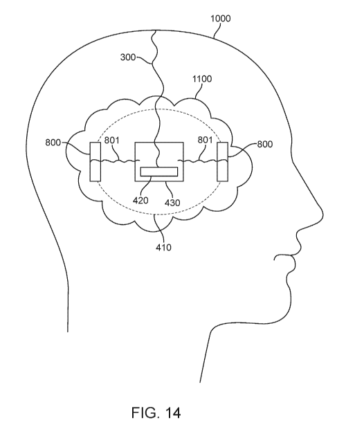
[0066] Fig. 14 illustrates an example of an illumination system that may
also
include electrical stimulation of the target treatment area. Here, after a
tumor is
resected from the brain in a patient's skull 1000, an illumination system is
disposed in the tumor cavity 1100. The illumination system may be any of the
systems disclosed herein and may include a tether 300 for providing power to
the illumination element 420 which may have one or more light sources coupled
to a substrate such as a PCB 430. The lights and substrate may be encapsulated
410 in a material that protects the device as well as facilitates delivery of
the
light to the target treatment area such as by serving as a light guide or by
diffusing the light. The encapsulation may be any of the encapsulation
examples
or light guides disclosed herein and also may help secure the device into the
tumor cavity. Extending from the PCB are conductors 801 which are attached to
electrodes 800 that are exposed to the sides of the illumination element and
can
provide electrical stimulation (deep brain stimulation) to the target
treatment
16
CA 03158283 2022-04-19
WO 2021/081059
PCT/US2020/056609
area either by direct contact with the brain tissue or by conduction through
interstitial fluid in the resected cavity. Thus, phototherapy and electrical
stimulation may be provided concurrently. An illumination system such as that
of Fig. 14 may be used to provide deep brain stimulation in patients with
neurodegenerative diseases such as Parkinson's disease, amyotrophic lateral
sclerosis (ALS). Alzheimer's disease, or any other condition where
neurostimulation is beneficial. The stimulation may be provided alone or in
combination with phototherapy where light may be used to treat a disease such
as by activating a photosensitizer including but not limited to brain cancers.
Electrodes tor tissue stimulation, whether in the brain or elsewhere in the
body
may be used with any of the examples of phototherapy systems described herein.
Additionally, the phototherapy system with electrodes are not limited to
implantation in a tumor cavity formed after tumor resection. The phototherapy
system may be placed in any tissue where phototherapy and/or electrical
stimulation are to be delivered to treat any disease or condition.
[0067] Fig, 15A shows an example of a reinforced substrate that may be
used
with any of the examples of light and/or electrical stimulating systems
disclosed
herein. For any substrate used in these examples, e.g. the substrate on which
the
light sources are mounted, or the light guide substrates, it may be
advantageous
to use a substrate that has reinforcing in the substrate to either provide a
stiffer
substrate or a substrate that can be bent, flexed or otherwise shaped to fit
the
target treatment region and retain that shape. Here, the substrate is similar
to
substrate 455 in Fig. 7 and is a flat planar substrate which may be trimmed to
a
desired size and shape to fit the target treatment area. The substrate may be
formed from a material that has stiffening features, or stiffening features
may be
built into the substrate. For example, here a two-dimensional grid of ribs 457
may be formed into the substrate to provide desirable stiffening
characteristics.
This helps the substrate maintain its shape once disposed in the tumor cavity
created after resection of the tumor. The illumination element 430 (which may
be any of those disclosed herein) may be positioned anywhere along the
substrate and a tether 300 is coupled to the illumination element 430 for
supply
of power. The stiffening features may be formed from a different material
(e.g.
17
CA 03158283 2022-04-19
WO 2021/081059
PCT/US2020/056609
a different polymer) having different mechanical properties (e.g. Young's
modulus of elasticity, durometer, etc.) compared to the base substrate.
[0068] Fig. 15B shows the device of Fig. 15A with stiffening features in
the
substrate implanted in a tumor cavity 1100 in a skull 1000. The tether 300
provides power to the illumination element (not shown in this view) which is
coupled to the substrate with stiffening or reinforcing members. The substrate
is
formed into a partially closed loop if two dimensional, or a partially closed
spheroid if three dimensional, to support and conform to the tissue and
illuminate the tissue. The substrate serves as a light guide to help
illuminate the
target treatment area.
[0069] Fig. 16 shows another example of a reinforced substrate such as the
example of Fig. 15A formed into a closed loop if two dimensional, or a closed
spheroid if three dimensional. Here, power is delivered via tether 300 to the
illumination element (not shown, and which may be any illumination element
disclosed herein) which is disposed on the reinforced substrate 458 in tumor
cavity 1100 in skull 1000. The reinforced substrate may be the same as shown
in Fig. 15A or different, and the ends may remain apposed with one another in
a
closed configuration due to the reinforcements in the substrate which maintain
the desired shape, or due to use of adhesives. This helps maintain the
substrate
in a desired configuration which supports the tumor cavity to ensure proper
illumination of the target treatment area.
[0070] Fig. 17 illustrates the use of additional support elements in a
tumor
cavity 1100 in the skull 1000 of a patient. The wallpaper substrate concept
described in Figs. 7, 15A-15B, and 16 may be used with additional support
elements disposed in the tumor cavity to help support the tissue in the target
treatment area and also to help distribute light to the target treatment area.
Here,
a tether 300 provides power to an illumination element which may be any of
those disclosed herein. The illumination element may be encapsulated in a
material, and the combination of the illumination element and encapsulant may
be coupled to a flat planar substrate 457 that is shapeable and trimmable to
fit in
the tumor cavity. The flat planar substrate 457 may be any of those described
herein and may be optically transparent to ensure the light passes through it.
Here, the wallpaper is formed into a partially closed loop if two dimensional,
or
18
CA 03158283 2022-04-19
WO 2021/081059
PCT/US2020/056609
a partial sphere if three dimensional. In some situations it may be beneficial
to
provide additional support elements 459 that help support tissue in the tumor
cavity and prevents collapse, and also that may be formed of optical material
thereby forming a light guide that helps distribute the light to the target
treatment
area. The support elements maybe thin planar sheets of material that are
trimmed
and shaped to fit into the tumor cavity or they may be prefabricated into
various
desired shapes. The support elements may then be secured to tissue in the
tumor
cavity using techniques known in the art such as with sutures, adhesives or
other
techniques. The additional support elements may also act as a spacer between
tissue and the illumination element, or an optical instrument to help deliver
the
light to the tissue.
[0071] Fig. 18 shows another example of the use of additional support
elements 490 in the tumor cavity 1100 in the skull 1000 of patient. Again, a
tether 300 provides power to an illumination element which may be any of the
illumination elements disclosed herein. A plurality of additional support
elements 490 may be coupled together (as indicated by the arrowheads) to form
a fully closed loop or a partially open loop (if two dimensional), or a fully
closed
or partially open spheroid if three dimensional, that results in a rigid or
semi-
rigid structure that supports the tissue in the tumor cavity and prevents it
from
collapsing inward. Thus, customization is possible during surgery, ensuring
that
the light provided by the illumination element can illuminate the entire
target
treatment area and the cavity is supported. The support elements may snap
together, press fit together, adhesively coupled together, or use coupling
mechanisms known in the art to form any desired shape, and they support
elements may be formed of an optical material to help distribute the light to
the
target treatment area.
[0072] Fig. 19 illustrates a second port for coupling the illumination
element
with an external light source. Here, tether 300 couples the illumination
element
(not illustrated) disposed in a flat planar substrate 455 as described in Fig.
7
above. The tether 300 delivers power to the illumination element which
delivers
light via the flat planar substrate 455 to the target treatment area. As
discussed
above, the flat planar substrate 455 may be shaped and trimmed to conform to
the target treatment area and it may be adhesively bonded to the tissue. In
some
19
CA 03158283 2022-04-19
WO 2021/081059
PCT/US2020/056609
cases, light may be used to help cure the adhesive such as when cyanoacryl ate
is
used. Therefore, an external light source 1300 may provide the required curing
light such as ultraviolet light via a port 1210 that may be releasably coupled
with
the external light source. The light is then delivered via an optical fiber
1200 to
the flat planar substrate which then delivers the light to the target
treatment area
facilitating curing of the adhesive. Once the substrate 455 is adhesively
bonded
to the tissue, the external light source may be turned off and de-coupled from
the
second port 1210 and removed. The port 1210 and fiber 1200 may remain
coupled to the flat planar substrate 455 or they may also be removed. Other
components which may be adhesively bonded such as the supplemental support
elements in Figs. 17-18, the encapsulation surrounding the illumination
element
(e.g. in Fig. 5), optical lightguide shapes in Figs. 6A-6D, and supplemental
light
guides in Figs. 9A-9D may also be adhesively bonded and cured using the
external light source coupled to light input port 1210 and delivered via
optical
fiber 1200 to the flat planar substrate for illuminating and curing the
adhesive.
In other examples, the second light port may be used to introduce light to the
target treatment area via the substrate 455 using an external light source in
order
to illuminate biologicals, chemicals, or other agents that react to the light.
[0073] Figs. 20A-20B illustrate the use of multiple light sources in the
illumination element and the resulting illumination pattern which provides
desirable control of the illumination.
[0074] A uniform light source may not be an optimal solution for an
asymmetrically distributed disease in human tissue. Depending on the
individual
patient and the particulars of the anatomy and tumor, there may be areas in
the
surgical cavity that will be more likely to contain residual tumor. It may be
of
benefit to concentrate light towards these areas in the interest of focusing
the
photodynamic therapy (PDT) effect. Clinical trials with intraoperative
applications of PDT show a direct relationship between input light (fluence,
Joules/cm^2) and clinical outcome. In a single application (as part of a
longer
series of treatments), the examples of devices disclosed herein can deliver a
customized output of light, with greater emphasis/output towards areas of
greater
tumor risk.
CA 03158283 2022-04-19
WO 2021/081059
PCT/US2020/056609
[0075] The total amount of available light is limited in an implanted
device,
due largely to wireless power transfer limitations and tissue heating
limitations.
Therefore, optimal use of the light sources is desirable and may be
accomplished
in a device that can control the output of a plurality of lights in such as
fashion
as to have variable combinations of lights in an on vs. off configuration.
[0076] Furthermore, this configuration may also apply not only to on vs. off
configurations but control of individual LED light output within a defined
range.
This can be controlled by firmware embedded within the implanted PCB or the
ho using, which would direct the power output of individual lights in the
implant. The control of this by the user may be accomplished through a user
interface designed to control the power transfer and output device. Planning
of
this "prescription" for each patient can be analogous to the spatial and
temporal
planning of radiotherapy treatment, coupled with proscriptive imaging of tumor
location in a patient.
[0077] In Fig. 20A, a tether 300 is coupled to the illumination element
which
in this example includes four independently controllable light sources 420,
here
LEDs. The LEDs are mounted in a substrate 430 such as a flexible PCB, and the
assembly may be encapsulated in any manner, as previously described. Because
the LEDs may be independently controlled, they may be turned on or off as
needed in order to direct light in a desired direction for a desired amount of
time.
[0078] Fig. 20B illustrates the intensity of the light in all four
directions
emitted by the four LEDs when light is emitted in a direction away from the
center of the PCB 430. Each direction has a lobe shaped pattern showing the
illumination pattern for each LED and the intensity of the light emitted. When
the total power provided by the tether is fixed, the intensity of each LED is
higher when only a single LED is activated at a time. Thus, illumination may
be
steered in a desired direction and light intensity may also be controlled.
[0079] Example of Method of Use
100801 Fig. 21 illustrates an example of a method of treating a tumor. The
treatment may be determined by a team of physicians and surgeons which may
include a neurosurgeon, neuroradiologist and a neurooncologist. Any of the
illumination devices and optional features disclosed herein may be used
according to the following method of use. This example is directed at
treatment
21
CA 03158283 2022-04-19
WO 2021/081059
PCT/US2020/056609
of brain cancer; but this is not intended to be limiting and one of skill in
the art
will appreciate that other diseases and conditions may also be treated.
10081] MRI (magnetic resonance imaging) scans may be used to determine
the size and shape of the tumor and based on that information, an appropriate
size device may be selected. Other imaging techniques known in the art may
also be used, such as computerized tomography (CT), positron emission
tomography (PET), radiographic imaging, etc. The illumination dosage may
also be determined based on the 'bioavailability of the photosensitizer
delivered
to target tissue, and efficacious light fluence and timing. In some examples,
artificial intelligence (Al) or an Al classifier may be employed at least as a
pattern recognition tool to detect and guide physicians towards an optimized
targeted therapy to improve outcomes for patients. A dose includes
photosensitizer drug dosage and frequency, as well as light fluence from the
implantable phototherapy device. Initial dosage may be higher or lower
depending on patient condition and expected severity of any remaining tumor
cells in the tumor margins,
100821 A craniotomy, often a 5-10 cm diameter circular section of bone is
removed from the skull and allows access to the patient's brain. After the
tumor
has been resected 2102, the device is implanted 2104 in the tumor cavity
formed
after a craniotomy and tumor resection surgery, where the bulk of a
glioblastoma
tumor or other diseased tissue is removed by a neurosurgeon with standard
surgical procedures. The light source portion of the device is secured in
place by
the neurosurgeon using methods known in the art including any of those
described herein. The light source may comprise a thin, flexible sheet or
wallpaper that is glued down in the cavity and cured in place. The surgeon can
trim it to fit the individual patient's tumor size and shape 2106.
[0083] The surgeon may adhesively bond or otherwise secure the device
into
the tumor cavity 2108 using cyanoacrylate, fibrin glue or a similar
biocompatible
tissue adhesive. The device may be able to "self-cure" by emitting the
wavelength of light required by the adhesive (glue). Either the LED emitter
can
be built-in to the device, or an external curing light can be coupled to the
lightguide such that the curing light can reach where it needs to be. For
example, if the adhesive is cured by light (e.g. UV curing adhesives), then
the
22
CA 03158283 2022-04-19
WO 2021/081059
PCT/US2020/056609
light source itself can be the source of its own curing light. The device may
have an attachment that connects to one point on the light source and can
transmit a curing light through the light source to the adhesive, such as the
example previously described above with respect to Fig. 19.
10084] If trimming is needed, the surgeon may trim the light source's
lightguide shape to better fit the patient's individual tumor cavity with
surgical
scissors or other cutting instrument. The light source may contain visual
markers
that indicate areas that should not be cut.
10085] Implantation of the illumination element is at the discretion of
the
neurosurgeon but should ensure that the light is directed at regions in the
target
treatment area that either contain residual tumor or are likely to experience
recurrence. For GErvi tumors which are dendritic and invasive, an additional
margin up to about 2 cm from a known margin may be a good boundary. For
smaller tumors, it may be possible to affix light elements such that all inner
cavity surfaces are illuminated.
10086] The device can then be tested 2110 to visually check that the
light
source is working and in the correct location.
10087] The tether can extend to the outside of the skull, and the
wireless
power portion of the device is secured in place on the outside of skull and
under
the scalp, in any location, such as behind the ear. The tether may also be
secured
to the skull, so it does not get pulled out by the patient. A clip or grommet
may
be attached to the skull adjacent the craniotomy opening and the tether may be
secured with the clip or grommet, serving to protect the wire from sharp edges
around the craniotomy and also to hold the tether in place. Excess wire can be
coiled around the dip or grommet. Additional suture, screws, adhesives, etc.
may be used to help secure the tether and coil if needed. A recessed region in
the skull may be formed to help accommodate the tether, coil, or housing
thereby preventing or minimizing bulging. The coil may be disposed on the
same side as the craniotomy or it may be disposed on the comralateral side.
Once the device is implanted and secured to the skull, the skull may be
closed,
and the scalp also may be closed.
10088] Optionally-, the illumination system may include radiopaque
markers
adjacent the illumination element to permit the surgeon or physician to
visualize
23
CA 03158283 2022-04-19
WO 2021/081059
PCT/US2020/056609
and confirm placement of the device in the patient's tumor cavity using
imaging
techniques known in the art, such as NMI, PET, x-ray, etc.
[0089] After surgical recovery, photodyn.amic therapy is activated 2112
by
powering the wireless power portion of the implant via an external transmitter
in
a clinic. The implant can control the power level it receives and directs the
majority of that power to the light source. Dosing will be monitored by the
transmitter, under the observance of a human operator. Sufficient dose for
effective photodynamic therapy will be required for each session. As the
sessions may be just a few hours at most, patients can take advantage of
regular
phototherapy sessions to control the recurrence of their tumor over months and
even years. An example of a duty cycle may include one minute of illumination
with thirty seconds without illumination, and then repeated. This helps reduce
heat and also may allow the tissue to reoxygenate between light cycles. The
power receiving element in any example of device may include a clock
oscillator
so that it can manage cycling of the light source. The energy transmitter may
be
able to reprogram the power receiver element to change the timing of the light
delivery.
[0090] Fractionation of dose can occur over several days, distinguishing
this
method from one-time treatments.
[0091] Using the disclosed devices and methods allow transmission of
significant power to the device efficiently while still having a light device
in the
center of the brain where traditional wireless power methods do not easily
reach.
[0092] The phototherary may optionally be combined with imaging and
mapping to help direct the illumination. Since the illumination element may
contain. multiple light sources, steeling of the light to a desired direction
is
possible so that if areas are identified having more tumor cells or expected
to
have more tumor cells, the photothera.py may be directed in that direction.
Also,
the phototherapy may be combined with algorithms and tumor recurrence
modelling, to steer light to areas of concern with dosages also predicted by
computer modelling. The devices, systems and methods described herein may
be used with any photosensitizer that provides a desired diagnostic or
therapeutic
effect. Examples of photosensitizers are described below. The patient is then
monitored for adverse reactions to the photodynainic therapy or the
24
CA 03158283 2022-04-19
WO 2021/081059
PCT/US2020/056609
photosensitizer drug at regular intervals, e.g. every 3 to 6 months using
brain
imaging techniques known in the art to look for tumor recurrence. Patient
dosages and therapy may be adjusted as needed.
[0093] Possible opportunities for personalizing treatment may occur at
various times during the patient's therapy including during resection, during
a
recovery period of about 6 weeks after resection, during a combination of 'TAU
(Teinozolomide) chemotherapy and radiotherapy for about 6 weeks, and during
the six-month follow-up periods. Use of phototherapy may be used alone or in
conjunction with any of these periods of time and treatment to provide an
enhanced outcome.
[0094] Wavelength of Light
[0095] The wavelength of the light delivered by the illumination element
is
selected based on the wavelength required to activate the photosensitizer and
depth of tissue penetration. For example, light in the red to near infrared
wavelength range of about 600 nm to 940 nm may have sufficient tissue
penetration in brain tissue. This wavelength may be used with any of the
examples of devices disclosed herein.
[0096] Examples of Photosensitizers
[0097] Any photosensitizer which may have a therapeutic effect when
exposed to light may be used with any of the examples of illumination systems
described herein. Examples of photosen.sitizers include but are not limited to
methyl aminolevulinate hydrochloride; padeliporfin potassium; talaporfin
sodium; SGX-301; fimaporfin gemcitabine; reda.porfin; aminolaevulinic acid -
i-
artemisinin; CTT-1700, IVX-MES; IVX-PDT; IVXT-02; M-
103; Photobac; YC-9; ADC + fimaporfin; bleomycin sulfate +
fimaporfin; lemuteporfin; methyl aminolevulinate hydrochloride; motexafin
lutetium; padoporfin; SL-01.7; Vangiolux; Deuteporfin; Small Molecule to
Activate ABCB1 for Graft Versus Host Disease; Recombinant Peptide to Target
EGFR for Oncology; Small Molecules to Target eNOS; tiNOS and NO Synthase
for Oncology; epirubicin hydrochloride + fimaporfin; porfimer sodium;
temoporfin; Palladium bacteriopheophorbide; rostaporfin; Verteporfin; and 5-
Aminolevulinic acid.
CA 03158283 2022-04-19
WO 2021/081059
PCT/US2020/056609
100981 These photosensitizers may be illuminated with light in the
treatment
of various cancers and other diseases including but not limited to Basal Cell
Carcinoma (e.g. Basal Cell Epithelioma); Squamous Cell Carcinoma; Actinic
(e.g. Solar) Keratosis; Skin Cancer; Solid Tumor; Prostate Cancer; Esophageal
Cancer; Transitional Cell Carcinoma (Urothelial Cell Carcinoma); Bile Duct
Cancer (e.g. Cholangiocarcinoma) ; Endobronchial Cancer; Kidney Cancer (e.g.
Renal Cell Cancer); Renal Cell Carcinoma; Choroidal Neovascularization; Brain
Tumor; Glioma; Neurofibroma; Head And Neck Cancer; Hepatocellular
Carcinoma; Metastatic Colorectal Cancer; Nasopharyngeal Cancer; Pancreatic
Cancer; Benign Prostatic Hyperplasia; Age Related Macular Degeneration;
Coronary Disease; Cutaneous Vascular Malformations; Peripheral Arterial
Disease (PAD); Peripheral Vascular Disease (PVD); Mycosis Fungoides;
Psoriasis; Glioblastoma Multiforme (e.g. GBM); Inflammatory Bowel Disease;
Colorectal Cancer; Malignant Mesothelioma; Ovarian Cancer; Viral Infections;
Colon Cancer; Graft Versus Host Disease (GVHD); Carcinomas; Sarcomas;
Acne Vulgaris; Coronary Artery Disease (CAD) (e.g. Ischemic Heart Disease);
Breast Cancer; Non-Small Cell Lung Cancer; Small-Cell Lung Cancer; and
Bladder Cancer.
100991 Experiments
1001001 A sample device having a coil for receiving RF energy, a rectifier for
converting the alternating current of the power received into a direct current
and
LEDs was tested. The LEDs emitted light at about 630 nm wavelength and the
fluence (energy density) was measured to be about 120 Pcm^2. Radiant power
was measured using an optical power meter over a range of driving current from
about 0.1 mA to about 20 mA. Based on the reported literature, this level of
fluence is estimated to have an extrapolated necrosis death depth of about 10-
20
mm.
1001011 The topic of photosensitizer drug activation is well understood in the
art. The effective application of photodynamic therapy requires a light at an
optimal wavelength of photosensitizer (PS) drug activation, at a sufficient
intensity and for a sufficient duration of time to deliver a minimum light
fluence
(Joules per square centimeter of area).
26
CA 03158283 2022-04-19
WO 2021/081059
PCT/US2020/056609
1001021 Known experimental protocols have used light fluence as a controlled
parameter (i.e., PS activation threshold or target) when demonstrating
efficacy
against tumors in pre-clinical and clinical testing. Therefore, because
examples
of the illumination systems disclosed herein deliver a discrete amount of
light
fluence consistent with what is known in the art, effective activation of PS
must
follow. In some examples, the target light fluence is 90-500J/cm^2. In some
examples, the target light fluence is optimized at 100-200 J/cm^2.
1001031 Fractionated or Metronomic PDT (mPDT) is of considerable interest
in the PDT research community. mPDT may achieve the same doses for PS
activation but over a longer period of time at low light intensity. The
scientific
literature has reported promising results with < 100uW/cm^2, 1000x less light
intensity than typical PDT protocols, over a period of 10 days in an animal
model. By using a much longer time period (1000x), the product of intensity
multiplied by time remains constant.
1001041 Other literature suggests that fluence rate (W/cm^2) does have an
impact, showing that with the same amount of dosage, a higher intensity kills
tumor cells to a greater depth. It also can cause more death of normal cells,
but
this does support the notion of a "threshold" for activation of the
photosensitizer.
1001051 Further literature also reports research with other light fluence
rates
are possible. Light fluence rates of 20 - 400 J/cm^2 were used (plurality
between 100-200 J/cinA2), and there is some evidence that higher rates
correspond to better outcomes.
1001061 Based on the data contained within these references, the implanted
phototherapy device therapy targets 100 J/cm^2 of light fluence. When divided
over many hours or even days, the instantaneous power required to deliver this
energy can be on the order of mW or tens of mW (e.g. 7mW/cmA2 over 4 hours).
Additional details may be found in Brendan J. Quirk et al., "Photodynamic
therapy (PDT) for malignant brain tumors ¨ Where do we stand?"
Photodiagnosis and Photodynamic Therapy 12.3 (2015): 530-544. As well as
Tudge, S.H. et al, Modulation of light delivery in photodynamic therapy of
brain
tumours, Journal of Clinical Neuroscience, 1999 6(3), 227-232; and Yamagishi,
Tissue-adhesive wirelessly powered optoelectronic device for metronomic
27
CA 03158283 2022-04-19
WO 2021/081059
PCT/US2020/056609
photodynamic cancer therapy, Nature Biomedical Engineering, January 2019;
the entire contents of which are incorporated herein by reference.
NOTES AND EXAMPLES
[00107] The following, non-limiting examples, detail certain aspects of the
present subject matter to solve the challenges and provide the benefits
discussed
herein, among others.
[00108] Example 1 is an implantable phototherapy device, comprising: a
power receiver element configured to receive power from an external power
transmitter; a light delivery element powered by the power provided by the
power receiver, and configured to deliver a phototherapy to a target treatment
area; and a tether element operably coupled to the light delivery element and
the
power receiver element, the tether element configured to deliver the power
from
the power receiver element to the light delivery element.
1001091 Example 2 is the device of Example 1, wherein the power receiver
element comprises a coil configured to receive the power from the external
power transmitter, and wherein the power comprises radiofrequency energy.
[00110] Example 3 is the device of any of Examples 1-2, wherein the power
receiver element comprises a sealed housing operably coupled with the tether,
the device further comprising electronic components disposed in the sealed
housing, the electronic components configured to control the power delivered
to
the light delivery element.
[00111] Example 4 is the device of any of Examples 1-3, wherein the light
delivery element comprises a light source encapsulated in an optical material
configured to protect the light source and wherein the optical material
facilitates
transmission of light from the light delivery element to the target treatment
area.
[00112] Example 5 is the device of any of Examples 1-4, further comprising an
optical lightguide coupled to the light delivery element, the optical
lightguide
shaped to facilitate delivery of light from the light delivery element to the
target
treatment area.
[00113] Example 6 is the device of any of Examples 1-5, wherein the light
delivery element comprises a plurality of light sources disposed on a
substrate,
28
CA 03158283 2022-04-19
WO 2021/081059
PCT/US2020/056609
and wherein the substrate is configured to be shaped to match the target
treatment area.
[00114] Example 7 is the device of any of Examples 1-6, wherein the substrate
is a lightguide configured to direct light to the target treatment area and
wherein
the substrate is configured to be trimmed to a desired shape to fit the target
treatment area.
[00115] Example 8 is the device of any of Examples 1-7, wherein the light
delivery element comprises a plurality of light sources configured to be
independently controllable relative to one another.
[001161 Example 9 is the device of any of Examples 1-8, wherein the light
delivery element further comprises a temperature sensor configured to measure
temperature at the target treatment area.
[00117] Example 10 is the device of any of Examples 1-9, wherein the light
delivery element is disposed in a radially expandable member having an
expanded configuration and a collapsed configuration, wherein in the expanded
configuration the radially expandable member conforms to the target treatment
area.
[00118] Example 11 is the device of any of Examples 1-10, wherein the light
delivery element further comprises a port configured to releasably receive an
optical fiber optically coupled to an external light source, and wherein light
from
the external light source is delivered to the light delivery element via the
optical
fiber for illumination of the target treatment area.
[00119] Example 12 is a phototherapy system comprising the device of any of
Examples 1-11, and is the device of any of Examples 1-10, the external power
transmitter configured to wirelessly transmit the power to the power receiver
element.
[00120] Example 13 is the system of Example 12, further comprising a planar
immersion lens disposed between the external power transmitter and the power
receiver element, the planar immersion lens configured to focus energy from
the
external power transmitter toward the power receiver element.
[00121] Example 14 is the system of any of Examples 12-13, further
comprising an electrode configured to provide electrical stimulation to the
target
treatment area.
29
CA 03158283 2022-04-19
WO 2021/081059
PCT/US2020/056609
[00122] Example 15 is the system of any of Examples 12-14, further
comprising at least one support element, the support element configured to
appose and support tissue in the target treatment area.
[00123] Example 16 is the system of any of Examples 12-15, further
comprising a photosensitizer.
[00124] Example 17 is a method for delivering phototherapy to a target
treatment region in a patient, the method comprising: providing an implantable
phototherapy device comprising a power receiver element, a light delivery
element, and a tether element; implanting the phototherapy device in a patient
at
the target treatment region; wirelessly transmitting power from an external
power transmitter to the power receiver element; transferring the power from
the
power receiver element to the light delivery element via the tether; and
illuminating the target treatment area with light from the light delivery
element.
[00125] Example 18 is the method of Example 17, wherein wirelessly
transmitting the power from the external power transmitter to the power
receiver
element comprises receiving radiofrequency energy with a coil.
[00126] Example 19 is the method of any of Examples 17-18, wherein the
illuminating comprises illuminating the target treatment region with a
plurality
of light emitting elements that are independently controllable.
[00127] Example 20 is the method of any of Examples 17-19, wherein
wirelessly transmitting the power comprises transmitting the power from the
external power transmitter and focusing the power toward the power receiver
element with a planar immersion lens.
[00128] Example 21 is the method of any of Examples 17-20, further
comprising electrically stimulating tissue in the target treatment region with
energy provided by an electrode adjacent the light delivery element.
[00129] Example 22 is the method of any of Examples 17-21, wherein the
target treatment region comprises a brain of the patient.
[00130] Example 23 is the method of any of Examples 17-22, releasably
coupling an optical fiber to the light delivery element; inputting light from
an
external light source to the light delivery element via the optical fiber; and
illuminating the target tissue with the light from the external light source.
CA 03158283 2022-04-19
WO 2021/081059
PCT/US2020/056609
[00131] Example 24 is the method of any of Examples 17-23, wherein the light
delivery element comprises a plurality of light sources disposed on a
substrate,
the method further comprising shaping the substrate to conform with the target
treatment area and directing light in a plurality of directions to illuminate
the
target treatment area.
[00132] Example 25 is the method of any of Examples 17-24, further
comprising trimming the substrate to a desired size or shape in order to fit
in the
target treatment area.
1001331 Example 26 is the method of any of Examples 17-25, further
comprising measuring temperature at the target treatment area with a
temperature sensor.
[00134] Example 27 is the method of any of Examples 17-26, wherein the light
delivery element comprises a plurality of light sources encapsulated in an
optical
material, the optical material being a lightguide that directs light from the
plurality of light sources to the target treatment area.
[00135] Example 28 is the method of any of Examples 17-27, wherein the light
delivery element is disposed in a radially expandable member, the method
further comprising radially expanding the radially expandable member to appose
and conform with the target treatment area.
1001361 Example 29 is the method of any of Examples 17-28, further
comprising disposing a support element in the target treatment area to help
support tissue in the target treatment area to ensure the tissue is
illuminated.
[001371 In Example 30, the devices, systems or methods of any one or any
combination of Examples 1 ¨29 can optionally be configured such that all
elements or options recited are available to use or select from.
[00138] The above detailed description includes references to the
accompanying drawings, which form a part of the detailed description. The
drawings show, by way of illustration, specific embodiments in which the
invention can be practiced. These embodiments are also referred to herein as
"examples." Such examples can include elements in addition to those shown or
described. However, the present inventors also contemplate examples in which
only those elements shown or described are provided. Moreover, the present
inventors also contemplate examples using any combination or permutation of
31
CA 03158283 2022-04-19
WO 2021/081059
PCT/US2020/056609
those elements shown or described (or one or more aspects thereof), either
with
respect to a particular example (or one or more aspects thereof), or with
respect
to other examples (or one or more aspects thereof) shown or described herein.
[00139] In the event of inconsistent usages between this document and any
documents so incorporated by reference, the usage in this document controls.
[00140] In this document, the terms "a" or "an" are used, as is common in
patent documents, to include one or more than one, independent of any other
instances or usages of "at least one" or "one or more." In this document, the
term "or" is used to refer to a nonexclusive or, such that "A or B" includes
"A
but not B," "B but not A," and "A and B," unless otherwise indicated. In this
document, the terms "including" and "in which" are used as the plain-English
equivalents of the respective terms "comprising" and "wherein." Also, in the
following claims, the terms "including" and "comprising" are open-ended, that
is, a system, device, article, composition, formulation, or process that
includes
elements in addition to those listed after such a term in a claim are still
deemed
to fall within the scope of that claim. Moreover, in the following claims, the
terms "first," "second," and "third," etc. are used merely as labels, and are
not
intended to impose numerical requirements on their objects.
1001411 The above description is intended to be illustrative, and not
restrictive.
For example, the above-described examples (or one or more aspects thereof)
may be used in combination with each other. Other embodiments can be used,
such as by one of ordinary skill in the art upon reviewing the above
description.
The Abstract is provided to allow the reader to quickly ascertain the nature
of the
technical disclosure. It is submitted with the understanding that it will not
be
used to interpret or limit the scope or meaning of the claims. Also, in the
above
Detailed Description, various features may be grouped together to streamline
the
disclosure. This should not be interpreted as intending that an unclaimed
disclosed feature is essential to any claim. Rather, inventive subject matter
may
lie in less than all features of a particular disclosed embodiment. Thus, the
following claims are hereby incorporated into the Detailed Description as
examples or embodiments, with each claim standing on its own as a separate
embodiment, and it is contemplated that such embodiments can be combined
with each other in various combinations or permutations. The scope of the
32
CA 03158283 2022-04-19
WO 2021/081059
PCT/US2020/056609
invention should be determined with reference to the appended claims, along
with the full scope of equivalents to which such claims are entitled.
33