Note: Descriptions are shown in the official language in which they were submitted.
CA 03160189 2022-05-03
WO 2021/096863 PCT/US2020/059842
FEEDBACK DETECTION FOR A TREATMENT DEVICE
CROSS-REFERENCE TO RELATED APPLICATIONS
[0001] This application claims the benefit of U.S. Provisional Patent
Application No.
62/934,583, filed on November 13, 2019, and entitled "Electromagnetic
Radiation Based
Treatment Devices and Methods," the entirety of which is incorporated by
reference herein.
BACKGROUND
[0002] Presently a number of energy-based devices are available for
fractionated treatment of the
dermis. These methods include ablative lasers, non-ablative lasers, micro-
needling, and RF
energy treatments. Generally, these presently available fractionated energy-
based treatments
require damage to outer portions of skin undergoing treatment (e.g.,
epidermis). Damage to the
epidermis, in most cases, causes the skin to appear inflamed, blemished, or
unhealthy
immediately after treatment. Additionally, severe damage to the epidermis can
lead to one or
more of infection and a need for additional medical treatment. This
undesirable appearance
results in a post-treatment downtime that lasts until the epidermis has
healed, which may take
days to weeks depending on treatment parameters used (e.g., ablative vs. non-
ablative). Most
patients do not return to their normal lives until after the post-treatment
down-time. It is therefore
desirable that a fractionated treatment system and method be made available,
which can
successfully affect the dermis while minimizing damage to the epidermis in
order to minimize
post-treatment downtime.
SUMMARY
[0003] Skin rejuvenation is often performed through fractional treatment.
Fractional or
fractionated energy-based treatment refers to a treatment in which only a
fraction of an area of
tissue is exposed to energy. For example, a fractional skin treatment may
treat 25% of an area of
skin with a laser beam and leave a remaining 75% of skin in that area
untreated. Energy-based
skin rejuvenation involves creating controlled small injury within a collagen
network. The small
injury causes a wound healing process in which new collagen is formed. The
newly formed
collagen tightens the skin, thereby causing the skin to appear more youthful.
Many skin
1
CA 03160189 2022-05-03
WO 2021/096863 PCT/US2020/059842
rejuvenation fractionated treatment systems work by targeting water as a
chromophore achieving
photothermolysis.
[0004] Fractionated treatment can generally be divided into two categories:
Ablative and non-
ablative. Ablative treatment causes removal of tissue and results in
superficial micro-injury in
addition to creating thermal damage within the dermis. Non-ablative treatment
typically does not
cause tissue removal, instead causing only thermal disruption. An advantage of
non-ablative
fractionated treatment over ablative fractionated treatment is a reduction in
down-time.
[0005] Energy-based fractionated treatment of tissue generally requires that a
high amount of
energy be delivered to and absorbed by a selective portion of tissue to effect
a desired disruption
or damage. This disruption or damage is repeated over an area of tissue, so
that small regions
(e.g., 0.1 to lOmm in diameter) of disrupted tissue are interlaced with
undamaged tissue. The
small regions of damaged tissue are then replaced with new tissue during a
post-treatment
healing process. Inducing damage within a dermis layer of skin while
minimizing damage to an
overlying epidermis layer presents a number of technical challenges, some of
which are
enumerated below.
[0006] First, there is no known chromophore within the dermis layer of tissue
which is not
present within the epidermis layer of tissue. This means that a radiation
selected to absorb within
the dermis will also be absorbed within the epidermis layer.
[0007] Second, as the EMR will be equally well absorbed by the epidermis layer
and the dermis
layer of the skin, a greater energy density must be delivered to the dermis
layer than to the
epidermis. In order to achieve this, the EMR profile must be varied, such that
a focal region (i.e.,
region of maximum energy density) of the EMR beam is located within the dermis
and only an
unfocused region (i.e., region of minimum energy density) of the EMR beam
subtends the
epidermis layer of the skin.
[0008] Third, skin tissue is a turbid medium, meaning that radiation
propagating through skin
scatters. The scattering of radiation within skin tissue, makes it more
difficult to form a focal
region (region of maximum energy density) at any depth within the tissue,
compounding the first
and second challenges above.
2
CA 03160189 2022-05-03
WO 2021/096863 PCT/US2020/059842
[0009] Fourth, the focal region (or region of maximum energy density) must be
accurately
positioned at a depth within the dermis layer of the skin. This ensures that
the region of
maximum energy density is located in the dermis and not located within the
epidermis, in order
to prevent unwanted damage to the epidermis.
[0010] Fifth, the EMR beam is delivered from outside the tissue; and,
therefore, the epidermis
experiences some minimal irradiation and minor thermal heating (i.e., less
than the dermis). In
response to this fifth challenge, the epidermis layer directly overly the
dermis layer being treated
must be actively cooled to prevent thermal damage to the epidermis.
[0011] Therefore, to provide fractionated therapeutic disruption to the dermis
layer of a skin
tissue, while minimizing damage to overlying epidermal layers, a need exists
for fractionated
treatment systems and methods that address all of the above-mentioned
challenges.
[0012] According to some embodiments, a system for fractionally treating
tissue includes: an
electromagnetic radiation (EMR) source configured to generate an EMR beam
having a
transverse ring energy profile; an optic configured to converge the EMR beam
to a focal region
located within a tissue; and, a window assembly located down-beam from the
optic configured to
cool the tissue when placed in contact with an outer surface of the tissue.
The window assembly
includes: a first window, a second window separated from the first window;
and, a coolant
chamber located between the first window and the second window, wherein the
coolant chamber
is configured to contain a coolant that is substantially non-absorbent of the
EMR beam.
[0013] In some embodiments of the system, the EMR beam has a wavelength in a
range between
about 1000nm and 4000nm.
[0014] In some embodiments of the system, the coolant includes at least one of
a dielectric fluid,
a fluorocarbon-based fluid, water, an antifreeze, ethylene glycol, and
propylene glycol.
[0015] In some embodiments of the system, the optic is additionally configured
to converge the
EMR beam at a numerical aperture (NA) of at least about 0.2.
3
CA 03160189 2022-05-03
WO 2021/096863 PCT/US2020/059842
[0016] In some embodiments of the system, the system also includes an optical
clearing medium
located between the window assembly and the tissue. In some cases, the optical
clearing medium
includes at least one of: glycerin, polyethylene glycol, and phosphate-
buffered saline.
[0017] In some embodiments of the system, the EMR source includes a beam
shaper configured
to shape the transverse ring energy profile. In some versions of the system,
the beam shaper
includes an axicon.
[0018] In some embodiments of the system, the system additionally includes a
controller. In
some cases, the controller is configured to control the EMR source to ensure
that the window
assembly cools the tissue to a determined temperature prior to generating the
EMR beam. In
some cases, the controller is configured to control the EMR source to ensure
that the window
assembly cools the tissue for a determined period prior to generating the EMR
beam.
[0019] According to some embodiments, a method for fractionally treating
tissue includes:
cooling, using a window assembly contacting an outer surface of the tissue,
the tissue;
generating, using an electromagnetic radiation (EMR) source, an EMR beam
having a transverse
ring energy profile; and converging, using an optic, the EMR beam to a focal
region located
within a tissue. In some cases, the window assembly includes: a first window,
a second window
separated from the first window, and a coolant chamber located between the
first window and the
second window; wherein, the coolant chamber is configured to contain a coolant
that is
substantially non-absorbent of the EMR beam.
[0020] In some embodiments of the method, the EMR beam has a wavelength in a
range
between about 1000nm and 4000nm.
[0021] In some embodiments of the method, the coolant includes at least one of
a dielectric
fluid, a fluorocarbon-based fluid, water, ethylene glycol, and propylene
glycol.
[0022] In some embodiments of the method, converging the EMR beam is performed
at a
numerical aperture (NA) of at least about 0.2.
[0023] In some embodiments of the method, the method additionally includes
introducing an
optical tissue clearing medium between the window assembly and the tissue. In
some cases, the
4
CA 03160189 2022-05-03
WO 2021/096863 PCT/US2020/059842
optical tissue clearing medium includes at least one of glycerin, polyethylene
glycol, and
phosphate-buffered saline.
[0024] In some embodiments of the method, the EMR source additionally includes
a beam
shaper configured to shape the transverse ring energy profile. In some
versions of the method,
the beam shaper includes an axicon.
[0025] In some embodiments of the method, the method additionally includes
controlling, using
a controller, the EMR source in order to ensure that the window assembly cools
the tissue to a
determined temperature prior to generating the EMR beam.
[0026] In some embodiments of the method, the method additionally includes
controlling, using
a controller, the EMR source in order to ensure that the window assembly cools
the tissue for a
determined period prior to generating the EMR beam.
[0027] According to some embodiments, a system for fractionally treating
tissue includes: an
electromagnetic radiation (EMR) source configured to generate an EMR beam
having a
wavelength in a range between about 1400nm and 3400nm; a beam shaper
configured to shape
the EMR beam into a transverse ring energy profile, wherein the beam shaper
includes an
axicon; an optic configured to converge the EMR beam at a numerical aperture
(NA) of at least
about 0.2 to a focal region located within a tissue; a window assembly located
down-beam from
the optic configured to cool the tissue when placed in contact with an outer
surface of the tissue,
wherein the window assembly includes: a first window, a second window
separated from the
first window, and a coolant chamber located between the first window and the
second window,
wherein the coolant chamber is configure to contain a coolant that is
substantially non-absorbent
of the EMR beam and includes a fluorocarbon-based fluid; and a controller
configured to control
the EMR source in order to ensure that the window assembly cools the tissue at
least one of: to a
determined temperature and for a determined time prior to generating the EMR
beam.
[0028] According to some embodiments, a system for fractionally treating
tissue includes: an
electromagnetic radiation (EMR) source configured to generate an EMR beam
having a
wavelength; a collimator configured to collimate the EMR beam to a width; a
beam shaper
includes a first axicon and a second axicon configured to shape the collimated
EMR beam into a
CA 03160189 2022-05-03
WO 2021/096863 PCT/US2020/059842
transverse ring energy profile, wherein the first axicon and the second axicon
are separated by a
distance along an optical axis that is chosen to effect a desired inner
diameter of the transverse
ring energy profile and the width of the collimated EMR beam is chosen to
effect a desired
thickness of the transverse energy profile; and an optic configured to
converge the EMR beam to
a focal region within a tissue, thereby affecting the tissue with the focal
region.
[0029] According to some embodiments, a system includes: an EMR source
configured to
generate an EMR beam having a transverse ring-shaped energy profile and a
wavelength in a
range of about 1200nm to about 12000nm; an optic configured to converge the
EMR beam to a
focal region located within a tissue; a beam scanning system configured to
scan the focal region
within the tissue; a window assembly located down-beam from the optic
configured to transmit
the EMR beam and cool the tissue when placed in contact with an outer surface
of the tissue,
wherein the window assembly includes: a first window; a second window
separated from the
first window; and, a coolant chamber located between the first window and the
second window,
wherein the coolant chamber is configured to contain a coolant including a
fluorocarbon-based
fluid that is substantially non-absorbent of the EMR beam; and a controller
configured to control
the EMR source to generate the EMR beam with a plurality of pulses, wherein at
least one pulse
of the plurality of pulses has a pulse duration that is no less than about 100
microseconds.
[0030] In some embodiments of the system, the at least one pulse of the
plurality of pulses has a
pulse energy that is no greater than about 100mJ.
[0031] In some embodiments of the system, the system additionally includes a
chiller configured
to cool the coolant to a temperature within a range of about -20 C to about 20
C.
[0032] In some embodiments of the system, the optic is further configured to
converge the EMR
beam at a numerical aperture (NA) of at least about 0.2.
[0033] In some embodiments of the system, the system additionally includes an
optical tissue
clearing medium located between the window assembly and the tissue, wherein
the optical tissue
clearing medium includes at least one of glycerin, polyethylene glycol, and
phosphate-buffered
saline.
6
CA 03160189 2022-05-03
WO 2021/096863 PCT/US2020/059842
[0034] In some embodiments of the system, the EMR source additionally includes
a beam shaper
configured to shape the transverse ring-shaped energy profile. In some
versions of the system,
the beam shaper includes an axicon.
[0035] In some embodiments of the system, the controller is configured to
control the EMR
source to ensure that the window assembly cools the tissue to a predetermined
temperature prior
to generating the EMR beam.
[0036] In some embodiments of the system, the controller is configured to
control the EMR
source to ensure that the window assembly cools the tissue for a predetermined
period prior to
generating the EMR beam.
[0037] In some embodiments of the system, at least one of the EMR source, the
optic, and the
beam scanning system is configured to control one or more parameters of the
EMR beam,
including one or more of an inner diameter of the ring-shaped energy profile,
an outer diameter
of the ring-shaped energy profile, a thickness of the ring-shaped energy
profile, and depth of the
focal region within the tissue.
[0038] According to some embodiments, a method includes: cooling, using a
window assembly
contacting an outer surface of the tissue, the tissue; generating, using an
EMR source, an EMR
beam having a transverse ring-shaped energy profile and a wavelength in a
range of about
1200nm to 12000nm; converging, using an optic, the EMR beam to a focal region
located within
a tissue; scanning, using abeam scanning system, the focal region within the
tissue; and,
controlling, using a controller, the EMR source to generate the EMT beam with
a plurality of
pulses, wherein at least one pulse of the plurality has a pulse duration that
is no less than about
100 microseconds. In some embodiments, the window includes: a first window; a
second
window separated from the first window: and, a coolant chamber located between
the first
window and the second window. The coolant chamber is configured to contain
coolant
including a fluorocarbon-based fluid that is substantially non-absorbent of
the EMR beam.
[0039] In some embodiments of the method, the at least one pulse of the
plurality of pulses has a
pulse energy that is no greater than about 100mJ.
7
CA 03160189 2022-05-03
WO 2021/096863 PCT/US2020/059842
[0040] In some embodiments of the method, the method additionally includes
cooling, using a
chiller, the coolant to a temperature within a range of about -20 C to about
20 C.
[0041] In some embodiments of the method, converging the EMR beam is performed
at a
numerical aperture (NA) of at least about 0.2.
[0042] In some embodiments of the method, the method additionally includes
introducing an
optical tissue clearing medium between the window assembly and the tissue,
wherein the optical
tissue clearing medium includes at least one glycerin, polyethylene glycol,
and phosphate
buffered saline.
[0043] In some embodiments of the method, the EMR source additionally includes
a beam
shaper configured to shape the transverse ring-shaped energy profile. In some
versions of the
method, the beam shaper includes an axicon.
[0044] In some embodiments of the method, the method additionally includes
controlling, using
the controller, the EMR source in order to ensure that the window assembly
cools the tissue to a
predetermine temperature prior to generating the EMR beam.
[0045] In some embodiments of the method, the method additionally includes
controlling, using
the controller, the EMR source in order to ensure that the window assembly
cools the tissue for a
predetermined period prior to generating the EMR beam.
[0046] In some embodiments of the method, the method additionally includes
controlling at least
one parameter of the EMR beam, including one or more of an inner diameter of
the ring-shaped
energy profile, an outer diameter of the ring-shaped energy profile, a
thickness of the ring-shaped
energy profile, and a depth of the focal region within the tissue.
[0047] According to some embodiments, the system includes: an EMR source
configured to
generate an EMR beam having a wavelength in a range between about 1400nm and
about
3500nm; a collimator configured to collimate the EMR beam to a collimated beam
width; a
beam shaper including a first axicon and a second axicon configured to shape
the EMR beam
into transverse ring-shaped energy profile, wherein the first axicon and the
second axicon are
separated by a separation distance along an optical axis, wherein an inner
diameter of the ring-
8
CA 03160189 2022-05-03
WO 2021/096863 PCT/US2020/059842
shaped energy profile is related to the separation distance and a thickness of
the ring-shaped
energy profile is related to the collimated beam width; an optic configured to
converge the EMR
beam at a numerical aperture of at least about 0.2 to a focal region located
within a tissue; a
beam scanning system configured to scan the focal region within the tissue; a
window assembly
located down-beam from the optic configured to transmit the EMR beam and cool
the tissue
when placed in contact with an outer surface of the tissue, wherein the window
assembly
includes: a first window; a second window separated from the first window;
and, a coolant
chamber located between the first window and the second window wherein the
coolant chamber
is configured to contain a coolant including a fluorocarbon-based fluid that
is substantially non-
absorbent of the EMR beam; a chiller configured to cool the coolant to a
temperature within a
range of about -20 C to about 20 C; a controller configured to control the EMR
source in order
to ensure that the window assembly cools the tissue to a predetermined
temperature or for a
predetermined time prior to generating the EMR beam; and, control the EMR
source to generate
the EMR beam with a plurality of pulses, wherein at least one pulse of the
plurality of pulses has
a pulse duration that is no less than 100 microseconds; and, wherein the at
least one of the EMR
source, the optic, and the beam scanning system is configured to control one
or more parameters
of the EMR beam, including one or more of the inner diameter of the ring-
shaped energy profile,
an outer diameter of the ring-shaped energy profile, the thickness of the ring-
shaped energy
profile, and depth of the focal region within the tissue.
BRIEF DESCRIPTION OF THE DRAWINGS
[0048] Embodiments of the disclosure will be more fully understood from the
following detailed
description taken in conjunction with the accompanying drawings, in which:
[0049] FIG. 1 schematically illustrates an apparatus for electromagnetic
radiation (EMR)
treatment, according to some embodiments;
[0050] FIG. 2 is a flowchart describing a method for EMR treatment, according
to some
embodiments;
[0051] FIG. 3A is a schematic of an exemplary embodiment of an apparatus for
EMR treatment,
according to some embodiments;
9
CA 03160189 2022-05-03
WO 2021/096863 PCT/US2020/059842
[0052] FIG. 3B is a cross-sectional view of the apparatus of FIG. 3A along
lines B-B;
[0053] FIG. 3C is a cross-sectional view of the apparatus of FIG. 3A along
lines C-C;
[0054] FIG. 3D is a detail view of the apparatus of FIG. 3A at circle D;
[0055] FIG. 3E is a back-facing isometric view of a contact window assembly,
according to
some embodiments;
[0056] FIG. 3F is a front-facing isometric view of the contact window assembly
of FIG. 3E,
according to some embodiments;
[0057] FIG. 3G is a front view of the contact window assembly of FIG. 3E,
according to some
embodiments;
[0058] FIG. 3H is a side-facing cross-sectional view of the window assembly of
FIG. 3E;
[0059] FIG. 4A is a schematic view of an optical path layout for a simulation
of a beam shaper,
according to some embodiments;
[0060] FIG. 4B shows a transverse Gaussian mode, according to some
embodiments;
[0061] FIG. 4C shows a transverse ring energy profile 0.5mm before focus,
according to some
embodiments;
[0062] FIG. 4D shows a transverse ring energy profile 0.2mm before focus,
according to some
embodiments;
[0063] FIG. 4E shows a transverse ring energy profile 0.1mm before focus,
according to some
embodiments;
[0064] FIG. 4F shows an energy profile of a Gaussian beam 0.5mm before focus,
according to
some embodiments;
[0065] FIG. 4G shows an energy profile of a transverse ring (i.e., donut)
energy profile 0.5mm
before focus, according to some embodiments;
CA 03160189 2022-05-03
WO 2021/096863 PCT/US2020/059842
[0066] FIG. 5A illustrates a horizontal histology of a tissue sample from
study No. 1 discussed
herein, according to some embodiments;
[0067] FIG. 5B illustrates a vertical histology of a tissue sample from study
No. 1 discussed
herein, according to some embodiments;
[0068] FIG. 5C illustrates a horizontal histology of a tissue sample from
study No. 1 discussed
herein, according to some embodiments;
[0069] FIG. 5D illustrates a vertical histology of a tissue sample from study
No. 1 discussed
herein, according to some embodiments;
[0070] FIG. 6A illustrates a vertical histology of a tissue sample from study
No. 2 discussed
herein, according to some embodiments;
[0071] FIG. 6B illustrates a vertical histology of a tissue sample from study
No. 2 discussed
herein, according to some embodiments;
[0072] FIG. 7A illustrates multiple vertical histological images of tissue
samples from study No.
3 discussed herein, according to some embodiments;
[0073] FIG. 7B illustrates multiple horizontal histological images of tissue
samples from study
No. 3 discussed herein, according to some embodiments;
[0074] FIG. 7C illustrates multiple horizontal histological images of tissue
samples from study
No. 3 discussed herein, according to some embodiments;
[0075] FIG. 7D illustrates multiple horizontal histological images of tissue
samples from study
No. 3 discussed herein, according to some embodiments;
[0076] FIG. 7E illustrates multiple horizontal histological images of tissue
samples from study
No. 3 discussed herein, according to some embodiments;
[0077] FIG. 8 schematically illustrates an optical scheme for beam shaping,
according to some
embodiments
11
CA 03160189 2022-05-03
WO 2021/096863 PCT/US2020/059842
[0078] FIG. 9A schematically illustrates an optical scheme for fractionated
treatment, according
to some embodiments;
[0079] FIG. 9B schematically illustrates an optical scheme for fractionated
treatment, according
to some embodiments; and,
[0080] FIG. 10 illustrates an exemplary embodiment of a treatment system;
[0081] FIG. 11A illustrates a front view of an exemplary embodiment of a
treatment system;
[0082] FIG. 11B illustrates a side view of an exemplary embodiment of a
treatment system;
[0083] FIG. 11C illustrates a cross-sectional view of the exemplary embodiment
of FIG. 11B;
[0084] FIG. 12 illustrates a line scan pattern, according to some embodiments;
[0085] FIG. 13 is a schematic illustration of a pre-objective scanning system;
[0086] FIG. 14 is an illustration of an exemplary pre-objective scanning
system;
[0087] FIG. 15 illustrates a beam folding plane for the pre-objective scanning
system in FIG. 14;
[0088] FIG. 16 illustrates an exemplary f-theta lens;
[0089] FIG. 17 is an illustration of an exemplary pre-objective scanning
system;
[0090] FIG. 18 is an illustration of an exemplary pre-objective scanning
system;
[0091] FIGS. 19A-19C illustrate exemplary scanning patterns associated with
pre-objective
scanning systems in FIGS. 14, 17, and 18;
[0092] FIG. 20 is an illustration of an exemplary pre-objective scanning
system;
[0093] FIG. 21 illustrates an exemplary prism system of the pre-objective
scanning system of
FIG 23;
[0094] FIG. 22 illustrates an exemplary scanning pattern associated of FIG.
25;
12
CA 03160189 2022-05-03
WO 2021/096863 PCT/US2020/059842
[0095] FIG. 23 is an illustration of an exemplary pre-objective scanning
system;
[0096] FIG. 24 is an illustration of an exemplary pre-objective scanning
system;
[0097] FIG. 25 is a schematic illustration of a rotary objective scanning
system;
[0098] FIG. 26 schematically represents a 1-dimensional (1D) beam scanning
system, according
to some embodiments;
[0099] FIG. 27 schematically represents a two-dimensional (2D) beam scanning
system,
according to some embodiments; and,
[0100] FIG. 28 is a schematic illustration of a post-objective objective
scanning system.
[0101] It is noted that the drawings are not necessarily to scale. The
drawings are intended to
depict only typical aspects of the subject matter disclosed herein, and
therefore should not be
considered as limiting the scope of the disclosure. The systems, devices, and
methods
specifically described herein and illustrated in the accompanying drawings are
non-limiting
exemplary embodiments.
DETAILED DESCRIPTION
[0102] Certain exemplary embodiments will now be described to provide an
overall
understanding of the principles of the structure, function, manufacture, and
use of the devices
and methods disclosed herein. One or more examples of these embodiments are
illustrated in the
accompanying drawings. Those skilled in the art will understand that the
devices and methods
specifically described herein and illustrated in the accompanying drawings are
non-limiting
exemplary embodiments and that the scope of the present disclosure is defined
solely by the
claims. The features illustrated or described in connection with one exemplary
embodiment may
be combined with the features of other embodiments. Such modifications and
variations are
intended to be included within the scope of the present disclosure.
[0103] Embodiments of the disclosure are discussed in detail below with
respect to fractionated
treatment including skin rejuvenation and skin resurfacing, for example skin
resurfacing for:
acne, chickenpox and surgical scars, periorbital and perioral wrinkles,
photoageing changes,
13
CA 03160189 2022-05-03
WO 2021/096863 PCT/US2020/059842
facial dyschromias, and stretch marks. Additional treatments related to the
disclosure include
treatment of pigmentary conditions of the skin, such as melasma, and other
pigmentary
conditions, such as granuloma annulare.
[0104] The disclosed embodiments can be employed for treatment of other
pigmentary and non-
pigmentary conditions and other tissue and non-tissue targets without limit.
Examples of
pigmentary conditions can include, but are not limited to, post inflammatory
hyperpigmentation
(PIH), dark skin surrounding eyes, dark eyes, café au lait patches, Becker's
nevi, Nevus of Ota,
congenital melanocytic nevi, ephelides (freckles) and lentigo. Additional
examples of pigmented
tissues and structures that can be treated include, but are not limited to,
hemosiderin rich
structures, pigmented gallstones, tattoo-containing tissues, and lutein,
zeaxanthin, rhodopsin,
carotenoid, biliverdin, bilirubin and hemoglobin rich structures. Examples of
targets for the
treatment of non-pigmented structures, tissues and conditions can include, but
are not limited to,
hair follicles, hair shafts, vascular lesions, infectious conditions,
sebaceous glands, acne, and the
like.
[0105] Methods of treating various skin conditions, such as for cosmetic
purposes, can be carried
out using the systems described herein. It is understood that, although such
methods can be
conducted by a physician, non-physicians, such as aestheticians and other
suitably trained
personnel may use the systems described herein to treat various skin
conditions with and without
the supervision of a physician.
[0106] Further, in the present disclosure, like-named components of the
embodiments generally
have similar features, and thus within a particular embodiment each feature of
each like-named
component is not necessarily fully elaborated upon. Additionally, to the
extent that linear or
circular dimensions are used in the description of the disclosed systems,
devices, and methods,
such dimensions are not intended to limit the types of shapes that can be used
in conjunction
with such systems, devices, and methods. A person skilled in the art will
recognize that an
equivalent to such linear and circular dimensions can easily be determined for
any geometric
shape. Sizes and shapes of the systems and devices, and the components
thereof, can depend at
least on the anatomy of the subject in which the systems and devices will be
used, the size and
14
CA 03160189 2022-05-03
WO 2021/096863 PCT/US2020/059842
shape of components with which the systems and devices will be used, and the
methods and
procedures in which the systems and devices will be used.
[0107] In general, high numerical aperture (NA) optical treatment systems are
described that can
focus electromagnetic radiation (EMR) (e.g., a laser beam) to a treatment
region in a tissue. The
focused laser beam can deliver optical energy to the treatment region without
harming the
surrounding tissue. The delivered optical energy can, for example, treat
tissue in a treatment
region of the dermal layer of the skin, without affecting the surrounding
regions (e.g., overlying
epidermal layer, other portions of the dermal layer, and the like). In other
implementations, the
delivered optical energy can cause tattoo removal or alteration, or hemoglobin-
related treatment.
[0108] Exemplary methods and devices for treating skin conditions with light
or optical energy
are disclosed in U.S. Patent Application Publication No. 2016/0199132,
entitled "Method and
Apparatus for Treating Dermal Melasma," and U.S. Provisional Application No.
62/438,818,
entitled "Method and Apparatus for Selective Treatment of Dermal Melasma,"
each of which is
hereby incorporated by reference herein in their entirety.
[0109] In general, systems and corresponding methods are provided for
treatment of
dermatological conditions. As discussed in greater detail below, the disclosed
systems and
methods employ electromagnetic radiation (EMR), such as laser beams, to
deliver predetermined
amounts of energy to a target tissue. The EMR can be focused to a focal region
and the focal
region can be translated or rotated in any direction with respect to the
target tissue. The
predetermined amount of radiation can be configured to thermally disrupt or
otherwise damage
portions of the tissue. In this manner, the predetermined amount of energy can
be delivered to
any position within the target tissue for treatment such as to improve the
appearance thereof.
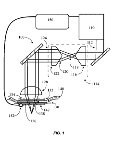
[0110] Referring now to FIG. 1, a system for radiative treatment 100 is shown.
An
electromagnetic radiation (EMR) source (e.g., a laser source) 110 generates an
EMR beam (e.g.,
a laser beam) 112 having a wavelength (within a range of about 1000nm to about
12,000nm, e.g.,
about 1550nm). According to some embodiments, the EMR beam 112 has a
transverse ring
energy profile (e.g., TEM 01*) natively from the EMR source 110. According to
other
embodiments, a beam shaper 114 shapes the EMR beam to produce a transverse
ring energy
profile. FIG. 1 illustrates a beam shaper 114 that employs two axicons. A
first axicon 116 having
CA 03160189 2022-05-03
WO 2021/096863 PCT/US2020/059842
a first wedge angle accepts the EMR beam 112 and produces a Bessel beam 118.
As the Bessel
beam propagates it forms a diverging ring energy profile. The diverging ring
energy profile 120
is collimated by a second axicon 122 into a collimated EMR beam having a
transverse ring
energy profile 124. According to some embodiments, the second axicon 122 has a
second wedge
angle that is substantially equal to the first wedge angle of the first axicon
116. The ring energy
profile 124 is then directed toward a focus optic 128. Some examples of the
focus optic 128
include converging optics (e.g., plano-convex lenses) and axicons. The focus
optic 128
converges the EMR beam and directs it toward a tissue 130 (e.g., skin). In
some cases, the focus
optic converges the EMR beam at a numerical aperture (NA) of at least about
0.2 (e.g., about 0.3
to about 0.5). According to some embodiments, a window assembly 132 is located
between the
focus optic 128 and the tissue 130. The window assembly 132 is substantially
transparent at the
wavelength of the EMR beam 124. Exemplary window materials include glass,
quartz and
sapphire. In some embodiments, the window assembly 132 is cooled and is used
to cool the
tissue 130 during treatment. Commonly, the window assembly 132 is placed in
contact with an
outer surface of the tissue during operation of the apparatus 100. According
to some
embodiments, the window assembly 132 includes two windows, a first window 134
and a second
window 136, with a cooling chamber 138 located between the two windows. The
cooling
chamber is configured to contain a coolant. In some embodiments, a flow of
coolant 140 passes
through the coolant chamber 138. In some embodiments, the coolant includes one
or more of: a
dielectric fluid, a fluorocarbon-based fluid, ethylene glycol, propylene
glycol, water, and an
antifreeze. In most cases, the coolant is selected to be generally (e.g.,
greater than about 50%)
transmissive at the wavelength of the EMR beam 124. For example, an exemplary
embodiment
includes an EMR beam 124 having a wavelength of 1550nm and a coolant that
includes a
fluorocarbon-based fluid (e.g., Flourinert TM from 3M), which is substantially
transmissive at
1550nm. In some embodiments, a medium 142 is placed between a bottom surface
of the
window assembly 132 and an outer surface of the tissue 130. In some versions,
this medium 142
acts to match an index of refraction of the window assembly 132 with the
tissue 130. In some
other versions, the medium penetrates the tissue. Examples of the medium 142
include: glycerol,
Phosphate-buffered saline (PBS), polyethylene glycol (PEG) 400, and other
suitable
biocompatible materials having and index of refraction approximately equal to
skin (e.g., about
1.4). In some embodiments, the system 100 further includes a controller 150 to
control the EMR
16
CA 03160189 2022-05-03
WO 2021/096863 PCT/US2020/059842
source 110. For example, it is advantageous in some embodiments for the EMR
source 110 to be
controlled in response to cooling of the tissue, to ensure only cooled tissue
is irradiated. In some
cases, the controller 150 is configured to control the EMR source to ensure
that the window
assembly cools the tissue to a determined temperature prior to generating the
EMR beam. In
some other cases, the controller 150 is configured to control the EMR source
to ensure that the
window assembly cools for a predetermined period of time prior to generating
the EMR beam.
According to some embodiments, a temperature sensor 152 (e.g., thermocouple,
or thermistor) is
used to directly measure tissue temperature. Alternatively, a temperature of a
component that is
in thermal communication with the tissue is measured with a temperature
sensor. For example,
temperature of coolant as it outflows the coolant chamber 138 can be used as
an indicator of
tissue temperature.
[0111] In some embodiments, the controller 150, in communication with one or
more of the
EMR source 110, the beam shaper 114, and the focus optic 128, is further
configured to control
one or more parameters of the EMR beam. Exemplary EMR beam parameters include
an inner
diameter of the ring-shaped energy profile, an outer diameter of the ring-
shaped energy profile, a
thickness of the ring-shaped energy profile, and depth of the focal region
within the tissue.
According to some embodiments, the EMR beam is scanned throughout the skin
tissue to
generate numerous locations of thermal disruptions within the tissue, for
example to provide a
fractionated treatment. Examples of systems and methods related to scanning
high NA EMR
beams are disclosed in U.S. Patent Application No. 16/219,801 entitled
"Electromagnetic
Radiation Beam Scanning System and Method" and International Application No.
PCT/US2018/065508, entitled "Scanning Systems for MR-Based Tissue Treatment,"
both of
which are incorporated herein by reference.
[0112] Referring to FIG. 2, a flowchart 200 represents a method for
irradiating a tissue according
to some embodiments. First, a tissue is cooled 210 using a window assembly,
which is placed in
contact with an outer surface of the tissue. According to some embodiments,
the window
assembly includes two windows, a first window and a second window, with a
cooling chamber
located between the two windows. The cooling chamber is configured to contain
a coolant. In
some embodiments, a flow of coolant passes through the coolant chamber. In
some
embodiments, the tissue is cooled to a predetermined temperature prior to any
subsequent steps
17
CA 03160189 2022-05-03
WO 2021/096863 PCT/US2020/059842
in the method 200. In some embodiments, the tissue is cooled for a
predetermined time prior to
any subsequent steps. Cooling the tissue to a predetermined temperature for a
predetermined
time in some cases can prevent thermal injury to outer layers of tissue (e.g.,
epidermis) and
thereby reduce down-time.
[0113] According to some embodiments, a temperature sensor is used to measure
temperature
that is related to the tissue, for example, a temperature of a component that
is in contact (and
therefore thermally communicative with) the tissue. Exemplary temperature
sensors include
thermistors, thermocouples, and infrared temperature sensors. The temperature
sensor in some
cases senses the tissue temperature directly and in other cases senses a
temperature of another
material that is related to tissue temperature (e.g., coolant outflowing a
contact cooling assembly,
which is in contact with the tissue).
[0114] Next, an electromagnetic radiation (EMR) beam is generated 220. The EMR
beam
includes a transverse ring energy profile (e.g., TEM 01* or donut energy
profile). The EMR
beam is then converged 230 forming a focal region. Typically, the EMR beam is
converged
using one or more optics (e.g., a converging lens and/or an axicon). In some
versions the EMR
beam is converged at a numerical aperture (NA) of about 0.2 or greater.
Finally, the EMR beam
is directed toward a tissue 240, such that the focal region is at least
partially located within (i.e.,
below an outer surface of) the tissue. In some versions, directing the EMR
toward a tissue 240
additionally includes scanning the EMR beam, such that the focal region is
moved within the
tissue. Scanning the EMR is typically done in at least one of three axes
(e.g., both axes
perpendicular to the optical axis and an axis parallel to the optical axis).
For example, the focal
region may be scanned in laterally in the tissue as well as in depth within
the tissue. In some
embodiments of the method 200, an optical tissue clearing medium is introduced
to the tissue.
For example, in some cases, the optical tissue clearing medium is introduced
onto a surface of
the tissue between the tissue and the window assembly. In some embodiments,
the method 200
additionally includes controlling at least one parameter of the EMR beam.
Exemplary parameters
of the EMR beam include an inner diameter of the ring-shaped energy profile,
an outer diameter
of the ring-shaped energy profile, a thickness of the ring-shaped energy
profile, and depth of the
focal region within the tissue.
18
CA 03160189 2022-05-03
WO 2021/096863 PCT/US2020/059842
Exemplary Embodiment
[0115] Referring now to FIG. 3A, an example system 300 is shown with a front
cover removed.
A fiber optic laser source 310 outputs a laser. An exemplary fiber laser
source 310 is a CW Er-
Yb laser having an average power of 20W (e.g., IPG Part No. ELR-20-1550-LP
from IPG
Photonics of Oxford, Massachusetts). The laser is collimated by a collimator
312 to a beam
width of approximately 4mm in diameter. The collimated laser beam is then
shaped by a beam
shaper 314 into a transverse ring (i.e., donut) energy profile (e.g., TEM
01*). The laser beam is
then directed along on optical train, ultimately being focused and directed
out of a window
assembly 316 at the bottom face of the example system 300.
[0116] FIG. 3B illustrates a cross-sectional view of the example system 300
and the beam shaper
314 taken along section line B-B in FIG. 3A. In FIG. 3B, the beam shaper
includes two identical
axicons, a first axicon 320 and a second axicon 322. An exemplary axicon is
Thorlabs Part No.
AX2510-C, which has a physical wedge angle of 100. A laser beam having a near
single order
mode (i.e., Gaussian transverse energy profile and M2 <= 1.5) is shaped by the
first axicon 320,
first to a Bessel beam then to a diverging transverse ring (i.e., donut)
energy profile. The second
axicon collimates the diverging transverse ring energy profile into a
collimated transverse ring
energy profile. The beam shaper is configured such that an inner diameter of
the transverse ring
energy profile is proportionally related to a separation distance between the
first axicon 320 and
the second axicon 322. According to the exemplary embodiment, the inner
diameter of the
transverse ring energy profile is nominally 4mm. The laser beam now shaped
into a transverse
ring (i.e., donut) energy profile propagates further along an optical path
ultimately exiting the
system 300 through the window assembly 316. FIG. 3C illustrates a cross-
sectional view of the
example system 300 and the window 316 taken along section line C-C in FIG. 3A.
A focus optic
330 is located up-beam from the window 316, such that the laser beam converges
as it transmits
through the window assembly 316. Ultimately, the focus optic 330 brings the
laser beam to a
focal region outside of the window assembly 316, such that when the window
assembly is placed
into contact with a tissue the focal region is located within the tissue. An
exemplary focus optic
is an aspherical lens, Thorlabs Part No. A240-C, having a nominal effective
focal length of 8mm.
In some embodiments, a Z-stage 331 houses the focus optic 330 and is
configured to adjust the
position of the focus optic 330 along an optical axis and thereby affect the
depth of the focal
19
CA 03160189 2022-05-03
WO 2021/096863 PCT/US2020/059842
region relative the window 316 (i.e., depth of the focal region within the
tissue). An exemplary
Z-stage 331 is Newscale PN: M3-FS from Newscale Technologies of Victor, New
York. In some
cases the controller 150 is configured to control the Z-stage in order to
affect changes in focal
region depth.
[0117] FIG. 3D illustrates a detail view of the example system 300 taken from
detail circle D in
FIG. 3C. The window assembly 316 is shown in greater detail in FIG. 3D. A
first window 340 is
shown proximal the focus optic 330. A second window 342 is shown separated
from the first
window 340. A coolant chamber 344 is found between the first window 340 and
the second
window 342. The coolant chamber 344 is hermetically sealed in order to contain
coolant as it
flows through the coolant chamber 344. The coolant is warmed through contact
with the window
assembly 316 and returned to a chiller. The coolant is then cooled by a
chiller, for example a
thermoelectric chiller (e.g., Part No. UC190 from Solid State Cooling of
Wappingers Falls, New
York) and recirculated to the window assembly 316. Disclosure related to
window assemblies for
cooling during irradiation is included in U.S. Patent Application No.
16/237,367 to Dresser et al.,
which is incorporated herein by reference. The exemplary window assembly
disclosed in
Reference to FIGS. 3A-D is described below in greater detail.
[0118] An exemplary window assembly 316 for cooling during irradiation is
schematically
represented in various views in FIGS. 3E-3H. FIG. 3E illustrates a top
isometric view of the
assembly 316 (i.e., portion of the cooling element 316 facing the EMIR source
/ facing away from
the target tissue). FIG. 3F shows a bottom isometric view of the window
assembly 316 (i.e.,
portion of the window assembly 316 facing the target tissue / facing away from
the EMIR
source). FIG. 3G shows a bottom view of the window assembly 316. FIG. 3H shows
a section
view of the window assembly 316, along the section lines shown in FIG. 3G. The
exemplary
window assembly 316 includes a frame 350. Referring to FIGS. 3E and 3G, the
frame 350 has
three datums 352. The datums 352 correspond to a mount on an energy-based
device (e.g., 300),
which can generate an irradiation, thereby allowing the window assembly 316 to
be removably
attached and replaced on the energy based device. According to some
embodiments, the datums
352 may approximate one or more geometric forms, for example a plane, a line,
and a point.
According to some versions, the datums 352 include a part of kinematic mount
(e.g., Maxwellian
or Kelvin mount). The three datums 352 of the window assembly 316 can be
located in a plane.
CA 03160189 2022-05-03
WO 2021/096863 PCT/US2020/059842
The exemplary window assembly 316 further includes a first window 340 sealed
to the frame
350 by a first seal 354 and a second window 342 being sealed to the frame 350
by a second seal
356. According to some embodiments, the first seal 354 and the second seal 356
includes an
adhesive. Examples, of adhesives can include light cure adhesives, silicones,
and epoxies.
According to other embodiments, the first seal 354 and/or the second seal 356
include a weld, a
braze, or a solder and the edges of the corresponding first window 340 and/or
the second window
342 can be metallized, sputtered, or coated with a material (e.g., metal)
allowing for this type of
seal. Additionally, the second window 342 is affixed to the frame 350 with one
or more fasteners
358. It can be seen in FIGS. 3G and 3H, the fastener 358 of the window
assembly 316 includes a
clamp plate held in place by 3 machine screws. Additional examples of a
fastener can include a
screw, a clamp, a snap a retaining ring, a tab, or any combination thereof.
Affixing the second
window 342 to the frame allows for the distal surface 360 of the second window
342 to be placed
firmly in contact with tissue, without introducing additional stress to the
second seal 356, which
can result in flexure or movement of a distal surface 360 of the second window
342.
[0119] A change in distance between the distal surface 360 and an optic
focusing an EMIR beam
affects a working distance of the beam and a location of a resulting focus
within a tissue.
According to some embodiments, the distal surface 360 of the second window 342
can be
located at a predetermined geometry (e.g., orientation, location, etc.)
relative the datum 352. For
example in some versions, the second window 342 is located parallel to a plane
approximated by
one or more datums 352 to within a desired tolerance (e.g., 0.5mrad).
Additionally, the second
window 342 can be located at a precise distance along the optical axis (e.g.,
z-axis) within a
desired tolerance (e.g., 0.05 mm). Additionally, according to some
embodiments, both the first
window 340 and the second widow 342 are located parallel and a prescribed
distance between
them can be within desired tolerances (e.g., 0.5mrad and 0.05mm). For various
reasons, the
distal surface 360 of the second window in some embodiments includes a non-
plano shape (e.g.,
convex or concave). For example, a convex shaped distal surface 360 can be
advantageous for
compressing a tissue when placed in contact with tissue.
[0120] FIG. 3H depicts a chamber 344 within the system 400. The chamber 344 is
bounded by
the frame 350, the first window 340, and the second window 342. The chamber
344 can be
sealed by the first seal 354 and the second seal 356. The chamber 344 is
configured to contain a
21
CA 03160189 2022-05-03
WO 2021/096863 PCT/US2020/059842
coolant. According to some embodiments, a flow of coolant is supplied to the
chamber 344
through one or more ports 362 in fluidic communication with the chamber 344.
According to
some embodiments, the port 362 can provide for the flow of coolant from a
coolant flow source,
which is in fluidic communication with the port 362. In some implementations,
the coolant flow
source can be in fluidic communication with the port 362 by way of one or more
fittings 364.
FIGS. 3E and 3F illustrate both a coolant supply fitting 364a and a coolant
return fitting 364b,
for supplying coolant to and returning coolant from the chamber 344.
[0121] According to some embodiments, the second window includes a material
having a high
thermal effusivity (e.g., quartz, sapphire, diamond, etc.). Higher thermal
effusivity can allow for
more heat to be transferred from the tissue surface to the flow of coolant.
Likewise, according to
some embodiments, the first window 340 includes a material having a lower
thermal effusivity
(e.g., a glass or a polymer). Implementations having a first window 340 with a
lower thermal
effusivity material can transfer less heat through the first window and into
the flow of coolant.
As a result, condensation can occur more slowly than in versions where the
first window 340
includes a high thermal effusivity material. Additionally, in some embodiments
the first window
has a thickness (e.g., about lmm), which is greater than that of the second
window (e.g., about
0.5mm), allowing thermal energy transfer to occur more freely across the
second window.
According to some versions, a non-condensing gas such as clean dry air,
nitrogen, carbon
dioxide, or argon can be blown against the first window to further prevent
condensation.
[0122] FIG. 4A illustrates a simulated optical layout 400 according to some
embodiments. A
collimated Gaussian beam 410 propagates incident and on-center to a first
axicon 412, which
forms a Bessel beam 414. The Bessel beam 414 propagates incident and on-center
to a second
axicon 416, which forms a collimated transverse ring (i.e., donut) energy
profile beam 418. The
collimated transverse ring energy profile beam 418 propagates incident and on-
center to an
aspherical focus optic 420, which forms a converging transverse energy profile
422 that focuses
to a focal region 424.
[0123] FIG. 4B illustrates a first simulated Gaussian beam profile 430 of the
collimated
Gaussian beam 410. FIG. 4C illustrates a first simulated transverse ring
(i.e., donut) beam profile
432 of the converging transverse ring energy profile 422 0.5mm before the
focal region 424.
22
CA 03160189 2022-05-03
WO 2021/096863 PCT/US2020/059842
FIG. 4D illustrates a second simulated transverse ring beam profile 434 of the
converging
transverse ring energy profile 422 0.2mm before the focal region 424. And,
FIG. 4E illustrates a
third simulated transverse ring beam profile 436 of the converging transverse
ring energy profile
422 0.1mm before the focal region 424. The converging transverse ring energy
profile beam 422
has a lower irradiance over the beam profile than a Gaussian mode beam would
have under the
same conditions. Referring, to FIG. 4F a Gaussian energy profile 440 for a
Gaussian beam
0.5mm from focus is shown. A transverse ring (i.e., donut) energy profile 442
for a transverse
ring energy profile beam 0.5mm from focus is shown in FIG. 4G. Both of the
beams
characterized in FIG. 4F and FIG. 4G have identical powers (e.g., 1W).
However, a local
maximum irradiance for the Gaussian beam is much larger (e.g., 1.29W/cm2) than
the transverse
ring energy profile beam (e.g., 0.75W/cm2). This allows the transverse ring
beam to deliver less
peak energy density to outer layers of skin (e.g., epidermis), while
delivering the same amount of
energy to deep layers of skin (e.g., dermis). Control of the reduction in peak
local energy density
in a transverse ring beam is accomplished by varying a width of an inner
diameter of the
transverse ring energy profile. Larger inner diameters push more energy to
outer portions of the
beam and reduce the peak energy density (or power density) within the beam.
Additionally, peak
local energy density can be reduced in both the Gaussian and transverse ring
energy profiles by
increasing a numerical aperture of the focus optic 420.
Exemplary Ex Vivo Studies
[0124] A number of studies were performed according to some embodiments. The
studies were
performed using a continuous wave (CW) Er-Yb fiber laser with a maximum
average power of
20W and a wavelength of 1550nm (IPG laser model: ELR-20-1550LP). Excised human
tissue
was irradiated using a high numerical aperture (e.g., NA greater than or equal
to 0.4) focusing
system. Fractional irradiation was accomplished by pulsing the CW fiber laser
as the human
tissue was scanned relative the focusing system on X-Y translation stages. The
human tissue was
then sectioned, stained, and reviewed. A nitro blue tetrazolium chloride
(NBTC) stain was used
to test for viability. Specifically, the NBTC stain acts on proteins within
the tissue. Once these
proteins are damaged (e.g., thermally denatured) they are no longer stained by
the NBTC and
appear unstained.
23
CA 03160189 2022-05-03
WO 2021/096863 PCT/US2020/059842
Study No. 1
[0125] A first study was conducted to determined pulse energy required for non-
ablative thermal
disruption of the tissue using a Gaussian beam. Parameters used in study
number 1 are shown
below:
Table 1 - Study No. 1 Parameters
LENS NA 0.5 Units
Human
Skin Abdominoplasty
Single layer Depth 0.5mm in
skin tissue4 tissue3 tissue2 tissuel
Single layer Depth 0.7mm in
skin tissue5 tissue6 tissue7 tissue8
Laser average power 15.5 15.5 15.5 15.5 W
Required Energy per spot 10 20 30 40 mJ
Pitch of spots 0.5 0.5 0.5 0.5 mm
Spot size 0.025 0.025 0.025 0.025 mm
Pulse duration 0.65 1.29 1.94 2.58 msec
stage speed 38.75 19.38 12.92 9.69 mm/sec
0.01 0.03 0.04 0.05 sec
laser pulse rep rate 77.5 38.75 25.83 19.38 Hz
Treatment time for
10x10mm2 5.2 10.3 15.5 20.6 Sec
[0126] Some representative results for Study No. 1 are shown in histological
slides in FIGS. 5A-
D. FIG. 5A illustrates a horizontal cross-section taken after irradiation with
pulses having an
energy of about 10mJ. FIG. 5B illustrates a vertical cross-section taken after
irradiation with
pulses having an energy of about 10mJ. Very slight thermal denaturing of
proteins is evidenced
by the NBTC stain. In contrast, irradiation at pulse energies in excess of
10mJ, for example
about 40mJ, is shown to result in pronounced thermal disruption. FIG. 5C
illustrates a horizontal
cross-section taken at about 300micrometers below a surface of the tissue post
40mJ per pulse
irradiation. And, FIG. 5D illustrates a vertical cross-section of tissue after
40mJ per pulse
irradiation. From study No. lit was concluded that given this set of
parameters 10mJ per pulse is
a threshold pulse energy below which little-to-no thermal disruption occurs.
24
CA 03160189 2022-05-03
WO 2021/096863
PCT/US2020/059842
Study No. 2
[0127] Study No. 2 was conducted to determine effects of optical tissue
clearing mediums on
fractionated non-ablative ex vivo irradiation. Samples of excised human tissue
were placed in
optical tissue clearing mediums for 4 hours prior to irradiation. The samples
were soaked in a
petri dish containing the medium epidermis down. Two optical tissue clearing
mediums were
tested: phosphate-buffered saline (PBS) and glycerol. Parameters used in study
number 2 are
shown below:
Table 2 - Study No. 2 Parameters
LENS 0.5NA Units Optical
Tissue
Human Clearing
Skin Abdominoplasty Medium
Single layer Depth tissue tissue tissue
0.5mm in skin tissue 4 3 2 1 GLYCEROL
Single layer Depth tissue tissue tissue
0.7mm in skin tissue 8 7 6 5 PBS
Laser power 15.5 15.5 15.5 15.5 W
Required Energy
per spot 20 10 7 5 mJ
Pitch of spots 0.5 0.5 0.5 0.5 Mm
Spot size< 0.025 0.025 0.025 0.025
Mm
pulse duration 1.290 0.645 0.452 0.323 msec
mm/se
stage speed 19.38 38.75 55.36 77.50 c
0.03 0.01 0.01 0.01 Sec
110.7 155.0
laser pulse rep rate 38.75 77.50 1 0 Hz
Treatment time for
10x10mm2 10.3 5.2 3.6 2.6
[0128] Thermal disruption was only visible at 20mJ per pulse in the optical
tissue clearing
medium soaked tissue samples. No thermal disruption was apparent through NBTC
viability
staining at the lower testing pulse energies (5mJ, 7mJ, and 10mJ). Some
representative results
for Study No. 2 are shown in histological slides in FIGS. 6A-B. FIG. 6A
illustrates a vertical
CA 03160189 2022-05-03
WO 2021/096863 PCT/US2020/059842
cross-section taken in a glycerol soaked tissue, irradiated at 20mJ per pulse.
FIG. 6B illustrates a
vertical cross-section taken in a PBS soaked tissue, irradiated at 20mJ per
pulse.
Study No. 3
[0129] Study No. 2 was conducted to determine effects of a transverse ring
(i.e., donut) energy
profile on fractionated non-ablative ex vivo irradiation. Samples of excised
human tissue were
placed in optical tissue clearing mediums for 4 hours prior to irradiation.
The samples were
soaked in a petri dish containing the medium epidermis down. Two optical
tissue clearing
mediums were tested: phosphate-buffered saline (PBS) and glycerol. The laser
beam was shaped
into a transverse ring energy profile as described above and focused into the
tissue. Parameters
used in study number 2 are shown below:
Table 3 - Study No. 3 Parameters
Bessel Beam with two
axicons, with 1550nm
coated windows
Beam
Abdominoplasty skin Units OTC
shape
Single layer Depth
0.5mm in skin tissue 4 tissue 3 tissue 2 tissue 1 PBS
Donut
tissue 7 tissue 6 tissue 5 -
Glycerol Donut
Laser power 16.2 16.2 16.2 16.2 W
Required Energy per
spot 20 10 7 5 mJ
Pitch of spots 0.5 0.5 0.5 0.5 Mm
Spot size< 0.025 0.025 0.025 0.025 Mm
pulse duration 1.235 0.617 0.432 0.309 Msec
mm/se
stage speed 20.25 40.50 57.86 81.00 c
laser pulse rep rate 0.02 0.01 0.01 0.01 Sec
40.50 81.00 115.71 162.00 Hz
Treatment time for
10x10mm2 9.9 4.9 3.5 2.5
[0130] Histological results from Study No. 3 are described in reference to
FIGS. 7A-E. FIG. 7A
shows four histological images in a Cartesian layout with glycerol soaked
tissue above, PBS
soaked tissue below, 10mJ per pulse energies on a left side, and 20mJ per
pulse energies on a
26
CA 03160189 2022-05-03
WO 2021/096863 PCT/US2020/059842
right side. In general, wider and deep thermal disruptions are apparent with
20mJ than with 10mJ
pulse energies. FIG. 7B illustrates horizontal histological images taken of
tissue soaked in
glycerol and irradiated with 10mJ pulse energies. FIG. 7C illustrates
horizontal histological
images taken of tissue soaked in glycerol and irradiated with 20mJ pulse
energies. FIG. 7D
illustrates horizontal histological images taken of tissue soaked in PBS and
irradiated with 10mJ
pulse energies. FIG. 7E illustrates horizontal histological images taken of
tissue soaked in PBS
and irradiated with 20mJ pulse energies. In horizontal histologies of tissue
irradiated with a
transverse ring energy profile, ring shaped damage can be seen (e.g., FIG.
7C). The damage that
appears as a ring in a horizontal histology is in three-dimensions a thin-
walled hollow cone of
damage, that comes to a point deep (e.g., 300 ¨ 1000 micrometers) within the
tissue. Within the
cone of damage there exists healthy unaffected tissue as evidenced by the
rings of damage in the
horizontal cross-sections (e.g., FIG. 7C) and the 'Y' shaped damage in the
vertical cross-sections
(e.g., FIG. 7A). A benefit of this irradiation pattern is that less epidermis
is damaged than with
current fractionated irradiation techniques; and, that the epidermis that is
damaged is damaged in
a small narrow width (e.g., 1 ¨ 100 micrometers) that is surrounded by healthy
(i.e., unaffected)
tissue.
Parameter Selection
[0131] Parameters relevant to practice of embodiments of the present
disclosure are outlined in a
table below:
Table 4 - Exemplary Parameters and Ranges
Minimum Maximum Nominal
EMR Wavelength 200 20000 1550
(nm)
Numerical Aperture 0.01 1 0.5
(-)
Focal Region Width 0.05 5000 4
(micrometers, pm)
Focal Region Length 0.005 500 0.5
(mm)
Focal Region Depth 0 10 0.3
Below Tissue
Surface (mm)
Pulse Energy (mJ) 0.1 300 30
27
CA 03160189 2022-05-03
WO 2021/096863 PCT/US2020/059842
Pulse Duration (nS) 1 1,000,000,000 5,000,000
Average Power (W) 0.01 100 10
Peak Power (N) 1 1,000,000 20
Precool Time (S) 0.1 200 10
Precool Temperature -200 (for cryogen 10 2
( C) cooling)
Transverse ring (i.e., 0.05 50 4
donut) energy
profile, inner
diameter (mm)
Transverse ring (i.e., 0.05 50 2
donut) energy
profile, annular
width (mm)
Optical Tissue Glycol, Phosphate-buffered Saline (PBS), Polyethylene
Glycol (PEG)
Clearing 400.
Constituents
Coolant Constituents Water, alcohol, propylene glycol, fluorocarbon-based
fluids, and anti-
freeze
Scanning System Translation Stage(s), galvanometers
[0132] In some embodiments, aspects of the ring-shaped energy profile are
controllable. FIG. 8A
shows a pair of axicons 800 configured to generate a ring-shaped energy
profile. A relationship
between separation (S) 810 of the two axicons 800 and major diameter 812 of
the resultant ring-
shaped beam can be expressed:
DMajor
=
2 * tan((n ¨ 1) * a)
[0133] Wherein, n is index of refraction of the first and second axicon and a
is a wedge angle of
the first and second axicon 800.
[0134] As described above, a collimated beam diameter 814, as it enters the
axicon pair 800,
determines the ring-shaped energy profile width 816. Therefore, in some
embodiments, the width
of the ring-shaped energy profile 816 is controlled by varying the collimated
beam diameter 814.
For example, in some cases a beam expander (e.g., Gallian beam expander or
Keplerian beam
expander) is used to expand (or reduce) the collimated beam diameter 814,
before it reaches the
axicon pair 800. Minor (i.e., inner) diameter 818 can be expressed in terms of
a major (i.e., outer)
28
CA 03160189 2022-05-03
WO 2021/096863 PCT/US2020/059842
diameter 812 of the ring energy profile. Specifically, minor diameter 818 is
equal to the major
diameter 812 less the diameter of the collimated beam 814, or:
Ominor = 0Major Obeam
where, 05
-minoris the minor diameter 818; Omaj, is the major diameter 812; and, Obeam
is the
beam diameter 814. According to some embodiments, one or more parameters
related to the
ring-shaped energy profile is controlled by a controller, which manipulates
the above described
parameters (e.g., axicon pair 800 separation distance 810 and/or beam expander
rate). For
example, in some cases the separation distance 810 between the axicon pair 800
may be
electronically manipulated by use of a motorized stage (e.g., Thorlabs PN: PT1-
Z8). Likewise, in
some cases (e.g., a Gallian beam expander) an optical path distance between
two optics controls
a beam expansion (or beam reduction) rate of a beam expander. In this case, a
motorized stage
may also be used to control the width of the beam 814 as it enters the axicon
pair 800.
[0135] Small diameter fractional treatments result in smaller injury and
faster healing. For
example, it has been found that fractional damage greater than a certain width
(e.g., about
0.15mm, 0.25mm, or 0.5mm) can cause scarring in some individuals. Small
fractional damage
widths even below what is now commercially achievable will further minimize
down-time up to
a threshold minimum fractional damage width size. Specifically, beam sizes
that are smaller than
a single cell (e.g., about 20micr0meter5) result in practically the smallest
possible fractionated
damage. As described above, in some cases the above described exemplary
optical systems
achieve thermal injury to tissue which is on this scale. In additional
exemplary embodiments,
small fractionated injury to this tissue is achieved through another exemplary
optical system.
[0136] One skilled in the art will appreciate further features and advantages
based on the above-
described embodiments. Accordingly, the disclosed embodiments are not to be
limited by what
has been particularly shown and described, except as indicated by the appended
claims. All
publications and references cited herein are expressly incorporated herein by
reference in their
entirety.
Additional Embodiments.
29
CA 03160189 2022-05-03
WO 2021/096863 PCT/US2020/059842
[0137] An additional embodiment for affecting fractionated damage at a tens of
micrometer
scale is described in reference to FIGS. 9A-B. Referring to FIG. 9A, an
optical scheme 900 is
displayed that produces a Bessel beam focal region 910. Bessel beam focal
regions, unlike
normal diffraction-limited focal regions, have a focal width and a focal
region length that can be
decoupled from one another. Normally, a focal region length (i.e., a depth of
field) is
proportionally related to the square of the focal region radius (e.g.,
Rayleigh range). Decoupling
focal region length from focal region width allows for formation of very long
(e.g., greater than
0.5mm long) focal regions, which are also very narrow (e.g., less than about
0.1mm wide).
[0138] FIG. 9A schematically illustrates an optical path that can be used to
generate a long
narrow beam. Three axicons are used in this configuration. A first axicon 912
and a second
axicon 914 are used to shape the beam into a collimated annular beam 916 and a
third axicon 918
is used to focus the beam to a Bessel beam focal region 910.
[0139] According to some exemplary embodiments, a width of damage for a
fractional treatment
is related to a width of a first lobe of the Bessel beam focal region 910. The
half width, w0, of the
first lobe of a Bessel beam focal region 910 is a function of the wavelength,
k, the wedge angle
of the axicon, a, and the index of refraction of the axicon, n:
2.4048
(.0 = _____________
0
A* Sin((n ¨ 1) * a)
[0140] Therefore, according to some embodiments, selection of this optical
parameter is
achieved through selection of an axicon wedge angle for the third axicon 918.
A table below
illustrates some exemplary first lobe diameter for a 1550nm beam based upon
axicon wedge
angle.
Table 5 - Wedge Angle to First Lobe Diameter (wavelength = 1550nm)
Wedge Angle First Lobe Diam.
(deg) (um)
0.5 296
1 148
2 74
CA 03160189 2022-05-03
WO 2021/096863 PCT/US2020/059842
5 30
10 15
20 7
30 5
[0141] A length of damage by a fractionated treatment is related to a length
of the Bessel beam
focal region 910. The length of a Bessel beam (e.g., depth of field [DOF]) 920
formed by an
axicon is a function of a width of the beam at the axicon. When an annular
beam is used the focal
region length is a function of a width of an annulus 922. The width of the
annulus is in turn a
function of (e.g., half of) a width of the collimated beam, 924, which is
shaped to form the
annular beam. The length of the Bessel beam can be approximated using an
equation below:
DOF = _______
2(n ¨ 1) * tan(a)
[0142] For example, with a 4mm output beam, a wavelength of 1550nm, and a 20
wedge angle,
the length of the Bessel beam focal region is approximated to 15mm.
[0143] A working distance (WD) 926 between the tip of the third focusing
axicon 918 and the
Bessel beam focal region 910 is a function of an inner diameter 928 of the
annular ring 916. The
working distance 926 measured from the tip of the axicon 918 can be
approximated using an
equation below, with reference to FIGS. 9A-B:
WD= r * tan(a)
tan((n ¨ 1) * a)
[0144] The above equation is derived from two below equations for X1 and XO.
FIG. 9B
illustrates the relationship between these equations.
X0 = ¨r * tan(a)
= __________
tan((n ¨ 1) * a)
WD = Xo +
[0145] As can be seen above the minor (i.e., inner) diameter 928 of the ring
energy profile 916
affects the working distance 926. For example, a non-annular beam being acted
upon by an
31
CA 03160189 2022-05-03
WO 2021/096863 PCT/US2020/059842
axicon results in a Bessel beam focal region that begins at the tip of the
axicon. In some
embodiments, the focal region 910 is controlled to a depth within a tissue
(e.g., below a surface
of a tissue) by controlling a minor diameter 928 of the annular beam incident
the focusing axicon
918 thereby affecting the working distance 926 of the focal region 910. In
some versions, the
minor diameter 928 of the annular beam 916 is a function of separation between
the first axicon
912 and the second axicon. Minor diameter can be expressed in terms of a major
(i.e., outer)
diameter 930 of the ring energy profile 916. Specifically, minor diameter 928
is equal to the
major diameter 930 less the diameter of the collimated beam 924, or:
Ominor = 0Major Obeam
[0146] FIG. 10 illustrates one exemplary embodiment of a treatment system
1010. As shown,
the treatment system 1010 includes a platform 1012, and emitter 1014, and a
controller 1016.
The platform 1012 can include one or more manipulator or arm 1020. The arm
1020 can be
coupled to the emitter 1014 for performing various treatments on a target
tissue 1022 of a subject
1024. Operation of the platform 1012 and emitter 1014 can be directed by a
user, manually or
using the controller 16 (e.g., via a user interface). In certain embodiments
(not shown), the
emitter can have a hand-held form factor and the platform 1012 can be omitted.
In other
embodiments, the platform can be a robotic platform and the arms can be
communicatively
coupled to the controller for manipulation of the emitter.
[0147] The emitter 1014 and controller 1016 (and optionally the platform 1012)
can be in
communication with one another via a communications link 1026, which can be
any suitable
type of wired and/or wireless communications link carrying any suitable type
of signal (e.g.,
electrical, optical, infrared, etc.) according to any suitable communications
protocol.
[0148] Embodiments of the controller 1016 can be configured to control
operation of the emitter
1014. In one aspect, the controller 1016 can control movement of EMR 1030. As
discussed in
detail below, the emitter 1014 can include a source 1032 for emission of the
EMR 1030 and a
scanning system 1034 for manipulation of the EMR 1030. As an example, the
scanning system
1034 can be configured to focus EMR 1030 to a focal region and translate
and/or rotate this focal
region in space. The controller 1016 can send signals to the source 1032, via
the
communications link 1026 to command the source 1032 to emit the EMR 1030
having one or
32
CA 03160189 2022-05-03
WO 2021/096863 PCT/US2020/059842
more selected properties, such as wavelength, power, repetition rate, pulse
duration, pulse
energy, focusing properties (e.g., focal volume, Rayleigh length, etc.). In
another aspect, the
controller 1016 can send signals to the scanning system 1034, via the
communications link 1026
to command the scanning system 1034 to move the focal region of the EMR 1030
with respect
the target tissue 1022 in one or more translation and/or rotation operations.
[0149] Embodiments of the treatment system 1010 and methods are discussed
herein in the
context of treatment within skin tissue, such as a dermal layer. However, the
disclosed
embodiments can be employed for treatment of any tissue in any location of a
subject, without
limit. Examples of non-skin tissues can include, but are not limited to,
surface and sub-surface
regions of mucosal tissues, genital tissues, internal organ tissues, and
gastrointestinal tract
tissues.
Exemplary Manual Scanned System
[0150] In some embodiments, a hand-held system 1100 is used which is manually
scanned (i.e.,
manually moved by a clinician) over a treatment area. FIGS. 11A-11C illustrate
an exemplary
embodiment, which can be scanned manually. FIG. 11A illustrates a front view
of the system
1100; FIG. 11B illustrates a side view of the system 1100; and, FIG. 11C
illustrates a cross-
sectional view of the system 1100. Referring to FIGS. 11A ¨ 11C, a fiber laser
outputs a laser
beam by way of a collimator 1110. The laser beam can be of any wavelength.
Specifics of
wavelength selection are described in detail above. The collimated laser beam
is acted upon by a
beam shaper 1112. As described in detail above, the beam shaper 1112 takes the
collimated laser
beam from the collimator 1110 and shapes it to a transverse ring energy
profile. The beam
shaper, as shown in the cross-sectional view (FIG. 11C), has a first axicon
1112A, an alignment
mirror 1112B, and a second axicon 1112C. The two axicons 1112A and 1112C are
used to shape
the beam. The alignment mirror 1112B is used to align the laser beam onto the
second axicon
1112C. Typically, axicons are very sensitive to misalignment, especially
misalignment of
centration. After the beam shaper 1112, the laser beam is then reflected by a
first galvanometer
mirror 1114 and directed into and through a beam expander 1116. The beam
expander is a
Keplerian beam expander and includes a first positive optical element 1116A,
which focuses the
beam to an intermediate focus and a second positive optical element 1116B,
which collimated
33
CA 03160189 2022-05-03
WO 2021/096863 PCT/US2020/059842
the laser beam. In some cases, one or more of the beam expander optics is
dynamic and can be
moved along the optical axis. A linear stage 1116C moves the second positive
optical element
1116B along the optical axis. An exemplary linear stage is a Newscale M3-LS-
3.4-15 from
Newscale Technologies of Victor, NY. The beam expander 1116 expands the
collimated ring
beam, for example by a factor of between 2 - 20X. Upon exiting the beam
expander 1116, the
laser beam is reflected by a static fold mirror 1118, focused by an objective
lens 1119 (e.g., An
aspherical focus optic, for example, Asphericon PN: AFL25-40, from Asphericon
of Jena,
Germany), reflected by a second galvanometer mirror 1120, and directed to
emerge from a
contact window 1122. In some versions, the beam expander is an afocal relay
system that is
placed at conjugate distances between the first galvanometer mirror 114 and
the objective lens
1119. The contact window 1122, like many described in detail above, includes a
first window
1122A, a second window 1122B separated from the first window, and a coolant
chamber 1122C
located between the first window 1122A and the second window 1122B. The second
window
1122B has a tissue contacting surface (i.e., outer surface) that is convex.
This shape is
advantageous in some circumstances as it helps to ensure positive contact with
the tissue being
treated and slides more easily of the tissue.
[0151] The handheld system 1100 described in FIGS. 11A-11C is used to treat an
area manually.
As the clinician moves the handheld system 1100 over the treatment tissue, the
second
galvanometer mirror 1120 scans a line of points over the surface of the skin
side to side.
Referring now to FIG. 12, an exemplary line of scanned points 1200 is shown.
The exemplary
line 1200 includes eight individual points 1210, where laser energy is
delivered. The eight points
1210 are scanned to sequentially as shown in FIG. 12 from top to bottom (i.e.,
A-H). After the
final point has had energy delivered at it (i.e., point H), the line scan is
repeated and starts again.
The line has a width 1212 that is approximately equal to the number of points
1210 multiplied by
a pitch 1214 (i.e., distance between adjacent points). Referring again briefly
to FIGS. 11A-C the
line is scanned by the second galvanometer mirror 1120 of the handheld system
1100 along a
manual scan direction 1216, which is generally perpendicular to the direction
of the line scan
1200.
Exemplary Beam Scanning Systems
34
CA 03160189 2022-05-03
WO 2021/096863 PCT/US2020/059842
[0152] In some embodiments, beam scanning systems and methods are provided.
Disclosure
related to these embodiments and beam scanning systems and methods is
described below.
Generally, beam scanning systems and methods can be categorized in one or more
of the
following types pre-objective scanning, objective scanning, and post-objective
scanning. Pre-
objective scanning includes embodiments, in which the beam is scanned (e.g.,
deflected, tipped,
and/or tilted) before (i.e., up beam from) being directed incident upon the
objective. Objective
scanning includes embodiments, in which scanning (e.g., deflecting, tipping
and/or tilting) the
beam is performed at the objective, for example by moving the objective. Post-
objective
scanning includes embodiments, in which scanning (e.g., deflecting, tipping,
and/or tilting) is
performed after (i.e., down beam from) the objective.
Pre-Objective Scanning
[0153] FIG. 13 is a schematic illustration of a pre-objective scanning system
2100, which
includes an objective 2110 and a scanning unit 2112. The scanning unit 2112
can receive a laser
beam 2104 from a laser source 2102 and direct the laser beam 2104 to the
objective 2110. The
objective 2110 can receive the laser beam 2104 and direct a focused laser beam
2106 to a focal
volume 2108 in the treatment region of a tissue 2116 (e.g., skin). The
scanning system 2112 can
alter the direction of the laser beam 2104 directed towards the objective
2110. For example, the
scanning system 2112 can alter the direction of the outgoing laser beam along
one or more scan
directions. Change in the direction of the laser beam 2104 impinging the
objective 2110 can
cause the focal volume 2108 to trace a treatment path 2114 in the tissue 2116.
The focal volume
2108 traverses the treatment path 2114 at a scan rate. The scanning unit 2112
includes one or
more optical elements that can direct the laser beam 2104 (or a portion of the
laser beam 2104) to
the objective 2110. The pre-objective scanning system 2100 can include a
contacting surface
(e.g., as shown in FIG. 24) that can be positioned between the objective 2110
and the tissue
2116. The contacting surface can apply pressure the surface of the tissue
2116, and allow for
dissipation of heat from the surface of the tissue 2116.
[0154] FIG. 14 is an illustration of an exemplary pre-objective scanning
system 2200. The
scanning system 2200 includes a polygon scanner 2202 which can receive the
incident laser
beam 2104 (e.g., from a laser source 2102) and direct the incident laser beam
2104 towards an
objective 2110 (e.g., f-theta lens). The outgoing direction of the laser beam
2104 (e.g., incidence
CA 03160189 2022-05-03
WO 2021/096863 PCT/US2020/059842
angle with which the laser beam 2104 impinges on the objective 2110) can
determine the
location of the focal volume 2108 in the tissue 2116 (e.g., in the x-y plane).
According to some
embodiments, the laser source 2102 provides a plurality of laser pulses
resulting in a plurality of
corresponding focal volumes. A distance between two focal volumes resulting
from sequential
laser pulses is focal volume pitch.
[0155] The polygon scanner 2202 can include multiple reflecting surfaces
(e.g., 2202 a-c). The
polygon scanner 2202 can rotate about an axis 2204 along a rotational
direction 2206. As the
reflecting surfaces 2202a-c rotate around the axis 2204 (e.g., angular
position of the reflecting
surfaces 2202a-c with respect to the axis 2204 changes), the angle of
incidence of the incident
laser beam 2104 in the y-z plane changes. This varies the direction of the
outgoing laser beam
2104 along a first scan direction (e.g., along the y-axis). For example, if a
reflecting surface
(e.g., 2202b) is rotating about the axis 2204 along the rotational direction
2206, the direction of
the outgoing laser beam sweeps from a higher y-value to a lower y-value.
[0156] The axis 2204 can tilt / rotate about the z-axis and/or the x-axis.
This can cause the
angle of incidence of the incident laser beam 2104 in the x-z plane to change,
which varies the
direction of the outgoing laser beam 2104 along a second scan direction (e.g.,
along the x-axis).
Rotation of the polygon scanner 2202 and the rotation/tilting of the axis 2204
can allow for
varying of the direction of the outgoing laser beam 2104 that can result in
the scanning of the
outgoing laser beam 2104 in the x-y plane.
[0157] Based on the variation of the direction of the outgoing laser beam
2104, the objective
2110 can trace the focal volume 2108 along one or more treatment paths in the
tissue 2116. For
example, variation of the direction of the outgoing laser beam 2104 due to
rotation of the
polygon scanner 2202 can cause the focal volume 2108 to move along the y-axis.
Variation of
the direction of the outgoing beam due to tilting of the axis 2204 can cause
the focal volume
2108 to move along the x-axis. In one implementation, the pre-objective
scanning system 2200
can be moved along the x-axis relative to the tissue 2116. This can result in
the tracing of the
focal volume 2108 location along the x-axis.
[0158] Focal volume 2108 can also be moved along a third treatment path,
namely, along the z-
axis. This can be done by varying the objective 2110 along the z-axis (e.g.,
away from or
36
CA 03160189 2022-05-03
WO 2021/096863 PCT/US2020/059842
towards the tissue 2116). Alternatively or additionally, lens 2240 can be
placed in the beam path
of the incident or outgoing laser beam 2104. By varying the position of the
lens 2240 along the
beam propagation direction 2242 (also referred to as optical axis), the
location focal volume
2108 can be traced along the z-axis (e.g., depth of the tissue 2116).
[0159] FIG. 15 illustrates a beam folding plane 2300 for the pre-objective
scanning system 2200.
The scanning system 2200 can be made compact (e.g., by reducing the extent of
the scanning
system 2200 along the z-axis) by folding the scanning system 2200 about the
beam folding plane
2300. This can be achieved, for example, by placing a mirror (e.g., a flat
mirror) in the beam
folding plane and orienting the mirror parallel to the x-y plane.
[0160] FIG. 16 illustrates an exemplary f-theta lens 2400 that can be used as
an objective in the
pre-objective scanning system 2200. The incident laser beam 2104 can impinge
on a reflecting
surface 2402 (e.g., reflective surface 2202b of the polygon scanner 2202)
which can direct an
outgoing laser beam 2104 to the f-theta lens 2400. The orientation of the
reflecting surface 2402
can determine the incidence angle at which the outgoing laser beam 2104
impinges on the f-theta
lens (e.g. angle of incidence in the y-z plane). The incidence angle can
determine the location of
the focal volume 2108 (e.g., along the y-axis).
[0161] FIG. 17 is an illustration of an exemplary pre-objective scanning
system 2500. The
scanning system 2500 includes a mirror system which can receive the laser beam
2104 (e.g.,
through an optical fiber 2520) and direct the laser beam 2104 towards an
objective 2110 (e.g., f-
theta lens). The direction of the outgoing laser beam 2104c can determine the
location of the
focal volume 2108 in the tissue 2116 (e.g., in the x-y plane).
[0162] The mirror system can include two scanning mirrors. The first scanning
mirror 2506 can
rotate about a first axis 2522 (e.g., clockwise counter clockwise, etc.), and
the second scanning
mirror 2508 can rotate about a second axis 2524 (e.g., clockwise, counter
clockwise, etc.). As
the first scanning mirror 2506 rotates the angle of incidence of the incident
laser beam 2104 on
the mirror 2506 changes. This varies the direction of the outgoing laser beam
2104b along a first
scan direction (e.g., along the y-axis). As the second scanning mirror 2508
rotates the angle of
incidence of the laser beam 2104b on the scanning mirror 2508 changes. This
varies the
direction of the outgoing laser beam 2104c along a second scan direction
(e.g., along the x-axis).
37
CA 03160189 2022-05-03
WO 2021/096863 PCT/US2020/059842
Rotation of the first scanning mirror 2506 and the second scanning mirror 2508
can allow for
varying of the direction of the outgoing laser beam 2104c that can result in
the scanning of the
outgoing laser beam 2104c in the plane of the objective.
[0163] Based on the variation of the direction of the outgoing laser beam
2104c, the objective
2110 can trace the focal volume 2108 (not shown) along one or more treatment
paths in the
tissue 2116. For example, variation of the direction of the outgoing laser
beam 2104c due to
rotation of the first scanning mirror 2506 can cause the focal volume 2108 to
move along a first
treatment path. Variation of the direction of the outgoing laser beam 2104c
due to rotation of the
second scanning mirror 2508 can cause the focal volume 2108 to move along a
second treatment
path.
[0164] The scanning system 2500 can include a lens 2540 that can be placed in
the beam path of
laser beams 2104a, 2104b or 2104c. By varying the position of the lens 2540
along the beam
propagation direction, the location focal volume 2108 can be traced along the
depth of the tissue
2116.
[0165] In some implementations of the scanning mirror system, the variation in
the direction of
the laser beam 2104b by the first scanning mirror 2506 can be large. This can
prevent the laser
beam 2104b from impinging on the second scanning mirror 2508. Additionally,
large angles of
incidence of the laser beam 2104b on the second scanning mirror 2508 can
result in curved
treatment path of the focal region. These effects can be prevented / reduced
by including a third
scanning mirror between the first scanning mirror 2506 and the second scanning
mirror 2508.
FIG. 18 is an illustration of an exemplary pre-objective scanning system 2600
that includes a
third scanning mirror 2507 which is downstream from the first scanning mirror
2506 and
upstream from the second scanning mirror 2508. The third scanning mirror 2507
can allow for
smaller second scanning mirror 2508, and can prevent / reduce the curvature of
the focal region
treatment path.
[0166] FIGS. 19A-19C illustrates various scanning patterns of an outgoing beam
(e.g., outgoing
laser beam 2104) from the scanning unit 2112 (e.g., polygon scanner 2202,
mirror system 2502,
etc.). FIG. 19A illustrates a first scanning pattern in which the outgoing
beam scans in the
following sequence:(a) left to right movement (e.g., along the x-axis), (b)
top to down movement
38
CA 03160189 2022-05-03
WO 2021/096863 PCT/US2020/059842
(e.g., along the y-axis), and (c) right to left movement (e.g., along the
negative x-axis). FIG. 19B
illustrates a second scanning pattern in which the outgoing beam scans in the
following
sequence: (a) left to right movement (e.g., along the x-axis), (b) a
superposition of top to down
movement and right to left movement, and (c) left to right movement. FIG. 19C
illustrates a
third scanning pattern in which the outgoing beam scans in the following
sequence: (a)
superposition of left to right movement and top to down movement, and (b)
superposition of
right to left movement and top to down movement. Movements of the light beam
(e.g., from left
to right, from right to left, from top to down, etc.) can be obtained by
clockwise or anticlockwise
rotation of scanning mirrors 2506, 2507, 2508, or by rotation / axis tilting
of the polygon scanner
2202.
[0167] FIG. 20 is an illustration of an exemplary pre-objective scanning
system 2800. The
scanning system 2800 includes a prism system 2802 which can receive an
incident laser beam
2104 (e.g., through an optical fiber 2820) and transmit an outgoing beam 2105
(see FIG. 21)
towards an objective 2110 (e.g., f-theta lens). The direction of the outgoing
beam 2105 can
determine the location of the focal volume 2108 in the tissue 2116.
[0168] FIG. 21 illustrates a prism system 2802 that can be used with the pre-
objective scanning
system 2800. The prism system 2802 includes a first prism 2806 and a second
prism 2808 that
can rotate about a common axis 2822. Each of the prisms can alter the
direction of an incident
light beam by a characteristic angle. If both the first prism 2806 and the
second prism 2808 are
perfectly aligned, the direction of an incident laser beam is altered by twice
the characteristic
angle. If the first prism 2806 and second prism 2808 are perfectly misaligned,
the direction of
the incident laser beam remains unchanged. For all other orientations of the
prisms 2806 and
2808, the direction of the incident laser beam can be altered by an angle that
lies in the range
between zero degrees and twice the characteristic angle.
[0169] If both the prisms 2806 and 2808 are rotating at the same angular
velocity (e.g., their
relative orientation does not change during rotation), the outgoing beam 2105
scans along a
circular treatment path. If the prisms 2806 and 2808 are rotating at different
angular velocities,
their relative orientation will change during rotation. For example, the prism
pair will swing
between the states of perfect alignment (where the direction of the outgoing
beam is deviated by
39
CA 03160189 2022-05-03
WO 2021/096863 PCT/US2020/059842
twice the characteristic angle) and perfect misalignment (where the direction
of the outgoing
beam remains unchanged).
[0170] FIG. 22 illustrates a scanning pattern of the outgoing beam 2105
resulting from the prism
system 2802 where the angular velocities of the first and second prisms are
different. The
outgoing beam forms a spiral pattern ¨ the outgoing beam 2105 can spiral
inwards (e.g., until it
reaches the center) which can be followed by outward spiral.
[0171] FIG. 23 is an illustration of an exemplary pre-objective scanning
system 3100. The
scanning system 3100 includes a scanning unit 3102 coupled to an optical fiber
3110 that can
guide the laser beam 2104. The scanning unit 3102 can include a first actuator
3106 and a
second actuator 3108. The first actuator can rotate a portion of the optical
fiber 3110 (e.g., tip of
the fiber proximal to the objective 3112) about the x-axis. This varies the
direction of the
outgoing laser beam 2104 along a first scan direction (e.g., along the y-
axis). The second
actuator 3108 can rotate a portion of the optical fiber 3110 (e.g., tip of the
fiber proximal to the
objective 3112) about the y-axis. This varies the direction of the outgoing
laser beam 2104 along
a second scan direction (e.g., along the x-axis). Actuation by the first and
second actuators can
allow for varying of the direction of the outgoing laser beam 2104 that can
result in the scanning
of the outgoing laser beam 2104 in the plane of the objective 3112 (e.g., x-y
plane). Based on
the variation of the direction of the outgoing laser beam 2104, the objective
3112 (e.g., f-theta
lens) can trace the focal volume 2108 along one or more treatment paths in the
tissue 2116.
[0172] FIG. 24 is an illustration of an exemplary pre-objective scanning
system 3200. The
scanning system 3200 includes a scanning unit 3202 coupled to an optical fiber
3210 (e.g.,
rigidly coupled) that can guide the laser beam 2104. The scanning unit 3202
can include a six-
axis actuator 3206 and a support arm 3208. A portion of the optical fiber 3210
can be rigidly
coupled to a mounting location 3230 on the six-axis actuator 3206. The support
arm 3208 can
support the portion of the optical fiber proximal to the tissue 2116.
[0173] The six-axis actuator 3206 can move the optical fiber 3210 along the x,
y and z axes.
Additionally or alternatively, the six-axis actuator 3206 can rotate the
optical fiber 3210 about
the x, y and z axes. Tip of the optical fiber 3210 can be coupled to the
objective 3212 that can
focus the outgoing laser beam 2104 to a focal volume 2108 in the tissue 2116.
The pre-objective
CA 03160189 2022-05-03
WO 2021/096863 PCT/US2020/059842
scanning system 3200 can also include a contacting surface 3216 that can lie
in the optical path
of the outgoing laser beam 2104 between the objective 3212 and the tissue
2116.
[0174] The focal volume 2108 can be moved along a first treatment path (e.g.,
along the x axis)
by rotating the optical fiber around the y-axis. The focal volume 2108 can
also be moved along
a second treatment path (e.g., along the y axis) by rotating the optical fiber
around the x axis. In
some implementations, it may be desirable to alter the distance between the
tip of the optical
fiber 3210 and the tissue 2116 (e.g., by moving the tip of the optical fiber
along the z-axis)
during rotation (e.g., along the x axis, y axis, etc.) to ensure that the
focal volume 2108 remains
at a fixed depth in the tissue 2116.
Objective Scanning
[0175] FIG. 25 is a schematic illustration of a rotary objective scanning
system 3300. The rotary
objective scanning system 3300 can receive a laser beam 3304 from a laser
source 3302. The
scanning system 3300 includes an objective (not shown) that focus the laser
beam 3304 and
directs a focused laser beam 3306 to a focal region 3308 in the treatment
region 3310 of a tissue
3311 (e.g., skin). As the objective moves (e.g., relative to the scanning
system 3300 and/or due
to movement of the entire scanning system 3300), the focal region can trace a
treatment path
3312 through the treatment region 3310. The treatment path 3312 can have path
geometries
(e.g., circular, elliptical, and the like). The scanning system 3300 includes
optical elements that
can direct the laser beam 3304 (or a portion of the laser beam 3304) towards
the moving
objective.
[0176] The scanning system 3300 can also include an interface (also referred
to as "base,"
"window," or "contacting surface") that can stabilize the treatment region
3310 and/or facilitate
control and uniformity of the irradiation profile. For example, the interface
can immobilize the
treatment region 3310 through application of pressure and/or by including a
gel pad between the
interface and the treatment region. Pressure applied by the interface on the
treatment region
3310 can be detected by a pressure detector. The interface can also include a
contact sensor that
detect relative motion between the skin and the interface. Pressure provided
by the interface on
the treatment region can also blanche (or remove some blood from) the volume
of treatment
region being irradiated. This can result in selectivity of absorption of
focused laser beam 3306
41
CA 03160189 2022-05-03
WO 2021/096863 PCT/US2020/059842
by the treatment region (e.g., pigmented cells in the treatment region) while
reducing a risk of
unwanted damage to blood vessels.
[0177] The interface can cool / dissipate heat from the treatment region 3310
that can be
generated, for example, by heating of the treatment region 3310 due to the
focused laser beam
3306. The interface can be made of materials suitable for heat dissipation
(e.g., sapphire,
diamond, glass, and the like). In some implementations, the interface can
include a cooling
system that can prevent the temperature of the treatment region from crossing
a threshold
temperature. The cooling system can include a temperature sensor that can
detect the
temperature of the treatment region. If the temperature exceeds the threshold
temperature, a user
can be notified and/or a cooling unit (e.g., Peltier device, cryospray,
conductive cold conduit, and
the like) can be activated to cool the treatment region.
[0178] The rotary objective scanning system can have various embodiments. Two
exemplary
embodiments of the rotary objective scanning system include an in-plane rotary
objective
scanning system and a transverse rotary objective scanning system, both of
which are described
below.
[0179] FIG. 26 schematically represents a system 3400 for scanning an
electromagnetic radiation
(EMR) beam 3402 according to some embodiments. A motor 3404 generates a
rotational
movement 3406. The motor 3404 is operatively coupled to a reciprocating
mechanism 3408, such
that the rotational movement 3406 drives the reciprocating mechanism 3408. The
reciprocating
mechanism 3408 converts the rotational movement 3406 into a reciprocating
movement 3410 that
acts linearly generally along a first scanned axis 3412 (e.g., an x-axis).
According to some
embodiments, the reciprocating mechanism includes one or more of the
following: a cam and
follower, a crank and slider, a Scotch yoke, and a multi-bar linkage.
According to some
embodiments, the reciprocating movement 3410 moves with a plurality of strokes
(e.g., two
strokes, a forward stroke and a backward stroke). Typically, the reciprocating
mechanism 3408 is
configured to provide the reciprocating movement 3410 with a constant speed.
Said another way,
the reciprocating movement 3410 has a velocity profile that is substantially
flat over some portion
of at least one stroke.
42
CA 03160189 2022-05-03
WO 2021/096863 PCT/US2020/059842
[0180] Embodiments of the constant speed can adopt a predetermined or desired
constant speed.
For instance, the desired constant speed can be selected from the range of
about 2mm/S to about
5m/S. In certain embodiments, the constant speed can be a selected percentage
of the desired
constant speed. As an example, the selected percentage can be selected from
the range of about
10% to about 90% of the desired constant speed (e.g., about 50%).
[0181] The portion of the stroke of the reciprocating movement 3410 over which
constant speed
is provided can vary. For instance, the portion of the stroke having constant
speed can be selected
from the range of about 5% to about 95%. (e.g., at least about 10%).
[0182] A focus optic 3414 is operatively coupled to the reciprocating
mechanism 3408, such that
it experiences and moves according to the reciprocating movement 3410. The
focus optic 3414 is
configured to focus the EMR beam 3402 to a focus 3416 along an optical axis
3418. The
reciprocating movement 3410 of the focus optic 3414 thereby moves the focus
3416 and the optical
axis 3418 along the first scanned axis 3412.
[0183] According to some embodiments, the EMR beam 3402 is generated by an
electromagnetic
radiation (EMR) source 3420. Examples of EMR sources are described in detail
below. The EMR
beam 3402 is delivered from the EMR source 3420 and directed incident upon the
focus optic 3414
by an optical system 3422. Typically, the optical system 3422 includes one or
more reflective
and/or transmissive optics. According to some embodiments, the optical system
3422 includes one
or more dynamic optical elements 3424 that move. For example, the dynamic
optical element 3424
in the form of a reflector placed along the optical axis 3418, and
mechanically affixed to the focus
optic 3414, therefore experiences and moves according to the reciprocating
movement 3410. As
discussed in greater detail below, the EMR source 3420 can be configured to
operate in a pulsed
mode according to a predetermined repetition rate. A relationship between the
repetition rate of
the EMR source and the constant speed of the reciprocating movement 3410 can
determine a
nominal pitch between sequential pulsed focuses along the first scanned axis
3412.
[0184] According to some embodiments, a housing 3426 is disposed between the
focus optic 3414
and the focus 3416 along the optical axis. The housing 3426 is configured to
contact a target
surface, e.g., a surface of a target tissue 3428, via a contacting surface. As
shown, the focus 3416
is positioned down beam of the surface of the target tissue 3428. The housing
3426 is described
43
CA 03160189 2022-05-03
WO 2021/096863 PCT/US2020/059842
in greater detail below. In one embodiment, the contacting surface can be
configured to cool the
target tissue 3428. In another embodiment, one or more sensors (e.g., a
pressure sensor, a contact
sensor, a temperature sensor, etc.) can be located within the housing and
configured to measure
one or more variables of the target tissue. The one or more variables can
include at least one
pressure, contact between the contacting surface and the target tissue, and
temperature
[0185] According to some embodiments, a controller 3430 is used to control one
or more of the
motor 3404, the reciprocating mechanism 108, and the EMIR source 3420. In some
versions, the
controller 3430 takes input from one or more sensors 3432 that measure at
least one of the
rotational movement 3406 and the reciprocating movement 3410.
[0186] FIG. 27 schematically represents a system 3500 that scans an
electromagnetic radiation
(EMR) beam in two axes. A motor 3502 generates and delivers a rotational
movement 3504 to a
reciprocating mechanism 3506 that converts the rotational movement 3504 to a
reciprocating
movement 3508 along a first scanned axis 3510. According to some embodiments,
the
reciprocating movement 3508 includes a linear stroke and has a constant
velocity over a portion
of the linear stroke. A focus optic 3512 is mechanically affixed to an output
of the reciprocating
mechanism 3506, such that it experiences and moves according to the
reciprocating movement
3508. An intermittent mechanism 3514 is operatively coupled with the
reciprocating mechanism
3506. The intermittent mechanism 3514 outputs an intermittent movement 3516
intermittently.
According to some embodiments, the intermittent mechanism includes one or more
of: a ratchet
mechanism, a Geneva wheel mechanism, a cam mechanism, and an intermittent gear
mechanism.
According to some embodiments, the intermittent movement 3516 is linear and
acts generally
along a second scanned axis 3518, which is generally orthogonal to the first
scanned axis 3510.
[0187] According to some embodiments, the intermittent mechanism 3514 is
configured to (e.g.,
timed to) introduce the intermittent movement 3516 when the reciprocating
movement 3508 is at
or near a specific location, for example at a beginning of a stroke, a middle
of a stroke, or an end
of a stroke.
[0188] According to some embodiments, a controller 3530 is used to control one
or more of the
motor 3502, the reciprocating mechanism 3506, and the intermittent mechanism
3514. In some
versions, the controller 3530 takes input from one or more sensors 3532 that
measure at least one
44
CA 03160189 2022-05-03
WO 2021/096863 PCT/US2020/059842
of the rotational movement 3504, the reciprocating movement 3508, and the
intermittent
movement 3516.
Post-Objective Scanning
[0189] FIG. 28 is a schematic illustration of a post-objective scanning system
3600. The post-
objective scanning system 3600 includes an objective 3610 and a scanning unit
3612. The
objective 3610 can receive a laser beam 3604 from a laser source 3602 and
direct focused laser
beam 3606 to the scanning unit 3612. The scanning unit 3612 can receive the
focused laser
beam 3606 and direct it to a focal region 3608 in the treatment region of a
tissue 3616 (e.g.,
skin). The scanning unit 3612 can allow the focal region 3608 to trace a
treatment path 3614.
The scanning unit 3612 includes one or more optical elements that can direct
the focused laser
beam 3606 (or a portion of the focused laser beam 3606) towards the skin.
[0190] Example parameters according to some embodiments of pre-objective and
post-objective
beam scanners are disclosed below in the table below:
Example Scanning Parameters
Parameter Typical Minimum Nominal Typical Maximum
Treatment Path 0.5 10 100
Distance (mm)
Focal Volume Pitch, 1 25 1000
x-y plane (p.m)
Focal Volume Pitch, 1 50 200
z-axis (p.m)
Scan Speed, x-y plane 0.001 1000 50000
(mm/S)
Numerical Aperture of 0.3 0.5 0.9
Objective (-)
Focal Region Depth 20 200 2000
Beneath Skin Surface
(11m)
Average Power of 0.5 10 30
Laser (W)
Repetition Rate of 1 20000 C.W.
Laser (Hz)
CA 03160189 2022-05-03
WO 2021/096863 PCT/US2020/059842
Pulse Duration of 1 100 100000
Laser (nS)
Energy per Pulse (mJ) 0.1 2 20
Wavelength (nm) 300 1064 3000
[0191] The subject matter described herein can be implemented in digital
electronic circuitry, or
in computer software, firmware, or hardware, including the structural means
disclosed in this
specification and structural equivalents thereof, or in combinations of them.
The subject matter
described herein can be implemented as one or more computer program products,
such as one or
more computer programs tangibly embodied in an information carrier (e.g., in a
machine
readable storage device), or embodied in a propagated signal, for execution
by, or to control the
operation of, data processing apparatus (e.g., a programmable processor, a
computer, or multiple
computers). A computer program (also known as a program, software, software
application, or
code) can be written in any form of programming language, including compiled
or interpreted
languages, and it can be deployed in any form, including as a stand-alone
program or as a
module, component, subroutine, or other unit suitable for use in a computing
environment. A
computer program does not necessarily correspond to a file. A program can be
stored in a
portion of a file that holds other programs or data, in a single file
dedicated to the program in
question, or in multiple coordinated files (e.g., files that store one or more
modules, sub
programs, or portions of code). A computer program can be deployed to be
executed on one
computer or on multiple computers at one site or distributed across multiple
sites and
interconnected by a communication network.
[0192] The processes and logic flows described in this specification,
including the method steps
of the subject matter described herein, can be performed by one or more
programmable
processors executing one or more computer programs to perform functions of the
subject matter
described herein by operating on input data and generating output. The
processes and logic
flows can also be performed by, and apparatus of the subject matter described
herein can be
implemented as, special purpose logic circuitry, e.g., an FPGA (field
programmable gate array)
or an ASIC (application specific integrated circuit).
[0193] Processors suitable for the execution of a computer program include, by
way of example,
both general and special purpose microprocessors, and any one or more
processor of any kind of
46
CA 03160189 2022-05-03
WO 2021/096863 PCT/US2020/059842
digital computer. Generally, a processor will receive instructions and data
from a read only
memory or a random access memory or both. The essential elements of a computer
are a
processor for executing instructions and one or more memory devices for
storing instructions and
data. Generally, a computer will also include, or be operatively coupled to
receive data from or
transfer data to, or both, one or more mass storage devices for storing data,
e.g., magnetic,
magneto optical disks, or optical disks. Information carriers suitable for
embodying computer
program instructions and data include all forms of non-volatile memory,
including by way of
example semiconductor memory devices, (e.g., EPROM, EEPROM, and flash memory
devices);
magnetic disks, (e.g., internal hard disks or removable disks); magneto
optical disks; and optical
disks (e.g., CD and DVD disks). The processor and the memory can be
supplemented by, or
incorporated in, special purpose logic circuitry.
[0194] To provide for interaction with a user, the subject matter described
herein can be
implemented on a computer having a display device, e.g., a CRT (cathode ray
tube) or LCD
(liquid crystal display) monitor, for displaying information to the user and a
keyboard and a
pointing device, (e.g., a mouse or a trackball), by which the user can provide
input to the
computer. Other kinds of devices can be used to provide for interaction with a
user as well. For
example, feedback provided to the user can be any form of sensory feedback,
(e.g., visual
feedback, auditory feedback, or tactile feedback), and input from the user can
be received in any
form, including acoustic, speech, or tactile input.
[0195] The techniques described herein can be implemented using one or more
modules. As
used herein, the term "module" refers to computing software, firmware,
hardware, and/or various
combinations thereof. At a minimum, however, modules are not to be interpreted
as software
that is not implemented on hardware, firmware, or recorded on a non-transitory
processor
readable recordable storage medium (i.e., modules are not software per se).
Indeed "module" is
to be interpreted to always include at least some physical, non-transitory
hardware such as a part
of a processor or computer. Two different modules can share the same physical
hardware (e.g.,
two different modules can use the same processor and network interface). The
modules
described herein can be combined, integrated, separated, and/or duplicated to
support various
applications. Also, a function described herein as being performed at a
particular module can be
performed at one or more other modules and/or by one or more other devices
instead of or in
addition to the function performed at the particular module. Further, the
modules can be
47
CA 03160189 2022-05-03
WO 2021/096863 PCT/US2020/059842
implemented across multiple devices and/or other components local or remote to
one another.
Additionally, the modules can be moved from one device and added to another
device, and/or
can be included in both devices.
[0196] The subject matter described herein can be implemented in a computing
system that
includes a back end component (e.g., a data server), a middleware component
(e.g., an
application server), or a front end component (e.g., a client computer having
a graphical user
interface or a web browser through which a user can interact with an
implementation of the
subject matter described herein), or any combination of such back end,
middleware, and front
end components. The components of the system can be interconnected by any form
or medium
of digital data communication, e.g., a communication network. Examples of
communication
networks include a local area network ("LAN") and a wide area network ("WAN"),
e.g., the
Internet.
[0197] Approximating language, as used herein throughout the specification and
claims, may be
applied to modify any quantitative representation that could permissibly vary
without resulting in
a change in the basic function to which it is related. "Approximately,"
"substantially,"
or "about" can include numbers that fall within a range of 1%, or in some
embodiments within a
range of 5% of a number, or in some embodiments within a range of 10% of a
number in either
direction (greater than or less than the number) unless otherwise stated or
otherwise evident from
the context (except where such number would impermissibly exceed 100% of a
possible value).
Accordingly, a value modified by a term or terms, such as "about,"
"approximately," or
"substantially," are not to be limited to the precise value specified. In at
least some instances, the
approximating language may correspond to the precision of an instrument for
measuring the
value. Here and throughout the specification and claims, range limitations may
be combined
and/or interchanged, such ranges are identified and include all the sub-ranges
contained therein
unless context or language indicates otherwise.
[0198] The articles "a" and "an" as used herein in the specification and in
the claims, unless
clearly indicated to the contrary, should be understood to include the plural
referents. Claims or
descriptions that include "or" between one or more members of a group are
considered satisfied
if one, more than one, or all of the group members are present in, employed
in, or otherwise
relevant to a given product or process unless indicated to the contrary or
otherwise evident from
48
CA 03160189 2022-05-03
WO 2021/096863 PCT/US2020/059842
the context. The disclosure includes embodiments in which exactly one member
of the group is
present in, employed in, or otherwise relevant to a given product or process.
The disclosure also
includes embodiments in which more than one, or all of the group members are
present in,
employed in, or otherwise relevant to a given product or process. Furthermore,
it is to be
understood that the disclosed embodiments provide all variations,
combinations, and
permutations in which one or more limitations, elements, clauses, descriptive
terms, etc., from
one or more of the listed claims is introduced into another claim dependent on
the same base
claim (or, as relevant, any other claim) unless otherwise indicated or unless
it would be evident
to one of ordinary skill in the art that a contradiction or inconsistency
would arise. It is
contemplated that all embodiments described herein are applicable to all
different aspects of the
disclosed embodiments where appropriate. It is also contemplated that any of
the embodiments
or aspects can be freely combined with one or more other such embodiments or
aspects
whenever appropriate. Where elements are presented as lists, e.g., in Markush
group or similar
format, it is to be understood that each subgroup of the elements is also
disclosed, and any
element(s) can be removed from the group. It should be understood that, in
general, where the
disclosed embodiments, or aspects of the disclosed embodiments, is/are
referred to as comprising
particular elements, features, etc., certain embodiments of the disclosure or
aspects of the
disclosure consist, or consist essentially of, such elements, features, etc.
For purposes of
simplicity those embodiments have not in every case been specifically set
forth in so many
words herein. It should also be understood that any embodiment or aspect of
the disclosure can
be explicitly excluded from the claims, regardless of whether the specific
exclusion is recited in
the specification. For example, any one or more active agents, additives,
ingredients, optional
agents, types of organism, disorders, subjects, or combinations thereof, can
be excluded.
[0199] Where ranges are given herein, embodiments of the disclosure include
embodiments in
which the endpoints are included, embodiments in which both endpoints are
excluded, and
embodiments in which one endpoint is included and the other is excluded. It
should be assumed
that both endpoints are included unless indicated otherwise. Furthermore, it
is to be understood
that unless otherwise indicated or otherwise evident from the context and
understanding of one
of ordinary skill in the art, values that are expressed as ranges can assume
any specific value or
subrange within the stated ranges in different embodiments of the disclosure,
to the tenth of the
unit of the lower limit of the range, unless the context clearly dictates
otherwise. It is also
49
CA 03160189 2022-05-03
WO 2021/096863 PCT/US2020/059842
understood that where a series of numerical values is stated herein, the
disclosure includes
embodiments that relate analogously to any intervening value or range defined
by any two values
in the series, and that the lowest value may be taken as a minimum and the
greatest value may be
taken as a maximum. Numerical values, as used herein, include values expressed
as percentages.
[0200] Although a few variations have been described in detail above, other
modifications or
additions are possible.
[0201] In the descriptions above and in the claims, phrases such as "at least
one of' or "one or
more of' may occur followed by a conjunctive list of elements or features. The
term "and/or"
may also occur in a list of two or more elements or features. Unless otherwise
implicitly or
explicitly contradicted by the context in which it is used, such a phrase is
intended to mean any
of the listed elements or features individually or any of the recited elements
or features in
combination with any of the other recited elements or features. For example,
the phrases "at
least one of A and B;" "one or more of A and B;" and "A and/or B" are each
intended to mean "A
alone, B alone, or A and B together." A similar interpretation is also
intended for lists including
three or more items. For example, the phrases "at least one of A, B, and C;"
"one or more of A,
B, and C;" and "A, B, and/or C" are each intended to mean "A alone, B alone, C
alone, A and B
together, A and C together, B and C together, or A and B and C together." In
addition, use of the
term "based on," above and in the claims is intended to mean, "based at least
in part on," such
that an unrecited feature or element is also permissible.
[0202] The subject matter described herein can be embodied in systems,
apparatus, methods,
and/or articles depending on the desired configuration. The implementations
set forth in the
foregoing description do not represent all implementations consistent with the
subject matter
described herein. Instead, they are merely some examples consistent with
aspects related to the
described subject matter. Although a few variations have been described in
detail above, other
modifications or additions are possible. In particular, further features
and/or variations can be
provided in addition to those set forth herein. For example, the
implementations described above
can be directed to various combinations and sub-combinations of the disclosed
features and/or
combinations and sub-combinations of several further features disclosed above.
In addition, the
logic flows depicted in the accompanying figures and/or described herein do
not necessarily
CA 03160189 2022-05-03
WO 2021/096863
PCT/US2020/059842
require the particular order shown, or sequential order, to achieve desirable
results. Other
implementations may be within the scope of the following claims.
51