Note: Descriptions are shown in the official language in which they were submitted.
CA 03160211 2022-05-04
WO 2021/092004 PCT/US2020/058866
METHODS AND KITS FOR QUANTITATING RADIATION EXPOSURE
FIELD OF THE INVENTION
[0001] The invention relates to methods and kits for quantitating radiation
exposure in a
subject exposed to radiation, at risk of exposure to radiation or suspected of
having been
exposed to radiation.
STATEMENT REGARDING FEDERALLY-SPONSORED RESEARCH
[0002] This invention was made with federal support under HHSN272201700013C
awarded
by the Department of Health and Human Services. The U.S. government has
certain rights in the
invention.
BACKGROUND
[0003] Explosion of an improvised nuclear device (IND) or other radiation
producing event in
a major U.S. city could lead to the exposure of tens of thousands of
individuals to radiation
levels sufficient to cause acute illness. Currently, no diagnostic tools exist
that could be used in
such an emergency to test the large number of potentially exposed individuals,
assess their
radiation exposure, and aid in selecting the appropriate course of treatment.
SUMMARY OF THE INVENTION
[0004] In embodiments, the disclosure provides a multiplexed immunoassay
method
comprising: quantifying the amounts of at least four biomarkers in a
biological sample, wherein
the at least four biomarkers comprise (a) IL-15, (b) CD5, (c) Flt-3L, and (d)
salivary amylase,
wherein the quantifying comprises measuring the concentrations of the at least
four biomarkers
in a multiplexed assay format to simultaneously measure the concentrations of
the least four
biomarkers in a biological sample wherein the multiplexed immunoassay
comprises: a)
combining, in one or more steps: (i) the biological sample; (ii) at least a
first, second, third, and
fourth binding reagent, wherein the first, second, third, and fourth binding
reagent is a binding
partner of IL-15, CD5, Flt-3L, and salivary amylase, respectively; b) forming
at least a first,
second, third, and fourth binding complex comprising the binding reagents and
the biomarkers;
c) measuring the concentration of the biomarkers in each of the complexes.
[0005] In embodiments, the disclosure provides a multiplexed immunoassay
method
comprising: quantifying the amounts of at least four biomarkers in a
biological sample, wherein
the at least four biomarkers comprise IL-15, CD5, Flt-3L, and salivary
amylase, wherein the
quantifying comprises measuring the concentrations of the at least four
biomarkers in a
multiplexed assay format to simultaneously measure the concentrations of at
least four
1
CA 03160211 2022-05-04
WO 2021/092004 PCT/US2020/058866
biomarkers in the biological sample, wherein the multiplexed immunoassay
comprises: a)
combining, in one or more steps: (i) the biological sample; (ii) at least a
first antibody to IL-15; a
first antibody to CD5; a first antibody to Flt-3L; and a first antibody to
salivary amylase,
wherein each of the first antibodies is immobilized on separate binding
domains; (iii) at least a
second antibody to IL-15; a second antibody to CD5; a second antibody to Flt-
3L; and a second
antibody to salivary amylase, wherein each second antibody is connected to a
detectable label;
b) forming at least a first, second, third, and fourth binding complex on an
at least first, second,
third, and fourth binding domains comprising at least IL-15, CD5, Flt-3L, and
salivary amylase,
and the first and second antibodies for their respective biomarker; c)
measuring the
concentration of the at least IL-15, CD5, Flt-3L, and salivary amylase on the
at least first,
second, third, and fourth binding domains.
[0006] In embodiments, the disclosure further provides a method of determining
radiation
exposure in a human, comprising a) conducting the multiplexed immunoassay as
described
herein on a biological sample of a human, b) detecting the concentration of
biomarker IL-15,
biomarker CD5, biomarker Flt-3L, and biomarker salivary amylase, c)
determining if: (i) the
concentration of biomarker IL-15 is higher compared to a control; (ii) the
concentration of
biomarker CD5 is lower compared to a control; (iii) if the concentration of
biomarker Flt-3L is
higher compared to a control; (iv) if the concentration of salivary amylase is
higher or the same
compared to a control, wherein if any of (i), (ii), (iii) or (iv) is true,
reporting that the human has
been exposed to radiation, wherein the control of (i), (ii), (iii), and (iv)
is from a human who has
not been exposed to radiation. The roles of IL-15, CD5, Flt-3L, and salivary
amylase in radiation
response are described herein. In embodiments, the determining is performed by
an
immunoassay, e.g., a multiplexed immunoassay described herein.
[0007] In further embodiments, the disclosure provides a method of determining
radiation
exposure in a human, comprising a) detecting CD5 in a biological sample of a
human, b)
determining if a concentration of CD5 in the biological sample is lower than a
control
concentration of CD5 in a non-irradiated control sample, c) if the
concentration in the biological
sample is lower than in the non-irradiated control sample, reporting that the
human was exposed
to radiation.
[0008] In yet further embodiments, the present disclosure further provides a
kit comprising, in
one or more vials, containers, or components: (a) a surface comprising at
least a first, second,
third, and fourth binding reagent immobilized on an associated first, second,
third, and fourth
binding domain, wherein the first, second, third, and fourth binding reagent
is a binding partner
of IL-15, CD5, Flt-3L, and salivary amylase, respectively; (b) a detection
reagent that
2
CA 03160211 2022-05-04
WO 2021/092004 PCT/US2020/058866
specifically binds to biomarker IL-15; (c) a detection reagent that
specifically binds to CD5; (d)
a detection reagent that specifically binds to Flt-3L; and (e) a detection
reagent that specifically
binds to salivary amylase.
[0009] In embodiments, IL-18 is added as a biomarker, or IL-15 is replaced
with IL-18. In
embodiments, the biomarkers are human biomarkers.
BRIEF DESCRIPTION OF THE DRAWINGS
[0010] The following drawings form part of the present specification and are
included to
further demonstrate exemplary embodiments of certain aspects of the present
invention.
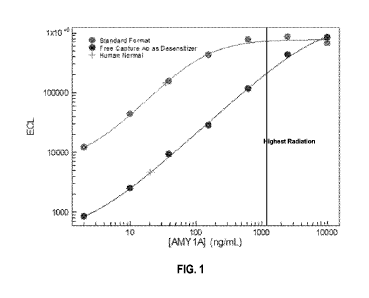
[0011] FIG. 1 relates to embodiments of Example 1. FIG. 1 shows an example
immunoassay
for salivary amylase (AMY1A), a high abundance biomarker, in standard (light
grey) and
desensitized (dark grey) formats. The crosses on the plot indicate normal
AMY1A level in
human plasma, while the vertical line represents the AMY1A level in plasma
with the highest
dose of radiation.
[0012] FIGS. 2A-2K relate to embodiments of Example 2. FIGS. 2A-2K show,
respectively,
calibration curves of immunoassays for CD5, CD27, CD177, CD20, Flt-3L, IL-
12/23, IL-15, IL-
18, thyroid peroxidase (TPO), erythropoietin (EPO), and AMY1A. Bold-lined and
thin-lined
crosses indicate measured levels for a set of normal plasma samples from 18
human donors and
18 nonhuman primate (NHP) models, respectively, in FIGS. 2A-2K.
[0013] FIGS. 3A-3B relate to embodiments of Example 2. FIG. 3A shows various
example
assay parameters of multiplexed biomarker panels, including the coefficient of
variation
(column labeled "Precision"), assay measurement range (limit of detection
(LOD), lower limit of
quantitation (LLOQ), and upper limit of quantitation (ULOQ)), and range of
biomarker
concentration values measured for human and NHP plasma samples. FIG. 3B shows
results of
measured biomarker concentrations in human and NHP plasma samples relative to
the LOD
(bold-lined bars), LLOQ and ULOQ (thin-lined bars). Arrows above each column
represent the
direction in which the concentration is expected to change after radiation
exposure.
[0014] FIGS. 4A-4B relate to embodiments of Example 3. FIG. 4A shows the
linearity-on-
dilution assessment of normal plasma samples diluted in assay calibrator
diluent. Analytes
marked with asterisk (*) had normal levels near the LLOQ. FIG. 4B shows the
spike recovery
assessment of a purified calibrator biomarker spiked into plasma samples.
[0015] FIG. 5 relates to embodiments of Example 4. FIG. 5 shows a summary of
the samples
used in a NHP radiation study. Six animals were exposed to each dose condition
shown in the
row under "Dose (Gy"), and plasma samples were collected at different time
points before (0
3
CA 03160211 2022-05-04
WO 2021/092004 PCT/US2020/058866
days) and after radiation as shown in the first column. The numbers in each
cell indicate the
number of samples tested for each dose-time combination.
[0016] FIG. 6 relates to embodiments of Example 4.1. FIG. 6 shows changes in
CD5, CD20,
CD27, and CD177 biomarkers in NHP plasma as a function of time (for the first
9 days) and
radiation dose. Error bars represent the standard deviation in the measured
biomarker level
across the different replicate animals. The lowest horizontal line in each
plot represents the assay
LOD. The upper and middle horizontal lines in each plot represents the
quantitation range
defined by the LLOQ (middle line) and ULOQ (upper line).
[0017] FIG. 7 relates to embodiments of Example 4.2. FIG. 7 shows changes in
IL-12, IL-15,
IL-18, Flt-3L, EPO, and TPO biomarkers in NHP plasma as a function of time
(for the first 9
days) and radiation dose. Error bars represent the standard deviation in the
measured biomarker
level across the different replicate animals. The lowest horizontal line in
each plot represents the
assay LOD. The upper and middle horizontal lines in each plot represents the
quantitation range
defined by the LLOQ (middle line) and ULOQ (upper line).
[0018] FIG. 8 relates to embodiments of Example 4.3. FIG. 8 shows changes in
AMY1A,
AMY2A measured using a desensitized assay in undiluted (neat) samples, and
AMY2A also
measured in diluted samples in NHP plasma as a function of time (for the first
9 days) and
radiation dose. Error bars represent the standard deviation in the measured
biomarker level
across the different replicate animals. The lowest horizontal line in each
plot represents the assay
LOD. The upper and middle horizontal lines in each plot represents the
quantitation range
defined by the LLOQ (middle line) and ULOQ (upper line).
[0019] FIG. 9 relates to embodiments of Example 5. FIG. 9 shows a summary of
the subjects
in the stem cell transplant (SCT) patient study. AML: acute myeloid leukemia;
ALL: acute
lymphoblastic leukemia; KGF: keratinocyte growth factor.
[0020] FIG. 10 relates to embodiments of Example 5.1. FIG. 10 shows changes in
CD5,
CD20, CD27, and CD177 biomarkers in human plasma from SCT patients as a
function of time
during fractionated total body irradiation (TBI) regimen. Each curve
represents samples from a
different patient. The two horizontal dashed lines near the top and bottom of
each plot provide
the quantitation range defined by the LLOQ (lower line) and ULOQ (upper line).
The two
horizontal lines in the middle of each plot represent the 1 standard
deviation range for a set of
normal human plasma samples tested at the same time as the SCT patient
samples.
[0021] FIG. 11 relates to embodiments of Example 5.2. FIG. 11 shows changes in
IL-12, IL-
15, IL-18, Flt-3L, EPO, and TPO biomarkers in human plasma from SCT patients
as a function
4
CA 03160211 2022-05-04
WO 2021/092004 PCT/US2020/058866
of time during fractionated total body irradiation (TBI) regimen. Each curve
represents samples
from a different patient. The two horizontal dashed lines near the top and
bottom of each plot
provide the quantitation range defined by the LLOQ (lower line) and ULOQ
(upper line). The
two horizontal lines in the middle of each plot represent the 1 standard
deviation range for a set
of 10 normal human plasma samples tested at the same time as the SCT patient
samples.
[0022] FIG. 12 relates to embodiments of Example 5.3. FIG. 12 shows changes in
salivary
amylase (AMYIA), C-reactive protein (CRP), and cardiac troponin (cTn1) in
human plasma
from SCT patients as a function of time during fractionated total body
irradiation (TBI) regimen.
Each curve represents samples from a different patient. The two horizontal
dashed lines near the
top and bottom of each plot provide the quantitation range defined by the LLOQ
(lower line)
and ULOQ (upper line). The two horizontal lines in the middle of each plot
represent the 1
standard deviation range for a set of 10 normal human plasma samples tested at
the same time as
the SCT patient samples.
[0023] FIGS. 13A-13C relate to embodiments of Example 6. FIG. 13A shows a
training data
set of a biomarker concentration plotted against dose (Gy) and time (days post
irradiation). FIG.
13B shows an exemplary prediction model based on measured concentrations of
two
biomarkers, wherein the best predicted dose falls between the best individual
matches for each
of the two biomarkers. FIG. 13C shows the root mean square error (RMSE) in
dose prediction
across all test samples for all possible combinations of biomarkers.
[0024] FIGS. 14A-14B relate to embodiments of Example 7. FIGS. 14A and 14B
show NHP
and human plasma samples, respectively, tested with a five-biomarker panel in
a multiplexed
assay. In FIG. 14A, error bars represent the standard deviation in the
measured biomarker level
across the different replicate animals. The lowest horizontal line in each
plot represents the assay
LOD. The upper and middle horizontal lines in each plot represents the
quantitation range
defined by the LLOQ (middle line) and ULOQ (upper line). In FIG. 14B, each
curve represents
samples from a different patient. The two horizontal dashed lines near the top
and bottom of
each plot provide the quantitation range defined by the LLOQ (lower line) and
ULOQ (upper
line). The two horizontal lines in the middle of each plot represent the 1
standard deviation
range for a set of 10 normal human plasma samples tested at the same time as
the SCT patient
samples.
[0025] FIGS. 15A-15B relate to embodiments of Example 7. FIGS. 15A and 15B
show plots
of predicted vs. actual radiation doses for NHP samples tested with five- and
six-biomarker
panels, respectively.
CA 03160211 2022-05-04
WO 2021/092004 PCT/US2020/058866
[0026] FIG. 16 relates to embodiments of Example 8. FIG. 16 shows the average
coefficient
of variation (CV) for control samples measured in 15 assays plates tested in 5
processing batches
on 3 different days using an automated ultra-high throughput (UHT) system.
[0027] FIG. 17 relates to embodiments of Example 9. FIG. 17 shows a comparison
of the
assay parameters (LOD, LLOQ, and ULOQ) for manual and UHT assay formats.
[0028] FIGS. 18A-18C relate to embodiments of Example 10. FIG. 18A shows
results of a
multiplexed assay performed using a five-biomarker panel on NHP plasma samples
obtained
from individuals subjected to radiation. FIG. 18B shows a plot of biomarker
levels in human
plasma samples from subjects in different categories, e.g., age or disease.
FIG. 18C shows
results of a multiplexed assay performed using a five-biomarker panel on human
plasma samples
obtained from individuals subjected to radiation.
[0029] FIG. 19 illustrate an example NHP dose response data set that can be
used to train a
cost function algorithm and a linear model algorithm, as disclosed in
embodiments herein.
[0030] FIGS. 20A and 20B illustrate an example of the sensitivity and
specificity plot for the
cost function algorithm and the linear model algorithm. FIG. 20A illustrates
an example of
random sub-sampling to measure the specificity and sensitivity as a function
of a cutoff value
using the cost function algorithm and FIG. 20B illustrates an example of
random sub-sampling
to measure the specificity and sensitivity as a function of a cutoff value
using the linear model
algorithm.
[0031] FIGS. 21A and 21B illustrate an example of the accuracy of the cost
function
algorithm and the linear model algorithm, as described herein. FIG. 21A
illustrates an example
of the accuracy of the cost function algorithm and FIG. 21B illustrates an
example of the
accuracy of the linear model algorithm.
[0032] FIG. 22 illustrates the data from human patients used to test the cost
function algorithm
and the linear model algorithm.
[0033] FIG. 23 illustrates an example of the results for the test of the cost
function algorithm
and the linear model algorithm using the data from FIG. 22.
[0034] FIG. 24 contain tables showing the observed specificities for the
different classes of
subjects as shown in FIG. 23. The Table A shows predicted specificity for the
cost function
algorithm, and the Table B shows predicted specificity for the linear model
algorithm.
[0035] FIG. 25 illustrates data from a human stem cell transplant (SCT).
6
CA 03160211 2022-05-04
WO 2021/092004 PCT/US2020/058866
[0036] FIG. 26A illustrates an example of dose prediction for SCT patient
samples as a
function of total dose for the cost function algorithm and FIG. 26B
illustrates an example of
dose prediction for SCT patient samples as a function of total dose for the
linear model
algorithm.
[0037] FIG. 27 shows an alternative dosing regimen for the NHP study described
in
embodiments of Example 4.4.
[0038] FIG. 28 relates to embodiments of Example 4.4. FIG. 28 contains a table
showing the
specificity and sensitivity of classification using the regression model.
[0039] FIG. 29 relates to embodiments of Example 11. FIG. 29 contains a table
of components
that are commonly present in sample matrices that can interfere with biomarker
level
measurements, also known as interferents. The components in FIG. 29 are
organized by
category. The expected highest level of each interferent in a plasma sample is
shown as the
Target Concentration (1X). Each interferent was spiked into plasma samples at
four times the
target concentration, shown as the 4X Screening concentration.
[0040] FIG. 30 relates to embodiments of Example 11. FIG. 30 shows the results
of five-
biomarker assay panels with four plasma samples that were spiked with the
interferents in FIG.
29 at 4X Screening concentration. The five-biomarker assay panel measured
CD20, IL-15,
AMY1A, CD5, and Flt-3L.
[0041] FIG. 31 relates to embodiments of Example 11. FIG. 31 shows the results
of titrating
the concentrations of interferents hemolysate, lipid, unconjugated bilirubin,
and conjugated
bilirubin into plasma samples at decreasing spike concentrations: 4X, 2X, lx,
0.5X, 0.25X,
0.125X, and OX, where lx represents the expected highest level of the
interferent in a plasma
sample.
[0042] FIG. 32 relates to embodiments of Example 13. FIG. 32 shows the results
of a stability
test for an assay plate containing a five-biomarker assay panel for measuring
levels of CD20, IL-
15, AMY1A, CD5, and Flt-3L. The plates were stored in open air at either 25 C
(22 to 27 C)
or 37 C (35 to 40 C), then used to measure biomarker concentrations in two
control samples
and three plasma samples. The measured concentrations were plotted after
normalization to a
plate that was stored at 4 C and used immediately following removal from
storage (4 C, 0 hr
condition).
[0043] FIG. 33 relates to embodiments of Example 13. FIG. 33 shows the results
of a stability
test for control samples containing known amounts of CD20, IL-15, AMY1A, CD5,
and Flt-3L.
Lyophilized control samples were reconstituted and stored for up to 24 hours
at 4 C or 25 C,
7
CA 03160211 2022-05-04
WO 2021/092004 PCT/US2020/058866
then analyzed using the five-biomarker assay panel for measuring levels of
CD20, IL-15,
AMY1A, CD5, and Flt-3L. The measured concentrations were compared to the
concentrations
measured immediately after reconstitution (0 hr condition).
[0044] FIG. 34 relates to embodiments of Example 13. FIG. 34 shows the results
of a stability
test for calibration standards for each of the biomarkers CD20, IL-15, AMY1A,
CD5, and Flt-
3L. Lyophilized calibration standards were reconstituted and stored for up to
24 hours at 4 C or
25 C. The calibration standards were then used in the five-biomarker assay
panel for measuring
levels of CD20, IL-15, AMY1A, CD5, and Flt-3L to determine concentrations of
the biomarkers
in two control samples and three plasma samples. The measured concentrations
of biomarkers in
the control samples and plasma samples were compared to the concentrations
that were
measured using calibration standards that were used immediately after
reconstitution (0 hr
condition).
[0045] FIG. 35 relates to embodiments of Example 14. FIG. 35 shows the
temperature and
length of time that plasma samples were stored, prior to testing for stability
by measuring
biomarker levels using a five-biomarker assay panel for CD20, IL-15, AMY1A,
CD5, and Flt-
3L.
[0046] FIGS. 36A-36C relate to embodiments of Example 14. FIGS. 36A-36C show
the
results of a stability test for plasma samples. Ten different plasma samples
were stored
according to the conditions shown in FIG. 35. FIG. 36A shows the measured
concentrations of
CD20, IL-15, AMY1A, CD5, and Flt-3L in plasma samples 1-4. FIG. 36B shows the
measured
concentrations of CD20, IL-15, AMY1A, CD5, and Flt-3L in plasma samples 5-8.
FIG. 36C
shows the measured concentrations of CD20, IL-15, AMY1A, CD5, and Flt-3L in
plasma
samples 9-10.
[0047] FIG. 37 relates to embodiments of Example 15.1. FIG. 37 shows the
results of a
control experiment using a multiplexed five-biomarker panel assay for CD20, IL-
15, AMY1A,
CD5, and Flt-3L on a positive control sample, a negative control sample, and a
pooled plasma
sample. Measurements were performed on 21 assay plates over the course of 7
days with the
samples in duplicate. The measured concentration is shown after normalization
to the median
value across the runs, and the inset table shows the measured coefficient of
variations (CVs) for
each control/assay combination across the experiment. The table also shows the
percentage of
the controls that provided the correct dose classification result (the
negative and pooled plasma
control should be classified as having a dose < 2 Gy and the positive control
should be classified
as having a dose? 2 Gy).
8
CA 03160211 2022-05-04
WO 2021/092004
PCT/US2020/058866
[0048] FIG. 38 relates to embodiments of Example 15.2. FIG. 38 shows the
results of a
multiplexed five-biomarker panel assay for CD20, IL-15, AMY1A, CD5, and Flt-3L
and an
AMY2A assay performed on NHP plasma samples that were subjected to different
doses of
radiation.
[0049] FIG. 39 relates to embodiments of Example 15.2. FIG. 39 shows the
performance
accuracy of the dose assessment algorithms (cost function or error
minimization and linear
regression), with the plots showing predicted dose as a function of actual
dose with points
colored based on time from exposure. The tables provide the classification
accuracy for all
negative and positive samples, or stratified by dose (top: error minimization
algorithm; bottom:
linear regression algorithm).
[0050] FIG. 40 relates to embodiments of Example 15.3. FIG. 40 shows the
results of a
multiplexed five-biomarker panel assay for CD20, IL-15, AMY1A, CD5, and Flt-3L
and an
AMY2A assay performed on NHP plasma samples that were subjected to 0 or 6 Gy
of radiation
and subjected to no treatment (control arm) or 10 pg/kg G-CSF daily, starting
at day 1 post-
exposure (treatment arm).
[0051] FIG. 41 relates to embodiments of Example 15.3. FIG. 41 shows the
performance
accuracy of the dose assessment algorithms (cost function or error
minimization and linear
regression), with the plots showing predicted dose as a function of actual
dose and whether the
study animals received G-CSF after irradiation. The tables provide the
classification accuracy
for all negative and positive samples, stratified by drug treatment arm (top:
error minimization
algorithm; bottom: linear regression algorithm).
[0052] FIG. 42 relates to embodiments of Example 15.4. FIG. 42 shows the
results of a
multiplexed five-biomarker panel assay for CD20, IL-15, AMY1A, CD5, and Flt-3L
performed
on human plasma samples from normal or special populations based on age,
injury, disease or
special condition.
[0053] FIG. 43 relates to embodiments of Example 15.4. FIG. 43 shows the
specificity of the
dose assessment algorithms (cost function or error minimization and linear
regression). The
tables show the observed specificities for the different classes of subjects
(top: error
minimization algorithm; bottom: linear regression algorithm).
[0054] FIG. 44 relates to embodiments of Example 15.5. FIG. 44 shows the
results of a
multiplexed five-biomarker panel assay for CD20, IL-15, AMY1A, CD5, and Flt-3L
performed
on human plasma samples from patients having been subjected to total body
irradiation (TBI)
prior to stem cell transplant therapy.
9
CA 03160211 2022-05-04
WO 2021/092004 PCT/US2020/058866
[0055] FIG. 45 relates to embodiments of Example 15.5. FIG. 45 shows the
performance of
dose assessment algorithms (cost function or error minimization and linear
regression), with the
dose prediction for SCT patient samples as a function of total dose. The
tables show specificity
and sensitivity for the full data set, and after removing data from subjects
with undetectable
CD20 at baseline ((top: error minimization algorithm; bottom: linear
regression algorithm).
[0056] FIG. 46 illustrates an exemplary assay surface described in embodiments
herein. FIG.
46 shows a well of an exemplary 96-well assay plate, comprising ten distinct
binding domains
("spots").
DETAILED DESCRIPTION OF THE INVENTION
[0057] In embodiments, the present disclosure provides multiplexed
immunoassays for
quantifying amounts of at least four biomarkers in a sample. In embodiments,
the disclosure also
provides kits for performing the multiplexed assays.
I. Definitions
[0058] Unless otherwise defined herein, scientific and technical terms used in
the present
disclosure shall have the meanings that are commonly understood by one of
ordinary skill in the
art. Further, unless otherwise required by context, singular terms shall
include pluralities and
plural terms shall include the singular. The articles "a" and "an" are used
herein to refer to one or
to more than one (i.e., to at least one) of the grammatical object of the
article. By way of
example, "an element" means one element or more than one element.
[0059] The use of the term "or" in the claims is used to mean "and/or," unless
explicitly
indicated to refer only to alternatives or the alternatives are mutually
exclusive, although the
disclosure supports a definition that refers to only alternatives and
"and/or."
[0060] As used herein, the terms "comprising" (and any variant or form of
comprising, such as
"comprise" and "comprises"), "having" (and any variant or form of having, such
as "have" and
"has"), "including" (and any variant or form of including, such as "includes"
and "include") or
"containing" (and any variant or form of containing, such as "contains" and
"contain") are
inclusive or open-ended and do not exclude additional, unrecited, elements or
method steps.
[0061] The use of the term "for example" and its corresponding abbreviation
"e.g." (whether
italicized or not) means that the specific terms recited are representative
examples and
embodiments of the disclosure that are not intended to be limited to the
specific examples
referenced or cited unless explicitly stated otherwise.
CA 03160211 2022-05-04
WO 2021/092004 PCT/US2020/058866
[0062] As used herein, "between" is a range inclusive of the ends of the
range. For example, a
number between x and y explicitly includes the numbers x and y, and any
numbers that fall
within x and y.
Overview
[0063] Measurement of biomarker values and levels before and after a
particular event, e.g.,
cellular or environmental event, may be used to gain information regarding an
individual's
response to the event. For example, samples or model organisms can be
subjected to stress- or
disease-inducing conditions, or a treatment or prevention regimen, and a
particular biomarker
can then be detected and quantitated in order to determine its changes in
response to the
condition or regimen. However, the opposite, i.e., measuring biomarker values
and levels to
determine whether an organism has been subjected to stress- or disease-
inducing condition,
tends to be much more complicated, as changes in the levels of a single
biomarker typically
cannot be definitively associated with a particular condition.
[0064] While single biomarkers generally do not provide sufficient
information, e.g., for
prediction and/or diagnosis of a disease or condition, certain combinations of
biomarkers may be
used to provide a strong prediction and/or diagnosis. Although a linear
combination of
biomarkers (i.e., the combination comprises biomarkers that individually
provide a relatively
strong correlation) can be utilized, linear combinations may not be available
in many situations,
for example, when there are not enough biomarkers available and/or with strong
correlation. In
alternative approaches, a biomarker combination is selected such that the
combination is capable
of achieving improved performance (i.e., prediction or diagnosis) compared
with any of the
individual biomarkers, each of which may not be a strong correlator on its
own. Biomarkers for
inclusion in a biomarker combination can be selected for based on their
performance in different
individuals, e.g., patients, wherein the same biomarker may not have the same
performance in
different individuals, but when combined with the remaining biomarkers,
provide an
unexpectedly strong correlation for prediction or diagnosis in a population.
For example, Bansal
et al., Statist Med 32: 1877-1892 (2013) describe methods of determining
biomarkers to include
in such a combination, noting in particular that optimal combinations may not
be obvious to one
of skill in the art , especially when subgroups are present or when individual
biomarker
correlations are different between cases and controls. Thus, selecting a
combination of
biomarkers for providing a consistent and accurate prediction and/or diagnosis
can be
particularly challenging and unpredictable.
[0065] Even when a suitable combination of biomarkers is determined, utilizing
the
combination of biomarkers in an assay poses its own set of difficulties. For
example, detecting
11
CA 03160211 2022-05-04
WO 2021/092004 PCT/US2020/058866
and/or quantitating each biomarker in the combination in its own separate
assay may not be
feasible with small samples, and using a separate assay to measure each
biomarker in a sample
may not provide consistent and comparable results. Furthermore, running an
individual assay for
each biomarker in a combination can be a cumbersome and complex process that
can be
inefficient and costly.
[0066] A multiplexed assay that can simultaneously measure the concentrations
of multiple
biomarkers can provide reliable results while reducing processing time and
cost. Challenges of
developing a multi-biomarker assay (such as, e.g., a multiplexed assay
described in
embodiments herein) include, for example, determining compatible reagents for
all of the
biomarkers (e.g., capture and detection reagents described herein should be
highly specific and
not be cross-reactive; all assays should perform well in the same diluents);
determining
concentration ranges of the reagents for consistent assay (e.g., comparable
capture and detection
efficiency for the assays described herein); having similar levels in the
condition and sample
type of choice such that the levels of all of the biomarkers fall within the
dynamic range of the
assays at the same dilution; minimizing non-specific binding between the
biomarkers and
binding reagents thereof or other interferents; and accurately and precisely
detecting a
multiplexed output measurement.
[0067] In embodiments, the present disclosure provides a multi-biomarker assay
for
determining radiation exposure. Individuals exposed to ionizing radiation,
either as a result of a
major radiological or nuclear event, during medical treatment, or as a result
of an accidental
exposure, may suffer from systemic and organ-specific damage. For example,
acute effects of
high-dose ionizing radiation (>2 Gy) include depletion of specific types of
peripheral blood
cells, immune suppression, mucosal damage, and potential injury to other sites
such as bone and
bone marrow niche cells, gastrointestinal system, lungs kidneys, and central
nervous system.
Exposure to low or moderately high doses (1-3 Gy) of ionizing radiation can
result in increased
mortality, especially if accompanied by physical injuries, opportunistic
infections, and/or
hemorrhage. Long-term effects include dysfunction or fibrosis in a wide range
of organs and
tissues, cataracts, and a higher risk of cancer. In many cases, the effects of
radiation exposure
can be mitigated by early triage and treatment.
[0068] Although radioactive material can be detected using instruments,
assessment of
radiation dose or injury that an individual has received is more difficult.
Moreover, current
medical countermeasures for radiation injuries are often expensive, labor-
intensive, and time-
consuming to administer and monitor, have limited availability, and can be
associated with
serious toxicities, they should only be administered to individuals most
likely to benefit from
12
CA 03160211 2022-05-04
WO 2021/092004 PCT/US2020/058866
their use. Thus, fast and accurate radiation dose and tissue injury assessment
can greatly
facilitate identification of exposed individuals who may benefit from early
medical intervention.
[0069] Current methods of diagnosing radiation exposure, e.g., the dicentric
chromosome
assay, can be labor intensive and slow to produce results, and no diagnostic
method is available
to reliably discriminate levels of radiation exposure based on samples
collected at a single time
point. It was discovered by the present inventors that radiation exposure can
be assessed using a
combination of biomarkers. Thus, in embodiments, the present disclosure
provides a
multiplexed assay method for detecting and/or quantitating biomarkers related
to radiation
exposure.
[0070] In embodiments, the disclosure provides a multiplexed immunoassay
method
comprising: quantifying the amounts of at least four biomarkers in a
biological sample, wherein
the at least four biomarkers comprise (a) IL-15, (b) CD5, (c) Flt-3L, and (d)
salivary amylase,
wherein the quantifying comprises measuring the concentrations of the at least
four biomarkers
in a multiplexed assay format to simultaneously measure the concentrations of
the least four
biomarkers in a biological sample wherein the multiplexed immunoassay
comprises: a)
combining, in one or more steps: (i) the biological sample; (ii) at least a
first, second, third, and
fourth binding reagent, wherein the first, second, third, and fourth binding
reagent is a binding
partner of IL-15, CD5, Flt-3L, and salivary amylase, respectively; b) forming
at least a first,
second, third, and fourth binding complex comprising the binding reagents and
the biomarkers;
c) measuring the concentration of the biomarkers in each of the complexes.
[0071] In embodiments, the disclosure further provides a multiplexed
immunoassay method
comprising: quantifying the amounts of at least four biomarkers in a
biological sample, wherein
the at least four biomarkers comprise IL-15, CD5, Flt-3L, and salivary
amylase, wherein the
quantifying comprises measuring the concentrations of the at least four
biomarkers in a
multiplexed assay format to simultaneously measure the concentrations of at
least four
biomarkers in the biological sample, wherein the multiplexed immunoassay
comprises: a)
combining, in one or more steps: (i) the biological sample; (ii) at least a
first antibody to IL-15; a
first antibody to CD5; a first antibody to Flt-3L; and a first antibody to
salivary amylase,
wherein each of the first antibodies is immobilized on separate binding
domains; (iii) at least a
second antibody to IL-15; a second antibody to CD5; a second antibody to Flt-
3L; and a second
antibody to salivary amylase, wherein each second antibody is connected to a
detectable label;
b) forming at least a first, second, third, and fourth binding complex on an
at least first, second,
third, and fourth binding domains comprising at least IL-15, CD5, Flt-3L, and
salivary amylase,
and the first and second antibodies for their respective biomarker; c)
measuring the
13
CA 03160211 2022-05-04
WO 2021/092004 PCT/US2020/058866
concentration of the at least IL-15, CD5, Flt-3L, and salivary amylase on the
at least first,
second, third, and fourth binding domains.
Biomarkers and Samples
[0072] As used herein, the term "biomarker" refers to a biological substance
that is indicative
of a normal or abnormal process, e.g., disease, infection, or environmental
exposure. Biomarkers
can be small molecules such as ligands, signaling molecules, or peptides, or
macromolecules
such as antibodies, receptors, or proteins and protein complexes. A change in
the levels of a
biomarker can correlate with the risk or progression of a disease or
abnormality or with the
susceptibility of the disease or abnormality to a given treatment. A biomarker
can be useful in
the diagnosis of disease risk or the presence of disease in an individual, or
to tailor treatments
for the disease in an individual (e.g., choices of drug treatment or
administration regimes). In
evaluating potential drug therapies, a biomarker can be used as a surrogate
for a natural endpoint
such as survival or irreversible morbidity. If a treatment alters a biomarker
that has a direct
connection to improved health, the biomarker serves as a "surrogate endpoint"
for evaluating
clinical benefit. Biomarkers are further described in, e.g., Mayeux, NeuroRi,c
1(2): 182-188
(2004); Strimbu et al., Curr Opin HIV AIDS 5(6): 463-466 (2010); and Bansal et
al., Statist Med
32: 1877-1892 (2013). The term "biomarker," when used in the context of a
specific organism
(e.g., human, nonhuman primate or another animal), refers to the biomarker
native to that
specific organism. For example, "human biomarker" salivary amylase refers to
salivary amylase
found in humans, i.e., AMY1A, while "nonhuman primate biomarker" salivary
amylase refers to
salivary amylase found in nonhuman primates, i.e., AMY2A. Unless specified
otherwise, the
biomarkers referred to in embodiments herein encompass human biomarkers.
[0073] As used herein, the term "level" in the context of a biomarker refers
to the amount,
concentration, or activity of a biomarker. The term "level" can also refer to
the rate of change of
the amount, concentration, or activity of a biomarker. A level can be
represented, for example,
by the amount or synthesis rate of messenger RNA (mRNA) encoded by a gene, the
amount or
synthesis rate of polypeptide corresponding to a given amino acid sequence
encoded by a gene,
or the amount or synthesis rate of a biochemical form of a biomarker
accumulated in a cell,
including, for example, the amount of particular post-synthetic modifications
of a biomarker
such as a polypeptide, nucleic acid or small molecule. "Level" can also refer
to an absolute
amount of a biomarker in a sample or to a relative amount of the biomarker,
including amount or
concentration determined under steady-state or non-steady-state conditions.
"Level" can further
refer to an assay signal that correlates with the amount, concentration,
activity or rate of change
of a biomarker. The level of a biomarker can be determined relative to a
control marker in a
14
CA 03160211 2022-05-04
WO 2021/092004 PCT/US2020/058866
sample. The terms "level" and "concentration" are used interchangeably herein,
except when the
context clear dictates otherwise.
[0074] Biomarkers for assessing radiation exposure can include, e.g., stress
and/or damage
response markers and damage repair markers. In embodiments, the biomarker for
assessing
radiation exposure is a DNA damage biomarker. In embodiments, the DNA damage
biomarker
is p53, p21, ATM serine/threonine kinase (ATM), phosphorylated H2AX histone (y-
H2AX),
GADD45A, or combination thereof Biomarkers for assessing radiation exposure
are, in
embodiments, not significantly affected by chronic diseases with high
prevalence in the human
population, such as diabetes, asthma, high blood pressure, heart disease,
arthritis and/or other
chronic inflammatory or autoimmune diseases. Biomarkers for assessing
radiation exposure are,
in embodiments, also not affected by other types of trauma (e.g., wounding,
burns and/or mental
stress) that may also be experienced by individuals in a radiation event.
[0075] In embodiments, the biomarker for assessing radiation exposure is an
inflammatory
response biomarker. An inflammatory response biomarker is a biomarker that is
up- or down-
regulated during systemic or localized inflammatory response, e.g., caused by
radiation
exposure. In embodiments, the inflammatory response biomarker is IL-1, IL-2,
IL-3, IL-4, IL-5,
IL-6, IL-7, IL-10, IL-12, IL-23, TNF-a, INF-y, C-reactive protein (CRP), serum
amyloid A
(SAA), CXCL1 (also known as KC/GRO), or combination thereof
[0076] In embodiments, the biomarker for assessing radiation exposure is an
acute phase
protein. Acute phase proteins (APPs) are a class of proteins whose plasma
concentrations
increase (positive APPs) or decrease (negative APPs) in response to
inflammation, e.g., caused
by radiation exposure. See, e.g., Ossetrova et al., Radiat Meas 46(9): 1019-
1024 (2011); and
Sproull et al., Radiat Res 184(1): 14-23 (2015). In embodiments, the acute
phase protein is C-
reactive protein (CRP).
[0077] In embodiments, the biomarker for assessing radiation exposure is a
tissue damage
biomarker. A tissue damage biomarker is a biomarker released from a tissue as
a result of local
tissue damage, e.g., caused by radiation. In embodiments, the tissue damage
biomarker is
salivary amylase, citrullinated proteins, creatine kinase BB (CKBB), creatine
kinase MB
(CKMB), creatine kinase MM (CKMM), S100B, surfactant protein D (SP-D), fatty
acid binding
protein 2 (FABP2), bacterial/permeability-increasing protein (BPI), glial
fibrillary acidic protein
(GFAP), thrombospondin (TSP), neuron-specific enolase (NSE), cancer antigen 15-
3 (CA15-3),
or combination thereof
CA 03160211 2022-05-04
WO 2021/092004 PCT/US2020/058866
[0078] In embodiments, the biomarker for assessing radiation exposure is a
salivary gland
damage biomarker. Radiation exposure has been shown to affect salivary gland
function (see,
e.g., Marmary etal., Cancer Res 76(5): 1170-1180 (2016); Nanduri et al., Radi
other Oncol
99(3): 367-372 (2011); Hakim etal., Clin Oral Investig 8(1): 30-35 (2004)). In
embodiments,
the salivary gland damage biomarker is salivary amylase (AMY1A or AMY2A).
[0079] In embodiments, the biomarker for assessing radiation exposure is a
tissue damage
repair biomarker. A tissue damage repair biomarker is a biomarker that is up-
or down-regulated
during repair, regeneration, or fibroblastic phase during tissue damage.
Tissue damage repair
biomarkers can also include proteins associated with soft-tissue repair
processes, including but
not limited to fibroblast formation, collagen synthesis, and tissue remodeling
and realignment. In
embodiments, the tissue damage repair biomarker is FMS-like tyrosine kinase 3
ligand (Flt-3L),
thyroid peroxidase (TP0), granulocyte-colony stimulating factor (G-CSF),
granulocyte-
macrophage colony stimulating factor (GM-CSF), keratinocyte growth factor
(KGF), stromal
cell-derived factor-1 (SDF-1a), erythropoietin (EPO), or combination thereof
[0080] In embodiments, the biomarker for assessing radiation exposure is a
hematopoietic
repair factor. In embodiments, the biomarker is a hematopoietic cytokine. In
embodiments, the
biomarker is a hematopoietic progenitor. In embodiments, the biomarker is
present on an
erythrocyte. In embodiments, the biomarker is present on a platelet. In
embodiments, the
biomarker is a pro-inflammatory cytokine. In embodiments, the biomarker is
present on an
innate immune system cell. In embodiments, the hematopoietic repair factor or
cytokine is Flt-
3L, erythropoietin (EPO), thyroid peroxidase (TP0), IL-12, IL-15, IL-18, or
combination
thereof
[0081] In embodiments, the biomarker for assessing radiation exposure is a
hematology
surrogate biomarker. Hematology surrogate biomarkers are generally cell-
surface markers on
blood cells, which may be used as surrogates to traditional blood cell counts,
e.g., for assessing
the effect of radiation on specific blood-cell populations. Hematology
surrogate biomarkers
include markers found on general classes of cells (e.g., leukocytes), or more
specific ell types
within those classes, such as lymphocytes, neutrophils, platelets, or even
more specifically, T-
cells or B-cells. In embodiments, the hematology surrogate biomarker is CD5,
CD16b, CD20,
CD26, CD27, CD40, CD45, CD177, or combination thereof
[0082] In embodiments, the biomarker for assessing radiation exposure is a
hematopoietic
damage marker. In embodiments, the hematopoietic damage marker is an immune
cell surface
marker. In embodiments, the hematopoietic damage marker is a T cell surface
marker, a B cell
16
CA 03160211 2022-05-04
WO 2021/092004
PCT/US2020/058866
surface marker, a lymphocyte surface marker, or a neutrophil surface marker.
In embodiments,
the hematopoietic damage marker is CD5, CD20, CD27, CD177, or combination
thereof
[0083] In embodiments, the method comprises quantifying a combination of the
biomarkers
described herein in a sample, e.g., a biological sample. In embodiments, the
sample is obtained
from a subject exposed to radiation, at risk of exposure to radiation, or
suspected of having been
exposed to radiation exposure. In embodiments, the biomarker combination
comprises an
inflammatory response biomarker, a tissue damage biomarker, and a tissue
damage repair
biomarker, a hematology surrogate marker. In embodiments, the biomarker
combination
comprises a hematopoietic damage marker, a hematopoietic repair factor, a
hematopoietic
cytokine, and a salivary gland damage marker. In embodiments, the amount of
radiation
exposure of the subject is determined based on the quantitated amounts of the
biomarkers in the
combination. In embodiments, quantifying the biomarker combination provides a
more accurate
and precise determination of the amount of radiation exposure, compared with
quantifying each
biomarker in the combination individually.
[0084] In embodiments, the method comprises quantifying the amounts of at
least four
biomarkers described herein, in a sample, e.g., a biological sample. In
embodiments, the sample
is obtained from a subject exposed to radiation, at risk of exposure to
radiation, or suspected of
having been exposed to radiation exposure. In embodiments, the at least four
biomarkers
comprise IL-15, CD5, Flt-3L, and salivary amylase. In embodiments, the at
least four
biomarkers comprise IL-15, CD5, Flt-3L, salivary amylase, and CD20. In
embodiments, the at
least four biomarkers comprise IL-15, CD5, Flt-3L, salivary amylase, and IL-
18. In
embodiments, the at least four biomarkers comprise IL-15, CD5, Flt-3L,
salivary amylase, and
CD27. In embodiments, the at least four biomarkers comprise IL-15, CD5, Flt-
3L, salivary
amylase, and TPO. In embodiments, the at least four biomarkers comprise IL-15,
CD5, Flt-3L,
salivary amylase, and one or more of CD20, IL-18, CD27, and TPO. In
embodiments,
quantifying the amount of at least four biomarkers described herein provides
an accurate and
precise determination of the amount of radiation exposure. In embodiments,
quantifying the
amount of at least four biomarkers described herein provides a more accurate
and precise
determination of the amount of radiation exposure, compared with quantifying
less than four of
the biomarkers described herein.
[0085] In embodiments, the biomarker combination comprises IL-15. IL-15 is a
cytokine that
regulates activation and proliferation of T and natural killer (NK) cells. In
embodiments, IL-15
levels increase in a subject, e.g., a human subject, after radiation exposure.
In embodiments, IL-
15 levels are higher in a subject exposed to radiation, compared with a
subject who has not been
17
CA 03160211 2022-05-04
WO 2021/092004 PCT/US2020/058866
exposed to radiation. In embodiments, IL-15 levels in a subject exposed to
radiation are about
5%, about 10%, about 15%, about 20%, about 25%, about 30%, about 35%, about
40%, about
45%, about 50%, about 55%, about 60%, about 65%, about 70%, about 75%, about
80%, about
85%, about 90%, about 95%, about 100%, about 150%, about 200%, about 250%,
about 500%,
or more than 500% higher compared with a subject who has not been exposed to
radiation.
[0086] In embodiments, the biomarker combination comprises CD5. It was
discovered that
changes in CD5, which is typically known as a lymphocyte surface marker, can
be used to
assess radiation exposure in serum and/or plasma samples. It was further
discovered that CD5
had relatively normal baseline levels in certain populations that may be
subjected to or at risk of
radiation exposure, e.g., cancer patients undergoing stem cell transplant
(SCT), while baseline
levels of biomarkers that have been used to assess radiation exposure, e.g.,
CD20, vary
substantially in these populations. In embodiments, the inclusion of CD5 in
the biomarker
combination provides improved consistency, redundancy, and accuracy in
assessing radiation
exposure. In embodiments, CD5 levels decrease in a subject, e.g., a human
subject, after
radiation exposure. In embodiments, CD5 levels are lower in a subject exposed
to radiation,
compared with a subject who has not been exposed to radiation. In embodiments,
CD5 levels in
a subject exposed to radiation are about 5%, about 10%, about 15%, about 20%,
about 25%,
about 30%, about 35%, about 40%, about 45%, about 50%, about 55%, about 60%,
about 65%,
about 70%, about 75%, about 80%, about 85%, about 90%, about 95%, about 100%,
about
150%, about 200%, about 250%, about 500%, or more than 500% lower compared
with a
subject who has not been exposed to radiation.
[0087] In embodiments, the biomarker combination comprises Flt-3L. Flt-3L can
function as a
cytokine and growth factor that increases the number of immune cells (e.g.,
lymphocytes such as
B cells and T cells) by activating hematopoietic progenitors. In embodiments,
Flt-3L levels
increase in a subject, e.g., a human subject, after radiation exposure. In
embodiments, Flt-3L
levels are higher in a subject exposed to radiation, compared with a subject
who has not been
exposed to radiation. In embodiments, Flt-3L levels in a subject exposed to
radiation are about
5%, about 10%, about 15%, about 20%, about 25%, about 30%, about 35%, about
40%, about
45%, about 50%, about 55%, about 60%, about 65%, about 70%, about 75%, about
80%, about
85%, about 90%, about 95%, about 100%, about 150%, about 200%, about 250%,
about 500%,
or more than 500% higher compared with a subject who has not been exposed to
radiation.
[0088] In embodiments, the biomarker combination comprises salivary amylase.
As discussed
herein, radiation exposure can damage salivary gland function, and
accordingly, salivary
amylase levels can be used to assess radiation exposure. In embodiments,
salivary amylase
18
CA 03160211 2022-05-04
WO 2021/092004 PCT/US2020/058866
levels increase in a subject, e.g., a human subject, after radiation exposure.
In embodiments,
salivary amylase levels are higher in a subject exposed to radiation, compared
with a subject
who has not been exposed to radiation. In embodiments, salivary amylase levels
in a subject
exposed to radiation are about 5%, about 10%, about 15%, about 20%, about 25%,
about 30%,
about 35%, about 40%, about 45%, about 50%, about 55%, about 60%, about 65%,
about 70%,
about 75%, about 80%, about 85%, about 90%, about 95%, about 100%, about 150%,
about
200%, about 250%, about 500%, or more than 500% higher compared with a subject
who has
not been exposed to radiation.
[0089] In embodiments, the biomarker combination comprises CD20. CD20 is a
membrane-
embedded surface biomarker that plays a role in the development and
differentiation of B cells
into plasma cells. In embodiments, CD20 levels decrease in a subject, e.g., a
human subject,
after radiation exposure. In embodiments, CD20 levels are lower in a subject
exposed to
radiation, compared with a subject who has not been exposed to radiation. In
embodiments,
CD20 levels in a subject exposed to radiation are about 5%, about 10%, about
15%, about 20%,
about 25%, about 30%, about 35%, about 40%, about 45%, about 50%, about 55%,
about 60%,
about 65%, about 70%, about 75%, about 80%, about 85%, about 90%, about 95%,
about 100%,
about 150%, about 200%, about 250%, about 500%, or more than 500% lower
compared with a
subject who has not been exposed to radiation.
[0090] In embodiments, the biomarker combination comprises IL-18. IL-18 is a
proinflammatory cytokine that can modulate innate and adaptive immunity. In
embodiments, IL-
18 levels increase in a subject, e.g., a human subject, after radiation
exposure. In embodiments,
IL-18 levels are higher in a subject exposed to radiation, compared with a
subject who has not
been exposed to radiation. In embodiments, IL-18 levels in a subject exposed
to radiation are
about 5%, about 10%, about 15%, about 20%, about 25%, about 30%, about 35%,
about 40%,
about 45%, about 50%, about 55%, about 60%, about 65%, about 70%, about 75%,
about 80%,
about 85%, about 90%, about 95%, about 100%, about 150%, about 200%, about
250%, about
500%, or more than 500% higher compared with a subject who has not been
exposed to
radiation.
[0091] In embodiments, the biomarker combination comprises CD27. CD27 is a co-
stimulatory immune checkpoint molecule. In embodiments, CD27 levels decrease
in a subject,
e.g., a human subject, after radiation exposure. In embodiments, CD27 levels
are lower in a
subject exposed to radiation, compared with a subject who has not been exposed
to radiation. In
embodiments, CD27 levels in a subject exposed to radiation are about 5%, about
10%, about
15%, about 20%, about 25%, about 30%, about 35%, about 40%, about 45%, about
50%, about
19
CA 03160211 2022-05-04
WO 2021/092004 PCT/US2020/058866
55%, about 600o, about 650o, about 700o, about 750o, about 800o, about 850o,
about 900o, about
950o, about 10000, about 1500o, about 2000o, about 2500o, about 5000o, or more
than 5000o
lower compared with a subject who has not been exposed to radiation.
[0092] In embodiments, the biomarker combination comprises thyroid peroxidase
(TPO), also
known as thyroperoxidase or iodide peroxidase. In embodiments, TPO levels
increase in a
subject, e.g., a human subject, after radiation exposure. In embodiments, TPO
levels are higher
in a subject exposed to radiation, compared with a subject who has not been
exposed to
radiation. In embodiments, TPO levels in a subject exposed to radiation are
about 5%, about
100o, about 150o, about 200o, about 25%, about 300o, about 350o, about 400o,
about 450o, about
500o, about 55%, about 600o, about 65%, about 700o, about 750o, about 800o,
about 85%, about
900o, about 950o, about 1000o, about 1500o, about 2000o, about 2500o, about
5000o, or more than
5000o higher compared with a subject who has not been exposed to radiation.
[0093] In embodiments, the biomarker combination comprises CD177. In
embodiments,
CD177 levels increase in a subject, e.g., a human subject, after radiation
exposure. In
embodiments, CD177 levels are higher in a subject exposed to radiation,
compared with a
subject who has not been exposed to radiation. In embodiments, CD177 levels in
a subject
exposed to radiation are about 5%, about 100o, about 150o, about 200o, about
25%, about 300o,
about 350o, about 400o, about 450o, about 500o, about 55%, about 600o, about
65%, about 700o,
about 750o, about 800o, about 85%, about 900o, about 950o, about 1000o, about
1500o, about
2000o, about 2500o, about 5000o, or more than 5000o higher compared with a
subject who has
not been exposed to radiation.
[0094] In embodiments, the biomarker combination comprises erythropoietin
(EPO). In
embodiments, EPO levels increase in a subject, e.g., a human subject, after
radiation exposure.
In embodiments, EPO levels are higher in a subject exposed to radiation,
compared with a
subject who has not been exposed to radiation. In embodiments, EPO levels in a
subject exposed
to radiation are about 5%, about 100o, about 150o, about 200o, about 25%,
about 300o, about
35%, about 40%, about 45%, about 50%, about 55%, about 60%, about 65%, about
70%, about
750o, about 800o, about 85%, about 900o, about 950o, about 1000o, about 1500o,
about 2000o,
about 2500o, about 5000o, or more than 5000o higher compared with a subject
who has not been
exposed to radiation.
[0095] In embodiments, the biomarker combination comprises IL-12. In
embodiments, IL-12
levels increase in a subject, e.g., a human subject, after radiation exposure.
In embodiments, IL-
12 levels are higher in a subject exposed to radiation, compared with a
subject who has not been
exposed to radiation. In embodiments, IL-18 levels in a subject exposed to
radiation are about
CA 03160211 2022-05-04
WO 2021/092004 PCT/US2020/058866
5%, about 1000, about 150o, about 200o, about 250o, about 300o, about 350o,
about 400o, about
450o, about 500o, about 55%, about 600o, about 650o, about 700o, about 750o,
about 800o, about
85%, about 90%, about 95%, about 1000o, about 150%, about 200%, about 2500o,
about 500%,
or more than 5000o higher compared with a subject who has not been exposed to
radiation.
[0096] In embodiments, the biomarker combination comprises C-reactive protein
(CRP). In
embodiments, CRP levels increase in a subject, e.g., a human subject, after
radiation exposure.
In embodiments, CRP levels are higher in a subject exposed to radiation,
compared with a
subject who has not been exposed to radiation. In embodiments, CRP levels in a
subject exposed
to radiation are about 5%, about 100o, about 150o, about 200o, about 25%,
about 300o, about
350o, about 400o, about 450o, about 500o, about 55%, about 600o, about 65%,
about 700o, about
750o, about 800o, about 85%, about 900o, about 950o, about 1000o, about 1500o,
about 2000o,
about 2500o, about 5000o, or more than 5000o higher compared with a subject
who has not been
exposed to radiation.
[0097] In embodiments, changes in a subject's biomarker levels are observable
(e.g., increase
or decrease in the manner described herein) within about 10 minutes to about 1
year, about 30
minutes to about 6 months, about 1 hour to about 1 month, about 12 hours to
about 2 weeks,
about 1 day to about 7 days, about 2 days to about 6 days, or about 3 days to
about 4 days after
the subject is exposed to radiation. In embodiments, changes in a subject's
biomarker levels are
observable about 30 minutes, about 1 hour, about 2 hours, about 3 hours, about
4 hours, about 5
hours, about 6 hours, about 7 hours, about 8 hours, about 9 hours, about 10
hours, about 11
hours, about 12 hours, about 1 day, about 2 days, about 3 days, about 4 days,
about 5 days, about
6 days, about 1 week, about 2 weeks, about 1 month, about 3 months, about 6
months, or about 1
year after a subject is exposed to radiation. Different biomarkers in the same
subject may have
varying magnitude of change in response to radiation, for example, depending
on whether the
biomarker is an acute response biomarker or a biomarker related to a long-term
effect. Thus, a
multi-biomarker assay for a combination of biomarkers should consider the
response timing of
each of the biomarkers.
[0098] In embodiments, changes in a subject's biomarker levels are observable
(e.g., increase
or decrease in the manner described herein) when the subject has been exposed
to about 0.5 Gy
to about 10 Gy, about 1.0 Gy to about 9.0 Gy, about 1.5 Gy to about 8.5 Gy,
about 2.0 Gy to
about 8.0 Gy, about 2.5 to about 7.5 Gy, about 3.0 Gy to about 7.0 Gy, about
3.5 Gy to about 6.5
Gy, about 4.0 Gy to about 6.0 Gy, or about 4.5 Gy to about 5.5 Gy of
radiation. In embodiments,
changes in a subject's biomarker levels are observable when the subject has
been exposed to
about 0.1 Gy, about 0.2 Gy, about 0.3 Gy, about 0.4 Gy, about 0.5 Gy, about
0.6 Gy, about 0.7
21
CA 03160211 2022-05-04
WO 2021/092004 PCT/US2020/058866
Gy, about 0.8 Gy, about 0.9 Gy, about 1.0 Gy, about 1.5 Gy, about 2.0 Gy,
about 2.5 Gy, about
3.5 Gy, about 4.0 Gy, about 4.5 Gy, about 5.0 Gy, about 5.5 Gy, about 6.0 Gy,
about 6.5 Gy,
about 7.0 Gy, about 7.5 Gy, about 8.0 Gy, about 8.5 Gy, about 9.0 Gy, about
9.5 Gy, or about 10
Gy of radiation. In embodiments, the present method comprising quantifying a
combination of
biomarkers is capable of accurately determining low dose radiation exposure
(e.g., less than 1
Gy). In embodiments, the present method comprising quantifying a combination
of biomarkers
is capable of accurately determining moderate dose radiation exposure (e.g.,
about 1 Gy to about
3 Gy). In embodiments, the present method comprising quantifying a combination
of biomarkers
is capable of accurately determining high dose radiation exposure (e.g.,
higher than about 2 Gy
or higher than about 3 Gy). In embodiments, doses at or above 2 Gy signify the
triage threshold
requiring treatment. In embodiments, a method comprising quantifying a
combination of the
biomarkers provided herein has improved sensitivity in determining low dose
radiation
compared with methods in which only a single biomarker is quantified. In
embodiments, a
method comprising quantifying a combination of the biomarkers provided herein
has improved
dynamic range in determining radiation exposure compared with methods in which
a single
biomarker is quantified.
[0099] In embodiments, the biomarkers described herein are measured in a
sample, e.g., a
biological sample. In embodiments, the sample comprises a mammalian fluid,
secretion, or
excretion. In embodiments, the sample is a purified mammalian fluid,
secretion, or excretion. In
embodiments, the mammalian fluid, secretion, or excretion is whole blood,
plasma, serum,
sputum, lachrymal fluid, lymphatic fluid, synovial fluid, pleural effusion,
urine, sweat,
cerebrospinal fluid, ascites, milk, stool, bronchial lavage, saliva, amniotic
fluid, nasal secretions,
vaginal secretions, a surface biopsy, sperm, semen/seminal fluid, wound
secretions and
excretions, or an extraction, purification therefrom, or dilution thereof
Further exemplary
biological samples include but are not limited to physiological samples,
samples containing
suspensions of cells such as mucosal swabs, tissue aspirates, tissue
homogenates, cell cultures,
and cell culture supernatants. In embodiments, the biological sample is whole
blood, serum,
plasma, cerebrospinal fluid, urine, saliva, or an extraction or purification
therefrom, or dilution
thereof In embodiments, the biological sample is serum or plasma. In
embodiments, the plasma
is in EDTA, heparin, or citrate.
[00100] In embodiments, the sample is obtained from an individual, e.g., a
human. In
embodiments, the sample comprises a plasma (e.g., in EDTA, heparin, or
citrate) sample from
an individual. In embodiments, the sample comprise a serum sample from an
individual. In
embodiments, the sample is obtained from a healthy individual. In embodiments,
the sample is
22
CA 03160211 2022-05-04
WO 2021/092004 PCT/US2020/058866
obtained from an individual not exposed to radiation. In embodiments, the
sample is obtained
from an individual exposed to radiation, at risk of exposure to radiation, or
suspected of having
been exposed to radiation. In embodiments, the sample is obtained from an
individual exposed
to a known dose of radiation. In embodiments, the sample is obtained from an
individual having
or at risk of disease, e.g., as a result of exposure to radiation. In
embodiments, the sample is
obtained from a patient exposed to radiation as part of a treatment. In
embodiments, the patient
is undergoing or has undergone stem cell transplant (SCT) therapy. In
embodiments, the
biological sample is obtained from an individual within about 1 to about 7
days, e.g., 1, 2, 3, 4,
5, 6, or 7 days, of exposure or suspected exposure to radiation.
[00101] Samples may be obtained from a single source described herein, or may
contain a
mixture from two or more sources, e.g., pooled from one or more individuals
who may have
been exposed to radiation in a similar manner.
Assay Methods and Components
[00102] Levels of the biomarkers described herein can be measured using a
number of
techniques available to a person of ordinary skill in the art, e.g., direct
physical measurements
(e.g., mass spectrometry) or binding assays (e.g., immunoassays, agglutination
assays and
immunochromatographic assays). Biomarkers identified herein can be measured by
any suitable
immunoassay method, including but not limited to, ELISA, microsphere-based
immunoassay
methods, lateral flow test strips, antibody based dot blots or western blots.
The method can also
comprise measuring a signal that results from a chemical reactions, e.g., a
change in optical
absorbance, a change in fluorescence, the generation of chemiluminescence or
electrochemiluminescence, a change in reflectivity, refractive index or light
scattering, the
accumulation or release of detectable labels from the surface, the oxidation
or reduction or redox
species, an electrical current or potential, changes in magnetic fields, etc.
Suitable detection
techniques can detect binding events by measuring the participation of labeled
binding reagents
through the measurement of the labels via their photoluminescence (e.g., via
measurement of
fluorescence, time-resolved fluorescence, evanescent wave fluorescence, up-
converting
phosphors, multi-photon fluorescence, etc.), chemiluminescence,
electrochemiluminescence,
light scattering, optical absorbance, radioactivity, magnetic fields,
enzymatic activity (e.g., by
measuring enzyme activity through enzymatic reactions that cause changes in
optical absorbance
or fluorescence or cause the emission of chemiluminescence). Alternatively,
detection
techniques can be used that do not require the use of labels, e.g., techniques
based on measuring
mass (e.g., surface acoustic wave measurements), refractive index (e.g.,
surface plasmon
resonance measurements), or the inherent luminescence of a biomarker.
23
CA 03160211 2022-05-04
WO 2021/092004 PCT/US2020/058866
[00103] Binding assays for measuring biomarker levels can use solid phase or
homogenous
formats. Suitable assay methods include sandwich or competitive binding
assays. Examples of
sandwich immunoassays are described in U.S. Patent No. 4,168,146 and U.S.
Patent No.
4,366,241. Examples of competitive immunoassays include those disclosed in
U.S. Patent No.
4,235,601, U.S. Patent No. 4,442,204, and U.S. Patent No. 5,208,535.
[00104] Multiple biomarkers can be measured using a multiplexed assay format,
e.g.,
multiplexing through the use of binding reagent arrays, multiplexing using
spectral
discrimination of labels, multiplexing of flow cytometric analysis of binding
assays carried out
on particles, e.g., using the LUMINEXO system. Suitable multiplexing methods
include array
based binding assays using patterned arrays of immobilized antibodies directed
against the
biomarkers of interest. Various approaches for conducting multiplexed assays
have been
described (see, e.g., US 2003/0113713; US 2003/0207290; US 2004/0022677; US
2004/0189311; US 2005/0052646; US 2005/0142033; US 2006/0069872; U.S. Patent
No.
6,977,722; U.S. Patent No. 7,842,246; U.S. Patent No. 10,189,023; and U.S.
Patent No.
10,201,812). One approach to multiplexing binding assays involves the use of
patterned arrays
of binding reagents, e.g., as described in U.S. Patent No. 5,807,522 and U.S.
Patent No.
6,110,426; Delehanty, "Printing functional protein microarrays using
piezoelectric capillaries,"
Methods Mol Bio 278: 135-144 (2004); Lue et al., "Site-specific immobilization
of biotinylated
proteins for protein microarray analysis," Methods Mol Blot 278: 85-100
(2004); Lovett,
"Toxicogenomics: Toxicologists Brace for Genomics Revolution," Science 289:
536-537 (2000);
Berns, "Cancer: Gene expression in diagnosis," Nature 403: 491-492 (2000);
Walt, "Molecular
Biology: Bead-based Fiber-Optic Arrays," Science 287: 451-452 (2000). Another
approach
involves the use of binding reagents coated on beads that can be individually
identified and
interrogated. See, e.g., WO 99/26067, which describes the use of magnetic
particles that vary in
size to assay multiple analytes; particles belonging to different distinct
size ranges are used to
assay different analytes. The particles are designed to be distinguished and
individually
interrogated by flow cytometry. Vignali, "Multiplexed Particle-Based Flow
Cytometric Assays,"
Immunol Meth 243: 243-255 (2000) has described a multiplex binding assay in
which 64
different bead sets of microparticles are employed, each having a uniform and
distinct
proportion of two. A similar approach involving a set of 15 different beads of
differing size and
fluorescence has been disclosed as useful for simultaneous typing of multiple
pneumococcal
serotypes (Park et al., "A Latex Bead-Based Flow Cytometric Immunoassay
Capable of
Simultaneous Typing of Multiple Pneumococcal Serotypes (Multibead Assay),"
Clin Diag Lab
Immunol 7: 4869 (2000)). Bishop et al. have described a multiplex sandwich
assay for
simultaneous quantification of six human cytokines (Bishop et al.,
"Simultaneous Quantification
24
CA 03160211 2022-05-04
WO 2021/092004 PCT/US2020/058866
of Six Human Cytokines in a Single Sample Using Microparticle-based Flow
Cytometric
Technology," Clin Chem 45:1693-1694 (1999)).
[00105] A diagnostic test can be conducted in a single assay chamber, such as
a single well of
an assay plate or an assay chamber that is an assay chamber of a cartridge.
The assay modules,
e.g., assay plates or cartridges or multi-well assay plates, methods and
apparatuses for
conducting assay measurements suitable for the present invention, are
described, e.g., in US
2004/0022677; US 2004/0189311; US 2005/0052646; and US 2005/0142033. Assay
plates and
plate readers are commercially available (MULTI-SPOT and MULTI-ARRAY plates
and
SECTOR instruments, MESO SCALE DISCOVERY , a division of Meso Scale
Diagnostics,
LLC, Rockville, MD).
[00106] In embodiments, the present disclosure provides a multiplexed
immunoassay method
comprising quantifying the amounts of at least four biomarkers in a biological
sample. In
embodiments, the quantifying comprises measuring the concentrations of the at
least four
biomarkers in a multiplexed assay format. In embodiments, the concentrations
of the at least
four biomarkers are measured simultaneously. In embodiments, the
concentrations of the at least
four biomarkers are measured sequentially. In embodiments, the multiplexed
immunoassay
comprises combining, in one or more steps, the biological sample and at least
a first, second,
third and fourth binding reagent, wherein the first, second, third and fourth
binding reagent is a
binding partner of each of the four biomarkers.
Binding
[00107] In embodiments, the biological sample is combined with the first,
second, third, and
fourth binding reagents simultaneously. In embodiments, the biological sample
is combined with
the first, second, third, and fourth binding reagents sequentially. In
embodiments, the first,
second, third, and fourth binding reagents are pre-mixed, and the combining
comprises
contacting the biological sample with the pre-mixed binding reagent mixture.
[00108] In embodiments, the binding reagent is an antibody, antigen, ligand,
receptor,
oligonucleotide, hapten, epitope, mimotope, or aptamer. In embodiments, the
binding reagent is
an antibody or a variant thereof, including an antigen/epitope-binding portion
thereof, an
antibody fragment or derivative, an antibody analogue, an engineered antibody,
or a substance
that binds to antigens in a similar manner to antibodies. In embodiments, the
binding reagent
comprises at least one heavy or light chain complementarity determining region
(CDR) of an
antibody. In embodiments, the binding reagent comprises at least two CDRs from
one or more
antibodies. In embodiments, the binding reagent is an antibody. In
embodiments, the binding
CA 03160211 2022-05-04
WO 2021/092004 PCT/US2020/058866
reagent for IL-15 is a first antibody to IL-15; the binding reagent for CD5 is
a first antibody to
CD5; the binding reagent for Flt-3L is a first antibody to Flt-3L; and the
binding reagent for
salivary amylase is a first antibody to amylase.
[00109] In embodiments, the first binding reagent and a first biomarker of the
at least four
biomarkers form a first binding complex. In embodiments, the second binding
reagent and a
second biomarker of the at least four biomarkers form a second binding
complex. In
embodiments, the third binding reagent and a third biomarker of the at least
four biomarkers
form a third binding complex. In embodiments, the fourth binding reagent and a
fourth
biomarker of the at least four biomarkers form a fourth binding complex. In
embodiments, the at
least four biomarkers comprise IL-15, CD5, Flt-3L, and salivary amylase. In
embodiments, the
first binding reagent is a binding partner of IL-15, the second binding
reagent is a binding
partner of CD5, the third binding reagent is a binding partner of Flt-3L, and
the fourth binding
reagent is a binding partner of salivary amylase. In embodiments, the first
binding reagent and
IL-15 form a first binding complex, the second binding reagent and CD5 form a
second binding
complex, the third binding reagent and Flt-3L form a third binding complex,
and the fourth
binding reagent and salivary amylase form a fourth binding complex.
[00110] In embodiments, each of the binding reagents are immobilized on
separate binding
domains. In embodiments, the first, second, third, and fourth binding reagents
are immobilized
on associated first, second, third, and fourth binding domains. In
embodiments, each binding
domain comprises a targeting agent capable of binding to a targeting agent
complement, wherein
the targeting agent complement is connected to a linking agent, and each
binding reagent
comprises a supplemental linking agent capable of binding to the linking
agent. Thus, in
embodiments, the binding reagent is immobilized on the binding domain by: (1)
binding each
binding reagent to the targeting agent complement via the supplemental linking
agent and the
linking agent; and (2) binding each product of step (1) to a binding domain
comprising the
targeting agent, wherein (i) each binding domain comprises a different
targeting agent, and (ii)
each targeting agent selectively binds to one of the targeting agent
complements, thereby
immobilizing each binding reagent to its associated binding domain.
[00111] In embodiments, the first, second, third, and fourth binding domains
respectively
comprise first, second, third, and fourth targeting agents. In embodiments,
the first, second,
third, and fourth targeting agents are respective binding partners of first,
second, third, and
fourth targeting agent complements. In embodiments, the first, second, third,
and fourth
targeting agent complements are each connected to a linking agent. In
embodiments, the first,
second, third, and fourth binding reagents each comprise a supplemental
linking agent. In
26
CA 03160211 2022-05-04
WO 2021/092004 PCT/US2020/058866
embodiments, the first, second, third, and fourth binding reagents bind to the
first, second, third,
and fourth targeting agent complements, respectively, via the supplemental
linking agent on
each of the binding reagents and the linking agent connected to each of the
targeting agent
complements. In embodiments, the first, second, third, and fourth binding
reagents, each bound
to its respective targeting agent complement, are contacted with the first,
second, third and
fourth binding domains and bind to first, second, third, and fourth targeting
agents, respectively,
via the targeting agent complement on each of the binding reagent and the
targeting agent on
each of the binding domains.
[00112] In embodiments, an optional bridging agent, which is a binding partner
of both the
linking agent and the supplemental linking agent, bridges the linking agent
and supplemental
linking agent, such that the first, second, third, and fourth binding
reagents, each bound to its
respective targeting reagent complement, are contacted with the first, second,
third and fourth
binding domains and bind to first, second, third, and fourth targeting agents,
respectively, via the
bridging agent, the targeting agent complement on each of the binding reagent,
and the targeting
agent on each of the binding domains.
[00113] In embodiments, the targeting agent and targeting agent complement are
two members
of a binding partner pair selected from avidin-biotin, streptavidin-biotin,
antibody-hapten,
antibody-antigen, antibody-epitope tag, nucleic acid-complementary nucleic
acid, aptamer-
aptamer target, and receptor-ligand. In embodiments, the targeting agent and
targeting agent
complement are cross-reactive moieties, e.g., thiol and maleimide or
iodoacetamide; aldehyde
and hydrazide; or azide and alkyne or cycloalkyne. In embodiments, the
targeting agent is biotin,
and the targeting agent complement is avidin or streptavidin.
[00114] In embodiments, the linking agent and supplemental linking agent are
two members of
a binding partner pair selected from avidin-biotin, streptavidin-biotin,
antibody-hapten,
antibody-antigen, antibody-epitope tag, nucleic acid-complementary nucleic
acid, aptamer-
aptamer target, and receptor-ligand. In embodiments, the linking agent and
supplemental linking
agent are cross-reactive moieties, e.g., thiol and maleimide or iodoacetamide;
aldehyde and
hydrazide; or azide and alkyne or cycloalkyne. In embodiments, the linking
agent is avidin or
streptavidin, and the supplemental linking agent is biotin. In embodiments,
the targeting agent
and targeting agent complement are complementary oligonucleotides. In
embodiments, the
targeting agent complement is streptavidin, the targeting agent is biotin, and
the linking agent
and the supplemental linking agent are complementary oligonucleotides.
27
CA 03160211 2022-05-04
WO 2021/092004 PCT/US2020/058866
[00115] In embodiments that include the optional bridging agent, the bridging
agent is
streptavidin or avidin, and the linking agents and the supplemental linking
agents are each
biotin.
[00116] In embodiments, each binding domain is an element of an array of
binding elements. In
embodiments, the binding domains are on a surface. In embodiments, the surface
is a plate. In
embodiments, the surface is a well in a multi-well plate. In embodiments, the
array of binding
elements is located within a well of a multi-well plate. In embodiments, the
surface is a particle.
In embodiments, each binding domain is positioned on one or more particles. In
embodiments,
the particles are in a particle array. In embodiments, the particles are coded
to allow for
identification of specific particles and distinguish between each binding
domain.
Detection
[00117] In embodiments, the multiplexed immunoassay method further comprises
detecting the
binding complexes comprising each binding reagent and its target biomarker. In
embodiments,
each binding complex comprising a binding reagent and its target biomarker
further comprises a
detection reagent. In embodiments, the detection reagent is an antibody,
antigen, ligand,
receptor, oligonucleotide, hapten, epitope, mimotope, or aptamer. In
embodiments, the detection
reagent is an antibody or a variant thereof, including an antigen/epitope-
binding portion thereof,
an antibody fragment or derivative, an antibody analogue, an engineered
antibody, or a
substance that binds to antigens in a similar manner to antibodies. In
embodiments, detection
reagent comprises at least one heavy or light chain complementarity
determining region (CDR)
of an antibody. In embodiments, the detection reagent comprises at least two
CDRs from one or
more antibodies. In embodiments, the detection reagent is an antibody.
[00118] In embodiments, the components of the combining step in the
multiplexed
immunoassay method further comprise at least a first, second, third, and
fourth detection reagent
that each bind a biomarker, and the binding complexes further comprise the at
least first, second,
third, and fourth detection reagents. Thus, in embodiments, the first binding
complex in the first
binding domain comprises the first biomarker, the first binding reagent, and
the first detection
reagent; the second binding complex in the second binding domain comprises the
second
biomarker, second first binding reagent, and the second detection reagent; the
third binding
complex in the third binding domain comprises the third biomarker, the third
binding reagent,
and the third detection reagent; and the fourth binding complex in the fourth
binding domain
comprises the fourth biomarker, the fourth binding reagent, and the fourth
detection reagent.
28
CA 03160211 2022-05-04
WO 2021/092004 PCT/US2020/058866
[00119] In embodiments, the at least four biomarkers comprise IL-15, CD5, Flt-
3L, and
salivary amylase. In embodiments, the first binding complex comprises IL-15
and its binding
and detection reagents, the second binding complex comprises CD5 and its
binding and
detection reagents, the third binding complex comprises Flt-3L and its binding
and detection
reagents, and the fourth binding complex comprises salivary amylase and its
binding and
detection reagents. In embodiments, the first, second, third, and fourth
binding complexes are
immobilized on the first, second, third, and fourth binding domains in the
manner described
herein.
[00120] In embodiments, the binding reagent for IL-15 is a first antibody to
IL-15; the binding
reagent for CD5 is a first antibody to CD5; the binding reagent for Flt-3L is
a first antibody to
Flt-3L; and the binding reagent for salivary amylase is a first antibody to
amylase. In
embodiments, the detection reagent is an antibody. In embodiments, the
detection reagent for
IL-15 is a second antibody to IL-15; the detection reagent for CD5 is a second
antibody to CD5;
the detection reagent for Flt-3L is a second antibody to Flt-3L; and the
detection reagent for
salivary amylase is a second antibody to amylase. Accordingly, in embodiments,
the first
binding complex in the first binding domain comprises IL-15 and its first and
second antibodies;
the second binding complex in the second binding domain comprises CD5 and its
first and
second antibodies; the third binding complex in the third binding domain
comprises Flt-3L and
its first and second antibodies; and the fourth binding complex in the fourth
binding domain
comprises salivary amylase and its first and second antibodies.
[00121] In embodiments, the detection reagent comprises a detectable label. In
embodiments,
measuring the concentration of the biomarkers in each of the binding complexes
comprises
measuring the presence and/or amount of the detectable label. In embodiments,
the detectable
label is measured by light scattering, optical absorbance, fluorescence,
chemiluminescence,
electrochemiluminescence (ECL), bioluminescence, phosphorescence,
radioactivity, magnetic
field, or combination thereof In embodiments, the detectable label comprises
an
electrochemiluminescence label. In embodiments, the detectable label comprises
ruthenium. In
embodiments, measuring the concentration of the biomarkers comprises measuring
the presence
and/or amount of the detectable label by electrochemiluminescence. In
embodiments, the
measuring of the detectable label comprises measuring an
electrochemiluminescence signal.
[00122] In embodiments, the surface comprising the binding domains described
herein
comprises an electrode. In embodiments, the electrode is a carbon ink
electrode. In
embodiments, the measuring of the detectable label comprises applying an
electrode and
measuring electrochemiluminescence. In embodiments, applying a potential to
the electrode
29
CA 03160211 2022-05-04
WO 2021/092004 PCT/US2020/058866
generates an electrochemiluminescence signal. In embodiments, the strength of
the
electrochemiluminescence signal is based on the biomarker concentration in the
binding
complex.
[00123] In embodiments, a method for performing the multiplexed immunoassays
described
herein includes:
[00124] 1. Coupling the supplemental linking agent to the binding reagent. In
embodiments, the
supplemental linking agent is biotin, and the binding reagent is an antibody.
Methods of
biotinylating proteins, e.g., antibodies, are known to the skilled artisan.
The coupling may
include agitation, e.g., vortexing or shaking, and incubation, e.g., for about
10 minutes to about
2 hours, about 20 minutes to about 1 hour, or about 30 minutes. After the
incubation, the
coupling reaction can be stopped by adding a stop solution followed by
agitation (e.g., vortex),
and incubation for about 10 minutes to about 2 hours, about 20 minutes to 1
hour, or about 30
minutes. In embodiments, the stop solution comprises a reagent that
inactivates one or more
reagents in the coupling reaction. In embodiments, the coupling further
comprises contacting the
binding reagent comprising the supplemental linking agent with a linking agent
connected to a
targeting agent complement or with a bridging agent linked to a linking agent
connected to a
targeting agent complement. In embodiments, each unique binding reagent is
contacted with a
linking agent connected to a unique targeting agent complement. In embodiment,
the targeting
agent complement is an oligonucleotide.
[00125] 2. Mixing binding reagents for each of the biomarkers in a solution.
In embodiments,
the mixture of binding reagents comprises a first, second, third, and fourth
binding reagents for
binding to IL-15, CD5, Flt-3L, and salivary amylase, respectively. In
embodiments, the mixture
of binding reagents further comprises at least one additional binding reagent
for binding to one
or more of CD20, IL-18, CD27, and TPO. In embodiments, the mixture of binding
reagents
comprises CD20, IL-15, AMY1A, CD5, and Flt-3L.
[00126] 3. Coating the binding domains with the mixture of binding reagents.
In embodiments,
the binding domains are arranged on a surface. In embodiments, the surface is
a well of a multi-
well plate. In embodiments, the multi-well plate is a 96-well assay plate. In
embodiments, each
well comprises ten distinct binding domains. In embodiments, the mixture of
binding reagents is
added to the well. In embodiments, each binding domain comprises a targeting
agent for one of
the unique targeting agent complements. In embodiments, the targeting agent is
a
complementary oligonucleotide of the targeting agent complement. In
embodiments, the mixture
of binding reagents is added to the well and incubated for about 10 minutes to
about 4 hours,
about 30 minutes to about 2 hours, or about 1 hour. In embodiments, the
incubation is at 20 C
CA 03160211 2022-05-04
WO 2021/092004 PCT/US2020/058866
to about 30 C, about 22 C to about 28 C, or about 24 C to about 26 C. In
embodiments, the
incubation is performed with agitation, e.g., shaking. In embodiments, the
surface comprising
the binding domains, e.g., the plate, is washed after incubation to remove
excess binding
reagent. An embodiment of a well in a 96-well assay plate, comprising ten
binding domains
("spots"), is shown in FIG. 46. In embodiments, the binding reagent for CD20
is immobilized in
Spot 1 of FIG. 46, the binding reagent for IL-15 is immobilized in Spot 2 of
FIG. 46, the binding
reagent for AMY1A is immobilized in Spot 3 of FIG. 46, the binding reagent for
CD5 is
immobilized in Spot 6 of FIG. 46, the binding reagent for Flt-3L is
immobilized in Spot 10 of
FIG. 46, and Spots 4, 5, 7, 8, and 9 of FIG. 46 do not comprise a specific
binding reagent, and in
embodiments, each comprises an immobilized BSA.
[00127] 4. Contacting the surface comprising the binding domains with the
detection reagent
for each biomarker and the sample comprising the biomarkers, calibration
reagent, or control
reagent. In embodiments, the detection reagents are added before or after the
other assay
components. In embodiments, the detection reagent is added at a volume of
about 10 pL to about
50 pt, about 20 pL to about 30 pL, or about 25 pL. In embodiments, the sample,
calibration
reagent, or control reagent is added at a volume of about 10 pL to about 50
pL, about 20 pL to
about 30 pL, or about 25 pL. In embodiments, the volume of the detection
reagent and sample,
calibration reagent, or control reagent is such that the final assay reaction
volume is about 50 pL.
In embodiments, the assay reactions are incubated for about 10 minutes to
about 4 hours, about
30 minutes to about 2 hours, or about 1 hour. In embodiments, the incubation
is at 20 C to
about 30 C, about 22 C to about 28 C, or about 24 C to about 26 C. In
embodiments, the
incubation is performed with agitation, e.g., shaking. In embodiments, the
surface comprising
the binding domains, e.g., the plate, is washed after incubation to remove
excess detection
reagent and unbound components of the sample.
[00128] 5. Adding read buffer and reading the assay immediately. In
embodiments, the read
buffer comprises an ECL co-reactant. In embodiments, the read buffer is 2X MSD
Read Buffer
T. In embodiments, the read buffer is a read buffer provided in, e.g., U.S.
Provisional
Application No. 62/787,892, filed on January 3, 2019. In embodiments, the read
buffer is added
at a volume of about 50 pL to about 200 pt, about 100 pL to about 180 pL, or
about 150 pL.
Assay Desensitization
[00129] As discussed herein, one particular challenge of developing the
multiplexed assay is
finding biomarkers effective for a determining a particular condition that
have levels within the
assay's dynamic range at the same dilution. The inventors discovered that such
was not the case
for AMY1A. AMY1A was much more abundant in samples than the other biomarkers.
To
31
CA 03160211 2022-05-04
WO 2021/092004 PCT/US2020/058866
overcome this problem, the inventors used excess, unlabeled, non-immobilized
binding reagents
for AMY1A to decrease the amount of AMY1A signal produced in the multiplexed
assay.
[00130] Thus, it was discovered that this non-immobilized competing reagent
can be included
in the assay for a high abundance biomarker to effectively lower the
concentration of that
biomarker available for binding to the immobilized binding reagent. In
embodiments, the
biomarker that binds to the non-immobilized competing reagent does not form a
complex with a
binding reagent and is consequently not available for subsequent detection
and/or quantitation.
Thus, in embodiments, a non-immobilized competing reagent is added to compete
with an
immobilized binding reagent for binding to its target biomarker, thereby
desensitizing the
measurement of that biomarker. As used herein, a "desensitized" measurement or
"desensitized"
assay means that the amount of biomarker available for binding to the
immobilized binding
reagent and detection by the detection reagent is reduced by the presence of
the non-
immobilized competing reagent. In general, an assay is desensitized to achieve
a desired
dynamic range.
[00131] In embodiments, the non-immobilized competing reagent is an antibody,
antigen,
ligand, receptor, oligonucleotide, hapten, epitope, mimotope, or aptamer. In
embodiments, the
non-immobilized competing reagent is an antibody or a variant thereof,
including an
antigen/epitope-binding portion thereof, an antibody fragment or derivative,
an antibody
analogue, an engineered antibody, or a substance that binds to antigens in a
similar manner to
antibodies. In embodiments, the non-immobilized competing reagent comprises at
least one
heavy or light chain complementarity determining region (CDR) of an antibody.
In
embodiments, the non-immobilized competing reagent comprises at least two CDRs
from one or
more antibodies. In embodiments, the non-immobilized competing reagent is an
antibody.
[00132] In embodiments, the non-immobilized competing reagent is substantially
the same
substance as the binding reagent, except the non-immobilized competing reagent
is not
immobilized to the binding domain. In embodiments, the non-immobilized
competing reagent is
substantially the same substance as the binding reagent, except the non-
immobilized competing
reagent does not comprise a supplemental linking domain for attachment to the
binding domain.
In embodiments, the non-immobilized competing reagent has substantially the
same binding
capability for the biomarker as the binding reagent. In embodiments, the non-
immobilized
competing reagent has substantially the same specificity for the biomarker as
the binding
reagent. In embodiments, the non-immobilized competing reagent is a different
substance than
the binding reagent and with substantially the same binding capability and/or
specificity for the
biomarker as the binding reagent. As used herein, the term "substantially" is
within a range that
32
CA 03160211 2022-05-04
WO 2021/092004 PCT/US2020/058866
the skilled artisan would understand to be functionally or biochemically
equivalent. For
example, two substances with "substantially the same" binding ability or
specificity to a given
biomarker can differ in their binding ability or specificity by about 1%,
about 2%, about 3%,
about 4%, about 5%, about 6%, about 7%, about 8%, about 9%, about 10%, about
15%, or about
20%, so long as the desired effect (e.g., assay desensitization) is achieved.
[00133] In embodiments, the non-immobilized competing reagent is substantially
the same
substance as the detection reagent, except the non-immobilized competing
reagent does not
comprise a detectable label. In embodiments, the non-immobilized competing
reagent has
substantially the same binding capability for the biomarker as the detection
reagent. In
embodiments, the non-immobilized competing reagent has substantially the same
specificity for
the biomarker as the detection reagent. In embodiments, the non-immobilized
competing reagent
is a different substance than the detection reagent and with substantially the
same binding
capability and/or specificity for the biomarker as the detection reagent.
[00134] In embodiments, the non-immobilized competing reagent is capable of
binding one of
the at least four biomarkers in the sample. In embodiments, the non-
immobilized competing
reagent competes with the first, second, third, or fourth binding reagent for
binding to its target
biomarker. In embodiments, the non-immobilized competing reagent competes with
the first,
second, third, or fourth detection reagent for binding to its target
biomarker. In embodiments,
one or more of the at least four biomarkers is present in the sample at a
higher abundance
compared with the remaining biomarkers. In embodiments, the at least four
biomarkers
comprise IL-15, CD5, Flt-3L, and salivary amylase. In embodiments, the non-
immobilized
competing reagent competes with the first binding reagent for binding to IL-
15. In embodiments,
the non-immobilized competing reagent competes with the first detection
reagent for binding to
IL-15. In embodiments, the non-immobilized competing reagent competes with the
second
binding reagent for binding to CD5. In embodiments, the non-immobilized
competing reagent
competes with the second detection reagent for binding to CD5. In embodiments,
the non-
immobilized competing reagent competes with the third binding reagent for
binding to Flt-3L. In
embodiments, the non-immobilized competing reagent competes with the third
detection reagent
for binding to Flt-3L. Typically, salivary amylase is present in a sample at a
higher
concentration compared with IL-15, CD5, and Flt-3L. In embodiments, the non-
immobilized
competing reagent competes with the fourth binding reagent for binding to
salivary amylase. In
embodiments, the non-immobilized competing reagent competes with the fourth
detection
reagent for binding to salivary amylase.
33
CA 03160211 2022-05-04
WO 2021/092004 PCT/US2020/058866
[00135] In embodiments where the binding reagent and detection reagent are
antibodies as
described herein, the non-immobilized competing reagent competes with the
first antibody for
binding to IL-15. In embodiments where the binding reagent and detection
reagent are
antibodies as described herein, the non-immobilized competing reagent competes
with the
second antibody for binding to IL-15. In embodiments, the non-immobilized
competing reagent
competes with the first antibody for binding to CD5. In embodiments, the non-
immobilized
competing reagent competes with the second antibody for binding to CD5. In
embodiments, the
non-immobilized competing reagent competes with the first antibody for binding
to Flt-3L. In
embodiments, the non-immobilized competing reagent competes with the second
antibody for
binding to Flt-3L. In embodiments, the non-immobilized competing reagent
competes with the
first antibody for binding to salivary amylase. In embodiments, the non-
immobilized competing
reagent competes with the second antibody for binding to salivary amylase.
Additional Biomarkers
[00136] In embodiments, quantifying the amounts of more than four biomarkers
in a sample
provides an improved assessment of radiation exposure. In embodiments,
quantifying the
amounts of more than four biomarkers in a sample increases accuracy of the
assessment for
radiation exposure. In embodiments, quantifying the amounts of more than four
biomarkers in a
sample increases sensitivity of the measurement. Improvements gained with
additional
biomarkers in the multiplexed immunoassay should be evaluated against the
potential increase
in difficulty of performing the assay, e.g., as described herein. In
embodiments, the method
comprises quantifying at least four biomarkers. In embodiments, the method
comprises
quantifying at least five biomarkers. In embodiments, the method comprises
quantifying at least
six biomarkers.
[00137] In embodiments, the method further comprises measuring, in the
multiplexed assay
format, at least one additional biomarker in the biological sample, wherein
the at least one
additional biomarker is CD20, IL-18, CD27, thyroid peroxidase (TPO), or
combination thereof
Thus, in embodiments, the multiplexed immunoassay further includes combining,
with the
biological sample, one or more additional binding reagents for the at least
one additional
biomarker. In embodiments, the one or more additional binding reagents are
binding partners of
CD20, IL-18, CD27, and/or TPO and form additional binding complexes on
additional binding
domains. In embodiments, the additional binding complexes further comprise one
or more
additional detection reagents for the at least one additional biomarker. In
embodiments, the one
or more additional detection reagents bind to CD20, IL-18, CD27, and/or TPO.
In embodiments,
the method further comprises measuring the concentrations of the additional
biomarkers in each
34
CA 03160211 2022-05-04
WO 2021/092004
PCT/US2020/058866
of the additional binding complexes. In embodiments, the method further
comprises measuring
the concentrations of CD20, IL-18, CD27, and/or TPO. In embodiments, the
multiplexed
immunoassay further includes combining, with the biological sample, one or
more additional
first and second antibodies for the at least one additional biomarker. In
embodiments, the one or
more additional first and second antibodies bind to CD20, IL-18, CD27, and/or
TPO.
[00138] Thus, in embodiments, the method comprises simultaneously measuring
the
concentrations of IL-15; CD5; Flt-3L; salivary amylase; and one or more of:
CD20, IL-18,
CD27, and TPO, in the biological sample. In embodiments, the method comprises
simultaneously measuring the concentrations of IL-15, CD5, Flt-3L, salivary
amylase, and
CD20 in the biological sample. In embodiments, the method comprises
simultaneously
measuring the concentrations of IL-15, CD5, Flt-3L, salivary amylase, and IL-
18 in the
biological sample. In embodiments, the method comprises simultaneously
measuring the
concentrations of IL-15, CD5, Flt-3L, salivary amylase, and CD27 in the
biological sample. In
embodiments, the method comprises simultaneously measuring the concentrations
of IL-15,
CD5, Flt-3L, salivary amylase, and TPO in the biological sample.
[00139] In embodiments, the method comprises simultaneously measuring the
concentrations
of IL-15, CD5, Flt-3L, salivary amylase, CD20, and IL-18 in the biological
sample. In
embodiments, the method comprises simultaneously measuring the concentrations
of IL-15,
CD5, Flt-3L, salivary amylase, CD20, and CD27 in the biological sample. In
embodiments, the
method comprises simultaneously measuring the concentrations of IL-15, CD5,
Flt-3L, salivary
amylase, CD20, and TPO in the biological sample. In embodiments, the method
comprises
simultaneously measuring the concentrations of IL-15, CD5, Flt-3L, salivary
amylase, IL-18,
and CD27 in the biological sample. In embodiments, the method comprises
simultaneously
measuring the concentrations of IL-15, CD5, Flt-3L, salivary amylase, IL-18,
and TPO in the
biological sample. In embodiments, the method comprises simultaneously
measuring the
concentrations of IL-15, CD5, Flt-3L, salivary amylase, CD27, and TPO in the
biological
sample.
[00140] In embodiments, the method comprises simultaneously measuring the
concentrations
of IL-15, CD5, Flt-3L, salivary amylase, CD20, IL-18, and CD27 in the
biological sample. In
embodiments, the method comprises simultaneously measuring the concentrations
of IL-15,
CD5, Flt-3L, salivary amylase, CD20, IL-18, and TPO in the biological sample.
In
embodiments, the method comprises simultaneously measuring the concentrations
of IL-15,
CD5, Flt-3L, salivary amylase, CD20, CD27, and TPO in the biological sample.
In
embodiments, the method comprises simultaneously measuring the concentrations
of IL-15,
CA 03160211 2022-05-04
WO 2021/092004 PCT/US2020/058866
CD5, Flt-3L, salivary amylase, IL-18, CD27, and TPO in the biological sample.
In
embodiments, the method comprises simultaneously measuring the concentrations
of IL-15,
CD5, Flt-3L, salivary amylase, CD20, IL-18, CD27, and TPO in the biological
sample.
[00141] The concentration of various reagents used in the assays described
herein may be
selected during assay optimization. The skilled artisan will understand that
the concentrations of
each reagent should be selected for each biomarker, such that the binding
reagent is capable of
binding all of the biomarker in the sample; the detection reagent is capable
of binding all of the
biomarker in the binding complex on the binding domain; and the non-
immobilized, competing
reagent is capable of effectively desensitizing the assay for the biomarker.
In embodiments, the
concentration of each binding reagent in the solution used to coat (i.e.,
coating solution) the
binding domain is about 0.05 pg/mL to about 5 pg/mL; about 0.1 pg/mL to about
1 pg/mL;
about 0.2 pg/mL to about 0.5 pg/mL; or about 0.25 to about 0.3 pg/mL. In
embodiments, the
concentration of each binding reagent in the solution used to coat the binding
domain is about
0.1 pg/mL, about 0.11 pg/mL, about 0.12 pg/mL, about 0.13 pg/mL, about 0.14
pg/mL, about
0.15 pg/mL, about 0.16 pg/mL, about 0.17 pg/mL, about 0.18 pg/mL, about 0.19
pg/mL, about
0.2 pg/mL, about 0.21 pg/mL, about 0.22 pg/mL, about 0.23 pg/mL, about 0.24
pg/mL, about
0.25 pg/mL, about 0.26 pg/mL, about 0.27 pg/mL, about 0.28 pg/mL, about 0.29
pg/mL, about
0.3 pg/mL, about 0.31 pg/mL, about 0.32 pg/mL, about 0.33 pg/mL, about 0.34
pg/mL, about
0.35 pg/mL, about 0.36 pg/mL, about 0.37 pg/mL, about 0.38 pg/mL, about 0.39
pg/mL, about
0.4 pg/mL, about 0.5 pg/mL, about 0.6 pg/mL, about 0.7 pg/mL, about 0.8 pg/mL,
about 0.9
pg/mL, or about 1 pg/mL.
[00142] In embodiments, the concentration of the binding reagent for IL-15 in
the coating
solution is about 0.1 pg/mL to about 1 pg/mL, about 0.2 pg/mL to about 0.5
pg/mL, about 0.25
pg/mL to about 0.3 pg/mL, or about 0.285 pg/mL. In embodiments, the
concentration of the
binding reagent for CD5 in the coating solution is about 0.1 pg/mL to about 1
pg/mL, about 0.2
pg/mL to about 0.5 pg/mL, about 0.25 pg/mL to about 0.3 pg/mL, or about 0.285
pg/mL. In
embodiments, the concentration of the binding reagent for Flt-3L in the
coating solution is about
0.1 pg/mL to about 1 pg/mL, about 0.2 pg/mL to about 0.5 pg/mL, about 0.25
pg/mL to about
0.3 pg/mL, or about 0.285 pg/mL. In embodiments, the concentration of the
binding reagent for
salivary amylase in the coating solution is about 0.1 pg/mL to about 1 pg/mL,
about 0.2 pg/mL
to about 0.5 pg/mL, about 0.25 pg/mL to about 0.3 pg/mL, or about 0.285 pg/mL.
In
embodiments, the concentration of the binding reagent for CD20 in the coating
solution is about
0.1 pg/mL to about 1 pg/mL, about 0.2 pg/mL to about 0.5 pg/mL, about 0.25
pg/mL to about
0.3 pg/mL, or about 0.285 pg/mL. In embodiments, the concentration of the
binding reagent for
36
CA 03160211 2022-05-04
WO 2021/092004 PCT/US2020/058866
IL-18 in the coating solution is about 0.1 ng/mL to about 1 ng/mL, about 0.2
ng/mL to about 0.5
ng/mL, about 0.25 ng/mL to about 0.3 ng/mL, or about 0.285 ng/mL. In
embodiments, the
concentration of the binding reagent for CD27 in the coating solution is about
0.1 ng/mL to
about 1 ng/mL, about 0.2 ng/mL to about 0.5 ng/mL, about 0.25 ng/mL to about
0.3 ng/mL, or
about 0.285 ng/mL. In embodiments, the concentration of the binding reagent
for TPO in the
coating solution is about 0.1 ng/mL to about 1 ng/mL, about 0.2 ng/mL to about
0.5 ng/mL,
about 0.25 ng/mL to about 0.3 ng/mL, or about 0.285 ng/mL.
[00143] In embodiments, the working concentration of each detection reagent is
about 0.5
ng/mL to about 20 ng/mL; about 1 ng/mL to about 10 ng/mL; or about 2 ng/mL to
about 5
ng/mL. In embodiments, the working concentration of each detection reagent is
about 0.5
ng/mL, about 0.6 ng/mL, about 0.7 ng/mL, about 0.8 ng/mL, about 0.9 ng/mL,
about 1 ng/mL,
about 1.1 ng/mL, about 1.2 ng/mL, about 1.3 ng/mL, about 1.4 ng/mL, about 1.5
ng/mL, about
1.6 ng/mL, about 1.7 ng/mL, about 1.8 ng/mL, about 1.9 ng/mL, about 2 ng/mL,
about 3
ng/mL, about 4 ng/mL, about 5 ng/mL, about 6 ng/mL, about 7 ng/mL, about 8
ng/mL, about 9
ng/mL, or about 10 ng/mL. In embodiments, the working concentration of the
detection reagent
for IL-15 is about 0.1 ng/mL to about 5 ng/mL, about 0.5 ng/mL to about 3
ng/mL, or about 1
ng/mL, or about 2 ng/mL. In embodiments, the working concentration of the
detection reagent
for CD5 is about 0.1 ng/mL to about 5 ng/mL, about 0.5 ng/mL to about 3 ng/mL,
or about 1
ng/mL, or about 2 ng/mL. In embodiments, the working concentration of the
detection reagent
for Flt-3L is about 0.1 ng/mL to about 5 ng/mL, about 0.5 ng/mL to about 3
ng/mL, or about 1
ng/mL, or about 2 ng/mL. In embodiments, the working concentration of the
detection reagent
for salivary amylase is about 5 ng/mL to about 20 ng/mL, about 8 ng/mL to
about 15 ng/mL, or
about 10 ng/mL. In embodiments, the working concentration of the detection
reagent for CD20
is about 0.1 ng/mL to about 5 ng/mL, about 0.5 ng/mL to about 3 ng/mL, or
about 1 ng/mL, or
about 2 ng/mL. In embodiments, the working concentration of the detection
reagent for IL-18 is
about 0.1 ng/mL to about 5 ng/mL, about 0.5 ng/mL to about 3 ng/mL, or about 1
ng/mL, or
about 2 ng/mL. In embodiments, the working concentration of the detection
reagent for CD27 is
about 0.1 ng/mL to about 5 ng/mL, about 0.5 ng/mL to about 3 ng/mL, or about 1
ng/mL, or
about 2 ng/mL. In embodiments, the working concentration of the detection
reagent for TPO is
about 0.1 ng/mL to about 5 ng/mL, about 0.5 ng/mL to about 3 ng/mL, or about 1
ng/mL, or
about 2 ng/mL.
[00144] In embodiments, the working concentration of the non-immobilized
competing reagent
is about 0.1 ng/mL to about 10 ng/mL; about 0.5 ng/mL to about 5 ng/mL; or
about 1 ng/mL to
about 3 ng/mL. In embodiments, the working concentration of the non-
immobilized competing
37
CA 03160211 2022-05-04
WO 2021/092004 PCT/US2020/058866
reagent for salivary amylase is about 0.5 pg/mL to about 5 pg/mL; about 1
pg/mL to about 5
pg/mL; or about 2 pg/mL.
[00145] In embodiments, the multiplexed immunoassay described herein further
comprises
measuring the concentration of one or more calibration reagents. In
embodiments, a calibration
reagent comprises a known concentration of a biomarker, e.g., IL-15, CD5, Flt-
3L, salivary
amylase, CD20, IL-18, CD27, or TPO. In embodiments, the calibration reagent
comprises a
mixture of known concentrations of multiple biomarkers, e.g., the at least
four biomarkers
measured in the multiplexed immunoassay. In embodiments, the multiplexed
immunoassay
further comprises measuring the concentration of multiple calibration reagents
comprising a
range of concentrations for one or more biomarkers. In embodiments, the
multiple calibration
reagents comprise concentrations of one or more biomarkers near the upper and
lower limits of
quantitation for the immunoassay. In embodiments, the multiple concentrations
of the
calibration reagent spans the entire dynamic range of the immunoassay. In
embodiments, the
calibration reagent is a negative control, i.e., containing no biomarkers.
[00146] In embodiments, the dynamic range of the assays described herein is
about 0.01 pg/mL
to about 5 pg/mL. In embodiments, the concentration of biomarker in the
calibration reagent is
about 0.01 pg/mL to about 5 pg/mL; about 0.05 pg/mL to about 4 pg/mL; about
0.1 pg/mL to
about 3 pg/mL; about 0.2 pg/mL to about 1 pg/mL; about 0.3 pg/mL to about 0.5
pg/mL; about
0.4 pg/mL to about 0.1 pg/mL; about 0.5 pg/mL to about 90 ng/mL; about 0.6
pg/mL to about
80 ng/mL; about 0.7 pg/mL to about 70 ng/mL; about 0.8 pg/mL to about 60
ng/mL; about 0.9
pg/mL to about 50 ng/mL; about 1 pg/mL to about 40 ng/mL; about 2 pg/mL to
about 30 ng/mL;
about 3 pg/mL to about 20 ng/mL; about 4 pg/mL to about 10 ng/mL; about 5
pg/mL to about 5
ng/mL; about 6 pg/mL to about 4 ng/mL; about 7 pg/mL to about 3 ng/mL; about 8
pg/mL to
about 2 ng/mL; about 9 pg/mL to about 1 ng/mL; about 10 pg/mL to about 700
pg/mL; about 20
pg/mL to about 600 pg/mL; about 30 pg/mL to about 500 pg/mL; about 40 pg/mL to
about 400
pg/mL; about 50 pg/mL to about 300 pg/mL; about 60 pg/mL to about 200 pg/mL;
about 70
pg/mL to about 150 pg/mL; about 80 pg/mL to about 120 pg/mL; or about 90 pg/mL
to about
100 pg/mL.
[00147] In embodiments, the calibration reagent comprises about 1000 pg/mL to
about 3000
pg/mL IL-15, about 3000 pg/mL to about 10000 pg/mL CD5, about 5000 pg/mL to
about 10000
pg/mL Flt-3L, about 1000000 pg/mL to about 5000000 pg/mL salivary amylase,
and/or about
50000 pg/mL to about 150000 pg/mL CD20. In embodiments, the calibration
reagent comprises
about 200 pg/mL to about 500 pg/mL IL-15, about 500 pg/mL to about 2000 pg/mL
CD5, about
1000 pg/mL to about 5000 pg/mL Flt-3L, about 100000 pg/mL to about 1000000
pg/mL
38
CA 03160211 2022-05-04
WO 2021/092004 PCT/US2020/058866
salivary amylase, and/or about 10000 pg/mL to about 50000 pg/mL CD20. In
embodiments, the
calibration reagent comprises about 50 pg/mL IL-15 to about 100 pg/mL, about
100 pg/mL to
about 500 pg/mL CD5, about 100 pg/mL to about 500 pg/mL Flt-3L, about 50000
pg/mL to
about 150000 pg/mL salivary amylase, and/or about 1000 pg/mL to about 5000
pg/mL CD20. In
embodiments, the calibration reagent comprises about 10 pg/mL to about 20
pg/mL IL-15, about
pg/mL to about 100 pg/mL CD5, about 10 pg/mL to about 100 pg/mL Flt-3L, about
10000
pg/mL to about 20000 pg/mL salivary amylase, and/or about 100 pg/mL to about
1000 pg/mL
CD20. In embodiments, the calibration reagent comprises about 1 pg/mL to about
10 pg/mL IL-
15, about 1 pg/mL to about 20 pg/mL CD5, about 1 pg/mL to about 20 pg/mL Flt-
3L, about
1000 pg/mL to about 5000 pg/mL salivary amylase, and/or about 50 pg/mL to
about 200 pg/mL
CD20. In embodiments, the calibration reagent comprises about 0.1 pg/mL to
about 1 pg/mL IL-
15, about 0.5 pg/mL to about 5 pg/mL CD5, about 1 pg/mL to about 5 pg/mL Flt-
3L, about 100
pg/mL to about 1000 pg/mL salivary amylase, and/or about 10 pg/mL to about 50
pg/mL CD20.
In embodiments, the calibration reagent comprises about 0.05 pg/mL to about
0.2 pg/mL IL-15,
about 0.1 pg/mL to about 0.5 pg/mL CD5, about 0.1 pg/mL to about 1 pg/mL Flt-
3L, about 50 to
about 200 pg/mL salivary amylase, and/or about 1 pg/mL to about 10 pg/mL CD20.
[00148] In embodiments, the calibration reagent comprises about 2 ng/mL IL-15,
about 6
ng/mL CD5, about 7 ng/mL Flt-3L, about 2 pg/mL salivary amylase, and/or about
75 ng/mL
CD20. In embodiments, the calibration reagent comprises about 0.4 ng/mL IL-15,
about 1.2
ng/mL CD5, about 1.4 ng/mL Flt-3L, about 0.4 pg/mL salivary amylase, and/or
about 15 ng/mL
CD20. In embodiments, the calibration reagent comprises about 80 pg/mL IL-15,
about 240
pg/mL CD5, about 280 pg/mL Flt-3L, about 0.8 pg/mL salivary amylase, and/or
about 3 ng/mL
CD20. In embodiments, the calibration reagent comprises about 16 pg/mL IL-15,
about 48
pg/mL CD5, about 56 pg/mL Flt-3L, about 160 ng/mL salivary amylase, and/or
about 0.6 ng/mL
CD20. In embodiments, the calibration reagent comprises about 3.2 pg/mL IL-15,
about 9.6
pg/mL CD5, about 11.2 pg/mL Flt-3L, about 3.2 ng/mL salivary amylase, and/or
about 120
pg/mL CD20. In embodiments, the calibration reagent comprises about 0.64 pg/mL
IL-15, about
1.92 pg/mL CD5, about 2.24 pg/mL Flt-3L, about 640 pg/mL salivary amylase,
and/or about 24
pg/mL CD20. In embodiments, the calibration reagent comprises about 0.128
pg/mL IL-15,
about 0.384 pg/mL CD5, about 0.448 pg/mL Flt-3L, about 128 pg/mL salivary
amylase, and/or
about 4.8 pg/mL CD20.
[00149] In embodiments, calibration curves are generated for each biomarker
for each
experiment using duplicates of at least 4, 5, 6, 7, 8, 9, or 10 known levels
of a mixture of
calibration reagents, and the signal levels of biomarkers with unknown
concentrations in test
39
CA 03160211 2022-05-04
WO 2021/092004 PCT/US2020/058866
samples in that experiment are back-fitted to the calibration curve to
calculate the levels of each
biomarker in each test sample.
[00150] In embodiments, the multiplexed immunoassay described herein further
comprises
measuring the concentration of one or more biomarkers in a control reagent. In
embodiments,
the control reagent is obtained from an individual not exposed to radiation.
In embodiments, the
control reagent is obtained from an individual exposed to a known dosage of
radiation. In
embodiments, the measured concentration of each of the biomarkers in the
control reagent is
used for comparison with the measured concentrations of each of the biomarkers
in the
biological sample, in order to determine radiation exposure according to
methods herein. In
embodiments, the control reagent is a negative control reagent that comprises
biomarkers at
concentrations expected to correspond to negative radiation results, e.g.,
less than about 3 Gy,
less than about 2 Gy, less than about 1 Gy, less than about 0.5 Gy, or less
than about 0.1 Gy. In
embodiments, the control reagent is a positive control reagent that comprises
biomarkers at
concentrations expected to correspond to positive radiation results, e.g.,
greater than about 1 Gy,
greater than about 2 Gy, greater than about 3 Gy, greater than about 4 Gy, or
greater than about
Gy.
[00151] In embodiments, concentration of biomarkers in the negative control
reagent is about 1
pg/mL to about 1000 ng/mL, or about 10 pg/mL to about 500 ng/mL, or about 50
pg/mL to
about 100 ng/mL, or about 100 pg/mL to about 50 ng/mL, or about 200 pg/mL to
about 10
ng/mL, or about 500 pg/mL to about 1 ng/mL, depending on the biomarker. In
embodiments, the
concentration of biomarkers in the positive control reagent is about 10 pg/mL
to about 1000
ng/mL, about 30 pg/mL to about 500 ng/mL, about 50 ng/mL to about 100 ng/mL,
about 100
pg/mL to about 10 ng/mL, or about 500 pg/mL to about 1 ng/mL, depending on the
particular
biomarker.
[00152] In embodiments, the positive control reagent comprises about 10 pg/mL
to about 20
pg/mL IL-15, about 50 pg/mL to about 200 pg/mL CD5, about 100 pg/mL to about
1000 pg/mL
Flt-3L, about 50000 pg/mL to about 200000 pg/mL salivary amylase, and/or about
500 pg/mL to
about 2000 pg/mL CD20. In embodiments, the positive control reagent comprises
about 15
pg/mL IL-15, about 120 pg/mL CD5, about 468 pg/mL Flt-3L, about 120000 pg/mL
salivary
amylase, and/or about 1000 pg/mL CD20.
[00153] In embodiments, the negative control reagent comprises about 20 pg/mL
to about 100
pg/mL IL-15, about 10 pg/mL to about 50 pg/mL CD5, about 1000 pg/mL to about
2000 pg/mL
Flt-3L, about 200000 pg/mL to about 500000 pg/mL salivary amylase, and/or
about 10 pg/mL to
about 100 pg/mL CD20. In embodiments, the positive control reagent comprises
about 56
CA 03160211 2022-05-04
WO 2021/092004 PCT/US2020/058866
pg/mL IL-15, about 35 pg/mL CD5, about 1600 pg/mL Flt-3L, about 230000 pg/mL
salivary
amylase, and/or about 60 pg/mL CD20.
[00154] In embodiments, the detection reagent is prepared in MSD Diluent 43
supplemented
with TRITONTm, mouse IgG, goat IgG, sodium chloride, and trehalose. In
embodiments, the
calibrator solution(s) is made in MSD Diluent 6, heated treated according to
embodiments
herein, and supplemented with trehelose.
[00155] In embodiments, the sample, calibration reagent, and/or control
reagent is stored prior
to measuring biomarker concentrations therein, e.g. using the methods
described herein. In
embodiments, the sample, calibration reagent, and/or control reagent is stored
at about -80 C to
about 45 C, about -70 C to about 40 C, about -60 C to about 35 C, about -
20 C to about 30
C, about -10 C to about 27 C, about -5 C to about 25 C, about 0 C to about 22
C, about 2
C to about 15 C, about 4 C to about 10 C prior to being measured using a
method described
herein. In embodiments, the sample, calibration reagent, and/or control
reagent is stored at
about 2 C to about 8 C prior to being measured using a method described
herein. In
embodiments, the sample, calibration reagent, and/or control reagent is stored
at about 22 C to
about 27 C prior to being measured using a method described herein. In
embodiments, the
sample, calibration reagent, and/or control reagent is stored at about -70 C
prior to being
measured using a method described herein. In embodiments, the sample,
calibration reagent,
and/or control reagent is stored for about 0 hours to about 72 hours, about 1
hour to about 60
hours, or about 2 hours to about 48 hours, or about 4 hours to about 24 hours,
or about 8 hours to
about 16 hours prior to being measured using a method described herein. In
embodiments, the
sample, calibration reagent, and/or control reagent is stored for about 0
hours to about 60 hours
at about 4 C prior to being measured using a method described herein. In
embodiments, the
sample, calibration reagent, and/or control reagent is stored for about 0
hours to about 48 hours
at about 23 C prior to being measured using a method described herein. In
embodiments,
storage of the sample as described herein (e.g., for about 48 hours at about
23 C) does not
substantially vary its biomarker concentrations. In embodiments, storage of
the sample does not
vary the measured biomarker concentrations by more than 5%, 10%, 20%, 25%, or
30% as
compared to a sample that was measured without storage. In embodiments, the
sample is a
plasma sample. In embodiments, biomarker concentrations do not substantially
vary upon
storage of the calibration reagent as described herein (e.g., for about 24
hours at about 25 C). In
embodiments, biomarker concentrations do not substantially vary upon storage
of the control
reagent as described herein (e.g., for about 24 hours at about 25 C). Methods
and conditions for
41
CA 03160211 2022-05-04
WO 2021/092004 PCT/US2020/058866
storing the samples, calibration reagents, and/or control reagents described
herein are known to
one of ordinary skill in the art.
[00156] In embodiments, biomarkers levels, e.g., IL-15, CD5, Flt-3L, salivary
amylase, and
CD20, in a plasma sample are not substantially affected by the presence of
compounds that are
commonly present in sample matrices and can interfere with biomarker
measurement, also
referred to herein as interferents. In embodiments, the levels of IL-15, CD5,
Flt-3L, salivary
amylase, and CD20 does not vary by more than 5%, 10%, 20%, 25%, or 30% in the
presence or
absence of an interferent. Non-limiting examples of interferents include host
antibodies, such as
host anti-mouse antibodies and rheumatoid factor; host plasma components, such
as conjugated
bilirubin, unconjugated bilirubin, hemoglobin (hemolysate), triglyceride-rich
lipoproteins,
pancreatic amylase, albumin, and y globulin; plasma additives, such as EDTA;
pain medications,
such as ibuprofen, acetaminophen, salicylic acid, acetylsalicylic acid, and
naproxen; radiation
countermeasure drugs, such as Neupogen (recG-CSF), Neulasta (PEG-recG-CSF),
DTPA, and
potassium iodide; antibiotics, such as cefoxitin, doxycycline, ampicillin, and
rifampin; anti-
emetics, such as ondansetron HC1; anti-diarrheal, such as loperamide HC1;
cholesterol
medications, such as atorvastatin (Lipitor); diabetes medications, such as
metformin
(Glucophage), and allergy medications, such as loratadine (Claritin).
Ultra-High Throughput Methods
[00157] In embodiments, the disclosure further provides an automated version
of the methods
of the invention using an ultra high-throughput robotic liquid handling
system. This system
allows simultaneous preparation of up to 1,520 samples with accuracy and
reproducibility
unmatched by a human operator. In embodiments, the automated system is a free-
standing, fully
integrated system for carrying out immunoassays using ECL technology. This
system, capable
of simultaneously running up to twenty 96-well assay plates, includes a
robotic lab automation
workstation for liquid handling and plate manipulation, physically integrated
with an ECL
reader. In embodiments, the workflow conducts the methods described herein,
e.g., the
multiplexed immunoassays, with minimal human intervention. In embodiments, the
ultra-high
throughput system produces results for about 1,520 samples in about 30 minutes
to about 300
minutes, or about 60 minutes to about 150 minutes, or about 70 minutes to
about 130 minutes.
The ultra-high throughput system described herein is capable of processing
about 10,000 single
samples in a day, or about 5,000 duplicate samples in a day.
42
CA 03160211 2022-05-04
WO 2021/092004 PCT/US2020/058866
Radiation Exposure Determination
[00158] In embodiments, the disclosure further provides a method of
determining radiation
exposure in a human, comprising a) conducting the multiplexed immunoassay as
described
herein on a biological sample of a human, b) detecting the concentration of
biomarker IL-15,
biomarker CD5, biomarker Flt-3L, and biomarker salivary amylase, c)
determining if: (i) the
concentration of biomarker IL-15 is higher compared to a control; (ii) the
concentration of
biomarker CD5 is lower compared to a control; (iii) if the concentration of
biomarker Flt-3L is
higher compared to a control; (iv) if the concentration of salivary amylase is
higher or the same
compared to a control, wherein if any of (i), (ii), (iii) or (iv) is true,
reporting that the human has
been exposed to radiation, wherein the control of (i), (ii), (iii), and (iv)
is from a human who has
not been exposed to radiation. The roles of IL-15, CD5, Flt-3L, and salivary
amylase in radiation
response are described herein. In embodiments, the determining is performed by
an
immunoassay, e.g., a multiplexed immunoassay described herein.
[00159] In embodiments, the detecting step of the method further comprises
detecting the
concentration of biomarker CD20, biomarker IL-18, biomarker CD27, biomarker
TPO, or
combination thereof, and wherein: (v) if the concentration of biomarker CD20
is lower
compared to a control; (vi) if the concentration of biomarker IL-18 is higher
compared to a
control; (vii) if the concentration of biomarker CD27 is lower compared to a
control; (viii) if the
concentration of biomarker thyroid peroxidase (TPO) is higher compared to a
control; wherein if
any of (i) to (viii) is true, reporting that the human has been exposed to
radiation, wherein the
control of (i) to (viii) is from a human who has not been exposed to
radiation. The roles of
CD20, IL-18, CD27, and TPO in radiation response are described herein.
[00160] In further embodiments, the disclosure provides a method of
determining radiation
exposure in a human, comprising a) detecting CD5 in a biological sample of a
human, b)
determining if a concentration of CD5 in the biological sample is lower than a
control
concentration of CD5 in a non-irradiated control sample, c) if the
concentration in the biological
sample is lower than in the non-irradiated control sample, reporting that the
human was exposed
to radiation. In embodiments, the biological sample is whole blood, serum,
plasma,
cerebrospinal fluid, urine, saliva, or an extraction or purification
therefrom, or dilution thereof
Biological samples are further described herein. As described herein, changes
in CD5 levels in
serum and/or plasma were discovered to be a reliable and accurate indicator of
radiation
exposure. In embodiments, the biological sample is serum or plasma. In
embodiments, the
method further comprises measuring one or more additional biomarkers selected
from the group
consisting of salivary amylase, IL-15, IL-18, Flt-3L and CD20. In embodiments,
the
43
CA 03160211 2022-05-04
WO 2021/092004 PCT/US2020/058866
concentration of CD5, salivary amylase, IL-15, IL-18, Flt-3L, and/or CD20 is
determined by an
immunoassay described herein, e.g., a multiplexed immunoassay.
[00161] Methods of determining radiation dose based on the measured
concentrations of a
combination of biomarkers are described, e.g., in U.S. Patent No. 10,436,784
and US
2018/0246100. In embodiments, the invention provides a cost function algorithm
for
determining radiation dose based on the measured concentrations of a
combination of
biomarkers. In embodiments, the invention provides a linear regression
algorithm (also referred
to herein as a "linear model") for determining radiation dose based on the
measured
concentrations of a combination of biomarkers. As compared with a cost
function algorithm, a
linear regression algorithm is more easily implemented, e.g., integrated in
software. Methods of
measuring the concentrations of biomarkers are described herein. In
embodiments, the
disclosure provides a radiation dose-calculation algorithm comprising (a)
determining measured
levels of a combination of radiation biomarkers in a patient sample; (b)
applying the measured
levels to a model that approximates a linear relationship between predicted
radiation doses and
predicted levels of the combination of radiation biomarkers; (c) performing a
regression analysis
on the model; (d) determining a calculated radiation dose based on the
regression analysis; and
optionally, (e) comparing the calculated radiation dose to a threshold value
to classify
individuals according to a dose received, for example, to distinguish exposed
from non-exposed
individuals or to identify patients who would benefit from a treatment option.
[00162] All or one or more parts of the algorithm(s), statistical method(s),
and statistical
model(s) disclosed herein can be performed by or executed on a processor,
general purpose or
special purpose or other such machines, integrated circuits or by any
combination thereof
Moreover, the software instructions for performing the algorithm(s),
statistical method(s), and
statistical model(s) disclosed herein may also be stored in whole or in part
on a computer-
readable medium, e.g., a storage device for use by a computer, processor,
general or special
purpose or other such machines, integrated circuits or by any combination
thereof A non-
limiting list of suitable storage devices includes but is not limited to a
computer hard drive,
compact disk, transitory propagating signals, a network, and/or a portable
media device to be
read by an appropriate drive or via an appropriate connection.
[00163] Various statistical models and algorithms can be utilized to calculate
the radiation
exposure based on levels of biomarkers present in a sample, e.g., sample of a
patient of a
particular species. In an embodiment, a cost function algorithm can be
utilized to calculate a
quality of match for various known levels of biomarkers at various radiation
doses. The cost
function algorithm can utilize a cost function model given by the equation:
44
CA 03160211 2022-05-04
WO 2021/092004
PCT/US2020/058866
mi LODE
F (dose) = log ( __________
Wi(dose) + LODi)
i=t
where mi is a measured level of biomarker i of n number of biomarkers, LOD is
an assay
detection limit, and Mi(dose) is a concentration on a response surface for a
given dose of
radiation (e.g., predicted biomarker level as a function of dose at a known
time post-exposure).
Median values for any given biomarker can be plotted versus dose and time to
create a predicted
response surface. A complete description of the algorithm utilizing the cost
function model can
be found in U.S. Patent Application Pub. No. 2018/0246100, the entire contents
of which are
incorporated herein by reference.
[00164] In an embodiment, a linear model can be trained and analyzed with a
linear regression
algorithm ("linear model algorithm") to calculate the radiation exposure based
on level of
biomarkers present. As described herein, a linear regression algorithm
includes simpler
calculations and is therefore more easily implemented in software. The linear
model can be
given in the following generalized forms depending on whether the biomarker
concentrations
are utilized as-is or after log transformation:
n+2
Dose = Ao + AiT +1Ai[Biomarker]i
i=2
or
n+2
Dose = Ao + AiT +1Ai loglo[Biomarker]i
i=2
[00165] Based on the assumption that the changes in biomarker concentration
(in linear or log-
transformed space) with radiation will be similar in different species,
although the normal
biomarker levels may be different, use of concentrations that are normalized
to the median
normal concentration may allow for use of a single equation a set of
coefficients for multiple
species as in:
n+2
Dose = Ao + AiT +1AiaBiomarker]i ¨ [Biomarker] i,norm)
i=2
or
n+2
[Biomarker]i
Dose = Ao + AiT +1Ai logio _______________________________
i=2 [Biomarker] i,norm
CA 03160211 2022-05-04
WO 2021/092004 PCT/US2020/058866
[00166] In some cases, it may be beneficial to add non-linear terms, which
have non-linear
combinations of one or more of the inputs (time or biomarker concentrations).
In particular,
since AMY tends to initially increase and then decrease with time over the
period of interest for
dose estimation, the relationship of dose with [AMY] be better represented if
the AMY
concentration or log-transformed AMY concentration is replaced in the equation
by the
concentration or log-transformed concentration divided by time.
[00167] In the different forms of the linear model shown above, T is a known
exposure time to
a radiation dose, [Biomarkerli is a measured level of a biomarker for i=1 to n
biomarkers,
[Biomarkerli,norm is a median normal level of the ith biomarker for a species
being tested, and Ap
is a (p +1) parameter vector (or regression coefficients) where Ao is an
intercept term.
[00168] Typically when using linear models, a training data set will be used
to fit the model by
linear regression and determine the coefficients for the model. A variety of
methodologies are
known in the art for fitting such models. In one embodiment, a training
methodology is used
that is designed to prevent over-fitting, such as Ridge, Lasso and Elastic Net
regression
approaches. In an embodiment, a ridge regression can be utilized to avoid
overfitting. The ridge
regression fitting can include a penalty, scaled by a factor, 2\,, for large
coefficients, where a 5-
fold x-validation analysis is utilized to select an optimal value of k
[00169] In an example, the cost function algorithm and the linear model
algorithm where tested
and analyzed for the particular set of [biomarkers] ([AMY], [CDS], [CD20]
[Flt3L], [IL151). In
this example, the linear model can be given by the equation:
A2 [AMY] [CDS]
Dose = A0 + AiT + ¨Tlogio ___________________ + A3 log10
[AMYinorm. [CD51
J norm
[CD 20] [F1t3L] [11,15]
A4 log10 [CD20] __________________________ [Fl ___
+ As log10 + A6 log10 ______
norm t3L]norm
[IL1S]norm
where T is a known exposure time to a radiation dose, [Biomarker] (e.g.,
[AMY]) is a measured
level of a biomarker, [Biomarker]norm (e.g., [AMY]norm)iS a median normal
level of a biomarker
for a species being tested, and A1...6 is parameter vector (or regression
coefficients) where Ao is
an intercept term. In this equation, the normalized and transformed [AMY]
value is divided by
T to account for a drop in biomarker level over time. To determine the dosage,
a linear
regression can be utilized to fit the above equation. In an embodiment, a
ridge regression can be
utilized to avoid overfitting. The ridge regression fitting can include a
penalty, scaled by a
factor, 2\,, for large coefficients, where a 5-fold x-validation analysis is
utilized to select an
optimal value of k
46
CA 03160211 2022-05-04
WO 2021/092004 PCT/US2020/058866
[00170] In this example, the cost function model algorithm and the linear
model algorithm can
be trained with NHP dose response studies and model response surfaces for each
[biomarker]
using spline fit to interpolate between points. When utilizing the cost
function trained with NHP
dose response surfaces, the response surfaces can be adjusted when utilized
for predicting
radiation dose for human patients. Each base line from the NHP dose response
surfaces can be
normalized to match median human baseline levels. For radiation response, the
magnitude of
response (e.g., fold changes) can be maintained. For dose scaling,
multiplicative scaling can be
applied, for example, Dhuman = 0.6666*DNnp (e.g., 2 Gy for human = 3 Gy for
rhesus).
[00171] When utilizing the linear model algorithm trained with NHP dose
response surfaces,
the response surfaces can be adjusted when utilized for predicting radiation
dose for human
patients. A baseline correction may not be necessary as the linear model
equation normalizes
for species differences in the baseline values. For dose scaling, A values for
human patient can
be scaled by two thirds for NHP dose response surfaces, 2/3*A (e.g., 3Gy for
NHP = 2Gy for
human patients).
[00172] FIG. 19 illustrates an example NHP dose response data set that can be
utilized to train
the cost function algorithm and the linear model algorithm. As illustrated,
the results for [CD5],
[CD20] [Flt3L], [IL151 from a human panel and [AMY] from the NHP specific
assay were
utilized to train the cost function algorithms and the linear algorithms. The
study exposed
animals to six doses of radiation including a zero (0) Gy sham condition.
Samples were
collected at five (5) time points including a zero (0) day point prior to
exposure. Each point
represents an average biomarker level measured across samples from ten (10)
animals. Results
from the five (5) biomarker assays in the biodosimetry panels without bold-
lined borders.
Because the [AMY] assay is optimized for human samples and amylase [AMY1A] and
does not
quantitate all samples from normal NHP, the samples were also tested with a
separate amylase
assay that was optimized for measuring amylase in NHP samples [AMY2A] (panel
with bold-
lined borders).
[00173] In this example, random sub-sampling was utilized to characterize the
robustness of the
cost function algorithm and the linear model algorithm. The random sub-
sampling was also
utilized to select optimal cutoff values for classifying samples as above or
below a critical triage
dose (e.g., 3 Gy for NHP). In the random sub-sampling, eight (8) out of ten
(10) animals were
randomly selected for each of the six (6) dose conditions. The random sub-
sampling was then
utilized to train the cost function algorithm and the linear model algorithm.
For the cost function
algorithm, the random sub-sampling was used as a training set to create dose
response surfaces.
For the linear model algorithm, the random sub-sampling was used as a training
set to fit the
47
CA 03160211 2022-05-04
WO 2021/092004 PCT/US2020/058866
linear regression equation. The two (2) remaining animal form the set were
utilized to evaluate
the performance per dose condition. A Receiver Operating Characteristic (ROC)
analysis was
utilized to measure sensitivity and specificity for distinguishing doses above
and below three (3)
Gy as a function of the cutoff predicted dose used for classification. This
process was repeated
two hundred and fifty (250) times and the sensitivity and the specificity were
plotted with
median and approximately 95% confidence intervals as a function of cut-off
From the plots, the
optimal cutoff value was selected. In all the analyses, samples form non-
irradiated subjects
(e.g., dose = 0 Gy) were included four (4) times in the analysis with four
different assigned time
values (e.g., one (1), three (3), five (5), and seven (7) days).
[00174] FIGS. 20A and 20B illustrate an example of the sensitivity and
specificity plot for the
cost function algorithm and the linear model algorithm. As illustrated,
measured specificity
(striped) and sensitivity (stippled) for the random sub-sampling were plotted
as a function of the
cutoff value and used to classify samples as above or below critical triage
dose (e.g., 3 Gy for
NHP). FIGS. 20A and 20B show median values (lines inside the striped or
stippled regions) and
approximately 95% confidence intervals (striped or stippled regions) for each
characteristic for
each of the cost function algorithm and the linear model algorithm. The
optimal cutoff for the
cost function algorithm was a predicted dose of around 2.4 Gy for NHP, which
corresponds to
1.6 Gy for humans. The optimal cutoff for the linear model algorithm was a
predicted dose of
around 3.0 Gy for NHP, which corresponds to 2.0 Gy for humans.
[00175] Using the NHP, the accuracy of the cost function algorithm and the
linear model
algorithm were evaluated using a complete data set from NHP dose response
study. FIGS. 21A
and 21B illustrate an example of the accuracy of the cost function algorithm
and the linear
model algorithm. FIGS. 21A and 21B show predicted dose as a function of actual
dose with
points indicated with different outline patterns based on time from exposure.
The dashed
vertical line is the critical triage threshold, and the dashed horizontal line
is the optimal cutoff
value for classifying samples as above or below the triage threshold. The
Tables in FIGS. 21A
and 21B provide the classification accuracy for all negative and positive
samples, or stratified by
dose. The approximately 95% confidence intervals were estimated based on the
binomial
distribution. Column 3 of Tables in FIG. 21A (cost function algorithm) and
FIG. 21B (linear
model algorithm) show the approximately 95% confidence interval range based on
dose. As
seen from FIGS. 21A and 21B, the cost function algorithm and the linear model
algorithm
provided good classification accuracy. The cost function algorithm provided
better specificity at
the high negative dose (e.g., 2 Gy). The linear model algorithm provided
better sensitivity at the
low positive dose (e.g., 4 Gy). In embodiments, the 95% confidence interval
for specificity in
48
CA 03160211 2022-05-04
WO 2021/092004 PCT/US2020/058866
determining doses of less than 3 Gy, is about 0.35 to about 1, about 0.4 to
about 1, about 0.45 to
about 1, about 0.5 to about 1, about 0.55 to about 1, about 0.6 to about 1,
about 0.65 to about 1,
about 0.7 to about 1, about 0.75 to about 1, about 0.8 to about 1, about 0.85
to about 1, about 0.9
to about 1, about 0.91 to about 1, about 0.92 to about 1, about 0.93 to about
1, about 0.94 to
about 1, about 0.95 to about 1, about 0.96 to about 1, about 0.97 to about 1,
about 0.98 to about
1, about 0.99 to about 1, or about 0.5, about 0.53, about 0.55, about 0.57,
about 0.6, about 0.63,
about 0.65, about 0.67, about 0.7, about 0.73, about 0.75, about 0.77, about
0.8, about 0.83,
about 0.85, about 0.87, about 0.9, about 0.91, about 0.92, about 0.93, about
0.94, about 0.95,
about 0.96, about 0.97, about 0.98, about 0.99, or about 1. In embodiments,
the 95% confidence
interval for sensitivity in determining doses of greater than 3 Gy is about
0.7 to about 1, about
0.73 to about 1, about 0.75 to about 1, about 0.77 to about 1, about 0.8 to
about 1, about 0.83 to
about 1, about 0.85 to about 1, about 0.87 to about 1, about 0.9 to about 1,
about 0.91 to about 1,
about 0.92 to about 1, about 0.93 to about 1, about 0.94 to about 1, about
0.95 to about 1, about
0.96 to about 1, about 0.97 to about 1, about 0.98 to about 1, about 0.99 to
about 1, about 0.7,
about 0.73, about 0.75, about 0.77, about 0.8, about 0.83, about 0.85, about
0.87, about 0.9,
about 0.91, about 0.92, about 0.93, about 0.94, about 0.95, about 0.96, about
0.97, about 0.98,
about 0.99, or about 1.
[00176] In another example, measured biomarker values from human patients for
the particular
set of [biomarkers] ([AMY], [CDS], [CD20] [Flt3L], [IL151) were used to test
the specificity of
the cost function algorithm and the linear model algorithm. FIG. 22
illustrates the data from
human patients used to test the cost function algorithm and the linear model
algorithm. As
illustrated, biomarker levels in plasma from one hundred and thirty-six (136)
normal adult
human donors were used. Biomarker levels in plasma from between five (5) to
fourteen (14)
samples from six (6) special populations (adolescents, geriatrics, chronic
kidney disease,
congestive heart failure, liver disease and rheumatoid arthritis) were also
utilized. The data was
applied to the cost function algorithm and the linear model algorithm to
evaluate specificity
using classification cutoff for predicted dose of 1.6 Gy for cost function and
2.0 Gy for linear
model algorithm. FIG. 23 illustrates an example of the results for the test of
the cost function
algorithm and the linear model algorithm using the data from FIG. 22. FIG. 23
shows predicted
doses for normal humans and special human populations (by age or disease)
using the cost
function algorithm. FIG. 23 shows predicted doses for normal humans and
special human
populations (by age or disease) using the linear model algorithm. The Tables
in FIG. 24 show
the observed specificities for the different classes of subjects. The
approximately 95%
confidence intervals were estimated based on a binomial distribution. Column 4
of Tables in
FIG. 24 top panel (cost function algorithm) and FIG. 24 bottom panel (linear
model algorithm)
49
CA 03160211 2022-05-04
WO 2021/092004 PCT/US2020/058866
show the approximately 95% confidence interval range for specificity based on
dose. As shown,
the cost function algorithm and the linear model algorithm both demonstrate
excellent
specificity. In embodiments, the 95% confidence interval for specificity in
determining a
radiation dose based on a cut-off value of about 1.5, about 1.6, about 1.7,
about 1.8, about 1.9, or
about 2 Gy is about 0.8 to about 1, about 0.85 to about 1, about 0.9 to about
1, about 0.91 to
about 1, about 0.92 to about 1, about 0.93 to about 1, about 0.94 to about 1,
about 0.95 to about
1, about 0.96 to about 1, about 0.97 to about 1, about 0.98 to about 1, about
0.99 to about 1, or
about 0.8, about 0.83, about 0.85, about 0.87, about 0.9, about 0.91, about
0.92, about 0.93,
about 0.94, about 0.95, about 0.96, about 0.97, about 0.98, about 0.99, or
about 1.
[00177] In another example, clinical trial data from a human stem cell
transplant (SCT) data set
was used to evaluate performance of the cost function algorithm and the linear
model algorithm
the particular set of [biomarkers] ([AMY], [CDS], [CD20] [Flt3L], [IL15]).
FIG. 25 illustrates
the data from the SCT data set. As illustrated, changes in radiation biomarker
levels in human
cancer patients exposed to radiation prior to stem cell transplant were
plotted. In the clinical
trial, samples were tested from nine (9) patients receiving 13.75 Gy in 1.25
Gy fractions over
four (4) days. Samples were collected prior to exposure, e.g., day zero (0),
and on days one (1)
to four (4). In the present study, patients with baseline CD20 <60 pg/mL are
indicated with
open circles. Per a clinical study plan, atypically low [CD20] in the patients
were addressed by
excluding any patients with [CD20] at or below the detection limit of the
assay, e.g., < 60
pg/mL. FIG. 25 shows original CD20 values. FIG. 25 after normalization to
correct for
atypically low baseline levels in the SCT patient population.
[00178] In this example, the accuracy of the cost function algorithm was
analyzed using
classification cutoff for predicted dose of 1.6 Gy. The accuracy of the linear
model algorithm
was analyzed using classification cut off for a predicted does of 2.0 Gy. The
performance was
evaluated using original [CD20] values and normalized CD20 values, FIG. 25.
FIGS. 26A and
26B illustrate the results of the evaluation. FIG. 26A shows dose prediction
for SCT patient
samples as a function of total dose (accumulated in 1.25 G fractions) for the
cost function
algorithm. Column 3 of the Table in FIG. 26A shows the approximately 95%
confidence
interval range based on dose. FIG. 26B shows dose prediction for SCT patient
samples as a
function of total dose (accumulated in 1.25 G fractions) for the linear model
algorithm. Column
3 of the Table in FIG. 26B shows the approximately 95% confidence interval
range based on
dose. In FIG. 26A and 26B, points are indicated with different outline
patterns based on time
from first fraction. For both figures, there are two subplots: the top used
the original [CD20]
values for dose prediction and the bottom uses [CD20] values that were
normalized to account
CA 03160211 2022-05-04
WO 2021/092004 PCT/US2020/058866
for the atypically low [CD20] baseline value in SCT patients. The [CD20]
adjustment
unexpectedly had minimal effects on the performance of either algorithm.
Overall, the linear
model algorithm likely performs better because the algorithm was more tolerant
of outlying
individual biomarkers. In embodiments, the 95% confidence interval for
specificity in
determining radiation dose using a cut-off value of about 1.5, about 1.6,
about 1.7, about 1.8,
about 1.9, or about 2 Gy is about 0.5 to about 1, about 0.55 to about 1, about
0.6 to about 1,
about 0.65 to about 1, about 0.7 to about 1, about 0.75 to about 1, about 0.8
to about 1, about
0.85 to about 1, about 0.9 to about 1, about 0.91 to about 1, about 0.92 to
about 1, about 0.93 to
about 1, about 0.94 to about 1, about 0.95 to about 1, about 0.96 to about 1,
about 0.97 to about
1, about 0.98 to about 1, about 0.99 to about 1, or about 0.5, about 0.53,
about 0.55, about 0.57,
about 0.6, about 0.63, about 0.65, about 0.67, about 0.7, about 0.73, about
0.75, about 0.77,
about 0.8, about 0.83, about 0.85, about 0.87, about 0.9, about 0.91, about
0.92, about 0.93,
about 0.94, about 0.95, about 0.96, about 0.97, about 0.98, about 0.99, or
about 1. In
embodiments, the 95% confidence interval for sensitivity in determining
radiation dose using a
cut-off value of about 1.5, about 1.6, about 1.7, about 1.8, about 1.9, or
about 2 Gy is about 0.5
to about 1, about 0.55 to about 1, about 0.6 to about 1, about 0.65 to about
1, about 0.7 to about
1, about 0.75 to about 1, about 0.8 to about 1, about 0.85 to about 1, about
0.9 to about 1, about
0.91 to about 1, about 0.92 to about 1, about 0.93 to about 1, about 0.94 to
about 1, about 0.95 to
about 1, about 0.96 to about 1, about 0.97 to about 1, about 0.98 to about 1,
about 0.99 to about
1, or about 0.5, about 0.53, about 0.55, about 0.57, about 0.6, about 0.63,
about 0.65, about 0.67,
about 0.7, about 0.73, about 0.75, about 0.77, about 0.8, about 0.83, about
0.85, about 0.87,
about 0.9, about 0.91, about 0.92, about 0.93, about 0.94, about 0.95, about
0.96, about 0.97,
about 0.98, about 0.99, or about 1.
[00179] In embodiments, the disclosure provides an injury severity calculation
algorithm that
comprises (a) determining measured levels of a combination of radiation
biomarkers in a patient
sample; (b) applying the measured levels to a model that approximates a linear
relationship
between predicted radiation doses and predicted levels of the combination of
radiation
biomarkers; (c) performing a regression analysis on the model; (d) determining
a calculated
radiation dose based on the regression analysis; and optionally, (e) comparing
the calculated
radiation dose to a threshold value to classify individuals according to a
dose received, for
example, to distinguish exposed from non-exposed individuals or to identify
patients who would
benefit from a treatment option.
[00180] The concentrations of biomarkers measured in the biological samples
described herein,
particularly in the biological samples obtained from individuals exposed to a
known dose of
51
CA 03160211 2022-05-04
WO 2021/092004 PCT/US2020/058866
radiation, can be used to train and characterize the algorithms described
herein. In embodiments,
random sub-sampling of the measured concentrations is used to avoid training
bias. For
example, the data can be randomly split (e.g., in a 4:2 ratio) into a
"training set" and a "test set,"
and the performance of the algorithm is measured with the test set. The
process of randomly
splitting the data for training and testing can be repeated many times to
improve the trained
algorithm.
[00181] Prediction models generated from the algorithms provided herein for
determining
radiation exposure can be trained using measured biomarker concentrations from
samples
obtained from one or more subjects exposed to a known amount of radiation,
optionally samples
obtained at different time intervals after radiation exposure. In embodiments,
the samples for
training the models are obtained from a non-human subject. In embodiments, the
samples for
training the models are obtained from a nonhuman primate, a mouse, or a rat.
Non-limiting
examples of nonhuman primates include monkey, including, e.g., rhesus monkey,
squirrel
monkey, pig-tailed monkey, baboon, macaque, marmoset, mangabey, lemur, and
capuchin.
Models trained using non-human samples can be used to assess radiation
exposure in humans by
accounting for the differential radiation sensitivity between species. For
example, humans have
approximately a 2.5-fold higher sensitivity to radiation dose relative to a
mouse model, and thus
a measured radiation exposure of approximately 9-10 Gy for a mouse model would
be
approximately 3-4 Gy in a human. In another example, humans have approximately
a 1.4-fold
higher sensitivity to radiation dose relative to a nonhuman primate (NHP)
model, and thus a
measured radiation exposure of approximately 4-7 Gy for an NHP model would be
approximately 3-5 Gy in a human. The estimated critical threshold for acute
radiation syndrome
is approximately 2 Gy for humans, approximately 3 Gy for an NHP model (e.g.
rhesus monkey),
and approximately 5 Gy for a mouse model.
[00182] Therefore, the methods of the present invention can be used to assess
an absorbed dose
of ionizing radiation in a patient sample by measuring levels of a combination
of biomarkers in a
sample and applying an algorithm to assess the absorbed dose in the sample
based on the levels
of the plurality of biomarker in the samples, wherein the combination of
biomarkers comprise
IL-15, CD5, Flt-3L, and salivary amylase. In embodiments, the algorithm
quantifies an absorbed
dose of ionizing radiation in the range of about 1-10 Gy, preferably between
about 1-6 Gy, more
preferably between about 2-6 Gy, or between about 6-10 Gy.
[00183] All or one or more parts of the algorithm(s) and statistical method(s)
disclosed herein
can be performed by or executed on a processor, general purpose or special
purpose or other
such machines, integrated circuits or by any combination thereof Moreover, the
software
52
CA 03160211 2022-05-04
WO 2021/092004 PCT/US2020/058866
instructions for performing the algorithm(s) and statistical methods(s)
disclosed herein may also
be stored in whole or in part on a computer-readable medium, i.e., a storage
device for use by a
computer, processor, general or special purpose or other such machines,
integrated circuits or by
any combination thereof A non-limiting list of suitable storage devices
includes but is not
limited to a computer hard drive, compact disk, transitory propagating
signals, a network, or a
portable media device to be read by an appropriate drive or via an appropriate
connection.
[00184] In addition to biomarker measurements, radiation exposure assessment
can benefit
from additional inputs, such as information regarding clinical symptoms. For
example, the
Biodosimetry Assessment Tool (BAT) is a software application that equips
healthcare providers
with diagnostic information (clinical signs and symptoms, physical dosimetry,
etc.) relevant to
the management of human radiation casualties. Designed primarily for prompt
use after a
radiation incident, the software application facilitates the collection,
integration, and archival of
data obtained from exposed persons. Data collected in templates are compared
with established
radiation dose responses, obtained from the literature, to provide multi-
parameter dose
assessments. The program archives clinical information (extent of radioactive
contamination,
wounds, infection, etc.) useful for casualty management, displays relevant
diagnostic
information in a concise format, and can be used to manage both military and
civilian radiation
accidents.
[00185] In embodiments, the method further comprises, in response to a
determination that the
human has been exposed to radiation, administering an agent for treating
radiation exposure in a
human. Exemplary agents for treating radiation exposure include, but are not
limited to,
potassium iodide (KI), Prussian blue, diethylenetriamine pentaacetate (DTPA),
and neupogen.
Suitable treatments and regimens can be determined by the skilled artisan and
are further
described, e.g., in Wagner et al., Radiographics 14(2): 387-396 (1994); Kazzi
et al., Emerg Med
Clin North Am 33(1): 179-196 (2015); and Yamamoto, Pediatr Emerg Care 29(9):
1016-1026
(2013).
Kits
[00186] In embodiments, the present disclosure further provides a kit
comprising, in one or
more vials, containers, or components: (a) a surface comprising at least a
first, second, third, and
fourth binding reagent immobilized on an associated first, second, third, and
fourth binding
domain, wherein the first, second, third, and fourth binding reagent is a
binding partner of IL-15,
CD5, Flt-3L, and salivary amylase, respectively; (b) a detection reagent that
specifically binds to
biomarker IL-15; (c) a detection reagent that specifically binds to CDS; (d) a
detection reagent
53
CA 03160211 2022-05-04
WO 2021/092004 PCT/US2020/058866
that specifically binds to Flt-3L; and (e) a detection reagent that
specifically binds to salivary
amylase.
[00187] The biomarkers IL-15, CD5, Flt-3L, and salivary amylase, and binding
and detection
reagents therefor are described herein. In embodiments, each binding reagent
and detection
reagent is an antibody. In embodiments, each detection reagent comprises a
detectable label. In
embodiments, the detection reagent is lyophilized. In embodiments, the
detection reagent is
provided in solution. In embodiments, the binding reagents are immobilized on
the binding
domain. In embodiments, the binding reagents are provided in solution. In
embodiments, the
reagents and other components of the kit are provided separately. In
embodiments, they are
provided separately according to their optimal shipping or storage
temperatures.
[00188] In embodiments, the surface of kit further comprises one or more
additional binding
reagents that are binding partners of CD20, IL-18, CD27, and/or TPO. In
embodiments, the kit
further comprises a detection reagent that specifically binds to CD20, a
detection reagent that
specifically binds to IL-18, a detection reagent that specifically binds to
CD27, a detection
reagent that specifically binds to TPO, or combination thereof In embodiments,
the surface of
kit further comprises one or more additional binding reagents that are binding
partners of CD20,
and/or IL-18. In embodiments, the kit further comprises a detection reagent
that specifically
binds to CD20, a detection reagent that specifically binds to IL-18, or both.
[00189] Reagents and methods for immobilizing binding reagents to surfaces,
e.g., via targeting
agents/targeting agent complements, linking agents/supplemental linking
agents, and bridging
agents are described herein. In embodiments, the surface is a plate. In
embodiments, the surface
is a multi-well plate. In embodiments, the surface is a particle. In
embodiments, the surface is a
cartridge. In embodiments, the surface comprises an electrode. In embodiments,
the electrode is
a carbon ink electrode.
[00190] In embodiments, the surface comprises one or more binding reagent(s)
described
herein immobilized on one or more binding domains on the surface. In
embodiments, the surface
is an assay plate. In embodiments, the assay plate is provided in a vacuum
sealed and/or
desiccated secondary container, e.g., a foil pouch. In embodiments, the assay
plate is removed
from the vacuum sealed and/or desiccated secondary container and stored in
open air (e.g., on a
laboratory bench) prior to use in a method as described herein. In
embodiments, the assay plate
is stored at about 0 C to about 40 C, about 2 C to about 30 C, or about 4
C to about 25 C
prior to use, e.g., in a method as described herein. In embodiments, the assay
plate is stored for
about 0 hours to about 80 hours, about 1 hour to about 60 hours, or about 2
hours to about 48
hours, or about 4 hours to about 24 hours, or about 8 hours to about 16 hours
prior to use, e.g., in
54
CA 03160211 2022-05-04
WO 2021/092004 PCT/US2020/058866
a method as described herein. In embodiments, the assay plate is stored for
about 0 hours to
about 72 hours at about 37 C prior to use, e.g., in a method as described
herein. In
embodiments, storage of the assay plate in open air (e.g., for about 72 hours
at about 37 C) does
not substantially vary the assay performance. In embodiments, storage of the
assay plate in open
air as described herein (e.g., for about 72 hours at about 37 C) does not
vary the biomarker
measurements by more than 5%, 10%, 20%, 25%, or 30% as compared to an assay
plate that
was immediately used upon removal from its container, e.g., a vacuum sealed
and/or desiccated
secondary container. In embodiments, the assay plate is stable to storage for
about 0 hours to
about 72 hours at about 37 C. As used herein, "stable to storage" means that
assay performance
of the assay plate does not vary substantially after being subjected to a
specified temperature and
amount of time. In embodiments, an assay plate that is stable to storage
provides biomarker
measurements that do not vary by more than 5%, 10%, 15%, 20%, 25%, or 30% as
compared to
biomarker measurements from an assay plate that is used immediately upon
removal from its
container (e.g., a vacuum sealed and/or desiccated container). In embodiments,
an assay plate
that is stable to storage provides biomarker measurements that do not vary by
more than 5%,
10%, 15%, 20%, 25%, or 30% as compared to biomarker measurements from an assay
plate that
is used after storage at about 2 C to about 8 C.
[00191] In embodiments, the kit further comprises at least one non-immobilized
competing
reagent. Non-immobilized competing reagents for competing with a binding
reagent for binding
to its target biomarker are described herein. In embodiments, the non-
immobilized competing
reagent is an antibody. In embodiments, the non-immobilized competing reagent
is lyophilized.
In embodiments, the non-immobilized competing reagent is provided in solution.
In
embodiments, the non-immobilized competing reagent is a binding partner of
salivary amylase.
In embodiments, the non-immobilized competing reagent is lyophilized. In
embodiments, the
non-immobilized competing reagent is provided in solution.
[00192] In embodiments, the kit further comprises a calibration reagent, a
control reagent, or
both. In embodiments, the calibration reagent comprises a known quantity of a
biomarker of
interest, e.g., a known quantity of IL-15, CD5, Flt-3L, or salivary amylase.
In embodiments,
multiple calibration reagents comprise a range of concentrations of the
biomarker. In
embodiments, the multiple calibration reagents comprise concentrations of a
biomarker near the
upper and lower limits of quantitation for the immunoassay. In embodiments,
the multiple
concentrations of the calibration reagent spans the entire dynamic range of
the immunoassay. In
embodiments, the control reagent comprises a sample obtained from an
individual not exposed
to radiation. In embodiments, the control reagent is used to provide a basis
of comparison for the
CA 03160211 2022-05-04
WO 2021/092004 PCT/US2020/058866
biological sample to be tested with the methods of the present disclosure. In
embodiments, the
calibration reagent, the control reagent, or both, are lyophilized. In
embodiments, the calibration
reagent, the control reagent, or both, are provided in solution.
[00193] In embodiments, the calibration reagent is lyophilized, reconstituted,
and stored prior
to use in a method as described herein. In embodiments, the reconstituted
calibration reagent is
stored for about 0 hours to about 48 hours, about 5 minutes to about 36 hours,
about 15 minutes
to about 24 hours, about 30 minutes to about 12 hours, about 2 hours to about
9 hours, about 4
hours to about 8 hours, or about 5 hours to about 6 hours prior to use in a
method as described
herein. In embodiments, the reconstituted calibration reagent is stored at
about 0 C to about 30
C, about 2 C to about 28 C, or about 4 C to about 25 C. In embodiments,
the reconstituted
calibration reagent is stored at about 4 C for about 0 hours to about 24
hours. In embodiments,
the reconstituted calibration reagent is stored at about 25 C for about 0
hours to about 6 hours.
[00194] In embodiments, the control reagent is lyophilized, reconstituted, and
stored prior to
use in a method as described herein. In embodiments, the reconstituted control
reagent is stored
for about 0 hours to about 48 hours, about 5 minutes to about 36 hours, about
15 minutes to
about 24 hours, about 30 minutes to about 12 hours, about 2 hours to about 9
hours, about 4
hours to about 8 hours, or about 5 hours to about 6 hours prior to use in a
method as described
herein. In embodiments, the reconstituted control reagent is stored at about 0
C to about 30 C,
about 2 C to about 28 C, or about 4 C to about 25 C. In embodiments, the
reconstituted
control reagent is stored at about 4 C for about 0 hours to about 24 hours.
In embodiments, the
reconstituted control reagent is stored at about 25 C for about 0 hours to
about 8 hours.
[00195] In embodiments, reconstitution and storage of the calibration reagent
as described
herein (e.g., for about 24 hours at 4 C or for about 6 hours at 25 C) does
not substantially vary
its assay performance. In embodiments, reconstitution and storage of the
calibration reagent
does not vary the biomarker measurements by more than 5%, 10%, 20%, 25%, or
30% as
compared to a calibration reagent that is immediately used upon
reconstitution. In embodiments,
reconstitution and storage of the control reagent as described herein (e.g.,
for about 24 hours at 4
C or for about 8 hours at 25 C) does not substantially vary its assay
performance. In
embodiments, reconstitution and storage of the control reagent does not vary
the biomarker
measurements by more than 5%, 10%, 20%, 25%, or 30% as compared to a control
reagent that
is immediately used upon reconstitution.
[00196] In embodiments, the kit further comprises a diluent for one or more of
the various
reagents in the kit. In embodiments, the diluent is subjected to heat during
manufacture. In
embodiments, the diluent is subjected to a temperature of about 50 C to about
80 C, about 55
56
CA 03160211 2022-05-04
WO 2021/092004 PCT/US2020/058866
C to about 75 C, about 60 C to about 70 C, about 61 C to about 65 C, or
about 62 C to
about 64 C during manufacture. In embodiments, heat treatment of the diluent
reduces
interference and/or non-specific binding when performing assays with the kit
components.
[00197] In embodiments, the kit further comprises one or more of a buffer,
e.g., assay buffer,
reconstitution buffer, storage buffer, read buffer, and the like; an assay
consumable, e.g., assay
modules, vials, tubes, liquid handling and transfer devices such as pipette
tips, covers and seals,
racks, labels, and the like; an assay instrument; and/or instructions for
carrying out the assay.
[00198] In embodiments, the kit comprises lyophilized reagents, e.g.,
detection reagent, non-
immobilized competing reagent, calibration reagent, and control reagent. In
embodiments, the
kit comprises one or more solutions to reconstitute the lyophilized reagents.
[00199] In embodiments, a kit comprising the components above include stock
concentrations
of the components that are 5X, 10X, 20X, 30X, 40X, 50X, 60X, 70X, 80X, 90X,
100X, 125X,
150X or higher fold concentrations of the concentrations (e.g., coating,
working, calibration, and
control concentrations) set forth above.
[00200] All references cited herein, including patents, patent applications,
papers, textbooks
and the like, and the references cited therein, to the extent that they are
not already, are hereby
incorporated herein by reference in their entirety.
Examples
[00201] A list of candidate radiation biomarkers is shown below in Table 1.
All of the
biomarkers listed in Table 1, with the exception of salivary amylase, are
sufficiently
homologous between humans and an nonhuman primate (NHP) (rhesus monkey) such
that the
same assay can be used for both species. Due to large differences in the
isoforms of amylase
produced by the salivary gland in humans (AMY1A) and rhesus (AMY1B), separate
salivary
amylase assays were developed for the two species.
Table 1.
Mechanism of Radiation Response Biomarker Comments
Hematopoietic Damage Markers CD5 T cell surface marker
CD20 B cell surface marker
CD27 Lymphocyte surface marker
CD177 Neutrophil surface marker
Hematopoietic Repair Factors and Flt-3L Hematopoietic progenitors
Cytokines EPO Erythrocytes
TPO Platelets
57
CA 03160211 2022-05-04
WO 2021/092004 PCT/US2020/058866
IL-12 Pro-inflammatory cytokine
IL-15 Cells of innate immune system
IL-18 Pro-inflammatory cytokine
Acute Phase Proteins CRP Acute phase response
Salivary Gland Damage Marker Salivary Amylase (AMY1A) Human marker
Salivary Amylase (AMY2A) NHP marker
[00202] Assays described herein are immunoassays utilizing a capture antibody
and a detection
antibody for each of the biomarkers described herein. Antibodies and
calibrators for CD20, IL-
15, AMY1A, CD5, and FLT-3L were obtained from Sinobiologicals, Origene, R&D
Systems,
Diaclone (Sapphire North America), Roche, Cell Sciences, and MSD.
[00203] Unless described otherwise, plates for the assays described herein are
pre-coated multi-
well plates. Calibrators were a blend of Cal-1 through Cal-8, each containing
different known
concentrations of each of the biomarkers in a particular panel. Detection
antibodies were
conjugated to MSD's SULFO-TAG label according to known methods. Capture
antibodies were
conjugated with biotin according to known methods. External controls include a
negative
control, i.e., calibration levels reflect negative radiation results (<2 Gy),
and a positive control,
i.e., calibration levels reflect positive radiation results (?2 Gy).
[00204] Unless described otherwise, the multiplexed assay is performed as
follows:
(1) couple linker to capture antibody;
(2) vortex and incubate 30 minutes;
(3) add stop solution, vortex, and incubate 30 minutes;
(4) mix all capture antibody-linkers in solution;
(5) coat plate with capture antibody solution, incubate at room temperature
for 1 hour
with shaking;
(6) wash plate;
(7) dispense 25 pL detection antibody and 25 pL calibrator or sample;
(8) incubate for 1 hour at room temperature with shaking and wash;
(9) add 150 pt/well of ECL assay read buffer;
(10) read assay plate immediately.
[00205] The biomarkers in Table 1 were evaluated using assays described in
embodiments
herein. Details and results of these assays are further described in the
following Examples.
58
CA 03160211 2022-05-04
WO 2021/092004 PCT/US2020/058866
Example 1. Desensitization of Assay for Human Salivary Amylase
[00206] To generate a single assay panel with multiple biomarkers, it was
necessary to combine
assays for biomarkers that should be run without dilution to achieve a desired
sensitivity (e.g.,
low abundance markers such as CD20) with assays for biomarkers that should be
diluted due to
the high levels present in plasma (e.g., high abundance markers such as
salivary amylase). An
assay desensitization was developed by adding non-immobilized ("free") capture
antibody to the
assay reaction mixture, which competed with the immobilized capture antibody
for the
biomarker, thereby providing the desired assay dynamic range.
[00207] The assay for salivary amylase AMY1A was desensitized by adding non-
immobilized
capture antibody for AMY1A to the assay reaction mixture. Calibration curves
of standard and
desensitized assay formats are shown in FIG. 1. In the standard format (light
grey), dynamic
range was not achieved between normal (cross) and irradiated (vertical line)
individuals, while
in the desensitized format (dark grey), the calibration curve was linear in
the range between
normal and irradiated individuals, indicating the desired dynamic range was
achieved.
Example 2. Calibration and Parameters of Multiplexed Biomarker Assays
[00208] Biomarker assays for the biomarkers in Table 1 were run in multiplexed
panels as
shown in FIG. 3A. Assay calibration curves were generated for the biomarkers
in Table 1. FIGS.
2A-2J show, respectively, the calibration curves for CD5, CD27, CD177, CD20,
Flt-3L, IL-
12/23, IL-15, IL-18, thyroid peroxidase (TPO), erythropoietin (EPO), and
AMY1A. Measured
levels for a set of normal plasma samples from 18 human donors (bold-lined
cross) and 18 NHP
(thin-lined cross) are superimposed on the curves. Arrows in each graph
represent the direction
to which the curve is expected to shift after exposure to radiation. As shown
in the calibration
curves in FIGS. 2A-2K, each of the assays are capable of measuring normal
levels and have
available dynamic range to measure changes after radiation exposure.
[00209] Assay control samples containing 8 replicates were measured over 2
assay plates, and
32 normal (non-irradiated) human plasma samples and 31 normal (non-irradiated)
NHP plasma
samples were measured using the multiplexed biomarker panels indicated in FIG.
3A. FIG. 3A
shows the coefficient of variation (column labeled "Precision") measured for
the two assay
control samples, assay measurement range (limit of detection (LOD), lower
limit of quantitation
(LLOQ), and upper limit of quantitation (ULOQ)), and range of biomarker
concentration values
measured for the human and NHP plasma samples. The LOD is set as the
concentration that
provides a signal 2.5 standard deviations above a blank sample; the LLOQ and
ULOQ are the
59
CA 03160211 2022-05-04
WO 2021/092004 PCT/US2020/058866
lowest and highest concentrations, respectively, that can be measured with
imprecision and
inaccuracy of less than or equal to 20%.
[00210] Results of the measured biomarker concentrations in the human and NHP
plasma
samples relative to the LOD (bold-lined bars), LLOQ and ULOQ (thin-lined bars)
are shown in
FIG. 3B. Arrows above each column represent the direction in which the
concentration is
expected to change after exposure to radiation. Samples that were
undetectable, if any, were
assigned a value of (2/3)xL0D, such that their values can be displayed on the
plot just under the
LOD bar. The assays labeled "Flt-3L MSD" and "Flt-3L Comm" are both Flt-3L
assays but
utilize different antibody pairs. The assay labeled "AMY2A Neat" is a
desensitized AMY2A
assay as described above (i.e., without dilution). The assay labeled "AMY2A
Dil" is an
AMY2A assay that was performed with CRP in a diluted, standard format.
[00211] As shown in FIGS. 3A and 3B, most of the assays provided a range of
quantitation that
includes the normal samples and provides room for the expected change in
levels after radiation,
with the exceptions of: (1) AMY1A, which had the appropriate range for human
samples but did
not detect normal amylase levels in some normal NHP samples; and (2) the
desensitized
AMY2A assay ("AMY2A Neat"), which is near the top of the quantitation range
for normal
NHP samples.
Example 3. Linearity-on-Dilution and Spike Recovery Assessment
[00212] Linearity-on-dilution and spike recovery are assessment methods for
validating and
assessing the accuracy of an assay, e.g., identify sample matrix sensitivities
and/or determine if
the calibration approach is appropriate. Linearity on dilution refers to the
extent in which a spike
or natural sample's (in a particular diluent) dose response is linear and in
the desired assay range.
Spike recovery is used to determine whether analyte detection is affected by
differences in the
standard curve diluent and biological sample matrix (e.g., an undiluted
biological sample or a
mixture of the biological sample with the matrix).
[00213] Linearity-on-dilution and spike recovery were assessed for the
multiplexed biomarker
panels shown in FIGS. 4A and 4B. Linearity-on-dilution was assessed for normal
plasma
samples diluted into assay calibrator diluent. FIG. 4A reports the measured
concentration as a
percentage of the expected concentration, based on the measured concentration
prior to dilution.
Analytes marked with asterisk (*) had normal levels near the LLOQ, and thus
purified calibrator
was added prior to performing the experiment.
[00214] Spike recovery was assessed for a purified calibrator biomarker spiked
into plasma
samples. FIG. 4B reports the measured increase in concentration due to the
spike, as a
CA 03160211 2022-05-04
WO 2021/092004
PCT/US2020/058866
percentage of the expected increase. In FIGS. 4A and 4B, each value represents
the average
recovery value across four different plasma samples.
[00215] As shown in FIGS. 4A and 4B, most of the biomarkers have recovery
values within a
target range of 75% to 125%. A notable exception was IL-15, which had very low
values on
dilution and very high values in spike recovery.
Example 3.1. Diluent Treatment
[00216] As discussed above in Example 3, most of the biomarkers in FIGS. 4A
and 4B have
recovery values within a target value of 75% to 125%, with the exception of IL-
15. Further
investigation determined that this effect observed with IL-15 may be due to an
interferent in the
calibration diluent that decreases the signal for the calibration standard
relative to the analyte in
the serum matrix.
[00217] The calibrator diluent (MSD Diluent 6) was subjected to heat
treatment. Briefly, the
diluent was thawed overnight at 4 C. The thawed diluent was incubated in a
water bath at 62 to
64 C for 60 minutes, with mixing every 5 minutes for equal heat distribution.
The heat treated
diluent was then cooled on ice, with mixing every 10 minutes for up to 30 to
40 minutes.
Trehalose was added (to low final concentration) and mixed for at least 30
minutes. The diluent
was then dispensed into designated bottles and frozen on dry ice. The heat
inactivated calibrator
appeared to mitigate the interferent effect.
Example 4. Nonhuman Primate (NHP) Radiation Study
[00218] An NHP radiation study was performed to characterize the biomarkers
and assess the
multiplexed assay panels described in Examples 1-3. FIG. 5 shows a summary of
the samples
used in the NHP radiation study. Six animals were exposed to each dose
condition shown in
FIG. 5. Plasma samples were collected at different time points before (0 days)
and after
radiation. The numbers in FIG. 5 indicate the number of samples tested for
each dose-time
combination.
Example 4.1 Hematopoietic Damage Markers
[00219] FIG. 6 shows changes in lymphocyte (CD5, CD20, CD27) and neutrophil
(CD177)
surface biomarkers in NHP plasma as a function of time (for the first 9 days)
and radiation dose.
Error bars represent the standard deviation in the measured biomarker level
across the different
replicate animals. The lowest horizontal line in each plot represents the
assay LOD. The upper
and middle horizontal lines in each plot represents the quantitation range
defined by the LLOQ
(middle line) and ULOQ (upper line).
61
CA 03160211 2022-05-04
WO 2021/092004 PCT/US2020/058866
[00220] As shown in FIG. 6, all three lymphocyte markers showed a decrease in
levels with
radiation and time for all doses. In particular, CD5 and CD20 levels in
irradiated animals were
clearly separated from levels in negative control animals even as early as one
day after total
body irradiation (TBI). CD27 showed a short spike at an early time, then
decreased below the
negative control level.
Example 4.2 Hematopoietic Repair Factors and Cytokines
[00221] FIG. 7 shows changes in growth factor and cytokine biomarkers (IL-12,
IL-15, IL-18,
Flt-3L, EPO, and TPO) in NHP plasma as a function of time (for the first 9
days) and radiation
dose. Error bars and LOD, LLOQ and ULOQ lines are indicated in the same manner
as in
Example 4.1.
[00222] As shown in FIG. 7, IL-15, IL-18, and Flt-3L all showed a significant
increase in levels
with from day one post-TBI. Elevations in EPO and TPO were not observed until
nearly one
week post-TBI, with changes in EPO being relatively small. Similar results for
Flt-3L were
obtained using a different antibody pair ("Flt-3L Comm") as shown in FIG. 3B.
Example 4.3 Salivary Gland Damage Marker and Acute Phase Proteins
[00223] FIG. 8 shows changes in salivary amylase (AMY1A, AMY2A measured using
a
desensitized assay in undiluted (neat) samples and AMY2A also measured in
diluted samples) in
NHP plasma as a function of time (for the first 9 days) and radiation dose.
Error bars and LOD,
LLOQ and ULOQ lines are indicated in the same manner as in Example 4.1.
[00224] As shown in FIG. 8, the human salivary amylase (AMY1A) assay was not
sensitive
enough to measure changes at lower dose levels. The NHP salivary amylase
(AMY2A)
desensitized assay performed with undiluted samples showed the expected
increase in levels at
early time points but did not have the dynamic range to accurately quantitate
the levels, as many
samples were above the ULOQ for the assay. The desensitized AMY2A assay showed
more
significant separation between the irradiated and control samples compared
with the standard
format AMY2A assay. C-reactive protein (CRP) provided an expected large spike
in the first 1
to 3 days post-TBI and remained elevated for the higher doses.
Example 4.4 Alternative Dosing Regimen for NHP Study
[00225] An alternative dosing regimen to the one shown in FIG. 5 can be seen
in FIG. 27. The
alternative dosing regimen includes a dose of 3 Gy, which is equivalent to a 2
Gy dose in
humans. The alternative dosing regimen would include 6 animals per dose for
all doses except
for 7.5 Gy and 11 Gy, which would include 3 animals each. The inclusion of
some high dose
samples would demonstrate that the algorithm is unlikely to hook at high
doses.
62
CA 03160211 2022-05-04
WO 2021/092004 PCT/US2020/058866
Example 5. Stem Cell Transplant (SCT) Patient Study
[00226] A stem cell transplant (SCT) patient study was performed to
characterize the
biomarkers and assess the multiplexed assay panels described in Examples 1-3.
FIG. 9 shows a
summary of the patients in the study. Study subjects were cancer patients
(either acute myeloid
leukemia (AML) or acute lymphoblastic leukemia (ALL)) receiving myeloablative
radiation
prior to stem cell transplant, with all but one of the patients in remission
at the time of radiation.
[00227] There was a period of at least three weeks between the last round of
induction
chemotherapy and the start of total body irradiation (TBI) (Day 0). All
patients received the
target dose indicated in FIG. 9 in 1.25 Gy fractions, with 2 to 3 fractions
per day over 4 days.
All patients received lung shielding and e-beam boost to chest, and most
patients received
keratinocyte growth factor (KGF).
[00228] Sampling was performed on Day 0 (pre-TBI), 1, 2, 3, 4, and 7 (post-
chemotherapy). A
sample was not obtained on Day 4 from all patients, and all patients received
myeloablative
chemotherapy on Day 4, and thus samples from Day 7 may be confounded by the
chemotherapy
treatment. The samples were archived from a previous study at Memorial Sloan
Kettering and
used in this study.
Example 5.1 Hematopoietic Damage Markers
[00229] FIG. 10 shows changes in lymphocyte (CD5, CD20, and CD27) and
neutrophil
(CD177) surface biomarkers in human plasma from SCT patients as a function of
time during
fractionated TBI regimen. Each curve represents samples from a different
patient. Points later
than Day 4 may be confounded by chemotherapy as discussed above. The two
horizontal dashed
lines near the top and bottom of each plot provide the quantitation range
defined by the LLOQ
(lower line) and ULOQ (upper line). The two horizontal lines in the middle of
each plot
represent the 1 standard deviation range for a set of 10 normal human plasma
samples tested at
the same time as the SCT patient samples.
[00230] As shown in FIG. 10, CD5 and CD20 showed a large decrease in levels
after radiation.
CD27 also decreases, but to a much lesser extent than was observed in the NHP
model (see FIG.
6). CD177 was relatively unaffected by the fractionated dose regimen in
humans.
[00231] As discussed herein, low baseline levels of CD20 in SCT patients can
complicate its
study. Thus, CD5 may be a promising biomarker as its levels decreased in both
NHP (see
Example 4.1) and humans, and baseline CD5 levels in SCT patients are roughly
normal.
63
CA 03160211 2022-05-04
WO 2021/092004 PCT/US2020/058866
Example 5.2 Hematopoietic Repair Factors and Cytokines
[00232] FIG. 11 shows changes in growth factor and cytokine biomarkers (IL-12,
IL-15, IL-18,
Flt-3L, EPO, and TPO) in human plasma from SCT patients as a function of time
during
fractionated TBI regimen. Each curve represents samples from a different
patient. LLOQ,
ULOQ, and standard deviation of normal plasma sample lines are indicated in
the same manner
as in Example 5.1.
[00233] As shown in FIG. 11, IL-15 and Flt-3L both increased significantly
after radiation
exposure, as was similarly observed for NHP (see Example 4.2). IL-18 in SCT
patient samples
did not increase in a similar manner as in the NHP model. Further studies may
be needed to
determine whether the discrepancy in IL-18 results between humans and NHP is
due to
differences in the IL-18 responses between the two species, or the use of
fractionated doses in
human patients, or some other biological difference associated with cancer or
prior
chemotherapy. TPO and EPO are late radiation markers, and thus the lack of a
significant
response in SCT patients was expected.
Example 5.3 Salivary Gland Damage Marker and Acute Phase Proteins
[00234] FIG. 12 shows changes in salivary amylase (AMY1A), C-reactive protein
(CRP), and
cardiac troponin (cTn1) in human plasma from SCT patients as a function of
time during
fractionated TBI regimen. Each curve represents samples from a different
patient. LLOQ,
ULOQ, and standard deviation of normal plasma sample lines are indicated in
the same manner
as in Example 5.1.
[00235] As shown in FIG. 12, amylase showed a strong increase at early time
points (1 to 3
days) after TBI. The change in CRP was unexpectedly small, possibly due to the
use of
fractionated doses. No response was observed with cTnl, a marker of damage to
heart muscle.
Example 6. Determination of Radiation Dose from Biomarker Concentration
[00236] Radiation dose can be determined based on the measured concentrations
of biomarkers
described herein, e.g., using the algorithms described herein. The measured
and fit values for the
biomarker levels may be added linearly or in quadrature to the limit of
detection (LOD) or lower
limit of quantitation to minimize the effect on the cost function of changes
in levels near to the
detection limit as in the function below:
+ LOD,
F(dose) = Log
i=1 I 1 i(dose)+ LODE)'
64
CA 03160211 2022-05-04
WO 2021/092004 PCT/US2020/058866
wherein m, is the measured value for biomarker I, M is the predicted biomarker
value as a
function of dose at a known time post-exposure, LOD, is the assay Limit of
Detection for
biomarker i, and n is the total number of biomarkers being used.
[00237] A training data set can be plotted as the average biomarker
concentration vs. dose and
time for each biomarker, for example, as illustrated in FIG. 13A. FIG. 13B
shows an exemplary
prediction model based on measured concentrations of two biomarkers, wherein
the best
predicted dose falls between the best individual matches for each of the two
biomarkers. FIG.
13C shows the root mean square error (RSME) in dose prediction across all test
samples for all
possible combinations of biomarkers, i.e., each point represents the
predictive ability obtained
by combining results from a specific set of biomarkers. The plot indicates
that the combination
of four or more biomarkers provides a good prediction for radiation dose.
Example 7. Five-Biomarker Panel Assays
[00238] A five-biomarker panel was prepared for both NHP and humans, which
included the
biomarkers from previous Examples that showed the best radiation responses.
The NHP
biomarker panel included AMY2A (desensitized assay format), CD20, CD5, Flt-3L,
and IL-15.
The human biomarker panel included AMY1A, CD20, CD5, Flt-3L, and IL-15.
[00239] The NHP and human plasma samples described in Examples 4 and 5 were
tested with
the five-biomarker panel, with results of the measured biomarker
concentrations shown in FIGS.
14A (NHP) and 14B (human). In the plots of FIG. 14A, the error bars and LOD,
LLOQ and
ULOQ lines are indicated in the same manner as in Example 4.1. In the plots of
FIG. 14B, the
curves, LLOQ, ULOQ, and standard deviation of normal plasma sample lines are
indicated in
the same manner as in Example 5.1.
[00240] FIG. 15A shows a plot of the predicted vs. actual radiation doses for
the NHP samples
tested with the five-biomarker panel. FIG. 15B shows a plot of the predicted
vs. actual radiation
doses for NHP samples tested with the same five-biomarker panel plus TPO. The
points are
indicated with different patterns by time post-TBI, and only the time points
from 1 to 9 days
post-TBI were analyzed. It can be seen from the plots that the five- or six-
biomarker panels
achieved good separation of the NHP exposed to above and below 3 Gy (dotted
horizontal line).
Example 8. Manual and Ultra-High Throughput Assay Development
[00241] A manual protocol was developed for an electrochemiluminescence (ECL)
detection
assay using a five-biomarker panel (AMY1A, CD5, CD20, Flt-3L, and IL-15) in a
96-well plate,
as follows:
(1) In an assay well, combine 25 tL plasma sample + 25 tL detection antibody
mix;
CA 03160211 2022-05-04
WO 2021/092004 PCT/US2020/058866
(2) Incubate 60 minutes with shaking;
(3) Wash thoroughly to remove excess sample and/or detection antibody;
(4) Add ECL read buffer and analyze with ECL plate reader.
[00242] An ultra-high throughput (UHT) automated system was also developed to
perform up
to 20 assay plates in parallel. The UHT system is capable of processing a
batch of 20 plates
containing 760 samples in duplicate, producing first results in approximately
70 minutes and last
results in approximately 130 minutes. Thus, the UHT system has a throughput of
1,520 sample
wells in 130 minutes and is capable of processing more than 10,000 single
samples in a day, or
more than 5,000 duplicate samples in a day.
[00243] In both the manual and UHT assays, antibody concentrations for each of
the
biomarkers are indicated in Table 2:
Table 2.
Biomarker Capture Ab Concentration Detection Ab Concentration
CD5 0.285 pg/mL 1 pg/mL
CD20 0.285 pg/mL 2 pg/mL
Flt-3L 0.285 pg/mL 1 pg/mL
IL-15 0.285 pg/mL 1 pg/mL
AMY1A Immobilized: 0.285 pg/mL; Non-immobilized: 2 10 pg/mL
m/mL
[00244] For quantitation of both the manual and UHT assays, eight calibrators
and two control
samples were run on each plate. Calibrators were prepared according to Table
3:
Table 3.
Target Concentration (pg/mL)
Analytes
Cal-1 Cal-2 Cal-3 Cal-4 Cal-5 Cal-6 Cal-7
*Cal-8
hu IL-15 2000 400 80 16 3.2 0.64 0.128 0
hu CD5 6000 1200 240 48 9.6 1.92 0.384 0
hu CD20 75,000 15000 3000 600 120 24 4.8 0
hu Flt-3L 7000 1400 280 56 11.2 2.24 0.448 0
hu AMY1A 2,000,000 400000 80000 16000 3200 640 128 0
66
CA 03160211 2022-05-04
WO 2021/092004
PCT/US2020/058866
[00245] Recovery of the control samples measured in 15 assay plates tested in
5 processing
batches on 3 different days was assessed. The controls included negative and
positive controls
indicating high negative (i.e., <2 Gy) and low positive (i.e., > 2 Gy)
samples, prepared
according to Table 4.
Table 4.
Target Conc. (pg/mL)
Analytes External Negative Control External
Positive Control
hu AMY1A 120,000 230,000
hu Flt3-Ligand 468 1600
hu CD20 1,000 60
hu CD5 120 35
hu IL-15 15 56
[00246] As shown in FIG. 16, the average coefficient of variation (CV) for
intra-plate recovery
is approximately 2.5%, and the average CV for inter-plate recovery is 3.4%.
Example 9. Assay Formats
[00247] A multiplexed assay panel for IL-15, EPO, IL-18, CD5, CD177, TPO,
AMY2A
(desensitized), and Flt-3L was tested in both manual and ultra-high throughput
(UHT) formats
using an automated UHT instrument as described in Example 8. The manual assay
format
utilized liquid reagents while the UHT format utilized lyophilized reagents
(i.e., detection
reagent, calibrators, and external positive and negative controls) that were
reconstituted in water
prior to the assay. FIG. 17 shows a comparison of the assay parameters (LOD,
LLOQ, and
ULOQ) for the manual and UHT formats. LODs and LLOQs were lower for some
biomarkers in
the manual format, and lower for other biomarkers in the UHT format, but both
assays generally
had similar performance, indicating feasibility for using lyophilized reagents
in an automated
UHT instrument.
Example 10. Multiplexed Assays
[00248] A multiplexed assay panel for AMY1A, CD5, CD20, Flt-3L, and IL-15 was
tested in
NHP and human plasma samples. NHP samples were obtained from individuals in a
dose
response study subjected to radiation doses of 0, 1, 2, 4, 6, and 8 Gy (TBI),
and sampling time
points of 0 (pre-irradiation), 1, 3, 5, and 7 days post irradiation. Human
samples for a biomarker
specificity study were obtained from 136 normal adult donors and 45 donors
belonging to
"special" populations based on age (adolescent or geriatric) or chronic
diseases (chronic kidney
disease, congestive heart failure, liver disease, or rheumatoid arthritis).
Human stem cell
67
CA 03160211 2022-05-04
WO 2021/092004 PCT/US2020/058866
transplant (SCT) patient samples were obtained from 8 individuals receiving
SCT for blood
cancers subjected to 12.5 to 13.25 Gy over 4 days in 1.25 Gy fractions.
[00249] Results are shown in FIGS. 18A (NHP), 18B (human biomarker specificity
study) and
18C (human SCT patient samples). NHP plasma samples were also tested
separately for
AMY2A, shown in FIG. 18A. The results in FIGS. 18A and 18C of NHP and humans
subjected
to radiation show that CD5 and CD20 decreased over time, while Flt-3L and IL-
15 increased
over time, as was observed in Examples 4 and 5. The Table in FIG. 28 shows the
classification
accuracy of a linear regression model for all negative (< 3 Gy) and positive
(> 3 Gy) samples.
Column 3 of the Table in FIG. 28 shows the approximately 95% confidence
interval range based
on dose. As seen in FIG. 28, the regression model provided good specificity
and sensitivity.
[00250] FIG. 18B shows a plot of biomarker levels for different subject
categories, e.g., age or
disease, indicating that the biomarkers included in the panel are not
significantly different
between normal and special populations. As discussed above, the Tables in
FIGS. 24A and 24B
show the observed specificities for the different classes of subjects.
Example 11. Sample Matrix Component Interference Testing
[00251] In this Example, components that are commonly present in sample
matrices and can
interfere with biomarker level measurements, also known as interferents, were
tested for their
ability to interfere with a biomarker panel assay. The list of interferents
tested, organized by
category, is shown in FIG. 29. The target concentration (1X) is the expected
highest level of the
interferent in a plasma sample.
[00252] The interferents were spiked into four different plasma samples at
four times the target
concentration, shown in FIG. 29 as the "4X Screening concentration." The four
plasma samples
were then tested using the biomarker assay panel for measuring CD20, IL-15,
AMY1A, CD5,
and Flt-3L, as described in Example 7.
[00253] Results of the biomarker panel assays for the four plasma samples are
shown in FIG.
30. The results indicate that for most of the interferents, their presence did
not change the
measured biomarker levels by more than 20%. The interferents that produced
results outside of
the 20% range for at least one biomarker were retested at lower
concentrations.
[00254] The experiment was repeated with four interferents, hemolysate, lipid,
conjugated
bilirubin, and unconjugated bilirubin, titrated into the four plasma samples
at decreasing spike
concentrations: 4X, 2X, lx, 0.5X, 0.25X, 0.125X, and OX, where lx represents
the expected
highest level of the interferent in a plasma sample, as discussed above. The
results are shown in
FIG. 31. The results indicate that for lipid, conjugated bilirubin, and
unconjugated bilirubin,
68
CA 03160211 2022-05-04
WO 2021/092004 PCT/US2020/058866
interference was completely eliminated by the lx target concentration
titration. Hemolysate
demonstrated some interference of the CD20 measurement at 1X target
concentration, but the
interference effect was reduced to acceptable levels by the 0.125X titration
level. Thus,
interference from hemolysate may be observed in samples with obvious red color
from
hemolysis.
Example 12. Interference/Cross-Reactivity Testing
[00255] In this Example, the cross-reactivity and interference effect of each
biomarker on the
other biomarkers in a biomarker assay panel were tested. Normal plasma was
individually
spiked with each of the biomarkers in the biomarker assay panel for measuring
CD20, IL-15,
AMY1A, CD5, and Flt-3L, as described in Example 7. The biomarkers were spiked
at levels
two times higher than their highest expected level in normal or irradiated
samples, as indicated
in Table 5. Table 5 also shows the effects of the spiked biomarker on the
measured
concentrations of the other biomarkers, presented as a percent change in the
measured
concentration. Thus, the results showed that the highest expected level of one
biomarker did not
change the measured concentration of any other biomarker by more than 10%.
Table 5.
Interference level
Spiked analyte CD20 IL-15 AMY1A CD5 FLT-3L
Assay / Spike Level 48 ng/mL 50 pg/mL 2 ug/mL 2.5 ng/mL 7 ng/mL
CD20 NA -0.2% -1% -2% 2%
IL-15 -3% NA 9% 9% 10%
AMY1A 0% -5% NA -4% 1%
CD5 -3% -2% -2% NA -2%
FLT-3L -4% -3% -2% -1% N/A
Example 13. Stability of Assay Plates, Control, and Calibrators
[00256] The in-use stability of assay plates, containing the five-biomarker
assay panel
described in Example 7, was tested. The specified storage condition for the
assay plate is storage
in a desiccated foil pouch at 4 C. To test the plates' robustness to normal
laboratory conditions,
the plates were removed from the desiccated pouches and stored for up to 72
hours in the open
air at either 25 C (22 to 27 C) or 37 C (35 to 40 C). The plates were then
used to measure the
concentrations of biomarkers in two control samples and three plasma samples.
The measured
concentrations were plotted after normalization to a plate that was stored at
4 C and used
immediately following removal from the foil pouch.
69
CA 03160211 2022-05-04
WO 2021/092004 PCT/US2020/058866
[00257] Results are shown in FIG. 32. The results indicate that under all
storage conditions, the
measured biomarker concentrations were within 20% of the concentration
measured under the
control condition. Based on this result, the assay plates were determined to
be stable to air
exposure for up to 72 hours at 37 C.
[00258] The in-use stability of control samples was also tested. To determine
the robustness of
lyophilized control samples to storage after reconstitution, the control
samples were
reconstituted and stored for up to 24 hours at 4 C or 25 C. The control
samples were then
analyzed using the biomarker panel assay described in Example 7, and the
measured
concentrations were compared to the concentrations measured immediately after
reconstitution.
[00259] Results are shown in FIG. 33. The results indicated that the measured
IL-15, AMY1A,
CD5, and Flt-3L levels under all storage conditions were within 20% of the
control condition,
indicating that the levels of these four biomarkers in the control samples
were stable to storage
for up to 24 hours at 25 C. CD20 displayed instability, especially at the
higher temperature
condition. At 4 C, CD20 in the reconstituted control samples was stable for
up to 8 hours.
[00260] The in-use stability of calibration standards ("calibrators") was also
tested. To
determine the robustness of lyophilized calibrators to storage after
reconstitution, the calibrators
were reconstituted and stored for up to 24 hours at 4 C or 25 C. The
calibrators were then used
in the biomarker panel assay described in Example 7 to determine the levels of
the biomarkers in
two control samples and three plasma samples. The measured concentrations of
biomarkers in
the control samples and plasma samples were compared to the concentrations
that were
measured using calibrators that were used immediately after reconstitution.
[00261] Results are shown in FIG. 34. The measured IL-15, AMY1A, CD5, and Flt-
3L levels
under all storage conditions were within 20% of the control condition,
indicating that the levels
of these four biomarkers in the calibration standards were stable to storage
for up to 24 hours at
25 C. CD20 displayed instability, especially at the higher test temperature.
At 4 C, CD20 in
the reconstituted calibration standards was stable for up to 6 hours.
Example 14. Stability of Plasma Samples
[00262] In this Example, the real time stability of plasma samples was tested.
Fresh whole
blood samples were obtained from ten individuals. Blood samples were processed
within four
hours of collection to produce plasma samples. Plasma samples were stored at
different
temperatures and for different amounts of time as described in FIG. 35. FIGS.
36A-36C show
the measured concentration of each biomarker, as measured by the biomarker
panel assay
CA 03160211 2022-05-04
WO 2021/092004
PCT/US2020/058866
described in Example 7, for all ten samples at each time and temperature
condition. The
concentrations were normalized relative to a control aliquot that was stored
at -80 C.
[00263] The results showed that the plasma samples were stable for all
biomarkers at 23 C for
up to 48 hours, with measured biomarker levels within 80% of the control
condition.
Example 15. Algorithm Verification Testing with Nonhuman Primate (NHP) and
Human Samples
[00264] Algorithm verification testing was performed with nonhuman primate
(NHP) and
human plasma samples. The cost function model (also called "error minimization
(EM)"
algorithm) and the linear regression model described herein were evaluated.
The goal of the
models is to identify samples from subjects that received doses above the
critical dose (2 Gy for
human, 3 Gy for NHP).
Example 15.1. Control Experiment
[00265] A control experiment was performed to with a positive control sample,
a negative
control sample, and a pooled plasma sample. FIG. 37 shows the results of
measurements for the
performed on 21 assay plates over the course of seven days with the samples in
duplicate. The
measured concentration is shown after normalization to the median value across
the runs, and
the inset table shows the measured coefficient of variations (CVs) for each
control/assay
combination across the experiment. The table also shows the percentage of the
controls that
provided the correct dose classification result (the negative and pooled
plasma control should be
classified as having a dose < 2 Gy and the positive control should be
classified as having a dose
> 2 Gy). As shown in FIG. 37, good reproducibility in control quantification
was observed, with
CVs ranging from about 2% to about 10%. All controls provided the expected
dose
classification.
Example 15.2. NHP Radiation Dose Response Study
[00266] In the NHP (rhesus macaque) Dose Response Study, the NHP test subjects
were
subjected to 0, 2, 4, 6, 8, or 10 Gy, and plasma samples were collected before
radiation (0 days)
and 1, 3, 5, and 7 days after radiation. Ten animals were tested per dose
condition. The
algorithm training used an independent cohort (Cohort 2) from the same study.
The doses for
Cohort 2 were 0, 1, 2, 4, 6, and 8 Gy. The levels of AMY1A, CD20, CD5, Flt-3L,
and IL-15 in
the plasma samples were measured using a multiplexed assay. A separate assay
was performed
to measure the levels of NHP AMY2A, since the multiplexed assay panel measured
for human
AMY1A and did not quantitate amylase in all samples from normal NHP, as
discussed in
Example 10. The AMY2A values were used for dose prediction.
71
CA 03160211 2022-05-04
WO 2021/092004 PCT/US2020/058866
[00267] Results for the biomarker measurements are shown in FIG. 38. The
biomarker level
changes with the dose and time were as expected. The results for CD5, CD20,
Flt-3L and IL-15
from the multiplexed panel and AMY2A from the NHP-specific AMY2A assay were
used to
assess accuracy of the dose assessment algorithms.
[00268] The performance accuracy of the dose assessment algorithms (cost
function or error
minimization and linear regression) is shown in FIG. 39, with the plots
showing predicted dose
as a function of actual dose with points colored based on time from exposure.
The dashed
vertical line represents the critical triage threshold level. The dashed
horizontal line represents
the cutoff predicted dose value used for classifying samples above or below
the triage threshold.
The tables provide the classification accuracy for all negative and positive
samples, or stratified
by dose (top: error minimization algorithm; bottom: linear regression
algorithm). The 95%
confidence intervals were estimated based on the binomial distribution. As
demonstrated by
these results, both algorithms provided good classification accuracy. The
primary (cost function)
algorithm provided better specificity at the high negative dose (2 Gy), while
the alternative
algorithm (linear regression) provided better sensitivity at the low positive
dose (4 Gy). Similar
results were observed during pre-verification.
Example 15.3. NHP G-CSF Study
[00269] The NHP (rhesus macaque) G-CSF Study examined the effects of
administration of a
radiation countermeasure, G-CSF, on dose prediction. In the NHP (rhesus
macaque) G-CSF
Study, the NHP test subjects were subjected to 0 or 6 Gy. For each of the two
doses, there were
two treatment arms: 0 or 10 pg/kg G-CSF daily, starting at day 1 post-
exposure. Plasma samples
were collected before radiation (0 days), and a number of time points after
radiation, from 1 to
33 days. The levels of AMY1A, CD20, CD5, Flt-3L, and IL-15 in the plasma
samples were
measured using a multiplexed assay. A separate assay was performed to measure
the levels of
NHP AMY2A, as discussed in Example 15.2.
[00270] Results for the biomarker measurements are shown in FIG. 40. Each data
point
represents the average level measured across samples from the four animals in
the G-CSF arm)
or the two animals in the control treatment arm. As shown in FIG. 40,
administration of G-CSF
did not have a significant effect on biomarker levels for non-irradiated
animals. G-CSF
administration reduced the rate at which CD20 levels dropped after radiation
exposure and
provided a faster recovery in the levels of both CD20 and Flt-3L. One animal
receiving
radiation, but in the control treatment arm, showed unexpected elevations in
AMY levels for
time points one month post radiation and later. The results at longer time
points showed that
72
CA 03160211 2022-05-04
WO 2021/092004 PCT/US2020/058866
biomarker changes observed at 7 days post radiation presist for at least
another week, regardless
of G-CSF administration.
[00271] Performance accuracy of the dose assessment algorithms (cost function
or error
minimization and linear regression) are shown in FIG. 41, with the plots
showing predicted dose
as a function of actual dose and whether the study animals received G-CSF
after irradiation. The
points are colored based on time from exposure. The dashed horizontal line is
the cutoff
predicted dose value used for classifying samples as above or below the triage
threshold. The
tables provide the classification accuracy for all negative and positive
samples, stratified by drug
treatment arm (top: error minimization algorithm; bottom: linear regression
algorithm). The 95%
confidence intervals were estimated based on the binomial distribution. As
demonstrated by
these results, both algorithms provided good specificity regardless of whether
the animals
received G-CSF. Some evidence of reduced sensitivity was observed for
irradiated animals in
the G-CSF arm, especially for the cost function algorithm, however, false
negatives were only
observed for one animal.
Example 15.4. Biomarker Levels in Human Plasma Samples from Normal and
Special Populations
[00272] In this Study, dose classification specificity and robustness to
potential confounding
factors based on age, injury, disease, or special conditions were evaluated.
Biomarker levels
were determined in archived human plasma samples from 100 normal adult donors
and samples
belonging to "special" populations based on (1) age: 8 adolescent (ages 18-21)
samples and 5
geriatric (ages 62-72) samples; (2) injury: 5 samples from burn patients (5-
15% second or third
degree burns) and 5 samples from wound patients (3 penetrating wounds, 2 non-
penetrating
wounds); (3) disease or special condition: 16 samples from anemia patients, 9
samples from
asthma patients, 5 samples from chronic kidney disease, 6 samples from
congestive heart failure
patients, 9 samples from chronic obstructive pulmonary disease (COPD)
patients, 6 samples
from diabetes patients, 6 samples from hypertension patients, 5 samples from
inflammatory
bowel disease patients, 6 samples from liver disease patients, 3 samples from
lupus patients, 10
samples from pregnant women, 14 samples from rheumatoid arthritis patients,
and 4 samples
from sepsis patients. The levels of AMY1A, CD20, CD5, Flt-3L, and IL-15 in the
plasma
samples were measured using a multiplexed assay.
[00273] Results for the biomarker measurements are shown in FIG. 42. The
horizontal line in
each panel represents the median normal biomarker level as determined during
pre-verification
studies. In general, large differences in biomarker levels were not observed
in the special
73
CA 03160211 2022-05-04
WO 2021/092004 PCT/US2020/058866
population samples relative to the normal adult samples. The measured
biomarker values were
used to test the specificity of the dose assessment algorithms.
[00274] Specificity of dose assessment algorithms (cost function or error
minimization and
linear regression) are shown in FIG. 43. The tables show the observed
specificities for the
different classes of subjects (top: error minimization algorithm; bottom:
linear regression
algorithm). The 95% confidence intervals were estimated based on the binomial
distribution. As
demonstrated by these results, excellent specificity was observed using both
algorithms. No
false positives were observed for the normal samples and samples from special
age groups or
injured subjects. The small number of false positives that were observed were
with the linear
regression algorithm and mostly fell within the lupus and sepsis groups,
possibly because of
slightly elevated IL-15 levels in these groups.
Example 15.5. Biomarker Levels in Human Plasma Samples from Stem Cell
Transplant (SCT) Patients
[00275] In this Study, dose classification algorithms were evaluated for human
patients
receiving total body irradiation (TBI). Biomarker levels were determined in
archived human
plasma samples from cancer (AML, ALL) patients receiving fractionated TBI
prior to stem cell
transplant. The total dose varied from 9.6 Gy to 13.2 Gy, with 1.2 to 1.5 Gy
per fraction and the
fractions delivered over 4 days. Sampling was performed on Day 0 (pre-TBI), 1,
2, 3, and 4. The
cohort size was 12 SCT patients. The levels of AMY1A, CD20, CD5, Flt-3L, and
IL-15 in the
plasma samples were measured using a multiplexed assay.
[00276] Result for the biomarker measurements are shown in FIG. 44. As
expected from
previous testing of SCT patients, several patients had atypically low baseline
CD20 levels,
which may be due to the loss of CD20 cells in previous rounds of chemotherapy.
Patients with
undetectable baseline CD20 (<37 pg/mL) are indicated with open circles and
were excluded in
the verification testing. The measured biomarker values were used to evaluate
performance of
the dose prediction algorithms.
[00277] Performance of dose assessment algorithms (cost function or error
minimization and
linear regression) are shown in FIG. 45 as the dose prediction for SCT patient
samples as a
function of total dose. Points are color coded based on time from first
fraction. Plots are
provided for the primary (cost function or error minimization) and alternative
(linear regression)
algorithms. Results from the two subjects with undetectable CD20 at baseline
are shown as open
circles. The tables show specificity and sensitivity for the full data set,
and after removing data
from subjects with undetectable CD20 at baseline (top: error minimization
algorithm; bottom:
74
CA 03160211 2022-05-04
WO 2021/092004 PCT/US2020/058866
linear regression algorithm). As demonstrated by these results, both
algorithms performed well,
especially after removing subjects with undetectable CD20 at baseline. The
specificity was
better for the cost function algorithm than the linear regression algorithm
when all subjects were
included. This may not be a result of the lower baseline CD20 levels for these
subjects, but
because these subjects also have higher than normal IL-15 levels.