Note: Descriptions are shown in the official language in which they were submitted.
CA 03160441 2022-05-05
WO 2021/097284
PCT/US2020/060511
IN THE UNITED STATES PATENT & TRADEMARK
RECEIVING OFFICE
INTERNATIONAL PCT PATENT APPLICATION
CHROMOSOME CONFORMATION CAPTURE FROM TISSUE SAMPLES
RELATED APPLICATIONS
[0001] This application claims the benefit of priority to US Provisional
Application No. 62/936,
042 filed November 15, 2019, which is hereby incorporated by reference in its
entirety for all
purposes.
BACKGROUND
[0002] The detection of chromosomal abnormalities is a frontline diagnostic
for a variety of
hematological cancers. Even state-of-the-art cancer cytogenetic methods have
limitations that
often require the use of multiple tests for diagnosis. Karyotyping methods
offer a genome-wide
view of chromosomal aberrations but have limited resolution. Methods like
fluorescence in situ
hybridization (FISH) allow only one or in some cases a few loci to be
interrogated at a time.
Chromosomal microarray analysis (CMA) is unable to call balanced
translocations, inversions,
elucidate complex rearrangements, and changes in ploidy. Furthermore, from a
cancer
diagnostic purposes, CMA is somewhat limited by the percent tumor composition
of a sample,
with an operational sensitivity in 20% abundance range. And while CMA and FISH
can be
applied to solid tumors in some cases, karyotyping is not a method that can be
routinely applied
to solid tumors. As such, the utility of cytogenomic methods in solid tumor
biomarker
discovery has lagged. There thus exists a need in the art for additional
methods that accurately
and rapidly identify chromosomal structural variants.
[0003] The present invention would address these needs by providing methods
that accurately
and rapidly identify chromosomal structural variants using chromosomal
conformational
capture methods.
SUMMARY
[0004] In one aspect, provided herein is a method comprising: providing a
tissue sample in
a solution in a vessel, the tissue sample comprising nucleic acid material;
dissociating the tissue
sample by exposing the tissue sample and the solution in the vessel to focused
acoustic energy
to release the nucleic acid material from the tissue sample; recovering the
nucleic acid material;
and performing chromosome conformation capture analysis on the nucleic acid
material. In
1
RECTIFIED SHEET (RULE 91) ISA/US
CA 03160441 2022-05-05
WO 2021/097284
PCT/US2020/060511
some cases, the solution is a non-solvent solution. In some cases, the tissue
sample is a
preserved tissue sample. In some cases, the tissue sample is a cross-linked
tissue sample. In
some cases, the tissue sample is a formalin fixed paraffin-embedded (FFPE)
sample. In some
cases, the disassociating step comprises exposing the FFPE sample to focused
acoustic energy
for a time sufficient to disassociate enough paraffin from the FFPE sample to
allow recovery
of the nucleic acid material from the tissue sample. In some cases, the
disassociating step
comprises disassociating more than 90% of paraffin attached to the FFPE
sample. In some
cases, the disassociating step comprises disassociating more than 98% of
paraffin attached to
the FFPE sample. In some cases, the disassociating step comprises rehydrating
the tissue
sample while exposing the tissue sample to focused acoustic energy. In some
cases, the
disassociating step comprises maintaining a temperature of the solution at
about 5 C to about
60 C or about 18 C to about 20 C. In some cases, the tissue sample has a
thickness of 5 to 25
microns and a length of less than 25 mm. In some cases, the dissociating step
comprises adding
a protease to the solution and the tissue sample in the vessel prior to
exposing the tissue sample
to focused acoustic energy. In some cases, comprising inactivating the
protease. In some cases,
the inactivating the protease comprises heating the vessel to about 98 C. In
some cases, the
method comprises maintaining the tissue sample in the vessel at below 50 C
until heating with
sample to 90-100 C. In some cases, the focused acoustic energy has a duty
factor of between
10% and 30%. In some cases, the focused acoustic energy has a duty factor of
about 15% or
about 20%. In some cases, the focused acoustic energy has a peak intensity
power of between
60W and 90W. In some cases, the focused acoustic energy has a peak intensity
power of about
75W. In some cases, the method further comprises performing a second
dissociating step
comprising exposing the tissue sample and the solution in the vessel to
focused acoustic energy
to release additional nucleic acid material from the tissue sample while
maintaining the vessel
at about 4 C to about 7 C. In some cases, the focused acoustic energy has a
duty factor of
between 10% and 30%. In some cases, the focused acoustic energy has a duty
factor of about
15% or about 20%. In some cases, the focused acoustic energy has a peak
intensity power of
between 60W and 90W. In some cases, the focused acoustic energy has a peak
intensity power
of about 75W. In some cases, the method further comprises isolating
supernatant following the
dissociating step in a vessel, adding additional solution to the vessel
comprising the tissue
sample and performing a second dissociating step on the tissue sample
comprising exposing
the tissue sample and the additional solution in the vessel to focused
acoustic energy to release
additional nucleic acid material from the tissue sample while maintaining the
vessel at about
C to about 60 C or about 18 C to about 20 C. In some cases, the focused
acoustic energy
2
CA 03160441 2022-05-05
WO 2021/097284
PCT/US2020/060511
has a duty factor of between 10% and 30%. In some cases, the focused acoustic
energy has a
duty factor of about 15% or about 20%. In some cases, the focused acoustic
energy has a peak
intensity power of between 60W and 90W. In some cases, the focused acoustic
energy has a
peak intensity power of about 75W. In some cases, the method further comprises
isolating
supernatant following the second dissociating step in a vessel, performing a
third dissociating
step on both the supernatant isolated following the second dissociating step
and the supernatant
isolated prior to the second dissociating step by exposing each of the
supernatants to focused
acoustic energy while maintaining the temperature of the vessels comprising
the supernatants
at about 4 C to about 7 C and combining the supernatants. In some cases, the
focused acoustic
energy has a duty factor of between 10% and 30%. In some cases, the focused
acoustic energy
has a duty factor of about 15% or about 20%. In some cases, the focused
acoustic energy has a
peak intensity power of between 60W and 90W. In some cases, the focused
acoustic energy
has a peak intensity power of about 75W. In some cases, the dissociating step
comprises
exposing the tissue sample to focused acoustic energy at an intensity suitable
to avoid shearing
the nucleic acid material. In some cases, a majority of the fragments of
nucleic acid material
after exposing the tissue sample to focused acoustic energy have a size of
1000 bp or greater.
In some cases, the dissociating step preserves formaldehyde crosslinks in the
tissue sample. In
some cases, the focused acoustic energy has a frequency of between about 100
kilohertz and
about 100 megahertz; the focused acoustic energy has a focal zone with a width
of less than
about 2 centimeters; and/or the focused acoustic energy originates from an
acoustic energy
source spaced from and exterior to the vessel, wherein at least a portion of
the acoustic energy
propagates exterior to the vessel. In some cases, the recovering step
comprises centrifuging the
tissue sample, thereby separating a supernatant solution containing nucleic
acid material
dissociated from insoluble contaminants. In some cases, the recovering step
comprises
purifying nucleic acid material by solid phase reversible immobilization. In
some cases,
performing chromosome conformation capture analysis on the nucleic acid
material comprises:
proximity ligating the nucleic acid material to form a library of proximity-
ligated
polynucleotides and identifying paired polynucleotide sequences in the library
of proximity-
ligated polynucleotides. In some cases, performing chromosome conformation
capture analysis
on the nucleic acid material comprises: fragmenting the nucleic acid material,
proximity
ligating the nucleic acid material to form a library of proximity-ligated
polynucleotides, and
identifying paired polynucleotide sequences in the library of proximity-
ligated polynucleotides.
In some cases, the identifying step comprising sequencing the proximity
ligations.
3
CA 03160441 2022-05-05
WO 2021/097284
PCT/US2020/060511
BRIEF DESCRIPTION OF THE DRAWINGS
[0005] FIGS. 1A-1E show an overview of an illustrative proximity ligation
method to detect
cytogenomic aberrations. (FIG. 1A) Cells from an individual are cross-linked,
forming
covalent bonds between chromatin in close proximity in the intact nucleus.
(FIG. 1B)
Frequency interactions captured by Hi-C are related to the proximity of the
two sequences
based on the linear distance between them on a chromosome. (FIG. 1C) A HiC
interaction
matrix from a karyotypically normal cell line. (FIG. 1D) A HiC matrix from a
cell line
containing a translocation between chr4 and chrl 1 observed by off-diagonal
signal on the heat
map (dashed gray box) and observed very clearly at a higher zoom of the region
(FIG. 1E).
[0006] FIG. 2 shows HiC-QC computed statistics for HiC libraries generated
from Phase
Genomics FFPE Hi-C methods.
[0007] FIGS. 3A-3D show analysis of clinical samples by HiC methods provided
throughout
this disclosure (FIG. 3A). All clinical samples exceed HiC-QC-measured quality
standard.
(FIG. 3B) Sample translocation and (FIG. 3C) deletion or amplifications
observed in clinical
Hi-C data. (FIG. 3D) Summary of detected aberrations that overlap with
combined karyotype,
FISH, and CMA data available for clinical samples. Only aberrations detectable
at 20%
abundance (limit of CMA detection) were considered.
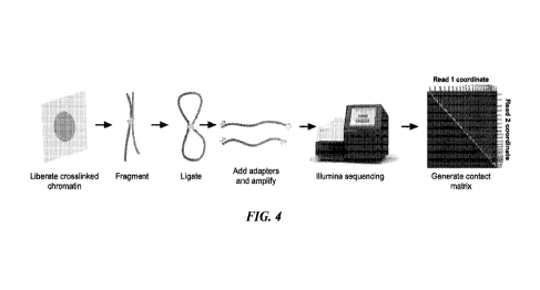
[0008] FIG. 4 shows an outline of Hi-C methodology. DNA sequences in close
physical
proximity are cross-linked during formalin fixation, fragmented by restriction
digest and
ligated together. Sequencing adapters are added and chimeric molecules are
sequenced.
Mapping reads 1 and 2 relative to each other creates a contact matrix heat
which allows
identification of chromosomal rearrangements.
[0009] FIG. 5A-5B shows the utility of AFA methods to generate Hi-C libraries
on clinical
samples. Libraries generated using above described methods from a single
section of FFPE
breast (FIG. 5A) or ovary (FIG. 5B) tumor sample is sufficient to identify non-
reciprocal
translocations between chromosomes X and 8 (FIG. 5A) and chromosomes 4 and 7
(FIG. 5B).
DETAILED DESCRIPTION
[0010] Provided
herein are methods and systems for the identification of chromosomal
structural variants using chromatin conformation capture techniques. In some
embodiments,
the disclosure further provides systems and methods for detecting chromosomal
structural
variants in tissue samples previously known to be refractory to karyotyping or
karyotyping by
sequencing (KBS) analyses (e.g., solid tissue or tumor samples). In some
embodiments, the
disclosure further provides systems and methods for relating chromosomal
structural variants
4
CA 03160441 2022-05-05
WO 2021/097284
PCT/US2020/060511
to biological information pertinent to the chromosomal structural variant (for
example, clinical
data). The chromatin conformation capture (3-C) techniques and systems and
methods for
relating chromosomal structural variants to biological information pertinent
to specific
chromosomal structural variants for use in the methods and systems provided
herein can be
those CCC techniques, systems and methods described in WO 2020/198704, which
is
incorporated herein by reference in its entirety.
[0011] In one
embodiment, a method for identifying chromosomal structural variants
provided herein comprises: (a) providing a tissue sample in a solution in a
vessel, the tissue
sample comprising nucleic acid material; (b) dissociating the tissue sample by
exposing the
tissue sample and the solution in the vessel to focused acoustic energy to
release the nucleic
acid material from the tissue sample; (c) recovering the nucleic acid
material; and (d)
performing chromosome conformation capture analysis on the nucleic acid
material. The tissue
sample can be a solid tumor sample. The tissue sample (e.g., solid tumor
sample) can be a
preserved tissue sample. The tissue sample (e.g., solid tumor sample) can be
paraffin-
embedded. The tissue sample (e.g., solid tumor sample) can be cross-linked or
fixed. In one
embodiment, the tissue sample is a formalin fixed paraffin-embedded (FFPE)
sample. The
dissociating of step (b) can be repeated one or more times. In one embodiment,
the dissociating
of step (b) is repeated once on the tissue sample and the solution in the
vessel. In another
embodiment, the method further comprises: (i) isolating the solution in the
vessel following
step (b) and prior to step (c); (ii) adding an additional volume of solution
to the tissue sample
remaining in the vessel from step (i); (iii) repeating the dissociating of
step (b) on the tissue
sample in the vessel to which the additional volume of solution was added;
(iv) isolating the
additional volume of solution added to the tissue sample in the vessel
following the additional
dissociating step; (v) dissociating the solutions isolated in steps (i) and
(iv) by exposing said
solutions to focused acoustic energy to release additional nucleic acid
material from any
remaining portions of the tissue sample in said solutions; and (vi) combining
the solutions
subjected to step (v). In one embodiment, the method further comprises
repeating steps (i)-(v)
one or more times. The solution used in each dissociating step can be a non-
solvent solution.
The non-solvent solution can be any solution that does not contain a solvent
that can cause
damage to the nucleic acid and/or proteinaceous material contained within the
tissue sample
exposed to any of the methods provided herein. The non-solvent solution can
include water
and a detergent.
[0012]
Chromatin conformation capture methods, such as 3-C, 4-C, 5-C, and Hi-C,
physically link DNA molecules in close proximity inside intact cells. These
methods measure
CA 03160441 2022-05-05
WO 2021/097284
PCT/US2020/060511
how often two loci co-associate in space in vivo. A two-dimensional contact
matrix is then
calculated from chromatin conformation capture data by mapping high throughput
sequencing
reads from a chromatin conformation capture library to a draft or reference
genome. In a contact
matrix, loci originating from the same chromosomes have a higher interaction
frequency than
loci on different chromosomes, and neighboring loci on the same chromosome
have a higher
interaction frequency than distal loci on that chromosome. Every individual's
genome exhibits
a slightly different contact matrix due to allelic variation within the
individual's population of
cells and mutations the individual was born with or acquired during their
lifetime. These
differences are termed variants. Some variants can be seen with the naked eye
by visualizing
the contact matrix as a contact map. Other variants can be detected by
analyzing the contact
matrix computationally. These variants include, but are not limited to,
balanced and unbalanced
translocations, inversions, and copy number variation such as insertions,
deletions, repeat
expansions, and other complex events. Some variants are known to have clinical
significance,
i.e. are associated with a disease and/or course of treatment. Other variants
are of unknown
clinical significance, or are novel (not previously described in the art).
Chromatin conformation
data and the methods and systems disclosed herein provide the means to
describe variants of
known clinical significance, and to discover variants of unknown clinical
significance and
novel variants.
[0013]
Karyotyping by sequencing (KBS) methods of the disclosure use chromatin
conformation data in clinical and research scenarios utilizing solid tissue
samples (e.g., solid
tumors) where karyotyping or karyotype-like data would be useful. This method
includes
multiple major applications. First, KBS methods are able to identify human
genomic
rearrangements observable by cytogenetic methods and to test for the presence
of known
clinically-reportable variants, in effect producing the same kind of
actionable information as
karyotyping but with highly different, powerful means. Second, KBS methods are
capable of
analyzing any sample to detect any structural variants, and classify these
variants using any
provided data about structural variation in the organism being sampled.
Subjects
[0014] The
disclosure provides methods and systems for detecting one or more
chromosomal structural variants in a sample obtained from a subject. The
samples can include
biopsy samples, surgical samples, tumor samples, whole organs, and other
samples.
[0015] The
subject can be any organism. In some embodiments, the subject is a eukaryote.
In some embodiments, the subject is a metazoan. In some embodiments, the
subject is a
6
CA 03160441 2022-05-05
WO 2021/097284
PCT/US2020/060511
vertebrate. In some embodiments, the subject is a mammal. In some embodiments,
the subject
is a human, a monkey, an ape, a rabbit, a guinea pig, a gerbil, a rat or a
mouse. In some
embodiments, the subject is an agricultural animal. Exemplary agricultural
animals include
horses, sheep, cows, pigs and chickens. In some embodiments, the subject is an
animal that is
kept as a pet (a veterinary subject). Exemplary pets include dogs and cats.
[0016] In some embodiments, the subject is a human.
[0017] In some embodiments, particularly those embodiments wherein the
subject is a
human, the subject has one or more symptoms of a disease or disorder which is
caused by one
or more chromosomal structural variants in the subject. In some embodiments,
the
chromosomal structural variant is one that is known in the art to cause a
disease or disorder, to
affect the function of a gene or genes that cause a disease or disorder. The
disease or disorder
can be any disease or disorder known in the art and/or provided herein to be
associated with or
caused by one or more chromosomal structural variants. In alternative
embodiments, the
chromosomal structural variant is a novel chromosomal structural variant, i.e.
a variant that has
not previously been described in the art. The disclosure provides systems and
methods to
identify both novel and known chromosomal structural variants.
[0018] The disclosure provides methods and systems for detecting one or
more
chromosomal structural variants in tissues and/or cells isolated or derived
from any tissue or
cell type in the subject. In some embodiments, the tissue is a healthy tissue
of the subject, for
example, healthy skin, bone marrow, liver, kidney, neural tissue or muscle. In
some
embodiments, the tissue has one or more symptoms of a disease or disorder. In
some
embodiments, the disease or disorder is cancer, and the tissue comprises
cancer cells. In some
embodiments, the cancer comprises a solid tumor and the tissue comprises tumor
cells. In some
embodiments, the tissue comprises a mixture of cells that comprise one or more
chromosomal
structural variants and cells that do not comprise one or more chromosomal
structural variants.
The tissue can be fresh. The tissue can be fresh-frozen. The tissue can be
fixed. The tissue can
be preserved. In one embodiment, the tissue is paraffin-embedded. In another
embodiment, the
tissue is formalin-fixed and paraffin-embedded (FFPE). In some cases, the
tissue sample has a
thickness of 5 to 25 microns and a length of less than 25 mm. In some cases,
the tissue samples
are curls (sections that are 10 microns or greater). The curls can be FFPE
curls.
[0019] In one embodiment, a sample (e.g., a biopsy) is taken from a patient
and placed in
a fixative (e.g., formalin) during a medical procedure. This fixed sample can
be subsequently
analyzed using the techniques of the present disclosure. For example, genomic
features such
as rearrangements relevant to cancer can be identified.
7
CA 03160441 2022-05-05
WO 2021/097284
PCT/US2020/060511
[0020] In one
embodiment, provided herein are methods and systems for detecting one or
more chromosomal structural variants in preserved samples from any tissue or
cell type in the
subject. The samples can be stored pursuant to basic research, translation
research, a surgical
excision or archived pursuant to a drug trial. The preserved sample can be
cross-linked for
example using at least one of a formaldehyde, a formalin, UV light, mitomycin
C, nitrogen
mustard, melphalan, 1,3-butadiene diepoxide, cis diaminedichloroplatinum(II)
and
cyclophosphamide. Alternatively, the preserved sample can be cross-linked
using formalin.
The preserved sample can maintain positional information as to nucleic acids
within it. In one
embodiment, the preserved sample is an embedded sample such as a formalin
fixed paraffin-
embedded (FFPE) sample. The preserved samples can be fixed directly and
without
homogenization, in some cases, by dropping the sample into a fixative
solution.
[0021] In one
embodiment, the preserved tissue sample is treated to isolate nucleic acids
such that protein DNA complexes are not destroyed. In some cases, the protein
DNA
complexes are isolated such that a first nucleic acid segment and a second
nucleic acid segment
in close proximity are held together independent of a phosphodiester backbone.
In some cases,
the preserved tissue sample is treated by protecting the sample from boiling
conditions. In some
cases, the preserved tissue sample is treated at a temperature not greater
than 40 C. In one
embodiment, the DNA protein complexes comprise chromatin. In some cases, the
preserved
tissue sample preserves positional information reflective of its configuration
in a tissue. In one
embodiment, the preserved tissue sample is not homogenized during preservation
or prior to
isolating nucleic acids, such that positional information of a DNA protein
complex excised
from the sample is preserved and available as part of the genome structural
analysis.
[0022] The
preserved tissue sample can be stored for at least 1 day, 2 days, 3 days, 4
days,
days, 6 days, 7 days, 8, days, 9 days, 10 days, 11 days, 12 days, 13 days, 2
weeks, 3 week, 1
month, 1.5 months, 2 months, 2.5 months, 3 months, 3.5 month, 4 months, 4.5
months, 5
months, 5.5 months, 6 months, 8 months, 10 months, 1 year, 2 years, 3 years,
4, years, 5 years,
years, 15 years, 20 years, 25 years, 30 years, 35 years, 40 years, 45 years,
or 50 years. The
preserved tissue sample can be stored for at most 1 day, 2 days, 3 days, 4
days, 5 days, 6 days,
7 days, 8, days, 9 days, 10 days, 11 days, 12 days, 13 days, 2 weeks, 3 week,
1 month, 1.5
months, 2 months, 2.5 months, 3 months, 3.5 month, 4 months, 4.5 months, 5
months, 5.5
months, 6 months, 8 months, 10 months, 1 year, 2 years, 3 years, 4, years, 5
years, 10 years,
years, 20 years, 25 years, 30 years, 35 years, 40 years, 45 years, or 50
years. The preserved
tissue sample can be stored for about 1 day, 2 days, 3 days, 4 days, 5 days, 6
days, 7 days, 8,
days, 9 days, 10 days, 11 days, 12 days, 13 days, 2 weeks, 3 week, 1 month,
1.5 months, 2
8
CA 03160441 2022-05-05
WO 2021/097284
PCT/US2020/060511
months, 2.5 months, 3 months, 3.5 month, 4 months, 4.5 months, 5 months, 5.5
months, 6
months, 8 months, 10 months, 1 year, 2 years, 3 years, 4, years, 5 years, 10
years, 15 years, 20
years, 25 years, 30 years, 35 years, 40 years, 45 years, or 50 years. In one
embodiment, the
preserved tissue sample is stored for at least one week prior to isolating
nucleic acids. In one
embodiment, the preserved tissue sample is stored for at least 6 months prior
to isolating
nucleic acids.
[0023] The
preserved tissue sample can be transported from a collection point prior to
isolating nucleic acids. The preserved tissue sample can be collected in a
sterile environment.
The preserved tissue sample can be positioned in a nonsterile environment
prior to isolating
nucleic acids.
[0024]
Preserved samples, such as formalin-fixed, paraffin embedded samples, often
comprise nucleic acids having damage, such as damage caused by fixative and/or
embedding
materials. A relevant component in making use of DNA is preserving the
integrity of DNA
physical linkage information of isolated DNA subject to a DNA damaging agent.
Although
DNA is a relatively stable molecule, the integrity of DNA can be subject to
environmental
factors and particularly time. The presence of nuclease contamination,
hydrolysis, oxidation,
chemical, physical and mechanical damages represent some of the major threats
to DNA
preservation. The mechanical, environmental and physical factors encountered
by DNA during
transportation frequently leave them in fragments and potentially lose long-
range information,
which are critical for genomic analysis. Existing methods for preserving DNA
information
mostly delay the decay of DNA but provide little protection to DNA damage over
time,
especially when fragmentation occurs. In many cases, such DNA damage can be
mitigated by
fixing and embedding samples intended for long term storage. For example, FFPE
(formalin-
fixation, paraffin embedded) samples can be preserved for a long time.
However, the
preservation process can result in DNA damage. Additionally, later DNA
extraction methods
can often be harsh and lead to further DNA damage and fragmentation.
[0025]
Disclosed herein are methods and systems related to recovering long-distance
genomic information from preserved and/or stored nucleic acid molecules, such
as nucleic acid
molecules in DNA complexes or chromatin aggregates, such as cross-linked
chromatin stored
in preserved (e.g., FFPE) samples (including tissue-based preserved samples
and cell culture-
based preserved samples). Methods and systems provided herein can be used for
the recovery
of nucleic acid samples from these preserved samples such that nucleic acid
physical linkage
information is preserved. Physical linkage information is preserved either by
preservation of
the nucleic acids themselves in the FFPE extraction process, or by preserving
nucleic acid
9
CA 03160441 2022-05-05
WO 2021/097284
PCT/US2020/060511
complexes such that physical linkage information is preserved independent of
any damage that
may occur to the nucleic acids themselves in the extraction process.
Adaptive Focused Acoustics (AFA)-based nucleic acid extraction
[0026] In one
embodiment, provided herein are methods and systems for detecting one or
more chromosomal structural variants in nucleic acid obtained, derived or
extracted from
preserved samples from any tissue or cell type in the subject using focused
acoustic energy. In
one embodiment, isolation or extraction of nucleic acid from a preserved
sample (e.g., FFPE
tissue sample) utilizes focused acoustic energy and an acoustic treatment
device as described
in W02014078650, which is herein incorporated by reference and described
briefly below.
[0027] In one
embodiment the preserved sample is an FFPE sample (e.g., solid tumor FFPE
sample) and the paraffin is disassociated from the FFPE sample using a non-
solvent solution.
In one embodiment, the non-solvent solution does not contain or expose the
FFPE sample to a
solvent during the process of paraffin disassociation. The non-solvent
solution can include
water and/or a detergent. The non-solvent solution may be used together with
suitable focused
acoustic energy to disassociate paraffin from the FFPE sample. Such paraffin
disassociation
may be done without exposing the sample to relatively high temperatures. For
example, the
paraffin may be suitably disassociated from the sample while maintaining the
sample
temperature below 5-60 C. The paraffin may be suitably dissociated from the
sample while
maintaining the sample temperature between 1-30 C. The paraffin may be
suitably dissociated
from the sample while maintaining the sample temperature from about 18-20 C or
from about
4-7 C. In one embodiment, the sample temperature is maintained at,
approximately 20 C. In
another embodiment, the sample temperature is maintained at approximately 7
C). The
paraffin disassociation utilized herein can increase nucleic acid material
yield by at least 2 to 4
times than found with processes known in the art for extraction nucleic acid
from FFPE. In one
embodiment, paraffin disassociation using the focus acoustic energy method
described herein
occurs in 3 minutes or less.
[0028] In one
embodiment, the sample is rehydrated during the paraffin disassociation
process. Rehydration can serve to improve bio-material yield as well.
[0029] In one
embodiment, the preserved tissue for use in the methods and systems
provided herein is an FFPE sample and the FFPE sample is provided in a vessel
such that
dissociation occurs in said vessel. A non-solvent, aqueous solution can be
provided in or added
to the vessel with the FFPE sample, and paraffin can be subsequently
disassociated from the
paraffin-embedded sample by exposing the sample and non-solvent solution in
the vessel to
CA 03160441 2022-05-05
WO 2021/097284
PCT/US2020/060511
acoustic energy to disassociate paraffin from the sample. Biomolecules, such
as nucleic acids,
proteins and/or other components, can then be recovered from the aqueous
portion of the
sample after disassociation of paraffin. In one embodiment, dissociation can
be performed one
or more additional times on either the aqueous portion of a sample after a
previous round of
disassociation of paraffin or the aqueous portion of a sample as well as the
tissue sample itself
after a previous round of disassociation of paraffin. Recovery of the aqueous
portion of any
sample following an initial or subsequent round of disassociation can be by
centrifuging and
pipetting the processed suspension from the vessel or by pipetting liquid
containing the
biomolecules from the vessel. The recovered biomolecules may be subjected to
any suitable
further processing as desired, such as DNA purification processing using
commercially
available techniques and equipment or further focused acoustic treatment, for
example, for
additional processing (e.g., fragmenting of nucleic acids) and/or to enhance
overall recovery
of biomolecules. In some cases, the recovering step comprises centrifuging the
tissue sample,
thereby separating a supernatant solution containing nucleic acid material
dissociated from
insoluble contaminants. In some cases, the recovering step comprises purifying
nucleic acid
material by solid phase reversible immobilization (SPRI). Any SPRI compatible
substrates
(e.g., SPRI beads) known in the art can be used during a recovery step
provided herein.
[0030] In one
embodiment, the recovered biomolecules are not subjected to any further
processing (e.g., fragmenting of nucleic acids) and instead are subjected to
chromosomal
conformation capture (e.g., Hi-C) methods as described herein.
[0031] In some
cases, the disassociating step comprises exposing the FFPE sample to
focused acoustic energy for a time sufficient to disassociate enough paraffin
from the FFPE
sample to allow recovery of the nucleic acid material and/or proteome material
from the tissue
sample. In some cases, the disassociating step comprises disassociating at
least, more than or
about 90%, 91%, 92%, 93%, 94%, 95%, 96%, 97%, 98%, 99%, 99.5% or 99.9% of
paraffin
attached to the FFPE sample. In some cases, the disassociating step comprises
disassociating
more than 90% of paraffin attached to the FFPE sample. In some cases, the
disassociating step
comprises disassociating more than 95% of paraffin attached to the FFPE
sample. In some
cases, the disassociating step comprises disassociating more than 98% of
paraffin attached to
the FFPE sample. In some cases, the disassociating step comprises
disassociating more than
99% of paraffin attached to the FFPE sample. Performing one or more additional
dissociation
steps can increase the disassociation of paraffin attached to the FFPE sample
by at least, at
most or about 5%, 10%, 15%, 20%, 25%, 30%, 35%, 40%, 45% or 50% as compared to
performing a single disassociation step. In some cases, the disassociating
step comprises
11
CA 03160441 2022-05-05
WO 2021/097284
PCT/US2020/060511
rehydrating the tissue sample while exposing the tissue sample to focused
acoustic energy. In
some cases, the disassociating step comprises maintaining a temperature of the
solution at
between 5 C and 60 C. The solution may be at a temperature of about 18 C, to
about 20 C, or
a temperature of about 4 C to about 7 C. The solution may be at a temperature
of about 40 C,
or a temperature of about 20 C, or a temperature of about 7 C. Thus,
disassociation may be
performed while the temperature of the sample is maintained below about 60 C,
e.g., below
about 45 C, below about 20 C, below about 10 C.
[0032] In some
cases, the method further comprises maintaining the tissue sample in the
vessel at below 50 C until heating with sample to 90-100 C.
[0033] In some
cases, the dissociating step comprises adding a protease (e.g., Proteinase K
or trypsin) to the solution and the tissue sample in the vessel prior to
exposing the tissue sample
to focused acoustic energy. The processed sample and protease-containing
solution may be
exposed to focused acoustic energy a second time, e.g., for a period of 10-30
seconds (or more)
to enhance the mixing of the protease with the sample and thereby enhance
enzymatic activity.
In one embodiment, acoustic treatment for 30 seconds or less (e.g., 10
seconds) may serve to
suitably mix the protease with the sample prior to incubating the sample with
the protease to
further hydrolyze the proteins in the sample. Also, the inclusion of a
glycerol material with the
protease can be used to further enhance the enzyme activity and the effect of
the acoustic energy
as a driver of the protease action. This mixing treatment may be performed
with the sample at
a temperature of between 5-46 C, e.g., with the coupling medium 16 at about 46
C, about
20 C, about 7 C, although other temperatures are possible. In some cases, the
method
comprises inactivating the protease. In some cases, inactivating the protease
comprises heating
the vessel to about 98 C.
[0034] In one
embodiment, the dissociating step comprises exposing the tissue sample
(e.g., FFPE sample) to focused acoustic energy at an intensity suitable to
avoid shearing the
nucleic acid material. The majority of the fragments of nucleic acid material
after exposing the
tissue sample to focused acoustic energy in one or more disassociating steps
can have a size of
1000 bp or greater. The nucleic acid material or the fragments of nucleic acid
material can then
be subjected to chromosomal conformation capture methods as provided herein.
[0035] The
method and systems provided herein can further comprise repeating the
dissociating step one or more times. In some cases, the method further
comprises repeating the
dissociating step while maintaining the vessel at about 4 C to about 7 C. In
some cases, the
method further comprises repeating the dissociating step one or more times
while maintaining
the vessel at about 18 C to about 20 C followed by a final dissociating step
while maintaining
12
CA 03160441 2022-05-05
WO 2021/097284
PCT/US2020/060511
the vessel at about 4 C to about 7 C. Similarly to the initial disassociation
step, each additional
disassociating step can be performed on tissue sample remaining in the vessel
following a
previous round of disassociation to which solution (e.g., non-solvent solution
as described
herein) is added. The final dissociating step is performed on the solution
(e.g., aqueous
solution) isolated from each previous round of disassociation.
[0036] In one
embodiment, an acoustic treatment device is utilized in the dissociation steps
present in the methods and system provided herein. The acoustic treatment
device can include
a vessel holding a formalin fixed, paraffin embedded tissue sample and a non-
solvent, aqueous
solution, and an acoustic energy source for providing acoustic energy to the
sample while the
sample is in the vessel and separated from the acoustic energy source. A
vessel holder may
support the vessel at a location at least partially in a focal zone of the
acoustic energy, and a
system control circuit may control the acoustic energy source to expose the
sample to focused
acoustic energy suitable to disassociate paraffin from the sample to allow
recovery of
biomolecules of the sample. The focused acoustic energy for use in the
dissociation steps
provided in the methods and systems provided herein can have a frequency of
between about
100 kilohertz and about 100 megahertz. The focused acoustic energy can have a
focal zone
with a width of less than about 2 centimeters. The focused acoustic energy can
originate from
an acoustic energy source spaced from and exterior to the vessel (e.g., an
acoustic treatment
device), wherein at least a portion of the acoustic energy propagates exterior
to the vessel. In
some cases, the focused acoustic energy has a duty factor of between 10% and
30%. In some
cases, the focused acoustic energy has a duty factor of about 15% or about
20%. In some cases,
the focused acoustic energy has a peak intensity power of between 60W and 90W.
In some
cases, the focused acoustic energy has a peak intensity power of about 75W. In
some cases,
each disassociating step in any method provided herein is performed with a
cycles per burst
(cpb) of 200. In some cases, any of the methods provided herein that entails
using focused
acoustic energy to extract nucleic acid from a preserved sample (e.g., FFPE
tissue sample)
comprises at least one dissociating step such that the AFA is run for 5 min
with a duty factor
of 20%, a peak intensity of 75W and 200 cycles/burst in at least one of the
dissociating steps.
In some cases, the method provided herein comprises a first and a second
dissociating step such
that the first dissociating step is performed using AFA run for 5 min with a
duty factor of 20%,
a peak intensity of 75W and 200 cycles/burst, while the second dissociating
step is performed
using AFA run for 10 min with a duty factor of 15%, a peak intensity of 75W
and 200
cycles/burst. In some cases, the method provided herein comprises more than
two dissociating
steps such that each dissociating step is performed using AFA run for 5 min
with a duty factor
13
CA 03160441 2022-05-05
WO 2021/097284
PCT/US2020/060511
of 20%, a peak intensity of 75W and 200 cycles/burst except for the final
dissociating step,
which is performed using AFA run for 10 min with a duty factor of 15%, a peak
intensity of
75W and 200 cycles/burst.
[0037] In one
embodiment, the dissociating step preserves formaldehyde crosslinks in the
tissue sample. Further to this embodiment, the processed sample is then
subjected to
chromosomal conformational capture (e.g., Hi-C) and chromosomal structural
variant
identification (e.g., via sequencing) as described herein.
Size Selection
[0038] Nucleic
acid obtained from preserved (e.g., FFPE) biological samples can be
fragmented to produce suitable fragments for analysis by chromosomal
conformation capture
methods provided herein. Template nucleic acids may be fragmented or sheared
to desired
length, using a variety of mechanical, chemical and/or enzymatic methods. DNA
may be
randomly sheared via sonication, e.g. Covaris method, brief exposure to a
DNase, or using a
mixture of one or more restriction enzymes, or a transposase or nicking
enzyme. RNA may be
fragmented by brief exposure to an RNase, heat plus magnesium, or by shearing.
The RNA
may be converted to cDNA. If fragmentation is employed, the RNA may be
converted to cDNA
before or after fragmentation. In some embodiments, nucleic acid from a
biological sample is
fragmented by sonication. In other embodiments, nucleic acid is fragmented by
a hydroshear
instrument. Generally, individual nucleic acid template molecules can be from
about 2 kb bases
to about 40 kb. In various embodiments, nucleic acids can be about 6kb-10 kb
fragments. In
one embodiment, nucleic acid from a preserved tissue sample is fragmented
using focused
acoustic energy as described in W02018195153, which is incorporated herein by
reference.
[0039] In one
embodiment, cross-linked DNA molecules may be subjected to a size
selection step. Size selection of the nucleic acids may be performed to cross-
linked DNA
molecules below or above a certain size. Size selection may further be
affected by the frequency
of crosslinks and/or by the fragmentation method, for example by choosing a
frequent or rare
cutter restriction enzyme. In some embodiments, a composition may be prepared
comprising
crosslinking a DNA molecule in the range of about 1 kb to 5 Mb, about 5kb to 5
Mb, about 5
kB to 2Mb, about 10 kb to 2Mb, about 10 kb to 1 Mb, about 20 kb to 1 Mb about
20 kb to 500
kb, about 50 kb to 500 kb, about 50 kb to 200 kb, about 60 kb to 200 kb, about
60 kb to 150
kb, about 80 kb to 150 kb, about 80 kb to 120 kb, or about 100 kb to 120 kb,
or any range
bounded by any of these values (e.g. about 150 kb to 1 Mb).
14
CA 03160441 2022-05-05
WO 2021/097284
PCT/US2020/060511
[0040] In some
embodiments, sample polynucleotides are fragmented into a population of
fragmented DNA molecules of one or more specific size range(s). In some
embodiments,
fragments can be generated from at least about 1, about 2, about 5, about 10,
about 20, about
50, about 100, about 200, about 500, about 1000, about 2000, about 5000, about
10,000, about
20,000, about 50,000, about 100,000, about 200,000, about 500,000, about
1,000,000, about
2,000,000, about 5,000,000, about 10,000,000, or more genome-equivalents of
starting DNA.
Fragmentation may be accomplished by methods known in the art, including
chemical,
enzymatic, and mechanical fragmentation. In some embodiments, the fragments
have an
average length from about 10 to about 10,000, about 20,000, about 30,000,
about 40,000, about
50,000, about 60,000, about 70,000, about 80,000, about 90,000, about 100,000,
about 150,000,
about 200,000, about 300,000, about 400,000, about 500,000, about 600,000,
about 700,000,
about 800,000, about 900,000, about 1,000,000, about 2,000,000, about
5,000,000, about
10,000,000, or more nucleotides. In some embodiments, the fragments have an
average length
from about 1 kb to about 10 Mb. In some embodiments, the fragments have an
average length
from about 1 kb to 5 Mb, about 5 kb to 5 Mb, about 5 kB to 2 Mb, about 10 kb
to 2 Mb, about
kb to 1 Mb, about 20 kb to 1 Mb about 20 kb to 500 kb, about 50 kb to 500 kb,
about 50 kb
to 200 kb, about 60 kb to 200 kb, about 60 kb to 150 kb, about 80 kb to 150
kb, about 80 kb to
120 kb, or about 100 kb to 120 kb, or any range bounded by any of these values
(e.g. about 60
to 120 kb). In some embodiments, the fragments have an average length less
than about 10 Mb,
less than about 5 Mb, less than about 1 Mb, less than about 500 kb, less than
about 200 kb, less
than about 100 kb, or less than about 50 kb. In other embodiments, the
fragments have an
average length more than about 5 kb, more than about 10 kb, more than about 50
kb, more than
about 100 kb, more than about 200 kb, more than about 500 kb, more than about
1 Mb, more
than about 5 Mb, or more than about 10 Mb.
[0041] In some
embodiments, the fragmentation is accomplished mechanically comprising
subjection sample DNA molecules to acoustic sonication. In some embodiments,
the
fragmentation comprises treating the sample DNA molecules with one or more
enzymes under
conditions suitable for the one or more enzymes to generate double-stranded
nucleic acid
breaks. Examples of enzymes useful in the generation of DNA fragments include
sequence
specific and non-sequence specific nucleases. Non-limiting examples of
nucleases include
DNase I, Fragmentase, restriction endonucleases, variants thereof, and
combinations thereof
For example, digestion with DNase I can induce random double-stranded breaks
in DNA in
the absence of Mg++ and in the presence of Mnt. In some embodiments,
fragmentation
comprises treating the sample DNA molecules with one or more restriction
endonucleases.
CA 03160441 2022-05-05
WO 2021/097284
PCT/US2020/060511
Fragmentation can produce fragments having 5' overhangs, 3 ' overhangs, blunt
ends, or a
combination thereof In some embodiments, such as when fragmentation comprises
the use of
one or more restriction endonucleases, cleavage of sample DNA molecules leaves
overhangs
having a predictable sequence. In some embodiments, the method includes the
step of size
selecting the fragments via standard methods such as column purification or
isolation from an
agarose gel.
Chromosomal Structural Variants
[0042] The
disclosure provides methods and systems for detecting one or more
chromosomal structural variants in a subject.
[0043] As used
herein, the term "chromosome" refers to a chromatin complex comprising
all or a portion of the genome of a cell. The genome of a cell is often
characterized by its
karyotype, which is the collection of all the chromosomes that comprise the
genome of the cell.
The genome of a cell can comprise one or more chromosomes. In humans, each
chromosome
has a short arm (termed "p" for "petit") and a long arm (termed "q" for
"queue").
[0044] Each
chromosome arm is divided into regions, or cytogenetic bands, that can be
seen in a conventional karyotype using a microscope. The bands are labeled pl,
p2, p3 etc.
counting from the centromere out towards the telomeres. Higher-resolution sub-
bands within
the bands are sometimes also used to identify regions in the chromosome. Sub-
bands are also
numbered from the centromere out towards the telomere. Information on
chromosome banding
and chromosome nomenclature can be found in pp. 37-39 of Strachan, T. and
Read, A.P. 1999.
Human Molecular Genetics, 2nd ed. New York: John Wiley & Sons.
[0045] The
terms "nucleic acid," "polynucleotide," and "oligonucleotide" are used
interchangeably and refer to a deoxyribonucleotide or ribonucleotide polymer
in either single-
or double-stranded form. For the purposes of the present disclosure, these
terms are not to be
construed as limiting with respect to the length of a polymer. The terms can
encompass known
analogues of natural nucleotides, as well as nucleotides that are modified in
the base, sugar
and/or phosphate moieties. In general, an analogue of a particular nucleotide
has the same base-
pairing specificity (e.g., an analogue of A will base pair with T. A
polynucleotide of
deoxyribonucleic acids (DNA) of specific identities and order is also referred
to herein as a
"DNA sequence." Chromosomes comprise polynucleotides complexed with proteins
(e.g.
histones).
[0046] As used
herein the terms "Structural Variant", "Chromosomal Structural Variant",
"CSV" or "SV" refer to a difference in the structure of an individual's
chromosome or
16
CA 03160441 2022-05-05
WO 2021/097284
PCT/US2020/060511
chromosomes relative to the chromosome(s) in the genomes of other individuals
within the
same species or in a closely related species. Differences in chromosomal
structure encompass
differences in the arrangement and identity of DNA sequences in a chromosome.
Differences
in the arrangement of DNA sequences in a chromosome include both differences
in the
positions of DNA sequences on the chromosome relative to other sequences
(e.g.,
translocations) and differences in orientation relative to other sequences
(e.g., inversions).
Differences in the identity of DNA sequences along a chromosome can include
both new
sequences and missing sequences, for example through the movement sequences
from one
chromosome to another non-homologous chromosome.
[0047]
Chromosomal structural variations can be small or large in size, encompassing
tens
of base pairs, hundreds of base pairs, kilobases, megabases, or even
significant portions (a half,
a third or three-quarters, e.g.) of an individual chromosome. All size of
chromosomal structural
variations are within the scope of the disclosure.
[0048] There
are multiple types of chromosomal structural variants, all of which are
envisaged as within the scope of the methods and systems of the disclosure.
Non-limiting
examples of types of chromosomal structural variants include a translocation,
a balanced
translocation, an unbalanced translocation, a complex translocation, an
inversion, a deletion, a
duplication, a repeat expansion or a ring.
[0049] As used
herein the term "translocation" refers to the exchange of DNA sequences
between non-homologous chromatids, between two or more positions on the same
chromatid,
or between homologous chromatids that is not as a result of crossover during
meiosis.
Translocations can create gene fusions, which occur when two genes that are
not normally
adjacent to each other are brought into proximity. Alternatively, or in
addition, translocations
can disrupt gene function by breaking genes at the borders of the
translocation. For example, a
translocation can separate an open reading frame (ORF) from a distal
regulatory element or
bring the open reading frame into proximity with a new regulatory element,
thereby affecting
gene expression. Alternatively, or in addition, the break point of the
translocation can occur in
the middle of a gene, thereby creating a gene truncation. A "breakpoint"
refers to the point or
region of a chromosome at which the chromosome is cleaved during a
translocation. A
"breakpoint junction" refers to the region of the chromosome at which the
different parts of
chromosomes involved in a translocation join. Alternatively, or in addition, a
translocation can
affect the expression of one or more genes contained within the translocation
by moving those
genes to a new chromatin environment in the nucleus, for example by moving a
DNA sequence
from a region of strong gene expression (e.g. euchromatin) to a region of low
gene expression
17
CA 03160441 2022-05-05
WO 2021/097284
PCT/US2020/060511
(e.g. heterochromatin) or vice versa. Depending on the translocation, the
translocation can have
no effect on gene expression, can effect a single gene, or can effect multiple
genes.
[0050] As used
herein the term "balanced translocation" refers to the reciprocal exchange
of DNA between non-homologous chromatids, or between homologous chromatids not
as a
result of crossover during meiosis. A "balanced translocation" is a
translocation in which there
is no loss of genetic material during the translocation, but all genetic
material is preserved
during the exchange. In an "unbalanced translocation" there is a loss of
genetic material during
the exchange.
[0051] As used
herein, the term "reciprocal translocation" refers to a translocation which
involves the mutual exchange of fragments between two broken chromosomes. In a
reciprocal
translocation, one part of one chromosome unites with the part of another
chromosome.
[0052] As used
herein, the terms "variant translocation", "abnormal translocation" or
"complex translocation" refer to the involvement of a third chromosome in a
secondary
rearrangement that follows a first translocation.
[0053]
Translocations can be intrachromosomal (the rearrangement breakpoints occur
within the same chromosome) or interchromosomal (the rearrangement breakpoints
are
between two different chromosomes).
[0054] As used
herein, the term "inversion" refers to the rearrangement of DNA sequences
within the same chromosome. Inversions change the orientation of a DNA
sequence within a
chromosome.
[0055] As used
herein, the term "deletion" refers to a loss of a DNA sequence. Deletions
can be any size, ranging from a few nucleotides to entire chromosomes.
Translocations are
frequently accompanied by deletions, for example at the translocation break
points.
[0056] As used
herein, the term "duplication" refers to a duplication of a DNA sequence
(e.g., the genome contains three copies of a DNA sequence, instead of two).
Duplications can
be any size, ranging from a few nucleotides to entire chromosomes.
Translocations are
frequently accompanied by duplications.
[0057] As used
herein, the term "repeat expansion" refers to tandem repeated sequences in
the genome that with variable copy numbers between subjects. When there are a
greater than
average number of repeats of a repetitive sequence, the repetitive sequence
has been expanded.
Repeated sequences can comprise 2, 3, 4, 5, 6, 7, 8, 9, 10 or more repeated
nucleotides.
Expanded repeats are associated with a number of genetic disorders, including
but not limited
to Huntington's disease, spinocerebellar ataxias, fragile X syndrome, myotonic
dystrophy,
Friedreich's ataxia and juvenile my oclonic epilepsy.
18
CA 03160441 2022-05-05
WO 2021/097284
PCT/US2020/060511
[0058] All
types of chromosomal structural variants can be identified using the methods
and systems of the disclosure.
[0059] In some
embodiments, the chromosomal structural variant identified by the
methods and systems of the disclosure is a chromosomal variant that is known
in the art. For
example, the chromosomal structural variant identified by the methods of the
disclosure is a
chromosomal structural variant that has been previously described and
characterized.
Descriptions of chromosomal structural variants in the art include mapping one
or more
breakpoints of the chromosomal structural variant using techniques known in
the art, for
example by karyotyping, sequencing or Southern blot. In those embodiments
wherein the
chromosomal structural variant is known to cause a disease or disorder,
descriptions of known
chromosomal structural variants include clinical data such as symptoms,
prognosis and
recommended courses of treatment.
[0060] In some
embodiments, the chromosomal structural variant identified by the
methods and systems of the disclosure is a novel chromosomal variant. Novel
chromosomal
structural variants are variants that have not previously been described in
the art. Novel
chromosomal structural variants may be similar to chromosomal structural
variants known in
the art. For example, a chromosomal structural variant may be both recurrent,
in that similar
variants occur independently across multiple individuals, and novel, in that
each individual
with a recurrent variant comprises a variant with slightly different break
points. In some
embodiments, a novel chromosomal structural variant has one or more
breakpoints that are
similarly placed compared to a break point of a chromosomal structural variant
known in the
art. A similarly placed break point comprises a break point that is within 50
bp, within 100 bp,
within 500 bp, within 1 kb, within 5 kb, within 10 kb, within 20 kb, within 50
kb, within 100
kb, within 200 kb or within 500 kb or within 1 Mb of a break point of a
chromosomal structural
variant known in the art. In some embodiments, a novel chromosomal structural
variant has
one or more breakpoints that are identical to a break point of a chromosomal
structural variant
known in the art, and one or more breakpoints that are not identical to a
break point of a
chromosomal structural variant known in the art. In some embodiments, a novel
chromosomal
structural variant does not have similar or identical break points to a
chromosomal structural
variant known in the art.
Representation of Chromosomal Structural Variants
[0061] The
disclosure provides systems and methods for detecting one or more
chromosomal structural variants in a subject, and representing the chromosomal
structural
19
CA 03160441 2022-05-05
WO 2021/097284
PCT/US2020/060511
variant or variants in a manner that can be readily interpreted by a person of
ordinary skill in
the art (for example, a clinician, a doctor, a patient or a researcher).
[0062] In some
embodiments, the chromosomal structural variant is represented as a
karyotype. Karyotyping is a traditional method used to identify chromosomal
structural
variants. In karyotyping, the development of cells is arrested during
metaphase, bound
chromatids are extracted, stained and photographed, and the structural
properties of the
chromatids are mapped using the cytogenetic banding patterns of the
chromosome.
Karyotyping is expensive, time consuming and of limited resolution.
Traditional karyotyping
relies on the cytogenetic bands and sub bands within the karyotype to map the
boundaries of
chromosomal structural variants, and so cannot resolve chromosomal structural
variants that
are finer (smaller) than the cytogenetic bands of the karyotype, which
typically have a
minimum resolution of about 5 Mb. In contrast, the systems and methods of the
disclosure are
able to achieve a resolution that is at least 1,000 finer than a traditional
karyotype.
[0063]
Traditional karyotype results can be represented as karyotype spreads, which
are
images of all the chromosomes analyzed in the karyotype, stained to identify
cytogenetic bands
and arranged in ordered pairs. While the methods of the disclosure provide a
resolution superior
to a traditional karyotype, the chromosomal structural variants identified by
the methods of the
disclosure can be represented as a karyotype or karyotype spread. This
facilitates interpretation
of chromosomal structural variant data of the disclosure by doctors and
clinicians, who may be
more familiar with and trained to identify chromosomal structural variants
based on traditional
karyotypes.
[0064] In some
embodiments, chromosomal structural variants of the disclosure are
represented as a karyotype.
Clinical Chromosomal Structural Variants
[0065] The
disclosure provides methods and systems for detecting one or more
chromosomal structural variants in a subject, and further relating the one or
more chromosomal
structural variants to relevant biological information. Relevant biological
information includes,
but is not limited to, the clinical significance of the variant, associated
diseases or disorders,
symptoms thereof, associated genes and/or genetic mutations, effects of the
chromosomal
structural variant on gene expression, and recommended courses of treatment or
therapies.
[0066] In some
embodiments, the chromosomal structural variants that are identified by
the systems and methods of the disclosure cause one or more diseases or
disorders.
CA 03160441 2022-05-05
WO 2021/097284
PCT/US2020/060511
[0067] In some
embodiments, the chromosomal structural variants that cause diseases or
disorders are inherited, i.e. the chromosomal structural variant is
transmitted from parent to
offspring via the germ line. All inherited chromosomal structural variants are
within the scope
of the systems and methods of the disclosure.
[0068] In other
alternative embodiments, the chromosomal structural variants that cause
diseases or disorders are somatic, i.e. the chromosomal structural variant
arise de novo in a cell
in the individual. Depending upon when in development a somatic chromosomal
structural
variant arises, somatic chromosomal structural variants can occur all the
cells in an organism
(the chromosomal structural variant arises prior to the first cell division),
or can occur in a
subset of the cells in the organism (the chromosomal structural variant occurs
later in
development, or in an adult). Exemplary disorders that can occur in every cell
include
aneuploidies such as Turner syndrome (X chromosome monosomy) and Down syndrome
(trisomy 21).
[0069]
Exemplary disorders caused by haploinsufficiencies resulting from deletions
include Williams syndrome, Langer¨Giedion syndrome, Miller¨Dieker syndrome,
and
DiGeorge/velocardiofacial syndrome. All somatic chromosomal structural
variants are within
the scope of the systems and methods of the disclosure.
[0070] In some
embodiments, the diseases or disorders caused by chromosomal structural
variants are caused by a chromosomal structural variant that occurs de novo in
the subject. In
some embodiments, the chromosomal structural variant that occurs de novo is a
recurrent
structural variant. Many chromosomal structural variants are recurrent, in
that the same or
similar chromosomal structural variants occur de novo in multiple individuals.
These
individuals are not necessarily related. In many cases, the recurrent
chromosomal structural
variants are caused by non-allelic homologous recombination mediated by
flanking segmental
duplications. In non-allelic homologous recombination, improper crossing over
between non-
homologous DNA sequences, for example DNA sequences that contain similar
repetitive DNA
sequences, leads to a tandem or direct duplication and a deletion. Non-
limiting examples of
diseases and disorders caused by recurrent chromosomal structural variants
include in Charcot
Marie Tooth disease, hereditary neuropathy with liability to pressure palsies,
Prader Willi,
Angelman, Smith Magenis, DiGeorge/velocardiofacial (DGSNCFS), Williams
Beurens, and
Sotos syndromes.
[0071]
Databases of chromosomal structural variants are well known to persons of
ordinary
skill in the art. For example, biological information regarding chromosomal
structural variants
and their associated diseases and disorders, and treatments for these diseases
and disorders can
21
CA 03160441 2022-05-05
WO 2021/097284
PCT/US2020/060511
be found in the Online Mendelian Inheritance in Man (omim.org), the Mitelman
Database of
Chromosome Aberration and Gene Fusion in Cancer
(cgap.nci.nih.gov/Chromosomes/Mitelman) and the NCBI
database
(ncbi. nlm. nih. gov/clinv ar?term=3 0 0 005 [MIM]).
[0072]
Chromosomal structural variants and associated diseases and disorders are also
described by the National Institute of Health's Genetic and Rare Diseases
Information Center
(raredi s eas es . info . nih. gov/di s eas es/di s eas es -by -category/3
6/chromosome-disorders).
[0073] In some
embodiments, chromosomal structural variants do not occur in every cell
in a tissue of the subject. In some embodiments, the cells with the
chromosomal structural
variant(s) are cancer cells in the subject. A subject with a cancer can have
cancer cells with one
or more chromosomal structural variants, while the non-cancerous cells of the
subject do not
have a chromosomal structural variant, or do not have the same chromosomal
structural
variants that are seen in the cancer cells of the subject.
[0074] Cancers
are diseases caused by the proliferation of malignant neoplastic cells, such
as tumors, neoplasms, carcinomas, sarcomas, blastomas, leukemias, lymphomas
and the like.
For example, cancers include, but are not limited to, mesothelioma, leukemias
and lymphomas
such as cutaneous T-cell lymphomas (CTCL), non-cutaneous peripheral T-cell
lymphomas,
lymphomas associated with human T-cell lymphotrophic virus (HTLV) such as
adult T-cell
leukemia/lymphoma (ATLL), B-cell lymphoma, acute nonlymphocytic leukemias,
chronic
lymphocytic leukemia, chronic myelogenous leukemia, acute myelogenous
leukemia,
lymphomas, and multiple myeloma, non-Hodgkin lymphoma, acute lymphatic
leukemia
(ALL), chronic lymphatic leukemia (CLL), Hodgkin's lymphoma, Burkitt lymphoma,
adult T-
cell leukemia lymphoma, acute-myeloid leukemia (AML), chronic myeloid leukemia
(CML),
or hepatocellular carcinoma. Further examples include myelodisplastic
syndrome, childhood
solid tumors such as brain tumors, neuroblastoma, retinoblastoma, Wilms'
tumor, bone tumors,
and soft-tissue sarcomas, common solid tumors of adults such as head and neck
cancers (e.g.,
oral, laryngeal, nasopharyngeal and esophageal), genitourinary cancers (e.g.,
prostate, bladder,
renal, uterine, ovarian, testicular), lung cancer (e.g., small-cell and non-
small cell), breast
cancer, pancreatic cancer, melanoma and other skin cancers, stomach cancer,
brain tumors,
tumors related to Gorlin's syndrome (e.g., medulloblastoma, meningioma, etc.)
and liver
cancer.
[0075] Most
cancers acquire one or more clonal chromosomal structural variants during
the development of the cancer, which can be identified by the systems and
methods of the
disclosure. In many cases, recurrent chromosomal structural variants are
associated with
22
CA 03160441 2022-05-05
WO 2021/097284
PCT/US2020/060511
particular morphological and clinical disease characteristics. Structural
variants in cancer cells
can affect the expression and/or function of proto-oncogenes and tumor
suppressors. Structural
variants in cancer cells can also facilitate the progression of the cancer
itself, as mutations and
changes in gene expression caused by the chromosomal structural variant(s)
promote increased
growth and invasiveness of tumor cells, and tumor vascularization. Identifying
the specific
chromosomal structural variants in a cancer cells in a cancer sample allows
for the more
effective selection of cancer therapies. These therapies can be tailored to
changes in gene
expression and cancer pathologies associated with the particular chromosomal
structural
variants in the cancer cells. Thus, the rapid and effective identification of
chromosomal
structural variants in cancers is a critical piece of the cancer diagnostic
and treatment arsenal.
[0076] In some
embodiments, structural variants in cancer cells create novel fusion
proteins which promote the progression of the cancer. A non-limiting,
exemplary list of
chromosomal structural variants that cause fusion proteins associated with
cancers is described
in Hasty, P. and Montagna, C. (2014) Mol. Cell. Oncol.: e29904. Currently
there are 21,477
documented gene fusions and 69,134 cases documented in the Cancer Genome
Anatomy
Project (cgap.nci.nih.gov/Chromosomes/Mitelman), all of which are envisaged as
falling
within the scope of the instant disclosure.
[0077] In some
embodiments, chromosomal structural variants in cancer cells lead to
changes in gene regulation and gene expression, which contribute to the
progression of the
cancer. A chromosomal structural variant can lead to the downregulation of one
or more the
tumor suppressors, which are genes that protect the cell from cancer. For
example, a
chromosomal structural variant with a break point near a tumor suppressor can
separate the
coding sequence of the tumor suppressor from a regulatory element.
Alternatively, or in
addition, a chromosomal structural variant can lead to the conversion of one
or more proto-
oncogenes into an oncogene which promotes cancer progression. For example, a
chromosomal
structural variant with a break point near a proto-oncogene can bring the
proto-oncogene into
proximity of a novel regulatory element, leading to upregulated expression.
Exemplary tumor
suppressors that can be down regulated by the chromosomal structural variants
of the disclosure
include, but are not limited to, p53, Rb, PTEN, INK4, APC, MADR2, BRCA1,
BRCA2, WT1,
DPC4 and p21. Exemplary oncogenes that can be upregulated by the chromosomal
structural
variants of the disclosure include, but are not limited to, Abll, HER-2, c-
KIT, EGFR, VEGF,
B-Raf, Cyclin D1, K-ras, beta-catenin, Cyclin E, Ras, Myc and MITF. All
chromosomal
structural elements which affect proto-oncogenes and tumor suppressor genes
are envisaged as
within the scope of the systems and methods of the disclosure.
23
CA 03160441 2022-05-05
WO 2021/097284
PCT/US2020/060511
Chromosomal Conformation Capture
[0078] Provided
herein are systems and methods that use chromosomal conformation
capture techniques to identify one or more chromosomal structural variants in
a subject.
[0079] The
terms "chromosomal conformational capture" and "chromosome conformation
analysis" are used interchangeably herein.
[0080] The
methods of the disclosure can use standard chromatin conformation data, such
as Hi-C data, generated from a tissue sample (e.g. cancerous or normal tissues
or cells) or
preserved tissue sample (e.g., FFPE sample). The computational methods
involves the training
of one or more classifiers, which can be used in more than one of the major
applications. The
set of classifiers chosen may include deep learning models, gradient descent
models, graph
network models, neural network models, support vector machine models, expert
system
models, decision tree models, logistic regression models, clustering models,
Markov models,
Monte Carlo models, or other machine learning models, as well as models which
fit observed
data to probabilistic models such as likelihood models. The set of classifiers
can be trained by
labeled or unlabeled data, which can be generated from real biological
samples, simulated
genomes which may have simulated mutations, or generated by another algorithm,
such as
algorithms used in a generative adversarial network. The training data
consists of chromatin
conformation data or data derived from it (such as a contact matrix, and may
be normalized,
filtered, compressed, or smoothed) and clinical or biological information
about the effects,
properties, implications, or outcomes associated with the data.
[0081] In some
embodiments of the systems and methods of the disclosure utilize one or
more classifiers that are trained using chromosomal conformation capture data.
In some
embodiments, the one or more classifiers are trained using experimentally
determined
chromosomal conformational capture data. In some embodiments, the one or more
classifiers
are trained using simulated chromosomal conformational capture data. In some
embodiments,
the one or more classifiers are trained using a combination of experimentally
determined and
simulated chromosomal conformational capture data.
[0082] In some
embodiments, the chromosomal conformational capture data used to train
the one or more machine learning classifiers comprises experimentally
determined
chromosomal conformational capture data. In some embodiments, the
experimentally
determined chromosomal conformational capture data comprises a plurality of
sets of reads
from healthy subjects. In some embodiments, the experimentally determined
chromosomal
conformational capture data comprises a plurality of sets of reads from
subjects with known
chromosomal structural variants.
24
CA 03160441 2022-05-05
WO 2021/097284
PCT/US2020/060511
[0083]
Chromosomal conformational data is generated by chemically cross-linking
regions
of the genome that are in close spatial proximity. In one embodiment, the
crosslinking for
chromosomal conformational capture or proximity ligation is essentially the
same as is
generated during the formalin fixation of solid tissues for histology, thereby
making Hi-C
compatible with FFPE tissues. Subsequently, the cross-linked chromatin can be
fragmented.
The fragments can be ligated together to create chimeric sequences which can
be detected using
any sequence detection method known in the art, such as, for example, CHIP
analysis, PCR
analysis or sequencing (e.g., Illumina paired end chemistry). Sequencing these
chimeric DNA
molecules can capture the signal of long-range chromatin interactions (such as
promoter-
enhancer interactions). The signal in proximity ligation sequencing can also
reflect the linear
distance between two sequences on a chromosome.
[0084] In one
embodiment, the methods and systems provided herein that utilize FFPE
tissue samples, utilize the cross-linking performed during preparation of the
FFPE sample for
chromosomal conformational capture. The cross linked nucleic acid (e.g., DNA)
can then be
fragmented and ligated to generate chromatin/nucleic acid (e.g., DNA)
complexes for
subsequent sequence detection. In one embodiment, the cross linked nucleic
acid (e.g., DNA)
is restriction enzyme digested and ligated to generate chromatin/nucleic acid
(e.g., DNA)
complexes which are identified by high-throughput sequencing. In one
embodiment, the
restriction enzyme used to digest the cross-linked nucleic acid (e.g., DNA)
during
chromosomal conformational capture is DpnII. The resultant sequence detected
(e.g., sequence
reads) can be mapped to a genome, for example a reference genome, to determine
the frequency
with which each interaction occurs within the population of cells that was
used to generate the
initial sample. When two loci are in close spatial proximity, they can
generate more reads that
comprise DNA sequences that map both loci than if the two loci are not in
close spatial
proximity.
[0085]
Experimentally determined chromosomal conformational capture data may form
part of an input file used by a system to carry out the methods described
herein. The set of
reads may be generated by any suitable method based on chromatin interaction
techniques or
chromosome conformation analysis techniques. Chromosome conformation analysis
techniques that may be used in accordance with the embodiments described
herein may
include, but are not limited to, Chromatin Conformation Capture (3C),
Circularized Chromatin
Conformation Capture (4C), Carbon Copy Chromosome Conformation Capture (5C),
Chromatin Immunoprecipitation (ChIP; e.g., cross-linked ChIP (XChIP), native
ChIP
(NChIP)), ChIP-Loop, genome conformation capture (GCC) (e.g., Hi-C, 6C),
Capture-C, Split-
CA 03160441 2022-05-05
WO 2021/097284
PCT/US2020/060511
pool barcoding (SPLiT-seq), Nuclear Ligation Assay (NLA), Single-cell Hi-C
(scHi-C),
Combinatorial Single-cell Hi-C, Concatamer Ligation Assay (COLA), Cleavage
Under Targets
and Release Using Nuclease (CUT& RUN), in vitro proximity ligation (e.g.
Chicago ), in situ
proximity ligation (in situ Hi-C), proximity ligation followed by sequencing
on an Oxford
Nanopore machine (Pore-C), proximity ligation sequenced on a Pacific
Biosciences machine
(SMRT-C), DNase Hi-C, Micro-C and Hybrid Capture Hi-C . In some embodiments,
the
dataset is generated using a genome-wide chromatin interaction method, such as
Hi-C.
[0086] In some
embodiments, chromosomal conformational data can be generated from a
population of cells. In some embodiments, chromosomal conformational capture
data is
generated by Chromatin Conformation Capture (3C). 3C is used to analyze the
organization of
chromatin in a cell by quantifying the interactions between genomic loci that
are nearby in 3-
D space. 3C quantifies interactions between a single pair of genomic loci. In
some
embodiments, chromosomal conformational capture data is generated by
Circularized
Chromatin Conformation Capture (4C). 4C captures interactions between one
locus and all
other genomic loci. In some embodiments, chromosomal conformational capture
data is
generated by Carbon Copy Chromosome Conformation Capture (5C). 5C detects
interactions
between all restriction fragments within a given region. In some embodiments,
the region is
one megabase or less. In some embodiments, chromosomal conformational capture
data is
generated by Chromatin Immunoprecipitation (ChIP; e.g., cross-linked ChIP
(XChIP), native
ChIP (NChIP)). In some embodiments, chromosomal conformational capture data is
generated
by ChIP-Loop. In some embodiments, chromatin immunoprecipitation based methods
incorporate chromatin immunoprecipitation (chIP) based enrichment and
chromatin proximity
ligation to determine long range chromatin interactions. In some embodiments,
chromosomal
conformational capture data is generated by Hi-C. Hi-C uses high-throughput
sequencing to
find the nucleotide sequence of fragments that map to both partners in all
interacting pairs of
loci. In some embodiments, chromosomal conformational capture data is
generated by Capture-
C. Capture-C selects and enriches for genome-wide, long-range contacts
involving active and
inactive promoters. In some embodiments, chromosomal conformational capture
data is
generated by SPLiT-seq. SPLiT-seq is a technique that can be used to
transcriptome profile
single cells. In some embodiments, chromosomal conformational capture data is
generated by
Nuclear Ligation Assay (NLA). Similar to 3C, NLA can be used to determine the
circularization frequencies of DNA following proximity based ligation. In some
embodiments,
chromosomal conformational capture data is generated by Concatamer Ligation
Assay
(COLA). COLA is a Hi-C based protocol that uses the Cvill restriction enzyme
to digest
26
CA 03160441 2022-05-05
WO 2021/097284
PCT/US2020/060511
chromatin. In some embodiments, using COLA results in smaller fragments
compared to
traditional Hi-C. In some embodiments, chromosomal conformational capture data
is generated
by Cleavage Under Targets and Release Using Nuclease (CUT& RUN). CUT & RUN
uses a
targeted nuclease strategy for high-resolution mapping of DNA binding sites.
For example,
CUT&RUN can use an antibody-targeted chromatin profiling method in which a
nuclease
tethered to protein A binds to an antibody of choice and cuts immediately
adjacent DNA,
releasing DNA bound to the antibody target. CUT & RUN can be carried out in
situ. CUT &
RUN can produce precise transcription factor or histone modification profiles,
as wells as
mapping long-range genomic interactions. In some embodiments, chromosomal
conformational capture data is generated by DNase Hi-C. DNase Hi-C uses DNase
I for
chromatin fragmentation, and can overcome restriction enzyme related
limitations in
conventional Hi-C protocols. In some embodiments, chromosomal conformational
capture data
is generated by Micro-C. Micro-C using micrococcal nuclease to fragment
chromatin into
mononucleosomes. In some embodiments, chromosomal conformational capture data
is
generated by Hybrid Capture Hi-C. Hybrid Capture Hi-C combines targeted
genomic capture
and with Hi-C to target selected genomic regions.
[0087] In some
alternative embodiments, chromosomal conformational capture data can
be generated from a single cell. For example, the chromosomal conformation
capture data can
be generated using Single-cell Hi-C (scHi-C) or Combinatorial Single-cell Hi-
C. Single-cell
Hi-C is an adaptation of Hi-C to single-cell analysis by including in-nucleus
ligation.
Combinatorial single-cell Hi-C is a modified single-cell Hi-C protocol that
adds unique cellular
indexing to measure chromatin accessibility in thousands of single cells per
assay.
[0088] In some
embodiments, chromosomal conformational capture data can be generated
from a proximity ligation based protocol that is carried out in situ, i.e. in
intact nuclei.
[0089] In some
embodiments, chromosomal conformational capture data can be generated
from a proximity ligation based protocol that is carried out in vitro.
Exemplary in vitro based
protocols include Chicago from Dovetail Genomics, which using high molecular
weight
DNA as a starting material. In some embodiments, the input DNA is about 20-200
kbp. In some
embodiments, the input DNA is about 50 kbp.
[0090] In one
embodiment, generation of chromosome conformation capture data from
nucleic acid material isolated from a preserved tissue sample obtained from a
subject comprises:
proximity ligating the nucleic acid material to form a library of proximity-
ligated
polynucleotides and identifying paired polynucleotide sequences in the library
of proximity-
ligated polynucleotides.
27
CA 03160441 2022-05-05
WO 2021/097284
PCT/US2020/060511
[0091] In one
embodiment, generation of chromosome conformation capture data from
nucleic acid material isolated from a preserved tissue sample obtained from a
subject comprises:
fragmenting the nucleic acid material, proximity ligating the nucleic acid
material to form a
library of proximity-ligated polynucleotides, and identifying paired
polynucleotide sequences
in the library of proximity-ligated polynucleotides.
[0092] The
identifying step can comprise any method known in the art for identifying or
detecting specific sequences such as, for example, PCR, CHIP or sequencing
analysis. In one
embodiment, the identifying step entails sequencing the proximity ligations in
order to generate
chromosomal conformational capture data.
[0093]
Chromosomal conformational capture data can be generated using any sequencing
methods or next generation sequencing platform known in the art. For example,
chromosomal
conformational capture data may be generated by proximity ligation followed by
sequencing
on an Oxford Nanopore machine (Pore-C), a Pacific Biosciences machine (SMRT-
C), a
Roche/454 sequencing platform, ABI/SOLiD platform, or an Illumina/Solexa
sequencing
platform.
[0094] In some
embodiments of the systems and methods of the disclosure further
comprise mapping reads generated by chromosomal conformational capture onto a
genome. In
some embodiments, the sets of reads may be aligned with the genome any
suitable alignment
method, algorithm or software package known in the art. Suitable short read
sequence
alignment software that may be used to align the set of reads with an assembly
include, but are
not limited to, BarraCUDA, BBMap, BFAST, BLASTN, BLAT, Bowtie, HIVE-hexagon,
BWA, BWA-PSSM, BWA-mem, CASHX, Cloudburst, CUDA-EC, CUSHAW, CUSHAW2,
CUSHAW2-GPU, CUSHAW3, drFAST, ELAND, ERNE, GASSST, GEM, Genalice MAP,
Geneious Assembler, GensearchNGS, GMAP and GSNAP, GNUMAP, IDBA-UD, iSAAC,
LAST, MAQ, mrFAST and mrsFAST, MOM, MOSAIK, Novoalign & NovoalignCS,
NextGENe, NextGenMap, Omixon, PALMapper, Partek, PASS, PerM, PRIMEX, QPalma,
RazerS, REAL, cREAL, RMAP, rNA, RTG Investigator, Segemehl, SeqMap, Shrec,
SHRIMP,
SLIDER, SOAP, SOAP2, SOAP3, SOAP3-dp, SOCS, SSAHA, SSAHA2, Stampy, SToRM,
subread and Subjunc, Taipan, UGENE, VelociMapper, XpressAlign, and Zoom.
[0095] In some
embodiments of the systems and methods of the disclosure further
comprise filtering out reads that align poorly to a reference genome prior to
applying classifiers
for detecting or predicting a likelihood that the subject from which the
sample (e.g., preserved
tissue sample) was obtained has a known chromosomal structural variant(s). The
classifier can
be any classifier known in the art for predicting such a likelihood. In one
embodiment, the
28
CA 03160441 2022-05-05
WO 2021/097284
PCT/US2020/060511
classifier is any classifier described in US 62/825,499 filed on March 28,
2019. In some
embodiments, the method comprises filtering out reads that align poorly in a
training dataset.
In some embodiments, the method comprises filtering out reads that align
poorly in the data
from the subject. In some embodiments, filtering out reads comprises mapping
the
chromosomal conformational capture reads onto a reference genome and filtering
out the low
quality alignment data. For example, reads can be aligned to a reference
genome using BWA-
mem, and low quality alignment data with less than MQ 20 is excluded.
Machine Learning Classifiers
[0096]
Disclosed herein are methods of treating a subject with a chromosomal
structural
variant comprising: (a) receiving a test set of reads from a sample from the
subject; (b) aligning
the test set of reads from the subject to a reference genome; (c) training a
classifier to
distinguish between sets of reads from healthy subjects and sets of reads
corresponding to
known chromosomal structural variants; (d) applying the classifier to the
mapped set of reads
from the subject; (e) computing a likelihood that the subject has a known
chromosomal
structural variant; and (0 generating a karyotype of the subject; wherein the
test set of reads,
the sets of reads from healthy subjects and the sets of reads corresponding to
known
chromosomal structural variants are generated by a chromosome conformation
analysis
technique.
[0097] In some
embodiments, the classifier is selected from the group consisting of a deep
learning model classifier, a gradient descent model classifier, a graph
network model classifier,
a neural network model classifier, a support vector machine, an export system
model classifier,
a decision tree model classifier, a logistic regression model classifier, a
clustering model
classifier, a Markov model, a Monte Carlo model or a likelihood model
classifier.
[0098] In some
embodiments, the classifier is a likelihood model classifier. Likelihood
model classifiers are a type of supervised machine learning classifier.
[0099] The
disclosure provides methods of training a likelihood model classifier
comprising (i) importing a plurality of sets of reads from healthy subjects
into the classifier; (i)
importing a plurality of sets of reads corresponding to known chromosomal
structural variants
into the classifier; (iii) representing each known chromosomal structural
variant as a bounding
rectangle comprising a start and an end location in a genome of the
chromosomal structural
variant, and a label; (iv) partitioning the sets of reads from (i) and (ii) by
genomic location; (v)
transforming the partitioned sets of reads from (iv) into a geometric data
structure; (vi)
modeling a frequency of links between any two genomic locations for each of
the sets of reads
29
CA 03160441 2022-05-05
WO 2021/097284
PCT/US2020/060511
from (i) and (ii) using a negative binomial distribution model; and (vii)
training the negative
binomial distribution model to recognize a null distribution from the
plurality of sets of reads
from healthy subjects, wherein the negative binomial distribution model is
trained to recognize
a null distribution at the bounding rectangle of each known chromosomal
structural variant.
[00100] The classifier is trained by importing labeled training data. In some
embodiments,
the training data comprises a representation of each known chromosomal
structural variant as
a bounding rectangle comprising a start and an end location in a genome of the
chromosomal
structural variant, and a label. In some embodiments, the training data
comprises a plurality of
sets of reads from healthy subjects and a plurality of sets of reads
corresponding to known
chromosomal structural variants. The sets of reads can be simulated,
experimentally
determined, or a mixture of both. In some embodiments, the sets of reads from
healthy subjects
comprise reads corresponding to the genomic locations of each known
chromosomal structural
variant. This allows the classifier to model the distribution of linkage
frequencies for the null
distribution (no CSV) for all the locations of all known chromosomal
structural variants. In
some preferred embodiments, the training data comprises sets of reads that are
independent
and identically distributed. In some embodiments, the imported training data
is partitioned by
genomic location, and transformed into geometric data structure such as a 2-d
k-d tree or a
matrix.
[00101] In some embodiments, a certain probability distribution in the testing
data from the
subject is assumed and its required parameters (e.g. probability model) are
calculated during
the training phase. In some embodiments, the probability model used by the
classifier is
determined by the training data. Exemplary probability models include
Bernoulli models,
binomial models, negative binomial models, multinomial models, Gaussian models
or Poisson
distributions.
[00102] In some embodiments, the probability model comprises a negative
binomial
distribution. Negative binomial distributions are advantageous over other
models in that it can
account for over-dispersion of read count data.
[00103] In the learning phase of the classifier, the input is the training
data and the output is
the parameters that are required for the classifier. Exemplary parameters
include maximum
likelihood Estimation (MLE), Bayesian estimation (maximum a posteriori) or
optimization of
loss criterion.
[00104] Following training, the likelihood model classifier is applied to a
mapped set of
chromosomal conformational capture reads from a subject. In some embodiments,
applying the
likelihood model classifier comprises fitting the transformed and partitioned
test set of reads
CA 03160441 2022-05-05
WO 2021/097284
PCT/US2020/060511
from the subject to the null model and to an alternate model for each known
chromosomal
structural variant. In some embodiments, the null model is the distribution of
linkage
frequencies seen in a subject that does not have a known chromosomal
structural variant. In
fitting to the null model, the likelihood model classifier identifies known
chromosomal
structural variants by looking for the absence of the null model, which is the
distribution of
linkages frequencies between every pair of loci found in a healthy subject,
rather than looking
for the presence of a known chromosomal structural variant. In some
embodiments, fitting the
transformed and partitioned test set of reads from the subject to the null
model comprises fitting
across the entire genome. In some alternative embodiments, the fitting
comprises fitting across
a portion of the genome corresponding to the bounding rectangle of each known
chromosomal
or subchromosomal structural variant.
[00105] In some embodiments, the methods comprise computing a likelihood ratio
of the fit
of the transformed and partitioned test set of reads to the null model versus
the alternative
models for each known chromosomal structural variant. Likelihood ratio tests
are statistical
tests used for comparing the goodness of fit of two statistical models, a null
model (no CSV)
and an alternative model (the presence of a known CSV). The test is based on
the ratio of
likelihoods of the two models, and expresses how many times more likely the
data are under
one model over the other model. Methods of computing likelihood or log-
likelihood ratios, or
transformations of these ratios scaled by constant factors, are well known to
persons of ordinary
skill in the art. In some embodiments, a proximity signal is represented in a
matrix, or in
rectangular subregions of the matrix can be further subdivided into quadrants
about a focal
coordinate (x, y). In some embodiments, the data in the matrix is binned. In
such embodiments,
a theoretical model can be developed to describe the changes in proximity
signal expected for
various structural variants, including balanced translocations, unbalanced
translocations,
inversions, insertions, deletions, or other copy number variations. Such
theoretical models can
include the use of beta, gamma, binomial, negative binomial, bimodal,
multimodal, empirically
fitted spline, Poisson, Dirichlet, uniform, linear, quadratic, polynomial,
exponential,
logarithmic, triangle, power law, Bayesian, or other suitable distributions,
or any combination
thereof, to model proximity signal or the apportionment thereof among regions
which would
theoretically be on the same chromosome, be on different chromosomes, be on
the same
chromosome with a given distance or range of distances between them, be on the
same
chromosome with a given relative arrangement, or have any other theoretical
structural
arrangement relative to each other. In such embodiments, theoretical models
may be trained
based on data in a single sample, trained against a multi-sample training set,
or tuned using
31
CA 03160441 2022-05-05
WO 2021/097284
PCT/US2020/060511
human-configured or fixed parameters. In such embodiments, the likelihood of a
given
theoretical model being present and centered on the focal coordinate can be
calculated by
measuring the likelihood of the observed data given the model. In such
embodiments, a series
of such theoretical models, reflecting the expected proximity signal of
various types of
structural variations being present, can be tested against observed proximity
signal in a given
region, and a region can be scanned for possible variant calls at various
focal coordinates using
maximum likelihood gradient descent, the Nelder-Mead method, the Broyden-
Fletcher-
Goldfarb-Shanno (BFGS) method, binary search, exhaustive search, entropy
minimization
techniques, or any other suitable optimization or minimization technique. In
such
embodiments, multiple theoretical models can be compared to combinations of
focal points to
identify more than one structural variant in a given region, yielding sets of
fitted models that
represent specific called variants at specific focal coordinates. In such an
embodiment, fitted
models may be weighted using Akaike information criterion (AIC), Bayesian
information
criterion (BIC), deviance information criterion (DIC), or any other suitable
information
criterion measure, in order to select the most likely combination of focal
coordinates and called
variants to have produced the observed data, thereby controlling for natural
variation,
background, or noise in the proximity signal and reducing the possibility of
false positive or
false negative variant calls. In some embodiments, the subject is determined
to have a known
chromosomal structural variant when the likelihood ratio for that known
chromosomal variant
is less than 0.5, 0.45, 0.40, 0.35, 0.30, 0.25, 0.20, 0.15, 0.10, 0.09, 0.08,
0.07, 0.06, 0.05, 0.04,
0.03, 0.02, 0.01, 0.009, 0.008, 0.007, 0.006, 0.005, 0.003, 0.002, 0.001,
0.0009, 0.0008, 0.007,
0.006, 0.005, 0.0004, 0.0003, 0.0002 or 0.0001. In some embodiments, the
likelihood ratio is
greater than 75%, 80%, 85%, 90%, 95%, 96%, 97, 98%, 99%, 99.1%, 99.2%, 99.3%,
99.4%,
99.5%, 99.6%, 99.7%, 99.8% or 99.9%. In some embodiments, the likelihood ratio
is expressed
as a log likelihood ratio.
[00106] The disclosure provides methods of detecting chromosomal structural
variants in a
subject comprising: (a) training a first classifier to detect at least one
region of a first contact
matrix comprising at least one chromosomal structural variant; (b) importing a
first contact
matrix from a subject into the first classifier, wherein the contact matrix is
produced by a
chromosome conformation analysis technique; (c) applying the first classifier
to the first
contact matrix to detect at least one region of the first contact matrix
containing at least one
chromosomal structural variant; (d) expressing each chromosomal structural
variant identified
by the first classifier as a bounding box comprising a start and an end in a
genome, and a label;
(e) training a second classifier to relate the at least one chromosomal
structural variant to
32
CA 03160441 2022-05-05
WO 2021/097284
PCT/US2020/060511
biological information; (f) importing the bounding box and the label of the at
least one
chromosomal structural variant identified by the first classifier into the
second classifier; and
(g) applying the second classifier; thereby identifying each chromosomal
structural variant of
the subject and the biological information related to each chromosomal
structural variant. In
some embodiments, the method further comprises after step (d) and before step
(e): (i)
generating an second contact matrix, wherein the second contact matrix
comprises the start and
end genomic locations of the bounding box, and wherein a resolution of the
second contact
matrix is finer than a resolution of the first contact matrix; (ii) applying
the first classifier to
the second contact matrix to detect at least one region of the second contact
matrix containing
the at least one chromosomal structural variant; and (iii) expressing the at
least one
chromosomal structural variant as a second bounding box comprising a start and
an end
genomic location of the at least one chromosomal structural variant, and the
label, wherein the
second bounding box comprises a higher resolution than the bounding box.
[00107] In some embodiments, the first classifier comprises a convolutional
neural network
(CNN). CNNs are a class of deep neural networks frequently used to analyze
visual imagery.
CNNs of the disclosure take an input contact matrix and assign importance
(learnable weights
and biases) to various aspects/objects in the contact matrix and be able to
differentiate between
contact matrices from datasets with and without chromosomal structural
variants and the type
and positions of the variants. The architecture of CNNs is designed to mimic
that of neural
networks in the human brain. In some embodiments, the CNN captures
relationships in a
contact matrix by the application of a series of filters.
[00108] In some embodiments, the CNN is trained on contact matrices generated
from
simulated and biological samples. In some embodiments, training the CNN
comprises: (i)
importing a first training dataset into the CNN, wherein the training dataset
comprises contact
matrices generated from simulated and biological samples; (ii) using transfer
learning to apply
a pre-trained model to the CNN; and (iii) re-training the CNN with a second
training dataset,
wherein the second training dataset consists of contact matrices from
biological samples. In
some embodiments, the first training dataset comprises or consists of contact
matrices from
subjects that do not have chromosomal structural variants. In alternative
embodiments, the first
training dataset comprises at least one contract matrix form a subject with a
chromosomal
structural variant. In further alternative embodiments, the first training
dataset comprises
contact matrixes comprising a plurality of chromosomal structural variants. In
some
embodiments, the first training dataset comprises full genome contract
matrices and contact
matrices consisting of portions of genomes.
33
CA 03160441 2022-05-05
WO 2021/097284
PCT/US2020/060511
[00109] "Transfer learning", as used herein, refers to a process in machine
learning wherein
a model developed for a first task is re-used as a starting point for
developing a model for a
second task. Applying transfer learning saves time and computing power when
training neural
networks. Methods for applying transfer learning to CNNs will be readily
apparent to one of
ordinary skill in the art.
[00110] In some embodiments, the second classifier comprises a recurrent
neural network,
a sense detector or a k-nearest neighbors model, all of which will be known to
a person of
ordinary skill in the art.
[00111] In some embodiments, the second classifier comprises as sense
detector. A sense
detector, also sometimes referred to as a text classifier, is a type of
machine learning classifier
that is trained, and used, to classify text based on meaning. There are many
machine learning
classifiers that can be trained as sense detectors, including, but not limited
to Naive Bayes,
Support Vector machines, Deep learning, convolutional neural networks,
recurrent neural
networks and hybrid systems that combine machine learning and rule based
systems.
[00112] Recurrent neural networks are a class of artificial neural networks
where
connections between nodes in the network form a directed graph along a
temporal sequence.
Loops between the nodes allow information to persist in the network.
[00113] A k-nearest neighbors model is a type of machine learning model that
is used to
classify and regress data. A k-nearest neighbors model is able to identify
what category or
categories data belongs in, and also estimate the relationships amongst
variables in a dataset.
In some embodiments, the k-nearest neighbors model is supervised machine
learning model
that is trained on a training dataset.
[00114] In some embodiments, the sense detector is trained using clinical
label data from
known chromosomal structural variations, diagnosis data, clinical outcome
data, drug or
treatment response data or metabolic data. Sources of such data are readily
known to persons
of ordinary skill in the art.
Methods of Treatment
[00115] Provided herein are methods of treating a subject with a disease or
disorder caused
by a chromosomal structural variant. The methods comprise identifying a
chromosomal
structural variant using the systems and methods of the disclosure,
associating the identified
chromosomal structural variant with relevant biological information,
recommending a course
of treatment, and administering the treatment to the subject.
34
CA 03160441 2022-05-05
WO 2021/097284
PCT/US2020/060511
[00116] By comprehensively identifying chromosomal structural variants and
relating these
variants to diseases and disorders and treatment methods, the systems and
methods of the
disclosure allow clinicians and doctors to tailor treatments to individual
subjects. For example,
chromosomal structural variants found in some cancers are associated with
better or worse
clinical outcomes for particular cancer therapies. In one specific example,
methods of the
disclosure can be used to identify breast cancers with copy number increases
in ERBB2
(epidermal growth factor receptor 2, or HER2), which can be targeted with EGFR
inhibitors as
part of a recommended course of treatment. Further examples of targeted cancer
therapies are
shown in Table 1 below:
Table 1. Genes and pathways affected by chromosomal structural variants and
targeted
therapies.
Target Pathway Agents
ERBB2 (HER2) RAS/Raf/MAPK and
trastuzumab, pertuzumab,
PI3K/Akt apatinib, afatinib, neratinib
EGFR PI3K/Akt erlotinib,
gefitinib,
dacomitinib,
neratinib,
simertinib,
rociletinib,
olmutinib
FLT3-ITD STAT, ERK, AKT, C-Myc sorafenib,
daunoribuicin,
cytarabine
VEGF and mTOR VEGF and mTOR sorafenib,
sunitinib,
pazopanib,
bevacizumab,
temsirolimus, everolimus
VEGFR Ras/Raf/MEK/ERK sorafenib, dovitinib
BCR-Abl imatinib, nilotinib,
dasatinib,
bosutinib,
ponatinib,
bafetinib
[00117] Any chromosomal structural variant that causes a disease or disorder
falls is
envisaged as within scope of the disorder.
[00118] Any chromosomal structural variant that causes a disease or disorder
with a
recommended treatment regimen falls is envisaged as within scope of the
disorder.
CA 03160441 2022-05-05
WO 2021/097284
PCT/US2020/060511
Examples
Example 1-Method for extracting nucleic acid from FFPE using Adaptive Focused
Acoustics (AFA) ultrasonication and preparing the isolated nucleic acid for
sequencing via
Hi-C.
[00119]
Dissociation of formalin fixed paraffin embedded (FFPE) samples was
performed on a Covaris0 M220 Focused-ultrasonicator using the microTUBE
adapter. FFPE
tissue slices were suspended in a solution of lx Tris-Buffered Saline (TBS)
with 0.1% sodium
dodecyl sulfate (SDS) and proteinase K at a final concentration of 60 ng/4 in
a 130 pi screw-
cap microTUBE (Covaris item # 500339). The solution was vortexed to mix and
incubated at
37 C for 10 minutes, with a brief vortex at 5 minutes. The microTUBE was
subjected to
Adaptive Focused Acoustics (AFA) ultrasonication using the following settings:
Time: 5 min;
Duty Factor: 20%; Peak Incident: 75W; 200 cycles/burst; 18-20 C.
[00120] The
solution along with the tissue sample was transferred to a plastic microtube
and heated to 98 C for 10 minutes to inactivate the proteinase K. The solution
was returned to
the micoTUBE, which was then subjected to AFA ultrasonication using the
following settings:
min; Duty Factor: 15%; Peak Incident: 75W; 200 cycles/burst; 4-7 C.
[00121] To
recover nucleic acid material, the solution was transferred to a microtube
and centrifuged for 5 minutes at 5,000 x g. The supernatant was transferred to
a new tube and
the nucleic acid yield quantified using QUBIT fluorometric quantitation.
[00122] A Hi-C
library was prepared. First, the nucleic acid material was bound to SPRI
beads and washed twice with lx CRB (1X TBS + 1 mM EDTA). Subsequent steps were
performed on the bead-bound nucleic acids. The nucleic acid material was
fragmented by
treatment with DpnII restriction endonuclease for 1 hour at 37 C, followed by
biotinylation
with T4 polymerase in the presence of biotin-dATP. The reaction was stopped
with 500 mM
EDTA at pH 8. Proximity ligation of blunted nucleic acid fragments was
performed using T4
ligase at 25 C for 4 hours, followed by heat inactivation at 65 C.
[00123] 5 pi of
Proteinase K at 20 ng/mL was added to the 100 nt sample
(approximately 1 ng/mL final concentration) and the solution was incubated at
65 C for at least
one hour. The bead-bound library was washed with 20% PEG-8000, 2.5M NaCl and
eluted
from the beads using 10mM Tris, pH 8.0, 0.1mM EDTA.
36
CA 03160441 2022-05-05
WO 2021/097284
PCT/US2020/060511
[00124] The
resulting biotinylated, proximity-ligated library was bound to streptavidin
beads, which were washed twice with lx NTB (5mM Tris-HC1, pH 8.0, 0.5 mM EDTA,
1 M
NaCl) and resuspended in 2X NTB (10 mM Tris-HC1, pH 8.0, 1 mM EDTA, 2 M NaCl)
and
incubated with blocking solution. The beads were washed twice with 1X NTB
+0.5% Tween
20 and then once with lx NTB, and resuspended in deionized water.
[00125] Nextera
tagmentation was used to sequence the library. Tagmentation was
performed essentially according to manufacturing instructions. The library was
then amplified
using Best 3.0 Polymerase and Illumina index primers, purified on SPRI beads,
and subjected
to high-throughput sequencing.
Example 2-Demonstration of next-generation cytogenomics by proximity ligation
sequencing
[00126] Hi-C has
is a valuable tool in the scaffolding of genome sequences, ordering
and orienting segments of DNA sequences into fully assembled chromosomes. The
method
begins by crosslinking chromatin in its native state within the intact nucleus
(FIG. 1A). The
crosslinks formed during formalin fixation are identical to those used in the
Hi-C method
making use of FFPE tissue possible. Cross-linked chromatin is fragmented;
fragments are
ligated to create chimeric sequences which can be sequenced using Illumina
paired end
chemistry. Sequencing these chimeric DNA molecules captures the signal of
ultra-long-range
chromatin interactions (such as promoter-enhancer interactions) but the
overwhelming
majority of the signal in proximity ligation sequencing reflects the linear
distance between two
sequences on a chromosome (FIG. 1B). This is easily observed when Hi-C is
performed on
the human genome and the mapping coordinates of the read pairs are plotted as
a heatmap (FIG.
1C). In the case of a normal human genome, the pairs of sequences map along
the diagonal,
reflecting Hi-C read pairs mapping along the linear length of the chromosome.
When Hi-C is
performed on a sample containing a chromosomal aberration, this strict
ordering of Hi-C read
pairs along the diagonal is disrupted relative to the human reference genome.
This is visualized
in the case of a cancer cell line which exhibits a translocation between
chromosome 4 and 11
(MV 4;11, FIG. 1D and 1E).
[00127]
Illuminating chromosome aberrations in solid tumors: Chromosome
aberrations in solid tumor biology have been historically difficult to
determine. Karyotyping
method are extremely difficult and often time impossible to apply to most
solid tumors. Whole
37
CA 03160441 2022-05-05
WO 2021/097284
PCT/US2020/060511
Genome Sequencing (WGS) surveys are also have limited practical value in
detecting
chromosome aberrations for several reasons. (1) WGS requires high coverage (30-
60X) to
detect aberrations with high confidence because there must be substantial
coverage at the
junction of the rearrangement. (2) Short read sequencing is insufficient to
span the length of
repetitive regions of the genome which frequently mediate rearrangements
making
identification of the rearrangement impossible. (3) Long read WGS which can
often times span
repetitive regions of the genome can successfully overcome the mapping
limitations and
identify breakpoints, but requires high molecular weight DNA which is
difficult to extract and
impossible to recover for FFPE tissue. Hi-C methods can surmount all three of
these limitations,
requiring only low-pass sequencing (1-5X), identifying breakpoints in
repetitive regions of the
genome by sequencing hundreds of reads that are proximal to repetitive
sequence breakpoints,
and is compatible with FFPE tissue.
[00128] Open source library evaluation using HiC QC: To assist in evaluating
library
quality, criteria was established that define the performance of libraries
from a small sample of
reads from an FFPE Hi-C library generated using the method described in
Example 1. Between
0.5-1M read pairs of sequence from the Hi-C library were used to judge library
quality with
the open source analytic tool, HiC QC. Among the key parameters evaluated
were: Same
strand high quality read pairs: This was indicative that the read was the
result of a proximity
ligation event which changes the orientation of the sequences relative to each
other. Doubling
this value gave an estimate of the total percentage of Hi-C junctions present
in the library. (5%
minimum value was found acceptable). Fraction of high quality read pairs >10
kb apart:
Hi-C library success is dependent on the fraction of reads that contain long-
range contact
information. This stat measured the percentage of high quality read pairs that
map >10 kb apart
in the reference genome. (2.5% minimum was found to be an acceptable value).
Duplicate
Reads: This measured the rate of PCR duplicate fragments present in the
library and fits a
saturation model to extrapolate the duplication rate at 100M read pairs. This
is a critical
measure of the complexity of a library. (40% maximum was found to be an
acceptable value).
Using these metrics, the FFPE Hi-C methods provided throughout this disclosure
were found
to be sufficient to meet the requirements for the KBS application (see FIG.
2).
[00129] Hi-C
libraries from clinical samples: To determine if Hi-C on clinical samples
can meet the quality threshold necessary for cytogenomic testing, "off-the-
shelf" academic
software was utilized to identify copy number variants with HiNT and using hic
breakfinder
38
CA 03160441 2022-05-05
WO 2021/097284
PCT/US2020/060511
to identify chromosome aberration breakpoints. Relying on previously well-
characterized
samples as a gold-standard, Hi-C was demonstrated to yield 2 false negative
calls in 19 known
aberrations (FIG. 3A-3D). Importantly the false negatives were low abundance (-
20%)
aberrations and included an aberration for which hic breakfinder is not
currently optimized to
detect (ring chromosomes). These values meet standards set for most
cytogenomics tests with
existing software and no optimization, albeit with a small sample size.
Advancements in variant
detection discussed below may further reduce false positive and negative rates
reciprocally
increasing the sensitivity and specificity of KBS.
[00130] Design and Methods
[00131] Design: A benchmarking study using the extensive experience
Intermountain
Precision Genomics and Phase Genomics will be conducted to evaluate the
application of
proximity ligation to cytogenomic testing. The benchmarking study will test
the applicability
of Hi-C proximity ligation sequencing to a cohort of triple negative breast
cancer tumors, a
class of cancer that has few actionable biomarkers. The triple negative breast
cancer (TNBC)
samples will be obtained through the Intermountain Biorepository. The study
will have two
related aims. First, it will be determined if the broad range of tissue sample
collection methods
used within a clinical cohort are sufficiently well-preserved to yield useful
chromosome
structure information. 200 Hi-C libraries will be generated from Intermountain
Biorepository
samples using the methods described in Example 1 and said Hi-C libraries will
be sequenced
by Intermountain Precision Genomics. Resulting data will be analyzed using the
HiC QC
software described in this example using the criteria described therein to
determine sufficiency.
The second phase of the study will be to use the Hi-C sequencing data to
determine the range
of chromosome aberrations present in the TNBC samples. In the preliminary data
section of
this example, we describe results from 'off-the-shelf software solutions were
described.
Samples will be analyzed using Phase Genomics, Inc. proprietary Artificial
Intelligence
platform to define the classes and breakpoints of aberrations observed in
TNBC. Within the
scope of this limited study, outcomes will be associated with classes of
aberrations observed.
[00132] Part 1: Benchmark the performance of KBS on 'real-world' FFPE
samples.
[00133] Methods: Sample selection criteria will be TNBC surgical resection
samples
identified from the Intermountain Biorepository for individuals who are no
longer living and
39
CA 03160441 2022-05-05
WO 2021/097284
PCT/US2020/060511
will be de-identified. We will work with Intermountain Biorepository to assure
the appropriate
IRB-approved exemptions for whole genome sequencing are in place if
applicable.
[00134] All FFPE samples are cross-linked in their native state creating
covalent bonds
between chromatin that are in close proximity within the nucleus (FIG. 4). The
chromatin from
two 5 p.m FFPE curls will be liberated using focused acoustic energy (AFA
ultrasonication)
without shearing and prepared for Hi-C. The liberated chromatin will be
processed for DNA
fragmentation by restriction enzyme digestion. Overhanging sequences created
by restriction
digest will be filled in with biotinylated nucleotides and ligated together
forming chimeric
DNA molecules. Streptavidin beads will be used to purify sequences containing
ligation
junctions and will be used as a template to create an Illumina-compatible
sequencing library.
Based on preliminary data, as little as 30M read pairs is estimated to suffice
for structural
variant (SV) calling purposes. However, an increased amount of sequencing is
anticipated to
be required to detect complex rearrangements in a mixed population of normal
and cancer cells.
To empirically determine these thresholds, sequencing will be done to a depth
of 10X whole
genome coverage and downsampling the sequencing data to understand coverage
requirements
will be performed.
[00135]
Interpretation of Results: Sequencing data will be analyzed using the open
source analysis software HiC QC. As described in the Preliminary Data section,
HiC QC
evaluates a variety of library statistics which were identified as informative
of library quality.
As highlighted above, the percent of read pairs mapping to the same strand,
long range (>10
kbp) interactions, and PCR/optical duplicates will be used, among other
measures to determine
how effective the described methods for chromatin extraction from FFPE samples
are for
evaluating structural variation and chromosome aberrations.
[00136] Part 2:
Define the capabilities of KBS to detect chromosome aberrations in
'real-world' FFPE tissue sections.
[00137] Methods:
A software pipeline is being developed that (a) maps Hi-C data to a
human reference genome to generate a contact frequency matrix; (b) analyzes
said contact
frequency matrix using a trained convolutional neural net (CNN), as well as a
background
model for healthy genome structure, to identify the location and type of
possible SVs including
copy number variants (CNVs) in the sample), and (c) cross-references detected
variants with
known clinical information to provide a report similar to those generated by
traditional
CA 03160441 2022-05-05
WO 2021/097284
PCT/US2020/060511
cytogenetic methods. This pipeline will be integrated into Phase Genomics'
existing cloud-
based platform to enable uploading and analyzing samples via the Phase Genomic
website.
[00138] CNN
Model Design: Based on preliminary results, two common CNN
architectures were found, resnet-50 and RetinaNet that provide a suitable
starting point for the
detection of structural variants in Hi-C matrixes. Using a small simulated Hi-
C dataset in a
modified resnet-50 network, 96.5% accuracy was achieved for detecting the
presence of
unbalanced translocations in a sample, with a loss of 3.29%. The bounding box
of such
translocations was identified with an accuracy of 59.5% and a loss of 3.58%.
Testing the same
data in RetinaNet, an average precision in excess of 95% was achieved for
detecting the
location simulated events over 1 Mbp, a significant improvement over the more
generic resnet-
50 network. These results demonstrate that performance at least comparable to
karyotyping is
achievable with this approach, despite only using a small amount of simulated
data and a
relatively stock CNN. With additional training data, customization of the CNN
model
(including testing other network approaches such as that illustrated by yolo-
v3), and
identification of optimal hyperparameters, we expect to be able to develop a
model with
performance characteristics at least equal to, if not in excess of, the best
results karyotype-
based methods can achieve. Due to the nature of identifying events with CNNs,
a variant-class
label and confidence score for each call made by the CNN will also be produced
and can be
used to classify events and filter out low-confidence events to improve
sensitivity and
specificity. Using this computational pipeline we will infer the structure of
genome
rearrangements present within the 200 samples sequenced in Aiml of the
proposal.
[00139]
Interpretation of Results: Based on the limited previous studies, we
anticipate
that we will observe at least 6 recurrent balanced translocations within the
cohort obtained from
Intermountain Biorepository. The very high rate of structural variants
observed in previous
studies of breast adenocarcinoma (>300 per tumor) using WGS suggests that we
will observe
a large number of other, unbalanced rearrangements. It is likely that a
significant fraction of
these events are the result of chaotic chromothripsis events and do not
reflect 'simple' deletions,
insertions, inversions, or translocations. Unlike WGS, the long-range sequence
information
that is recovered by Hi-C is able deconvolve these complex events and will
yield a high
proportion of phased events. This will result if in a more complete karyotype
of events than
can be resolved by existing technologies for FFPE tissue. The resulting
catalog of chromosome
41
CA 03160441 2022-05-05
WO 2021/097284
PCT/US2020/060511
aberrations will be used in exploratory data analysis to identify any
potential stratification in
patient outcomes.
Example 3 Comparison of methods for generating Hi-C libraries from preserved
tissues
samples
[00140] The objective of this example will be to determine and compare the
quality of Hi-
C libraries generated using Hi-C on nucleic acid isolated from formalin-fixed,
paraffin-
embedded (FFPE) tissue samples using either a chemical-based FFPE nucleic acid
extraction
procedure or an Adaptive Focused Acoustic (AFA)-based FFPE nucleic acid
extraction
procedure. The AFA-based FFPE extraction procedure used in this example will
not entail
shearing the nucleic acid prior to performing Hi-C.
[00141] Hi-C library generation using a chemical-based FFPE nucleic acid
extraction
procedure will be performed as described in W02017197300, which is
incorporated herein by
reference. Hi-C library generation using an AFA-based FFPE nucleic acid
extraction procedure
will be performed using the method described in Example 1 presented herein.
[00142]
Following Hi-C library generation using nucleic acid extracted from FFPE
using either of the FFPE nucleic acid extraction methods described in this
example, the Hi-C
libraries will be sequenced using Illumina NGS sequencing methods as described
in Example
1 above.
[00143] In order
to assess Hi-C library quality for each of the FFPE extraction methods,
two critical features (i.e., (1) library complexity and (2) long range
information) will be
assessed. Library complexity will be directly measured by determining the
percentage of reads
from the NGS sequencing of each Hi-C library that are unique, or conversely
the number of
duplicate reads. Duplicate reads arise typically as a result of PCR
amplification with less
complex libraries leading to a higher rate of duplicates. Duplicate reads will
be measured
during the library quality control process using SAMBlaster, an open-source
utility that is
widely used by the next-generation sequencing community. The more complex a
library is, the
more potentially useful information is present.
[00144] Long
range information can refer to the distance along the length of the
chromosome between which Hi-C read pairs map. Hi-C read pairs spanning all
distances can
be useful, but more distant contacts (i.e. greater than 10 kbp) are less
common and shorter range
contacts due to the dynamics of chromosome conformation. The presence of long
range Hi-C
42
CA 03160441 2022-05-05
WO 2021/097284
PCT/US2020/060511
read pairs can help to improve ability Hi-C computational analysis to
determine the structure
of chromosomes and will be ascertained for the Hi-C libraries generated from
nucleic acid
isolated from either of the FFPE extraction methods described in this example.
Reductions in
long range information in a Hi-C library can typically be due to low sample
quality or problem
in library preparation methodology.
Example 4-Demonstrating the utility of Adaptive Focused Acoustics (AFA)
ultrasonication
for preparing and analyzing Hi-C libraries from clinical FFPE samples.
[00145] The objective of this example was to demonstrate the utility of AFA
ultrasonication
for extracting nucleic acid from clinical formalin-fixed, paraffin-embedded
(FFPE) breast and
ovary tissue samples, generating Hi-C libraries therefrom and analyzing the Hi-
C libraries to
identify the presence of non-reciprocal translocations. The AFA-based FFPE
extraction
procedure used in this example was similar to the AFA ultrasonication nucleic
acid extraction
outlined in Example 1, but differs in that it employs an additional
dissociating step. Moreover,
the presence of non-reciprocal translocations in the Hi-C libraries generated
from the breast
and ovary clinical samples used in this method was determined using the
analytical methods
described in Example 2 (e.g., Part 2-CNN model) on next-generation sequencing
data (i.e.,
Illumina sequencing) obtained from the Hi-C libraries as described in Example
1.
Extraction of nucleic acid from FFPE breast and ovary tumor samples using
Adaptive
Focused Acoustics (AFA) ultrasonication
[00146]
Dissociation of each of the formalin fixed paraffin embedded (FFPE) breast and
ovary tumor samples was performed on a Covaris0 M220 Focused-ultrasonicator
using the
microTUBE AFA Fiber Pre-silt Snap-Cap 6X16 mm tubes as follows. FFPE curls
from each
tumor sample were individually suspended in 100 microliters of Lysis Buffer 2
(10mM Tris,
150 mM sodium chloride, 0.1% sodium dodecyl sulfate (SDS), pH 7.5) to which
0.3 microliters
of 20 mg/ml proteinase K was added. The solution was mixed by vortexing and
incubated at
37 C on a heat block for 5 minutes. The microTUBE was then moved to the
Covaris0 M220
AFA ultrasonicator and subjected to Adaptive Focused Acoustics (AFA)
ultrasonication using
the following settings: Time: 5 min; Duty Factor: 20%; Peak Incident: 75W; 200
cycles/burst;
18-20 C.
[00147] For
both the breast and ovary samples, the supernatant (i.e., supernatant 1) was
transferred to 0.2 ml PCR tube and stored at 4 C, while leaving the solids
behind in the Covaris
43
CA 03160441 2022-05-05
WO 2021/097284
PCT/US2020/060511
microTUBE. One hundred (100) microliters of Lysis Buffer 2 (10 mM Tris, 150 mM
sodium
chloride, 0.1% SDS, pH 7.5) and 0.3 microliters of 20 mg/ml proteinase K was
added to the
solids remaining in the microTUBE and incubated at 37 C on a heat block for 5
minutes. The
solution was then subjected to AFA ultrasonication using the following
settings: 5 min; Duty
Factor: 20%; Peak Incident: 75W; 200 cycles/burst; 18-20 C.
[00148] For both
the breast and ovary samples, the supernatant (i.e., supernatant 2) was
transferred to 0.2 ml PCR tube and stored at 4 C, while leaving the solids
behind in the Covaris
microTUBE. Both supernatant 1 and supernatant 2 were then incubated in their
respective 0.2
ml PCR tubes at 98 C for 10 minutes to inactivate any remaining proteinase K
and then stored
at 4 C until the AFA ultrasonicator cooled to 4 C. Each of supernatant 1 and 2
were then
transferred from the PCR tubes to fresh Covaris microTUBE AFA Fiber Pre-Slit
Snap-Cap
6x16 mm tubes. Each microTUBE containing either supernatant 1 or 2 was then
subjected to
AFA ultrasonication using the following settings: 10 min; Duty Factor: 15%;
Peak Incident:
75W; 200 cycles/burst; 4-7 C. The supernatants were then combined in a 1.5 ml
microcentrifuge tube.
[00149] For both
the breast and ovary samples, to recover the nucleic acid material, an
equal volume of Solid Phase Reversible Immboilization (SPRI) beads were added
to the
combined supernatants. After allowing chromatin to bind to the SPRI beads for
10 minutes at
room temperature, the beads were placed on magnetic rack, permitting the
removal of
supernatant. The beads off the magnetic rack were washed once with 200
microliters of 10
mM Tris, 150 mM sodium chloride, 0.1 mM ethylenediaminetetraacetic acid, pH
7.5.
Following wash, the beads were once again placed on the magnetic rack and the
wash solution
removed.
[00150] For both
the breast and ovary samples, a Hi-C library was prepared from the
bead-bound nucleic acid material. The nucleic acid material was fragmented by
treatment with
DpnII restriction endonuclease for 1 hour at 37 C, followed by end repair with
T4 polymerase
in the presence of biotin-dATP. The reaction was stopped with 20 mM EDTA at pH
8.
Proximity ligation of blunted nucleic acid fragments was performed using T4
ligase at 25 C
for 4 hours, followed by heat inactivation at 65 C.
[00151] 5 1,IL
of Proteinase K at 20 mg/mL was added to the 100 1,IL sample
(approximately 1 ng/mL final concentration) and the solution was incubated at
65 C for at least
one hour. The library bound to beads was washed with 20% PEG-8000, 2.5M NaCl,
washed
twice with 80% ethanol, and, following air drying of the beads, eluted from
the beads using
10mM Tris, pH 8.0, 0.1mM EDTA.
44
CA 03160441 2022-05-05
WO 2021/097284
PCT/US2020/060511
[00152] For both
the breast and ovary samples, the resulting biotinylated, proximity-
ligated library was bound to streptavidin beads, which were washed twice with
1X NTB (5mM
Tris-HC1, pH 8.0, 0.5 mM EDTA, 1 M NaCl) and resuspended in 2X NTB (10 mM Tris-
HC1,
pH 8.0, 1 mM EDTA, 2 M NaCl) and incubated with blocking solution. The beads
were washed
twice with 1X NTB +0.5% Tween 20 and then once with 1X NTB, and resuspended in
deionized water.
[00153] For both
the breast and ovary samples, Nextera tagmentation was used to
generate an Illumina-compatible sequencing library. Tagmention was performed
essentially
according to manufacturing instructions. The library derived from each of the
breast and ovary
samples was then amplified using a mixture of high-fidelity polymerase chain
reaction
enzymes, Bst 3.0 Polymerase and Illumina index primers, purified on SPRI
beads, and
subjected to high-throughput sequencing.
[00154] The
sequencing data obtained from the libraries generated from both the breast
and ovary samples were then analyzed for the presence of chromosomal
rearrangements using
the analytical methods described in the Examples provided herein.
Specifically, paired-end Hi-
C reads were aligned to a human reference genome (e.g., HG19, HG38, a
representative
genome from a human pangenome reference set of an appropriate background, or a
de novo
assembly of healthy tissue from the individual from which the sample was
obtained) using an
alignment method (e.g., Burrows-Wheeler alignment, local alignment, gapped
alignment,
paired-end alignment). A matrix was constructed from these alignments by a
series of steps.
First, a resolution was chosen or determined empirically from the data.
Second, the genome
was binned at the chosen resolution. Third, individual aligned read pairs were
examined to
determine which genome bins (x, y) corresponded to each aligned read pair and
counted in the
matrix at the corresponding (x, y) coordinates. Before, during, or after this
counting process,
aligned read pairs which had insufficient quality, which were secondary or non-
primary, which
may have originated as side effects of biochemical procedures such as
duplication by
polymerase chain reaction (PCR) processes, or which were otherwise undesirable
were
excluded from the counting. The matrix now contained "linkage counts"
expressing the number
of times a chromatin conformation read pair was observed linking all pairs of
genome bins.
Fourth, the matrix was normalized to account for sources of bias such as
choice of restriction
enzyme(s) used during sample preparation, the read depth observed in a given
genome bin, size
or sequence variation within the genome bins, biological factors known a
priori about the
genome (such as the expected number and type of sex chromosomes in the
genome), or other
CA 03160441 2022-05-05
WO 2021/097284
PCT/US2020/060511
possible sources of noise. The matrix now contained "linkage densities" which
expressed how
often a randomly formed chromatin conformation read pair would join each pair
of genome
bins. Fifth, the matrix was visualized in a 2-D graph or heatmap. Aberrations
in the expected
statistical properties of linkage densities were often visible to the eye in
these figures. For
example, as shown in FIG. 5A and 5B, translocations between chromosomes were
visible as
blocks of increased linkage density with clear edges and a distinct corner.
These blocks resulted
from the fact that, for the sequences in those regions, the reference genome
had those sequences
on a different chromosome than they were on in the sample, and because
chromatin
conformation read pairs form at a rate of an order of magnitude or greater
more often for
sequences on the same molecule, the chromatin conformation reads for
translocated sequences
express linkage densities far greater than one would expect in the reference
genome alone.
Results/Conclusions
[00155] As shown in FIG. 5A and 5B, libraries generated using above described
methods
from a single section of FFPE breast (FIG. 5A) or ovary (FIG. 5B) tumor sample
was sufficient
to identify non-reciprocal translocations between chromosomes X and 8 in the
breast tumor
sample (FIG. 5A) and chromosomes 4 and 7 in the ovary tumor sample (FIG. 5B).
NUMBERED EMBODIMENTS OF THE DISCLOSURE
[00156] Other
subject matter contemplated by the present disclosure is set out in the
following numbered embodiments:
[00157] 1. A method, comprising:
providing a tissue sample in a solution in a vessel, the tissue sample
comprising nucleic
acid material;
dissociating the tissue sample by exposing the tissue sample and the solution
in the
vessel to focused acoustic energy to release the nucleic acid material from
the tissue
sample;
recovering the nucleic acid material; and
performing chromosome conformation capture analysis on the nucleic acid
material.
[00158] 2. The method of embodiment 1, wherein the solution is a non-solvent
solution.
[00159] 3. The method of embodiment 1 or 2, wherein the tissue sample is a
preserved tissue
sample.
46
CA 03160441 2022-05-05
WO 2021/097284
PCT/US2020/060511
[00160] 4. The method of any one of the above embodiments, wherein the tissue
sample is
a cross-linked tissue sample.
[00161] 5. The method of any one of the above embodiments, wherein the tissue
sample is
a formalin fixed paraffin-embedded (FFPE) sample.
[00162] 6. The method of embodiment 5, wherein the disassociating step
comprises
exposing the FFPE sample to focused acoustic energy for a time sufficient to
disassociate
enough paraffin from the FFPE sample to allow recovery of the nucleic acid
material from the
tissue sample.
[00163] 7. The method of embodiment 5 or 6, wherein the disassociating step
comprises
disassociating more than 90% of paraffin attached to the FFPE sample.
[00164] 8. The method of any one of embodiments 5-7, wherein the
disassociating step
comprises disassociating more than 98% of paraffin attached to the FFPE
sample.
[00165] 9. The method of any one of the above embodiments, wherein the
disassociating
step comprises rehydrating the tissue sample while exposing the tissue sample
to focused
acoustic energy.
[00166] 10. The method of any one of the above embodiments, wherein the
disassociating
step comprises maintaining a temperature of the solution at about 5 C to about
60 C or about
18 C to about 20 C.
[00167] 11. The method of any one of the above embodiments, wherein the tissue
sample
has a thickness of 5 to 25 microns and a length of less than 25 mm.
[00168] 12. The method of any one of the above embodiments, wherein the
dissociating step
comprises adding a protease to the solution and the tissue sample in the
vessel prior to exposing
the tissue sample to focused acoustic energy.
[00169] 13. The method of embodiment 12, comprising inactivating the protease.
[00170] 14. The method of embodiment 13, wherein the inactivating the protease
comprises
heating the vessel to about 98 C.
47
CA 03160441 2022-05-05
WO 2021/097284
PCT/US2020/060511
[00171] 15. The method of any one of the above embodiments, comprising
maintaining the
tissue sample in the vessel at below 50 C until heating with sample to 90-100
C.
[00172] 16. The method of any one of the above embodiments, wherein the
focused acoustic
energy has a duty factor of between 10% and 30%.
[00173] 17. The method of embodiment 16, wherein the focused acoustic energy
has a duty
factor of about 15% or about 20%.
[00174] 18. The method of any one of the above embodiments, wherein the
focused acoustic
energy has a peak intensity power of between 60W and 90W.
[00175] 19. The method of embodiment 18, wherein the focused acoustic energy
has a peak
intensity power of about 75W.
[00176] 20. The method of any one of the above embodiments, further comprising
performing a second dissociating step comprising exposing the tissue sample
and the solution
in the vessel to focused acoustic energy to release additional nucleic acid
material from the
tissue sample while maintaining the vessel at about 4 C to about 7 C.
[00177] 21. The method of embodiment 20, wherein the focused acoustic energy
has a duty
factor of between 10% and 30%.
[00178] 22. The method of embodiment 20, wherein the focused acoustic energy
has a duty
factor of about 15% or about 20%.
[00179] 23. The method of any one of embodiments 20-22, wherein the focused
acoustic
energy has a peak intensity power of between 60W and 90W.
[00180] 24. The method of embodiment 23, wherein the focused acoustic energy
has a peak
intensity power of about 75W.
[00181] 25. The method of any one of embodiments 1-19, further comprising
isolating
supernatant following the dissociating step in a vessel, adding additional
solution to the vessel
comprising the tissue sample and performing a second dissociating step on the
tissue sample
comprising exposing the tissue sample and the additional solution in the
vessel to focused
acoustic energy to release additional nucleic acid material from the tissue
sample while
maintaining the vessel at about 5 C to about 60 C or about 18 C to about 20 C.
48
CA 03160441 2022-05-05
WO 2021/097284
PCT/US2020/060511
[00182] 26. The method of embodiment 25, wherein the focused acoustic energy
has a duty
factor of between 10% and 30%.
[00183] 27. The method of embodiment 20, wherein the focused acoustic energy
has a duty
factor of about 15% or about 20%.
[00184] 28. The method of any one of embodiments 25-27, wherein the focused
acoustic
energy has a peak intensity power of between 60W and 90W.
[00185] 29. The method of embodiment 28, wherein the focused acoustic energy
has a peak
intensity power of about 75W.
[00186] 30. The method of any one of embodiments 25-29, further comprising
isolating
supernatant following the second dissociating step in a vessel, performing a
third dissociating
step on both the supernatant isolated following the second dissociating step
and the supernatant
isolated prior to the second dissociating step by exposing each of the
supernatants to focused
acoustic energy while maintaining the temperature of the vessels comprising
the supernatants
at about 4 C to about 7 C and combining the supernatants.
[00187] 31. The method of embodiment 30, wherein the focused acoustic energy
has a duty
factor of between 10% and 30%.
[00188] 32. The method of embodiment 30, wherein the focused acoustic energy
has a duty
factor of about 15% or about 20%.
[00189] 33. The method of any one of embodiments 30-32, wherein the focused
acoustic
energy has a peak intensity power of between 60W and 90W.
[00190] 34. The method of embodiment 33, wherein the focused acoustic energy
has a peak
intensity power of about 75W.
[00191] 35. The method of any one of the above embodiments, wherein the
dissociating step
comprises exposing the tissue sample to focused acoustic energy at an
intensity suitable to
avoid shearing the nucleic acid material.
[00192] 36. The method of any one of the above embodiments, wherein a majority
of the
fragments of nucleic acid material after exposing the tissue sample to focused
acoustic energy
have a size of 1000 bp or greater.
49
CA 03160441 2022-05-05
WO 2021/097284
PCT/US2020/060511
[00193] 37. The method of any one of the above embodiments, wherein the
dissociating step
preserves formaldehyde crosslinks in the tissue sample.
[00194] 38. The method of any one of the above embodiments, wherein the
focused acoustic
energy has a frequency of between about 100 kilohertz and about 100 megahertz;
the focused
acoustic energy has a focal zone with a width of less than about 2
centimeters; and/or the
focused acoustic energy originates from an acoustic energy source spaced from
and exterior to
the vessel, wherein at least a portion of the acoustic energy propagates
exterior to the vessel.
[00195] 39. The method of any one of the above embodiments, wherein the
recovering step
comprises centrifuging the tissue sample, thereby separating a supernatant
solution containing
nucleic acid material dissociated from insoluble contaminants.
[00196] 40. The method of any one of embodiments 1-38, wherein the recovering
step
comprises purifying nucleic acid material by solid phase reversible
immobilization.
[00197] 41. The method of any one of the above embodiments, wherein performing
chromosome conformation capture analysis on the nucleic acid material
comprises: proximity
ligating the nucleic acid material to form a library of proximity-ligated
polynucleotides and
identifying paired polynucleotide sequences in the library of proximity-
ligated polynucleotides.
[00198] 42. The method of any one of embodiments 1-40, wherein performing
chromosome
conformation capture analysis on the nucleic acid material comprises:
fragmenting the nucleic
acid material, proximity ligating the nucleic acid material to form a library
of proximity-ligated
polynucleotides, and identifying paired polynucleotide sequences in the
library of proximity-
ligated polynucleotides.
[00199] 43. The method of embodiment 41 or embodiment 42, wherein the
identifying step
comprising sequencing the proximity ligations.
*****
INCORPORATION BY REFERENCE
[00200] All
references, articles, publications, patents, patent publications, and patent
applications cited herein are incorporated by reference in their entireties
for all purposes.
CA 03160441 2022-05-05
WO 2021/097284
PCT/US2020/060511
[00201] However, mention of any reference, article, publication, patent,
patent publication,
and patent application cited herein is not, and should not be taken as, an
acknowledgment or
any form of suggestion that they constitute valid prior art or form part of
the common general
knowledge in any country in the world.
51