Note: Descriptions are shown in the official language in which they were submitted.
CA 03160800 2022-05-09
WO 2021/097115 PCT/US2020/060269
EXTERNAL COUNTERPULSATION DEVICE
INCORPORATION BY REFERENCE TO ANY PRIORITY APPLICATIONS
[0001] Any and all applications for which a foreign or domestic
priority claim is
identified in the Application Data Sheet as filed with the present application
are hereby
incorporated by reference under 37 CFR 1.57.
BACKGROUND
Field
100011 This disclosure relates to external counterpulsation devices
configured to
provide medical and therapeutic treatments. In certain embodiments, this
disclosure relates to
systems and methods for external counterpulsation treatments.
Description of the Related Art
100021 Cardiac disease remains a significant health problem in the
United States
and in the world. A 2010 Update of the Heart and Disease and Stroke Statistics
found that
approximately 10.2 million people in the United States alone suffer from
angina pain where
conventional treatment such as surgical or medicinal treatment no longer
provides any benefit.
Despite enormous advances in medical therapy and revascularization procedures,
a growing
number of patients with stable angina pectoris struggle to cope with disabling
symptoms
caused by myocardial ischemia. Refractory angina (RA) is defined as a
persistent painful
condition characterized by the presence of coronary artery disease (CAD) that
is resistant to
medical therapy, percutaneous interventions and bypass surgery. This
refractoriness is due to
diffuse atherosclerosis, prior surgical procedures, lack of conduits, and
comorbidities such as
chronic kidney disease, heart failure and diabetes. RA affects 1.0-1.8 million
people in the US
and results in poor quality of life, restricted daily activities and
psychological distress. These
patients also have increased rates of hospitalization with an annual economic
burden of
¨$22,000 per person.
100031 Although there are a variety of pharmacological and
interventional
therapies to treat cardiac disease, many patients are not adequately helped by
traditional
treatments. In particular, the impaired health of many cardiac disease
patients create a
-1-
CA 03160800 2022-05-09
WO 2021/097115 PCT/US2020/060269
substantial risk of morbidity and mortality for interventional therapies such
as coronary bypass
surgery. Unsuitable coronary anatomy, prior revascularization attempts or
other comorbid
conditions may still preclude less-invasive therapies such as percutaneous
transluminal
coronary angioplasty and stent(s). Thus, the development of non-invasive
therapies may
provide additional health benefits to patient populations that cannot tolerate
or have gained
limited benefits from traditional treatments.
0004 I External counterpulsation (ECP) is a technique that has
demonstrated
effectiveness in treating angina and potentially congestive heart failure
(CHF). ECP is an
outgrowth of research from the 1950's directed at augmenting the low cardiac
output of
patients with advanced cardiac disease. External counterpulsation is a
noninvasive procedure
whereby cuffs are placed around the lower, and infrequently upper, extremities
of the body.
The cuffs are then inflated during the filling phase (diastole) of the heart,
and rapidly deflated
during and commonly before the contractile (systole) phase. ECP acts similar
to an intra-aortic
balloon pump by administering a strong pressure (200-350 mmHg) pulse via
external blood
pressure cuffs during diastole. This translates into increased coronary blood
flow during
diastole and over time, stimulates the formation of new collateral vessels.
During the filling
or diastolic phase of the heart, the chambers of the heart are passively
filled with venous blood
before the next contraction. By rapidly inflating the cuffs during diastole,
venous pressure is
increased in the peripheral regions of the body and venous blood return to the
heart is enhanced.
This increased ventricular filling or preloading results in an increased
ejection fraction of blood
from the ventricles during the next systolic phase, which can enhance the
cardiac output.
Increased arterial pressure during diastole may also enhance filling of the
coronary arteries.
The rapid deflation of the cuffs during the period of systole or contraction
lowers the peripheral
vascular resistance (PVR) which the heart pumps against and further enhances
cardiac output.
A reduction in PVR lessens the workload of an impaired heart by decreasing the
effort used to
maintain the forward flow of blood. To further enhance limb compression,
portions of the
limbs may be compressed sequentially from the distal limbs to the proximal
limbs, rather than
all portions simultaneously, to increase venous return of blood to the heart.
The
synchronization of inflation and deflation with the resting and contractile
phases of the heart
has been shown to increase blood flow to many vascular beds, including the
coronary arteries.
Furthermore, by increasing the diastolic pressure component of the mean
perfusion pressure
-2-
CA 03160800 2022-05-09
WO 2021/097115 PCT/US2020/060269
of the body tissues, the systolic pressure component used to maintain mean
perfusion pressure
may be reduced to further lowering the workload of the heart, and improving
perfusion to the
body other than specifically the heart. When external counterpulsation is
performed,
plethysmographic tracings of the blood pressure waveform will show a decrease
in the systolic
peak and an increase in the diastolic peak. A diastolic-to-systolic
effectiveness ratio, calculated
by dividing the peak diastolic amplitude by the peak systolic amplitude, is
commonly used to
measure the hemodynamic changes induced by external counterpulsation.
100051 Interestingly, although the standard ECP treatment consists of
thirty-five
hours of treatment over seven weeks, the benefits of ECP persist beyond the
thirty-five hours
during which ECP is applied to a patient and may benefit more than just the
cardiovascular
system. It has been hypothesized that the limited duration of enhanced blood
flow may
increase the shear stress in the endothelial walls of the vasculature. Shear
stress is considered
a major stimulus for angiogenesis and may upregulate the production of growth
factors such
as Vascular Endothelial Growth Factor and Hepatocyte Growth Factor. This shear
stress also
increases endothelial release of nitric oxide, which may have vasodilatory,
anti-platelet, anti-
thrombotic, anti-proliferative and anti-inflammatory effects on the
vasculature. Research also
suggests that nitric oxide may have beneficial antioxidant effects. Several
clinical trials have
provided evidence that ECP is both safe and efficacious in ameliorating angina
pectoris, long-
term left ventricular function, exercise capacity, and quality of life over a
period of five years
post-therapy. ECP also substantially decrease total annual hospitalization
rates with significant
projected cost savings.
100061 Despite these clinical benefits, provider preference and
patient compliance
remains suboptimal. Existing ECP systems are uncomfortable to the patient,
noisy, large,
heavy, and complicated to operate. Patients who require existing ECP systems
are frequently
fragile with reduced mobility due to age or heart-related condition. The
procedure is painful
because of the high pressure required to create retrograde flow. The
uncomfortable
characteristics inherent in existing ECP treatments also further reduces
patient's treatment
compliance (i.e., failure to complete the required number of treatments) and
many patients fail
to complete the full number of recommended sessions (e.g., a thirty-five
session procedure).
It is not uncommon for a patient to discontinue treatment because of
discomfort or the
inconvenience of traveling to a treatment location. As well, the size of
existing devices and
-3-
CA 03160800 2022-05-09
WO 2021/097115 PCT/US2020/060269
the size of the bulky equipment (i.e., 5501bs) limits ECP use to specialized
centers and space
restrictions in the primary care setting.
100071 The embodiment described herein permits a more comfortable
treatment, a
physically smaller system, and an ability to conduct treatments at a patient's
home. These
benefits increases treatment compliance reduces healthcare costs, reduces
dependence on
angina related drugs, reduces hospital visits, and improves a patient's health
and quality of life.
SUMMARY
100081 Embodiments for a method for performing external
counterpulsation are
provided. The method comprises providing an external counterpulsation device
having a first
compression member, a second compression member, and a third compression
member, and a
compression member controller. The method further comprises attaching the
first compression
member, the second compression member, and the third compression member to
three different
portions of a body of a patient. The method comprises compressing the first
compression
member and compressing the second compression member. The method includes
discontinuing compression of the first compression member while continuing
compressing the
second compression member. The third compression member is compressed. The
method
includes compressing the third compression member. Compression of the second
compression
member is discontinued while continuing compressing the third compression
member.
[00091 In some embodiments, the external counterpulsation device
comprises a
physiological sensor. The method may comprise sensing heart activity in the
patient.
Compression of the first, second, and third compression members may occur in
synchrony with
the heart activity of the patient.
[00101 Embodiments of a method for performing external
counterpulsation are
provided. The method comprises providing an external counterpulsation device
having a
plurality of compression members and a compression member controller. The
method includes
attaching the compression members to a body of a patient. The method comprises
compressing
a first compression member of the plurality of compression members and
compressing a
second compression member of the plurality of compression members. The method
comprises
discontinuing compression of the first compression member while continuing
compressing the
second compression member. In an embodiment, one compression member may be
placed in
the area immediately just above and behind the knee and a second member in the
upper inguinal
-4-
CA 03160800 2022-05-09
WO 2021/097115 PCT/US2020/060269
area of the upper thigh. At least one study performed by the inventors show
this location can
achieve equivalent diastolic-to-systolic effectiveness ratio using lower
pressure when
compared to an existing FDA cleared external counter pulsation device; though
the study did
not include the exact technologies described herein.
100111 In some embodiments, a method for performing external
counterpulsation
is disclosed. In some examples, the method can include providing an external
counterpulsation
apparatus having a first compression member, a second compression member, and
a third
compression member. In some examples, the method can include attaching the
first
compression member, the second compression member, and the third compression
member to
three treatment locations on a patient. In some examples, the method can
include pressurizing
the first compression member for a first period of time. In some examples, the
method can
include depressurizing the first compression member for a second period of
time and
pressurizing the second compression member for a third period of time, wherein
the second
period of time is longer than the first period of time. In some examples, the
method can include
depressurizing the second compression member for a fourth period of time and
pressurizing
the third compression member, wherein the fourth period of time is longer than
the third period
of time. In some examples, the method can include depressurizing the third
compression
member for a fifth period of time.
[00121 In other embodiments, the method for performing external
counterpulsation
is configured such that pressurizing the second compression member can occur
while the first
compression member is pressurized, and wherein the third period of time of
pressurizing the
second compression member overlaps more with the second period of time of
depressurizing
the first compression member than the first period of time of pressurizing the
first compression
member.
[00131 In other embodiments, the method for performing external
counterpulsation
is configured such that pressurizing the third compression member can occur
while the second
compression member is pressurized, and wherein the fifth period of time of
pressurizing the
third compression member overlaps more with the fourth period of time of
depressurizing the
second compression member than the third period of time of pressurizing the
second
compression member.
-5-
CA 03160800 2022-05-09
WO 2021/097115 PCT/US2020/060269
109141 In other embodiments, the method for performing external
counterpulsation
is configured such that pressurizing the second compression member can overlap
with the first
period of time of pressurizing the first compression member and with the fifth
period of time
of pressurizing the third compression member.
100151 In other embodiments, the method for performing external
counterpulsation
is configured such that at least two of the first, second, and third
compression members are
pressurized at the same time, and wherein at least two of the first, second,
and third
compression members are depressurized at the same time.
100161 In some embodiments, a method for performing external
counterpulsation
is disclosed. The method for performing external counterpulsation can include
providing an
external counterpulsation apparatus having at least one compression member and
a blood
pressure monitor. In some examples, the method can include activating the
blood pressure
monitor to obtain a measured diastolic pressure and a measured systolic
pressure of the patient.
In some examples, the method can include storing the measured diastolic
pressure and the
measured systolic pressure. In some examples, the method can include
preloading the at least
one compression member to a pressure less than or equal to the measured
diastolic pressure.
In some examples, the method can include pressurizing the at least one
compression member
to a treatment pressure approximately equal to the measured systolic pressure.
[00171 In other embodiments, in the method for performing external
counterpulsation, the blood pressure monitor is integrated with the external
counterpulsation
apparatus. In other embodiments, in the method for performing external
counterpulsation, the
blood pressure monitor is integrated into the at least one compression member.
In other
embodiments, in the method for performing external counterpulsation, the blood
pressure
monitor is external to the counterpulsation apparatus.
[00181 In some embodiments, a method for performing external
counterpulsation
is disclosed. In some examples, the method can include providing an external
counterpulsation
apparatus having a first compression member, a second compression member, and
a third
compression member. In some examples, the method can include attaching the
first
compression member to a first location on a patient. In some examples, the
method can include
attaching the second compression member to a second location on the patient.
In some
examples, the method can include attaching the third compression member to a
third location
-6-
CA 03160800 2022-05-09
WO 2021/097115 PCT/US2020/060269
on the patient, wherein the first location is more proximal to the heart than
the second location,
and wherein the second location is more proximal to the heart than the third
location. In some
examples, the method can include inflating the first compression member to a
first pressure.
In some examples, the method can include inflating the second compression
member to the
first pressure. In some examples, the method can include inflating the third
compression
member to the first pressure. In some examples, the method can include
deflating the first
compression member. In some examples, the method can include deflating the
second
compression member. In some examples, the method can include deflating the
third
compression member. In some embodiments, the above described inflating of the
plurality of
the compression members and the deflating of the compression members in
succession is
configured to produce an antegrade flow.
[00191 In other embodiments, in the method for performing external
counterpulsation, the first location of the first compression member is at an
upper thigh. In
other embodiments, in the method for performing external counterpulsation, the
second
location of the second compression member is at a lower thigh. In other
embodiments, in the
method for performing external counterpulsation, the third location of the
third compression
member is at a calf. In other embodiments, in the method for performing
external
counterpulsation, the third location of the third compression member is at the
buttocks. In
other embodiments, in the method for performing external counterpulsation, at
least one of the
compression members is attached to the groin and at least one of the
compression members is
attached behind the knee.
100201 In other embodiments, the method for performing external
counterpulsation
can include partially inflating the first compression member, second
compression member, and
third compression member to a pressure at or below a measured diastolic
pressure. In other
embodiments, the method for performing external counterpulsation can include
inflating the
third compression member to a second pressure. In other embodiments, the
method for
performing external counterpulsation can include inflating the second
compression member to
the second pressure. In other embodiments, the method for performing external
counterpulsation can include inflating the first compression member to the
second pressure. In
other embodiments, the method for performing external counterpulsation can
include deflating
the third compression member. In other embodiments, the method for performing
external
-7-
CA 03160800 2022-05-09
WO 2021/097115 PCT/US2020/060269
counterpulsation can include deflating the second compression member. In other
embodiments, the method for performing external counterpulsation can include
deflating the
first compression member.
100211 In other embodiments, in the method for performing external
counterpulsation, the first pressure is greater than the second pressure. In
other embodiments,
the method for performing external counterpulsation is used during
cardiopulmonary
resuscitation.
100221 In some embodiments, a method for performing external
counterpulsation
is disclosed. The method for performing external counterpulsation can include
providing an
external counterpulsation apparatus having a first compression member, a
second compression
member, and a third compression member. In some examples, the method can
include
attaching the first compression member to a first location on a patient. In
some examples, the
method can include attaching the second compression member to a second
location on the
patient. In some embodiments, the method can include attaching the third
compression
member to a third location on the patient, wherein the first location is more
distal to the heart
than the second location, and wherein the second location is more distal to
the heart than the
third location. In some examples, the method can include partially inflating
the first
compression member, second compression member, and third compression member to
a
pressure at or below a measured diastolic pressure. In some examples, the
method can include
inflating the first compression member. In some examples, the method can
include inflating
the second compression member. In some examples, the method can include
inflating the third
compression member. In some examples, the method can include deflating the
first
compression member. In some examples, the method can include deflating the
second
compression member. In some examples, the method can include deflating the
third
compression member.
100231 In other embodiments, in the method for performing external
counterpulsation, the first location of the first compression member is at a
calf In other
embodiments, in the method for performing counterpulsation, the first location
of the first
compression member is at the buttocks. In other embodiments, in the method for
performing
external counterpulsation, the second location of the second compression
member is at a lower
thigh. In other embodiments, in the method for performing external
counterpulsation, the third
-8-
CA 03160800 2022-05-09
WO 2021/097115 PCT/US2020/060269
location of the third compression member is at an upper thigh. In other
embodiments, in the
method for performing external counterpulsation, the method is used during
cardiopulmonary
resuscitation. In other embodiments, the method for performing external
counterpulsation can
include monitoring a patient's blood pressure to obtain a measured diastolic
pressure.
109241 In some embodiments, an external counterpulsation device is
disclosed. In
some examples, the external counterpulsation device can include a cuff
comprising a body, a
first end, and a second end, wherein the first end comprises an opening
extending along an end
of the cuff. In some examples, the external counterpulsation device can
include an opening
extending through the body of the cuff. In some examples, the external
counterpulsation
device can include a tubular connector having a first end and a second end and
configured to
extend through the opening, wherein the first end of the tubular connector is
configured to be
removably attached to a bladder; wherein the second end of the tubular
connector is configured
to be removably attached to an external fluid source; and wherein the tubular
connector is
configured to provide a fluid connection between the bladder and the external
fluid source. In
some examples, the external counterpulsation device can include a securing
material extending
across a length of the body of the cuff In some examples, the external
counterpulsation device
can include a u-shaped buckle having a first arm, a second arm, and an
opening, wherein the
first arm is configured to extend through the opening of the first end of the
cuff, wherein the
second arm is configured to form a roller, wherein the opening is formed
between the first arm
and the second arm, and the opening is configured to allow the body of the
cuff to extend
through the u-shaped buckle.
[00251 In other embodiments, in the external counterpulsation device,
the second
end of the cuff further comprises a handle. In other embodiments, in the
external
counterpulsation device, at least the tubular connector, bladder, and buckle
are removable. In
other embodiments, in the external counterpulsation device, the cuff is
washable or made of
biodegradable materials, or recyclable materials, so as to be disposable and
compatible with
the environment. In other embodiments, in the external counterpulsation
device, the first arm
is retained within the opening of the first end of the cuff by a removable
securing portion. In
other embodiments, in the external counterpulsation device, the removable
securing portion is
threaded to a first end of the first arm. In other embodiments, in the
external counterpulsation
device, the removable securing portion is friction fit to a first end of the
first arm.
-9-
CA 03160800 2022-05-09
WO 2021/097115 PCT/US2020/060269
109261 In some embodiments, a method for performing external
counterpulsation
on a patient is disclosed. In some examples, the method for performing
external
counterpulsation can include providing an external counterpulsation apparatus
having a first
compression member, a second compression member, and a third compression
member. In
some examples, the method for performing external counterpulsation can include
attaching the
first compression member on a lower thigh of the patient, attaching the second
compression
member on an upper thigh of the patient, and attaching the third compression
member on a
buttock of the patient. In some examples, the method for performing external
counterpulsation
can include pressurizing the first compression member for a first period of
time. In some
examples, the method for performing external counterpulsation can include
depressurizing the
first compression member for a second period of time and pressurizing the
second compression
member for a third period of time, wherein the second period of time is longer
than the first
period of time. In some examples, the method for performing external
counterpulsation can
include depressurizing the second compression member for a fourth period of
time and
pressurizing the third compression member, wherein the fourth period of time
is longer than
the third period of time. In some examples, the method for performing
counterpulsation can
include depressurizing the third compression member for a fifth period of
time.
[00271 In other embodiments, in the method for performing external
counterpulsation, pressurizing the second compression member can occur while
the first
compression member is pressurized, and the third period of time of
pressurizing the second
compression member overlaps more with the second period of time of
depressurizing the first
compression member than the first period of time of pressurizing the first
compression
member. In other embodiments, in the method for performing external
counterpulsation,
pressurizing the third compression member can occur while the second
compression member
is pressurized, and the fifth period of time of pressurizing the third
compression member
overlaps more with the fourth period of time of depressurizing the second
compression member
than the third period of time of pressurizing the second compression member.
In other
embodiments, in the method for performing external counterpulsation,
pressurizing the second
compression member can overlap with the first period of time of pressurizing
the first
compression member and with the fifth period of time of pressurizing the third
compression
member. In other embodiments, in the method for performing external
counterpulsation, at
-10-
CA 03160800 2022-05-09
WO 2021/097115 PCT/US2020/060269
least two of the first, second, and third compression members are pressurized
at the same time,
and at least two of the first, second, and third compression members are
depressurized at the
same time. In other embodiments, in the method for performing external
counterpulsation, a
first delay interval exists between sequential inflation of the first
compression member, a
second delay interval exists between sequential inflation of the second
compression member,
and a third delay interval exists between sequential inflation of the third
compression member.
In other embodiments, in the method for performing external counterpulsation,
at least one of
the first delay interval, the second delay interval, and the third delay
interval is a percentage of
the patient's heartrate. In other embodiments, in the method for performing
external
counterpulsation, at least one of the first delay interval, the second delay
interval, and the third
delay interval changes with a decrease or increase of the patient's heartrate.
In other
embodiments, in the method for performing external counterpulsation, the
patient has a heart
rate less than 90 beats per minute. In other embodiments, in the method for
performing external
counterpulsation the pressurization of the first compression member is to a
pressure less than
240 mmHg. In other embodiments, in the method for performing external
counterpulsation,
the pressurization of the second compression member is to a pressure less than
240 mmHg. In
other embodiments, in the method for performing external counterpulsation, the
pressurization
of the third compression member is to a pressure less than 240 mmHg.
[09281 In some embodiments, an external counterpulsation system for
performing
external counterpulsation on a patient is disclosed. In some examples, the
external
counterpulsation system can include a plurality of cuffs, wherein each of the
plurality of cuffs
comprises at least one bladder. In some examples, the external
counterpulsation system can
include an air compressor configured to pressurize each of the at least one
bladder. In some
examples, the external counterpulsation system can include at least one air
tank fluidly
connected to the air compressor. In some examples, the external
counterpulsation system can
include a pod fluidly connected to the air compressor comprising. In some
examples, the pod
of the external counterpulsation system can include a plurality of valves. In
some examples,
the pod of the external counterpulsation system can include a plurality of
connectors, wherein
each of the plurality of connectors is fluidly connected to one of the
plurality of valves. In
some examples, the pod of the external counterpulsation system can include a
plurality of
hoses, wherein each of the plurality of hoses fluidly connects one of the
plurality of connectors
-11-
CA 03160800 2022-05-09
WO 2021/097115 PCT/US2020/060269
with one of the at least one bladder of one of the plurality of cuffs. In some
examples, the
external counterpulsation system can include an ECG signal from an ECG monitor
connected
to the patient. In some examples, the external counterpulsation system can
include an ECG
signal receiver configured to receive the ECG signal from the ECG monitor. In
some
examples, the external counterpulsation system can include a programmable
logic controller
configured to receive the ECG signal and generate valve timing signals from
peaks in the ECG
signal. In some examples, each of the plurality of valves of the external
counterpulsation
system is configured to receive the valve timing signals from the programmable
logic
controller.
[09291 In other embodiments, in the external counterpulsation system,
the plurality
of cuffs can include a first cuff positioned at a lower thigh of the patient,
a second cuff
positioned at an upper thigh of the patient, and a third cuff positioned at
the buttocks of the
patient. In other embodiment, in the external counterpulsation system, the
second cuff is
positioned in the upper inguinal area of the upper thigh. In other
embodiments, in the external
counterpulsation system the third cuff is positioned at the superior-posterior
knee region. In
other embodiments, the external counterpulsation system is configured to
pressurize each of
the plurality of cuffs at a rate consistent with the heartrate of the patient
when the ECG signal
from the ECG monitor indicates that the patient has a heartrate less than or
equal to 90 beats
per minute. In other embodiments, the external counterpulsation system is
configured to
pressurize each of the plurality of cuffs at a rate half of the heartrate of
the patient when the
ECG signal from the ECG monitor indicates that the patient has a heartrate
greater than 90
beats per minute. In other embodiments, the external counterpulsation system
includes at least
one bladder of each of the plurality of cuffs is pressurized to a pressure
less than 240 mmHg.
In other embodiments, the external counterpulsation system weighs less than
100 lbs. In other
embodiments, in the external counterpulsation system, the at least one bladder
of each of the
plurality of cuffs is pressurized to a pressure less than 240 mmHg. In other
embodiments, in
the external counterpulsation system, a delay exists between the
pressurization of each of the
plurality of cuffs. In other embodiments, in the external counterpulsation
system, the delay is
calculated based on the patient's heart rate. In other embodiments, in the
external
counterpulsation system, the delay is configured to change based on the
patient's heart rate. In
other embodiments, in the external counterpulsation system, the air tank is
between 2.0 gallons
-12-
CA 03160800 2022-05-09
WO 2021/097115 PCT/US2020/060269
and 3.0 gallons. In other embodiments, the external counterpulsation system
has a supply
voltage between 100 VAC and 120 VAC. In other embodiments, in the external
counterpulsation system, the pod is configured to be movable relative to the
patient. In other
embodiments, in the external counterpulsation system, each of the at least one
bladder of the
plurality of cuffs are configured to be partially inflated to a pressure at or
below a measured
diastolic pressure. In other embodiments, in the external counterpulsation
system, the at least
bladder comprises a notch on the bladder, the notch configured to allow the
bladder to be
placed closer to the inguinal area of the patient. In other embodiments, in
the external
counterpulsation system, the notch is a semi-circle.
[00301 Further features and advantages of the presently disclosed
devices and
system will become apparent to those of skill in the art in view of the
disclosure herein, when
considered together with the attached drawings and claims.
BRIEF DESCRIPTION OF THE DRAWINGS
100311 These and other features, aspects, and advantages of the
present disclosure
will now be described with reference to the drawings of embodiments, which
embodiments are
intended to illustrate and not to limit the disclosure. One of ordinary skill
in the art would
readily appreciate that the features depicted in the illustrative embodiments
are capable of
combination in manners that are not explicitly depicted, but are both
envisioned and disclosed
herein.
100321 Figure 1 illustrates an embodiment of a patient undergoing ECP
treatment.
As shown, illustrated is an embodiment of the ECP device attached to a patient
while connected
to the ECP system;
100331 Figure 2A illustrates a posterior view of an embodiment of an
ECP device
placed on compression areas of a patient in one example use of the device.
100341 Figure 2B illustrates a side view of the ECP device of Figure
1A placed on
the left leg of a patient.
[00351 Figure 2C illustrates an anterior view of an embodiment of an
ECP device
placed on alternative compression areas of a patient while connected to the
ECP system.
100361 Figure 3 illustrates a schematic of the ECP device attached to
an ECP
system and integrated ECP monitor;
-13-
CA 03160800 2022-05-09
WO 2021/097115 PCT/US2020/060269
109371 Figure 4 illustrates a schematic of an embodiment of a
compressed fluid
system configured to supply fluid to the bladders of the ECP device;
100381 Figure 5 illustrates a schematic of an embodiment of a 120-volt
electrical
system configured to power an embodiment of the ECP system;
100391 Figure 6 illustrates a schematic of an embodiment of a 24-volt
electrical
system configure to power some components of an embodiment of the ECP system;
100401 Figures 7, 7A, and 7B illustrates a schematic of an embodiment
of a
programmable logic controller.
100411 Figure 8 illustrates a schematic of an embodiment of a mini air
compressor
configured to provide air pilot assist to the valves of an embodiment of the
ECP system;
100421 Figures 9A and 9B illustrate superior and side views of an
embodiment of
an inflatable bladder;
[00431 Figure 9C illustrates an alternate embodiment of the inflatable
bladder of
Figures 9A and 9B.
100441 Figure 9D illustrates another embodiment of the inflatable
bladder of
Figures 9A and 9B.
100451 Figures 10A and 10B illustrate outer and inner surfaces of an
embodiment
of a leg cuff;
100461 Figure 10C illustrates the leg cuff of Figure 10B without a
bladder;
100471 Figure 10D illustrates an inner surface of an embodiment of a
leg cuff with
the inflatable bladder of Figure 9C.
[00481 Figures 11A and 11B illustrate outer and inner surfaces of
another
embodiment of a buttock cuff;
100491 Figure 11C illustrates the buttock cuff of Figure 11B without a
bladder;
100501 Figures 12A and 12B illustrate outer and inner surfaces of
another
embodiment of a leg cuff;
100511 Figures 13A and 13B illustrate outer and inner surfaces of
another
embodiment of a buttock cuff;
100521 Figures 14A and 14B illustrate inner surfaces of another
embodiment of a
leg and a buttock cuff with padding attached to the inner surface;
-14-
CA 03160800 2022-05-09
WO 2021/097115 PCT/US2020/060269
10(531 Figures 15A and 15B illustrate another embodiment of an
inflatable bladder
configured to be used in a buttock cuff;
100541 Figures 16A and 16B illustrate outer and inner surfaces of
another
embodiment of a leg cuff with a pocket to receive an inflatable bladder;
100551 Figures 17A and 17B illustrate outer and inner surfaces of
another
embodiment of a buttock cuff with a pocket to receive an inflatable bladder;
100561 Figure 18 illustrates a patient connected to another embodiment
of an ECP
system with an inlet for connecting an external pressurized air supply;
100571 Figure 19 illustrates a schematic of an embodiment of a
pressurized fluid
system that can be configured to be used in the ECP system of Figure 18;
100581 Figure 20 illustrates a schematic of a 120-volt electrical
system that can be
configured to be used in the ECP system of Figure 18;
[00591 Figure 21 illustrates a schematic of a 24-volt electrical
system that can be
configured to be used in the ECP systems of Figures 18A-18B;
100601 Figure 22 illustrates a schematic of an embodiment of the ECP
system of
Figures 18A-18B wherein an external compressed air supply provides air pilot
assist to the
valves of the ECP system;
100611 Figure 23 illustrates another embodiment of the ECP system
wherein the air
valves are integrated into the system such that a table is not required;
100621 Figure 24 illustrates another embodiment of the ECP system with
an
integrated ECG monitor wherein the air valves are integrated into the system
such that a table
is not required;
100631 Figure 25 illustrates a schematic of an alternative embodiments
of an ECP
system.
100641 Figure 26 illustrates a schematic representation of another
embodiment of
an ECP system comprising a staggered pressurization and depressurization of a
plurality of
compression members.
100651 Figure 27A illustrates a schematic representation of another
embodiment of
an ECP system comprising a staggered pressurization and depressurization of a
plurality of
compression members.
-15-
CA 03160800 2022-05-09
WO 2021/097115 PCT/US2020/060269
109661 Figure 27B illustrates a graph illustrating a compression cycle
between
heartbeats of an ECP system in a patient with a target heart rate of 90 bpm
where the plurality
of compression members do not have a staggered pressurization and
depressurization.
100671 Figure 27C illustrates a graph illustrating an embodiment of a
compression
cycle between heartbeats of the ECP system of Figure 27A in a patient with a
target heart rate
of 90 bpm where the plurality of compression members has a staggered
pressurization and
depressurization.
100681 Figure 27D illustrates a graph illustrating a compression cycle
between
heartbeats of an ECP system in a patient with a target heart rate of 45 bpm
where the plurality
of compression members do not have a staggered pressurization and
depressurization.
100691 Figure 27E illustrates a graph illustrating an embodiment of a
compression
cycle between heartbeats of the ECP system of Figure 27A in a patient with a
target heart rate
of 45 bpm where the plurality of compression members has a staggered
pressurization and
depressurization.
100701 Figure 28 illustrates a graphical representation of a
comparison of treatment
pressure and preloaded bladder pressure across the duration of a plurality of
QRS waves.
100711 Figure 29 illustrates a graphical representation of the cuff
preload pressure
across the duration of a plurality of QRS waves.
[00721 Figures 30A-30B illustrate a plurality of views of another
embodiment of a
cuff with a split roller buckle for use in an ECP system.
100731 Figure 31A illustrates an examples of ECG placement of the ECP
system.
100741 Figure 31B illustrates an example of cuff placement of the ECP
system.
[00751 Figure 32 illustrates a flow chart of the multiple hemodynamic
and
peripheral effects of ECP treatment.
[00761 Figures 33A-33D illustrate an embodiment of a portable ECP
system;
Figure 33A illustrates the portable ECP with a lid attached and Figure 33B
illustrates the
portable ECP system with the lid removed.
[00771 Figure 33E illustrates the embodiment of the portable ECP
system of
Figures 33A-33D wherein the cuffs of the portable ECP system are secured to a
patient.
100781 Figure 33F illustrates a schematic of a pod of the portable ECP
system of
Figures 33A-33D.
-16-
CA 03160800 2022-05-09
WO 2021/097115 PCT/US2020/060269
on791 Figures 34A-34F illustrate and compare mid-cuff and lower cuff
pressures
and pressure-delays over time in the embodiment of the ECP system wherein the
ECP system
includes a 2.6-gallon tank wherein the patient heart rate is approximately 47
beats per minute.
100801 Figures 34G-34L illustrate lower and upper cuff pressures and
pressure-
delays over time in the embodiment of the ECP system wherein the ECP system
includes a
2.6-gallon tank wherein the patient heart rate is approximately 47 beats per
minute.
[ 00811 Figures 35A-35F illustrate and compare mid-cuff and lower cuff
pressures
and pressure-delays over time in the embodiment of the ECP system wherein the
ECP system
includes a 2.6-gallon tank wherein the patient heart rate is approximately 60
beats per minute.
[00821 Figures 35G-35L illustrate lower and upper cuff pressures and
pressure-
delays over time in the embodiment of the ECP system wherein the ECP system
includes a
2.6-gallon tank wherein the patient heart rate is approximately 60 beats per
minute.
100831 Figures 36A-36F illustrate and compare mid-cuff and lower cuff
pressures
and pressure-delays over time in the embodiment of the ECP system wherein the
ECP system
includes a 2.6-gallon tank wherein the patient heart rate is approximately 75
beats per minute.
[00841 Figures 36G-36L illustrate lower and upper cuff pressures and
pressure-
delays over time in the embodiment of the ECP system wherein the ECP system
includes a
2.6-gallon tank wherein the patient heart rate is approximately 75 beats per
minute.
[00851 Figures 37A-37F illustrate and compare mid-cuff and lower cuff
pressures
and pressure-delays over time in the embodiment of the ECP system wherein the
ECP system
includes a 2.6-gallon tank wherein the patient heart rate is approximately 87
beats per minute.
100861 Figures 37G-37L illustrate lower and upper cuff pressures and
pressure-
delays over time in the embodiment of the ECP system wherein the ECP system
includes a
2.6-gallon tank wherein the patient heart rate is approximately 87 beats per
minute.
DETAILED DESCRIPTION
100871 Despite the availability of ECP systems for several, over 40,
years and its
reimbursable status under Medicare and health insurance plans, use of ECP has
been hindered
by several limitations in the existing technologies and the methods used to
perform ECP.
Existing ECP systems are large, noisy and complicated to operate. The air
pressures used to
inflate the existing systems are high and can cause discomfort or even pain to
the limbs of
-17-
CA 03160800 2022-05-09
WO 2021/097115 PCT/US2020/060269
patients undergoing treatments. The high pressures also cause the air in the
ECP system to
heat up, further adding to patient discomfort. The high pressures also cause a
rapid jerking of
patients' limbs during inflation, as well as a repetitive chaffing that can
worsen skin conditions
and cause musculoskeletal pains. Additional patient injuries are also
possible. For example,
as recent as 2016, existing ECP healthcare providers have reported to the U.S.
Food and Drug
Administration of at least one patient injury event involving mitigating
factors that have
contributed to a patient's injury which include, for example: low heart rate,
skin fragility, and
even a patient's choice of positioning on a table. Patient discomfort may
result in
noncompliance with the treatment and discontinuation of ECP before the
conclusion of the
standard seven-week treatment.
I 00881 Existing ECP machines require high inflation pressures for
several reasons.
These machines use large inflation bladders placed against a large surface
area of the limbs to
attempt the greatest degree of limb compression. Larger bladders require
higher volumes and
higher pressures of air to obtain adequate airflow rates and limb compression.
The high
pressures can cause excessive skin irritation that an operator may attempt to
alleviate by
providing padding between the patient and the bladder. This additional
protective padding in
turn requires even higher pressures in the ECP system to provide sufficient
compression of the
limbs. The larger bladders of existing ECP systems also require larger air
fill lines to provide
satisfactory inflation and deflation airflow rates. Large air fill lines are
additional air reservoirs
that necessitate increased fluid volumes and pressures to operate the system
and increase the
noise and heat generated.
100891 Another consequence of the high pressures in existing ECP
systems is the
required detection of premature ventricular contractions and the subsequent
premature
deflation of the ECP machine. A premature ventricular contraction (PVC) is an
abnormal
heartbeat that occurs earlier than expected when compared to regular heart
activity. During an
ECP treatment, a PVC causes the heart to pump against a high peripheral
vascular resistance
or afterload created by inflation of the ECP system. This can severely
increase the workload
of the heart such that existing ECP systems avoid compression during PVC's by
detecting
PVC's and prematurely deflating the bladders. A typical patient undergoing
treatment using
the ECP system, however, can have advanced heart disease with an increased
frequency of
-18-
CA 03160800 2022-05-09
WO 2021/097115 PCT/US2020/060269
PVC's in their heart rhythms. In patients with frequent PVC's, the efficacy of
ECP is reduced
by frequent deflation caused by frequently detected PVC's.
100901 The high cost of existing ECP systems has also limited the
availability of
these systems. Existing ECP systems have built-in electrocardiogram (ECG)
modules for
providing a synchronization signal to the system and built-in plethysmographs
for monitoring
the pulse waveform. Treatment centers, however, likely have pre-existing stand-
alone ECG
monitors that can provide the synchronization signal. Using a stand-alone ECG
monitor would
allow the operator to use a machine that he or she is already familiar with
using and provides
a synchronization signal that is updateable as the stand-alone ECG monitor is
replaced.
Likewise, treatment centers already have stand-alone plethysmograph devices,
but the
waveform information provided by plethysmographs is not needed if the
operating parameters
of the ECP machine are not derived from the waveforms. Alternatively, in some
embodiments,
a pulse oximeter can be used with the ECP system (e.g., any of the above
described
counterpulsation systems) to noninvasively measure a patient's oxygen
saturation. In some
embodiments, the pulse oximeter or other device can display or output a pulse
waveform in
lieu of a plethysmograph.
100911 Existing ECP systems are also complicated to operate. Existing
ECP
systems require the operator to take several steps and make several decisions
before the
initiation of an ECP treatment. These ECP systems require the operator to set
several timing
intervals on the machine, including the delay interval between a heartbeat and
the onset of
bladder inflation and the duration of the inflation. Operators also have to
set the bladder
inflation pressure. Setting all these parameters may delay the start of a
treatment session and
can make a treatment session less efficient or effective if the operator sets
the wrong parameters
on the machine.
100921 Use of existing ECP systems is also made difficult by the
numerous cuffs
and air lines that must be connected to operate the system. Errors in
connecting cuffs to the
air lines or attaching cuffs to the limbs may delay the start of the treatment
session and reduce
the effectiveness of treatment. High pressure ECP systems also require cuffs
designed to
handle high bladder inflation pressures. These cuffs are not designed for
patient comfort or
ease-of-use by the operator. Because cuffs designed for high inflation
pressures are also
expensive to manufacture, the same set of cuffs have to be used by several
patients in order to
-19-
CA 03160800 2022-05-09
WO 2021/097115 PCT/US2020/060269
lower the usage cost of an ECP system, potentially increasing cross
contamination between
patients or requiring disinfection between patients.
100931 To address these limitations in existing ECP systems, one
embodiment of
the disclosed ECP system includes small bladders that inflate at lower
pressures and include
bladders that are positioned at limited sites of the body but are still
configured to produce
effective circulatory augmentation despite the smaller body surface area
compressed. By using
smaller bladders with smaller cuffs, effective compression of these sites can
be increased. In
some embodiments, the smaller sizes can allow deeper and more tightly fitted
contact of
targeted body areas. Also, because of anatomical narrowing or creasing, some
anatomical sites
are not effectively reached by large bladders fastened to large cuffs. The
term "contact", as
used herein, shall be given its ordinary meaning and shall also include the
ability to transmit
force to a patient through other layers or media, if any, between a bladder
and a patient.
Advantageous areas to compress with a smaller cuff and bladder system include
the superior-
posterior knee and inguinal regions of the body. The compressibility of the
femoral vein, the
principal deep vein trunk in the leg, is greatest at these two sites, but the
use of the disclosed
ECP system is not limited to this particular purpose or rationale.
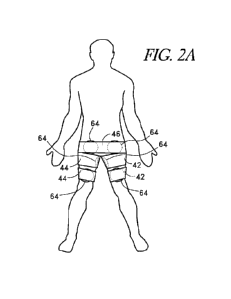
100941 FIGS. 2A and 2B illustrate one embodiment of an ECP device with
inflatable bladders 64 and cuffs 42, 44, 46 placed against the preferred
compression sites at the
superior-posterior knee regions, the inguinal regions and the buttocks. The
bladders 64 and
cuffs 42, 44, 46 are described in greater detail below. In some embodiments,
six bladders 64,
each having approximately thirty-six square inches of compression area, are
used to compress
the preferred body areas. Other compression sites are also contemplated. FIG.
2C illustrates
another embodiment of an ECP device with inflatable bladders 64 and cuffs 42,
44 placed
against alternative compression sites. In some embodiments, as illustrated in
FIG. 2C, the
cuffs and the attached inflatable bladders can be located in the upper
extremities. As discussed
above, each of the cuffs and attached inflatable bladders are placed near
vessels close to the
surface of the skin. In some embodiments, the bladder 64 and cuffs 42, 44 can
be located on
the upper arm. For example, the bladder 64 and cuffs 42, 44 can be located
inside the elbow
notch at a distal end of the bicep. In another example, the bladder 64 and
cuffs 42, 44 can be
located inside the armpit near the inside of the arm region. The placement of
the bladder 64
and cuffs 42, 44 inside the armpit can be used to aid in lymph node drainage
(e.g., after a
-20-
CA 03160800 2022-05-09
WO 2021/097115 PCT/US2020/060269
mastectomy). As well, the placement of the bladder 64 and cuffs 42, 44 can
also be used to
reduce tissue edema. In another example (not illustrated), the bladder 64 and
cuffs 42, 44 can
be located near the wrist on the inside of the arm. In some examples,
compression of the sites
in the upper extremities can be configured to provide circulation to the head
(e.g., the brain).
In some embodiments, the disclosed ECP system can be coupled with a patient's
existing
therapy. For example, in treating depressing, the ECP system in the upper
extremities can be
configured to increase blood flow to the brain.
100951 In some embodiments, the bladders 64 and cuffs 42, 44 at the
superior-
posterior knee region are placed on the inside of the legs. In some examples,
the bladders 64
and cuffs 42, 44 are located behind the kneecap (i.e., above the knee). In
some embodiments,
the bladders 64 and cuffs 42, 44 can be located on the calf (e.g., on the
upper inside of the calf
or on the Achilles tendon on the lower calf).
100961 In some embodiments, compression of the upper extremities
(i.e., the
bladders 64 and cuffs 42, 44 in FIG. 2C) and compression of the lower
extremities (i.e., the
bladders 64 and cuffs 42, 44, 46 in FIG. 2A-2B) can be synced. For example,
compression of
the upper extremity can be synced with compression of a corresponding part in
the extremity.
As an example, the bladders 64 and cuffs 42, 44 on the bicep can be compressed
at the same
time as the bladders 64 and cuffs 42, 44 behind the knee. As another example,
the bladders 64
and cuffs 42, 44 under the armpit can be compressed at the same time as the
bladders 64 and
cuffs 42, 44 at the groin region.
100971 In some embodiments, other numbers of cuffs and bladders may be
used.
Additional body areas may also be compressed, but are not necessary to achieve
effective
counterpulsation. Furthermore, increasing the body surface area compressed may
increase the
air volumes used and therefore increase patient discomfort and increase the
generation of noise
and heat. In some embodiments, existing ECP devices, using a plurality of
bladders for
compressing the lower limb, could be modified to have the capability of
selectively
inactivating a number of bladders during the treatment of a patient. In some
examples, the
remaining active bladders are located at the preferred compression sites and
the effective total
surface area of the remaining active bladders used to compress the body is
limited to about 240
square inches. In some embodiments, the active bladders used to compress the
body can be
-21-
CA 03160800 2022-05-09
WO 2021/097115 PCT/US2020/060269
greater or smaller than the above-reference 240 square inches depending on
treatment
requirements and the person's physical characteristics, (e.g., body mass index
(BMI)).
100981 In some examples, an ECP system employing lower inflation
volumes, not
only can lower pressures be used, but the timing of the inflation and
deflation cycles can be
simplified. Timing intervals can be easier to maintain because there is less
need to move large
volumes of compressed air in and out of the bladders in a short time interval.
This can allow
the duration of bladder inflation and the delay intervals between sequential
inflation of the
bladders to be preset in a low-volume ECP system.
100991 Another benefit of an ECP system using lower volumes and
pressures is that
bladder deflation during PVC's is unnecessary. With an inflation pressure of
about 160 mm
Hg to about 220 mm Hg, an ECP system does not need to deflate the bladders
when a PVC
occurs because the heart is no longer contracting against a supra-
physiological blood pressure.
Furthermore, the ECP system is simplified because there is no need to
differentiate between a
sinus beats from PVC's. More importantly, a low-pressure ECP system eliminates
the
inefficiency of the ECP session caused by excessive deflation from detected
PVC's.
[001001 In addition to angina and congestive heart failure, other uses for an
ECP
system may include but are not limited to adult and pediatric congenital heart
disorders,
pregnancy-related heart failure, ischemic bowel disease, peripheral vascular
disease including
carotid insufficiency and skin ulceration, Alzheimer's, cerebrovascular
accidents, dementia,
acute renal failure, chronic renal insufficiency and failure, liver disease,
weight loss, alopecia,
limb ischemia, sepsis and shock. Those skilled in the art are familiar with
other conditions that
may benefit from use of ECP.
[0014)11 FIG. 1 illustrates an embodiment of an ECP system 22 and a table 40.
In
some examples, the ECP system 22 comprises a pressurized air system, a
controller and a
plurality of bladders attached to cuffs 42, 44 and 46. The controller can
include, an ECG signal
connector 29 that accepts an ECG signal from an external ECG signal source 192
and an ECG
signal processor to generate at least one control signal from the ECG signal.
An external ECG
signal connector 29 can allow a patient to undergo ECP treatment concurrently
with any
ongoing ECG monitoring being performed on the patient without attaching a
duplicate set of
chest leads to the patient. This can be useful in an Intensive Care Unit (ICU)
setting where a
patient is already connected to an ECG monitor. An example embodiment of an
ECG signal
-22-
CA 03160800 2022-05-09
WO 2021/097115 PCT/US2020/060269
processor is described in further detail below. FIG. 3 shows another
embodiment of an ECG
monitor integrated into the ECP system 22 where unprocessed ECG chest lead
signals are
provided to the ECG monitor by chest leads attached to the patient. The chest
signal can
processed by the ECG monitor and relayed to the ECG signal processor to
generate the control
signal. In some examples, the ECG monitor output is optionally provided in
this embodiment
for providing ECG output to the telemetry monitors available in some hospital
wards.
[001021 In some embodiments the control signal is transmitted through a
control line
38 to table 40 for controlling the opening and closing of air valves that
inflate and deflate the
bladders. Pressurized air from the ECP system 22 can be transmitted to the
table 40 by an air
line 36. In some examples, the air is directed to the air valves from table 40
which distribute
the pressurized air using bladder air lines 48 to the right leg cuffs 42, left
leg cuffs 44 and
buttock cuffs 46 that hold inflatable bladders. In some embodiments, the
controller may
optionally have an on/off power switch 24 to control power to the ECP system
22 and/or a
timer switch 26 that sets the treatment time.
1001031 FIG. 4 illustrates a schematic embodiment of a pressurized air
subsystem.
In some embodiments, pressurized air is supplied by an air compressor 50 which
is capable of
providing pressurized air to an air tank 52 through a compressor air line 60.
In some examples,
air compressor 50 is capable of a total free air output of about four to about
eight cubic feet per
minute (cfm) at a pressure of about four pounds per square inch (psi). The
compressor air line
60 can comprise a flexible hose having an internal diameter of about 1/2 inch
to about 3/4 inch.
Air tank 52 can have a capacity of about five gallons and can be capable of
withstanding an
operating pressure of about 100 psi. In some embodiments, output from the air
tank 52 travels
through air line 36 which comprises a flexible hose with an internal diameter
of about one inch.
In some examples, air line 36 connects to a pressure regulator 54. Air tank 52
can also connect
to a pressure relief valve 56 by a pressure relief valve fitting 66. In some
examples, the pressure
relief valve 56 may be set to any pressure from about one psi to about five
psi and vent about
eight cfm or more of air. In some embodiments, the pressure regulator 54 can
be set to an
output pressure of about three to about five psi and feed at least one air
valve 58 through air
line 36. In some examples, pressure from air line 36 may be distributed to a
plurality of air
valves 58 by air line tees 68 or any other kind of pressure distributor having
multiple openings.
The air valves 58 can be connected to bladders 64 on the right leg cuffs 42,
left leg cuffs 44
-23-
CA 03160800 2022-05-09
WO 2021/097115 PCT/US2020/060269
and buttock cuff 46 by bladder lines 48. In some examples, the bladder lines
48 comprise 1/2
inch internal diameter flexible hose. In some embodiments, air valves 58 are
1/2 inch. In some
examples, the air valves 58 can be 24-volt. In some embodiments, the air
valves 58 can be
closed. In some examples, the air valves 58 can have two-positions. In some
embodiments,
the air valves 58 are three-way, air pilot assist valves having an open and a
closed
configuration. In some embodiments, non-pilot air valves can be used. In the
closed
configuration, the air valves 58 can be configured to prevent flow from air
tank 52 to bladders
64. When closed, the bladders 64 can be configured to also vent to the
atmosphere. In the
open configuration, the air valves 58 can be configured to allow air pressure
from air tank 52
to pressurize bladders 64 and prevent any venting. In some embodiments, ridged
threaded
barbs and hose clamps can be used to secure hoses 36, 48, 60 and 66 to the
other components
of the ECP system. One of skill in the art will understand that any of a
variety of other
mechanical fittings suitable for securing hoses may be used.
[001041 Another embodiment of an electrical power system for the ECP system is
illustrated in FIG. 5. A 120-volt system is described below, but one skilled
in the art will
understand how to adapt the ECP system for use in a 110-volt, 220-volt, 240-
volt or other
system. A 120-volt power cord 72 is configured to feed power to a re-settable
ground fault
interrupter (GFI) 74, which in turn can connect to an on/off power switch 24.
In some
embodiments, the power switch 24 is a two-position double-pole lighted switch.
Power switch
24 can connect, for example, to an EMI filter 76 that in turn connects to a
start switch 28 and
a start switch relay 78 having an engaged and disengaged position. In some
embodiments, the
start switch 28 is a momentary lighted single pole switch used to start ECP
system 22. In some
examples, the start switch relay 78 also connects to start switch 28. When
start switch 28 is in
the engaged position, start switch 28 is capable of sending power to timer
switch 26. In some
embodiments, the timer switch 26 has an active state and an inactive state.
The timer switch
26 can go from the active state to the inactive state after a user-settable
period. The power
output from timer switch 26 can be looped back to the output of start switch
28 to keep start
switch relay 78 in the engaged position so long as timer switch 26 is in the
active state. In
some embodiments, when the timer switch 26 is in the active state, timer
switch 26 provides
power to air compressor 50, a programmable logic controller (PLC) 80 and a 24-
volt power
supply 82. In some embodiment, timer switch 26 can be set from about zero
minutes to about
-24-
CA 03160800 2022-05-09
WO 2021/097115 PCT/US2020/060269
sixty minutes. In some examples, the timer switch 26 can be set for any period
of time. In
some embodiments, the timer switch 26 does not reset upon loss of power. The
wire 84 can
provide power to air compressor 50 from GFI or resettable breaker, or an
electrical safety
feature 74 through timer switch 26. In some examples, the wire 84 comprises 14-
gauge wire,
but one skilled in the art will understand that other wire gauges may be used.
Wires 86 can
provide power to start switch 28, programmable logic controller (PLC) 80 and
24-volt power
supply 82. In some embodiments, the wires 86 comprise 18-gauge wires, but
those skilled in
the art will understand that other wire gauges may be used. In some examples,
the PLC 80 is
a 120-volt unit with at least one input and at least three outputs. In some
embodiments, the
inputs range generally from about twelve volts to about twenty-four volts. The
outputs can
range generally from about twelve volts to about twenty-four volts.
[001051 FIG. 6 illustrates an embodiment of the external ECG input 90 and a 24-
volt system used to power ECG system 22. Although a 24-volt system is
described herein, one
skilled in the art will know that the system can be adapted to voltages from
about 6-volts to
about 30-volts. In some examples, a 24-volt power supply 82 supplies power to
PLC 80, an
ECG timing board 92, a PLC-to-air valve relay 94 and a mini-air compressor 96.
In some
embodiments, the ECG timing board 92 can be a relay board that amplifies and
relays the
signal from external ECG input 90 to PLC 80. In some examples, the PLC 80 uses
the
amplified ECG signal from timing board 92 to output control signals to air
valves 58 and PLC-
to-air valve relay 94. In some embodiments, the outputs are generally spaced
about forty
milliseconds apart after the first output. In some examples, the outputs are
generally spaced
about 10 milliseconds to about 100 milliseconds apart. A first output or
control signal can
regulate the air valve 58 connected to bladders contacting the upper posterior
knee or lower
thigh. In some embodiments, a second output regulates air valve 58 connected
to bladders
contacting the upper thigh or inguinal areas. In some examples, a third output
goes to PLC-
to-air valve relay 94, which passes the third output to air valves 58
controlling compression of
the buttocks. In some embodiments, the wires 86 used for the 24-volt system
can be 18-gauge
wires.
1001061 FIGS. 7A and 7B illustrate schematic representations of an embodiment
of
the programming of the PLC 80. In some embodiments the PLC 80 receives a
squared ECG
signal from ECG timing board 92. The PLC 80 can be configured to detect eight
squared R
-25-
CA 03160800 2022-05-09
WO 2021/097115 PCT/US2020/060269
wave signals and calculate the total time interval between the eight squared R
wave signals.
In some examples, if the total time interval is greater than about 10.7
seconds or less than about
5.3 seconds, the R wave counter is reset and the total time interval is
recollected. In some
examples, if the total time interval is between 5.3 and 10.7 seconds, the PLC
80 can initiate a
pump cycle. In some embodiments, following a delay after the last detected
peak in the squared
ECG signal, the PLC 80 can initiate a first control signal that is transmitted
to air valve 58
controlling bladders 64 at the lower thigh. In some examples, the delay can be
pre-set at about
280 milliseconds. Alternatively, the delay can be calculated based upon the
patient's heart rate
or peak-to-peak time interval based upon the EC signal. In some embodiments,
the delay is
about 25% of the average peak-to-peak interval of the last eight trailing QRS
complexes. In
some examples, the delay is about 25% of the longest of the trailing eight
peak-to-peak
intervals of the ECG signal. In some embodiments, after a fixed interval set
at about forty
milliseconds, a second control signal to air valve 58 controlling bladders in
the upper
thigh/inguinal regions can be initiated. In some examples, the first control
signal to the air
valve 58 controlling the bladders 64 of the lower thighs may be terminated
after the second
control signal is initiated. In some embodiments, the early termination of the
first control
signal can advantageously allow earlier filling of the thighs for the next
pump cycle. In some
examples, there may be a slight delay between the initiation of the second
control signal and
the termination of the first control signal to allow bladders 64 of the upper
thigh to fully inflate
before deflating bladder 64 at the lower thigh. In some embodiments, after
another fixed
interval of about 40 milliseconds, a third control signal to air valve 58
controlling the buttock
bladders is initiated. In some examples, after a fixed interval set at about
370 milliseconds
after the start of the third control signal, the three control signals can be
terminated and the
cycle is repeated. In some embodiments, the control signals continue for the
pre-set interval
irrespective of whether another ECG signal or PVC is detected during the
transmission of the
control signals. In some examples, the PLC 80 can terminate the signal cycle
if another signal
peak is detected and initiate the next cycle, but does not distinguish between
squared sinus
QRS complexes and squared PVC's. Although the embodiments herein have
described the
use of the ECG timing board 92 and the PLC 80 to process ECG signals and
provide control
signals to the valves, one skilled in the art will understand that computers,
microprocessors
and other electronic controllers can also be used to process ECG signals and
provide control
-26-
CA 03160800 2022-05-09
WO 2021/097115 PCT/US2020/060269
signals. One skilled in the art will understand that variations of the above
control systems, or
other known ECP control algorithms, may be used.
100107j FIG. 8 represents an embodiment of a mini air system used for
providing
pilot assist air to the air valves 58. In some embodiments, the mini air
compressor 96 is a 24-
volt mini compressor with an output of about 1/2 cfm at a pressure of about
twelve psi. The
mini air compressor 96 can connect to a mini air compressor pressure relief
valve 56 which is
set to vent air at about twelve psi. In some examples, the mini air compressor
pressure relief
valve 56 connects to the mini air compressor pressure regulator 102. The air
pressure regulator
102 can be a 1/4 inch pipe fitting set at about ten psi. The output from mini
air compressor
pressure regulator 102 can be configured to feed the actuators of at least one
air valve 58 using
at least one 1/4 inch air line tee 106 and 1/4 inch air line 104. In some
examples, by providing
a separate and smaller compressor to produce the higher-pressure smaller-
volume pilot assist
air for driving the pilot assist air valves, the air compressor 50 is not
unnecessarily producing
higher pressure for bladders 64. Thus, air compressor 50 thus can operate
efficiently at lower
pressures independent of the higher pressure used for the pilot assist air
needed by valves 58.
By having two different compressors for serving two different functions, the
total amount of
noise, heat and patient discomfort created by the ECP system can be reduced.
In some
examples, in embodiments that do not use pilot air assist valves, a system
other than the mini
air system may be used.
1001081 FIGS. 9A-9C and 10A-10D illustrate an embodiment of a cuff and bladder
design. This configuration may increase user comfort and improve ease of set
up and
placement of the cuffs. In some embodiments, the bladders can be disposable.
In some
examples, the bladders can be easily removed from the cuff, such that the
operator of the ECP
system can efficiently place the cuffs and properly position each of the
bladders. In some
embodiments, as discussed below, the bladders can come in a variety of
configurations such
that the operator can adapt the ECP procedure to the needs of the patient. In
some examples,
the disclosed ECP system can include a cuff that uses a split buckle that
reduces set-up time
and aids in placement.
1001091 FIGS. 9A and 9B illustrate an embodiment of a bladder 64 that can be
used
in ECP system 22. In some embodiments, the bladder 64 comprises a bladder
connector 114
attached to a first bladder wall 110. The bladder connector 114 can have an
internal diameter
-27-
CA 03160800 2022-05-09
WO 2021/097115 PCT/US2020/060269
of about 1/4 inch to about 3/4 inch. In some examples, the first bladder wall
110 is sealed to a
second bladder wall 110 along a bladder sealing area 116 along the edges of
bladder walls 110.
In some embodiments, the bladder sealing area 116 is approximately about 1/8
to about 3/8
inch wide. Attaching can be done in a manner to provide a hermetic seal and to
withstand
about a ten psi or more inflation pressure. Hermetic sealing may be performed
by heat sealing,
solvent sealing, adhesives, or any of a variety of hermetic sealing methods
known in the art
and incorporated by reference herein. In some embodiments, a single continuous
bladder wall
forms bladder 64. A hook fastener ring 112 can attaches to the area
surrounding bladder
connector 114. Hook fastener ring 112, can include but not be limited to those
made by Velcro
USA (Manchester, NH), facilitates affixation of bladder 64 to cuffs described
below. FIG. 9A
depicts balloon 64 with a circular shape, but other possible balloon shapes
include square,
rectangular, triangular or any other closed loop shape. In some embodiments, a
triangular
balloon shape may be suitable for compressing the body in areas with creasing.
The surface
area of bladder 64 when flat is about forty square inches on one side. In
another embodiment,
the surface area can be from about twenty square inches to about sixty square
inches. Bladders
64 may be made from polyester, polyurethane, polyvinylchloride, polyethylene
or any of a
variety of airtight materials known in the art and herein incorporated by
reference.
001101 FIG. 9C illustrates an embodiment of a bladder 64' that can be used in
the
ECP system 22. Similar to the bladder 64, the bladder 64' can include a
bladder connector
114' attached to a first bladder wall 110'. The bladder connector 114' can
have an internal
diameter of about 1/4 inch to about 3/4 inch. In some examples, the first
bladder wall 110' is
sealed to a second bladder wall 110' along a bladder sealing area 116' along
the edges of
bladder walls 110'. In some embodiments, the bladder sealing area 116' is
approximately
about 1/8 to about 3/8 inch wide. Attaching can be done in a manner to provide
a hermetic
seal and to withstand about a ten psi or more inflation pressure. Hermetic
sealing may be
performed by heat sealing, solvent sealing, adhesives, or any of a variety of
hermetic sealing
methods known in the art and incorporated by reference herein. In some
embodiments, a single
continuous bladder wall forms bladder 64'. A hook fastener ring 112' can
attach to the area
surrounding bladder connector 114'. Hook fastener ring 112', can include but
not be limited
to those made by Velcro USA (Manchester, NH), facilitates affixation of
bladder 64' to cuffs
described below. The bladder 64' can be very similar to the bladder 64 but
further includes a
-28-
CA 03160800 2022-05-09
WO 2021/097115 PCT/US2020/060269
notch 65'. In some examples, the notch 65' can be semicircular and is
configured to allow the
bladder 64' to be placed higher in to the groin area. In some embodiments, the
notch 65'
permits placing the balloon further into the inguinal area, getting the
bladder pressure into the
vasculature and closer to the surface in the inguinal area. As illustrated in
Figure 10D, the
notch 65' of the bladder 64' can help to properly orient the bladder 64' on
the cuff 44. In some
embodiments, the bladder 64' can be engaged to the cuff 44 with a connector
(e.g., an elbow)
that is configured to point away from the notch 65'. The configuration of the
connector can
therefore ensure proper orientation of the bladder 64' on the cuff 44.
1001111 Although FIG. 9C depicts the balloon 64' with a generally circular
shape,
but other possible balloon shapes include oval, ellipse, rounded, square,
rectangular, triangular
or any other closed loop shape that can be modified to also include a notch
65' that conforms
with a side of the cuff 44. Bladders 64' may be made from polyester,
polyurethane,
polyvinylchloride, polyethylene or any of a variety of airtight materials
known in the art and
herein incorporated by reference.
I 001121 FIG. 9D illustrates an embodiment of a bladder 64" that can be used
in the
ECP system 22. Similar to the bladder 64, the bladder 64', and the bladder 64"
can include a
bladder connector 114" attached to a first bladder wall 110". The bladder
connector 114" can
have an internal diameter of about 1/4 inch to about 3/4 inch. In some
examples, the first
bladder wall 110" is sealed to a second bladder wall 110" along a bladder
sealing area 116"
along the edges of bladder walls 110'. The bladder sealing area 116" indicates
where the first
bladder wall 110" is welded to the second bladder wall 110". In some
embodiments, the
bladder sealing area 116" is approximately about 1/8 to about 3/8 inch wide.
Attaching can be
done in a manner to provide a hermetic seal and to withstand about a ten psi
or more inflation
pressure. Hermetic sealing may be performed by heat sealing, solvent sealing,
adhesives, or
any of a variety of hermetic sealing methods known in the art and incorporated
by reference
herein. In some embodiments, a single continuous bladder wall forms bladder
64". The
bladder 64" can be very similar to the bladder 64 but further includes a notch
65". In some
examples, the notch 65" can be semicircular and is configured to allow the
bladder 64" to be
placed higher in to the groin area. In some embodiments, the notch 65" permits
placing the
balloon further into the inguinal area, getting the bladder pressure into the
vasculature and
closer to the surface in the inguinal area. As illustrated in Figure 10D, the
notch 65" of the
-29-
CA 03160800 2022-05-09
WO 2021/097115 PCT/US2020/060269
bladder 64" can help to properly orient the bladder 64" on the cuff 44. In
some embodiments,
the bladder 64" can be engaged to the cuff 44 with a connector (e.g., an
elbow) that is
configured to point away from the notch 65". The configuration of the
connector can therefore
ensure proper orientation of the bladder 64' on the cuff 44.
1001131 Although FIG. 9D depicts the balloon 64" with a generally circular
shape,
but other possible balloon shapes include oval, ellipse, rounded, square,
rectangular, triangular
or any other closed loop shape that can be modified to also include a notch
65" that conforms
to a side of the cuff 44. Bladders 64" may be made from polyester,
polyurethane,
polyvinylchloride, polyethylene or any of a variety of airtight materials
known in the art and
herein incorporated by reference.
1001141 FIGS. 10A and 10B depict embodiments of a left leg cuff 44 with
bladder
64 in place. In the embodiments illustrated in FIGS. 10A and 10B and other
embodiments
described below, a right leg cuff 42 may be a mirror image of left leg cuff 44
for use on the
right lower extremity. Alternatively, right leg cuff 42 and left leg cuff 44
may be identical or
similar in configuration. In some examples, cuff material 120 has an inner
surface 121, an
outer surface 123 and a hole 125 for insertion of bladder connector 114 of
bladder 64. In some
embodiments, the cuff 44 has an arcuate configuration that is particularly
suited to compress
anatomical structures that are located in areas of narrowing or creasing, but
is not limited to
this particular purpose. In some examples, cuff material 120 can be made of a
flexible non-
stretch material that is able to withstand repeated inflations of bladder 64.
In some
embodiments, the non-stretch material comprises a 600 denier polyester cloth
as used in
backpacks. A ring 128 around hole 125 can optionally be color-coded to
indicate which
complementary color-coded bladder air line 48 connects to which bladder 64. A
portion of
bladder 64 may be visible when viewing outer surface 123 of leg cuff 44, which
may facilitate
accurate placement of bladder 64 when securing cuff 44 to the patient. Outer
surface 123 may
also have identifying marks to show the position of underlying bladder 64 if
obscured by cuff
44. Identifying marks can be configured to allow accurate positioning of
bladder 64 on the
patient's body.
1001151 A buckle 122 with a buckle roller 124 can attach to one end of cuff
44. In
some embodiments, the buckle 122 comprises a frame 127 with a slot opening 129
for insertion
of a cuff end, the slot opening 129 having dimensions of about 1/4 inch to
about 3/4 inch in
-30-
CA 03160800 2022-05-09
WO 2021/097115 PCT/US2020/060269
one direction and about six inches in second direction. In some examples, the
buckle roller
124 can a tube with an internal diameter larger than the diameter of buckle
frame 127,
permitting buckle roller 124 to turn freely. In some examples, buckle roller
124 can reduce
the effort needed to tighten cuff 44 on the patient by allowing cuff 44 to
slide through the slot
opening of buckle 122 with reduced friction against buckle frame 127. In some
embodiments,
buckle 122 and buckle roller 124 are made from any of a variety of rigid
materials well known
in the art, including but not limited to a metal or a plastic. Buckle shield
126 may be made of
the same type of material as cuff material 120. Optionally, buckle shield 126
may be made
stiffer with any of a variety of materials attached or adhered to buckle
shield 126, including
but not limited to a thin polycarbonate. In some embodiments, buckle shield
126 attaches to
the inner surface 121 of cuff material 120 to provide protection from buckle
122. In some
examples, buckle shield 126 may reduce the pinching of the skin on the patient
when left leg
cuff 44 is tightened. Hook fastener 130 and loop fastener 132 can be attached
to the other end
of cuff material 120 by stitching, gluing, or any of a variety of methods well
known in the art
and incorporated by reference herein. Hook fastener 130 and loop fastener 132
can be used to
fasten right leg cuff 42 or left leg cuff 44 when the cuff is tightened on the
patient. In some
embodiments, the width of right leg cuff 42 or left leg cuff 44 is
approximately six inches with
a circumferential length of approximately 30 to 45 inches. In another
embodiment, cuffs 42,
44 have a width of about three inches to about eight inches and a
circumferential length of
about twenty to about sixty inches. Cutouts are optionally provided in cuff
material 120 for
vascular access or any other procedure requiring access to body areas covered
by cuff material
120.
[091161 FIG. 10B illustrates another embodiment of the invention comprising a
friction or non-slip material 134 on inner surface 121 of right leg cuff 42 or
left leg cuff 44.
Non-slip material 134 may be joined to cuff material 120 by stitching, gluing,
coating or any
other method of attachment as is known in the art. In some examples, the non-
slip material
134 may also be an inherent characteristic of cuff material 120. In some
embodiments, non-
slip material 134 may comprise any of a variety of flexible materials with a
coefficient of
friction sufficient to resist slippage of the cuff, including but not limited
to neoprene, rubber
or texturized versions of cuff material 120. Those skilled in the art will be
familiar with other
known non-slip materials that may be used.
-31-
CA 03160800 2022-05-09
WO 2021/097115 PCT/US2020/060269
1091171 FIG. 10C illustrates an example of the inner surface 121 of cuff 44
without
bladder 64. In some embodiments, to attach bladder 64 to cuff 44, bladder
connector 112 of
bladder 64 inserts through hole 125 such that hook fastener ring 112 of
bladder 64 engages
loop fastener ring 133 on cuff 44.
[OM 181 FIG. 10D illustrates an embodiment of a left leg cuff 44 with bladder
64'
in place. In the embodiment illustrated in FIG. 10D, a right leg cuff 42 may
be a mirror image
of left leg cuff 44 for use on the right lower extremity. As shown in FIG.
10D, the bladder 64'
is positioned such that the notch 65' is positioned adjacent to the side of
the cuff 44 to allow
the bladder 64' to be positioned more snuggly adjacent the groin. Similarly,
the bladder 64"
of FIG. 9D can be similarly positioned on the cuff 44 to allow the notch 65"
to be positioned
more snuggly adjacent the groin.
[001191 FIGS. 11A and 11B illustrate another embodiment comprising a buttock
cuff 46 with two bladders 64 attached to cuff 46. In some examples, the cuff
material 120 has
an inner surface 121, an outer surface 123 and a hole 125 for insertion of
bladder connector
114 of bladder 64. In some examples, the buttock cuff 46 can have a straight
configuration,
but may also be arcuate or any other configuration that is able to encompass a
circumference
of the body that includes the buttocks. The cuff material 120 can be made of
any flexible non-
stretch material able to withstand repeated inflations of bladder 64. In some
embodiments, the
non-stretch material comprises a 600 denier polyester cloth as used in
backpacks. The rings
128 around holes 125 are optionally color-coded to indicate which
complementary color-coded
bladder air lines 48 are to be connected to the bladders 64. A portion of
bladders 64 may be
visible when viewing outer surface 123 of buttock cuff 46, which may
facilitate accurate
placement of the bladders 64 when securing cuff 46 to the patient. Outer
surface 123 may also
have identifying marks to show the position of underlying bladder 64 obscured
by cuff 46.
[001201 In some embodiments, buttock cuff 46 comprises buckle 122 and
optionally
the buckle roller 124 and buckle shield 126 as previously described. Cuff
material 120 can be
made of any flexible non-stretch material able to withstand repeated
inflations of bladders 64.
In some embodiments, the non-stretch material comprises a 600 denier polyester
cloth as used
in backpacks. The hook fasteners 130 and loop fasteners 132 can be attached to
the other end
of cuff material 120 by stitching, gluing, or any of number of methods well
known in the art.
In some embodiments, the hook fasteners 130 and loop fasteners 132 are used to
secure buttock
-32-
CA 03160800 2022-05-09
WO 2021/097115 PCT/US2020/060269
cuff 46 when cuff 46 is tightened on the patient. In some examples, the width
of buttock cuff
46 is approximately 6 inches with a circumferential length of about 60 inches.
In some
embodiments, cuff 46 has a width of about four inches to about ten inches and
a circumferential
length of about fifty to about ninety inches. In some embodiments, buttock
cuff 46 comprises
a plurality of bladders 64 from about one bladder 64 to about four bladders
64. Cutouts are
optionally provided in cuff material 120 for vascular access or any other
procedure requiring
access to body areas covered by cuff material 120. FIG. 11B depicts an example
of the
invention comprising a non-slip material 134 on inner surface 121 of buttock
cuff 46, as
described in the previous leg cuff embodiment.
[OW 211 FIG. 11C illustrates the inner surface of cuff 42 without bladders 64.
Bladder connectors 112 of bladders 64 can be inserted through holes 125 and
rings 128 of cuff
material 120 to attach to bladder air lines 48.
100122i In some embodiments, hook fastener 130 is attached to cuff material
120 at
one end and one surface of cuffs 42, 44 and 46 and loop fastener 132 is joined
to cuff material
120 at the opposite end and opposite surface, allowing the cuffs 42, 44, 46 to
be secured to the
patient by wrapping one end of a cuff over the other end of the same cuff by
coupling hook
fastener 130 to loop fastener 132. In some embodiments, the buckle 122, buckle
roller 124,
and buckle shield are not included.
[OW 231 FIGS. 12A and 12B illustrate another embodiment of a left leg cuff
150.
Right leg cuff 156 may have a similar configuration or a mirror image
configuration of left leg
cuff 150 and can have similar construction and materials. Optional color-coded
ring 128
around bladder connector 114 indicates which color-coded bladder air line 48
is to be
connected to which bladder connector 114. In some examples, bladder connector
114 is
attached to bladder wall 142 by any of a variety of attachment methods
including heat sealing,
solvent sealing, gluing or any other hermetic sealing as known in the art. In
some
embodiments, the bladder wall 142 can be hermetically sealed to cuff material
144 using a
sealing area of about 1/4 inch on the outer edge of bladder wall 142, forming
a bladder. In
some embodiments, cuff material 144 is enlarged in width where bladder walls
142 are sealed
to cuff material 144. Cuff material 144 may also have identifying marks to
show the position
of underlying bladder wall 142 obscured by cuff material 144. In some
embodiments, the
sealing area is about 1/8 to about 1/2 inch on the outer edge of bladder wall
142. Hermetic
-33-
CA 03160800 2022-05-09
WO 2021/097115 PCT/US2020/060269
sealing may be performed by methods previously described. In some examples,
the bladder
wall 142 and cuff material 144 can include any of a variety of flexible non-
stretch airtight
materials, as previously described. Bladder wall 142 and cuff 144 may comprise
different
materials that are hermetically sealable together. For example, the bladder
wall 142 may
comprise any of a variety of non-stretch or semi-stretchable airtight
materials, including but
not limited to polyurethane materials made by Magister Corporation
(Chattanooga, TN), herein
incorporated by reference. Use of semi-stretchable airtight materials for
bladder wall 142 may
facilitate inward volume expansion and pressure transmission to the patient.
1001241 In some embodiments, the leg cuff 150 comprises buckle 122 and
optionally
buckle roller 124 and buckle shield 126 as previously described. A self-
adhesive hook 145
and loop fastener 146 can be attached to cuff material 144 near bladder wall
142. In some
embodiments, the self-adhesive hook 145 can be a distance of between 0.5
inches-2.0 inches,
between 0.5 inches-1.0 inches, between 1.0 inches-1.5 inches, and between 1.5
inches-2.0
inches from the edge of the bladder wall. In some embodiments, the self-
adhesive hook 145
is 0.5 inches, 0.6 inches, 0.7 inches, 0.8 inches, 0.9 inches, 1.0 inch, 1.1
inches, 1.2 inches, 1.3
inches, 1.4 inches, 1.5 inches, 1.6 inches, 1.7 inches, 1.8 inches, 1.9
inches, 2.0 inches from
the edge of the bladder wall. In some embodiments, only one side of self-
adhesive hook and
loop fastener 146 is attached to bladder wall 142. The topside of self-
adhesive hook and loop
fastener 146 is self-adhesive and covered with a wax paper-type protector.
This can allow the
operator to remove the protector and adhere the end of left leg cuff 150 to
the self-adhesive
when securing the cuff to the patient. This configuration can permit leg cuff
150 to be fitted
to the patient and allow the removal of leg cuff 150 as medical needs dictate
by separating the
hook fastener from the loop fastener. In some embodiments, both hook fastener
130 and loop
fastener 132 are pre-attached to leg cuff 150. In some examples, the width of
leg cuff 150 can
be approximately six inches with a length of approximately thirty to forty-
five inches. In some
embodiments, leg cuff 150 includes a self-adhesive non-slip material 148 on
the inner surface
of left leg cuff 150, of material and attached as previously described.
Cutouts 131 can be
optionally provided in cuff material 144 for vascular access or any other
procedure requiring
access to body areas covered by cuff material 144. This embodiment may also be
particularly
suited for use as a disposable cuff because of the simplified design and lower
cost of
manufacturing, but the embodiment is not limited to this particular use.
-34-
CA 03160800 2022-05-09
WO 2021/097115 PCT/US2020/060269
1091251 FIGS. 13A and 13B illustrate another embodiment of the buttock cuff
154.
As shown, two bladder connectors 114 can be provided in bladder walls 142. In
some
examples, color-coded rings 128 can be included around bladder connectors 114
to indicate
which color-coded bladder air lines 48 are to be connected to which bladder
connectors 114.
Bladder connectors 114 can be attached to bladder walls 142 by any of a
variety of attachment
methods including heat sealing, solvent sealing, gluing or any other hermetic
sealing method
as known in the art. In some embodiments, the bladder walls 142 are
hermetically sealed to
cuff material 144 using a sealing area of about 1/4 inch on the outer edge of
bladder wall 142,
forming a bladder. In another embodiment, the sealing area is about 1/8 to
about 1/2 inch on
the outer edge of bladder walls 142. In some examples, cuff material 144 is
enlarged in width
where bladder walls 142 are sealed to cuff material 144. The cuff material 144
may also have
identifying marks to show the position of the underlying bladder wall 142 that
is otherwise
obscured by cuff material 144. In some examples, hermetic sealing may be
performed by heat
sealing, solvent sealing, adhesives or any of a variety of hermetic sealing
methods known in
the art. In some embodiments, bladder walls 142 may comprise any of a variety
of non-
stretchable or semi-stretchable airtight materials known in the art. Use of
semi-stretchable
airtight materials for bladder wall 142 may facilitate inward volume expansion
and pressure
transmission to the patient.
[091261 In some examples, buttock cuff 154 comprises buckle 122 and optionally
buckle roller 124 and buckle shield 126 as previously described. A self-
adhesive hook 145
and loop fastener 146 can be attached to cuff material 144. In some examples,
the outer surface
of self-adhesive hook and loop fastener 146 is self-adhesive and covered with
a wax paper-
type protector. This can allow the operator to remove the protector and adhere
the end of
buttock cuff 154 to the self-adhesive after tightening on the patient. This
configuration permits
buttock cuff 154 to be fitted to the patient while also allowing the buttock
cuff 154 to be
removed as desired. In some examples, the buttock cuff 154 can be removed by
separating the
hook fastener from the loop fastener. In another embodiment, both hook
fastener 145 and loop
fastener 146 are pre-attached to the buttock cuff 154. In some examples, the
width of buttock
cuff 154 can be about six inches with a length of about sixty inches. In some
embodiments,
buttock cuff 154 comprises self-adhesive non-slip material 148 on the inner
surface of buttock
cuff 154, of material and attached as previously described. Cutouts are
optionally provided in
-35-
CA 03160800 2022-05-09
WO 2021/097115 PCT/US2020/060269
cuff material 144 for vascular access or any other procedure requiring access
to body areas
covered by the cuff material 144. This embodiment may also be particularly
suited for use as
a disposable cuff due to the simplified design and lower cost of
manufacturing, but the
embodiment is not limited to this particular use.
[001271 In some embodiments, the hook fastener is joined to the cuff material
144
at one end and one surface of the cuffs 150, 154, 156 while a loop fastener is
joined to the cuff
material 144 at the opposite end and the opposite surface. This configuration
can allow the
securing of cuffs 150, 154, 156 to the patient by wrapping one end of a cuff
over the other end
of the same cuff In some examples, this embodiment does not require buckle 120
and may
further simplify the cuff design and lower the cost of manufacturing.
1001281 FIG. 14A illustrates another embodiment of the leg cuff 44 wherein a
padding 152 is placed on inner surface 121 of leg cuff 44. In some examples,
the padding 152
is a cloth, foam or encapsulated gel material used to reduce skin irritation
resulting from
multiple hours of treatment or in patients with sensitive skin. One skilled in
the art will
understand that any type of skin-protective covering or padding may be used.
FIG. 14B
illustrates an embodiment of the buttock cuff 46 comprising padding 152.
1001291 FIGS. 15A and 15B illustrate another embodiment of a bladder
comprising
a single buttock bladder 158. In some examples, one bladder connector 114 is
attached to
bladder wall 110 having an hourglass shape and a surface area of about seventy-
two square
inches. Although FIG. 15A depicts buttock bladder 158 with an hourglass shape,
any closed
loop shape may be used, including squares, rectangles, triangles or a
combination thereof. A
second bladder connector 114 may be optionally attached to the other portion
of buttock
bladder 158. In some examples, bladder connector 114 can have an internal
diameter of about
1/4 inch to about 3/4 inch. In some embodiments, bladder wall 110 can be
hermetically
attached to a second bladder wall 110 having an hourglass shape and a surface
area of about
seventy-two square inches. In some examples, bladder wall 110 can be attached
to the second
bladder wall 110 by providing an air tight seal that is configured to
withstand about ten psi
inflation pressure. In some embodiments, the bladder sealing area 116 is
approximately about
1/8 inch to about 3/8 inch wide. In some examples, the hook fastener ring 112
can be adhered
to the area surrounding bladder connectors 114. In some embodiments, the
surface area of
single buttock bladder 158 when flat is about seventy-two square inches.
-36-
CA 03160800 2022-05-09
WO 2021/097115 PCT/US2020/060269
IONA FIGS. 16A and 16B illustrate another embodiment of the left leg cuff 150
with a leg bladder pocket 162 for holding and reversibly attaching bladder 64.
As discussed
above, the right leg cuff 156 can be identical or similar to the left leg cuff
150. Leg bladder
pocket 164 can include a flexible material attached to cuff material 144. In
some embodiments,
pocket 164 comprises the same material as cuff material 144. In some examples,
cutouts 131
are optionally provided in cuff material 144 for vascular access or any other
procedure
requiring access to body areas covered by cuff material 144.
100131i FIGS. 17A and 17B illustrate another embodiment of buttock cuff 154
with
an optional buttock bladder pocket 164 to allow the use of two bladders 64 or
single buttock
bladder 158. Buttock bladder pocket 164 can be made of a flexible material
able to be attached
to cuff material 144. In some embodiments, pocket 164 comprises the same
material as cuff
material 144.
100132i Although the embodiments described above describe inflatable bladders
and cuffs to provide the compression for ECP, one skilled in the art can adapt
other
compression mechanisms to provide ECP treatment using limited compression to
the upper-
posterior knees, inguinal regions and buttocks of a patient. For example, U.S.
Patent No.
6,620,116 to Lewis, herein incorporated by reference, discloses the use of
electromechanical
actuators in cuffs for compression. These electromechanical actuators can be
adapted as ECP
compression members to supply a total compression surface area of about 240
square inches
or less to the upper-posterior knees, inguinal regions and buttocks.
[001331 Other embodiments of the ECP device can include but are not limited to
the
use of other gases or liquids as an inflation fluid, including but not limited
to water, nitrogen
or helium. Helium has a lower fluid density and viscosity compared to
atmospheric air and
can advantageously provide higher fluid flow rates at the same pressures.
Other gases or
combination of gases may also be used. Because of the cost of helium, an
embodiment of the
device using helium may further comprise a closed fluid system whereby
deflation of the
bladders occurs by venting the valves into a reservoir rather than to the
atmosphere. One such
closed system for ECP is disclosed in U.S. Patent No. 6,572,621 to Zheng et
al., herein
incorporated by reference. The fluid vented to the reservoir is then
recompressed and stored
in air tank 52 for reuse in inflating bladders 64. Other alternative
embodiments of the ECP
system are described below.
-37-
CA 03160800 2022-05-09
WO 2021/097115 PCT/US2020/060269
1001341 In some embodiments, a temperature-controlled ECP system is provided.
A temperature-controlled system may be desirable for some patients with skin
conditions or
for use in critical care or surgical environments, including but not limited
to stroke treatment,
hypothermia, cardiovascular surgery and neurosurgery. In one embodiment,
heating and/or
cooling coils may be embedded or applied to the cuffs or bladders. In a
further embodiment
of the invention, a reversible heat pump is attached to a set of temperature
coils in the cuffs so
that cooling or heating may be performed with the same set of coils. In
another embodiment,
the gas or liquid inflating the bladders may be cooled or heated to provide
temperature control.
Any of a variety of temperature control systems, as is known in the art, may
be used to provide
a temperature-controlled ECP system.
1001351 FIG. 18A illustrates an embodiment of the ECP system 22 that is
capable
of using an external supply of compressed air. The external air supply tubing
167 can be
connected to external compressed air supply inlet 166 that is attached to
pressure regulator 54.
In some embodiment, ECP system 22 comprises air supply inlet 166 without air
compressor
50. In some embodiments, ECP system 22 comprises both air supply inlet 166 and
air
compressor 50 and either source may be used to supply compressed air to
bladders 64. .
1001361 In some embodiments, the ECP system 22 can include an 02 (or air or
CO2,
or other compressed gas) tank or source, a pneumatic counsel, a breathing
console, a mask
configured to be positioned on the patient's face, and the ECP system
comprising a plurality
of bladders and cuffs on the upper and lower extremities on the patient. In
particular, the
disclosed ECP system can be configured to maintain blood circulation and
enhance oxygen
circulation. Because of the relative portability of the above-described
system, the disclosed
ECP system 22 can be easily used in an emergency vehicle (i.e., an ambulance)
or even on a
plane or a boat.
[001371 In some embodiments, the ECP system can include a pneumatic console.
The pneumatic console can be configured to power the 02 tank and the breathing
console. In
some examples, the pneumatic console is configured to regulate the timing of
the cuff and the
pressure within the 02 tank is configured to inflate each of the plurality of
bladders on the
patient. In some embodiments, the pressure provided into each of the plurality
of bladders is
relative to atmospheric pressure. In some embodiments, the pneumatic console
is configured
-38-
CA 03160800 2022-05-09
WO 2021/097115 PCT/US2020/060269
to include a timer that cycles. In some examples, the pneumatic console can be
electrically
powered.
100138j In some examples, the ECP system 22 can include an 02 tank. In some
embodiments, the 02 tank can include at least one pony bottle that can provide
an additional
15-20 minutes of oxygen. As well, the coupling of the pony bottle(s) can allow
the ECP system
to have multiple sources of 02 and/or to switch sources of 02 with minimal
disruption. In
some embodiments, the 02 tank is fluidly connected to the breathing console.
In some
examples, the breathing console is configured to provide constant positive
pressure to the
diaphragm through the face mask. In some embodiments, the face mask is
configured to keep
a slight flow of air into and out of the lungs. In some embodiments, the
breathing console is
configured to force oxygen into the lungs. Thereafter the diaphragm naturally
collapses to
push the air out.
100139i In some embodiments, the 02 tank of the ECP system is configured to
inflate the plurality of cuffs with attached bladders on the patient. In some
embodiments, the
plurality of cuffs with attached bladders can be placed on the upper and/or
lower extremities
of the patient. In some embodiments, the cuffs can be included on the buttock.
In some
examples, the plurality of cuffs with attached bladders can be configured to
provide blood flow
between approximately 40 to 60 cycles per minute. In some embodiments, the
plurality of
cuffs with attached bladders on the upper extremities of the patient (e.g.,
the arms) can be
configured to provide blood flow and oxygen to the brain and to the heart. In
some examples,
the plurality of cuffs with attached bladders on the lower extremities of the
patient (e.g., the
leg) can be configured to provide blood flow and oxygen to the heart and major
organs.
[001401 FIG. 19 illustrates a schematic of an embodiment of the ECP system
including an external supply of compressed air. The air supply can connect to
air supply tubing
167 that attaches to pressure regulator 54. The remaining connections of this
embodiment are
otherwise similar to that shown in FIG. 3 above. FIG. 20 illustrates a
schematic of the 120-
volt electrical power system for this embodiment where an external source of
compressed air
is utilized. Similarly, FIG. 21 illustrates a schematic of the 24-volt
electrical system, without
the mini air compressor. FIG. 22 illustrates a schematic depicting the use of
externally
supplied compressed air for providing pilot assist air for air valves 58.
-39-
CA 03160800 2022-05-09
WO 2021/097115 PCT/US2020/060269
1091411 FIG. 23 illustrates another embodiment of an ECP system 178 and a
table
40 where the air line, the control line and the valves are integrated into the
housing of the ECP
system 178. In some examples, the air hoses 172, 174 and 176 directly connect
the ECP system
178 to cuffs 42, 44 and 46, so that any surface, such as a hospital bed, may
be used for patient
treatment instead of table 40. Thus, patients do not have to be moved to any
particular table
to undergo treatment. In some examples, the hose comprises flexible plastic
tubing of about
3/8 inch to about 5/8 inch internal diameter. Mechanical disconnects are
optionally provided
for partially disassembling system 178. In some examples, "Y" fittings 180 on
each hose
permit one hose to connect each pair of balloons. Each hose may be color-coded
to aid the
operator in properly connecting each hose to the correct balloon pair.
1001421 In some embodiments, as illustrated in FIG. 2, the ECP system 22 and
table
40 are configured to facilitate transport of the system. ECP system 22 and
table 40 may each
have at least one wheel 30 to permit rolling of each component when the
component is tilted
onto wheels 30. Handles 34 may be provided for gripping and leverage when
tilting. ECP
system 22 and table 40 can also have at least one leg 32 to prevent movement
of the
components without the use of a brake.
1001431 To utilize the ECP system previously described, a patient is laid on
table 40
and two right leg cuffs 42, two left leg cuffs 44, and buttock cuff 46 are
placed on the patient.
An off-the-shelf ECG monitor can be attached to the patient to provide an ECG
signal. In
some embodiments, an ECG/QRS detector can be incorporated into the ECP system
22. The
ECP system 22 can then be powered up using on/off power switch 24. The
treatment duration
for the patient can then be set on timer switch 26. In some examples, the
start switch 28 is
pressed to start the treatment. The intervals between the detection of a QRS
complex and the
initialization of the first output or control signal from PLC 80 can be
determined by taking the
average heart rate over the previous series of QRS complexes or over a
previous set period of
time. By basing the delay interval of the first control signal on the R-to-R
interval or peaks
between sequential QRS events or any consistent detected region of the
electrocardiogram
wave form or vascular pressure wave form, a patient population with a greater
range of resting
heart rates may be treated. For example, patients with resting heart rates
from about 30 beats
per minute (bpm) can undergo treatment up to patients with resting hearts
rates of about 110
bpm can safely be treated, though physical embodiment may truncate this range
to further
-40-
CA 03160800 2022-05-09
WO 2021/097115 PCT/US2020/060269
increase patient safety and reduce system size by requiring reduced air
capacity. The duration
of the first output, the duration and intervals of the subsequent outputs
originating from the
detected QRS complexes can be preset or calculated by the system. In one
embodiment, the
delay interval is 25% of the average of the last eight peak-to-peak intervals
of squared ECG
signal. The inflation pressures of bladders 64 can also be preset by the
system to a maximum
of about 200 mmHg. In the event of a power failure, the ECP system 22 can stop
operating
and not restart unless start switch 28 is pressed. Air valves 58 can also
revert to normally
closed positions and vent bladders 64 during a power outage when no control
signals are
provided by PLC 80. To stop the treatment before the time ends, an on/off
power switch 24
can be pressed. In some examples, the time remaining for treatment on timer
switch 26 does
not change due to stops or power failures.
[001441 A signal from the ECG monitor is sent to ECP system 22 through ECG
input
connector 29. The signal goes to ECG timing board 92 where it is amplified and
relayed to
programmable logic controller 80. Programmable logic controller 80 sends a
signal to air
valves 58 controlling right leg cuff 42 and left leg cuff 44 placed on the
lower thighs or upper
posterior knees. Approximately forty milliseconds later, programmable logic
controller 80
sends another signal to air valve 58 controlling right leg cuff 42 and left
leg cuff 44 placed on
the upper thighs or inguinal regions. After another approximately forty
milliseconds delay,
the programmable logic controller 80 sends a signal to two air valves 58
controlling buttock
cuff 46 placed on the buttocks. The signals terminate generally at the same
time after a fixed
interval following the detection of the QRS complex in that cycle.
100145j With the air assist provided from mini air compressor 96, the signals
from
the PLC 80 can open the air valves 58. The pressurized fluid from air
compressor 50 can pass
through air tank 52. In some examples, the fluid can pass through pressure
regulator 54. The
pressure can be set at a limit of about 155 to about 240 mm Hg by pressure
regulator 54. In
some embodiments, the pressure is preset to 200 mm Hg. In some examples,
Pressure buildup
over about 700 mm Hg can be vented by pressure relief valve 56. In some
embodiments, when
air valve 58 opens, it closes the exhaust port and allows pressurized fluid to
inflate balloon 64.
After a preset time of about 450 milliseconds from the start of lower thigh
inflation, the signals
from programmable logic controller 80 can be stopped. When the signals stop,
air valves 58
close at about the same time and can vent the pressures in balloons 64. In
some embodiments,
-41-
CA 03160800 2022-05-09
WO 2021/097115 PCT/US2020/060269
valves 58 allow balloons 64 to inflate if there is power and signal from
programmable logic
controller 80. In some examples, any interruption of power can cause air valve
58 to close and
exhaust balloons 64. The venting of balloons 64 can be a fail-safe in case of
power loss. This
cycle can be repeated until the treatment period finishes.
[OM 461 In some embodiments, right leg cuffs 42, left leg cuffs 44, and
buttock cuff
46 are placed on the patient. Right leg cuffs 42, left leg cuffs 44 and
buttock cuff 46 are
tightened by inserting the cuff end into buckle 122 and pulling the cuff end
tight. Once tight,
the cuff ends can be pressed to fasten hook fastener 130 to loop fastener 132.
In some
embodiments, right leg cuffs 42, left leg cuffs 44, and buttock cuff 46 are
tightened to give
effective treatment. Use of buckle 122 and buckle roller 124 can facilitates
tightening of the
cuffs by the operator. The buckle shield 126 can reduce pinching of the
patient's skin by
buckle 122. Balloons 64 of right leg cuffs 42, left leg cuffs 44 and buttock
cuff 46 can be
connected to balloon air lines 48. In some examples, balloon air lines 48 are
configured to
both inflate and deflate balloons 64. Balloon 64 can be held in place on right
leg cuff 42, left
leg cuff 44 or buttock cuff 46 with hook fastener ring 112 and loop fastener
132. This can
allow balloon 64 to be independently replaced without having to replace right
leg cuff 42, left
leg cuff 44 or buttock cuff 46. In some examples, the hook fastener ring 112
and loop fastener
132 can allow attachment of balloon 64 to the cuff without the use of cuff
pockets. In some
embodiments, balloon wall 110 can transfer the pressure to the patient without
any reduced
effect from added layers of material and result in more efficient treatment
while using less
pressure.
100147i In some examples, the cuffs described above (e.g., left leg cuff 150,
right
leg cuff 156 and buttock cuff 154) can be disposable. The disposable cuffs
150, 154 and 156
can be tightened in the same manner as previously described. The operator can
remove the
adhesive protector from self-adhesive hook and loop fastener 146 and press the
portions of
cuffs 150, 154 and 156 overlying self-adhesive hook and loop fastener 146 to
adhere fastener
146 to another portion of the cuff In some examples, cuffs 150, 154 and 156
may be
unfastened and refastened using the hook and loop fastening of self-adhesive
hook and loop
fastener 146. In some embodiments, vascular access to the femoral arteries and
veins, or a
vascular catheter already placed therein, are accessible through access
openings in cuff
material 144.
-42-
CA 03160800 2022-05-09
WO 2021/097115 PCT/US2020/060269
1091481 FIG. 25 illustrates a schematic representation of alternative
embodiments
of an ECP system. FIG. 25 depicts a comparison of the pressurization and
refill time of a first,
second, and third compression member (e.g., cuff set) in a first, concurrent
depressurization
embodiment of an external counterpulsation system 200 and a second, staggered
depressurization embodiment of an external counterpulsation system 300. FIG.
25 comprises
an axis showing time beginning after a QRS complex and ending at a QRS peak.
As shown in
FIG. 25, in the concurrent depressurization embodiment of external
counterpulsation systems
200, the first cuff set (e.g., the knee or lower thigh cuffs) may first be
pressurized following a
delay 202 after a QRS complex. Determination of the delay time can be
performed as
described herein. The pressurization time of the first cuff set is represented
by bar 204. After
a certain time, the second cuff set (e.g., the thigh cuffs) may be
pressurized. The pressurization
of the second cuff set is represented by bar 206. After another period of
time, the third cuff set
(e.g., the buttocks cuffs) may be pressurized. The pressurization of the third
cuff set is
represented by bar 208. The three sets of cuffs may then be simultaneously or
nearly
simultaneously depressurized. Following depressurization, there may be an
interval period
210, during which time the venous blood vessels in the vicinities of the cuff
sets may recover
and refill. While the concurrent depressurization embodiment 200 has been
shown using three
compression members or cuff sets, it will be appreciated that other numbers of
compression
members (e.g., 1, 2, 4, 5, greater than 5, etc.) are also possible.
1001491 In the second, staggered depressurization embodiment 300, the first
cuff set
(e.g., the knee or lower thigh cuffs) may first be pressurized following a
delay 302 after a QRS
complex, like in the concurrent pressurization embodiment 200. The
pressurization time of
the first cuff set is represented by the bar 304. After a period of time, the
second cuff set (e.g.,
the thigh cuffs) may be pressurized. The pressurization time of the second
cuff set is
represented by the bar 306. After a period of time, the third cuff set (e.g.,
the buttocks cuffs)
may be pressurized. The pressurization time of the third cuff set is
represented by the bar 308.
While the second cuff set is pressurized, the first cuff set may be
depressurized, beginning the
recovery or refilling period. The recovery period of the first cuff set is
represented by bar 312.
The recovery period of the first cuff set 312 is shown as beginning subsequent
to the
pressurization period 308 of the third cuff set. However, in some embodiments,
the recovery
period 312 of the first cuff set may begin prior to the pressurization period
308 of the third cuff
-43-
CA 03160800 2022-05-09
WO 2021/097115 PCT/US2020/060269
set. While the third cuff set is pressurized, the second cuff set may be
depressurized, beginning
the recovery or refilling period. The recovery period of the second cuff set
is represented by
bar 314. After the third cuff set has been pressurized for a period of time,
the third cuff set is
depressurized. At that point, all three cuff sets may be in a recovery period,
represented by bar
310. As shown in the staggered depressurization embodiment 300, the first cuff
set recovery
period 312 and the second cuff set recovery period 314 provide a greater
amount of recovery
time than in the concurrent depressurization embodiment. While the
staggered
depressurization embodiment 300 has been shown using three compression members
or cuff
sets, it will be appreciated that other numbers of compression members (e.g.,
1, 2, 4, 5, greater
than 5, etc.) are also possible.
[001501 In some embodiments, each cuff may be pressurized for a period of
about
350-400 ms. In some embodiment, each cuff is pressurized for a period of about
370 ms. In
some embodiments, each cuff is pressurized for a same amount of time. In some
embodiments,
the cuffs are pressurized for different amounts of time. The delay between
pressurization of
the first and second cuff members and the delay between pressurization of the
second and third
cuff members may be about 10-70 ms. In some embodiments, the delay between
pressurization
of the first and second cuff members and the delay between pressurization of
the second and
third cuff members is about 30-50 ms. In some embodiments, the delay between
pressurization
of the first and second cuff members and the delay between pressurization of
the second and
third cuff members is about 40 ms. The delay between pressurization of the
cuff members may
be the same or different. In some embodiments, the extra recovery period 312
that occurs in
the staggered depressurization embodiment 300 for the first set of cuffs may
be about 20 ms
to about 140 ms. In some embodiments, the extra recovery period 312 is about
80 ms. In some
embodiments, the extra recovery period 314 that occurs in the staggered
depressurization
embodiment 300 for the second set of cuffs may be about 10 ms to about 70 ms.
In some
embodiments, the extra recovery period 314 may be about 40 ms.
[00151] As illustrated by FIG. 25, staggering the depressurization of the cuff
sets so
that they are depressurized midcycle relative to other cuff sets, as in the
second embodiment
300 may give the venous system in a vicinity around the first and second cuff
sets greater
recovery time than in a concurrent depressurization embodiment 200. Greater
recovery time
may allow the venous vasculature more time to refill, which may allow for
increased refilling
-44-
CA 03160800 2022-05-09
WO 2021/097115 PCT/US2020/060269
of the venous vasculature. Increasing the volume of blood in the venous system
prior to
inflating the cuffs during diastole can increase the venous pressure, which
can further enhance
cardiac output. The depressurization during pressurization may give the
patient the feeling
that the treatment is smoother (e.g., less abrupt). The increased blood
available for each
subsequent compression cycle is expected to increase the treatment efficacy
and possibly
reduce the total treatment time needed to achieve clinically relevant results.
[001521 FIGS. 26 and 27A-27E illustrate additional schematic representations
of
alternative embodiments of an ECP system. FIG. 26 illustrates a staggered
pressurization and
depressurization embodiment of an external counterpulsation system 400. FIG.
27A illustrates
another embodiment of a staggered pressurization and depressurization
embodiment of an
external counterpulsation system 500. FIGS. 27B-27E illustrates various
examples of the
cycling of the external counterpulsation system 500 in patients having a
target heart rate of 90
bpm and patients having a target heart rate of 45 bpm.
[001531 In some embodiments, FIG. 26 depicts the pressurization and refill of
a
plurality of compression members (e.g., a cuff set comprising a first, second,
and third
compression member) wherein the external counterpulsation system 400 is
staggered. FIG.
26 illustrates an axis showing the cycle start 420 after systole 405 and
shortly after the start of
diastole 410. In some examples, the cycle start 420 occurs after a delay 415
of approximately
25% of the heart rate of the patient. In some examples, the cycle start 420
occurs after a delay
415 occurring approximately after a QRS complex (i.e., after a r-wave peak).
As shown in
FIG. 26, the external counterpulsation system 400 includes a staggered
pressurization and
depressurization of a plurality of compression members. In some examples, the
first cuff set
can be the most distal from the heart. In some embodiments, the first cuff set
is positioned
behind the knee or the lower thigh. In some examples, the first may be
positioned relative to
the target organ. For example, in order to perfuse the brain, a system
including only arm cuffs
at lower pressure may be sufficiently effective. In some examples, leg cuffs
may be used as a
supplement the previously-described system. By providing for brain perfusion
with only arm
cuffs, such a system can be small and portable, thereby making it easy to
treat disorders
effecting the brain (such as Alzheimer's, dementia, depression, etc.) and
potentially in a home
environment reducing the cost of healthcare.
-45-
CA 03160800 2022-05-09
WO 2021/097115 PCT/US2020/060269
1091541 In some examples, the first cuff set may first be pressurized after a
delay
415 of approximately 25% of the heart rate of the patient. In some examples,
the delay 415 is
approximately 40 ms after the r-wave peak. In some embodiments, the
determination of the
delay time 415 can be performed as described herein. The pressurization time
of the first cuff
set is represented by the bar 435. In some examples, the pressurization time
of the first cuff
set is approximately 40 ms. In some embodiments, the pressurization of the
first cuff set is
long enough to prevent back flow and before any reflow is allowed into
previously constricted
regions. In some examples, after a period of time, the second cuff set (e.g.,
located in the upper
inguinal/groin, upper interior thigh region) may be pressurized. The delay
time before the
pressurization of the second cuff set is represented by bar 445 and the
pressurization time of
the second cuff set is represented by bar 445. In some examples, the
pressurization time of the
second cuff set is approximately 40 ms. In some embodiments, the
pressurization of the second
cuff set is long enough to prevent back flow and before any reflow is allowed
into previously
constricted regions. In some examples, after a period of time, the third cuff
set (e.g., located
in the buttock, arms, etc.) may be pressurized. The delay time before the
pressurization of the
third cuff set is represented by bar 460 and the pressurization time of the
third cuff set is
represented by bar 465.
[00155] In some examples, while the second cuff set is pressurized at bar 435,
the
first cuff set may be depressurized, beginning the recovery or refilling
period. The recovery
period of the first cuff set is represented by bar 440. In some examples, the
recovery period
illustrated by bar 440 provides sufficient reflow to allow oxygenated blood
into previously
constricted areas. This can help to improve or restore tissue oxygenation to
the region
previously constricted and prepare the region for the next compression cycle.
As shown in
FIG. 26, in some examples, the recovery period of the first cuff set at bar
440 can occur during
the pressurization period of the second cuff set at bar 450. However, the
depressurization of
the first cuff set at bar 440 can occur before or after the pressurization
period of the second
cuff set at bar 450. While the third cuff set is pressurized at bar 465, the
second cuff set may
be depressurized, beginning the recovery or refilling period. The recovery
period of the second
cuff set is represented by bar 445. In some examples, the recovery period
illustrated by bar
455 provides sufficient reflow to allow oxygenated blood into previously
constricted areas.
This can help to improve tissue oxygenation. As shown in FIG. 26, in some
examples, the
-46-
CA 03160800 2022-05-09
WO 2021/097115 PCT/US2020/060269
recovery period of the second cuff set at bar 455 can occur during the
pressurization period of
the third cuff set at bar 465. However, the depressurization of the second
cuff set at bar 455
can occur before or after the pressurization period of the third cuff set at
bar 465. After the
third cuff set has been pressurized for a period of time at bar 465, the third
cuff bar can be
depressurized. The recovery period of the third cuff set is represented by bar
470. In some
examples, the recovery period illustrated by bar 470 provides sufficient
reflow to allow
oxygenated blood into previously constricted areas. This can help to improve
tissue
oxygenation. At that point, all three cuff sets may be in a recovery period
and the system
reaches the cycle end 425. As described above, each of the three described
compression
members or cuff sets illustrated in FIG. 26 can be pressurized and
depressurized in a staggered
configuration. In some embodiments, there can be an overlap in the
pressurization of the
compression members or cuff sets as illustrated in bars 430. While the
staggered
depressurization embodiment 400 has been shown using three compression members
or cuff
sets, it will be appreciated that other numbers of compression members (e.g.,
1, 2, 4, 5, greater
than 5, etc.) are also possible.
[001561 In some embodiments, each cuff may be pressurized for a period of
about
350-400 ms. In some embodiment, each cuff is pressurized for a period of about
370 ms. In
some embodiments, each cuff is pressurized for the same amount of time. In
some
embodiments, the cuffs are pressurized for different amounts of time. The
delay between
pressurization of the first and second cuff members and the delay between
pressurization of
the second and third cuff members may be about 10-70 ms. In some embodiments,
the delay
between pressurization of the first and second cuff members and the delay
between
pressurization of the second and third cuff members is about 30-50 ms. In some
embodiments,
the delay between pressurization of the first and second cuff members and the
delay between
pressurization of the second and third cuff members is about 40 ms. The delay
between
pressurization of the cuff members may be the same or different. In some
embodiments, the
recovery period 440 that occurs in the staggered depressurization embodiment
400 for the first
set of cuffs may be about 20 ms to about 140 ms. In some embodiments, the
recovery period
440 is about 80 ms. In some embodiments, the recovery period 455 that occurs
in the staggered
depressurization embodiment 400 for the second set of cuffs may be about 10 ms
to about 70
ms. In some embodiments, the recovery period 455 may be about 40 ms. In some
examples,
-47-
CA 03160800 2022-05-09
WO 2021/097115 PCT/US2020/060269
the recovery period 470 that occurs in the staggered depressurization
embodiment 400 for the
third set of cuffs can be variable ¨ depending on the length of the cycle. In
some embodiments,
the duration of the recovery period 470 is all of the remaining time available
before the cycle
end 425. In some examples, the duration of the recovery period 470 can be
dependent on the
heart rate. For example, in higher heart rates, there is less time available
for a patient's vessels
to refill. This is another reason for allowing the prior bladders (e.g., the
first and second cuff
sets) to recover prior to the next compression cycle.
100157i As shown in the external counterpulsation system 400, the staggered
pressurization and depressurization of the three compression members or cuff
sets can provide
a greater amount of recovery time than in concurrent depressurization
embodiments.
Furthermore, by allowing the reflow of blood to occur more frequently, this
can allow more
blood to circulate in the next cycle. By staggering the number of
pressurization and
depressurization of the plurality of compressions members or cuff sets, it
provides for the
creation of negative pressure that increases the reflow of blood and amount of
blood pulled
along the circulatory system, reducing the load on the heart to refill the
vessels.
[001581 In some examples, FIG. 27A depicts another embodiment of the
pressurization and refill of a plurality of compression members (e.g., a cuff
set comprising a
first, second, and third compression member). FIG. 27A illustrates an axis
showing the
plurality of compression members pressurizing and depressurizing over time. As
shown in
FIG. 27A, the external counterpulsation system 500 includes a staggered
pressurization and
depressurization of a plurality of compression members. In some examples, the
first cuff set
is located distal to the target organ (e.g., behind and above the knee) may
first be pressurized.
The pressurization time of the first cuff set is represented by bar 515. As
shown in FIG. 27A,
the pressurization of the first cuff set begins at compression start 505 and
ends at compression
end 510. In some examples, at the end of the pressurization of the first cuff
set at bar 515, the
first cuff set is depressurized for a set amount of time. For example, the
first cuff set can be
depressurized about 50 ms after the second cuff set is inflated. The
depressurization of the
first cuff set is represented by bar 520. In some examples, the first cuff set
is depressurized
long enough such that blood is allowed to reflow into previously constricted
areas. In some
examples, the pressurization and depressurization of the first cuff set cycles
as illustrated in
bar 525 and bar 530 respectively. In some embodiments, the pressurization time
represented
-48-
CA 03160800 2022-05-09
WO 2021/097115 PCT/US2020/060269
by bar 515 and bar 525 and the depressurization time represented by bar 520
and bar 530 can
be any of 0 ms, 5 ms, 10 ms, 15 ms, 20 ms, 25 ms, 30 ms, 35 ms, 40 ms, 45 ms,
50 ms, 55 ms,
60 ms, 65 ms, 70 ms, 75 ms, 80 ms, 85 ms, 90 ms, 95 ms, 100 ms, 105 ms, 110
ms, 115 ms,
120 ms, 125 ms, 130 ms, 135 ms, 140 ms, 145 ms, and 150 ms.
[091591 In some examples, the second cuff set (e.g., upper thigh groin area)
may be
pressurized after the first cuff set has been pressurized. As shown in FIG.
27A, in some
examples, the pressurization of the second cuff set (e.g., bar 535) can
overlap with the
pressurization of the first cuff set (e.g., bar 515). In other embodiments,
the pressurization of
the second cuff set at bar 535 can occur after the end of the pressurization
of the first cuff set
at bar 515 such that there is no overlap between the pressurization of the
first cuff set (e.g., bar
515) and the pressurization of the second cuff set (e.g., bar 535). In some
examples, at the end
of the pressurization of the second cuff set at bar 535, the second cuff set
can be depressurized
for a set amount of time. For example, the second cuff set can be
depressurized about 50 ms
after the third cuff set is inflated. The depressurization of the second cuff
set is represented by
bar 540. In some examples, the second cuff set is depressurized long enough
such that blood
is allowed to reflow into previously constricted areas. In some examples, the
pressurization
and depressurization of the second cuff set cycles, as illustrated in bar 545
and bar 550
respectively. In some embodiments, the pressurization time represented by bar
535 and bar
545 and the depressurization time represented by bar 520 and bar 550 can be
any of 0 ms, 5
ms, 10 ms, 15 ms, 20 ms, 25 ms, 30 ms, 35 ms, 40 ms, 45 ms, 50 ms, 55 ms, 60
ms, 65 ms, 70
ms, 75 ms, 80 ms, 85 ms, 90 ms, 95 ms, 100 ms, 105 ms, 110 ms, 115 ms, 120 ms,
125 ms,
130 ms, 135 ms, 140 ms, 145 ms, and 150 ms.
[091601 In some examples, the third cuff set (e.g., buttocks) can be
pressurized after
the first and second cuff set have been pressurized. As shown in FIG. 27A, in
some examples,
the pressurization of the third cuff set (e.g., bar 555) can overlap with the
pressurization of the
first cuff set (e.g., bar 515), the second cuff set (e.g., bar 535), or both.
In other embodiments,
the pressurization of the third cuff set at bar 555 can occur after the end of
the pressurization
of the second cuff set at bar 535 such that there is no overlap between the
pressurization of the
second cuff set (e.g., bar 535) and the pressurization of the third cuff set
(e.g., bar 555). In
other embodiments, the pressurization of the third cuff set at bar 555 can
occur after the end of
the pressurization of the first cuff set at bar 515 such that there is no
overlap between the
-49-
CA 03160800 2022-05-09
WO 2021/097115 PCT/US2020/060269
pressurization of the first cuff set (e.g., bar 515) and the pressurization of
the third cuff set
(e.g., bar 555). In some examples, at the end of the pressurization of the
third cuff set at bar
555, the third cuff set can be depressurized for a set amount of time. For
example, the third
cuff set can be depressurized about 50 ms before the next r-wave. In some
embodiments, the
timing of the depressurization of the third cuff set is configured such that
the vessels are ready
for the blood flow to refill. The depressurization of the third cuff set is
represented by bar 560.
In some examples, the third cuff set is depressurized long enough such that
blood is allowed
to reflow into previously constricted areas. In some examples, the
pressurization and
depressurization of the third cuff set cycles, as illustrated in bar 565 and
bar 570 respectively.
In some embodiments, the pressurization time represented by bar 555 and bar
565 and the
depressurization time represented by bar 560 and bar 570 can be any of 0 ms, 5
ms, 10 ms, 15
ms, 20 ms, 25 ms, 30 ms, 35 ms, 40 ms, 45 ms, 50 ms, 55 ms, 60 ms, 65 ms, 70
ms, 75 ms, 80
ms, 85 ms, 90 ms, 95 ms, 100 ms, 105 ms, 110 ms, 115 ms, 120 ms, 125 ms, 130
ms, 135 ms,
140 ms, 145 ms, and 150 ms.
1001611 As discussed above, the external counterpulsation system 500 is
configured
to provide the staggered cycling of the pressurization and depressurization of
a plurality of
compression members. As illustrated in FIG. 27A, more than one of the
plurality of
compression members can be depressurized at the same time. This therefore
allows for the
reflow of blood in multiple target locations at the same time. As discussed
with the other
counterpulsation systems above, the staggered pressurization and
depressurization of the
compression members can provide for the creation of negative pressure that
increases the
reflow of blood and amount of blood pulled along the circulatory system. In
some examples,
the recovery period illustrated by bar 520 and bar 530 for the first cuff set
can be configured
to provide sufficient reflow to allow oxygenated blood into previously
constricted areas. This
can help to improve tissue oxygenation. As shown in FIG. 27A, in some
examples, the
recovery period of the first cuff set at bar 520 and bar 530 can overlap with
the pressurization
or depressurization of the second and third cuff sets. In some examples, the
recovery period
illustrated by bar 540 and bar 550 for the second cuff set can be configured
to provide sufficient
reflow to allow oxygenated blood into previously constricted areas. This can
help to improve
tissue oxygenation. As shown in FIG. 27A, in some examples, the recovery
period of the
second cuff set at bar 540 and bar 550 can overlap with the pressurization or
depressurization
-50-
CA 03160800 2022-05-09
WO 2021/097115 PCT/US2020/060269
of the first and third cuff sets. In some examples, the recovery period
illustrated by bar 560
and bar 570 can be configured to provide sufficient reflow to allow oxygenated
blood into
previously constricted areas. This can help to improve tissue oxygenation. As
shown in FIG.
27A, in some examples, the recovery period of the third cuff set at bar 560
and 570 can overlap
with the pressurization or depressurization of the first and second cuff sets.
In some
embodiments, the plurality of compression members (e.g., the first, second,
and third cuff sets)
can overlap in cycle such that all of the plurality of compression members are
pressurized at
the same time. In some embodiments, the plurality of compression members
(e.g., the first,
second, and third cuff sets) can overlap in cycle such that all of the
plurality of compression
members are depressurized at the same time. While the staggered
depressurization
embodiment 500 has been shown using three compression members or cuff sets, it
will be
appreciated that other numbers of compression members (e.g., 1, 2, 4, 5,
greater than 5, etc.)
are also possible.
[001621 In some embodiments, each cuff may be pressurized for a period of
about
350-400 ms. In some embodiment, each cuff is pressurized for a period of about
370 ms. In
some embodiments, each cuff is pressurized for a same amount of time. In some
embodiments,
the cuffs are pressurized for different amounts of time. The delay between
pressurization of
the first and second cuff members and the delay between pressurization of the
second and third
cuff members may be about 10-70 ms. In some embodiments, the delay between
pressurization
of the first and second cuff members and the delay between pressurization of
the second and
third cuff members is about 30-50 ms. In some embodiments, the delay between
pressurization
of the first and second cuff members and the delay between pressurization of
the second and
third cuff members is about 40 ms. The delay between pressurization of the
cuff members may
be the same or different. In some embodiments, the recovery periods 520, 530
that occur in
the staggered depressurization embodiment 500 for the first set of cuffs may
be about 20 ms
to about 140 ms. In some embodiments, the recovery periods 520, 530 is about
80 ms. In
some embodiments, the recovery periods 540, 550 that occur in the staggered
depressurization
embodiment 500 for the second set of cuffs may be about 10 ms to about 70 ms.
In some
embodiments, the recovery periods 540, 550 may be about 40 ms.
[001631 As shown in the external counterpulsation system 500, the staggered
pressurization and depressurization of the three compression members or cuff
sets can provide
-51-
CA 03160800 2022-05-09
WO 2021/097115 PCT/US2020/060269
a greater amount of recovery time than in concurrent depressurization
embodiments.
Furthermore, by allowing the reflow of blood occur more frequently, this can
allow more blood
to circulate in the next cycle. By staggering the number of pressurization and
depressurization
of the plurality of compressions members or cuff sets, it provides for the
creation of negative
pressure that increases the reflow of blood and amount of blood pulled along
the circulatory
system.
[001641 In some embodiments, in any of the counterpulsation systems described
above, the compression members or cuff sets can be pressurized at
approximately 40 ms after
the r-wave peak. In some embodiments, the cuffs can be the same size as the
calf cuffs. In
some examples, the compression members or cuff sets can be pressurized with a
value at or
below diastolic pressure. The use of below diastolic pressure can enhance the
flow and reflow
of blood. In some embodiments, any of the above-described counterpulsation
systems can be
applied to bladder systems include those located in the upper extremities.
[001651 FIGS. 27B-27E illustrates example graphs of the compression cycle
between heartbeats of ECP systems in patients with target heart rates of 90
bpm and 45 bpm.
As an illustration of the benefit of a system with staggered pressurization
and depressurization
of the plurality of compression members, FIG. 27B illustrates a compression
cycle with a
target heart rate of 90 bpm without staggering the pressurization and
depressurization of the
plurality of compression members while FIG. 27C illustrates a compression
cycle with a target
heart rate of 90 bpm that staggers the pressurization and depressurization of
the plurality of
compression members. Similarly, FIG. 27D illustrates a compression cycle with
a target heart
rate of 45 bpm without staggering the pressurization and depressurization of
the plurality of
compression members while FIG. 27E illustrates a compression cycle with a
target heart rate
of 45 bpm that staggers the pressurization and depressurization of the
plurality of compression
members.
1001661 Turning first to FIGS. 27B and 27D, the compression member on the
lower
thigh, upper thigh, and butt are each progressively pressurized until the
plurality of
compression members at all three locations are pressurized at the same time.
FIG. 27B
illustrates the compression cycle for a patient with a target heartrate of 90
bpm while FIG. 27D
illustrates the compression cycle for a patient with a target heartrate of 45
bpm. As illustrated,
the plurality of compression members are thereafter depressurized at the same
time. As
-52-
CA 03160800 2022-05-09
WO 2021/097115 PCT/US2020/060269
discussed above, the increased pressurization time of each of the plurality of
compression
members during each cycle reduces the amount of time for the reflow of blood
into previously
constricted regions.
1001671 In contrast, FIGS. 27C and 27E illustrates the staggered
pressurization and
depressurization of the plurality of compression members on the lower thigh,
upper thigh, and
butt. FIG. 27C illustrates the staggered compression cycle for a patient with
a target heart rate
of 90 bpm and FIG. 27E illustrates the staggered compression cycle for a
patient with a target
heart rate of 45 bpm. As discussed above, the compression members at each
location is
pressurized and depressurized after a next set of compression members is
pressurized. For
example, as illustrated in both FIGS. 27C and 27E, the compression members
located on the
lower thigh can be pressurized and thereafter depressurized any of 0 ms, 5 ms,
10 ms, 15 ms,
20 ms, 25 ms, 30 ms, 35 ms, 40 ms, 45 ms, 50 ms, 55 ms, 60 ms, 65 ms, 70 ms,
75 ms, 80 ms,
85 ms, 90 ms, 95 ms, 100 ms, 105 ms, 110 ms, 115 ms, 120 ms, 125 ms, 130 ms,
135 ms, 140
ms, 145 ms, and 150 ms after the compression members on the upper thigh are
pressurized. In
some embodiments, the compression members located on the lower thigh can be
depressurized
any of between about 0 ms to 50 ms, between about 50 ms to 100 ms, between
about 100 ms
to 150 ms, between about 0 ms to 5 ms, between about 5 ms to 10 ms, between
about 10 ms to
15 ms, between about 15 ms to 20 ms, between about 20 ms to 25 ms, between
about 25 ms to
30 ms, between about 30 ms to 35 ms, between about 35 ms to 40 ms, between
about 40 ms to
45 ms, between about 45 to 50 ms, between about 50 ms to 55 ms, between about
55 ms to 60
ms, between about 60 ms to 65 ms, between about 65 ms to 70 ms, between about
70 ms to 75
ms, between about 75 ms to 80 ms, between about 80 ms to 85, between about 85
ms to 90 ms,
between about 90 ms to 95 ms, between about 95 ms to 100 ms, between about 100
ms to 110
ms, between about 110 ms to 120 ms, between about 120 ms to 130 ms, between
about 130 ms
to 140 ms, and between about 140 ms to 150 ms after the compression members
located on the
upper thigh are pressurized.
1001681 Similarly, the compression members located on the upper thigh can be
pressurized and thereafter depressurized any of 0 ms, 5 ms, 10 ms, 15 ms, 20
ms, 25 ms, 30
ms, 35 ms, 40 ms, 45 ms, 50 ms, 55 ms, 60 ms, 65 ms, 70 ms, 75 ms, 80 ms, 85
ms, 90 ms, 95
ms, 100 ms, 105 ms, 110 ms, 115 ms, 120 ms, 125 ms, 130 ms, 135 ms, 140 ms,
145 ms, and
150 ms after the compression members on the butt is pressurized. In some
embodiments, the
-53-
CA 03160800 2022-05-09
WO 2021/097115 PCT/US2020/060269
compression members located on the upper thigh can be depressurized any of
between about
0 ms to 50 ms, between about 50 ms to 100 ms, between about 100 ms to 150 ms,
between
about 0 ms to 5 ms, between about 5 ms to 10 ms, between about 10 ms to 15 ms,
between
about 15 ms to 20 ms, between about 20 ms to 25 ms, between about 25 ms to 30
ms, between
about 30 ms to 35 ms, between about 35 ms to 40 ms, between about 40 ms to 45
ms, between
about 45 to 50 ms, between about 50 ms to 55 ms, between about 55 ms to 60 ms,
between
about 60 ms to 65 ms, between about 65 ms to 70 ms, between about 70 ms to 75
ms, between
about 75 ms to 80 ms, between about 80 ms to 85, between about 85 ms to 90 ms,
between
about 90 ms to 95 ms, between about 95 ms to 100 ms, between about 100 ms to
110 ms,
between about 110 ms to 120 ms, between about 120 ms to 130 ms, between about
130 ms to
140 ms, and between about 140 ms to 150 ms after the compression members
located on the
butt are pressurized.
100169i Lastly, the compression members located on the butt can be pressurized
and
thereafter depressurized any of 0 ms, 5 ms, 10 ms, 15 ms, 20 ms, 25 ms, 30 ms,
35 ms, 40 ms,
45 ms, 50 ms, 55 ms, 60 ms, 65 ms, 70 ms, 75 ms, 80 ms, 85 ms, 90 ms, 95 ms,
100 ms, 105
ms, 110 ms, 115 ms, 120 ms, 125 ms, 130 ms, 135 ms, 140 ms, 145 ms, and 150 ms
before the
next r-wave. In some embodiments, the compression members located on the butt
can be
depressurized any of between about 0 ms to 50 ms, between about 50 ms to 100
ms, between
about 100 ms to 150 ms, between about 0 ms to 5 ms, between about 5 ms to 10
ms, between
about 10 ms to 15 ms, between about 15 ms to 20 ms, between about 20 ms to 25
ms, between
about 25 ms to 30 ms, between about 30 ms to 35 ms, between about 35 ms to 40
ms, between
about 40 ms to 45 ms, between about 45 to 50 ms, between about 50 ms to 55 ms,
between
about 55 ms to 60 ms, between about 60 ms to 65 ms, between about 65 ms to 70
ms, between
about 70 ms to 75 ms, between about 75 ms to 80 ms, between about 80 ms to 85,
between
about 85 ms to 90 ms, between about 90 ms to 95 ms, between about 95 ms to 100
ms, between
about 100 ms to 110 ms, between about 110 ms to 120 ms, between about 120 ms
to 130 ms,
between about 130 ms to 140 ms, and between about 140 ms to 150 ms before the
next r-wave.
Example Of An ECP system And Method Of Use
[001701 In both FIGS. 27C and 27E, reflow occurs upon depressurization of the
compression members. As such, reflow is allowed to occur in staggered
configuration. In
-54-
CA 03160800 2022-05-09
WO 2021/097115 PCT/US2020/060269
comparing FIGS. 27C and 27E with FIGS. 27B and 27D, significantly more reflow
is allowed
to occur at each of the target locations that the compression members are
located. As discussed
previously, by staggering the number of pressurization and depressurizations
of the plurality
of compression members or cuff sets, it provides for the creation of negative
pressure that
increases not only the reflow of blood and amount of blood pulled along the
circulatory system,
but also reduces the load on the heart to refill the vessels. The reduced load
also increases
patient comfort and can therefore enable the ECP system to be use for a great
period of time.
100171i The staggered pressurization and depressurization of the above-
described
ECP system can be analogized to the milking of a dairy animal by hand. To
efficiently milk
an animal, a person starts by gently squeezing the teat at a top end with the
thumb and index
finger. Individual fingers are then added one finger at a time in a downward
direction until all
fingers are on the teat. This effectively forces milk into the pail. As
pressure on the teat is
done in a staggered configuration, negative pressure is created which allows
milk to reflow
into the teat. In contrast to simply pulling on the teat with the entire hand,
the staggered
pressure provides for the increased flow of milk into the teat such that the
maximum amount
of milk can be expelled with each squeeze of the hand.
1001721 As discussed previously, ECP therapy is a non-invasive, non-surgical
method to increase coronary blood flow. The ECP device comprises three basic
components:
a set of cuffs, an air compressor/pump, and a computer system. Three set of
cuffs are wrapped
around the buttocks, lower thighs, upper thighs, and are then attached to the
air compressor by
hoses, which allows the cuffs to be cyclically inflated and deflated in
synchrony with the
patient's cardiac cycle. In early diastole, pressure (i.e., 100-300 mmHg) is
sequentially
applied in a distal to caudal fashion, which produces retrograde aortic flow
that increases
coronary artery peak diastolic pressure by > 90%. The cuffs are subsequently
deflated just
before systole with an electrocardiogram (ECG) guide. A typical course of ECP
includes 35
one-two hour sessions over 7 weeks.
001731 As illustrated in FIG. 32, ECP has multiple hemodynamic and peripheral
effects. As confirmed by angiography, intracoronary Doppler and
echocardiography, ECP can
lead to increased coronary blood flow velocity and pressure, improved
diastolic filling,
decreased left ventricular (LV) end-diastolic pressure, improved LV time to
peak filling rate,
-55-
CA 03160800 2022-05-09
WO 2021/097115 PCT/US2020/060269
and increased LV end-diastolic volume. ECP can also improve peripheral
endothelial function,
likely through shear-stress induced increases in nitric oxide and decreased
bradykinin levels.
Overview
1001741 The disclosed ECP system is designed to solve the barriers in
treatment
from both the clinical perspective (e.g., size, operational complexity, and
cost) and patient
(e.g., accessibility, pain, and discomfort) perspectives.
1001751 To accomplish the aforementioned goals, in some embodiments, the
disclosed ECP system can include design characteristics that provide for a
substantially smaller
and portable technology. The below discussed changes can substantially reduce
electrical
energy requirements and noise and heat generation. This can permit the design
of a smaller,
portable device without compromising performance. In some embodiments, the
reduction in
size without a reduction of performance can allow the creation of a unit that
is small and
portable enough to treat in a home environment. This can reduce the cost of
care to treat a
patient with, for example, angina.
I 001761 Lower Heart Rate ¨ In some embodiments, the ECP system is designed
for use on patients with a heart rate less than 90 bpm. This can help to
eliminate the use of
pharmaceutical and their inherent risk. Alternatively, in some embodiments,
the ECP system
can be designed to modify treatment in patient with a heart rate over 90 bpm.
For example,
the ECP can reduce the frequency of pressurization to once every other
heartbeat in patients
with heart rates over 90 bpm. This keeps the system requirements low while
also minimizing
risk on higher risk patients.
100177i Targeted Placement of Bladders ¨ In some examples, the placement of
the cuffs (and attached bladders) are optimized. For example, the bladders are
placed at blood
pressure pulse points where blood is closer to the body surface. This can
include, for example,
the groin area, and behind and just above the knee joint. These targeted areas
are regions where
the blood vessels are closest to the body surface. Less pressure is therefore
necessary to
generate retrograde flow. This therefore reduces patient discomfort as "brute
force" of
retrograde flow is no longer necessary.
1001781 Reduced Pressure ¨ The disclosed ECP system is further able to deliver
the same standard of care at significantly lower diastolic pressure (i.e. <240
mmHg) compared
to current ECP technologies. The resulting device can therefore be smaller,
more portable, and
-56-
CA 03160800 2022-05-09
WO 2021/097115 PCT/US2020/060269
more gentle on the patient than existing ECP technologies while still
providing the same
standard of care. In some embodiments, treatment pressure is reduced, thereby
allowing the
ECP system to downsize to a single pump. In some examples, the reservoir can
be smaller and
used as an accumulator for maintaining both pressure and an adequate air
reserve to smooth
the inflation pressure pulse. In some embodiments, the treatment pressure is
less than 240
mmHg, less than 230 mmHg, less than 220 mmHg, less than 210 mmHg, less than
200 mmHg,
between 0 mmHg ¨ 240 mmHg, between 0 mm Hg ¨ 220 mmHg, between 0 mmHg ¨ 200
mmHg, between 200 mmHg ¨ 240 mmHg, and between 100 mmHg ¨ 200 mmHg.
1001791 The disclosed system can be smaller and more portable than existing
ECP
systems currently on the market. Existing ECP systems frequently weigh more
than 200-250
lbs. In some embodiments, the ECP system can be less than 100 lbs., less than
95 lbs., less
than 90 lbs., less than 85 lbs., less than 80 lbs., less than 75 lbs., less
than 70 lbs., less than 65
lbs., less than 60 lbs., less than 55 lbs., less than 50 lbs., between 50 and
100 lbs., between 50
and 60 lbs., between 60 and 70 lbs., between 70 and 80 lbs., between 80 and 90
lbs., and
between 90 and 100 lbs. As a result of the smaller size and reduced weight,
the ECP system
can be easily portable and therefore used in a variety of settings. The ECP
system is also
designed for easy set-up that ensures that portability of the system. In some
embodiments, the
bladders, cuffs and hoses used with the ECP systems are smaller and easier to
connect. In
some examples, the cuffs and bladders can be disposable. Furthermore, the ECP
system can
be configured to use less electricity as a by-product of the size of the ECP
system. For example,
the ECP system can be configured to be used with the 10 Amp outlets common to
residential
homes. This can allow the disclosed ECP system to provide home-care treatment.
[NI NI General User Comfort ¨ A goal of the disclosed ECP system is to improve
user comfort so as to increase the likelihood of patient compliance for the
entirety of the
duration of treatment. This is particularly important as patients who undergo
ECP treatment
tend to be generally fragile, frail, and somewhat ambulatory. As a result, an
ECP system that
causes pain or discomfort decreases the likelihood of the patient completing
the full course of
treatments. This is a reoccurring problem with existing systems on the market.
Patients stop
treatment mid-session as the existing products on the market cause discomfort.
As discussed
in detail above, the disclosed ECP system uses preloaded bladders and
staggered pressurization
to provide gradual pressurization that is less jarring and painful to the
patient.
-57-
CA 03160800 2022-05-09
WO 2021/097115 PCT/US2020/060269
1091 811 For example, in using the ECP system, the patient relaxes on a
comfortable
padded surface while the operator wraps the muscular areas of the patient's
thighs and buttocks
with the cuffs and bladders. Each of the cuffs and bladders can be similar to
blood pressure
cuffs which are familiar to the patient. In some examples, hoses are used to
connect each of
the cuffs and bladders to an air pressure source. In some embodiments, a
plurality of ECG
electrodes are placed on the patient's chest so the computer can monitor heart
rate. In some
examples, three ECG electrodes are used. An additional sensor can be placed on
the patient's
finger or ear to detect each pulse wave. The patient should be comfortable and
not experience
pain during the aforementioned procedure. Should the patient experience pain,
such as
pinching, the patient can ask the operator to adjust the cuffs. During the
procedure, the patient
should experience the sensation of a "hug" moving from the lower extremities
to the buttocks.
The procedure can be relaxing and allow the patient to watch TV, a video,
listen to music, or
to even take a nap. The system can be automated such that the operator can
start the procedure
and the system is configured to automatically stop when the treatment timer
stops. In some
examples, the ECP procedure does not produce any adverse effects that are
associated with
invasive procedures.
1001821 Patient Customization ¨ In some embodiments, the ECP system can adjust
the treatment protocol by automatically detecting delays. In some embodiments,
the settings
of the ECP system are established by calculations based on the patient's heart
rate and QRS
peak. However, as the heart rate slows during treatment, the amount of delay
can change. For
example, the amount of delay can be adjusted to permit a longer refill time
for blood to refill
the previously compressed area. As discussed above, the delay is based on a
percentage of
time between two QRS peaks (e.g., heart beats). However, as a patient's heart
rate decreases,
the amount delay will decrease. The decrease in a patient's heart rate ¨ and
accordingly the
amount of delay ¨ can occur when a patient is more comfortable or less
nervous. This can
frequently occur during treatment or over subsequent treatment sessions as a
patient acclimates
to the ECP treatment. The increase in delay time can allow for more blood to
be pushed during
the next compression cycle as an increased delay time allows more refill time
and more blood
to be pushed through the system. This can thereby improve treatment and
augmentation.
System Overview
-58-
CA 03160800 2022-05-09
WO 2021/097115 PCT/US2020/060269
1091 831 The ECP system can include a console, at least one ECG lead and/or
patch,
a pod, and a cuff set. In some examples, the ECP system can include a console.
The console
can be configured to provide air to each of the at least one bladder on each
of the cuffs on the
cuff set. In some examples, the console of the ECP system is configured to
control the
treatment provided by the ECP system. The ECP system can include at least one
lead and
patch. In some embodiments, each of the at least one lead and patch can be
used to connect
the patient to the system. In some examples, the ECP system can include a pod.
The pod can
contain a plurality of valves and hose connections to inflate each of the at
least one bladders
on the cuffs of the cuff set. The ECP system can include a cuff set including
a plurality of
cuffs. In some embodiments, the cuff set can include two lower thigh cuffs,
two upper thigh
cuffs, and one buttock cuff In some examples, each of the cuffs of the cuff
set is used for
locating and attaching bladders.
100184i The ECP system can include a front panel that allows a user to engage
with
the ECP system and receive information regarding the operation of the ECP
system. In some
embodiments, the front panel of the ECP system can include a power switch, a
timer, a button
to start/stop treatment, and a display. In some examples, the power switch of
the ECP system
can be used by the operator to turn the system on or off. In some embodiments,
the power
switch can be used to stop the ECP system in an emergency. In some
embodiments, the ECP
system can include a timer. In some examples, the timer can be used to set
treatment duration
up to 2 hours. The ECP system can also include a button to start and/or stop
treatment. In
some embodiments, the button to start and/or stop treatment can be used to
start, pause, and/or
stop treatment. In some examples, stopping or pausing treatment does not
affect and/or reset
the timer of the ECP system. In some embodiments, the ECP system includes a
display. The
display can be configured to show an ECG wave form. In some embodiments, the
display can
illustrate the heart rate in beats per minute.
1001851 The ECP system can include a rear panel that is configured to engage
with
a plurality of externalities. In some embodiments, the rear panel of the ECP
system can include
a rear circuit breaker, a plurality of ECG leads, a power cord, a cable
connection to valves, a
handle, an air hose connection, and a plurality of hoses. In some examples,
the rear circuit
breaker of the ECP system is configured to serve as a safety feature. For
example, the rear
circuit breaker is configured to turn off the ECP system when the ECP system
receives
-59-
CA 03160800 2022-05-09
WO 2021/097115 PCT/US2020/060269
excessive power. In some embodiments, the ECG leads of the ECP system is
configured to
conduct ECG from the patient to the device of the ECP system. In some
examples, the power
cord of the ECP system is a hospital-grade power cord that can be used to
connect the ECP
system to an electrical power outlet. In some embodiments, the ECP system can
include at
least one cable connection to a plurality of valves. The cable connection can
be used to connect
the cables from the Pod to the ECP system. In some embodiments, the cable
connection can
serve as a conduit for activation signals to the valves which provide air to
each of the bladders
of the cuffs of the cuff set. In some examples, the cable connection is
configured to provide
low voltage power to each of the plurality of valves. The ECP system can
include a handle.
The handle may be configured to aid in moving the ECP system. In some
embodiments, the
handle can be retractable and/or pivotable to allow for easy storage. In some
examples, the air
hose connection of the ECP system is used to connect an air source to each of
the plurality of
valves. The hoses of the ECP system can be used to carry air from the console
to each of the
bladders on the cuff.
1001861 The ECP system can engage with a number of accessories. In some
embodiments, at least one ECG electrode(s) and/or patch(es) can be configured
to be used with
the ECP system. In some examples, the ECP system can engage with an ECG cable.
The ECG
cable can be used to connect the ECG electrode wires from the patient to the R-
wave detector.
In some embodiments, the ECP system can be configured to receive ECG
information from an
external device. For example, the ECP system can receive ECG data via
Bluetooth, wifi, or
other remote communication technologies.
100187i As mentioned above, in some embodiments, the ECP system can be used
with a plurality of cuffs. The cuffs can receive at least one bladder. In some
examples, a
plurality of tights can be used with the ECP system. The tights can be
configured to help keep
the bladders from scraping or pinching the skin of the patient. In some
embodiments, the ECP
system can be used with a pulse oximeter and/or a plethysmograph. In some
examples, the
pulse oximeter and/or the plethysmograph can be used to deterine diastolic and
systolic
pressure waveforms.
Technical Specifications
[001881 FIGS. 33A-33E illustrate an embodiment of the portable ECP system.
FIGS. 33A-33B illustrate the ECP system with a lid and the lid removed
respectively. FIG
-60-
CA 03160800 2022-05-09
WO 2021/097115 PCT/US2020/060269
33C illustrates the control panel of the ECP system. FIG. 33D illustrates the
ECP system with
the control panel removed. FIG. 33E illustrate an embodiment of the cuffs of
the portable
ECP system attached to a patient. FIG. 33F illustrates a schematic of an
embodiment of the
pod of the ECP system.
[091891 FIGS. 33A-33B illustrates an embodiment of the ECP system 900. The
ECP system 900 can include a body 910 and a lid 920. In some embodiments, the
body 910 is
configured to house the components of the ECP system 900. As the ECP system
900 is
configured to be portable, the ECP system 900 can weigh less than 100 lbs.,
less than 90 lbs.,
less than 80 lbs., less than 70 lbs., between 90 lbs. ¨ 100 lbs., between 80
lbs. ¨90 lbs., between
70 lbs. ¨ 80 lbs., between 60 ¨ 70 lbs. In some examples, to allow the ECP
system 900 to be
easily transported, the ECP system 900 can include a handle 911 and at least
one wheel 912 to
allow a user to easily move the ECP system 900 from one location to another.
In some
embodiments, the handle 911 is retractable to allow the handle 911 to be
stored within the body
910 to reduce the profile of the ECP system 900.
1001901 FIG. 33C illustrates the body 910 of the ECP system 900 with the lid
920
removed. To allow a user to easily remove and secure the lid 920 to the body
910, in some
embodiments, the body 910 can include a latch 913a and a latch 913b. In some
embodiments,
the latch 913a and the latch 913b are configured to engage with a shelf 921a
and a shelf 921b
located on either side of the lid 920. The lid 920 can include a carrying
handle 922 to allow
the lid 920 to be easily removed from the ECP system 900. In some embodiments,
the lid 920
can include a compartment 923 to stores components of the system. As
illustrated in FIG.
33B, the compartment 923 can include a compartment lid 924 to protect the
contents of the
compartment 923. In some examples, the compartment 923 can be sized to secure
and hold
the electrodes and/or patches of the ECP system 900.
[001911 In some examples, the ECP system 900 can include a control panel 930.
As
illustrated in FIG. 33C, once the lid 920 of the ECP system 900 is removed,
the user can easily
access the components of the ECP system 900. In some embodiments, the control
panel 930
can include a plurality of connectors. For examples, the control panel 930 can
include a cable
connector 932, an air hose connection 933, a valve cable connection 934, a
power input 935,
and an air inlet 936. In some embodiments, the cable connector 932 can receive
an ECG cable
(not illustrated). The ECG cable can be connected to a plurality of electrodes
attached to the
-61-
CA 03160800 2022-05-09
WO 2021/097115 PCT/US2020/060269
patient. In some embodiments, the cable connector 932 can receive ECG signals
from the
patient. In some examples, the air hose connection 933 receives a main airline
980 (discussed
in more detail below). The air hose connection 933 can be configured to
provide air to the at
least one bladder of the ECP system 900. In some embodiments, the valve cable
connection
934 is configured to receive the valve controller wires from the pod 950. As
will be discussed
in more detail below, the pod 950 can house the valves for filling and
exhausting each of the
plurality of bladders. In some examples, the power input 935 can receive a
power cable to
power the ECP system 900. As discussed above, the ECP system 900 can operate
on a lower
power requirement. This can allow the ECP system 900 to provide home-care
treatment as the
power requirements of the ECP system 900 are consistent with the 10 Amp
outlets common to
residential homes. In some embodiments, the air inlet 936 of the control panel
930 provides
an air inlet to the lower area of the body 910 of the ECP system 900.
100192i The control panel 930 can include a display 931. In some embodiments,
the display 931 can provide user information (e.g., information from the ECG
attached to the
patient, patient heart rate information, etc.). In some examples, the display
931 can provide
treatment information (e.g., information regarding the fill rate of the
bladder, the exhaust rate
of the bladder, which of the bladders is being pressurized, the pressure
exerted by each of the
bladders). In some embodiments, the display 931 can provide the user with the
capability to
control the ECP system and the treatment applied to the patient (e.g.,
starting the ECG, starting
treatment, etc.).
[001931 In some embodiments, the control panel 930 can include a plurality of
controls to allow the user to operate the ECP system 900. In some examples,
the control panel
930 can include a power switch 937 that can power on and power off the ECP
system 900. In
some embodiments, the control panel 930 can include an ECG switch 938 that can
power on
and power off the ECG system attached to the patient. In some examples, the
control panel
930 can include a treatment switch 939 that can power on and power off the ECP
treatment
once the cuffs are properly placed and secured to the patient.
[001941 FIG. 33D illustrates an embodiment of the interior of the body 910 of
the
ECP system 900. In some examples, the body 910 of the ECP system 900 can house
at least
one reservoir 970, a compressor 995, and a programmable logic controller (PLC)
(not
pictured). In some embodiments, depending on the size of the reservoir 970,
the ECP system
-62-
CA 03160800 2022-05-09
WO 2021/097115 PCT/US2020/060269
900 can include a plurality of reservoirs 970. For example, as illustrated in
FIG. 33D, the ECP
system 900 can include a first reservoir 970a and a second reservoir 970b. As
discussed in
more detail below, because of the reduced power and air requirements of the
ECP system 900,
the at least one reservoir 970 can be smaller in size. In some embodiments,
the reservoir 970
can be 2.0 gallons, 2.1 gallons, 2.2 gallons, 2.3 gallons, 2.4 gallons, 2.5
gallons, 2.6 gallons,
2.7 gallons, 2.8 gallons, 2.9 gallons, 3.0 gallons, and between 2.0 gallons ¨
3.0 gallons.
[001951 In some embodiments, the body 910 can house the compressor 995 which
is fluidly attached to the at least one reservoir 970 (e.g., the first
reservoir 970a and the second
reservoir 970b). The compressor 995 can be fluidly connected to the pod 950 by
the airline
980 to supply air to each of the plurality of cuffs 940.
1001961 In some examples, the body 910 can house the PLC (not illustrated). As
discussed above, the PLC can be configured to receive ECG information and to
output control
signals to a plurality of valves 952 that are located in the pod 950. In some
embodiments, a
valve controller wire 990 is attached to the PLC and, as will be discussed
below, is connected
to the pod 950. The PLC can control the opening and closing of the plurality
of valves 952
through the valve controller wire 990. This can therefore control the
pressurization of each of
the bladders on the plurality of cuffs 940.
1001971 FIG. 33E illustrates an embodiment of the plurality of cuffs 940 of
the ECP
system 900 that are attached to the patient during treatment. Because of the
portability of the
ECP system 900, a specialized treatment table is no longer necessary for
performing ECP
treatment. As is shown in FIG. 33E, the patient can undergo treatment while
sitting and/or
reclining on any surface (e.g., a chair or a bed). In some examples, instead
of a specialized
treatment bed that includes built in valves, the ECP system 900 includes the
pod 950 that
houses the plurality of valves 952 that are configured to pressurize each of
the bladders on the
plurality of cuffs 940.
1001981 In some embodiments, the ECP system 900 includes a plurality of cuffs
940.
In some embodiments, the plurality of cuffs 940 can include at least one cuff
940a, at least one
cuff 940b, and a third cuff 940c. In some examples, the at least one cuff 940a
is located on the
lower thigh (i.e., just above and behind the knee). In some embodiments, the
at least one cuff
940b is located on the upper thigh (i.e., adjacent to the groin area). In some
examples, the cuff
940c is located on the buttocks. Each of the plurality of cuffs 940 can
include at least one
-63-
CA 03160800 2022-05-09
WO 2021/097115 PCT/US2020/060269
bladder. In some embodiments, each of the bladders on the plurality of cuffs
940 can be
pressurized by receiving air through the plurality of pairs of hoses 942. As
illustrated in FIG.
33E, the pair of hoses 942a are configured to inflate the bladders located on
the pair of cuff
940a, wherein one cuff 940a is positioned on the left and right lower thigh of
the patient. As
shown, the pair of hoses 942b can be positioned to inflate the bladders
located on the pair of
cuff 940b, wherein one cuff 940b is positioned on the left and right upper
thigh of the patient.
The pair of hoses 942c can be configured to pressurize the cuff 940c
positioned on the buttocks
of the patients. As illustrated in FIG. 33E, one of each of the pair of hoses
942c is located on
either side of the cuff 940c.
[001991 FIG. 33E illustrates an embodiment of the pod 950 that includes a
plurality
of connectors 954 that can provide a fluid connection to each of the plurality
of pairs of hoses
942 from the compressor 995. In some examples, the pod 950 is a portable,
positional holder
of the plurality of valves and hose connections. In some embodiments, the
portability of the
pod 950 can allow the pod 950 to be positioned in various locations to
increase the comfort of
the patient being treated. As well, the positional flexibility of the pod 950
can also increase
the treatment location flexibility of the pod 950.
1002001 FIG. 33F illustrates a schematic of the pod 950. In some embodiments,
the
pod 950 includes a plurality of valves 952. In some examples, the plurality of
valves 952 can
include a valve 952a, a valve 952b, and a valve 952c. As illustrated in FIG.
33F, each of the
plurality of valves 952 can be fluidly connected to one of the plurality of
connectors 954 to
provide airflow to the attached bladders. In some examples, each of the
plurality of valves 952
can be fluidly connected to the airline 980 to provide airflow from the
compressor 995. In
some embodiments, each of the plurality of valves 952 can be connected to the
valve controller
wire 990 to receive signals from the PLC. In some examples, the PLC can
control when each
of the plurality of valves 952 opens and closes to allow airflow into and out
of the fluidly
connected bladders. In some embodiments, when each of the plurality of valves
952 are closed,
each of the plurality of valves 952 can be configured to vent to the
atmosphere.
[002011 For example, the valve 952a can receive airflow from the airline 980
and a
control signal from the PLC through the valve controller wire 990. The valve
952a can be
fluidly connected to the connector 954a. The connector 954a can include an
inlet 955a, a first
outlet 956a, and a second outlet 957a. The inlet 955a of the connector 954a
can be fluidly
-64-
CA 03160800 2022-05-09
WO 2021/097115 PCT/US2020/060269
connected to the valve 952a to receive airflow. Each of the first outlet 956a
and second outlet
957a can each be fluidly attached to one of the pair of hoses 942a. As
illustrated in FIG. 33E,
the pair of hoses 942a are configured to inflate the bladders located on the
cuffs 940a positioned
on the left and right lower thigh of the patient.
[092021 In some embodiments, the valve 952b can receive airflow from the
airline
980 and a control signal from the PLC through the valve controller wire 990.
The valve 952b
can be fluidly connected to the connector 954b. The connector 954b can include
an inlet 955b,
a first outlet 956b, and a second outlet 957b. The inlet 955b of the connector
954b can be
fluidly connected to the valve 952b to receive airflow. Each of the first
outlet 956b and second
outlet 957b can each be fluidly attached to one of the pair of hoses 942b. As
illustrated in FIG.
33E, the pair of hoses 942b are configured to inflate the bladders located on
the cuff 940b
positioned on the left and right upper thigh of the patient.
100203i In some examples, the valve 952c can receive airflow from the airline
980
and a control signal from the PLC through the valve controller wire 990. The
valve 952c can
be fluidly connected to the connector 954c. The connector 954c can include an
inlet 955c, a
first outlet 956c, and a second outlet 957c. The inlet 955c of the connector
954c can be fluidly
connected to the valve 952c to receive airflow. Each of the first outlet 956c
and second outlet
957c can each be fluidly attached to one of the pair of hoses 942c. As
illustrated in FIG. 33E,
the pair of hoses 942c are configured to inflate the bladders located on the
cuff 940c positioned
on the buttocks of the patient.
[002041 The disclosed ECP system can be portable such that no treatment
surface is
required. In some embodiments, the ECP system can be used to treat patients in
an office, a
clinic, a mobile clinic, at home, etc. In some examples, the ECP system can be
operated for
approximately 1 hour, 1.1 hours, 1.2 hours, 1.3 hours, 1.4 hours, 1.5 hours,
1.6 hours, 1.7 hours,
1.8 hours, 1.9 hours, 2 hours, or between 1-2 hours. In some embodiments, the
ECP system is
configured to operate on a patient with a maximum of 90 bpm.
1002051 Alternatively, in some embodiments, the ECP system can be configured
to
operate on a patient with a preset maximum heart rate. In some embodiments,
the preset
maximum heart rate can be between 85 bpm ¨90 bpm, 85 bpm, 86 bpm, 87 bpm, 88
bpm, 89
bpm, and 90 bpm. If the ECP system detects that the patient has a heart rate
that exceeds the
preset maximum heart rate, the ECP system can be programmed to cycle every
other QRS
-65-
CA 03160800 2022-05-09
WO 2021/097115 PCT/US2020/060269
complex (e.g., every other heartbeat). In some examples, this would allow the
disclosed ECP
system to treat patients with a heart rate as high as 160 bpm.
100206j In some examples, the ECP system has a width of approximately 24
inches,
a depth of approximately 24 inches, and a height of approximately 36 inches.
In some
embodiments, the ECP system can include a handle for easy transportation. In
some examples,
the entirety of the ECP system weighs less than 100 lbs., less than 90 lbs.,
less than 80 lbs.,
less than 70 lbs., between 90 lbs. ¨ 100 lbs., between 80 lbs. ¨ 90 lbs.,
between 70 lbs. ¨ 80
lbs., between 60 ¨ 70 lbs. In some embodiments, the ECP system can include a
reservoir (e.g.,
an air tank, an air storage container) of between 1 ¨ 2 gallons, 2 gallons,
1.9 gallons, 1.8
gallons, 1.7 gallons, 1.6 gallons, 1.5 gallons, 1.4 gallons, 1.3 gallons, 1.2
gallons, 1.1 gallons,
and 1 gallon.
002071 In some embodiments, the operating environment of the ECP system is
approximately 70 F (22 C). This temperature allows the patient and operator to
be
comfortable. In some examples, the heart rate of the patient should be
approximately between
40 beats per minute and 90 beats per minute. In some embodiments, the pressure
range of the
ECP system during operation should be less than 240 mmHg, less than 230 mmHg,
less than
220 mmHg, less than 210 mmHg, less than 200 mmHg, between 0 mmHg ¨ 240 mmHg,
between 0 mm Hg ¨ 220 mmHg, between 0 mmHg ¨ 200 mmHg, between 200 mmHg ¨ 240
mmHg, and between 100 mmHg ¨ 200 mmHg.
1002081 In some embodiments, the ECP system can have a supply voltage of
between 100-120 VAC at 60 Hz. In some embodiments, the supply voltage can be
120 VAC,
115 VAC, 110 VAC, 105 VAC, 100 VAC, less than 120 VAC, less than 115 VAC, less
than
110 VAC, less than 105 VAC, and less than 100 VAC. In some examples, the
current rating
of the ECP system can have a maximum of 15A. In some embodiments, the Fuse
rating of the
ECP system can be T3.0A/250V (100-120VAC). In some embodiments, the ECP system
can
include an input option comprising a ECG 4 pin circular socket. In some
embodiments, the
ECP system should be placed and used in a cool dry place. In some examples,
the ECP system
can be stored in a temperature ranging between 20 C to 55 C. In some
embodiments, the
relative humidity for operating the ECP system can be between 10% to 90% such
that it is non-
condensing.
-66-
CA 03160800 2022-05-09
WO 2021/097115 PCT/US2020/060269
Benefits Of Technical Specifications
1002091 Treatable Heart Rate (BPM) ¨ As discussed above, in some
embodiments, the ECP system can be configured to treat a patient with a
maximum heart rate
of 90 bpm. This is in contrast to existing ECP systems that are directed to
patients with much
higher heart rates such as 120 bpm. An ECP system designed to treat a patient
with a heart
rate of 120 bpm takes 33% more air volume than a patient with a heart rate of
90 bpm. This
can eliminate the risk of patient pharmaceutical use prior to treatment. As
well, by tailoring
the disclosed ECP system to a lower heart rate, the size and number of pumps
in the ECP
system can be reduced. The reduced size and/or number of pumps also reduces
the power
requirements of the ECP system. As well, in some embodiments, this reduces the
size of the
ECP system and allows it to be portable.
002101 Alternatively, as discussed above, in some embodiments, the ECP system
can be configured to operate on a patient with a preset maximum heart rate. In
some
embodiments, the preset maximum heart rate can be between 85 bpm ¨ 90 bpm, 85
bpm, 86
bpm, 87 bpm, 88 bpm, 89 bpm, and 90 bpm. If the ECP system detects that the
patient has a
heart rate that exceeds the preset maximum heart rate, the ECP system can be
programmed to
cycle every other QRS complex (e.g., every other heartbeat). By cycling every
other QRS
complex, again, the power requirements of the ECP system is decreased.
1092111 Treatment Pressure ¨ In some embodiments, the pumps of the disclosed
ECP system operate at a lower pressure. Operating the ECP system at a higher
pressures (i.e.,
375 mmHg) requires pumps that are capable of creating higher pressures. Higher
pressures
increase the patient risk as the retrograde pressure (e.g., the pressure
generated from
counterpulsation and/or retrograde flow) can potentially increase diastolic
pressure above
systolic pressure. As well, use of higher pressures in ECP systems requires a
larger storage
tank to modulate the pressure such that there are no pressure surges felt by
the patient during
treatment.
1002121 As discussed above, the pressure provided by the pumps can be less
than
240 mmHg, less than 230 mmHg, less than 220 mmHg, less than 210 mmHg, less
than 200
mmHg, between 0 mmHg ¨ 240 mmHg, between 0 mm Hg ¨ 220 mmHg, between 0 mmHg ¨
200 mmHg, between 200 mmHg ¨ 240 mmHg, and between 100 mmHg ¨ 200 mmHg. By
reducing the pump pressure, the pump size of the ECP system can be reduced. In
some
-67-
CA 03160800 2022-05-09
WO 2021/097115 PCT/US2020/060269
embodiments, this means that the diaphragm pump can be smaller, quieter, and
lighter when
compared to other pumping technologies. In some examples, the storage tank
size of the ECP
system can also be reduced. In some embodiments, by reducing the pressure
requirement by
almost 50% can reduce patient risk during compression by reducing the
likelihood of causing
an aneurism or vessel damage due to the high pressure.
1002131 Preloaded Bladders ¨ In some embodiments, the bladders of the
disclosed
ECP system can be preloaded with a predetermined pressure. By pressurizing the
bladder from
a predetermined pressure and exhausting the bladder down to a predetermined
pressure, the
present ECP system can be configured to perform the ECP therapy at less
pressure for a longer
duration. Not only is this more comfortable for the patient, but can also
allow for a reduced
usage of air. Instead of exhausting all of the air out of the bladders during
each cycle, the ECP
system retains and reuses a portion of the air in the bladder. In some
examples, the preloaded
bladder can increase the volume of blood retained below the bladder before
each cycle. This
can allow a greater volume of blood to be circulated with each cycle of the
ECP system, thereby
allowing a reduction of pressure of the ECP methodology to have the patient
receive the same
benefits from treatment.
1002141 Cuff & Bladder Placement ¨ In some examples, the ECP system
comprises a plurality of cuffs that can be positioned on the upper thigh, the
lower thigh, and
the buttocks. This differs from existing technologies wherein the cuffs are
placed on the calf,
the lower thigh, and the buttocks. In some embodiments, by moving the cuffs
positioned on
the calf to just above the knee joint (instead of the calf muscle), the ECP
system can create
equivalent increased retrograde diastolic pressure by using lower system
pressures within the
bladders. This allows for smaller pumps, reservoirs/air tanks, and bladder
sizes. In some
embodiments, the changed positions of the cuffs supports the reduction to a
single pump. In
some examples, this reduces the patient risk to higher treatment pressures. In
some
embodiments, the use of smaller and fewer pumps require less power to operate.
This can
support a smaller portable system that increases the number of places that a
patient can receive
treatment.
1002151 Reduction in Pumps ¨ In existing ECP systems, high treatment pressure
in
conjunction with the treatment of persons with a 130 bpm heart rate required
the use of multiple
pumps (or fewer, but larger pumps). In contrast, the disclosed ECP system can
have a reduced
-68-
CA 03160800 2022-05-09
WO 2021/097115 PCT/US2020/060269
number of pumps required. In some examples, the pumps in the disclosed ECP
system can be
smaller than existing ECP systems. As discussed above, the disclosed ECP
system operates at
a lower pressure and treats patients with a lower heart rate. Furthermore, the
strategic
placement of the bladders on the patient's body allow the disclosed ECP system
to provide the
required treatment using fewer and smaller pumps. For example, instead of the
three of more
pumps required in existing ECP systems, the disclosed ECP system can have less
than three
pumps, 2 pumps, or 1 pump. Furthermore, the bladders attached to the cuffs in
the ECP system
can be smaller. This can improve the portability of the ECP system and, as
mentioned above,
increase the number of places that a patient can receive treatment. In some
embodiments, the
pump can have a capacity of any of between 4-6 cubic feet per minute (cfm),
between 4-5 cfm,
between 5-6 cfm, less than 6 cfm, 4 cfm, 5 cfm, 6 cfm, 4.1 cfm, 4.2 cfm, 4.3
cfm, 4.4 cfm, 4.5
cfm, 4.6 cfm, 4.7 cfm, 4.8 cfm, 4.9 cfm, 5.0 cfm, 5.1 cfm, 5.2 cfm, 5.3 cfm,
5.4 cfm, 5.5 cfm,
5.6 cfm, 5.7 cfm, 5.8 cfm, 5.9 cfm, and 6.0 cfm.
[0921.61 Elimination of Treatment Surface ¨ In existing ECP systems, the
patient
is provided with a treatment bed to lie on during treatment. In some
embodiments, the
treatment bed can include valves that provide treatment. In contrast, the
disclosed ECP system
does not require a specialized treatment bed as the ECP treatment can be
provided anywhere.
For example, the patient can receive treatment on a table or any recliner
chair. As well, the
valves to the ECP system are located in the ECP system (e.g., the pod of the
ECP system). A
treatment bed is not required in the disclosed ECP system as a result of the
substantially smaller
valve CV (flow coefficient). As discussed above, the disclosed ECP system has
lower pressure
requirements and therefore smaller bladders can be used. As a result, less air
can be required
to fill the smaller and fewer bladders. In some embodiments, the valves of the
disclosed ECP
system can be contained in a pod adjacent to or between the patient's legs. As
the disclosed
ECP system does not require a treatment bed, this also allows for the
disclosed ECP system to
be portable.
1002171 Reduced Amperage and/or Voltage Requirements ¨ In some
embodiments, the ECP system has lower voltage and amperage requirement. For
example, in
some embodiments, the ECP system requires AC 120V single phase and/or 10 AMP
60 Hz.
The higher amperage and higher voltage circuits required in existing ECP
systems restricts
where the ECP system can be used. To use many of the ECP systems, an
electrician may be
-69-
CA 03160800 2022-05-09
WO 2021/097115 PCT/US2020/060269
needed to install new circuits. As a result, ECP treatment is limited to
commercial and medical
settings that have the power supply capable of powering the ECP system. In
contrast, the
reduced power requirements of the disclosed ECP system supports smaller and
more portable
systems. This can increase the locations where treatment is an option.
Furthermore, in some
embodiments, the reduced power requirements can reduce risks and costs
associated with
higher voltage/amperage circuits.
[002181 Miscellaneous ¨ In some embodiments, the disclosed ECP system does not
require an integrated Sp02 sensor. In existing ECP systems, the Sp02 sensor
provides the
operator with blood pressure wave forms. As the disclosed ECP system operates
at lower
pressures on patients with lower heart rates, less monitoring is necessary to
ensure the safety
of the patient. In some embodiments, this can significantly reduce the system
cost and software
validation requirements. In some embodiments, the disclosed ECP system can be
configured
to be used with a Sp02 sensor.
System Setup And ECP Treatment Overview
1002191 In some embodiments, the ECP system can be set up as described below.
In some examples, prior to setting up the ECP system, the patient is asked to
empty his or her
(urinary) bladder before treatment. This can avoid disrupting the ECP
treatment as patients
frequently need to urinate during treatment. In some embodiments, before the
ECP system is
attached to the patient, the patient is asked to put on tights. As discussed
above, the tights can
help to prevent the bladders on the cuffs from scraping or pinching the
patient's skin. In some
examples, the operator of the ECP system should ensure that the tights fit
well and that there
are no creases on the tights when the patient is wearing the tights as creases
can be places for
potential irritation and pinching.
I 002201 In some examples, the operator can position the ECG patches on the
patient.
Figure 31A illustrates an example of the placement of the ECG patches. In some
embodiments,
"RA" indicates that the ECG patch can be placed on the right arm or right
below the right
clavicle. In some examples, "LA" indicates that the ECG patch can be placed on
the left leg
or upper left quadrant. In some embodiments, "LL" indicates that the ECG patch
can be placed
on the left leg or upper left quadrant. Once the ECG patches are positioned on
the patient,
ECG leads can be used to connect the electrodes on the ECG patches to the
cable on the console
of the ECG system. In some examples, a lead can be attached to the ECG
electrode before
-70-
CA 03160800 2022-05-09
WO 2021/097115 PCT/US2020/060269
placement on the patient. In some embodiments, the operator can ensure that
there is proper
electrode placement by confirming that there is an ECG signal.
100221j In some embodiments, the operator can position a lower cuff set behind
and
just above the knee joint. This can be done, for example, by feeling for a
pulse on the leg to
ensure that the bladder is positioned above and centered on the pulse point.
The cuffs of the
lower cuff set can then be tightened to secure the cuff set to the leg of the
patient. In some
embodiments, the operator can position a cuff on the upper thigh. The upper
thigh cuffs can
be positioned as close and as high into the groin as possible. The cuffs of
the upper thigh cuff
set can then be tightened to secure the cuff set to the thigh of the patient.
In some embodiments,
the operator can position a cuff on the buttocks of the patient. In the
buttocks cuff, the bladders
are located within the buttock notch as much as possible. The cuffs on the
buttocks can then
be tightened to secure the cuffs.
100222i In some examples, a pulse oximeter can be attached to the patient. In
some
embodiments, the operator can also attach a CPAP device to the patient. The
CPAP device
can ensure that a patient's oxygen levels do not decrease during treatment.
[002231 In some embodiments, the operator can position the plurality of
bladders on
the patient. FIG. 31B illustrates an embodiment for the placement of the
cuffs. In some
embodiments, the outside of the cuff includes an indicator to mark the proper
bladder location
and ensure proper bladder placement. In some examples, the operator places the
bladders at
the lower extremity first and finishes with placement of the bladders on the
buttocks. As
illustrated in FIG. 31B, in some embodiments, bladders can be placed in the
inguinal region,
above the flexible portion of the knee on the back of the thigh, and also on
the buttocks. In
order to secure the bladders to the patient, the operator can pull the
tightening strap through
the roller clamp. To confirm that the bladder is properly secured, the
operator should be able
to fit 1-2 fingers under the cuffs. The operator can then secure the strap
with the Velcro
fastener.
1002241 In some embodiments, color coded hoses can be connected to the
applicable
bladders. In some examples, the hoses are color coded in order to ensure that
the bladders are
properly attached to the ECP system. The operator can optionally connect the
patient to a
plethysmograph. Once attached, the operator can turn on the main power on the
rear panel by
pressing the power switch. The operator can then set the timer to the
treatment duration. In
-71-
CA 03160800 2022-05-09
WO 2021/097115 PCT/US2020/060269
some embodiments, the treatment duration is 60 minutes. Once the cuffs,
bladders, and ECG
electrodes are properly placed and the timer for treatment duration has been
placed, the
operator can press the start switch to begin treatment. In some embodiments,
the treatment
duration can be between 60 minutes and 120 minutes, 60 minutes ¨ 70 minutes,
70 minutes ¨
80 minutes, 80 minutes ¨ 90 minutes, 90 minutes ¨ 100 minutes, 100 minutes ¨
110 minutes,
110 minutes ¨ 120 minutes, 60 minutes, 65 minutes, 70 minutes, 75 minutes, 80
minutes, 85
minutes, 90 minutes, 95 minutes, 100 minutes, 105 minutes, 110 minutes, 115
minutes, and
120 minutes,
100125] In some examples, to end treatment, the power switch can be turned
off.
The patient can then be disconnected from the equipment and the electrodes can
be discarded.
Safety Features Of ECP System
[002261 In some embodiments, the disclosed ECP system include built in safety
shut-offs that can be automatic, semiautomatic and/or manual. For example, in
some
embodiments, the ECP system can include pressure release valves that ensure
that the patient
will not be subjected to excessive cuff pressures over 240 mmHg. In some
embodiments, the
ECP system can ensure that the patient is subjected to cuff pressures that are
less than or equal
to 240 mm Hg.
1002271 In some embodiments, if the patient's heart rate is less than 40 or
greater
than 90 beats per minute (bpm), the ECP system will stop treatment. In some
examples, the
treatment timer does not stop during either high or low heart rate events.
This can occur, for
example, with isolated extra systoles or during a premature ventricular
contraction (PVCs),
which follow early after a normally occurring R-wave for as long as they
persist.
Alternative ECP Methods For Medical Treatments
[002281 There is evidence that blood pressure can change during ECP treatment.
For example, a patient's blood pressure can drop as treatment progresses as
the patient is lulled
into a non-anxious state. In other examples, patients can have conditions such
as CHF that
require their blood pressure to be closely monitored to prevent the ECP system
to cause the
patient's blood pressure from going too high. To address this concern, in some
embodiments,
any of the above disclosed ECP system can be configured to monitor blood
pressure.
-72-
CA 03160800 2022-05-09
WO 2021/097115 PCT/US2020/060269
1092291 As will be described in more detail below, the blood pressure
monitoring
can be integrated into the ECP system and can be either continuous or
periodic. The purpose
of the integrated continuous or periodic blood pressure monitoring can be to
limit the
maximum treatment pressure to some definable limit above or below systolic
pressure. In
some examples, the ECP system can be provided with a fixed or variable
diastolic/systolic
ratio to implement a treatment safety feature. In some embodiments, the ECP
system is
provided with a feature that permits the patient to acclimate to treatment
conditions. For
example, this pressure limit can be adjustable by the user or completely
bypassed by the user.
The user can be the healthcare provide, operator of the ECP system, or the
patient himself As
will be described in more detail below, the ECP system can be hooked up to a
noninvasive
blood pressure monitoring system that is configured to receive information
from the user. In
some embodiments, the ECP system is configured to periodically monitor the
blood pressure
during treatment and to reintegrate this information into the treatment plan.
In some examples,
the described feedback loop can provide a tailored treatment plan for the
patient. For example,
the patient, based on his or her health condition, may require an increased or
lowered pressure
be used.
1002301 In some examples, the ECP system can be configured to automatically
record the user's systolic and/or diastolic pressures using an integrated or
external blood
pressure monitor. In some embodiments, as will be discussed in more detail
below, the blood
pressure can be automatically measured by a blood pressure monitor
incorporated into an
additional bladder. In some examples, a blood pressure monitor can be
incorporated into one
or more of the existing bladders in the system. In some embodiments, the ECP
system can
allow a user to manually enter his or her systolic and/or diastolic pressures.
ECP system Comprising an Integrated Blood Pressure Monitor
[002311 In some embodiments, the ECP system comprises an integrated blood
pressure monitor. In some examples, the ECP system includes at least one air
source (e.g., one
bladder) that has an integrated pressure transducer. This air source can be a
bladder that is
used specifically to monitor blood pressure, or it can be one of the bladders
that is being used
for treatment. In some embodiments, prior to treatment, the bladder for
monitoring blood
pressure can be inflated to occlude the target area. This is similar to what
is commonly used
in blood pressure measurement equipment. Once the blood pressure monitoring
bladder is
-73-
CA 03160800 2022-05-09
WO 2021/097115 PCT/US2020/060269
inflated, both systolic and diastolic pressures can be collected and stored.
In some
embodiments, only systolic pressure can be used. This systolic value can be
used as a treatment
pressure upper limitation. In some embodiments, the systolic value can be
adjusted by the
operator to be upward, downward, or by-passed completely. In some examples,
once the blood
pressure limitation is determined, treatment can begin and continues until
either the treatment
is complete or is interrupted. In some embodiments, if the treatment is
interrupted, the user
may or may not be able to reestablish the limitation. In some embodiments, if
the treatment is
interrupted, the ECP system, as part of resuming treatment, may acquire or
require the
establishment of a pressure limitation.
ECP system Comprising an External Blood Pressure Monitor
1002321 In some examples, the ECP system can include an external blood
pressure
monitor. The external blood pressure monitor can be, for example, located on a
portion of the
body of the patient that is not being treated. In some embodiments, the
external blood pressure
monitor can be used to measure one or both of systolic and diastolic pressure.
In some
examples, prior to treatment, the patient's systolic and diastolic pressures
are stored. In some
embodiments, the patient's systolic and diastolic pressures are entered into
the ECP treatment
software by the user. In some embodiments, the patient's systolic and
diastolic pressures are
automatically entered into the treatment software. This systolic value can be
used as a
treatment pressure upper limitation. In some examples, the stored systolic
pressure can be
adjusted either upward or downward before or during treatment. In some
embodiments, the
stored systolic pressure can be bypassed completely before or during
treatment. In some
examples, the blood pressure (e.g., systolic and/or diastolic pressure) can be
periodically
measured during the treatment at the untreated extremity. In some examples,
the stored
systolic value can be used to periodically adjust the treatment pressure upper
limit. In some
embodiments, if the treatment is interrupted, the treatment may be resumed. In
some
embodiments, the user may or may not reestablish the limitation to resume
treatment. In some
embodiments, the system requires the establishment of the pressure limitation
in order to
resume treatment.
ECP system Configured to Cycle Arterial and Venous Blood
1002331 In some examples, the ECP system can be configured to cycle both
arterial
and venous blood. This can be used, for example, to augment heart function, or
to reduce heart
-74-
CA 03160800 2022-05-09
WO 2021/097115 PCT/US2020/060269
work load. During cardiopulmonary resuscitation or other conditions where
blood flow from
the heart is compromised, the disclosed method of the ECP system is configured
to aid arterial
and venous blood flow in the expected direction of blood flow.
1002341 Existing systems for ECP generally only address the venous side of the
cardiovascular system in the treatment of cardiovascular disease. For example,
existing ECP
systems are configured to compress the lower limbs sequentially starting
distally. This
compression occurs for a period of time, and the compression is then released
either
sequentially or all at once. As noted above, this method is configured to work
well for the
venous side of the cardiovascular system, but is not configured to address the
arterial side.
[092351 In treating conditions where the blood flow from the heart is
compromised,
the ECP system can be configured to connect with an ECG signal. Although not
required, the
ECP system can be configured such that when the ECG signal is detected,
compression can be
stopped and/or pressures reduced upon detection. In other embodiments, the ECP
system is
configured such that a user or operator can disable treatment as needed.
100236] The arterial vascular system is generally located deep within the
body. The
arterial vascular system only nears the surface of the body at locations where
a pulse can be
felt. In order to treat the arterial side of the cardiovascular system, ECP
bladders of the
disclosed ECP system can be configured to be located at these "pulse points"
such that the
treatment using the ECP system can impact arterial flow. In some embodiments,
the ECP
bladders on the cuffs are initially inflated. In some examples, as the
bladders on these cuffs
are located at "pulse points," the inflated bladders are configured to impact
the venous flow at
the pulse points. In some embodiments, in order to impact the venous flow at
the pulse points,
each of the bladders of the ECP system at the pulse points remain inflated for
a preset amount
of time. In some examples, each of the bladders are then deflated in a
proximal direction. This
can create a negative pressure at the pulse point so as to assist in pulling
blood to the pulse
point from the arterial side of the heart. In some embodiments, an inflated
cuff is positioned
at the pulse points in the groin and/or behind the knee. These locations can
help to reduce the
heart's effort to pump blood
1002371 In some examples, as will be discussed below, the method for impacting
the
arterial vascular system can first include strategically placing a plurality
of ECP bladders. In
some embodiments, each of the plurality of ECP bladders are placed at pulse
points. In some
-75-
CA 03160800 2022-05-09
WO 2021/097115 PCT/US2020/060269
examples, each of the plurality of ECP bladders are inflated in a proximal to
distal direction to
impact venous flow. In some embodiments, each of the plurality of ECP bladders
are deflated
in a distal to proximal direction. Alternatively, in some examples, each of
the plurality of ECP
bladders are deflated in a proximal to distal direction. In some examples the
aforementioned
method is configured to create negative pressure at the pulse point which
assist in pulling blood
to the pulse point from the arterial side of the heart.
[002381 In some embodiments, in order to aid in arterial and venous blood
flow, the
ECP system can apply a treatment that includes cycling pressure and/or cuff
compression. In
some embodiments, the ECP system can be used during the arterial flow cycle to
enhance
and/or mimic blood flow away from the heart and to the rest of the body.
During the arterial
flow cycle, the heart compression period frequently coincides with the QRS
period. During
the QRS period, blood flows from the heart in a proximal to distal direction;
from the proximal-
most portion of the body to the distal extremities. To mimic arterial blood
flow, the bladders
can be inflated sequentially from the proximal-most bladder to the distal-most
bladder. For
example, bladders can be placed on an upper thigh, lower thigh, and calf. In
some examples,
the bladders are initially deflated before being inflated in a proximal to
distal order. In the
aforementioned example, the upper thigh bladder is first inflated, followed by
the lower thigh
bladder, and followed finally by the calf bladder. Alternatively, in some
embodiments, the calf
bladder can be first inflated, followed by the lower thigh bladder, and
followed by the upper
thigh bladder. The aforementioned example is not intended to be limiting and
additional or
fewer bladders can be used in various locations where the bladders are
inflated from the
proximal-most bladder to the distal-most bladder.
[09239] In other examples, bladders can be placed on the buttocks, upper
thigh, and
lower thigh. In some examples, the bladders are initially deflated before
being inflated in a
proximal to distal order. In the aforementioned example, the bladder located
on the buttocks
is first inflated, followed by the upper thigh bladder, and followed finally
by the lower thigh
bladder. Alternatively, in some embodiments, the lower thigh bladder can be
first inflated,
followed by the upper thigh bladder, and followed finally by the bladder on
the buttocks. The
aforementioned example is not intended to be limiting and additional or fewer
bladders can be
used in various locations where the bladders are inflated from the distal-most
bladder to the
proximal-most bladder or from the proximal-most bladder to the distal-most
bladder.
-76-
CA 03160800 2022-05-09
WO 2021/097115 PCT/US2020/060269
1092401 In some embodiments, the ECP system can be used during the venous flow
cycle to enhance and/or mimic blood flow from the body back to the heart.
During the venous
flow cycle, heart relaxation or repolarization frequently occurs between the
QRS periods.
During the periods between the QRS periods, blood flows in a distal to
proximal direction,
with vessels returning blood back to the heart. To mimic venous blood flow,
the bladders can
be inflated sequentially from the distal-most bladder to the proximal-most
bladder. For
example, bladders can be placed on the calf (or calves), lower thigh(s), upper
thigh(s), and
buttocks. In some examples, the bladders are initially partially inflated. In
some embodiments,
the pressure can be just below diastolic so as to permit blood reflow back
into the venous
vessels. In some examples, the calf bladder(s) are first inflated, followed by
the lower thigh
bladder(s), followed by the upper thigh bladder(s), and followed finally by
the buttock
bladder(s). In some examples, the lower thigh bladder(s) are first inflated,
followed by the
upper thigh bladder(s), and followed finally by the buttock bladder(s). The
aforementioned
example is not intended to be limiting and additional or fewer bladders can be
used in various
locations where the bladders are inflated from the distal-most bladder to the
proximal-most
bladder.
100241i In some examples, in the methods described above, the pressures used
by
the ECP system during the arterial cycle can be higher when compared to the
pressures used
by the ECP system during the venous vessels to reach the arterial vessels. In
some examples,
the above-described methods can be conducted, such that compression of blood
in the arterial
direction precedes compression of blood in the venous direction. In some
embodiments, cuffs
and bladders can be placed on other extremities such as the arms. In some
examples, the
majority of the cuffs and bladders are placed in the lower extremities. In
some embodiments,
any of the individual cuffs or bladders in the ECP system can be disabled as
needed to optimize
treatment or blood movement.
1002421 In some embodiments, the ECP system can be configured to integrate
with
a CPAP device or 02 device during treatment. In some examples, by providing
additional 02
to the patient using a CPAP device, a patient's heart rate can be lowered. In
some examples,
a CPAP device can be used on all patients and not just on patients who are 02
compromised.
In some embodiments, the increased oxygen can help to reduce a patient's heart
rate and
thereby decrease the pressures used by the ECP system. By decreasing the
pressures used,
-77-
CA 03160800 2022-05-09
WO 2021/097115 PCT/US2020/060269
discomfort caused by the treatment using the ECP system can be decreased and
the time a
patient can be treated can be increased. In some embodiments, improved comfort
can facilitate
treating the patient for extended periods of time (e.g., at night). In some
examples, the
decreased pressure can facilitate the removal of post-op anesthesia so as to
reduce recovery
time.
1002431 In some examples, the ECP system can be configured to treat patients
with
kidney disorders. In patients with functioning kidneys, an indication of
whether the ECP
system is working is whether a patient needs to void their bladders after
treatment. If a patient
does not empty their urinary bladder (void) before treatment, it is not
uncommon for the
treatment to be paused to allow the person to empty their bladder, followed by
completing the
treatment.
Alternative ECP systems For Medical Use
100244i In some embodiments, the ECP systems disclosed above can be configured
to enhance the perfusion of drugs to various organs in the body. This can
allow certain
treatments to use lower dosages of drugs to achieve higher effectiveness.
Alternative ECP Methods For Therapeutic Use
1002451 In some examples, the ECP system can be configured for therapeutic
uses.
For example, the ECP system can be used to improve well-being, provide
improvement in
vasodilation, increase oxygen consumption (V02), and increase blood flow. In
some
examples, the disclosed ECP system can be configured for massage and
relaxation technology.
[002461 Therapeutic treatments with the ECP system can be done with or without
synchronization with the heart. In some embodiments, the ECP system can
synchronize with
the heart such that it can adjust to the user's heart rate during treatment
with the ECP system.
In some embodiments, the ECP system is configured to have the capability to
set heart rate
limits for safety purposes. In some embodiments, the ECP system can
synchronize with the
heart so as to improve lulling or enhance the feeling of relaxation.
[002471 In some embodiments, the ECP system can include or be integrated with
a
blood pressure monitor. In some examples, the cuff pressure may be adjusted so
as not to
exceed a specified value. In some embodiments, the specified value is related
to the user's
systolic pressure before treatment.
-78-
CA 03160800 2022-05-09
WO 2021/097115 PCT/US2020/060269
1002481 In some examples, the ECP system can be used in healthy patients for
non-
medical and/or relaxation purposes. For example, in some embodiments,
treatment with the
ECP system can be configured to permit a user to achieve an orgasm or more
erect penis by
improving blood flow to penile tissues. In other examples, the cuffs can be
applied to different
parts of the body to provide additional wellness treatments. For example, the
ECP system can
be configured to provide or enhance facials, upper body massage. In some
embodiments, the
above-described wellness treatments (e.g., facials, massages) can be applied
in conjunction
with any of the above-described treatments.
1002491 In some embodiments, the therapeutic treatments described above can
include using full-sized cuffs, half-sized bladders, or lower pressure in
order to provide greater
relaxation for the patient. In some examples, there are no restrictions on the
location where
the bladder can be placed. In some embodiments, the pressurization of any of
the cuffs can
start at the QRS peak for a duration of 40 ms. In some embodiments, any of
the therapeutic
treatments added above can include arm cuffs. In some examples, any of the
cuffs in the
therapeutic treatments described above can include cuffs that are the same
size as the arm cuffs.
Method of Preloading Bladders or Disabling Bladders
1002501 In some examples, the bladders of any of the ECP systems described
above
can be preloaded. In some embodiments, by preloading the bladders, the total
inflation time
and bladder inflation experience to the user can be reduced. In particular,
preloading the
bladder can reduce the discomfort experienced by the user as the transition
between "deflation"
and "inflation" will be from diastolic to treatment pressure instead of from a
deflated bladder
(e.g., approximately 0 mmHg) to the treatment pressure. Using a preloaded
bladder can give
the patient the sensation of a softer start as well as less jolting during
inflation. The more
comfortable treatment can increase patient compliance of the prescribed
treatment. In some
embodiments, the treatment can be as many as 35 hours.
1002511 In some embodiments, a preloaded bladder can also result in less air
consumption as part of the air requirement remains in the bladder. The reduced
air
consumption can allow the production of a smaller, portable, more readily
available product to
treat various diseases and medical conditions. The change in pressure (Ap) of
the bladder (i.e.,
maximum pressure ¨ preloaded pressure) can be the basis for estimating air
consumption. The
Ap of the bladder is expected to make treatment more comfortable as the
patient will not
-79-
CA 03160800 2022-05-09
WO 2021/097115 PCT/US2020/060269
experience the jolt common to each ECP pressure pulsation during an inflation
and deflation
cycle.
100252j In some examples, the cuff and bladder design permits the placing of
the
bladder in a position wherein the pressure is concentrated and produces a more
effective
retrograde or anterograde flow using less energy.
1002531 In some embodiments, in treatments involving an ECP system having
preloaded bladders, the ECP system can be configured to detect or determine
the patient's
diastolic pressure. In some examples, the ECP system can then set the preload
bladder inflation
pressure to a value about 0 mmHg and less than the patient's recorded
diastolic pressure. In
some examples, if the ECP system is incorporated with real-time blood pressure
monitoring,
the preloaded pressure could be adjusted as diastolic pressure changes. This
can provide for
further optimizing treatment conditions which would enhance the patient's
treatment
experience. In some examples, the initial bladder pressure is set below the
patient's measured
diastolic pressure. In some embodiments, this preloaded pressure is maintained
during the
entirety of the treatment session.
[002541 FIG. 28 illustrates a graphical illustration 600 of a comparison of
the
treatment pressure with the bladder preloaded pressure across the duration of
a plurality of
QRS waves. As shown in FIG. 28, the preloaded pressure of the bladder can be
less than the
cuff or treatment pressure. For example, in some embodiments, as illustrated
at point 610, the
bladder preload pressure can be less than or equal to the user's diastolic
pressure. In some
examples, the bladder preload pressure is less than the user's diastolic
pressure permitting (1)
less perceived bladder pushing during inflation and (2) blood flow exists into
the previously
compressed area even while preloaded. In some examples, as illustrated at
point 610, the
treatment pressure is approximately the user's systolic pressure. In some
embodiments, at
point 630 the preload of the bladder is maintained. In some embodiments, at
point 640, the
bladder can be completely exhausted. In some examples, at point 650 exhaustion
of the
bladders can occur.
[002551 As discussed above, in some examples, by providing a preloaded
bladder,
the user can experience greater comfort during treatment using the ECP system.
For example,
the patient can experience a gentler pulsing experience thereby providing
improved comfort
during treatment. In some examples, the increased comfort of treatment using
the preloaded
-80-
CA 03160800 2022-05-09
WO 2021/097115 PCT/US2020/060269
bladder can also result in increased treatment compliance as patients are more
likely to
complete treatment and/or return for treatment in the future. As well, the use
of the preloaded
bladder can also provide for less air consumption during treatment. This can
provide for a
lower environmental impact, less electricity usage, and reduced waste of air.
[002561 FIG. 29 illustrates a graphical illustration 700 of the bladder
preload
pressure across the duration of a plurality of QRS waves. As shown in FIG. 29,
the preloaded
pressure of the bladder can be below the diastolic pressure of the user. In
some embodiments,
as illustrated at point 710, the bladder pressure can start at 0 mm Hg or
below diastolic pressure.
In some examples, at point 720, the cuff pressure is set or pre-loaded at
about diastolic pressure.
In some examples, at point 730, the cuff pressure would be maintained at
diastolic pressure.
1002571 In some examples, a preloaded cuff can provide a user with increased
comfort during treatment using the ECP system. As with the preloaded bladder,
the patient
can experience a gentler pulsing experience, thereby providing improved
comfort during
treatment. In some examples, the increased comfort of treatment using the
preloaded cuff can
also result in increased treatment compliance as patients are more likely to
complete treatment
and/or return for treatment in the future. In some embodiments, the use of the
preloaded cuff
can provide for less air consumption during treatment. This can provide for a
lower
environmental impact, less electricity usage, and reduced waste of air.
[002581 In some embodiments, the ECP system (e.g., any of the above described
counterpulsation systems) can be configured to allow individual bladders to be
disabled. This
can permit usage of the ECP system on patients where an extremity is missing
in-whole or in-
part. Patients can include, for example, veterans, diabetics, cancer patients,
and others where
an extremity is not present. This permits the specific tailoring of the
treatment on patients
where the use of certain bladders is not beneficial to the patient of his or
her wellbeing.
Method and System of Controlled Deflation of Bladders
1002591 In some embodiments, the bladders of any of the above described ECP
systems can be configured such that each of the bladders can be deflated in a
controlled
manner. In some examples, each of the bladders is configured to include an
exhaust hose with
an adjustable restriction. In some embodiments, the adjustable restriction can
be a valve that
is configured to regulate the rate that air can flow out of the bladder. In
some examples, the
valve can be adjusted to increase or decrease the rate of airflow out of the
bladder
-81-
CA 03160800 2022-05-09
WO 2021/097115 PCT/US2020/060269
1092601 In some embodiments, the valve can be configured to provide a fixed
rate
of air leaving the bladder. In some examples, the valve can be configured to
provide a fixed
rate of deflation of the bladder.
1002611 In some examples, by providing each of the plurality of bladders of
the ECP
system with an adjustable restriction to control the rate of deflation of the
bladder, this can
allow for increased patient comfort. In particular, a controlled deflation of
the bladder would
allow for each of the bladders to be adjusted to a customizable pressure that
is comfortable for
each individual patient.
Use of Plethysmograph Detection with ECP system
100201 In some embodiments, the ECP system (e.g., any of the above described
counterpulsation systems) can be configured to be used with a plethysmograph.
A
plethysmograph is an instrument that can be configured to measure changes in
volume within
an organ or within the body. This is done by measuring the fluctuations in the
volume of blood
or air within the body and/or organ. In some embodiments, a pulse oximeter or
other device
that can display or output a pulse waveform may be used in lieu of a
plethysmograph. As
discussed above, a pulse oximeter can be used with the ECP system (e.g., any
of the above
described counterpulsation systems) to noninvasively measure a patient's
oxygen saturation.
Any of the below described ECP systems can be used with a pulse oximeter.
100201 Plethysmography can be used in a variety of contexts. In some
embodiments, the ECP system can be configured to be used with air cuff
plethysmography.
Air cuff plethysmography is a technique for measuring changes in the
circumference of a limb
by recording the changes in pressure in an air-filled cuff surrounding the
limb. In other
embodiments, the ECP system can be configured to be used with infra-red
plethysmograph.
An infra-red plethysmograph is an infrared photoelectric sensor that can be
used to record
changes in pulsatile blood flow from a finger, ear, or toe. In some
embodiments, the infra-red
plethysmograph is attached to the temple or forehead of the patient to detect
cranial flow. In
other embodiments, the infra-red plethysmograph can be attached to other
locations on the
head/facial cranial area which can include, for example, the left side of the
head, the right side
of the head, both sides of the head, the center of the head, and/or the
temple.
-82-
CA 03160800 2022-05-09
WO 2021/097115 PCT/US2020/060269
1002641 There exist many benefits for using a plethysmograph with treatment
using
the above-described ECP system. For example, use of the plethysmograph can, by
monitoring
the volume of blood or air within the patient help to increase treatment
comfort and to tailor
treatments to each individual patient. In some embodiments, use of the
plethysmograph can
help to increase patient compliance to complete the full treatment and
prophylactic usage. This
in turn can help to lower healthcare costs, lower equipment costs, and can be
used to ensure
that blood is flowing to the brain or head region.
100265i In some embodiments, the method of performing treatment with the ECP
system with the plethysmograph can include starting the cycle but not applying
pressure to the
bladders. In some embodiments, the patient's heart rate is initially measured
and the systolic
and diastolic peaks are identified with the plethysmograph. In some
embodiments, a timer is
set and pressure is applied to the bladders to create a pre-defined rate of
pressure increase. In
some embodiments, the pressure to the bladders can be increased until the
diastolic peak is met
by any of the following user selectable criteria: wherein a user selectable
preselected
percentage of the maximum diastolic peak is (1) < 100% diastolic is less than
systolic, (2)
100% diastolic = systolic, (3) > 100% diastolic is greater than systolic. In
some embodiments,
the equipment is thereafter disconnected at the end of the treatment.
Split Roller Buckle
[002661 FIG. 30A illustrates an embodiment of a cuff 800 comprising a buckle
810.
FIG. 30A illustrates a plurality of views of the cuff 800. As will be
discussed below, in some
embodiments, the buckle 810 is a split roller buckle that is configured to aid
in installing and
adjusting the cuff 800.
[00267] In some examples, the cuff 800 can include a body 806 having a first
end
802 and a second end 804. In some examples, the cuff 800 can be made of any
durable material
such as nylon or bamboo. In some embodiments, the cuff 800 can be made of any
flexible and
non-stretch material. In some embodiments, the cuff 800 includes the buckle
810 at a first end
802 of the cuff 800. As illustrated in FIG. 30A, in some examples, the buckle
810 is a U-
shape having a first arm 814 and a second arm 816. In some embodiments, the
cuff 800 has
an opening 812 at the first end 802 that is configured to allow the first arm
814 to extend
through the first end 802 of the cuff 800. In some examples, the buckle 810
can form a roller
820 on the second arm 816. As will be discussed in more detail below, the
roller 820 is
-83-
CA 03160800 2022-05-09
WO 2021/097115 PCT/US2020/060269
configured to allow the cuff 800 to be easily and optimally secured to a
target location on the
patient. In some embodiments, the first arm 814 can be secured to the first
end 802 of the cuff
800 and the second arm 816 can be secured to the roller 820 with securing
portion 830a and
securing portion 830b respectively. In some examples, the securing portion
830a and the
securing portion 830b are removable. The securing portions 830a. 830b can, for
example, be
threaded, friction fit, snap-on, etc. such that the securing portion 830a and
the securing portion
830b are easily removable. This can allow the body 806 of the cuff 800 to be
washable. In
some embodiments, the buckle 810 can be made of a material that does not creep
under stress.
This can allow the buckle 810 to withstand large amounts of force in retaining
the cuff 800
against the target location on the patient at the required amount of pressure.
100201 In some examples, the cuff 800 includes a fold 840 at the first end 802
of
the cuff 800. The fold 840 can serve to protect the patient from the buckle
810 and to prevent
pinching to the contact portion of the skin.
[09269] In some embodiments, the cuff 800 can include at least one opening 870
located on the body 806 of the cuff 800. In some examples, the opening 870 is
configured to
allow a connector 880 to extend through and provide a connection to a
removable bladder 850.
In some embodiments, the connector 880 is a tubular connector that provides a
fluid connection
to a removable air hose (not illustrated). In some embodiments, each of the
bladders 850 can
include a gripping material (not illustrated) on each side of the bladder 850.
In some
embodiments, this can help to prevent slippage of the bladder 850.
[002701 In order to help secure the cuff 800 to a target location on the body
of the
patient, the cuff 800 can include a securing material 860 that extends on a
surface of the body
806 between the first end 802 and the second end 804. In some embodiments, the
securing
material 860 is located on an opposite surface of the cuff 800 as where the
bladder 850 is
attached. In some examples, the cuff 800 can include a handle 890 at a second
end 804 of the
cuff 800. As will be discussed in more detail below, the handle 890 can
provide a hand-hold
for the patient or user (e.g., person operating the ECP system) to wrap the
cuff 800 around the
target location of the patient and to tighten and/or secure the body 806 of
the cuff 800 through
the buckle 810.
[002711 As discussed above, the buckle 810 is configured to secure the cuff
800 to
a target location on the patient's body. In some embodiments, the buckle 810
includes an 818
-84-
CA 03160800 2022-05-09
WO 2021/097115 PCT/US2020/060269
at one end of the buckle 810. This can enable the body 806 of the cuff 800 to
slide into the
opening 818 of the cuff 800. The opening 818 of the buckle 810 provides for
quick adjustment
of the cuff 800 and also reduces loosening of the cuff 800 during treatment.
This can help to
reduce set up time for patient treatment. As well, in situations where an
emergency occurs
(e.g., if the patient does not tolerate treatment well), each of the cuff 800
can be easily removed.
1002721 In some embodiments, the buckle 810 can be positioned over a portion
of
the bladder 850 (e.g. the center) as an additional aid for positioning the
bladder 850 over the
appropriate vasculature bed.
1002731 In some embodiments, the cuff 800 can have a variety of lengths and
sizes
to provide an appropriate fit for patients of different sizes. As an example,
FIG. 30B illustrates
a cuff 800a, cuff 800b, and cuff 800c ¨ wherein each of the cuffs 800a, 800b,
800c has a
different length. Cuff 800a illustrates a reverse side of the cuff (as
compared to cuff 800b and
cuff 800c) and illustrates the cuff 800 with the bladder 850 removed. As
discussed above, the
buckle 810 allows the cuff 800 to be easily adjustable and secured to the
patient. The roller
820 on the buckle 810 allows the body 806 of the cuff 800 to be secured to the
patient at any
point along the length of the cuff 800. As no discrete "notches" are required
to secure the cuff
800 to a patient, each version of the cuff 800 can be used on a wider range of
patients. This
can help to reduce the cost of treatment using the ECP system as fewer cuff
sizes are needed.
As well, the roller 820 on the buckle 810 allows each cuff 800 to be
customized to the size of
the target location of the patient as well as the type of pressure required
for the specific
treatment.
Comparison Of Two Example ECP Systems
1002741 FIGS. 34A-34L, 35A-35L, 36A-36L, and 37A-37L illustrate a 2.6-gallon
ECP system. The 2.6-gallon ECP system was tested at 47 cycles/min, 60
cycles/min, 75
cycles/min, and 87 cycles/min in order to mimic the performance of the two ECP
systems on
patients with a heart rate of 45 beats per minute (bpm), 60 bpm, 75 bpm, and
90 bpm
respectively. Because of the simulation run, data could not be collected at
the 45 cycles/min
(45 bpm) and the 90 cycles/min (90 bpm) data points. The simulator was
adjusted to move
inwards from the boundary conditions to 47 cycles/min and 87 cycles/min in
order to represent
the data that would likely be seen at 45 cycles/min and 90 cycles/min.
-85-
CA 03160800 2022-05-09
WO 2021/097115
PCT/US2020/060269
1002751 Table 1 provides a summary of the tests that were conducted to
represent
how the 2.6-gallon ECP system is expected to perform at a patient heart rate
of 45 bpm, 60
bpm, 75 bpm, and 90 bpm at various cuffs:
Table 1 - Summary
Mid & Lower Cuff Pressure Upper & Lower Cuff Pressure
45 bpm FIGS. 34A-34F FIGS. 34G-34L
(47 cycles/min)
60 bpm
FIGS. 35A-35F FIGS. 35G-35L
(60 cycles/min)
75 bpm
FIGS. 36A-36F FIGS. 36G-36L
(75 cycles/min)
90 bpm
FIGS. 37A-37F FIGS. 37G-37L
(87 cycles/min)
1002761 Tables 2.1 ¨ 2.5 provide a summary of the data generated from the
tests
conducted on the 2.6-gallon ECP system, intended to replicate the performance
of the 2.6-
gallon ECP system on a patient with a heart rate of 45 bpm, 60 bpm, 75 bpm,
and 87 bpm.
[002771 Table 2.1 summarizes the peak pressure applied on the upper cuff, the
middle cuff, and the lower cuff on the 2.6-gallon ECP system. As discussed
above, the upper
cuff can be placed on the buttocks, the middle cuff can be placed on the upper
thigh, and the
lower cuff can be placed on the lower thigh.
Table 2.1 ¨ Peak Pressure
Upper Cuff (Butt) Middle Cuff Lower Cuff
[mmHg] (Upper Thigh) (Lower Thigh)
[mmHg] [mmHg]
FIGS. 34G-34H FIGS. 34A-34B FIGS.
34C-34D; 34I-34J
45 bpm 140 145 145
(47 cycles/min)
FIGS. 35G-35H FIGS. 35A-35B FIGS.
35C-35D; 35I-35J
60 bpm
140 148 145
(60 cycles/min)
FIGS. 36G-36H FIGS. 36A-36B FIGS.
36C-36D; 36I-36J
75 bpm
143 145 140
(75 cycles/min)
FIGS. 37G-37H FIGS. 37A-37B FIGS.
37C-37D; 37I-37J
90 bpm
145 148 145
(87 cycles/min)
1002781 In order to ensure consistent and reliable treatment, the ECP system
should
provide consistent pressure across the system. Table 2.1 illustrates that the
peak pressure in
each of the bladders on the upper cuff, the middle cuff, and the lower cuff
were consistent and
independent of heart rate. The data in Table 2.1 illustrates that, in the
system tested, the
pressures were very consistent within each of the bladders.
-86-
CA 03160800 2022-05-09
WO 2021/097115
PCT/US2020/060269
1092791 Table 2.2 summarizes the fill rate of each of the upper cuff, the
middle cuff,
and the lower cuff on the 2.6-gallon ECP system.
Table 2.2 ¨ Fill Rate
Upper Cuff Middle Cuff Lower Cuff
(Butt) (Upper Thigh) (Lower Thigh)
[mmHg] [mmHg] [mmHg]
FIGS. 34G-34H FIGS. 34A-34B FIGS.
34C-34D; 34I-34J
45 bpm 0.395 0.375 0.375
(47 cycles/min)
FIGS. 35G-35H FIGS. 35A-35B FIGS.
35C-35D; 35I-35J
60 bpm
0411 0.381 0.341
(60 cycles/min)
FIGS. 36G-36H FIGS. 36A-36B FIGS.
36C-36D; 36I-36J
75 bpm
0.398 0405 0.320
(75 cycles/min)
FIGS. 37G-37H FIGS. 37A-37B FIGS.
37C-37D; 37I-37J
90 bpm
0.379 0.381 0.335
(87 cycles/min)
1002801 Table 2.2 illustrates that the system provides a consistent
fill rate across the
range of heart rates. The treatment is therefore applied consistently across
the system as no set
of cuffs are slower or faster than any other cuffs within the system.
[092811 Table 2.3 summarizes the exhaust rate of each of the upper cuff, the
middle
cuff, and the lower cuff on the 2.6-gallon ECP system.
Table 2.3 ¨ Exhaust Rate
Upper Cuff Middle Cuff Lower Cuff
(Butt) (Upper Thigh) (Lower Thigh)
[mmHg] [mmHg] [mmHg]
FIGS. 34G-34H FIGS. 34A-34B FIGS.
34C-34D; 34I-34J
45 bpm
0.147 0.166 0.163
(47 cycles/min)
FIGS. 35G-35H FIGS. 35A-35B FIGS.
35C-35D; 35I-35J
60 bpm
0.209 0.233 0.238
(60 cycles/min)
FIGS. 36G-36H FIGS. 36A-36B FIGS.
36C-36D; 36I-36J
75 bpm
0.290 0.312 0.345
(75 cycles/min)
FIGS. 37G-37H FIGS. 37A-37B FIGS.
37C-37D; 37I-37J
90 bpm
0.413 0.483 0.462
(87 cycles/min)
1002821 Table 2.2 illustrates that the system provides a consistent exhaust
rate across
the range of heart rates. In spite of the compression placed on the bladders
by the body (e.g.,
by the patient sitting on the cuffs), each of the bladders within the system
were able to provide
an exhaust rate that was similar in spite of the different locations each of
the cuffs were placed
-87-
CA 03160800 2022-05-09
WO 2021/097115 PCT/US2020/060269
on. Table 2.3 therefore illustrates that treatment is applied consistently at
each heart rate. As
shown, no set of cuffs exhausts slower or faster than any other cuffs within
the system.
100283! Table 2.4 summarizes the time delay between the bottom cuff and the
middle cuff and the time delay between the bottom cuff and the top cuff on the
2.6-gallon ECP
system.
Table 2.4 ¨ Time Delay
Lower Cuff to Middle Cuff Lower Cuff to Upper Cuff
[ins] [ins]
FIGS. 34A-34F FIGS.34G-34L
45 bpm
32 60
(47 cycles/min)
FIGS. 35A-35F FIGS. 35G-35L
60 bpm
30 64
(60 cycles/min)
FIGS. 36A-36F FIGS. 36G-36L
75 bpm
32 64
(75 cycles/min)
FIGS. 37A-37F FIGS. 37G-37L
90 bpm
32 64
(87 cycles/min)
[002851 In some embodiments, the time delay between initiating the filling of
cells
is expected to be 40 ms from the lowest cell (e.g., the lower cuff) to the
middle cell (e.g., the
middle cuff). In some examples, the time delay between the initiating of
filling of cells is
expected to be 80 ms from the lowest cell (e.g., the lower cuff) to the upper
cell (e.g., the upper
cuff). As seen in the data of Table 2.4, the time delays that can be less than
the anticipated 40
ms and 80 ms ¨ the time delay from the lower cuff to the middle cuff can be
approximately 32
ms and the time delay from the lower cuff to the upper cuff can be
approximately 64. As
expected, the time delay from the lower cuff to the upper cuff is
approximately two times the
time delay from the lower cuff to the middle cuff
[002861 While embodiments of this invention have been particularly shown and
described with references to embodiments thereof, it will be understood by
those skilled in the
art that various changes in form and details may be made therein without
departing from the
scope of the invention. For all of the embodiments described above, the steps
of the methods
need not be performed sequentially.
[002871 It is to be understood that the term "embodiment" as used herein
refers to
an aspect or implementation of the invention disclosed herein, and that
embodiments may be
combined with one another.
-88-
CA 03160800 2022-05-09
WO 2021/097115 PCT/US2020/060269
1092881 Unless the context requires otherwise, use of the word "comprise" and
variations thereof, such as, "comprises" and "comprising" in the description
and claims is open
ended and synonymous with "including" or "including but not limited to" and
intended to also
include the narrower terms "consisting of' and "consisting essentially of,"
the latter term
meaning that the scope is limited to the recited elements or steps and any
others that do not
materially affect the basic and novel characteristics of what is already
recited.
-89-