Note: Descriptions are shown in the official language in which they were submitted.
STOMACH LINING FUNNEL WITH ANASTOMOSIS
FIELD
[0001] The present disclosure relates to medical devices, and more
particularly,
but without limitation, to bariatric surgical therapies.
BACKGROUND
[0002] Millions of adults in the United States and elsewhere are obese. Many
adults with obesity further suffer from Type 2 Diabetes Mellitus (T2DM) and/or
with
hypertension. Obesity related disorders, including diabetes, cost the United
States and
worldwide healthcare systems billions of dollars annually.
[0003] Bariatric surgeries, such as vertical sleeve gastrectomy and Roux-en-Y
gastric bypass, are effective treatments for both obesity and T2DM. Recent
clinical
studies demonstrated bariatric surgeries generally provide significantly more
excess
weight loss in obese patients as compared to lifestyle and medical therapies.
Some
studies have shown that more than half of bariatric surgery patients also
achieve
remission of diabetes within a year of surgery.
[0004] Safety, early and long-term complications, side effects, and
cost
associated with bariatric surgeries are some of the major barriers for
patients and
primary doctors. Often times, patients who qualify for bariatric surgery will
forego such
intervention in view of one or more of these barriers. Less invasive and more
cost-
effective treatments could improve patient outcomes.
SUMMARY
[0005] This disclosure includes an anastomosis device suitable for endoscopic
delivery and implantation. The anastomosis device includes a funnel configured
to cover
an internal surface of a gastric wall of a patient and direct food entering
the stomach
through the funnel and into the small intestine via an anastomosis component,
thus
restricting nutrient uptake, which can lead to significant weight loss for a
patient.
[0006] In one example, this disclosure is directed to an anastomosis
device
comprising a collapsible frame forming a funnel with a wide opening narrowing
to a
central lumen and a membrane covering the collapsible frame. The collapsible
frame
and the membrane provide a collapsed configuration suitable for endoluminal
delivery to
a stomach of a patient, and an expanded configuration suitable for lining an
internal
Date Recue/Date Received 2022-05-27
surface of a gastric wall of the stomach. The anastomosis device further
includes an
anastomosis component extending from the central lumen of the collapsible
frame and
being configured to pass through a first hole in the gastric wall and a second
hole in a
small intestine of the patient and form a sealed connection between the first
hole in the
gastric wall and the second hole in the small intestine. The funnel is
configured to
substantially close off the pylorus and direct food entering the stomach via a
patient's
esophagus into the wide opening, through the funnel and into the small
intestine via the
anastomosis component.
[0007] In another example, this disclosure is directed to an assembly
comprising
an endoscopic delivery catheter, and an anastomosis device. The anastomosis
device
comprises a collapsible frame forming a funnel with a wide opening narrowing
to a
central lumen, and a membrane covering the collapsible frame. The collapsible
frame
and the membrane provide a collapsed configuration suitable for endoluminal
delivery to
a stomach of a patient, and an expanded configuration suitable for lining an
internal
surface of a gastric wall of the stomach. The anastomosis device further
includes an
anastomosis component extending from the central lumen of the collapsible
frame and
being configured to pass through a first hole in the gastric wall and a second
hole in a
small intestine of the patient and form a sealed connection between the first
hole in the
gastric wall and the second hole in the small intestine. The funnel is
configured to
substantially close off the pylorus and direct food entering the stomach via a
patient's
esophagus into the wide opening, through the funnel and into the small
intestine via the
anastomosis component. The collapsible frame and the membrane are in the
collapsed
configuration within a distal end of the endoscopic delivery catheter to
facilitate
endoscopic delivery and implantation of the anastomosis device within the
stomach.
[0008] In a further example, this disclosure is directed to a method of
implanting
an anastomosis device within the stomach of a patient comprising inserting an
endoscopic delivery catheter through an esophagus of the patient to locate a
distal end
of the endoscopic delivery catheter within a stomach of the patient, opening a
first hole
in a gastric wall of the stomach, opening a second hole in a small intestine
of the
patient, the second hole being generally coincident with the first hole, and
delivering an
anastomosis device in a collapsed configuration to the stomach via the
endoscopic
delivery catheter. The anastomosis device includes a collapsible frame forming
a funnel
with a wide opening narrowing to a central lumen, a membrane covering the
collapsible
frame, and an anastomosis component extending from the central lumen of the
2
Date Recue/Date Received 2022-05-27
WO 2019/009918 PCT/US2017/041069
collapsible frame. The method further includes inserting the anastomosis
component
through the first hole in the gastric wall and the second hole in the small
intestine to
form a sealed connection between the first hole in the gastric wall and the
second hole
in the small intestine, and deploying the anastomosis device from the distal
end of the
endoscopic delivery catheter to expand the anastomosis device from the
collapsed
configuration to an expanded configuration and line an internal surface of the
gastric
wall. Once deployed, the funnel is configured to substantially close off the
pylorus and
direct food entering the stomach via the esophagus into the wide opening,
through the
funnel and into the small intestine via the anastomosis component.
BRIEF DESCRIPTION OF THE DRAWINGS
[0009] The accompanying drawings are included to provide a further
understanding of the disclosure and are incorporated in and constitute a part
of this
disclosure, illustrate embodiments, and together with the description serve to
explain
the principles of the disclosure.
[0010] FIGS. 1A ¨ 1B illustrate an anastomosis device including a funnel and
an
anastomosis component, the anastomosis device being suitable for endoscopic
delivery
and implantation within the stomach of a patient, according to some examples.
[0011] FIGS. 2A ¨ 2D are conceptual illustrations of an endoscopic
implantation
of the anastomosis device of FIGS. 1A ¨ 1B, according to some examples.
[0012] FIG. 3 is a conceptual illustration of an implanted anastomosis device
including a tubular liner configured to extend into the small intestine of the
patient,
according to some examples.
[0013] FIG. 4 illustrates the regions of a lumen apposing metal stent shown in
a
flat cut pattern.
[0014] FIG. 5 illustrates a petal of the lumen apposing metal stent of
FIG. 4.
[0015] FIG. 6 illustrates a cross section of the lumen apposing metal
stent of FIG.
4.
[0016] FIGS. 7A and 7B illustrate top and side views of the lumen apposing
metal
stent of FIG. 4.
[0017] FIGS. 8A ¨ 8D illustrate incremental deployment of one set of petals of
the
lumen apposing metal stent of FIG. 4.
3
Date Recue/Date Received 2022-05-27
DETAILED DESCRIPTION
[0018] Persons skilled in the art will readily appreciate that various
aspects of
the present disclosure can be realized by any number of methods and apparatus
configured to perform the intended functions. It should also be noted that the
accompanying drawing figures referred to herein are not necessarily drawn to
scale, but
may be exaggerated to illustrate various aspects of the present disclosure,
and in that
regard, the drawing figures should not be construed as limiting.
[0019] The present disclosure is directed to implantable devices for
connecting
organ and other tissue structures, for example, to circumvent a conduit or
organ
blockage, such as by creating a direct passage between organ tissue structures
(e.g.
connecting a stomach and a portion of a gastrointestinal tract) to create an
anastomosis
that facilitates material flow therebetween. The devices described herein are
endoscopically deployable or deliverable via a catheter and may include self-
expanding
apposition mechanisms that facilitate a secure connection between the tissue
structures
(such a connection may also be referred to as a "shunt," "passageway," "shunt
passageway," or "tunnel," for example). Such design features simplify
implantation and
reduce the likelihood of complications. In some embodiments, the devices
provided
herein are configured to be removable after implantation. In some examples,
the device
remains implanted until the body grows a tissue-anastomosis around the device,
and
then the device is removed. In other embodiments, tissue ingrowth into and/or
around
the device permanently implants the device, and the device is not removed. The
devices described herein can provide alternative treatments for patients who
are not
suitable candidates for other types of treatments (e.g., such as vertical
sleeve
gastrectomy and Roux-en-Y gastric bypass) and/or to avoid known complications
of
other types of treatments.
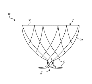
[0020] FIGS. 1A ¨ 1B illustrate an anastomosis device 10, in
accordance with
some embodiments provided herein, that can be implanted in a patient to create
a
fluidic connection between two organs, spaces, tissue structures, conduits,
and the like,
and combinations thereof. In some examples, the anastomosis device 10 is
suitable for
endoscopic delivery and implantation within a patient. More specifically, FIG.
1A
illustrates a top view of the anastomosis device 10, whereas FIG. 1B
illustrates a side
view of the anastomosis device 10. The anastomosis device 10 includes a
collapsible
frame 20, a membrane 30 covering the collapsible frame 20, and an anastomosis
component 40.
4
Date Recue/Date Received 2022-05-27
[0021] The collapsible frame 20 forms a funnel with a wide opening 27
narrowing to a central lumen 25. The collapsible frame 20 is formed from one
or more
elongated elements shaped to form a set of concentric interwoven or
interconnected
undulating rings 21, 22, 23 radiating from the central lumen 25. In different
examples,
the undulating rings may represent separate rings or include a single wire in
arranged in
a coil to form more than one, such as all, of rings 21, 22, 23. The first
undulating ring 21
forms a series of peaks and valleys surrounding the central lumen 25. The
second
undulating ring 22 forms a series of peaks and valleys with the valleys woven
through or
interconnected with the peaks of the first undulating ring 21. The third
undulating ring 23
forms a series of peaks and valleys with the valleys woven through or
interconnected
with the peaks of the second undulating ring 22. For example, overlapped
apices can be
held in place with adhesive or the graft material.
[0022] The concentric arrangement of the undulating rings 21, 22, 23
helps
provide a collapsed configuration suitable for endoscopic delivery to a
stomach of a
patient, as shown in FIG. 2A, as well as an expanded configuration suitable
for lining an
internal surface of a gastric wall of a stomach, as shown in FIG. 2C. The
concentric
arrangement can additionally or alternatively assist with device flexibility,
bendability,
conformability, or to achieve other characteristics. In various examples, the
collapsible
frame 20, when in the expanded configuration is compliant to remain in contact
with the
internal surface of the gastric wall during peristalsis, for example, or other
movement of
the stomach wall.
[0023] In some examples, the collapsible frame 20 is partially or
entirely formed
from a metal material, such as a metal wire. In some examples, the collapsible
frame 20
is partially or entirely formed from a superelastic material, such as a
nitinol wire. Such
examples may allow a collapsed configuration suitable for endoscopic delivery
through
elastic deformation of the expanded configuration. Additionally or
alternatively, the
collapsible frame 20 can be partially or entirely formed from a cut tube, such
as a nitinol
tube. Such examples may provide interconnected connections between the
undulating
rings 21, 22, 23.
[0024] The collapsible frame 20 serves as a skeleton to support the
membrane
30, and the membrane 30 covers the collapsible frame 20. The membrane 30 is
suitable
to limit nutrient contact when the anastomosis device 10 is lined against an
internal
surface of a gastric wall of a stomach. In some examples, the membrane 30 may
include or be formed entirely, or primarily (e.g., 80% or greater) from
expanded
Date Recue/Date Received 2022-05-27
polytetrafluoroethylene (ePTFE). Using ePTFE provides a thin, durable,
impermeable
material to limit nutrient contact from lined surfaces of the gastric wall. In
some
examples, the membrane 30 may include elastomer imbibing or a folded structure
to
allow the membrane 30 to be compliant so as to remain in contact with the
internal
surface of the gastric wall during peristalsis and other movement of a
patient. As
implanted within a patient, the funnel formed by the collapsible frame 20 and
the
membrane 30 is configured to substantially close off the pylorus and direct
food entering
the stomach via a patient's esophagus into the wide opening 27, through the
funnel and
into the small intestine via the anastomosis component 40.
[0025] In some embodiments, the membrane 30 comprises a fluoropolymer,
such as an ePTFE polymer, polytetrafluoroethylene (PTFE) polymer, or
polyvinylidene
fluoride (PVDF) polymer. In some embodiments, the membrane 30 comprises a
polyester, a silicone, a urethane, another biocompatible polymer, polyethylene
terephthalate (e.g., Dacron ), copolymers, or combinations thereof.
[0026] In some embodiments, the membrane 30 (or portions thereof) is
modified
by one or more chemical or physical processes that enhance one or more
properties of
the material. For example, in some embodiments, a hydrophilic coating is
applied to the
membrane 30 to improve the wettability and echo translucency of the material.
In some
embodiments, the membrane 30, or portions thereof, is modified with chemical
moieties
that facilitate one or more of cell attachment, cell migration, cell
proliferation, and
resistance to or promotion of thrombosis. In some embodiments, the membrane
30, or
portions thereof, is modified to resist biofouling. In some embodiments, the
membrane
30, or portions thereof, is modified with one or more covalently attached drug
substances (e.g., heparin, antibiotics, and the like) or impregnated with the
one or more
drug substances. The drug substances can be released in situ to promote
healing,
reduce tissue inflammation, reduce or inhibit infections, and to promote
various other
therapeutic treatments and outcomes. In some embodiments, the drug substance
is a
corticosteroid, a human growth factor, an anti-mitotic agent, an
antithrombotic agent, a
stem cell material, or dexamethasone sodium phosphate, to name some
embodiments.
In some embodiments, a pharmacological agent is delivered separately from the
membrane 30 to the target site to promote tissue healing or tissue growth.
[0027] Coatings and treatments may be applied to the membrane 30
before or
after the membrane 30 is joined or disposed on the framework of the
anastomosis
device 10. Additionally, one or both sides of the membrane 30, or portions
thereof, may
6
Date Recue/Date Received 2022-05-27
be coated. In some embodiments, certain coatings and/or treatments are applied
to
portions of the membrane 30 located on some portions of the anastomosis device
10,
and other coatings and/or treatments are applied to the material(s) located on
other
portions of the anastomosis device 10. In some embodiments, a combination of
multiple
coatings and/or treatments are applied to the membrane 30, or portions
thereof. In
some embodiments, certain portions of the membrane 30 are left uncoated and/or
untreated. In some embodiments, the anastomosis device 10 is fully or
partially coated
to facilitate or frustrate a biological reaction, such as, but not limited to,
cell attachment,
cell migration, cell proliferation, and resistance to or promotion of
thrombosis.
[0028] In some embodiments, a first portion of the membrane 30 is
formed of a
first material and a second portion of the membrane 30 is formed of a second
material
that is different than the first material. In some embodiments, the membrane
30 includes
multiple layers of materials, which may be the same or different. In some
embodiments,
portions of the membrane 30 have one or more radiopaque markers attached
thereto to
enhance in vivo radiographic visualization of the anastomosis device 10, or
one or more
echogenic areas to enhance ultrasonic visibility.
[0029] In some embodiments, one or more portions of the membrane 30
are
attached to the framework of the anastomosis device 10, such as the
collapsible frame
20 and/or a support structure of the anastomosis component 40. The attachment
can be
accomplished by a variety of techniques such as, but not limited to, stitching
the
membrane 30 to the framework of the anastomosis device 10, adhering the
membrane
30 to the framework of the anastomosis device 10, laminating multiple layers
of the
membrane 30 to encompass portions of the elongate members of the anastomosis
device 10, using clips or barbs, or laminating multiple layers of the membrane
30
together through openings in the framework of the anastomosis device 10. In
some
embodiments, the membrane 30 is attached to the framework of the anastomosis
device 10 at a series of discrete locations thereby facilitating the
flexibility of the
framework. In some embodiments, the membrane 30 is loosely attached to the
framework of the anastomosis device 10. It is to be appreciated that the
membrane 30
may be attached to the framework of the anastomosis device 10 using other
techniques
or combinations of techniques described herein.
[0030] In some embodiments, the framework of the anastomosis device 10
(or
portions thereof) is coated with a bonding agent (e.g., fluorinated ethylene
propylene or
other suitable adhesive) to facilitate attachment of the membrane 30 to the
framework.
7
Date Recue/Date Received 2022-05-27
Such adhesives may be applied to the framework using contact coating, powder
coating, dip coating, spray coating, or any other appropriate means.
[0031] The membrane 30 can adapt to changes in the length and/or
diameter of
the collapsible frame 20 in a variety of manners. In a first example, the
membrane 30
can be elastic such that the membrane 30 can stretch to accommodate changes in
the
length and/or diameter of the anastomosis device 10. In a second example, the
membrane 30 can include slackened material in the low-profile delivery
configuration
that becomes less slackened or totally unslackened when the anastomosis device
10 is
in the expanded configuration. In a third example, the membrane 30 can include
folded
portions (e.g., pleats) that are folded in the low-profile configuration and
less folded or
totally unfolded when the anastomosis device 10 is in the expanded
configuration. In
other embodiments, an axial adjustment member is free of the membrane 30. In
some
embodiments, combinations of such techniques, and/or other techniques can be
used
whereby the membrane 30 can adapt to changes in the length and/or diameter of
the
collapsible frame 20.
[0032] The anastomosis component 40 functions to direct food entering
the
stomach via a patient's esophagus into the small intestine by sealing a hole
in the
stomach proximate to central lumen 25 to a corresponding hole in the small
intestine. In
some examples, the anastomosis component 40 may include a first tissue
apposition
portion to seal to the hole in the stomach, and a second tissue apposition
portion to seal
to the hole in the small intestine. The tissue apposition portions of the
anastomosis
component 40 may include series of petal shaped wire frames surrounding the
central
lumen 25, although any variety of other configurations of the anastomosis
component
40 may serve as suitable alternatives, such as solid petals rather than wire
frame
petals. An apposition force of the anastomosis component 40 is applied to the
external
surface of the gastric wall, as described with respect to stent 300, including
flanges 302
with petals 303 (FIGS. 4 ¨ 7B). In contrast to stent 300, the collapsible
frame 20
expands much larger on one side, rather than symmetric, although anastomosis
component 40 may be functionally the same or similar to stent 300.
[0033] Some examples include an optional central portion therebetween,
such
as barrel 304 of stent 300 (FIGS. 4 ¨ 7B), providing an extended central lumen
25 that
extends longitudinally from a first end of the anastomosis component 40 to a
second
end of the anastomosis component 40. In any event, central lumen 25 acts as a
connection (e.g., a shunt passageway) between the stomach and the intestine,
such
8
Date Recue/Date Received 2022-05-27
that the stomach is in fluid communication with the intestines. In some
example, the
central lumen may be larger than the holes in the stomach and intestinal
tissues to
provide a slight outward radial force on the holes to aid in sealing, e.g.,
via an
interference fit due to the elasticity of the tissues.
[0034] While any number of anastomosis configurations is suitable for
adaptation as anastomosis component 40, some of such suitable examples are
disclosed in United States Patent Application Publication No. 2015/0313598 by
Todd et
al., titled ANASTOMOSIS DEVICES.
[0035] In some examples, the support structure of the anastomosis
component
40 may be formed from a metal material, such as a metal wire. In the same or
different
examples, the support structure of the anastomosis component 40 may be formed
from
a superelastic material, such as a nitinol material. Such examples may allow a
collapsed configuration suitable for endoscopic delivery through elastic
deformation of
the expanded configuration. The support structure of the anastomosis component
40
may be formed from a substantially similar material to that of the collapsible
frame 20.
For example, the collapsible frame 20 and the support structure of the
anastomosis
component 40 may include a monolithic frame element forming at least a portion
of the
collapsible frame 20 and the support structure of the anastomosis component
40. In
some examples, the collapsible frame 20 and the support structure of the
anastomosis
component 40 may be formed from a single woven wire, such as a nitinol wire.
Alternatively, the collapsible frame 20 and the support structure of the
anastomosis
component 40 may be formed from a cut tube structure, such as a cut nitinol
tube. In
further examples, the collapsible frame 20 and the support structure of the
anastomosis
component 40 may be formed from more than one element including any
combination
of wire elements, and/or cut tube elements.
[0036] In various examples, the membrane 30 may cover the support
structure
of the anastomosis component 40, or it may not cover the support structure of
the
anastomosis component 40. In some particular examples, the anastomosis
component
40 may be covered in a material that resists ingrowth and adhesion. This may
allow the
anastomosis device 10 to be removed later without significant trauma to the
surrounding
tissues of the gastric wall 102.
[0037] Suitable materials for the frame elements of the collapsible
frame 20 and
the support structure of the anastomosis component 40, include a variety of
metallic
materials including alloys exhibiting shape memory, elastic and super-elastic
9
Date Recue/Date Received 2022-05-27
characteristics. Shape memory refers to the ability of a material to revert to
an originally
memorized shape after plastic deformation by heating above a critical
temperature.
Elasticity is the ability of a material to deform under load and return or
substantially
return to its original shape when the load is released. Most metals will
deform elastically
up to a small amount of strain. Super-elasticity refers to the ability of a
material to
deform under strain to much larger degree than typical elastic alloys, without
having this
deformation become permanent. For example, the super-elastic materials
included in
the frame elements of some anastomosis device embodiments provided herein are
able
to withstand a significant amount of bending and flexing and then return or
substantially
return to the frame's original form without deformation. In some embodiments,
suitable
elastic materials include various stainless steels which have been physically,
chemically, and otherwise treated to produce a high springiness, metal alloys
such as
cobalt chrome alloys (e.g., ELGILOYTM, MP35N, L605), platinum/tungsten alloys.
Embodiments of shape memory and super-elastic alloys include the NiTi alloys,
ternary
shape memory alloys such as NiTiPt, NiTiCo, NiTiCr, or other shape memory
alloys
such as copper-based shape memory alloys. Additional materials could combine
both
shape memory and elastic alloys such as a drawn filled tube where the outer
layer is
constructed of nitinol and the internal core is a radiopaque material such as
platinum or
tantalum. In such a construct, the outer layer provides the super-elastic
properties and
the internal core remains elastic due to lower bending stresses.
[0038] In some embodiments, the frame elements used to construct the
various
device examples can be treated in various ways to increase the radiopacity of
the
devices for enhanced radiographic visualization. In some embodiments, the
devices are
at least partially a drawn-filled type of NiTi containing a different material
at the core,
such as a material with enhanced radiopacity. In some embodiments, the devices
include a radiopaque cladding or plating. In some embodiments, one or more
radiopaque markers are attached to the devices. In some embodiments, the
elongate
frame elements and/or other portions of the devices provided herein are also
visible via
ultrasound.
[0039] FIGS. 2A ¨ 2D illustrate endoscopic implantation of the
anastomosis
device 10 within the stomach 100 of a patient. The anastomosis device 10 is
introduced
to the stomach 100 as part of an assembly 60 in a collapsed configuration
within an
endoscopic delivery catheter 50. The illustrated portion of the patient's
anatomy in
FIGS. 2A ¨ 20 includes the stomach 100, the esophagus 110, the pylorus 112,
the
Date Recue/Date Received 2022-05-27
duodenum 114, and the jejunum 116 of the patient's small intestine. The
stomach 100
includes the gastric wall 102, the antrum 104 and the fundus 106.
[0040] As shown in FIG. 2A, the anastomosis device 10 is delivered to
the
stomach 100 via an endoscopic delivery catheter 50. In some examples, the
anastomosis device 10 is carried into the stomach 100 within the distal end 52
of the
endoscopic delivery catheter 50. In other examples, the endoscopic delivery
catheter 50
may be passed through the esophagus 110 to locate the distal end 52 within the
stomach 100 before the anastomosis device 10 is pushed through a central lumen
of
the endoscopic delivery catheter 50, for example, by first loading the
anastomosis
device 10 in a proximal end (not shown) of the endoscopic delivery catheter 50
before
traversing the length of the central lumen of the endoscopic delivery catheter
50. In
such examples, the endoscopic delivery catheter 50 maybe used to facilitate
the
endoscopic delivery of multiple tools and implants to the stomach 100, such as
cameras, surgical tools, and multiple the anastomosis devices 10.
[0041] In one exemplary technique of implanting the anastomosis device
10
within the stomach 100, a clinician first inserts the endoscopic delivery
catheter 50
through the esophagus 110 to locate the distal end 52 of the endoscopic
delivery
catheter 50 within the stomach 100. The endoscopic delivery catheter 50
provides
access to the stomach 100 for imaging equipment and surgical tools. The
clinician then
inserts a cutting instrument (not shown) through the endoscopic delivery
catheter 50 to
a location on an internal surface of the gastric wall 102. The clinician opens
a first hole
103 in the gastric wall 102, and a second hole 117 in the small intestine of
the patient,
such as in the jejunum 116, the second hole 117 being generally coincident
with the first
hole 103.
[0042] The clinician may then withdraw the cutting instrument from the
endoscopic delivery catheter 50, and deliver the anastomosis device 10 in a
collapsed
configuration to the stomach 100 via the endoscopic delivery catheter 50, by
pushing
the anastomosis device 10 through the central lumen of the endoscopic delivery
catheter 50 with the plunger 53, as shown in FIG. 2A.
[0043] As shown in FIG. 2B, the clinician may then locate the distal
end 52 of
the endoscopic delivery catheter 50 proximate the hole 103 and direct the
distal end of
the anastomosis component 40, which protrudes from the distal end 52 of the
endoscopic delivery catheter 50, through the holes 103, 117.
11
Date Recue/Date Received 2022-05-27
[0044] As shown in FIG. 2C, once the distal end of the anastomosis
component
40 extends through the holes 103, 117, the clinician may partially deploy the
anastomosis device 10 from the distal end 52 of the endoscopic delivery
catheter 50.
Once deployed, the anastomosis component 40 forms a sealed connection between
the
first hole 103 in the gastric wall 102 and the second hole 117 in the in the
jejunum 116.
Next, the clinician may deploy the anastomosis device 10 from the distal end
52 of the
endoscopic delivery catheter 50, e.g., by pushing the anastomosis device 10
through
the central lumen of endoscopic delivery catheter 50 with the plunger 53.
[0045] As shown in FIG. 2D, once deployed, the anastomosis device 10
expands from a collapsed configuration within the central lumen of the
endoscopic
delivery catheter 50 to an expanded configuration. In the expanded
configuration, the
collapsible frame 20 and the membrane 30 line an internal surface of the
gastric wall
102. In this expanded configuration, the collapsible frame 20 lays flat
against the
internal surfaces of the gastric wall 102 such that the collapsible frame 20
and the
membrane 30 limit nutrient contact from lined portions of the internal surface
of the
gastric wall 102. For example, in the expanded configuration, the collapsible
frame 20
and the membrane 30 may be configured to cover the fundus 106 and a greater
curvature of the stomach 100. Such stomach lining may effectively exclude a
significant
proportion of the hormone producing stomach cells, mimicking a vertical sleeve
gastrectomy.
[0046] In addition, once anastomosis device 10 is deployed, the funnel
of the
collapsible frame 20 and the membrane 30 is configured to substantially close
off the
pylorus 112 and direct food entering the stomach 100 via the esophagus 110
into the
wide opening 27, through the funnel and into the small intestine via the
anastomosis
component 40. The funnel portion of the collapsible frame 20 is of low radial
stiffness
such that it is compliant and remains in contact with the inner surface of the
gastric wall
102 during peristalsis. The funnel portion of the collapsible frame 20 can be
sized and
shaped to any relevant geometry. In this manner, the anastomosis device 10 can
effectively exclude lined portions of the stomach 100 and duodenum 114 and
also
accelerate food delivery to the jejunum 116, mimicking Roux-en-Y gastric
bypass
surgery.
[0047] FIG. 3 is conceptual illustration of an implanted anastomosis
device 210.
The anastomosis device 210 is substantially similar to the anastomosis device
10 and
all described variations and equivalents thereof, with the addition of a
tubular liner 250.
12
Date Recue/Date Received 2022-05-27
The tubular liner 250 is configured to extend into the jejunum 116 of the
small intestine
of the patient beyond the anastomosis component 40. For brevity, only the
tubular liner
250 is described with respect to anastomosis device 210, as all other elements
of the
anastomosis device 210 are described with respect to the anastomosis device
10.
[0048] The tubular liner 250 forms a central lumen fluidly connected
to the
lumen 25. The tubular liner 250 is configured to extend from the anastomosis
component 40 into the small intestine, such as into the jejunum 116, to line
an inner
surface of the small intestine to limit nutrient contact from the lined inner
surface of the
small intestine. This may provide additional efficacy for the patient as
compared to the
anastomosis device 10 by further limiting portions of the small intestine
available for
nutrient uptake. In addition, tubular liner 250 may function to help anchor
anastomosis
device 210 within stomach 100.
[0049] The tubular liner 250 includes a frame element 252 and a
membrane
254. In some examples, the tubular liner may represent a stent graft. In some
examples,
the frame element may 252 be an extension of the collapsible frame 20 and/or
the
support structure of the anastomosis component 40. In other examples, the
frame
element 252 may be a separate component. In any event, the variations and
descriptions above with respect to the collapsible frame 20 and/or the support
structure
of the anastomosis component 40 are also applicable to frame element 252. As
examples, frame element 252 may be formed from a wound wire or individual ring
elements or formed from a cut tube. In any of these examples, frame element
252 may
be formed from nitinol or stainless steel, and may be either self-expandable,
balloon
expandable or a combination thereof.
[0050] In addition, the membrane 254 may be the same or similar to or
even an
extension of membrane 30. For example, membrane 254 may comprise a
fluoropolymer, such as an ePTFE membrane, or PVDF. In some embodiments, the
membrane 30 comprises a polyester, a silicone, a urethane, another
biocompatible
polymer, polyethylene terephthalate (e.g., Dacron ), copolymers, or
combinations
thereof.
[0051] In some examples, as part of the implantation of the
anastomosis device
210, the endoscopic delivery catheter 50 may be directed through holes 103,
117 and
into the small intestine to facilitate deployment of tubular liner 250 within
the small
intestine, such as within the jejunum 116. For example, if tubular liner 250
is self-
13
Date Recue/Date Received 2022-05-27
expandable, endoscopic delivery catheter 50 may function to maintain tubular
liner 250
in a collapsed configuration within the small intestine prior to deployment.
[0052] FIG. 4 illustrates regions of a lumen opposing metal stent 300
shown in a
flat cut pattern. In some examples, the stent 300 may be a self-expanding,
covered
nitinol stent. In the same or different examples, the stent 300 may be
suitable as an
implantable internal gallbladder drainage device, or the design of the stent
300 may
form the anastomosis component 40 of the anastomosis device 10 or the
anastomosis
device 210. In various examples, the stent 300 may intended for minimally
invasive
endoscopic ultrasound (EUS) guided transluminal drainage applications,
including
internal gallbladder drainage.
[0053] The stent 300 includes two flanges 302 connected by a cylindrical
barrel
304 with transition regions 305 between the flanges 302 and the flanges 302.
In some
examples these flanges may correspond to elements of the anastomosis component
40
of the anastomosis device 10 or the anastomosis device 210. Stent 300 may be
implanted such that each of the flanges 302 is located on the intra-luminal
side of the
connected tissues and the barrel 304 spans the combined thickness of both
tissue
walls. This creates a conduit for contents to pass through the barrel 304.
[0054] As shown in FIG. 4, the barrel 304 is defined by the struts
that make up
the cylindrical portion of the stent 300. The transition regions 306 are
include the struts
that bend perpendicular to the barrel axis and connect barrel 304 to the
petals forming
the flanges 302. The flanges 302 are defined by the struts that form the
individual petals
which collectively form the flanges 302 once shapeset. In some examples, the
barrel
304 and the transition regions 306 are symmetric. In the same or different
examples, all
struts within the barrel 304 and the transition regions 306 may be of equal
length and
width. In the same or different examples, the flanges 302 may be of equal
design or one
flange may be larger than the other, or may include additional struts, as with
the
anastomosis device 10 and the anastomosis device 210. In the same or different
examples, petals of the flanges 302 may be offset 1/2n (n = # petals) of the
tube
circumference such that the each petal will align equally between two of the
opposite
flange petals.
[0055] FIG. 5 illustrates a petal 303 of the flanges 302 of the stent
300. The petal
303 contains both petal struts 312, which define the petal shape, and tether
struts 313,
which connect roughly midway along the petal struts 312 to the transition
struts 316.
14
Date Recue/Date Received 2022-05-27
The tether struts 313 are configured to pull the connected transition strut
apex 317
down during crush loading and facilitate a consistent crushed device profile.
[0056] FIG. 6 illustrates a cross section of the stent 300. The cross
section of the
stent 300 may also represent the profile of tooling used to form the stent 300
to its final
shape. Specifically, FIG. 6 illustrates a flange diameter 318, a barrel
diameter 320, a
barrel flat 322 and a barrel length 324. The barrel length 324 is sized
appropriately to
accommodate the anticipated combined maximum tissue thickness of the intended
treatment range. FIG. 6 further illustrates a petal gap 326, a transition
radius 328, a
petal radius 330, a petal tip length 332, a petal tip angle 334 and a petal
recurve radius
336. The petal gap 326 is less than the barrel length 324, and is generally no
larger
than a minimum tissue thickness of the intended treatment range, so that the
petals 303
are configured to displace outward to accommodate the combined tissue
thickness,
such as stomach and intestinal tissues in the anastomosis device 10 or the
anastomosis
device 210 once implanted. This creates strain in the petal 303 and transition
struts 316
which results in an apposition force to keep the tissue walls in contact.
[0057] FIGS. 7A and 7B illustrate top and side views of stent 300. FIG. 7A is
a
front view down the barrel 304. As shown, stent 300 includes five petals 303
per flange
302. Petals are rotationally offset 36 degrees such that they are each
centered about
two opposing petals. This offset may limit peak pressure points the stent 300
applies to
tissue walls between the flanges 302. Additionally, this offset may help
balance size-up
and crush strain within the stent frame. FIG. 7B is a side view showing the
cylindrical
barrel 304 and the flanges 302. Note the flange profile is the same as shown
in FIG. 6.
[0058] FIGS. 8A ¨ 8D illustrate incremental deployment of one flange 302 of
the
stent 300 from a catheter 350. The transition radius 328 and the petal radius
330 (FIG.
6) are selected facilitate the deployment and in-vivo performance, e.g., by
designing for
desired apposition forces, as well as allowing for elastic deformation during
crush
loading. As the stent 300 is bent back against that curvature during crush
loading a
strain is induced along the length of the petal 303. This strain is released
as the stent
300 is unconstrained during deployment. As the stent 300 is incrementally
deployed the
distal flange 302 opens and spreads while the barrel 304 and proximal region
are
maintained at the crush profile (FIG. 8D). At this point the stent 300 will
resist a traction
force applied to it, which allows the two lumens to be pulled into apposition.
[0059] Once the two lumens have been pulled into apposition, the
barrel and
proximal flange are fully deployed (refer to FIG. 7B). Once again the strain
induced in
Date Recue/Date Received 2022-05-27
the transition radius 328 and the petal radius 330 (FIG. 6) is recovered
quickly which
forces the proximal flange 302 to snap open and capture the tissue wall. This
must
occur rapidly because as soon as the stent 300 is fully released it loses
contact with the
delivery catheter 350 and the traction force the user has applied to provide
tissue
apposition is lost and the anatomy will return to a near native position.
[0060] Once fully deployed, the stent is designed to apply an
apposition
pressure to both tissue walls. This is created by the strain induced along the
petal frame
as the petal gap 326 (FIG. 6) is forced larger than its initial value. That
displacement is
generated by the in-vivo tissue thickness being greater than the petal gap
326. The
petals 303 on either flange 302 are directly connected to one another by the
transition
struts 316 and the barrel struts 314 (FIG. 5), both of which are axially
rigid. This allows
each petal 303 to directly oppose its opposites on the other flange 302. This
equilibrates
the total apposition pressure created by both flanges 302 and allows the
device to self-
center.
[0061] In some examples, as previously mentioned the petals 303 may be
offset
on either flange 302 balance the crush strain the stent 300. This offset may
also make
the apposition pressure created at the petal tips to be more distributed
around the
circumference of the flanges 302.
[0062] The IGBD stent 300 is one application of a lumen apposing metal
stent.
However, this disclosure also applies to other uses, including in combination
with the
anastomosis device 10 or other device designs for any application with a need
to divert
flow, provide drainage, provide access, anchor, or occlude orifices.
[0063] In various examples, this disclosure covers each of following
clauses, as
well as the claims provided below, although this disclosure is not limited by
the listings
of clauses and claims.
[0064] Clause 1: An anastomosis device comprising: a collapsible frame
forming
a funnel with a wide opening narrowing to a central lumen; a membrane covering
the
collapsible frame, the collapsible frame and the membrane providing a
collapsed
configuration suitable for endoluminal delivery to a stomach of a patient, and
an
expanded configuration suitable for lining an internal surface of a gastric
wall of the
stomach; and an anastomosis component extending from the central lumen of the
collapsible frame and being configured to pass through a first hole in the
gastric wall
and a second hole in a small intestine of the patient and form a sealed
connection
between the first hole in the gastric wall and the second hole in the small
intestine,
16
Date Recue/Date Received 2022-05-27
wherein the funnel is configured to substantially close off the pylorus and
direct food
entering the stomach via a patient's esophagus into the wide opening, through
the
funnel and into the small intestine via the anastomosis component.
[0065] Clause 2: The anastomosis device of clause 1, wherein the
anastomosis
component comprises a support structure extending from the collapsible frame
and
being configured to pass through the first hole in the gastric wall and the
second hole in
the small intestine and lay flat against an internal wall of the small
intestine when the
anastomosis device is implanted within the patient.
[0066] Clause 3: The anastomosis device of clause 2, wherein the
membrane
covers the support structure of the anastomosis component.
[0067] Clause 4: The anastomosis device of clause 1, wherein the
collapsible
frame and the anastomosis component include a monolithic frame element forming
at
least a portion of the collapsible frame and a support structure of the
anastomosis
component.
[0068] Clause 5: The anastomosis device of clause 1, wherein the
collapsible
frame and a support structure of the anastomosis component are formed from a
cut
tube structure.
[0069] Clause 6: The anastomosis device of clause 1, wherein the
collapsible
frame, when in the expanded configuration, is configured to cover a fundus and
a
greater curvature of the stomach.
[0070] Clause 7: The anastomosis device of clause 1, wherein the
collapsible
frame, when in the expanded configuration, is configured to limit nutrient
contact from
lined portions of the internal surface of the gastric wall.
[0071] Clause 8: The anastomosis device of clause 1, wherein the
second hole
in the small intestine of the patient enters a jejunum of the patient.
[0072] Clause 9: The anastomosis device of clause 1, wherein the
membrane
includes expanded polytetrafluoroethylene (ePTFE).
[0073] Clause 10: The anastomosis device of clause 1, further
comprising a
tubular liner configured to extend from the anastomosis component into the
small
intestine to line an internal surface of the small intestine to limit nutrient
contact from the
lined internal surface of the small intestine.
[0074] Clause 11: The anastomosis device of clause 10, wherein the
tubular
liner includes a stent graft comprising a frame element and the membrane.
17
Date Recue/Date Received 2022-05-27
[0075] Clause 12: An assembly comprising: an endoscopic delivery
catheter;
and an anastomosis device comprising: a collapsible frame forming a funnel
with a wide
opening narrowing to a central lumen; a membrane covering the collapsible
frame, the
collapsible frame and the membrane providing a collapsed configuration
suitable for
endoluminal delivery to a stomach of a patient, and an expanded configuration
suitable
for lining an internal surface of a gastric wall of the stomach; and an
anastomosis
component extending from the central lumen of the collapsible frame and being
configured to pass through a first hole in the gastric wall and a second hole
in a small
intestine of the patient and form a sealed connection between the first hole
in the gastric
wall and the second hole in the small intestine, wherein the funnel is
configured to
substantially close off the pylorus and direct food entering the stomach via a
patient's
esophagus into the wide opening, through the funnel and into the small
intestine via the
anastomosis component, wherein the collapsible frame and the membrane are in
the
collapsed configuration within a distal end of the endoscopic delivery
catheter to
facilitate endoscopic delivery and implantation of the anastomosis device
within the
stomach.
[0076] Clause 13: The assembly of clause 12, wherein the anastomosis
component comprises a support structure extending from the collapsible frame
and
being configured to pass through the first hole in the gastric wall and the
second hole in
the small intestine and lay flat against an internal wall of the small
intestine when the
anastomosis device is implanted within the patient.
[0077] Clause 14: The assembly of clause 12, wherein the collapsible
frame and
the anastomosis component include a monolithic frame element forming at least
a
portion of the collapsible frame and a support structure of the anastomosis
component.
[0078] Clause 15: The assembly of clause 12, wherein the collapsible
frame,
when in the expanded
[0079] Clause 16: The assembly of clause 12, wherein the collapsible
frame,
when in the expanded configuration, is configured to limit nutrient contact
from lined
portions of the internal surface of the gastric wall.
[0080] Clause 17: The assembly of clause 12, wherein the membrane
includes
expanded polytetrafluoroethylene (ePTFE).
[0081] Clause 18: The assembly of clause 12, further comprising a
tubular liner
configured to extend from the anastomosis component into the small intestine
to line an
18
Date Recue/Date Received 2022-05-27
internal surface of the small intestine to limit nutrient contact from the
lined internal
surface of the small intestine.
[0082] Clause 19: A method of implanting an anastomosis device within
a
stomach of a patient, the method comprising: inserting an endoscopic delivery
catheter
through an esophagus of the patient to locate a distal end of the endoscopic
delivery
catheter within the stomach of the patient; opening a first hole in a gastric
wall of the
stomach; opening a second hole in a small intestine of the patient, the second
hole
being generally coincident with the first hole; delivering the anastomosis
device in a
collapsed configuration to the stomach via the endoscopic delivery catheter,
wherein the
anastomosis device includes: a collapsible frame forming a funnel with a wide
opening
narrowing to a central lumen; a membrane covering the collapsible frame; and
an
anastomosis component extending from the central lumen of the collapsible
frame;
inserting the anastomosis component through the first hole in the gastric wall
and the
second hole in the small intestine to form a sealed connection between the
first hole in
the gastric wall and the second hole in the small intestine; and deploying the
anastomosis device from the distal end of the endoscopic delivery catheter to
expand
the anastomosis device from the collapsed configuration to an expanded
configuration
and line an internal surface of the gastric wall, wherein, once deployed, the
funnel is
configured to substantially close off the pylorus and direct food entering the
stomach via
the esophagus into the wide opening, through the funnel and into the small
intestine via
the anastomosis component.
[0083] Clause 20: The method of clause 19, wherein inserting the
anastomosis
component through the first hole in the gastric wall and the second hole in
the small
intestine to form the sealed connection comprises: locating a distal portion
of a support
structure of the anastomosis component through the first hole in the gastric
wall and the
second hole in the small intestine while the anastomosis device is in the
collapsed
configuration at least partially proximate the distal end of the endoscopic
delivery
catheter; and deploying the anastomosis device from the distal end of the
endoscopic
delivery catheter to allow a transition of the anastomosis device from the
collapsed
configuration to the expanded configuration such that the support structure of
the
anastomosis component lays flat against an internal wall of the small
intestine and seals
the anastomosis component to the internal wall of the small intestine.
[0084] The invention of this application has been described above both
generically and with regard to specific embodiments. It will be apparent to
those skilled
19
Date Recue/Date Received 2022-05-27
in the art that various modifications and variations can be made in the
embodiments
without departing from the scope of the disclosure. Thus, it is intended that
the
embodiments cover the modifications and variations of this invention provided
they
come within the scope of the appended claims and their equivalents.
Date Recue/Date Received 2022-05-27