Note: Descriptions are shown in the official language in which they were submitted.
CA 03160974 2022-05-10
WO 2021/096693
PCT/US2020/057953
Blood Treatment Systems
TECHNICAL FIELD
This invention relates to blood treatment systems and methods used for
extracorporeal blood treatment procedures.
BACKGROUND
Renal dysfunction or failure and, in particular, end-stage renal disease,
causes the
body to lose the ability to remove water and minerals, maintain acid-base
balance, and
control electrolyte and mineral concentrations within physiological ranges.
Toxic uremic
waste metabolites, including urea, creatinine, and uric acid, accumulate in
the body's
tissues which can result in a person's death if the filtration function of the
kidney is not
replaced.
In treating chronic renal failure, various methods of purification and
treatment of
blood with machinery are used for removing substances usually eliminated with
the urine
and for withdrawing fluids. Diffuse mass transport is predominant in
hemodialysis (HD),
while in hemofiltration (HF) convective mass transport through a membrane is
used.
Hemodiafiltration (HDF) is a combination of the two methods.
During HD, blood passes from the patient through a dialyzer that includes a
semi-
permeable membrane to separate the blood from a large volume of externally-
supplied
dialysis solution, also referred to as dialysate. The waste and toxins,
including excess
fluids, dialyze out of the blood through the semi-permeable membrane into the
dialysate,
which is then typically discarded. The transportation of the small molecular
substances
through the semi-permeable membrane is determined mainly by the differences in
concentration between the dialysate and the blood. The dialysate is referred
to as "fresh
dialysate" prior to receiving the dialyzed components of the blood, and the
dialysate that
exits the dialyzer after receiving the dialyzed components is referred to as
"spent
dialysate."
During HDF, part of the serum withdrawn through the semi-permeable membrane
is replaced by a sterile substitution fluid which is passed to the
extracorporeal blood
1
CA 03160974 2022-05-10
WO 2021/096693
PCT/US2020/057953
stream either upstream of the dialyzer or downstream of the dialyzer. The
supply of
substitution fluid upstream of the dialyzer is also referred to as pre-
dilution, and the
supply downstream of the dialyzer is also referred to as post-dilution.
SUMMARY
Dialyzer systems described herein can include a magnetically driven and
magnetically levitating pump rotor integrated into the dialyzer. Such a
dialyzer is
configured for use with treatment modules described herein that include a
magnetic field-
generating pump drive unit. In some embodiments, the dialyzers include
pressure sensor
chambers with flexible membranous walls against which corresponding pressure
transducers of the treatment modules can interface to detect arterial and/or
venous
pressures. Additional features, as described herein, can be incorporated into
the dialyzers
and treatment modules to consolidate components, simplify setup, and enhance
blood
treatment performance.
In one aspect, this disclosure is directed to a blood treatment machine. The
blood
treatment machine includes a treatment module with a structure for releasably
coupling
with a dialysis treatment apparatus such as a dialyzer. The blood treatment
machine also
includes a blood treatment machine console that controls the treatment module.
The
blood treatment machine can also include one or more sensors that are operable
to
determine an orientation or motion of the blood treatment module in relation
to the blood
treatment machine console. In some embodiments, the treatment module is
mounted to
an arm extending from the blood treatment machine console.
Such a blood treatment machine may optionally include one or more of the
following features in any combination(s). In some embodiments, the treatment
module is
cantilevered from the arm. The arm can include one or more adjustable joints
by which
the arm can be manually articulated into multiple differing positions relative
to the blood
treatment machine console. In particular embodiments, the blood treatment
machine
console includes facilities for making dialysate. The treatment module may
include a
drive unit that generates dynamic magnetic fields to levitate and rotate a
pump rotor
contained within the dialyzer while the dialyzer is coupled with the treatment
module.
2
CA 03160974 2022-05-10
WO 2021/096693
PCT/US2020/057953
The treatment module may also include a first pressure transducer positioned
to abut
against a first membrane of a first pressure detection chamber of the dialyzer
while the
dialyzer is coupled with the treatment module, and/or a second pressure
transducer
positioned to abut against a membrane of a second pressure detection chamber
of the
dialyzer while the dialyzer is coupled with the treatment module. In some
embodiments,
the treatment module also includes a first pair of conduits configured to
connect with a
first substituate liquid port and a first dialysate port defined by the
dialyzer while the
dialyzer is coupled with the treatment module, and/or a second pair of
conduits
configured to connect with a second sub stituate liquid port and a second
dialysate port
defined by the dialyzer while the dialyzer is coupled with the treatment
module. In
certain embodiments, the treatment module also includes a first door
configured to open
and shut a first opening, and/or a second door configured to open and shut a
second
opening. The first pressure transducer and/or the first pair of conduits can
be adjacent the
first door. The second pressure transducer and/or the second pair of conduits
can be
adjacent the second door. In some embodiments, in a first configuration of the
treatment
module, the first and second openings are shut by the first and second doors
respectively.
The first pressure transducer and/or the first pair of conduits can be
retracted behind the
first door while the first door is shut. The second pressure transducer and/or
the second
pair of conduits can be retracted behind the second door while the second door
is shut. In
some embodiments, in a second configuration of the treatment module, the first
and
second openings are open. The first pressure transducer and/or the first pair
of conduits
can be extended through the first opening while the first door is open. The
second
pressure transducer and/or the second pair of conduits can be extended through
the
second opening while the second door is open. The structure for releasably
coupling with
a dialyzer may comprise a slot that is shaped to slidably receive a portion of
the dialyzer.
In another aspect, this disclosure is directed to a dialysis treatment
apparatus. The
dialysis treatment apparatus includes a housing, a bundle of hollow fibers
within an
interior of the housing, an arterial patient line connected to a first end of
the housing, and
a venous patient line connected to a second end of the housing opposite of the
first end.
The hollow fibers define lumens. The arterial patient line is configured to be
connected
3
CA 03160974 2022-05-10
WO 2021/096693
PCT/US2020/057953
to a vasculature of a patient and communicative with the lumens of the hollow
fibers.
The venous patient line is configured to be connected to the vasculature of
the patient and
communicative with the lumens of the hollow fibers. In some embodiments, the
arterial
patient line and/or the venous patient line is/are less than a meter in
length.
Such a dialysis treatment apparatus may optionally include one or more of the
following features in any combination(s). The dialysis treatment apparatus may
be, or
comprise, a dialyzer. The dialysis treatment apparatus may also include a pump
rotor
within the housing. The pump rotor may be magnetically-drivable to force fluid
through
the lumens of the hollow fibers. The dialysis treatment apparatus (e.g.,
dialyzer) may be
configured to direct fluid (e.g., blood) to enter the first end transverse to
the longitudinal
axis of the dialyzer. The first end may be configured to deliver the fluid to
a center of the
pump rotor. After radially exiting the pump rotor, the fluid may enter a
toroidal space
defined around the pump rotor by the first end. From the toroidal space, the
fluid may be
directed by the first end to flow toward the hollow fibers. Prior to reaching
the hollow
fibers, the fluid may pass through one or more openings defined by an internal
support
plate within the first end. The first end may define an arterial pressure
detection
chamber. An exterior wall of the arterial pressure detection chamber may
include a first
flexible membrane. The second end may define a venous pressure detection
chamber.
An exterior wall of the venous pressure detection chamber may include a second
flexible
membrane. In some embodiments, the first end defines: (i) a first dialysate
port in fluid
communication with the interior of the housing external to the lumens of the
hollow
fibers and/or (ii) a first substituate liquid port located along a fluid flow
path between the
arterial patient line and the lumens of the hollow fibers. In particular
embodiments, the
second end defines: (i) a second dialysate port in fluid communication with
the interior of
the housing external to the lumens of the hollow fibers and/or (ii) a second
substituate
liquid port located along a fluid flow path between the venous patient line
and the lumens
of the hollow fibers. The second end may include a deaeration chamber and/or
an air
purge member. In some embodiments, the second end includes a port for
administering
medicaments or extracting a fluid sample.
4
CA 03160974 2022-05-10
WO 2021/096693
PCT/US2020/057953
In another aspect, this disclosure is directed to a blood treatment system.
The
blood treatment system includes a blood treatment machine and a dialysis
treatment
apparatus that can be, or comprise, a dialyzer. The blood treatment machine
can include
a treatment module including a structure for releasably coupling with a
dialyzer, a blood
treatment machine console that controls the treatment module, and one or more
sensors
operable to determine an orientation or motion of the blood treatment module
in relation
to the blood treatment machine console. In some embodiments, the treatment
module is
movably coupled to the blood treatment machine console. The dialysis treatment
apparatus can include a housing, a bundle of hollow fibers within an interior
of the
housing, an arterial patient line connected to a first end of the housing, and
a venous
patient line connected to a second end of the housing opposite of the first
end. The
hollow fibers can define lumens. The arterial patient line can be configured
to be
connected to a vasculature of a patient and communicative with the lumens of
the hollow
fibers. The venous patient line can be configured to be connected to the
vasculature of
the patient and communicative with the lumens of the hollow fibers. In some
embodiments, the arterial patient line and/or the venous patient line is/are
less than a
meter in length. In some embodiments, the structure for releasably coupling
with a
dialyzer may comprise a slot that is shaped to slidably receive a portion of
the dialyzer.
Embodiments can include one or more of the following advantages.
In some embodiments, multiple technologies and functionalities of blood
treatment systems are consolidated in a significantly refined and integrated
fashion into
the dialyzer and treatment module systems described herein. For example, in
some
embodiments a single dialyzer unit as described further below can replace
significant
portions of a conventional hollow-fiber dialyzer, tubing set, air removal
system, sample
port, and pump. Moreover, the end caps of some dialyzers described herein can
include
accessible pressure chambers with flexible membranous walls for convenient
measuring
of arterial and venous pressures in a non-invasive manner. In some
embodiments, the
end caps of the dialyzers can include (a) a port to receive fresh dialysate
fluid from the
treatment module and (b) a port to return spent dialysate fluid to the
treatment module
after passing over the dialysis membrane. In some embodiments, the end caps of
the
5
CA 03160974 2022-05-10
WO 2021/096693
PCT/US2020/057953
dialyzers can also include ports by which substituate liquid can be directly
added to the
blood prior to and/or after the blood passes through the hollow fiber blood
treatment
section of the dialyzer. Additionally, in some embodiments, the same dialyzer
and
treatment module systems are configured for carrying out any of multiple
different types
of blood treatments, including, for example, HD and HDF.
In contrast to typical HD and HDF machines, some example embodiments reduce
the number of required setup steps, which may result in reduced setup time and
reduced
opportunity for human error. In a clinic setting, this can free up valuable
nursing
resources and streamline patient care. This simplification can free up nursing
or other
personnel resources in a clinic or home setting, and also makes the process
easier and
more feasible for patients to set up the dialysis machine themselves.
In some embodiments, the consolidated dialyzer and treatment module systems
described herein, provide important functional advantages. For example, the
consolidation can reduce the amount of tubing needed for the extracorporeal
circuit to be
used for a blood treatment session. Moreover, the treatment module can be
mounted on
an arm extending from a blood treatment machine console so that the treatment
module
and dialyzer can be located very close to a patient. These features allow the
length of
extracorporeal tubing needed for a blood treatment session to be significantly
minimized.
Accordingly, the volume of priming solution required is advantageously
reduced.
Additionally, exposure of the patient's blood to contact with foreign surfaces
is also
advantageously reduced. The consolidated form factor also gives rise to
additional
advantages such as less potential for leaks, less hemolysis, less biohazard
waste, less
packaging waste, and reduced transportation expenses.
In some embodiments, a magnetic pump rotor is integrated to the dialyzer in a
liquid-tight manner. Such an integrated pump rotor can be bearing-less,
magnetically
levitated, and rotationally driven by an external pump drive unit that
generates dynamic
magnetic fields. This arrangement provides advantages such as lower hemolysis
as
compared to conventional pumping systems used for extracorporeal blood
treatments,
and a bearing-free design that reduces system maintenance requirements and the
potential
6
CA 03160974 2022-05-10
WO 2021/096693
PCT/US2020/057953
for contamination. Moreover, since the pump drive unit and pump rotor are
separated,
easier cleaning of machine interfaces is advantageously facilitated.
In some embodiments, the consolidated dialyzer and treatment module systems
described herein are also easier to set-up and use as compared to conventional
systems.
Accordingly, set-up times can be reduced and potential for errors can be
mitigated. In
result, the treatment costs per patient can be reduced in some embodiments.
The details of one or more embodiments are set forth in the accompanying
drawings and the description below. Other aspects, features, and advantages
will be
apparent from the description and drawings, and from the claims.
DESCRIPTION OF DRAWINGS
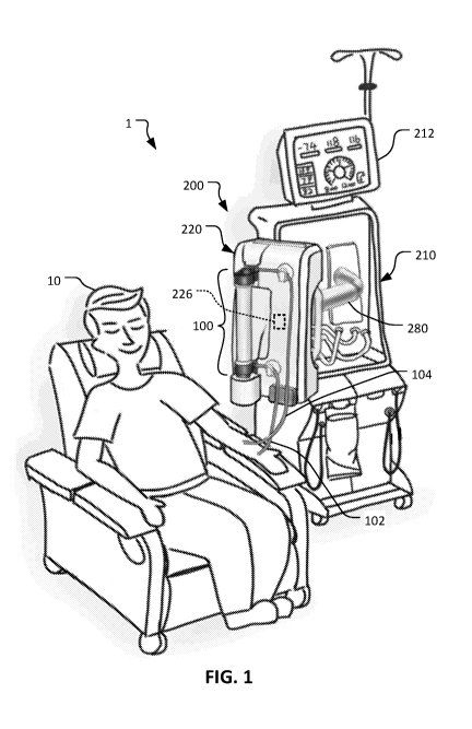
FIG. 1 depicts a patient receiving an extracorporeal blood treatment using a
blood
treatment system.
FIG. 2 is an exploded perspective view of a dialyzer and treatment module
system
of the blood treatment system of FIG. 1.
FIG. 3 is a perspective view of the dialyzer and the treatment module system
of
FIG. 2 in an assembled configuration.
FIG. 4 is a schematic depiction of the dialyzer of the blood treatment system
of
FIG. 1, showing the blood flow path through the dialyzer.
FIG. 5 is another schematic depiction of the dialyzer of the blood treatment
system of FIG. 1, showing the blood flow path through the dialyzer and
substituate
addition locations.
FIG. 6 is another schematic depiction of the dialyzer of the blood treatment
system of FIG. 1, showing the dialysate flow path through the dialyzer.
FIG. 7 is another schematic depiction of the dialyzer of the blood treatment
system of FIG. 1, showing the blood and dialysate flow paths and the
substituate addition
locations.
FIG. 8 is a rear view of the dialyzer of the blood treatment system of FIG. 1.
FIG. 9 is a front view of the dialyzer of the blood treatment system of FIG.
1.
7
CA 03160974 2022-05-10
WO 2021/096693
PCT/US2020/057953
FIG. 10 is a side view of the dialyzer of the blood treatment system of FIG. 1
with
the arterial and venous lines shown in section.
FIG. 11 is a top view of the dialyzer of the blood treatment system of FIGs 1
with
the arterial and venous lines shown in section.
FIG. 12 is a cross-sectional view of the dialyzer of the blood treatment
system of
FIG. 1 taken along section line A-A of FIG. 10.
FIG. 13 is a broken cross-sectional view of the dialyzer of the blood
treatment
system of FIG. 1 taken along section line B-B of FIG. 11.
FIG. 14 is a cross-sectional view of the second end cap of the dialyzer of the
blood treatment system of FIG. 1 taken along section line C-C of FIG. 11, with
the
position of a dialyzer potting shown in broken lines.
FIG. 15 is a cross-sectional view of the dialyzer of the blood treatment
system of
FIG. 1 taken along section line D-D of FIG. 10.
FIG. 16 is a cross-sectional view of the dialyzer of the blood treatment
system of
FIG. 1 taken along section line E-E of FIG. 10.
FIG. 17 is a broken cross-sectional view of the dialyzer of the blood
treatment
system of FIG. 1 taken along section line B-B of FIG. 11, with the bundle of
hollow
fibers and the pottings shown in broken lines.
FIG. 18 is a cross-sectional view of the dialyzer of the blood treatment
system of
FIG. 1 taken along section line F-F of FIG. 10.
FIG. 19 is a cross-sectional view of the dialyzer of the blood treatment
system of
FIG. 1 taken along section line G-G of FIG. 10.
FIG. 20 is a perspective view of a first end cap of the dialyzer of the blood
treatment system of FIG. 1.
FIG. 21 is a rear view of the first end cap of FIG. 20.
FIG. 22 is another perspective view of the first end cap of FIG. 20.
FIG. 23 is a perspective view of the first end cap of FIG. 20 shown in a
partial
longitudinal cross-sectional view and depicting blood flow therethrough.
FIG. 24 is a perspective view of a pump rotor that is configured to be located
in
the first end cap of FIG. 20.
8
CA 03160974 2022-05-10
WO 2021/096693
PCT/US2020/057953
FIG. 25 is a perspective view of an alternative pump rotor that can be used in
the
first end cap of FIG. 20.
FIG. 26 is a perspective view of a second end cap of the dialyzer of the blood
treatment system of FIG. 1.
FIG. 27 is a rear view of the second end cap of FIG. 26.
FIG. 28 is another perspective view of the second end cap of FIG. 26.
FIG. 29 is a cross-sectional view of an alternative second end cap.
FIG. 30 is a perspective view of the treatment module of the blood treatment
system of FIG. 1 in a first configuration.
FIG. 31 is a perspective view of the treatment module of FIG. 30 in a second
configuration.
FIG. 32 is an exploded perspective view showing the first end cap of FIG. 20
and
a first pressure sensor and a first pair of conduits of the treatment module
of FIG. 30.
FIG. 33 is a top perspective view of the first end cap, the first pressure
sensor, and
the first pair of conduits of FIG. 32 shown in a separated configuration.
FIG. 34 is a top perspective view of the first end cap, the first pressure
sensor, and
the first pair of conduits of FIG. 32 shown in an operative, coupled
configuration.
FIG. 35 is a perspective view of an alternative treatment module.
FIG. 36 is a perspective view of an alternative first (arterial) end cap shown
in a
partial longitudinal cross-sectional view.
FIG. 37 is a rear view of an example dialyzer that is configured similar to
the
dialyzer of the blood treatment system of FIG. 1, except without HDF
capability.
FIG. 38 is a front view of the dialyzer of FIG. 37.
FIG. 39 is a side view of the dialyzer of FIG. 37.
FIG. 40 is a longitudinal cross-sectional view of an alternative second
(venous)
end cap.
FIG. 41 is a perspective view showing a portion of the venous end cap of FIG.
40.
FIG. 42 is a perspective view of a portion of another alternative second
(venous)
end cap.
FIG. 43 is a longitudinal cross-sectional view of the venous end cap of FIG.
42.
9
CA 03160974 2022-05-10
WO 2021/096693
PCT/US2020/057953
FIG. 44 is another perspective view of the venous end cap of FIG. 42.
FIG. 45 is a longitudinal cross-sectional view of another alternative second
(venous) end cap. The venous end cap is shown in a first configuration.
FIG. 46 is a longitudinal cross-sectional view of the venous end cap of FIG.
45 in
a second configuration.
FIG. 47 is a perspective view showing a portion of the venous end cap of FIG.
45.
Like reference symbols in the various drawings indicate like elements.
DETAILED DESCRIPTION
This disclosure describes dialyzer systems that can include a magnetically
driven,
magnetically levitating pump rotor integrated into the dialyzer. Such a
dialyzer can be
used with treatment modules described herein that include a dynamic magnetic
field-
generating pump drive unit. In some embodiments, the dialyzer includes one or
more
pressure sensor chambers with flexible exterior membrane walls with which
corresponding pressure transducers of the treatment modules interface to
detect arterial
and/or venous pressures. The dialyzer systems described herein consolidate
multiple
diverse technologies and functionalities of blood treatment systems in a
significantly
integrated fashion to consolidate components, reduce costs, simplify setup,
and enhance
performance.
With reference to FIG. 1, a patient 10 is depicted as receiving an
extracorporeal
blood treatment using a blood treatment system 1 that includes a disposable
set connected
to a blood treatment machine 200. The disposable set includes a dialyzer 100
that is
coupled to a treatment module 220 of the blood treatment machine 200. In some
cases,
the patient 10 may receive treatment for a health condition such as renal
failure.
Accordingly, the system 1 can be used to provide one or more types of
treatment to the
patient 10, including hemodialysis (HD), hemodiafiltration (HDF), or some
other type of
blood treatment. For such treatments, blood is withdrawn from the patient 10
via an
arterial line 102 and, after passing through the dialyzer 100, treated blood
is returned to
the patient 10 via a venous line 104. The dialyzer 100 is a single-use
disposable item,
CA 03160974 2022-05-10
WO 2021/096693
PCT/US2020/057953
whereas the blood treatment machine 200 is a durable reusable system. In some
cases, a
single dialyzer 100 may be reused two or more times for a particular
individual patient.
The blood treatment machine 200 includes a blood treatment machine console
210, the treatment module 220, and an arm 280 that connects the treatment
module 220 to
the blood treatment machine console 210. The arm 280 extends from the blood
treatment
machine console 210, and the treatment module 220 is mounted to the other end
of the
arm 280. In other words, the treatment module 220 is cantilevered from the
blood
treatment machine console 210 by the arm 280.
The arm 280 includes one or more adjustable joints so that the arm 280 can be
manually articulated to position the treatment module 220 in various
positions/orientations relative to the blood treatment machine console 210
and/or relative
to the patient 10. For example (as depicted in FIG. 1), in some cases the arm
280 can be
extended so that the treatment module 220 is positioned close to the patient
10.
Accordingly, the arterial line 102 and the venous line 104 can be quite short
as compared
to conventional blood treatment systems. For example, in some embodiments, the
arterial line 102 and the venous line 104 have a length less than one meter
(e.g., less than
90cm, less than 80cm, less than 70cm, less than 60cm, less than 50cm, less
than 40cm,
less than 30cm, or less than 20cm).
In some embodiments, the treatment module 220 and/or the arm 280 can include
one or more sensors 226 that output signals that can indicate the position,
orientation,
and/or motion of the treatment module 220 relative to the blood treatment
machine
console 210. For example, in some cases sensors such as accelerometers (e.g.,
3D
accelerometers), gyroscopic sensors, ultrasonic sensors, proximity sensors,
optical
sensors, magnetometers, global positioning sensors, radio triangulation
sensors (e.g., like
in keyless access systems for cars or based on WiFi, Bluetooth or similar
technologies),
electronic spirit levels, electric spirit levels, and/or the like, within the
treatment module
220 and/or the arm 280 may be utilized to indicate the position, orientation,
and/or
motion of the treatment module 220 relative to the blood treatment machine
console 210.
In some embodiments, the signal output(s) from such sensors 226 can be used by
the control system of the blood treatment system 1 as input(s), for example,
to activate or
11
CA 03160974 2022-05-10
WO 2021/096693
PC T/US2020/057953
deactivate certain modes of operation of the blood treatment system 1 or,
alternatively, to
determine the current situation of the treatment module 220. For example, a
certain
orientation of the treatment module 220 might be used to indicate that a
maintenance
mode should be activated. Pulling the treatment module 220 forward, towards
the
patient, might initiate preparations for a treatment mode. Another particular
orientation
of the treatment module 220 might be defined as indicative for activating a
deaeration.
mode. Pushing back the treatment module 220 toward the blood treatment machine
console 210 might act as input for pausing operation of the blood treatment
system 1, and
so on. Other modes of operation of the blood treatment system I that can be
activated in
response to a particular position, orientation, or motion of the treatment
module can
include, but are not limited to, a "nurse mode," a debugging mode, and a
filling, or
priming mode, to provide a few examples. Including the one or more sensors 226
that
output signals that can indicate the position, orientation, and/or motion of
the treatment
module 22.0 relative to the blood treatment machine console 210 allows user
control
interactions with the blood treatment system 1, conveniently and intuitively,
by the
manual handling of the arm-mounted treatment module 220. The electronics
and/or
controls that receive and interpret output signals from the sensors 226 can be
located in
the blood treatment machine console 210, the treatment module 220, the arm
280, and/or
elsewhere. In some embodiments, the raw data from one or more sensors 226
is/are
processed in a separate step to generate the sensor output that is used in
further steps. In
some embodiments, the processor carrying out this processing step is located
in the
treatment module 220. In some embodiments, the processor carrying out this
processing
step is located in the arm 280. In some embodiments, the processor carrying
out this
processing step is located in the blood treatment machine console 210.
In some embodiments, there are additionally or alternatively sensors located
in the
arm 280 to determine the position and/or orientation of the treatment module
220. Such
sensors can be angle sensors, path sensors, range sensors, and/or other types
of sensors.
In some embodiments, such sensors can be used to recognize if a situation of
mechanical
shock has occurred, such as in case of a mechanical impact of a person or an
object
making contact with the treatment module 220. The detection of the impact
event can be
12
CA 03160974 2022-05-10
WO 2021/096693
PCT/US2020/057953
used to identify alarms as false alarms, when they occur at the same time in
other sensors
triggered by the impact event. For example, an ultrasonic air bubble detector
could
produce sensor readings causing an alarm in case of an impact event. The
accelerometer
or position sensor(s) in the treatment module 220 and/or arm 280 could enable
detecting
an impact event that occurred at the time of that alarm. In this case, the
treatment module
controls could deescalate that alarm taking into account the air bubble
detector readings
would likely have been be falsified due to the detected impact event.
Further advantages of the using such sensors as described above include, in
combination with a de-aeration mode or priming mode, utilizing the sensor
readout for
initiating a certain operating state to reduce the work load for personnel
handling the a
treatment module 220. Additionally, the haptic input channel would allow for a
more
intuitive way of handling the treatment module 220. Further, these concepts
can help
avoid errors and mistakes in handling and treatments, and false alarms can be
identified.
In some embodiments, the output signal(s) from the sensor(s) 226 may be guided
to a control unit in the treatment module 220, and/or the console 210, and the
control unit
may be configured or programmed to disable or enable predefined processes of
the blood
treatment system 1 on the basis of the signal(s). In some embodiments, the
priming
phase of the dialyzer 100 (which means filling the dialyzer 100 with liquid
and de-
aerating the dialyzer 100) and/or the treatment phase of the blood treatment
system 1 is
only enabled when the signal indicates a vertical position of the dialyzer
100. In some
embodiments, the signal(s) from the sensor(s) 226 must indicate that treatment
module
220 is in an angled position in relation to the ground (level in relation to
the earth), so
that any liquid that could flow out of the liquid circuit is not dropping to
the earth but
conducted along the surface of the treatment module 220 and may be guided into
a liquid
collection port of the treatment module 220. The liquid collection port may by
a rail
along the lower end of the treatment module 220 and being connected to a
container to
collect leaking liquid.
The control unit may further be connected to a user interface, such as the
user
interface 212. The user interface may be a graphical user interface and
optical light
system, a sound generating system, or any combination thereof The user
interface may
13
CA 03160974 2022-05-10
WO 2021/096693
PCT/US2020/057953
be configured to display the orientation of the treatment module 220 (as
provided by the
signal(s) from the sensor(s) 226) and the display may change in visible
appearance as a
function of the enabled processes.
In one example embodiment, the graphical user interface will show the
orientation
of the treatment module 220 when the next process step is, for example, the
priming
phase. Only if the treatment module 220 is in the upright position 220 (as
detected by the
signal(s) from the sensor(s) 226) will the orientation be displayed in green
and the
operator will be able to manually initiate the priming phase via user
interface actions
(e.g., speech, button, gesture, etc.), or the system will automatically
initiate the next
process step.
Although the illustrated example includes a treatment module 220 that is
moveable relative to the base console 210, it should be understood that some
other
examples do not include a separately positionable treatment module 220. In
such
examples, the base console 210 may incorporate the features described with
respect to the
illustrated treatment module 220 other than those specific to the
positionability.
The blood treatment machine console 210 includes a user interface 212, a
control
system, facilities for making dialysate, and the like.
In the blood treatment system 1, much of the componentry associated with
conventional systems is incorporated into the dialyzer 100 and portions of the
blood
treatment module 220 that interfaces with the dialyzer 100. Conventional blood
treatment systems generally include a disposable tubing set and/or cassette
(in addition to
a dialyzer). Such a tubing set and/or cassette is used to interface with one
or more
hardware items such as pumps, sensors, valve actuators, and the like. However,
the
dialyzer 100 and the blood treatment machine 200 integrate multiple
functionalities in a
highly consolidated fashion (as described further below).
Referring also to FIGs. 2 and 3, the dialyzer 100 is releasably coupleable to
the
treatment module 220 in a convenient manner. For example, in the depicted
embodiment,
the dialyzer 100 is slidably coupleable with the treatment module 220.
Accordingly, the
dialyzer 100 and treatment module 220 include complementary structural
features to
facilitate slidable coupling. That is, the dialyzer 100 includes a first
projection 106 that is
14
CA 03160974 2022-05-10
WO 2021/096693
PCT/US2020/057953
slidably coupleable with a first complementarily shaped slot 222 of the
treatment module
220, and the dialyzer 100 includes a second projection 108 that is slidably
coupleable
with a second complementarily shaped slot 224 of the treatment module 220. In
some
embodiments, other means of releasably connecting the dialyzer 100 to the
treatment
module 220 can be used. For example, in some embodiments a connection style
such as
a snap-in connection, a thumb screw connection, a clamp connection, a suction
connection, and the like can be used.
The dialyzer 100 includes a housing 110 that defines an interior space. A
bundle
of hollow fiber semi-permeable membranes (or simply "hollow fibers") are
disposed
within the interior of the housing 110. The arterial line 102 and the venous
line 104 each
extend from the housing 110 (e.g., from opposite ends of the housing 110) and
are in
fluid communication with the interior of the housing 110, and with lumens of
the hollow
fibers.
The housing 110 includes a first end cap 120 and a second end cap 140. The
first
end cap 120 includes the first projection 106 and the second end cap 140
includes the
second projection 108. Moreover, the arterial line 102 is coupled to the first
end cap 120
and the venous line 104 is coupled to the second end cap 140.
The treatment module 220 includes a pump drive unit 230 that is configured to
releasably receive a portion of the first end cap 120. As described further
below, the
pump drive unit 230 generates dynamic magnetic fields to levitate and rotate a
pump
rotor that is housed within the portion of the first end cap 120. In some
embodiments, the
pump drive unit 230 includes no moving parts.
The pump rotor is configured such that rotation of the pump rotor forces blood
of
the patient 10 through the lumens of the hollow fibers of the dialyzer 100 in
the direction
from the first end cap 120 toward the second end cap 140. Accordingly, blood
from the
patient 10 flows into the dialyzer 100 via the arterial line 102, flows
through the lumens
of the hollow fibers, and flows out of the dialyzer 100 via the venous line
104.
The treatment module 220 also includes other devices that interface with the
arterial line 102 and/or the venous line 104. For example, the depicted
treatment module
220 includes a tubing interface module 240 configured to releasably receive a
portion of
CA 03160974 2022-05-10
WO 2021/096693
PCT/US2020/057953
the arterial line 102 and/or a portion of the venous line 104. The tubing
interface module
240 can include devices that can perform functions such as flow rate
detection, gaseous
bubble detection, and the like. That is, the tubing interface module 240 can
include
sensors for detecting one or more parameters such as a flow rate of the blood
within the
arterial line 102 and/or the venous line 104, hematocrit (Hct) and other blood
properties,
and/or for detecting gaseous bubbles (e.g., air bubbles) in the blood within
the arterial
line 102 and/or the venous line 104. In some embodiments, the flow rate
detection and/or
the bubble detection are performed using sensors such as ultrasonic sensors,
optical
sensors, or other suitable types of sensors. In other embodiments, sensors for
detecting
gaseous bubbles can be located at or in an end cap of the disposable of the
dialyzer 100.
The treatment module 220 also includes an arterial line clamp 242 and a venous
line clamp 244. The clamps 242 and 244 are used to either fully restrict or
fully un-
restrict (e.g., in an on/off valve fashion) the flow of blood within the
arterial line 102
and/or the venous line 104, respectively.
The treatment module 220 also includes devices for interfacing with the
dialyzer
100 to measure pressure at particular locations within the dialyzer 100, as
described
further below. Additionally, as described further below, the treatment module
220
includes conduits that can selectively interface with the dialyzer 100 to
facilitate flow of
liquids such as sub stituate and/or dialysate between the dialyzer 100 and the
treatment
module 220.
FIGs. 4-7 are schematic diagrams of the dialyzer 100. For ease of
understanding,
FIG. 4 depicts exclusively the flow of blood through the dialyzer 100; FIG. 5
depicts the
flow of blood and substituate; FIG. 6 depicts exclusively the flow of
dialysate; and FIG. 7
depicts the flow of blood, substituate, and dialysate.
FIGs. 4-7 are simplified to show general flow relationships in the dialyzer
100.
For example, the first potting 115 and the second potting 116, which secure
the two
respective ends of each of the fibers of the bundle of hollow fibers 114, are
omitted to
simplify the illustration. In addition to securing the bundle of hollow
fibers, these
pottings 115 and 116, maintain a barrier between the blood and the dialysate.
The
16
CA 03160974 2022-05-10
WO 2021/096693
PCT/US2020/057953
pottings 115 and 116 and the associated flow routing are described in further
detail below
in connection with FIGs. 8 to 29.
Referring to FIG. 4, the housing 110 of the dialyzer 100 includes the first
end cap
120, the second end cap 140, and a middle housing portion 112 that extends
between the
first end cap 120 and the second end cap 140. The middle housing portion 112
contains
the majority of the length of the bundle of hollow fibers 114. As indicated
above, a more-
detailed description of the construction of the dialyzer 100, including the
bundle of
hollow fibers 114 is provided below in connection with the description of
FIGs. 8 to 29.
The first end cap 120 includes a pump housing 130. A rotatable centrifugal
pump
rotor 132 is located within the pump housing 130. The pump rotor 132 is
enclosed or
encased within the pump housing 130. Accordingly, the pump rotor 132 is
contained at a
fixed position relative to the bundle of hollow fibers 114.
In accordance with some embodiments, the pump rotor 132 is a radially pumping
pump wheel with a hollow central volume. The blades (or vanes) of the pump
wheel of
the pump rotor 132 are arranged so that they project or extend at least
partially radially.
In some cases, the blades are arranged to project or extend entirely radially.
In some
cases, the blades are arranged to project or extend partially radially and
partially
tangentially.
As described further herein, the pump rotor 132 is operated and controlled by
interfacing with the pump drive unit 230 (shown in FIGs. 2 and 3) of the
treatment
module 220. That is, the pump rotor 132 can be levitated and rotated by
magnetic fields
that are caused to emanate from the pump drive unit 230 during use.
The housing 110 defines one or more pressure detection chambers. The depicted
embodiment includes an arterial pressure detection chamber 122 and a venous
pressure
detection chamber 142. The arterial pressure detection chamber 122 is located
prior to
the pump rotor 132. That is, the arterial pressure detection chamber 122 is
arranged to
facilitate measuring pre-pump arterial pressure. Additionally or
alternatively, in some
embodiments, pressure can be measured post-pump (but prior to the hollow
fibers 114).
As described further below, the pressure detection chambers 122 and 142 are
each
configured to interface with a respective pressure transducer of the treatment
module 220.
17
CA 03160974 2022-05-10
WO 2021/096693
PCT/US2020/057953
The flow path of blood through the dialyzer 100 will now be explained in
reference to the dashed lines shown in FIG. 4. Blood flows into the first end
cap 120 via
the arterial line 102 (shown in FIGs. 2 and 3). The fluid flow path entering
the first end
cap 120 is transverse to a longitudinal axis of the dialyzer 100. The arterial
pressure
detection chamber 122 is located along the flow path after entering the first
end cap 120
but prior to the pump rotor 132. The blood flow path transitions to parallel
to the
longitudinal axis of the dialyzer 100 to deliver the blood to the pump rotor
132. The
blood is directed to a center of the pump rotor 132. Rotations of the
centrifugal pump
rotor 132 force the blood radially outward from the pump rotor 132. Then,
after flowing
radially outward from the pump rotor 132, the blood turns and flows
longitudinally
toward the middle housing portion 112. The blood enters the lumens of the
bundle of
hollow fibers 114 and continues flowing longitudinally toward the second end
cap 140.
After passing through the middle housing portion 112, the blood exits the
bundle of
hollow fibers 114, enters the second end cap 140, and flows transversely out
of the
second end cap 140 via the venous line 104. The venous pressure detection
chamber 142
is located along the blood flow path in the second end cap 140. In some
embodiments, a
one-way check valve is located along the blood flow path as the blood exits
the second
end cap 140 into the venous line 104. In some embodiments, a one-way check
valve is
included on side-arm connections to the blood flow pathway to prevent back-
fluid flow
or blood entering the side arm connection.
The second end cap 140 can also be configured to deaerate the blood as it
enters
and flows through the second end cap 140. Accordingly, the second end cap 140
includes
an air purge member 144 that allows air and other gases to exit the second end
cap 140
while preventing fluids such as blood from exiting therethrough. The air purge
member
144 can also be used as an access port. That is, the air purge member 144 can
be
configured for uses such as sample extraction and administration of
medicaments (e.g.,
heparin). The air purge member 144 can comprise a plastic tube extending from
the
second end cap 140. An elastomeric seal member located within the plastic tube
is
configured to open when a syringe without a needle is coupled with the air
purge member
144.
18
CA 03160974 2022-05-10
WO 2021/096693
PCT/US2020/057953
Again, blood passing through the dialyzer 100 for its purification and
treatment
flows through the lumens of the hollow fibers 114 (while dialysate flows
through the
dialyzer 100 over/along the outsides of the hollow fibers 114 in the spaces
between the
outsides of the hollow fibers 114, as described further herein). This is in
direct contrast to
how blood flows through extracorporeal blood oxygenator devices (which also
use
hollow fibers made of a permeable material). Extracorporeal blood oxygenators
are used
to perform treatments such as extracorporeal membrane oxygenation ("ECMO")
and, in
conjunction with a heart-lung machine, for surgical procedures such as
coronary artery
bypass grafting ("CABG"), heart valve replacement/repair, heart transplant,
and the like.
While extracorporeal blood oxygenators, like the dialyzer 100, can include a
bundle of
hollow fibers made of a permeable material, blood passing through the
extracorporeal
blood oxygenators flows over/along the outsides of the hollow fibers (as
opposed to
through the lumens of the hollow fibers as is the case for the dialyzer 100),
and gases
flow through the lumens of the hollow fibers.
Accordingly, because of the fundamentally differing types of blood flow paths
of
the dialyzer 100 in comparison to an extracorporeal blood oxygenator, there is
a
significant difference between the pressure and flow parameters of blood
passing through
the dialyzer 100 in comparison to blood passing through an extracorporeal
blood
oxygenator. Table 1 below shows some blood pressure and flow parameters for
Dialysis
(using a dialyzer) and for Extracorporeal Oxygenation (using an extracorporeal
blood
oxygenator).
Parameter Dialysis Extracorporeal Oxygenation
300 mL/min (typical) 1000 to 5000 mL/min
(typical)
Flow Rate
650 mL/min (maximum) 10000 mL/min (maximum)
500 mmHg (667 mbar) to 1500
Pressure 500 mmHg (667 mbar) (typical)
mmHg (2000 mbar) (typical)
700 mmHg (933 mbar) at 250 mmHg (333 mbar) at
Example Pressure
at Flow Rate 300 mL/min 1000 mL/min
19
CA 03160974 2022-05-10
WO 2021/096693
PCT/US2020/057953
Example Ratio of
Pressure to Flow 933 mbar / 300 mL/min = 3.11
333 mbar / 1000 mL/min = 0.33
Rate ("Hemolysis
Risk Factor")
Table 1
The ratio of the pressure to the flow rate that is associated with blood
flowing
through a dialyzer or an extracorporeal oxygenator can also be termed as the
"hemolysis
risk factor." The risk of causing hemolysis (damage to red blood cells) tends
to increase
as the pressure to flow rate ratio is increased. Accordingly, the term
"hemolysis risk
factor" quantifies a useful parameter associated with the physical
construction and usage
of dialyzer and extracorporeal oxygenator devices.
From Table 1, it can be observed that blood experiences a much higher
hemolysis
risk factor (the ratio of pressure to flow rate during usage) using the
dialyzer 100, for
example, than during extracorporeal oxygenation. For example, in the example
of Table
1, the hemolysis risk factor is 3.11 for dialysis and 0.33 for extracorporeal
oxygenation.
That is approximately a 10 to 1 difference. In other words, the ratio of
pressure to flow
rate, or the hemolysis risk factor, is approximately 10 times greater during
dialysis than
during extracorporeal oxygenation. This comparison is one way to illustrate
and
understand the substantial physical differences between dialyzers (such as the
dialyzer
100, for example) and extracorporeal oxygenator devices.
Referring to FIG. 5, the dialyzer 100 is also configured to receive one or
more
additions of substituate fluid that are combined with the blood within the
dialyzer 100.
For example, in the depicted embodiment, the first end cap 120 defines a first
substituate
liquid port 124 and the second end cap 140 defines a second substituate liquid
port 148.
The first substituate liquid port 124 is in direct fluid communication with
the incoming
blood flow path defined by the first end cap 120, and is confluent therewith
prior to the
arterial pressure detection chamber 122. Alternatively, in some embodiments
substituate
fluid can be added to the blood after exiting the pump housing 130 (i.e.,
after being
pressurized by the pump rotor 132) but prior to entering the lumens of the
hollow fibers
114. The second substituate liquid port 148 is in direct fluid communication
with the
CA 03160974 2022-05-10
WO 2021/096693
PCT/US2020/057953
outgoing blood flow path defined by the second end cap 140, and is confluent
therewith
after the venous pressure detection chamber 142. Each of the substituate
liquid ports 124
and 148 can include a respective one-way check valve therein that prevents
liquid from
exiting the end caps 120 and 140 via the sub stituate liquid ports 124 and
148.
Referring to FIG. 6, the dialyzer 100 is also configured to receive dialysate,
and
to direct the dialysate to flow through the housing 110. For example, in the
depicted
embodiment, the second end cap 140 defines a dialysate inlet port 149 and the
first end
cap 120 defines a dialysate outlet port 125. The dialysate flows into the
second end cap
140 via the dialysate inlet port 149, and then enters the middle housing
portion 112
containing the bundle of hollow fibers 114. The dialysate flows through the
middle
housing portion 112 via the spaces defined between the outer diameters of the
fibers of
the bundle of hollow fibers 114. In other words, while the blood flows within
the lumens
of the fibers of the bundle of hollow fibers 114, the dialysate liquid flows
along the
outsides of the fibers. The semi-permeable walls of the fibers of the bundle
of hollow
fibers 114 separate the dialysate liquid from the blood. The dialysate liquid
flows out of
the middle housing portion 112 and into the first end cap 120. The dialysate
liquid exits
the first end cap 120 via the dialysate outlet port 125.
Referring to FIG. 7, the flow paths of blood, substituate, and dialysate (as
each
are described in reference to FIGs. 4-6 above) are now shown in combination
(e.g., as
would occur during use of the dialyzer 100). When substituate is added, the
substituate is
combined directly with the blood in the end cap(s) 120 and/or 140. In
contrast, the
dialyzer 100 keeps the dialysate separated from the blood. However, waste
products
from the blood (e.g., urea, creatinine, potassium, and extra fluid) are
transferred by
osmosis from the blood to the dialysate through the semi-permeable walls of
the fibers of
the bundle of hollow fibers 114 in the dialyzer 100.
Referring to FIGs. 8-10, the description of the structure and function of the
dialyzer 100 provided above in the context of the schematic diagrams of FIGs.
4-7 can be
used to promote an understanding of the structure and function of the actual
embodiment
of the dialyzer 100 shown here. The dialyzer 100 includes the housing 110
comprising
the first end cap 120, the middle housing portion 112 containing the bundle of
hollow
21
CA 03160974 2022-05-10
WO 2021/096693
PCT/US2020/057953
fibers 114, and the second end cap 140. The arterial line 102 is connected to
the first end
cap 120. The venous line 104 is connected to the second end cap 140. In this
example,
the arterial line 102 and the venous line 104 are permanently bonded (e.g.,
solvent
bonded, laser welded, etc.) to the first end cap 120 and the second end cap
140,
respectively. It should be understood, however, that in other examples, one or
both of
these connections may utilize any other suitable permanent or removable fluid-
tight
connection, including, for example, press fits and latchable connectors.
The first end cap 120 includes the pump housing 130, the first substituate
liquid
port 124, and the dialysate outlet port 125. The first end cap 120 also
includes the arterial
pressure detection chamber 122. The exterior wall of the arterial pressure
detection
chamber 122 (as visible in the rear view of FIG. 8) comprises a flexible
membrane 160.
As described further herein (e.g., in reference to FIGs. 31-33), a pressure
transducer of
the treatment module 220 (e.g., FIGs. 1-3 and 30) interfaces with (e.g., abuts
against) the
flexible membrane 160 of the arterial pressure detection chamber 122 while the
dialyzer
100 is operational with the treatment module 220.
The second end cap 140 includes the second substituate liquid port 148, the
dialysate inlet port 149, and venous pressure detection chamber 142. The
exterior wall of
the venous pressure detection chamber 142 (as visible in the rear view of FIG.
8)
comprises a flexible membrane 162. As described further herein (e.g., in
reference to
FIGs. 31-33), a pressure transducer of the treatment module 220 (e.g., FIGs. 1-
3 and 30)
interfaces with (e.g., abuts against) the flexible membrane 162 of the venous
pressure
detection chamber 142 while the dialyzer 100 is operational with the treatment
module
220. The air purge member 144 is also attached to the second end cap 140 and
is in fluid
communication with the interior of the second end cap 140.
Referring to FIGs. 20-22, here the first end cap 120 is shown in isolation
from
other portions of the dialyzer 100 so that structural details of the first end
cap 120 are
visible in greater detail. In FIGs. 21 and 22, the arterial flexible membrane
160 is not
shown in order to facilitate illustration of other features of the arterial
pressure detection
chamber 122. Referring also to the cross-sectional view of FIG. 16, blood to
be treated
in the dialyzer 100 flows into the first end cap 120 via the arterial line
102. The blood
22
CA 03160974 2022-05-10
WO 2021/096693
PCT/US2020/057953
enters an arterial mixing chamber 163, from which the blood then flows into
the arterial
pressure detection chamber 122. The blood can either pass through the arterial
mixing
chamber 122 undiluted or be mixed with substituate fluid, such as, for
example, when the
blood treatment system 1 is operating in a pre-dilution HDF mode.
In situations (e.g., pre-dilution HDF) where substituate is added to the
arterial
mixing chamber, the substituate flows into the first end cap 120 from a first
substituate
supply conduit 254 via the first substituate liquid port 124. The substituate
then flows
through an arterial substituate supply tube 165. The substituate then passes
through a
check valve 167 and into the arterial mixing chamber 163. This flow of
substituate is
illustrated via the series of arrows in FIG. 16 extending from the first
substituate liquid
inlet port 124 to the outlet of the check valve 167. In the arterial mixing
chamber 163,
the substituate mixes with the incoming arterial blood flow (illustrated by an
upwardly
pointing arrow) before passing through an arterial pressure detection chamber
inlet 122i.
The check valve 167 prevents the flow of blood into the arterial substituate
supply tube
165 and the first substituate liquid inlet port 124. This prevents blood
contamination of
the first substituate supply conduit 254.
The blood (either undiluted or diluted with substituate, depending on the mode
of
operation of the treatment system 1) flows through the arterial pressure
detection
chamber inlet 122i and into the arterial pressure detection chamber 122. The
flow of the
blood through the arterial pressure detection chamber 122 allows an arterial
pressure
transducer 250 (illustrated in FIGs. 31-33) of the blood treatment module 200
to measure
the arterial blood pressure via membrane 160. The blood exits the arterial
pressure
detection chamber 122 via an arterial pressure detection chamber outlet 122o,
as
illustrated by the arrows in FIG. 13. After exiting the arterial pressure
detection chamber
122, the blood then flows through a rotor supply tube 103 toward the pump
housing 130.
The rotor supply tube 103 defines a fluid flow path that is transverse to the
longitudinal
axis Z of the dialyzer 100.
The first end cap 120 also includes the dialysate outlet port 125. The
dialysate
flows from a peripheral inner wall area of the first end cap 120 through a
dialysate outlet
tube 126 to the dialysate outlet port 125. As illustrated in FIG. 16, a one-
way flow valve
23
CA 03160974 2022-05-10
WO 2021/096693
PCT/US2020/057953
16 (e.g., check valve) can be included in the first substituate liquid port
124 and the
arterial line 102.
Referring to FIGs. 13 and 23, the flow path of the blood (which, as indicated
above, may be undiluted or diluted with sub stituate) through the first end
cap 120 can be
visualized to a greater extent in the longitudinal cross sectional view of the
dialyzer 100
of FIG. 13 and the partial longitudinal cross-sectional perspective view of
the first end
cap 120 in FIG. 23. The blood flows toward the pump housing 130 through the
rotor
supply tube 103. A90 elbow at the end of the rotor supply tube 103 directs
the blood to
turn and flow parallel along the longitudinal central axis Z of the dialyzer
100 at the
center of the first end cap 120. From the exit of the rotor supply tube 103,
the blood is
delivered to a center of a pump rotor 132 located within the pump housing 130.
Referring also to FIG. 24, the example pump rotor 132 includes a first plate
133,
a magnetic disc 136, and a plurality of vanes 135 (or blades) extending
between the first
plate 133 and the magnetic disc 136. In accordance with some embodiments, the
pump
rotor 132 is a pump impeller comprising a radially pumping pump wheel with a
hollow
central volume. Accordingly, the depicted pump rotor 132 can also be referred
to a pump
impeller. The blades (or vanes) of the pump wheel of the pump rotor 132 can be
arranged
so that they project or extend at least partially radially. In some cases, the
blades are
arranged to project or extend entirely radially. In some cases, the blades are
arranged to
project or extend partially radially and partially tangentially.
The first plate 133 is an annular ring that defines a central aperture 134. In
some
embodiments, the first plate 133 is omitted and the vanes 135 extend from the
magnetic
disc 136 and terminate without the first plate 133. The magnetic disc 136
defines a
central lumen 131 (FIG. 23) that extends along the longitudinal central axis Z
of the
dialyzer 100. The magnetic disc 136 can include an un-encapsulated or an
encapsulated
bi-pole magnet (e.g., a rare earth magnet, a ferrite ceramic magnet, and other
suitable
types of magnets). In the depicted embodiment, the vanes 135 are arcuate
members.
Rotation of the pump rotor 132 causes blood to flow as depicted by the large
arrows of FIGs. 13 and 23. In some embodiments, the pump rotor 132 is driven
during
operation to rotate at a speed (revolutions per minute) in a range of 5,000
rpm to 25,000
24
CA 03160974 2022-05-10
WO 2021/096693
PCT/US2020/057953
rpm, or 5,000 rpm to 22,000 rpm, or 7,000 rpm to 20,000 rpm, or 9,000 rpm to
18,000
rpm, or 11,000 rpm to 16,000 rpm, or 12,000 rpm to 15,000 rpm, or 13,000 rpm
to 14,000
rpm, without limitation.
In some embodiments, the height of the vanes 135 (measured along the
longitudinal central axis Z) is in a range of 2 mm to 10 mm, or 2 mm to 8 mm,
or 2 mm
to 6 mm, or 3 mm to 5 mm, or 3 mm to 4 mm, without limitation.
In some embodiments, the diameter of the exit of the rotor supply tube 103 is
in a
range of 5 mm to 10 mm, or 6 mm to 9 mm, or 7 mm to 8 mm, without limitation.
In
some embodiments, the diameter of the central aperture 134 of the pump rotor
132 is in a
range of 4 mm to 12 mm, or 5 mm to 11 mm, or 6 mm to 10 mm, or 7 mm to 9 mm.
In
some embodiments, the diameter of the central lumen 131 is in a range of 2 mm
to 10
mm, or 3 mm to 9 mm, or 4 mm to 8 mm, or 5 mm to 7 mm, without limitation.
Accordingly, in some embodiments the diameter of the central aperture 134 of
the pump
rotor 132 is larger than, equal to, or smaller than the diameter of the exit
of the rotor
supply tube 103. Further, in some embodiments the diameter of the central
lumen 131 of
the pump rotor 132 is larger than, equal to, or smaller than the diameter of
the exit of the
rotor supply tube 103. Moreover, in some embodiments the diameter of the
central
aperture 134 of the pump rotor 132 is larger than, equal to, or smaller than
the diameter of
the exit of the rotor supply tube 103.
In some embodiments, during operation (e.g., while the pump rotor 132 is
levitating) the clearance space between the top surface of the first plate 133
and the
opposing lower surface of the internal support plate 121 is in a range of 1 mm
to 3 mm,
or 2 mm to 3 mm, or 1.5 mm to 2.5 mm, or lmm to 5 mm, without limitation.
Similarly,
in some embodiments, during operation (e.g., while the pump rotor 132 is
levitating) Othe
clearance space between the bottom of the magnetic disc 136 and the opposing
surface of
the pump housing 130 is in a range of 1 mm to 3 mm, or 2 mm to 3 mm, or 1.5 mm
to 2.5
mm, or lmm to 5 mm, without limitation. In some embodiments, during operation
the
ratio of the clearance spaces between: (i) the top surface of the first plate
133 and the
opposing lower surface of the internal support plate 121, in comparison to
(ii) the bottom
of the magnetic disc 136 and the opposing surface of the pump housing 130 is
in a range
CA 03160974 2022-05-10
WO 2021/096693
PCT/US2020/057953
of 1.1:1.0 to 1.2:1.0, or 0.8:1.0 to 1.0:1.0, or 1.0:1.0 to 1.3:1.0, or
0.9:1.0 to 1.1:1.0,
without limitation.
In some embodiments, the outer diameter of the magnetic disc 136 is in a range
of
15 mm to 25 mm, or 17 mm to 22 mm, or 18 mm to 20 mm, without limitation. In
some
embodiments, the inner diameter of the cylindrical inner wall of the pump
housing 130 is
in a range of 15 mm to 25 mm, or 17 mm to 23 mm, or 18 mm to 22 mm, or 19 mm
to 21
mm, without limitation. Accordingly, in some embodiments the radially
clearance space
between the cylindrical outer wall of the pump rotor 132 and the cylindrical
inner wall of
the pump housing 130 is in a range of 0.3 mm to 1.1 mm, or 0.4 mm to 0.9 mm,
or 0.5
mm to 0.8 mm, or 0.6 mm to 0.7 mm, without limitation.
The blood flows toward the pump rotor 132, passes through the central aperture
134, and is forced radially outward from the pump rotor 132 by the rotation of
the vanes
135. Referring again to FIGs. 13 and 23, as the blood flows generally radially
away
from the pump rotor 132, the blood enters a toroidal space 128 defined by the
pump
housing 130 and/or the arterial end cap 120. Within the toroidal space 128,
the blood is
forced by the inner wall of the pump housing 130 to turn and flow parallel to
the
longitudinal axis Z of the dialyzer 100 toward the hollow fiber bundle 114.
In some embodiments, the diameter of the toroidal space 128 is larger than the
diameter of the cylindrical inner wall of the pump housing 130 (which contains
the
magnetic disc 136) by a range of 10 mm to 17 mm, or 11 mm to 16 mm, or 12 mm
to 15
mm, or 13 mm to 15 mm, or 14 mm to 15 mm, without limitation.
The first end cap 120 includes an internal support plate 121. The rotor supply
tube 103 can be attached to and/or supported by the internal support plate
121. The
internal support plate 121 is also attached to circumferential portions of an
inner wall of
the first end cap 120, while defining multiple openings (e.g., slots, circular
openings, etc.)
123 therebetween. The openings/slots 123 provide passages for the blood to
flow from
the pump housing 130 toward the hollow fiber bundle. In the depicted
embodiment, there
are four arcuate slots 123 through which the blood can flow. In some
embodiments, there
is a single opening/slot 123, or two openings/slots 123, three openings/slots
123, four
26
CA 03160974 2022-05-10
WO 2021/096693
PCT/US2020/057953
openings/slots 123, five openings/slots 123, six openings/slots 123, seven
openings/slots
123, eight openings/slots 123, or more than eight openings/slots 123.
Due to the increased pressure created by the rotating pump rotor 132, the
blood is
pushed through the interior spaces (or lumens) of each of the hollow fibers of
the bundle
of hollow fibers 114. The blood enters the fibers via openings exposed on the
surface of
potting 115. Since the potting 115 is sealed against the arterial end cap 120,
the
pressurized blood is forced through the lumens of the hollow fibers of the
bundle of
hollow fibers 114, which pass through and are supported by the potting 115. In
this
example, the potting 115 is sealed against the arterial end cap 120 by a
gasket 170, which
is axially (i.e., in the direction of longitudinal axis Z) compressed between
the outer
periphery of the potting 115 and the interior wall of the arterial end cap
120. A second
gasket 171 performs an analogous function with respect to the venous end cap
140 and
potting 116.
As the blood flows axially through the lumens of the bundle of hollow fibers
114,
dialysis takes place across the semipermeable fiber membranes with the
dialysate flowing
(in a counterflow direction) in the space surrounding the fibers 114. The
blood then
flows, still within the hollow fibers 114, through a second potting 116 in the
venous end
cap 140, and into an interior space 146 in the upper dome 145 of the venous
end cap 140.
Again, while the dialyzer 100 is being used, dialysate flows from the venous
end
cap 140 to the arterial end cap 120 along the outer surfaces of the hollow
fibers 114 such
as within the spaces defined between the hollow fibers 114. If flow rate
measurements of
the dialysate were taken at various points along a radius of a cross-section
transverse to
the longitudinal axis Z, the measurements would show that in many cases the
axial flow
rate of the dialysate is not entirely uniform within the hollow fibers 114.
That is, in many
cases it would be seen that the flow rate of the dialysate is higher near the
outer areas of
the bundle of hollow fibers 114 than at the inner areas of the bundle of
hollow fibers 114.
In other words, there is a tendency for more dialysate to flow through the
dialyzer 100
along the outer annular portions of the bundle of hollow fibers 114 than
through the
central portion of the bundle of hollow fibers 114.
27
CA 03160974 2022-05-10
WO 2021/096693
PCT/US2020/057953
The arterial end cap 120 is advantageously designed to direct blood to flow
through the bundle of hollow fibers 114 in a manner that enhances dialysis
efficiency in
view of the non-uniform flow rate of the dialysate as described above. For
example, the
arterial end cap 120 includes the arcuate slots 123 through which blood is
directed to
flow in route to entering the bundle of hollow fibers 114. The radial
locations of the
arcuate slots 123 are biased toward outer annular portions of the bundle of
hollow fibers
114 (as compared to the central portion of the bundle of hollow fibers 114).
Accordingly,
the arterial end cap 120 causes blood to flow through the outer annular
portions of the
bundle of hollow fibers 114 at a higher rate than the central portion of the
bundle of
hollow fibers 114 in a manner that advantageously matches the higher flow
regions of the
dialysate. This matching of the flow rate profiles of the blood and the
dialysate is
conducive to enhancing dialysis efficiency, as compared to having disparate
flow rate
profiles of the blood and dialysate.
The arterial end cap 120 is also advantageously designed to reduce the
potential
for blood hemolysis (damage to red blood cells). As described above, blood
exiting the
rotor 132 flows generally radially from the vanes 135 into the toroidal space
128.
However, by virtue of the rotation of the rotor 132, the blood within the
toroidal space
128 also has a tendency to flow substantially circularly (e.g., like a
vortex). If the blood
was forced to flow into the lumens of the hollow fibers 114 while still
flowing in such a
substantially circular manner, the resulting dynamic shear stresses would tend
to cause
hemolysis. Fortunately, the internal support plate 121 of the arterial end cap
120 is
designed to reduce the circular flow of the blood, and thereby reduce the
potential for
hemolysis. For example, the arcuate slots 123, through which blood is directed
to flow in
route to entering the bundle of hollow fibers 114, reduce the circular flow of
the blood.
Instead, the arcuate slots 123 cause the blood to flow more axially toward the
entries to
the lumens of the hollow fibers 114. Accordingly, by reducing the circular
flow of the
blood as it enters the lumens of the hollow fibers 114, the arcuate slots 123
of the internal
support plate 121 reduce the potential for dynamic shear stresses of the
blood, and reduce
the potential for hemolysis.
28
CA 03160974 2022-05-10
WO 2021/096693
PCT/US2020/057953
As described above, the pump rotor 132 defines the central lumen 131. The
central lumen 131 extends through the pump rotor 132 from the area of the
vanes 135 and
all the way through the magnetic disc 136. In other words, the central lumen
131
provides for fluid communication between the area of the vanes 135 and the
clearance
spaces that exist between the cylindrical outer wall of the pump rotor 132 and
the
cylindrical inner wall of the pump housing 130. By virtue of the fluid
communication
provided by the central lumen 131, the potential for blood to become stagnant
in areas
within the pump housing 130 is mitigated. That is, the central lumen 131 helps
to keep
the blood that is in the clearance spaces between the cylindrical outer wall
of the pump
rotor 132 and the cylindrical inner wall of the pump housing 130 moving and
flowing out
therefrom. Accordingly, the potential for thrombosis in the pump housing 130
is reduced
as a result of the central lumen 131 of the pump rotor 132.
Referring also to FIG. 25, an alternative pump rotor 137 includes a first
plate 138,
a magnetic disc 143, and a plurality of vanes 139 radially extending between
the first
plate 138 and the magnetic disc 143. The first plate 138 is annular and
defines a central
aperture 141. The magnetic disc 143 can include an un-encapsulated or an
encapsulated
bi-pole magnet (e.g., a rare earth magnet, a ferrite ceramic magnet, and other
suitable
types of magnets). In the depicted embodiment, the vanes 139 are linear
members.
In accordance with some embodiments, the pump rotor 137 is a pump impeller
comprising a radially pumping pump wheel with a hollow central volume.
Accordingly,
the depicted pump rotor 137 can also be referred to a pump impeller. The
blades (or
vanes) of the pump wheel of the pump rotor 137 can be arranged so that they
project or
extend at least partially radially. In some cases, the blades are arranged to
project or
extend entirely radially. In some cases, the blades are arranged to project or
extend
partially radially and partially tangentially.
The blood flows toward the pump rotor 137, passes through the central aperture
141, and is then forced radially outward from the pump rotor 137 by the
rotation of the
vanes 139. As the blood flows radially away from the pump rotor 137, the blood
is
forced by the inner wall of the pump housing 130 to turn and flow parallel to
the
longitudinal axis of the dialyzer 100 (toward the hollow fiber bundle). The
blood then
29
CA 03160974 2022-05-10
WO 2021/096693
PCT/US2020/057953
passes through the slots 123 defined between the internal support plate 121
and the inner
wall of the first end cap 120. The slots 123 provide passages for the blood to
flow from
the pump housing 130 toward the hollow fiber bundle.
Referring to FIGs. 27-29, here the venous end cap 140 (or "second end cap
140")
is shown in isolation from other portions of the dialyzer 100 so that
structural details of
the second end cap 140 are visible in greater detail.
As shown, for example, in FIGs. 13 and 14, blood that has passed through the
fiber bundle 114 in the dialyzer 100 and into the second end cap 140 exits the
upper dome
145 via a blood exit tube 105.
The second end cap 140 also includes the air purge member 144. The air purge
member 144 can be located at the apex of the upper dome 145. The air purge
member
144 can serve multiple purposes, such as for purging air (venting) and as an
access port
(e.g., for sample extraction or administering medicaments). FIG. 29 shows a
cross-
sectional view of another example venous end cap 340 that differs from end cap
140 in
that includes an access port 380 (in this case, a needleless access port) in
addition to the
air purge member 344. The access port 380 may be used to administer
medicaments or
extract samples.
From the blood exit tube 105, the blood enters the venous pressure detection
chamber 142 (having its exterior flexible membranous wall 162) via a venous
pressure
detection chamber inlet 142i. The blood exits the venous pressure detection
chamber 142
via a venous pressure detection chamber outlet 142o. The flow of the blood
through the
venous pressure detection chamber 142 allows a venous pressure transducer 252
(illustrated in FIG. 31) of the blood treatment module 200 to measure the
venous blood
pressure via membrane 162.
After exiting the venous pressure detection chamber 142, the blood then flows
into a venous mixing chamber 164. The blood can either pass through the venous
mixing
chamber 142 without post-dilution or be mixed with sub stituate fluid, such
as, for
example, when the blood treatment system 1 is operating in a post-dilution HDF
mode.
In situations (e.g., post-dilution HDF) where substitute is added to the
venous
mixing chamber, the substituate flows into the second end cap 140 from a
second
CA 03160974 2022-05-10
WO 2021/096693
PCT/US2020/057953
substituate supply conduit 256 (illustrated in FIG. 31) via the second
substituate liquid
inlet port 148. The substituate flows through a venous substituate supply tube
166. The
substituate then passes through a check valve 168 and into the venous mixing
chamber
164. This flow of substituate is illustrated via the series of arrows in FIG.
15 extending
from the second substituate liquid inlet port 148 to the outlet of the check
valve 168. In
the venous mixing chamber 164, the substituate mixes with the incoming venous
blood
flow from the venous pressure detection chamber 142. The check valve 168
prevents the
flow of blood into the venous substituate supply tube 166 and the second
substituate
liquid inlet port 148. This prevents blood contamination of the second
substituate supply
conduit 256.
The blood (whether or not post-diluted) exits the venous mixing chamber 164
into
the venous blood line 104 which conveys the dialyzed blood back to the
patient.
The second end cap 140 also includes the dialysate inlet port 149. The
dialysate
flows from the dialysate inlet port 149 through a dialysate supply tube 150 to
a peripheral
inner wall area of the second end cap 140.
The flow path of the dialysate from the dialysate supply conduit 257 to the
dialysate outlet conduit (or spent dialysate conduit) 255 is illustrated in
FIGs. 17 to 19.
The blood treatment module 200 is actuated to a) fluid-tightly engage the
dialysate
supply conduit 257 (illustrated in FIG. 31) with the dialysate inlet port 149
and b) fluid-
tightly engage the spent dialysate conduit 255 with the spent dialysate outlet
port 125.
The flow of dialysate then begins with the dialysate flowing through the
dialysate supply
tube 150 into the space between the venous end cap 140 and the potting 116.
The
dialysate flows from this space axially beyond the potting 116 and radially
inwardly
through openings 118 between axially extending fingers 174 of the middle
housing
portion 112. The ends of the fingers 174 are embedded in and support the
potting 116.
The dialysate path is sealed from the blood volume in the venous end cap 140
by the
gasket 171.
The dialysate's radial inflow via the openings 118 (with the fingers 174
helping to
distribute the dialysate flow) causes the dialysate to be distributed
circumferentially in a
ring-like manner as it flows radially into the spaces between the hollow
fibers 114. This
31
CA 03160974 2022-05-10
WO 2021/096693
PCT/US2020/057953
peripherally concentrated dialysate flow aligns with, or coincides with, the
flow of the
blood through the lumens of the hollow fibers 114 in that the blood enters the
hollow
fibers 114 through the peripherally-located openings/slots 123 of the first
end cap 120.
Accordingly, the design of the dialyzer 100 causes the highest flow
concentrations of the
dialysate and the blood in the region of the hollow fibers 114 to be matched
with each
other. This matching of blood and dialysate flow concentrations enhances the
blood
treatment efficiency of the dialyzer 100.
After passing through the openings 118, the dialysate flows between the hollow
fibers 114 and continues to flow axially downwardly until reaching the
arterial end cap
120. Since potting 115 blocks further axial flow between the fibers 114, the
dialysate
flows radially outwardly through openings 117 between fingers 173 of the
middle
housing portion 112, which are embedded in and support the potting 115. The
dialysate
path is sealed from the blood volume in the arterial end cap 120 by the gasket
170. The
dialysate then flows into the space between the arterial end cap 120 and the
potting 115.
The dialysate then enters the spent dialysate outlet tube 126 via a spent
dialysate tube
inlet 127. Spent dialysate tube 126 then conveys the dialysate to the
dialysate outlet port,
where it flows into the spent dialysate conduit 255 (illustrated in FIGs. 31-
33) of the
blood treatment module 220.
Referring to FIGs. 30 and 31, the treatment module 220 defines the first
complementarily shaped slot 222 and the second complementarily shaped slot 224
that
configure the treatment module 220 to be slidably coupleable with the first
projection 106
and the second projection 108 of the dialyzer 100 (e.g., FIGs. 2, 10, and 17).
The
treatment module 220 also includes the arterial line clamp 242 and the venous
line clamp
244. The clamps 242 and 244 are used to either fully restrict or fully un-
restrict the flow
of blood within the arterial line 102 and/or the venous line 104 (e.g., in an
on/off valve
fashion), or to modulate the flow of blood through the arterial line 102
and/or the venous
line 104 (e.g., across a range of partially restricting clamp settings).
The treatment module 220 also includes the tubing interface module 240
configured to releasably receive a portion of the arterial line 102 and/or a
portion of the
venous line 104. The tubing interface module 240 can include devices to
perform
32
CA 03160974 2022-05-10
WO 2021/096693
PCT/US2020/057953
functions such as flow rate detection, gaseous bubble detection, and the like.
That is, the
tubing interface module 240 can include sensors for detecting, for example, a
flow rate of
the blood within the arterial line 102 and/or the venous line 104, and/or for
detecting
gaseous bubbles (e.g., air bubbles) in the blood within the arterial line 102
and/or the
venous line 104. The flow rate detection and/or the bubble detection can be
performed
using sensors such as ultrasonic sensors, optical sensors, or other suitable
types of
sensors.
The treatment module 220 also includes the pump drive unit 230. The pump
drive unit 230 is configured to releasably receive the pump housing 130 of the
dialyzer
100 (shown in FIGs. 8, 9, 13, and 15) when the dialyzer 100 is coupled to the
treatment
module 220. During operation of the treatment module 220, one or more
electrical coils
within the pump drive unit 230 are dynamically energized by the control system
of the
blood treatment machine console 210 (shown in FIG. 1). The energization of the
one or
more electrical coils generates dynamic magnetic fields (magnetic fields that
move or
modulate) that cause the magnetic pump rotor (e.g., rotor 132 or rotor 137) to
levitate out
of contact with the walls of the pump housing 130 and to rotate at a desired
rotational
speed. Alternatively, in some embodiments, a mechanical coupling can be used
to couple
a pump drive unit to a pump rotor within a dialyzer.
The pump drive unit 230 in conjunction with the control system of the blood
treatment machine console 210 (shown in FIG. 1) can also be used for
monitoring various
conditions of the dialyzer 100. For example, it can be detected whether the
pump
housing 130 of the dialyzer 100 is in the operative position relative to the
pump drive unit
230. Additionally, the presence of air in the pump housing 130 can be
detected. If air is
detected within the pump housing 130, substituate can be added via the first
substituate
liquid port 124 to prime the magnetic pump rotor. Occlusions within the
dialyzer 100 can
also be detected by the pump drive unit 230 in conjunction with its control
system.
The treatment module 220 also includes pressure measurement devices that
interface with the dialyzer 100 to measure pressures in the arterial pressure
detection
chamber 122 and the venous pressure detection chamber 142 (shown in FIGs. 8,
11, 12,
18, and 19). Moreover, the treatment module 220 includes conduits for
supplying
33
CA 03160974 2022-05-10
WO 2021/096693
PCT/US2020/057953
substituate (via the substituate liquid ports 124 and 148) to the dialyzer 100
and for
conveying dialysate (via the dialysate ports 125 and 149) to and from the
dialyzer 100.
Such pressure measurement devices and conduits can be controlled by the
treatment
module 220 to extend to engage with the dialyzer 100, and retract to disengage
from the
dialyzer 100.
In FIG. 30, the pressure measurement devices and conduits are retracted and
covered by a first door 246 and a second door 248. In FIG. 31, the doors 246
and 248 are
opened, and the pressure measurement devices and conduits are extended (as
they would
be in order to interface with the dialyzer 100). When closed, the doors 246
and 248 allow
for convenient wiping to clean the outer surfaces of the treatment module 220.
Additionally, with the pressure measurement devices and conduits retracted
internally
within the treatment module 220 (and the doors 246 and 248 closed), the
pressure
measurement devices and conduits can be automatically cleaned and prepared for
subsequent use while they are within the treatment module 220.
In FIG. 31, the doors 246 and 248 are in their opened positions and the
pressure
measurement devices and conduits are extended into their operative positions
(as if a
dialyzer 100 was coupled with the treatment module 220). For example, a first
pressure
transducer 250 is extended to interface with the flexible membrane wall of the
arterial
pressure detection chamber 122 of the dialyzer 100, and a second pressure
transducer 252
is extended to interface with the flexible membrane wall of the venous
pressure detection
chamber 142 of the dialyzer 100.
Additionally, the treatment module 220 includes two pairs of conduits that can
automatically interface with the dialyzer 100 to facilitate flow of liquids
such as
substituate and/or dialysate between the dialyzer 100 and the treatment module
220. For
example, a first pair of conduits (a first substituate supply conduit 254 and
a dialysate
outlet conduit 255) is positioned to respectively couple with the first
substituate liquid
port 124 and the dialysate outlet port 125 located on the first end cap 120 of
the dialyzer
100. In addition, a second pair of conduits (a second substituate supply
conduit 256 and a
dialysate supply conduit 257) is positioned to respectively couple with the
second
substituate liquid port 148 and the dialysate inlet port 149 located on the
second end cap
34
CA 03160974 2022-05-10
WO 2021/096693
PCT/US2020/057953
140 of the dialyzer 100. The extension and retraction of the conduits 254-257
and the
pressure measurement transducers 250 and 252 can be controlled by the control
system of
the blood treatment machine 200 (FIG. 1).
Referring to FIGs. 32-34, isolated views showing greater detail of how the
first
end cap 120 interfaces with the first pressure transducer 250, the first
substituate supply
conduit 254, and the dialysate outlet conduit 255 are provided. It should be
understood
that the relative arrangement of the second end cap 140 in relation to the
second pressure
transducer 252, the second substituate supply conduit 256, and the dialysate
supply
conduit 257 is analogous.
The face of the first pressure transducer 250 (when extended, as shown in FIG.
24) abuts against a flexible membrane 122m that serves as an exterior wall of
the arterial
pressure detection chamber 122. The first substituate supply conduit 254 (when
extended, as shown in FIG. 24) fluidly couples with the first substituate
liquid port 124 in
a liquid-tight manner. The dialysate outlet conduit 255 (when extended, as
shown in
FIG. 24) fluidly couples with the dialysate outlet port 125 in a liquid-tight
manner.
In order to provide a highly efficacious interface between the flexible
membrane
122m and the first pressure transducer 250, the arterial pressure detection
chamber 122 is
pressurized prior to extending the first pressure transducer 250 into contact
with the
flexible membrane 122m. While the arterial pressure detection chamber 122 is
pressurized, the flexible membrane 122m will bulge outward to present a convex
surface
to the first pressure transducer 250. Then, while the flexible membrane 122m
is bulged
outward, the first pressure transducer 250 is extended to abut against the
flexible
membrane 122m so as to seal the interface therebetween. This technique can
help to
establish strong coupling cohesion between the first pressure transducer 250
and the
flexible membrane 122m by reducing the potential for air pockets therebetween,
for
example. In some embodiments, negative air pressure (vacuum) can be applied to
create
or enhance the coupling cohesion between the first pressure transducer 250 and
the
flexible membrane 122m.
FIG. 35 shows another example blood treatment module 1220 and dialyzer 1100.
This arrangement differs from that of module 220 and dialyzer 100 in that the
dialysate
CA 03160974 2022-05-10
WO 2021/096693
PCT/US2020/057953
and substituate ports and the pressure chambers and membranes are instead
located in the
arterial end cap. Accordingly, the blood treatment module 1220 interfaces only
the
arterial end cap 1120 for supplying fresh dialysate, receiving spent
dialysate, supplying
pre- and post-dilution substituate fluid, and monitoring arterial and venous
pressures. In
this arrangement, a pair of tubes 1190 are provided to convey the fresh
dialysate and the
post-dilution sub stituate from the arterial end cap 1120 to the venous end
cap 1140.
FIG. 36 is a perspective view of an alternative first (arterial) end cap 520
shown
in a partial longitudinal cross-sectional view. The end cap 520 can be used
with the
dialyzer 100 as an alternative to the end cap 120, for example.
The incoming blood flows toward the pump housing 530 through the rotor supply
tube 503 that is supported by an internal support plate 521. A90 elbow at the
end of the
rotor supply tube 503 directs the blood to turn and flow parallel along the
longitudinal
central axis of the dialyzer 100 at the center of the first end cap 520. From
the exit of the
rotor supply tube 503, the blood is delivered to a center of a pump rotor 532
located
within the pump housing 530. The blood radially exits the pump rotor 532 into
a toroidal
space 528 circumferentially surrounding the portion of the rotor 532 that
includes blades
535. The toroidal space 528 is shaped so as to direct the blood axially toward
the bundle
of hollow fibers. The toroidal space 528 is partially defined by annular
concave wall
surface of the housing 530, which is opposite of the bundle of hollow fibers.
After being
redirected from radial flow to longitudinal flow in the toroidal space 528,
then the blood
passes through one or more openings 523 defined in the internal support plate
521 and
continues flowing toward the bundle of hollow fibers. In some embodiments, the
openings 523 are slots (e.g., linear or arcuate slots). Any number of openings
523, such
as one, two, three, four, five, six, seven, eight, or more than eight can be
included.
The pump rotor 532 includes first end portion 537 and a second end portion 538
that are on opposite ends of the pump rotor 532. The first end portion 537
houses, or has
attached thereto, one or more magnets, such as a magnetic disc 536. The second
end
portion 538 comprises a first plate 533 and a plurality of vanes 535 extending
between
the first plate 533 and the magnetic disc 536. The first end portion 537 is
diametrically
smaller than the second end portion 538.
36
CA 03160974 2022-05-10
WO 2021/096693
PCT/US2020/057953
In accordance with some embodiments, the pump rotor 532 is a pump impeller
comprising a radially pumping pump wheel with a hollow central volume.
Accordingly,
the depicted pump rotor 532 can also be referred to a pump impeller. The vanes
535 of
the pump wheel (second end portion 538) of the pump rotor 532 can be arranged
so that
they project or extend at least partially radially. In some cases, the blades
are arranged to
project or extend entirely radially. In some cases, the blades are arranged to
project or
extend partially radially and partially tangentially. The first plate 533 is
an annular ring
that defines a central aperture 534. The magnetic disc 536 defines a central
lumen 531
that extends along the longitudinal central axis Z of the dialyzer 100. The
magnetic disc
536 can include one or more encapsulated or non-encapsulated bi-pole magnets
(e.g., a
rare earth magnet, a ferrite ceramic magnet, and other suitable types of
magnets). In the
depicted embodiment, the vanes 535 are arcuate members, but the vanes 535 can
be
linear members in some embodiments.
In some embodiments, the components of the end cap 520 can have the same
physical dimensions and dimensional interrelations as described above in
reference to the
components of the end cap 120. However, the end cap 520 differs from the end
cap 120
at least in the following aspects. The outer edges of the vanes 535 are not
parallel to the
center axis. Instead, an acute angle is defined between the outer edges of the
vanes 535
and the center axis. In some embodiments, the acute angle is in a range of 00
to 60 , or 0
to 45 , or 5 to 40 , or 10 to 35 , or 20 to 35 , or 25 to 35 , or 30 to
45 , without
limitation. In addition, in some embodiments the height of the vanes 535 are
less than the
height of the vanes 135. For example, in some embodiments the height of the
vanes 535
(measured along the longitudinal central axis Z) is in a range of 1 mm to 8
mm, or 1 mm
to 6 mm, or 1 mm to 5 mm, or 1 mm to 4 mm, or 1 mm to 3 mm, or 2 mm to 3 mm,
without limitation. Further, the toroidal space 528 is shaped differently from
the toroidal
space 128. For example, the inner surface of the housing that defines the
lower wall of
the toroidal space 528 is concave (curved downward), whereas the lower surface
of the
toroidal space 128 is planar or curved upward. The shape of the toroidal space
528
promotes vortexing in the flow that exits radially from the pump rotor 532 and
promotes
a transition (redirection) of the flow toward the upward axial direction.
37
CA 03160974 2022-05-10
WO 2021/096693
PCT/US2020/057953
These physical features of the end cap 520, and its pump rotor 532, serve to
maximize axial thrust of the blood flow and to stabilize the pump rotor 532
during
operation. In essence, pump rotor 532 and the toroidal space 528 redirect the
blood flow
by 180 instead of 90 . In some embodiments, blood is introduced axially in
the "top" of
the pump rotor 522 and is conveyed to the "bottom" of the rotor 532.
The blood exits the end cap 520 via the one or more openings 523 in a circular
pattern that is concentric to the central aperture 534. The one or more
openings 523 can
be a symmetrical circular arrangement of holes, or one or more slits in the
shape of
circular/arcuate segments. Accordingly, no eccentric forces act upon the pump
rotor 532
(unlike most centrifugal pumps having a tangential outlet). As a consequence,
the pump
rotor 532 is more stable (e.g., with substantially reduced tilting moment)
during
operation, and the dimensional gaps between it and the surrounding housing
surfaces
remain within tolerance. Advantageously, because the pump rotor 532 is more
stable
during operation, the strength of the magnetic field required for levitating
and driving the
pump rotor 532 is lessened. So, for example, in some embodiments lower cost
hard
ferrite magnets can be used, thereby substantially reducing the cost of the
pump rotor
532.
The shape of the toroidal space 528 promotes a transition (redirection) of the
flow
of blood from radial toward the upward axial direction. That upward blood flow
from the
toroidal space 528 is substantially concentrated at the periphery or
circumference of the
exit from the toroidal space 528. That concentration of blood flow also
advantageously
aligns with the locations of the openings 523 (which are, in turn, in
alignment with outer
peripheral portions of the bundles of hollow fibers). Moreover, as described
above in
reference to FIGs. 17-19, the dialysate's flow is concentrated in a
circumferential ring-
like manner as it flows radially into the spaces between the hollow fibers
114. The
peripherally-concentrated dialysate flow aligns with, or coincides with, the
peripherally-
concentrated flow of the blood through the lumens of the hollow fibers 114.
Accordingly,
the design of the dialyzer 100 advantageously causes or aligns the highest
flow
concentrations of the dialysate and the blood to be matched in the same areas
with each
38
CA 03160974 2022-05-10
WO 2021/096693
PCT/US2020/057953
other. This matching of blood and dialysate flow concentrations enhances the
blood
treatment efficiency of the dialyzer 100.
While certain embodiments have been described, other embodiments are possible,
and are within the scope of this disclosure.
While systems capable of HDF are described, some embodiments omit substituate
ports. Such machines may perform hemodialysis but not include HDF capability.
For
example, a dialyzer 2100 that is configured like the dialyzer of the blood
treatment
system of FIG. 1, except without HDF capability is depicted in FIGS. 37-39.
The
housing 2110 of the dialyzer 2100 includes a first end cap 2120, a second end
cap 2140,
and a middle housing portion 2112 that extends between the first end cap 2120
and the
second end cap 2140. The middle housing portion 2112 contains the majority of
the
length of a bundle of hollow fibers 2114.
The first end cap 2120 includes a pump housing 2130. A rotatable centrifugal
pump rotor (not visible) is located within the pump housing 2130. As described
further
herein, the pump rotor is operated and controlled by interfacing with the pump
drive unit
(e.g., as shown in FIGs. 2 and 3) of the treatment module 220. That is, the
pump rotor
can be levitated and rotated by magnetic fields that are caused to emanate
from the pump
drive unit during use.
The housing 2110 defines one or more pressure detection chambers. The depicted
embodiment includes an arterial pressure detection chamber 2122 and a venous
pressure
detection chamber 2142. The arterial pressure detection chamber 2122 is
located prior to
the pump rotor. That is, the arterial pressure detection chamber 2122 is
arranged to
facilitate measuring pre-pump arterial pressure. Additionally or
alternatively, in some
embodiments, pressure can be measured post-pump (but prior to the hollow
fibers). The
pressure detection chambers 2122 and 2142 are each configured to interface
with a
respective pressure transducer of the treatment module 220.
The dialyzer 2100 is configured to receive dialysate, and to direct the
dialysate to
flow through the housing 2110. For example, in the depicted embodiment, the
second
end cap 2140 defines a dialysate inlet port 2149 and the first end cap 2120
defines a
dialysate outlet port 2125. The dialysate flows into the second end cap 2140
via the
39
CA 03160974 2022-05-10
WO 2021/096693
PCT/US2020/057953
dialysate inlet port 2149, and then enters the middle housing portion 2112
containing the
bundle of hollow fibers 2114. The dialysate flows through the middle housing
portion
2112 via the spaces defined between the outer diameters of the fibers of the
bundle of
hollow fibers 2114. In other words, while the blood flows within the lumens of
the fibers
of the bundle of hollow fibers 2114, the dialysate liquid flows along the
outsides of the
fibers. The semi-permeable walls of the fibers of the bundle of hollow fibers
2114
separate the dialysate liquid from the blood. The dialysate liquid flows out
of the middle
housing portion 2112 and into the first end cap 2120. The dialysate liquid
exits the first
end cap 2120 via the dialysate outlet port 2125.
Referring to FIGs. 40 and 41, an alternative second (venous) end cap 600 can
be
used with any of the dialyzers described herein. The venous end cap 600 is
configured
with particular features to encourage separation of gases such as air from the
extracorporeal circuit during priming and during use. The venous end cap 600
includes a
spiral inlet lumen 610 (or spiral lumen 610), an outlet 620, an angled baffle
630, a dome
640, an air purge member 650, and a chamber 660. In FIG. 41, the dome 640 and
air
purge member 650 are not shown in order to provide better visibility of the
structures
inside of the chamber 660. An upper portion of the venous end cap 600
comprises the
dome 640 and the attached air purge member 650. A lower or bottom portion of
the
venous end cap 600 defines the spiral inlet lumen 610 and its outlet 620, and
comprises
the angled baffle 630. The spiral inlet lumen 610 and the angled baffle 630
can be
integrally formed with the lower portion of the venous end cap 600. The outlet
of the
spiral inlet lumen 610 is located between the upper portion of the venous end
cap 600 and
the outlet 620 of the chamber 660.
In use, blood exits the lumens of the hollow fibers and flows to the chamber
660
via an inlet to the spiral inlet lumen 610 and by the spiral inlet lumen 610
itself In other
words, the spiral inlet lumen 610 provides fluid communication between the
chamber 660
and areas exterior to the chamber 660. The inlet to the spiral inlet lumen 610
is
configured on the bottom side of the bottom portion of the venous end cap 600.
The inlet
to the spiral inlet lumen 610 has a larger area than a transverse cross-
section of the spiral
inlet lumen 610. The outlet of the spiral inlet lumen 610 is configured on the
upper side
CA 03160974 2022-05-10
WO 2021/096693
PCT/US2020/057953
of the bottom portion. The spiral inlet lumen 610 extends from the lower
portion of the
venous end cap 600 and spirals vertically toward the upper portion of the
venous end cap
600 (toward the dome 640). The spiral inlet lumen 610 is configured so that
the blood
entering the chamber is flowing essentially horizontally (i.e., transverse to
the
longitudinal axis of the dialyzer). The outlet of the spiral inlet lumen 610
(i.e., where the
spiral inlet lumen 610 terminates within the chamber 660) is near to the
peripheral wall of
the chamber 660. In other words, the outlet of the spiral inlet lumen 610 is
offset from
the central axis of the dialyzer and of the venous end cap 600 itself
Accordingly, blood
flowing into the chamber 660 may tend to impact the peripheral wall of the
chamber 660,
which will engender a spiral flow path to the blood.
The angled baffle 630 is located adjacent to the outlet of the spiral inlet
lumen
610, such that blood exiting the spiral inlet lumen 610 will tend to impact on
the angled
baffle 630 and be deflected upward toward the dome 640, which is a rigid
portion of the
housing such that it defines the fixed shape of the upper portion of the
chamber 660. The
impact surface of the angled baffle 630 can be angled at an acute angle in
relation to the
essentially horizontal blood flow direction as the blood exits the spiral
inlet lumen 610.
For example, in some embodiments the angle of the angled baffle 630 relative
to
horizontal, and/or relative to the central longitudinal axis of the dialyzer
and venous end
cap 600, is in a range of 100 to 70 , or 20 to 60 , or 30 to 50 , or 30 to
40 , without
limitation.
The air purge member 650 allows air and other gases to exit the venous end cap
600 while preventing fluids such as blood from exiting therethrough. The air
purge
member 650 can also be used as an access port. That is, the air purge member
650 can be
configured for uses such as sample extraction and administration of
medicaments (e.g.,
heparin).
To perform optimally as an air separator during use, the venous end cap 600
needs
to be substantially cleared of air by priming prior to the start of a blood
treatment. That
is, sufficient air needs to be cleared from the chamber 600 during the priming
phase for
the chamber 660 to be optimally effective for separating air later on during
the blood
treatment. During priming, it is intended that air in the chamber 660 is
substantially
41
CA 03160974 2022-05-10
WO 2021/096693
PCT/US2020/057953
flushed out of the chamber 660 by a priming solution. By virtue of the
velocity and
directional flow engendered by the structure of the venous end cap 600, the
efficacy of
the priming solution to remove air from the chamber 660 is enhanced (e.g.,
with air
flushed out through a rinse port positioned on the blood treatment machine).
Otherwise,
air remaining in the chamber 660 can also be manually removed via the air
purge
member 650 by connecting a syringe to the air purge member 650, for example.
During use, the flow velocity engendered by the structure of the venous end
cap
600 presents a challenge for air separation as air in the blood needs time to
be influenced
by the forces of gravity and can remain trapped in the blood. The structure of
the venous
end cap 600 causes a circular, spiral-like flow that can act to slow the
velocity of blood
flow. Accordingly, air tends to migrates toward the center of the spiral flow
where
velocity is the lowest, and where effects of gravity have time to act on the
air so that it
can separate from the blood and be collected at the top of the dome 640.
While the structures of the venous end cap 600 that function to deaerate
liquids
are described above in the context of an end cap of a dialyzer, it should be
understood
that the structures for deaeration can be incorporated in conjunction with
various other
types of devices, or as a deaeration device to itself That is, the structures
for deaeration
of the venous end cap 600 can be incorporated as portions of a deaeration
chamber that
can be implemented in a wide variety of suitable embodiments. Additionally,
while the
venous end cap 600 is primarily intended to deaerate blood, priming solution,
or other
medical liquids, it should be understood that the structures for deaeration of
the venous
end cap 600 can be implemented in other embodiments so as to deaerate other
types of
liquids.
Referring to FIGs. 42-44, another alternative second (venous) end cap 700 can
be
used with any of the dialyzers described herein. The venous end cap 700 is
configured
with particular features to encourage separation of gases such as air from the
extracorporeal circuit during priming and during use.
The venous end cap 700 includes an upper or top portion comprising a dome 710
and an attached air purge member 730 (shown in FIG. 43, but not shown in FIGs.
42 and
44). The venous end cap 700 includes a lower or bottom portion comprising an
inlet
42
CA 03160974 2022-05-10
WO 2021/096693
PCT/US2020/057953
passage member 740, and defining a chamber outlet 750 (FIG. 44). A chamber 720
is
defined between the upper and lower portions of the venous end cap 700.
The inlet passage member 740 comprises a projection extending axially from the
bottom portion of the venous end cap 700 along a central axis (e.g.,
longitudinal axis) of
the venous end cap 700 (and a dialyzer as a whole). The inlet passage member
740 can
be integrally formed with the lower portion of the venous end cap 700. The
outlet of the
inlet passage member 740 is at a terminal end of the projection, elevated
above the
chamber outlet 750, and elevated above a middle elevation of the chamber 720.
The
outlet of the inlet passage member 740 is radially offset from the central
axis (e.g.,
longitudinal axis) of the venous end cap 700 (and a dialyzer as a whole). The
dome 710
is a rigid upper portion of the housing such that it defines the fixed shape
of the upper
portion of the chamber 720.
The blood after being treated by the hollow fiber membrane enters the chamber
720 of the venous end cap 700 though the inlet passage member 740 formed in
the axial
middle of the venous end cap 700. An outlet end portion at the terminal end of
the inlet
passage member 740 is configured spirally (e.g., with a beveled surface, at an
acute angle
relative to the central axis, along which the blood exiting the inlet passage
member 740
will flow). Accordingly, the outlet end portion at the terminal end of the
inlet passage
member 740 is configured to engender a spiral component to the flow path of
the blood
as it exits the inlet passage member 740 to enter the chamber 720. The blood
enters the
chamber 720 after spilling over from the terminal outlet end portion of the
inlet passage
member 740. The blood can be degassed by means of gravity (bubbles will tend
to rise in
relation to the blood and to separate from the blood) as it flows in a thin
layer, and with a
spiral flow, into the chamber 720 from the end portion of the inlet passage
member 740
and toward the chamber outlet 750.
While the depicted embodiment the middle inlet passage member 740 only
includes one spiral-channel outlet of the inlet passage member 740 (to enter
the chamber
720), in some embodiments the inlet passage member 740 could include multiple
spiral-
channel outlets. In some of those embodiments, the multiple spiral-channel
outlets can
43
CA 03160974 2022-05-10
WO 2021/096693
PCT/US2020/057953
be symmetrically or evenly distributed in the venous end cap 700 so as to
minimize
turbulence in the blood and to balance the flow symmetrically within the
chamber 720.
While the structures of the venous end cap 700 that function to deaerate
liquids
are described above in the context of an end cap of a dialyzer, it should be
understood
that the structures for deaeration can be incorporated in conjunction with
various other
types of devices, or as a deaeration device to itself That is, the structures
for deaeration
of the venous end cap 700 can be incorporated as portions of a deaeration
chamber that
can be implemented in a wide variety of suitable embodiments. Additionally,
while the
venous end cap 700 is primarily intended to deaerate blood, priming solution,
or other
medical liquids, it should be understood that the structures for deaeration of
the venous
end cap 700 can be implemented in other embodiments so as to deaerate other
types of
liquids.
Referring to FIGs. 45-47, another alternative second (venous) end cap 800 can
be
used with any of the dialyzers described herein. The venous end cap 800 is
configured
with particular features to encourage separation and collection of gases, such
as air, from
the extracorporeal circuit. For example, the venous end cap 800 includes a
reconfigurable popper cap, as described further below.
The venous end cap 800 includes one or more peripheral inlets 810 (or a
plurality
of peripheral inlets 810), an outlet 820, a reconfigurable dome 840 (or
flexible dome), an
air purge member 850, and a chamber 860. In FIG. 47, the dome 840 and air
purge
member 850 are not shown in order to provide better visibility of the
structures inside of
the chamber 860. In FIG. 45, the reconfigurable dome 840 is in a first,
inverted
configuration such that the chamber 860 essentially does not exist, or only
minimally
exists. In FIG. 46, the reconfigurable dome 840 is in a second, domed
configuration such
that the chamber 860 is defined. The chamber 860 is larger when the
reconfigurable
dome 840 is in the second configuration as compared to the first
configuration.
The one or more of peripheral inlets 810 are passageways that allow liquid
exiting
from the hollow fibers of the dialyzer to enter the chamber 860. After
entering the
chamber 860, the liquid dwells for a time in the chamber 860 and then exits
the chamber
860 via the outlet 820. The outlet 820 is in the side wall of a lower portion
of the housing
44
CA 03160974 2022-05-10
WO 2021/096693
PCT/US2020/057953
and at a lower elevation than the one or more peripheral inlets 810. Said
another way, the
when the reconfigurable dome 850 is in the second, domed configuration, the
outlet 820
is on an opposite side of the one or more peripheral inlets 810 in comparison
to the
reconfigurable dome 850.
In some embodiments, the outlet 820 is positioned at other locations. For
example, in some embodiments the outlet 820 is positioned at the center and
bottom of
the concave lower portion of the chamber 860, as depicted by an outlet 820'
shown in
FIG. 47. In this location, the outlet 820' is surrounded by the one or more
peripheral
inlets 810 and equidistant from each inlet of the one or more peripheral
inlets 810. In
some embodiments, multiple outlets are included. For example, in some
embodiments
the outlet 820 and the outlet 820' are each included in a single embodiment.
In some embodiments, there are multiple peripheral inlets 810 (e.g., six in
the
depicted embodiment) spaced apart from each other around the periphery of the
chamber
860 so that the liquid (e.g., priming solution, blood, etc.) entering the
chamber 860 does
so at a low velocity. By maintaining a low liquid flow velocity in the chamber
860, more
time is allowed for air in the liquid to rise (i.e., to separate from the
liquid) due to the
effects gravity. However using this low velocity approach tends to make
flushing air
from a conventional chamber in a conventional end cap during the priming phase
more
difficult. The special popper cap (i.e., the reconfigurable dome 840) of the
venous end
cap 800 helps to mitigate this issue.
The reconfigurable dome 840 (or flexible dome 840) is a hemi-spherical member
made of a semi-flexible material. The natural, least stressed configuration of
the
reconfigurable dome 840 is the shape shown in FIG. 46 (the dome shape, domed
configuration, or the second configuration). The second configuration (domed
shape) of
the reconfigurable dome 840 is a more stable configuration than the first
configuration
(inverted configuration). However, the reconfigurable dome 840 will also
maintain its
inverted configuration shown in FIG. 45. The inverted configuration is the
initial
configuration of the reconfigurable dome 840 (i.e., the configuration of the
reconfigurable dome 840 prior to priming or use). The reconfigurable dome 840
(or
flexible dome 840) will reconfiguration from the first configuration (inverted
CA 03160974 2022-05-10
WO 2021/096693
PCT/US2020/057953
configuration) to the second configuration (domed configuration) in response
to
pressurization within the chamber 860.
During priming, as liquid passes through the one or more inlets 810, the
liquid
will apply forces to the inner surface of the inverted reconfigurable dome
840. The
reconfigurable dome 840 will begin to deflect upward in response to the forces
of the
liquid, and the chamber 860 will thereby begin to form. When the
reconfigurable dome
840 has deflected upward to threshold extent, the reconfigurable dome 840 will
naturally
tend to break through or pop open toward the dome configuration shown in FIG.
46 in
which the chamber 860 is fully formed. Advantageously, because the chamber 860
essentially does not exist, or only minimally exists, during initial priming,
there is
essentially no air that needs to be flushed out by the liquid priming process.
However,
after the chamber 860 has been formed, the chamber 860 functions to separate
air/gas
from the blood during use.
While the structures of the venous end cap 800 that function to deaerate
liquids
are described above in the context of an end cap of a dialyzer, it should be
understood
that the structures for deaeration can be incorporated in conjunction with
various other
types of devices, or as a deaeration device to itself That is, the structures
for deaeration
of the venous end cap 800 can be incorporated as portions of a deaeration
chamber that
can be implemented in a wide variety of suitable embodiments. Additionally,
while the
venous end cap 800 is primarily intended to deaerate blood, priming solution,
or other
medical liquids, it should be understood that the structures for deaeration of
the venous
end cap 800 can be implemented in other embodiments so as to deaerate other
types of
liquids.
Multiple differing types of dialyzer venous end caps with structures for
deaerating
liquids are described above (e.g., venous end cap 600, venous end cap 700, and
venous
end cap 800). It should be understood that features of the various venous end
caps 600,
700, and/or 800 can be mixed, combined, added on, substituted for other
features, and the
like so, as to create hybrid designs that are within the scope of this
disclosure. For
example, while the venous end cap 800 is described as having the
reconfigurable dome
840, in some embodiments a rigid/fixed dome (e.g., the dome 640, or the dome
710) can
46
CA 03160974 2022-05-10
WO 2021/096693
PCT/US2020/057953
be substituted for the reconfigurable dome 840. Conversely, while the venous
end cap
600 and the venous end cap 700 are described as having rigid/fixed domes, in
some
embodiments a reconfigurable dome (such as the reconfigurable dome 840) can be
substituted instead of the rigid/fixed domes. The inlet and/or outlet
configurations and/or
locations of the various venous end caps 600, 700, and/or 800 can also be
substituted, or
added, across the various designs. By way of these examples, it should be
understood
that all possible hybrid designs using the features of the various venous end
caps 600,
700, and/or 800 are envisioned and within the scope of this disclosure.
The deaeration chambers described herein are designed to separate gases (e.g.,
air) from liquids (e.g., blood) by facilitating the gases that have a lower
density than
liquids to naturally migrate upward toward the domes of the deaeration
chambers.
Accordingly, it can be said that the domes are, or comprise, the upper portion
of the
deaeration chambers. The end of the deaeration chamber that is opposite of the
dome can
be referred to as the lower or bottom portion, or referred to as positioned
below the dome.
Hence, terms such as above, below, upper, lower, top, and bottom can be used
to define
particular portions, positions, or directions in the context of the deaeration
chambers
described herein. Additionally, the dialyzers described herein can be
configured for
attachment to a blood treatment machine (e.g., the treatment module 220) such
that the
second end cap (venous end cap) is above the first end cap (arterial end cap).
The devices and methods described above are examples of the innovative aspects
disclosed herein. As described below, without limitation, other embodiments
and
alternatives are also encompassed by the scope of this disclosure.
While the clamps 242 and 244 are described as functioning as on/off valves, in
some embodiments, the clamps 242 and 244 are used to variably modulate the
flow of
blood through the arterial line 102 and/or the venous line 104 (e.g., across a
range of
partially restricted clamp settings).
While the first end cap 120 and the second end cap 140 have been described as
having a particular arrangement of ports and pressure chambers, in some
embodiments
the end caps have other arrangements of the ports and pressure chambers.
47
CA 03160974 2022-05-10
WO 2021/096693
PCT/US2020/057953
While the treatment module 220 is described as being cantilevered from the
blood
treatment machine console 210 by the adjustable arm 280, in some embodiments
the
treatment module 220 is attached to the blood treatment machine console 210 by
a pivot
mechanism, directly attached, or integrated therein. In some such cases, the
arterial line
102 and the venous line 102 can be more than a meter in length.
While the dialyzer 100 has been described as having the integral pressure
detection chambers 122 and 142, in some embodiments arterial and/or venous
pressure
detection is performed at positions along the arterial line 102 and/or the
venous line 102
rather than at dialyzer 100. In such a case, the pressure detection chambers
122 and/or
142 are eliminated from the dialyzer 100 (although the dialyzer 100 may still
include an
integrated magnetic pump rotor, e.g., rotor 132 or rotor 137).
While the dialyzer 100 has been described as having an integral magnetic pump
rotor (e.g., rotor 132 or rotor 137), in some embodiments a peristaltic pump
acting on the
arterial line 102 is included instead. In such a case, the rotor is eliminated
from the
dialyzer 100 (although the dialyzer 100 may still include the integrated
pressure detection
chamber(s) 122 and/or 142). Some examples utilize other blood-pumping
mechanisms
(e.g., diaphragm pumps, screw pumps, piston pumps, peristaltic pumps, and the
like).
While components of the dialyzer 100 such as the magnetic pump rotor (e.g.,
rotor 132 or rotor 137) and the pressure detection chambers 122 and 142 have
been
described as being integrated into the end caps 120 and 140 of the dialyzer
100, in some
embodiments one or more of such components can be integrated into portions of
the
dialyzer 100 other than the end caps 120 and 140.
While the blood flow path through the dialyzer 100 has been illustrated as
proceeding upward from the first end cap 120 at the bottom of the dialyzer 100
to the
second end cap 140 at the top of the dialyzer 100, in some embodiments the
blood flow
path through the dialyzer 100 can proceed downward from the second end cap 140
at the
top of the dialyzer 100 to the first end cap 120 at the bottom of the dialyzer
100. In such
a case, in some embodiments the integrated magnetic pump rotor can be located
in the
second end cap 140 at the top of the dialyzer 100.
48
CA 03160974 2022-05-10
WO 2021/096693
PCT/US2020/057953
While some examples include a treatment module 220 that is cantilevered from a
blood treatment machine console 210 by an arm 280, it should be understood
that other
examples have these components integrated as a single unit in a shared
housing.
Moreover, some examples have a treatment module that is not mechanically
supported by
the console. For example, some have treatment modules that are mounted to
another
structure (e.g., a wall or wall bracket, or a floor stand), or which are to be
placed on a
surface, such as a desk or table. Such examples may include flexible fluid
tubes and
electrical cables between the modules and consoles to transfer fluids and
electricity/signals. Other examples have treatment modules that can receive
power
separately from the console and/or have wireless communication channels with
the
console.
While deaeration chambers have been described in the context of venous end
caps
of dialyzers, the deaeration chamber concepts can also be implemented in the
context of
standalone medical fluid deaeration chamber devices, apart from dialyzers, or
as a part of
any other suitable fluid handling device.
A number of embodiments of the invention have been described. Nevertheless, it
will be understood that various modifications may be made without departing
from the
spirit and scope of the invention. Accordingly, other embodiments are within
the scope of
the following claims.
49