Note: Descriptions are shown in the official language in which they were submitted.
METHOD AND APPARATUS FOR CHEMICAL DETECTION
CROSS REFERENCE TO PRIOR APPLICATIONS
[0001] The present application claims priority under Paris Convention to
US Application
Number 61/798,450, filed March 15, 2013.
TECHNICAL FIELD
[0002] The following relates generally to a method and apparatus for
chemical detection.
BACKGROUND
[0003] Determining the presence and concentration of bio-molecules and
other chemicals in
a fluid is important in many applications. For example, an instrument that can
determine the
concentration of one or more specific chemical targets in a gas or liquid
containing various
chemicals may have applications in medical diagnostics, high throughput drug
development,
environmental testing, defense and laboratory-based research. Such techniques
are also
important for biomolecular interaction analysis in which reaction kinetics (on
and off rates),
affinity, and specificity are determined, along with other important
parameters.
[0004] A common strategy to detect a chemical target is to use an
instrument with a capture
molecule which binds to the target chemical of interest and a transducer that
allows the user to
observe the binding event. Preferably, the capture molecule preferentially or
exclusively binds
to the chemical target. In the case of bio-molecular targets, antibodies,
aptamers and polymers
are used as capture molecules.
[0005] Optical transduction of binding events is a common detection
method. To optically
observe a binding event between a capture molecule and a target, various
spectrometric
techniques can be employed. These techniques may require that capture
molecules be labeled
with a transducer or tag, such as a fluorescent molecule for fluorescence
spectroscopy or a
Raman tag for Raman spectroscopy. A technique used for medical diagnostics is
enzyme
linked immunosorbant assay (ELISA) that utilizes fluorescently-labeled
antibodies to detect
various target chemicals, including bio-molecules, in human biological fluids
to detect disease.
1
Date Recue/Date Received 2022-06-08
[0006] Labeled assays may be disadvantageous because labeled capture
molecules may
have adverse effects on assay results due to steric hindrances. Assays
comprising labeled
capture molecules are also not compatible with real-time testing. Labeling
capture molecules
also increases device complexity and cost.
[0007] Label-free assays, which do not require the addition of a labeled
capture molecule,
are advantageous because the target chemical is not sterically hindered from
binding to the
capture molecule by a label. Label-free assays may also measure binding events
in real time,
which improves the performance and sensitivity of the assay. Label-free assays
can also be
used for biomolecular interaction analysis as they provide real time data.
[0008] Metal nanoparticles, between 1 nm and 1000 nm in various dimensions,
may be
used as transducers in diagnostic assays. Some nanoparticle based diagnostic
assays are
label-free'. Metal nanoparticle transducers can be used to monitor binding
events in real time
without additional labels through a phenomenon known as localized surface
plasmon resonance
(LSPR).
[0009] LSPR is a phenomenon associated with noble metal nanoparticles that
creates sharp
spectral absorbance and scattering peaks and produces strong electromagnetic
near-field
enhancements. These spectral peaks can be monitored using absorbance
spectroscopy. The
spectral peak changes with refractive index changes in the immediate vicinity
of the
nanoparticle surface. When chemical targets are bound near the surface of a
metal
nanoparticle, a shift in the spectral peak occurs due to changes in the local
refractive index. This
can be used to determine the concentration of a specific target in a complex
medium.
[0010] LSPR sensors operate through the immobilization of metal
nanoparticles onto a flat
surface. The nanoparticles are functionalized with specific capture molecules,
which may be an
antibody. The sample fluid of interest is flowed over the top of the metal
nanoparticles, the
target chemicals of interest bind to their respective capture molecules, and
the overall spectral
peak of the sensor shifts according to the concentration of the chemical
target on the capture
molecules. In order to measure this shift, reflectance absorbance spectroscopy
may be
employed. Quantification is possible through comparing results to a previously-
developed
standard curve.
2
Date Recue/Date Received 2022-06-08
[0011] However, LSPR sensors suffer from low sensitivity and inadequate
detection limits
for a number of reasons.
[0012] LSPR sensors with nanoparticles on planar surfaces operate by
flowing the sample
longitudinally over the surface. In order for the sensor to determine the
target concentration with
the highest sensitivity and accuracy, the sensor must reach chemical
equilibrium. Equilibrium
occurs when the maximum fraction of capture molecule binding sites are
occupied by chemical
targets on the sensor surface, resulting in the largest sensor response in a
reaction-limited
assay. Lengthy incubation times are required to reach equilibrium.
[0013] Long incubation times are not suitable for many applications
including point-of-care
diagnostics. Long incubation times may be problematic for types of planar
sensors other than
LSPR sensors.
[0014] Reflectance LSPR signals from nanoparticles on a planar surface
are also weak,
leading to poor signal to noise ratios and poor detection limits. This may be
addressed by using
nanostructured surfaces to increase the surface area and nanoparticle density,
resulting in a
.. larger LSPR signal. However, this has the negative effect of increasing the
time it takes to
reach equilibrium and obtain the highest fraction of surface coverage since
the number of
surface sites is greatly increased. Essentially this improves signal to noise
ratio but worsens the
time to reach equilibrium, and overall does not greatly improve sensor
performance. Moreover,
these techniques rely on reflection measurement systems because the materials
used are
.. opaque at LSPR wavelengths and will not allow for transmission
measurements.
SUM MARY
[0015] In one aspect, there is provided a sensor for LSPR detection of a
target chemical.
The sensor comprises a substantially transparent, porous membrane having
nanoparticles such
as metal nanoparticles immobilized on the surface of its pores, the
nanoparticles being
functionalized with one or more capture molecules.
In a further aspect there is provided an sensing apparatus comprising at least
one LSPR light
source; at least one detector and at least one sensor for LSPR detection of a
target chemical
located between the detector and the light source, the sensor comprising a
substantially
transparent, porous membrane, the membrane comprising nanoparticles
immobilized on the
surface of its pores, the nanoparticles being functionalized with one or more
capture molecules.
3
Date Recue/Date Received 2022-06-08
BRIEF DESCRIPTION OF THE DRAWINGS
[0016] Embodiments will now be described by way of example only with
reference to the
appended drawings wherein:
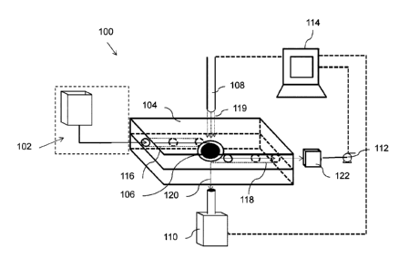
[0017] FIG. 1 is a diagram of a transmission-based three-dimensional
chemical sensing
system;
[0018] FIG. 2 is a side view of the fluidic cartridge of FIG. 1;
[0019] FIG. 3 is a top view of a fluidic cartridge of the chemical
sensor of FIG. 1;
[0020] FIG. 4A is graphical representation an enlarged top view of a
sensor of FIG. 3;
[0021] FIG. 4B is an enlarged view of the graphical representation of
the sensor of FIG. 4A
.. depicting functionalized metal nanoparticles immobilized in pores;
[0022] FIG. 40 is an enlarged side view of a pore of FIG. 4A depicting
functionalized metal
nanoparticles;
[0023] FIG. 4D is another enlarged view of a pore depicting various
types of functionalized
metal nanoparticles immobilized the pore;
[0024] FIG. 5 is a process flow diagram outlining an example for obtaining
a reading from
the chemical-sensing system of FIG. 1;
[0025] FIG. 6 is a plot showing the resonance peak shift due to binding
of the target
chemical with capture molecules on a sensor;
[0026] FIG. 7 is an overhead view of a fluidic cartridge similar to that
of FIG. 3 comprising
multiple sensors;
[0027] FIG. 8 is scanning electron microscope (SEM) image of 20 nm gold
nanoparticles
immobilized on an anodized aluminum oxide (AAO) membrane with a 200 nm pore
diameter;
[0028] FIG. 9A is a photograph of gold nanoparticles (GNP) immobilized
on a glass slide;
4
Date Recue/Date Received 2022-06-08
[0029] FIG. 9B is a photograph of gold nanoparticles immobilized on an
AAO membrane;
[0030] FIG. 10 is an example plot showing the relationship between
absorbance and
wavelength for an AAO membrane having gold nanoparticles immobilized in its
pores with
respect to a glass slide having gold nanoparticles immobilized on its surface;
[0031] FIG. 11 is an example plot showing resonance peak shift with respect
to target
concentration in a solution;
[0032] FIG. 12 is a process flow diagram of an example process for GNP
immobilization on
the pores of an AAO substrate and subsequent functionalization with capture
molecules and
blocking molecules;
[0033] FIG. 13 is an image of AAO membrane with gold nanoparticles
immobilized thereon
without the use of BSA additive or coating;
[0034] FIG. 14 is a photograph of a clean AAO membrane;
[0035] FIG. 15 is a diagram showing an example method to enhance the
LSPR shift for
small molecule targets.
[0036] FIG. 16 is a graph showing a comparison of reflection and
transmission signal of
AAO membrane with 150nm pores and 50um thick, with 45nm gold shell
nanoparticles
immobilized inside the pores. The LSPR peak is much larger and sharper in the
transmission
measurement.
[0037] FIG. 17 is graph showing transmission of AAO membranes of various
pore size (P, in
nm) and thickness (T, in um). Membranes were measured dry in air.
[0038] FIG. 18 is a graph transmission of AAO membranes of various pore
size (P, in nm)
and thickness (T, in um). Membranes were measured wetted with water in air.
[0039] FIG. 19 is an absorbance spectrum of an AAO membrane containing
nanoparticles
with two different LSPR peak positions, one located at approximately 495nm and
the other at
approximately 580nm.
5
Date Recue/Date Received 2022-06-08
[0040] FIG. 20 shows the response of the T3D sensor to serial injections
of the streptavidin
(SA) protein, from 0.5nM to 20nM. The streptavidin binds to the biotinylated
surface of the
nanoparticles inside the AAO membrane.
[0041] FIG. 21 shows the response of the conventional 2D sensor to
serial injections of the
streptavidin (SA) protein, from 5nM to 80nM. The streptavidin binds to the
biotinylated surface of
the nanoparticles which are immobilized onto a glass surface.
[0042] FIG. 22 shows that BSA prevents nanoparticles from binding to a
PAH treated
surface, on a glass slide
DETAILED DESCRIPTION
[0043] It has now been realized that the long incubation times associated
with existing
LSPR sensors are due, at least in part, to the diffusion time required for
target chemicals in a
fluid to reach capture molecules on the sensor. One method to reduce the
diffusion time of the
chemicals in the fluid is to reduce the diffusion length. It has been realized
that the diffusion
length may be reduced by flowing a higher proportion of the sample a closer
distance to the
capture molecules. Specifically, it has been found that the diffusion time may
be reduced by
flowing a sample fluid through a relatively narrow-size pore having capture
molecules
immobilized on its surface. This causes the mean distance between target
chemicals and
capture molecules in a set volume of sample fluid to be reduced with respect
to flowing the
same volume of sample fluid over a planar surface comprising capture
molecules.
[0044] Referring now to FIG. 1, an example three-dimensional chemical
sensing system
100 is provided. The chemical sensing system 100 comprises a fluidic cartridge
104, an inlet
port 102, a signal processor 114, a light source 108 for LSPR measurement, and
a detector
110. The chemical sensing system 100 may further comprise an outlet reservoir
122 and a fluid
driving element such as a pump 112 or pressure source (not shown).
[0045] The pump 112, the light source 108, and the detector 110, may be
electrically
powered, for example, by a battery, a power outlet, or a combination of both.
The chemical
sensing system 100 may be located within a housing (not shown), for example, a
portable
housing such as a hand-held housing.
6
Date Recue/Date Received 2022-06-08
[0046] Inlet port 102, which is in communication with a fluid inlet 116,
is operable to receive
a fluid sample, for example from a syringe, and feed the fluid sample to the
fluid inlet 116. The
inlet port 102 may further comprise, or be linked to, a filter or mixing
element to filter, pre-treat or
mix a sample fluid.
[0047] The inlet port 102 may vary in form depending on the type of fluid
sample that is
being tested. For example, in the case of a blood sample for biological
diagnostics, a sterile
needle in a lancing device may be employed to obtain the sample similar to a
glucose monitor. It
will be appreciated that the inlet port 102 may comprise various other forms
including Luer taper
fittings, press fittings, or an open reservoir. It will be appreciated that
the inlet port 102 may be
built into the fluidic cartridge 104.
[0048] Referring now to FIGs. 2 and 3, the fluidic cartridge 104
comprises a fluid inlet 116, a
sensor 106, and a fluid outlet 118. The fluid inlet 116 is fluidically
connected to the sensor 106
and thereby, operable to deliver fluid to the sensor 106 to cause one or more
target chemicals in
the sample fluid to bind to capture molecules in the sensor 106, as is further
described herein.
It will be appreciated that the same sample may be transported to two or more
sensors, for
example, in a multiplexed design as demonstrated below with reference to FIG.
7.
[0049] The fluidic cartridge 104 may be disposable or designed for
repeated use. The
fluidic cartridge is composed of a material 129, 130 that is substantially
optically transparent in
the LSPR wavelengths being used. For example, polydimethylsiloxane (PDMS), is
optically
transparent over many LSPR wavelengths. In the example of FIG. 3, the sensor
106 is
sandwiched between two layers of PDMS. Other transparent materials could be
used to form
the fluidic cartridge including, glass, poly(methyl methacrylate) (PMMA),
cyclic-olefin polymer, or
another material through which micro-channels may be formed to produce the
fluid inlet 116 and
the fluid outlet 118.
[0050] The fluid outlet 118 is also fluidically connected to the sensor 106
to receive fluid
from the sensor 106 and allow fluid to egress from the sensor 106. Optionally,
the fluid outlet
118 may deliver, to the outlet reservoir 122, fluid that has passed through
the sensor 106.
Alternatively, the fluidic cartridge 104 may retain the sample.
[0051] Although the fluid inlet 116 and fluid outlet 118 are shown in
the simplest form in FIG.
3, the fluid inlet 116 and fluid outlet 118 may take different routes through
the fluidic cartridge
7
Date Recue/Date Received 2022-06-08
depending on specific requirements such as flow rate and sample volume or the
need for mixing
and pre-treatment steps. The dimensions and path of the fluid inlet 116 and
fluid outlet 118 may
be chosen depending on the desired fluid speed and mixing properties.
[0052] A pump 112, or other fluid driving element, may optionally drive
the fluid from the
inlet fluid channel 116, through the sensor 106 and out of the fluidic
cartridge through the fluid
outlet 118. For example, the fluid driving element may also comprise a
pressure source or a
vacuum source at the outlet port 118. The pump 112 can be controlled by the
signal processor
114 or other controller. The direction of fluid flow can be rapidly and
automatically switched via
software control to move the sample back and forth transversely through a
membrane in the
sensor 106, allowing for prolonged interaction times with a small sample
volume, thereby
potentially increasing the performance of the sensor 106.
[0053] Alternatively, the fluid may be driven through the sensor 106
using, for example,
electro-osmotic pumps, gravity, wicking of a membrane, or be driven by a
syringe or other fluid
source at the inlet port 102.
[0054] The signal processor 114 comprises, or is linked to, a memory, a
processor, and a
user interface which may include a display and an input device such as a touch
screen or
keyboard and mouse. The signal processor 114 may be linked to another input
device, for
example, a barcode scanner, an RFID scanner, or an NFC reader to identify a
fluidic cartridge
comprising an identifier, for example, a barcode, RFID tag, or NFC chip. It
will be appreciated
that other identification methods may be used including, for example, image
analysis or a simple
identification code which may be entered by the user. The identifier may
comprise, or be linked
to fluidic cartridge information such as relevant standard curves, the type of
sensor being used,
manufacturing date, etc.
[0055] The signal processor 114 may, in various examples, comprise a
computer such as a
laptop computer, desktop computer, microcomputer, cloud-based processor, or a
mobile device.
The memory of the signal processor 114 may contain fitting algorithms and
standard curves.
The signal processor 114 may be linked to one or more of the pump 112, light
source 108, or
detector 110 via a wired connection, for example a local area network or USB
connection.
Alternatively, or in addition, signal processor 114 may be linked to one or
more of the pump 112,
light source 108, or detector 110 via a wireless connection such as Bluetooth,
VVi-Fi, or cellular
8
Date Recue/Date Received 2022-06-08
connection. In some embodiments, the signal processor 114 may be located
remotely from the
fluidic cartridge 104.
[0056] The signal processor 114 may control the light source 108 to emit
light 119, 120 into
the sensor 106. An example light source 108 comprises a white light emitting
diode (LED) and
is coupled to a detector 110 comprising a UV-visible spectrometer. White light
sources other
than LEDs such as halogen bulbs and others such as red, green, and blue LEDs
separate or
combined together, may also be used. A light source for the visible range (400-
800nm) could
be a white light source such as a halogen bulb or an LED, a combination of
colored LED light
sources such as red, blue, and green, a single colored LED light source, or a
laser at a specific
wavelength. For operation below the visible range (100-400nm) of the spectrum,
an ultraviolet
(UV) light source such as a UV LED could be used. For operation above the
visible range (800-
2500nm) an infrared (IR) source such as an IR LED could be used.
[0057] The detector 110 may comprise a charge coupled device, a
photodetector, a
spectrometer, a photodiode array, or a combination thereof, to obtain LSPR
light intensity
readings. The detector 110 may comprise a spectrometer or photodetector
designed for parts
of the electromagnetic spectrum outside the visible range, including the
ultraviolet (UV) range,
the near infrared (NI R), or IR range. The detector 110 may comprise a
combination of two
types of detectors, for example, a photodetector and a spectrometer. The
detector 110 is
selected in combination with an appropriate light source 108.
[0058] The light emitted by the light source 108 is transmitted though the
fluidic cartridge
104 and sensor 106 and is received, at least in part, by a detector 110. As
mentioned above,
the fluidic cartridge 104 and sensor 106 must be at least partially
transparent to the LSPR
wavelengths emitted by the light source 108. The detector 110 generates a
transmission signal,
for example a digital transmission signal, based on the light transmitted
through the sensor 106
and provides the signal to the signal processor 114. The signal processor 114
is operable to
produce a spectrograph based on the transmission signal. The signal processor
114 may also
be operable to select an output based on a predetermined transmission signal
or a comparison
between the transmission signal and one or more reference signals or
thresholds. For example,
the signal processor 114 may output the concentration of a target chemical
based on a
transmission signal that is consistent with one or more reference signals, or
exceeds a threshold
of an established reference signal. The signal processor may also be used to
determine
biointeraction analysis parameters between the target and capture molecule,
which may include
9
Date Recue/Date Received 2022-06-08
reaction kinetic information (on and off rates), affinity, and specificity. A
simple photodetector
may be used to measure intensity changes due to the spectra shifts.
[0059] In the example of FIG. 1, the sensor 106 is substantially planar
and is located
laterally between the path of the light source 108 and the detector 110. As a
sample fluid flows
through the sensor, the light source 108 emits light and the spectrometer 110
receives light
continuously, or at predetermined intervals, to monitor changes in the
resonance peak of the
detected light. The light source 108 and detector 110 may be arranged for
maximal illumination
of the sensor and maximal capture of the light transmitted through the
membrane, respectively.
FIG. 1 shows a transmission based LSPR arrangement, wherein the light source
108 shines
through the membrane and to the detector 110.
[0060] It has been found that to address, at least in part, the
sensitivity of LSPR-based
chemical detection, a three-dimensional porous membrane is used as a substrate
for sensor
106. Functionalized metal nanoparticles are immobilized within the pores of
the membrane and
a sample is flowed through the pores. Such a sensor design may be referred to
herein as a
transmission-based three-dimensional sensor (T3D). The three-dimensional
porous membrane
is substantially transparent for the wavelengths of incident light that are
used to obtain a signal.
[0061] Turning to FIGs. 4A and 4B, the sensor 106 comprises a nanoporous
membrane.
Pores 148 of the membrane comprise functionalized metal nanoparticles 150
immobilized on
their surfaces. Specifically, the membrane is characterized by nanopores,
which are channels
10 nm ¨ 1000 nm in diameter and can be up to 200 pm long. For example, the
nanoporous
membrane may comprise pores that are about 25, 50, 100, 150, 200, 250, 300,
350, 400, 450,
and 500 nm in diameter, and about 1, 5,25, 50, 75, 100, 150 and 200pm in
length.
[0062] A sample is flowed through the pores 148 of the membrane, which
have relatively
small diameters. The relatively small pore diameters, for example about 100
nm, limits the
diffusion distance between the chemical targets and their corresponding
capture molecules on
the nanoparticle surface, thereby reducing the mean diffusion time required
for a target
chemical to reach a capture molecule. By reducing the diffusion time, the time
required for the
target-capture molecule system to reach equilibrium may be decreased. The
nanoparticles may
also be bound to the top and bottom surfaces of the nanoporous membrane.
Date Recue/Date Received 2022-06-08
[0063] The working area of a sensor is limited to the width of the
coherent light source,
which may be approximately 1-4 mm. It is therefore impractical to increase the
sensitivity of
sensors of the prior art simply by creating a larger planar (two-dimensional)
sensor. However,
because functionalized nanoparticles are immobilized onto pore surfaces in a
three-dimensional
membrane, the number of nanoparticles over a given sensor area can be
increased with respect
to a similar two-dimensional design. In many cases, the number of
nanoparticles immobilized
within a given sensor area may be increased substantially with respect to a
two-dimensional
design.
[0064] The membrane may be selected to optimize the thickness, pore
size, and pore
periodicity for a particular chemical sensing application. The diameter of the
sensing membrane
can be selected based on cost and performance. In an example, the diameter of
the sensing
membrane may be as small as 1pm or as large as 13mm. The light beam size may
be close to
the diameter of the sensing membrane to maximizing the strength of the
absorbance signal.
The diameter of the sensor may be small to reduce cost.
[0065] Anodized aluminum oxide (AAO) is an example nanoporous membrane
material.
The dimensions of nanopores in an AAO membrane may be controlled when
producing the
AAO material. The material is sufficiently optically transparent at LSPR
wavelengths to allow for
transmission-based spectroscopic measurements to be performed. The
transmission of light
through the membrane is dependent on several factors including pore size and
thickness that
impact the absorbance of the membrane material and the scattering it produces
in the LSPR
wavelengths of interest.
[0066] Although both reflection and transmission measurements are
possible with this
system, is has surprisingly been found that transmission measurements provide
better results
than reflection measurements. In reflectance mode the reflection off of the
top surface of the
membrane is strong, and accounts for the majority of the signal returning to
the detector. This
reflected light carries very little nanoparticle absorbance information with
it as it is only
interacting with the nanoparticles on the top surface of the membrane. In
transmission mode,
the majority of the light passes through the entire thickness of the membrane
before it reaches
the detector, interacting with all of the nanoparticles throughout the entire
thickness of the
membrane. This greatly increases the absorbance component caused by the
nanoparticles,
increasing the signal to noise ratio and thereby increasing sensor
performance. This also
ensures that the measured signal is from target binding sites throughout the
membrane rather
11
Date Recue/Date Received 2022-06-08
than just those at the top surface. It has further been discovered that a
larger proportion of
scattering light reaches the detector in reflection mode versus transmission
mode, increasing
the noise. Also, the AAO pores act as a waveguide, allowing light to propagate
within the pores
and providing enhanced interaction with the nanoparticles, resulting in
unexpectedly high
transmission of light with a large LSPR absorbance component, providing
further enhancements
to the transmission signal which are not obtained when employing reflection
signal. FIG.16
illustrates the dramatic improvement in the LSPR signal when measuring in
transmission versus
reflection.
[0067] However, a transmission system is more difficult to build as the
LSPR wavelengths
must be matched to the AAO properties to allow sufficient light transmission.
Variables such as
pore size, and thickness may be tuned depending on the wavelengths used so as
not to
interfere with the optical signal generated by the immobilized metal
nanoparticles. The
transmission apparatus can be realized by having a light source on one side of
the membrane
and a detector on the other side of the membrane. It is also possible to make
a pseudo-
transmission setup with the light source and detector on the same side of the
membrane, if a
highly reflective surface is placed beneath the membrane. The reflective
surface will cause the
transmitted light to travel back through the membrane and to the detector on
the opposite side,
which is effectively a transmission measurement.
[0068] In FIG. 17 and FIG. 18, the transmission of various AAO membranes
with different
pore sizes and thicknesses have been measured both dry and wet with water. In
general,
smaller pores result in higher transmission compared to larger pores. With
respect to thickness,
thinner membranes (50um) have higher transmission compared to thicker
membranes (100um),
but the impact is not as significant as pore size. To use AAO as a material in
this sensor, the
pore size and thickness should be selected to be as small as possible while
still allowing the
nanoparticles to fit into the pores without blocking them. Now examining the
dry vs wet
membranes, the transmission through wet membranes is improved, due to
refractive index
matching, especially for membranes with larger pore sizes (100 and 150nm). To
get the best
results, measurements should be taken while the membrane is wet. To further
improve the
transmission, a high refractive index solution can be introduced after the
sample has passed
through the sensor. The high refractive index solution will reduce scattering
even further,
improving the signal to noise ratio.
12
Date Recue/Date Received 2022-06-08
[0069] Any optical signal that is generated by the membrane itself can
be removed from
spectroscopic measurement by the signal processor 114 using baseline
correction methods. A
reference measurement of an area of the membrane which is not coated in
nanoparticles may
be taken to subtract out the optical signature of the membrane using the
processor 114, which
may enhance the signal produced by the nanoparticles. Pores of a membrane, for
example an
AAO membrane, can be chemically treated to allow metal nanoparticles 150 to be
associated
with its surface. For example, the chemicals used to treat AAO include, but
are not limited to,
polyelectrolytes such as polyallylamine hydrochloride (PAH) and poly-(succinyl-
sulphonate)
(PSS) that can create a positive or negative electrostatic charge on the AAO
surface. Metal
nanoparticles stabilized in water by charged surface groups can be associated
to the membrane
through electrostatic forces. Other methods to immobilize nanoparticles
include using a silane
based linker, which will covalently bind to the surface of the AAO. For
example, a silane-thiol
molecule could be used, with the silane covalently binding to the AAO pore
wall and the thiol
covalently binding to the nanoparticle. The thiol could be replaced with any
chemical group that
.. associates to the nanoparticle surface, such as an amine group, for
example. The AAO may be
pretreated to generate hydroxyl groups on the surface of the pores to promote
silanization,
through a procedure such as incubation in a 1:1 solution of hydrochloric acid
and methanol for
30 minutes, or any other method that can be used to generate hydroxyl groups
the surface.
[0070] It will be appreciated that various other nanoporous membrane
materials may be
used including various organic and inorganic membranes that are at least
partially transparent
at the LSPR wavelengths of interest. Advantageously, metal nanoparticles may
be associated
directly to these membranes if the membranes contain thiol-, amide-, phospho-
or other
functional groups.
[0071] The membrane material could also be chemically treated with
polyelectrolytes such
as PAH, PSS or other charged polymers to associate the metal nanoparticles to
the surface
through electrostatic interactions. Reaction chemistry such as N-
Hydroxysuccinimide/ethyl(dimethylaminopropyl) carbodiimide (NHS/EDC) coupling
among other
coupling chemistries may also be used to bind the metal nanoparticles to the
surface. It will be
appreciated that there exist other methods of immobilizing functionalized
metal nanoparticles
150 to an optically transparent nanoporous material.
[0072] Turning now to FIG. 40, an example of functionalized metal
nanoparticles 150
functionalized to a pore surface of an AAO membrane is provided. The AAO
membrane 154
13
Date Recue/Date Received 2022-06-08
has a bilayer of charged polyelectrolyte 156 on its surface that facilitates
the association of
charged metal nanoparticles 150 to the walls of the pores.
[0073] The metal nanoparticles shown in the example of FIG. 40 have two
different metal
core shapes, a sphere 160 and a rod 162, which may be used together or
independently. Both
are functionalized with antibodies 164 as the capture molecule with their
distinctive Y-shape and
form a self-assembled monolayer (SAM) on the surface of the nanoparticle.
Blocking
molecules 166 prevent non-specific binding to empty binding sites on the metal
surface. The
capture molecules may also comprise aptamers, polymers, or DNA
[0074] The SAMs may comprise an antibody, aptamer, polymer, DNA or other
capture
molecule that is bound to the nanoparticle 150 surface and capable of
selectively binding to the
chemical of interest. In the case of gold nanoparticles, this binding
typically occurs
spontaneously between the gold surface and thiol groups that are natural to
the capture
molecule or have been chemically added to the capture molecule or nanoparticle
surface.
[0075] To prevent non-specific binding of non-target chemicals, inert
blocking molecules are
used to pacify empty binding locations on the nanoparticle and pore surfaces.
Blocking
molecules for metal nanoparticles may comprise thiolated compounds with inert
end groups that
provide aqueous stability such as carboxyl, methyl, or polyethylene glycol
(PEG). For the pores,
blocking molecules may be silane based compounds with inert end groups such as
carboxyl,
methyl, or PEG. Bovine serum albumin (BSA) is another example of a possible
blocking
molecule that may be used on nanoparticle and pore surfaces. Together, the
capture and
blocking molecules form a SAM that imparts functionality onto the metal
nanoparticle.
[0076] To create a functional sensor 106, the metal nanoparticles are
first immobilized on
the nanoporous membrane surface then the nanoparticle is functionalized with a
SAM.
Alternatively, the particle may first be functionalized with a SAM and the
nanoparticles may be
immobilized on the membrane.
[0077] The size, shape and elemental composition of metal particles
affect the location and
intensity of the LSPR absorbance peak in the electromagnetic spectrum. As
such, the size,
shape, and composition of nanoparticles are selected to allow for a
measureable transmission
signal through the nanoporous membrane. The size and shape of the
nanoparticles are also
selected to avoid physical clogging of the pores of the selected sensor
membrane.
14
Date Recue/Date Received 2022-06-08
[0078] Various metal nanoparticles have different bulk refractive index
sensitivities and
electromagnetic decay lengths, which may be tuned to produce the optimal LSPR
sensor
response for a given capture-target system. Decay length and sensitivity are
typically not
independent parameters, and both can be tuned with the size, shape, and
composition of the
nanoparticle. For example, increasing the size of a spherical nanoparticle
increases both the
sensitivity and the decay length. Other nanoparticle shapes may have different
trends with
respect to size, sensitivity, and decay length. Preferably, a nanoparticle
will have the highest
sensitivity with a decay length that is similar to the thickness of the
capture molecule-target
complex. For example, if the total size of the capture molecule-target complex
is 8nm, the
optimal decay length would be near 8nm. As such, the size, shape, and
composition of the
nanoparticle can be tuned based on the sensitivity and decay length parameters
for a particular
capture molecule-target complex. This also must correspond with the necessary
optical
properties to allow sufficient transmission of the LSPR signal through the
membrane.
[0079] Therefore, the nanoparticles can be selected depending on the one
or more specific
chemical target being investigated by the sensor 106.
[0080] Compositions of metal nanoparticles that can be used for LSPR
include gold, silver,
platinum, gold coated silver, silver coated gold, combinations of these
metals, and others. The
nanoparticles may also be metal-coated non-metal nanoparticles. The shape of
the
nanoparticles used can also vary. Useful nanoparticle shapes include but are
not limited to,
rods, stars, urchins, decahedra, hexagons, triangles, shells, prisms,
platelets, spheres, rice,
plates, cubes, cages, stars and bipyramids. The dimensions of the metal
nanoparticles can
range between about 1 nm and 1000 nm with a variety of area to volume ratios.
[0081] Referring now to both of FIGs. 4C and 4D, a combination of two or
more types of
nanoparticles may be immobilized on the surface of a single membrane. Each of
the
nanoparticle types may have been functionalized with a specific capture
molecule. For
example, nanoparticle 150 may be functionalized with capture molecules to
capture a first target
chemical whereas nanoparticle 151 may be functionalized with capture molecules
to capture a
second target chemical.
[0082] With a combination of different nanoparticles (shapes, sizes
and/or metals)
functionalized to capture different targets, antibodies on the spherical
nanoparticle may be
selective to different targets from antibodies on the rod nanoparticle, each
nanoparticle
Date Recue/Date Received 2022-06-08
producing a distinct signal. Therefore, the concentration of each target can
be determined from
a single absorbance spectrum if the spectrum from each of the different
particles does not
overlap to such a degree that deconvolution of the peaks is impossible.
Various particles are
used to allow the detection of multiple targets on a single membrane. FIG. 19
shows two
different particles immobilized in the same membrane, one with a resonance at
approximately
495nm and the other with a resonance at approximately 580nm. The two peaks are
clearly
distinguishable and can be used for generating two independent signals from a
single sensor.
Any combination and any number of nanoparticles with different LSPR peak
positions may be
used to achieve multiple measurements. The incorporation of multiple particles
with different
LSPR peak positions within a membrane allows for a higher density of each
particle to be
present compared to the case if multiple particles were incorporated onto a
planar substrate.
Higher particle densities may offer better sensor performance due to high
signal to noise ratios.
This system may allow detection of multiple targets using a small initial
sample which may be
very advantageous in the case where sample is limited or difficult to obtain.
Detecting multiple
targets in tandem may also speed the time to obtaining results.
[0083] Various particles could also be attached to distinct areas of the
same membrane
using a tool similar to a protein spotter or various microchannels to allow
geometrical separation
of the particles. This is advantageous if using a photodetector rather than a
spectrometer, or if
cross reactions may occur between particles due to their chemically modified
surfaces. The use
of different particles on a single membrane can also facilitate the use of a
control sensor to
compensate for errors induced by non-specific binding, temperature change,
bulk refractive
index change, or other factors. This could be achieved by functionalizing one
nanoparticle with
a capture molecule and blocking molecule, and functionalizing a second
particle with only a
blocking molecule similar to the blocking molecules used in the first
nanoparticle. Any peak
changes that are detected from the second nanoparticle are erroneous, and as
such, the
second nanoparticle acts as a control. The different nanoparticles may be
spectrally distinct or
geometrically distinct.
[0084] Nanoparticles are immobilized onto the membrane walls through
contact between
the walls and a colloidal nanoparticle mixture. To permeate the nanoparticles
throughout pores
of the membrane, a variety of techniques may be employed. The nanoparticles
may be
physically pumped through the membrane, they can be driven into the membrane
through the
use of electrical potential, and they could enter by diffusion among, other
methods.
16
Date Recue/Date Received 2022-06-08
[0085] The metal nanoparticles must remain relatively dispersed while
immobilized in the
pores, as extensive aggregation will affect the quality of the measurements
due to peak
broadening or pore clogging. It has been discovered that in order to improve
the penetration
and dispersion of the nanoparticles throughout the membrane pores, surface
stabilizing
.. additives can be included in the colloidal nanoparticle mixture.
Alternatively, or in addition, the
additives may be applied before the nanoparticles are immobilized on the
membrane by
pumping or incubating the additives through the membrane before the colloidal
nanoparticle
mixture is applied. It has been discovered that surface blocking additives are
especially
important in order to immobilize large nanoparticles into small pores. For
example, AAO
membranes with 100nm pores were treated with PSS then PAH to render the
surface positively
charged. Gold nanoparticles 20nm in diameter and coated in negatively charged
citrate were
pumped through the PSS/PAH coated membrane. No gold particles were observed to
bind to
the AAO pores. Upon addition of 0.01% BSA to the 20nm gold colloidal solution
and pumping
through the membrane, gold nanoparticle binding was observed on the AAO pores.
It is
hypothesized that the zwitterionic nature of BSA acts to help stabilize the
nanoparticles,
possibly by screening the charges at the top surface of the AAO membrane,
allowing the
nanoparticles to more easily enter and travel through the membrane. In another
experiment, a
PSS/PAH modified 150nm AAO membrane was dipped into a 1% BSA solution and
rinsed with
water. A 20nm gold colloidal solution, without any BSA additive, was pumped
through the
membrane, and nanoparticle binding was observed. Binding was observed in the
pores at a
high density, with a low density of binding on the membrane surface.
Increasing the BSA dip
concentration to 10% caused a reduction in the binding density of the
nanoparticles within the
pores, with almost no nanoparticles present on the surface of the membrane. A
control
experiment was done on glass with a coating of PAH. BSA at 0.01% was incubated
in a small
.. droplet on the surface of the glass and rinsed away. A 20nm gold colloid
solution was then
applied to the glass. Nanoparticle binding was observed to occur in areas in
which there was no
BSA, demonstrating that BSA may prevent the nanoparticles from binding to the
PAH treated
surface. This is shown in Figure 22. A final experiment was performed with a
PSS/PAH modified
150nm membrane dipped in 1% BSA. 0.001% BSA was added to the gold colloid
solution and
pumped through the membrane. Binding was observed in the pores and on the
surface but at a
lower density than without the BSA additive. BSA, acting as a surface blocker,
can be used to
reduce membrane surface binding, helping to prevent agglomeration. BSA can
also act as a
stabilizer, allowing larger particles to more easily enter smaller pores. As
we have
demonstrated, smaller pore sizes are more transparent and so may be preferable
to use. Also,
17
Date Recue/Date Received 2022-06-08
larger nanoparticles have higher sensitivities, so they may also be preferable
to use. So the use
of surface stabilizing additives may allow a better sensor to be fabricated.
Other possible
additives include various inorganic salts, various surfactants such as Tween
20 or Triton X, or
PEG, along with a variety of other potential molecules and combinations. It is
also assumed that
these additives will have similar effects when other nanoparticle binding
methods are used other
than charge interactions based on PSS/PAH, such as covalent methods using
thiol chemistry as
described previously.
[0086] The nanoparticles may be functionalized with capture molecules
before or after they
are immobilized on the pore walls. If they are immobilized prior to being
functionalized, they can
be immobilized on the pore walls through electrostatic interactions or using
functional groups
bound to the membrane that would be capable of binding to the nanoparticles,
for example, in
the case of a gold surface of a nanoparticle a thiolated polymer may be used.
If the
nanoparticles are functionalized prior to being immobilized, as they may be in
the case of a
multiplexed sensor, for example, a functional group would be used.
[0087] Alternatively, nanoparticles 150 and 151 may be functionalized to
capture the same
target chemical and any difference in signal could be used to generate a
baseline signal,
thereby removing uncertainty in the signal.
[0088] Turning to FIG. 5, an example process for sampling a fluid is
provided. In step 184,
a sample fluid enters the inlet port 102. In 186, the pump 112 drives the
sample into the fluidic
cartridge 104 and the sample flows through the sensor 106 in step 188. In step
190, the light
source 108 emits light onto the sensor 106 in the fluidic cartridge 104, which
causes LSPR
interactions in 192. In 194, the detector receives light transmitted though
the sensor 106,
measures the light intensity and wavelength in 196 and generates a
transmission signal in 197.
In 198, the signal processor 114 determines whether a shift in the resonant
peak is observed
and in 199, detects the presence and/or determines the concentration of the
target chemical in
the sample based on the resonant peak shift, as described above.
[0089] Referring now to FIG. 6, the change in peak position 180, 182 or
intensity induced by
binding of the target chemical to the capture probe can be monitored in real
time or by
comparing the peak position 180 prior to the target chemicals binding to the
capture molecules
in the sensor 106 with the peak position 182 after the target chemical has
bound to the capture
molecules in the sensor 106. This binding may be referred to as a binding
event.
18
Date Recue/Date Received 2022-06-08
[0090] The change in peak position can be compared to data obtained from
a standard
curve of peak change vs. concentration by the signal processor 114, and using
a fitting
algorithm the presence of a target chemical in a sample may be detected, and
the concentration
of the target in the sample may be determined, by the signal processor 114.
Alternatively, or in
addition, the rate of change (the slope) of the signal may be used to more
rapidly determine the
concentration by comparing the calculated slope to data obtained from a
standard curve of
slope vs. concentration. Various data processing mechanisms may be employed by
the signal
processor 114 during data collection to improve the signal to noise ratio of
the optical signal,
such as smoothing and averaging functions. High speed acquisition is used to
facilitate real time
averaging and smoothing. Various peak fitting algorithms may be employed that
offer a high
level of stability in the peak position.
[0091] An indication of whether a target chemical was detected may be
output by the signal
processor 114. The indication may be output, by way of example, on a display,
as an indicator
light, as a warning signal, or as an electronic message. Alternatively, or in
addition to the
indication, the concentration of the target chemical may be output by the
signal processor 114
or on a display linked to the signal processor such as the display of a
cellular phone. Results of
assays can be stored in built in memory on the device, on peripheral devices,
or stored in a
cloud-based server.
[0092] The spectrometric optical measurements taken through the membrane
expose the
.. surface area of the membrane and all of the particles contained therein,
and hence, the sensor
may be referred to as a three-dimensional sensor. A single sensor 106 may be
adapted to
detect multiple targets within a single fluid sample through the inclusion of
multiple membranes
or functionalized particles with different shapes, sizes or metals.
[0093] A multiplexed design for the fluidic cartridge 140 is provided in
FIG. 7. The inlet 116
is split and leads through several independent sensors 106, which are
fluidically connected to
the outlet 118. The sensors 106 are oriented to allow light transmission and
spectrometric
measurement. A multiplexed design can also be achieved using several fluidic
microchannels
coming from a single channel that carry the sample through different areas of
the same
membrane that have been functionalized for the same or different targets.
[0094] Multiplexed measurements can be taken with multiple light sources
and
spectrometers (or other light measurement mechanisms such as a photodetector),
a multiplexed
19
Date Recue/Date Received 2022-06-08
spectrometer such as a hyperspectral imager, or by moving the chip to align
each sensor under
a single light source using the chemical sensing system 100 shown in FIG. 1.
The flow through
a multiplexed fluidic cartridge may be controlled by a single pump 112,
however, designs that
employ multiple pumps or other components to facilitate flow may also be used.
[0095] FIG. 8 is a scanning electron microscope (SEM) image of 20 nm gold
nanoparticles
172 immobilized in the pores 170 of a 200 nm pore size AAO membrane. This
figure
demonstrates that the metal nanoparticles 172 may be immobilized throughout
the pores 170 in
a substantially dispersed manner. An agglomeration 174 is also shown on the
surface of the
pore 170 which may result if surface stabilizing additives are not employed.
[0096] FIGs 9A and 9B, 10, 11, 16, 20, and 21 illustrate advantages of an
example three-
dimensional AAO membrane in comparison with conventional two-dimensional
sensors. The
glass slide of FIG. 9A comprises gold nanoparticles immobilized onto its
surface. In contrast,
the membrane of FIG. 9B is an AAO membrane comprising gold nanoparticles
immobilized on
the surface throughout its pores. The red appearance of both images is due to
the LSPR of the
immobilized gold nanoparticles. The AAO membrane appears as a very dark red in
comparison
to the glass slide due to the larger number of immobilized gold particles per
given unit of sensor
surface area. Therefore, for each given unit of surface area, there is a
stronger LSPR
interaction due to the greater number of immobilized nanoparticles.
[0097] FIG. 10 is a chart of the absorbance spectrums of the glass slide
with respect to the
AAO membrane of FIGs 9A and 9B, respectively, taken in absorbance mode. The
absorbance
peak of the AAO membrane is approximately 6 times larger than the glass slide
absorbance
peak, which demonstrates that an AAO membrane may accommodate a higher number
of gold
nanoparticles, and therefore, may accommodate a higher number of accessible
capture
molecules with respect to a planar glass slide.
[0098] FIG. 11 is a plot showing the simulated response of a sensor based
on a glass slide
(the conventional LSPR design) with respect to a sensor based on an AAO
membrane, which
may also be referred to herein as a transmissive three-dimensional (T3D) LSPR
design (New
T3D LSPR Design). Although not directly visible from the plot of FIG. 11, the
detection limit of
the sensor comprising the AAO membrane at (2pg/m1) has been predicted by
computer
simulations to be improved by approximately 1000 times when compared with the
2D
arrangement (2000pg/m1).
Date Recue/Date Received 2022-06-08
[0099] FIG. 12 is an example procedure for immobilizing gold
nanoparticles on an AAO
membrane. FIG. 13 shows the effect of immobilizing gold nanoparticles on a 13
mm AAO
membrane without the use of surface stabilizing additives to prevent
agglomeration.
Agglomeration is particularly apparent on the outer surfaces of the membrane
surface, i.e., the
.. portions of the membrane between the pores. The gold nanoparticles
agglomerate on the
exterior surface of the membrane. The surface agglomeration makes the membrane
ineffective.
For reference, FIG. 14 is an example AAO membrane that has not been
immobilized with gold
nanoparticles. FIG. 14 may be compared with FIG. 9B to observe the differences
between a
clean AAO membrane and an AAO membrane with gold nanoparticles properly
immobilized
according to the procedure provided herein.
[00100] Referring to FIG. 15, a method to enhance the LSPR shift for
small molecule targets
is outlined. For small molecular weight chemical target detection, for example
ions or
hormones, chemicals, DNA, RNA, aptamers or polymers 202 may not be effective
without
modification. However, a heavy molecule 204, such as a globular inert protein
(bovine serum
albumin), an inert heavy chained polymer or a surface inactivated small
nanoparticle could be
bound to the free end of the capture molecule 202 which may comprise, for
example, an
aptamer or capture polymer. When the target 203 comes into contact with the
capture molecule
202, the capture molecule changes its conformation at 205, thereby bringing
the heavy
molecule 207 closer to the immobilized nanoparticle 201 surface. This enhances
the LSPR
effect that will be detectable by the spectrometer and allow for the detection
of small chemical
targets.
[00101] Another method of enhancing the LSPR signal for the detection of very
small targets
or targets at very low concentrations is the inclusion a signal-enhancing
molecule that can be
pumped into the mixture after target binding has taken place or mixed with the
target prior to
target binding. This is similar to the sandwich assay concept used in ELISA,
where an
immobilized antibody captures the target of interest and then a second
molecular dye-labelled
antibody is added to bind to the now immobilized target and signal its
detection through
fluorimetry. To adapt this concept to the sensor 106 as described herein, the
sample fluid is
pumped through the sensor and capture molecules on the nanoparticles bind the
target
chemicals. A secondary mixture of solution-based molecular dye labelled
capture molecules
are pumped through the sensor and bind to the captured targets. An energy
transfer process
occurs to enhance the LSPR effect due to the dye molecules being brought into
close proximity
21
Date Recue/Date Received 2022-06-08
with the metal nanoparticles. This may produce a larger absorption peak shift
to be measured
by the detector 110.
[00102] The incorporation of shift enhancers can also be used to improve the
detection of
chemicals. For example, a secondary capture molecule could be introduced to
bind to the target
after it has bound to the nanoparticle surface through the primary capture
molecule. This would
enhance the LSPR shift due to the enhanced localized change in the refractive
index due to the
additional mass. Another alternative to potentially enhance the shift of the
resonance peak is
the addition of other enhancer entities, such as free-floating polymer or
metal nanoparticles
functionalized with secondary capture molecules, after the target chemicals
are bound to the
capture molecules in the sensor 106. Such entities would be made of materials
with a high
refractive index and molecular weight to allow a significant enhancement of
the signal shift when
they bind to the target. Metal particle enhancer entities could be made of an
LSPR generating
material to further enhance the resonance shift by resonance coupling. The
entities could also
be a magnetic material such as iron oxide, which has a very high refractive
index and molecular
weight. They could also be metal coated polymer particles, polymer coated
metals particles,
polymers particles, or metal particles.
[00103] Another method to enhance the performance of the LSPR sensor is to
choose
nanoparticle and pore sizes that cause the particles to be close enough
together so that their
three dimensional electromagnetic fields overlap. When this occurs, a single
binding event on
the surface of one nanoparticle will result in changes to its own electric
field in addition to
changes in the electromagnetic fields of those neighbouring nanoparticles
close enough to have
their fields overlap. This will result in a larger response from a single
binding event, increasing
the signal change.
[00104] An example of the improvements offered by the T3D design versus the
traditional 2D
design is shown by comparison of FIG. 20 and FIG. 21. FIGURE 20 shows serial
additions of
the protein streptavidin into an AAO membrane with 150nm pores and 50pm thick.
Nanoparticles are immobilized in the pores and have an LSPR peak around 627nm.
The
nanoparticles are functionalized with a biotin capture layer, with biotin
being chemically bound to
the nanoparticles via a thiol bond. The non-specific sites are blocked with
PEG. The sensor
shows a response at 0.5nM streptavidin up to 20nM in this case. 200pL of
streptavidin is
introduced at 20pL/min. The detection limit is approximately 0.5nM for this
system. FIG. 6X
shows serial additions of streptavidin over a glass substrate with the same
nanoparticles
22
Date Recue/Date Received 2022-06-08
immobilized on the surface of the glass. Biotin is attached to the surface of
the nanoparticles
using the same chemistry as the T3D sensor. Streptavidin is injected serially
at 20plimin in
volumes of 200pL. The sensor shows a response at 5.0nM up to 80nM in this
case. The
detection limit is approximately 5.0nM. This demonstrates that the T3D sensor
exhibits a 10X
improvement in detection limit over the traditional 2D sensor, in this example
case.
[00105] Example 1:Test Protocol for Fibronectin LSPR Sensor Testing
Preparation Steps
Buffer Preparation ("Tris")
Buffer: 20 mM Tris, 100 mM NaCI, 0.005% Tween20, in nuclease free water, pH
7.4
1. Autoclave glassware and pipette tips (200 pL and 1000 pL) to ensure
nuclease free
conditions.
2. For a 500 mL mixture weigh: 1.21 g Tris, 2.92 g NaCI and 0.025 g Tween 20
dissolve
into 500 mL of dH20 then measure the pH of the solution. Adjust the pH up or
down to
7.4 using a NaOH or HCI solution.
3. Store in a sealed container at 4 C.
Fibronectin Protein Preparation
Stock: 0.5mg/m1 already dissolved in buffer
1. Aliquot into 54 pL volumes at 0.5mg/m1 in Tris Buffer (Aliquot
concentration = 1.14pM)
2. Stored in -20 C Freezer
BSA Preparation
1. Take 10.5mg BSA, add to 2m1 vial, add 1m1 Tris buffer. This gives a 1% BSA
solution.
For a 0.1% solution add 70uL of 1% BSA to 630uL Tris buffer.
2. Store in a sealed container at 4 C.
Fibronectin Aptamer Preparation
Stock: 0.2um01 lypholized aptamer powder
1. Add 2m1 of Tris buffer to dry aptamer (makes 0.1mM stock concentration).
2. Aliquot into 50uL vials at 0.1mM.
3. Stored at -20 C.
Functionalization and Testing Procedure
1. Thaw one aptamer aliquot to room temperature. Add 430pL of Tris buffer to
the aptamer
aliquot to obtain a final concentration of 10pM (once the 20 pL of TCEP is
added in the
next step). Vortex for 30 seconds to mix.
23
Date Recue/Date Received 2022-06-08
2. Make 2 ml of 40mM stock TCEP by adding 20mg TCEP to 2m1 of Tris buffer and
mixing
well. From this stock add 20 pL to the aptamer vial to give a final volume of
500 pL.
Vortex for 30. Leave at room temperature for 2 hours.
3. Fill up a 20 ml beaker with water and place on the hot plate set at 90 C.
Place the
aptamer vial into the water bath for 3.5 minutes. Allow the aptamer to cool to
room
temperature.
4. Connect the T3D sensor and flow cell to the flow injection analysis system
which
consists of a 6 port injection valve, sample loop, and syringe pump. Set the
pump speed
to pump Tris buffer at 0.01m1/min. Load 500 pL of aptamer solution into a 1 mL
syringe
ensuring no bubbles are present. Load the aptamer into a 500 pL sample loop
and inject
the solution via the injection valve. The interaction time between the gold
nanoparticles
and the aptamer will be approximately 50 minutes.
5. Once the solution has been fully pumped through the flow cell, pump fresh
Tris buffer
through the system for 30 minutes.
6. Turn the injection valve back to "load", and rinse out the sample loop
using Tris buffer.
Use a 1m1 syringe and load 500 pL of 0.1% BSA into the sample loop. Inject at
a
pumping speed of 0.01 ml/min.
7. Once the solution has been fully pumped through the flow cell (after 50
minutes hour),
pump fresh buffer through the system for 30 minutes.
8. Disconnect the flow cell from the flow injection analysis system and remove
the T3D
sensor. Dry the sensor gently with nitrogen gas. If storing, store in a sealed
container
back filled with nitrogen.
9. Load the dry T3D sensor into the testing flow cell. Insert the flow cell
into the reader unit
and fill the cell with Tris buffer. Begin acquiring optical transmission
spectra and begin
tracking the LSPR peak position using the software. Acquire a baseline peak
position in
Tris buffer.
10. Add 550pL of Tris buffer to a Fibronectin protein aliquot. This gives a
Fibronectin
concentration of 0.1 pM. Use a 1m1 syringe and load 200 pL of the Fibronectin
sample
into the inlet port of the flow cell. Activate the pump at 0.01 ml/min. The
protein sample
will pump through the membrane interact with the aptamer functionalized
surface for 20
minutes.
24
Date Recue/Date Received 2022-06-08
11. Once the solution has been fully pumped through, acquire another peak
baseline in Tris.
Find the difference between the peak baseline from before and after the test.
Using this
value and the standard curve of concentration vs. peak shift, find the
experimentally
determine concentration of Fibronectin in the sample.
12. Dispose of the sensor and flow cell.
[00106] Example 2: Test Protocol for Streptavidin LSPR Sensor Testing
Functionalization and Testing Procedure
[00107] Pump 10mM 11-MUA (11-mercaptoundecanoic acid) and 1-0T (1-octanethiol)
mixture (in a 3:1 ratio) in ethanol through the flow cell for 60 minutes at
0.01m1/min.
[00108] Pump 13mM PBS (phosphate buffered saline) through the flow cell for 30
minutes at
0.1m1/min.
[00109] Pump a 5mM EDC/NHS solution through the flow cell for 60 minutes at
0.01m1/min.
[00110] Rinse with PBS for 30 minutes at 0.1m1/min.
[00111] Pump 1mM of Amine-PEG3-Biotin in 13mM PBS at pH 6.5 at 0.01m1/min for
50
minutes. (PEG ¨ polyethylene glycol)
[00112] Rinse with 13mM PBS at 0.1m1/min for 30 minutes.
[00113] Pump 500pL of 25pM low molecular weight PEG at 0.01m1/min.
[00114] Pump 13mM PBS through the flow cell for 30 minutes at 0.1m1/min.
[00115] Disconnect the flow cell from the flow injection analysis system
and remove the T3D
sensor. Dry the sensor gently with nitrogen gas. If storing, store in a sealed
container back filled
with nitrogen.
[00116] Load the dry T3D sensor into the testing flow cell. Insert the
flow cell into the reader
unit and fill the cell with PBS buffer. Begin acquiring optical transmission
spectra and begin
tracking the LSPR peak position using the software. Acquire a baseline peak
position in buffer.
Date Recue/Date Received 2022-06-08
[00117] Load 200pL of the streptavidin sample at 10nM in PBS into the
inlet port of the flow
cell. Activate the pump at 0.01 ml/min. The protein sample will pump through
the membrane
interact with the functionalized surface for 20 minutes.
[00118] Once the solution has been fully pumped through, acquire another
peak baseline in
buffer. Find the difference between the peak baseline from before and after
the test. Using this
value and the standard curve of concentration vs. peak shift, find the
experimentally determine
concentration of streptavidin in the sample.
[00119] Dispose of the sensor and flow cell.
[00120] Example 3: Immobilization of Gold Nanoparticles into AAO Membrane
Pores
Procedure
[00121] Take AAO membrane from storage, inspect for cracks, chips or weak
points
[00122] Rinse AAO membrane with deionized H20 (dH20)
[00123] Load AAO membrane into flow cell and attach syringe connected to
syringe pump
[00124] Pump 3 mL of dH20 through the membrane at 0.1 mlimin
[00125] Pump 2 mL of 2 pM poly-(succinyl-sulphonate)/ 0.4 pM CaCl2 solution
at pH 3.1
through the membrane at 0.1 mlimin
[00126] Pump 6 mL of dH20 through the membrane at 0.1 mlimin
[00127] Pump 2 mL of 2 pM polyallylamine hydrochloride/ 0.4 pM CaCl2
solution at pH 3.1
through the membrane at 0.1 mL/min
[00128] Pump 6 mL of dH20 through the membrane at 0.1 mL/min
[00129] Remove membrane from flow cell, examine for cracks and rinse surface
with dH20
[00130] Pipette 0.01% w/v bovine serum albumin solution onto the surface of
the AAO
membrane, leave for 1 minute then rinse with dH20
26
Date Recue/Date Received 2022-06-08
[00131] Load AAO membrane into flow cell in reverse orientation as
previous and attach
syringe connected to syringe pump
[00132] Pump 3 mL of dH20 through the membrane at 0.1 mL/min
[00133] Pump 10 mL of 0.1 nM gold nanoparticle mixture through the membrane at
0.05
mlimin
[00134] Pump 6 mL of dH20 through the membrane at 0.1 mL/min
[00135] If imaging: remove membrane from the flow cell, rinse with dH20
[00136] If testing: keep in flow cell and proceed to capture probe
functionalization steps.
Connect flow cell to flow injection analysis system.
[00137] Although examples are provided with reference to LSPR detection
techniques, the
above may also be adapted to other spectrometric detection assays, for
example, ELISA.
[00138] Although the above has been described with reference to certain
specific example
embodiments, various modifications thereof will be apparent to those skilled
in the art without
departing from the scope of the claims appended hereto.
27
Date Recue/Date Received 2022-06-08