Note: Descriptions are shown in the official language in which they were submitted.
CA 03161964 2022-05-17
WO 2021/102128 PCT/US2020/061264
PNEUMATIC OR HYDRAULIC POWERED TISSUE CLOSURE DEVICES
CROSS-REFERENCE To RELATED APPLICATIONS
[0001] The present application claims the benefit of priority under 35
U.S.C. 119 to U.S.
Provisional Patent Application No. 62/937,980, filed November 20, 2019, which
application is
incorporated herein by reference in its entirety for all purposes.
FIELD
[0002] The present disclosure relates to the treatment of tissue defects,
and, more particularly,
to systems, devices, and methods for assisted tissue closure.
BACKGROUND
[0003] In some medical procedures, it is beneficial to fixedly connect a
portion of tissue to
another portion of tissue, such as to hold together a wound or damaged tissue.
For example, one
or more sutures or staples may be used to connect tissue portions. Often, an
assembly, including
staples, or a needle, and a suture coupled to the needle, is used to secure
tissue together.
[0004] Furthermore, both suturing and stapling closure methods are
desirable for tissue
resection or various bariatric procedures. One drawback with suturing and
stapling systems is the
challenge of deliverable force to the distal end of the system. It is with
these considerations in
mind that a variety of advantageous medical outcomes may be realized by the
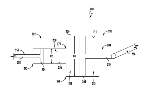
devices, systems,
and methods of the present disclosure.
SUMMARY
[0005] In one or more embodiments, a medical device may include an
endoscopic device
operable to close an opening in a target tissue and an actuator operable with
the endoscopic device,
the actuator including a piston within a chamber. The piston may include a
piston head engaged
with an interior surface of the chamber, and a piston rod coupled to a tissue
engagement
component of the endoscopic device, wherein pressure from a fluid within the
chamber actuates
the tissue engagement component. In some embodiments, the fluid is a gas or a
liquid. In some
embodiments, the actuator further comprises a valve operable to permit the
fluid to enter the
chamber, wherein the valve controls a flow of the fluid through an inlet
conduit and an outlet
conduit, and wherein the inlet conduit and the outlet conduit are fluidly
connected with the
chamber. In some embodiments, the tissue engagement component is a needle
passer or an
endoscopic stapler head. In some embodiments, the endoscopic device is a
suturing device, the
suturing device including an elongate member, and a suture arm at one end of
the elongate
member, wherein the needle passer is operable to deliver a needle between the
elongate member
1
CA 03161964 2022-05-17
WO 2021/102128 PCT/US2020/061264
and a distal end of the suture arm for suturing the target tissue. In some
embodiments, the piston
rod is disposed within an interior of the elongate member, and wherein the
piston rod is coupled
to the needle passer. In some embodiments, the fluid within the chamber
impacts the piston head
to actuate the piston rod and the needle passer in an axial direction. In some
embodiments, the
endoscopic stapler head may include a first jaw opposite a second jaw, wherein
the piston rod is
coupled to the first jaw, and a staple cartridge along the second jaw, wherein
movement of the
piston rod causes the first jaw to make or break contact with the staple
cartridge. In some
embodiments, the actuator may further include a second piston within a second
chamber, wherein
the second chamber is fluidly connected with the first chamber, and wherein a
first diameter of
the first chamber is larger than a second diameter of the second chamber. In
some embodiments,
the second piston may include a second piston head engaged with an interior
surface of the second
chamber, and a second piston rod extending from the second piston head,
wherein the second
piston head faces the piston head.
[0006] In one or more embodiments, a system may include an endoscope and
a device
including an actuator operable to close an opening in a target tissue. The
actuator may include a
piston within a chamber, the piston including a piston head engaged with an
interior surface of
the chamber, and a piston rod coupled to a tissue engagement component of the
endoscopic
device, wherein pressure from a fluid within the chamber actuates the piston
rod and the tissue
engagement component. In some embodiments, the actuator may further include a
valve operable
to permit the fluid to enter the chamber, wherein the valve controls a flow of
the fluid through an
inlet conduit and an outlet conduit fluidly connected with the chamber. In
some embodiments,
the device may further include a suturing needle passer or an endoscopic
stapler head. In some
embodiments, the device may be a suturing device, wherein the suturing device
includes an
elongate member and a suture arm at one end of the elongate member, wherein
the tissue
engagement component is a needle passer operable to deliver a needle between
the elongate
member and a distal end of the suture arm for suturing the target tissue. In
some embodiments,
the device may be a stapler head, the stapler head including a first jaw
opposite a second jaw,
wherein the piston rod is coupled to the first jaw, and a staple cartridge
along the second jaw,
wherein movement of the piston rod causes the first jaw to make or break
contact with the staple
cartridge. In some embodiments, the actuator may further include a second
piston within a second
chamber, wherein the second chamber is fluidly connected with the first
chamber, and wherein a
first diameter of the piston head is larger than a second diameter of a second
piston head of the
second piston.
2
CA 03161964 2022-05-17
WO 2021/102128 PCT/US2020/061264
[0007] In one or more embodiments, a method may include inserting an
endoscopic medical
device within a patient, the endoscopic medical device including an endoscopic
device operable
to engage a target tissue, and an actuator coupled to the endoscopic device,
the actuator including
a piston within a chamber. The piston may include a piston head engaged with
an interior surface
of the chamber, and a piston rod coupled to a tissue engagement component of
the endoscopic
device. The method may further include controlling an amount of a fluid within
the chamber to
actuate the piston rod and the tissue engagement component relative to the
target tissue, and
engaging the target tissue with the tissue engagement component to close an
opening of the target
tissue. The method may further include engaging the target tissue using a
suturing device, the
suturing device including an elongate member and a suture arm at one end of
the elongate
member, wherein the tissue engagement component is a needle passer containing
a needle. The
method may further include delivering the needle between the elongate member
and a distal end
of the suture arm to close the opening of the target tissue. The method may
further include
engaging the target tissue using a stapler head, the stapler head including a
first jaw opposite a
second jaw, wherein the piston rod is coupled to the first jaw, and actuating
the piston rod to close
the first and second jaws about the target tissue to close the opening of the
target tissue. The
method may further include providing a second piston within a second chamber,
wherein the
second chamber is fluidly connected with the first chamber, and wherein a
first diameter of the
piston head is larger than a second diameter of a second piston head of the
second piston, and
actuating the second piston to increase a pressure of the fluid in the second
chamber and the
chamber.
[0008] Various one or more of the features summarized above, may be
interchanged,
exchanged, combined, or substituted with, or for, other features summarized
above, for use in
connection with the medical systems and methods summarized above, and with
respect to the
embodiments described in greater detail below and embodiments otherwise within
the scope of
the present disclosure.
BRIEF DESCRIPTION OF THE DRAWINGS
[0009] Non-limiting embodiments of the present disclosure are described
by way of example
with reference to the accompanying figures, which are not intended to be drawn
to scale. In the
figures, each identical or nearly identical component illustrated is typically
represented by a single
numeral. For purposes of clarity, not every component is labeled in every
figure, nor is every
component of each embodiment shown where illustration is not necessary to
allow those of
ordinary skill in the art to understand the disclosure. Furthermore, some of
the figures include
cross-sectional views in the form of "slices", or "near-sighted" cross-
sectional views, omitting
3
CA 03161964 2022-05-17
WO 2021/102128 PCT/US2020/061264
certain background lines or features otherwise visible in a "true" cross-
sectional view, for
illustrative clarity. In the figures:
[0010] FIG. 1 is a side cross-sectional view of an actuator of a medical
device according to
embodiments of the present disclosure;
[0011] FIG. 2A is a side view of an endoscopic device in a first state
according to
embodiments of the present disclosure;
[0012] FIG. 2B is a side view of the endoscopic device of FIG. 2A in a
second state according
to embodiments of the present disclosure;
[0013] FIG. 3 is a side cross-sectional view of another actuator for use
with a medical device
according to embodiments of the present disclosure;
[0014] FIG. 4A is a side view of an endoscopic device in a first state
according to
embodiments of the present disclosure;
[0015] FIG. 4B is a side view of the endoscopic device of FIG. 4A in a
second state according
to embodiments of the present disclosure;
[0016] FIG. 5 is a perspective view of an endoscopic stapler head according
to embodiments
of the present disclosure;
[0017] FIG. 6 is a perspective view of an endoscopic stapler head
according to embodiments
of the present disclosure; and
[0018] FIG. 7 is a flow diagram of a method according to embodiments of
the present
disclosure.
DETAILED DESCRIPTION
[0019] The present disclosure is not limited to the particular
embodiments described herein.
The terminology used herein is for the purpose of describing particular
embodiments only, and is
not intended to be limiting beyond the scope of the appended claims. Unless
otherwise defined,
all technical terms used herein have the same meaning as commonly understood
by one of
ordinary skill in the art to which the disclosure belongs.
[0020] One trend in medicine includes moving from laparoscopic and open
surgical
procedures to miniaturized, endoscopic procedures. Endoscopists can perform
ever more
complex procedures noninvasively and under direct visualization. As a result,
there exists a need
for endoscopic medical devices possessing specific built-in treatment
capabilities. Such medical
devices would facilitate both a broad range of procedural interventions more
prevalent in
4
CA 03161964 2022-05-17
WO 2021/102128 PCT/US2020/061264
hospitals, and further lead to the development of significantly more complex
and capable scope
designs.
[0021] Embodiments herein address at least the above deficiencies by
integrating complex
functions into either a single-use scope or reusable endoscope. For example,
the functions
available according to the systems, medical devices, and methods of the
present disclosure may
include one or more of the following: suturing, stapling, cutting,
cauterizing, clip deployment,
injection, tissue manipulation, and more. Furthermore, by using the suture
devices disclosed
herein only a single time, the infection risk can be greatly minimized.
[0022] The disclosure pertains to medical devices, e.g., endoscopes,
gastroscopes,
bronchoscopes, colonoscopes, ureteroscopes, endoscopic stapler heads, and the
like, having
integrated features for acquiring, manipulating, and closing openings in
target tissue. Although
single-use endoscope medical devices are described herein, it is understood
that embodiments of
the present disclosure may be included in reusable medical devices such as
endoscopes as well.
Embodiments herein address at least the above deficiencies by integrating
complex functions into
a single medical device.
[0023] Furthermore, embodiments herein address at least the above
deficiencies of the prior
art, such as the elevated level of force required to actuate a stapler head or
push a needle through
tissue. Prior art systems use either pull wires or the force that an operator
can apply by pushing
and/or pulling on a catheter or similar. These prior art actuation methods can
be unreliable in
tortuous anatomy, translate too small a force to the distal end of the device,
and/or be fatiguing to
the operator. Embodiments of the present disclosure enable higher forces,
which may be required
at a distal end of the medical device, by introducing pneumatics and/or
hydraulics into suturing
or stapling devices. Pneumatics and hydraulics can increase the force applied
at a proximal end
user interface, thus allowing distal end mechanisms to more easily puncture
tissue or similar.
Embodiments included herein describe various configurations that support
either suturing or
stapling-based devices. However, it is recognized that the end use is not so
limited, and the
general ideas and designs may be applied to many similar devices and end
effectors requiring
similar force and motions.
[0024] With reference to FIG. 1, an actuator 102 of a system or
endoscopic medical device
(hereinafter "device") 100 according to embodiments of the disclosure will be
described. As
shown, the actuator 102 may include a piston 104 within a chamber 106, the
piston 104 having a
piston head 108 connected with a piston rod 110. The piston head 108 may
engage an interior
surface 111 of the chamber 106, generally dividing the chamber 106 into a
first portion 113, and
a second portion 115. As shown, the first portion 113 of the chamber 106 may
be fluidly
5
CA 03161964 2022-05-17
WO 2021/102128 PCT/US2020/061264
connected with an inlet conduit 116, while the second portion 115 of the
chamber 106 may be
fluidly connected with an outlet conduit 118. In some embodiments, a pressure
difference
between the first portion 113 and the second portion 115 of the chamber 106
will cause the
piston 104 to move axially (e.g., left or right in the figure) within the
chamber 106.
[0025] The actuator 102 may further include a valve 120 operable to permit
a fluid 122 from
a fluid supply 125 to enter and exit the chamber 106. More specifically, the
valve 120 may control
the flow of the fluid 122 through both the inlet conduit 116 and the outlet
conduit 118. Although
not shown, the valve 120 may be controlled at a handle or user interface of
the device 100. In
some embodiments, the outlet conduit 118 may include one or more relief valves
121, while the
inlet conduit 116 may be connected to the fluid supply 125 for delivering the
fluid 122 to the
chamber 106. Although non-limiting, the fluid 122 may be pressured air or CO2.
[0026] As shown, the piston rod 110 may be coupled to a tissue engagement
component 124.
In non-limiting embodiments, as shown in FIGS. 2A and 2B, the tissue
engagement
component 124 may be a needle passer 126 of an endoscopic device, such as a
suturing
device 131. In some embodiments, the needle passer 126 may contain, or be
coupled with, a
needle 130 operable to engage a target tissue. The device 100 may further
include the suturing
device 131 coupled to the piston rod 110 of the actuator 102. In some
embodiments, the suturing
device 131 may include an elongate member 134 coupled to, or integrally formed
with, a suture
arm 136. The elongate member 134 may be a flexible hollow tube, endoscope,
catheter, etc., and
may include a proximal end 137 opposite a distal end 138. In some embodiments,
the elongate
member 134 may be a flexible material, such as silicone, a thermoplastic
elastomer including
polyamide and polyether backbone blocks, polyurethane, etc., to allow for
scope flexing. In other
embodiments, the elongate member 134 may be a rigid material, such as
polycarbonate,
acrylonitrile butadiene styrene (ABS), etc., to provide a more direct
positioning response.
[0027] In some embodiments, the suture arm 136 may be part of a distal
assembly, including
a body 141 having a proximal section 142 extending from the distal end 138 of
the elongate
member 134. The proximal section 142 and the elongate member 134 may be
integrally
connected such that the body 141 and the elongate member 134 form an
integrated, single use
device. In other embodiments, the distal assembly may be removably coupleable
to the elongate
member 134 of a single use or a reusable device.
[0028] As further shown, the suture arm 136 may extend to an endcap 143,
which is
configured to releasably engage and disengage the needle 130. In some
embodiments, the
needle 130 may be delivered by the needle passer 126 between the distal end
138 of the elongate
member 134 and the endcap 143, which is located at a distal end 145 of the
suture arm 136. The
6
CA 03161964 2022-05-17
WO 2021/102128 PCT/US2020/061264
needle 130 may be connected to a suture (not shown) used for tensioning and
closing an opening
in a target tissue 146 (FIG. 2A) retained within a suture cavity 147 defined
by the suture arm 136.
[0029] With reference to FIGS. 1, 2A, and 2B, operation of the device 100
will be described
in greater detail. In some embodiments, the device 100 may be a pneumatic
device employing a
compressible fluid, such as air, to drive operation of the piston 104.
Initially, the needle
passer 126 and the needle 130 may be recessed in a position adjacent the
proximal section 142 of
the body 141, as demonstrated in FIG. 2A. Once the target tissue 146 is
retained within the suture
cavity 147, e.g., by a tissue grasper (not shown) delivered through a channel
of the body 141, the
valve 120 may be opened to cause an increase in the volume of the fluid 122
within the first
portion 113 of the chamber 106. As the volume of the fluid 122 increases,
increased pressure
upon the piston head 108 causes the piston 104, the needle passer 126, and the
needle 130 to be
biased towards the target tissue 146. The needle 130 may then puncture the
target tissue 146
before being retained within the endcap 143, as shown in FIG. 2B. Depending on
the size and
type of tissue opening, the needle 130 may be passed back and forth between
the proximal
section 142 and distal end 145 of the suture arm 136, the needle 130 including
one or more
pointed tips for puncturing the target tissue 146 with each pass.
[0030] Turning now to FIG. 3, an actuator 202 of an endoscopic medical
device (hereinafter
"device") 200 according to embodiments of the disclosure will be described in
greater detail. As
shown, the actuator 202 may include a first piston 204 within a first chamber
206, the first
piston 204 having a first piston head 208 connected with a first piston rod
210. The first piston
head 208 may extend across an entire interior area of the first chamber 206,
engaging an interior
surface 211 thereof. As shown, the first piston head 208 may generally divide
the first
chamber 206 into a first portion 213 and a second portion 215.
[0031] As shown, the first portion 213 of the first chamber 206 may be
fluidly connected with
a second chamber 250 containing a second piston 252. The second piston 252 may
include a
second piston head 254 engaged with an interior surface 256 of the second
chamber 250, and a
second piston rod 258 extending from the second piston head 254. In some
embodiments, the
second piston rod 258 may extend outside the second chamber 250. As shown, the
second piston
head 254 faces the first piston head 208.
[0032] In some embodiments, a first diameter 'D1 of the first chamber 206
is larger than a
second diameter 'D2' of the second chamber 250, thereby providing a mechanical
advantage, or
force multiplication, during uses of the first and second pistons 204, 252. It
will be appreciated
that the device 200 may be a hydraulic device employing an incompressible
hydraulic liquid such
as oil or water to drive operation of the first and second pistons 204, 252.
During use, a smaller
7
CA 03161964 2022-05-17
WO 2021/102128 PCT/US2020/061264
input force on the second piston 252 results in a greater output force on the
first piston 204,
thereby reducing an initial force required by a user. In some embodiments, the
second piston 252
may be controlled by any variety of actuators (e.g., a lever) located at a
user interface (not shown).
Furthermore, in other embodiments, one or more additional pistons may be
employed to provide
more force multiplication.
[0033] With reference now to FIGS. 3, 4A, and 4B, an endoscopic stapler
head 260 according
to embodiments of the present disclosure will be described. As shown, the
first piston rod 210
may be coupled to the endoscopic stapler head 260. In non-limiting
embodiments, the endoscopic
stapler head 260 includes a first jaw 262 opposite a second jaw 264. The first
jaw 262 and/or the
second jaw 264 may be coupled to the first piston rod 210, for example, via
one or more
mechanical linkages 266 (FIG. 3) for connecting the stapler head 260 to the
actuator 202.
[0034] In some embodiments, the endoscopic stapler head 260 may include a
staple
cartridge 268 along an interior of the second jaw 264, wherein movement of the
first piston
rod 210 causes the first jaw 262, sometimes referred to as an anvil, to engage
or disengage with
the staple cartridge 268. The first and second jaws 262, 264 are pivotally
attached to one another
to clamp a target tissue (not shown) to the staple cartridge 268. More
specifically, the endoscopic
stapler head 260 may be provided in an initial position, as shown in FIG. 4A,
in which the first
piston rod 210 and the mechanical linkage 266 may be drawn back along a first
direction 273
(e.g., towards a handle or user interface). The mechanical linkage 266 may be
connected to a
biasing device (e.g., a slider) 282 having a first section 283 coupled to the
second jaw 264 and a
second section 284 coupled to or extending around the first jaw 262. In some
embodiments, the
second section 284 may include a shroud having an opening for the first jaw
262. As the biasing
device 282 moves relative to the first jaw 262, an inner surface of the second
section 284 may
engage an outer surface 287 of the first jaw 262, thereby forcing the first
jaw 262 to close. In
some embodiments, the first jaw 262 also moves or pivots within the second
section 284 of the
biasing device 282 via a pin 271 of the first jaw 262 that slides within a pin
slot 272 of the biasing
device 282.
[0035] As further shown, the first section 283 of the biasing device 282
may include a
flange 288 engaged with a ledge 290 of the second jaw 264. The biasing device
282 may be
further engaged with the second jaw 264 along a second flange 292. Embodiments
herein are not
limited in this context, however.
[0036] In some embodiments, to open the endoscopic stapler head 260, the
second piston 252
may be biased towards a proximal end 275 of the second chamber 250, causing a
decreased
pressure within the second chamber 250 and the first portion 213 of the first
chamber 206. The
8
CA 03161964 2022-05-17
WO 2021/102128 PCT/US2020/061264
decreased pressure in turn may cause the first piston head 208 to move axially
towards a proximal
end 276 of the first chamber 206. As the first piston head 208 moves towards
the proximal
end 276 of the first chamber 206, so does the first piston rod 210 and the
mechanical linkage 266,
causing the biasing device 282 to move along the first direction 273. Without
the second
section 284 of the biasing device 282 engaged with the outer surface 287 of
the first jaw 262, the
first jaw 262 may be free to pivot away from the second jaw 264, for example,
in response to a
spring force or lever mechanism.
[0037] In other embodiments, the first piston rod 210 and the mechanical
linkage 266 may be
coupled to the first jaw 262. During use, pulling the first piston rod 210
along the first
direction 273 may pull the first jaw 262 within the first section 283 of the
biasing device 282 to
lock the first jaw 262 in a closed position. In some embodiments, a separate
piston rod (not
shown), may be coupled to the biasing device 282 to staple and cut tissue by
the first and second
jaws 262, 264 as the biasing device 282 moves in a second direction 277 (e.g.,
away from a handle
or user interface).
[0038] To close the endoscopic stapler head 260, as depicted in FIG. 4B,
the first piston
rod 210, the mechanical linkage 266, and the biasing device 282 may be biased
in the second
direction 277. To accomplish this, the second piston 252 may be biased towards
a distal end 279
of the second chamber 250, causing an increased pressure within the second
chamber 250 and the
first portion 213 of the first chamber 206. The increased pressure in turn may
cause the first piston
head 208 to move axially towards a distal end 280 of the first chamber 206. As
the first piston
head 208 moves towards the distal end 280 of the first chamber 206, so does
the first piston
rod 210 and the mechanical linkage 266, causing the biasing device 282 to move
in the second
direction 277, thus causing the first jaw 262 to pivot or rotate towards the
second jaw 264.
[0039] As the endoscopic stapler head 260 closes, the first jaw 262 may
deform staples (not
shown) driven up from staple holes in the staple cartridge 268 into a closed
shape. When the
endoscopic stapler head 260 is in a closed position, its cross-sectional area,
as well as the first
piston rod 210, may be suitable for insertion through a small surgical
opening, such as through a
cannula of a trocar. Alternatively, the stapler head 260 may be inserted into
a body opening via
attachment to the end of a scope, or inserted through a working channel of the
scope. In some
embodiments, correct placement and orientation of the endoscopic stapler head
260 is facilitated
by controls on the handle/user interface.
[0040] Although the endoscopic stapler head 260 is shown as being biased
by the
actuator 202, it will be appreciated that the endoscopic stapler head 260
could be coupled to the
actuator 102 shown in FIG. 2 and described herein. For example, the piston rod
110 of the
9
CA 03161964 2022-05-17
WO 2021/102128 PCT/US2020/061264
actuator 102 could be coupled to the mechanical linkage 266, which is used to
control the first
jaw 262. Alternatively, the suturing device 131 shown in FIGS. 2A- and 2B and
described herein,
could be biased by the actuator 202. For example, the first piston rod 210 may
be coupled to the
needle passer 126. Embodiments herein are not limited in this context.
[0041] Turning to FIG. 5, an alternative endoscopic stapler head 360
according to
embodiments of the present disclosure, will be described. As shown, the
endoscopic stapler
head 360 may include a first jaw 362 opposite a second jaw 364, and a biasing
device 382 coupled
to a piston rod 310, which may be the same or similar to the first piston rod
210 and/or mechanical
linkage 266 of FIGS. 4A and 4B for connecting the stapler head 360 to the
actuator 202.
[0042] The biasing device (e.g., a slider) 382 may have a first section 383
coupled to the first
jaw 362 and a second section 384 coupled to the second jaw 364. In some
embodiments, the first
section 383 may include a flange 388 engaged with a protrusion or ledge 389 of
the first jaw 362.
During use, the piston rod 310 causes the biasing device 382 to move between a
distal end 357
and a proximal end 358 of the endoscopic stapler head 360. As the biasing
device 382 traverses
along the first jaw 362 and the second jaw 364, an inner surface of the second
section 384 may
engage an outer surface 387 of the second jaw 364 to bring the first jaw 362
and the second
jaw 364 together.
[0043] Turning to FIG. 6, another alternative endoscopic stapler head 460
according to
embodiments of the present disclosure will be described. As shown, the
endoscopic stapler
.. head 460 may include a first jaw 462 opposite a second jaw 464, and a
biasing device 482 coupled
to a piston rod 410, which may be the same or similar to the first piston rod
210 and/or mechanical
linkage 266 of FIGS. 4A- and 4B for connecting the stapler head 460 to the
actuator 202.
[0044] Although shown disengaged with the first jaw 462 in FIG. 6, the
biasing device (e.g.,
a slider) 482 may have a first section 483 coupleable with the first jaw 462
and a second
section 484 coupled to the second jaw 464. In some embodiments, the first
section 483 may
include a flange 488 operable to slide along a protrusion or ledge 489 of the
first jaw 462. During
use, the piston rod 410 causes the biasing device 482 to move between a distal
end 457 and a
proximal end 458 of the endoscopic stapler head 460. As the biasing device 482
traverses along
the first jaw 462 and the second jaw 464, the first jaw 462 and the second jaw
464 are brought
together to staple and cut tissue.
[0045] In the non-limiting embodiment shown, the stapler head 460 may
further include a
pivot arm 492 coupled between the first jaw 462 and a second piston rod 494.
The second piston
rod 494 may be biased by an actuator, such as the actuator 102 or the actuator
202 described
herein. The pivot arm 492 may be fixed to rotate about a pivot point 496,
wherein movement of
CA 03161964 2022-05-17
WO 2021/102128 PCT/US2020/061264
the second piston rod 494 towards the distal end 457 of the stapler head 460
causes the first
jaw 462 to open, and movement of the second piston rod 494 towards the
proximal end 458 causes
the first jaw 462 to close.
[0046] FIG. 7 is a flow diagram of a method 500 according to embodiments
of the present
disclosure. At block 501, the method 500 may include inserting an endoscopic
medical device
within a patient, the endoscopic medical device including an endoscopic device
operable to
engage a target tissue and an actuator coupled to the endoscopic device, the
actuator including a
piston within a chamber. In some embodiments, the piston may include a piston
head engaged
with an interior surface of the chamber, and a piston rod coupled to a tissue
engagement
.. component of the endoscopic device. In some embodiments, the method may
include providing
a second piston within a second chamber, wherein the second chamber is fluidly
connected with
the first chamber, and wherein a first diameter of the piston head is larger
than a second diameter
of a second piston head of the second piston. The method may further include
actuating the
second piston to increase a pressure of the fluid in the second chamber and
the first chamber.
[0047] At block 503, the method may include controlling an amount of a
fluid within the
chamber to actuate the piston rod and the tissue engagement component relative
to the target
tissue. In some embodiments, a valve may be opened/closed to control the fluid
entering the
chamber. In some embodiments, the actuator may include an inlet conduit and an
outlet conduit
fluidly connected with the chamber, wherein the inlet conduit is positioned
along a first side of
the piston head, and wherein the outlet conduit is positioned along a second
side of the piston
head.
[0048] At block 505, the method 500 may include engaging the target
tissue with the tissue
engagement component to close an opening of the target tissue. In some
embodiments, the
method may include engaging the target tissue using a suturing device, the
suturing device
including an elongate member, and a suture arm at one end of the elongate
member, wherein the
tissue engagement component is a needle passer containing a needle. The method
may further
include delivering the needle between the elongate member and a distal end of
the suture arm to
close the opening of the target tissue. In some embodiments, the method may
include engaging
the target tissue using a stapler head, the stapler head including a first jaw
opposite a second jaw,
wherein the piston rod is coupled to the first jaw. The method may further
include actuating the
piston rod to close the first and second jaws about the target tissue to close
the opening of the
target tissue.
[0049] It will be appreciated that a variety of different materials may
be used in forming the
devices described herein. In some cases, a variety of different metals may be
used. Illustrative
11
CA 03161964 2022-05-17
WO 2021/102128 PCT/US2020/061264
but non-limiting examples of suitable metals include titanium, stainless
steel, magnesium, cobalt
chromium and others. In some embodiments, for example, the devices described
herein may
include any suitable polymeric material, including biocompatible materials
such as polyurethane
or silicone. Other suitable polymers include but are not limited to
polytetrafluoroethylene (PTFE),
ethylene tetrafluoroethylene (ETFE), fluorinated ethylene propylene (FEP),
polyoxymethylene
(POM, for example, DELRIN available from DuPont), polyether block ester,
polyurethane (for
example, Polyurethane 85A), polypropylene (PP), polyvinylchloride (PVC),
polyether-ester (for
example, ARNITEL available from DSM Engineering Plastics), ether or ester
based copolymers
(for example, butylene/poly(alkylene ether) phthalate and/or other polyester
elastomers such as
HYTREL available from DuPont), polyamide (for example, DURETHAN available
from
Bayer or CRISTAMID available from Elf Atochem), elastomeric polyamides,
block
polyamide/ethers, polyether block amide (PEBA, for example available under the
trade name
PEBAX ), ethylene vinyl acetate copolymers (EVA), silicones, polyethylene
(PE), Marlex high-
density polyethylene, Marlex low-density polyethylene, linear low density
polyethylene (for
example REXELL ), polyester, polybutylene terephthalate (PBT), polyethylene
terephthalate
(PET), polytrimethylene terephthalate, polyethylene naphthalate (PEN),
polyetheretherketone
(PEEK), polyimide (PI), polyetherimide (PEI), polyphenylene sulfide (PPS),
polyphenylene
oxide (PPO), poly paraphenylene terephthalamide (for example, KEVLARCI),
polysulfone,
nylon, nylon-12 (such as GRILAMID available from EMS American Grilon),
perfluoro(propyl
vinyl ether) (PFA), ethylene vinyl alcohol, polyolefin, polystyrene, epoxy,
polyvinylidene
chloride (PVdC), poly(styrene-b-isobutylene-b-styrene) (for example, SIBS
and/or SIBS 50A),
polycarbonates, ionomers, biocompatible polymers, other suitable materials, or
mixtures,
combinations, copolymers thereof, polymer/metal composites, and the like.
[0050] Some embodiments may be described using the expression "coupled"
and "connected"
along with their derivatives. These terms are not intended as synonyms for
each other. For
example, some embodiments may be described using the terms "connected" and/or
"coupled" to
indicate that two or more elements are in direct physical or electrical
contact with each other. The
term "coupled," however, may also mean that two or more elements are not in
direct contact with
each other, but yet still co-operate or interact with each other.
[0051] Although non-limiting, as used herein with respect to the devices
herein, the term
"proximal end" may refer to a portion of the device, or a portion of a
component of the device,
closest to a handle or user interface of the device. The term "distal end" may
refer to a portion of
the device, or a portion of a component of the device, farthest from the
handle or user interface of
the device.
12
CA 03161964 2022-05-17
WO 2021/102128 PCT/US2020/061264
[0052] As used herein, the singular forms "a," an, and the are intended
to include the
plural forms as well, unless the context clearly indicates otherwise. It will
be further understood
that the terms "comprises" and/or "comprising," or "includes" and/or
"including" when used
herein, specify the presence of stated features, regions, steps elements
and/or components, but do
not preclude the presence or addition of one or more other features, regions,
integers, steps,
operations, elements, components and/or groups thereof.
[0053] Furthermore, the terms "substantial" or "substantially," as well
as the terms
"approximate" or "approximately," can be used interchangeably in some
embodiments, and can
be described using any relative measures acceptable by one of skill. For
example, these terms
.. can serve as a comparison to a reference parameter, to indicate a deviation
that will still provide
the intended function. Although non-limiting, the deviation from the reference
parameter can be,
for example, in an amount of less than 1%, less than 3%, less than 5%, less
than 10%, less than
15%, less than 20%, and so on.
[0054] Although specific embodiments have been illustrated and described
herein, it should
be appreciated that any arrangement calculated to achieve the same purpose may
be substituted
for the specific embodiments shown. This disclosure is intended to cover any
and all adaptations
or variations of various embodiments. It is to be understood that the above
description has been
made in an illustrative fashion, and not a restrictive one. Combinations of
the above
embodiments, and other embodiments not specifically described herein will be
apparent to those
of skill in the art upon reviewing the above description. Thus, the scope of
various embodiments
includes any other applications in which the above compositions, structures,
and methods are
used.
[0055] Still furthermore, although the illustrative method 500 is
described above as a series
of acts or events, the present disclosure is not limited by the illustrated
ordering of such acts or
events unless specifically stated. For example, some acts may occur in
different orders and/or
concurrently with other acts or events apart from those illustrated and/or
described herein, in
accordance with the disclosure. In addition, not all illustrated acts or
events may be required to
implement a methodology in accordance with the present disclosure.
[0056] Although the subject matter has been described in language
specific to structural
features and/or methodological acts, it is to be understood that the subject
matter defined in the
appended claims is not necessarily limited to the specific features or acts
described above. Rather,
the specific features and acts described above are disclosed as example forms
of implementing
the claims.
13