Note: Descriptions are shown in the official language in which they were submitted.
WO 2021/074087
PCT/EP2020/078653
SYSTEMS AND METHODS FOR DETECTING MULTIPLE ANALYTES
CROSS REFERENCE TO RELATED APPLICATIONS
[0001] This application claims the benefit of U.S. Provisional Patent
Application No.
62/916.073, filed on October 16, 2019 and entitled "Methods and Compositions
for the
Enrichment and Detection of Nucleic Acids," the entire contents of which are
incorporated by
reference herein.
[0002] This application also claims the benefit of U.S. Provisional Patent
Application No.
63/014.913, filed on April 24. 2020 and entitled "Bead-Based System for
Optically Detecting
Multiple Analytes," the entire contents of which are incorporated by reference
herein.
[0003] This application also claims the benefit of U.S. Provisional Patent
Application No.
63/014.905, filed on April 24. 2020 and entitled "Amplifying Optical Detection
of Analytes
Using Multiple Fluorophores," the entire contents of which are incorporated by
reference
herein.
SEQUENCE LISTING
[0004] The instant application contains a Sequence Listing which has been
submitted
electronically in ASCII format and is hereby incorporated by reference in its
entirety. Said
ASCII copy, created on October 9, 2020, is named P82825.W001
SequenceListing.txt and is
2,059 bytes in size.
BACKGROUND
[0005] The detection of specific nucleic acid sequences present in a
biological sample has
been used, for example, as a method for identifying and classifying
microorganisms,
diagnosing infectious diseases, detecting and characterizing genetic
abnormalities, identifying
genetic changes associated with cancer, studying genetic susceptibility to
diseases, and
measuring response to various types of treatment. A common technique for
detecting
specific nucleic acid sequences in a biological sample is nucleic acid
sequencing.
[0006] Nucleic acid sequencing methodology has evolved from the chemical
degradation
methods used by Maxam and Gilbert and the strand elongation methods used by
Sanger.
Several sequencing methodologies are now in use which allow for the parallel
processing of
1
CA 03162326 2022- 6- 17
WO 2021/074087
PCT/EP2020/078653
thousands of nucleic acids all on a single chip. Some platforms include bead-
based and
micruarray formats in which silica beads are functionalized with probes
depending on the
application of such formats in applications including sequencing, genotyping,
or gene
expression profiling.
[0007] Some sequencing systems use fluorescence-based detection, whether for -
sequencing-
by-synthesis" or for genotyping, in which a given nucleotide is labeled with a
fluorescent
label, and the nucleotide is identified based on detecting the fluorescence
from that label.
SUMMARY
[0008] In some examples provided herein is a method for detecting different
analytes. The
method may include mixing different analytes with sensing probes, wherein at
least some of
the sensing probes are specific to respective ones of the analytes. The method
may include
respectively capturing the analytes by the sensing probes that are specific to
those analytes.
The method may include respectively coupling fluorophores to sensing probes
that captured
respective analytes. The method may include mixing the sensing probes with
beads, wherein
the beads are specific to respective ones of the sensing probes, and wherein
the beads include
different codes identifying the analytes to which those sensing probes are
specific. The
method may include respectively coupling the sensing probes to beads that are
specific to
those sensing probes. The method may include identifying the beads that are
coupled to the
sensing probes that captured analytes using at least fluorescence from the
fluorophores
coupled to those sensing probes. The method may include identifying the
analytes that are
captured by the sensing probes coupled to the identified beads using at least
the codes of
those beads.
[0009] In some examples, each of the beads includes a first oligonucleotide
having a
sequence specific to one of the sensing probes, and wherein each of the
sensing probes
includes a second oligonucleotide having a sequence that is complementary to
the first
oligonucleotide. In some examples, the different codes include
oligonucleotides having
different sequences than one another.
[0010] In some examples, at least one of the analytes includes a nucleotide
analyte. In some
examples, the sensing probe includes an oligonucleotide sequence specific to
hybridize to the
nucleotide analyte. In some examples, the nucleotide analyte includes a DNA
analyte. Tn
some examples, the nucleotide analyte includes an RNA analyte.
2
CA 03162326 2022- 6- 17
WO 2021/074087
PCT/EP2020/078653
[0011] In some examples, at least one of the analytes includes a non-
nucleotide analyte. In
some examples, the non-nucleotide analyte includes a protein. In some
examples, the non-
nucleotide analyte includes a metabolite. In some examples, the sensing probe
includes an
antibody selective to the non-nucleotide analyte. In some examples, the
sensing probe
includes an aptamer selective to the non-nucleotide analyte.
[0012] In some examples, the different analytes include a plurality of
nucleotide analytes and
a plurality of non-nucleotide analytes.
[0013] In some examples, the fluorophores are coupled to the sensing probes
after the
analytes are captured by the sensing probes. In some examples, the
fluorophores are coupled
to the sensing probes before the sensing probes are coupled to the beads. In
some examples,
the fluorophores are coupled to the sensing probes after the sensing probes
are coupled to the
beads. In some examples, providing the fluorophores includes coupling multiple
fluorophores to the analytes. In some examples, coupling multiple fluorophores
to the
analytes includes using a hybridization chain reaction (HCR).
[0014] In some examples provided herein is a system for detecting a plurality
of different
analytes. The system may include sensing probes that are specific to, and can
capture,
respective ones of the different analytes. The system may include beads that
are specific to,
and can couple to, respective ones of the sensing probes and that include
different codes
respectively identifying the analytes to which those sensing probes are
specific. The system
may include fluorophores to respectively couple to sensing probes that capture
analytes. The
system may include detection circuitry to identify beads that are coupled to
the sensing
probes that captured analytes, and to identify the analytes that are captured
by the sensing
probes coupled to those beads using at least the codes of those beads.
[0015] In some examples, each of the beads includes a first oligonucleotide
having a
sequence specific to one of the sensing probes, and each of the sensing probes
includes a
second oligonucleotide having a sequence that is complementary to the first
oligonucleotide.
Tn some examples, the different codes include oligonucleotides having
different sequences
than one another.
[0016] In some examples, at least one of the analytes includes a nucleotide
analyte. In some
examples, the sensing probe includes an oligonucleotide sequence specific to
hybridize to the
3
CA 03162326 2022- 6- 17
WO 2021/074087
PCT/EP2020/078653
nucleotide analyte. In some examples, the nucleotide analyte includes a DNA
analyte. In
some examples, the nucleotide analyte includes an RNA analyte.
[0017] In some examples, at least one of the analytes includes a non-
nucleotide analyte. In
some examples, the non-nucleotide analyte includes a protein. In some
examples, the non-
nucleotide analyte includes a metabolite. In some examples, the sensing probe
includes an
antibody selective to the non-nucleotide analyte. In some examples, the
sensing probe
includes an aptamer selective to the non-nucleotide analyte.
[0018] In some examples, the different analytes include a plurality of
nucleotide analytes and
a plurality of non-nucleotide analytes.
[0019] In some examples, the fluorophores are coupled to the sensing probes
after the
analytes are captured by the sensing probes. In some examples, the
fluorophores are coupled
to the sensing probes before the sensing probes are coupled to the beads. In
some examples,
the fluorophores are coupled to the sensing probes after the sensing probes
are coupled to the
beads. In some examples, multiple fluorophores are coupled to the analytes.
[0020] In some examples, the multiple fluorophores are coupled to the analytes
using a
hybridization chain reaction (HCR).
[0021] Some examples of the methods and compositions provided herein include a
method
for identifying target nucleic acids, comprising: (a) hybridizing a plurality
of probes to a
plurality of nucleic acids comprising the target nucleic acids, wherein each
probe comprises a
3' end capable of hybridizing to a target nucleic acid and a 5' end capable of
hybridizing to a
capture probe; (b) extending the hybridized probes with a blocked nucleotide;
(c) removing
the plurality of nucleic acids and non-extended probes from the extended
probes; and (d)
hybridizing the extended probes to a plurality of capture probes immobilized
on a surface. In
some examples, (a) ¨ (c) arc performed in solution.
[0022] Some examples also include repeating (a) and (b).
[0023] In some examples, the blocked nucleotide comprises a detectable label.
In some
examples, the label comprises a fluorophore.
[0024] In some examples, (b) comprises polymerase extension. In some examples,
(b)
comprises ligase extension.
4
CA 03162326 2022- 6- 17
WO 2021/074087
PCT/EP2020/078653
[0025] In some examples, (c) comprises enzymatic degradation. In some
examples, (c)
comprises contacting the plurality of nucleic acids and the non-extended
probes with a 3' to 5'
exonuclease. In some examples, the 3' to 5' exonuclease is selected from the
group consisting
of Exonuclease I, Thermolabile Exonuclease I, Exonuclease T, Exonuclease III,
and Klenow
I fragment.
[0026] In some examples, the probes each comprise a 5' end resistant to
enzymatic
degradation. In some examples. the 5' end resistant to enzymatic degradation
comprises a
phosphorothioate bond. In some examples, (c) comprises contacting the
plurality of nucleic
acids with a 5' to 3' exonuclease. In some examples, the 5' to 3' exonuclease
is selected from
the group consisting of RecJf, T7 Exonuclease. truncated Exonuclease VIII,
Lambda
Exonuclease, T5 Exonuclease, Exonuclease VII, Exonuclease V, and Nuclease BAL-
31.
[0027] In some examples, a plurality of beads comprise the surface.
[0028] In some examples, the surface comprises a planar surface.
[0029] In some examples, a flow cell comprises the surface.
[0030] In some examples, (d) further comprises amplifying a signal from the
hybridized
extended probes.
[0031] In some examples, (d) further comprises identifying the location of the
hybridized
extended probes on the surface.
[0032] In some examples, the capture probes are different from each other.
[0033] In some examples, the plurality of capture probes comprise a decoded
array of capture
probes. Some examples also include decoding the location of the capture probes
on the
surface. In some examples, the plurality of capture probes each comprise a
primer binding
site and a decode polynucleotide. In some examples, decoding comprises:
hybridizing a
sequencing primer to the primer binding site, extending the hybridized primer,
and
identifying the decode polynucleotide.
[0034] In some examples, the plurality of nucleic acids comprises genornic
DNA. In some
examples, the target nucleic acids comprise a single nucleotide polymorphism
(SNP).
CA 03162326 2022- 6- 17
WO 2021/074087
PCT/EP2020/078653
[0035] Some examples of the methods and compositions provided herein include a
system
for identifying target nucleic acids, comprising: an extension solution
comprising: a plurality
of nucleic acids comprising the target nucleic acids, a plurality of probes,
wherein each probe
comprises a 3' end capable of hybridizing to a target nucleic acid and a 5'
end capable of
hybridizing to a capture probe, a plurality of blocked nucleotides, an
extension enzyme; a
degradation solution comprising a 3' to 5' exonuclease; an array of capture
probes
immobilized on a surface; and a detector to identify the location of an
extended probe
hybridized to a capture probe on the surface. In some examples, a flow cell
comprise the
array of capture probes immobilized on a surface.
[0036] Some examples of the methods and compositions provided herein include a
system
for identifying target nucleic acids, comprising: a flow cell comprising a
surface, an inlet for
adding solutions to the surface, and an outlet for removing solutions from the
surface,
wherein an array of capture probes is immobilized on the surface; an extension
solution in
contact with the inlet, the extension solution comprising: a plurality of
nucleic acids
comprising the target nucleic acids, a plurality of probes, wherein each probe
comprises a 3'
end capable of hybridizing to a target nucleic acid and a 5' end capable of
hybridizing to a
capture probe, a plurality of blocked nucleotides, an extension enzyme; a
degradation
solution comprising a 3' to 5' exonuclease; and a detector to identify the
location of an
extended probe hybridized to a capture probe on the surface.
[0037] In some examples, the blocked nucleotide comprises a detectable label.
In some
examples, the label comprises a fluorophore.
[0038] In some examples, the extension enzyme comprises a polymerasc. In some
examples,
the extension enzyme comprises a ligase.
[0039] In some examples, the 3' to 5' exonuclease is selected from the group
consisting of
Exonuclease I, Thermolabile Exonuclease I, Exonuclease T, Exonuclease III, and
Klenow I
fragment.
[0040] In some examples, the probes each comprise a 5' end resistant to
enzymatic
degradation. In some examples. the 5' end resistant to enzymatic degradation
comprises a
phosphorothioate bond. In some examples, the degradation solution further
comprises a 5' to
3' exonuclease. In some examples, the 5' to 3' exonuclease is selected from
the group
6
CA 03162326 2022- 6- 17
WO 2021/074087
PCT/EP2020/078653
consisting of RecJf, T7 Exonuclease, truncated Exonuclease VIII, Lambda
Exonuclease, T5
Exonuclease, Exunuclease VII, Exonuclease V, and Nuclease BAL-31.
[0041] In some examples, the surface comprises a plurality of beads.
[0042] In some examples, the capture probes are different from each other.
[0043] In some examples, the plurality of capture probes comprise a decoded
array of capture
probes. In some examples, the plurality of capture probes each comprise a
primer binding site
and a decode polynucleotide.
[0044] In some examples, the plurality of nucleic acids comprises genomic DNA.
In some
examples, the target nucleic acids comprise a single nucleotide polymorphism
(SNP).
[0045] In some examples provided herein is a method for detecting an element.
The method
may include coupling an element to a substrate. The method may include
coupling a plurality
of fluorophores to the element. The method may include detecting the element
using at least
fluorescence from the plurality of fluorophores.
[0046] In some examples, the element includes an analyte. In some examples,
the analyte is
coupled to a sensing probe. In some examples, the analyte is coupled to the
substrate via the
sensing probe. In some examples, the plurality of fluorophores is coupled to
the element via
the sensing probe. In some examples, the plurality of fluorophores is coupled
to the element
via the substrate.
[0047] In some examples, the plurality of fluorophores is coupled to the
element before the
element is coupled to the substrate. In some examples, the plurality of
fluorophores is
coupled to the element after the element is coupled to the substrate.
[0048] In some examples, the substrate includes a bead.
[0049] In some examples, the plurality of fluorophores is coupled to the
element using rolling
circle amplification. In some examples, the rolling circle amplification
generates an
elongated, repeated sequence, and wherein the plurality of fluorophores is
coupled to
respective, repeated portions of that sequence. In some examples, the
fluorophores are
coupled to DNA intercalators that couple to the elongated, repeated sequence.
In some
7
CA 03162326 2022- 6- 17
WO 2021/074087
PCT/EP2020/078653
examples, the oligonucleotides including fluorophores and quenchers are
hybridized to the
repeated portions.
[0050] In some examples, the element is coupled to a trigger oligonucleotide
to which a
plurality of fluorescently labeled hairpins self-assemble. In some examples,
the element is
coupled to a trigger oligonucleotide including a first trigger sequence A' and
a second trigger
sequence B', and wherein coupling the plurality of fluorophores to the element
includes
contacting the trigger oligonucleotide with a plurality of first
oligonucleotide hairpins and a
plurality of second oligonucleotide hairpins. Each of the first
oligonucleotide hairpins may
include a first fluorophore, a single-stranded toehold sequence A
complementary to first
trigger sequence A', a first stem sequence B complementary to second trigger
sequence B', a
second stem sequence B' that is temporarily hybridized to first stem sequence
B, and a
single-stranded loop sequence C' disposed between the first stem sequence B
and the second
stem sequence B'. Each of the second oligonucleotide hairpins may include a
second
fluorophore, a single-stranded toehold sequence C complementary to single-
stranded loop
sequence C', a first stem sequence B complementary to second trigger sequence
B', a second
stem sequence B' that is temporarily hybridized to first stem sequence B. and
a single-
stranded loop sequence A' disposed between the first stem sequence B and the
second stem
sequence B'.
[0051] In some examples, responsive to hybridization of the single-stranded
toehold
sequence A of one of the first oligonucleotide hairpins to first trigger
sequence A' of the
trigger oligonucleotide, the second stem sequence B' of that first
oligonucleotide hairpin
dehybridizes from the first stem sequence B of that first oligonucleotide
hairpin; the single-
stranded toehold sequence C of one of the second oligonucleotide hairpins
hybridizes to the
single-stranded loop sequence of that first oligonucleotide hairpin; and the
second stem
sequence B' of that second oligonucleotide hairpin dehybridizes from the first
stem sequence
B of that second oligonucleotide hairpin.
[0052] In some examples, responsive to hybridization of the single-stranded
toehold
sequence A of another one of the first oligonucleotide hairpins to single-
stranded loop
sequence A' of that second oligonucleotide hairpin, the second stem sequence
B' of that first
oligonucleotide hairpin dehybridizes from the first stem sequence B of that
first
oligonucleotide hairpin; the single-stranded toehold sequence C of another one
of the second
oligonucleotide hairpins hybridizes to the single-stranded loop sequence of
that first
8
CA 03162326 2022- 6- 17
WO 2021/074087
PCT/EP2020/078653
oligonucleotide hairpin; and the second stem sequence B' of that second
oligonucleotide
hairpin dellybridizes from the first stem sequence B of that second
oligonucleotide hairpin.
[0053] In some examples, the element is coupled to an oligonucleotide primer.
Coupling the
plurality of fluorophores to the element may include hybridizing an
amplification template to
the oligonucleotide primer; and extending the oligonucleotide primer, using at
least the
amplification template, with a plurality of fluorescently labeled nucleotides
to generate an
extended strand including the plurality of fluorophores. In some examples, at
least one of the
fluorophores is different than at least one other of the fluorophores. In some
examples, the
method further includes dehybridizing the amplification template and forming
the extended
strand into a hairpin structure.
[0054] In some examples, the element is coupled to an oligonucleotide primer.
Coupling the
plurality of fluorophores to the element may include hybridizing an
amplification template to
the oligonucleotide primer; extending the oligonucleotide primer, using at
least the
amplification template, with a plurality of nucleotides that are respectively
coupled to
additional oligonucleotide primers; hybridizing additional amplification
templates to the
additional nucleotide primers; and extending the additional nucleotide
primers, using at least
the additional amplification templates, with a plurality of nucleotides that
are either
respectively coupled to fluorophores or are respectively coupled to further
additional
oligonucleotide primers. In some examples, the method further includes
hybridizing further
additional amplification templates to the further nucleotide primers; and
extending the
additional nucleotide primers, using at least the additional amplification
templates, with a
plurality of nucleotides that are either respectively coupled to fluorophores
or are respectively
coupled to still further additional oligonucleotide primers.
[0055] In some examples, the element is coupled to a DNA origami including the
plurality of
fluorophores. In some examples, the DNA origami includes a combination of
different
fluorophores. In some examples, the element is coupled to the DNA origami via
copper(I)-
catalyzed click reaction, strain-promoted azide-alkyne cycloaddition,
hybridization of an
oligonucleotide to a complementary oligonucleotide, biotin-streptavidin
interaction, NTA-
His-Tag interaction, or Spytag-Spycatcher interaction.
[0056] In some examples, the element is coupled to an oligonucleotide, and the
oligonucleotide includes the plurality of fluorophores. In some examples, the
oligonucleotide
9
CA 03162326 2022- 6- 17
WO 2021/074087
PCT/EP2020/078653
includes a hairpin. In some examples, the oligonucleotide further includes a
radical
scavenger.
[0057] In some examples, the element is directly coupled to a first
oligonucleotide, and the
first oligonucleotide is hybridized to a second oligonucleotide that includes
the plurality of
fluorophores.
[0058] In some examples provided herein is a method for detecting a
nucleotide. The
method may include adding the nucleotide to a first polynucleotide using at
least a sequence
of a second polynucleotide, wherein the added nucleotide includes a first
moiety. The
method may include coupling a label to the added nucleotide by reacting the
first moiety with
a second moiety of the label, wherein the label includes a plurality of
fluorophores. The
method may include detecting the added nucleotide using at least fluorescence
from the
plurality of fluorophores.
[0059] In some examples provided herein is another method for detecting a
nucleotide. The
method may include adding the nucleotide to a first polynucleotide using at
least a sequence
of a second polynucleotide, wherein the added nucleotide is coupled to a label
including a
plurality of fluorophores. The method may include detecting the added
nucleotide using at
least fluorescence from the plurality of fluorophores.
[0060] In some examples provided herein is another method for detecting a
nucleotide. The
method may include adding the nucleotide to a first polynucleotide using at
least a sequence
of a second polynucleotide, wherein the added nucleotide includes a first
moiety. The
method may include coupling a label to the added nucleotide by reacting the
first moiety with
a second moiety of the label. The method may include coupling multiple
fluorophores to the
coupled label. The method may include detecting the added nucleotide using at
least
fluorescence from the plurality of fluorophores.
[0061] In some examples provided herein is a composition. The composition may
include a
substrate; an oligonucleotide coupled to the substrate; a nucleotide coupled
to the
oligonucleotide; and a moiety coupled to the nucleotide. The composition also
may include a
label coupled to the moiety, wherein the label includes a plurality of
fluorophores. The
composition also may include detection circuitry configured to detect the
nucleotide using at
least fluorescence from the plurality of fluorophores.
CA 03162326 2022- 6- 17
WO 2021/074087
PCT/EP2020/078653
[0062] It is to be understood that any respective features/examples of each of
the aspects of
the disclosure as described herein may be implemented together in any
appropriate
combination, and that any features/examples from any one or more of these
aspects may be
implemented together with any of the features of the other aspect(s) as
described herein in
any appropriate combination to achieve the benefits as described herein.
BRIEF DESCRIPTION OF DRAWINGS
[0063] FIGS. 1A-1B schematically illustrate example components of a bead-based
system for
optically detecting multiple analytes.
[0064] FIG. 1C illustrates an example process flow for detecting multiple
analytes in a bead-
based system.
[0065] FIGS. 2A-2C schematically illustrate example hybridization-based
process flows for
optically detecting DNA analytes in a bead-based system.
[0066] FIG. 2D depicts an example for identifying a target nucleic acid which
includes
hybridization of a target-specific probe to the target genomic DNA fragment
containing a
single nucleotide polymorphism (SNP), single base extension of the hybridized
probe with a
modified nucleotide having a 3' fluorophore, enzymatic degradation of
unextended probes
and genomic DNA, and hybridization of the extended probe to a capture probe
immobilized
on a bead in a decoded array of capture probes.
[0067] FIG. 2E depicts an example for identifying target nucleic acids, which
example
includes linear signal amplification by performing multiple cycles of probe
hybridization and
extension.
[0068] FIG. 2F depicts examples of enzymatic degradation of non-extended
target-specific
probes and genomic DNA, including the use of Exonuclease I, Klenow I fragment,
and
Exonuclease III.
[0069] FIGS. 3A-3B schematically illustrate example hybridization-based
process flows for
optically detecting RNA analytes in a bead-based system.
11
CA 03162326 2022- 6- 17
WO 2021/074087
PCT/EP2020/078653
[0070] FIGS. 4A-4B schematically illustrate example antibody-based process
flows for
optically detecting protein analytes in a bead-based system.
[0071] FIGS. 5A-5C schematically illustrate example aptamer-based process
flows for
optically detecting protein or metabolite analytes in a bead-based system.
[0072] FIGS. 6A-6C schematically illustrates example schemes for optically
quantifying
analyte concentrations in a bead-based system.
[0073] FIGS. 7A-7D schematically illustrate example process flows for labeling
an analyte
with multiple fluorophores in a bead-based system.
[0074] FIGS. 8A-8C schematically illustrate example process flows for using
rolling circle
amplification (RCA) to label an analyte with multiple fluorophores in a bead-
based system.
[0075] FIGS. 9A-9C schematically illustrate example process flows for using a
hybridization
chain reaction (HCR) to label an analyte with multiple fluorophores.
[0076] FIG. 10A schematically illustrates another example process flow for
using a
hybridization chain reaction (HCR) to label an analyte with multiple
fluorophores.
[0077] FIG. 10B schematically illustrates example components that may be used
in the
process flow of FIG. 10A.
[0078] FIGS. 11A-11B schematically illustrate example process flows for using
an
amplification template to label an analyte with multiple fluorophores.
[0079] FIG. 11C schematically illustrates an example scheme for four-analyte
discrimination
that labels the elements with multiple fluorophores and uses an amplification
template.
[0080] FIGS. 11D-11F schematically illustrate example analytes labeled with
alternative
multiple fluorophores using an amplification template.
[0081] FIG. 11G illustrates example sequences for use in a process flow for
using an
amplification template to label an analyte with multiple fluorophores.
[0082] FIG. 11H schematically illustrates an alternative example process flow
for using an
amplification template to label a nucleotide with multiple fluorophores.
12
CA 03162326 2022- 6- 17
WO 2021/074087
PCT/EP2020/078653
[0083] FIGS. 11I-11J are plots illustrating example amplifications that may be
obtained using
the process flow of FIG. 11H.
[0084] FIG. 12 schematically illustrates an example process flow for using DNA
origami to
label an analyte with multiple fluorophores.
[0085] FIG. 13A schematically illustrates an example process flow for
incorporating a DNA
analyte labeled with a hairpin having multiple fluorophores into a
polynucleotide.
[0086] FIG. 13B schematically illustrates an example process flow for
incorporating a DNA
analyte coupled to a first oligonucleotide into a polynucleotide, followed by
hybridizing to
the first oligonucleotide to a second oligonucleotide with multiple
fluorophores.
[0087] FIG. 14 illustrates an example process flow for detecting an analyte
using at least
multiple fluorophores.
[0088] FIGS. 15A-15C schematically illustrate example process flows for
detecting a
nucleotide using at least multiple fluorophores.
[0089] FIG. 16A is a plot illustrating measured fluorescence from DNA analytes
respectively
labeled with single fluorophores.
[0090] FIG. 16B is a plot illustrating measured fluorescence from DNA analytes
respectively
labeled with multiple fluorophores using HCR.
[0091] FIG. 16C schematically illustrates an example process flow used to
respectively label
a plurality of DNA analytes with multiple fluorophores using HCR.
[0092] FIGS. 16D-16E are plots illustrating genotyping performance using at
least the
measured fluorescence from DNA analytes respectively labeled with multiple
fluorophores
using HCR.
[0093] FIG. 16F is a gel image showing a single base extension of a primer at
the expected
size (ddNTP-DNA 1st base) for variants of an SBS polymerase.
[0094] FIG. 16G is a plot illustrating that percent turnover of the ddNTPs,
calculated via gel
densitometry, is similar to that of their native counterparts.
DETAILED DESCRIPTION
13
CA 03162326 2022- 6- 17
WO 2021/074087
PCT/EP2020/078653
[0095] A bead-based system for optically detecting multiple analytes is
provided herein.
Also provided herein is amplification of optical detection of analytes using
multiple
fluorophores.
[0096] For example, the present application provides methods for expanding
bead-based
genotyping assays to support detection of multiple different analytes, i.e., -
multiomic"
detection. The analytes may include nucleic acids, such as DNA analytes or RNA
analytes,
well as analytes other than nucleic acids, such as proteins or metabolites.
The present
methods may employ solution-phase capture, for example by sensing probes, of
any suitable
combination of different analytes. Each of the different sensing probes may
include, for
example, a nucleic acid, antibody, or aptamer that is specific to a respective
analyte. The
analytes may be coupled to fluorophores, e.g., before or after the analytes
are captured by
respective sensing probes. After the analytes are captured, the different
sensing probes may
be selectively coupled to different substrates at which fluorescence from the
fluorophores
may be detected. The substrates may include codes based upon which the
identity of the
captured analyte may be read out. As such, the bead pool may generate a common
signal for
detection, and optionally quantification, of analytes (including any suitable
combination of
nucleotide analytes and non-nucleotide analytes). Such detection may provide
high
specificity by linking analyte capture to generation of fluorescent signal.
[0097] Additionally, the present application provides methods for amplifying
optical signals
from analytes. For technologies that use fluorescent labels to detect
analytes, such as
nucleotides, the intensity and uniformity of the fluorescence can affect the
accuracy of the
detection. As such, it may be desirable to provide labels that can generate
significantly more
fluorescence (e.g., 30 times more fluorescence) than a single fluorophore may
be able to
generate. Additionally, it may be desirable to provide labels that can
generate a relatively
consistent amount of fluorescence per analyte, e.g., per nucleotide, so as to
permit
quantitative determination of the relative abundance of analytes within a
sample, or between
samples. Accordingly, signal amplification strategies that generate relatively
high signal and
correspondingly low detection limits, while providing relatively high signal
uniformity, are
desirable. Provided herein are several example methods for using multiple
fluorophores to
amplify the optical detection of analytes. Such methods optionally may be
utilized in
conjunction with the bead-based system and methods for optically detecting
multiple analytes
such as described elsewhere herein. However, it will be appreciated that the
present methods
14
CA 03162326 2022- 6- 17
WO 2021/074087
PCT/EP2020/078653
for amplifying optical detection using multiple fluorophores are not limited
thereto, and
suitably may be adapted to couple multiple fluorophores to any desired
element.
[0098] Some terms used herein will be briefly explained. Then, some example
compositions
and example methods for amplification of optical detection of nucleotides
using multiple
fluorophores will be described.
Terms
[0099] Unless defined otherwise, all technical and scientific terms used
herein have the same
meaning as is commonly understood by one of ordinary skill in the art. The use
of the term
"including" as well as other forms, such as "include," "includes," and
"included," is not
limiting. The use of the term "having- as well as other forms, such as "have,-
"has,- and
"had," is not limiting. As used in this specification, whether in a
transitional phrase or in the
body of the claim, the terms "comprise(s)" and "comprising" are to be
interpreted as having
an open-ended meaning. That is, the above terms are to be interpreted
synonymously with
the phrases "having at least" or "including at least." For example, when used
in the context
of a process, the term "comprising" means that the process includes at least
the recited steps,
but may include additional steps. When used in the context of a compound,
composition, or
device, the term "comprising" means that the compound, composition, or device
includes at
least the recited features or components, but may also include additional
features or
components.
[0100] The terms "substantially", "approximately", and "about" used throughout
this
Specification are used to describe and account for small fluctuations, such as
due to
variations in processing. For example, they can refer to less than or equal to
5%, such as
less than or equal to 2%, such as less than or equal to 1%, such as less
than or equal to
0.5%, such as less than or equal to 0.2%, such as less than or equal to
0.1%, such as less
than or equal to 0.05%.
[0101] As used herein, "analyte" is intended to mean a chemical element that
is desired to be
detected. An analyte may be referred to as a "target." Analytes may include
nucleotide
analytes and non-nucleotide analytes. Nucleotide analytes may include one or
more
nucleotides. Non-nucleotide analytes may include chemical entities that are
not nucleotides.
CA 03162326 2022- 6- 17
WO 2021/074087
PCT/EP2020/078653
An example nucleotide analyte is a DNA analyte, which includes a
deoxyribonucleotide or
modified deoxyribunucleutide. DNA analytes may include any DNA sequence or
feature that
may be of interest for detection, such as single nucleotide polymorphisms or
DNA
methylation. Another example nucleotide analyte is an RNA analyte, which
includes a
ribonucleotide or modified ribonucleotide. RNA analytes may include any RNA
sequence or
feature that may be of interest for detection, such as the presence or amount
of mRNA or of
cDNA. An example non-nucleotide analyte is a protein analyte. A protein
includes a
sequence of polypeptides that are folded into a structure. Another example non-
nucleotide
analyte is a metabolite analyte. A metabolite analyte is a chemical element
that is formed or
used during metabolism. Additional example analytes include, but are not
limited to,
carbohydrates, fatty acids, sugars (such as glucose), amino acids,
nucleosides,
neurotransmitters, phospholipids, and heavy metals. In the present disclosure,
analytes may
be detected in the context of any suitable application(s), such as analyzing a
disease state,
analyzing metabolic health, analyzing a microbiome, analyzing drug
interaction, analyzing
drug response, analyzing toxicity, or analyzing infectious disease.
Illustratively, metabolites
can include chemical elements that are upregulated or downregulated in
response to disease.
Nonlimiting examples of analytes include kinases, serine hydrolases,
metalloproteases,
disease-specific biomarkers such as antigens for specific diseases, and
glucose.
[0102] As used herein, elements being "different" is intended to mean that one
of the
elements has at least one variation relative to the other element that renders
the elements
distinguishable one another. For example, nucleotide analytes that are
different than one
another may have nucleotide sequences that vary relative to another by at
least one
nucleotide. As another example, proteins that are different than one another
may have
peptide sequences that vary relative to one another by at least one peptide.
As another
example, metabolites may vary relative to one another by at least one chemical
group. As
provided herein, different analytes can be distinguished from one another
using the present
systems and methods. For example, nucleotide analytes varying by at least one
nucleotide
relative to one another can be detected and distinguished from one another. As
another
example, proteins having peptide sequences varying by at least one peptide
relative to one
another can be detected and distinguished from one another. As another
example,
metabolites varying by at least one chemical group relative to one another can
be detected
and distinguished from one another.
16
CA 03162326 2022- 6- 17
WO 2021/074087
PCT/EP2020/078653
[0103] As used herein, the term "nucleotide" is intended to mean a molecule
that includes a
sugar and at least one phosphate group, and optionally also includes a
nucleobase. A
nucleotide that lacks a nucleobase can be referred to as "abasic." Nucleotides
include
deoxyribonucleotides, modified deoxyribonucleotides, ribonucleotides, modified
ribonucleotides, peptide nucleotides, modified peptide nucleotides, modified
phosphate sugar
backbone nucleotides, and mixtures thereof. Examples of nucleotides include
adenosine
monophosphate (AMP), adenosine diphosphate (ADP), adenosine triphosphate
(ATP),
thymidine monophosphate (TMP), thymidine diphosphate (TDP), thymidine
triphosphate
(TTP), cytidine monophosphate (CMP), cytidine diphosphate (CDP), cytidine
triphosphate
(CTP), guano sine monophosphate (GMP), guanosine diphosphate (GDP), guanosine
triphosphate (GTP), uridine monophosphate (UMP), uridine diphosphate (UDP),
uridine
triphosphate (UTP), deoxyadenosine monophosphate (dAMP), deoxyadenosine
diphosphate
(dADP), deoxyadenosine triphosphate (dATP), deoxythymidine monophosphate
(dTMP),
deoxythymidine diphosphate (dTDP), deoxythymidine triphosphate (dTTP),
deoxycytidine
diphosphate (dCDP), deoxycytidinc triphosphate (dCTP), dcoxyguanosinc
monophosphate
(dGMP), dcoxyguano sine diphosphate (dGDP), dcoxyguanosinc triphosphate
(dGTP),
deoxyuridine monophosphate (dUMP), deoxyuridine diphosphate (dUDP), and
deoxyuridine
triphosphate (dUTP).
[0104] As used herein, the term "nucleotide" also is intended to encompass any
nucleotide
analogue which is a type of nucleotide that includes a modified nucleobase,
sugar and/or
phosphate moiety compared to naturally occurring nucleotides. Example modified
nucleobases include inosine, xathanine, hypoxathanine, isocytosine,
isoguanine, 2-
aminopinine, 5-methylcytosine, 5-hydroxymethyl cytosine, 2-aminoadenine, 6-
methyl
adenine, 6-methyl guanine, 2-propyl guanine, 2-propyl adenine, 2-thiouracil, 2-
thiothymine,
2-thiocytosine, 15-halouracil, 15-halocytosine, 5-propynyl uracil, 5-propynyl
cytosine, 6-azo
uracil, 6-azo cytosine, 6-azo thymine, 5-uracil, 4-thiouracil, 8-halo adenine
or guanine, 8-
amino adenine or guanine, 8-thiol adenine or guanine. 8-thioalkyl adenine or
guanine, 8-
hydroxyl adenine or guanine, 5-halo substituted uracil or cytosine, 7-
methylguanine, 7-
methyladenine, 8-azaguanine, 8-azaadenine, 7-deazaguanine, 7-deazaadenine, 3-
deazaguanine, 3-deazaadenine or the like. As is known in the art, certain
nucleotide
analogues cannot become incorporated into a polynucleotide, for example,
nucleotide
analogues such as adenosine 5'-phosphosulfate.
17
CA 03162326 2022- 6- 17
WO 2021/074087
PCT/EP2020/078653
[0105] As used herein, the term "polynucleotide" refers to a molecule that
includes a
sequence of nucleotides that are bonded to one another. A polynucleotide is
one nonlimiting
example of a polymer. Examples of polynucleotides include deoxyribonucleic
acid (DNA),
ribonucleic acid (RNA), and analogues thereof. A polynucleotide can be a
single stranded
sequence of nucleotides, such as RNA or single stranded DNA, a double stranded
sequence
of nucleotides, such as double stranded DNA or double stranded RNA, or can
include a
mixture of a single stranded and double stranded sequences of nucleotides.
Double stranded
DNA (dsDNA) includes genomic DNA, and PCR and amplification products. Single
stranded
DNA (ssDNA) can be converted to dsDNA and vice-versa. Polynucleotides can
include non-
naturally occurring DNA, such as enantiomeric DNA. The precise sequence of
nucleotides in
a polynucleotide can be known or unknown. The following are example examples
of
polynucleotides: a gene or gene fragment (for example, a probe, primer,
expressed sequence
tag (EST) or serial analysis of gene expression (SAGE) tag), genomic DNA,
genomic DNA
fragment, exon, intron, messenger RNA (mRNA), transfer RNA, ribosomal RNA,
ribozyme,
cDNA, recombinant polynucleotide, synthetic polynucleotide, branched
polynucleotide,
plasmid, vector, isolated DNA of any sequence, isolated RNA of any sequence,
nucleic acid
probe, primer or amplified copy of any of the foregoing.
[0106] As used herein, -polynucleotide" and -nucleic acid", may be used
interchangeably,
and can refer to a polymeric form of nucleotides of any length, such as either
ribonucleotides
or deoxyribonucleotides. Thus, this term includes single-, double-, or multi-
stranded DNA or
RNA. The term polynucleotide also refers to both double and single-stranded
molecules.
Examples of polynucleotides include a gene or gene fragment, genomic DNA,
genomic DNA
fragment, exon, intron, messenger RNA (mRNA), transfer RNA, ribosomal RNA, non-
coding RNA (ncRNA) such as PIWI-interacting RNA (piRNA), small interfering RNA
(siRNA), and long non-coding RNA (lncRNA), small hairpin (shRNA), small
nuclear RNA
(snRNA), micro RNA (miRNA), small nucleolar RNA (snoRNA) and viral RNA,
ribozyme,
cDNA, recombinant polynucleotide, branched polynucleotide, plasmid, vector,
isolated DNA
of any sequence, isolated RNA of any sequence, nucleic acid probe, primer or
amplified copy
of any of the foregoing. A polynucleotide can include modified nucleotides,
such as
methylated nucleotides and nucleotide analogs including nucleotides with non-
natural bases,
nucleotides with modified natural bases such as aza- or deaza-purines. In some
examples, a
polynucleotide can be composed of a specific sequence of four nucleotide
bases: adenine (A);
cytosine (C); guanine (G); and thymine (T). Uracil (U) can also be present,
for example, as a
18
CA 03162326 2022- 6- 17
WO 2021/074087
PCT/EP2020/078653
natural replacement for thymine when the polynucleotide is RNA. Uracil can
also be used in
DNA. Thus, the term 'sequence' refers to the alphabetical representation of a
polynucleotide
or any nucleic acid molecule, including natural and non-natural bases.
[0107] As used herein, "target nucleic acid" or grammatical equivalent thereof
can refer to
nucleic acid molecules or sequences that it is desired to identify, sequence,
analyze and/or
further manipulate. In some examples, a target nucleic acid can include a
single nucleotide
polymorphism (SNP) to be identified. In some examples, a SNP can be identified
by
hybridizing a probe to the target nucleic acid, and extending the probe. In
some examples,
the extended probe can be detected by hybridizing the extended probe to a
capture probe.
[0108] As used herein, the term "sensing probe" is intended to mean an element
that can
specifically capture an analyte and that can bind to a substrate. Sensing
probes can be free-
floating elements in a solution, e.g., can be mixed in a common solution with
different
analytes, and can be bound to respective substrates after capturing the
analytes to which those
sensing probes are specific. A sensing probe can include a "capture probe"
which is intended
to mean a sub-component that can specifically capture an analyte, and also can
include a
"code- that is specific to a substrate which has a complementary code. By
"capture- it is
meant to become coupled to an analyte that is in solution. By "code" it is
meant a moiety
(such as an oligonucleotide sequence) that is specific to bind to another
moiety (such as a
complementary oligonucleotide sequence). Thus, the capture probe of a sensing
probe can
capture an analyte in a solution, and the code of that sensing probe
subsequently can bind to a
code of a substrate with specificity, thus binding the analyte to the
substrate with specificity.
[0109] Accordingly, in some examples, a -capture probe" can refer to a
polynucleotide
having sufficient complementarity to specifically hybridize to a target
nucleic acid or other
probe, such as an extended probe. A capture probe can function as an affinity
binding
molecule for isolation of a target nucleic acid or other probe from other
nucleic acids and/or
components in a mixture. In some examples, a target nucleic acid or other
probe, such as an
extended probe, can be specifically bound by a capture probe through
intervening molecules.
Examples of intervening molecules include linkers, adapters and other bridging
nucleic acids
having sufficient complementarity to specifically hybridize to both a target
sequence and a
capture probe.
19
CA 03162326 2022- 6- 17
WO 2021/074087
PCT/EP2020/078653
[0110] As used herein, "hybridize" is intended to mean noncovalently attaching
a first
polynucleotide to a second polynucleotide along the lengths of those
polynucleotides via
specific hydrogen bonding pairing of nucleotide bases. The strength of the
attachment
between the first and second polynucleotides increases with the length and
complementarity
between the sequences of monomer units within those polymers. For example, the
strength
of the attachment between a first polynucleotide and a second polynucleotide
increases with
the complementarity between the sequences of nucleotides within those
polynucleotides, and
with the length of that complementarity. By "temporarily hybridized" it is
meant that
polymer sequences are hybridized to each other at a first time, and
dehybridized from one
another at a second time.
[0111] For example, as used herein, "hybridization", "hybridizing" or
grammatical
equivalent thereof, can refer to a reaction in which one or more
polynucleotides react to form
a complex that is formed at least in part via hydrogen bonding between the
bases of the
nucleotide residues. The hydrogen bonding can occur by Watson-Crick base
pairing,
Hoogstein binding, or in any other sequence-specific manner. The complex can
have two
strands forming a duplex structure, three or more strands forming a multi-
stranded complex, a
single self-hybridizing strand, or any combination of thereof. The strands can
also be cross-
linked or otherwise joined by forces in addition to hydrogen bonding.
[0112] As used herein, a -polymerase" is intended to mean an enzyme having an
active site
that assembles polynucleotides by polymerizing nucleotides into
polynucleotides. A
polymerase can bind a primed single stranded polynucleotide template, and can
sequentially
add nucleotides to the growing primer to form a polynucleotide having a
sequence that is
complementary to that of the template.
[0113] As used herein, the term "primer" is defined as a polynucleotide having
a single
strand with a free 3' OH group. A primer can also have a modification at the 5
terminus to
allow a coupling reaction or to couple the primer to another moiety. The
primer length can be
any number of bases long and can include a variety of non-natural nucleotides.
A primer can
be blocked at the 3' end to inhibit polymerization until the block is removed.
[0114] As used herein, "extending", "extension" or any grammatical equivalents
thereof can
refer to the addition of dNTPs to a primer, polynucleotide or other nucleic
acid molecule by
an extension enzyme such as a polymerase, or ligase.
CA 03162326 2022- 6- 17
WO 2021/074087
PCT/EP2020/078653
[0115] As used herein, "ligation" or "ligating" or other grammatical
equivalents thereof can
refer to the joining of two nucleotide strands by a phosphodiester bond. Such
a reaction can
be catalyzed by a ligase. A ligase can include an enzyme that catalyzes this
reaction with the
hydrolysis of ATP or a similar triphosphate.
[0116] As used herein, the term -label" is intended to mean a structure that
is coupled to an
element and based upon which the presence of an element can be detected. A
label may
include a fluorophore, or may include a moiety to which a fluorophore may be
coupled
directly or indirectly. For example, the fluorophore may be directly to the
analyte, or may be
coupled indirectly to the analyte by being coupled to a sensing probe or to a
bead to which
the analyte is or previously was coupled.
[0117] As used herein, the term "substrate" refers to a material used as a
support for
compositions described herein. Example substrate materials may include glass,
silica, plastic,
quartz, metal, metal oxide, organo-silicate (e.g., polyhedral organic
silsesquioxanes (POSS)),
polyacrylates, tantalum oxide, complementary metal oxide semiconductor (CMOS),
or
combinations thereof. An example of POSS can be that described in Kehagias et
al.,
Microelectronic Engineering 86 (2009), pp. 776-778, which is incorporated by
reference in
its entirety. In some examples, substrates used in the present application
include silica-based
substrates, such as glass, fused silica, or other silica-containing material.
In some examples,
silica-based substrates can include silicon, silicon dioxide, silicon nitride,
or silicone hydride.
In some examples, substrates used in the present application include plastic
materials or
components such as polyethylene, polystyrene, poly(vinyl chloride),
polypropylene, nylons,
polyesters, polycarbonates, and poly(methyl methacrylate). Example plastics
materials
include poly(methyl methacrylate), polystyrene, and cyclic olefin polymer
substrates. In
some examples, the substrate is or includes a silica-based material or plastic
material or a
combination thereof. In particular examples, the substrate has at least one
surface including
glass or a silicon-based polymer. In some examples, the substrates can include
a metal. In
some such examples, the metal is gold. In some examples, the substrate has at
least one
surface including a metal oxide. In one example, the surface includes a
tantalum oxide or tin
oxide. Acrylamides, enones, or acrylates may also be utilized as a substrate
material or
component. Other substrate materials can include, but are not limited to
gallium arsenide,
indium phosphide, aluminum, ceramics, polyimide, quartz, resins, polymers and
copolymers.
In some examples, the substrate and/or the substrate surface can be, or
include, quartz. In
21
CA 03162326 2022- 6- 17
WO 2021/074087
PCT/EP2020/078653
some other examples, the substrate and/or the substrate surface can be, or
include,
semiconductor, such as GaAs Or ITO. The foregoing lists are intended to be
illustrative of,
but not limiting to the present application. Substrates can include a single
material or a
plurality of different materials. Substrates can be composites or laminates.
In some
examples, the substrate includes an organo-silicate material.
[0118] Substrates can be flat, round, spherical, rod-shaped, or any other
suitable shape.
Substrates may be rigid or flexible. In some examples, a substrate is a bead
or a flow cell, or
a bead located in a flow cell.
[0119] Substrates can be non-patterned, textured, or patterned on one or more
surfaces of the
substrate. In some examples, the substrate is patterned. Such patterns may
include posts,
pads, wells, ridges, channels, or other three-dimensional concave or convex
structures.
Patterns may be regular or irregular across the surface of the substrate.
Patterns can be
formed, for example, by nanoimprint lithography or by use of metal pads that
form features
on non-metallic surfaces, for example.
[0120] In some examples, a substrate described herein forms at least part of a
flow cell or is
located in or coupled to a flow cell. Flow cells may include a flow chamber
that is divided
into a plurality of lanes or a plurality of sectors. Example flow cells and
substrates for
manufacture of flow cells that can be used in methods and compositions set
forth herein
include, but are not limited to. those commercially available from lllumina,
Inc. (San Diego,
CA). Beads may be located in a flow cell.
[0121] As used herein, "surface" can refer to a part of a substrate or support
structure that is
accessible to contact with reagents, beads or analytes. The surface can be
substantially flat or
planar. Alternatively, the surface can be rounded or contoured. Example
contours that can be
included on a surface are wells, depressions, pillars, ridges, channels or the
like. Example
materials that can be used as a substrate or support structure include glass
such as modified or
functionalized glass; plastic such as acrylic, polystyrene or a copolymer of
styrene and another
material, polypropylene, polyethylene, polybutylene, polyurethane or TEFLON;
polysaccharides or cross-linked polysaccharides such as agarose or Sepharose;
nylon;
nitrocellulose; resin; silica or silica-based materials including silicon and
modified silicon;
carbon-fibre; metal; inorganic glass; optical fibre bundle, or a variety of
other polymers. A
single material or mixture of several different materials can form a surface
useful in certain
22
CA 03162326 2022- 6- 17
WO 2021/074087
PCT/EP2020/078653
examples. In some examples, a surface comprises wells. In some examples, a
support structure
can include one or more layers. Example support structures can include a chip,
a film, a multi-
well plate, and a flow-cell.
[0122] As used herein, "bead" can refer to a small body made of a solid
material. The material
of the bead may be rigid or semi-rigid. The body can have a shape
characterized, for example,
as a sphere, oval, microsphere, or other recognized particle shape whether
having regular or
irregular dimensions. In some examples, a bead or a plurality of beads can
comprise a surface.
Example materials that are useful for beads include glass such as modified or
functionalized
glass; plastic such as acrylic, polystyrene or a copolymer of styrene and
another material,
polypropylene, polyethylene, polybutylene, polyurethane or TEFLON;
polysaccharides or
cross-linked polysaccharides such as agarose or Sepharose; nylon;
nitrocellulose; resin; silica
or silica-based materials including silicon and modified silicon; carbon-
fiber; metal; inorganic
glass; or a variety of other polymers. Example beads include controlled pore
glass beads,
paramagnetic beads, thoria sol, Sepharose beads, nanocrystals and others known
in the art.
Beads can be made of biological or non-biological materials. Magnetic beads
are particularly
useful due to the ease of manipulation of magnetic beads using magnets at
various processes
of the methods described herein. Beads used in certain examples can have a
diameter, width
or length from about 5.0 nm to about 100 pm, e.g., from about 10 nm to about
100 pm, e.g.,
from about 50 nm to about 50 pm, e.g., from about 100 nm to about 500 nm. In
some examples,
beads used in certain examples can have a diameter, width or length less than
about 100 pm,
50 pm, 10 pm, 5 pm, 1 pm, 0.5 lam, 100 nm, 50 nm, 10 nm, 5 nm, 1 nm, 0.5 nm,
100 pm, or
any diameter, width or length within a range of any two of the foregoing
diameters, widths or
lengths. Bead size can be selected to have reduced size, and hence get more
features per unit
area, whilst maintaining sufficient signal (template copies per feature) in
order to analyze the
features.
[0123] In some examples, polynucleotides, such as capture probes or codes can
be coupled to
beads. In some examples, the beads can be distributed into wells on the
surface of a substrate,
such as a flow cell. Example bead arrays that can be used in certain examples
include randomly
ordered BEADARRAY technology (IIlumina Inc., San Diego CA). Such bead arrays
are
disclosed in Michael et at., Anal Chem 70, 1242-8 (1998); Walt, Science 287,
451-2 (2000);
Fan et at., Cold Spring Harb Symp Quant Biol 68:69-78 (2003); Gunderson et
al., Nat Genet
37:549-54 (2005); Bibikova et at. Am J Pathol 165:1799-807 (2004); Fan et al.,
Genome Res
23
CA 03162326 2022- 6- 17
WO 2021/074087
PCT/EP2020/078653
14:878-85 (2004); Kuhn et al., Genome Res 14:2347-56 (2004); Yeakley et al.,
Nat Biotechnol
20:353-8 (2002); and Bibikova et al., Genuine Res 16:383-93 (2006), each of
which is
incorporated by reference in its entirety.
[0124] As used herein, a "polymer" refers to a molecule including a chain of
many subunits
that are coupled to one another and that may be referred to as monomers. The
subunits may
repeat, or may differ from one another. Polymers can be biological or
synthetic polymers.
Example biological polymers that suitably can be included within a bridge or a
label include
polynucleotides, polypeptides, polysaccharides, polynucleotide analogs, and
polypeptide
analogs. Example polynucleotides and polynucleotide analogs suitable for use
in a bridge or a
label include DNA, enantiomeric DNA, RNA, PNA (peptide-nucleic acid),
morpholinos, and
LNA (locked nucleic acid). Polymers may include spacer phosphoramidites, which
may be
coupled to polynucleotides but which lack nucleobases, such as commercially
available from
Glen Research (Sterling, VA). Example synthetic polypeptides can include
charged or neutral
amino acids as well as hydrophilic and hydrophobic residues. Example synthetic
polymers
that suitably can be included within a bridge or label include PEG
(polyethylene glycol), PPG
(polypropylene glycol), PVA (polyvinyl alcohol), PE (polyethylene), LDPE (low
density
polyethylene), HDPE (high density polyethylene), polypropylene, PVC (polyvinyl
chloride),
PS (polystyrene), NYLON (aliphatic polyamides). TEFLON (tetrafluoroethylene),
thermoplastic polyurethanes, polyaldehydes, polyolefins, poly(ethylene
oxides), poly(co-
alkenoic acid esters), poly(alkyl methacrylates), and other polymeric chemical
and biological
linkers such as described in Hermanson, Bioconjugate Techniques, third
edition, Academic
Press. London (2013). Synthetic polymers may be conductive, semiconductive, or
insulating.
[0125] As used herein, DNA with "tertiary structure" is intended to mean DNA
that is folded
into a three-dimensional tertiary structure having internal cross-linking
holding the folds in
place. In comparison, DNA that has a primary structure (e.g., a particular
sequence of
monomers linked together) and a secondary structure (e.g., local structure)
but no internal
cross-linking holding folds into place would not be considered to have a
tertiary structure as
the term is used
Bead-Based System and Methods for Optically Detecting Multiple Analytes
[0126] Provided herein are a bead-based "universal" system and methods for
detection of
multiple analytes, which also may be referred to as providing multiomic
detection. Multiple,
24
CA 03162326 2022- 6- 17
WO 2021/074087
PCT/EP2020/078653
different analytes (e.g., any suitable combination of any nucleotide analytes
and non-
nucleotide analytes) may be detected by capturing those analytes using a
plurality of different
sensing probes that are specific to those analytes, coupling fluorophores to
those sensing
probes, and then coupling those sensing probes (and the fluorophores coupled
thereto) to
different, respective beads that are all configured similarly to one another
while being
specific for the respective sensing probes. For example, each of the sensing
probes can
include a capture probe that is specific to bind one of the analytes, and a
code (such as an
oligonucleotide sequence) that is specific to one of the beads. Additionally,
each of the beads
can include a code (such as an oligonucleotide sequence) that is specific to
one of the sensing
probes. As such, the sensing probes which had captured analytes, and the
fluorophores
coupled thereto, become bound to a specified bead that may be decoded. As
such, the beads
themselves need not be specifically functionalized to bind analytes or
fluorophores, but rather
may be configured to couple to sensing probes (e.g., may include
oligonucleotide sequences
that are complementary to oligonucleotide sequences of the sensing probes).
[0127] Indeed, the present design provides substantial flexibility in how
analyte enrichment
may be performed because analyte capture is independent of analyte
identification and
quantification. The present design easily may be extended to detection of any
type of
analyte, including any suitable combination of nucleotide analytes and non-
nucleotide
analytes. Examples of nucleotide analytes include copy number variation, gene
expression,
RNA splice variants, and methylation, which may be detected using nucleotide-
based sensing
probes. Examples of non-nucleotide analytes include proteins and metabolites,
which may be
detected using non-nucleotide based sensing probes (such as antibodies) or
with nucleotide-
based sensing probes (such as aptamers). On-bead fluorescence detection and
decode are
performed in the same manner for both nucleotide analytes and non-nucleotide
analytes,
allowing for a common read-out across different types of analytes on a single
system. In
addition to supporting such a common read-out, the present system provides a
flexible
content design that is completely customizable.
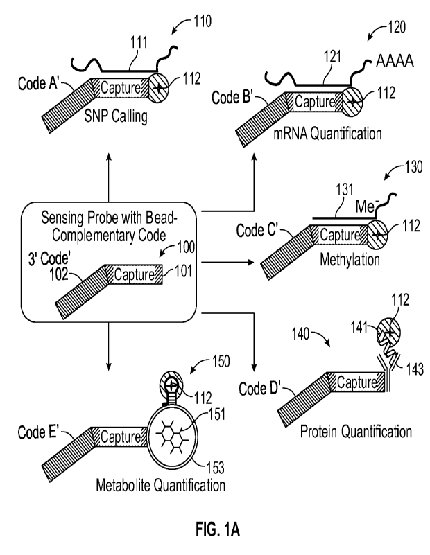
[0128] FIGS. 1A-1B schematically illustrate example components of a bead-based
system for
detecting multiple analytes. The different analytes may include any suitable
number and
mixture of nucleotide analytes (e.g., zero, one, or a plurality of nucleotide
analytes), and any
suitable number of non-nucleotide analytes (e.g., zero, one, or a plurality of
non-nucleotide
analytes). The different analytes may be mixed in a common solution with one
another, and
CA 03162326 2022- 6- 17
WO 2021/074087
PCT/EP2020/078653
may be derived from any suitable source or combination of sources, such as
blood, tissue,
saliva, urine, or the like.
[0129] As illustrated in FIG. 1A, the present system includes different
sensing probes 100
that are specific to, and can capture, respective ones of the different
analytes. That is, each
different sensing probe selectively captures one particular type of analyte in
the solution with
which such sensing probes are mixed. In some examples, as many different
sensing probes
may be provided in the solution as the number of different types of analytes
it is desired to
detect in that solution. For example, if it is desired to detect 10,000
different types of
analytes, then 10,000 different sensing probes that are respectively specific
to those analytes
may be provided. It will be appreciated that any suitable number of different
sensing probes
may be provided, e.g., more than 100, more than 1,000, more than 10,000, more
than
100,000, or more than 1,000,000. It will also be appreciated that any given
solution may not
necessarily include all possible analytes that it may be desired to detect. As
such, some
sensing probes may not necessarily have an analyte to capture in a given
solution. However,
at least some of the sensing probes can capture the analytes to which those
sensing probes are
specific.
[0130] In examples such as illustrated in FIG. 1A, different sensing probes
100 (sensing
probe with bead-complementary code) include different capture probes 101 and
different
codes 102 than one another. Each capture probe 101 may be specific to capture
a particular
analyte. Some of the analytes may be nucleotide analytes, and some of the
analytes may be
non-nucleotide analytes. In example 110 in FIG. lA (SNP calling), one of the
capture probes
101 is specific to a first nucleotide analyte, such as a specific DNA sequence
111 for which it
is desired to detect a SNP. In example 120 in FIG. lA (mRNA quantification),
one of the
capture probes 101 is specific to a second nucleotide analyte, such as a
specific mRNA
sequence for which it optionally may be desired to detect that sequence's
quantity. In
example 130 in FIG. lA (methylation), one of the capture probes 101 is
specific to a third
nucleotide analyte, such as a specific DNA sequence for which it is desired to
detect
methylation of a particular nucleotide. In example 140 in FIG. lA (protein
quantification),
one of the capture probes 101 is specific to a first non-nucleotide analyte,
such as a protein
for which it optionally may be desired to detect that protein' s quantity. In
example 150 in
FIG. lA (metabolite quantification), one of the capture probes 101 is specific
to a second
non-nucleotide analyte, such as a metabolite for which it optionally may be
desired to detect
26
CA 03162326 2022- 6- 17
WO 2021/074087
PCT/EP2020/078653
that metabolite's quantity. One or more of the capture probes may include an
oligonucleotide. The oligonucleotide may hybridize with a nucleotide analyte,
or may
provide an aptamer that may capture a non-nucleotide analyte. Additionally, or
alternatively,
one or more of the capture probes may include a non-oligonucleotide moiety,
such as an
antibody, to capture a non-nucleotide analyte. Fluorophores may be coupled to
sensing
probes 110 that captured respective ones of the different analytes. For
example, in each of
examples 110, 120, 130, 140, and 150, fluorophore 112 is coupled to the
sensing probes that
captured the respective analytes. Further details of example manners in which
different
sensing probes may respectively capture different analytes, and may be coupled
to
fluorophores, are provided below with reference to FIGS. 2A-2F, 3A-3B, 4A-4B,
and 5A-5C,
and further details of the manner in which the quantities of analytes may be
detected are
provided below with reference to FIGS. 6A-6B.
[0131] Referring now to FIG. 1B, the present system also includes different
beads 160
(together providing a universal bead array) that are specific to, and can
couple to, respective
ones of the different sensing probes. That is, each different bead selectively
couples to one
particular type of sensing probe in the common solution. In some examples, as
many
different beads 160 in the universal bead array may be provided in the present
system as the
number of different types of analytes it is desired to detect. For example, if
it is desired to
detect 10,000 different types of analytes, then 10,000 different beads that
are respectively
specific to sensing probes that, in turn, are specific to and can capture
those analytes may be
provided. It will be appreciated that any suitable number of different beads
may be provided,
e.g., more than 100, more than 1,000, more than 10,000, more than 100,000, or
more than
1,000,000. It will also be appreciated that a particular solution may not
necessarily include
all possible analytes that it may be desired to detect, but may include a
complete set of
sensing probes. As such, some beads may be coupled to sensing probes that may
not
necessarily have captured an analyte. However, at least some of the beads can
be coupled to
sensing probes that have captured the analytes to which those sensing probes
are specific.
[0132] In some examples, each bead 160 in the universal bead array has the
same
components as each other bead, regardless of the particular analyte that the
sensing probes
can capture. For example, each bead 160 illustrated in FIG. 1B includes
substrate 161 and an
oligonucleotide including code 162 and primer 163. Codes 162 of different
beads 160 have
different oligonucleotide sequences than one another that can selectively
couple to respective
27
CA 03162326 2022- 6- 17
WO 2021/074087
PCT/EP2020/078653
ones of the different codes 102 of sensing probes 100. Such codes 162
respectively identify
the analytes to which those sensing probes are specific, and thus can be used
to identify
which analytes are captured from the common solution, and optionally also used
to quantify
those analytes. For example, in a manner such as indicated at process 170
illustrated in FIG.
1B (hybridize to decoded array), each of the beads 160 includes
oligonucleotide 162 having a
sequence specific to one of the sensing probes 100, and each of the sensing
probes 100
includes oligonucleotide 102 having a sequence that is complementary to
oligonucleotide
162. Note that capture probe 101 of sensing probe 100 may not necessarily
hybridize to
primer 162 of bead 160, and instead the end of capture probe 101 may extend
into the
solution.
[0133] As noted above, fluorophores 112 are coupled only to sensing probes 100
that
captured an analyte to which those sensing probes are specific. As such,
fluorophores 112
become coupled to bead 160 via those sensing probes 110, to which those beads
160 are
specific. The beads 160 can be coupled to a surface, e.g., immobilized to a
surface within a
flow cell. In some examples, such coupling of beads 160 to a surface may be
performed
before the sensing probes 100 are coupled to the beads; for example, a
solution including
sensing probes 100 may be flowed over the beads coupled to the surface, and
the beads may
capture from the solution the sensing probes to which those beads arc
specific. In other
examples, such coupling of beads 160 to a surface may be performed after the
sensing probes
100 are coupled to the beads; for example, a solution including sensing probes
100 may be
mixed with a solution including beads 160 resulting in respective couplings
between beads
160 and the sensing probes to which those beads are specific, and the heads
subsequently
may be coupled to a surface, for example using bioorthogonal conjugation
chemistries such
as copper(I)-catalyzed click reaction (between azide and alkyne), strain-
promoted azide-
alkyne cycloaddition (between azide and DBCO (dibenzocyclooctyne),
hybridization of an
oligonucleotide to a complementary oligonucleotide, biotin-streptavidin, NTA-
His-Tag, or
Spytag-Spycatcher, charge-based immobilization such as amino-silane or poly-
lysine, or non-
specific such as with a polymer-coated surface.
[0134] As illustrated at process 180 illustrated in FIG. 1B (detect and decode
on sequencer),
the beads then can be detected via fluorescence from fluorophores 112, e.g.,
using a suitable
imaging camera and detection circuit. Using at least the detected
fluorescence, beads 160 can
be identified that are coupled to sensing probes 100 that had captured an
analyte (detect
28
CA 03162326 2022- 6- 17
WO 2021/074087
PCT/EP2020/078653
operation); in comparison, beads 160 that are coupled to sensing probes 100
that had not
captured an analyte may not be coupled to a fluorophore, and thus not detected
via
fluorescence. The sensing probes 110 and fluorophores 112 then may be removed,
e.g., by
dehybridization, a primer 164 coupled to primer region 163 of bead 160, and
code 162 then
decoded using sequencing by synthesis or other suitable method. For example,
fluorescently
labeled nucleotides can be added to primer 164 in a sequence that is
complementary to the
sequence of code 162. The identity of the analyte may be determined using at
least the
sequence of code 162. For example, the detection circuit may include memory
storing
different codes 162 and the analytes corresponding to those codes, and may be
configured to
compare the sequence of code 162 to the stored codes and to determine the
analyte
corresponding to the code of bead 160 (decode operation).
[0135] Note that fluorophores 112 may be coupled to respective sensing probes
100 at any
suitable time during process flows such as illustrated in FIGS. 1A-1B. For
example,
fluorophores 112 may be coupled to the sensing probes after the analytes are
captured by the
sensing probes, e.g., may be coupled to capture probe 101 using at least the
sequence of
nucleotide analyte 112, 121, 131. Or, for, example, fluorophores may be
coupled to the
sensing probes before the sensing probes are coupled to the beads, e.g., may
be coupled to
protein 141 prior to capture of that protein by antibody 143, or may be
coupled to metabolite
151 prior to coupling of the sensing probe to the bead. Or, for example,
fluorophores 112
may be coupled to the sensing probes after the sensing probes are coupled to
the beads, e.g.,
in a manner such as described with reference to FIGS. 7A-7B. In some examples,
multiple
fluorophores are coupled to the analytes, e.g., using a hybridization chain
reaction (HCR) in a
manner such as described with reference to FIG. 10A. Other examples of
coupling multiple
fluorophores to analytes are provided elsewhere herein.
[0136] FIG. 1C schematically illustrates an example process flow 1000 for
detecting multiple
analytes in a bead-based system. Process flow 1000 illustrated in FIG. 1C
includes mixing
different analytes with sensing probes, wherein at least some of the sensing
probes are
specific to respective ones of the analytes (process 1002). Examples of
sensing probes that
are specific to respective analytes are provided elsewhere herein, e.g., with
reference to FIGS.
3A-3B, 4A-4B, and 5A-5C. The sensing probes may be provided in excess relative
to the
respective analytes, so as to increase the likelihood that each given sensing
probe captures the
analyte to which that probe is specific. For example, the sensing probes may
be provided in
29
CA 03162326 2022- 6- 17
WO 2021/074087
PCT/EP2020/078653
an excess of greater than 10 times, greater than 100 times, greater than 1,000
times, or greater
than 10,000 times in excess of the analytes to which those probes are
specific. Illustratively,
a given analyte may have a concentration of 1-10 pM, and the sensing probe
specific to that
analyte may have a concentration of greater than 10 nM, e.g., 10-100 nM.
Process flow 1000
illustrated in FIG. 1C includes respectively capturing the analytes by the
sensing probes that
are specific to those analytes (process 1004). Some of the sensing probes in
the mixture may
be specific for analytes that are not necessarily present in the mixture, and
thus will not be
coupled to such analytes. Process flow 1000 includes respectively coupling
fluorophores to
sensing probes that captured respective analytes (process 1006). Example
manners in which
fluorophores may be coupled to sensing probes are described elsewhere herein.
[0137] Process flow 1000 illustrated in FIG. 1C includes mixing the sensing
probes with
beads, wherein the beads are specific to respective ones of the sensing
probes, and wherein
the beads include different codes identifying the analytes to which those
sensing probes are
specific (process 1008). Such mixing may occur by combining sensing probes in
solution
with beads in solution. Alternatively, such mixing may occur by flowing a
solution that
includes sensing probes over beads that are coupled to a surface. Process flow
1000 includes
respectively coupling sensing probes to beads that are specific to those
sensing probes
(process 1010). For example, each given bead may include a plurality of codes
which arc the
same as one another and that respectively are selective couple to the code of
a given sensing
probe. Accordingly, any sensing probes in the solution may become selectively
coupled to
that bead. Process flow 1000 includes identifying the beads that are coupled
to the sensing
probes that captured analytes using at least fluorescence from the
fluorophores coupled to
those sensing probes (process 1012). For example, the beads may be coupled to
a surface
(e.g., before or after being coupled to respective sensing probes) and regions
of fluorescence
on that surface may be imaged. Process flow 1000 includes identifying the
analytes that are
captured by the sensing probes coupled to the identified beads using at least
the codes of
those beads (process 1014). For example, the codes of the beads may be decoded
using
sequencing-by-synthesis, and the decoded codes used to determine which analyte
was
specific to the sensing probe to which the bead was specific.
[0138] Some non-limiting examples of analytes and sensing probes for
specifically capturing
such analytes, now will be described. It should be appreciated that the
present sensing probes
suitably may be modified to respectively capture any suitable analyte with
specificity.
CA 03162326 2022- 6- 17
WO 2021/074087
PCT/EP2020/078653
Example nucleotide analytes are described with reference to FIGS. 2A-2F and 3A-
3B, and
example non-nucleotide analytes are described with reference to FIGS. 4A-4B
and 5A-5C.
Any suitable combination of such analytes may be detected using the present
systems and
methods. For example, a solution that is mixed with the sensing probes may
include one or
more non-nucleotide analytes, or may include one or more nucleotide analytes.
For example,
the solution may include a mixture of nucleotide analytes and non-nucleotide
analytes. In
some examples, the different analytes are mixed together in a solution, and
portions of that
solution are mixed with respective types of sensing probes that target
respective types of
analytes. For example, a first portion of the solution may be mixed with
sensing probes that
are specific to one or more types of nucleotide analytes, and a second portion
of the solution
may be mixed with sensing probes that target one or more other types of
nucleotide analytes.
Or, for example, a first portion of the solution may be mixed with sensing
probes that are
specific to one or more types of nucleotide analytes, and a second portion of
the solution may
be mixed with sensing probes that target one or more types of non- nucleotide
analytes. Or,
for example, a first portion of the solution may be mixed with sensing probes
that are specific
to one or more types of non-nucleotide analytes, and a second portion of the
solution may be
mixed with sensing probes that target one or more other types of non-
nucleotide analytes.
[0139] In some examples, a sensing probe can include an oligonucleotide
sequence specific
to hybridize to a nucleotide analyte, such as a DNA analyte or RNA analyte.
For example,
FIGS. 2A-2C schematically illustrate example hybridization-based process flows
for
detecting DNA analytes in a bead-based system. In the example illustrated in
FIG. 2A, DNA
analytes 211, 211' include DNA sequences that differ from one another by a SNP
that it is
desired to detect. For example, DNA analyte 211 includes sequence 214 with A
at a given
location, while DNA analyte 211' the same sequence but with G instead of A at
the given
location, and it is desired to detect the respective presence of the A and G
in that sequence
214. As illustrated in FIG. 2A, sensing probes 200 are hybridized to these
targets of interest
at process 210 (hybridize probes to targets of interest). More specifically,
one copy of
sensing probe 200 may be hybridized to DNA analyte 211, and another copy of
sensing probe
200 may be hybridized to DNA analyte 211'. In this example, each sensing probe
200
includes capture probe 201 including a sequence that is complementary to
sequence 214 of
DNA analytes 211, 211' but that terminates at the nucleotide immediately
preceding the
location with the SNP (e.g., A or G) that it is desired to detect. Each
sensing probe 200 also
can include the same code 202 as one another which can be coupled to a
specific bead in a
31
CA 03162326 2022- 6- 17
WO 2021/074087
PCT/EP2020/078653
manner such as described with reference to FIGS. 1A-1C. In some examples, the
DNA
analytes (e.g., the SNP that it is desired to detect) are detected via
differences in fluorescence
that are caused by differences between the analytes. Illustratively, at
process 220, the
respective capture probes 201 of the sensing probes 200 are each extended by a
single base
with fully functional nucleotides (ffNs) that are fluorescently labeled
(single base extension
with ffNs). Because the sequences of DNA analytes 211. 211' differ from one
another by the
SNP (e.g., A or G), addition of differently fluorescently labeled ffNs to the
location with that
SNP result in different optical signals that may be distinguished from one
another. The
sensing probes can be coupled to one or more beads, e.g., to respective beads,
optically
detected, and the corresponding beads decoded in a manner such as described
with reference
to FIGS. 1A-1C (detect and decode on universal bead array).
[0140] The present systems and methods also may be used to detect and quantify
DNA
methylation in any suitable manner. For example, biochemical conversion of
methylated or
non-methylated nucleotides to a different base (for example, with bisulfite
treatment) can be
performed before capturing the analyte with a sensing probe. After capture, a
single base
extension is performed with ffNs at the site of potential methylation; the
methylation status
can be determined by the ffN-fluorophore that was incorporated. Such a
workflow may
provide for single-based resolution of DNA methylation.
[0141] For example, as illustrated in FIG. 2B, DNA analytes 221, 221' include
DNA
sequences that differ from one another by a nucleotide methylation that it is
desired to detect.
In this non-limiting example, DNA analyte 221 includes sequence 224 with
methylated-C
(Me-C) at a given location, while DNA analyte 221' includes the same sequence
but with
non-methylated C at the given location, and it is desired to detect the
respective presence of
the methylation in that sequence 224. At process 225, either the methylated or
non-
methylated nucleotide is selectively converted to a different base, for
example with bisulfite
treatment (biochemical conversion of methylated or non-methylated bases).
Here, the non-
methylated C is selectively converted to T, while Me-C is unchanged by the
treatment due to
the methylation. As illustrated in FIG. 2B, sensing probes 200' are hybridized
to these
targets of interest at process 210" (hybridize probes to targets of interest).
More specifically,
one copy of sensing probe 200' may be hybridized to DNA analyte 221, and
another copy of
sensing probe 200' may be hybridized to DNA analyte 221'. In this example,
each sensing
probe 200' includes capture probe 201' including a sequence that is
complementary to
32
CA 03162326 2022- 6- 17
WO 2021/074087
PCT/EP2020/078653
sequence 224 of DNA analytes 221, 221' but that terminates at the nucleotide
immediately
preceding the location with the methylation that it is desired to detect. Each
sensing probe
200' also can include the same code 202' as one another which can be coupled
to a specific
bead in a manner such as described with reference to FIGS. 1A-1C. In some
examples, the
DNA analytes (e.g., the methylation that it is desired to detect) are detected
via differences in
fluorescence that are caused by differences between the analytes.
Illustratively, at process
220', the respective capture probes 201' of the sensing probes 200' are each
extended by a
single base with ffNs 222, 222' that are fluorescently labeled (single base
extension with
ffNs). Because the sequences 224 of DNA analytes 221, 221' differ from one
another as a
result of the methylation and conversion (Me-C or T), addition of differently
fluorescently
labeled ffNs 222, 222' to the location with that methylation result in
different optical signals
that may be distinguished from one another. The sensing probes can be coupled
to one or
more beads, e.g., to respective beads, optically detected, and the
corresponding beads
decoded in a manner such as described with reference to FIGS. 1A-1C (detect
and decode on
universal bead array).
[0142] In another example of methylation detection, target DNA can be
hybridized to the
capture probe without prior processing, and then a single base extension is
performed with
ffNs. After the extension, fluorophore-conjugated antibodies against the
methylated target
nucleotide are added, which bind the methylated bases. Total target capture
may be
quantified by the fluorescence intensity of the ffN, and the extent of
methylation may be
measured by the fluorescence intensity of the antibody-fluorophore. This
approach may not
necessarily allow for single base resolution, as antibodies may bind all
methylated
nucleotides in proximity to the capture site. However, the approach may be
performed
without upfront biochemical processing of the sample DNA, and for assessing
regions with
many methylation events, multiple antibody binding events may amplify the
fluorescent
signal.
[0143] For example, as illustrated in FIG. 2C, DNA analytes 231, 231' include
DNA
sequences that differ from one another by one or more nucleotide methylations
that it is
desired to detect. In this non-limiting example, DNA analyte 231 includes
sequence 234 with
methylated-C (Me-C) at one or more given locations, while DNA analyte 231'
includes the
same sequence but with non-methylated Cs at the given locations, and it is
desired to detect
the respective presence of the methylation(s) in that sequence 234. As
illustrated in FIG. 2C,
33
CA 03162326 2022- 6- 17
WO 2021/074087
PCT/EP2020/078653
sensing probes 200" are hybridized to these targets of interest at process 235
(hybridize
probes to targets of interest). More specifically, one copy of sensing probe
200" may be
hybridized to DNA analyte 231, and another copy of sensing probe 200" may be
hybridized
to DNA analyte 231'. In this example, each sensing probe 200" includes capture
probe 201"
including a sequence that is complementary to sequence 234 of DNA analytes
231, 231' but
that terminates at the nucleotide immediately preceding the location with one
of the
methylations that it is desired to detect. Each sensing probe 200" also can
include the same
code 202" as one another which can be coupled to a specific bead in a manner
such as
described with reference to FIGS. 1A-1C. In some examples, the DNA analytes
(e.g., the
methylation that it is desired to detect) are detected via differences in
fluorescence that are
caused by differences between the analytes. Illustratively, at process 236,
the respective
capture probes 201" of the sensing probes 200" are each extended by a single
base with ffNs
232 that are fluorescently labeled (single base extension with ffNs). Because
the sequences
234 of DNA analyte 231, 231' are the same as one another except for the
methylation(s) (Me-
C), the same fluorescently labeled ffNs 232 will be added to the terminal
location with that
methylation. In this example, at process 237 fluorescently labeled antibodies
232' are added
to detect the methylation status of sequence 234 (detect methylation status
with antibodies).
For example, the fluorescently labeled antibodies 232 may selectively bind to
Me-C. The
sensing probes then can he coupled to one or more beads. e.g., to respective
heads,
fluorescence from the differently fluorescently labeled sensing probes
optically detected, and
the corresponding beads decoded in a manner such as described with reference
to FIGS. 1A-
1C (detect and decode on universal bead array).
[0144] Some examples of the methods and systems provided herein relate to the
detection of
target nucleic acids, which also may be referred to as DNA analytes. In some
examples,
target nucleic acids are detected by hybridizing a plurality of nucleic acids
comprising target
nucleic acids to probes (sensing probes) capable of hybridizing to the target
nucleic acids;
extending the hybridized probes; and detecting the extended probes, thereby
detecting the
target nucleic acids. In some examples, the hybridization and extension
processes are
performed in solution. In other examples they are performed in conjunction
with a solid
support, such as a microfluidics device. In some examples, the extended probes
are enriched
by removing unextended probes, and optionally the plurality of nucleic acids,
from the
extended probes. In some examples, the extended probes are detected by
hybridizing the
extended probes to an array of capture probes immobilized on a surface. In
some examples,
34
CA 03162326 2022- 6- 17
WO 2021/074087
PCT/EP2020/078653
an array of capture probes is a decoded array wherein each capture probe has a
unique
signature or bar code and the position of each capture probe is decoded prior
to use. In some
examples, the array of capture probes comprises a universal array.
[0145] Some examples provided herein include methods for increasing the
performance of a
genotyping assay and enabling the use of universally decoded arrays by using a
sample
preparation strategy that may employ solution-phase target capture, probe
extension, and
enrichment, followed by bead-based genotyping. Some such examples address
known
challenges including the inefficient capture of DNA targets for genotyping;
template-
independent probe extension and increased background signal due to high local
concentrations of probes after immobilization on beads; and the ability to use
universal arrays
to detect target nucleic acids. In some examples, such challenges are solved
by performing
target capture and probe extension in solution, followed by enzymatic
enrichment of targets-
of-interest and introduction to arrays; by performing target capture and first
base extension in
solution; and by decoupling probe sequences from immobilized oligonucleotides.
[0146] Challenges associated with inefficient hybridization in certain methods
to detect target
nucleic acids include performing target capture on pre-assembled arrays, for
example
hybridizing target nucleic acids to target-specific probes immobilized on an
array. In one
example, performing target capture on pre-assembled arrays can place a limit
on the
probe:target ratio because the number of target-specific probes in this
example may be
ultimately fixed by the number of beads loaded into the array. Additionally,
samples used for
genotyping can contain an excess of non-targeted DNA, may be viscous due to
high DNA
concentrations, and can potentially suffer from re-hybridization of targets to
their solution-
phase complements. In some examples these challenges are addressed by
hybridizing target-
specific probes to target nucleic acids in solution. In some examples, in-
solution
hybridization can enable the use of a large probe:target ratio, which can lead
to an increase in
hybridization kinetics. In some examples, biochemical enrichment of sequences
of interest
prior to introduction to the array addresses these challenges by removing
oligonucleotides
that may negatively affect hybridization.
[0147] Challenges associated with inefficient DNA concentrations in samples in
certain
methods to detect target nucleic acids can limit genotyping performance. Low
DNA
concentrations may utilize a whole genome amplification process to obtain
sufficient
concentrations of sample, prior to introduction of DNA samples onto arrays. In
some
CA 03162326 2022- 6- 17
WO 2021/074087
PCT/EP2020/078653
examples provided herein such challenges are addressed by increasing
hybridization
efficiency by removing non-targeted DNA. In some examples, the amount of
extended target-
specific probes can be selectively, ultimately increasing the rate of bead-
based capture of
extended probes.
[0148] In certain methods to identify target nucleic acids, biochemistry on
immobilized
probes can be complicated by surface architecture, for example beads used in
certain
commercial arrays can contain high local concentrations of probes, which
promote inter-oligo
interactions and lead to an increase in background signal due to off target
incorporation.
Optimization of probe surface density can prevent these interactions to some
extent but
ultimately may involve a tradeoff between optimizing the bead architecture for
target capture
and preventing non-targeted probe extension. Additionally, the bead presents a
surface that
nucleotides can bind to, which can lead to an elevated noise level. In some
examples
provided herein such challenges are addressed by performing probe-target
hybridization and
extension reactions in solution, such that target concentration gradients and
adsorption of
reagents to surfaces are minimized.
[0149] In certain methods to identify target nucleic acids, commercial array
formats may not
allow for easily adding custom probes or designing custom 2enotyping panels
because beads
are pooled in large batches prior to loading on arrays. In some examples
provided herein such
challenges are addressed by performing hybridization in solution to allow use
of universal
arrays with decode sequences that are complementary to a terminal extension of
the solution-
phase probes.
[0150] One example includes performing all biochemical probe manipulations in
solution, as
well as an additional sample enrichment process, prior to genotyping on
arrays. One
advantage provided by this example includes the ability to load arrays with
beads that are
functionalized with decoded oligonucleotides, and not target-specific probes.
This allows an
end-user to more easily add custom SNPs to methods and compositions for
detecting target
nucleic acids using arrays.
[0151] An example of a method for identifying a target nucleic acids is
depicted in FIG. 2D
which includes the following processes: (1) Hybridization of target-specific
probe (sensing
probe) to target nucleic acid (DNA analyte, such as a genomic DNA fragment
including a
SNP) in solution which can have an increased probe:target ratio relative to
hybridization on
36
CA 03162326 2022- 6- 17
WO 2021/074087
PCT/EP2020/078653
an array to promote binding; and single base extension of hybridized probes
using
fluorophore labeled nucleotides that ultimately act as a signal for genotyping
(hybridization
of probes and extension with ffNs). A genomic DNA fragment includes the target
nucleic
acids and contains a single nucleotide polymorphism (SNP), the target specific
probe
hybridizes at a location immediately adjacent to the SNP. The target-specific
probe contains
or includes a 3' end capable of hybridizing to the target nucleic acid, and a
5' end capable of
hybridizing to a capture probe. The target-specific probe hybridizes to the
target nucleic acid,
and is extended with a single modified nucleotide having a 3' fluorophore
which inhibits
degradation of the extended probe by 3'-5' exonucleases (3'-fluor protects
from degradation).
(2) (enzymatic degradation with exonuclease(s)) and (3) (digestion of
unmodified DNA)
Enrichment of fluorophore-labeled probes in which 3'-OH specific exonucleases
degrade
unextended probes and any oligonucleotides that are not intended for capture
and genotyping
on arrays. Non-extended probes and genomic DNA fragments are degraded by 3'-
.5'
exonucleases. (4) Hybridization of fluorescent extended probes to arrays via
sequence
complementary to decode the polynucleotides (hybridization to decoded array).
Each target-
specific probe contains a sequence at its 5' terminus that is complementary to
a decoded
sequence that identifies the position of a particular bead type within an
array. These
sequences enable hybridization of fluorophore-labeled probes to specific sites
on the array for
genotyping; the heads include primer binding sites and codes. (5) Genotyping
is performed
either by direct detection of fluorescent nucleotides or, if necessary or
appropriate, after
additional signal amplification. Note that although the array may be decoded
prior to
hybridization of the fluorescent extended probes at (4) in a manner such as
shown in FIG. 2D,
the target-extended probes instead may be hybridized to a suspension of beads,
loaded onto
an array, and the beads then decoded.
[0152] An example of a method for identifying a target nucleic acids is
depicted in FIG. 2E
(one pot add-on assay for signal generation and amplification). The left panel
of FIG. 2E
(assay input) depicts probes (sensing probes, user determined probes including
probe and
code complement) with a 5' overhang complementary to a universal bead pool are
mixed with
a genomic DNA sample (genomic dsDNA) and amplification reagent (excess of
probes with
5' extension complementary to bead pool; lyophilized amplification reagent ¨
polymerase,
FFNs, buffer; enables flexible content and shear-free sample prep). The center
panel of FIG.
2E (signal generation and amplification) depicts thermal cycling of a mixture
to increase the
concentration of extended probes, which ultimately enhances bead-based capture
of
37
CA 03162326 2022- 6- 17
WO 2021/074087
PCT/EP2020/078653
sequences of interest. For example, target-specific probes are hybridized to
target nucleic
acids, and extended. The extended probes are dellybridized from the target
nucleic acids.
More target-specific probes are hybridized to target nucleic acids, and
extended. The cycles
are repeated to amplify the number of extended probes, for example by 20
cycles (for
example, at about 30 seconds per process, for a total of about 30 minutes for
20 cycles). Such
processes can rapidly increase the amount of material for bead hybridization.
After signal
generation and amplification, extended probes are enriched by exonuclease-
catalyzed
degradation of genomic DNA and unextended probes (endonuclease catalyzed
hydrolysis of
non-modified DNA (non-extended probes). The right panel of FIG. 2E (hyb to
bead pool)
depicts bead-based capture and genotyping of extended probes. Hybridization
time may be
increased by at least the same factor as the signal amplification. There
potentially a greater
benefit from hybridizing in the absence of non-specific sequences. Thus,
faster bead-based
hybridization is provided, as is a universal bead pool.
[0153] Examples of aspects of enriching for extended probes are depicted in
FIG. 2F. Whole
genome amplification products may include a mixture of single stranded DNA,
duplex DNA
with 5' overhang, and duplex DNA with 3' overhang. The single-stranded DNA in
the whole
genome amplification products may be hybridized to sensing probes at process
1) followed
by single base extension (SBE) with 3'-fluorophore labeled ffNs at process 2)
to form probe-
target complexes. Enrichment of probes for genotyping is achieved by selective
degradation
of oligonucleotides that are not 3'-fluorophore labeled (oligonucleotides
targeted for
degradation). This is enabled by the highly specific nature of restriction
exonucleases that
each target specific impurities. The following exonucleases are examples of
classes which
can be utilized to enrich for selected oligonucleotides: (1) Klenow I fragment
targets 3'
duplex DNA containing 3'-overhangs; (2) Exonuclease Ill (ExoIII) targets the
3' end of
duplex DNA; and (3) Exonuclease I (ExoI) degrades single stranded library
fragments as well
as unreacted primers. Probe-target complexes that have been extended with 3'-
fluorophore
ffNs may be hybridized to a decoded array (or non-decoded array) and genotype
processes
performed.
[0154] In some examples, it is possible that non-specific incorporation of
nucleotides to the
3' termini of library fragments may occur. In this case, probes may be
designed with, for
example, phosphorothioate bonds at their 5'-termini. This would allow for
selectively
38
CA 03162326 2022- 6- 17
WO 2021/074087
PCT/EP2020/078653
degrading library fragments. Some examples include the use of target-specific
probes having
a 5' end resistant to enzymatic degradation.
[0155] Some examples provided herein include methods for identifying target
nucleic acids.
Some such examples include (a) hybridizing a plurality of probes to a
plurality of nucleic
acids comprising the target nucleic acids, wherein each probe comprises a 3'
end capable of
hybridizing to a target nucleic acid and a 5' end capable of hybridizing to a
capture probe; (b)
extending the hybridized probes with a blocked nucleotide; (c) removing the
plurality of
nucleic acids and non-extended probes from the extended probes; and (d)
hybridizing the
extended probes to a plurality of capture probes immobilized on a surface.
[0156] In some examples, the capture probes each comprises a 3' end capable of
hybridizing
to a target nucleic acid. In some examples, the capture probe is capable of
hybridizing to a
location on a target nucleic acid immediately proximal to a single nucleotide
polymorphism
(SNP), or other single nucleotide feature to be examined in the target nucleic
acid. In some
examples, the 3' end capable of hybridizing to a target nucleic acid is the
most 3' end of the
probe. In some examples, the 3' end capable of hybridizing to a target nucleic
acid is at least
3, 5, 10, 15, 20, 25, 30. 35, 40, 45, 50, 55, 60 consecutive nucleotides in
length, or any
number of nucleotides between any two of the foregoing numbers. In some
examples, the
capture probes each comprise a 5' end capable of hybridizing to a capture
probe. In some
examples, the 5' end capable of hybridizing to a target nucleic acid is the
most 5' end of the
probe. In some examples, the 5' end capable of hybridizing to a target nucleic
acid is at least
3, 5, 10, 15, 20, 25, 30, 35, 40, 45, 50, 55, 60 consecutive nucleotides in
length, or any
number of nucleotides between any two of the foregoing numbers. In some
examples, the
most 5' end of the probe is resistant to enzymatic degradation. For example,
the most 5' end
of the probe can include a phosphorothioate bond.
[0157] In some examples, the hybridizing the plurality of probes to a
plurality of nucleic
acids comprising the target nucleic acids, the extending the hybridized probes
with a blocked
nucleotide, and the removing the plurality of nucleic acids and non-extended
probes from the
extended probes, are performed in solution. For example, the probes, the
nucleic acids, and
the extended probes, are not immobilized on a surface.
[0158] In some examples, the amount of extended probe can be increased by
performing an
amplification process. In some such examples, the plurality of probes is
hybridized to the
39
CA 03162326 2022- 6- 17
WO 2021/074087
PCT/EP2020/078653
plurality of nucleic acids comprising the target nucleic acids, and the
hybridized probes are
extended with a blocked nucleotide; and the hybridization and extension
repeated. For
example, a cycle includes a first hybridization and extension, and then the
extended probes
are de-hybridized from target nucleic acids; non-extended probes are
hybridized to the target
nucleic acids, the hybridized probes are extended with a blocked nucleotide.
In some
examples, the cycle is repeated for more than 2, 5, 10, 20, 30 or 50 cycles,
or any number
between any two of the foregoing numbers.
[0159] In some examples, the extension is performed with a polymerase, or a
ligase. In some
such examples, the extension adds a blocked nucleotide at the most 3' end of
the probe to
generate an extended probe. As used herein, a "blocked nucleotide" can include
a nucleotide
which confers resistance to exonuclease degradation on an extended probe. For
example, an
extended probe will be resistant to enzymatic degradation by a 3' to 5'
exonuclease. In some
examples, a blocked nucleotide can include a detectable label, such as a
fluorophore. In some
such examples, the fluorophore which can provide resistance to enzymatic
degradation by a
3' to 5' exonuclease.
[0160] Some examples include removing unextended probes from the extended
probes.
Some examples also include removing the plurality of nucleic acids and
unextended probes
from the extended probes. Some such examples include enzymatic degradation of
the
plurality of nucleic acids and unextended probes. In some examples, the
plurality of nucleic
acids and the non-extended probes are contacted with a 3' to 5' exonuclease.
Examples of 3'
to 5' exonucleases include Exonuclease I, Thermolabile Exonuclease I,
Exonuclease T,
Exonuclease III, and Klenow I fragment. In some examples, the plurality of
nucleic acids
and unextended probes are substantially removed from the extended probes, for
example, at
least 30%, 40%, 50%, 60%, 70%, 80%, 90%, 95%, 99%, 100%, or any percentage
between
any two foregoing percentages of amounts of the plurality of nucleic acids and
unextended
probes are at least substantially removed from the extended probes.
[0161] In some examples, the probes each comprise a 5' end resistant to
enzymatic
degradation, for example, the 5' end resistant to enzymatic degradation
comprises a
phosphorothioate bond. In sonic such examples, the plurality of nucleic acids
can be
removed from extended probes by contacting the plurality of nucleic acids with
a 5' to 3'
exonuclease. Examples of 5' to 3' exonucleases include RecJf, T7 Exonuclease,
truncated
CA 03162326 2022- 6- 17
WO 2021/074087
PCT/EP2020/078653
Exonuclease VIII, Lambda Exonuclease, T5 Exonuclease, Exonuclease VII,
Exonuclease V,
and Nuclease BAL-31.
[0162] Some examples include hybridizing the extended probes to capture
probes. IN some
examples, the capture probes are immobilized on a surface. In some examples, a
bead
comprises the surface. In some examples, a plurality of beads comprise the
surface. In some
examples, a planar surface comprises the surface. Ti some examples a flow cell
comprises
the surface. In some examples, a flow cell comprises beads which comprise the
surface.
[0163] Some examples include amplifying a signal from an extended probe
hybridized to a
capture probe. In some such examples, a signal is amplified using labelled
primary
antibodies against the blocked nucleotide, such as the fluorophore. Some
examples also
include the use of secondary antibodies against the primary antibodies and
further labeled.
[0164] In some examples, the capture probes are different from each other. For
example,
different capture probes can be capable of hybridizing to extended probes
which have been
generated by hybridizing probes to different target nucleic acids. In some
examples, the
plurality of capture probes comprises a decoded array of capture probes. For
example, an
array can include a plurality of wells on a surface, each well containing a
bead comprising a
capture probe. Some examples include decoding the location of the capture
probes on a
surface. In some examples, the plurality of capture probes each comprises a
primer binding
site and a decode polynucleotide. In some examples, decoding comprises:
hybridizing a
sequencing primer to the primer binding site, extending the hybridized primer,
and
identifying the decode polynucleotide. In some examples, the decode
polynucleotide is
capable of hybridizing to an extended probe. Some examples include identifying
the location
of the hybridized extended probes on the surface, such as a surface comprising
a decoded
array of capture probes, thereby identifying the target nucleic acid.
[0165] Some examples provided herein include kits and systems. In some
examples, a kit or
system for identifying target nucleic acids includes an extension solution
comprising: a
plurality of nucleic acids comprising the target nucleic acids, a plurality of
probes, wherein
each probe comprises a 3' end capable of hybridizing to a target nucleic acid
and a 5' end
capable of hybridizing to a capture probe, a plurality of blocked nucleotides,
an extension
enzyme; a degradation solution comprising a 3' to 5' exonuclease; an array of
capture probes
immobilized on a surface; and a detector to identify (capable of identifying)
the location of an
41
CA 03162326 2022- 6- 17
WO 2021/074087
PCT/EP2020/078653
extended probe hybridized to a capture probe on the surface. In some examples,
a flow cell
comprises the array of capture probes immobilized on a surface.
[0166] In some examples, a kit or system for identifying target nucleic acids
includes a flow
cell comprising a surface, an inlet for adding solutions to the surface, and
an outlet for
removing solutions from the surface, wherein an array of capture probes is
immobilized on
the surface; an extension solution in contact with the inlet, the extension
solution comprising:
a plurality of nucleic acids comprising the target nucleic acids, a plurality
of probes, wherein
each probe comprises a 3' end capable of hybridizing to a target nucleic acid
and a 5' end
capable of hybridizing to a capture probe, a plurality of blocked nucleotides,
an extension
enzyme; a degradation solution comprising a 3' to 5' exonuclease; and a
detector to identify
(capable of identifying) the location of an extended probe hybridized to a
capture probe on
the surface.
[0167] In some examples, the blocked nucleotide comprises a detectable label.
In some
examples, the label comprises a fluorophore. In some examples, the extension
enzyme
comprises a polymerase. In some examples, the extension enzyme comprises a
ligase. In
some examples, the 3' to 5' exonuclease is selected from the group consisting
of Exonuclease
I. Thermolabile Exonuclease I. Exonuclease T. Exonuclease Ill, and Klenow I
fragment. In
some examples, the probes each comprises a 5' end resistant to enzymatic
degradation. In
some examples, the 5' end resistant to enzymatic degradation comprises a
phosphorothioate
bond. In some examples, the degradation solution further comprises a 5' to 3'
exonuclease. In
some examples, the 5' to 3' exonuclease is selected from the group consisting
of RecJf, T7
Exonuclease, truncated Exonuclease VIII, Lambda Exonuclease, T5 Exonuclease,
Exonuclease VII, Exonuclease V, and Nuclease BAL-31. In some examples, the
surface
comprises a plurality of beads. In some examples, the capture probes are
different from each
other. In some examples, the plurality of capture probes comprises a decoded
array of capture
probes. In some examples, the plurality of capture probes each comprises a
primer binding
site and a decode polynucleotide. In some examples, the plurality of nucleic
acids comprises
genomic DNA. In some examples, the target nucleic acids comprise a single
nucleotide
polymorphism (SNP).
[0168] DNA is only one example of a nucleotide analyte that may be detected
using the
present systems and methods. Similarly as for DNA analytes, a sensing probe
can include an
oligonucleotide sequence specific to hybridize to an RNA analyte. For example,
strategies for
42
CA 03162326 2022- 6- 17
WO 2021/074087
PCT/EP2020/078653
the capture and detection of RNA also may be amenable to using cDNA libraries
and may be
adapted to use RNA directly. In-solution hybridization of RNA (such as cDNA)
molecules to
a sensing probe including a target identification code followed by single base
extension with
ffNs, similarly as for DNA workflows, may be used. For example, FIGS. 3A-3B
schematically illustrate example hybridization-based process flows for
detecting RNA
analytes in a bead-based system.
[0169] In the example illustrated in FIG. 3A, RNA analytes 311, 311' include
RNA
sequences that differ from one another and for which it is desired to detect
relative
abundances. For example, RNA analyte 311 includes sequence 314, while RNA
analyte 311'
includes sequence 314', and it is desired to detect the abundance of RNA
analyte 311 relative
to that of RNA analyte 311'. As illustrated in FIG. 3A, sensing probes 300,
300' respectively
are hybridized to these targets of interest at process 310 (hybridize probes
to targets of
interest). More specifically, one copy of sensing probe 300 may be hybridized
to each of
RNA analytes 311, and one copy of sensing probe 300' may be hybridized to RNA
analyte
311'. In this example, each sensing probe 300 includes capture probe 301
including a
sequence that is complementary to sequence 314 of RNA analyte 311, while each
sensing
probe 300' includes capture probe 301' including a different sequence that is
complementary
to sequence 314' of RNA analyte 311'. Each sensing probe 300 also can include
the same
code 302 as one another which can be coupled to a specific bead in a manner
such as
described with reference to FIGS. 1A-1C, while each sensing probe 300' also
can include the
same code 302' as one another which can be coupled to a different specific
bead in a manner
such as described with reference to FIGS. 1A-1C. In some examples, the RNA
analytes are
detected via differences in the codes of the sensing probes. Illustratively,
at process 320
(single base extension with ffNs), the respective capture probes 301 of the
sensing probes 300
and capture probes 301' of the sensing probes 300' are each extended by a
single base with
ffNs that are fluorescently labeled. The sensing probes can be coupled to
respective beads,
optically detected, and the corresponding beads decoded in a manner such as
described with
reference to FIGS. 1A-1C (detect and decode on universal bead array).
[0170] In other examples, the present systems and methods may be used to
quantify
alternative splicing events and to obtain estimations of transcript isoform
abundance.
Illustratively, each type of ffN can be coupled to a different fluorophore,
and the fluorophore
identity can reflect which splicing event took place. Informative measurements
of alternative
43
CA 03162326 2022- 6- 17
WO 2021/074087
PCT/EP2020/078653
splicing can be obtained by providing a different nucleotide immediately
adjacent to the
splicing site for each of the possible exults.
[0171] In the example illustrated in FIG. 3B, RNA analytes 321, 321' include
RNA
sequences that that include different splice isoforms and for which it is
desired to detect
relative abundances. For example, RNA analyte 321 includes splice isoform 324,
while RNA
analyte 321' includes different splice isoform 324', and it is desired to
detect the abundance
of RNA analyte 321 relative to that of RNA analyte 321'. As illustrated in
FIG. 3B, sensing
probe 300" respectively is hybridized to these targets of interest at process
310' (hybridize
probes to targets of interest). More specifically, one copy of sensing probe
300" may be
hybridized to each of RNA analytes 311, 311'. In this example, each sensing
probe 300"
includes capture probe 301" including a sequence that is complementary to one
or more
exons in both of RNA analytes 311, 311' and that terminates immediately prior
to the splice
isofat _______ la it is desired to detect and quantify. Each sensing probe
300" also can include the
same code 302" as one another which can be coupled to a specific bead in a
manner such as
described with reference to FIGS. 1A-1C. In some examples, the RNA analytes
(e.g., the
splice isoforms that it is desired to detect) are detected via differences in
fluorescence that are
caused by differences between the analytes. Illustratively, at process 320'
(single-base
extension with ft-Ns). the respective capture probes 301" of the sensing
probes 300 are each
extended by a single base with ffNs that are fluorescently labeled. Because
the sequences of
RNA analytes 311, 311' differ from one another by the splice isoform (e.g.,
exon3 or exon5),
addition of differently fluorescently labeled ffNs to the location with that
splice isoform
result in different optical signals that may be distinguished from one
another. The sensing
probes can be coupled to one or more beads, e.g., to respective beads,
optically detected, and
the corresponding beads decoded in a manner such as described with reference
to FIGS. 1A-
1C (detect and decode on universal bead array).
[0172] In examples such as described with reference to FIGS. 2A-2F and 3A-3B,
note that
the ffN optionally may be fluorescently labeled after being added to the
capture probe, rather
than before being added to the capture probe. Additionally, or alternatively,
the ffN may be
coupled to multiple fluorophores so as to provide an amplified optical signal.
Example
methods for adding multiple fluorophores to a nucleotide are described in
greater detail
below with reference to FIGS. 7A-16E.
44
CA 03162326 2022- 6- 17
WO 2021/074087
PCT/EP2020/078653
[0173] While certain examples of nucleotide analytes are described with
reference to FIGS.
2A-2F and 3A-3B, the present sensing probes suitably may be adapted to
selectively couple
to any type of analyte, such as non-nucleotide analytes. Examples of non-
nucleotide analytes
include proteins and metabolites. Examples of sensing probes suitable for
selectively
coupling to non-nucleotide analytes include antibodies, such as described
below with
reference to FIGS. 4A-4B, or aptamers, such as described below with reference
to FIGS. 5A-
5C. Such sensing probes may be selectively coupled to beads, based upon which
the analyte
identity may be determined by decoding the beads in a manner such as described
with
reference to FIGS. 1A-1C.
[0174] For example, FIGS. 4A-4B schematically illustrate example antibody-
based process
flows for detecting protein analytes in a bead-based system. In the example
illustrated in
FIG. 4A, a solution may include a plurality of different proteins, and it may
be desired to
detect proteins 411, 411' which are different than one another. At process 410
(general
protein label), the proteins in the solution may be labeled with fluorophores
412 using a
general protein dye (such as amine-reactive fluorophores or haptens).
Nonlimiting examples
of proteins 411, 411' include kinases, serine hydrolases, metalloproteases,
and disease-
specific biomarkers such as antigens for specific diseases. This fluorescent
labeling may be
followed by in solution binding of sensing probes to enrich for the proteins
of interest at
process 420 (enrich for targets of interest). For example, sensing probe 400
may include
antigen 413 which is specific to protein 411, and code 402 which is specific
to a particular
bead, and sensing probe 400' may include antigen 413' which is specific to
protein 411', and
code 402' which is specific to a particular bead. Antigen 413 may specifically
bind protein
411, which may cause sensing probe 400 to become fluorescently labeled via
fluorophore 412
coupled to protein 411. Antigen 413' may specifically bind protein 411', which
may cause
sensing probe 400' to become fluorescently labeled via fluorophore 412'
coupled to protein
411'. Sensing probes 400, 400' may be coupled to respective beads, and non-
bound protein
may be washed out. The fluorescence from fluorophores 412, 412' may be
respectively
detected via imaging. Sensing probes 400, 400' may be removed from the beads,
a primer
annealed to the beads, and the beads decoded in a manner such as described
with reference to
FIGS. 1A-1C (detect and decode on universal bead array) to identify the
analytes that are
respectively bound to the sensing probe. Note that all sensing probes in the
solution may
become coupled to respective beads, but only sensing probes that captured a
protein will also
generate a fluorescent signal.
CA 03162326 2022- 6- 17
WO 2021/074087
PCT/EP2020/078653
[0175] In other examples, proteins are first captured by sensing probes to
select targets of
interest, prior to fluorescent labeling. In the example illustrated in FIG.
4B, a solution again
may include a plurality of different proteins, and it may be desired to detect
proteins 421,
421' which are different than one another. At process 410' (enrich for targets
of interest), in-
solution binding of sensing probes to enrich for the proteins of interest is
perfoimed. For
example, sensing probe 430 may include antigen 423 which is specific to
protein 421, and
code 432 which is specific to a particular bead, and sensing probe 430' may
include antigen
423' which is specific to protein 421', and code 432' which is specific to a
particular bead.
Antigen 423 may specifically bind protein 421, and antigen 423' may
specifically bind
protein 421'. The bound proteins 421, 421' then may be fluorescently labeled
at process 420'
(detect binding with fluorescent antibody). For example, antibodies 424, 424'
coupled to
fluorophores 442 may be respectively coupled to bound proteins 421, 421'
before or after
sensing probes 430, 430' are coupled to respective beads, and non-bound
protein then may be
washed out. The fluorescence from fluorophores 412, 412' may be respectively
detected via
imaging. Sensing probes 430, 430' may be removed from the beads, a primer
annealed to the
beads, and the beads decoded in a manner such as described with reference to
FIGS. 1A-1C
(detect and decode on universal bead array) to identify the analytes that are
respectively
bound to the sensing probes. Note that all sensing probes in the solution may
become
coupled to respective beads, but only sensing probes that captured a protein
will also generate
a fluorescent signal. Note that antibodies 424, 424' may target different
epitopes of the
respective proteins than one another, so that both antibodies 423, 424 may be
simultaneously
bound to protein 411, and so that antibodies 423', 424' may be simultaneously
bound to
protein 411'. In examples such as described with reference to FIG. 4B,
background
fluorescence signal from non-specific binding of antigens to proteins may be
suppressed by
providing two independent antibody binding events to generate fluorescent
signal,
substantially increasing specificity.
[0176] Note that antigens coupled to codes in a manner such as described with
reference to
FIGS. 4A-4B may be or include barcoded antibodies such as commercially
available from
BioLegend, Inc. (San Diego, California). In such barcoded antibodies, the 5'
end of the
nucleic acid code used for sample identification (via bead binding and
decoding such as
described with reference to FIGS. 1A-1C) is covalently coupled to an antibody.
The content
of such barcoded antibodies may be customizable to provide detection of
desired non-
nucleotide analytes, such as proteins.
46
CA 03162326 2022- 6- 17
WO 2021/074087
PCT/EP2020/078653
[0177] Other example process flows use sensing probes having aptamers to
capture analytes.
Aptamers may be considered to be antibodies that are made out of nucleic acid
sequences,
and can be used to capture proteins and small molecules (such as metabolites)
with high
specificity. For example, FIGS. 5A-5C schematically illustrate example aptamer-
based
process flows for detecting protein or metabolite analytes in a bead-based
system.
[0178] For example, in a manner such as described with reference to FIG. 4A, a
general
protein fluorescent dye followed by in solution capture of target proteins
with evolved
aptamers may be used. In the example shown in FIG. 5A, at process 510 (general
protein
label), different proteins 511 are labeled with a general protein label such
as described with
reference to FIG. 4A. Nonlimiting examples of proteins 511 include kinases,
serine
hydrolases, metalloproteases, and disease-specific biomarkers such as antigens
for specific
diseases. At process 520 (enrich for targets of interest), the labeled
proteins are mixed with
sensing probes 500 which include codes 502 coupled to aptamers 503, optionally
via linkage
504. The aptamer 503 (aptamer with target specificity) which is specific to
protein 511
captures that protein, together with fluorophore 512 coupled to that protein.
As such, sensing
probe 500 becomes fluorescently labeled with specificity to protein 511.
Sensing probe 500
may be specifically coupled to a bead, and non-bound protein may be washed
out. The
fluorescence from fluorophore 512 may be detected via imaging. Sensing probe
500 may be
removed from the bead, a primer annealed to the bead, and the bead decoded in
a manner
such as described with reference to FIGS. 1A-1C (detect and decode on
universal bead array)
to identify the analyte that was respectively bound to the sensing probe. Note
that aptamers
503 may be selected so as to be specific to the respective combination of a
given protein 511
and the fluorophore 512 coupled to that protein. Alternatively, aptamers 503
may be selected
so as to bind to respective region(s) of a given protein 511 that do not
contain reactive amino
acid residues that would be labeled with a fluorophore 512, so that
fluorophore 512 may not
interfere with binding between the aptamers and the proteins to which those
aptamers are
specific.
[0179] In other approaches, fluorescent read-out of analyte capture may be
obtained by
linking aptamer binding of the target analyte to a conformational change that
introduces a
fluorescent signal. Conformational changes in aptamers upon target binding is
well
documented, including the spinach aptamer and riboswitches. For example, the
spinach
aptamer, which causes the compound 3,5-difluoro-4-hydroxybenzylidene
imidazolinone
47
CA 03162326 2022- 6- 17
WO 2021/074087
PCT/EP2020/078653
(DHFBI) to fluoresce upon binding, can be conjugated to additional
riboswitches or aptamers
that render Spinach inactive until they have also bound their respective
ligand. An aptamer
that has not bound its target will not be able to fluoresce. In the example
shown in FIG. 5B,
at process 520' (enrich for targets of interest), analytes such as proteins or
metabolites (or a
mixture thereof) are mixed with sensing probes 500' which include codes 502'
coupled to
aptamers 503', optionally via linkage 504'. The aptamer 503' (aptamer with
target
specificity) which is specific to protein or metabolite 511' captures that
protein or metabolite
which activates fluorophore 512' (fluorescent transducer). As such, sensing
probe 500'
becomes fluorescently labeled with specificity to protein or metabolite 511'.
Sensing probe
500' may be specifically coupled to a bead, and non-bound protein and
metabolites may be
washed out. The fluorescence from fluorophore 512' may be detected via
imaging. Sensing
probe 500' may be removed from the bead, a primer annealed to the bead, and
the bead
decoded in a manner such as described with reference to FIGS. 1A- 1 C (detect
and decode on
universal bead array) to identify the analyte that was respectively bound to
the sensing probe.
[0180] In still other approaches, fluorescent read-out of analyte capture may
be obtained by
linking aptamer binding of the target analyte to a conformational change that
reveals a
moiety, such as an oligonucleotide sequence, that can bind a fluorophore. Only
an aptamer
that has bound its target will reveal the moiety, thus linking target binding
specifically to
fluorescent signal. In the example shown in FIG. 5C, at process 520" (enrich
for targets of
interest), analytes such as proteins or metabolites (or a mixture thereof) are
mixed with
sensing probes 500' which include codes 502" coupled to aptamers 503",
optionally via
linkage 504". The aptamer 503" (aptamer with target specificity) which is
specific to protein
or metabolite 511" captures that protein or metabolite which reveals moiety
560 (binding site
for fluorophore). At process 521, fluorophore 512" may be coupled to moiety
560 via moiety
561 to which the fluorophore is coupled. Moiety 561 may, for example, include
an
oligonucleotide sequence that is complementary to an oligonucleotide sequence
of moiety
560. As such, sensing probe 500" becomes fluorescently labeled with
specificity to protein
or metabolite 511". Sensing probe 500" may be specifically coupled to a bead,
and non-
bound protein and metabolites may be washed out. The fluorescence from
fluorophore 512"
may be detected via imaging. Sensing probe 500- may be removed from the bead,
a primer
annealed to the bead, and the bead decoded in a manner such as described with
reference to
FIGS. 1A-1C (detect and decode on universal bead array) to identify the
analyte that was
respectively bound to the sensing probe.
48
CA 03162326 2022- 6- 17
WO 2021/074087
PCT/EP2020/078653
[0181] Note that aptamers coupled to codes in a manner such as described with
reference to
FIGS. 5A-5C may be or include barcoded aptamers for proteins and small
molecules such as
commercially available from SomaLogic, Inc. (Boulder, Colorado). In such
barcoded
aptamers, the 5' end of the nucleic acid code used for sample identification
(via bead binding
and decoding such as described with reference to FIGS. 1A-1C) is covalently
coupled to an
aptamer. The content of such barcoded aptamers may be customizable to provide
detection
of desired non-nucleotide analytes, such as proteins. For further details
regarding aptamer
designs, see Stojanovic et al., "Modular aptameric sensors," J. Am. Chem. Soc.
126: 9266-
9270 (2004), the entire contents of which are incorporated by reference
herein. Protocols for
generating aptamers to be used in conjunction with Spinach to create sensing
complexes are
described in Litke et al., "Developing fluorogenic riboswitches for imaging
metabolite
concentration dynamics in bacterial cells," Methods in Enzymology, Volume 527,
Chapter
14: 315-333 (2016), the entire contents of which are incorporated by reference
herein. For
examples of aptamers that are specific to small molecules, see Pfeiffer et
al., "Selection and
biosensor application of aptamers for small molecules,- Frontiers in Chemistry
4: 25 (2016),
the entire contents of which are incorporated by reference herein. For
examples of aptamers
for cardiac biomarker detection, see Grabowska et al., "Electrochemical
aptamers-based
biosensors for the detection of cardiac biomarkers," ACS Omega 3(9): 12010-
12018 (2018),
the entire contents of which are incorporated by reference herein.
[0182] Note that the present sensing probes may include any suitable
functionality for
capturing analytes with specificity, and are not limited to aptamers,
antigens, or
oligonucleotides such as exemplified elsewhere herein. For example, the
present sensing
probes may include peptide or protein ligands that may be used to capture
protein analytes
with specificity. An example engineered peptide for capturing human serum
albumin is
described in Ogata et al.. "Virus-enabled biosensor for human serum albumin,"
Analytical
Chemistry 89(2): 1373-1381 (2017), the entire contents of which are
incorporated by
reference herein. An example engineered peptide for capturing a prostate-
specific membrane
antigen is described in Arter et al., "Virus-polymer hybrid nanowires tailored
to detect
prostate-specific membrane antigen," Analytical Chemistry 84: 2776-2783
(2012), the entire
contents of which are incorporated by reference herein. Example peptide ligand
libraries for
detecting cancer biomarkers are described in Boschetti et al., "Protein
biomarkers for early
detection of diseases: The decisive contribution of combinatorial peptide
ligand libraries,"
49
CA 03162326 2022- 6- 17
WO 2021/074087
PCT/EP2020/078653
Journal of Proteomics 188: 1-14 (2018), the entire contents of which are
incorporated by
reference herein.
[0183] In addition to detecting different analytes, in some circumstances it
also may be useful
to quantify the relative or absolute amounts of such analytes. One example
approach to
address this is to incorporate a measurement of total available binding sites.
For example,
FIGS. 6A-6C schematically illustrates example schemes for quantifying analyte
concentrations in a bead-based system. The example shown in FIG. 6A is similar
to the
example illustrated in FIG. 4A, in that proteins 611 may be labeled with
fluorophores 612
using a general protein and captured by sensing probe 600 including antigen
613 which is
specific to protein 611 and code 602 which is specific to a particular bead.
The binding of
protein 611 by antigen 613 causes sensing probe 600 to become fluorescently
labeled via
fluorophore 612 coupled to protein 611. Additionally, each sensing probe 600
includes
fluorophore 614. The sensing probes 600 may be specifically coupled to beads
in a manner
such as described with reference to FIGS. 1A-1C, and the fluorescence from
fluorophores
612, 614 may be respectively detected via imaging. The fluorescence from
fluorophores 614
(signal representing all possible binding sites) indicates total available
antibodies coupled to
each bead, and the fluorescence from fluorophores 612 (signal representing
analyte binding)
indicates the antibodies that captured protein 611. The fluorescence from
fluorophores 612
may be scaled using at least (e.g., divided by) the fluorescence from
fluorophores 614 to
calculate or estimate the relative or absolute amount of captured protein 611.
The
fluorescence from fluorophores 612 also or alternatively may be used to help
normalize
across bead types, for example if one capture bead happens to have higher
capture efficiency.
[0184] The example shown in FIG. 6B is similar to the example illustrated in
FIG. 5A, in that
proteins 611' may be labeled with fluorophores 612' using a general protein
and captured by
sensing probe 600' including aptamer 603 which is specific to protein 611' and
code 602'
which is specific to a particular bead. The binding of protein 611' by aptamer
603 causes
sensing probe 600' to become fluorescently labeled via fluorophore 612'
coupled to protein
611'. Additionally, each sensing probe 600' includes fluorophore 614'. The
sensing probes
600' may be specifically coupled to beads in a manner such as described with
reference to
FIGS. 1A-1C, and the fluorescence from fluorophores 612', 614' may be
respectively
detected via imaging. The fluorescence from fluorophores 614' (signal
representing all
possible binding sites) indicates total available antibodies coupled to each
bead, and the
CA 03162326 2022- 6- 17
WO 2021/074087
PCT/EP2020/078653
fluorescence from fluorophores 612' (signal representing analyte binding)
indicates the
antibodies that captured protein 611'. The fluorescence from fluorophores 612'
may be
scaled using at least (e.g., divided by) the fluorescence from fluorophores
614' to calculate or
estimate the relative or absolute amount of captured protein 611'. The
fluorescence from
fluorophores 612' also or alternatively may be used to help normalize across
bead types, for
example if one capture bead happens to have higher capture efficiency.
[0185] As an alternative to, or in addition to, detecting abundance of an
analyte, activity of
the analyte may be detected by using a molecule in place of the aptamer or
antibody (which
recognizes an epitope on the protein, optionally in both active and inactive
forms). That
molecule may be a substrate mimic for an enzyme, in which the molecule binds
in the active
site and forms a covalent bond. For example, the molecule may be a non-
hydrolyzable
analog of the natural enzyme substrate in a manner such as described for
serine hydrolases in
Liu et al., "Activity-based protein profiling: The serine hydrolases," PNAS
96(26): 14694-
14699 (1999); or for metalloproteases in Saghatelian et al., "Activity-based
probes for the
proteomic profiling of metalloproteases," PNAS 101(27): 10000-10005 (2004);
the entire
contents of both of which are incorporated by reference herein. As a result,
while both active
and inactive forms of the enzyme are detected and quantified using
aptamers/antibodies, only
active forms may be detected with the activity-based probe. These probes may
also or
alternatively be used to provide a handle with which to use an
aptamer/antibody, or a
molecule such as streptavidin.
[0186] In some examples, multiple sensing probes may be expected to end up
coupled to the
same bead as each other, even if those sensing probes capture different
analytes than one
another. For example, different nucleotide analytes (SNPs such as A and G in
FIG. 2A;
methylations such as Me-C and C in FIG. 2B; methylations such as Me-C and C in
FIG. 2C;
or RNA splice isoforms such as exon3 and exon5 in FIG. 3B) may be captured by
the same
type of sensing probe as one another, and may be fluorescently labeled
differently than one
another due to differences between the analytes. Or, for example, different
non-nucleotide
analytes may be captured by the same type of sensing probe as one another, and
may be
fluorescently labeled differently than one another due to differences between
the analytes.
The differences between levels of fluorescence from different fluorophores
coupled to a
given bead may be used to obtain quantitative information about the relative
amounts of the
51
CA 03162326 2022- 6- 17
WO 2021/074087
PCT/EP2020/078653
different analytes that had been captured by the sensing probes coupled to
that bead. For
example, the relative levels of signal may reflect the overall biology of the
sample.
[0187] In the example shown in FIG. 6C (sensing multiple fluorophore signal
intensity per
bead allows quantification of ratiometric data types), a given bead is
configured to hybridize
to a single type of sensing probe that may capture different analytes (bead
for a single target
hybridized to capture probes). In panel (A) of FIG. 6C, fluorescence from only
a single type
of fluorophore (e.g., "blue") is measured from that bead (signals measured).
For an example
interpretation for DNA SNP assay, such as described with reference to FIG. 2A,
a 100% blue
signal from the bead may be interpreted as meaning that the sample was
homozygous for the
genotype at the locus at which the blue fluorophore became coupled. For an
example
interpretation for DNA methylation assay, such as described with reference to
FIGS. 2B or
2C, a 100% blue signal from the bead may be interpreted as meaning that the
sample was
100% methylated at the locus at which the blue fluorophore became coupled. For
an example
interpretation for RNA splice junction assay, such as described with reference
to FIG. 3B, a
100% blue signal from the bead may be interpreted as meaning that the sample
contained
100% of splice junction 1 at the locus at which the blue fluorophore became
coupled.
[0188] In comparison, in panel (B) of FIG. 6C, fluorescence from multiple
types of
fluorophores (e.g., "red" and "blue") is measured from a given bead (signals
measured). For
an example interpretation for DNA SNP assay, such as described with reference
to FIG. 2A, a
50% blue signal and 50% red from the bead may be interpreted as meaning that
the sample
was heterozygous for the genotype at the locus at which the red and blue
fluorophores
became coupled. For an example interpretation for DNA methylation assay, such
as
described with reference to FIGS. 2B or 2C, a 50% blue signal and 50% red
signal from the
bead may be interpreted as meaning that the sample was 50% methylated at the
locus at
which the blue and red fluorophores became coupled. For an example
interpretation for RNA
splice junction assay, such as described with reference to FIG. 3B, a 50% blue
signal and
50% red signal from the bead may be interpreted as meaning that the sample
contained 50%
of splice junction 1 and 50% of splice junction 2 at the location to which the
blue and red
fluorophores became coupled. It will be appreciated that any suitable number
and color of
fluorophores may become coupled to any suitable beads, so long as the
respective
fluorescence from those fluorophores may be distinguished from one another,
and that the
52
CA 03162326 2022- 6- 17
WO 2021/074087
PCT/EP2020/078653
relative levels of fluorescence from those fluorophores may be used to
quantify the relative
amounts of analytes in a sample, e.g., nucleotide analytes or non-nucleotide
analytes.
[0189] Additionally, increasing sensitivity of analyte detection can be
beneficial. For
example, it may be more challenging to detect relatively rare analytes using
labels that
include only a single fluorophore, as compared to using labels that include
multiple
fluorophores. Example labels, and example methods of coupling labels with
multiple
fluorophores to nucleotides, analytes, sensing probes, or other chemical
entities, are provided
further below with reference to FIGS. 7A-16E.
Amplifying Optical Detection of Analytes Using Multiple Fluorophores
[0190] Technologies that use fluorescent labels to detect analytes, such as
nucleotides, may
be limited by signal intensity, uniformity, and linear dynamic range. These
include
sequencing applications where low signal intensity may become an issue,
particularly as
feature sizes in flow cells become smaller, resulting in a decrease in the
number of
sequencing templates per cluster. Another example is genotyping array
platforms where
detection of a low number of molecules captured per bead would benefit from
enhancement
in signal relative to a single fluorescent labeling event. For certain
applications such as
detecting methylation, identifying copy number variations, or measuring RNA
abundance
(e.g., as described above with reference to FIGS. 2A-3B) a large linear
dynamic range may
be useful. Other examples include on-flow cell applications such as single
molecule
sequencing, spatial transcriptomics, or multi-omics (e.g., as described above
with reference to
FIGS. 2A-6B), where a relatively high level of signal amplification or a
relatively large
dynamic range, or both, may be useful. Provided herein are several example
methods for
using multiple fluorophores to amplify the optical detection of analytes. Such
methods
optionally may be utilized in conjunction with the bead-based system and
methods for
optically detecting multiple analytes such as described elsewhere herein.
However, it will be
appreciated that the present methods for amplifying optical detection using
multiple
fluorophores are not limited thereto, and suitably may be adapted to couple
multiple
fluorophores to any desired element.
[0191] FIGS. 7A-7D schematically illustrate example process flows for labeling
an analyte
with multiple fluorophores in a bead-based system. In some examples, the bead-
based
53
CA 03162326 2022- 6- 17
WO 2021/074087
PCT/EP2020/078653
system illustrated in FIGS. 7A-7D may be similar to that described with
reference to FIGS.
1A-6B. For example, FIG. 7A illustrates bead 760 including substrate 761 and
oligonucleotide 762 which may include code and primer regions in a manner such
as
described with reference to FIG. 1B. Sensing probe 700 may include an
oligonucleotide,
which may include a capture probe and code region in a manner such as
described with
reference to FIGS. lA or 2A-6B. The capture probe may be coupled to a
plurality of
fluorophores 712, e.g., as a result of capturing an analyte in a manner such
as described with
reference to FIGS. lA or 2A-6B. At process 710 illustrated in FIG. 7A, sensing
probe 700
may be coupled to bead 760 in a manner such as described with reference to
FIG. 1B. The
plurality of fluorophores 712 can amplify optical detection of sensing probe
700, e.g., as
bound to bead 760, and thus enhance detection of an analyte that was captured
by the sensing
probe.
[0192] While fluorophores may be coupled to oligonucleotides or other sensing
probes prior
to those oligonucleotides being coupled to a bead, e.g., as shown in FIG. 7A,
fluorophores
also may be coupled to oligonucleotides after the oligonucleotides are coupled
to beads. For
example, FIG. 7B illustrates bead 760 including substrate 761 and
oligonucleotide 762 which
may include code and primer regions in a manner such as described with
reference to FIG.
1B. Sensing probe 700' may include an oligonucleotide, which may include a
capture probe
and code region in a manner such as described with reference to FIGS. lA or 2A-
6B. The
capture probe may be coupled to a moiety 711, e.g., as a result of capturing
an analyte in a
manner such as described with reference to FIGS. lA or 2A-6B. At process 710'
illustrated
in FIG. 7B, sensing probe 700' may be coupled to bead 760 in a manner such as
described
with reference to FIG. 1B. At process 720 illustrated in FIG. 7B, a plurality
of fluorophores
712' may be coupled to moiety 711. The plurality of fluorophores 712' can
amplify optical
detection of sensing probe 700', e.g., as bound to bead 760, and thus enhance
detection of an
analyte that was captured by the sensing probe.
[0193] In still other examples, fluorophores may be coupled to beads rather
than to
oligonucleotides or other sensing probes. For example, FIG. 7C illustrates
bead 760
including substrate 761 and oligonucleotide 762 which may include code and
primer regions
in a manner such as described with reference to FIG. 1B. Sensing probe 700"
may include an
oligonucleotide, which may include a capture probe and code region in a manner
such as
described with reference to FIGS. 1A or 2A-6B. The capture probe optionally
may be
54
CA 03162326 2022- 6- 17
WO 2021/074087
PCT/EP2020/078653
coupled to an analyte in a manner such as described with reference to FIGS. lA
or 2A-6B.
Al process 710" illustrated in FIG. 7C, sensing probe 700" may be coupled to
bead 760 in a
manner such as described with reference to FIG. 1B. At process 720'
illustrated in FIG. 7C,
nucleotide 730 coupled to a plurality of fluorophores 712" may be coupled to
oligonucleotide
762, e.g., using at least the sequence of the oligonucleotide of sensing probe
700". The
plurality of fluorophores 712" can amplify optical detection of bead 760.
[0194] While fluorophores may be coupled to nucleotides prior to those
nucleotides being
coupled to a bead, e.g., as shown in FIG. 7C, fluorophores also may be coupled
to nucleotides
after the nucleotides are coupled to beads. For example, FIG. 7D illustrates
bead 760
including substrate 761 and oligonucleotide 762 which may include code and
primer regions
in a manner such as described with reference to FIG. 1B. Sensing probe 700"
may include an
oligonucleotide, which may include a capture probe and code region in a manner
such as
described with reference to FIGS. 1A or 2A-6B. The capture probe optionally
may be
coupled to an analyte in a manner such as described with reference to FIGS. lA
or 2A-6B.
At process 710" illustrated in FIG. 7D, sensing probe 700" may be coupled to
bead 760 in a
manner such as described with reference to FIG. 1B. At process 720"
illustrated in FIG. 7D,
nucleotide 730' coupled to moiety 711' may be coupled to oligonucleotide 762,
e.g., using at
least the sequence of the oligonucleotide of sensing probe 700". At process
740 illustrated in
FIG. 7D, sensing probe 700" may be dehybridized from bead 760. At process 750
illustrated
in FIG. 7D, a plurality of fluorophores 712" may be coupled to moiety 711'.
The plurality of
fluorophores 712" can amplify optical detection of bead 760.
[0195] It should be appreciated that in examples such as described with
reference to FIGS.
7A-7D, any suitable nucleotide or oligonucleotide may be coupled to a
plurality of
fluorophores and then coupled to a bead. Additionally, the oligonucleotide is
not limited to
being a sensing probe and is not required to have captured an analyte.
[0196] Multiple fluorophores may be added to nucleotides, oligonucleotides,
sensing probes,
beads, or any other suitable element using any of a variety of methods.
Examples of these
methods are provided with reference to FIGS. 8A-16E, but will be appreciated
that other
suitable methods readily may be envisioned.
[0197] FIGS. 8A-8C schematically illustrate example process flows for using
rolling circle
amplification (RCA) to label an analyte, such as a nucleotide, with multiple
fluorophores in a
CA 03162326 2022- 6- 17
WO 2021/074087
PCT/EP2020/078653
bead-based system. In FIG. 8A, nucleotide 830 coupled to moiety 811 is coupled
to
substrate 861 of a bead in a manner similar to that described with regard to
FIG. 7D. Moiety
811 may be or include an oligonucleotide primer. Processive polymerase 801 is
configured
to bind the oligonucleotide primer and circular DNA template 802, and to
extend the primer
using at least the sequence of the circular DNA template using RCA. For
example, at process
810 (rolling circle amplification). the RCA generates an elongated, repeated
sequence 803
using at least the sequence of circular DNA template 802. The repeated
sequence may
include a plurality of repeated portions that can be respectively coupled to
fluorophores.
Such coupling may be non-specific to the repeated portions. For example, as
illustrated in
FIG 8B, a plurality of fluorescently labeled DNA intercalators may be coupled
to elongated,
repeated sequence 803. The use of non-specific intercalators may, for example,
include four
wells to measure incorporation of the four different nucleotides, followed by
washing of
excess, followed by addition of RCA reagents and comparison of which generates
product.
Alternatively, such coupling may be specific to the repeated portions. For
example, as
illustrated in FIG. 8C, a plurality of oligonucleotides 804, each including
fluorophorc 811'
and quencher (Q) 812 may be hybridized to the repeated portions and may act as
molecular
beacons. Optionally, such oligonucleotides 804 may be introduced as hairpins
that unfold
when brought sufficiently close to respective portions of sequence 803. It
will be appreciated
that any suitable element may be coupled to oligonucleotide primer 811, e.g.,
an analyte,
sensing probe, oligonucleotide, bead, or other element besides a nucleotide,
so as to label
such element with a plurality of fluorophores in a manner so as to amplify
optical detection of
that element.
[0198] In examples such as described with reference to FIGS. 8A-8C, different
nucleotides
803 may be optically distinguished from one another by providing
oligonucleotide primers
that are different than one another, as well as circular DNA templates than
one another.
Depending on the particular nucleotide 830 (and thus the particular
oligonucleotide 801
coupled thereto), processive polymerase 801 may bind a particular one of the
circular DNA
templates 802 and thus generate a particular elongated, repeated sequence 803
that based
upon which the particular nucleotide may be uniquely identified. For example,
the elongated,
repeated sequences 803 corresponding to different nucleotides may interact
with different
fluorescently labeled DNA intercalators than one another, or there may be
incorporation of
fluorescent nucleotides within the RCA product (specific to a template), thus
providing
different fluorescent labeling in a manner similar to that described with
reference to FIG. 8B.
56
CA 03162326 2022- 6- 17
WO 2021/074087
PCT/EP2020/078653
Or, for example, the elongated, repeated sequences 803 corresponding to
different
nucleotides may interact with different oligonucleotides 804 (e.g., molecular
beacons) than
one another, providing different fluorescent labeling in a manner similar to
that described
with reference to FIG. 8C. For further details regarding coupling fluorophores
to RCA
products, see the following references, the entire contents of each of which
are incorporated
by reference herein: Krieg et al., "G-quadruplex formation in doubles trand
DNA probed by
NMM and CV fluorescence," Nucleic Acids Research 43(16): 7961-7970 (2015); Li
et al.,
"Dual functional Phi29 DNA polymerase-triggered exponential rolling circle
amplification of
target DNA embedded in long-stranded genomic DNA," Scientific Reports 7: 6263
(2017);
Le et al., "Direct incorporation and extension of a fluorescent nucleotide
through rolling
circle DNA amplification for the detection of microRNA 24-3P," Bioorganic &
Medicinal
Chemistry Letters 28(11): 2035-2038 (2018); and Ali et al., "Rolling circle
amplification: a
versatile tool for chemical biology, materials science and medicine," Chem.
Soc. Rev.
43(10): 3324-41 (2014).
[0199] In still other examples, the hybridization chain reaction (HCR) is used
to couple a
plurality of fluorophores to an analyte, such as a nucleotide. This method can
make use of a
moiety such as a "trigger" oligonucleotide to initiate assembly of metastable
hairpin
oligonucleotides with complementary sequences. Such a method can be applied to
a range of
analyte detection schemes, such as nucleotide detection schemes. For example,
FIGS. 9A-9C
schematically illustrate example process flows for using a hybridization chain
reaction (HCR)
to label an analyte with multiple fluorophores.
[0200] In FIG. 9A, nucleotide 930 coupled to moiety 911 is coupled to
substrate 961 of a
bead in a manner similar to that described with regard to FTG. 7D. For
example,
oligonucleotide 900, e.g., a sensing probe, may be coupled to oligonucleotide
962 of the
bead, and nucleotide 930 may be added to oligonucleotide 962 using at least
the sequence of
oligonucleotide 900. Moiety 911 may be or include an oligonucleotide primer,
which may be
referred to as a "trigger" oligonucleotide. Oligonucleotide 900 then may be
dehybridized
(dehyb targets) and HCR (hyb chain reaction) performed using a set of
kinetically stable
hairpins "A" and -B," either or both of which are fluorescently labeled, to
couple a plurality
of fluorophores to nucleotide 930 by forming elongated sequence 903 which may
be
significantly longer than suggested in FIG. 9A. For example, in a manner such
as elaborated
in FIG. 9B, nucleotide 930 may be coupled to trigger oligonucleotide 911 which
may include
57
CA 03162326 2022- 6- 17
WO 2021/074087
PCT/EP2020/078653
a first trigger sequence A' and a second trigger sequence B'. A plurality of
fluorophores may
be coupled to nucleotide 930 by contacting trigger oligonucleotide 911 with a
plurality of
kinetically stable hairpins, e.g., a plurality of first oligonucleotide
hairpins 914 and a plurality
of second oligonucleotide hairpins 915. Each of the first oligonucleotide
hairpins 914
includes a first fluorophore 912, a single-stranded toehold sequence A
complementary to first
trigger sequence A', a first stem sequence B complementary to second trigger
sequence B', a
second stem sequence B' that is temporarily hybridized to first stem sequence
B, and a
single-stranded loop sequence C' disposed between the first stem sequence B
and the second
stem sequence B'. Each of the second oligonucleotide hairpins includes a
second fluorophore
913, a single-stranded toehold sequence C complementary to single-stranded
loop sequence
C', a first stem sequence B complementary to second trigger sequence B', a
second stem
sequence B' that is temporarily hybridized to first stem sequence B, and a
single-stranded
loop sequence A' disposed between the first stem sequence B and the second
stem sequence
B'.
[0201] As illustrated at process 920 in FIG. 9B (on target path toehold
hybridization),
responsive to hybridization of the single-stranded toehold sequence A of one
of the first
oligonucleotide hairpins 914 to first trigger sequence A' of the trigger
oligonucleotide 911,
and at process 930 (off target path toehold hybridization) the second stem
sequence B' of that
first oligonucleotide hairpin dehybridizes from the first stem sequence B of
that first
oligonucleotide hairpin. Subsequently, in strand invasion process 940, the
single-stranded
toehold sequence C of one of the second oligonucleotide hairpins 915
hybridizes to the
single-stranded loop sequence C' of that first oligonucleotide hairpin; and
the second stem
sequence B' of that second oligonucleotide hairpin dehybridizes from the first
stern sequence
B of that second oligonucleotide hairpin. In a subsequent polymer growth
process,
responsive to hybridization of the single-stranded toehold sequence A of
another one of the
first oligonucleotide hairpins 914 to single-stranded loop sequence A' of that
second
oligonucleotide hairpin 915, the second stem sequence B' of that first
oligonucleotide hairpin
914 dehybridizes from the first stem sequence B of that first oligonucleotide
hairpin; the
single-stranded toehold sequence C of another one of the second
oligonucleotide hairpins 915
hybridizes to the single-stranded loop sequence of that first oligonucleotide
hairpin; and the
second stem sequence B' of that second oligonucleotide hairpin 915
dehybridizes from the
first stem sequence B of that second oligonucleotide hairpin. In such a
manner, a plurality of
first and second hairpins 914, 915, one or both of each of which may include a
fluorophore,
58
CA 03162326 2022- 6- 17
WO 2021/074087
PCT/EP2020/078653
may become coupled to trigger oligonucleotide 911 and thus to bead substrate
961. In
comparison, at off target path process 930, hybridization of toehold A of
first hairpin 914 to
single-stranded loop sequence A' of second hairpin 915, prior to
dehybridization of first
hairpin stem sequences B, B' from one another initiated by trigger nucleotide
911, results in
kinetically unfavorable hybridization processes.
[0202] In methods such as described with reference to FIGS. 9A-9B, the
specificity of signal
generation may be based, in part, on the kinetic stability of the DNA
hairpins. Hybridization
of the trigger oligonucleotide to the toehold of one of the hairpins followed
by strand
invasion (repeated adding of first and second hairpins) yields a duplex with
single stranded
regions complementary to one another. An example benefit of using such HCR to
couple
multiple fluorophores to a nucleotide (or other suitable element) is ease of
use. For example,
signal amplification by HCR can use a single reagent solution including a
mixture of hairpin
sequences, can be performed at room temperature, and does not require
specialty reagents
such as custom produced and covalently modified antibodies. Fluorescently
labeled hairpin
sequences are readily available from multiple commercial sources and are
produced by
routine methods. Additionally, HCR is an enzyme-free technique with a
polymerase chain
reaction-like level of sensitivity. Another example benefit of using such HCR
to couple
multiple fluorophores to a nucleotide (or other suitable element) is limited
background. For
example, HCR can assemble bright multiple-fluorophore structures from a single
nucleation
point (the trigger oligonucleotide) with specificity, whereas non-specific
binding events may
produce low background fluorescence relative to the self-assembled structures
such as
described with reference to FIGS. 9A-9B. This means that relatively high
fluorescence
intensity can be achieved without significant increases in background.
[0203] Other example benefits of using such HCR to couple multiple
fluorophores to a
nucleotide (or other suitable element) are specificity and tunability. For
example, the use of
oligonucleotides as signal generating moieties provides ease of customization.
Illustratively,
the sequence of the hairpin oligonucleotides may be modified to increase their
kinetic
stability or the rate of polymerization. The fluorescent properties of the
hairpin
oligonucleotides may be readily modified by including any of a wide range of
commercially
available fluorescent base modifications, alternative base modifications such
as biotin or
dinitrophenol that introduce affinity handles for additional signal generation
schemes, or
reactive sites such as amines or azides that can be used for post-synthetic
modification.
59
CA 03162326 2022- 6- 17
WO 2021/074087
PCT/EP2020/078653
Because of the defined structure of the DNA double helix, the positioning of
each of these
modifications is known and can be used to prevent intermolecular self-
quenching or to
intentionally introduce interactions for FRET pairs or quenched dyes.
[0204] Another example benefit of using such HCR to couple multiple
fluorophores to a
nucleotide (or other suitable element) is extension of strategy for increased
or defined signal
generation. For example, an alternative implementation of HCR can be used to
create
defined supramolecular structures with relatively uniform numbers of
fluorophores. Such an
approach may be particularly useful when relative quantitation is desired. For
example, FIG.
9C schematically illustrates another example process flow for using HCR to
label an analyte,
such as a nucleotide, with multiple fluorophores. In the example shown in FIG
9C, trigger
oligonucleotide 911' includes a plurality of binding sites, e.g., binding site
1 901, binding site
2 902, binding site 3 903, and binding site 4 904 to which corresponding
sequences of hairpin
915' can hybridize. In a manner similar to that described with reference to
FIG. 9B,
oligonucleotide hairpin 915^ includes fluorophore 913', a single-stranded
toehold sequence A
complementary to single-stranded sequence A' of trigger 911', a first stem
sequence B
complementary to single-stranded sequence B' of trigger 911', a second stem
sequence B'
that is temporarily hybridized to first stem sequence B, a single-stranded
loop sequence A'
disposed between the first stem sequence B and the second stem sequence B'
(hairpin toehold
binds to trigger A' and trigger B' invades hairpin stem). Hybridization of
toehold sequence
A to single-stranded sequence A' at any one of binding sites 1, 2, 3, or 4 of
trigger 911'
causes strand invasion of trigger single stranded sequence B' and
hybridization to stem
sequences B, displacing stem sequence B'. Then, this process repeats at the
others of binding
sites 1, 2, 3, 4 forming a layer including a plurality of hairpins (hairpins 1-
4), as well as at
hairpins that have already hybridized to trigger oligonucleotide 911',
generating three
additional layers of hairpins (hairpins 5-7, hairpins 8-9, and hairpins 10).
[0205] It will be appreciated that any suitable element may be coupled to a
trigger
oligonucleotide for use in HCR, e.g., an analyte, sensing probe,
oligonucleotide, bead, or
other element besides a nucleotide, so as to label such element with a
plurality of
fluorophores in a manner so as to amplify optical detection of that element.
Illustratively, a
trigger oligonucleotide, via which multiple fluorophores can be coupled via
HRC, can be
covalently coupled to a protein target, detection body, or aptamer, such as
described with
reference to FIGS. 4A-5C. As one nonlimiting example, FIG. 10A schematically
illustrates
CA 03162326 2022- 6- 17
WO 2021/074087
PCT/EP2020/078653
another example process flow for using a hybridization chain reaction (HCR) to
label an
analyte with multiple fluorophores. For example, in a manner similar to that
described with
reference to FIG. 4A, sensing probe 1000' may include antigen 1013' which
specifically
captures protein 1011', and code 1002' which is specific to a particular bead.
However,
instead of protein 1011' being coupled to a fluorophore before being captured
by sensing
probe 1000', protein 1011' is labeled with moiety 1012', which may be or
include a trigger
oligonucleotide such as described with reference to FIGS. 9A-9C. Additionally,
or
alternatively, in a manner similar to that described with reference to FIG.
4B, sensing probe
1000" may include antigen 1013" which specifically captures protein 1011", and
code 1002"
which is specific to a particular bead. Additionally, antibody 1014" coupled
to moiety 1012,"
which may be or include an trigger oligonucleotide such as described with
reference to FIGS.
9A-9C, may be respectively coupled to bound protein 1011". At process 1020'
(hybridization chain reaction), HCR is performed by sequentially coupling a
plurality of
fluorescently labeled hairpins to trigger oligonucleotide 1012'. to form
elongated sequences
having a plurality of fluorophores 1012' via which sensing probe 1000' or
protein 1011' may
be detected. Similarly, at process 1020" (hybridization chain reaction). HCR
is performed by
sequentially coupling a plurality of fluorescently labeled hairpins to trigger
oligonucleotide
1012", to form elongated sequences having a plurality of fluorophores 1012"
via which
sensing probe 1000" or protein 1011" may be detected. Processes 1020' and
1020"
optionally may be conducted in the same mixture as one another.
[0206] It will be appreciated that any suitable ligands may be used to couple
moieties, such
as trigger oligonucleotides, to elements to which it is desired to couple
multiple fluorophores.
For example, moieties such as trigger oligonucleotides may be conjugated to
proteins via
reactive protein ligands. FIG. 10B schematically illustrates example
components that may be
used in the process flow of FIG, 10A. In the example shown in FIG. 10B, moiety
1012'
(HCR trigger) may include reactive protein ligand 1050, linker 1052, and
signal element
1054. In one specific example illustrated in FIG. 10B, reactive protein ligand
1050' may
include His-Tag, Spytag, maltose binding protein (MBP). linker 1052' may
include PEG
groups of various lengths (PEG4, 8, 12, 24 etc.) or amino acid residues such
as glycine, and
signal element 1054' may include an trigger oligonucleotide (oligo trigger)
for signal
amplification. It will be appreciated that protein conjugation may be achieved
via classical
methods such as amide formation, urea and thiourea formation, and reductive
amination at
Lys residues on the protein; or disulfide exchange, alkylation and conjugate
addition to
61
CA 03162326 2022- 6- 17
WO 2021/074087
PCT/EP2020/078653
maleimides via Cys residues on the protein. In addition, there are more modern
methods of
conjugation such as 6n-Aza-electrocyclization reaction via Lys residues which
may provide
faster reaction kinetics for solvent accessible Lys residues. Similarly, a
moiety such as an
trigger oligonucleotide can be incorporated into an aptamer, and optionally
may become
available responsive to binding of a target analyte, following which HCR may
be used to
couple multiple fluorophores to that trigger oligonucleotide. Regardless of
the particular
reaction chemistry used, a protein, analyte, or other element can be
covalently coupled to a
moiety via which multiple fluorophores can be coupled, providing optical
amplification for
use in detecting that element.
[0207] In examples such as described with reference to FIGS. 9A-9C and 10A-
10B, different
analytes, such as nucleotides, may be optically distinguished from one another
by providing
oligonucleotide targets that are different than one another, as well as
different hairpin
oligonucleotides, than one another. For example, depending on the particular
analyte, e.g.,
nucleotide. (and thus the particular trigger oligonucleotide coupled thereto),
different
fluorescently labeled hairpins may be coupled thereto, providing different
fluorescent
labeling to different analytes.
[0208] In other example approaches. signal amplification in bead based systems
may make
use of analytes, nucleotides, beads, sensing probes, or other elements of
interest that are
labeled with oligonucleotide primers that may be used for in situ synthesis of
labels with
multiple fluorophores in a spatially defined manner. For example, signal
intensity may be
increased by hybridizing the oligonucleotide primers to respective
amplification templates,
and enzymatically extending the amplification templates (e.g., using a
suitable polymerase) in
such a manner as to couple a plurality of fluorophores to the primer, and thus
to the element
of interest. In some examples, the spacing and type of fluorophores may be
controlled using
the amplification templates. Such controlled spacing may be used to inhibit
intramolecular
quenching. Such controlled type may be used to distinguish the elements of
interest from one
another, e.g., by coupling different fluorophores to different amplification
templates.
Additionally, in some examples, precision of intensity measurements may be
increased by
providing amplification templates that predefine the number of fluorophores
that may be
coupled thereto. As such, elongated labels including multiple fluorophores may
be built from
monomeric components, which may increase signal while retaining a similar
level of noise
(background) as from standard ffNs labeled with single fluorophores.
62
CA 03162326 2022- 6- 17
WO 2021/074087
PCT/EP2020/078653
[0209] In some examples, nucleotides such as ddNTPs or 3' -blocked NTPs are
modified to
include respective oligonucleotide labels in a manner such as described below
in the Working
Examples section, and these oligonucleotide labels are used as primers to
which amplification
templates are respectively hybridized. For example. FIGS. 11A-11B
schematically illustrate
example process flows for using an amplification template to label an analyte
with multiple
fluorophores. In FIG. 11A, amplification template 1113 is hybridized to
oligonucleotide
primer 1111 of nucleotide 1130 (hyb amp template). Nucleotide 1130 may be an
ffN such as
indicated in FIG. 11A, or may be coupled to any suitable element, e.g., may be
incorporated
into or at a terminal end of a polynucleotide strand, for example coupled to a
bead or to a
sensing probe. At process 1110 (extend amp template), amplification template
1113 is used
to extend oligonucleotide primer 1111 in such a manner as to synthesize
elongated strand
1103 including multiple nucleotides that are coupled to respective
fluorophores 1112. For
example, nucleotide 1130 having oligonucleotide primer 1111 with amplification
template
1113 hybridized to may be mixed with a solution of ffNs. some of which are
labeled with
single fluorophores. For example, one type of ffN in the solution may be
labeled with one
type of fluorophorc, and another type of ffN in the solution may be labeled
with another type
of fluorophore. As the different ffNs are added to elongated strand 1103,
fluorescently
labeled ffNs are incorporated using at least the sequence of amplification
template 1113. The
particular sequence of elongated strand 1103, and thus the number, sequence,
spacing, and
types of fluorophores in elongated strand 1103 may be defined by the sequence
of
amplification template 1113. Different levels of and colors of fluorescence
may be provided
by tuning the length and sequence of amplification template 1113 so as to
affect the number,
density, and colors of fluorescently labeled nucleotides coupled thereto.
[0210] It will be appreciated that different nucleotides (or other elements)
that it is desired to
optically detect may have different oligonucleotide primers 1111 than one
another, and thus
may be hybridized to different amplification templates 1113 to which different
numbers and
types of fluorophores may be coupled in such a manner as to permit optically
distinguishing
the nucleotides or other elements from one another. Additionally, one or more
of the
oligonucleotide primers 1111 may be selectively blocked so as to further
permit
distinguishing the nucleotides (or other elements) from one another. For
example, in FIG.
11B, at process 1120 (extend A/G) the amplification templates hybridized to
the
oligonucleotide primers of nucleotides A and G are used to generate elongated
strands with
different types of fluorophores 1114, 1115 relative to one another, while the
oligonucleotide
63
CA 03162326 2022- 6- 17
WO 2021/074087
PCT/EP2020/078653
primers of nucleotides T and C are chemically blocked at 1116, 1117. The
nucleotides then
can be imaged so as to detect the A and G nucleotides. For example, as noted
above, the
nucleotides may be coupled to substrates such as beads (e.g., before or after
process 1120),
the beads coupled to a surface, and the beads imaged to detect fluorescence
from the
elongated strands. Because the A and G nucleotides have different fluorophores
1114, 1115
than one another, beads to which A is coupled may be optically distinguished
from beads to
which G is coupled, with high confidence because of the relatively large
number of
fluorophores coupled to each of those nucleotides.
[0211] At process 1130, deblock/cleave process removes the chemical blocks
1116, 1117
from T and C, and also cleaves fluorophores 1114. 1115 from the elongated
strands coupled
to A and G while otherwise leaving the elongated strands in place. At process
1140 (extend
T/C), the amplification templates hybridized to the oligonucleotide primers of
nucleotides T
and C are used to generate elongated strands with different types of
fluorophores 1118, 1119
relative to one another (these fluorophores may be the same as, or different
than, the
fluorophores that are used to label A and G), while the elongated strands
previously coupled
to nucleotides A and G at process 1120 inhibit any further addition of
fluorophores to those
nucleotides The nucleotides then can be imaged so as to detect the T and C
nucleotides. For
example, as noted above, the nucleotides may be coupled to substrates such as
beads (e.g..
before process 1140), the beads coupled to a surface, and the beads imaged to
detect
fluorescence from the elongated strands. Because the T and C nucleotides have
different
fluorophores 1118, 1118 than one another, beads to which T is coupled may be
optically
distinguished from beads to which C is coupled, with high confidence because
of the
relatively large number of fluorophores coupled to each of those nucleotides.
Thus, via
processes such as 1120, 1130. 1140, four different nucleotides (or other
elements) may be
optically distinguished from one another using the present amplification
templates and two or
more different fluorophores. Other processes using one fluorophore, two
different
fluorophores, three different fluorophores, or four different fluorophores
readily may be
envisioned. As one example, a cleavable linker may be provided between each
nucleotide (or
other element) and its oligonucleotide primer, or within the oligonucleotide
primer, so as to
permit selective cleavage of the entire elongated strand from that nucleotide
in place of the
fluorophore cleavage process 1130 described with reference to FIG. 11B.
64
CA 03162326 2022- 6- 17
WO 2021/074087
PCT/EP2020/078653
[0212] FIG. 11C schematically illustrates an example scheme for four-element,
e.g., four-
base, discrimination that labels the elements with multiple fluorophores and
uses an
amplification template. In FIG. 11C, different elements (e.g., nucleotides A,
T, C, and G) are
fluorescently labeled using processes such as described with reference to FIG.
11B. Panel
(A) in FIG. 11C corresponds to A/T discrimination, panel (B) corresponds to
C/G
discrimination, panel (C) corresponds to A/G discrimination, and panel (D)
corresponds to
C/G discrimination. A/C and G/T discrimination can be determined using at
least both color
and image differences. For example, additional SNPs such as G/T can be
distinguished using
at least differently colored fluorophores than one another (e.g., il-red vs.
i2-green), and C/A
also can be distinguished using at least differently colored fluorophores than
one another
(e.g., i2-red vs. il-green).
[0213] Any other suitable strategy for distinguishing elements, such as
analytes (e.g.,
nucleotides) from one another, may be used. For example, FIGS. 11D-11F
schematically
illustrate example analytes labeled with alternative multiple fluorophores
using an
amplification template (amp template). In FIG. 11D (mixed dyes on a single
template),
different combinations of fluorophores may be added to elongated strands for
different
elements using at least sequences of respective amplification templates,
permitting those
elements to be optically distinguished from one another. In FIG. 11E (single
dyes multiple
levels), different numbers of fluorophores may be added to elongated strands
for different
elements using at least sequences of respective amplification templates, again
permitting
those elements to be optically distinguished from one another. In FIG. 11F,
elongated strand
1103' may include different labels "A" and "B" using at least the sequence of
the
amplification template. The template then may be dehybridized, responsive to
which
elongated strand 1103' may form hairpin 1103" having a structure using at
least the sequence
of the amplification template. Hairpin 1103" may bring labels A and B in
sufficient
proximity to one another as to create an optically detectable signal. In
various examples of
labeling options shown in FIG. 11F, label A may be a fluorophore and may be
the only
fluorophore in the hairpin (fluor A only); label B may be a different
fluorophore and may be
the only fluorophore in the hairpin (fluor B only); both labels A and B may be
the same
fluorophore as one another (2X fluor A); both labels A and B may be a
different fluorophore
but the same fluorophore as one another (2X fluor B); labels A and B may both
be in the
hairpin and may be different than one another (fluor A + fluor B); labels A
and B may both
be in the hairpin and may form a FRET pair (fluor A + fluor B ¨ FRET); label A
may be a
CA 03162326 2022- 6- 17
WO 2021/074087
PCT/EP2020/078653
fluorophore and label B may be a quencher for that fluorophore (fluor A +
quencher A); or
label A may be a different fluorophore and label B may be a quencher for that
fluorophore
(fluor B + quencher B).
[0214] FIG. 11G illustrates example sequences for use in a process flow for
using an
amplification template to label an analyte with multiple fluorophores. In non-
limiting, purely
illustrative examples, oligonucleotide primers (which also may be referred to
as recognition
sequences) 1111' (SEQ ID NO:1), 1111 (SEQ ID NO:2) may have different
sequences than
one another and may be coupled to different respective elements such as
analytes (e.g.,
nucleotides) in a manner such as described elsewhere herein. Amplification
templates 1113',
1113" (amp template + complement to recognition sequence) also may have
different
sequences than one another, e.g., may include underlined portions which
respectively are
complementary to, and hybridize to, oligonucleotide primers 1111', 1111".
Additionally,
amplification templates 1113' (SEQ ID NO:3), 1113" (SEQ ID NO:4) may have
sequences
designed to couple to different fluorescently labeled nucleotides than one
another. For
example, amplification template 1113' may include the repeating sequence ATCT,
and to the
A of which fluorescently labeled T may be coupled in a repeating manner so as
to provide an
elongated strand including multiple fluorophores; while amplification template
1113" may
include the repeating sequence GTCT, and to the G of which fluorescently
labeled C may be
coupled in a repeating manner so as to provide an elongated strand including
multiple
fluorophores that are different than for the strand using at least template
1113'.
[0215] In some circumstances, it may be desired to provide tunable gain for
sensing over a
larger dynamic range. For example, for applications such as detecting analytes
for which
abundance may vary by multiple orders of magnitude, such as RNA, proteins, or
metabolites,
optical systems set for high sensitivity may experience saturation for targets
with relatively
high abundance, while optical systems set for low sensitivity may
insufficiently detect targets
with relatively low abundance. By implementing multiple cycles of
amplification template
hybridization and extension as provided herein, signal may be amplified
exponentially and in
a defined manner, enabling detection over a larger dynamic range. For example,
FIG. 11H
schematically illustrates an alternative example process flow for using an
amplification
template to label a nucleotide with multiple fluorophores. In FIG. 11H, at
process 1110' an
amplification template (amp template) is hybridized to the oligonucleotide
primer of an
element, e.g., an analyte such as a nucleotide (FFN with optional spacer), in
a manner such as
66
CA 03162326 2022- 6- 17
WO 2021/074087
PCT/EP2020/078653
described with reference to FIG. 11A. The amplification template may include
fluorophore
1101' to provide an initial low signal (e.g., for detecting high abundance
analytes), and
additional fluorophores may be added using subsequent processes to detect
lower and lower
abundance analytes.
[0216] For example, at process 1120' of FIG. 11H (extend template with oligo-
NTPs), the
oligonucleotide primer is extended using at least the sequence of the
amplification template.
However, rather than incorporating fluorescently labeled nucleotides during
process 1120',
nucleotides may be incorporated that include their own oligonucleotide
primers, generating a
branch point. At process 1130' (hyb fluor-modified amp template), additional
amplification
templates may be hybridized to each of these oligonucleotide primers. Each of
these
amplification templates may include fluorophore 1102' to provide an increased
signal relative
to that added at process 1110' (e.g., for detecting lower abundance analytes),
and additional
fluorophores may be added using subsequent processes to detect still lower
abundance
analytes. At process 1140' (extend template with oligo-NTPs), the
oligonucleotide primers
are extended using at least the sequence of the amplification template, e.g.,
either by
incorporating fluorescently labeled nucleotides, or by incorporating
nucleotides that include
their own oligonucleotide primers to generate additional branch points.
Further branch points
may be generated by hybridizing additional amplification templates (which may
be
fluorescently labeled) to such oligonucleotide primers, followed by either by
incorporating
fluorescently labeled nucleotides, or by incorporating nucleotides that
include their own
oligonucleotide primers to generate additional branch points. As such,
relatively large
numbers of fluorophores may be coupled to elements, e.g., analytes such as
nucleotides.
FIGS. 11I-11J are plots illustrating example amplifications that may be
obtained using the
process flow of FIG. 11H. In FIG. 111 (templates with 5 branch points), an
example amount
of amplification that can be provided by using templates with five branch
points as a function
of the number of cycles (repetition of processes 1120'4140') is illustrated,
and in FIG. 11J
(templates with 2 branch points), an example amount of amplification that can
be provided by
using templates with two branch points as a function of the number of cycles
(repetition of
processes 1120'-1140') is illustrated.
[0217] As such, approaches such as described with reference to FIGS. 11A-11J
may provide
for signal amplification that harnesses the sequence and structural tunability
of
oligonucleotides, as well as their high fidelity intra- and intermolecular
interactions. Because
67
CA 03162326 2022- 6- 17
WO 2021/074087
PCT/EP2020/078653
of the molecular purity of the components of these systems, these approaches
may achieve a
relatively high degree of signal amplification while generating a similar or
identical intensity
of signal per initiation event and a relatively large dynamic range of
intensity measurements.
Additionally, these approaches may provide a relatively large number of
possible
combinations of fluorophores, quenchers, and FRET pairs for labeling elements,
which may
provide for multi-cycle incorporation followed by scanning that may reduce the
number of
fluidic and imaging cycles in SBS. In comparison, previously known antibody-
or
streptavidin-based sensing approaches may have some degree of heterogeneity in
labeling
efficiency and the number of binding events per signal amplification cycle may
be poorly
controlled.
[0218] In still other examples, the multiple fluorophores may be coupled to a
preformed,
unitary structure that may be coupled to an element that it is desired to
optically detect, e.g.,
an analyte such as a nucleotide. In some examples, the multiple fluorophores
are provided in
a "DNA origami," referring to DNA with an intended tertiary structure, which
also may be
referred to as a supramolecular structure. FIG. 12 schematically illustrates
an example
process flow for using DNA origami to label an analyte with multiple
fluorophores. DNA
origami may be constructed by mixing a single long DNA molecule 1270, which
may be
referred to as a -template." with short complementary sequences 1281 which may
be called
"staples" or "staple strands." Each staple may bind to specific regions within
the long DNA
molecule and pull the long DNA molecule into a desired shape 1290, a
nonlimiting example
of which is illustrated in FIG. 12 (annealing). Each staple may have a unique
sequence and
may end up in a well-defined location in the final tertiary structure 1290.
Because every
staple optionally and independently may be individually functionalized, this
allows for exact
placement of specific functional elements, such as fluorophores 1282, on the
tertiary structure
1290. Tertiary structure 1290 may include chemically addressable handle 1271,
that may be
coupled to an element that it is desired to optically detect, e.g., an analyte
such as a
nucleotide. Relatively large DNA origami structures may be formed from
multiple, smaller
DNA origami structures. For further details regarding DNA origami design and
preparation,
see the following reference, the entire contents of which are incorporated by
reference herein:
Wang et al., "The Beauty and Utility of DNA Origami,- Chem 2: 359-382 (2017).
[0219] In some examples, the DNA origami 1290 is directly coupled to an
element, e.g., an
analyte such as an ffN, via chemically addressable handle 1271 by biorthogonal
conjugation
68
CA 03162326 2022- 6- 17
WO 2021/074087
PCT/EP2020/078653
chemistries such as copper(I)-catalyzed click reaction (between azide and
alkyne), strain-
promoted azide-alkyne cycluaddition (between azide and DBCO
(dibenzocycluoctyne), or
hybridization of an oligonucleotide to a complementary oligonucleotide. That
element may
be coupled to a substrate, such as a bead, in a manner such as illustrated in
FIG. 7A or 7C. In
other examples, the DNA origami 1290 is coupled to an element using a
secondary labeling
scheme. For example, a nucleotide may be incorporated into a polynucleotide
(such as an
oligonucleotide coupled to a bead or forming part of a sensing probe), and the
DNA origami
subsequently coupled to that nucleotide, e.g., in manner such as illustrated
in FIG. 7B or 7D.
Such an arrangement may be useful in situations where coupling the DNA origami
to the
nucleotide prior to incorporating the nucleotide to the polynucleotide may
inhibit such
incorporation, e.g., through steric effects. In various examples, the DNA
origami 1290 is
coupled to an already-incorporated nucleotide via chemically addressable
handle 1271 using
any suitable proteins, tags, or other specific interactions such as biotin-
streptavidin. NTA-
His-Tag, Spytag-Spycatcher, or hybridization of an oligonucleotide to a
complementary
oligonucleotide. Differentiation between elements, such as different
nucleotides, may be
achieved by selectively coupling to such elements different DNA origamis that
may have
different numbers, types, or combinations of fluorophores than one another in
a similar
manner as described with reference to FIGS. 11A-11F.
[0220] It should be appreciated that DNA origami may be useful for signal
amplification for
a variety of reasons. For example, DNA origami may be relatively easy to use.
More
specifically, DNA origami may be pre-assembled and may be easily customized to
vary the
supramolecule size, fluorophore identity, and location and number of
fluorophores. As
another example, DNA origami may provide relatively high signal uniformity.
Because of
the defined structure of the DNA origami, the positioning of fluorophores may
be controlled
and as a result, may be used to minimize intramolecular self-quenching or to
promote FRET
interactions. The controlled assembly of DNA origami may provide relatively
high signal
uniformity and reproducible intensities between uses. Additionally, DNA
origami may
provide specificity and tunability. For example, the fluorescent properties of
DNA origami
may be modified through a wide range of commercially available fluorophores,
and a single
chemically addressable handle such as amine, azide. TCO, tetrazine, DBCO,
affinity handle
(such as biotin), or oligonucleotide may be easily introduced during the
synthesis of the DNA
scaffold.
69
CA 03162326 2022- 6- 17
WO 2021/074087
PCT/EP2020/078653
[0221] As noted elsewhere herein, it can be useful to increase the overall
signal level in
fluorescence based systems, such as for sequencing. For example, as the size
of nanowells
for performing sequencing on clusters decreases, so do the number of strands
in in those
clusters. The amount of signal may be increased by using relatively high
intensity lasers to
induce greater fluorescence. However, the energy from such lasers may damage
DNA.
Examples provided herein may incorporate features that reduce DNA damage and
may
increase fluorescence, while potentially simplifying incorporation of
fluorescently labeled
nucleotides into polynucleotides. As such, improved sequencing quality and
improved
modularity for ffN synthesis may be obtained.
[0222] In some examples, a nucleotide may be labeled with an oligonucleotide
in a manner
similar to that described with reference to FIGS. 7D, 8A, 9A, and 11A. The
oligonucleotide
itself may include a plurality of fluorophores. For example, FIG. 13A
schematically
illustrates an example process flow for incorporating a DNA analyte labeled
with a hairpin
having multiple fluorophores into a polynucleotide. An ffN (e.g., ffC) 1303
may be coupled
to oligonucleotide hairpin 1311 via optional linker 1304. Hairpin 1311
(labeled hairpin
(DNA or PNA or LNA)) may include a plurality of fluorophores (dyes) 1312, and
optionally
one or more additional moieties 1313, such as an oxygen scavenger (radical
scavenger).
Optional linker 1304 may be used to increase the distance between ffN 1303 and
hairpin
1311, e.g., may be a 30-mer or greater. Fluorophores 1312 may be added to
hairpin 1311 in a
separate reaction, and then coupled to linker 1304. PNA or LNA may be used as
an
alternative to DNA in hairpin 1311 for example, to alter stability and
incorporation
properties. At process 1310, ffN 1303 coupled to multiply fluorescently
labeled hairpin 1311
is incorporated into first oligonucleotide 1350 using at least the sequence of
second
oligonucleotide 1351. Second oligonucleotide 1351 may be coupled to a
substrate, such as a
bead that may be located in a flow cell, or otherwise located in a flow cell.
Thus, the multiple
fluorophores 1312 become coupled to the substrate.
[0223] In other examples, the oligonucleotide to which the nucleotide is
coupled (e.g., in a
manner similar to that described with reference to FIGS. 7D, 8A, 9A, and 11A)
is not
fluorescently labeled, but may be fluorescently after incorporation of the
nucleotide into a
polynucleotide. For example, FIG. 13B schematically illustrates an example
process flow for
incorporating a DNA analyte coupled to a first oligonucleotide into a
polynucleotide,
followed by hybridizing to the first oligonucleotide to a second
oligonucleotide with multiple
CA 03162326 2022- 6- 17
WO 2021/074087
PCT/EP2020/078653
fluorophores. In FIG. 13B, an ffN (e.g., ffC) 1303' may be coupled to
unlabeled
oligonucleotide 1311' (unlabeled DNA oligo) via optional linker 1304. Optional
linker 1304
may be used to increase the distance between ffN 1303' and oligonucleotide
1311', e.g., may
be a 30-mer or greater. At process 1310', ffN 1303' coupled to oligonucleotide
1311' is
incorporated into first oligonucleotide 1350' using at least the sequence of
second
oligonucleotide 1351'. Second oligonucleotide 1351' may be coupled to a
substrate, such as
a bead that may be located in a flow cell, or otherwise located in a flow
cell. At process
1320', oligonucleotide 1311" labeled with multiple fluorophores 1312' may
hybridize with
oligonucleotide 1311' so as to couple those fluorophores to ffN 1303', and to
the substrate.
Optionally, oligonucleotide 1311" may include one or more additional moieties,
such as an
oxygen scavenger in a manner such as described with reference to FIG. 13A. A
modular
approach such as illustrated in FIG. 13B may provide ease of changing
fluorophores and their
positions and optical properties. In one specific implementation, the scheme
illustrated in
FIG. 13B may be modified to use heterodimeric protein coiled-coil motifs
rather than DNA
oligonucleotides. For example, in the configuration illustrated in FIG. 13B,
oligonucleotide
1311' may be replaced with a first coiled-coil, and oligonucleotide 1311" may
be replaced by
a second coiled-coil that includes multiple fluorophores 1312' and optionally
one or more
additional moieties, such as an oxygen scavenger. The second coiled-coil may
interact with
the first coiled-coil so as to couple the multiple fluorophores to the DNA
analyte. For further
details regarding coiled-coils and their interactions with one another, see
Thomas et al., "A
set of de novo designed parallel heterodimeric coiled coils with quantified
dissociation
constants in the micromolar to sub-nanomolar regime," J. Am. Chem. Soc.
135(13): 5161-
5166 (2013), and Crick, "The packing of cc-helices: Simple coiled-coils," Acta
Cryst. 6: 689-
697 (1953).
[0224] It will be appreciated that ffN designs such as described with
reference to FIGS. 13A-
13B may provide signal amplification due to increased number of fluorophores
per ffN.
Commercial oligonucleotide synthesis is well established and suited for
installing
fluorescently modified bases at specific locations and quantities within
oligonucleotides.
Selection of different oligonucleotide or hairpin lengths may control the
distance between
fluorophores so as to further enhance detection of the ffN.
[0225] Additionally, ffN designs such as described with reference to FIGS. 13A-
13B may be
expected to reduce or inhibit laser-induced DNA damage. For example, laser-
induced DNA
71
CA 03162326 2022- 6- 17
WO 2021/074087
PCT/EP2020/078653
damage may be attributed to locally generated radical species which attack the
proximal
DNA. The use of extended linkers 1304, 1304' and labeled oligonucleotides
1311, 1311"
may increase the distance between the DNA and the site where radicals are most
likely to be
generated. This, in turn, may reduce or inhibit the radical species from
reaching and
damaging the DNA on the substrate surface. Additionally, the hairpin
oligonucleotide 1311
or hybridized oligonucleotide 1311- may be expected to act as a shield or a
scavenger for
radical species, inhibiting these radicals from reaching DNA on substrate
surface. Additional
functionality, such as oxygen (radical) scavenging groups (e.g., COT
(cyclooctatetraene) or
methyl viologen) may be incorporated into hairpin oligonucleotide 1311 or
hybridized
oligonucleotide 1311" to further inhibit DNA damage. It will be appreciated
that such
oxygen or radical scavenging groups may be incorporated into any other
suitable elements
described herein.
[0226] Accordingly, it will be appreciated that a wide variety of methods for
coupling
multiple fluorophores to an element are provided herein, via which optical
detection of that
element may be amplified. For example, FIG. 14 schematically illustrates an
example
process flow 1400 for detecting an analyte using at least multiple
fluorophores. Process flow
1400 illustrated in FIG. 14 includes coupling an element to a substrate
(process 1402). The
element may include an analyte, such as a nucleotide analyte (such as a SNP,
methylated
nucleotide, or RNA) or a non-nucleotide analyte (such as a protein or
metabolite), or may
include a sensing probe, a nucleotide, or any other suitable element. Example
structures that
may be formed by coupling an element to a substrate are described with
reference to FIGS.
7A-7D. In some examples, such as described with reference to FIGS. 1A-6B, the
analyte
may be coupled to a sensing probe, and the analyte may be coupled to the
substrate via the
sensing probe. In other examples, such as described with reference to FIGS. 8A-
8C and 9A,
the analyte may be coupled to an oligonucleotide that is coupled to the
substrate. An
example substrate is a bead, which may be free floating in solution or may be
immobilized in
a flow cell before or after process 1402.
[0227] Process flow 1400 illustrated in FIG. 14 includes coupling a plurality
of fluorophores
to the element (process 1404). In some examples, the plurality of fluorophores
may be
coupled to the element via the sensing probe. Illustratively, a plurality of
fluorophores may
be coupled to a sensing probe based upon that sensing probe having captured
that element,
e.g., in a manner such as described with reference to FIG. 10A. In other
examples, the
72
CA 03162326 2022- 6- 17
WO 2021/074087
PCT/EP2020/078653
plurality of fluorophores may be coupled to the element via the substrate.
Illustratively, a
plurality of fluoruphores may be coupled to an oligonucleotide coupled to a
substrate based
upon that substrate having been coupled to that element, e.g., in a manner
such as described
with reference to FIGS. 8A-8C and 9A.
[0228] The plurality of fluorophores may be coupled to the element before the
element is
coupled to the substrate, for example as described with reference to FIGS. 7A
and 7C.
Alternatively, the plurality of fluorophores may be coupled to the element
after the element is
coupled to the substrate, for example as described with reference to FIGS. 7B
and 7D.
[0229] Process flow 1400 illustrated in FIG. 14 further includes detecting the
element using
at least fluorescence from the plurality of fluorophores (process 1406). The
plurality of
fluorophores provide enhanced fluorescence as compared to a single
fluorophore.
[0230] Examples for performing process 1404 are provided throughout the
present
application. For example, the plurality of fluorophores may be coupled to the
element using
rolling circle amplification in a manner such as described with reference to
FIGS. 8A-8C.
The rolling circle amplification may generate an elongated, repeated sequence,
and the
plurality of fluorophores may be coupled to respective, repeated portions of
that sequence.
The fluorophores may be coupled to DNA intercalators that couple to the
elongated, repeated
sequence in a manner such as described with reference to FIG. 8B.
Alternatively, the
oligonucleotides may include fluorophores and quenchers hybridized to the
repeated portions
in a manner such as described with reference to FIG. 8C.
[0231] Alternatively, the element may be coupled to a trigger oligonucleotide
to which a
plurality of fluorescently labeled hairpins self-assemble in a manner such as
described with
reference to FIGS. 9A-9C or 10A-10B. The trigger oligonucleotide and hairpins
may have
sequences, and may interact with one another, in a manner such as described
with reference
to FIGS. 9A-9C or 10A-10B.
[0232] In other examples, the element may be coupled to an oligonucleotide
primer, and
coupling the plurality of fluorophores to the element may include hybridizing
an
amplification template to the oligonucleotide primer; and extending the
oligonucleotide
primer, using at least the amplification template, with a plurality of
fluorescently labeled
nucleotides to generate an extended strand including the plurality of
fluorophores, in a
manner such as described with reference to FIGS. 11A-11J. Optionally, at least
one of the
73
CA 03162326 2022- 6- 17
WO 2021/074087
PCT/EP2020/078653
fluorophores is different than at least one other of the fluorophores, e.g.,
as described with
reference to FIG. 11D. The _method further may include dellybridizing the
amplification
template and forming the extended strand into a hairpin structure, e.g., as
described with
reference to FIG. 11F.
[0233] In still other examples, the element may be coupled to an
oligonucleotide primer, and
coupling the plurality of fluorophores to the element may include hybridizing
an
amplification template to the oligonucleotide primer; extending the
oligonucleotide primer,
using at least the amplification template, with a plurality of nucleotides
that are respectively
coupled to additional oligonucleotide primers; hybridizing additional
amplification templates
to the additional nucleotide primers; and extending the additional nucleotide
primers, using at
least the additional amplification templates, with a plurality of nucleotides
that are either
respectively coupled to fluorophores or are respectively coupled to further
additional
oligonucleotide primers, in a manner such as described with reference to FIGS.
11H-11J.
The method optionally further includes hybridizing further additional
amplification templates
to the further nucleotide primers; and extending the additional nucleotide
primers, using at
least the additional amplification templates, with a plurality of nucleotides
that are either
respectively coupled to fluorophores or are respectively coupled to still
further additional
oligonucleotide primers, in a manner such as described with reference to FIGS.
11H-111.
[0234] In yet other examples, the element is coupled to a DNA origami that
includes the
plurality of fluorophores, for example as described with reference to FIG. 12.
Optionally, the
DNA origami may include a combination of different fluorophores. In some
examples, the
element may be coupled to the DNA origami via copper(I)-catalyzed click
reaction, strain-
promoted azide-alkyne cycloaddition, hybridization of an oligonucleotide to a
complementary oligonucleotide, biotin-streptavidin interaction, NTA-His-Tag
interaction, or
Spytag-Spycatcher interaction.
[0235] In still further examples, the element is coupled to an
oligonucleotide, wherein the
oligonucleotide includes the plurality of fluorophores, in a manner such as
described with
reference to FIGS. 13A-13B. Optionally, the oligonucleotide further includes a
radical
scavenger. The oligonucleotide may include a hairpin, e.g., as described with
reference to
FIG. 13A. Alternatively, the element may be directly coupled to a first
oligonucleotide, and
the first oligonucleotide may be hybridized to a second oligonucleotide that
includes the
plurality of fluorophores, e.g., as described with reference to FIG. 13B.
74
CA 03162326 2022- 6- 17
WO 2021/074087
PCT/EP2020/078653
[0236] Although the present methods may be used to label any suitable elements
with
multiple fluorophores so as to amplify the elements' optical detection, an
example element
that is particularly useful to label with multiple fluorophores is a
nucleotide. FIGS. 15A-15C
schematically illustrate example process flows for detecting a nucleotide
using at least
multiple fluorophores. Example process flow 1500 illustrated in FIG. 15A
includes adding a
nucleotide to a first polynucleotide using at least a sequence of a second
polynucleotide,
wherein the added nucleotide includes a first moiety (process 1502). Example
moieties are
described elsewhere herein. Process flow 1500 illustrated in FIG. 15A includes
coupling a
label to the added nucleotide by reacting the first moiety with a second
moiety of the label,
wherein the label includes a plurality of fluorophores (process 1502). Process
flow 1500
illustrated in FIG. 15A includes detecting the added nucleotide using at least
fluorescence
from the plurality of fluorophores (process 1504). Non-limiting examples of
particular
arrangements of elements that may be formed using processes 1502-1506 are
provided with
reference to FIGS. 7D, 12, and 13B.
[0237] Example process flow 1510 illustrated in FIG. 15B includes adding a
nucleotide to a
first polynucleotide using at least a sequence of a second polynucleotide,
wherein the added
nucleotide is coupled to a label includes a plurality of fluorophores (process
1512). Process
flow 1510 also includes detecting the added nucleotide using at least
fluorescence from the
plurality of fluorophores (process 1514). Non-limiting examples of particular
arrangements
of elements that may be formed using processes 1512-1514 are provided with
reference to
FIGS. 7C and 13A.
[0238] Example process flow 1530 illustrated in FIG. 15B includes adding the
nucleotide to a
first polynucleotide using at least a sequence of a second polynucleotide,
wherein the added
nucleotide includes a first moiety (process 1522). Process flow 1530 also
includes coupling a
label to the added nucleotide by reacting the first moiety with a second
moiety of the label
(process 1524). Process flow 1530 also includes coupling multiple fluorophores
to the
coupled label (process 1526). Process flow 1530 also includes detecting the
added nucleotide
using at least fluorescence from the plurality of fluorophores (process 1528).
Non-limiting
examples of particular arrangements of elements that may be fanned using
processes 1522-
1528 are provided with reference to FIGS. 8A-8C, 9A-9C, 10A-10B, and 11A-11J.
NON-LIMITING WORKING EXAMPLES
CA 03162326 2022- 6- 17
WO 2021/074087
PCT/EP2020/078653
[0239] The following examples are purely illustrative, and not intended to be
limiting.
[0240] Hybridization chain reaction (HCR) was used to amplify optical signals
in bead-based
genotyping, e.g., in which the analyte of interest was a SNP. As described
below, HCR was
found to increase signal by 8-30 fold depending on the sample input without
any
corresponding increase in background, and the same strategy was found to work
with four
unique trigger sequences and hairpin pairs on a standard whole-genome-
amplified DNA
sample and 10k-plex bead pool.
[0241] In order to implement HCR on Illumina flow cells using SBS polymerases,
ddNTPs
modified with 30-mer trigger oligonucleotides were synthesized using reaction
schemes 1
and 2 shown below:
Reaction scheme 1 ¨ ddCTP modified with 30-mer trigger oligonucleotide:
= o'aig
.3
o
NH
4 /-ol
J12 ___________________________
**--N
0 0 0 I
N 0 0 0 LN,tQ
HO-15-04-0-01-0-v,..... IC Vo -11.-o vLoj
6H 6H 6H '41 OH OH
Molecular Weight: 777.61 Molecular Wet.
12078.1
12078.1
Reaction scheme 2 ¨ ddCTP modified with 30-mer trigger oligonucleotide:
3
o
*
"3----413-411-14H
NH
4 DOCO-oligo
¨
o 0 0 LNJO 0 0 0 0
NH
o
OH eni OH 6H 6H 6H
Molecular Weight: 778.49 Molecular
Weight: 12563.4
76
CA 03162326 2022- 6- 17
WO 2021/074087
PCT/EP2020/078653
[0242] Each ddNTP-oligo conjugate was prepared by adding DBCO-oligo (1 eq, 5
mM) in
water to ddNTP-PEG4-azide (1 eq. 5 mM) in 2x PBS (pH 7.4) and stirred at room
temperature for 4 hours. The reaction mixture was purified on reversed phase
C18 and eluted
with a mixture of acetonitrile and 50 mM TEAA buffer (pH 7.4). The identity of
the product
was confirmed with LCMS. Sequences of trigger oligonucleotides, which are
purely
examples and should not be construed as limiting, are shown in Table 1. It
should also be
appreciated that use of DBCO-azide click reaction is only one example of a
reaction that may
be used to couple a trigger oligonucleotide to a nucleotide, analyte, or other
element.
Table 1 ¨Sequences of oligos for ddNTP
ddNTP Oligo sequence SEQ ID
NO:
A AAAGTCTAATCCGTCCCTGCCTCTATATCTCCACTC 5
GCATTCTTTCTTGAGGAGGGCAGCAAACGGGAAGAG 6
CACTTCATATCACTCACTCCCAATCTCTATCTACCC 7
CACATTTACAGACCTCAACCTACCTCCAACTCTCAC 8
[0243] It was confirmed that SBS polymerases were able to incorporate the
modified ddNTPs
into a polynucleotide by extending a primer with the modified ddNTPs for a 5
minute
incubation period at 37 C in a solution of lx ethanolamine at pH 9.9, 0.02%
CHAPS, 9 mM
MgSO4, 1 uM polymerase, 200 nM PIT, and 10 uM dNTP/ddNTP. For example, FIG.
16F is
a gel image showing a single base extension of a primer at the expected size
(ddNTP-DNA Pt
base) for two variants of an SBS polymerase. FIG. 16G is a plot illustrating
percent turnover
of the ddNTPs, calculated via gel densitometry, is similar to that of their
native counterparts.
[0244] As an initial proof of concept, the modified ddNTPs were employed in a
gcnotyping
assay that made use of beads with oligonucleotides similar to those described
with reference
to FIG. 1B. A pool of 332 different bead types loaded into a flow cell was
tested. The
oligonucleotide of each bead included a code that may be decoded using SBS
chemistry to
identify the bead, a spacer region to move the code far enough from the bead
surface to avoid
steric issues, a primer binding site, and a capture probe designed to capture
a DNA analyte.
More specifically, the DNA analytes were sequences for which single base
extension of the
capture probe with ffNs identified a SNP in a manner similar to that described
with reference
to FIG. 2A. Here, however, no separate sensing probe was used, and thus the
single base
extension was performed in a manner such as described with reference to FIG.
7D. In a first
set of experiments, the single base extension was performed with nucleotides
that were
77
CA 03162326 2022- 6- 17
WO 2021/074087
PCT/EP2020/078653
labeled with single fluorophores, more specifically ffG labeled with a single
green
fluorophore, and ffC labeled with a red fluorophore, and the fluorescence from
the respective
beads was measured. In a second set of experiments, the single base extension
was
performed with modified ddNTPs, more specifically ddUTP with a first trigger
oligonucleotide "A", and ddCTP with a second trigger oligonucleotide "B". HCR
using four
different hairpins ¨ one set Al and A2 to add to trigger oligonucleotide A and
labeled with
red fluorophores, and one set B1 and B2 to add to trigger oligonucleotide B
and labeled with
green fluorophores, and the fluorescence from the respective beads was
measured. FIG. 16A
(direct detection of single nucleotide extension) is a plot illustrating
measured red
fluorescence and green fluorescence from the DNA analytes respectively labeled
with single
fluorophores (mean (A) vs. mean(G)), and FIG. 16B (hyb chain reaction) is a
plot illustrating
measured red fluorescence and green fluorescence from the DNA analytes
respectively
labeled with multiple fluorophores using HCR (mean (A) vs. mean (G). Each
point is the
average intensity from one of 332 different bead types included in the
experiment.
Comparing FIG. 16A to FIG. 16B demonstrates that compared to single
fluorophore
incorporation, HCR provides an average of 8 fold increase in intensity.
[0245] In an additional set of experiments, beads were hybridized to DNA
analytes and
genotyped on a sequencer. More specifically, FIG. 16C schematically
illustrates an example
process flow used to respectively label a plurality of DNA analytes with
multiple
fluorophores using HCR. A fragmented whole genome amplification (WGA) DNA
sample
was mixed in solution with a 10k-plex bead pool at process 1610, resulting in
the sample
being hybridized to the beads. The beads then were loaded into a flow cell at
process 1620,
and the probes extended by a single base, more specifically ffG labeled with a
single green
fluorophore, ffA labeled with a red fluorophore, ddUTP labeled with a first
trigger
oligonucleotide "A", and ddCTP with a second trigger oligonucleotide "B". One
recognition
sequence was provided per each NTP. At process 1640 HCR was performed using
four
different hairpins ¨ one set Al and A2 to add to trigger oligonucleotide A and
labeled with
red fluorophores, and one set B1 and B2 to add to trigger oligonucleotide B
and labeled with
green fluorophores, was performed and the fluorescence from the respective
beads was
measured. The beads were scanned on an Illumina HiSeq machine at process 1650,
and the
beads decoded at operation 1660. FIGS. 16D-16E are plots illustrating
genotyping
performance using at least the measured fluorescence from DNA analytes
respectively
labeled with multiple fluorophores using HCR. Each point is the average signal
intensity in
78
CA 03162326 2022- 6- 17
WO 2021/074087
PCT/EP2020/078653
red and green channel from a single bead type. For each nucleotide
incorporated, a different
trigger and set of hairpins are used to generate signal. It may be understood
from FIGS. 16D-
16E that correct genotyping calls are maintained for the majority of bead
types, while
increasing signal and signal/background by approximately 8 fold.
[0246] Accordingly, it may be understood that the use of multiple fluorophores
may
significantly increase signal obtained from labeled elements. It will be
appreciated that
multiple fluorophores suitably may be coupled to any element, including but
not limited to
elements such as described herein.
Other examples
[0247] While various illustrative examples are described above, it will be
apparent to one
skilled in the art that various changes and modifications may be made therein
without
departing from the invention. The appended claims are intended to cover all
such changes
and modifications that fall within the true spirit and scope of the invention.
79
CA 03162326 2022- 6- 17