Note: Descriptions are shown in the official language in which they were submitted.
CA 03162518 2022-05-20
WO 2021/108637 PCT/US2020/062330
COMPOSITIONS AND METHODS FOR TREATING DISEASES AND CONDITIONS
BY DEPLETION OF MITOCHONDRIAL OR GENOMIC DNA FROM CIRCULATION
CROSS-REFERENCE TO RELATED APPLICATIONS
[0001] This application includes a claim of priority under 35 U.S.C.
119(e) to U.S.
provisional patent application No. 62/940,457, filed November 26, 2019, the
entirety of which is
hereby incorporated by reference.
FIELD OF INVENTION
[0002] This invention relates to the therapeutics for treating diseases
and conditions such
as cancer, cardiac infarction and traumatic brain injury.
BACKGROUND
[0003] All publications herein are incorporated by reference to the same
extent as if each
individual publication or patent application was specifically and individually
indicated to be
incorporated by reference. The following description includes information that
may be useful in
understanding the present invention. It is not an admission that any of the
information provided
herein is prior art or relevant to the presently claimed invention, or that
any publication
specifically or implicitly referenced is prior art.
[0004] Prostate cancer (PCa) is the second leading cause of cancer-
related death of men
in the United States. Since 2004, taxanes have become and remain an important
mainstay of
therapy for advanced PCa. Taxanes, inclusive of docetaxel, paclitaxel, and
cabazataxel,
hyperstabilize microtubules, to inhibit intracellular trafficking and
signaling, cause mitotic arrest,
and induce apoptotic cell death for numerous solid tumor types, inclusive of
ovarian, breast, lung,
head and neck, and prostate. Docetaxel was the first taxane to provide an
overall survival benefit
for men with metastatic, castrate-resistant prostate cancer. Its ability to
even inhibit androgen
signaling support its importance in PCa anticancer activity. Phase 2 studies
have tested the use of
taxanes, prior to androgen-targeted therapy failure, and demonstrated positive
biochemical tumor
response. Significantly, in the CHAARTED (Chemohormonal Therapy Versus
Androgen
Ablation Randomized Trial for Extensive Disease in Prostate Cancer) trial, the
combined use of
hormone therapy and docetaxel for castrate-sensitive PCa patients with high
volume disease
provided a significant survival advantage compared to castration therapy
alone. In the
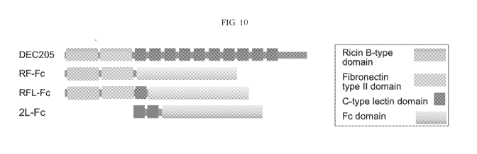
STAMPEDE (Systemic Therapy in Advancing or Metastatic Prostate Cancer:
Evaluation of Drug
CA 03162518 2022-05-20
WO 2021/108637 PCT/US2020/062330
Efficacy) trial, docetaxel improved survival primarily in for men with
metastatic castration-
sensitive prostate cancer. Despite the importance of taxanes in the management
of PCa, its utility
is limited by toxicity and acquisition of chemo-resistance.
[0005] Accordingly, there is a need in the art for treatments that
overcome these issues.
SUMMARY OF THE INVENTION
[0006] The following embodiments and aspects thereof are described and
illustrated
in conjunction with compositions and methods which are meant to be exemplary
and
illustrative, not limiting in scope.
[0007] Various embodiments provide for a protein, comprising: a
polypeptide that binds
to mitochondrial DNA (mtDNA), genomic DNA (gDNA), or both; and a Fc fragment
of IgG
receptor gamma (FcgRIIb) or a fragment thereof.
[0008] In various embodiments, the polypeptide that binds to mtDNA, gDNA
or both can
comprise a fragment of DEC205 or a fragment of DEC205 with one or more amino
acid
deletions, additions or substitutions.
[0009] In various embodiments, the fragment of DEC205 can be a
polypeptide at least
90% identical to at least one domain selected from the group consisting of
Ricin B-type lectin
domain, fibronectin type II lectin domain, and at least one C-type lectin
domain. In various
embodiments, the fragment of DEC205 can be a polypeptide at least 90%
identical to at least two
domains selected from the group consisting of Ricin B-type lectin domain,
fibronectin type II
lectin domain, and at least one C-type lectin domain. In various embodiments,
the fragment of
DEC205 can be a polypeptide at least 90% identical to at least three domains
selected from the
group consisting of Ricin B-type lectin domain, fibronectin type II lectin
domain, and at least one
C-type lectin domain. In various embodiments, the fragment of DEC205 can be a
polypeptide at
least 90% identical to Ricin B-type lectin domain, fibronectin type II lectin
domain, or both. In
various embodiments, the fragment of DEC205 can be a polypeptide at least 90%
identical to
Ricin B-type lectin domain and fibronectin type II lectin domain. In various
embodiments, the
fragment of DEC205 can be a polypeptide at least 90% identical to Ricin B-type
lectin domain,
fibronectin type II lectin domain, and at least one C-type lectin domain. In
various embodiments,
the fragment of DEC205 can be a polypeptide at least 90% identical to at least
one C-type lectin
domain. In various embodiments, the fragment of DEC205 can be a polypeptide at
least 90%
identical to at least two C-type lectin domains. In various embodiments, the
fragment of DEC205
can comprise a polypeptide is at least 90% identical to a sequence selected
from the group
consisting of SEQ ID NO:1, SEQ ID NO:2, and SEQ ID NO:3. In various
embodiments, the
2
CA 03162518 2022-05-20
WO 2021/108637 PCT/US2020/062330
fragment of DEC205 can comprise a polypeptide that has a sequence selected
from the group
consisting of SEQ ID NO:1, SEQ ID NO:2, and SEQ ID NO:3. In various
embodiments, the
fragment of DEC205 can comprise a polypeptide is at least 90% identical to a
sequence
comprising SEQ ID NO:4. In various embodiments, the fragment of DEC205 can
comprise a
polypeptide having at least 168 consecutive amino acids of SEQ ID NO:4. In
various
embodiments, the fragment of DEC205 can comprise a polypeptide having 168 to
414
consecutive amino acids of SEQ ID NO:4. In various embodiments, the fragment
of DEC205 can
comprise a polypeptide having 183 to 368 consecutive amino acids of SEQ ID
NO:4. In various
embodiments, the fragment of DEC205 can comprise a polypeptide having 202 to
322
consecutive amino acids of SEQ ID NO:4. In various embodiments, the fragment
of DEC205 can
comprise a polypeptide having 220 to 276 consecutive amino acids of SEQ ID
NO:4.
[0010] In various embodiments, the Fc fragment of IgG receptor gamma
(FcgRIIb)
comprises a human IgG1 Fc domain or a human IgG1 Fc domain with up to 22 amino
acid
additions, deletions, and/or substitutions. In various embodiments, the Fc
fragment of IgG
receptor gamma (FcgRIIb) or the fragment thereof can comprise at least 205
consecutive amino
acids as set forth in SEQ ID NO:5. In various embodiments, the Fc fragment of
IgG receptor
gamma (FcgRIIb) or the fragment thereof can comprise a sequence with at least
90% sequence
identity with SEQ ID NO:5. In various embodiments, the Fc fragment of IgG
receptor gamma
(FcgRIIb) can comprise a polypeptide having the sequence as set forth in SEQ
ID NO:5.
[0011] In various embodiments, the Fc fragment of IgG receptor gamma
(FcgRIIb) can
be a mouse IgG1 Fc domain, or a mouse IgG1 Fc domain with up to 21 amino acid
additions,
deletions, and/or substitutions. In various embodiments, the Fc fragment of
IgG receptor gamma
(FcgRIIb) or the fragment thereof can comprise at least 209 consecutive amino
acids as set forth
in SEQ ID NO:6. In various embodiments, the Fc fragment of IgG receptor gamma
(FcgRIIb) or
the fragment thereof can comprise a sequence with at least 90% sequence
identity with SEQ ID
NO:6. In various embodiments, the Fc fragment of IgG receptor gamma (FcgRIIb)
can comprise
a polypeptide having the sequence as set forth in SEQ ID NO:6.
[0012] In various embodiments, the protein can further comprise a signal
sequence, a
linker, or both. In various embodiments, the signal sequence can comprise the
amino acids as set
forth in SEQ ID NO:7.
[0013] In various embodiments, the protein can be selected from a protein
having the
sequence as set forth in any one of amino acids 24-435 of SEQ ID NO:8, amino
acids 24-583 of
SEQ ID NO:9, amino acids 24-529 of SEQ ID NO:10, amino acids 24-440 of SEQ ID
NO:11,
amino acids 24-588 of SEQ ID NO:12, or amino acids 24-534 of SEQ ID NO: 13.
3
CA 03162518 2022-05-20
WO 2021/108637 PCT/US2020/062330
[0014] In various embodiments, the protein can be selected from a protein
comprising the
sequence of SEQ ID NO:1 and SEQ ID NO:5; or SEQ ID NO:2 and SEQ ID NO:5; or
SEQ ID
NO:3 and SEQ ID NO:5; or SEQ ID NO:1 and SEQ ID NO:6; or SEQ ID NO:2 and SEQ
ID
NO:6; or SEQ ID NO:3 and SEQ ID NO:6.
[0015] In various embodiments, the protein can be selected from a protein
having the
sequence as set forth in any one of SEQ ID NO:8, SEQ ID NO:9, SEQ ID NO:10,
SEQ ID
NO:11, SEQ ID NO:12, or SEQ ID NO: 13.
[0016] In various embodiments, the polypeptide that binds to mtDNA, gDNA
or both
comprises a fragment of toll-like receptor 9 (TLR9) or a fragment of TLR9 with
one or more
amino acid deletions, additions or substitutions.
[0017] In various embodiments, the protein can further comprise an Fc
region of an
antibody or a fragment thereof.
[0018] In various embodiments, the protein can be capable of depleting
circulating
mtDNA. In various embodiments, the protein can be capable of depleting
circulating genomic
DNA (gDNA).
[0019] Various embodiments of the present invention provide for a nucleic
acid encoding
any one of the proteins of the present invention as described herein.
[0020] Various embodiments of the present invention provide for a cell
producing any
one of the proteins of the present invention.
[0021] Various embodiments of the present invention provide for a cell
comprising any
one of the nucleic acids of the present invention.
[0022] In various embodiments, the cell can be a bacterial cell, a
Chinese hamster
ovarian cell (CHO) or a baby hamster kidney cell (BEIK). In various
embodiments, the bacterial
cell is Bacillus sub tilis or Lactococus lactis.
[0023] Various embodiments of the present invention provide for a
combination,
comprising: any one of the proteins of the present invention; and a
therapeutic agent.
[0024] In various embodiments, the therapeutic agent can be selected from
the group
consisting of an anti-tumor agent, a chemotherapeutic agent, an androgen
ablating agent, a
cardiac infarction treatment agent, a traumatic brain injury treatment agent,
and combinations
thereof. In various embodiments, the therapeutic agent can be a taxane,
anthracycline, or a
platinum based antineoplastic drug. In various embodiments, the therapeutic
agent can be
docetaxel, paclitaxel, cabazataxel, doxorubicin, epirubicin, idarubicin,
valrubicin, cisplatin,
oxaliplatin, carboplatin, irinotecan, or fluorouracil (5FU). In various
embodiments, the
therapeutic agent can be an androgen receptor antagonist, an androgen
synthesis inhibitor, or an
4
CA 03162518 2022-05-20
WO 2021/108637 PCT/US2020/062330
anti-gonadotropin. In various embodiments, the therapeutic agent can be
selected from the group
consisting of bicalutamide, enzalutamide, apalutamide, flutamide, nilutamide,
darolutamide,
cyproterone acetate, megestrol acetate, chlormadinone acetate, spironolactone,
oxendolone,
ketoconazole, abiraterone acetate, seviteronel, aminoglutethimide,
finasteride, dutasteride,
epristeride, alfatradiol, saw palmetto extract, leuprorelin, cetorelix and
combinations thereof. In
various embodiments, the therapeutic agent can be aspirin, a thrombolytic
agent, heparin, an
antiplatelet agent, nitroglycerin, a beta blocker, an ACE inhibitor, a statin,
and combinations
thereof. In various embodiments, the therapeutic agent can be a diuretic, an
anti-seizure drug, a
coma-inducing drug, or combinations thereof.
[0025] Various embodiments of the present invention provide for a device,
comprising:
at least one inlet; at least one outlet; at least one chamber comprising a
solid substrate; and any
one of the proteins of the present invention immobilized on the solid
substrate.
[0026] In various embodiments, the device can be a microfluidic device.
[0027] In various embodiments, the solid substrate can be dextran beads
or sepharose
beads.
[0028] Various embodiments of the present invention provide for a device,
comprising
any one of the proteins of the present invention immobilized onto a solid
substrate.
[0029] In various embodiments, the solid substrate can be a multi-well
plate. In various
embodiments, the solid substrate can be a bead.
[0030] In various embodiments, the protein can be further conjugated or
immobilized to
a conductive substrate to produce a detectable signal upon binding to mtDNA,
gDNA, or both.
[0031] In various embodiments, the conductive substrate can be gold,
silver, platinum,
iridium, or copper.
[0032] In various embodiments, the protein can be further conjugated or
immobilized to
silicone.
[0033] Various embodiments of the present invention provide for a method
of reducing
circulating mitochondrial DNA (mtDNA), genomic DNA (gDNA), or both in a
mammalian
subject, comprising: administering any one of the proteins of the present
invention, or
administering any one of the combinations of the present invention to the
mammalian subject, or
removing circulating mtDNA, gDNA, or both from the mammalian subject's blood,
or
administering any one of the bacterial cells of the present invention to the
mammalian subject.
[0034] In various embodiments, the mammalian subject can have or can be
suspected to
have a disease or condition caused by or related to elevated levels of
circulating mitochondrial
DNA (mtDNA), genomic DNA (gDNA), or both.
CA 03162518 2022-05-20
WO 2021/108637 PCT/US2020/062330
[0035] In various embodiments, the disease or condition can be selected
from the group
consisting of a tumor, cancer, cardiac infarct, cardiac disease, physical
trauma, traumatic brain
injury, infection, stroke, inflammation, autoimmune disease, cachexia, and
lupus.
[0036] In various embodiments, the cancer can be a solid tumor cancer. In
various
embodiments, the cancer can be prostate cancer or breast cancer.
[0037] In various embodiments, removing circulating mtDNA from the
mammalian
subject's blood can comprise passing the subject's blood through any one of
the devices of the
present invention.
[0038] Various embodiments of the present invention provide for a method
of measuring
circulating mitochondrial DNA (mtDNA), genomic DNA, or both comprising:
obtaining a
biological sample; contacting any one of the proteins of the present invention
to the biological
sample; detecting the binding of the protein to the mtDNA, gDNA, or both; and
quantifying the
amount of protein-mtDNA binding conjugate, protein-gDNA binding conjugate, or
both.
[0039] In various embodiments, the protein can further comprise a label
to produce a
detectable signal. In various embodiments, the detectable signal can be
colorimetric,
fluorescence, or luminescence.
[0040] In various embodiments, the protein can be contacted to the
biological sample
using any one of the devices of the present invention.
[0041] In various embodiments, the device can comprise a conductive
substrate and the
protein is conjugated or immobilized to the conductive substrate to produce a
detectable signal
upon binding to mtDNA, gDNA or both, wherein the detectable signal is
impedance, resistance,
change in current, or change in electrochemical impedance spectrum, and
wherein the conductive
substrate is selected from the group consisting of gold, silver, platinum,
iridium, copper.
[0042] Other features and advantages of the invention will become
apparent from the
following detailed description, taken in conjunction with the accompanying
drawings, which
illustrate, by way of example, various features of embodiments of the
invention.
BRIEF DESCRIPTION OF THE FIGURES
[0043] Exemplary embodiments are illustrated in referenced figures. It is
intended that
the embodiments and figures disclosed herein are to be considered illustrative
rather than
restrictive.
[0044] Figure 1, panels A-H, depicts Activation of TLR9 and C3a by mtDNA.
A
Mitochondrial DNA was measured from conditioned medium (CM) of prostatic
epithelia after 48
6
CA 03162518 2022-05-20
WO 2021/108637 PCT/US2020/062330
hrs of incubation (n=3). B Protein expression in CAF treated with LNCaP-CM was
visualized by
western blot. C DEC205 expressions was measured in NAF and CAF treated with
LNCaP-CM
by western blot. D DEC205 was immunoprecipitated, crosslinked, and subjected
to mtDNA PCR
amplification for MT-0O2 following CAF incubation with LNCaP-CM. CAF cell
lysate prior to
immunoprecipitation or IgG immunoprecipitant was used as total input and
negative controls,
respectively. E mRNA expression profiles of the NF-KB signaling targets in CAF
incubated with
LNCaP-CM were compared to control-CM in the heat-map (n=4). F Volcano plot
showing
distribution of differential mRNA expression level in CAF and CAF incubated
with LNCaP-CM.
While only secreted proteins were illustrated in the heatmap, all 84 NF-KB
target genes were
represented in the volcano plot. G TLR9 and anaphylatoxin C3a protein
expression was
visualized in CAF incubated with LNCaP-CM incubated with or without DNasel
treatment.
DNase activity was heat inactivated after 10 min. H LNCaP-CM contain mtDNA
that binds
DEC205 for internalization in CAF cells for subsequent TLR9 signaling and
anaphylatoxin C3a
expression. *P < 0.05, **P < 0.01.
[0045] Figure 2, panels A-G, depicts Mechanism of C3a generation by CAF.
A TLR9
signaling was tested in mouse prostatic fibroblasts from cultured wild type
(WT) or TLR9-
knockout (TLR9-'-) mice treated with CpG-ODN or LNCaP-CM in the presence and
absence of
DNasel treatment. B Secreted C3a was measured by ELISA from CAF and NAF
conditioned
media following treatment with CpG-ODN, LNCaP-CM or TRAMPC2-CM (n=3). C Flow
cytometry was used to quantitate intracellular reactive oxygen by DCFDA
fluorescence in CAF
incubated with control, CpG-ODN or LNCaP-CM as determined by quantitating
DCFDA + cells
absent of 7AAD staining. D Green DCFDA fluorescence was cytoplasmically
localized with
DAPI nuclear counter stain by fluorescence microscopy. The scale bar
represents 16 p.m. E
Catalase activity was quantitated in CAF following incubation with fresh media
(control), CpG-
ODN or LNCaP-CM. F Protein expression of complement C3 and anaphylatoxin C3a
in CAF
was western blotted. The CAF were incubated with either CpG-ODN in the
presence and absence
of catalase inhibitor, 3-Amino-1,2,4-triazole (3AT) or LNCaP-CM in the
presence or absence of
reactive oxygen inhibitor, n-acetyl cysteine (NAC). G MtDNA from in LNCaP-CM
binds
DEC205 for internalization in CAF cells for subsequent TLR9 signaling. LNCaP-
CM inhibited
catalase activity allow ROS production makes C3a in the CAF. *P < 0.05, **P <
0.01, ***P <
0.001, and ns - not significant.
[0046] Figure 3, panels A-E, depicts Role of C3a in PCa progression. A
PCa cell lines
incubated in the absence and presence of C3a receptor agonist peptide (48 hr)
was western blotted
for cell survival and proliferation protein expression. B C57BL/6 mice were
allografted with
7
CA 03162518 2022-05-20
WO 2021/108637 PCT/US2020/062330
tissue recombinants of luciferase-expressing TRAMPC2 with wild type (wt) or
T1r94-fibroblasts.
The mice were treated with saline or TLR9 antagonist, SB290157. Luciferase
bioluminescence
was used to image tumor progression. C Mean tumor volume (mm3) and standard
deviation
(S.D.) for each treatment condition are depicted (n=8). D H&E and
immunohistochemistry for
phosphorylated-AKT, phosphorylated-histone-H3 and TUNEL staining of the tumor
tissues were
performed and quantitated. The corresponding graphs illustrate the mean and
S.D. expression of
the staining (n=4). *P < 0.05; **P < 0.01. The scale bar represents 10 pm. E
FACS analysis of
the tumor tissues demonstrated C3a antagonist and TLR9-knockout fibroblasts
had similar CD3+
T cell infiltration, however their activation state as determined by
CD8+/CD69+ expression
differed significantly, compared to control.
[0047] Figure 4, panels A-G, depicts Docetaxel promotes mtDNA release
from PCa cells
and paracrine TLR9 signaling contribute to therapeutic resistance. A Plasma
levels of mtDNA
was quantified from PCa patients before and after docetaxel treatment (n=9). B
MtDNA content
of plasma from mice treated with docetaxel was quantitated (n=3). Data
represent the mean
S.D., *P<0.05. C MtDNA secreted by PCa cell lines treated with docetaxel was
elevated in a
dose-dependent manner (n=3). Significance was determined by repeated measures
ANOVA. D
LNCaP cells treated with vehicle or docetaxel was subjected to subcellular
fractionation. The
mitochondrial localization of mitophagy markers, p62, Pinkl, and Beclin, was
confirmed by the
co-expression of Tom20. The cytoplasmic fraction was confirmed by the
expression of Rho A. E
MtDNA secretion resulting from ER-stress, as indicated by CHOP expression, was
evident in
LNCaP and PC3 cells treated with docetaxel. F Treatment of a three-dimensional
co-culture
model of PC3 and CAF cells with docetaxel and TLR9 antagonist, 5B290157,
supported
differential epithelial proliferation as determined by quantitating EPCaW/Ki-
67+ cells by FACS
analysis (n=3). G Synergistic cooperativity was identified in PC3 cell
viability measured by MTT
assay following treatment with docetaxel and 5B290157 through the Chou-Talalay
method (n=4).
Values below the confidence interval (CI) of 1 (line) are considered to
indicate a synergistic
combination.
[0048] Figure 5, panels A-C, depicts Synergistic effect of docetaxel and
SB 290157
inhibit tumor growth. A Subcutaneous xenografts of PC3 and CAF tumor volumes
were
longitudinally measured. When tumor average volume reached 80 mm3 mice were
treated with
vehicle or docetaxel in the presence or absence of 5B290157 for 20 days (n =
4). Representative
images show each group of mice (inset). B Immunoblots of the tumor tissues for
the respective
treatments are demonstrated (n=3). C Immunolocalization of phosphorylated-
TAK1, complement
C3, phosphorylated-AKT, phosphorylated-histoneH3 and TUNEL expression in tumor
tissues
8
CA 03162518 2022-05-20
WO 2021/108637 PCT/US2020/062330
(brown) was counterstained with hematoxylin (blue). The corresponding bar
graph illustrate the
mean and S.D. expression of the respective staining (n=5). Data represent the
mean S.D. by
one-way ANOVA (*P <0.05; **P <0.01). The scale bar represents 10 pm.
[0049] Figure 6 depicts Schematic illustration of the PCa epithelia and
CAP' reciprocal
interaction. PCa cells generate mtDN,A. that can bind endocytic DEC205 on the
cell surface of
CAF. TLR9 signaling downstream of epithelial-derived nitDNA results in NF-KB
mediated C3
expression. The accumulation of ROS in CAF enables Oa maturation and paracrine
signaling
with PCa cells that enables cell survival and proliferation. Docetaxel
treatment of PCa cells
potentiate ER stress and mitophagy for the expanded secretion of mtDNA in
perpetuating the
further C3a expression by CAF.
[0050] Figure 7, panels A-F, depicts A Relative mRNA expression of TLR9
was
measured in the presence and absence of BPH1 conditioned medium (CM) and LNCaP-
CM in
cultured NAF or CAF. B Measurement of telomere and mitochondrial DNA
concentration from
conditioned medium of cultured human prostate cancer cells. C Protein
expression of caspasel
and IL-10 from cultured CAF treated with LNCaP-CM. Lower molecular weight
cleaved-
caspasel and mature active-IL1f3 induced by LNCaP-CM was limited by DNasel
treatment and
subsequent heat inactivation. s-actin expression was used as a loading
control. D LNCaP-CM
induced TLR9 mRNA expression by cultured CAF was limited by DNasel, but not
sonication of
the conditioned media. E Inhibition of dynamin-mediated exosome secretion with
increasing
doses of dynasore had no effect on the secretion of mtDNA by LNCaP cells. F
Protein expression
of HMGB1 and HMGA2 by NAF and CAF was subjected to western blotting following
LNCaP-
CM treatment. *P < 0.05, **P < 0.01, ***P < 0.001.
[0051] Figure 8, panels A-C, depicts A C3a receptor (C3a-R) mRNA
expression was
similarly expressed by cultured LNCaP, PC3 and TrampC2 cells. B Western blot
for the
expression of DEC205, TLR9, HMGB1, and C3a was performed on the indicated PCa
epithelial
cell lines. C Proliferation of LNCaP, PC3 and TrampC2 cells was quantitated by
measuring Ki-
67 through FACS analysis following treatment with C3aR agonist or scrambled
peptides for 48 h,
(n = 3).
[0052] Figure 9, panels A-C, depicts A nn interaction index and
confidence intervals
were calculated by Chou-Talalay method for determining a synergistic
relationship between
5B290157 and docetaxel treatment at the indicated treatment concentrations of
PC3 cells by the
MTT viability assay. B Mice harboring subcutaneous xenografts of PC3/CAF
tumors were
weighed throughout the saline, docetaxel alone, or combination with SB290157
treatment course.
9
CA 03162518 2022-05-20
WO 2021/108637 PCT/US2020/062330
Data represent the mean S.D. among groups by oneway ANOVA (ns - not
significant). C H&E
images of resulting tumors from each treatment group of subcutaneous
xenografted mice.
[0053] Figure 10 depicts the extracellular domain of DEC205 contain
multiple lectin
domains: Ricin B-type lectin, fibronectin type II lectin domain, and ten C-
type lectin domains.
Three antibody Fc domain conjugates were generated containing the Ricin B-type
and fibronectin
type II domains (RF-Fc), the Ricin B-type, fibronectin type II domains and C-
type lectin (RFL-
Fc), and two C-type lectin domains.
[0054] Figure 11 depicts three DEC205 fragments, RF, RFL, and 2L
conjugated to the
IgG1 Fc domain. Conditioned media from CHO-Kl cells stably expressing the
respective
constructs were subjected to protein G affinity purification, run on 10%
acrylamide gel, and
visualized by Coomassie staining.
[0055] Figure 12 depicts ELISA testing the binding of RF-Fc and RFL-Fc of
(A) mtDNA
and (B) gDNA. RF-Fc binds mtDNA 2-fold over gDNA. RFL-Fc has similar capacity
to bind
mtDNA and gDNA. Absorbance was taken at 570 nm. OD values are normalized for
their
respective Fc concentrations. **P < 0.01, ***P < 0.001, ****P < 0.0001.
[0056] Figure 13 depicts The basis for docetaxel resistance potentiated
by mtDNA is the
expression of complement C3 by cancer associated fibroblastic cells (PNAS 2020
11:8515).
When conditioned media from prostate cancer cells (PC3) were incubated with
cancer associated
fibroblastic cells C3 expression was significantly downregulated by the
depletion of mtDNA
using RF-Fc. **P < 0.01.
DESCRIPTION OF THE INVENTION
[0057] All references cited herein are incorporated by reference in their
entirety as
though fully set forth. Unless defined otherwise, technical and scientific
terms used herein have
the same meaning as commonly understood by one of ordinary skill in the art to
which this
invention belongs. Singleton et al., Dictionary of Microbiology and Molecular
Biology 3rd ed.,
Revised, J. Wiley & Sons (New York, NY 2006); March, Advanced Organic
Chemistry
Reactions, Mechanisms and Structure 7th ed., J. Wiley & Sons (New York, NY
2013); and
Sambrook and Russel, Molecular Cloning: A Laboratory Manual zith ed., Cold
Spring Harbor
Laboratory Press (Cold Spring Harbor, NY 2012), provide one skilled in the art
with a general
guide to many of the terms used in the present application. For references on
how to prepare
antibodies, see D. Lane, Antibodies: A Laboratory Manual 2nd ed. (Cold Spring
Harbor Press,
Cold Spring Harbor NY, 2013); Kohler and Milstein, (1976) Eur. J. Immunol. 6:
511; Queen et
al. U. S. Patent No. 5,585,089; and Riechmann et al., Nature 332: 323 (1988);
U.S. Pat. No.
CA 03162518 2022-05-20
WO 2021/108637 PCT/US2020/062330
4,946,778; Bird, Science 242:423-42 (1988); Huston etal., Proc. Natl. Acad.
Sci. USA 85:5879-
5883 (1988); Ward et al., Nature 334:544-54 (1989); Tomlinson I. and Holliger
P. (2000)
Methods Enzymol, 326, 461-479; Holliger P. (2005) Nat. Biotechnol.
Sep;23(9):1126-36).
[0058] One
skilled in the art will recognize many methods and materials similar or
equivalent to those described herein, which could be used in the practice of
the present invention.
Indeed, the present invention is in no way limited to the methods and
materials described. For
purposes of the present invention, the following terms are defined below.
[0059] As
used herein the term "about" when used in connection with a referenced
numeric indication means the referenced numeric indication plus or minus up to
5% of that
referenced numeric indication, unless otherwise specifically provided for
herein. For example, the
language "about 50%" covers the range of 45% to 55%. In various embodiments,
the term
"about" when used in connection with a referenced numeric indication can mean
the referenced
numeric indication plus or minus up to 9%, 8%, 7%, 6%, 5%, 4%, 3%, 2%, or 1%
of that
referenced numeric indication, if specifically provided for in the claims.
[0060] The
term "biological sample" as used herein denotes a sample taken or isolated
from
a biological organism. Exemplary biological samples include, but are not
limited to body fluids,
whole blood, plasma, serum, stool, intestinal fluids or aspirate, and stomach
fluids or aspirate,
cerebral spinal fluid (CSF), urine, sweat, saliva, tears, pulmonary
secretions, breast aspirate, prostate
fluid, seminal fluid, cervical scraping, amniotic fluid, intraocular fluid,
mucous, and moisture in
breath. In various embodiments, the biological sample may be whole blood. In
various
embodiments, the biological sample may be serum. In various embodiments, the
biological sample
may be plasma. The term also includes a mixture of the above-mentioned
samples.
[0061] As
used herein, the term "label" refers to a composition capable of producing a
detectable signal indicative of the presence of a target. Suitable labels
include fluorescent
molecules, radioisotopes, nucleotide chromophores, enzymes, substrates,
chemiluminescent
moieties, magnetic particles, bioluminescent moieties, and the like. As such,
a label is any
composition detectable by spectroscopic, photochemical, biochemical,
immunochemical,
electrical, optical or chemical means needed for the methods and devices
described herein. For
example, the peptides can be labeled with a detectable tag which can be
detected using an
antibody specific to the label.
[0062]
Exemplary fluorescent labeling reagents include, but are not limited to,
Hydroxycoumarin, Succinimidyl ester, Aminocoumarin, Methoxycoumarin, Cascade
Blue,
Hydrazide, Pacific Blue, Maleimide, Pacific Orange, Lucifer yellow, NBD, NBD-
X, R-
Phycoerythrin (PE), a PE-Cy5 conjugate (Cychrome, R670, Tri-Color, Quantum
Red), a PE-Cy7
11
CA 03162518 2022-05-20
WO 2021/108637 PCT/US2020/062330
conjugate, Red 613, PE-Texas Red, PerCP, Peridinin chlorphyll protein, TruRed
(PerCP-Cy5.5
conjugate), FluorX, Fluoresceinisothyocyanate (FITC), BODIPY-FL, TRITC, X-
Rhodamine
(XRITC), Lissamine Rhodamine B, Texas Red, Allophycocyanin (APC), an APC-Cy7
conjugate,
Alexa Fluor 350, Alexa Fluor 405, Alexa Fluor 430, Alexa Fluor 488, Alexa
Fluor 500, Alexa
Fluor 514, Alexa Fluor 532, Alexa Fluor 546, Alexa Fluor 555, Alexa Fluor 568,
Alexa Fluor
594, Alexa Fluor 610, Alexa Fluor 633, Alexa Fluor 647, Alexa Fluor 660, Alexa
Fluor 680,
Alexa Fluor 700, Alexa Fluor 750, Alexa Fluor 790, Cy2, Cy3, Cy3B, Cy3.5, Cy5,
Cy5.5 or Cy7.
[0063] Percent (%) sequence identity with respect to a reference
polypeptide sequence is
the percentage of amino acid residues in a candidate sequence that are
identical with the amino acid
residues in the reference polypeptide sequence, after aligning the sequences
and introducing gaps, if
necessary, to achieve the maximum percent sequence identity, and not
considering any conservative
substitutions as part of the sequence identity. Alignment for purposes of
determining percent amino
acid sequence identity can be achieved in various ways that are known for
instance, using publicly
available computer software such as BLAST, BLAST-2, ALIGN or Megalign
(DNASTAR)
software. Appropriate parameters for aligning sequences are able to be
determined, including
algorithms needed to achieve maximal alignment over the full length of the
sequences being
compared. For purposes herein, however, % amino acid sequence identity values
are generated
using the sequence comparison computer program ALIGN-2. The ALIGN-2 sequence
comparison
computer program was authored by Genentech, Inc., and the source code has been
filed with user
documentation in the U.S. Copyright Office, Washington D.C., 20559, where it
is registered under
U.S. Copyright Registration No. TXU510087. The ALIGN-2 program is publicly
available from
Genentech, Inc., South San Francisco, Calif, or may be compiled from the
source code. The
ALIGN-2 program should be compiled for use on a UNIX operating system,
including digital
UNIX V4.0D. All sequence comparison parameters are set by the ALIGN-2 program
and do not
vary.
[0064] In situations where ALIGN-2 is employed for amino acid sequence
comparisons,
the % amino acid sequence identity of a given amino acid sequence A to, with,
or against a given
amino acid sequence B (which can alternatively be phrased as a given amino
acid sequence A that
has or comprises a certain % amino acid sequence identity to, with, or against
a given amino acid
sequence B) is calculated as follows: 100 times the fraction X/Y, where X is
the number of amino
acid residues scored as identical matches by the sequence alignment program
ALIGN-2 in that
program's alignment of A and B, and where Y is the total number of amino acid
residues in B. It
will be appreciated that where the length of amino acid sequence A is not
equal to the length of
amino acid sequence B, the % amino acid sequence identity of A to B will not
equal the % amino
12
CA 03162518 2022-05-20
WO 2021/108637 PCT/US2020/062330
acid sequence identity of B to A. Unless specifically stated otherwise, all %
amino acid sequence
identity values used herein are obtained as described in the immediately
preceding paragraph using
the ALIGN-2 computer program.
[0065] Described herein, we examined the role of the PCa microenvironment
in
docetaxel chemo-resistance. Stromal-epithelial interactions define tumor
initiation, progression,
and therapeutic resistance. In the prostate tumor microenvironment, stromal
fibroblasts co-evolve
with the cancer epithelia in a reciprocal relationship. The central role of
cancer-associated
fibroblasts (CAF) was recognized when their absence was found to result in
reduced tumor
volumes. CAF have been shown to produce paracrine growth factors, proteolytic
enzymes and
components of the extracellular matrix, presumably in response to cues from
tumor cells. In fact,
CAF derived from breast cancer patients treated with docetaxel were found to
secrete greater
tumor supportive factors compared to the CAF derived from treatment-naïve
patients. However,
the mechanisms regulating this crosstalk occurs are not well elucidated in the
context of
chemotherapy.
[0066] There is a large body of evidence describing the role of
mitochondrial DNA
(mtDNA) in PCa. Proteins in mitochondrial complexes I, III, IV, and V involved
in oxidative
phosphorylation are encoded by mtDNA. Mutations found in mtDNA increase
tumorigenicity in
PCa and deregulated mitochondrial metabolism is known to promote prostate
carcinogenesis.
PCa cells have greater mitochondrial content than benign prostate epithelium
and alterations in
mtDNA copy number may reflect disruption of the normal prostate glandular
architecture.
Furthermore, mtDNA instability is a hallmark of human cancers. PCa patients
are found to have
measurable concentrations of mtDNA in serum. Further described herein, we
tested whether
secreted mtDNA functions as a mediator of epithelia-CAF crosstalk. We reasoned
the mtDNA
could potentially signal adjacent cells through pattern recognition receptors,
such as toll-like
receptor 9 (TLR9). We identified a stromal-epithelia reciprocal signaling
cascade initiated by
docetaxel involving the TLR9 signaling in CAF and downstream paracrine
response by PCa
epithelia contributing to taxane therapy resistance.
[0067] As further described herein mitochondrial DNA (mtDNA) is expelled
by cells
undergoing stress, often in the form of endoplasmic reticulum stress (ER
stress), in response to
stimuli such as chemotherapy, androgen ablation treatment, cardiac infarction,
and traumatic
brain injury. In each case, mtDNA released by the cells can be perceived by
neighboring or
potentially distant cells through a specialized receptor, Toll like receptor 9
(TLR9). TLR9
signaling can promote an inflammatory cascade that causes recruitment of
inflammatory cells,
13
CA 03162518 2022-05-20
WO 2021/108637 PCT/US2020/062330
promotion of tumor cell growth, and cause longer term ramifications such as
increased risk for a
cardiac event or dementia associated diseases of the brain. Therefore, ridding
the body of mtDNA
so that TLR9 is not activated, can prevent the downstream inflammatory signals
that contribute to
multiple pathologies. The inventors' found mtDNA's impact on tumor expansion
and therapeutic
resistance. The inventors further designed methods by which mtDNA is depleted
from circulation
by an engineered antibody inclusive of the application TLR9 mtDNA-binding
domain and
DEC205 as a method of capturing mtDNA for excretion by targeting to the
hepatic vasculature.
[0068] Inflammation suppressors such as steroids and non-steroidal
analgesics are
available. But, there are no inhibitors that remove the initiator of such
inflammatory cascades
associated with mtDNA secretion. We have designed methods of capturing mtDNA
from
circulation by the use of an antibody variable region mimicking TLR9 or
DEC205.
[0069] The work provides an advance in functionally defining the cross-
talk of tumor
epithelia with cancer-associated fibroblastic cells contributing to tumor
progression and
therapeutic resistance. Independent of protein-based signaling molecules,
prostate cancer cells
secreted mitochondrial DNA to induce associated fibroblasts to generate
anaphylatoxin C3a to
support tumor progression in a positive feed-back loop. Interestingly, the
standard of care
chemotherapy, docetaxel, used to treat castrate resistant prostate cancer was
found to further
potentiate this novel paracrine signaling axis to mediate therapeutic
resistance. Blocking
anaphylatoxin C3a signaling cooperatively sensitized prostate cancer tumors to
docetaxel. We
revealed that docetaxel resistance is not a cancer cell-autonomous phenomena
and targeting an
immune modulator derived from cancer associated fibroblasts can limit the
expansion of
docetaxel-resistant tumors.
[0070] Our data show reciprocal paracrine signaling between PCa and
associated
fibroblasts promote cancer progression and facilitates docetaxel resistance.
We hypothesized
mtDNA could be the paracrine signaling molecule generated by PCa cells (Figure
6). The
docetaxel-induced mtDNA secretion from PCa cells into the tumor
microenvironment was
significantly greater than the basal levels of mtDNA secreted by PCa cells.
Accordingly, prostate
tumors in both murine models and men harboring prostate tumors demonstrated
elevated
circulating mtDNA when treated with docetaxel. For subsequent CAF signaling,
the mtDNA
required entry into the cytoplasm for TLR9 activation. Based on the previous
demonstration of
DEC205 capture of CpG in dendritic cells (24), a similar scenario was explored
for the prostatic
CAF. Instead of unmethylated bacterial DNA, we demonstrated that in fact,
DEC105 could
directly bind mtDNA on CAF cells for classic pattern recognition receptor,
TLR9, activation of
TAK1 and NF-KB (37). TLR9 was identified to be essential for complement C3
expression by
14
CA 03162518 2022-05-20
WO 2021/108637 PCT/US2020/062330
CAF in response to mtDNA, but the accumulation of reactive oxygen resulting
from PCa-CM
contributed to C3 cleavage and anaphylatoxin C3a generation. Released C3a in
the tumor
microenvironment increased proliferation of cancer cells and potentiated
resistance to docetaxel
treatment.
[0071] It is apparent that PCa-induced paracrine NF--03 activation in CAF
dramatically
potentiated complement C3 expression (>12 log-fold Figure 1). There is a well-
described
immune defense for bacterial pathogens that include Toll-like receptor-
mediated complement
expression and generation of anaphylatoxin. However, the novel mechanism of
TLR9 induction
by PCa-derived mtDNA paracrine signal transduction mechanism in CAF cells was
not observed
in NAF cells (Figure 1). Cell-free circulating mtDNA release in plasma at low
levels under
cellular stress is reported, in instances of cancer, trauma, infections,
stroke, autoimmune,
metabolic and rheumatic diseases. While activated T cells can signal dendritic
cells through
exosome-based delivery of mtDNA, this was likely not the means of paracrine
communication
between PCa and CAF. Dynamin inhibition or sonication of the PCa-CM had little
effect on
TLR9 expression/activity by CAF (Figure 7). The especially low levels of
telomeric DNA
secreted by the PCa is noteworthy as it is a known to inhibit TLR9 signaling.
Uniquely, DEC205
was expressed by CAF in the context of PCa-CM for endocytic delivery of mtDNA
and TLR9
activation. This is the first time PCR amplification of the mitochondrial MT-
0O2 gene following
immunoprecipitation of DEC205 has been reported. Docetaxel potentiated PCa
release of
mtDNA by over 5-fold (Figures 1 and 4). Docetaxel treatment is reported to
induce mTOR-
mediated autophagy in prostate cancer cells. Treatment with chemotherapeutic
drugs can cause
ER-stress that enhance autophagic efflux from cells. Our identification of the
combined ER-stress
with mitophagy revealed means for the secretion of non-degraded mtDNA from PCa
cells (Figure
2). Thus, the initiation of the fibroblastic inflammatory cascade can be
attributed to tumor-derived
mtDNA signaling and the expression of complement C3. However, the activation
of the
complement system in response to pathogens involves three major pathways: 1)
the classical
pathway, via antigen-antibody complexes, 2) the lectin pathway, via binding of
pattern-
recognizing mannose-binding lectins, and 3) the alternative pathway, via any
permissive microbe
surfaces. In all three complement activation pathways, the C3 convertase
complex cleaves C3
molecules to form anaphylatoxins C3a. Yet another mechanism of C3 conversion
identified in
neutrophils involving hydrogen peroxide-related oxygen radicals, such as
hypochlorite, was the
mechanism considered for the stromal-epithelial signaling axis. We found that
catalase inhibition
in CAF by PCa cells to be essential for ROS accumulation and maturation of
anaphylatoxin C3a
from C3 (Figure 2). These findings explained the absence of C3a in CpG-ODN
treated CAF cells
CA 03162518 2022-05-20
WO 2021/108637 PCT/US2020/062330
in spite of NF-x13 activation. Tumor-stromal interaction via mtDNA resulting
in C3a expression
by prostate fibroblasts was dependent on TLR9 activation and ROS-mediated
complement
maturation.
[0072] Our findings provide a paradigm in which the activation of
complement was
distinctly important for promoting tumor growth. There are studies that have
reported a positive-
growth effect of complement in cancer. The systemic level of complement
proteins has an
indirect effect on cancer growth by alteration of the immune response of host
to the tumor. Wang
et. al. showed that B16 melanoma growth was slower in C3 deficient mice than
that in wild-type
mice. Anaphylatoxin receptors signal through the PI3K/AKT pathway in cancer
cells and the
proliferative effect of C5aR and C3aR stimulation can be eliminated by AKT
silencing. Here, we
show PCa cells express receptors for C3a (Figure 8). The CAF-derived C3a
resulted in
phosphorylated-AKT, phosphorylated-ERK1/2 and BCL2 upregulation in PCa
epithelia (Figure
3). Antagonizing the TLR9-C3a axis with SB290157 or stromal knockout of TLR9
significantly
inhibited tumor expansion. We found a similar CD3+ T cells infiltration
regardless of TLR9-C3a
signaling alterations. However, CD8+/CD69+ activated cytotoxic T cells were
significantly
reduced by C3 antagonist and further diminished to nearly a third of control
in tumors with
TLR9-knockout fibroblasts. Accordingly, T cell-mediated tumor cell lysis was
not the mechanism
of the observed reduction in tumor size. Instead, C3a was likely acting
directly on the tumor cells
in a paracrine manner.
[0073] Docetaxel resistance is major clinical problem in many cancers
including PCa.
Activation of several survival signaling pathways can promote a resistant
phenotype in response
to docetaxel treatment. Docetaxel and complement signaling in PCa epithelia
were observed to
activate such survival signaling pathways (e.g. AKT and ERK with BCL2
expression) as well as
autophagy (Figures 3 and 4). While autophagy in itself is a means of survival
for neighboring
cells through cellular catabolism, here we showed it also contributes to
docetaxel-induced
mtDNA secretion from PCa cells in its extension to mitophagy. The breakdown of
mitochondria
through mitophagy is inclusive of its DNA. However, in the context of ER
stress, mitophagy can
result in inadequate mtDNA degradation. Not surprisingly, docetaxel elicited
ER stress on the
PCa cells. The contribution of the CAF on PCa ER stress, while likely, was not
explored.
However, the CAF reciprocated the PCa-derived mtDNA signal by the TLR9-C3
paracrine axis
to trigger a survival/proliferation signal in PCa cells. Investigation from
mouse prostate tumor
revealed docetaxel treatment enhanced C3a anaphylatoxin formation and mediated
increased
proliferation signaling. This proliferation signaling was reduced by blocking
of C3a receptor
(Figure 4). Remarkably, antagonizing anaphylatoxin C3a signaling with SB290157
was able to
16
CA 03162518 2022-05-20
WO 2021/108637 PCT/US2020/062330
sensitize an otherwise resistant PC3 cell line to docetaxel. The synergism of
docetaxel and
SB290157 allowed for reduced docetaxel doses to effectively limit tumor
growth. Better
understanding of the complement signaling axis in cancer cells is needed, as
its implications can
have a far-reaching impact on many cancer types currently treated with
taxanes. Currently,
docetaxel is in clinical trials in combination with immune checkpoint
inhibition therapy to
explore the potential for cooperative activity in stimulating infiltrating
cytotoxic T cells. The
induction of the stromal anaphylatoxin C3a mediated by docetaxel may
contribute to immune-
mediated cancer cell death (Figure 3). We observed combining SB290157 with
docetaxel did not
result in greater apoptosis compared to docetaxel alone (Figure 5). But,
complement inhibition
significantly limited proliferation and effectively reduced tumor size over
that of docetaxel alone.
Thus, one must weigh the benefits to immune-surveillance induced by docetaxel
with the tumor
intrinsic proliferative role of complement signaling.
[0074] Another implication of our results is that fibroblast response to
taxane therapy is
consequential to cancer epithelial therapeutic response. Circulating mtDNA has
been reported to
be a prognostic indicator for poor outcome for PCa patients. But, with the
limited number of
patients analyzed, we were unable to demonstrate a correlation with the level
of mtDNA in
circulation with length of docetaxel responsiveness. While the epithelial
response to docetaxel
can be uncoupled from that of the stromal fibroblasts, the stromal influence
on therapeutic
resistance is a result of a paracrine signaling axis, in this report,
initiating from the PCa epithelia.
Again, we cannot rule out the direct impact of docetaxel on CAF that could
also influence
epithelial viability. It should be noted that TLR-mediated NF-KB signaling is
not a phenomenon
limited to mammals. It was originally identified in Drosophila (Toll), with
To119 involved in
hematopoietic and digestive tract development. Although NF-KB regulation
remains conserved
the gene targets are species, tissue, and cell type specific ¨ in this case,
it seems to be dependent
on DEC205 expression. The fact that NF-KB exquisitely mediates fibroblastic
complement C3
expression and acts as a repurposing of a signaling axis for chemotherapy
resistance suggests the
hardwiring of this pathway originates in mesenchymal cells.
[0075] The inventors describe the compositions, therapies, detection of
mtDNA and
gDNA and diagnostics of the present invention, based in part by these
findings.
Agents and Compositions
[0076] Various embodiments of the present invention provide for a
protein. The protein
is useful for binding cell free, circulating mtDNA, genomic DNA (gDNA) and
depleting the
circulating mtDNA and gDNA from circulation. The protein is structurally
similar to an antibody
17
CA 03162518 2022-05-20
WO 2021/108637 PCT/US2020/062330
wherein a fragment of the protein binds to circulating mtDNA, gDNA or both,
and a fragment of
the protein directs the entire protein to the liver for processing and
removing of the mtDNA,
gDNA or both. In various embodiments, these two fragments are on an antibody
backbone to
maintain or extend circulatory half-life.
[0077] Various embodiments of the present invention provide for a
protein, comprising:
a polypeptide that binds to mitochondrial DNA (mtDNA); and a Fc fragment of
IgG receptor
gamma (FcgRIIb) or a fragment thereof.
[0078] Various embodiments of the present invention provide for a
protein, comprising:
a polypeptide that binds to genomic DNA (gDNA); and a Fc fragment of IgG
receptor gamma
(FcgRIIb) or a fragment thereof
[0079] Various embodiments of the present invention provide for a
protein, comprising:
a polypeptide that binds to both mitochondrial DNA (mtDNA) and genomic DNA
(gDNA); and a
Fc fragment of IgG receptor gamma (FcgRIIb) or a fragment thereof
[0080] In various embodiments, the polypeptide that binds to mtDNA, gDNA
or both
comprises a fragment of DEC205 or a fragment of DEC205 with one or more amino
acid
deletions, additions or substitutions. In various embodiments, there are 1-10,
11-20, 21-30, 31-
40, 41-50, 51-60, 61-70, 71-80, 81-90, or 91-100 amino acid deletions,
additions or substitutions.
[0081] In various embodiments, the fragment of DEC205 is a polypeptide at
least 90%
identical to at least one domain selected from the group consisting of Ricin B-
type lectin domain,
fibronectin type II lectin domain, and at least one C-type lectin domain. In
various embodiments,
the fragment of DEC205 is a polypeptide at least 95, 96, 97, 98 or 99%
identical to at least one
domain selected from the group consisting of Ricin B-type lectin domain,
fibronectin type II
lectin domain, and at least one C-type lectin domain. In various embodiments,
the fragment of
DEC205 is a polypeptide comprising at least one domain selected from the group
consisting of
Ricin B-type lectin domain, fibronectin type II lectin domain, and at least
one C-type lectin
domain.
[0082] In various embodiments, the fragment of DEC205 is a polypeptide at
least 90%
identical to at least two domains selected from the group consisting of Ricin
B-type lectin
domain, fibronectin type II lectin domain, and at least one C-type lectin
domain. In various
embodiments, the fragment of DEC205 is a polypeptide at least 95, 96, 97, 98,
or 99 % identical
to at least two domains selected from the group consisting of Ricin B-type
lectin domain,
fibronectin type II lectin domain, and at least one C-type lectin domain. In
various embodiments,
the fragment of DEC205 is a polypeptide comprising at least two domains
selected from the
18
CA 03162518 2022-05-20
WO 2021/108637 PCT/US2020/062330
group consisting of Ricin B-type lectin domain, fibronectin type II lectin
domain, and at least one
C-type lectin domain.
[0083] In various embodiments, the fragment of DEC205 is a polypeptide at
least 90%
identical to at least three domains selected from the group consisting of
Ricin B-type lectin
domain, fibronectin type II lectin domain, and at least one C-type lectin
domain. In various
embodiments, the fragment of DEC205 is a polypeptide at least 95, 96, 97, 98
or 99% identical to
at least three domains selected from the group consisting of Ricin B-type
lectin domain,
fibronectin type II lectin domain, and at least one C-type lectin domain. In
various embodiments,
the fragment of DEC205 is a polypeptide comprising at least three domains
selected from the
group consisting of Ricin B-type lectin domain, fibronectin type II lectin
domain, and at least one
C-type lectin domain.
[0084] In various embodiments, the fragment of DEC205 is a polypeptide at
least 90%
identical to Ricin B-type lectin domain, fibronectin type II lectin domain, or
both. In various
embodiments, the fragment of DEC205 is a polypeptide at least 95, 96, 97, 98
or 99% identical to
Ricin B-type lectin domain, fibronectin type II lectin domain, or both. In
various embodiments,
the fragment of DEC205 is a polypeptide comprises Ricin B-type lectin domain,
fibronectin type
II lectin domain, or both.
[0085] In various embodiments, the fragment of DEC205 is a polypeptide at
least 90%
identical to Ricin B-type lectin domain and fibronectin type II lectin domain.
In various
embodiments, the fragment of DEC205 is a polypeptide at least 95, 96, 97, 98
or 99% identical to
Ricin B-type lectin domain and fibronectin type II lectin domain. In various
embodiments, the
fragment of DEC205 is a polypeptide comprises Ricin B-type lectin domain and
fibronectin type
II lectin domain.
[0086] In various embodiments, the fragment of DEC205 is a polypeptide at
least 90%
identical to Ricin B-type lectin domain, fibronectin type II lectin domain,
and at least one C-type
lectin domain. In various embodiments, the fragment of DEC205 is a polypeptide
at least 95, 96,
97, 98 or 99% identical to Ricin B-type lectin domain, fibronectin type II
lectin domain, and at
least one C-type lectin domain. In various embodiments, the fragment of DEC205
is a
polypeptide comprising Ricin B-type lectin domain, fibronectin type II lectin
domain, and at least
one C-type lectin domain.
[0087] In various embodiments, the fragment of DEC205 is a polypeptide at
least 90%
identical to at least one C-type lectin domain. In various embodiments, the
fragment of DEC205
is a polypeptide at least 95, 96, 97, 98 or 99% identical to at least one C-
type lectin domain. In
19
CA 03162518 2022-05-20
WO 2021/108637 PCT/US2020/062330
various embodiments, the fragment of DEC205 is a polypeptide comprising at
least one C-type
lectin domain.
[0088] In various embodiments, the fragment of DEC205 is a polypeptide at
least 90%
identical to at least two C-type lectin domains. In various embodiments, the
fragment of DEC205
is a polypeptide at least 95, 96, 97, 98 or 99% identical to at least two C-
type lectin domains. In
various embodiments, the fragment of DEC205 is a polypeptide comprising at
least two C-type
lectin domains.
[0089] There are 10 C-type lectin domains on DEC205; thus, in various
embodiments of
the present invention, the at least one C-type lectin domain can be 1, 2, 3,
4, 5, 6, 7, 8, 9 or 10 C-
type lectin domains.
[0090] In various embodiments, the fragment of DEC205 is a polypeptide at
least 90, 95,
96, 97, 98 or 99% identical 3, 4, 5, 6, 7, 8, 9 or 10 C-type lectin domains.
In various
embodiments, the fragment of DEC205 is a polypeptide comprising 3,4, 5, 6, 7,
8, 9 or 10 C-type
lectin domains.
[0091] In various embodiments, the fragment of DEC205 comprises a
polypeptide is at
least 90% identical to a sequence selected from the group consisting of SEQ ID
NO:1, SEQ ID
NO:2, and SEQ ID NO:3. In various embodiments, the fragment of DEC205
comprises a
polypeptide is at least 95, 96, 97, 98 or 99% identical to a sequence selected
from the group
consisting of SEQ ID NO:1, SEQ ID NO:2, and SEQ ID NO:3. In various
embodiments, the
fragment of DEC205 comprises a polypeptide that has a sequence selected from
the group
consisting of SEQ ID NO:1, SEQ ID NO:2, and SEQ ID NO:3.
[0092] In various embodiments, the fragment of DEC205 comprises a
polypeptide is at
least 90% identical to a sequence comprising SEQ ID NO:4. In various
embodiments, the
fragment of DEC205 comprises a polypeptide is at least 95, 96, 97, 98, or 99%
identical to a
sequence comprising SEQ ID NO:4. In various embodiments, the fragment of
DEC205 comprises
a polypeptide having the sequence as set forth in SEQ ID NO:4.
[0093] In various embodiments, the fragment of DEC205 comprises a
polypeptide
having at least 168 consecutive amino acids of SEQ ID NO:4. In various
embodiments, the
fragment of DEC205 comprises a polypeptide having 168 to 414 consecutive amino
acids of SEQ
ID NO:4. In various embodiments, the fragment of DEC205 comprises a
polypeptide having 183
to 368 consecutive amino acids of SEQ ID NO:4. In various embodiments, the
fragment of
DEC205 comprises a polypeptide having 202 to 322 consecutive amino acids of
SEQ ID NO:4.
In various embodiments, the fragment of DEC205 comprises a polypeptide having
220 to 276
consecutive amino acids of SEQ ID NO:4. The determination of consecutive amino
acids can
CA 03162518 2022-05-20
WO 2021/108637 PCT/US2020/062330
start at amino acid number 1-292 of SEQ ID NO:4. For example, if it started at
amino acid
number 292, it will include all the amino acids until the end of SEQ ID NO:4.
In various
embodiments, these fragments of DEC205 has one or more amino acid additions,
deletions or
substitutions; for example, 1-5, 6-10, 11-15, 16-20 or 21-25 amino acid
additions, deletions or
substitutions.
[0094] In various embodiments, the Fc fragment of IgG receptor gamma
(FcgRIIb)
comprises a human IgG1 Fc domain, or a human IgG1 Fc domain with up to 22
amino acid
additions, deletions, and/or substitutions. In various embodiments, it has 1,
2, 3, 4, 5, 6, 7, 8, 9,
10, 11, 12, 13, 14, 15, 16, 17, 18, 19, 20, 21, or 22 amino acid additions,
deletions, and/or
substitutions.
[0095] In various embodiments, the Fc fragment of IgG receptor gamma
(FcgRIIb) or the
fragment thereof comprises at least 205 consecutive amino acids as set forth
in SEQ ID NO:5. In
various embodiments, the Fc fragment of IgG receptor gamma (FcgRIIb) or the
fragment thereof
comprises 205-215, 216-227 consecutive amino acids as set forth in SEQ ID
NO:5. The
determination of consecutive amino acids can start at amino acid number 1-22.
[0096] In various embodiments, the Fc fragment of IgG receptor gamma
(FcgRIIb) or the
fragment thereof comprises a sequence with at least 90% sequence identity with
SEQ ID NO:5.
In various embodiments, the Fc fragment of IgG receptor gamma (FcgRIIb) or the
fragment
thereof comprises a sequence with at least 95, 96, 97, 98, or 99% sequence
identity with SEQ ID
NO:5. In various embodiments, the Fc fragment of IgG receptor gamma (FcgRIIb)
or the
fragment thereof comprises a polypeptide having the sequence as set forth in
SEQ ID NO:5.
[0097] In various embodiments, the Fc fragment of IgG receptor gamma
(FcgRIIb)
comprises a polypeptide having the sequence as set forth in SEQ ID NO:5.
[0098] In various embodiments, the Fc fragment of IgG receptor gamma
(FcgRIIb) is a
mouse IgG1 Fc domain, or a mouse IgG1 Fc domain with up to 21 amino acid
additions,
deletions, and/or substitutions. In various embodiments, it has 1, 2, 3, 4, 5,
6, 7, 8, 9, 10, 11, 12,
13, 14, 15, 16, 17, 18, 19, 20, or 21 amino acid additions, deletions, and/or
substitutions.
[0099] In various embodiments, the Fc fragment of IgG receptor gamma
(FcgRIIb) or the
fragment thereof comprises at least 209 consecutive amino acids as set forth
in SEQ ID NO:6. In
various embodiments, the Fc fragment of IgG receptor gamma (FcgRIIb) or the
fragment thereof
comprises 209-214, 215-219, 220-224, 225-229, 230-232 consecutive amino acids
as set forth in
SEQ ID NO:6. The determination of consecutive amino acids can start at amino
acid number 1-
23.
21
CA 03162518 2022-05-20
WO 2021/108637 PCT/US2020/062330
[0100] In various embodiments, the Fc fragment of IgG receptor gamma
(FcgRIIb) or the
fragment thereof comprises a sequence with at least 90% sequence identity with
SEQ ID NO:6.
In various embodiments, the Fc fragment of IgG receptor gamma (FcgRIIb) or the
fragment
thereof comprises a sequence with at least 95, 96, 07, 08, or 99% sequence
identity with SEQ ID
NO:6. In various embodiments, the Fc fragment of IgG receptor gamma (FcgRIIb)
comprises a
polypeptide having the sequence as set forth in SEQ ID NO:6.
[0101] In various embodiments, the protein further comprises a signal
sequence, a linker,
or both. In various embodiments, the signal sequence comprises the amino acids
as set forth in
SEQ ID NO:7. In various embodiments the signal sequence is at the N-terminus
end of the
protein. In various embodiment, the linker is between the polypeptide that
binds to mitochondrial
DNA (mtDNA), genomic DNA (gDNA), or both, and the Fc fragment of IgG receptor
gamma
(FcgRIIb) or the fragment thereof. In various embodiments, the linker is
between the signal
sequence and the polypeptide that binds to mitochondrial DNA (mtDNA), genomic
DNA
(gDNA), or both. In various embodiments the linker is 1, 2, 3, 4, 5, 6, 7, 8,
9, 10, 11, 12, 13, 14,
or 15 amino acids in length.
[0102] In various embodiments, the protein is selected from a protein
having the
sequence as set forth in any one of SEQ ID NOs:8-13. In various embodiments,
the protein is the
protein having the sequence as set forth in SEQ ID NO:8. In various
embodiments, the protein is
a protein having a sequence at least 95, 96, 97, 98 or 99% identical to SEQ ID
NO:8. In various
embodiments, the protein is the protein having the sequence as set forth in
SEQ ID NO:9. In
various embodiments, the protein is a protein having a sequence at least 95,
96, 97, 98 or 99%
identical to SEQ ID NO:9. In various embodiments, the protein is the protein
having the sequence
as set forth in SEQ ID NO:10. In various embodiments, the protein is a protein
having a sequence
at least 95, 96, 97, 98 or 99% identical to SEQ ID NO:10. In various
embodiments, the protein is
the protein having the sequence as set forth in SEQ ID NO:11. In various
embodiments, the
protein is a protein having a sequence at least 95, 96, 97, 98 or 99%
identical to SEQ ID NO:11.
In various embodiments, the protein is the protein having the sequence as set
forth in SEQ ID
NO:12. In various embodiments, the protein is a protein having a sequence at
least 95, 96, 97, 98
or 99% identical to SEQ ID NO:12. In various embodiments, the protein is the
protein having the
sequence as set forth SEQ ID NO:13. In various embodiments, the protein is a
protein having a
sequence at least 95, 96, 97, 98 or 99% identical to SEQ ID NO:13.
[0103] In various embodiments, the protein is the protein having the
sequence as set forth
in any one of amino acids 24-435 of SEQ ID NO:8, amino acids 24-583 of SEQ ID
NO:9, amino
22
CA 03162518 2022-05-20
WO 2021/108637 PCT/US2020/062330
acids 24-529 of SEQ ID NO:10, amino acids 24-440 of SEQ ID NO:11, amino acids
24-588 of
SEQ ID NO:12, or amino acids 24-534 of SEQ ID NO: 13.
[0104] In various embodiments, the protein is selected from a protein
comprising the
sequence of SEQ ID NO:1 and SEQ ID NO:5; or SEQ ID NO:2 and SEQ ID NO:5; or
SEQ ID
NO:3 and SEQ ID NO:5; or SEQ ID NO:1 and SEQ ID NO:6; or SEQ ID NO:2 and SEQ
ID
NO:6; or SEQ ID NO:3 and SEQ ID NO:6.
[0105] In various embodiments, the polypeptide that binds to mtDNA, gDNA
or both
comprises a fragment of toll-like receptor 9 (TLR9) or a fragment of TLR9 with
one or more
amino acid deletions, additions or substitutions.
[0106] In various embodiments, the protein further comprises an Fc region
of an
antibody or a fragment thereof.
[0107] In various embodiments, the protein of the present invention is
capable of
depleting circulating mtDNA.
[0108] In various embodiments, the protein of the present invention is
capable of
depleting circulating genomic DNA (gDNA).
[0109] Various embodiments of the present invention provide for a nucleic
acid encoding
any one of the proteins of the present invention as described herein.
[0110] Various embodiments of the present invention provide for a cell
for producing
any one of the proteins of the present invention as described herein.
[0111] Various embodiments of the present invention provide for a cell
comprising the
nucleic acid encoding any one of the proteins of the present invention as
described herein.
[0112] In various embodiments, the cell is a bacterial cell, a Chinese
hamster ovarian cell
(CHO) or a baby hamster kidney cell (BHK).
[0113] In various embodiments, the bacterial cell is Bacillus subtilis or
Lactococus lactis.
In various embodiments, the bacterial cell is a gram positive bacteria that
make no endotoxin,
which include but are not limited to: Lactococcus kimchii, other Lactococcus
lactis subspecies;
Lc. lactis subsp. cremoris, Lc. lactis subsp. hordniae, Lc. lactis subsp.
lactis, and Lc. lactis subsp.
tructae. Additional Bacillus include but are not limited to Bacillus clausii
and Bacillus
coagulans.
[0114] Various embodiments provide for a method of producing a protein of
the present
invention as described herein, comprising culturing a cell of the present
invention as described
herein; and isolating the protein from the cell or the cell culture media.
23
CA 03162518 2022-05-20
WO 2021/108637 PCT/US2020/062330
[0115] Various embodiments of the present invention provide for a
combination,
comprising: any one of the proteins of the present invention as described
herein; and a therapeutic
agent.
[0116] In various embodiments, the therapeutic agent is selected from the
group
consisting of an anti-tumor agent, a chemotherapeutic agent, an androgen
ablating agent, a
cardiac infarction treatment agent, a traumatic brain injury treatment agent,
and combinations
thereof. In various embodiments, the therapeutic agent is a taxane,
anthracycline, or a platinum
based antineoplastic drug. In various embodiments, the therapeutic agent is
docetaxel, paclitaxel,
cabazataxel, doxorubicin, epirubicin, idarubicin, valrubicin, cisplatin,
oxaliplatin, carboplatin,
irinotecan, or fluorouracil (5FU). In various embodiments, the therapeutic
agent is an androgen
receptor antagonist, an androgen synthesis inhibitor, or an anti-gonadotropin.
In various
embodiments, the therapeutic agent is selected from the group consisting of
bicalutamide,
enzalutamide, apalutamide, flutamide, nilutamide, darolutamide, cyproterone
acetate, megestrol
acetate, chlormadinone acetate, spironolactone, oxendolone, ketoconazole,
abiraterone acetate,
seviteronel, aminoglutethimide, finasteride, dutasteride, epristeride,
alfatradiol, saw palmetto
extract, leuprorelin, cetorelix and combinations thereof. In various
embodiments, the therapeutic
agent is aspirin, a thrombolytic agent, heparin, an antiplatelet agent,
nitroglycerin, a beta blocker,
an ACE inhibitor, a statin, and combinations thereof. In various embodiments,
the therapeutic
agent is a diuretic, an anti-seizure drug, a coma-inducing drug, or
combinations thereof.
Devices
[0117] Various embodiments of the present invention provide for a device,
comprising:
at least one inlet; at least one outlet; at least one chamber comprising a
solid substrate; and any
one of the proteins of the present invention as described herein, immobilized
on the solid
substrate.
[0118] In various embodiments, the device is a microfluidic device. In
various
embodiments, the solid substrate is dextran beads or sepharose beads.
[0119] Various embodiments of the present invention provide for a device,
comprising
any one of the proteins of the present invention as described herein,
immobilized onto a solid
substrate.
[0120] In various embodiments, the solid substrate is a multi-well plate.
In various
embodiments, the device is a plate suitable for an ELISA assay.
[0121] In various embodiments, the solid substrate is a bead. In various
embodiments,
the bead is suitable for a multiplex assay.
24
CA 03162518 2022-05-20
WO 2021/108637 PCT/US2020/062330
[0122] In various embodiments, the protein is further conjugated or
immobilized to a
conductive substrate to produce a detectable signal upon binding to mtDNA,
gDNA, or both. In
various embodiments, the conductive substrate is gold, silver, platinum,
iridium, or copper. In
various embodiments, the protein is further conjugated or immobilized to
silicone.
[0123] In various embodiments, a device or system as described in
International
Application No. PCT/US2016/053145 filed September 22, 2016, the entirety of
which is herein
incorporated by reference, is used to detect the mtDNA.
[0124] For example, a device comprising, consisting of or consisting
essentially of a
sample chamber having at least one analyte inlet, and a sensor component
comprising an
electrically conductive metal substrate or electrically conductive metal
deposited or formed on a
substrate. The conductive metal provides a reaction surface capable of binding
circulating
mtDNA having a functional group comprising sulfur or modified to comprise
sulfur. The sensor
component further comprises electrodes electrically coupled to the conductive
metal and to a
component for determining an electrical parameter of the metal, such as
impedance, resistance,
and/or conductance, subsequent to mtDNA binding to the metal surface. For
example, if the
parameter is impedance, the device further comprises a component for measuring
impedance.
The electrically conductive metal may be any suitable metal, but typically is
selected from gold,
silver, platinum, iridium, and combinations thereof, with gold being a
particularly suitable metal.
The electrically conductive metal may define a fluid flow path over which an
analyte solution
flows, the metal typically having a thickness of from 1 to 500 nanometers, a
width of from 0.1 to
about 20 millimeters, and a length of from about 0.1 to about 200 millimeters.
The electrically
conductive metal may be configured as a straight, curve, winding, and/or
tortuous path. The
sample chamber may define plural electrically insulated reaction surfaces. The
device also may
comprise plural sample chambers, arranged in parallel or in series. The
disclosed embodiments
can be a point of care device, and even more particularly a point of care
device for detecting an
amount of mtDNA in a sample from a subject.
[0125] Certain aspects of the present invention concern the recognition
that a molecule
reacting with a metal surface, such as a gold surface, induces an impedance
change in the metal,
and that impedance change can be directly correlated with the amount of the
molecule reacting
with the metal surface, or interacting with a capture molecule bound,
typically covalently, to the
metal surface. For example, the conductive metal substrate may comprise a
receptor biomolecule
coupled to a portion of the metal surface through a thiol functional group. In
such embodiments,
a remaining portion of the metal surface may comprise a blocking agent, such
as a thiolated
polyethylene glycol, to preclude target molecule binding to the surface. In
certain embodiments,
CA 03162518 2022-05-20
WO 2021/108637 PCT/US2020/062330
the receptor molecule is a peptide, such as an antibody or extracellular
receptor domain, that is
coupled to the metal surface. One method of coupling a peptide to the surface
is by modifying
the peptide to include at least one pendant cysteine.
[0126] Systems comprising embodiments of the disclosed device also are
disclosed.
Disclosed systems may include a sensor device that defines a disposable sensor
unit comprising
the electrically conductive metal for coupling to a detection device for
detecting a change in an
electrical parameter of the conductive metal subsequent to mtDNA binding.
Alternatively, the
system can comprise a reusable sensor unit comprising the electrically
conductive metal.
Disclosed systems can further comprise one or more of a central processing
unit for controlling
functions of the system; a temperature sensor; a data storage unit; a fluid
pump for flowing
analyte and/or enzyme solutions to and/or through the device; a sample
collector; a sample
reservoir or cartridge; one or more filtration modules positioned to filter a
fluid stream into the
system or between components of the system; an enzyme reservoir or cartridge;
an enzyme
reaction module; a buffer reservoir or cartridge; a power supply; and
combinations thereof.
[0127] Certain disclosed method embodiments comprise using the device or
system to
measure an mtDNA in a sample. The mtDNA typically comprises a functional group
comprising
a sulfur atom or modified to comprise a sulfur atom. Alternatively, the mtDNA
may have a
functional group that is converted to a thiol enzymatically, chemically or
thermally. As yet
another alternative, the mtDNA may be reacted with cysteine to provide a
terminal cysteine
moiety for detection and measurement using the device.
[0128] The mtDNA is detected, and the mtDNA amount quantified, using an
electrical
parameter. If the electrical parameter is impedance, the measured impedance
value may be
correlated with an mtDNA amount in the sample, such as by using a standard
curve.
[0129] Certain disclosed embodiments comprise using a device wherein the
conductive
metal substrate comprises a receptor biomolecule coupled to a portion of the
metal surface
through a thiol functional group. A remaining portion of the metal surface may
comprise a
blocking agent to preclude target molecule binding to the surface. The
receptor molecule may be,
for example, a peptide or an extracellular receptor domain that is coupled to
the metal surface by
cysteine. The peptide may be modified to include a pendant cysteine amino
acid.
Methods
[0130] Various embodiments of the present invention provide for methods
of treatment.
Various methods combine a therapeutic agent with a circulating mtDNA depleting
agent to treat a
patient. As discussed, mtDNA is expelled by cells undergoing stress caused by
a therapeutic
26
CA 03162518 2022-05-20
WO 2021/108637 PCT/US2020/062330
agent that is used to treat the disease or condition. As such, an increase in
circulating mtDNA
which promotes and inflammatory cascade which impacts tumor expansion and
therapeutic
resistance. While not wishing to be bound by any particular theory, depleting
mtDNA from
circulation allows for the therapeutic agent to continue working and/or allows
for decreases tumor
expansion.
[0131] Various embodiments of the present invention provide for a method
of treating a
disease or condition, comprising: administering a protein of the present
invention to a mammalian
subject to treat the disease or condition.
[0132] Various embodiments of the present invention provide for a method
of treating a
disease or condition, comprising: administering a combination of a protein of
the present
invention and a therapeutic agent to a mammalian subject to treat the disease
or condition.
[0133] In various embodiments, the disease or condition is selected from
the group
consisting of a tumor, cancer, cardiac infarct, and traumatic brain injury.
[0134] In various embodiments, the cancer is a solid tumor cancer. In
various
embodiments, the cancer is prostate cancer or breast cancer.
[0135] Various embodiments of the present invention provide for a method
of reducing
circulating mitochondrial DNA (mtDNA) in a mammalian subject, comprising:
administering any
one of the proteins of the present invention as described herein to the
mammalian subject.
[0136] Various embodiments of the present invention provide for a method
of reducing
circulating mitochondrial DNA (mtDNA) in a mammalian subject, comprising:
administering any
one of the combination of the present invention as described herein to the
mammalian subject.
[0137] Various embodiments of the present invention provide for a method
of reducing
circulating mitochondrial DNA (mtDNA) in a mammalian subject, comprising:
removing
circulating mtDNA from the mammalian subject's blood.
[0138] Various embodiments of the present invention provide for a method
of reducing
circulating mitochondrial DNA (mtDNA) in a mammalian subject, comprising:
administering any
one of the bacterial cells of the present invention as described herein.
[0139] Various embodiments of the present invention provide for a method
of reducing
circulating genomic DNA (gDNA) in a mammalian subject, comprising:
administering any one of
the proteins of the present invention as described herein to the mammalian
subject.
[0140] Various embodiments of the present invention provide for a method
of reducing
circulating genomic DNA (gDNA) in a mammalian subject, comprising:
administering any one of
the combination of the present invention as described herein to the mammalian
subject.
27
CA 03162518 2022-05-20
WO 2021/108637 PCT/US2020/062330
[0141] Various embodiments of the present invention provide for a method
of reducing
circulating genomic DNA (gDNA) in a mammalian subject, comprising: removing
circulating
mtDNA from the mammalian subject's blood.
[0142] Various embodiments of the present invention provide for a method
of reducing
circulating genomic DNA (gDNA) in a mammalian subject, comprising:
administering any one of
the bacterial cells of the present invention as described herein.
[0143] In various embodiments, the mammalian subject has or is suspected
to have a
disease or condition caused by or related to elevated levels of circulating
mitochondrial DNA
(mtDNA). In various embodiments, the mammalian subject has or is suspected to
have a disease
or condition caused by or related to elevated levels of genomic DNA (gDNA).
[0144] In various embodiments, the mammalian subject has or is suspected
to have a
disease or condition caused by or related to elevated levels of circulating
mitochondrial DNA
(mtDNA) and genomic DNA (gDNA).
[0145] In various embodiments, the disease or condition caused by or
related to elevated
levels of mtDNA, gDNA, or both, is selected from the group consisting of a
tumor, cancer,
cardiac infarct, cardiac disease, physical trauma, traumatic brain injury,
infection, stroke,
inflammation, autoimmune disease, cachexia, and lupus. In various embodiments,
the disease or
condition caused by or related to elevated levels of mtDNA is selected from
the group consisting
of a tumor, cancer, cardiac infarct, cardiac disease, physical trauma,
traumatic brain injury,
infection, stroke, inflammation, autoimmune disease, and cachexia. In various
embodiments, the
disease or condition caused by or related to elevated levels of gDNA is lupus.
[0146] In various embodiments, the cancer is a solid tumor cancer. In
various
embodiments, the cancer is prostate cancer or breast cancer.
[0147] In various embodiments, removing circulating mtDNA from the
mammalian
subject's blood comprises passing the subject's blood through any one of the
devices of the
present invention.
[0148] In various embodiments, removing circulating mtDNA may be done in
conjunction with chemotherapy, which can sensitize the subject to the
chemotherapy. For
example, one or more cycles of treatment to remove mtDNA may be given to the
subject. In a
non-limiting example, a first cycle can be on days 1 and 4 having initial
doses of 3 mg/kg IV for
one dose on day 1, followed by 7mg/kg on day 4, followed by full dose regimen
of 10 mg/kg IV
for one dose on days 8, 15 and 22. The second cycle can be 10 mg/kg IV for one
dose on days 1,
8, 15 and 22. These calculations for dosages are based on a max weight of 85
kg. One of skill in
the art can adjust dosages based on the subject's weight and health. As such,
in various
28
CA 03162518 2022-05-20
WO 2021/108637 PCT/US2020/062330
embodiments, the method comprises removing circulating mtDNA from the
subject's blood, and
administering a chemotherapeutic treatment to the subject.
[0149] Various embodiments of the present invention provide for a method
of measuring
circulating mitochondrial DNA (mtDNA), genomic DNA, or both comprising:
obtaining a
biological sample; contacting any one of the proteins of the present invention
as described herein
to the biological sample; detecting the binding of the protein to the mtDNA,
gDNA, or both; and
quantifying the amount of protein-mtDNA binding conjugate, protein-gDNA
binding conjugate,
or both.
[0150] In various embodiments, the protein further comprises a label to
produce a
detectable signal. The label can be any label as exemplified herein.
[0151] In various embodiments, the detectable signal is colorimetric,
fluorescence, or
luminescence.
[0152] In various embodiments, the protein is contacted to the biological
sample using
any one of the devices of the present invention as described herein.
[0153] In various embodiments, the device comprises a conductive
substrate and the
protein is conjugated or immobilized to the conductive substrate to produce a
detectable signal
upon binding to mtDNA, gDNA or both, wherein the detectable signal is
impedance, resistance,
change in current, or change in electrochemical impedance spectrum, and
wherein the conductive
substrate is selected from the group consisting of gold, silver, platinum,
iridium, copper.
[0154] In various embodiments, the method of measuring circulating
mitochondrial DNA
(mtDNA), genomic DNA, or both comprises using an ELISA based assay. In various
embodiments, the method of measuring circulating mitochondrial DNA (mtDNA),
genomic
DNA, or both comprises using a multiplex based assay.
[0155] Various embodiments also provide for a method of measuring
circulating
mtDNA. These methods can be useful to identify subject who are in need of an
mtDNA
depleting agent of the present invention.
[0156] Various embodiments provide for a method of measuring circulating
mitochondrial DNA (mtDNA), comprising: obtaining a biological sample;
contacting a protein of
the present invention to the biological sample; detecting the binding of the
protein to the mtDNA;
and quantifying the amount of protein-mtDNA.
[0157] In various embodiments, the protein further comprises a label to
produce a
detectable signal. A label can be any label as exemplified herein.
29
CA 03162518 2022-05-20
WO 2021/108637 PCT/US2020/062330
[0158] In
various embodiments, the protein is further conjugated to a conductive
substrate to produce a detectable signal upon binding to mtDNA. In various
embodiments, the
conductive substrate is gold, silver, platinum, iridium, or copper. In various
embodiments the
protein is further conjugated to silicone. In various embodiments, the
detectable signal is
impedance, resistance, conductance, change in current, or change in
electrochemical impedance
spectrum.
[0159] In
various embodiments, the present invention provides pharmaceutical
compositions including a pharmaceutically acceptable excipient along with a
therapeutically
effective amount of the inventive protein of the present invention, or the
combination of the
present invention. "Pharmaceutically acceptable excipient" means an excipient
that is useful in
preparing a pharmaceutical composition that is generally safe, non-toxic, and
desirable, and
includes excipients that are acceptable for veterinary use as well as for
human pharmaceutical
use. Such excipients may be solid, liquid, semisolid, or, in the case of an
aerosol composition,
gaseous.
[0160] In
various embodiments, the pharmaceutical compositions comprise one or more
of surfactants (e.g., polysorbate 20 and 80), carbohydrates (e.g.,
cyclodextrin derivatives) and
amino acids (e.g., arginine and histidine) can help prevent aggregation by
this mechanism. Other
components can be used to stabilize the protein including but not limited to:
cyclodextrin,
pluronic F68, trehalose, glycine and amino acids such as arginine, glycine,
glutamate and
hi sti dine.
[0161] In
certain embodiments, the compounds of the present invention may contain one
or more acidic functional groups and, thus, are capable of forming
pharmaceutically acceptable
salts with pharmaceutically acceptable bases. The term "pharmaceutically
acceptable salts, esters,
amides, and prodrugs" as used herein refers to those carboxylate salts, amino
acid addition salts,
esters, amides, and prodrugs of the compounds of the present invention which
are, within the
scope of sound medical judgment, suitable for use in contact with the tissues
of patients without
undue toxicity, irritation, allergic response, and the like, commensurate with
a reasonable
benefit/risk ratio, and effective for their intended use of the compounds of
the invention. The term
"salts" refers to the relatively non-toxic, inorganic and organic acid
addition salts of compounds
of the present invention. These salts can be prepared in situ during the final
isolation and
purification of the compounds or by separately reacting the purified compound
in its free base
form with a suitable organic or inorganic acid and isolating the salt thus
formed. These may
include cations based on the alkali and alkaline earth metals such as sodium,
lithium, potassium,
CA 03162518 2022-05-20
WO 2021/108637 PCT/US2020/062330
calcium, magnesium and the like, as well as nontoxic ammonium, quaternary
ammonium, and
amine cations including, but not limited to ammonium, tetramethylanunonium,
tetraethyl
ammonium, methyl amine, dimethyl amine, trimethylamine, triethylamine,
ethylamine, and the
like (see, e.g., Berge S. M., et al. (1977) J. Pharm. Sci. 66, 1, which is
incorporated herein by
reference).
[0162] The term "pharmaceutically acceptable esters" refers to the
relatively nontoxic,
esterified products of the compounds of the present invention. These esters
can be prepared in
situ during the final isolation and purification of the compounds, or by
separately reacting the
purified compound in its free acid form or hydroxyl with a suitable
esterifying agent. Carboxylic
acids can be converted into esters via treatment with an alcohol in the
presence of a catalyst. The
term is further intended to include lower hydrocarbon groups capable of being
solvated under
physiological conditions, e.g., alkyl esters, methyl, ethyl and propyl esters.
[0163] As used herein, "pharmaceutically acceptable salts or prodrugs"
are salts or
prodrugs that are, within the scope of sound medical judgment, suitable for
use in contact with
the tissues of subject without undue toxicity, irritation, allergic response,
and the like,
commensurate with a reasonable benefit/risk ratio, and effective for their
intended use.
[0164] The term "prodrug" refers to compounds that are rapidly
transformed in vivo to
yield the functionally active one or more peptides as disclosed herein or a
mutant, variant, analog
or derivative thereof. A thorough discussion is provided in T. Higachi and V.
Stella, "Pro-drugs
as Novel Delivery Systems," Vol. 14 of the A. C. S. Symposium Series, and in
Bioreversible
Carriers in: Drug Design, ed. Edward B. Roche, American Pharmaceutical
Association and
Pergamon Press, 1987, both of which are hereby incorporated by reference. As
used herein, a
prodrug is a compound that, upon in vivo administration, is metabolized or
otherwise converted
to the biologically, pharmaceutically or therapeutically active form of the
compound. A prodrug
of the one or more peptides as disclosed herein or a mutant, variant, analog
or derivative thereof
can be designed to alter the metabolic stability or the transport
characteristics of one or more
peptides as disclosed herein or a mutant, variant, analog or derivative
thereof, to mask side effects
or toxicity, to improve the flavor of a compound or to alter other
characteristics or properties of a
compound. By virtue of knowledge of pharmacodynamic processes and drug
metabolism in vivo,
once a pharmaceutically active form of the one or more peptides as disclosed
herein or a mutant,
variant, analog or derivative thereof, those of skill in the pharmaceutical
art generally can design
prodrugs of the compound (see, e.g., Nogrady (1985) Medicinal Chemistry A
Biochemical
Approach, Oxford University Press, N. Y., pages 388-392). Conventional
procedures for the
selection and preparation of suitable prodrugs are described, for example, in
"Design of
31
CA 03162518 2022-05-20
WO 2021/108637 PCT/US2020/062330
Prodrugs," ed. H. Bundgaard, Elsevier, 1985. Suitable examples of prodrugs
include methyl,
ethyl and glycerol esters of the corresponding acid.
[0165] In various embodiments, the pharmaceutical compositions according
to the
invention may be formulated for delivery via any route of administration.
"Route of
administration" may refer to any administration pathway known in the art,
including but not
limited to aerosol, nasal, oral, transmucosal, transdermal or parenteral.
"Transdermal"
administration may be accomplished using a topical cream or ointment or by
means of a
transdermal patch. "Parenteral" refers to a route of administration that is
generally associated
with injection, including intraorbital, infusion, intraarterial,
intracapsular, intracardiac,
intradermal, intramuscular, intraperitoneal, intrapulmonary, intraspinal,
intrasternal, intrathecal,
intrauterine, intravenous, subarachnoid, subcapsular, subcutaneous,
transmucosal, or
transtracheal. Via the parenteral route, the compositions may be in the form
of solutions or
suspensions for infusion or for injection, or as lyophilized powders. Via the
enteral route, the
pharmaceutical compositions can be in the form of tablets, gel capsules, sugar-
coated tablets,
syrups, suspensions, solutions, powders, granules, emulsions, microspheres or
nanospheres or
lipid vesicles or polymer vesicles allowing controlled release. Via the
parenteral route, the
compositions may be in the form of solutions or suspensions for infusion or
for injection. Via the
topical route, the pharmaceutical compositions based on compounds according to
the invention
may be formulated for treating the skin and mucous membranes and are in the
form of ointments,
creams, milks, salves, powders, impregnated pads, solutions, gels, sprays,
lotions or suspensions.
They can also be in the form of microspheres or nanospheres or lipid vesicles
or polymer vesicles
or polymer patches and hydrogels allowing controlled release. These topical-
route compositions
can be either in anhydrous form or in aqueous form depending on the clinical
indication. Via the
ocular route, they may be in the form of eye drops.
[0166] The pharmaceutical compositions according to the invention can
also contain any
pharmaceutically acceptable carrier. "Pharmaceutically acceptable carrier" as
used herein refers
to a pharmaceutically acceptable material, composition, or vehicle that is
involved in carrying or
transporting a compound of interest from one tissue, organ, or portion of the
body to another
tissue, organ, or portion of the body. For example, the carrier may be a
liquid or solid filler,
diluent, excipient, solvent, or encapsulating material, or a combination
thereof. Each component
of the carrier must be "pharmaceutically acceptable" in that it must be
compatible with the other
ingredients of the formulation. It must also be suitable for use in contact
with any tissues or
organs with which it may come in contact, meaning that it must not carry a
risk of toxicity,
32
CA 03162518 2022-05-20
WO 2021/108637 PCT/US2020/062330
irritation, allergic response, immunogenicity, or any other complication that
excessively
outweighs its therapeutic benefits.
[0167] The pharmaceutical compositions according to the invention can
also be
encapsulated, tableted or prepared in an emulsion or syrup for oral
administration.
Pharmaceutically acceptable solid or liquid carriers may be added to enhance
or stabilize the
composition, or to facilitate preparation of the composition. Liquid carriers
include syrup, peanut
oil, olive oil, glycerin, saline, alcohols and water. Solid carriers include
starch, lactose, calcium
sulfate, dihydrate, terra alba, magnesium stearate or stearic acid, talc,
pectin, acacia, agar or
gelatin. The carrier may also include a sustained release material such as
glyceryl monostearate
or glyceryl distearate, alone or with a wax.
[0168] The pharmaceutical preparations are made following the
conventional techniques
of pharmacy involving milling, mixing, granulation, and compressing, when
necessary, for tablet
forms; or milling, mixing and filling for hard gelatin capsule forms. When a
liquid carrier is
used, the preparation will be in the form of a syrup, elixir, emulsion or an
aqueous or non-
aqueous suspension. Such a liquid formulation may be administered directly
p.o. or filled into a
soft gelatin capsule.
[0169] The pharmaceutical compositions according to the invention may be
delivered in
a therapeutically effective amount. The precise therapeutically effective
amount is that amount of
the composition that will yield the most effective results in terms of
efficacy of treatment in a
given subject. This amount will vary depending upon a variety of factors,
including but not
limited to the characteristics of the therapeutic compound (including
activity, pharmacokinetics,
pharmacodynamics, and bioavailability), the physiological condition of the
subject (including
age, sex, disease type and stage, general physical condition, responsiveness
to a given dosage,
and type of medication), the nature of the pharmaceutically acceptable carrier
or carriers in the
formulation, and the route of administration. One skilled in the clinical and
pharmacological arts
will be able to determine a therapeutically effective amount through routine
experimentation, for
instance, by monitoring a subject's response to administration of a compound
and adjusting the
dosage accordingly. For additional guidance, see Remington: The Science and
Practice of
Pharmacy (Gennaro ed. 20th edition, Williams & Wilkins PA, USA) (2000).
Kits
[0170] The present invention is also directed to a kit to treat a disease
or condition as
described herein or for measuring the amount of circulating mtDNA, gDNA, or
both. The kit is
useful for practicing the inventive method of treating a disease or condition
as described herein,
33
CA 03162518 2022-05-20
WO 2021/108637 PCT/US2020/062330
or for measuring the amount of circulating mtDNA, gDNA, or both. The kit is an
assemblage of
materials or components, including at least one of the inventive compositions.
Thus, in some
embodiments the kit contains a composition including the inventive protein, as
described above.
[0171] The exact nature of the components configured in the inventive kit
depends on its
intended purpose. For example, some embodiments are configured for the purpose
of treating the
disease or condition, and some embodiments are configured for the purposes of
measuring
circulating mtDNA, gDNA, or both. In one embodiment, the kit is configured
particularly for the
purpose of treating mammalian subjects. In another embodiment, the kit is
configured
particularly for the purpose of treating human subjects. In further
embodiments, the kit is
configured for veterinary applications, treating subjects such as, but not
limited to, farm animals,
domestic animals, and laboratory animals.
[0172] Instructions for use may be included in the kit. "Instructions for
use" typically
include a tangible expression describing the technique to be employed in using
the components of
the kit to provide a desired outcome, such as to treat a disease or condition,
or to measure
circulating mtDNA, gDNA, or both. Optionally, the kit also contains other
useful components,
such as, diluents, buffers, pharmaceutically acceptable carriers, syringes,
catheters, applicators,
pipetting or measuring tools, bandaging materials or other useful
paraphernalia as will be readily
recognized by those of skill in the art.
[0173] The materials or components assembled in the kit can be provided
to the
practitioner stored in any convenient and suitable ways that preserve their
operability and utility.
For example, the components can be in dissolved, dehydrated, or lyophilized
form; they can be
provided at room, refrigerated or frozen temperatures. The components are
typically contained in
suitable packaging material(s). As employed herein, the phrase "packaging
material" refers to
one or more physical structures used to house the contents of the kit, such as
inventive
compositions and the like. The packaging material is constructed by well-known
methods,
preferably to provide a sterile, contaminant-free environment. As used herein,
the term
"package" refers to a suitable solid matrix or material such as glass,
plastic, paper, foil, and the
like, capable of holding the individual kit components. Thus, for example, a
package can be a
glass vial used to contain suitable quantities of an inventive composition
containing the inventive
protein of the present invention, or the combination of the present invention.
The packaging
material generally has an external label which indicates the contents and/or
purpose of the kit
and/or its components.
34
CA 03162518 2022-05-20
WO 2021/108637 PCT/US2020/062330
Table of Sequences
Name Sequence SEQ
ID
NO:
RF MSGRAANDPFTIVHGNTGKCIKPVYGWIVADDCDETEDKLWKWVS 1
QHRLFHLHSQKCLGLDITKSVNELRMFSCDSSAMLWWKCEHHSLYG
AARYRLALKDGHGTAISNASDVWKKGGSEESLCDQPYHEIYTRDGN
SYGRPCEFPFLIDGTWEIHDCILDEDHSGPWCATTLNYEYDRKWGIC
RFL MSGRAANDPFTIVHGNTGKCIKPVYGWIVADDCDETEDKLWKWVS 2
QHRLFHLHSQKCLGLDITKSVNELRMFSCDSSAMLWWKCEHHSLYG
AARYRLALKDGHGTAISNASDVWKKGGSEESLCDQPYHEIYTRDGN
SYGRPCEFPFLIDGTWEIHDCILDEDHSGPWCATTLNYEYDRKWGICL
KPENGCEDNWEKNEQFGSCYQFNTQTALSWKEAYVSCQNQGADLL
SINSAAELTYLKEKEGIAKIFWIGLNQLYSARGWEWSDHKPLNFLNW
DPDRPSAPTIGGSSCARMDAESGLWQSFSCEAQLPYVCRKPLNNTVY
PYDVPDYA
2L MELTDVWTYSDTRCDAGWLPNNGFCYLLVNESNSWDKAHAKCKA 3
FSSDLISIHSLADVEVVVTKLHNEDIKEEVWIGLKNINIPTLFQWSDGT
EVTLTYWDENEPNVPYNKTPNCVSYLGELGQWKVQSCEEKLKYVC
KRKGEKLNDASSDKMCPPDEGWKRHGETCYKIYEDEVPFGTNCNLT
ITSRFEQEYLNDLMKKYDKSLRKYFWTGLRDVDSCGEYNWATVGG
RRRAVTFSNWNFLEPASPGGCVAMSTGKSVGKWEVKDCRSFKALSI
CK
RF2L MSGRAANDPFTIVHGNTGKCIKPVYGWIVADDCDETEDKLWKWVS 4
QHRLFHLHSQKCLGLDITKSVNELRMFSCDSSAMLWWKCEHHSLYG
AARYRLALKDGHGTAISNASDVWKKGGSEESLCDQPYHEIYTRDGN
SYGRPCEFPFLIDGTWEIHDCILDEDHSGPWCATTLNYEYDRKWGIC
MELTDVWTYSDTRCDAGWLPNNGFCYLLVNESNSWDKAHAKCKA
FSSDLISIHSLADVEVVVTKLHNEDIKEEVWIGLKNINIPTLFQWSDGT
EVTLTYWDENEPNVPYNKTPNCVSYLGELGQWKVQSCEEKLKYVC
KRKGEKLNDASSDKMCPPDEGWKRHGETCYKIYEDEVPFGTNCNLT
ITSRFEQEYLNDLMKKYDKSLRKYFWTGLRDVDSCGEYNWATVGG
RRRAVTFSNWNFLEPASPGGCVAMSTGKSVGKWEVKDCRSFKALSI
CK
human IgG1 DKTHTCPPCPAPELLGGPSVFLFPPKPKDTLMISRTPEVTCVVVDVSH 5
Fc domain EDPEVKFNWYVDGVEVHNAKTKPREEQYNSTYRVVSVLTVLHQDW
(Chain A, Ig LNGKEYKCKVSNKALPAPIEKTISKAKGQPREPQVYTLPPSREEMTK
Gamma-1 NQVSLTCMVEGFYPSDIAVEWESNGQPENNYKTTPPVLDSDGSFFLY
Chain C SKLTVDKSRWQQGNVFSCSVMHEALEINHYTQKSLSLSPGK
region)
mouse IgG1 PRGPTIKPCPPCKCPAPNLLGGPSVFIFPPKIKDVLMISLSPIVTCVVVD 6
Fc domain VSEDDPDVQISWFVNNVEVHTAQTQTHREDYNSTLRVVSALPIQHQ
DWMSGKEFKCKVNNKDLPAPIERTISKPKGSVRAPQVYVLPPPEEEM
TKKQVTLTCMVTDFMPEDIYVEWTNNGKTELNYKNTEPVLDSDGSY
FMYSKLRVEKKNWVERNSYSCSVVEIEGLHNEIHTTKSFSRTPGK
Signal MYRMQLLSCIALSLALVTNS 7
sequence
DEC205- MYRMQLLSCIALSLALVTNSAIAMSGRAANDPFTIVHGNTGKCIKPV 8
RF-Fc, YGWIVADDCDETEDKLWKWVSQHRLFHLHSQKCLGLDITKSVNELR
human MF S CD S S AMLWWKCEEIHSLYGAARYRLALKD GHGTAI SNASDVWK
CA 03162518 2022-05-20
WO 2021/108637
PCT/US2020/062330
KGGSEE SLCD QPYHEIYTRDGNSYGRPCEFPFLID GTWEIHD CILDEDH
S GPWCATTLNYEYDRKWGICLEDKTHT CPPCPAPELLGGP SVFLFPP
KPKDTLMISRTPEVTCVVVDVSHEDPEVKFNWYVDGVEVHNAKTKP
REEQYNS TYRVV SVLTVLHQDWLNGKEYKCKV SNKALPAPIEKTI SK
AKGQPREPQVYTLPP SREEMTKNQV SLTCMVEGFYP SDIAVEWE SNG
QPENNYKTTPPVLD SD GSFFLY SKLTVDKSRWQ QGNVF SC SVMHEA
LHNHYTQKSL SLSPGK
DEC2 0 5 MYRMQLL SCIALSLALVTNSAIAMSGRAANDPFTIVHGNTGKCIKPV 9
RFL-Fc, YGWIVADDCDETEDKLWKWVS QHRLFHLHS QKCLGLDITKSVNELR
human MF S CD S S AMLWWKCEEIHSLY GAARYRL ALKD GHGTAI SNA SDVWK
KGGSEE SLCD QPYHEIYTRDGNSYGRPCEFPFLID GTWEIHD CILDEDH
S GPW C ATTLNYEYDRKW GICLKPENGCEDNWEKNEQF GS CYQFNT
Q T AL SWKEAYV S CQNQ GADLL SINS AAELTYLKEKE GIAKIFWIGLN
QLYSARGWEWSDHKPLNFLNWDPDRPSAPTIGGS SCARMDAESGLW
Q SF S CE AQLPYV CRKPLNNTVYPYDVPDYALEDKTHT CPPCPAPELL
GGPSVFLFPPKPKDTLMISRTPEVTCVVVDVSHEDPEVKFNWYVDGV
EVHNAKTKPREEQYNSTYRVVSVLTVLHQDWLNGKEYKCKVSNKA
LPAPIEKTI SKAKGQPREPQVYTLPP SREEMTKNQV SLTCMVEGFYP S
DIAVEWE SNGQPENNYKTTPPVLD SD GSFFLY SKLTVDKSRWQ Q GN
VF SCSVMHEALHNHYTQKSL SL SP GK
DEC2 0 5 - MYRMQLL S CIAL SLALVTNS AIAMELTDVWTY SD TRCD AGWLPNNG 10
2L-Fc, FCYLLVNESNSWDKAHAKCKAFS SDLISIHSLADVEVVVTKLHNEDI
human KEEVWIGLKNINIPTLF QW SD GTEVTLTYWDENEPNVPYNKTPNCV S
YL GEL GQWKVQ S CEEKLKYV CKRKGEKLND A S SDKMCPPDEGWKR
HGETCYKIYEDEVPFGTNCNLTITSRFEQEYLNDLMKKYDKSLRKYF
WTGLRDVDSCGEYNWATVGGRRRAVTF SNWNFLEPA SP GGCVAMS
T GKSVGKWEVKDCRSFKAL SICKLEDKTHTCPPCPAPELLGGP SVFLF
PPKPKDTLMI SRTPEVT CVVVDV SHEDPEVKFNWYVD GVEVHNAKT
KPREEQYNS TYRVV SVLTVLHQDWLNGKEYKCKV SNKALPAPIEKTI
SKAKGQPREPQVYTLPP SREEMTKNQV SLTCMVEGFYP SDIAVEWE S
NGQPENNYKTTPPVLDSDGSFFLYSKLTVDKSRWQQGNVF SCSVMH
EALHNHYTQKSLSLSPGK
DEC2 0 5 - MYRMQLL SCIALSLALVTNSAIAMSGRAANDPFTIVHGNTGKCIKPV ii
RF-Fc, YGWIVADDCDETEDKLWKWVS QHRLFHLHS QKCLGLDITKSVNELR
mouse MF S CD S S AMLWWKCEEIHSLY GAARYRL ALKD GHGTAI SNA SDVWK
KGGSEE SLCD QPYHEIYTRDGNSYGRPCEFPFLID GTWEIHD CILDEDH
SGPWCATTLNYEYDRKWGICLEPRGPTIKPCPPCKCPAPNLLGGPSVF
IFPPKIKDVLMISL SPIVTCVVVDVSEDDPDVQISWFVNNVEVHTAQT
QTHREDYNS TLRVVS ALPIQHQDWMS GKEFKCKVNNKDLPAPIERTI
SKPKGSVRAPQVYVLPPPEEEMTKKQVTLTCMVTDFMPEDIYVEWT
NNGKTELNYKNTEPVLD SD GSYFMYSKLRVEKKNWVERNSYS C SV
VEIEGLHNEIHTTKSF SRTPGK
DEC2 0 5 MYRMQLL SCIALSLALVTNSAIAMSGRAANDPFTIVHGNTGKCIKPV 12
RFL-Fc, YGWIVADDCDETEDKLWKWVS QHRLFHLHS QKCLGLDITKSVNELR
mouse MF S CD S S AMLWWKCEEIHSLY GAARYRL ALKD GHGTAI SNA SDVWK
KGGSEE SLCD QPYHEIYTRDGNSYGRPCEFPFLID GTWEIHD CILDEDH
S GPW C ATTLNYEYDRKW GICLKPENGCEDNWEKNEQF GS CYQFNT
Q T AL SWKEAYV S CQNQ GADLL SINS AAELTYLKEKE GIAKIFWIGLN
QLYSARGWEWSDHKPLNFLNWDPDRPSAPTIGGS SCARMDAESGLW
Q SF S CEA QLPYV CRKPLNNTVYPYDVPDYALEPRGPTIKP CPP CKCPA
36
CA 03162518 2022-05-20
WO 2021/108637 PCT/US2020/062330
PNLLGGPSVFIFPPKIKDVLMISLSPIVTCVVVDVSEDDPDVQISWFVN
NVEVHTAQTQTHREDYNSTLRVVSALPIQHQDWMSGKEFKCKVNN
KDLPAPIERTISKPKGSVRAPQVYVLPPPEEEMTKKQVTLTCMVTDF
MPEDIYVEWTNNGKTELNYKNTEPVLDSDGSYFMYSKLRVEKKNW
VERNSYSCSVVEIEGLHNEIHTTKSFSRTPGK
DEC205- MYRMQLL SCIALSLALVTNSAIAMELTDVWTYSDTRCDAGWLPNNG 13
2L-Fc, FCYLLVNESNSWDKAHAKCKAFS SDLISIHSLADVEVVVTKLHNEDI
mouse KEEVWIGLKNINIPTLFQWSDGTEVTLTYWDENEPNVPYNKTPNCVS
YL GEL GQWKVQ S CEEKLKYV CKRKGEKLND A S SDKMCPPDEGWKR
HGETCYKIYEDEVPFGTNCNLTITSRFEQEYLNDLMKKYDKSLRKYF
WT GLRDVD S C GEYNWATVGGRRRAVTF SNWNFLEPA SP GGCVAMS
TGKSVGKWEVKDCRSFKALSICKLEPRGPTIKPCPPCKCPAPNLLGGP
SVFIFPPKIKDVLMISLSPIVTCVVVDVSEDDPDVQISWFVNNVEVHT
AQTQTEREDYNSTLRVVSALPIQHQDWMSGKEFKCKVNNKDLPAPI
ERTISKPKGSVRAPQVYVLPPPEEEMTKKQVTLTCMVTDFMPEDIYV
EWTNNGKTELNYKNTEPVLDSDGSYFMYSKLRVEKKNWVERNSYS
C SVVHEGLHNEIHTTKSF SRTPGK
EXAMPLES
[0174] The following examples are provided to better illustrate the
claimed invention and
are not to be interpreted as limiting the scope of the invention. To the
extent that specific
materials are mentioned, it is merely for purposes of illustration and is not
intended to limit the
invention. One skilled in the art may develop equivalent means or reactants
without the exercise
of inventive capacity and without departing from the scope of the invention.
Example]
[0175] Animal experiments and cultured cells: Male C57BL/6 mice aged 7-8
weeks
were housed in a pathogen-free environment at the Cedars-Sinai Medical Center
Animal Facility
under the approval of the Institutional Animal Care and Use Committee (#
3679). Sub-renal
capsule was performed with wild type mouse fibroblasts (6x10⁵) or TLR94- mouse
fibroblasts
(6x10⁵) combining with mouse prostate epithelial cell TRAMP-C2 (2x10⁵).
Treatment with
5B290157 (1 mg/kg; i.p. daily) was started after two weeks of grafting and
continued for five
weeks. All mouse kidney, spleen, lymph nodes were harvested after five weeks
of treatment were
fixed in paraffin embedded for IHC or dissociated for FACS analysis.
Subcutaneous xenograft
was done in male nude mice aged 7-8 weeks in combination of PC3 (5 x10⁵) and
CAF (15 x105).
Grafts were monitored by caliper throughout the time course of treatment with
docetaxel (6
mg/kg/week) and 5B290157 (1 mg/kg; IP, every day). Harvested tissues were
fixed in paraffin
embedded for IHC or dissociated for immunoblot analysis.
37
CA 03162518 2022-05-20
WO 2021/108637 PCT/US2020/062330
[0176] Cultured primary NAF and CAF (derived in our laboratory) were
treated with
LNCaP-CM, CpG-ODN (5 p,M, InvivoGen, San Diego, CA), docetaxel (10 nM,
SanofiAventis),
N-acetylcysteine (10mM, Sigma-Aldrich, St. Louis, MO), 5B290157 (1 p,M,
Calbiochem) for 48
hours. Conditioned medium was treated with DNasel (0.1 mg/ml, Sigma-Aldrich)
in 37 C for 1
hour followed by heat inactivation.
[0177] Immunodetection: Paraffin embedded tissue were processed for
Immunohistochemical localization was performed with antibodies against p-AKT,
p-TAK, p-
histoneH3 (Cell Signaling, Danvers, MA), C3 (Santa Cruz Biotechnology, Santa
Cruz, CA), and
TUNEL (Thermo Fisher Scientific Inc.) as previously described before (52, 53).
All the slides
were scanned using Leica SCN400 (Leica Micro System, Buffalo Grove, IL) and
analyzed by
Tissue IA Optimizer (Leica). The values of positively stained cells were
measured in an unbiased
manner. C3a concentration of cultured medium and serum was assayed by sandwich
ELISA
using human C3a ELISA kit (BD Bioscience, San Jose, CA) according to the
manufacturer's
instructions. Western blots separated by 10, 12, or 15% SDS-polyacrylamidegels
were incubated
with primary antibodies for TLR9, DEC205 (LS Bio Seattle, WA), phospho-TAK1,
TAK1,
phospho-AKT, AKT, phosphor-ERK1/2, ERK, BCL2, beclin, CHOP (Cell Signaling),
LC3
(Abcam, Cambridge, MA), C3 (Santa Cruz Biotechnology) and p62
(ProgenBiotechnik,
Heidelberg, Germany). Western blots were visualized using alkaline phosphatase-
conjugated
secondary antibodies (Sigma-Aldrich). The ELISA for anaphylatoxin C3a for
performed
according to manufacturer guidelines (LSBio Inc.).
[0178] DNA quantitation: Total DNA from serum or cultured medium was
isolated by
quick-cfDNATm serum and plasma kit (Zymo Research, Irvine, CA). The purified
total DNA
from serum and cultured medium were PCR amplified by mitochondria specific MT-
0O2 gene
(using the following primers: 5'- CCT GCG ACT CCT TGA CGT TG-3' (SEQ ID NO:14)
and
5'- AGC GGT GAA AGT GGT TTG GTT-3' (SEQ ID NO:15)). Quantitation was achieved
through the use of a standard curve method by realtime PCR. Telomere-specific
sequence
(TTAGGG)14 (SEQ ID NO:16) was measured using the TRAPEZE RT Telomerase
Detection
Kit (Millipore, Burlington, MA).
[0179] Mitochondria' DNA immune precipitation (mDIP): We followed the
manufacturer's ChIP protocol of Zymo-Spin CHIP Kit (Zymo Research). Briefly,
mtDNA from
conditioned medium was immunoprecipitated either by normal rabbit IgG
antibodies as a
negative control or anti DEC205 antibody (Santa Cruz Biotechnology). 100 ng of
mitochondrial
DNA was added to condition medium as a positive control. Non-
immunoprecipitated DNA was
38
CA 03162518 2022-05-20
WO 2021/108637 PCT/US2020/062330
used as total input control. The purified immunoprecipitated DNA was PCR
amplified by
mitochondria specific primers (MT-0O2) as mentioned above and was compared to
input DNA.
[0180] Detection of reactive oxygen species: FACS and fluorescent staining
was
performed for ROS detection in CAF by using 2',7'-Dichlorofluorescin Diacetate
(H2-DCFDA)
(Sigma-Aldrich). Cells were labeled with 10 p,M H2-DCFDA for 30 minutes at 37
C in dark and
ROS production was monitored under fluorescence microscopy and quantified
through flow
cytometric analysis. FlowJo software (Tree Star Inc. Ashland, OR) was used for
FACS analysis.
[0181] Catalase activity assay: Catalase activity was measured in CAF
lysate by using
OxiSelectTM Catalase Activity Assay Kit (Cell Biolabs, INC San Diego, CA)
according to
manufacture protocol and absorbance was taken 520 nm in a 96 well pate. 3-
Amino-1,2,4-triazole
(Santa Cruz Biotechnology) 10mM was used as catalase inhibitor.
[0182] 3D organotypic co-culture: 3D organotypic co-culture was performed
in a
collagen matrix. PC3 and CAF were combined in a 1:3 ratio in collagen matrix
contain 50% rat
tail collagen I, 20% of matrigel, 10% of 10x DMEM medium and lx ready DMEM 5%,
lx ready
RPMI 5%, FBS 5% and Nu serum 5%. Cells were treated with docetaxel and
5B290157 for 48
hours after 72 hours of expansion in the matrix. The cells were dissociated
from the matrix with
collagenase and dispase for Ki67 FACS analysis.
[0183] Statistical analysis: Experiments were done a minimum of three
times. Results
were shown in terms of mean S.D. Student's t-test and one-way ANOVA were
used for
comparisons among groups and repeated measured ANOVA was applied for
determining the
significance of two or more data series. Statistical tests utilized are
reported in the figure legends,
along with the associated P values was performed by using Origin software
(OriginLab,
Northampton, MA). Cell viability was examined by using MTT assay as indicated
by the
manufacturer (Thermo Fisher, Canoga Park, CA) for the calculation of
synergistic drug
interactions was performed by Chou-Talalay method (R Package).
Example 2
[0184] Activation of TLR9 and anaphylatoxin C3a in cancer associated
fibroblast
through mitochondria' DNA. Based on reported elevation of mtDNA in PCa patient
blood, we
measured the mtDNA content in the conditioned media of prostate cell lines. We
found that PCa
lines (PC3, LNCaP, and TRAMPC2) expressed 3 to 10 times more mtDNA in the
conditioned
media than a benign prostate epithelial cell line, BPH1 (Figure 1A). To
determine if there was a
paracrine mechanism for PCa epithelial proliferation, we incubated CAF with
conditioned media
from PCa epithelia. We tested for the expression of the mtDNA cognate
receptor, TLR9 and its
39
CA 03162518 2022-05-20
WO 2021/108637 PCT/US2020/062330
downstream effectors. TLR9 mRNA expression by CAF was only found to be
significantly
upregulated by LNCaP-conditioned media (CM), compared to normal prostate
tissue associated
fibroblasts (NAF) or treatment of either CAF/NAF with BPH1-CM (Figure 7A).
Examining the
DNA content of the LNCaP-CM, we found mtDNA to be approximately 10-fold
greater than
telomeric DNA (Figure 7B). PCa epithelial conditioned media treatment of CAF
resulted in
upregulation of TLR9 and downstream phosphorylated-TAK1, NF-x13 p65
phosphorylation,
cleaved-caspasel and IL-1B protein expression (Figure 1B and Figure 7C).
Sonication of the
conditioned media did not significantly alter the TLR9 expression when
compared to DNase
treatment indicated that exosome-based signaling may not be involved (Figure
7D). This was
further verified by inhibiting exosome generation by the LNCaP cells with
dynasore (dynamin
inhibitor) resulting in no appreciable changes the mtDNA content in the media
(Figure 7E). Heat
inactivation alone was used as a control as it can activate growth factors in
serum. Since TLR9 is
a cytoplasmic receptor, we sought to identify a mediator for DNA entry into
the cell. Candidate
mediators with the capacity to bind DNA, such as HMGB1, HMGA2, and DEC205 were
found to
be expressed by CAF in response to LNCaP-CM (Figure 7F). HMGB1 expression was
similarly
induced by both NAF and CAF cells in response to LNCaP-CM, but HMGA2
expression was
constitutively expressed regardless of LNCaP-CM treatment. LNCaP-CM
effectively induced the
DEC205 in CAF, but not NAF (Figure 1C). DEC205 is a transmembrane endocytic
receptor
reported to bind and internalize unmethylated-CpG by dendritic cells. We
tested whether mtDNA
could bind DEC205 in CAF cells by adapting the methodology of a chromatin
immunoprecipitation assay, we termed mtDNA immune precipitation (mDIP). We
were able to
PCR amplify the mitochondrial MT-0O2 gene following immunoprecipitation of
DEC205 in the
presence of LNCaP-CM, but not in its absence (Figure 1D). Following the
discovery that NF--03
signaling in CAF is a result of PCa-derived mtDNA, we performed a focused qPCR
array to
determine the effect of NF-x13 on downstream target genes. As expected, LNCaP-
CM induced
the expression of multiple inflammatory cytokines by CAF, including IL-6,
CXCL8, and CCL11
(Figure 1E). Interestingly, complement C3 was the highest differentially
expressed CAF gene by
>12 Log-fold, depicted by the volcano plot (Figure 1F). The role of complement
C3 in fighting
invasive pathogens is well described. More recently, C3 has been implicated in
potentiating
tumor cell growth. However, the active component, anaphylatoxin C3a, is a
product of a tightly
regulated proteolytic cleavage of C3. Interestingly, we found that LNCaP-CM
induced TLR9 and
C3a expression was sensitive to DNase treatment (Figure 1G). Thus, mtDNA
secreted by PCa
epithelia can bind DEC205 on the cell surface of CAF and is associated with
TLR9 and C3a
maturation (Figure 1H).
CA 03162518 2022-05-20
WO 2021/108637 PCT/US2020/062330
[0185] Critically, C3a is reported to promote cancer epithelial
proliferation, but the
pathway of tumor-associated complement activation is unclear. To explore the
role of TLR9 in
C3a expression, prostatic fibroblasts from wild type and TLR9-knockout mice
were treated with
CpG oligonucleotides, ODN 1826 (synthetic ligand for TLR9, CpG-ODN) or LNCaP-
conditioned medium. TAK1 phosphorylation and C3a expression by LNCaP-CM was
found to be
dependent on TLR9 expression (Figure 2A). DNasel treatment of LNCaP-CM reduced
TLR9
protein expression as well as C3a expression by wild type mouse fibroblasts.
Testing prostatic
fibroblasts generated from TLR9 knockout mice demonstrated no TAK1 activation
or C3a
expression under the same conditions. However, treating the prostatic
fibroblasts with CpG-ODN
generated dramatically less C3a compared to treatment with LNCaP-CM and was
comparable to
that found when LNCaP-CM was treated with DNase. ELISA studies corroborated
TRAMPC2-
CM and LNCaP-CM, but not CpG-ODN, triggered the release of C3a into the media
of CAF at a
significantly greater levels compared to NAF (Figure 2B). These results showed
LNCaP-CM
induces TLR9 and C3a protein expression and this is inhibited by DNase
treatment of the CM,
indicating PCa-derived mtDNA can mediate paracrine signaling with CAF for the
induction of
TLR9 downstream signaling.
[0186] It is interesting to note that while CpG/mtDNA was sufficient to
activate TLR9
downstream of DEC105 in CAF, only PCa epithelial CM was sufficient for the
expression and
secretion of C3a. Complement processing can occur through an enzymatic
activation cascade or
alternative pathways culminating in the cleavage of complement C3. Cleavage of
C3 results in
generation of C3a and C3b that is well described for microbe opsonization and
activation of
proinflammatory signaling. As the classical pathway involving the complex of
complement
proteins Clb and C2b for C3 cleavage was not likely in cultured fibroblasts,
the alternative
pathway involving reactive oxygen mediated cleavage was tested in CAF. Not
surprisingly, the
treatment of CAF with LNCaP-CM, resulted in reactive oxygen generation, as
shown by DCFDA
fluorescent quantitation by FACS analysis and visualized by fluorescent
microscopy (Figure 2C,
D). CpG-ODN treatment promoted no such reactive oxygen signal and N-
acetylcysteine (used as
a reactive oxygen inhibitor) suppressed LNCaP-induced reactive oxygen as well
as C3a
generation. As catalase can mitigate the reactive oxygen content of cells, its
activity was
measured in CAF. We found that catalase activity in CAF was significantly
suppressed by
LNCaP-CM, compared to either untreated control or CpG-ODN treatment (Figure
2E). Western
blotting demonstrated N-acetylcysteine supplementation blocked the conversion
of C3 to C3a
induced by LNCaP-CM (Figure 2F). Inhibition of catalase by 3-Amino-1,2,4-
triazole had no
effect on C3a generation when combined with CpG-ODN. Ultimately, both CpG-ODN
and
41
CA 03162518 2022-05-20
WO 2021/108637 PCT/US2020/062330
LNCaP-CM induced C3 expression in CAF, but C3a expression was dependent on the
suppression of catalase activity and the induction of reactive oxygen by LNCaP-
CM (Figure 2G).
[0187] C3a signaling enhances PCa expansion. In an effort to determine
the reciprocal
epithelial response to anaphylatoxin C3a expressed by CAF, we tested the
impact of established
complement agonists and antagonists on PCa expansion. We found that LNCaP,
PC3, and
TRAMPC2 all expressed the anaphylatoxin C3a receptor (C3aR, Figure 8A). In
reinforcing the
requirement of a paracrine TLR9-mediated anaphylatoxin C3a signaling axis, we
found limited
expression of DEC205, TLR9, and C3a by the three PCa epithelial lines,
although HMGB1 was
heterogeneously expressed (Figure 8B). Next, the effect of C3a signaling on
PCa cells was tested
by incubating LNCaP, PC3, and TRAMPC2 with an agonist peptide of C3aR or
scrambled
peptide. Agonist peptides to C3aR were used rather than C3a itself because
anaphylatoxins are
extremely labile. Exposure of 0.1 p,M C3aR agonist for 48 hours increased
proliferation
measured by Ki67 expression in LNCaP (28%), PC3 (30%) and TRAMPC2 (21%)
compared to
scrambled peptide treated cells (Figure 8C). We further investigated the
effect of C3aR agonist
peptides on the PI3K/AKT signaling pathway in PCa cells and found an enhanced
phosphorylation of AKT as a result of stimulation of C3aR (Figure 3A). We also
found that C3a
strongly activated downstream MAP kinase signaling pathways by phosphorylation
of p42/44
MAPK (p-ERK1/2). Up-regulation of Bc1-2 expression was identified as a
downstream signaling
molecule of AKT, in support of cell survival.
[0188] To validate the observation of C3a signaling, we allografted mouse
prostatic
fibroblasts with PCa epithelia into syngeneic C57B/6 mice. We grafted either
wild type or TLR9-
knockout fibroblasts and recombined them with luciferase-expressing TRAMPC2
cells under the
renal capsule. After the tumors were visible by bioluminescent imaging, the
mice were treated
with either vehicle (control) or SB290157, a C3aR antagonist. Within 3 weeks
of grafting, the
tumors with wild type fibroblasts expanded reproducibly, however, the
treatment with SB290157
had significantly smaller tumor size than the vehicle treated mice (Figure 3B,
C). Interestingly,
the allografts with TLR9-knockout fibroblasts had negligible tumor growth,
confirming the role
of the associated paracrine signaling axis dependent on TLR9 and C3a. We were
only able to
perform immunohistochemistry on the grafts with wild type fibroblasts and
TRAMPC2, as not
enough tissue was available from grafts with TLR9-knockout fibroblasts. Tumor
cell mitosis, as
determined by phosphorylated-histone H3 expression was significantly lower
when the host mice
were treated with SB29157 (Figure 3D). The activation of AKT, localized by
phosphorylated
AKT staining, was elevated in the tumor cells of mouse allografted with wild
type fibroblasts and
42
CA 03162518 2022-05-20
WO 2021/108637 PCT/US2020/062330
reversed in SB290157 treated mice. SB290157 was found to both significantly
diminish tumor
proliferation and elevate cell death, as localized by TUNEL staining.
[0189] Complement anaphylatoxins have a wide spectrum of proinflammatory
effects.
C3a is particularly regarded for the chemotaxis of mast cells, basophils and
eosinophils. As T
lymphocytes are confirmed regulators of tumor progression and known to respond
to C3a, we
measured the impact of C3a antagonism on T cell recruitment to the tumors.
FACS analysis of
the CD3+ T cells showed they were similarly recruited to the tumors regardless
of C3a antagonist
or fibroblast TLR9 status (Figure 3E). However, CD8+ T cell activation, as
determined by the
expression of the costimulatory molecule CD69+, was appreciably downregulated
by C3a
antagonist and further downregulated in tumors with TLR9-knockout fibroblasts.
These findings
suggested that the recruitment of cytotoxic T cells is not likely the mediator
of the tumor stromal-
epithelial reciprocal TLR9/C3a signaling axis.
[0190] Synergistic effect of docetaxel and SB 290157 inhibit tumor
expansion. Based on
the observed activation of AKT by C3a in PCa epithelia, we were curious as to
the role of this
pro-survival signal in the context of a mediator of cell death, such as
chemotherapy. To initially
determine the clinical relevance of the TLR9/C3a signaling axis in PCa
patients, we measured
plasma mtDNA content in men pre-treatment and undergoing docetaxel treatment,
in a paired
fashion. Docetaxel induced a dramatic increase in circulating mtDNA in the PCa
patients (P =
0.006, Figure 4C). In parallel, mice treated with docetaxel (6mg/kg/week) for
three weeks
showed significant elevation of plasma mtDNA content (P < 0.05, Figure 4B). In
examining the
direct effect of docetaxel on PCa epithelia, we found docetaxel to profoundly
elevate mtDNA
secretion by LNCaP, PC3, and TRAMPC2 cells in a dose dependent manner (Figure
4C). Of
note, higher doses of docetaxel were used for PC3 cells compared to the other
two lines due to its
inherent resistance. In trying to determine why greater mtDNA secretion was
associated with
docetaxel treatment we revealed elevated LC3 activation, p62, and beclin
expression in both
cytoplasmic and mitochondrial subcellular fractions, suggesting both autophagy
and mitophagy
induction (Figure 4D). Curiously, chemotherapy-induced cell death resulted in
the release of
mtDNA without its degradation. Docetaxel induction of endoplasmic reticulum
(ER)-stress
proteins, p62 and CHOP, concomitant of mitophagy, with beclin upregulation, in
LNCaP and
PC3 supported a means of mtDNA degradation escape (Figure 4E). The influence
of stromal-
epithelial crosstalk in the development of docetaxel resistance was tested in
co-cultured PC3 cells
and CA within a 3-dimentional matrix of collagen I and Matrigel. EpCAM+/Ki67+
proliferative
epithelia with PC3 and CAF co-culture was double that when PC3 cells were
grown alone (Figure
4F). Additional treatment with docetaxel did not appreciably reduce the CAF-
induced
43
CA 03162518 2022-05-20
WO 2021/108637 PCT/US2020/062330
proliferative epithelial fraction. The C3 antagonist, SB290157, restored the
sensitivity of PC3
cells to docetaxel. Drug interaction studies reveled that low doses of
SB290157 sensitized the
otherwise resistant PC3 cells to docetaxel in a synergistic manner (fractional
inhibitory
concentration index < 0.5, Figure 4G, Figure 9A).
[0191] The therapeutic significance of the observed stromal-epithelial
crosstalk was
tested in male nude mice with tissue recombinant xenografts of CAF and PC3
cells. Tumor
growth curves indicate tumor volume was not significantly reduced by treatment
with the low
dose of docetaxel (6mg/kg/week) alone, compared to vehicle treatment (Figure
5A). However,
the combined treatment of docetaxel and SB290157 significantly limited tumor
growth (P <
0.05). No significant impact on the body weight was observed in any of
treatment groups, in
support of a taxane therapy strategy with minimal toxicity (Figure 9B).
Western blotting of the
tumor tissues revealed the activation of TAK1, AKT, and ERK1/2 in docetaxel-
treated mice,
compared to vehicle treatment (Figure 5B). The upregulation of plasma mtDNA in
docetaxel-
treated mice supported the elevated phosphorylation of TAK. It was also
observed SB290157
reduced AKT and ERK1/2 phosphorylation induced by docetaxel and downstream C3a
was
unaffected by SB290157. Importantly, B1c2 was reduced by SB290157. Histology
of the tumors
and immunohistochemistry of the corresponding tissues allowed us to localize
and quantitate the
relevant signaling molecules (Figure 5C, Figure 9C). The significant docetaxel-
induction of
phosphorylated-TAK1, C3, and TUNEL staining was not altered by C3 antagonism.
However,
docetaxel treatment induction of cell survival pathway and mitosis, as
respectively quantitated by
phosphorylation of AKT and phosphorylated histone-H3, was significantly
diminished by the
combined treatment of docetaxel and SB290157. Combination of SB290157 with low
doses of
docetaxel also resulted in limited tumor growth.
Example 3
[0192] Based on the identification that DEC205 (LY75, CD205, DEC-205)
binds
mitochondrial DNA (mtDNA; PNAS 2020 11:8515), the protein domains responsible
for mtDNA
were identified (Figure 10 and Figure 11). ELISA designed by immobilizing
mtDNA or
genomic DNA (gDNA) to a 96-well plate and subsequent incubation with RF-Fc or
RFL-Fc
demonstrated binding of both DNA subtypes in a concentration dependent manner
compared to
Fc domain alone. The ricin type B lectin and fibronectin type II lectin
domains (RF) conjugated
to IgG1 Fc (RF-Fc) demonstrated a two-fold higher affinity for mtDNA over gDNA
(Figure 12).
In comparison, the ricin type B lectin, fibronectin type II lectin domains,
with a single C-type
lectin domain (RFL) conjugated to IgG1 Fc (RFL-Fc) demonstrated similar
affinity for mtDNA
44
CA 03162518 2022-05-20
WO 2021/108637 PCT/US2020/062330
and gDNA. The remaining C-type lectin domains were found to bind both genomic
DNA
(gDNA) and mtDNA at similar affinity based on binding analysis of 2L-Fc and 6L-
Fc (data not
shown). The conjugation of the DEC205 domains to the antibody Fc domain
allowed for superior
protein folding, expression, and stability. The binding of RF-Fc and RFL-Fc to
DNA was
normalized to that of Fc binding at the same respective concentration. Each of
the DEC205
domains were conjugated to a mouse IgG1 antibody Fc domain. The highly
conserved human
IgG1 antibody Fc domain can replace the mouse Fc domain for human therapeutic
application.
[0193] The expression of complement C3 by cancer associated fibroblasts
in response to
mtDNA generated by cancer cells was demonstrated to mediate chemotherapy
resistance in
prostate cancer cells, namely docetaxel (PNAS 2020 11:8515). The depletion of
mtDNA by RF-
Fc significantly reduced C3 expression (Figure 13).
Example 4
Application of the RF-Fc or RFL-Fc
[0194] 1) The RF-Fc and RFL-Fc can be used for the detection of blood
mtDNA content.
Currently, DNA first needs to be extracted prior to a PCR-based assay for
mtDNA content is
required. Thus, a straightforward sandwich ELISA-based assay with RF-Fc for
the detection of
mtDNA. RFL-Fc can similarly be used for the detection of total DNA (gDNA and
mtDNA) in
circulation. The detection assay could include an ELISA similar to that
demonstrated in Figure
12, where the plasma from a subject is coated in a 96 well plate for
subsequent incubation with
RF-Fc or RFL-Fc that is directly conjugated to horseradish peroxidase (EIRP)
or via a secondary
antibody strategy for development by a standard colorimetric peroxidase
reaction. Other
iterations employing the RF-Fc or RFL-Fc cold be where the Fc conjugates are
immobilized on
the plate prior to plasma incubation, washing and subsequent HRP-crosslinked
RF-Fc for the
detection of mtDNA for a sandwich ELISA technique. Similarly, HRP-crosslinked
RFL-Fc can
be used for gDNA detection. Alternatively, either RF-Fc or RFL-Fc can be
immobilized on a
bead as part of a bead-array in a multiplexing assay (e.g. Lumina box). Other
direct
immobilization techniques such as on a gold substrate can enable a change in
impedance.
Circulating cell-free DNA is a mediator of systemic inflammation. Elevated
circulating mtDNA
are found in patients with cancer, trauma, infections, stroke, autoimmune,
cachexia, and cardiac
disease. Lupus patients are diagnosed by the detection of circulating cell
free DNA. There are
many triggers of cell-free mtDNA secretion inclusive of inflammatory cytokines
and therapeutics
used in cancer patients (e.g. docetaxel, cisplatin, doxorubicin, and androgen
targeted therapy). A
CA 03162518 2022-05-20
WO 2021/108637 PCT/US2020/062330
facile detection of mtDNA or gDNA in circulation could identify subjects that
may require DNA
depletion therapy.
[0195] 2) RF-Fc and RFL-Fc can be used to deplete circulating mtDNA/gDNA
to
sensitize tumors to chemotherapy.
[0196] i) Direct injection intravenously. The introduction of RF-Fc or
RFL-Fc into
individual with elevated circulating mtDNA or gDNA can be used to deplete the
antigen by way
of Fc-gamma receptors found on liver endothelial cells. The captured mtDNA or
gDNA would
then be excreted through the stool.
[0197] The Fc conjugates can be given at a dosing schedule as follows for
28 days during
the time of chemotherapy treatment: split dose regimen (Max weight for dose
calculation = 85
kg):
for cycle 1, days 1 and 4: initial doses: 3 mg/kg IV for one dose on day 1,
followed by
7mg/kg on day 4, followed by full dose regimen: 10 mg/kg IV for one dose on
days 8, 15 and 22.
cycle 2 onwards: 10 mg/kg IV for one dose on days 1, 8, 15 and 22
[0198] ii) Immobilize RF-Fc or RFL-Fc to dextran beads or Sepharose beads
or other
solid support for passing blood through a blood filtration system that can
selectively remove
mtDNA/gDNA from circulating blood. Blood would enter through medical tubing of
the devices,
the filtration chamber would be exposed to the subject's blood, providing an
opportunity to
capture the DNA. The blood would then be restored back into the individual.
Such a process can
be used prior to chemotherapy infusion cycles for a cancer patient.
[0199] iii) The RF-Fc or RFL-Fc proteins that are expressed by Bacillus
subtilis (or other
enteric bacteria such as Lactococus lactis) and introduced into the gut
microbiome by ingestion.
The colonization of the enteric bacteria can be done prior to chemotherapy
treatment.
Chemotherapy is known to cause "leaky gut," disintegration of the tight-
junctions of colonic
epithelia, separating the contents of the colon and circulation. Chemotherapy
sensitization can be
enabled by the introduction of RF-Fc or RFL-Fc in circulation and depletion of
mtDNA/gDNA
by liver Fc-gamma receptor for excretion. Enteric bacteria inclusive of,
Bacillus subtilis and
Lactococus lactis are known to improve gut health.
[0200] Applications for RF-Fc or RFL-Fc is inclusive of cancer patients,
cachexic
patients are also known to have elevated mtDNA in circulation associated with
toll like receptor
mediated inflammation that causes muscle wasting. The depletion of mtDNA in
cachexic patients
can limit muscle wasting. Similarly, lupus patients are recognized to have
gDNA in circulation
associated with disease inflammation. RFL-Fc can be used for lupus subjects.
46
CA 03162518 2022-05-20
WO 2021/108637 PCT/US2020/062330
[0201] Various embodiments of the invention are described above in the
Detailed
Description. While these descriptions directly describe the above embodiments,
it is understood
that those skilled in the art may conceive modifications and/or variations to
the specific
embodiments shown and described herein. Any such modifications or variations
that fall within
the purview of this description are intended to be included therein as well.
Unless specifically
noted, it is the intention of the inventors that the words and phrases in the
specification and
claims be given the ordinary and accustomed meanings to those of ordinary
skill in the applicable
art(s).
[0202] The foregoing description of various embodiments of the invention
known to the
applicant at this time of filing the application has been presented and is
intended for the purposes
of illustration and description. The present description is not intended to be
exhaustive nor limit
the invention to the precise form disclosed and many modifications and
variations are possible in
the light of the above teachings. The embodiments described serve to explain
the principles of
the invention and its practical application and to enable others skilled in
the art to utilize the
invention in various embodiments and with various modifications as are suited
to the particular
use contemplated. Therefore, it is intended that the invention not be limited
to the particular
embodiments disclosed for carrying out the invention.
[0203] While particular embodiments of the present invention have been
shown and
described, it will be obvious to those skilled in the art that, based upon the
teachings herein,
changes and modifications may be made without departing from this invention
and its broader
aspects and, therefore, the appended claims are to encompass within their
scope all such changes
and modifications as are within the true spirit and scope of this invention.
It will be understood
by those within the art that, in general, terms used herein are generally
intended as "open" terms
(e.g., the term "including" should be interpreted as "including but not
limited to," the term
"having" should be interpreted as "having at least," the term "includes"
should be interpreted as
"includes but is not limited to," etc.).
[0204] As used herein the term "comprising" or "comprises" is used in
reference to
compositions, methods, and respective component(s) thereof, that are useful to
an embodiment,
yet open to the inclusion of unspecified elements, whether useful or not. It
will be understood by
those within the art that, in general, terms used herein are generally
intended as "open" terms
(e.g., the term "including" should be interpreted as "including but not
limited to," the term
"having" should be interpreted as "having at least," the term "includes"
should be interpreted as
"includes but is not limited to," etc.). Although the open-ended term
"comprising," as a synonym
of terms such as including, containing, or having, is used herein to describe
and claim the
47
CA 03162518 2022-05-20
WO 2021/108637 PCT/US2020/062330
invention, the present invention, or embodiments thereof, may alternatively be
described using
alternative terms such as "consisting of' or "consisting essentially of."
[0205] Unless stated otherwise, the terms "a" and "an" and "the" and
similar references
used in the context of describing a particular embodiment of the application
(especially in the context
of claims) may be construed to cover both the singular and the plural. The
recitation of ranges of
values herein is merely intended to serve as a shorthand method of referring
individually to each
separate value falling within the range. Unless otherwise indicated herein,
each individual value is
incorporated into the specification as if it were individually recited herein.
All methods described
herein may be performed in any suitable order unless otherwise indicated
herein or otherwise clearly
contradicted by context. The use of any and all examples, or exemplary
language (for example,
"such as") provided with respect to certain embodiments herein is intended
merely to better
illuminate the application and does not pose a limitation on the scope of the
application otherwise
claimed. The abbreviation, "e.g." is derived from the Latin exempli gratia,
and is used herein to
indicate a non-limiting example. Thus, the abbreviation "e.g." is synonymous
with the term "for
example." No language in the specification should be construed as indicating
any non-claimed
element essential to the practice of the application.
48