Note: Descriptions are shown in the official language in which they were submitted.
CA 03162704 2022-05-24
WO 2021/127004 PCT/US2020/065349
METHODS AND SYSTEMS FOR TREATING VENOUS THROMBOEMBOLIC
DISEASE
Cross-Reference to Related Applications
[0001] This application claims the priority benefit under 35 U.S.C.
119(e) of U.S.
Provisional Patent Application No. 62/950,058, filed December 18, 2019 and
U.S. Provisional
Patent Application No. 63/064,273, filed August 11, 2020. The entirety of each
of which is
hereby are incorporated by reference herein.
BACKGROUND OF THE INVENTION
[0002] Thrombotic restrictions and occlusions within a patient's blood
vessels are
a significant medical problem and often require intervention to remove these
restrictions and
blockages to restore health to patients. While applicable to a wide range of
vascular
applications, the following background illuminates the problems through the
example of
patients suffering with Pulmonary Embolisms.
[0003] Venous thromboembolic disease (VTE) is a worldwide crisis.
There are
over 10 million cases of deep vein thrombosis (DVT) and pulmonary embolism
(PE) diagnosed
globally per year, with 1 million cases occurring in the United States and
over 700,000 in
France, Italy, Germany, Spain, Sweden, and the United Kingdom combined each
year. There
are approximately 60,000 to 100,000 deaths from PE in the United States each
year. DVT and
PE are part of the same continuum of disease, with over 95% of emboli
originating in the lower
extremities. When PE occurs, the severity depends on the embolic burden and
its effect on the
right ventricle as well as underlying cardiopulmonary comorbidities. Death can
result from
the acute increase in pulmonary artery (PA) pressure with increased right
ventricular (RV)
afterload and dysfunction.
[0004] Patients with high-risk pulmonary embolism (PE) were treated
primarily
with thrombolytic therapy delivered systemically or more locally through
Catheter Directed
Thrombolytics. These approaches result in multiple catheterization lab visits,
lengthy hospital
stays and often lead to bleeding complications. Newer approaches to PE
treatment include
single session thrombectomy treatments without the use of thrombolytics. These
thrombectomy treatments include delivering a catheter into the PA to remove
the thrombus
-1-
CA 03162704 2022-05-24
WO 2021/127004 PCT/US2020/065349
through aspiration, and secondary tools may also macerate or disrupt the
thrombus prior to
aspiration. While thrombectomy results in fewer bleeding complications and
reduced hospital
stays compared to thrombolytics, there is much to be improved upon given the
challenges of
the procedure itself, including the ability to capture a broad spectrum of
thrombus types and
reduce the total volume of blood loss during the procedure.
[0005] The thrombectomy catheter is introduced through an introducer
puncture in
a large diameter vein. A flexible guide wire is passed through the introducer
into the vein and
the introducer is removed. The flexible guidewire provides a rail for a
flexible guide catheter
to be advanced through the right atrium into the right ventricle and into the
pulmonary artery.
The flexible guidewire is removed and replaced with a stiff guidewire. The
large diameter
thrombectomy catheter with support dilator is then advanced over the stiff
guidewire to the
pulmonary artery and the dilator is removed. If the large diameter
thrombectomy catheter is
not successful in accessing or aspirating thrombus in a more distal portion of
the vessel, a
smaller diameter catheter may be inserted through the large diameter catheter.
This procedure,
with multiple accessory devices and exchanges, is expensive, requires advanced
catheter skills,
results in a high volume of blood loss, and may not result in optimal patient
outcomes.
SUMMARY
[0006] There is provided in accordance with one aspect of the
invention, a system
for advancing a large bore catheter to a remote site, such as a central
pulmonary artery. The
system comprises an elongate, flexible tubular catheter, having a proximal
end, a distal end
and a catheter hub on the proximal end, and an elongate, flexible rail, having
a proximal end,
a distal end and a rail hub on the proximal end. The distal end of the rail
extends at least about
cm or 10 cm or 15 cm or more beyond the distal end of the catheter when the
catheter hub is
adjacent the rail hub.
[0007] The system may further comprise an engagement structure on the
catheter
hub, configured to releasably engage a complementary engagement structure on
the rail hub.
The rail may increase in flexibility in a distal direction, and may include a
guidewire
lumen. The guidewire lumen may be configured to accommodate a guidewire having
a
diameter of no greater than about .035" and the catheter has an outside
diameter of at least
about 0.025" smaller than the inside diameter of the aspiration catheter. The
catheter hub may
comprise a hemostasis valve.
-2-
CA 03162704 2022-05-24
WO 2021/127004 PCT/US2020/065349
[0008] In accordance with another aspect of the invention there is
provided a
method of advancing a catheter to a target vascular site. The method comprises
the steps of
providing a catheter having a guiding rail extending therethrough, the
catheter having a
catheter distal end and the rail having a rail distal end. With the rail
distal end positioned at
least about 10 cm distal to the catheter distal end, advancing the rail distal
end to the target
vascular site; and thereafter advancing the catheter along the guiding rail to
the target vascular
site. The advancing the rail step may be accomplished by advancing the rail
over a guidewire.
The advancing step may be accomplished while the rail distal end is at least
about 15 cm distal
to the catheter distal end. The method may further comprise the step of
unlocking the catheter
from the guiding rail prior to the advancing the catheter along the guiding
rail step.
[0009] The advancing the rail distal end step may comprise advancing
the rail distal
end from the vena cava through the tricuspid and pulmonary valves of the heart
into the central
pulmonary artery while the distal end of the catheter remains in the vena
cava. The advancing
the catheter step may comprise advancing the catheter distal end from the vena
cava through
the tricuspid and pulmonary valves of the heart into the central pulmonary
artery over the
guiding rail, following locating the distal end of the rail in the central
pulmonary artery.
[0010] In accordance with a further aspect of the invention , there is
provided a
method of removing a clot from a pulmonary artery to treat a pulmonary
embolism. The
method comprises the steps of providing a large bore catheter having a guiding
rail extending
therethrough, the large bore catheter having a large bore catheter distal end
and the rail having
a rail distal end. With the rail distal end at least about 15 cm distal to the
large bore catheter
distal end, the rail distal end is advanced from the vena cava through the
tricuspid and
pulmonary valves of the heart into the central pulmonary artery while the
distal end of the large
bore catheter remains in the vena cava. The large bore catheter is thereafter
advanced distally
over the rail until the large bore catheter distal end is at least as far as
the central pulmonary
artery. The rail is thereafter proximally removed from the large bore
catheter, and at least a
portion of a clot is drawn from a pulmonary artery into the large bore
catheter. The drawing
step may be accomplished using vacuum.
[0011] The method may further comprise the step of advancing a clot
capture
catheter through the large bore catheter following the proximally removing the
rail step, and
-3-
CA 03162704 2022-05-24
WO 2021/127004 PCT/US2020/065349
may further comprise the step of advancing a clot engagement tool through the
clot capture
catheter. The clot engagement tool may be manually rotated to engage the clot.
[0012] There is also provided a method of removing foreign material
from the
vascular system, comprising the steps of positioning the distal tip of a
sensing catheter in
proximity to a target foreign material; propagating a signal from the sensing
catheter; receiving
a return signal; and capturing and removing at least a portion of the foreign
material when the
return signal is indicative of a foreign material located within a capture
zone. The capturing
and removing steps may be accomplished by the sensing catheter. The method may
further
comprise removing the sensing catheter following the receiving a return signal
step, and
introducing a clot capture catheter to accomplish the capturing and removing
steps.
[0013] The foreign material may be a clot, which may be in the venous
system,
such as a deep vein thrombosis or a pulmonary embolism.
[0014] The return signal may enable characterization of tissue within
the capture
zone, and may enable differentiation between clot and vessel wall within the
capture zone. The
propagating a signal step may comprise propagating an ultrasound signal or an
electromagnetic
signal such as in the UV-visible range. The electromagnetic signal may
comprise multiple
wavelengths.
[0015] The propagating a signal step may comprise propagating visible
light
through the sensing catheter and beyond the distal tip. The method may further
comprise
receiving the return signal using a sensor carried by the sensing catheter. A
visible light
pathway may be created through blood between the distal tip and the target
foreign material.
The step of creating a visible light pathway through blood between the distal
tip and the target
foreign material may comprise infusing an optically transparent medium to
displace blood
from the pathway. The method may further comprise the step of deploying a
barrier to
temporarily contain at least a portion of the optically transparent medium
within the pathway.
The barrier may be deployed from the sensing catheter or from the aspiration
catheter.
[0016] The differentiation may be accomplished by a clinician,
observing an image
generated by the return signal. The differentiation may be accomplished by a
processor
configured to differentiate between return signals indicative of either a
foreign material or a
vessel wall. The processor may further be configured to generate an indicium
in response to
the differentiation between a foreign material and a vessel wall. The indicium
may comprise
-4-
CA 03162704 2022-05-24
WO 2021/127004 PCT/US2020/065349
an audio or visual signal. The sensing catheter may be axially reciprocally
introduced through
an access catheter. The method may further comprise the step of proximally
retracting the
sensing catheter through the access catheter following the receiving a return
signal, and may
further comprise the step of distally advancing a clot capture catheter
through the access
catheter and capturing and removing at least a portion of the foreign material
using the clot
capture catheter.
[0017] Any of the methods disclosed herein may further comprise the
step of
deflecting the tip laterally in response to detecting vessel wall within the
capture zone, prior to
the capturing and removing steps.
[0018] In accordance with a further aspect of the invention, there is
provided a dual
dilator access system, comprising a large diameter access catheter, having an
elongate tubular
body with a proximal end, a distal end and a central lumen extending axially
therethrough. A
small diameter catheter is axially movably slidable through the central lumen.
A first dilator is
extendable through the central lumen, in between the small diameter catheter
and the large
diameter catheter; and a second dilator extendable through the small diameter
catheter. The
large diameter catheter may be an aspiration catheter. The small diameter
catheter may be a
clot grabber catheter.
[0019] The system may further comprise a proximal coupling for
interlocking the
large diameter catheter and the small diameter catheter. The first dilator may
have a tapered
distal end. The large diameter access catheter may be at least about 14
French. The tapered
distal end may be positionable beyond the distal end of the small diameter
catheter. The clot
grabber catheter may include a distal tip with a helical thread. The small
diameter catheter
may comprises an imaging catheter.
[0020] The first dilator may have a split line along which it can
split for proximal
retraction and removal. The split line may comprise a weakening in the wall or
an axial scoring
line.
[0021] The large diameter access catheter may comprise an inside
surface defining
the central lumen, and the inside surface comprises at least one surface
discontinuity for
influencing the behavior of material drawn into the central lumen. The surface
discontinuity
may comprise a ridge. A plurality of axially extending, circumferentially
spaced apart ridges
may be provided along at least a distal zone of the catheter. The distal zone
may extend
-5-
CA 03162704 2022-05-24
WO 2021/127004 PCT/US2020/065349
proximally from the distal end within the range of from about 1 to about 20
cm, and the
discontinuity may extend all the way to the proximal end of the catheter. The
ridge may be in
a spiral configuration. The surface discontinuity may comprise at least one
ramp and edge for
permitting material to travel proximally in the central lumen and resisting
distal travel of the
material in the lumen. There may be a plurality of ramps which incline
radially inwardly in
the proximal direction and each terminate in a proximal edge. The central
lumen may have a
non circular transverse cross sectional configuration.
BRIEF DESCRIPTION OF THE DRAWINGS
[0022] Figure 1A is a schematic side elevational view of a
thromboembolic
imaging catheter.
[0023] Figure 1B is a distal end view of the catheter of Figure 1A.
[0024] Figure 1C illustrates the catheter of Figure 1A, extending
through a lumen
in an aspiration catheter.
[0025] Figure 2A is a schematic side elevational view of a
thromboembolic thermal
sensing catheter.
[0026] Figure 2B is a distal end view of the catheter of Figure 2A.
[0027] Figure 3A is a schematic side elevational view of a
thromboembolic force
sensing catheter.
[0028] Figure 3B is a distal end view of the catheter of Figure 3A.
[0029] Figure 4A is a schematic side elevational view of a
thromboembolic
ultrasound catheter.
[0030] Figure 4B is a distal end view of the catheter of Figure 4A.
[0031] Figure 5A is a schematic side elevational view of a
thromboembolic
electromagnetic spectrum imaging catheter.
[0032] Figure 5B is a distal end view of the catheter of Figure 5A.
[0033] Figure 5C is a distal end view of a variation of the catheter
of Figure 5A.
[0034] Figure 6 is a side elevational view of the components in a
thromboembolic
visualization and aspiration system.
[0035] Figure 7A is a side elevational view of a catheter having an
internal stop
ring.
-6-
CA 03162704 2022-05-24
WO 2021/127004 PCT/US2020/065349
[0036] Figure 7B is a longitudinal cross section through the catheter
of Figure 8A,
and detail view of the stop ring.
[0037] Figure 7C is a side elevational view of a thrombus engagement
tool having
a complementary limit for engaging the stop ring of Figures 7A and 7B.
[0038] Figure 7D is a side elevational view of a distal portion of the
thrombus
engagement tool of Figure 7C.
[0039] Figure 7E is a longitudinal cross section through the thrombus
engagement
tool of Figure 7D.
[0040] Figure 7F is a perspective cut away view of a distal portion of
the thrombus
engagement tool of Figure 7C.
[0041] Figure 7G is a transverse cross section through a distal
stopper carried by
the thrombus engagement tool.
[0042] Figure 7H is a transverse cross section through an alternative
distal stopper.
[0043] Figures 8A-8C are side elevational and cross sectional views of
tip profiles,
showing proximal and distal tapers of the helical thread envelope.
[0044] Figure 9 is an end elevational view of a helical tip having
circumferentially
varying major diameter creating a radially non-uniform separation from the
catheter lumen
wall.
[0045] Figure 10 is an end perspective view of a cannulated helical
tip element with
a lumen for a guidewire or other devices or infusion or aspiration of fluids.
[0046] Figure 11 is a schematic side elevational view of an over the
wire helical
tipped structure.
[0047] Figure 12 is a schematic side elevational view of a helical
tipped structure
with a fixed guide wire tip.
[0048] Figure 13 is a side elevational view of a large bore catheter.
[0049] Figure 14 is a side elevational partial cross section of the
catheter of Figure
13, having a cannulated guide rail extending therethrough over a guidewire.
[0050] Figure 15 is a cross sectional view through a dual dilator
system such as that
shown in Figure 16.
[0051] Figure 16 is a side elevational cross section of a distal
portion of a dual
dilator system of the present invention.
-7-
CA 03162704 2022-05-24
WO 2021/127004 PCT/US2020/065349
[0052] Figure 17 is a cross section as in Figure 16, with a distal tip
formed by the
tubular dilator.
[0053] Figure 18 is a side elevational view of a portion of a tubular
dilator having
a separation line to allow longitudinal splitting of the sidewall during
proximal retraction from
the system.
[0054] Figure 19A is a longitudinal cross-sectional view through a
distal zone of a
catheter, having axially extending surface structures on the inside surface of
the catheter wall.
[0055] Figure 19B is a longitudinal cross-sectional view as in Figure
19A, having
helical surface structures on the inside surface of the catheter wall.
[0056] Figure 20 illustrates transverse cross-sectional views through
the catheter
of Figure 19A, showing different ridge and groove configurations.
[0057] Figure 21 illustrates an inside surface of a catheter wall
having differential
friction surface structures for facilitating proximal movement of thrombus and
inhibiting distal
movement of thrombus.
[0058] Figure 22 shows an angled distal catheter tip.
DETAILED DESCRIPTION OF THE PREFERRED EMBODIMENT
[0059] The devices and systems of the present invention include
catheter-based
technology that enables accessing and retrieving a vascular obstruction. In
some
implementations of the system, a separate facilitator maybe provided for
advancing through an
aspiration catheter to facilitate engagement of the obstruction. In other
implementations of the
system, sensors are provided which provide the clinician with information
about the presence,
amount, and characteristics of tissue in front of the catheter. This enables
valuable diagnostic
information such as the identity of tissue within a clot capture zone adjacent
and beyond the
distal tip of the catheter, e.g., whether the catheter is aimed at clot or at
the vessel wall, and
potentially assists in developing an appropriate treatment strategy. As used
herein, terms like
clot, thrombus, embolization, foreign matter and the like will be considered
synonymous unless
otherwise described.
[0060] For instance, when planning to remove a pulmonary embolism from
a
pulmonary artery, it may be valuable to differentiate thrombus from vascular
tissue and
confirm 1) the presence and location of the thrombus, 2) the size and shape of
the thrombus,
and 3) the morphology and composition of the thrombus. All of these can be
accomplished
-8-
CA 03162704 2022-05-24
WO 2021/127004 PCT/US2020/065349
utilizing the single, low profile catheter in accordance with the present
invention. The present
sensing catheter described in further detail below includes, but is not
limited to one or more
sensors of the following modalities: CMOS imaging (or CCD) to enable
visualization; thermal
sensing; force sensing; ultrasound imaging; infrared imaging; spectroscopy
tomography; or
electrochemical sensing.
[0061] The sensing catheter is thus enabled to provide clinical data
of the following
types: Location of target obstruction; thrombus versus tissue wall; size and
shape; mechanical
properties like hardness/stiffness; temperature differences; or morphology /
age.
[0062] Although primarily described in the context of a pulmonary
artery
embolectomy catheter with a target tissue characterization feature, catheters
of the present
invention can readily be adapted for use in removal of deep vein thrombosis or
other vascular
(e.g., neurovascular, other peripheral vascular, coronary), emboli or
obstructions as will be
understood in the art. Any of the devices disclosed herein can also be
modified to incorporate
additional structures, such as clot grabbing and retrieval features, partial
length or full length
guidewire lumen for over the wire or rapid exchange guidance, permanent or
removable
column strength enhancing mandrels, two or more lumens such as to permit drug,
contrast or
irrigant or optical field clearing infusion or to supply inflation media to an
inflatable balloon
carried by the catheter.
[0063] Any of the catheters disclosed herein may have a deflectable or
preshaped
curved or angled distal steering zone. At least one and optionally two or
three or more pull
wires may axially extend through corresponding pull wire lumen(s), to enable
lateral deflection
of the distal tip of the catheter. A single pull wire can provide deflection
in a single direction
and plane, to cooperate with rotation of the catheter to achieve 360 degree
manuverability.
Two or three or more pull wires, typically spaced equidistantly around the
circumference of
the catheter body, enable greater steerability without the need for catheter
rotation. Deflection
or preshaped curvature or angulation of the distal tip enables redirection of
the tissue capture
zone away from a first target tissue (e.g., healthy vessel wall) to a second
target tissue (e.g., a
clot). Catheters of the present invention can include any combination of the
foregoing features,
depending upon the intended clinical application and desired functionality as
will be readily
apparent to one of skill in the art in view of the disclosure herein.
-9-
CA 03162704 2022-05-24
WO 2021/127004 PCT/US2020/065349
[0064] In addition, the present invention will be described primarily
in the context
of removing obstructive material from the pulmonary artery but may have
applicability for use
throughout the body wherever it may be desirable to characterize a target
tissue to support a
clinical decision to remove or treat a first target tissue or redirect the
catheter to a different,
second target tissue. For example, sensing catheter shafts in accordance with
the present
invention may be dimensioned for use throughout the coronary, peripheral, and
neurovasculature, both arterial and venous, the gastrointestinal tract, the
urethra, ureters,
Fallopian tubes and other lumens and potential lumens, as well. The sensing
catheter of the
present invention may also be used to provide minimally invasive percutaneous
tissue access,
such as for diagnostic or therapeutic access to a solid tissue target (e.g.,
breast or liver or brain
biopsy or tissue excision), access to bones such as the spine for surface
characterization and
other applications.
[0065] Referring to Figure lA and 1B, a sensing catheter generally
comprises an
elongated flexible tubular body 10 extending between a proximal end 12 and a
distal functional
end 14. The length of the tubular body depends upon the desired application.
For example,
catheter lengths from about 120 cm to about 150 cm or more are typical for use
in femoral
access percutaneous transluminal coronary applications. Intracranial or other
applications may
call for a different catheter shaft length depending upon the vascular access
site, as will be
understood in the art.
[0066] In certain embodiments intended to treat pulmonary embolism via
a femoral
vein access site, the catheter 10 will generally have an axial length within
the range of from
about 80 cm to about 110 cm for a primary treatment catheter and from about
130 cm to about
150 cm for a secondary catheter intended to advance through a primary
catheter. Outside
diameters may be within the range of from about 8F to about 32F depending upon
the
procedure and intended clinical performance.
[0067] The distal end 14 of catheter 10 is provided with at least one
sensor 16 for
characterizing the clot, vessel wall, or other target tissue. In an embodiment
intended for
optical visualization, an optical sensor such as a CMOS or CCD chip may be
located at the
distal end of the catheter, or in a proximal handpiece or module, and
optically coupled to a
fiber optic element extending axially throughout the length of the catheter. A
light source such
-10-
CA 03162704 2022-05-24
WO 2021/127004 PCT/US2020/065349
as an LED is also provided, either at the distal end of the catheter, or at
the proximal end of the
catheter and optically coupled to a fiber optic light guide extending through
the catheter body.
[0068] The catheter 10 may additionally be provided with a guide wire
lumen 17
extending between a guide wire port on the distal end 14 of the catheter 10
and a proximal
guide wire port. The proximal guide wire port may be through a sidewall of the
catheter 10 in
a rapid exchange implementation, or may be provided on the hub 27 in an over
the wire
configuration. One or two or more additional ports or electrical connectors
may be provided
on the proximal hub 27, depending upon the functionality of the catheter.
[0069] The catheter is provided with at least one infusion lumen 18,
and two in the
illustrated embodiment, extending from a proximal infusion port 22 on a
proximal hub 27 to a
corresponding exit port on the distal end 14 of the catheter. A deflection
mechanism may be
provided, for laterally deflecting a distal steering zone on the catheter 10.
In one
implementation, a pull wire lumen 19 extends from a proximal deflection
control (not
illustrated) carried by the hub 27 and extending distally to the deflection
mechanism. The
proximal control may comprise a rotatable control such as a ring that may be
rotatable about
the longitudinal axis of the catheter, or a rotatable knob, a slider switch,
or other suitable
control for placing a control wire under tension or compression. The
deflection mechanism
may form a deflection zone on a distal portion of the catheter 10, in which an
axial length of
the catheter sidewall is provided on a first side with a plurality of
transverse slots, leaving an
opposing spine side with relatively higher column strength. The deflection
wire may be
attached to the side wall distally of the slots. Proximal retraction of the
deflection wire causes
axial compression of the slotted side of the tubular body thereby deflecting
the axis away from
the spine side and towards the slotted side of the tubular body.
[0070] In use, a fluid media, optically transparent in the visible
range (e.g., water
or saline) is infused from a source 33 and through lumen 18 to displace blood
in a visualization
and capture zone 21 in front of the catheter and create an optical path
between the sensor and
target tissue. A temporary barrier such as a hood 20 may be desirable to
lengthen the dwell
time of the optically transmissive media within the optical path, before it is
replaced with blood
and become optically opaque in the visual range.
[0071] In the illustrated embodiment, the barrier is in the form of an
imaging hood
20 such as a self expandable cone, to protect the viewing area from blood
flow. The barrier
-11-
CA 03162704 2022-05-24
WO 2021/127004 PCT/US2020/065349
is carried by the distal end of the sensing catheter and may be self expanding
upon release from
a restraint such as the outer access catheter which may also be an aspiration
catheter.
[0072] Alternatively, referring to Figure 1C, the imaging hood 20 or
other barrier
may be carried by the aspiration catheter 26. In this implementation, the
sensing catheter 10
may be advanced distally through a lumen 24 in the aspiration catheter 22, and
the imaging
hood 20 utilized as described to facilitate establishment of an optical path
between the sensor
and target tissue. The ID of the lumen 24 may be at least about 0.005" and in
some
implementations at least about 0.010" or 0.015" or more greater than the OD of
the imaging
catheter 10 to provide an aspiration lumen while the imaging catheter 10 is in
place, and also
accommodate a guidewire 28.
[0073] Following confirmation by the sensing catheter 10 that the
aspiration
catheter 26 is positioned at the desired site, the sensing catheter 10 may be
proximally retracted
and the central lumen 24 can be used for direct aspiration or to receive a
clot capture catheter
(discussed below) therethrough. At this point, the imaging hood 20 can perform
the additional
and distinct function of helping advance the clot proximally into the
aspiration catheter.
[0074] Image data from the image sensor is carried proximally through
the catheter
by one or more conductors, to a connector 30, and via cable 32 into a
processor 25 for
converting into a visual image or other visual and/or audible indicium of
characterization of
the target tissue. The image can be displayed on a conventional display such
as a laptop, tablet,
wall hung display or wearable display.
[0075] As an alternative to direct visualization in the visible light
range, a variety
of other characterizing modalities may be used to characterize target tissue.
For example,
referring to Figures 2A-2B, in a given environment, the foreign material and
healthy wall may
have different surface temperatures. In this situation, a thermal sensing
catheter 34 may be
provided with one or two or more distal temperature sensors 36. Intravascular
thermal sensors
are described, for example, in U.S. Pat No. 9,420,955 entitled "Intravascular
temperature
Monitoring System and Method", the disclosure of which is hereby incorporated
by reference
in its entirety herein.
[0076] Alternatively, hemoglobin reflectivity measurement and optional
simultaneous optical coherence tomography imaging capabilities may be added to
the catheter,
as described in US published patent application No. 2011/0077528 to Kemp et
al, entitled
-12-
CA 03162704 2022-05-24
WO 2021/127004 PCT/US2020/065349
Method and Apparatus for Simultaneous Hemoglobin Reflectivity Measurement and
OCT
Measurement, Thrombus Detection and Treatment, and OCT Flushing, published
March 31,
2011, which is hereby incorporated in its entirety herein by reference.
Hemoglobin reflectivity
measurement enables differentiation between 'red' thrombus and 'white'
thrombus, which
differ largely in the concentration of red blood cells.
[0077] Alternatively, a chemistry-based sensor may be used which uses
the
chemical compostion of the tissue to create a signal which can differentiate
between the foreign
body and non-target tissue, or characterize different tissue types. For
example, Fibrinogen has
been reported to have been detected using an electrochemical impedance
biosensor (EIB)
formed by draping an erythrocyte membrane (EM) configured for the detection of
fibrinogen.
Measurements with the EIB may reveal that the specific (selective) adsorption
of fibrinogen
onto the EM causes a clear rise in the value of inteifacial charge transfer
resistance. The sensing
ability of the EIB for fibrinogen detection may show a wide linear range from
0.0001 to 5
mg/mI, with a limit of detection of 49 (144 pM).
[0078] Preferably the sensors in general detect a relatively high
level of
hemoglobin, or fibrinogen, or prothrombin, or other clotting pathway factors
which would not
be as abundant in vascular wall tissues.
[0079] The clot and native vessel wall may also vary in physical
properties such as
compressibility or hardness. Referring to Figures 3A-3B, this may be measured
by one or two
or more force, pressure, or displacement sensors 28 carried by the distal end
14 of a force
sensing catheter 40.
[0080] In certain embodiments it may be desirable to interrogate
tissue within the
capture zone with a first signal, and then capture reflected or rebounded
signal for comparison
to characterize the target tissue. For example, referring to Figures 4A-4B,
unitrasound catheter
44 may be provided with ultrasound transmitter and receiver chips 46 which may
be carried
by the catheter 44 to interrogate tissue in the capture zone.
[0081] Depending upon the nature of the target tissue and adjacent
healthy tissue,
any of a variety of other signals in the electromagnetic spectrum may be
propagated from an
EMS imaging catheter 50 with reflected signal captured by the catheter to
identify and define
matter in front of catheter. See, e.g. Figures 5A-5C.
-13-
CA 03162704 2022-05-24
WO 2021/127004 PCT/US2020/065349
[0082] EMS imaging catheter 50 may comprise one or two or more
transmitters 52
for transmitting EMS imaging signals to target tissue and one or two or more
receivers 53 for
sensing reflected EMS signals. Aspiration lumen 54 may be provided, if
aspiration is desired.
One or more working channels or delivery channels 56 may be provided,
depending upon the
desired functionality.
[0083] The number and orientation of lumen, electrical conductors and
other
structures within the catheter body can be varied widely depending upon the
desired
functionality of the catheter. For example, Figure 5C illustrates an alternate
configuration for
the catheter of Figure 5 A, in which a guide wire lumen 17 has been provided
along with a pull
wire lumen 19 in a steerable implementation. An EMS sensor or sensor /
receiver array 58 may
be provided, along with one or two fluid lumen 18 such as for the delivery of
saline and / or
drug delivery and / or aspiration.
[0084] Intravascular sensing systems that may be adapted into the
catheters of the
present invention for characterizing material within the capture zone 21 as
native vascular wall
or foreign material are disclosed, for example, in US patent No. 10,534,129,
issued January
14, 2020 and entitled "System and method providing intracoronary laser speckle
imaging for
the detection of vulnerable plaque"; US patent No. 7,473,230, issued January
6, 2009 and
entitled "Instrumented catheter with distance compensation to sense vulnerable
plaque"; and
US patent No 7,450,241, issued November 11, 2008 and entitles "Detecting
vulnerable
plaque", the disclosures of which are hereby incorporated in their entireties
herein by reference.
[0085] In general, the distal end 14 of the sensing catheter 10 may be
provided with
at least one signal transmitting surface and at least one signal receiving
surface. The
transmitting surface is adapted to transmit a signal from the distal end of
the catheter and
generally in the distal direction with respect to the longitudinal axis of the
catheter. The
receiving surface is adapted for receiving a reflected return signal with at
least a component of
the signal traveling in a generally proximal direction with respect to the
distal end of the
catheter. In one embodiment, the transmitting surface comprises the distal end
of a fiber optic
or fiber optic bundle, a distal light source, or a transparent window which
may be a lens
positioned at the distal end of a fiber optic or fiber optic bundle or distal
light source. Similarly,
the receiving surface may comprise a distal end of a receiving fiber optic or
a distal sensor, a
transparent window which may be a lens positioned distally of the receiving
fiber optic or
-14-
CA 03162704 2022-05-24
WO 2021/127004 PCT/US2020/065349
sensor. In one embodiment, two transmitting surfaces and two receiving
surfaces may be
provided each communicating with a spectrometer via unique communication
lines.
[0086] Electrical signals from the sensors may be transmitted to a
spectrometer or
other device suitable for the sensed signal, which remains outside of the
patient. The
construction and use of spectrometers such as to measure RGB and other UV,
visible and IR
wavelengths is well understood in the pulse oximetry art, among others, and
will not be
disclosed in detail herein. In general, a transmitter/detector may be able to
transmit multiple
wavelengths of light, which propagate beyond the transmit surface and into a
target beyond
the distal end of the sensing catheter. Some of the transmitted light is
absorbed in the target,
while other transmitted light is reflected back and received at the receiving
surface. The
reflected light is thereafter propagated for processing. The optical
absorption / reflection
characteristics of the clot compared to healthy vessel wall enable
differentiation of the target
tissue types.
[0087] Any of the foregoing sensing catheters may be utilized in the
image-guided
PE or DVT thrombectomy system and procedure in accordance with the present
invention.
[0088] Referring to Figure 6, the system for accessing and retrieving
thrombo-
emboli in accordance with the present invention generally comprises an access
catheter 40
such as a large bore (e.g. 24 Fr) catheter, an aspiration or evacuation
catheter 62, optionally an
imaging functionality, which may be a separate sensing catheter 64, and a clot
retrieval tool 66
configured to be extendable through the evacuation catheter 62.
[0089] One example of the method and use of the system is described
below.
[0090] Femoral vein or internal jugular vein access is achieved using
conventional
techniques.
[0091] An access catheter 60 such as a 24Fr catheter is advanced
through the right
heart chambers and into the Pulmonary Artery.
[0092] Aspiration may be applied to the access catheter for clot
removal in the
proximal pulmonary artery. If that fails, a mechanical facilitator device such
as clot retrieval
tool 66 may be advanced through the access catheter 60 to assist with clot
removal. If that fails,
the evacuation catheter 62 may be advanced through the 24Fr access catheter 40
and advanced
to the thrombus.
-15-
CA 03162704 2022-05-24
WO 2021/127004 PCT/US2020/065349
[0069] If desired, the imaging catheter 64 may be advanced through the
evacuation
catheter 62 to identify and verify thrombus. Alternatively, when the imaging
and evacuation
catheters have been integrated, only one catheter with both imaging elements
and an
evacuation lumen would need to be advanced through the 24Fr access catheter 40
at this stage.
[0093] Aspiration is turned on to hold thrombus in place at the
evacuation
collection funnel 20 while exchanging Imaging Catheter 64 for the clot
retrieval catheter 62.
[0094] The thrombus engagement tool 66 may be advanced into the
thrombus for
thrombus engagement.
[0095] The thrombus engagement tool 66, evacuation catheter 62 and
engaged
thrombus are proximally retracted through the 24Fr catheter 60. Retraction may
be
accomplished under optional aspiration to maintain attachment of the thrombus.
[0096] Optionally, the imaging catheter 64 may be reintroduced to
confirm target
thrombus removal and identify additional thrombus to remove.
[0097] Aspiration, exchange, engage, extract, re-image may be repeated
as desired.
[0098] Following aspiration, all catheters may be removed.
[0099] Additional details of the devices useful in the methods and
systems of the
present invention are discussed below.
[0100] Referring to Figures 7A and 7B, any of the aspiration catheters
disclosed
herein may be provided with an axial restraint for cooperating with a
complementary stopper
on a thrombus engagement tool 2401 (Figure 7C) to permit rotation of the
thrombus
engagement tool 2401 but limit the distal axial range of travel of the
thrombus engagement
tool 2401.
[0101] The method of limiting distal advance of the core wire and
helical tip
element may be achieved by a limit attached to the core wire or torque member
in sliding
contact with an internal stop as described above, or in sliding contact with a
stop surface carried
on any of a variety of accessories or devices attached to the proximal end of
the catheter in
which the helical member assembly is contained. This includes a Tuohey-Borst
or other
hemostasis valve accessory.
[0102] In the illustrated implementation, the restraint comprises at
least one
projection extending radially inwardly through the sidewall or from the inside
surface of the
tubular body, configured to restrict the inside diameter of the aspiration
lumen and engage a
-16-
CA 03162704 2022-05-24
WO 2021/127004 PCT/US2020/065349
distal face carried by the thrombus engagement tool. The restraint may
comprise one or two
or three or four or more projections such as tabs, or, as illustrated, may
comprise an annular
ring providing a continuous annular proximally facing restraint surface. In
the illustrated
implementation, the restraint is positioned in a distal location in the
catheter e.g., within about
20 cm or 10 cm or less from the distal end. This allows precise positioning of
the distal
thrombus engagement tool tip with respect to the distal end of the catheter,
decoupled from
bending of the catheter shaft, and prevent the distal tip from extending
beyond a preset position
such as the distal end of the catheter.
[0103] In other implementations, the restraint may be located at the
proximal end
of the catheter such as at the proximal hub, or even external to the catheter,
such as on the
proximal end of the hub. For example, the thrombus engagement tool 66 may be
provided
with a handle 70 as illustrated in Figure 6. Handle 70 comprise an axially
elongate body 72
configured to be twirled about its longitudinal axis between two or three
fingers of a single
hand. Surface friction enhancing structures such as a plurality of axially
extending flats or
ridges 74 may be provided on an exterior surface of the body 72. Distal end 76
may be
configured to rotatably slide against a proximal surface 78 on the hub 27 for
evacuation
catheter 62.
[0104] In certain clinical applications, it may be desirable for the
helical tip to be
able to advance beyond the tip of the surrounding catheter, thus an axial
limit system may be
omitted, or may be configured to permit the desired axial orientation. For
example, distal
extension of the distal end of the helical tip beyond the distal end of the
catheter may be limited
to no more than about 5 mm or 3mm or 1.5 mm or 1.0 mm or less.
[0105] The limit on distal advance of the helical tip may include a
first
configuration in which distal advance is limited to a first position proximate
the distal end of
the evacuation catheter to prevent injury to the vascular wall. Upon a user
initiated adjustment,
the helical tip may be advanced to a second position out of the distal end of
the catheter for
inspection and cleaning purposes. This adjustment of the limiting mechanism
may be locked
out following cleaning or inspection, to limit distal travel to the first
position to prevent an
undesired degree of exposure of the helical tip element when the system is
within the patient's
vasculature.
-17-
CA 03162704 2022-05-24
WO 2021/127004 PCT/US2020/065349
[0106] In the illustrated embodiment, the restraint may be a metal
(e.g., nitinol,
stainless steel, aluminum, etc.) circular band or ring or protrusion 2402
mounted on or built
into a sidewall 2403 of the catheter near the proximal hub, on the proximal
hub on the proximal
end of the catheter shaft or near the distal tip. The restriction element 2402
extends into the
ID of the catheter. Further, the restriction element 2402 may be radiopaque
for visibility under
fluoroscopy. The restriction element 2402 carries a proximally facing surface
2405 for example
an annular circumferential bearing surface that extends into the inner
diameter of the catheter
to provide a sliding interface with a stopper such as distal stopper 2414
(Figure 7C) on the
rotating core assembly. For example, the stopper 2414 may be a radially
outwardly extending
feature on the rotating assembly which interfaces with the restriction element
2402 of the
catheter to permit rotation but limit the distal advancement and prevent
distal tip displacement
beyond a desired relationship with the catheter distal tip.
[0107] In one implementation, in its relaxed form prior to securing
within the
catheter lumen, the ring 2402 is a C-shaped or cylinder shaped with an axially
extending slit
to form a split ring. The ring 2402 is compressed using a fixture that
collapses the ring to a
closed circle shape, allowing it to slide inside the (e.g., 0.071") catheter.
When the ring is
released from the fixture, the ring expands radially to the largest diameter
permitted by the
inside diameter of the catheter. The radial force of the ring engages the
insider surface of the
catheter and resists axial displacement under the intended use applied forces.
In another
implementation, the ring is a fully closed, continuous annular structure (like
a typical marker
band) and its distal end is slightly flared in a radially outwardly direction
to create a locking
edge. The ring is inserted into the catheter from the distal end. The flared
section with the
locking edge keeps the ring in place when axial force is applied from the
proximal side.
[0108] Referring to Figure 7D and 7F, distal segment 2407 of the
rotatable core
wire comprises a torque coil 2412 surrounding a core wire 2410. The
illustrated torque coil
2412 comprises an outer coil 2413 concentrically surrounding an inner coil
2415 having
windings in opposite directions.
[0109] Alternatively, for an over-the-wire embodiment (e.g., Fig 11)
alternative
torqueable structures may be utilized, including a polymeric tube with an
embedded metallic
wire braid intended to be entirely inserted over and rotated around a central
guidewire.
Additionally, the geometry of the lumen of the torqueing member may minimize
the space
-18-
CA 03162704 2022-05-24
WO 2021/127004 PCT/US2020/065349
between the lumen inner diameter and the guidewire so as to minimize fluid or
airflow and
virtually eliminate blood flow out of the lumen under biologically typical
pressures or air
leakage to significantly reduce vacuum pressures when a vacuum pump is used to
create a
vacuum through the aspiration catheter (60) near the distal end of the
elongate structure.
[0110] Although the coil 2412 is shown in Figures 7D, 7E and 7F as
having a
constant diameter, this leaves an internal entrapped space between the coil
and the core wire,
as a result of the tapering core wire 2410. When the area of the aspiration
lumen between the
coil and the inside wall of the corresponding catheter is optimally maximized,
the diameter of
the coil 2412 can taper smaller in the distal direction to track the taper of
the core wire. This
may be accomplished by winding the coil onto the core wire which functions as
a tapered
mandrel, or using other techniques known in the art. In this execution, the OD
of the core wire
tapers smaller in the distal direction, while the area of the aspiration lumen
tapers larger in the
distal direction.
[0111] As illustrated further in Figures 7D and 7E, the torque coil
2412 extends
between a proximal end 2430 and a distal end 2432. The proximal end 430 is
secured to a
tapered portion of the core wire 2410. As illustrated in Figure 7E, the core
wire 2410 tapers
from a larger diameter in a proximal zone to a smaller diameter in a distal
zone 2434 with a
distal transition 436 between the tapered section and the distal zone 2434
which may have a
substantially constant diameter throughout. The inside diameter of the inner
coil 2415 is
complementary to (approximately the same as) the outside diameter at the
proximal end 2430
of the core wire 2410. The tapered section of the core wire 2410 extends
proximally from the
distal transition 436 to a proximal transition (not illustrated) proximal to
which the core wire
2410 has a constant diameter.
[0112] The torque coil 2412 may additionally be provided with a
proximal
radiopaque marker and / or connector such as a solder joint 2438. In the
illustrated
implementation, the proximal connector 2438 is in the form of an annular
silver solder band,
surrounding the inner coil 2415 and abutting a proximal end of the outer coil
2413.
[0113] The axial length of the torque coil 2412 may be within the
range of from
about 10 mm to about 50 mm and in some embodiments within the range of from
about 20 mm
to about 40 mm. The distal transition 2436 may be positioned within the range
of from about
-19-
CA 03162704 2022-05-24
WO 2021/127004 PCT/US2020/065349
mm to about 20 mm and in some implementations within the range of from about 8
mm to
about 12 mm from the proximal end of the distal cap 2420.
[0114] Referring to Figures 7E and 7F, the distal stopper 2414 may be
provided
with one or two or three or more spokes 440, extending radially outwardly from
the outer coil
2413, and optionally supported by an annular hub 442 carried by the torque
coil 2412. The
spoke 440 may support a slider 441 having a peripheral surface 443, configured
for a sliding
fit within the inside diameter of the delivery catheter lumen. Preferably at
least three or four
or five or more spokes 440 are provided, circumferentially spaced apart
equidistantly to
provide rotational balance. In the illustrated embodiment, three spokes 440
are provided,
spaced at approximately 120 intervals around the circumference of the torque
coil 2412.
[0115] The distal stopper 2414 carries a plurality of distal surfaces
446, such as on
the slider 441. The distal surface 446 is configured to slidably engage a
proximal surface of a
stop on the inside diameter of the delivery catheter, such as a proximally
facing surface 2405
on a radially inwardly extending annular flange or ring 2402. See Figure 7B
discussed
previously. This creates an interference fit with a bearing surface so that
the distal stopper
2414 can rotate within the delivery catheter, and travel in an axial distal
direction no farther
than when distal surface 446 slideably engages the proximal surface 2405 on
the stop ring
2402.
[0116] Referring to Figure 7E, the distal end 432 of the torque coil
2412 is provided
with a distal cap 2420. Distal cap 2420 may comprise an annular band such as a
radiopaque
marker band, bonded to the outside surface of the inner coil 2415, and axially
distally adjacent
or overlapping a distal end of the outer coil 2413. A proximally extending
attachment such as
an annular flange 2417 may be provided on the thrombus engagement tool tip
2416, for
bonding to the distal cap 2420 and in the illustrated embodiment to the outer
coil 2413. The
distal cap 2420 may also be directly or indirectly bonded to a distal end of
the core wire 2410.
[0117] The thrombus engagement tool tip 2416 is provided with a distal
end 450,
and a clot engagement element such as a plurality of proximally and/or
radially facing
engagement surfaces. In the illustrated implementation, the clot engagement
element
comprises a helical flange 452 that increases in diameter in the proximal
direction. The flange
may extend at least about one full revolution and generally less than about
five or four or three
revolutions about an extension of the longitudinal axis of the core wire 2410.
The helical flange
-20-
CA 03162704 2022-05-24
WO 2021/127004 PCT/US2020/065349
may be provided with a rounded, blunt edge 454, configured for slidably
rotating within the
tubular delivery catheter. Additional tip configurations are discussed in
connection with
Figures 8 and 9, below.
[0118] The maximum OD for the tip 2416 is generally at least about
.005 inches
and preferably at least about 0.01 inches or 0.015 inches or more smaller than
the ID of the
catheter aspiration lumen through which the embolism treatment system 2401 is
intended to
advance, measured at the axial operating location of the tip 2416 when the
stopper 2414 is
engaged with the stop ring. For example, a tip having a maximum OD in the
range of from
about 0.050 ¨ 0.056 inches will be positioned within a catheter having a
distal ID within the
range of from about 0.068 to about 0.073 inches, and in one embodiment about
0.071 inches.
With the tip centered in the lumen of the delivery (aspiration) catheter, the
tip is spaced from
the inside wall of the catheter by a distance in all directions of at least
about 0.005 inches and
in some embodiments at least about 0.007 inches or 0.010 inches or more.
[0119] Thus an unimpeded flow path is created in the annular (if
centered)space
between the maximum OD of the tip, and the ID of the catheter lumen. This
annular flow path
cooperates with the vacuum and helical tip to grab and pull obstructive
material into the
catheter under rotation and vacuum. The annular flow path is significantly
greater than any
flow path created by manufacturing tolerances in a tip configured to shear
embolic material
between the tip and the catheter wall.
[0120] Additional aspiration volume is obtained as a result of the
helical channel
defined between each two adjacent threads of the tip. A cross sectional area
of the helical flow
path of a tip having a maximum OD in the range of from about 0.050 to about
0.056 inches
will generally be at least about 0.0003 square inches, and in some embodiments
at least about
0.00035 or at least about 0.000375 inches. The total aspiration flow path
across the helical tip
is therefore the sum of the helical flow path through the tip and the annular
flow path defined
between the OD of the tip and the ID of the catheter lumen.
[0121] The combination of a rounded edge 454 on the thread 452 and
space
between the thread 452 and catheter inside wall enables aspiration both
through the helical
channel formed between adjacent helical threads as well as around the outside
of the tip 2416
such that the assembly is configured for engaging and capturing embolic
material but not
shearing it between a sharp edge and the inside wall of the catheter. The
axial length of the tip
-21-
CA 03162704 2022-05-24
WO 2021/127004 PCT/US2020/065349
2416 including the attachment sleeve 2417 is generally less than about 6 mm,
and preferably
less than about 4 mm or 3 mm or 2.5 mm or less depending upon desired
performance.
[0122] The pitch of the thread 452 may vary generally within the range
of from
about 25 degrees to about 80 degrees, depending upon desired performance.
Thread pitches
within the range of from about 40 ¨ 50 degrees may work best for hard clots,
while pitches
within the range of from about 50 to 70 degrees may work best for soft clots.
For some
implementations the pitch will be within the range of from about 40 ¨ 65
degrees or about 40
¨ 50 degrees.
[0123] The tip 2416 may additionally be provided with a feature for
attracting and
/ or enhancing adhesion of the clot to the tip. For example, a texture such as
a microporous,
microparticulate, nanoporous or nanoparticulate surface may be provided on the
tip, either by
treating the material of the tip or applying a coating. A coating of a clot
attracting moiety such
as a polymer or drug may be applied to the surface of the tip. For example, a
roughened
Polyurathane (Tecothane, Tecoflex) coating may be applied to the surface of at
least the threads
and optionally to the entire tip. The polyurethane may desirably be roughened
such as by a
solvent treatment after coating, and adhesion of the coating to the tip may be
enhanced by
roughening the surface of the tip prior to coating. The entire tip may
comprise a homogeneous
construct of any of the materials described above, or other polymeric
materials, rather than just
the coating.
[0124] Alternatively, the core wire 2410 may be provided with an
insulating
coating to allow propagation of a negative electric charge to be delivered to
the tip to attract
thrombus. Two conductors may extend throughout the length of the body, such as
in a coaxial
configuration, or a single conductor and an external grounding electrode may
be used. Energy
parameters and considerations are disclosed in US patent no. 10,028,782 to
Orion and US
patent publication No. 2018/0116717 to Taff et al., the disclosures of each of
which are hereby
expressly incorporated by reference in their entireties herein. As a further
alternative, the tip
2416 can be cooled to cryogenic temperatures to produce a small frozen
adhesion between the
tip and the thrombus. Considerations for forming small cryogenic tips for
intravascular
catheters are disclosed in US patent publication Nos. 2015/0112195 to Berger
et al., and
2018/0116704 to Ryba et al., the disclosures of each of which are hereby
expressly
incorporated by reference in their entireties herein.
-22-
CA 03162704 2022-05-24
WO 2021/127004 PCT/US2020/065349
[0125] Referring to Figure 7G, there is illustrated a cross section
through a distal
stopper 2414 in which the slider 441 is a continuous circumferential wall
having a continuous
peripheral bearing surface 442. Three struts 440 are spaced apart to define
three flow
passageways 443 extending axially therethrough. The sum of the surface areas
of the leading
edges of the struts 440 is preferably minimized as a percentage of the sum of
the surface areas
of the open flow passageways 443. This allows maximum area for aspiration
while still
providing adequate support axially for the distal surface 446 (see Figure 7F)
to engage the
complementary stop surface on the inside wall of the catheter and prevent the
tip 2416 from
advancing distally beyond a preset relationship with the catheter. The sum of
the leading (distal
facing) surface area of the struts is generally less than about 45% and
typically is less than
about 30% or 25% or 20% of the sum of the areas of the flow passageways 443.
[0126] In an embodiment having a torque coil 2412 with an OD of about
0.028
inches, the OD of the stopper 2414 is about 0.068 inches. The wall thickness
of the struts is
generally less than about .015 inches and typically less than about 0.010
inches and in some
implementations less than about 0.008 inches or 0.005 inches or less. The
struts 440 have a
length in the catheter axial direction that is sufficient to support the
assembly against distal
travel beyond the catheter stop ring, and may be at least about 50% of the OD
of the stopper
2414. In a stopper 2414 having an OD of about 0.68 inches, the struts 2440
have an axial
length of at least about 0.75 mm or 0.95 mm.
[0127] Referring to Figure 7H, there is illustrated a stopper 2414
having three
distinct sliders 441 each supported by a unique strut 440. The sum of the
circumference of
the three peripheral surfaces is preferably no more than about 75% and in some
implementations no more than about 50% or 40% of the full circumference of a
continuous
circumferential peripheral surface 442 as in Figure 7G. This further increases
the cross
sectional area of the flow paths 443. In a catheter having an ID of no more
than about 0.07
inches, an OD of the hub 443 of at least about 0.026 or 0.028 or 0.030 or
more, the sum of the
flow paths 443 is at least about .0015 inches, and preferably at least about
0.020 or 0.022 inches
or more. The area of the leading edges of the struts 440 and sliders 441 is
preferably less than
about .003 inches, and preferably less than about 0.001 inches or 0.0008
inches or less. In the
catheter axial direction, the length of the struts 440 is at least about 0.50
mm or 0.75 mm, and
in one embodiment the length of the struts 440 and sliders 441 is about 1 mm.
-23-
CA 03162704 2022-05-24
WO 2021/127004 PCT/US2020/065349
[0128] Referring to Figure 8A, a modified distal tip 50 includes a
helical thread 52
extending from a distal tip 54 to a proximal end 56 and supported by a core
wire 58. The axial
length of the distal tip 50 is at least about 2mm or 5mm or 10 mm and in some
embodiments
no more than about 30 mm or 20 mm measured along the core wire 58. The helical
thread 52
wraps around the axis at least about 1 or 2 or 4 or more full revolutions, but
in some
embodiments no more than about 10 or 6 revolutions. In some embodiments the
axial length
along the threaded portion of the tip is within the range of from about 1 to
about 8 revolutions.
[0129] The helical thread 52 on this implementation may have a
constant pitch
throughout its length. The pitch may be within the range of from about 10 to
about 20 threads
per inch, or about 5 to about 10 threads per inch depending upon desired
performance. Alternatively, the thread may have multiple pitches designed to
engage,
transport and grasp thrombus within the catheter lumen. A distal pitch may be
less than a
proximal pitch. The pitch may vary continuously along the length of the
thread, or may step
from a first, constant pitch in a proximal zone to a second, different pitch
in a distal zone of
the thread. The thread 52 may comprise a continuous single helical flange, or
may have a
plurality of discontinuities to produce a plurality of teeth or serrations,
arranged helically
around the core wire.
[0130] The side elevational profile or envelope scribed by the distal
tip as it rotates
may have a linear or nonlinear taper on one or both ends which provide varying
diameter and
thus clearance along its length from the generally cylindrical ID of the
catheter lumen. The
maximum outer diameter 60 of the envelope (Max OD) is defined by the major
diameter of the
thread, and the tapers may be optimized for improved thrombus engagement
and/or thrombus
clearance as the helical thread element is rotated in the catheter.
[0131] Referring to Figure 8A, the Max OD 60 in a first zone may be up
to the
diameter of a sliding fit within the catheter lumen, and may generally be at
least about 0.015
inches or 0.010 inches smaller than the catheter lumen ID. In some
implementations, the Max
OD of the tip may be significantly less than the inside diameter of the
catheter lumen to allow
more space for the thrombus, but still create significant grasping force via
engagement of the
helical threads with the thrombus. In one implementation, the maximum helical
thread
diameter is about 0.110 inches and the catheter lumen ID is about 0.275 inches
(24F) (a 0.165
inch gap between the helical threads and catheter wall.
-24-
CA 03162704 2022-05-24
WO 2021/127004 PCT/US2020/065349
[0132] In the illustrated embodiment, the side elevational view
profile of the helical
thread OD tapers down in a proximal direction in a second zone, and tapers
down in a distal
direction in a third zone, such that the Max OD occurs with the central 30% or
20% or 10% of
the axial length between distal tip 54 and proximal end 56.
[0133] The radial depth of the threads from the core 58 (minor
diameter) to the
outermost free edge 53 of the thread elements (major diameter) can be varied
by varying either
the major diameter as described above, as well as by varying the minor
diameter (by varying
the diameter of the core). The core may have a constant diameter throughout
its length, or may
taper, typically smaller in the distal direction as seen in Figure 8.
[0134] The profile of the tip 50 viewed along the axis of rotation may
be circular,
or may vary to create a non circular pattern around the axis of rotation as
seen in Figure 9. The
tip as seen in an end elevational view thus exibits a major diameter 62 and a
minor diameter
64. The minor diameter may be no more than about 95% or 90% or 80% or 70% of
the major
diameter, depending upon desired performance.
[0135] In certain applications, the Max OD of the tip is no more than
about 35% or
about 50% or about 60 % of the ID of the catheter, to leave a substantial tip
bypass flow path.
Since this implementation does not have any centering structures, the tip will
normally be
pushed to one side of the aspiration lumen. When a clot becomes lodged between
the tip and
the opposing wall of the catheter, manual rotation of the tip can engage the
clot like a worm
gear and either grasp the clot (e.g., by pinning it against the opposing
catheter sidewall) for
retraction or facilitate freeing the blockage and aid in ingestion of the clot
into the catheter.
[0136] A variation of the distal tip 50 is illustrated in Figures 8B
and 8C. The
illustrated tip 50 includes a distal advance segment 55 extending between an
atraumatic distal
tip at 54 and a transition 57. Helical thread 52 extends proximally from
transition 57 to a
proximal end 56 of the helical thread 52. A trailing segment 59 extends
between the proximal
end 56 of the thread and the proximal end of the tip. The thread may be
inclined in a proximal
direction, to produce a proximally facing undercut and a distal surface that
inclines radially
outwardly in a proximal direction.
[0137] The axial length of the advance segment 55 may be at least
about 1 cm or 2
cm and in some implementations is within the range of from about 2 cm to about
4 cm. The
-25-
CA 03162704 2022-05-24
WO 2021/127004 PCT/US2020/065349
axial length of the helical thread 52 along the longitudinal axis is typically
within the range of
from about 1 cm to about 5 cm and in certain implementations between about 2
cm and 3 cm.
[0138] The outside diameter of the advance segment 55 at distal tip 54
is generally
less than about 0.024 inches, or less than about 0.020 inches and, in one
implementation, is
about 0.018 inches. The maximum outside diameter of the advance segment 55 and
helical
thread 52 may be within the range from about 0.020 to about 0.050 inches, and,
in one
implementation, is less than about 0.040 inches, such as about 0.035 inches.
The advance
segment, helical thread and trailing segment of the tip 50 may be molded over
the core wire 58
using any of a variety of polymers known in the catheter arts.
[0139] Referring to Figure 8C, a first radiopaque marker 63 may be
carried on the
core wire 58 beneath the advance segment 55. A second radiopaque marker 65 may
be carried
on the core wire 58 within the trailing segment 59. Each radiopaque marker may
comprise a
coil of radiopaque wire such as a platinum iridium alloy wire having a
diameter about 0.002
inches, and wrapped around the core wire 58 and soldered to the core wire 58
to produce an
RO coil having an outside coil diameter of less than about 0.020 inches, such
as about 0.012
inches. The radiopaque markers may also function as an axial interference fit
between the core
wire 58 and the molded advance segment 55 and trailing segment 59 to resist
core wire pull
out from the tip 50.
[0140] In one implementation, the maximum OD of the thread 52 exceeds
the
maximum OD of the advance segment 55 by at least about 15% or 25% or 30% or
more of the
OD of the advance segment 55, to facilitate crossing the clot with the advance
segment 55 and
engaging the clot with the thread. The thread pitch may be within the range of
from about 0.75
to about 0.30, or within the range of from about 0.10 and about 0.20, such as
about 0.14 inches.
[0141] Preferably, the maximum OD of the tip 50 is less than about 60%
or 50%
of the aspiration catheter ID, and may be within the range of from about 35%
to about 55% of
the catheter ID. In certain implementations, the maximum OD of the tip 50 may
be within the
range of from about 0.044 inches to about 0.050 inches within a catheter
having an ID within
the range from about 0.068 inches to about 0.073 inches.
[0142] For this configuration of the tip 50, the distal stop on the
proximal end of
the core wire 58 is configured to permit distal advance of the tip 50 such
that the distal end 54
may be advanced at least about 2 to 3 cm and preferably as much as 4 to 8 cm
beyond the distal
-26-
CA 03162704 2022-05-24
WO 2021/127004 PCT/US2020/065349
end of the catheter. In one implementation, the distal stop limits distal
advance of the tip 50 so
that the proximal end is within two or within one or than 0.5 cm in either the
distal or proximal
direction from the distal end of the aspiration catheter.
[0143] Referring to Figure 10, the helical tip element may be part of
an over-the-
wire structure which can be moved over a guide wire. This structure allows the
guide wire to
be positioned and the helical tipped structure can be inserted with a
surrounding catheter, or
independently into a surrounding catheter which is already in place and over
the
guidewire. The helical tip structure can be rotated freely around the guide
wire. The lumen
66 may be used for a guide wire or other devices or fluids to communicate from
proximal end
of the torque member through the distal tip of the helical tip element.
[0144] Figure 11 illustrates one embodiment of an over-the-wire
helical tipped
engagement wire 46 with a central lumen having guidewire 70 extending
therethrough. Similar to other helical tip structures disclosed therein, this
is intended to operate
within a coaxial surrounding catheter as has been previously discussed. As
with
implementations discussed elsewhere herein, engagement wire 46 includes an
elongate flexible
body configured to transmit torque between a proximal hub or handle 70 and the
distal tip 50.
The body may comprise a torque coil construction as disclosed elsewhere
herein, having two
or more concentric coils typically wound in a reverse direction from the
adjacent coil. The
engagement wire 46 optionally includes an axially extending longitudinal
stabilizer 82.
Preferably, the torque coil is enclosed within a polymeric jacket such as a
shrink wrap tube.
[0145] The helical tip may alternatively have a fixed guide wire 72
distal advance
segment extending from the distal end of the helical tip (See Figure 12) or a
polymeric distal
extension to provide an atraumatic tip (See Figure 8B). This fixed guide wire
advance segment
is typically easily bent when interacting with the patient's anatomy to avoid
injuring
tissue. The fixed guide wire advance segment may be between about lcm and
about 10cm but
may be shorter or longer depending on the application.
[0146] In accordance with another aspect of the invention, a catheter
dilator sheath
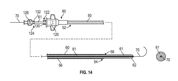
assembly is provided to enable easy, safe, and efficient tracking of a large
diameter catheter or
catheter system from insertion into a large peripheral blood vessel and
advanced to the target
location of interest. The large diameter catheter, such as for aspiration or
other mechanical
-27-
CA 03162704 2022-05-24
WO 2021/127004 PCT/US2020/065349
means of removal of embolus or thrombus from large vessels, has a diameter
within the range
from about 8F (0.105") to about 24F (0.315").
[0147] Referring to Figures 6 and 13, one implementation of the
catheter 60
includes an elongate flexible tubular body 50, having a proximal end 52, a
distal end 54 and a
side wall 56 defining a central lumen 58. Referring to Figure 14, an elongate
flexible
cannulated rail or dilator 61 is shown extending over the guidewire 70 and
occupying the space
between the guidewire 70 and the large inside diameter of the central lumen 58
of the large
diameter catheter 60 to provide support to the catheter and/or an atraumatic
tip during delivery.
[0148] This catheter-cannulated rail-guidewire assembly is intended to
easily track
through anatomical challenges more easily than the catheter. The catheter-rail-
guidewire
assembly then acts as a first stage of the catheter delivery system and
enables the large diameter
catheter or catheter system to be inserted and independently advanced over
this first stage into
a blood vessel (e.g. the femoral vein) percutaneously over a guidewire and
advanced through
potentially tortuous vasculature to the remote target location of interest
without requiring
advanced skills or causing kinking of the catheter.
[0149] The cannulated rail 61 may comprise a soft flexible cylindrical
body having
a guidewire lumen with a diameter of no more than about 0.040" and an outside
diameter no
less than about 0.025" or about 0.010" smaller than the inner diameter of the
large diameter
catheter. Thus the wall thickness of the cannulated rail 61 is typically at
least about 0.010"
less than the radius of the large diameter catheter and in some
implementations at least about
0.120" or more, depending upon the size of the annular space between the
inside diameter of
the catheter and the outside diameter of the guidewire. Depending upon the ID
of the access
catheter, the rail 61 may have a wall thickness of at least about 0.05 inches,
at least about 0.075
inches, at least about 0.100 inches and in some implementations at least about
0.12 inches.
The wall thickness of the rail may exceed the inside diameter of the guidewire
lumen.
[0150] The cannulated rail 61 may have an elongated tapered distal tip
62 that may
project beyond the distal end 54 of the catheter 60. The thick sidewall of the
cannulated rail
61 may comprise one or more flexible polymers, and may have one or more
embedded column
strength enhancing features such as axially extending wires, metal or
polymeric woven or
braided sleeve or a metal tube, depending upon the desired pushability and
tracking
performance along the length of the dilator.
-28-
CA 03162704 2022-05-24
WO 2021/127004 PCT/US2020/065349
[0151] Optionally, the proximal segment of the rail or dilator which
is not intended
to extend out of the distal end of the catheter may be a structure which is
not coaxial with the
guidewire, but a control wire which extends alongside the guidewire in the
catheter and allows
the distal tubular telescoping segment of the rail or dilator to be retracted
or extended.
(analogous to rapid exchange catheters) without the entire length of the rail
structure being
over the wire. This allows removal or insertion of the rail or dilator over a
shorter guidewire
because of the shorter coaxial segment tracking over the guidewire.
[0152] Catheter 60 may be provided with a proximal hub 120, having a
port for
axially movably receiving the rail 61 therethrough. The hub 120 may be
provided with an
engagement structure such as a first connector 122 for releasably engaging a
second
complementary connector 124 on a hub 126 on the proximal end of the rail 61.
First connector
122 may comprise an interference structure such as at least one radially
moveable projection
130, for releasably engaging a complementary engagement structure such as a
recess 132 (e.g.,
an annular ridge or groove) on the hub 126. Distal advance of the rail 61 into
the catheter 60
causes the projection 130 to snap fit into the recess 132, axially locking the
catheter 60 and rail
61 together so that they may be manipulated as a unit.
[0153] The dilator is inserted through the hemostasis valve in the hub
120 of a large
bore (e.g., 24F) catheter 60 and advanced through the catheter until the
retention clip on the
dilator hub 126 or catheter hub 120 snaps into the complementary recess on the
other hub. In
this engaged configuration, the flexible distal end of the 24F rail dilator 61
will extend at least
about 5 cm or 10 cm, and in some implementations at least about 15 cm or 20 cm
beyond the
distal end 54 of the 24F catheter 60. The rail dilator and 24F catheter system
are thereafter
distally advanced over a previously placed guidewire and into the introducer
sheath.
[0154] The dilator and catheter combination of the present invention
differentiate
over prior systems both because of the flexibility of a distal zone of the
dilator and greater
length of the dilator than the corresponding catheter. Typically, a dilator is
a uniform stiffness
and length-matched to its catheter, with only a short atraumatic tip of the
dilator extending
beyond the distal end of the catheter. The dilator of the present invention
has a supportive
proximal end and a flexible distal end, with a total dilator length much
longer than the catheter
60 to enable, as an example, the following procedure.
-29-
CA 03162704 2022-05-24
WO 2021/127004 PCT/US2020/065349
[0155] In use, a guidewire 70 such as an 0.035" guidewire is advanced
under
fluoroscopy using conventional techniques into a selected vessel. The
cannulated rail 61,
optionally with the catheter 60 mounted thereon, is loaded over the proximal
end of the
guidewire 70 and advanced distally over the wire until the distal end of the
rail is in position
at the target site.
[0156] The 24F catheter 60 is thereafter unlocked from the rail 61 and
advanced
over the rail 61 to the desired site, supported by the rail 61 and guidewire
70 combination.
Because the uncovered advance section of the rail has already traversed the
challenging
tortuosity through the heart, the catheter 61 now just slides over the advance
section of the rail
for easy passage to the final target location. The supportive proximal zone
and flexible distal
advance section of the rail enables ease of delivery through the most
challenging anatomy in,
for example, a PE procedure going from the vena cava through the tricuspid and
pulmonary
valves of the heart into the central pulmonary artery without concern about
damaging the tissue
(atraumatic, flexible tip) or damaging the dilator (high kink resistance due
to flexible, high
wall thickness "solid" dilator construction.
[0157] The cannulated rail 61, or the cannulated rail 61 and the
guidewire 70
combination, may thereafter be proximally withdrawn, leaving the large bore
catheter 60 in
position to direct a procedure catheter such as any of the aspiration
catheters disclosed
elsewhere herein to the target site.
[0158] Referring to Figure 15, the large diameter (LD) catheter 60 may
in some
situations have a smaller diameter (SD) catheter though its central lumen for
the purposes of
introducing an additional functionality (e.g., clot grabber catheter 62,
imaging catheter 10, or
mechanical thrombectomy tool 66) and / or telescoping the SD catheter to more
distal locations
in the anatomy. In order to enable delivery of the LD catheter 60 and SD
catheter as a single
system, the SD catheter may have a core dilator 68 for support, and the gap
between the outer
diameter of the SD catheter and inner diameter of the LD catheter 60 may be
maintained or
supported by a second, tubular dilator 71. The tubular dilator 71 may have a
shaped distal tip
72 for a smooth tapered transition from the SD catheter 41 to the LD catheter
40. The distal
end 34 of the core dilator may be provided with a complementary taper to the
distal taper of
the thin wall SD dilator (Figure 16) or may end at the distal end of the LD
catheter (Figure 17).
-30-
CA 03162704 2022-05-24
WO 2021/127004 PCT/US2020/065349
[0159] The core dilator 68 inside the SD catheter 41 and tubular
dilator 70 between
the two catheters may have an interlocking feature to create a single (SD +
LD) catheter +
(core + tubular) dilator system. For example, complementary connectors may be
provided on
hubs on the proximal ends of the system components.
[0160] Referring to Figure 17, the tip of the tubular dilator 70 may
be configured
to taper to the guidewire lumen 76, thus covering and extending distally
beyond the small
diameter catheter 41 if it is in place. The tip of the tubular dilator 70 may
be provided with a
longitudinally extending slit 78, scored or perforated one or more times to
allow the tip to split
longitudinally and be pulled back into the space between the LD and SD
catheters and fully
expose the distal end of the small diameter catheter 41. See Figure 18.
[0161] The single (SD + LD) catheter + (core + tubular) dilator system
may be pre-
assembled and detachably interlocked at the proximal hub. Additional tubular
dilators having
a series of outside diameters and wall thicknesses may be provided such that
the SD catheter
may be used in combination with different diameter LD catheters. A LD catheter
may be used
with different SD catheters by providing tubular dilators having the same OD
but a series of
different inside diameters. The core + tubular dilators may simply be pulled
proximally to
withdraw both dilators as a single system, or the tubular dilator may be
configured with a tab
or handle at the proximal end and a slit, scoring, perforation or other
mechanism so as to split,
peel, or tear it along the longitudinal axis during withdrawal to allow the
tubular dilator to peel
from the SD catheter as it slides proximally out of the space between the LD
and SD catheters.
(Figure 18)
[0162] Any of the thrombectomy catheters disclosed herein may be
provided with
a surface configuration on the inside surface of the central lumen to affect
the behavior of clot
drawn into the lumen. In general, the catheter, with diameter within the range
from about 8F
(0.105") to about 24F (0.315"), includes an elongate flexible tubular body,
having a proximal
end, a distal end and a side wall defining a central lumen. The access or
evacuation catheters
may also include a rotatable core wire or other apparatus that extends though
the catheter lumen
for the purposes of engaging thrombus at the distal end of the catheter as has
been discussed.
[0163] The central lumen 90 is defined by an inside surface 100 of the
tubular body
104, which in some embodiments is a smooth cylindrical surface. However,
referring to
Figures 19A and 19B, the inner surface 100 of at least a distal zone 102 of
the tubular body
-31-
CA 03162704 2022-05-24
WO 2021/127004 PCT/US2020/065349
104 may be provided with a surface configuration to engage thrombus and limit
unrestricted
sliding of the thrombus along the inner surface 100. The distal zone 102 may
have an axial
length within the range of from about 0.5" to about 12" or more, depending
upon desired
performance and/or manufacturing technique.
[0164] For example, referring to Figure 19A, at least one or five or
ten or more
axially extending surface structures such as radially inwardly extending
ridges 106 separated
by grooves 108 may be provided to facilitate proximal ingestion of the
thrombus and / or to
engage thrombus and resist rotation of the thrombus within the lumen when the
core wire or
other thrombus grabbing apparatus is rotated within the lumen.
[0165] The corrugation pattern may also increase the transport of the
thrombus
proximally by decreasing the surface area of contact between the thrombus and
the inside
surface of the tubular body. The spacing circumferentially may be regular or
irregular, and the
crest and trough pattern, dimensions and distribution may be varied. Examples
of trough cross
sections include the illustrated rectangular, semicircular or triangular,
among others.
[0166] In one implementation illustrated in Figure 19B, the surface
discontinuity,
i.e. grooves or ridges, may extend in a circumferential (e.g. helical)
configuration having a
constant or variable pitch along the length of the distal zone 102. The
helical guide may spiral
in a first direction in order to oppose rotation of the core wire or other
apparatus in a second
direction, or in the same direction to cooperate with the rotation of the core
wire in order to
facilitate thrombus ingestion proximally into the catheter. The grooves or
ridges may have a
curved profile or generally rectilinear, having generally flat or cylindrical
side walls.
[0167] In another embodiment, the inner surface 100 of the distal zone
102 of the
tubular body 104 may have a three-dimensional pattern that reduce friction of
the thrombus
moving proximally in the catheter and create resistance to the thrombus moving
distally in the
catheter after it has been ingested into the catheter. This pattern may be
regular throughout or
in the distal end of the tubular body, or irregular and decrease gradually and
progressively in
density, pattern, or geometry along the length of the catheter or the distal
section. This pattern
may also be provided in combination with lubricious coatings or surfaces.
[0168] The three-dimensional differential friction pattern may be
defined by a
regular or irregular pattern of protuberances 110 or depressions on the inner
surface of the
catheter, each of which presents an exposed face 112 inclined radially
inwardly in the proximal
-32-
CA 03162704 2022-05-24
WO 2021/127004 PCT/US2020/065349
direction, with each inclined exposed face 112 terminating proximally in a
proximally facing
engagement surface such as a dropoff edge 114, which may be curved in an axial
direction as
shown in Figure 21 to provide a ratchet or fish scale type of configuration.
[0169] The scales may flex, hinge or pivot at the distal end, which
will not impact
the proximal ingestion of the thrombus, but will create additional resistance
to the thrombus
moving distally in the catheter after it has been ingested into the catheter.
[0170] In alternative embodiments, the differential friction surface
structures on
the inner surface of the distal zone may be relatively shallow convex or
concave "dimples" or
other regular or irregular surface discontinuities that are generally
triangular, oval, oblong arcs,
serpentine shapes or a plurality of angled fibers that incline radially
inwardly in a proximal
direction.
[0171] Referring to Figure 22, the lumen 116 of the tubular body may
be non-
cylindrical and instead be oval or other geometry lacking rotational symmetry
and containing
a minor axis which is less than a major axis due to one or more longitudinal
deformations of
the lumen. These longitudinal deformations of at least the inner surface of
the tubular body
serve as structures to increase the rotational resistance to thrombus or
embolic matter in the
lumen. These deformations may contain their own lumens within the wall of the
tubular body
for pull wires of a steerable tip or to deliver fluids or to measure pressure
or to transmit a
vacuum or other functions.
[0172] The distal end 118 of the tubular body may be angled such that
the distal
face of the catheter resides on a plane or a curve with an end to end secant
that crosses the
longitudinal axis of the tubular body at an angle within the range from about
30 degrees to
about 60 degrees. The distal face of the angled tip may be non-planar and may
include one or
two or more inflection points or curves. This angled tip may improve catheter
navigation and
thrombus ingestion by providing more surface area for engagement between the
catheter and
the thrombus. (see, e.g., Figure 31D and associated description in US
publication No.
2020/0001046 Al to Yang, et al., which is hereby incorporated in its entirety
herein by
reference.)
[0173] The opening at the distal end of the tubular body may be
expandable from
a first inside diameter for transluminal navigation to a second, larger inside
diameter providing
a funnel like tip with an enlarged distal opening to facilitate aspiration of
thrombus into the
-33-
CA 03162704 2022-05-24
WO 2021/127004 PCT/US2020/065349
lumen. The diameter at the distal opening of the fully open funnel exceeds the
diameter of the
cylindrical distal end of the tubular body by at least about 5% or 10% or
more. (see Figures
4A ¨41 and associated description in US patent No. 10,441,745 to Yang, et al.,
which is hereby
incorporated in its entirety herein by reference.)
[0174] The distal end of the catheter may have an increasing inner
diameter while
maintaining a constant outer diameter, representing a tapered distal wall, or
the inner and outer
diameters may both increase, representing a conical funnel like tip.
[0175] The funnel tip is may be made of a material that is rigid
enough to maintain
structural integrity under aspiration and flexible enough that it may deform
to accommodate
and enable thrombus across a range of size, shape, and maturity to be
aspirated into the catheter
lumen.
[0176] The funnel tip may telescope out of the thrombectomy catheter.
A hollow
cylindrical structure at the end of a long, flexible hypo tube or at the end
of two or more stiff
wires may be advanced through the catheter, and when the hollow cylindrical
structure extends
beyond the distal catheter tip and its associated circumferential constraint,
the structure
expands into a conical funnel shape. Additional details of these features may
be found in US
patent No. 10,441,745 to Yang, et al., previously incorporated in its entirety
herein by
reference.)
[0177] The thrombectomy catheter with inner lumen features to enhance
the
extraction of thrombus and/or distal end features may enable easier, more
efficient removal of
a broad spectrum of thrombus size, shapes, and maturity from vascular conduits
including the
pulmonary arteries, resulting in shorter procedure times and a lower total
volume of blood loss.
This is achieved by promoting proximal movement of the thrombus while creating
resistance
to the thrombus moving distally in the catheter after it has been ingested
into the distal catheter.
[0178] The foregoing inventions and improvements will enable the
engagement
and capture of a very wide range of thrombus, from acute to mature in nature,
thus enabling
the extraction of the thrombus from the blood vessel in which it may be
impeding blood
flow. Additionally, the configurations described above enhance safety by
reducing the risk of
vascular tissue injury due to mechanical engagement of the helical tip element
and/or catheter
tip with the vessel wall.
-34-
CA 03162704 2022-05-24
WO 2021/127004 PCT/US2020/065349
[0179] In the foregoing description, similar features in different
drawings are
sometimes identified by slightly differing terminology. This is not intended
to imply
differences that do not exist. Slightly different features are illustrated in
different drawings,
and those of skill in the art will recognize that any of the features
disclosed here in can be re-
combined with any of the catheters or other structures disclosed here in.
-35-