Note: Descriptions are shown in the official language in which they were submitted.
CA 03162878 2022-05-25
WO 2021/105131
PCT/EP2020/083227
1
ELECTROSURGICAL RE SECTOR TOOL
FIELD OF THE INVENTION
The invention relates to an electrosurgical resector
tool, for cutting, coagulating and ablating biological tissue
using electromagnetic (EM) energy. In particular, the
invention relates to an electrosurgical resector tool having
first and second blade elements which are movable relative to
each other between open and closed positions, and further
having a travel limiting mechanism operable to limit a maximum
extent of relative movement between the first and second blade
elements in the open position and/or the closed position.
BACKGROUND TO THE INVENTION
Surgical resection is a means of removing sections of
organs from within the human or animal body. The organs may
be highly vascular. When tissue is cut (i.e. divided or
transected), small blood vessels may be damaged or ruptured.
Initial bleeding is followed by a coagulation cascade where
the blood is turned into a clot in an attempt to plug the
bleed. During an operation it is desirable for a patient to
lose as little blood as possible, so various devices have been
developed in an attempt to provide bleeding-free cutting. For
endoscopic procedures, it is also undesirable for a bleed to
occur and not to be dealt with expediently, since the flow of
blood may obscure the operator's vision. Instead of a sharp
blade, it is known to use RF energy to cut biological tissue.
The method of cutting using RF energy operates using the
principle that as an electric current passes through a tissue
matrix (aided by the ionic cell contents), the impedance to
electron flow across the tissue generates heat. When a pure
sine wave is applied to the tissue matrix, enough heat is
generated within the cells to vaporize the water content of
the tissue. There is thus a huge rise in the internal cell
pressure that cannot be controlled by the cell membrane,
resulting in rupture of the cell. When this occurs over a
large area, it can be seen that the tissue is transected.
CA 03162878 2022-05-25
WO 2021/105131
PCT/EP2020/083227
2
The above procedure works elegantly in lean tissue, but
it is less efficient in fatty tissue because there are fewer
ionic constituents to aid the passage of electrons. This
means that the energy required to vaporize the contents of the
cells is much greater, since the latent heat of vaporization
of fat is much greater than the latent heat of vaporization of
water. RF coagulation operates by applying a less efficient
waveform to the tissue, whereby instead of being vaporized,
the cell contents are heated to around 65 C, drying out the
tissue by desiccation and denaturing the proteins in the
vessel walls. This denaturing acts as a stimulus to the
coagulation cascade, so clotting is enhanced. At the same
time the collagen in the wall is denatured, turning from a
rod-shaped to a coil-shaped molecule, causing the vessel to
contract and reduce in size, giving the clot an anchor point,
and a smaller area to be plugged.
However, RF coagulation is less efficient when fatty
tissue is present because the electrical effect is diminished.
It can thus be very difficult to seal fatty bleeders. Instead
of having clean white margins, the tissue has a blackened
burned appearance.
SUMMARY OF THE INVENTION
At its most general the present invention provides a
development to the electrosurgical resector tool concept
discussed in GB2567480. The electrosurgical resector tool has
an energy delivery structure that facilitates biological
tissue cutting and sealing using electromagnetic (EM) energy.
In particular, the invention relates to combined actuation and
energy delivery mechanisms that are compact enough to enable
the tool to be insertable through an instrument channel of a
surgical scoping device, such as an endoscope, gastroscope or
bronchoscope. The device could also be used to perform
laparoscopic or open surgery, i.e. the bloodless resection of
a liver lobe with the abdominal cavity open.
The electrosurgical resector tool has an instrument tip
having first and second blade elements which are movable
relative to each other between open and closed positions, and
the development may include a travel limiting mechanism
operable to limit a maximum extent of relative movement
CA 03162878 2022-05-25
WO 2021/105131
PCT/EP2020/083227
3
between the first and second blade elements in the open and/or
the closed positions. In this way, over-stressing the resector
tool jaws can be avoided and smooth, predictable jaw movement
can be ensured.
Additionally, the electrosurgical resector tool may
include a control rod for controlling relative movement
between the first and second blade elements, and the
development may include a set of overlapping tubes which
provide a channel through which the control rod can slide and
which is fixed to the instrument tip. In this way, movement of
the control rod can be smooth and predictable.
According to a first aspect of the present invention,
there is provided an electrosurgical resector tool comprising:
a shaft defining a lumen; an energy conveying structure for
carrying electromagnetic (EM) energy through the lumen of the
shaft; an instrument tip mounted at a distal end of the shaft,
wherein the instrument tip comprises: a static portion
comprising a first blade element; and a movable portion
comprising a second blade element, wherein the movable portion
is movable relative to the static portion between a closed
position in which the first blade element and second blade
element lie alongside each other to an open position in which
the second blade element is spaced from the first blade
element by a gap for receiving biological tissue; a travel
limiting mechanism operable to limit a maximum extent of
relative movement between the second blade element and the
first blade element in the open position and/or the closed
position; a first electrode, a second electrode and a planar
dielectric body, the first and second electrodes being spaced
apart and electrically isolated from each other by the planar
dielectric body, and wherein the first electrode and the
second electrode are connected to the energy conveying
structure for delivery of the EM energy from the instrument
tip; and an actuator for controlling relative movement between
the movable portion and the static portion. The actuator may
be a separate element to the instrument tip, but connected to
the instrument tip in order to open and close the blade
elements.
Optionally, one of the first blade element and the second
blade element comprises the planar dielectric body extending
longitudinally and having the first electrode on a first
CA 03162878 2022-05-25
WO 2021/105131
PCT/EP2020/083227
4
laterally facing surface thereof, and wherein, in the closed
position, the other of the first blade element and the second
blade element lies adjacent to a second laterally facing
surface of the longitudinally extending planar dielectric body
opposite to the first laterally facing surface thereof.
Optionally, the second blade element has a length
commensurate with a length of the first blade element.
Optionally, the energy conveying structure comprises a
coaxial transmission line extending in a longitudinal
direction through the lumen. The coaxial transmission line
comprises an inner conductor separated from an outer conductor
by a dielectric material. The inner conductor is connected to
one of the first electrode and the second electrode and the
outer conductor is connected to the other of the first
electrode and the second electrode, for delivery of the EM
energy from the instrument tip.
Optionally, the energy conveying structure is for
carrying radiofrequency (RF) electromagnetic (EM) energy and
microwave EM energy, and wherein the first electrode and the
second electrode are operable: as active and return electrodes
for delivering RF energy conveyed from the energy conveying
structure; and a microwave field emitting structure for
delivering microwave energy conveyed from the energy conveying
structure. The electrosurgical resector tool may provide a
plurality of operational modalities that facilitate biological
tissue cutting and sealing using radiofrequency (RF)
electromagnetic energy and/or microwave EM energy. In one
example, the electrosurgical resector tool may comprise a pair
of blade elements that provide a scissor-like mechanism that
can provide three complimentary modalities: (i) a gliding RF-
based cut when the blade elements are closed, (ii) a scissor-
type cut performed on tissue grasped between the blade
elements using a combination of RF energy and applied
pressure, and (iii) a coagulation or vessel sealing operation
performed on tissue grasped between the blade elements using a
combination of microwave energy and applied pressure.
Moreover, the RF and/or microwave energy may be supplied in
any of these modalities at a power level sufficient to cause
tissue ablation. By suitable configuration of a pair of
electrodes on the blade elements, the supplied RF or microwave
energy in each of these operational modalities can be focussed
CA 03162878 2022-05-25
WO 2021/105131
PCT/EP2020/083227
in the region required. The pair of electrodes may be both on
the same blade element, or there may be an electrode on each
blade element. However, it is to be understood that in some
embodiments, only RF EM energy, or only microwave EM energy
5 may be delivered.
In this structure, the first and second blade elements
may resemble a scissors-type closure mechanism. Thus, the
second blade element may be arranged to slide past the first
blade element during movement between the open position and
closed position, e.g. to effect mechanical cutting through
application of a shearing force. The movable portion may be
movable relative to the static portion in a plane parallel to
a plane defined by the planar dielectric body. Herein the term
"static" may mean that fixed in relation to the distal end of
the shaft when in use (i.e. when the second blade element is
moved between the open and closed position).
The shaft may be flexible, e.g. suitable for bending or
other steering to reach the treatment site. A flexible shaft
may enable the device to be usable in a surgical scoping
device such as an endoscope. In other examples, the shaft may
be rigid, e.g. for use in open surgery or with a laparoscope.
The first electrode and second electrode may be disposed
at the cutting interface. In one example, both electrodes are
on the same blade element, which may be on either the movable
portion or the static portion. For example, the second
electrode may be located on the second laterally facing
surface of the longitudinally extending planar dielectric
body. This may assist in provide uniform energy delivery at
the cutting interface. Where both electrodes are on one blade
element, the other blade element may be electrically inert,
e.g. made of plastic or other insulator.
In another example, the first electrode may be on one of
the blade elements, and the second electrode on the other
blade element. For example, the longitudinally extending
planar dielectric body may be on the first blade element, and
the second electrode may extend along a side of the second
blade element.
The first and second electrodes may thus be disposed
along each side of the cutting interface, with the planar
dielectric body in between. In this arrangement, if RF EM
energy is applied to the electrodes the RF EM energy flows
CA 03162878 2022-05-25
WO 2021/105131 PCT/EP2020/083227
6
preferentially between the first and second blade elements
across the cutting interface. Similarly, if microwave EM
energy is applied while the blade elements are open, a
microwave field emitted by the electrodes has a much higher
field strength within the gap between the blade elements than
elsewhere.
When in the closed position, the second electrode is
separated from the first electrode along much of its length by
the planar dielectric body. If RF EM energy is applied in
this position, the RF EM energy preferentially flows around a
distal tip and side edge of the closed blade elements, which
facilitates a RF-only gliding cut performed by sliding the
instrument tip through tissue.
The movable portion and thus the second blade element may
be formed from an insulator-coated conductive material which
is further coated with parylene N. For example, the movable
portion may be a cast piece of stainless steel having a
ceramic (e.g. alumina spray), synthetic plastic (e.g.
Bakelite), diamond-like carbon (DLC), enamel coating, or a
silicon-based paint coating. The second electrode may be
formed at a side portion of the second blade element where the
insulator coating and the parylene N coating is removed. The
second electrode may be the exposed conductive material of the
movable portion, or may comprise an additional conductive
layer (e.g. of gold or the like) deposited or otherwise
affixed to the exposed conductive material.
The second blade element may comprise a laterally
protruding flange along its side portion. The flange thus
protrudes towards the first blade element when in the closed
position. The second electrode may be formed on a laterally
facing edge of the laterally protruding flange.
The travel limiting mechanism may be a feature of the
instrument tip. As such, structural features of the instrument
tip may cooperate to define the relative positions of the
first and second blade elements in the open and/or closed
positons. This results in open and/or closed positions which
are consistent and do not vary between applications. This may
be different to conventional techniques in which the actuator
or control rod defines these relative positions by having a
limited travel. That is, conventionally, the amount of
distance the control rod can slide within the shaft may be
CA 03162878 2022-05-25
WO 2021/105131
PCT/EP2020/083227
7
limited, for example, by a handpiece at a proximal end of the
shaft. Given the flex of the various elements in the shaft,
this type of mechanism can result in a variable open position
and/or closed position, which can be undesirable in certain
precision operations that the instrument tip is used to
perform. The travel limiting mechanism may be formed by one or
more pairs of cooperating structures formed on the static
portion and the movable portion. That is, for each pair, one
cooperating structure is formed on the static portion and the
other cooperating structure is formed on the movable portion.
One pair of cooperating structures may function to limit a
maximum extent of relative movement between the second blade
element and the first blade element in the open position,
whereas another pair of cooperating structures may function to
limit a maximum extent of relative movement between the second
blade element and the first blade element in the closed
position. The travel limiting mechanism may limit a maximum
angle between the first and second blade elements in the open
position to be about 60 degrees.
A first pair of cooperating structures may include a
raised protrusion and a cooperating stop surface (which may be
substantially flush with surrounding surfaces), wherein the
raised protrusion and the stop surface are configured or
arranged in use to abut each other in the open position. That
is, moving the moveable portion into the open position moves
the raised protrusion into contact with the stop surface such
that further opening of the first and second blade elements is
prevented. That is, the second blade element is prevented from
moving further past the first blade element. The stop surface
and/or the raised protrusion may be specially formed
structures which are sized and/or shaped to limit how far
apart the first and second blade elements can move. In an
embodiment, the moveable portion comprises the raised
protrusion and the static portion comprises the stop surface.
Specifically, the raised protrusion may be formed on a top
surface of the moveable portion and distally of a connection
(e.g. pivotal connection) between the movable portion and the
static portion. Also, the stop surface may be formed on a top
surface of the static portion and proximally of the connection
between the movable portion and the static portion. The stop
CA 03162878 2022-05-25
WO 2021/105131
PCT/EP2020/083227
8
surface may be provided by a slot formed in the static portion
by a support arm to which the movable portion is attached.
A second pair of cooperating structures may include a
pair of abutment surfaces, wherein the pair of abutment
surfaces are configured in use to abut each other in parallel
formation in the closed position. That is, moving the movable
portion into the closed position moves the two abutment
surfaces together such that they contact each other and are
substantially parallel to each other. By contacting along a
surface rather than a point, the tool can provide a strong and
reliable closure mechanism which can be advantageous, for
example, when severing tissue using the first and second blade
elements. In an embodiment, a first abutment surface is formed
on a top surface of the movable portion and proximally of a
connection (e.g. pivotal connection) between the moveable
portion and the static portion. The first abutment surface may
be formed as the top surface of an attachment plate of the
movable portion, wherein the attachment plate is a proximal
extension of the movable portion that extends proximally of
the connection to the static portion. The attachment plate may
be sized and/or shaped to limit how far the second blade
element can move past the first blade element in the closing
direction (i.e. the direction of travel from the open position
to the closed positon). Also, a second abutment surface is
formed on an under surface of the static portion and
proximally of the connection between the moveable portion and
the static portion. The second abutment surface may be formed
as an underside of a support arm of the static portion. The
support arm may be a lateral and forward (i.e. distally
extending) extension of the static portion which defines a
slot to accommodate movement of the movable portion relative
to the static portion. The moveable portion may be connected
(e.g. pivotally connected) to the static portion by the
support arm. The support arm may be sized and/or shaped to
limit how far the second blade element can move past the first
blade element in the closing direction (i.e. the direction of
travel from the open position to the closed positon).
As mentioned, the static portion may comprise a support
arm on which the movable portion is mounted, and the support
arm may define a slot in the static portion for receiving part
of the movable portion. A length of the slot (i.e. the
CA 03162878 2022-05-25
WO 2021/105131
PCT/EP2020/083227
9
dimension in line with the shaft length) may be between lmm
and 3mm (preferably less than about 2mm). A width of the slot
(i.e. the dimension in line with the pivot axis) may be
between 0.2mm and 1.2mm (preferably more than about 0.7mm). A
depth of the slot may be between 0.2mm and 1.2mm (preferably
more than about 0.6mm). The slot may be necessary in order to
provide space for part of the moveable portion (e.g. a
proximal part) to move relative to the static portion between
the open and closed positions. The support arm may form part
of an electrical connection between the energy conveying
structure and the second electrode. For example, the static
portion (e.g. the support arm) may be formed from an
insulator-coated conductive material which is further coated
with parylene N, and may comprise a proximal contact portion
at which the insulator coating and the parylene N coating is
removed and which is electrically connected to the inner
conductor or outer conductor of the coaxial transmission line.
An advantage of limiting dimensions of the slot is that it is
possible to ensure a higher quality coating (e.g. of insulator
and/or parylene N). For example, it is easier to ensure that
the coating is complete and even. The static portion (e.g. the
support arm) may have a proximal recess for attachment to a
distal end of the coaxial transmission line. Other types of
electrical connection may also be used. For example, a
flexible conductor may be connected between the energy
conveying structure (e.g. the inner conductor or outer
conductor of the coaxial transmission line) and the first
electrode or second electrode. Preferably the length of any
flexible conductor is equal to or less than an eighth of a
wavelength of the microwave energy, in order to prevent it
from affecting the emitted field.
The coaxial transmission line may be adapted to convey
either of or both of RF EM energy and microwave EM energy.
Alternatively, the energy conveying structure may comprise
different routes for the RF EM energy and microwave EM energy.
For example, the microwave EM energy may be delivered through
the coaxial transmission line, whereas the RF EM energy can be
delivered via twisted pair wires or the like. Where a
separate energy delivery route is provided, the first and
second electrodes may comprise separate RF electrode portions
and microwave electrode portions to enable the RF energy and
CA 03162878 2022-05-25
WO 2021/105131
PCT/EP2020/083227
microwave energy to be delivered from different regions of the
instrument tip. For example, the microwave energy may be
delivered from one of the blade elements, whereas the RF
energy may be delivered between the blade elements. In another
5 embodiment, the electrosurgical tool is only configured to
deliver only one of RF EM energy and microwave EM energy.
The movable portion may be mounted to the support arm via
a pivot connection. For example, the support arm may provide
a clevis-type structure that supports a pivot axle on which
10 the movable portion is mounted. The electrical connection
between the energy conveying structure and the second
electrode may pass through the pivot connection. For example,
the pivot axle may be formed from a conductive material, and
the insulator coating (and the parylene N coating) of the
movable portion and the support arm may be removed where they
respectively contact the pivot axle.
The dielectric material and inner conductor of the
coaxial transmission line may extend beyond a distal end of
the outer conductor. The inner conductor may include an
exposed distal portion that is electrically connected to the
first electrode, e.g. by directly overlapping with and
contacting a proximal portion of the first electrode.
The movement between the movable portion and the static
portion may be rotational or translational or a combination of
the two. In one example, the movable portion may be pivotable
relative to the static portion, whereby the second blade
element is angled relative to the first blade element in the
open position. This example may resemble a conventional
scissor-type closure. The second blade element may be movable
through only an acute angle (i.e. not an obtuse angle) between
the open position and the closed position. In an embodiment,
the travel limiting mechanism may be configured to limit the
acute angle to between 90 degrees and 40 degrees, and
preferably between 80 degrees and 50 degrees, and more
preferably about 60 degrees. Additionally or alternatively,
the travel limiting mechanism may be configured to limit a
maximum distance between the jaws in the open position to
about 3.5mm.
The actuator may comprise a control rod slidably mounted
in the flexible shaft. The control rod may have an attachment
feature engaged with the movable portion, whereby longitudinal
CA 03162878 2022-05-25
WO 2021/105131
PCT/EP2020/083227
11
movement of the control rod in the shaft causes movement of
the movable portion relative to the static portion. The
attachment feature may be a hook or any suitable engagement
for transmitting push and pull forces to the movable portion.
The movable portion may include an aperture (e.g. a circular
hole) and the attachment feature (e.g. hook) may be configured
to fit within the hole to drive movement of the second blade
element past the first blade element. The circular hole
diameter may be only slightly larger than the control rod
diameter, so that the attachment feature (e.g. hook) is
prevented from moving longitudinally inside the hole. This may
ensure that the jaw movement is smooth and predictable since
most or all control rod longitudinal sliding movement is
translated into jaw movement.
The static portion may comprise a support arm that
provides a mounting base (e.g. a pivot base) for the movable
portion. The planar dielectric body may be a separate piece
of material mounted on, e.g. adhered or otherwise affixed to,
the support arm. The planar dielectric body may be formed
from ceramic (e.g. alumina). Herein, reference to "planar"
material may mean a flat piece of material having a thickness
that is substantially less that its width and length. The
planar dielectric body may have a length dimension aligned in
the longitudinal direction, a thickness dimension aligned in a
lateral direction, and a width dimension orthogonal to both
the length and thickness dimensions. A plane of the planar
dielectric body is that in which the length and width
dimensions lie, i.e. a plane orthogonal to the width
dimension.
The first electrode may be a conductive material (e.g.
gold) deposited or otherwise mounted on the first laterally-
facing surface of the planar dielectric body. The second
laterally-facing surface of the planar dielectric body that
faces in an opposite direction to the first laterally-facing
surface may be exposed at the cutting interface.
The instrument tip may comprise a shield mounted around
the static portion. The shield may comprise an insulting
covering mounted around the static portion. For example, the
insulating shield may cover the support arm of the static
portion. The insulating shield may also be using to partly
cover the first electrode, e.g. to ensure that an exposed
CA 03162878 2022-05-25
WO 2021/105131 PCT/EP2020/083227
12
portion of the first electrode has a desired shape for
controlling the delivery of RF or microwave energy. The
insulating covering may have one or more field-shielding
conductive regions, e.g. patches of metallisation on its outer
surface. These conductive regions may provide shielding for
the electric fields, e.g. to prevent leakage of energy from
the instrument in unwanted locations. The shield may moulded
over the instrument tip following assembly. Alternatively,
the shield may be formed from a tube of insulating material
that can be cut (e.g. laser cut) to the desired shape and then
mounted over the blade elements. The shield may be formed
from a suitable insulating plastic, e.g. PEEK or the like.
The material for the shield may preferably be resistant to
high temperatures.
The first blade element may be shaped as a longitudinally
extending finger having an upstanding tooth at its distalmost
end. The second blade element may be shaped in a
corresponding way, e.g. as an elongate finger having a
downwardly extending tooth at its distalmost end. The
distalmost teeth may assist in retaining tissue in the gap
between the jaws as they are closed. Additionally, the second
blade element may be shaped to include a second downwardly
extending tooth at a point in-between the distalmost end and
proximalmost end. For example, the second downwardly extending
tooth may be located at or near a midway point along the
second blade element between the distalmost and proximalmost
ends. The upstanding tooth and the two downwardly extending
teeth may combine together to provide improved tissue
retention in the gap between the jaws as they are closed.
A longitudinally extending insert may be mounted in the
lumen of the flexible shaft to prevent relative movement of
the actuator or coaxial cable with the shaft from resulting in
lost or jerky movement of the instrument tip. The insert may
comprise a tubular body having a plurality of longitudinal
sub-lumens formed therein, wherein each of the plurality of
longitudinal sub-lumens breaks the outer surface of the
tubular body. The tubular body is sized to fit snugly within
the lumen so that its broken circumferential surface defines a
plurality of feet that abut the inner surface of the shaft to
resist relative movement therebetween.
CA 03162878 2022-05-25
WO 2021/105131
PCT/EP2020/083227
13
The coaxial transmission line may comprise a coaxial
cable mounted in a first sub-lumen of the tubular body. The
actuator may comprise a control rod slidably mounted in a
second sub-lumen of the tubular body. The control rod may
have a low friction coating (e.g. of PTFE or the like) to
facilitate longitudinal sliding relative to the insert.
Alternatively, the second sub-lumen may have a low friction
tube (aka first tube) mounted therein, wherein the control rod
can be slidably mounted in the low friction tube.
The electrosurgical resector tool may include a set of
overlapping tubes which together provide a channel through
which the control rod may slide to open and close the jaws.
The set of overlapping tubes may be bonded to the instrument
tip (e.g. the static portion) such that the control rod can
slide within the channel in a predictable and reliable manner.
For example, movement of the channel relative to the
instrument tip is prevented which could otherwise interfere
with the smooth movement (e.g. sliding) of the control rod
and, by association, the smooth opening and closing of the
jaws. Specifically, there may be provided a first tube (aka
guide wire tube), a second tube (aka distal guide wire tube)
and a third tube (aka short base tube). The first tube
surrounds a majority of the control rod except a distal end
region of the control rod. The first tube may surround a
majority or entirety of the control rod except the distal end
region. That is, the first tube may extend proximally all the
way to, and possibly inside of, a handpiece for manually
controlling opening and closing of the jaws. The distal end
region of the control rod may be the final 4mm to 8mm (e.g.
5mm). The first tube may be formed from PTFE or the like. The
second tube surrounds the distal end region of the control rod
except the attachment feature of the control rod, and the
second tube protrudes proximally into the first tube to define
an overlap region where the first tube overlaps the second
tube. The attachment feature may account for the distalmost
2mm or less of the control rod. A length of the overlap region
may be about half of the length of the second tube, for
example, the overlap region may be about 4mm to 6mm long, and
the length of the second tube may be about 8mm to 12mm. The
second tube may be formed from PTFE or the like. Also, the
third tube surrounds the overlap region and a proximal end
CA 03162878 2022-05-25
WO 2021/105131
PCT/EP2020/083227
14
region of the static portion. A length of the overlap region
may be about half of the length of the third tube, for
example, the overlap region may be about 4mm to 6mm long, and
the length of the third tube may be about 8mm to 12mm. The
third tube may be formed from polyether block amide (aka PEBA,
PEBAX or thermoplastic elastomer). The first, second and third
tubes may be bonded to each other and to the static portion.
Bonding may be via glue or adhesive, and/or via an
interference fit between the overlapping tubes. For instance,
the first, second and third tubes may be substantially clear
(i.e. transparent) and may be bonded to the instrument tip
(e.g. the static portion) by ultra-violet adhesive.
The instrument tip may be dimensioned to fit within an
instrument channel of a surgical scoping device. Accordingly,
a second aspect the invention provides an electrosurgical
apparatus comprising: an electrosurgical generator for
supplying EM energy; a surgical scoping device having an
instrument cord for insertion into a patient's body, the
instrument cord having an instrument channel extending
therethrough; and an electrosurgical resector tool of the
first aspect inserted through the instrument channel of the
surgical scoping device.
Optionally, the electrosurgical generator is capable of
supplying radiofrequency (RF) EM energy and microwave EM
energy.
According to a third aspect of the invention, there is
provided an electrosurgical resector tool comprising: a shaft
defining a lumen; an energy conveying structure for carrying
electromagnetic (EM) energy through the lumen of the shaft; an
instrument tip mounted at a distal end of the shaft, wherein
the instrument tip comprises: a static portion comprising a
first blade element; and a movable portion comprising a second
blade element, wherein the movable portion is movable relative
to the static portion between a closed position in which the
first blade element and second blade element lie alongside
each other to an open position in which the second blade
element is spaced from the first blade element by a gap for
receiving biological tissue; a first electrode, a second
electrode and a planar dielectric body, the first and second
electrodes being spaced apart and electrically isolated from
each other by the planar dielectric body, and wherein the
CA 03162878 2022-05-25
WO 2021/105131 PCT/EP2020/083227
first electrode and the second electrode are connected to the
energy conveying structure for delivery of the EM energy from
the instrument tip; an actuator for controlling relative
movement between the movable portion and the static portion,
5 the actuator comprising a control rod slidably mounted in the
shaft, the control rod having an attachment feature engaged
with the movable portion, whereby longitudinal movement of the
control rod in the shaft causes movement of the movable
portion relative to the static portion; and a first tube, a
10 second tube and a third tube, wherein the first tube surrounds
the control rod except a distal end region of the control rod,
wherein the second tube surrounds the distal end region of the
control rod except the attachment feature of the control rod,
and the second tube protrudes proximally into the first tube
15 to define an overlap region where the first tube overlaps the
second tube, and wherein the third tube surrounds the overlap
region and a proximal end region of the static portion.
The third aspect is analogous to the first aspect other
than that: (i) the travel limiting mechanism is optional in
the third aspect; and, (ii) the first, second and third tubes
are essential in the third aspect. The further features and
advantages of the first aspect are equally applicable and are
hereby restated in respect of the second aspect.
The term "surgical scoping device" may be used herein to
mean any surgical device provided with an insertion tube that
is a rigid or flexible (e.g. steerable) conduit that is
introduced into a patient's body during an invasive procedure.
The insertion tube may include the instrument channel and an
optical channel (e.g. for transmitting light to illuminate
and/or capture images of a treatment site at the distal end of
the insertion tube. The instrument channel may have a
diameter suitable for receiving invasive surgical tools. The
diameter of the instrument channel may be 5 mm or less.
Herein, the term "inner" means radially closer to the
centre (e.g. axis) of the instrument channel and/or coaxial
cable. The term "outer" means radially further from the centre
(axis) of the instrument channel and/or coaxial cable.
The term "conductive" is used herein to mean electrically
conductive, unless the context dictates otherwise.
Herein, the terms "proximal" and "distal" refer to the
ends of the elongate probe. In use the proximal end is closer
CA 03162878 2022-05-25
WO 2021/105131
PCT/EP2020/083227
16
to a generator for providing the RF and/or microwave energy,
whereas the distal end is further from the generator.
In this specification "microwave" may be used broadly to
indicate a frequency range of 400 MHz to 100 GHz, but
preferably the range 1 GHz to 60 GHz. Specific frequencies
that have been considered are: 915 MHz, 2.45 GHz, 3.3 GHz, 5.8
GHz, 10 GHz, 14.5 GHz and 24 GHz. In contrast, this
specification uses "radiofrequency" or "RF" to indicate a
frequency range that is at least three orders of magnitude
lower, e.g. up to 300 MHz, preferably 10 kHz to 1 MHz, and
most preferably 400 kHz.
BRIEF DESCRIPTION OF THE DRAWINGS
Embodiments of the invention are discussed in detail with
reference to the accompanying drawings, in which:
Fig. 1 is a schematic diagram of an electrosurgical
system that is an embodiment of the invention;
Fig. 2A is a perspective view of an instrument tip of an
electrosurgical resector instrument that is an embodiment the
invention in a closed configuration;
Fig. 2B is a side view of the instrument tip of Fig. 2A
in the closed configuration;
Fig. 2C is a side view of the instrument tip of Fig. 2A
in an open configuration;
Fig. 2D is a perspective view of the instrument tip of
Fig. 2A in the open configuration;
Figs. 3A and 3B are side and perspective views,
respectively, of the instrument tip of Fig. 2A but with an
outer sleeve removed to reveal internal parts;
Fig. 4 is a schematic partially cut-away side view of an
electrosurgical resector instrument that is an embodiment the
invention;
Fig. 5 is a reproduction of Fig. 2D but including labels
corresponding to Fig. 4, to illustrate how the schematic view
of Fig. 4 can translate onto the instrument tip of Fig. 2A;
Fig. 6A is a perspective view of the contents of an
instrument shaft that can be used with an electrosurgical
resector instrument that is an embodiment of the invention;
and
CA 03162878 2022-05-25
WO 2021/105131
PCT/EP2020/083227
17
Fig. 6B is a cross-section of the instrument shaft shown
in Fig. 6A.
DETAILED DESCRIPTION OF THE DRAWINGS
Fig. 1 is a schematic diagram of a complete
electrosurgical system 100 that is an embodiment of the
invention. The system is arranged to treat (e.g. cut or seal)
biological tissue using electromagnetic (EM) energy (e.g.
radiofrequency (RF) and/or microwave EM energy) from an
instrument tip. The system 100 comprises a generator 102 for
controllably supplying the EM energy (e.g. RF and/or microwave
EM energy). A suitable generator for this purpose is
described in WO 2012/076844, which is incorporated herein by
reference. The generator 102 is connected to a handpiece 106
by an interface cable 104. The handpiece 106 may also be
connected to receive a fluid supply 107 from a fluid delivery
device 108, such as a syringe, although this is not essential.
If needed, the handpiece 106 may house an instrument actuation
mechanism that is operable by an actuator 109, e.g. a thumb
operated slider or plunger. For example the instrument
actuation mechanism may be used to operate a pivotable blade
element of a resector instrument as discussed herein. Other
mechanisms may also be included in the handpiece. For
example, a needle movement mechanism may be provided (operable
by a suitable trigger on the handpiece) for deploying a needle
at the instrument. A function of the handpiece 106 is to
combine the inputs from the generator 102, fluid delivery
device 108 and instrument actuation mechanism, together with
any other inputs which may be required, into a single flexible
shaft 112, which extends from the distal end of the handpiece
106. The handpiece 106 may be as described in GB2567480.
The flexible shaft 112 is insertable through the entire
length of an instrument (working) channel of a surgical
scoping device 114. The flexible shaft 112 has an instrument
tip 118 that is shaped to pass through the instrument channel
of the surgical scoping device 114 and protrude (e.g. inside
the patient) at the distal end of the endoscope's insertion
tube. The instrument tip 118 includes a pair of blade
elements for gripping biological tissue and an energy delivery
structure arranged to deliver EM energy (e.g. RF and/or
CA 03162878 2022-05-25
WO 2021/105131
PCT/EP2020/083227
18
microwave EM energy) conveyed from the generator 102.
Optionally the instrument tip 118 may also include a
retractable hypodermic needle for delivering fluid conveyed
from the fluid delivery device 108. The handpiece 106 includes
an actuation mechanism for opening and closing the blade
elements of the instrument tip 118. The handpiece 106 may also
include a rotation mechanism for rotating the instrument tip
118 relative to the instrument channel of the surgical scoping
device 114.
The structure of the instrument tip 118 may be arranged
to have a maximum outer diameter suitable for passing through
the working channel. Typically, the diameter of a working
channel in a surgical scoping device such as an endoscope is
less than 4.0 mm, e.g. any one of 2.8 mm, 3.2 mm, 3.7 mm, 3.8
mm. The flexible shaft 112 may have a maximum diameter less
than this, e.g. 2.65 mm. The length of the flexible shaft 112
can be equal to or greater than 1.2 m, e.g. 2 m or more. In
other examples, the instrument tip 118 may be mounted at the
distal end of the flexible shaft 112 after the shaft has been
inserted through the working channel (and before the
instrument cord is introduced into the patient).
Alternatively, the flexible shaft 112 can be inserted into the
working channel from the distal end before making its proximal
connections. In these arrangements, the distal end assembly
118 can be permitted to have dimensions greater than the
working channel of the surgical scoping device 114. The system
described above is one way of introducing the instrument into
a patient. Other techniques are possible. For example, the
instrument may also be inserted using a catheter.
Although the examples herein are present in the context
of a surgical scoping device, it is to be understood that the
electrosurgical resector instrument may be embodiment in a
device suitable for open surgery or use with a laparoscope.
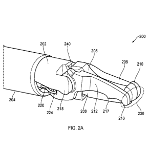
Figs. 2A-2D are different views of an instrument tip 200
of an electrosurgical resector instrument that is an
embodiment the invention. Fig. 2A is an isometric view of the
instrument tip 200 in a closed position, Fig. 2B is a side
view of the instrument tip 200 in the closed position, Fig. 2C
is a side view of the instrument tip 200 in an open position,
and Fig. 2D is another side view of the instrument tip 200 in
the open position. The instrument tip 200 is mounted at the
CA 03162878 2022-05-25
WO 2021/105131
PCT/EP2020/083227
19
distal end of a flexible shaft 204, which may correspond to
the flexible shaft 112 discussed above. In this embodiment,
the instrument tip 200 comprises a static portion 202 that
carries a first electrode 206 (e.g. see Fig. 2D), and a
movable portion 212 that carries a second electrode 214 (e.g.
see Fig. 2D). However, the invention need not be limited to
this configuration. In other examples both electrodes may be
provided on either the static portion 202 or the movable
portion 212.
The static portion 202 has a proximal region that is
secured to a distal end of the flexible shaft 204. The static
portion 202 extends in a longitudinal direction away from the
distal end of the flexible shaft 204. At its distal end, the
static portion 202 defines a first blade element 205, which is
a longitudinally extending finger having an upstanding tooth
210 at its distalmost end. The first electrode 206 extends
along a lateral surface of the first blade element 205.
However, in another embodiment, the first electrode 206 could
instead extend along only an upper surface of the first blade
element 205.
The movable portion 212 is pivotably mounted on the
static portion 202. In this embodiment, the movable portion
212 comprises a second blade element 207 (e.g. see Fig. 2D),
which is an elongate finger having a length commensurate with
the first blade element 205. The second blade element 207 has
a first downwardly extending tooth 216 at its distalmost end.
Additionally, the second blade element 207 has a second
downwardly extending tooth 217 approximately midway along the
second blade element 207.
The movable portion is pivotable about a pivot axis 219
(see Figs. 2B and 2C) located at a proximal end of the first
blade element 205, whereby the second blade element 207 can
swing between an open position (shown Figs. 2C and 2D) in
which it is angled away from the first blade element 205 and a
closed position (shown in Figs. 2A and 2B) where is lies
alongside (i.e. laterally adjacent) to the first blade element
205. The range of movement of the movable portion may be such
to allow the second blade element 207 to adopt an acute angle
relative to the first blade element 205, e.g. about 60
degrees. This may be particular useful for ensuring that the
jaws do not over extend so that the opening and closing
CA 03162878 2022-05-25
WO 2021/105131
PCT/EP2020/083227
mechanism remains smooth and consistent throughout the entire
range of motion of the jaws.
The first blade element 205 and second blade element 207
may thus define a scissor-type closure mechanism in which
5 tissue located in a gap between the blade elements 205, 207
when in the open position can have pressure applied to it as
the second blade element 207 is moved to the closed position.
The upstanding tooth 210 on the first blade element 205 and
the downwardly extending teeth 216, 217 on the second blade
10 element 207 act to retain tissue in the gap as second blade
element 207 moves to the closed position.
The first blade element 205 comprises a planar dielectric
body 208, e.g. made from ceramic or other suitable
electrically insulating material. The planar dielectric body
15 208 defines a plane that is parallel to a plane through which
the second blade element 207 pivots. The planar dielectric
body 208 provides an insulating barrier between the first
electrode 206 and the second blade element 207. For example,
the second blade element 207 is arranged to slide past a first
20 surface of the planar dielectric body 208, and the first
electrode 206 is formed on a second surface of the planar
dielectric body 208, the second surface being on the opposite
side of the planar dielectric body 208 from the first surface.
The first electrode 206 may be made from a conductor which
exhibits high conductivity, e.g. gold or the like.
The second electrode 214 extends along a side surface of
the second blade element 207 that slides past an adjacent side
surface of the first blade element 205 (i.e. the first surface
of the planar dielectric body 208 mentioned above) when the
second blade element 207 is moved into the closed position.
The second electrode 214 extends along the inside laterally
facing surface of the movable portion 212. The second blade
element 207, and the movable portion 212, may be formed from
an electrically conductive material that is coated with an
insulating material. For example, it may be made from
stainless steel with a ceramic (e.g. alumina), diamond-like
carbon (DLC) coating, enamel coating, or a silicon-based paint
coating. Next, the material may be further coated with
Parylene N in order to seal the insulating coating. For
example, the Parylene N coating may have a thickness of
between 2 and 10 micrometers, and preferably between about 3
CA 03162878 2022-05-25
WO 2021/105131
PCT/EP2020/083227
21
and 7 micrometers, and more preferably about 5 micrometers.
The Parylene N coating penetrates the pores in the insulator
coating and effectively makes it waterproof. In turn, this
increases the breakdown voltage of the insulator coating when
it is wet. The insulating coating and Parylene N coating may
be removed, e.g. etched away, from regions where it is not
required. For example, the second electrode 214 may be formed
by etching away the insulating coating and Parylene N coating
from the inside bottom edge of the movable portion 212. A
gold layer may be deposited over the etched surface to form
the electrode. Other portions of the coatings may be removed
to enable an electrical connection to be made to the outer
conductor of the coaxial cable, as explained below.
The flexible shaft 204 defines a lumen through which
extends a coaxial cable (not shown) for conveying EM energy
(e.g. RF and/or microwave EM energy), and a longitudinally
slidable control rod (shown in Fig. 2C) for controlling
movement of the movable portion 212.
As discussed in more detail with reference to Fig. 4, the
first electrode 206 is electrically connected to an inner
conductor of a coaxial cable inside the shaft 204 and the
second electrode 214 is electrically connected to an outer
conductor of the coaxial cable. The instrument tip thus
provides an energy delivery structure that is operable to
deliver EM energy. For example, RF energy may be delivered
along a current path (e.g. through tissue) between the first
electrode and second electrode, and/or microwave energy may be
delivered through a microwave field emitted by the first
electrode and second electrode.
The instrument tip 200 may provide three operational
modalities. In a first modality, the instrument can be used
with the blade elements 205, 207 in the closed position to
deliver RF EM energy to cut through biological tissue. In
this first modality, the RF EM energy passes primarily between
the first electrode 206 and second electrode 214 in a distal
cutting zone 230 adjacent to the upstanding tooth 210 on the
first blade element 205 and the downwardly extending tooth 216
on the second blade element 207 (e.g. see Fig. 2A). The
instrument may thus be used to sweep or glide across or
through tissue to effect cutting.
CA 03162878 2022-05-25
WO 2021/105131
PCT/EP2020/083227
22
In a second modality, the blade elements 205, 207 may be
used to perform a grasping cut, i.e. a cut through tissue
captured between the blade elements. In this modality cutting
is done by a combination of physical pressure applied by
closing the blade elements 205, 207 and RF EM energy applied
during the closing process.
In a third modality, the blade elements 205, 207 may be
used to grasp and seal tissue, such as a blood vessel or the
like. In this modality, microwave EM energy is delivered to
the electrodes, which set up a microwave field that acts to
coagulate the tissue held within the blade elements.
The static portion 202 may have a dielectric shield
mounted over its outer surface. In this example, the
dielectric shield is a thermoplastic polymer, e.g. polyether
ether ketone (PEEK), or the like. The dielectric shield may
be moulded over the device, or may be a cover (e.g. formed by
laser cutting a suitably size tube) that can slide over the
instrument tip when the blade elements are in the closed
position. The dielectric shield can be used to control the
shape of the first electrode 206, e.g. to ensure that the
first electrode 206 is exposed substantially only at an upper
surface of the first blade element 205. In turn this can
ensure that the EM energy (e.g. RF and/or microwave energy)
delivered from the electrodes is focussed into the desired
region.
The opening and closing operation of the instrument tip
200 will now be described with reference to Figs. 2A to 2D.
Figs. 2C and 2D illustrate the instrument tip 200 in an
open position, with the movable portion 212 disposed so that
the second blade element 207 is sits at an acute angle (e.g.
60 degrees) to the first blade element 205. As seen best in
Fig. 2A, the static portion 202 includes a longitudinally
extending arm 218 that provides a pivot base to which the
movable portion 212 is attached. The arm 218 has a pivot axle
(not shown) rotatably mounted therein. The pivot axle defines
the laterally extending pivot axis 219 (i.e. the pivot axis is
orthogonal to the longitudinal direction defined by the
flexible shaft 204).
The support arm 218 is formed on the static portion 202
so as to define a slot in the static portion 202. The slot may
be necessary in order to provide space for part of the
CA 03162878 2022-05-25
WO 2021/105131
PCT/EP2020/083227
23
moveable portion 212 (e.g. a proximal part, such as attachment
plate 222) to move relative to the static portion 202 as the
movable portion 212 moves between the open and closed
positions. The static portion 202 and the support arm 218 may
form part of an electrical connection between a conductor in
the shaft 204 and the second electrode 214. For example, the
static portion 202 (e.g. the support arm 218) may be formed
from an insulator-coated conductive material which is further
coated with parylene N, and may comprise a proximal contact
portion at which the insulator coating and the parylene N
coating is removed and which is electrically connected to the
conductor in the shaft 204. For example, the Parylene N
coating may have a thickness of between 2 and 10 micrometers,
and preferably between about 3 and 7 micrometers, and more
preferably about 5 micrometers. As mentioned above, the
Parylene N coating may be used to improve waterproofness and
increase breakdown voltage of the insulating coating in wet
conditions. In order to facilitate the creation of coatings
which cover the required areas of the static portion 202 and
are uniform, it may be beneficial to limit certain dimensions
of the slot so that the coating materials can penetrate all
interior surfaces of the slot. Thus, a length of the slot
(i.e. the dimension in line with the length of shaft 204) may
be between lmm and 3mm (preferably less than 2mm). A width of
the slot (i.e. the dimension in line with the pivot axis 219)
may be between 0.2mm and 1.2mm (preferably more than 0.7mm). A
depth of the slot may be between 0.2mm and 1.2mm (preferably
more than 0.6mm).
The slidable control rod 220 protrudes from the flexible
shaft 204. The static portion 202 has a guide channel (not
shown) formed therein through which the control rod 220
passes. The control rod 220 has a distal attachment feature
223 that is engaged with the movable portion 212. In this
example, the distal attachment feature 223 is a hook that
engages a circular aperture 224 formed in an attachment plate
222 of the movable portion 212. Other types of engagement may
be used. Longitudinal sliding motion of the control rod 220
is transformed into pivoting motion of the attachment plate
222. The attachment plate 222 may be integrally formed with
or otherwise operably coupled to the second blade element 207.
The attachment feature 223 and aperture 224 may be formed such
CA 03162878 2022-05-25
WO 2021/105131
PCT/EP2020/083227
24
that longitudinal movement of the attachment feature 223 in
the aperture 224 is substantially prevented. For example, the
control rod diameter may be only slightly less than a diameter
of the aperture 224 such that the attachment feature 223 can
rotate within the aperture 224 but cannot move longitudinally
within the aperture 224. In this way, all longitudinal
movement of the control rod can be translated into movement of
the jaws.
Figs. 2A and 2B show the instrument tip 200 in the closed
position. Moving from the open position of Figs. 2C and 2D to
the closed position of Figs. 2A and 2B is achieved by
retracting the control rod 220 into the flexible sleeve 204,
for example, via a handpiece such as handpiece 106 of Fig. 1.
Also shown in Figs. 2A to 2D is a travel limiting
mechanism of the instrument tip 200. The travel limiting
mechanism operates to limit a maximum extent of relative
movement between the second blade element 207 and the first
blade element 205 in the open position and the closed
position.
As seen best in Figs. 2B and 2C, the static portion 202
and the movable portion 212 together include at least one pair
of cooperating structures arranged to provide the travel
limiting mechanism. A first pair of cooperating structures
includes a raised protrusion (or shoulder) 240 and a
cooperating stop surface 242. The raised protrusion 240 is
formed on a top surface of the moveable portion 212 and
distally of a connection between the movable portion and the
static portion (e.g. distally of the pivot axis 219). Also,
the stop surface 242 is formed on a top surface of the static
portion 202 and proximally of the connection between the
movable portion and the static portion. In an embodiment, the
stop surface 242 is formed on the top surface of the support
arm 218 of the static portion 202.
As seen on Fig. 2C, in use, the raised protrusion 240 and
the stop surface 242 are configured to abut each other in the
open position. In this way, the first pair of cooperating
structures limits a maximum extend of relative movement
between the second blade element 207 and the first blade
element 205 in the open position. That is, the first pair of
cooperating structures limits how wide the jaws may open. In
an embodiment, the first pair of cooperating structure are
CA 03162878 2022-05-25
WO 2021/105131
PCT/EP2020/083227
configured (e.g. sized, shaped, positioned) to limit a maximum
angle between the first and second blade elements to be about
60 degrees. It is noted that in the absence of the first pair
of cooperating structures, the jaw may be able to open wider.
5 Therefore, the first pair of cooperating structures may limit
how wide the jaws can open in order to ensure that jaw
operation (e.g. movement) is consistent and reliable
throughout the entire permitted range of travel. For example,
in the absence of the first pair of cooperating structures,
10 the extremes of the range of movement of second blade element
may become jerky and may exert proportionally more strain on
the instrument tip compared to the middle portion of the range
of movement. Thus, by limiting the maximum extend by which the
second blade element can rotate away from the first blade
15 element, the overall movement of the jaws can be kept more
consistent and reliable. Additionally, it may be desirable to
limit how far the jaws can open such that they do not get
stuck or locked in the open position. Further, it may be
desirable to limit how far the jaws can open such that the
20 overall profile of the instrument tip can be kept smaller
which may be beneficial in tight spaces or locations. Such
advantages are particularly important in precision surgical
operations.
In the embodiment shown, the moveable portion comprises
25 the raised protrusion and the static portion comprises the
stop surface. However, it is to be understood that in at least
some other embodiments, the raised protrusion may be located
on the static portion and the stop surface may be located on
the moveable portion. Additionally, in some other embodiments,
the first pair of cooperating structures may include two
raised protrusions, rather than a raised protrusion and a stop
surface.
Additionally, the travel limiting mechanism may include a
second pair of cooperating structures that includes a pair of
abutment surfaces 246 and 248. The abutment surface 246 is
formed on a top surface of the movable portion 212 and
proximally of a connection between the moveable portion 212
and the static portion 202 (e.g. proximally of the pivot axis
219). The abutment surface 248 is formed on an under surface
of the static portion 202 and proximally of the connection
between the moveable portion 212 and the static portion 202.
CA 03162878 2022-05-25
WO 2021/105131
PCT/EP2020/083227
26
In an embodiment, the abutment surface 248 is formed on an
underside of the support arm 218.
As seen in Fig. 2B, in use, the second pair of
cooperating structures are configured to abut each other in
parallel formation in the closed position. That is, in the
closed position, the abutment surface 246 is substantially
parallel to and in contact with the abutment surface 248. In
this way the moveable portion 212 and the second blade element
207 are prevented from moving further past the static portion
202 and the first blade element 205. As such, the second pair
of cooperating structures limit the relative positions of the
moveable portion 212 and the second blade element 207 with
respect to the static portion 202 and the first blade element
205 in the closed position. For example, the second pair of
cooperating structures may be configured (e.g. sized, shaped,
positioned) to ensure that the second blade element 207 (e.g.
tooth 216 and/or tooth 217) does not protrude below the planar
dielectric body 208 in the closed position. For instance, a
dimension (e.g. length or width) of the attachment plate 222
may be set to define the closed position. It is noted that in
the absence of the second pair of cooperating structures, the
second blade element 207 may be able to protrude below the
bottom surface of the planar dielectric body 208 (e.g.
considering the orientation shown in Fig. 2B). This may be
undesirable since it may cause unintended damage to tissue
located at the underside of the instrument tip 200. Also, if
the second blade element 207 is able to pivot past and below
the planar dielectric body 208, subsequent opening of the jaws
may unintentionally cut any tissue which is positioned in-
between the top surface of the distal tip of the movable
portion 212 and the bottom surface of the distal tip of the
static portion 202.
Figs. 3A and 3B illustrate a mechanism for coupling the
control rod 220 to the instrument tip 200. In Figs. 3A and 3B
an outer sleeve of the shaft 204 has been omitted for clarity
so that the elements beneath are visible. It is to be
understood that after the arrangement of Figs. 3A and 3B has
been formed, an outer sleeve would be added, as shown in Figs.
2A to 2C.
In Figs. 3A and 3B, the elements of the instrument tip
200 are as described above, and corresponding reference signs
CA 03162878 2022-05-25
WO 2021/105131
PCT/EP2020/083227
27
are shown. Figs. 3A and 3B illustrate how the control rod 220
extends from its connection with the movable portion 212 into
the shaft. Furthermore, the static portion 202 includes a
guide channel 250 which receives the control rod 220. At least
a portion of the guide channel 250 may be substantially U-
shaped to accommodate the control rod 220. The coaxial cable
248 can be seen behind the control rod 220 along the length of
the shaft. As is explained below with reference to Figs. 6A
and 6B, the control rod 220 extends along the shaft 204 within
a guide wire tube (aka first tube) 252. The guide wire tube
252 ensures that the control rod 220 moves smoothly (i.e. with
reduced friction) within the shaft 204. The proximal end of
the guide wire tube 252 terminates at or inside a handpiece
(e.g. handpiece 106 of Fig. 1). The distal end of the guide
wire tube 252 terminates at (i.e. just before) the proximal
end of the static portion 202, as shown in Figs. 3A and 3B. A
proximal end region 254 of the static portion 202 may have a
generally circular cross-section and have a reduced width
(e.g. diameter) compared to features of the static portion 202
which are positioned distally of it. Also, the proximal end
region 254 may include one or more surface ribs. Since a
distal end of the guide wire tube 252 terminates just before
the proximal end of the static portion 202, the guide wire
tube 252 surrounds a majority or an entirety of the control
rod 220 except a distal end region of the control rod 220. The
distal end region of the control rod 220 may be the final 4mm
to 8mm.
A distal guide wire tube (aka second tube) 256 surrounds
the distal end region of the control rod 220 except the
attachment feature 223 of the control rod 220. The attachment
feature may account for the distalmost 2mm or less of the
control rod 220. Also, the distal guide wire tube 256
protrudes proximally into the guide wire tube 252 to define an
overlap region 258 where the guide wire tube 252 overlaps the
distal guide wire tube 256. A length of the overlap region 258
may be about half of the length of the distal guide wire tube
256, for example, the overlap region 250 may be about 4mm to
6mm long, and the length of the distal guide wire tube 256 may
be about 8mm to 12mm.
A base short tube (aka third tube) 260 surrounds the
overlap region 258 and a proximal part of proximal end region
CA 03162878 2022-05-25
WO 2021/105131
PCT/EP2020/083227
28
254 of the static portion 202. The base short tube 260 fits
around the proximal end region 254 and may be held in place by
frictional engagement which is enhanced by the aforementioned
ribs. A length of the overlap region 258 may be about half of
the length of the base short tube 260, and a proximal end of
the base short tube 260 may extend proximally past the
proximal end of the overlap region 258. For example, the
overlap region 258 may be about 4mm to 6mm long, and the
length of the base short tube 260 may be about 8mm to 12mm.
The base short tube 260 is then bonded to the proximal end
region 254 and to both the guide wire tube 252 and the distal
guide wire tube 256. For example, bonding may be via an
interference fit and/or an adhesive. In an embodiment, the
three tubes are transparent and they are bonded together and
to the proximal end region using an ultra-violet adhesive. The
aforementioned rib features on the proximal end region 254 may
help to ensure that the base short tube 260 remains attached
to the static portion 202.
Accordingly, the control rod 220 free to slide within a
channel formed by the guide wire tube 252 and the distal guide
wire tube 256. As such the control rod 220 does not snag or
catch on any features as it is deployed and retracted within
the shaft 204 to open and close the jaws. Also, this channel
extends through the connection between the shaft 204 and the
static portion 202 meaning that snagging and catching is also
prevented as the control rod 220 moves relative to the static
portion 202. Further, the base short tube 260 fixes the
channel relative to the instrument tip 200 meaning that the
channel cannot move relative to the instrument tip 200. In
turn, this ensures that the movement of the control rod 220
remains smooth and consistent.
It is noted that as a final step, an outer sleeve of the
shaft 204 is positioned over the top of the base short tube
206, as is shown in Figs. 2A to 2C. The outer sleeve may be
bonded in position by adhesive. In an embodiment, the guide
wire tube 252 and the distal guide wire tube 256 may be formed
from PTFE or the like. On the other hand, the base short tube
may be formed from polyether block amide (aka PEBA, PEBAX or
thermoplastic elastomer).
Fig. 4 is a schematic partly cut-away side view of an
instrument tip 300 for an electrosurgical resector instrument
CA 03162878 2022-05-25
WO 2021/105131
PCT/EP2020/083227
29
that is an embodiment of the invention. The instrument tip
300 is located at the distal end of a flexible sleeve 302,
which conveys a coaxial cable 304 and a control rod 312. The
control rod 312 is for controlling pivoting motion of a
movable portion 322 relative to a static portion 318 in the
same way as discussed above. The static portion 318 has a
planar dielectric body 314 secured to it, e.g. by a suitable
adhesive, the planar dielectric body 314 extending in a
longitudinal direction away from the static portion 318 to
form a first blade element. A first electrode 316 is formed
on one side of the planar dielectric body 314.
The moveable portion 322 is pivotably mounted on the
static portion 318 via a pivot axle (not visible in Fig. 4) at
an opposite side of the planar dielectric body 314 to the
first electrode 316. The moveable portion 322 comprises a
second blade element that is arrange to slide past the first
blade element in a similar manner to the first and second
blade elements 205, 207 discussed above. The moveable portion
322 includes a second electrode 324 thereon that lies adjacent
the opposite side of the planar dielectric body 314 when the
blade elements are in a closed position.
The coaxial cable 304 comprises an inner conductor 306
that is separated from an outer conductor 310 by a dielectric
material 308. The dielectric material 308 and inner conductor
306 extend beyond a distal end of the outer conductor 310. A
distal end of the dielectric material 308 abuts a proximal end
of the planar dielectric body 314. The inner conductor 306
extends distally from this junction to overlap with and
electrically contact a proximal portion of the first electrode
316. The invention need not be limited to this arrangement.
In other examples, the inner conductor may be electrically
connected to an electrode on the movable portion, for example.
The static body 318 includes a support arm on which the
movable portion is mounted. The planar dielectric body 314
may also be mounted on the support arm, e.g. using adhesive of
the like. The static portion (e.g. the support arm) is formed
from an electrically conductive material (e.g. stainless
steel) with an electrically insulating coating. As mentioned
above, this insulating coating may be further coated with
Parylene N in order to improve waterproofness and increase
breakdown voltage of the insulating coating in wet conditions.
CA 03162878 2022-05-25
WO 2021/105131
PCT/EP2020/083227
The coatings are removed at a proximal contact portion 320
which is electrically connected to the outer conductor 310 of
the coaxial cable 304. The movable portion 322 is also formed
from an electrically conductive material (e.g. stainless
5 steel) with an electrically insulating coating. Again, this
insulating coating may be further coated with Parylene N. The
movable portion 322 is physically engaged with the static
portion 318 at the pivot connection. An electrical connection
between the second electrode 324 and the outer conductor 310
10 of the coaxial cable 304 passes through the pivot connection.
For example, the pivot axle itself may be formed from an
electrical conductive material (e.g. stainless steel). The
insulating coating and the Parylene N coating of the static
portion 318 may be removed at a region of sliding engagement
15 (e.g. an aperture or recess for receiving the pivot axle)
between the static portion 318 and the movable portion 322.
Similarly, the insulating coating and the Parylene N coating
of the movable portion 322 may be removed at this region. As
the second electrode 324 may be or may be electrically
20 connected to the electrically conductive material of the
movable portion 322, a complete electrical connection to the
outer conductor can be formed.
Fig. 5 is a reproduction of Fig. 2D and shows the shaft
302 as partly transparent to illustrate how the schematic
25 features of Fig. 4 may map on to the device shown in Figs. 2A
2D. Features in common with Fig. 4 are given the same
reference numbers and are not described again.
Fig. 6A is a cut-away perspective view of the instrument
shaft 612 as it travels towards the instrument tip. The
30 instrument shaft 612 comprises an outer sleeve 648 that
defines a lumen for conveying the coaxial cable 626 and
control rod 636. In this example, the coaxial cable 626 and
control rod 636 are retained in a longitudinally extending
insert 650. The insert 650 is an extrusion, e.g. formed from
a deformable polymer such as PEEK or other plastic with
similar mechanical properties. As shown more clearly in Fig.
6B, the insert 650 is a cylindrical element having a series of
sub-lumens 664 cut away around its outer surface. The sub-
lumens 664 break through the outer surface of the insert 650
to define a plurality of discrete feet 662 around the
circumference thereof. The sub-lumens 664 can be sized to
CA 03162878 2022-05-25
WO 2021/105131
PCT/EP2020/083227
31
convey components such as the coaxial cable 626 or control rod
636, or may be for the purpose of allowing fluid flow along
the lumen of the sleeve 648.
It may be beneficial for the insert not to include any
enclosed sub-lumens. Fully enclosed sub-lumens can be prone
to retaining deformations if stored in a bent condition. Such
deformations can lead to jerky motion in use.
The insert 650 may comprise a sub-lumen for receiving the
coaxial cable 626. In this example, the coaxial cable 626
comprises an inner conductor 658 separated from an outer
conductor 654 by a dielectric material 656. The outer
conductor 654 may in turn have a protective cover or sheath
652, e.g. formed from PTFE or other suitably low friction
material to permit relative longitudinal movement between the
insert and coaxial cable as the shaft with flexing of the
shaft.
Another sub-lumen may be arranged to receive a standard
PFTE tube 660 through which the control rod 636 extends (this
may be the guide wire tube 252 of Figs. 3A and 3B). In an
alternative embodiment, the control rod 636 may be provided
with a low-friction (e.g. PFTE) coating before use, so that a
separate PFTE tube is not required.
The insert is arranged to fill, i.e. fit snugly within,
the lumen of the sleeve 648 when mounted with the coaxial
cable 626 and control rod 636. This means that the insert
functions to restrict relative movement between the coaxial
cable, control rod and sleeve during bending and rotation of
the shaft 612. Moreover, by filling the sleeve 648, the
insert helps to prevent the sleeve from collapsing and losing
rotation if rotated excessively. The insert is preferably
made from a material that exhibits rigidity to resist such
movement.
The presence of the insert may furthermore prevent "lost"
travel of the control rod caused by deformation of the
instrument shaft 612.
The extruded insert discussed above provides cam-like
feet that jam on the inside of the sleeve and impede the
wrapping of the control rod around the axis of the sleeve.
This will reduce the lost travel discussed above.