Note: Descriptions are shown in the official language in which they were submitted.
CA 03164248 2022-06-06
WO 2021/113761 PCT/US2020/063475
DESCRIPTION
EXOSOMES-BASED THERAPY FOR LIVER FIBROSIS AND OTHER DISEASES
ASSOCIATED WITH FIBROSIS
CROSS-REFERENCE TO RELATED APPLICATIONS
[0001] This application claims the benefit of priority of U.S. Provisional
Patent
Application No. 62/943,943 filed December 5, 2019, which is hereby
incorporated by
reference in its entirety.
BACKGROUND
1. Field
[0002] The present invention relates generally to the field of medicine. More
particularly, it concerns compositions and methods for treating fibrosis and
diseases
associated with fibrosis.
2. Background
[0003] Liver fibrosis is characterized by excessive extracellular matrix (ECM)
deposition in the liver, replacing the functional parenchyma and severely
impacting health
worldwide (Hernandez-Gea et al., 2001). Currently, there are no effective anti-
fibrosis
therapies, except for abating continued liver injury or liver transplantation
(B ataller et al.,
2005). Effective treatments for liver fibrosis urgently need innovative new
approaches.
Among the critical regulators of liver fibrosis, signal transducer and
activator of transcription
3 (STAT3) signaling pathway is centrally implicated, driving the activation of
fibroblasts and
hepatic stellate cells (HSCs) and their conversion into myofibroblast-like
phenotype
(Chakraborty et al., 2017; Xiang et al., 2018; Pechkovsky et al., 2012). STAT3
is a
transcription factor that is phosphorylated by Janus tyrosine kinases (JAK) in
response to
cytokine activation. Upon activation, the phosphorylated STAT3 dimerizes and
translocates
into the nucleus to activate the transcription of cytokine-responsive
downstream genes
(Chakraborty et al., 2017). Cytokines that activate STAT3 include TG931 (Meng
et al.,
2016), IL-6 family of cytokines and growth hormone (GH). STAT3 activation has
been
reported in fibrotic liver observed in patients and mouse models (Xiang et
al., 2018; Choi et
al., 2019), and STAT3 inhibition using Sorafenib or other inhibitors partially
ameliorates
CC14-induced liver fibrosis in mice (Choi et al., 2019; Su et al., 2015).
Although STAT3 has
1
CA 03164248 2022-06-06
WO 2021/113761 PCT/US2020/063475
emerged as an important vulnerability for liver fibrosis, therapeutic
targeting of STAT3
remains a challenge due to lack of STAT3 specific inhibitors (Bartneck et al.,
2014). As such,
new means of inhibiting STAT3 signaling are needed in order to develop
specific anti-
fibrotic therapies.
SUMMARY
[0004] Embodiments of the disclosure include nanoparticles, compositions,
pharmaceutical compositions, nucleic acids, inhibitory RNA molecules, methods
for
preparation of therapeutic compositions, methods for isolation of exosomes,
methods for
preparation of lipid-based nanoparticles, and methods for treatment of a
subject.
Compositions of the disclosure can include at least 1, 2, 3, 4, 5, or more of
the following
components: liposomes, exosomes, inhibitory RNA, siRNA, shRNA, miRNA, growth
factors, unmodified antisense oligonucleotides, modified antisense
oligonucleotides, and
antimicrobial agents. In some embodiments, any one of more of these components
may be
excluded from a composition of the disclosure. Methods of the disclosure can
include at least
1, 2, 3, 4, or more of the following steps: administering a pharmaceutical
composition,
administering an exosome, administering a liposome, administering an
inhibitory RNA,
generating a liposome, obtaining an exosome from a subject, purifying exosomes
from
mesenchymal cells, generating an inhibitory RNA, synthesizing an siRNA,
preparing a lipid
nanoparticle, introducing an inhibitory RNA into a lipid-based nanoparticle,
encapsulating an
inhibitory RNA in a nanoparticle, diagnosing a subject as having fibrosis, and
treating a
subject for fibrosis. It is contemplated that, in some embodiments, any one or
more of these
steps may be excluded from a method of the disclosure.
[0005] In some embodiments, provided herein are compositions comprising a
lipid-
based nanoparticle that contains an inhibitory RNA that hybridizes to a STAT3
polynucleotide. In some aspects, the lipid-based nanoparticle comprises CD47
on its surface.
In some aspects, the lipid-based nanoparticle comprises a growth factor on its
surface. In
some aspects, the lipid-based nanoparticle is a liposome or an exosome. In
some aspects, the
inhibitory RNA is a siRNA, shRNA, antisense oligonucleotide, miRNA, or pre-
miRNA. In
certain aspects, the antisense oligonucleotide is modified. In some aspects,
the inhibitory
RNA knocks down the expression of STAT3 protein. In some aspects, the
inhibitory RNA
has a size between 18 and 30 nucleotides. In some embodiments, the inhibitory
RNA
comprises a sequence having at least, having at most, or having 80, 81, 82,
83, 84, 85, 86, 87,
2
CA 03164248 2022-06-06
WO 2021/113761 PCT/US2020/063475
88, 89, 90, 91, 92, 93, 94, 95, 96, 97, 98, 99, 99.5, 99.9, or 100% identity,
or any range
derivable therein, with any one of SEQ ID NOs:1-5. In some embodiments, the
inhibitory
RNA comprises a sequence having at least, having at most, or having 80, 81,
82, 83, 84, 85,
86, 87, 88, 89, 90, 91, 92, 93, 94, 95, 96, 97, 98, 99, 99.5, 99.9, or 100%
identity, or any
range derivable therein, with SEQ ID NO: 1. In some embodiments, the
inhibitory RNA
comprises SEQ ID NO:l. In some embodiments, the inhibitory RNA comprises a
sequence
having at least, having at most, or having 80, 81, 82, 83, 84, 85, 86, 87, 88,
89, 90, 91, 92, 93,
94, 95, 96, 97, 98, 99, 99.5, 99.9, or 100% identity, or any range derivable
therein, with SEQ
ID NO:2. In some embodiments, the inhibitory RNA comprises SEQ ID NO:2. In
some
embodiments, the inhibitory RNA comprises a sequence having at least, having
at most, or
having 80, 81, 82, 83, 84, 85, 86, 87, 88, 89, 90, 91, 92, 93, 94, 95, 96, 97,
98, 99, 99.5, 99.9,
or 100% identity, or any range derivable therein, with SEQ ID NO:3. In some
embodiments,
the inhibitory RNA comprises SEQ ID NO:3. In some embodiments, the inhibitory
RNA
comprises a sequence having at least, having at most, or having 80, 81, 82,
83, 84, 85, 86, 87,
88, 89, 90, 91, 92, 93, 94, 95, 96, 97, 98, 99, 99.5, 99.9, or 100% identity,
or any range
derivable therein, with SEQ ID NO:4. In some embodiments, the inhibitory RNA
comprises
SEQ ID NO:4. In some embodiments, the inhibitory RNA comprises a sequence
having at
least, having at most, or having 80, 81, 82, 83, 84, 85, 86, 87, 88, 89, 90,
91, 92, 93, 94, 95,
96, 97, 98, 99, 99.5, 99.9, or 100% identity, or any range derivable therein,
with SEQ ID
NO:5. In some embodiments, the inhibitory RNA comprises SEQ ID NO:5. It is
contemplated that, in some embodiments, any one or more of these components
may be
excluded from a composition of the disclosure. Also disclosed herein, in some
embodiments,
are methods of preparing therapeutic compositions comprising introducing an
inhibitory
RNA of the disclosure (e.g., an inhibitory RNA that hybridizes to a STAT3
polynucleotide)
into a lipid-based nanoparticle (e.g., a liposome, an exosome, etc.).
[0006] In some embodiments, provided herein are pharmaceutical compositions
comprising lipid-based nanoparticles of any one of the present embodiments and
an
excipient. In some aspects, the composition is formulated for parenteral
administration. In
certain aspects, the composition is formulated for intravenous, intramuscular,
sub-cutaneous,
or intraperitoneal injection. In certain aspects, the compositions further
comprise an
antimicrobial agent. In certain aspects, the antimicrobial agent is
benzalkonium chloride,
benzethonium chloride, benzyl alcohol, bronopol, centrimide, cetylpyridinium
chloride,
chlorhexidine, chlorobutanol, chlorocresol, chloroxylenol, cresol, ethyl
alcohol, glycerin,
3
CA 03164248 2022-06-06
WO 2021/113761 PCT/US2020/063475
exetidine, imidurea, phenol, phenoxyethanol, phenylethl alcohol,
phenlymercuric nitrate,
propylene glycol, or thimerosal.
[0007] In some embodiments, provided herein are methods of treating fibrosis
or a
condition associated with fibrosis in a patient in need thereof comprising
administering a
composition of any one of the present embodiments to the patient. In some
aspects,
administering the pharmaceutical composition results in delivery of the
inhibitory RNA to a
cell in the patient. In some aspects, the fibrosis is liver fibrosis, lung
fibrosis, pulmonary
fibrosis, cystic fibrosis, idiopathic pulmonary fibrosis (IPF), or radiation-
induced lung injury.
In some embodiments, the fibrosis is liver fibrosis. In some aspects, the
pharmaceutical
composition is administered via systemic administration. In certain aspects,
the systemic
administration is intravenous administration. In certain aspects, the methods
further comprise
administering at least a second therapy to the patient. In some aspects, the
patient is a human.
In certain aspects, the lipid-based nanoparticles are exosomes, wherein the
exosomes are
autologous to the patient. In certain aspects, the exosomes are obtained from
a body fluid
sample obtained from the patient. In certain aspects, the body fluid sample is
blood, lymph,
saliva, urine, cerebrospinal fluid, bone marrow aspirates, eye exudate/tears,
or serum. In
certain aspects, the exosomes are obtained from a mesenchymal cell. In certain
aspects, the
composition is administered more than once. In some embodiments, the
composition is
administered at least or at most 2, 3, 4, 5, 6, 7, 8, 9, 10, 11, 12, 13, 14,
or 15 times, or any
range derivable therein. In certain aspects, administering a composition of
the disclosure
(e.g., a composition comprising a lipid-based nanoparticle that contains an
inhibitory RNA
that hybridizes to a STAT3 polynucleotide) reduces expression of one or more
STAT3-
associated genes in cells (e.g., hepatic cells) of a patient. In some
embodiments,
administering the composition reduces expression of Collal in hepatic cells of
the patient. In
some embodiments, administering the composition reduces expression of Acta2 in
hepatic
cells of the patient. In some embodiments, administering the composition
reduces expression
of Colla2 in hepatic cells of the patient. In some embodiments, administering
the
composition reduces expression of Vim in hepatic cells of the patient.
[0008] As used herein, "essentially free," in terms of a specified component,
is used
herein to mean that none of the specified component has been purposefully
formulated into a
composition and/or is present only as a contaminant or in trace amounts. The
total amount of
the specified component resulting from any unintended contamination of a
composition is
4
CA 03164248 2022-06-06
WO 2021/113761 PCT/US2020/063475
therefore well below 0.05%, such as below 0.01%. In some embodiments, a
composition
"essentially free" of a specified component contains or contains at most
0.05%, 0.04%,
0.03%, 0.02%, 0.01%, 0.005%, 0.001%, 0.0001%, or less of the specified
component. In
some embodiments, a composition "essentially free" of a specified component is
one in
which no amount of the specified component can be detected with standard
analytical
methods.
[0009] As used herein the specification, "a" or "an" may mean one or more. As
used
herein in the claim(s), when used in conjunction with the word "comprising,"
the words "a"
or "an" may mean one or more than one.
[0010] The use of the term "or" in the claims is used to mean "and/or" unless
explicitly indicated to refer to alternatives only or the alternatives are
mutually exclusive,
although the disclosure supports a definition that refers to only alternatives
and "and/or." As
used herein "another" may mean at least a second or more.
[0011] Throughout this application, the term "about" is used to indicate that
a value
includes the inherent variation of error for the measurement or quantitation
method.
[0012] As used in this specification and claim(s), the words "comprising" (and
any
form of comprising, such as "comprise" and "comprises"), "having" (and any
form of having,
such as "have" and "has"), "including" (and any form of including, such as
"includes" and
"include") or "containing" (and any form of containing, such as "contains" and
"contain") are
inclusive or open-ended and do not exclude additional, unrecited elements or
method
steps. It is contemplated that embodiments described herein in the context of
the term
"comprising" may also be implemented in the context of the term "consisting
of' or
"consisting essentially of."
[0013] Any method in the context of a therapeutic, diagnostic, or physiologic
purpose
or effect may also be described in "use" claim language such as "Use of' any
compound,
composition, or agent discussed herein for achieving or implementing a
described
therapeutic, diagnostic, or physiologic purpose or effect.
[0014] Use of the one or more sequences or compositions may be employed based
on
any of the methods described herein. Other embodiments are discussed
throughout this
application. Any embodiment discussed with respect to one aspect of the
disclosure applies to
CA 03164248 2022-06-06
WO 2021/113761 PCT/US2020/063475
other aspects of the disclosure as well and vice versa. For example, any step
in a method
described herein can apply to any other method. Moreover, any method described
herein may
have an exclusion of any step or combination of steps. The embodiments in the
Example
section are understood to be embodiments that are applicable to all aspects of
the technology
described herein.
[0015] Other objects, features and advantages of the present invention will
become
apparent from the following detailed description. It should be understood,
however, that the
detailed description and the specific examples, while indicating certain
embodiments of the
invention, are given by way of illustration only, since various changes and
modifications
within the spirit and scope of the invention will become apparent to those
skilled in the art
from this detailed description.
BRIEF DESCRIPTION OF THE DRAWINGS
[0016] The following drawings form part of the present specification and are
included
to further demonstrate certain aspects of the present invention. The invention
may be better
understood by reference to one or more of these drawings in combination with
the detailed
description of specific embodiments presented herein.
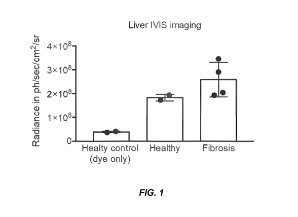
[0017] FIG. 1. Liver IVIS imaging.
[0018] FIGS. 2A-2B. Damage of Liver Parenchyma. FIG. 2A shows H&E-stained
sections of liver from fibrotic mice treated with various exosomes treatments.
FIG. 2B shows
a quantification of the level of fibrosis seen in FIG. 2A.
[0019] FIGS. 3A-3B. Damage of Lung Parenchyma. FIG. 3A shows H&E-stained
sections of lungs from fibrotis mice treated with various exosomes treatments.
FIG. 3B shows
a quantification of the level of fibrosis seen in FIG. 3A.
[0020] FIGs. 4A-4F. Knockdown efficiency of STAT3 in primary HSCs and
biodistribution of exosomes. FIGs. 4A and 4B show relative Stat3 expression in
HSC
treated with 5 1.tg/2 billion iExosaNA-STAT3 (FIG. 4A) or iExomASO-STAT3 (FIG.
4B). FIG. 4C
shows representative images of the listed organs analyzed for presence of DiR-
labeled
exosomes in non-fibrotic (sham) (left panel) and fibrotic mice (right panel)
(n = 1). FIGs. 4D-
4F show immunofluorescence imaging (FIGs. 4D) and quantification (FIGs. 4E and
4F) of
6
CA 03164248 2022-06-06
WO 2021/113761 PCT/US2020/063475
AF647 labeled exosomes and DAPI in frozen liver tissues of the listed groups
(3 visual fields
for each tissue analyzed). Scale bar: 100 pm. Data are represented as mean
SEM. For FIGs.
4A and 4D, an unpaired two¨tailed t¨test was used. For FIG. 4B, a one¨way
ANOVA with
Sidak's post¨hoc analysis was used. *p < 0.05, **p <0.01, ***p <0.001, ****p
<0.0001.
[0021] FIGs. 5A-5K. FIG. 5A shows images of mouse hepatic stellate cells
(HSCs)
cultured for 7 days (left panel). Scale bar: 100 pm. Immunofluorescence
staining for a¨SMA
and DAPI of primary mouse HSCs (center and right panels). Scale bar: 100 pm.
FIGs. 5B and
5C show qPCR analysis of STAT3. FIG. 5D shows a schematic of CC14 and
iExosomes/siRNA/mASO treatment schedule. Upper arrows indicate CC14 injections
(Day 0), and
lower arrows indicate iExosomes/siRNA/mASO injections (Day 9). FIGs. 5E and 5F
show
immunohistochemical staining of Collagen I (FIG. 5E) and quantification (FIG.
5F) in mice treated
with 1 [tg of 1 billion iExosiRNA STAT3 or iExomASO STAT3. (3 visual fields
for each tissue analyzed). n=3;
Scale bar: 100 inn. FIGs. 5G and 5H show immunofluorescence staining of a-SMA
(FIG. 5G) and
quantification (FIG. 5H) in mice treated with 1 [tg of 1 billion iExosiRNA
STAT3 or iExolliASO STAT3. (3
visual fields for each tissue analyzed). n=3. Scale bar: 100 inn. FIG. 51
shows H&E staining of liver
from mice treated with 1 [tg of 1 billion iExosiRNA STAT3 or iExoll'ASO STAT3
(5 visual fields for each
tissue analyzed), n=5; Scale bar: 100 inn. FIGs. 5J and 5K show percentage of
necrotic (FIG. 5J) and
degenerated hepatocytes (FIG. 5K). The data is presented as mean SEM.
Individual dots in graphs
depict distinct mice. FIG. 5B (left panel), unpaired two¨tailed Student's
t¨test. FIG. 5B (right panel)
and FIGs. 5C-5K One-way ANOVA with Sidak's post¨hoc analysis; p values are
indicated in all of
the graphs. *p <0.05; **p <0.01; ****p <0.0001; ns: not significant.
[0022] FIGs. 6A-6J. iExosomes targeting STAT3 reduced liver fibrosis. FIGs. 6A
and 6B show relative mRNA expression of STAT3 in liver of mice treated with 1
mil billion
(FIG. 6A) or 5 1.tg/2 billion (FIG. 6B) iExos1RNA-STAT3 or iExomASO-STAT3 of
the indicated
treatments. n = 4-5 distinct mice in 5 1.tg/2 billion groups; n = 4-5 distinct
mice, one-way
ANOVA was used in 1 mil billion group. FIGs. 6C and 6D show representative
Sirius red
staining (FIG. 6C) and quantification (FIG. 6D) of liver sections from the 1
mil billion
treatment group (3 visual fields for each tissue analyzed). n = 5-6 distinct
mice, One-way
ANOVA. Scale bar: 100 rim. The graph depicts the percent Sirius red positive
area. FIGs. 6E
and 6F show representative Sirius red staining (FIG. 6E) and quantification
(FIG. 6F) of liver
sections from the 5 1.tg/2 billion treatment group (3 visual fields for each
tissue analyzed). n =
3-5 distinct mice. Scale bar: 100 rim. The graph depicts the percent Sirius
red positive area.
FIGs. 6G and 6H show representative images (3 visual fields for each tissue
analyzed) of
7
CA 03164248 2022-06-06
WO 2021/113761 PCT/US2020/063475
immunohistochemical staining for Collagen I (FIG. 6G) and quantification of
the percent of
Collagen r area per visual field (100x) (FIG. 6H). n=3. FIGs. 61 and 6J show a-
SMA
immunofluorescence staining (FIG. 61; 3 visual fields for each tissue
analyzed) and
quantification (FIG. 6J) of the number of a-SMA cells per visual field
(100x). n = 3-4
distinct mice; Scale bar: 100 rim. The data are presented as mean SEM.
Individual dots in
graphs depict distinct mice. One-way ANOVA or 2-tailed unpaired t test, unless
otherwise
indicated; p values are indicated in all of the graphs. *p < 0.05; **p < 0.01;
***p < 0.001;
****p < 0.0001; ns: not significant.
[0023] FIGs. 7A-7K. iExosomes targeting STAT3 preserved liver functional
parenchyma. FIGs. 7A-7D show relative Collal (FIGs. 7A and 7B) and Acta2
(FIGs. 7C
and 7D) expression in livers with the indicated treatments. n = 4-5 distinct
mice in 5 1.tg/2
billion group; n = 5 distinct mice in 1 mil billion groups, one-way ANOVA was
used for
statistical analysis in 1 mil billion group. FIGs. 7E and 7F show serum levels
of ALT in
mice with the indicated treatments. n = 4-5 distinct mice in 5 1.tg/2 billion
groups; n = 4-5
distinct mice, one-way ANOVA was used for statistical analysis in 1 mil
billion group.
FIGs. 7G and 7H show serum levels of AST in mice with the indicated
treatments. n = 4-5
distinct mice in 5 1.tg/2 billion group; n = 4-5 distinct mice in 1 mil
billion groups, one-way
ANOVA was used for statistical analysis in 1 mil billion group. FIG. 71 shows
H&E
staining of paraffin-embedded liver sections (3-5 visual fields for each
tissue analyzed). n =
4-5 distinct mice; Scale bar: 100 rim. FIGs. 7J and 7K show percentage of
necrotic and
degenerated hepatocytes. The data is presented as mean SEM. Individual dots
in graphs
depict distinct mice. One-way ANOVA or unpaired two¨tailed t¨test; p values
are indicated
in all of the graphs. *p <0.05; **p <0.01; ****p <0.0001; ns: not significant.
[0024] FIGs. 8A and 8B. FIG. 8A shows H&E staining of the listed organs in
mice
treated with 5 1.tg/2 billion iExosaNASTAT3 or iExomASO-STAT3. FIG. 8B shows
H&E staining of
SO-
the listed organs in mice treated with 1 mil billion iExosaNASTAT3 or iExo niA
STAT3.
[0025] FIGs. 9A-9H. Reprogramming of the fibrotic liver transcriptome during
iExosomes treatment. FIG. 9A shows a heat map depicting relative intensity of
all probes
amongst the experimental groups (siCntrl iExo (n=3), siSTAT3 iExo (n=3), mASO
Scrbl
iExo (n=3) and mASO STAT3 iExo (n=3). Euclidean clustering of both rows and
columns
using 10g2-transformed mRNA-Seq expression data. FIGs. 9B and 9C show volcano
plots
depicting the number of differentially regulated genes in the livers of the
listed experimental
8
CA 03164248 2022-06-06
WO 2021/113761 PCT/US2020/063475
groups. FIG. 9D shows a heat map of STAT3 signaling. FIG. 9E shows selected
genes
associated with ECM deposition and remodeling. FIGs. 9F and 9G show a
representation of
differences in target genes by using gene ontology (GO) analysis (WebGestalt)
enrichment.
FIG. 9H shows an interaction network generated by the NetworkAnalyst for the
STAT3
signaling and ECM-associated genes.
[0026] FIGs. 10A-10H. Collal knockout in activated hepatic stellate cells.
FIG. 10A
shows Sirius Red staining to assess ECM and collagen I associated fibrosis,
demonstrating
significant decrease upon genetic loss of type I collagen from activated
hepatic stellate cells
or aSMA+ myofibroblasts (CollalcK ) in the context of fibrosis. FIG. 10B shows
results
demonstrating a significant reduction of Collagen I in CollalcK with liver
fibrosis. FIG. 10C
shows results demonstrating a significant improvement in liver histology in
CollalcK with
liver fibrosis. FIGs. 10D-10F show quantitation of the results from FIGs. 10A-
10C. FIGs.
10G and 10H show gene expression data demonstrating that many of the global
expression
patterns associated with liver fibrosis are significantly improved in CollalcK
mice with liver
fibrosis.
DETAILED DESCRIPTION
[0027] Provided herein are exosomes that have been engineered to carry
inhibitory
RNA molecules, including anti-sense oligonucleotides (ASO) and siRNA,
targeting STAT3, a
mediator of organ fibrosis, including liver and lung fibrosis. These
engineered exosomes can
be used to treat fibrosis, including liver fibrosis and lung fibrosis. Since
exosomes obtained
from mesenchymal stem cells have very high distribution to the lung and the
liver, the
delivery of the ASO or siRNA is efficient.
I. Lipid-based Nanoparticles
[0028] A lipid-based nanoparticle may be a liposome, an exosome, a lipid
preparation, or another lipid-based nanoparticle, such as a lipid-based
vesicle (e.g., a
DOTAP:cholesterol vesicle). Lipid-based nanoparticles may be positively
charged,
negatively charged, or neutral. Lipid-based nanoparticles may comprise the
necessary
components to allow for transcription and translation, signal transduction,
chemotaxis, or
other cellular functions. It is contemplated that one or more of these items
may be excluded
in an embodiment.
9
CA 03164248 2022-06-06
WO 2021/113761 PCT/US2020/063475
[0029] Lipid-based nanoparticles may comprise CD47 on their surface. CD47
(Integrin Associated Protein) is a transmembrane protein that is expressed on
most tissues
and cells. CD47 is a ligand for Signal Regulatory Protein Alpha (SIRP-a),
which is expressed
on phagocytic cells such as macrophages and dendritic cells. Activated CD47-
SIRP-a
initiates a signal transduction cascade that inhibits phagocytosis. Thus,
without being bound
by theory, expression of CD47 on the surface of exosomes may prevent
phagocytosis by
macrophages (see WO 2016/201323, which is incorporated herein by reference in
its
entirety).
A. Liposomes
[0030] A "liposome" is a generic term encompassing a variety of single and
multilamellar lipid vehicles formed by the generation of enclosed lipid
bilayers or aggregates.
Liposomes may be characterized as having vesicular structures with a bilayer
membrane,
generally comprising a phospholipid, and an inner medium that generally
comprises an
aqueous composition. Liposomes provided herein include unilamellar liposomes,
multilamellar liposomes, and multivesicular liposomes. Liposomes provided
herein may be
positively charged, negatively charged, or neutrally charged. In certain
embodiments, the
liposomes are neutral in charge.
[0031] A multilamellar liposome has multiple lipid layers separated by aqueous
medium. Such liposomes form spontaneously when lipids comprising phospholipids
are
suspended in an excess of aqueous solution. The lipid components undergo self-
rearrangement before the formation of closed structures and entrap water and
dissolved
solutes between the lipid bilayers. Lipophilic molecules or molecules with
lipophilic regions
may also dissolve in or associate with the lipid bilayer.
[0032] In specific aspects, a polypeptide, a nucleic acid, or a small molecule
drug
may be, for example, encapsulated in the aqueous interior of a liposome,
interspersed within
the lipid bilayer of a liposome, attached to a liposome via a linking molecule
that is
associated with both the liposome and the polypeptide/nucleic acid, entrapped
in a liposome,
complexed with a liposome, or the like.
[0033] A liposome used according to the present embodiments can be made by
different methods, as would be known to one of ordinary skill in the art. For
example, a
phospholipid, such as for example the neutral phospholipid
dioleoylphosphatidylcholine
CA 03164248 2022-06-06
WO 2021/113761 PCT/US2020/063475
(DOPC), is dissolved in tert-butanol. The lipid(s) is then mixed with a
polypeptide, nucleic
acid, and/or other component(s). Tween 20 is added to the lipid mixture such
that Tween 20
is about 5% of the composition's weight. Excess tert-butanol is added to this
mixture such
that the volume of tert-butanol is at least 95%. The mixture is vortexed,
frozen in a dry
ice/acetone bath and lyophilized overnight. The lyophilized preparation is
stored at -20 C and
can be used up to three months. When required the lyophilized liposomes are
reconstituted in
0.9% saline.
[0034] Alternatively, a liposome can be prepared by mixing lipids in a solvent
in a
container, e.g., a glass, pear-shaped flask. The container should have a
volume ten-times
greater than the volume of the expected suspension of liposomes. Using a
rotary evaporator,
the solvent is removed at approximately 40 C under negative pressure. The
solvent normally
is removed within about 5 min to 2 h, depending on the desired volume of the
liposomes. The
composition can be dried further in a desiccator under vacuum. The dried
lipids generally are
discarded after about 1 week because of a tendency to deteriorate with time.
[0035] Dried lipids can be hydrated at approximately 25-50 mM phospholipid in
sterile, pyrogen-free water by shaking until all the lipid film is
resuspended. The aqueous
liposomes can be then separated into aliquots, each placed in a vial,
lyophilized and sealed
under vacuum.
[0036] The dried lipids or lyophilized liposomes prepared as described above
may be
dehydrated and reconstituted in a solution of a protein or peptide and diluted
to an
appropriate concentration with a suitable solvent, e.g., DPBS. The mixture is
then vigorously
shaken in a vortex mixer. Unencapsulated additional materials, such as agents
including but
not limited to hormones, drugs, nucleic acid constructs and the like, are
removed by
centrifugation at 29,000 x g and the liposomal pellets washed. The washed
liposomes are
resuspended at an appropriate total phospholipid concentration, e.g., about 50-
200 mM. The
amount of additional material or active agent encapsulated can be determined
in accordance
with standard methods. After determination of the amount of additional
material or active
agent encapsulated in the liposome preparation, the liposomes may be diluted
to appropriate
concentrations and stored at 4 C until use. A pharmaceutical composition
comprising the
liposomes will usually include a sterile, pharmaceutically acceptable carrier
or diluent, such
as water or saline solution.
11
CA 03164248 2022-06-06
WO 2021/113761 PCT/US2020/063475
[0037] Additional liposomes which may be useful with the present embodiments
include cationic liposomes, for example, as described in W002/100435A1, U.S
Patent
5,962,016, U.S. Application 2004/0208921, W003/015757A1, W004/029213A2, U.S.
Patent 5,030,453, and U.S. Patent 6,680,068, all of which are hereby
incorporated by
reference in their entirety without disclaimer.
[0038] In preparing such liposomes, any protocol described herein, or as would
be
known to one of ordinary skill in the art may be used. Additional non-limiting
examples of
preparing liposomes are described in U.S. Patents 4,728,578, 4,728,575,
4,737,323,
4,533,254, 4,162,282, 4,310,505, and 4,921,706; International Applications
PCT/U585/01161
and PCT/U589/05040, each incorporated herein by reference.
[0039] In certain embodiments, the lipid-based nanoparticle is a neutral
liposome
(e.g., a DOPC liposome). "Neutral liposomes" or "non-charged liposomes", as
used herein,
are defined as liposomes having one or more lipid components that yield an
essentially-
neutral, net charge (substantially non-charged). By "essentially neutral" or
"essentially non-
charged", it is meant that few, if any, lipid components within a given
population (e.g., a
population of liposomes) include a charge that is not canceled by an opposite
charge of
another component (i.e., fewer than 10% of components include a non-canceled
charge, more
preferably fewer than 5%, and most preferably fewer than 1%). In certain
embodiments,
neutral liposomes may include mostly lipids and/or phospholipids that are
themselves neutral
under physiological conditions (i.e., at about pH 7).
[0040] Liposomes and/or lipid-based nanoparticles of the present embodiments
may
comprise a phospholipid. In certain embodiments, a single kind of phospholipid
may be used
in the creation of liposomes (e.g., a neutral phospholipid, such as DOPC, may
be used to
generate neutral liposomes). In other embodiments, more than one kind of
phospholipid may
be used to create liposomes. Phospholipids may be from natural or synthetic
sources.
Phospholipids include, for example, phosphatidylcholines,
phosphatidylglycerols, and
phosphatidylethanolamines; because phosphatidylethanolamines and phosphatidyl
cholines
are non-charged under physiological conditions (i.e., at about pH 7), these
compounds may
be particularly useful for generating neutral liposomes. In certain
embodiments, the
phospholipid DOPC is used to produce non-charged liposomes. In certain
embodiments, a
lipid that is not a phospholipid (e.g., a cholesterol) may be used.
12
CA 03164248 2022-06-06
WO 2021/113761 PCT/US2020/063475
[0041] Pho spholipids include glyceropho spholipids and certain sphingo lipid
s .
Phospholipids include, but are not limited to, dioleoylphosphatidylycholine
("DOPC"), egg
pho sphatidylcholine ("EPC"), dilauryloylpho sphatidylcholine
("DLPC"),
dimyristoylpho sphatidylcholine ("DMPC"), dip almito ylpho sphatidylcholine
("DPPC"),
di stearo ylpho sphatidylcholine ("D S PC") , 1-myri s to y1-2-p almito yl pho
sphatidylcholine
("MPPC"), 1-p almito y1-2-myri sto yl pho sphatidylcholine ("PMPC"), 1-p
almito y1-2- stearoyl
pho sphatidylcholine ("PS PC") , 1- s tearo y1-2 -p almitoyl pho
sphatidylcholine ("SPPC"),
dilauryloylphosphatidylglycerol ("DLPG"), dimyristoylphosphatidylglycerol
("DMPG"),
dip almitoylpho sphatidylglycerol ("DPPG"), di stearo ylpho sphatidylglycerol
("D S PG") ,
di stearo yl sphingomyelin ("DS SP"), di stearo ylphophatidylethanolamine ("D
S PE") ,
dioleoylphosphatidylglycerol ("DOPG"), dimyristoyl phosphatidic acid ("DMPA"),
dipalmitoyl phosphatidic acid ("DPPA"), dimyristoyl phosphatidylethanolamine
("DMPE"),
dipalmitoyl phosphatidylethanolamine ("DPPE"), dimyristoyl phosphatidylserine
("DMPS"),
dipalmitoyl phosphatidylserine ("DPPS"), brain phosphatidylserine ("BPS"),
brain
sphingomyelin ("B SP"), dipalmitoyl sphingomyelin
("DP S P"), dimyristyl
phosphatidylcholine ("DMPC"), 1,2-distearoyl-sn-glycero-3-phosphocholine
("DAPC"), 1,2-
diarachido yl- sn-glycero-3 -pho sphocholine
("DB PC"), 1,2-dieico senoyl- sn-glycero-3 -
pho sphocholine ("DEPC"), dioleoylphosphatidylethanolamine ("DOPE"),
palmitoyloeoyl
pho sphatidylcholine ("POPC"), palmitoyloeoyl phosphatidylethanolamine
("POPE"),
lysopho sphatidylcholine, lysopho sphatidylethanol amine, and dilinoleoylpho
sphatidylcholine.
B. Exosomes
[0042] The terms "microvesicle" and "exosomes," as used herein, refer to a
membranous particle having a diameter (or largest dimension where the
particles is not
spheroid) of between about 10 nm to about 5000 nm, more typically between 30
nm and 1000
nm, and most typically between about 50 nm and 750 nm, wherein at least part
of the
membrane of the exosomes is directly obtained from a cell. An exosome of the
disclosure
may have a diameter of at least, at most, or about 10, 20, 30, 40, 50, 60, 70,
80, 90, 100, 200,
300, 400, 500, 600, 700, 800, 900, 1000, 2000, 3000, 4000, or 5000 nm, or any
range
derivable therein. Most commonly, exosomes will have a size (average diameter)
that is up to
5% of the size of the donor cell. Therefore, especially contemplated exosomes
include those
that are shed from a cell.
13
CA 03164248 2022-06-06
WO 2021/113761 PCT/US2020/063475
[0043] Exosomes may be detected in or isolated from any suitable sample type,
such
as, for example, body fluids. As used herein, the term "isolated" refers to
separation out of its
natural environment and is meant to include at least partial purification and
may include
substantial purification. As used herein, the term "sample" refers to any
sample suitable for
the methods provided by the present invention. The sample may be any sample
that includes
exosomes suitable for detection or isolation. Sources of samples include
blood, bone marrow,
pleural fluid, peritoneal fluid, cerebrospinal fluid, urine, saliva, amniotic
fluid, malignant
ascites, broncho-alveolar lavage fluid, synovial fluid, breast milk, sweat,
tears, joint fluid, and
bronchial washes. In one aspect, the sample is a blood sample, including, for
example, whole
blood or any fraction or component thereof. A blood sample suitable for use
with the present
invention may be extracted from any source known that includes blood cells or
components
thereof, such as venous, arterial, peripheral, tissue, cord, and the like. For
example, a sample
may be obtained and processed using well-known and routine clinical methods
(e.g.,
procedures for drawing and processing whole blood). In one aspect, an
exemplary sample
may be peripheral blood drawn from a subject with cancer.
[0044] Exosomes may also be isolated from tissue samples, such as surgical
samples,
biopsy samples, tissues, feces, and cultured cells. When isolating exosomes
from tissue
sources it may be necessary to homogenize the tissue in order to obtain a
single cell
suspension followed by lysis of the cells to release the exosomes. When
isolating exosomes
from tissue samples it is important to select homogenization and lysis
procedures that do not
result in disruption of the exosomes. Exosomes contemplated herein are
preferably isolated
from body fluid in a physiologically acceptable solution, for example,
buffered saline, growth
medium, various aqueous medium, etc.
[0045] Exosomes may be isolated from freshly collected samples or from samples
that have been stored frozen or refrigerated. In some embodiments, exosomes
may be isolated
from cell culture medium. Although not necessary, higher purity exosomes may
be obtained
if fluid samples are clarified before precipitation with a volume-excluding
polymer, to
remove any debris from the sample. Methods of clarification include
centrifugation,
ultracentrifugation, filtration, or ultrafiltration. Most typically, exosomes
can be isolated by
numerous methods well-known in the art. One preferred method is differential
centrifugation
from body fluids or cell culture supernatants. Exemplary methods for isolation
of exosomes
are described in (Losche et al., 2004; Mesri and Altieri, 1998; Morel et al.,
2004).
14
CA 03164248 2022-06-06
WO 2021/113761 PCT/US2020/063475
Alternatively, exosomes may also be isolated via flow cytometry as described
in (Combes et
al., 1997).
[0046] One accepted protocol for isolation of exosomes includes
ultracentrifugation,
often in combination with sucrose density gradients or sucrose cushions to
float the relatively
low-density exosomes. Isolation of exosomes by sequential differential
centrifugations is
complicated by the possibility of overlapping size distributions with other
microvesicles or
macromolecular complexes. Furthermore, centrifugation may provide insufficient
means to
separate vesicles based on their sizes. However, sequential centrifugations,
when combined
with sucrose gradient ultracentrifugation, can provide high enrichment of
exosomes.
[0047] Isolation of exosomes based on size, using alternatives to the
ultracentrifugation routes, is another option. Successful purification of
exosomes using
ultrafiltration procedures that are less time consuming than
ultracentrifugation, and do not
require use of special equipment have been reported. Similarly, a commercial
kit is available
(EXOMIRTm, Bioo Scientific) which allows removal of cells, platelets, and
cellular debris on
one microfilter and capturing of vesicles bigger than 30 nm on a second
microfilter using
positive pressure to drive the fluid. However, for this process, the exosomes
are not
recovered, their RNA content is directly extracted from the material caught on
the second
microfilter, which can then be used for PCR analysis. HPLC-based protocols
could
potentially allow one to obtain highly pure exosomes, though these processes
require
dedicated equipment and are difficult to scale up. A significant problem is
that both blood
and cell culture media contain large numbers of nanoparticles (some non-
vesicular) in the
same size range as exosomes. For example, some miRNAs may be contained within
extracellular protein complexes rather than exosomes; however, treatment with
protease (e.g.,
proteinase K) can be performed to eliminate any possible contamination with
"extraexosomal" protein.
[0048] In another embodiment, cancer cell-derived exosomes may be captured by
techniques commonly used to enrich a sample for exosomes, such as those
involving
immunospecific interactions (e.g., immunomagnetic capture). Immunomagnetic
capture, also
known as immunomagnetic cell separation, typically involves attaching
antibodies directed to
proteins found on a particular cell type to small paramagnetic beads. When the
antibody-
coated beads are mixed with a sample, such as blood, they attach to and
surround the
particular cell. The sample is then placed in a strong magnetic field, causing
the beads to
CA 03164248 2022-06-06
WO 2021/113761 PCT/US2020/063475
pellet to one side. After removing the blood, captured cells are retained with
the beads. Many
variations of this general method are well-known in the art and suitable for
use to isolate
exosomes. In one example, the exosomes may be attached to magnetic beads
(e.g.,
aldehyde/sulphate beads) and then an antibody is added to the mixture to
recognize an
epitope on the surface of the exosomes that are attached to the beads.
Exemplary proteins that
are known to be found on cancer cell-derived exosomes include ATP-binding
cassette sub-
family A member 6 (ABCA6), tetraspanin-4 (TSPAN4), SLIT and NTRK-like protein
4
(SLITRK4), putative protocadherin beta-18 (PCDHB18), myeloid cell surface
antigen CD33
(CD33), and glypican-1 (GPC1). Cancer cell-derived exosomes may be isolated
using, for
example, antibodies or aptamers to one or more of these proteins.
[0049] It should be noted that not all proteins expressed in a cell are found
in
exosomes secreted by that cell. For example, calnexin, GM130, and LAMP-2 are
all proteins
expressed in MCF-7 cells but not found in exosomes secreted by MCF-7 cells
(Baietti et al.,
2012). As another example, one study found that 190/190 pancreatic ductal
adenocarcinoma
patients had higher levels of GPC1+ exosomes than healthy controls (Melo et
al., 2015,
which is incorporated herein by reference in its entirety). Notably, only 2.3%
of healthy
controls, on average, had GPC1+ exosomes.
1. Exemplary Protocol for Collecting Exosomes from Cell Culture
[0050] On Day 1, seed enough cells (e.g., about five million cells) in T225
flasks in
media containing 10% FBS so that the next day the cells will be about 70%
confluent. On
Day 2, aspirate the media on the cells, wash the cells twice with PBS, and
then add 25-30 mL
base media (i.e., no PenStrep or FBS) to the cells. Incubate the cells for 24-
48 hours. A 48
hour incubation is preferred, but some cells lines are more sensitive to serum-
free media and
so the incubation time should be reduced to 24 hours. Note that FBS contains
exosomes that
will heavily skew NanoSight results.
[0051] On Day 3/4, collect the media and centrifuge at room temperature for
five
minutes at 800 x g to pellet dead cells and large debris. Transfer the
supernatant to new
conical tubes and centrifuge the media again for 10 minutes at 2000 x g to
remove other large
debris and large vesicles. Pass the media through a 0.2 p.m filter and then
aliquot into
ultracentrifuge tubes (e.g., 25 x 89 mm Beckman Ultra-Clear) using 35 mL per
tube. If the
volume of media per tube is less than 35 mL, fill the remainder of the tube
with PBS to reach
35 mL. Ultracentrifuge the media for 2-4 hours at 28,000 rpm at 4 C using a SW
32 Ti rotor
16
CA 03164248 2022-06-06
WO 2021/113761 PCT/US2020/063475
(k-factor 266.7, RCF max 133,907). Carefully aspirate the supernatant until
there is roughly
1-inch of liquid remaining. Tilt the tube and allow remaining media to slowly
enter aspirator
pipette. If desired, the exosomes pellet can be resuspended in PBS and the
ultracentrifugation
at 28,000 rpm repeated for 1-2 hours to further purify the population of
exosomes.
[0052] Finally, resuspend the exosomes pellet in 210 i.IL PBS. If there are
multiple
ultracentrifuge tubes for each sample, use the same 210 0_, PBS to serially
resuspend each
exosomes pellet. For each sample, take 10 0_, and add to 990 0_, H20 to use
for nanoparticle
tracking analysis. Use the remaining 200 0_, exosomes-containing suspension
for
downstream processes or immediately store at -80 C.
2. Exemplary Protocol for Extracting Exosomes from Serum
Samples
[0053] First, allow serum samples to thaw on ice. Then, dilute 250 0_, of cell-
free
serum samples in 11 mL PBS; filter through a 0.2 p.m pore filter.
Ultracentrifuge the diluted
sample at 150,000 x g overnight at 4 C. The following day, carefully discard
the supernatant
and wash the exosomes pellet in 11 mL PBS. Perform a second round of
ultracentrifugation
at 150,000 x g at 4 C for 2 hours. Finally, carefully discard the supernatant
and resuspend the
exosomes pellet in 100 0_, PBS for analysis.
C. Exemplary Protocol for Electroporation of Exosomes and Liposomes
[0054] Mix 1 x 108 exosomes (measured by NanoSight analysis) or 100 nm
liposomes (e.g., purchased from Encapsula Nano Sciences) and 1 1.tg of siRNA
(Qiagen) or
shRNA in 400 [IL of electroporation buffer (1.15 mM potassium phosphate, pH
7.2, 25 mM
potassium chloride, 21% Optiprep). Electroporate the exosomes or liposomes
using a 4 mm
cuvette (see, e.g., Alvarez-Erviti et al., 2011; El-Andaloussi et al., 2012).
After
electroporation, treat the exosomes or liposomes with protease-free RNAse
followed by
addition of 10x concentrated RNase inhibitor. Finally, wash the exosomes or
liposomes with
PBS under ultracentrifugation methods, as described above.
II. Inhibitory RNAs
A. Antisense Oligonucleotides
[0055] Antisense oligonucleotide (AS 0) therapeutic agents are single stranded
nucleic acid therapeutics, typically about 16 to 30 nucleotides in length, and
are
17
CA 03164248 2022-06-06
WO 2021/113761 PCT/US2020/063475
complementary to a target nucleic acid sequence in the target cell, either in
culture or in an
organism.
[0056] In some embodiments, the agent is a single-stranded antisense RNA
molecule,
a single-stranded antisense DNA molecule, or a single-stranded antisense
polynucleotide
comprising both DNA and RNA. In a particular embodiment, the antisense
molecule is an
ASO comprising both DNA and RNA. An antisense molecule is complementary to a
sequence within the target mRNA, e.g., a STAT3 mRNA. Antisense molecules can
inhibit
translation in a stoichiometric manner by base pairing to the mRNA and
physically
obstructing the translation machinery. The antisense molecule may have at
least or at most
15-30 nucleotides that are complementary to the target mRNA. For example, the
antisense
molecule may have a sequence of at least or at most 15, 16, 17, 18, 19, 20,
21, 22, 23, 24 or
25, or any range or value derivable therein,contiguous nucleotides that are
complementary to
the target mRNA.
[0057] In some embodiments, the ASO comprises at least or at most 8, 9, 10,
11, 12,
13, 14, 15, 16, 17, 18, 19, 20, 21, 22, 23, 24, 25, 26, 27, 28, 29, 30, 31,
32, 33, 34, 35, 36, 37,
38, 39, 40, 41, 42, 43, 44, 45, 46, 47, 48, 49 or 50 nucleotides, or any range
or value derivable
therein. Any of these values may be used to define a range for the number of
nucleotides in
the ASO. For example, the ASO may comprise, comprise at least or, or comprise
at most 8-
50, 15-30, or 20-25 nucleotides. In some embodiments, the ASO consists of 8,
9, 10, 11, 12,
13, 14, 15, 16, 17, 18, 19, 20, 21, 22, 23, 24, 25, 26, 27, 28, 29, 30, 31,
32, 33, 34, 35, 36, 37,
38, 39, 40, 41, 42, 43, 44, 45, 46, 47, 48, 49 or 50 nucleotides, or any range
or value derivable
therein. Any of these values may be used to define a range for the number of
nucleotides in
the ASO. For example, the ASO may consist of 8-50, 15-30, or 20-25
nucleotides.
[0058] In one aspect of the disclosure, the agent is a single-stranded
antisense nucleic
acid molecule (ASO). Antisense oligonucleotides (AS0s) are synthetic molecules
and, in
some embodiments, comprise between 18-21 nucleotides in length and are
complementary to
the mRNA sequence of the target gene. ASOs bind cognate mRNA sequences through
sequence-specific hybridization resulting in cleavage or disablement of the
mRNA and
inhibition of the expression of the target gene.
18
CA 03164248 2022-06-06
WO 2021/113761 PCT/US2020/063475
1. Modification of ASOs
[0059] In certain embodiments, the ASOs of the disclosure may be modified. A
"modified ASO" refers to a molecule in which one or more of the components of
the nucleic
acid, namely sugars, bases, and phosphate moieties, are different from that
which occur in
nature, for example, different from that which occurs in the human body.
Several
modifications to ASOs are described in the art. These modifications are aimed
at improving
ASO properties such as resistance to nucleases, permeability across biological
membranes,
solubility, stability, or modulation of pharmacokinetic and pharmacodynamics
properties
while maintaining specificity to the target mRNA. For example, the
modifications on the
nucleotides can include, but are not limited to, LNA, HNA, CeNA, 2'-
methoxyethyl, 2'-O-
alkyl, 2'-0-allyl, 2'-C-allyl, 2'-fluoro, 2'-deoxy, 2'-hydroxyl, and
combinations thereof. It is
contemplated that one or more of these modifications may be excluded in an
embodiment.
[0060] Patents directed to antisense nucleic acids, chemical modifications,
and
therapeutic uses are provided, for example, in U.S. Pat. No. 5,898,031 related
to chemically
modified RNA-containing therapeutic compounds, and U.S. Pat. No. 6,107,094
related
methods of using these compounds as therapeutic agent. U.S. Pat. No. 7,432,250
related to
methods of treating patients by administering single-stranded chemically
modified RNA-like
compounds; and U.S. Pat. No. 7,432,249 related to pharmaceutical compositions
containing
single-stranded chemically modified RNA-like compounds. U.S. Pat. No.
7,629,321 is related
to methods of cleaving target mRNA using a single-stranded oligonucleotide
having a
plurality RNA nucleosides and at least one chemical modification. Each of the
patents listed
in this paragraph are incorporated herein by reference in their entirety.
a. Modified Bases
[0061] Therapeutic nucleic acid may include natural (i.e. A, G, U, C, or T) or
modified (e.g. 7-deazaguanosine, inosine, etc.) bases. Modification of bases
includes the
incorporation of modified bases (or modified nucleoside or modified
nucleotides) that are
variations of standard bases, sugars and/or phosphate backbone chemical
structures occurring
in ribonucleic (i.e., A, C, G and U) and deoxyribonucleic (i.e., A, C, G and
T) acids. Included
within this scope are, for example: Gm (2'-methoxyguanylic acid), Am (2'-
methoxyadenylic
acid), Cf (2'-fluorocytidylic acid), Uf (2'-fluorouridylic acid), Ar
(riboadenylic acid). The
aptamers may also include cytosine or any cytosine-related base including 5-
methylcytosine,
4-acetylc yto sine, 3 -methylc yto sine, 5-hydroxymethyl cytosine, 2-thioc yto
sine, 5-
19
CA 03164248 2022-06-06
WO 2021/113761 PCT/US2020/063475
haloc yto sine (e.g., 5-fluoroc yto sine, 5-bromocyto sine, 5-chloroc yto
sine, and 5-iodoc yto sine),
5-prop ynyl cytosine, 6-azocyto sine, 5-trifluoromethylc yto sine, N4,N4-
ethanoc yto sine,
phenoxazine cytidine, phenothiazine cytidine, carbazole cytidine or
pyridoindole cytidine.
The aptamer may further include guanine or any guanine-related base including
6-
methylguanine, 1-methylguanine, 2,2-dimethylguanine, 2-methylguanine, 7-
methylguanine,
2-propylguanine, 6-propylguanine, 8-haloguanine (e.g., 8-fluoroguanine, 8-
bromoguanine, 8-
chloroguanine, and 8-iodoguanine), 8-aminoguanine, 8-sulfhydrylguanine, 8-
thioalkylguanine, 8-hydroxylguanine, 7-methylguanine, 8-azaguanine, 7-
deazaguanine or 3-
deazaguanine. The aptamer may still further include adenine or any adenine-
related base
including 6-methyladenine, N6-isopentenyladenine, N6-methyladenine, 1-
methyladenine, 2-
methyladenine, 2-methylthio-N6-isopentenyladenine, 8-haloadenine (e.g., 8-
fluoroadenine, 8-
bromoadenine, 8-chloroadenine, and 8-iodo adenine), 8-aminoadenine, 8-
sulfhydryladenine,
8-thioalkyladenine, 8-hydroxyladenine, 7-methyladenine, 2-haloadenine (e.g., 2-
fluoroadenine, 2-bromoadenine, 2-chloroadenine, and 2-iodoadenine), 2-
aminoadenine, 8-
azaadenine, 7-deazaadenine or 3-deazaadenine. Also included are uracil or any
uracil-related
base including 5-halouracil (e.g., 5-fluorouracil, 5-bromouracil, 5-
chlorouracil, 5-iodouracil),
5-(carboxyhydroxylmethyl)uracil, 5-carboxymethylaminomethy1-2-thiouracil,
5-
carboxymethylaminomethyluracil, dihydrouracil, 1 -methylp s eudouracil,
5-
methoxyaminomethy1-2-thiouracil, 5'-methoxycarbonylmethyluracil, 5-
methoxyuracil, 5-
methy1-2-thiouracil, 2-thiouracil, 4-thiouracil, 5-methyluracil, uracil-5-
oxyacetic acid
methylester, uracil-5-oxyacetic acid, pseudouracil, 5-methyl-2-thiouracil, 2-
thiouracil, 3-(3-
amino-3-N-2-carboxypropyl)uracil, 5-methylaminomethyluracil, 5-propynyl
uracil, 6-
azouracil, or 4-thiouracil.
[0062] Examples of other modified base variants known in the art include,
without
limitation, e.g., 4-acetylcytidine, 5-(carboxyhydroxylmethyl)uridine, 21-
methoxycytidine, 5-
carboxymethylaminomethy1-2-thioridine, 5-
carboxymethylaminomethyluridine,
dihydrouridine, 2 '-0-methylpseudouridine, b-D-galactos
ylqueo sine, ino sine, N6-
isopentenyladeno sine, 1 -methyladeno sine, 1 -methylp s eudouridine, 1 -
methylguano sine, 1 -
methylino sine, 2,2-dimethylguano sine, 2-methyladeno sine, 2-methylguano
sine, 3 -
methylc ytidine, 5-methylcytidine, N6-methyladeno sine,
7-methylguano sine, 5-
methylaminomethyluridine, 5-methoxyaminomethy1-2-thiouridine, b-D-
mannosylqueosine,
5-methoxycarbonylmethyluridine, 5-methoxyuridine, 2-methylthio-N6-
isopentenyladenosine,
N-((9-b-D-ribofuranosy1-2-methylthiopurine-6-yl)carbamoyl)threonine, N-
((9-b-D-
CA 03164248 2022-06-06
WO 2021/113761 PCT/US2020/063475
ribofurano s ylpurine-6- yl)N-methyl-c arb amo yl)threonine,
urdine-5-oxyacetic acid
methylester, uridine-5-oxyacetic acid (v), wybutoxosine, pseudouridine,
queosine, 2-
thiocytidine, 5-methyl-2-thiouridine, 2-thiouridine, 4-thiouridine, 5-
methyluridine, N-((9-b-
D-ribofuranosylpurine-6-yl)carbamoyl)threonine, 2 '-
0-methyl-5-methyluridine, 2 '-0-
methyluridine, and wybutosine, 3-(3-amino-3-carboxypropyl)uridine.
[0063] Also included are the modified nucleobases described in U.S. Pat. Nos.
3,687,808, 3,687,808, 4,845,205, 5,130,302, 5,134,066, 5,175,273, 5,367,066,
5,432,272,
5,457,187, 5,459,255, 5,484,908, 5,502,177, 5,525,711, 5,552,540, 5,587,469,
5,594,121,
5,596,091, 5,614,617, 5,645,985, 5,830,653, 5,763,588, 6,005,096, and
5,681,941, each of
which is incorporated herein by reference in its entirety.
b. Modified Sugars
[0064] Modified sugar moieties for use in ASOs are well known in the art and
are
described for example in U.S. Pat. No. 9,045,754 which is incorporated by
reference herein in
its entirety. Modified sugars can be used to alter, typically increase, the
affinity of the ASO
for its target and/or increase nuclease resistance. For example, in some
embodiments, the
binding affinity of the ASOs to their target can be increased by incorporating
substituent
groups in the nucleoside subunits of the ASOs. In some embodiments, the
substituent groups
are T substituent groups, substituent groups located at the 2' position of the
pentofuranosyl
sugar moieties of the nucleoside subunits of the ASOs. Substituent groups
include, but are not
limited to, fluoro, alkoxy, amino-alkoxy, allyloxy, imidazolylalkoxy and
polyethylene glycol.
Alkoxy and aminoalkoxy groups generally include lower alkyl groups,
particularly C1-C9
alkyl. In a particular embodiment, the 2' substituent group is 2'-0-methyl.
Polyethylene
glycols are of the structure (0¨CH2¨CH2)n-0-alkyl. In a particular embodiment,
the
substituent is a polyethylene glycol substituent of the formula (-0¨CH2¨CH2)n-
0-
alkyl, wherein n=1 and alky1=CH3. This modification has been shown to increase
both
affinity of an oligonucleotide for its target and nuclease resistance of an
oligonucleotide. See
U.S. Pat. No. 7,629,321 cited above. A further particularly useful 2'-
substituent group for
increasing the binding affinity is the 2'-fluoro group.
[0065] Examples of modified nucleoside and nucleotide sugar backbone variants
known in the art include, without limitation, those having, e.g., 2' ribosyl
substituents such as
F, SH, SCH3, OCN, Cl, Br, CN, CF3, OCF3, SOCH3, S02, CH3, 0NO2, NO2, N3, NH2,
OCH2CH2OCH3, 0(CH2)20N(CH3)2, OCH2OCH2N(CH3)2, 0(C1-10 alkyl), 0(C2-10
21
CA 03164248 2022-06-06
WO 2021/113761 PCT/US2020/063475
alkenyl), 0(C2-10 alkynyl), S(C1-10 alkyl), S(C2-10 alkenyl), S(C2-10
alkynyl), NH(C1-10
alkyl), NH(C2-10 alkenyl), NH(C2-10 alkynyl), and 0-alkyl-0-alkyl. Desirable
2' ribosyl
substituents include 2'-methoxy (2'-OCH3), 2'-aminopropoxy (2' OCH2CH2CH2NH2),
2'-0-
allyl (21-CH2¨CH=CH2), 2'-0-ally1 (21-0¨CH2¨CH=CH2), 2'-amino (2'-NH2), and 2'-
fluoro (2'-F). The 2'-substituent may be in the arabino (up) position or ribo
(down) position.
One or more of these variants may be excluded from embodiments of the
disclosure.
[0066] Another class of modified ASOs known in the art and that may be
utilized in
the ASOs of the disclosure contain alkyl modifications at the 2' position of
the ribose moiety.
These ASOs were developed to improve the binding affinity and hybridization
stability with
target mRNA, and to increase the nuclease resistance of the ASOs. In this
category, the most
commonly used ASOs are 2'-0-Methyl (2'-OME) and 2'-0-Methoxyethyl (2'-M0E)
ASOs.
ASOs with this type of modification are incapable of activating RNAse H.
Therefore, to
induce RNAse H activation, chimeric ASOs have been developed in which a
central gap
region consisting of a phosphorothioate deoxyribose core is flanked with
nuclease resistant
arms such as 2'-OME or 2'-MOE that possess greater nuclease resistance. A
"gapmer" is
produced as a result, in which RNAse H can sit in the central gap and activate
target specific
mRNA degradation, while the arms prevent the ASO degradation. ASOs in this
category
possess higher affinity for mRNA, show better tissue uptake, and have
increased resistance to
nucleases, longer in vivo half life, and lesser toxicity, as compared to the
modified ASOs of
the first class.
[0067] A further class of ASOs known in the art and that may be utilized in
the ASOs
of the disclosure contain modifications of the furanose ring along with
modifications of the
phosphate linkage, the ribose moiety, or the nucleotides. These modifications
were designed
to improve the nuclease stability, target affinity and pharmacokinetic
profiles of the ASOs.
Common examples of third category of ASOs are Locked nucleic acid (LNA),
Peptide
nucleic acid (PNA) and Morpholino phosphoroamidates (MF) ASOs in this category
are
more stable in biological fluids because of their high resistance to
degradation by nucleases
and peptidases. They also exhibit a strong hybridization affinity with the
mRNA. Further,
PNAs recognize double stranded DNA, and are able to modulate gene expression
or induce
mutation by strand invasion of chromosomal duplex DNA. ASOs in this category
also do not
activate RNAse H and rely on sterically hindering the ribosomal machinery to
cause
translational arrest. They do not bind to serum proteins as they are
uncharged. Lack of charge
22
CA 03164248 2022-06-06
WO 2021/113761 PCT/US2020/063475
reduces the odds of non-specific interactions but increases the rate of
clearance from the
body. Their electrostatically neutral backbones may reduce solubility and make
uptake more
difficult.
[0068] A representative list of preferred modified sugars includes but is not
limited to
bicyclic modified sugars (BNA's), including methyleneoxy (4'-CH2-0-2') BNA and
ethyleneoxy (4'-(CH2)2-0-2' bridge) BNA; substituted sugars, especially 2'-
substituted
sugars having a 2'-F, 2'-OCH3 or a 2'-0(CH2)2-0CH3 substituent group; and 4'-
thio
modified sugars. Sugars can also be replaced with sugar mimetic groups among
others.
Methods for the preparations of modified sugars are well known to those
skilled in the art.
Some representative patents and publications that teach the preparation of
such modified
sugars include, but are not limited to, U.S. Pat. Nos. 4,981,957; 5,118,800;
5,319,080;
5,359,044; 5,393,878; 5,446,137; 5,466,786; 5,514,785; 5,519,134; 5,567,811;
5,576,427;
5,591,722; 5,597,909; 5,610,300; 5,627,053; 5,639,873; 5,646,265; 5,658,873;
5,670,633;
5,792,747; 5,700,920; 6,531,584; and 6,600,032; and WO 2005/121371.
c. Modified Internucleotide Linkages
[0069] Nucleic acid therapeutics may further comprise at least one
phosphorothioate
or methylphosphonate internucleotide linkage. The phosphorothioate or
methylphosphonate
internucleotide linkage modification may occur on any nucleotide of the sense
strand or
antisense strand or both (in nucleic acid therapeutics including a sense
strand) in any position
of the strand. For instance, the internucleotide linkage modification may
occur on every
nucleotide on the sense strand or antisense strand; each internucleotide
linkage modification
may occur in an alternating pattern on the sense strand or antisense strand;
or the sense strand
or antisense strand may contain both internucleotide linkage modifications in
an alternating
pattern. The alternating pattern of the internucleotide linkage modification
on the sense strand
may be the same or different from the antisense strand, and the alternating
pattern of the
internucleotide linkage modification on the sense strand may have a shift
relative to the
alternating pattern of the internucleotide linkage modification on the
antisense strand.
[0070] In certain embodiments, the ASOs of the disclosure comprise one or more
nucleoside subunits connected by phosphorus linkages including phosphodiester,
phosphorothioate, 3 '(or -5 ')deoxy-3 '-(or -51)thio-phosphorothioate,
phosphorodithioate,
phosphoroselenates, 3'-(or -5')deoxy phosphinates, borano phosphates, 3'-(or
5'-)amino
phosphoramidates, hydrogen phosphonates, borano phosphate esters,
phosphoramidates,
23
CA 03164248 2022-06-06
WO 2021/113761 PCT/US2020/063475
alkyl or aryl phosphonates and phosphotriester phosphorus linkages. In some
embodiments,
the ASOs of the disclosure comprise nucleoside subunits connected by
carbonate, carbamate,
silyl, sulfur, sulfonate, sulfonamide, formacetal, thioformacetyl, oxime,
methyleneimino,
methylenemethylimino, methylenehydrazo, methylenedimethylhydrazo and
methyleneoxymethylimino linkages.
[0071] For example, one class of modified ASO described in the art and that
may be
utilized in the ASOs of the disclosure are those that have one of the non-
bridging oxygen
atoms in the phosphate group of the ASO replaced with either a sulfur group
(phosphorothioates), a methyl group (methyl phosphonates) or an amine group
(phosphoramidates). These ASOs have greater resistance to nucleases and longer
plasma half
life as compared with phosphodiester oligonucleotides. They are capable of
activating RNAse
H, carry negative charges which facilitate their delivery to cells, and have
suitable
pharmacokinetics. Among these modifications, phosphorothioate modifications
are used most
widely. For example, Vitravene, an FDA approved ASO drug, and most of the
other ASO
drugs in clinical trials are phosphorothioate ASOs.
[0072] In addition, the bases in nucleotide may be joined by a linkage other
than a
phosphodiester bond, so long as it does not interfere with hybridization.
Thus, inhibitory
nucleic acids may be peptide nucleic acids in which the constituent bases are
joined by
peptide bonds rather than phosphodiester linkages. The inhibitory nucleic
acids may be
prepared by converting the RNA to cDNA using known methods (see, e.g., Ausubel
et. al.,
Current Protocols in Molecular Biology Wiley 1999). The inhibitory nucleic
acids can also be
cRNA (see, e.g., Park et. al., (2004) Biochem. Biophys. Res. Commun.
325(4):1346-52).
B. Small Interfering RNAs
[0073] siRNA (e.g., siNA) are well known in the art. For example, siRNA and
double-stranded RNA have been described in U.S. Pat. Nos. 6,506,559 and
6,573,099, as well
as in U.S. Patent Applications 2003/0051263, 2003/0055020, 2004/0265839,
2002/0168707,
2003/0159161, and 2004/0064842, all of which are herein incorporated by
reference in their
entirety.
[0074] Within a siRNA, the components of a nucleic acid need not be of the
same
type or homogenous throughout (e.g., a siRNA may comprise a nucleotide and a
nucleic acid
or nucleotide analog). Typically, siRNA form a double-stranded structure; the
double-
24
CA 03164248 2022-06-06
WO 2021/113761 PCT/US2020/063475
stranded structure may result from two separate nucleic acids that are
partially or completely
complementary. In certain embodiments of the present disclosure, the siRNA may
comprise
only a single nucleic acid (polynucleotide) or nucleic acid analog and form a
double-stranded
structure by complementing with itself (e.g., forming a hairpin loop). The
double-stranded
structure of the siRNA may comprise, comprise at least, or comprise at most
16, 20, 25, 30,
35, 40, 45, 50, 60, 65, 70, 75, 80, 85, 90, 100, 150, 200, 250, 300, 350, 400,
450, 500 or more
contiguous nucleobases, including all ranges and values therein. The siRNA may
comprise 17
to 35 contiguous nucleobases, 18 to 30 contiguous nucleobases, 19 to 25
nucleobases, 20 to
23 contiguous nucleobases, 20 to 22 contiguous nucleobases, or 21 contiguous
nucleobases
that hybridize with a complementary nucleic acid (which may be another part of
the same
nucleic acid or a separate complementary nucleic acid) to form a double-
stranded structure.
[0075] Agents of the present disclosure useful for practicing the methods of
the
present disclosure include, but are not limited to siRNAs. Typically,
introduction of double-
stranded RNA (dsRNA), which may alternatively be referred to herein as small
interfering
RNA (siRNA), induces potent and specific gene silencing, a phenomenon called
RNA
interference or RNAi. RNA interference has been referred to as
"cosuppression," "post-
transcriptional gene silencing," "sense suppression," and "quelling." RNAi is
an attractive
biotechnological tool because it provides a means for knocking out the
activity of specific
genes.
[0076] In designing RNAi there are several factors that need to be considered,
such as
the nature of the siRNA, the durability of the silencing effect, and the
choice of delivery
system. To produce an RNAi effect, the siRNA that is introduced into the
organism will
typically contain exonic sequences. Furthermore, the RNAi process is homology
dependent,
so the sequences must be carefully selected so as to maximize gene
specificity, while
minimizing the possibility of cross-interference between homologous, but not
gene-specific
sequences. Preferably the siRNA exhibits or exhibits greater than 80%, 85%,
90%, 95%,
98%, or even 100% identity, or any range or value derivable therein, between
the sequence of
the siRNA and the gene to be inhibited. Sequences less than about 80%
identical to the target
gene are substantially less effective. Thus, the greater homology between the
siRNA and the
gene to be inhibited, the less likely expression of unrelated genes will be
affected.
[0077] In addition, the size of the siRNA is an important consideration. In
some
embodiments, the present disclosure relates to siRNA molecules that include,
include at least,
CA 03164248 2022-06-06
WO 2021/113761 PCT/US2020/063475
or include at most 19-25 nucleotides, or any range or value derivable therein,
and are able to
modulate gene expression. In the context of the present disclosure, the siRNA
is, in some
embodiments,less than 500, 200, 100, 50, or 25 nucleotides in length. In some
embodiments,
the siRNA is from about 19 nucleotides to about 25 nucleotides in length.
[0078] A target gene generally means a polynucleotide comprising a region that
encodes a polypeptide, or a polynucleotide region that regulates replication,
transcription, or
translation or other processes important to expression of the polypeptide, or
a polynucleotide
comprising both a region that encodes a polypeptide and a region operably
linked thereto that
regulates expression. Any gene being expressed in a cell can be targeted.
Preferably, a target
gene is one involved in or associated with the progression of cellular
activities important to
disease or of particular interest as a research object.
[0079] siRNA can be obtained from commercial sources, natural sources, or can
be
synthesized using any of a number of techniques well-known to those of
ordinary skill in the
art. For example, one commercial source of predesigned siRNA is Ambion ,
Austin, Tex.
Another is Qiagen (Valencia, Calif.). An inhibitory nucleic acid that can be
applied in the
compositions and methods of the present disclosure may be any nucleic acid
sequence that
has been found by any source to be a validated downregulator of a protein of
interest.
Without undue experimentation and using the disclosure of this disclosure, it
is understood
that additional siRNAs can be designed and used to practice the methods of the
disclosure.
[0080] The siRNA may also comprise an alteration of one or more nucleotides.
Such
alterations can include the addition of non-nucleotide material, such as to
the end(s) of the 19
to 25 nucleotide RNA or internally (at one or more nucleotides of the RNA). In
certain
aspects, the RNA molecule contains a 3'-hydroxyl group. Nucleotides in the RNA
molecules
of the present disclosure can also comprise non-standard nucleotides,
including non-naturally
occurring nucleotides or deoxyribonucleotides. The double-stranded
oligonucleotide may
contain a modified backbone, for example, phosphorothioate,
phosphorodithioate, or other
modified backbones known in the art, or may contain non-natural
internucleoside linkages.
Additional modifications of siRNAs (e.g., 2'-0-methyl ribonucleotides, 2'-
deoxy-2'-fluoro
ribonucleotides, "universal base" nucleotides, 5-C-methyl nucleotides, one or
more
phosphorothioate internucleotide linkages, and inverted deoxyabasic residue
incorporation)
can be found in U.S. Application Publication 2004/0019001 and U.S. Pat. No.
6,673,611
26
CA 03164248 2022-06-06
WO 2021/113761 PCT/US2020/063475
(each of which is incorporated by reference in its entirety). Collectively,
all such altered
nucleic acids or RNAs described above are referred to as modified siRNAs.
[0081] As exosomes are known to comprise DICER and active RNA processing
RISC complex (see PCT Publn. WO 2014/152622, which is incorporated herein by
reference
in its entirety), shRNA transfected into exosomes can mature into RISC-complex
bound
siRNA within the exosomes themselves. Alternatively, mature siRNA can itself
be
transfected into exosomes or liposomes. Any inhibitory nucleic acid can be
applied in the
compositions and methods of the present disclosure if such inhibitory nucleic
acid has been
found by any source to be a validated downregulator of a protein of interest.
III. Treatment of Diseases
[0082] A number of serious diseases of mammals, including humans, are
associated
with fibrosis. As used herein, "fibrosis" includes any condition involving the
formation of
fibrous tissue (whether or not such formation is desirable or undesirable).
Such conditions
include, but are not limited to: connective tissue inflammation, fibroma
formation
(fibromatosis), fibrosis (including lung fibrosis and liver fibrosis),
fibroelastosis (endocardial
fibers), fibromyopathy formation, fibroid formation, fibroidoma formation,
fibromyxoma
formation, and fibrocystitis (including cystic fibrosis).
[0083] In some embodiments, fibrosis that can be treated in an animal using an
inhibitor of STAT3 expression include, but are not limited to liver fibrosis,
lung fibrosis,
pulmonary fibrosis, cystic fibrosis, idiopathic pulmonary fibrosis (IPF), or
radiation-induced
lung injury. In some embodiments, fibrosis that can be treated include, but
are not limited to,
lung fibrosis (e.g., pulmonary fibrosis, cystic fibrosis, idiopathic pulmonary
fibrosis (IPF),
radiation-induced lung injury, or radiation-induced lung injury resulting from
treatment for
cancer), skin fibrosis, kidney fibrosis, liver fibrosis (e.g., cirrhosis),
gastrointestinal fibrosis
(e.g., fibrosis of the gastrointestinal tract, fibrosis associated with
gastrointestinal
inflammation, fibrosis associated with inflammatory bowel disease, fibrosis
associated with
ulcerative colitis, fibrosis associated with Crohn's disease, intestine
fibrosis, small intestine
fibrosis, ilium fibrosis, cecum fibrosis, or colon fibrosis), heart fibrosis
(e.g., atrial fibrosis,
endomyocardial fibrosis, or myocardial infarction), brain fibrosis (e.g.,
glial scar), or other
forms of fibrosis including but not limited to arterial stiffness,
arthrofibrosis (e.g., knee,
shoulder, or other joints), Crohn's disease (e.g., intestine), dupuytren's
contracture (e.g., hand
27
CA 03164248 2022-06-06
WO 2021/113761 PCT/US2020/063475
or finger), keloid (e.g., skin), mediastinal fibrosis (e.g., soft tissue of
the mediastinum),
myelofibrosis (e.g., bone marrow), peyronie's disease (e.g., penis),
nephrogenic systemic
fibrosis (e.g., skin), progressive massive fibrosis (e.g., a complication of
coal workers'
pneumoconiosis), retroperitoneal fibrosis (e.g., soft tissue of the
retroperitoneum),
scleroderma/systemic sclerosis (e.g., skin or lung), adhesive capsulitis
(e.g., shoulder), or
other organ fibrosis.
[0084] Animals that can be treated include but are not limited to mammals,
rodents,
primates, monkeys (e.g., macaque, rhesus macaque, pig tail macaque), humans,
canine,
feline, porcine, avian (e.g., chicken), bovine, mice, rabbits, and rats. As
used herein, the term
"individual," "subject," and "patient" are used interchangeably and can refer
to both human
and non-human subjects. In some instances, the animal is in need of the
treatment (e.g., by
showing signs of disease or fibrosis).
[0085] As used herein, the term "treating" (and its variations, such as
"treatment") is
to be considered in its broadest context. In particular, the term "treating"
does not necessarily
imply that an animal is treated until total recovery. Accordingly, "treating"
includes
amelioration of the symptoms, relief from the symptoms or effects associated
with a
condition, decrease in severity of a condition, or preventing, preventively
ameliorating
symptoms, or otherwise reducing the risk of developing a particular condition.
As used
herein, reference to "treating" an animal includes but is not limited to
prophylactic treatment
and therapeutic treatment. Any of the compositions (e.g., pharmaceutical
compositions)
described herein can be used to treat an animal.
[0086] As related to treating fibrosis (e.g., liver fibrosis, lung fibrosis,
pulmonary
fibrosis, cystic fibrosis, idiopathic pulmonary fibrosis (IPF), or radiation-
induced lung
injury), treating can include but is not limited to prophylactic treatment and
therapeutic
treatment. As such, treatment can include, but is not limited to: preventing
fibrosis (e.g., liver
fibrosis, lung fibrosis, pulmonary fibrosis, cystic fibrosis, idiopathic
pulmonary fibrosis
(IPF), or radiation-induced lung injury); reducing the risk of fibrosis (e.g.,
liver fibrosis, lung
fibrosis, pulmonary fibrosis, cystic fibrosis, idiopathic pulmonary fibrosis
(IPF), or radiation-
induced lung injury); ameliorating or relieving symptoms of fibrosis (e.g.,
liver fibrosis, lung
fibrosis, pulmonary fibrosis, cystic fibrosis, idiopathic pulmonary fibrosis
(IPF), or radiation-
induced lung injury); eliciting a bodily response against fibrosis (e.g.,
liver fibrosis, lung
fibrosis, pulmonary fibrosis, cystic fibrosis, idiopathic pulmonary fibrosis
(IPF), or radiation-
28
CA 03164248 2022-06-06
WO 2021/113761 PCT/US2020/063475
induced lung injury); inhibiting the development or progression of fibrosis
(e.g., liver
fibrosis, lung fibrosis, pulmonary fibrosis, cystic fibrosis, idiopathic
pulmonary fibrosis
(HT), or radiation-induced lung injury); inhibiting or preventing the onset of
symptoms
associated with fibrosis (e.g., liver fibrosis, lung fibrosis, pulmonary
fibrosis, cystic fibrosis,
idiopathic pulmonary fibrosis (IPF), or radiation-induced lung injury);
reducing the severity
of fibrosis (e.g., liver fibrosis, lung fibrosis, pulmonary fibrosis, cystic
fibrosis, idiopathic
pulmonary fibrosis (IPF), or radiation-induced lung injury); causing a
regression of fibrosis
(e.g., liver fibrosis, lung fibrosis, pulmonary fibrosis, cystic fibrosis,
idiopathic pulmonary
fibrosis (HT), or radiation-induced lung injury) or one or more of the
symptoms associated
with fibrosis (e.g., a decrease in the amount of fibrosis); causing remission
of fibrosis (e.g.,
liver fibrosis, lung fibrosis, pulmonary fibrosis, cystic fibrosis, idiopathic
pulmonary fibrosis
(HT), or radiation-induced lung injury); or preventing relapse of fibrosis
(e.g., liver fibrosis,
lung fibrosis, pulmonary fibrosis, cystic fibrosis, idiopathic pulmonary
fibrosis (IPF), or
radiation-induced lung injury). In some embodiments, treating does not include
prophylactic
treatment of fibrosis (e.g., preventing or ameliorating future fibrosis).
[0087] Treatment of an animal (e.g., human) can occur using any suitable
administration method (such as those disclosed herein) and using any suitable
amount of a
STAT3 expression inhibitor (e.g., siRNA or ASO). In some embodiments, methods
of
treatment comprise treating an animal for fibrosis (e.g., liver fibrosis, lung
fibrosis,
pulmonary fibrosis, cystic fibrosis, idiopathic pulmonary fibrosis (IPF), or
radiation-induced
lung injury). Some embodiments of the disclosure include a method for treating
a subject
(e.g., an animal such as a human or primate) with a composition comprising one
or more
STAT3 expression inhibitors (e.g., siRNA or ASO) (e.g., a pharmaceutical
composition)
which comprises one or more administrations of one or more such compositions;
the
compositions may be the same or different if there is more than one
administration.
[0088] Treatment using a STAT3 expression inhibitor may result in decreased
expression of one or more genes associated with (e.g., regulated by) STAT3
activity. In some
embodiments, a treatment of the disclosure reduces expression of one or more
genes
associated with STAT3 activity including, for example, Collal, Acta2, Colla2,
and/or Vim.
In some embodiments, a treatment of the disclosure reduces expression of
Collal in target
cells (e.g., hepatic cells) of the patient. Example target cells of the
disclosure include hepatic
cells, such as activated hepatic stellate cells and aSMA myofibroblasts.
29
CA 03164248 2022-06-06
WO 2021/113761 PCT/US2020/063475
[0089] In some embodiments, other fibrosis treatments are optionally included,
and
can be used with the inventive treatments described herein. Other fibrosis
treatments can
include any known fibrosis treatment that is suitable to treat fibrosis.
Examples of known
fibrosis treatments include but are not limited to administration of:
antibiotics (e.g.,
penicillins, methicillin, oxacillin, nafcillin, cabenicillin, ticarcillin,
piperacillin, mezlocillin,
azlocillin, ticarcillin clavulanic acid, piperacillin tazobactam,
cephalosporins, cephalexin,
cefdinir, cefprozil, cefaclor, cefepime, sulfa, sulfamethoxazole,
trimethoprim,
erythromycin/sulfisoxazole, macrolides, erythromycin, clarithromycin,
azithromycin,
tetracyclines, tetracycline, doxycycline, minocycline, tigecycline,
vancomycin, imipenem,
meripenem, colistimethate/colistin, aminoglycosides, tobramycin, amikacin,
gentamicin,
quinolones, aztreonam, or linezolid), anti-inflammatory drugs (e.g., NSAIDs,
aspirin,
ibuprofen, naproxen, corticosteroids, cortisol, corticosterone, cortisone, or
aldosterone),
bronchodilators (e.g., albuterol or levalbuterol hydrochloride), mucus
thinners (e.g.,
hypertonic saline or Domase alfa), and antifibrotic medications (e.g.,
pirfenidone, nintedanib,
N- acetylcysteine, ivacaftor, or lumacaftor/ivacaftor). Other fibrosis
treatment can also
include administering a non-drug respiratory therapy such as but not limited
to airway
clearance techniques (e.g., postural drainage and chest percussion, exercise,
breathing
exercises, or use of mechanical equipment such as high-frequency chest
compression vest or
positive expiratory pressure therapy). Other fibrosis treatment can also
include organ
transplantation (e.g., lung, skin, kidney, liver, heart, small intestine, or
colon). It is
contemplated that one or more other fibrosis treatments may be excluded in
embodiments of
the disclosure.
[0090] In some embodiments, administration of an opioid receptor inhibitor,
naltrexone, pirfenidone, nintedanib, or a combination thereof can be used as
part of the
treatment regime (i.e., as another fibrosis treatment); administration of an
opioid receptor
inhibitor, naltrexone, pirfenidone, nintedanib, or a combination thereof, can
include separate
administrations (i.e., in a separate composition from the STAT3 expression
inhibitor) or can
be added to the composition comprising the STAT3 expression inhibitor.
[0091] In some embodiments, additional optional treatments (e.g., as another
fibrosis
treatment) can also include one or more of surgical intervention, hormone
therapies,
immunotherapy, and adjuvant systematic therapies. It is contemplated that one
or more
additional optional treatments may be excluded in embodiments of the
disclosure.
CA 03164248 2022-06-06
WO 2021/113761 PCT/US2020/063475
[0092] For the treatment of disease, the appropriate dosage of a therapeutic
composition will depend on the type of disease to be treated, as defined
above, the severity
and course of the disease, the patient's clinical history and response to the
agent, and the
discretion of the attending physician. The agent is suitably administered to
the patient at one
time or over a series of treatments.
[0093] Therapeutic and prophylactic methods and compositions can be provided
in a
combined amount effective to achieve the desired effect. A tissue, tumor, or
cell can be
contacted with one or more compositions or pharmacological formulation(s)
comprising one
or more of the agents, or by contacting the tissue, tumor, and/or cell with
two or more distinct
compositions or formulations. Also, it is contemplated that such a combination
therapy can
be used in conjunction with chemotherapy, radiotherapy, surgical therapy, or
immunotherapy.
[0094] Administration in combination can include simultaneous administration
of two
or more agents in the same dosage form, simultaneous administration in
separate dosage
forms, and separate administration. That is, the subject therapeutic
composition and another
therapeutic agent can be formulated together in the same dosage form and
administered
simultaneously. Alternatively, subject therapeutic composition and another
therapeutic agent
can be simultaneously administered, wherein both the agents are present in
separate
formulations. In another alternative, the therapeutic agent can be
administered just followed
by the other therapeutic agent or vice versa. In the separate administration
protocol, the
subject therapeutic composition and another therapeutic agent may be
administered a few
minutes apart, or a few hours apart, or a few days apart.
IV. Pharmaceutical Compositions
[0095] It is contemplated that exosomes that express or comprise a therapeutic
agent
can be administered systemically or locally to enhance telomerase activity.
They can be
administered intravenously, intrathecally, and/or intraperitoneally. They can
be administered
alone or in combination with a second drug.
[0096] It is not intended that the present invention be limited by the
particular nature
of the therapeutic preparation. For example, such compositions can be provided
in
formulations together with physiologically tolerable liquid, gel, solid
carriers, diluents, or
excipients. These therapeutic preparations can be administered to mammals for
veterinary
use, such as with domestic animals, and clinical use in humans in a manner
similar to other
31
CA 03164248 2022-06-06
WO 2021/113761 PCT/US2020/063475
therapeutic agents. In general, the dosage required for therapeutic efficacy
will vary
according to the type of use and mode of administration, as well as the
particular
requirements of individual subjects.
[0097] Where clinical applications are contemplated, it may be necessary to
prepare
pharmaceutical compositions comprising exosomes in a form appropriate for the
intended
application. Generally, pharmaceutical compositions may comprise an effective
amount of
one or more exosomes and/or additional agents dissolved or dispersed in a
pharmaceutically
acceptable carrier. The phrases "pharmaceutical or pharmacologically
acceptable" refers to
molecular entities and compositions that do not produce an adverse, allergic,
or other
untoward reaction when administered to an animal, such as, for example, a
human, as
appropriate. The preparation of a pharmaceutical composition comprising
exosomes as
disclosed herein, or additional active ingredient will be known to those of
skill in the art in
light of the present disclosure, as exemplified by Remington's Pharmaceutical
Sciences, 18th
Ed., 1990, incorporated herein by reference. Moreover, for animal (e.g.,
human)
administration, it will be understood that preparations should meet sterility,
pyrogenicity,
general safety, and purity standards as required by the FDA Office of
Biological Standards.
[0098] Further in accordance with certain aspects of the present disclosure,
the
composition suitable for administration may be provided in a pharmaceutically
acceptable
carrier with or without an inert diluent. As used herein, "pharmaceutically
acceptable carrier"
includes any and all aqueous solvents (e.g., water, alcoholic/aqueous
solutions, ethanol,
saline solutions, parenteral vehicles, such as sodium chloride, Ringer's
dextrose, etc.), non-
aqueous solvents (e.g., fats, oils, polyol (for example, glycerol, propylene
glycol, and liquid
polyethylene glycol, and the like), vegetable oil, and injectable organic
esters, such as
ethyloleate), lipids, liposomes, dispersion media, coatings (e.g., lecithin),
surfactants,
antioxidants, preservatives (e.g., antibacterial or antifungal agents, anti-
oxidants, chelating
agents, inert gases, parabens (e.g., methylparabens, propylparabens),
chlorobutanol, phenol,
sorbic acid, thimerosal or combinations thereof), isotonic agents (e.g.,
sugars and sodium
chloride), absorption delaying agents (e.g., aluminum monostearate and
gelatin), salts, drugs,
drug stabilizers, gels, resins, fillers, binders, excipients, disintegration
agents, lubricants,
sweetening agents, flavoring agents, dyes, fluid and nutrient replenishers,
such like materials
and combinations thereof, as would be known to one of ordinary skill in the
art. The carrier
should be assimilable and includes liquid, semi-solid, i.e., pastes, or solid
carriers. In
32
CA 03164248 2022-06-06
WO 2021/113761 PCT/US2020/063475
addition, if desired, the compositions may contain minor amounts of auxiliary
substances,
such as wetting or emulsifying agents, stabilizing agents, or pH buffering
agents. The pH and
exact concentration of the various components in a pharmaceutical composition
are adjusted
according to well-known parameters. The proper fluidity can be maintained, for
example, by
the use of a coating, such as lecithin, by the maintenance of the required
particle size in the
case of dispersion, and by the use of surfactants.
[0099] A pharmaceutically acceptable carrier is particularly formulated for
administration to a human, although in certain embodiments it may be desirable
to use a
pharmaceutically acceptable carrier that is formulated for administration to a
non-human
animal but that would not be acceptable (e.g., due to governmental
regulations) for
administration to a human. Except insofar as any conventional carrier is
incompatible with
the active ingredient (e.g., detrimental to the recipient or to the
therapeutic effectiveness of a
composition contained therein), its use in the therapeutic or pharmaceutical
compositions is
contemplated. In accordance with certain aspects of the present disclosure,
the composition is
combined with the carrier in any convenient and practical manner, i.e., by
solution,
suspension, emulsification, admixture, encapsulation, absorption, and the
like. Such
procedures are routine for those skilled in the art.
[00100] Certain embodiments of the present disclosure may comprise
different
types of carriers depending on whether it is to be administered in solid,
liquid, or aerosol
form, and whether it needs to be sterile for the route of administration, such
as injection. The
compositions can be administered intravenously, intradermally, transdermally,
intrathecally,
intraarterially, intraperitoneally, intranasally, intravaginally,
intrarectally, intramuscularly,
subcutaneously, mucosally, orally, topically, locally, by inhalation (e.g.,
aerosol inhalation),
by injection, by infusion, by continuous infusion, by localized perfusion
bathing target cells
directly, via a catheter, via a lavage, in lipid compositions (e.g.,
liposomes), or by other
methods or any combination of the forgoing as would be known to one of
ordinary skill in the
art (see, for example, Remington's Pharmaceutical Sciences, 18th Ed., 1990,
incorporated
herein by reference).
[00101] The exosomes can be formulated for parenteral
administration, e.g.,
formulated for injection via the intravenous, intramuscular, sub-cutaneous, or
even
intraperitoneal routes. Typically, such compositions can be prepared as either
liquid solutions
or suspensions; solid forms suitable for use to prepare solutions or
suspensions upon the
33
CA 03164248 2022-06-06
WO 2021/113761 PCT/US2020/063475
addition of a liquid prior to injection can also be prepared; and the
preparations can also be
emulsified.
[00102] The pharmaceutical forms suitable for injectable use include
sterile
aqueous solutions or dispersions; formulations including sesame oil, peanut
oil, or aqueous
propylene glycol; and sterile powders for the extemporaneous preparation of
sterile injectable
solutions or dispersions. In all cases the form must be sterile and must be
fluid to the extent
that it may be easily injected. It also should be stable under the conditions
of manufacture and
storage and must be preserved against the contaminating action of
microorganisms, such as
bacteria and fungi.
[00103] Upon formulation, solutions will be administered in a manner
compatible with the dosage formulation and in such amount as is
therapeutically effective.
The formulations are easily administered in a variety of dosage forms, such as
formulated for
parenteral administrations, such as injectable solutions, or aerosols for
delivery to the lungs,
or formulated for alimentary administrations, such as drug release capsules
and the like.
[00104] The term "unit dose" or "dosage" refers to physically
discrete units
suitable for use in a subject, each unit containing a predetermined quantity
of the therapeutic
composition calculated to produce the desired responses discussed above in
association with
its administration, i.e., the appropriate route and treatment regimen. The
quantity to be
administered, both according to number of treatments and unit dose, depends on
the effect
desired. The actual dosage amount of a composition of the present disclosure
administered to
a patient or subject can be determined by physical and physiological factors,
such as body
weight, the age, health, and sex of the subject, the type of disease being
treated, the extent of
disease penetration, previous or concurrent therapeutic interventions,
idiopathy of the patient,
the route of administration, and the potency, stability, and toxicity of the
particular
therapeutic substance. For example, a dose may also comprise from about 1
jig/kg/body
weight to about 1000 mg/kg/body weight (this such range includes intervening
doses) or
more per administration, and any range derivable therein. In non-limiting
examples of a
derivable range from the numbers listed herein, a range of about 5 iig/kg/body
weight to
about 100 mg/kg/body weight, about 5 jig/kg/body weight to about 500
mg/kg/body weight,
etc., can be administered. As another example, a dose may also comprise from
about 1 billion
to about 500 billion exosomes (this such range includes intervening doses) or
more per
administration, and any range derivable therein. In non-limiting examples of a
derivable
34
CA 03164248 2022-06-06
WO 2021/113761 PCT/US2020/063475
range from the numbers listed herein, a range of about 1 million exosomes to
about 500
billion exosomes, about 5 million exosomes to about 250 billion exosomes,
etc., can be
administered. In one example, a dose may comprise about 150 billion exosomes
in a 5 mL
volume, and such dose may be administered to a human patient weighing 70 kg.
The
practitioner responsible for administration will, in any event, determine the
concentration of
active ingredient(s) in a composition and appropriate dose(s) for the
individual subject.
[00105] The actual dosage amount of a composition administered to an
animal
patient can be determined by physical and physiological factors, such as body
weight,
severity of condition, the type of disease being treated, previous or
concurrent therapeutic
interventions, idiopathy of the patient, and on the route of administration.
Depending upon
the dosage and the route of administration, the number of administrations of a
preferred
dosage and/or an effective amount may vary according to the response of the
subject. The
practitioner responsible for administration will, in any event, determine the
concentration of
active ingredient(s) in a composition and appropriate dose(s) for the
individual subject.
[00106] In certain embodiments, pharmaceutical compositions may
comprise,
for example, at least or at most about 0.1% of an active compound. In other
embodiments, an
active compound may comprise between about 2% to about 75% of the weight of
the unit, or
between about 25% to about 60%, for example, and any range derivable therein.
Naturally,
the amount of active compound(s) in each therapeutically useful composition
may be
prepared in such a way that a suitable dosage will be obtained in any given
unit dose of the
compound. Factors, such as solubility, bioavailability, biological half-life,
route of
administration, product shelf life, as well as other pharmacological
considerations, will be
contemplated by one skilled in the art of preparing such pharmaceutical
formulations, and as
such, a variety of dosages and treatment regimens may be desirable.
[00107] In other non-limiting examples, a dose may also comprise
from about 1
microgram/kg/body weight, about 5 microgram/kg/body weight, about 10
microgram/kg/body weight, about 50 microgram/kg/body weight, about 100
microgram/kg/body weight, about 200 microgram/kg/body weight, about 350
microgram/kg/body weight, about 500 microgram/kg/body weight, about 1
milligram/kg/body weight, about 5 milligram/kg/body weight, about 10
milligram/kg/body
weight, about 50 milligram/kg/body weight, about 100 milligram/kg/body weight,
about 200
milligram/kg/body weight, about 350 milligram/kg/body weight, about 500
CA 03164248 2022-06-06
WO 2021/113761 PCT/US2020/063475
milligram/kg/body weight, to about 1000 milligram/kg/body weight or more per
administration, and any range derivable therein. In non-limiting examples of a
derivable
range from the numbers listed herein, a range of about 5 milligram/kg/body
weight to about
100 milligram/kg/body weight, about 5 microgram/kg/body weight to about 500
milligram/kg/body weight, etc., can be administered, based on the numbers
described above.
V. Kits and Diagnostics
[00108] In various aspects of the disclosure, a kit is envisioned
containing the
necessary components to purify exosomes from a body fluid or tissue culture
medium. In
other aspects, a kit is envisioned containing the necessary components to
isolate exosomes
and transfect them with a therapeutic nucleic acid, therapeutic protein, or an
inhibitory RNA.
The kit may comprise one or more sealed vials containing any of such
components. In some
embodiments, the kit may also comprise a suitable container means, which is a
container that
will not react with components of the kit, such as an eppendorf tube, an assay
plate, a syringe,
a bottle, or a tube. The container may be made from sterilizable materials
such as plastic or
glass. The kit may further include an instruction sheet that outlines the
procedural steps of the
methods set forth herein, and will follow substantially the same procedures as
described
herein or are known to those of ordinary skill. The instruction information
may be in a
computer readable media containing machine-readable instructions that, when
executed using
a computer, cause the display of a real or virtual procedure of purifying
exosomes from a
sample and transfecting the exosomes with a therapeutic cargo.
VI. Examples
[00109] The following examples are included to demonstrate certain embodiments
of the invention. It should be appreciated by those of skill in the art that
the techniques
disclosed in the examples which follow represent techniques discovered by the
inventor to
function well in the practice of the invention, and thus can be considered to
constitute
preferred modes for its practice. However, those of skill in the art should,
in light of the
present disclosure, appreciate that many changes can be made in the specific
embodiments
which are disclosed and still obtain a like or similar result without
departing from the spirit
and scope of the invention.
36
CA 03164248 2022-06-06
WO 2021/113761 PCT/US2020/063475
Example 1 ¨ Localization of exosomes to the livers of a mouse model of liver
fibrosis
induced by CC14
[00110] Liver fibrosis was induced in Balb/c adult mice with bi-
weekly i.p.
injections of CC14 for six weeks. Mice, both with and without liver fibrosis,
were
intravenously administered either 109 dye-labeled exosomes or dye only. Three
hours after
administration, the mice were euthanized. Mice given DiR-labeled exosomes were
assayed
using IVIS imaging (FIG. 1). Mice given PHK67-labeled exosomes were assayed
using
confocal microscopy. Exosomes localized to the liver in both healthy and
fibrotic mice;
however, the level of localization was increased in the fibrotic mice relative
to the healthy
mice.
Example 2¨ Reduction in liver fibrosis following administration of exosomes
loaded
with STAT3 inhibitory RNA
[00111] Liver fibrosis was induced in C57B16 adult mice with bi-
weekly i.p.
injections of CC14 for six weeks. Then MSC-derived exosomes (1.5 billion
exosomes/dose
comprising 1.5 i.t.g siRNA or ASO/injection) were administered every 48 hours.
The
exosomes contained scrambled siRNA, STAT3 siRNA, unmodified STAT3 anti-sense
oligonucleotide, modified scrambled anti-sense oligonucleotide, or modified
STAT3 anti-
sense oligonucleotide. Following treatment, the mice were euthanized, their
livers were
sectioned and processed for hematoxylin and eosin (H&E) staining, and the
stained sections
were imaged (FIG. 2A). The level of fibrosis was quantified (FIG. 2B). The
results show that
both STAT3 siRNA, unmodified STAT3 anti-sense oligonucleotide, and modified
STAT3
anti-sense oligonucleotide reduced the level of fibrosis.
Example 3¨ Reduction in lung fibrosis following administration of exosomes
loaded
with STAT3 inhibitory RNA
[00112] Lung fibrosis was induced in mice using bleomycin. Then MSC-
derived exosomes (2 billion exosomes/dose comprising 5 i.t.g siRNA or
ASO/injection) were
administered every 48 hours. The exosomes contained STAT3 siRNA, unmodified
STAT3
anti-sense oligonucleotide, modified scrambled anti-sense oligonucleotide, or
modified
STAT3 anti-sense oligonucleotide. Following treatment, the mice were
euthanized, their
lungs were sectioned and processed for hematoxylin and eosin (H&E) staining,
and the
37
CA 03164248 2022-06-06
WO 2021/113761 PCT/US2020/063475
stained sections were imaged (FIG. 3A). The level of fibrosis was quantified
(FIG. 3B). The
results show that STAT3 siRNA provided the greatest decrease in the level of
fibrosis.
Example 4¨ Exosome-mediated therapeutic targeting of STAT3 in liver fibrosis
Results
[00113] To determine the efficiency of iExosomes (exosomes
engineered to
deliver a nucleic acid payload) loaded with siRNA or ASO targeting STAT3,
primary hepatic
stellate cells (HSCs) isolated from wild-type (WT) mouse were cultured for 7
days, which led
to the spontaneously activation of HSCs (FIG. 4A) (Zhai et al., 2019; Lu et
al., 2014;
Mederacke et al., 2013). The activation of HSCs is characterized by the
expression of alpha-
smooth muscle actin (a-SMA) (FIG. 4B). iExosaNA-STAT3 or iExomASO-
STAT3treatment
significantly reduced Stat3 mRNA levels in HSCs (FIGs. 4C and 4D) with similar
efficiency
compared to lipid-based transfection reagent (FIGs. 5A and 5B).
[00114] The tropism of exogenously administered exosomes in mice was
previously reported (Mendt et al., 2018; Kamerkar et al., 2017), which
included several GI
organs such as the pancreas and the liver. The inventors confirmed the liver
tropism of human
mesenchymal stromal cells (MSCs)-derived exosomes to the liver of healthy mice
(FIG. 4C).
To further investigate the biodistribution of exosomes in fibrotic tissue, DiR
labeled
exosomes were administered intraperitoneally (i.p.) into fibrotic liver and WT
mice induced
by carbon tetrachloride (CC14). The results showed that a specific
accumulation signal
associated with exosomes in the normal liver and pancreas, and lower amount of
signal
detected in the kidney, bowel and spleen (FIG. 4C). However, the fibrotic
liver exhibited
higher enrichment of DiR labeled exosomes compared to the healthy liver.
Furthermore, the
inventors electroporated Alexa-Fluor 647 (AF647)-tagged siSTAT3 or modified
ASO
(mASO) STAT3 into exosomes (iExo saNA647-STAT3 or iExomAS0647-STAT3µ,
) following i.p.
injection (24 hours later) of tagged iExosomes/siSTAT3/mASO STAT3, and the
results
revealed accumulation of fluorescently labeled siRNA and ASO in the liver at a
higher rate
than naked siRNA or ASO (FIGs. 4D-F).
[00115] In order to verify the function of iExosomes in the
treatment of liver
fibrosis in vivo, WT mice were subjected to i.p. injection of CC14 twice
weekly to induce
chronic liver fibrosis (FIG. 5D). The mice were also treated with naked siRNA
or ASO, or
38
CA 03164248 2022-06-06
WO 2021/113761 PCT/US2020/063475
iExosIRNA or iExomAs on day 9 post induction of fibrosis (FIG. 5D). iExosomes
with siRNA
or ASO targeting STAT3 were administered at two dosages, 1 billion exosomes
electroporated with 1 i.t.g siRNA or ASO (1 i.t.g / 1 billion iExo) and 2
billion exosomes
electroporated with 5 i.t.g siRNA or ASO (5 i.t.g / 2 billion iExo). While the
siRNA targets both
human and mouse STAT3, the ASO is designed to target human STAT3, and presents
with 3
nucleotide mis-matches against the mouse sequence. ASO design included an
unmodified
(umASO) and modified ASO (mASO, see Methods). Controls included untreated mice
and
mice treated with exosomes containing non-targeting control siRNA (siCntrl) or
ASO
(modified Scramble [mASO Scrbl] or umASO STAT3). iExosomes targeting STAT3
using
siRNA or mASO significantly reduced Stat3 expression in fibrotic livers upon
treatment with
both 1 mil billion iExosIRNA-STAT3 and 5 1.tg/2 billion iExosaNA-STAT3 or
iExomASO-STAT3
(FIGs. 6A and 6B). Superior efficacy was observed at 5 1.tg/2 billion iExosaNA-
STAT3 or
jExomASO-STAT3 compared to siRNA (siRNA-STAT3) or ASO (mASO-STAT3) alone
(FIGs.
6A and 6B). umASO did not significantly suppress Stat3 in vivo, possible as a
result of
diminished stability in this setting, whereas the enhanced stability of the
mASO correlated
with robust targeting of Stat3 (FIGs. 6A and 6B). Both siRNA and mASO
targeting
iExosomes showed similar efficacy in suppressing Stat3 expression in fibrotic
liver (FIGs.
6A and 6B).
[00116] Repetitive exposure to the hepatotoxin CC14 induces
prominent
inflammation and liver damage, which drive a progressive fibrosis and
accumulation of
activated HSCs or myofibroblasts (Mederacke et al., 2013). Sirius red staining
and collagen I
staining of liver sections were applied to assess the extracellular matrix
(ECM) deposition.
The results showed a significant reduction in ECM in mice treated with 5
1.tg/2 billion
jExosIRNA-STAT3 or iExomASO-STAT3, whereas a modest reduction in fibrosis was
observed in
mice treated with 1 mil billion iExosaNA-STAT3 or iExomASO-STAT3 (FIGs. 6C-6H,
FIG. 5D
and E). Type I collagen deposition was also inhibited significantly by
treatment with 5 1.tg/2
billion iExos1NA-STAT3 or iExomASO-STAT3 (FIGs. 6E-H). Notably, the expression
pattern of a-
SMA, a well-established marker of activated HSCs (aHSCs) in the fibrotic
livers (FIG. 4B),
was significantly inhibited in mice treated with 5 1.tg/2 billion iExosIRNA-
STAT3 or iExomAs -
STAT3 as compared to mice treated with siRNA-STAT3 or mASO-STAT3 alone (FIG.
6J),
while no significant reduction was observed in mice treated with 1 mil billion
iExosIRN1-
STAT3 or iExomASO-STAT3 (FIG. 5H).
39
CA 03164248 2022-06-06
WO 2021/113761 PCT/US2020/063475
[00117] In addition to the downregulation of STAT3 at the
transcriptional
level, treatment with 5 1.tg/2 billion iExos1RNA-STAT3 or iEx0mASO-STAT3
resulted in a significant
transcriptional suppression of alpha 1 chain of type I collagen (Calla]) and
smooth muscle
actin (Acta2), compared to treatment with siRNA-STAT3 or mASO-STAT3 (FIGs. 7A-
7D).
Liver function was significantly improved in mice treated with 5 1.tg/2
billion iExosiRNA-STAT3
and iEx0mASO-STAT3 and to some extent with treatment with siRNA-STAT3 or mASO-
STAT3
also, as measured by alanine aminotransferase (ALT) and aspartate
aminotransferase (AST)
levels (FIGs. 7E-7H). Both ALT and AST levels were reduced to levels nearing
those of
healthy control mice with 5 1.tg/2 billion iExos1RNA-STAT3 and iExomASO-STAT3
treatment (FIGs.
7E-7H). Histopathological evaluation of CC14-induced hepatic fibrosis showed
hepatocyte
degeneration, focal bridging necrosis, and significant structural disruption
of the lobule
architecture (FIGs. 7I-7K, Untreated group). It was noted that the percentage
of hepatocyte
necrosis and degeneration was significantly reduced when mice were
administered 5 1.tg/2
billion iExosiRNA-STAT3 and iExomASO-STAT3 compared to control treatments,
including treatment
with siRNA-STAT3 or mASO-STAT3 (FIGs. 7I-7K). Insignificant improvement in
liver
histopathology was observed in mice when administered 1 mil billion iExos1RNA-
STAT3 or
jExomASO-STAT3 (FIGs. 5I-5K). iExos1RNA-STAT3 or iExomASO-STAT3 treatment did
not result in
observable cytotoxicity to other organs, (FIGs. 8A and 8B).
[00118] To determine the impact of iExosomes treatment on target
cell gene
expression, the inventors carried out RNA sequencing of the whole livers from
5 1.tg/2 billion
jExosiRNA-STAT3 and iExomASO-STAT3 treated mice. Differentially expressed
genes (DEGs) in
each experimental group were plotted in a heat map (FIG. 9A), and the
significant change in
the expression of a given gene was defined with a ratio greater than two-fold
increase or
decrease and an adjusted p-value < 0.05. The heat map and volcano plot
indicated that
jExosiRNA-STAT3 and iExomASO-STAT3 treatment resulted in gene expression
changes when
compared to their respective controls (FIGs. 9A-9C). Liver transcript analyses
from the
jExosiRNA-STAT3 group showed the increased expression of 1,918 genes, and the
decreased
expression of 2,460 genes, whereas liver transcript analyses in iExomASO-STAT3
group showed
an increase in expression of 2,140 genes and a decrease in expression of 2,021
genes (FIGs.
9B and 9C). A cluster of genes involved in STAT3 signaling were suppressed
following
iExosomes treatment, including SPP1 and Thbsl (Arriazu et al., 2017; Breitkopf
et al., 2005),
which play a vital role in the development of liver fibrosis (FIG. 9D).
Commonly
differentially deregulated genes related to STAT3 signaling in liver fibrosis
were associated
CA 03164248 2022-06-06
WO 2021/113761 PCT/US2020/063475
with ECM deposition and remodeling (FIG. 9E). iExosomes treatment repressed
the
expression of canonical fibrosis-associated genes, including Collal , Acta2,
Colla2, and Vim
(FIG. 9E), suggesting that STAT3 is a key mediator of liver fibrosis. Over-
representation
analysis demonstrated that the DEGs were mainly enriched in ECM-receptor
interaction
pathway (FIGs. 9F and 9G) and also indicated that the downregulated genes were
enriched
for pathways involved in metabolism of xenobiotics by cytochrome P450, protein
digestion
and absorption, primary bile acid biosynthesis, linoleic acid metabolism, and
chemical
carcinogenesis (FIGs. 9F and 9G). A similar set of altered downstream pathways
was
observed for both iExosIRNA-STAT3 and iExomASO-STAT3 treat,emt (FIGs. 9F and
9G), and
supports this dataset as a useful tool for further inquiry into STAT3
regulated pathways in
liver fibrosis. To further investigate the association between STAT3 signaling
and targeted
ECM genes in liver fibrosis, an ECM regulatory network associated with STAT3
mRNA and
liver fibrosis was constructed based on DEGs. As shown in FIG. 9H, the network
generated
displayed a connection in 24 ECM-associated genes. This network analysis
identified STAT3
as an important node of regelation for ECM deposition for future clinical
treatment.
[00119] Taken together, these studies support previous reports on
the critical
role of STAT3 in hepatocytes and stellate cells in promoting liver fibrosis
(Wang, Lafdil,
Kong et al., 2011). STAT3 deregulation in liver fibrosis is complex, with a
protective
function in hepatocytes, and a pro-fibrotic in aHSCs/myofibroblasts
Chakraborty et al., 2017;
Wang, Lafdil, Kong et al., 2011; Wang, Lafdil, Wang et al., 2011). The anti-
fibrotic outcome
of the iExosomes approach to target STAT3 may reflect a preferential uptake by
aHSCs/myofibroblasts (FIG. 5C). This is also in accordance with previous
reports using
exosomes from adipose-derived mesenchymal stem cells, which were shown to
prevent liver
fibrosis via exosomal miR-181-5p that suppress STAT3 expression (Qu et al.,
2017).
Although various inhibitors have shown efficacy in mice, their specificity in
targeting STAT3
remains to be validated (Beebe et al., 2018). The disclosed approach offers
gene targeting
specificity and may be used in combination with additional siRNA targets.
Furthermore, the
role of exosomes in the therapeutic approaches for liver cancer has also
emerged (Lou et al.,
2020). Transformed hepatocytes in liver cancer rely on STAT3 expression (Wang,
Lafdil,
Wang et al., 2011; Jung et al., 2017), and iExosomes targeting STAT3 could
also provide
benefits in limiting liver cancer progression. Previous studies on pancreatic
cancer have set
forth the development of clinical grade exosomes with siRNA targeting of
oncogenic Kras
(Mendt et al., 2018; Kamerkar et al., 2017). This supports the potential of a
clinical
application for the inhibition of STAT3 using exosomes in liver fibrosis.
41
CA 03164248 2022-06-06
WO 2021/113761 PCT/US2020/063475
Methods
HSCs isolation and a-SMA staining
[00120]
Mouse primary HSCs were isolated from 8-week-old female Balb/c
mice according to the methods previously described (Vinas et al., 2003). They
were cultured
in high-glucose Dulbecco's modified Eagle's medium (DMEM, Gibco) containing
20% FBS
(Gemini). The culture-activated primary HSCs (on the day 7) were immunostained
with Cy3-
a-SMA antibody (Sigma, C6198, 1:200) overnight. Representative images at 200x
magnification were taken with Axiovert 200 and Axiocam HRc camera (Zeiss).
Real-time PCR Analyses
[00121]
Total RNA was isolated from liver tissue using TRIzolTm (Invitrogen,
15596018) and cDNA was generated via the High-Capacity cDNA Reverse
Transcriptase Kit
(Life Technology) according to the manufacturers' instructions. Quantitative
RT-PCR was
performed using SYBR Green PCR Master Mix. Total amount of mRNA of the target
genes
was normalized to GAPDH expression. The following primer sequences were used:
GAPDH
Forward 5 ' -CTGGAGAAACCTGCCAAGTA-3 ' . Reverse 5' -
AAGAGTGGGAGTTGCTGTTG-3'. STAT3 Forward 5'-
AGAACCTCCAGGACGACTTTG-3' , Reverse 5'-TCACAATGCTTCTCCGCATCT-3';
Collal Forward 5' -CATGTTCAGCTTTGTGGACCT-3', Reverse 5'-
GCAGCTGACTTCAGGGATGT-3'; Acta2 Forward 5' -
GTCCCAGACATCAGGGAGTAA-3', Reverse 5'- TCGGATACTTCAGCGTCAGGA-3'.
Statistical analyses for variance were performed on the ACt. The fold change
is presented and
normalized to the control group, setting the control comparative group to 1.
Purification and Electroporation of exosomes
[00122]
Bone marrow-derived MSCs were obtained from the Cell Therapy
Laboratory at the University of Texas MD Anderson Cancer Center and cultured
in aMEM
(Corning) supplemented with 20% FBS, 1% penicillin-streptomycin (Corning), 1%
L-
glutamine (Corning), and 1% non-essential amino acids (NEAA, Gibco). Passage 4
to 6
MSCs were used for exosome collections. Exosomes were purified by differential
centrifugation processes according to our established protocols (Mendt et al.,
2018; Kamerkar
et al., 2017). Briefly, cells were grown to 70-80% confluency, washed with 1X
PBS
(Corning), and cultured in serum-free media (aMEM with 1% penicillin-
streptomycin, 1% L-
glutamine, and 1% NEAA) for 48 hours. Supernatant was collected, centrifuged
at 800 x g
42
CA 03164248 2022-06-06
WO 2021/113761 PCT/US2020/063475
for 5 minutes followed by 2,000 x g for 10 minutes, and filtered with a 0.2
p.m filter (Thermo
Fisher). Filtered supernatant was centrifuged at 100,000 x g in a SW 32 Ti
rotor (Beckman)
for 3 hours at 4 C. The supernatant was aspirated, and the pellet was
resuspended in 100 0_,
of 1X PBS. Exosomes concentration and size were verified by nanoparticle
tracking analysis
(NTA, NanoSight LM10, Malvern). Aliquots of 10 billion exosomes were stored at
¨80 C
prior to use. Low-dose mixture contained 1 billion of total exosomes according
to NTA and 1
pg of siRNA or antisense oligos (ASO) in 100 pl of PlasmaLyte (Medline,
BHL2B2544XH),
while high-dose mixture contained 2 billion of total exosomes and 5 pg of
siRNA or ASO in
100 pl of PlasmaLyte. 400 pl of the RNAi-exosomes mixture was loaded in the
cuvette and
then electroporated at 400V, 125pF and co ohms. The cuvette was immediately
transferred to
ice. The siSTAT3 sequence: sense strand 5'- GUUGAAUUAUCAGCUUAAA-3' (SEQ ID
NO:1), anti-sense 5'- UUUAAGCUGAUAAUUCAAC-3' (SEQ ID NO:2) (Sigma-Aldrich).
The mAS 0 Scrbl sequence was 5'-
mG*mG*mC*mU*mA*C*U*A*C*G*C*mC*mG*mU*mC*mA-3' (SEQ ID NO:3). The
umASO STAT3 sequence was 5'-CTATTTGGATGTCAGC-3' (SEQ ID NO:4). The mASO
STAT3 sequence was 51-mC*mU*mA*mU*mU*U*G*G*A*U*G*mU*mC*mA*mG*mC-
3' (SEQ ID NO:5). `m' denotes 2' 0-methoxy-ethyl bases, * denotes
phosphorothioate
bonds. The siCntrl was obtained from Sigma-Aldrich (SIC001, Sigma-Aldrich).
The ASOs
were synthesized by Integrated DNA Technologies, Inc. The siRNA was designed
with equal
potential efficiency to target mouse and human STAT3. The ASO was also
designed to target
mouse and human STAT3, but with potentially lower efficacy against mouse STAT3
due to a
3 nucleotides mismatch with the mouse sequence.
Visualization of exosome biodistribution in vivo.
[00123]
Mice were treated with CC14 to induce liver fibrosis (as detailed
below). For the biodistribution of MSC-derived exosomes, 8x10⁹ purified
exosomes labeled
with XenoLight DiR (1,1'-dioctadecyltetramethyl indotricarbocyanine iodide,
Perkin Elmer,
catalog 125964) were injected i.p (100 pl) in healthy (sham) and fibrotic
Balb/c mice as
previously described (Mendt et al., 2018). Diluted DiR (100 pl) was injected
as a control.
After 6 hours of injection, the mice were euthanized, and various tissues
(kidneys, spleen,
liver, pancreas and bowel) were harvested and imaged immediately. Briefly,
every 5 x 10⁹
MSC-derived exosomes were labeled with 1 pM DiR, and then incubated for 1 hour
at 37 C
and 15 minutes at 4 C and then washed at 4 C for 3 hours by
ultracentrifugation 40,000g in
ml of 1XPBS (Mendt et al., 2018). The labeled exosomes (8 x 10⁹) were
resuspended in
43
CA 03164248 2022-06-06
WO 2021/113761 PCT/US2020/063475
100 pi of 1X PBS. For the control group, 1 pi of DiR was diluted in 11 ml 1X
PBS by
ultracentrifugation for 3 hours according to the same procedure as above.
Control samples
(only DiR) were resuspended in 100 pi 1X PBS. The fluorescent intensity of
variant organs
was imaged and quantified by using the IVIS 200 small animal imaging system
(PerkinElmer) with the Em filter at 780 nm and the Ex filter at 710 nm.
Visualization of labeled siSTAT3 and mASO STAT3 localization in the liver
tissue
[00124] Mice were treated with CC14 to induce liver fibrosis (as
detailed
below). The MSC-derived exosomes were electroporated with AF647 tagged siRNA
and
mASO prior to the injection. These AF647 labeled iExosomes and AF647 tagged
naked
siRNA or mASO were then injected i.p. into WT Balb/c and fibrotic mice.
Sectioned liver
specimen were stained with DAPI and then mounted. Images were obtained by
confocal laser
scanning microscope (Zeiss LSM800) and then quantified by counting the number
of nuclei
of AF647 positive cells and divided by the total number of nuclei. Three
random visual fields
were captured per organ (200X).
Mice
[00125] Liver fibrosis was induced in Balb/c mice (8-week old female
purchased from the Jackson laboratory). Liver injury was induced with i.p.
injections of CC14
(Sigma, 56-23-5) at a dosage of 10% in 100111 olive oil twice a week for 37
days. Control
mice were administered with olive oil devoid of CC14 (FIG. 5D). 9 days later,
the mice were
randomly assigned into 13 groups. Mice were also administered 1 i.t.g
siRNA/ASO of 1
billion engineered exosomes or 5 i.t.g siRNA/ASO of 2 billion engineered
exosomes i.p. in
100 ill volume of PlasmaLyte (Medline) or siRNA-STAT3/mASO-STAT3 alone every
other
day. The mice were euthanized within 24h after the last iExosomes injection.
All protocols
and procedures were approved by the Institute for Animal Care and Use
Committee at
MDACC.
Sirius red staining and quantification
[00126] Livers were fixed in 10% neutral buffered formalin and
embedded in
paraffin. 5 1.tm in thickness paraffin sections were used for Sirius red
staining. After being
rinsed for three times and stained with Weigert's haematoxylin for 8 min, the
slides were
counterstained by picrosirius red for 1 hour. To quantify liver fibrosis,
three independent
44
CA 03164248 2022-06-06
WO 2021/113761 PCT/US2020/063475
Sirius red-stained sections were analyzed from each mouse using a counting
grid. The percent
area of liver fibrosis was calculated as previously described (Whittaker et
al., 1994).
Immunohistochemistry
[00127] Tissues were fixed in 10% formalin overnight, dehydrated,
and
embedded in paraffin. 5 1.tm sections were then processed for analyses. Heat-
mediated antigen
retrieval in 1 mM EDTA-TE (pH 9.0) for 1 hour was performed. Sections for
Collagen I
(Southern Biotech, 1310-01, 1:200) staining were blocked with 4% CWFS gelatin
(Aurion) in
TBS, 1 hour prior to overnight incubation with the primary antibodies. After
incubated the
biotinylated anti¨goat (Vector Laboratories, BA9500, 1: 400), the sections
were reacted with
ABC for half an hour and then developed by DAB according to the manufacturer's
protocol.
Liver function evaluation
[00128] Mice blood was collected from the retro-orbital plexus.
Serum was
then immediately isolated by centrifugation 6,000 rpm at 4 C for 10 min and
stored at -80 C
until use. The measurements of ALT and AST contents of the serum were
performed by the
department of Veterinary Pathology at MDACC.
Haematoxylin and eosin staining
[00129] Liver tissue samples were fixed in 10% buffered formalin and
embedded in paraffin. Tissue sections at a thickness of 5 1.tm were stained
with haematoxylin
and eosin (H&E). Five distinct 200X visual fields were randomly selected for
each slide and
the number of necrotic and degenerated hepatocytes was manually counted using
the count
tool of Adobe Photoshop 7Ø Hepatocytes were defined as necrotic according to
condensation and dark staining of the cytoplasm and absence of nucleus
(Krishna et al.,
2017). Hepatocytes degeneration was determined by cell swelling and
enlargement found
particularly as previously reported (Lackner et al., 2008).
RNA sequencing
[00130] Total RNA was extracted from livers using TRIzolTm
(Invitrogen,
15596018) and purified according to manufacturer instructions. RNA integrity
was
determined using RNA 6000 Nano Assay by the MDACC Sequencing and ncRNA Program
core. Liver RNA sequencing was performed using Illumina TrueSeq stranded
mRNAseq
MDACC Sequencing and ncRNA Program core. Genome mapping was performed using
TopHat software (v2Ø9; available on the World Wide Web at
CA 03164248 2022-06-06
WO 2021/113761 PCT/US2020/063475
ccb.jhu.edu/software/tophat/index.shtml). The inventors used Cufflinks
algorithm for
identification of transcripts from RNA-Seq data and determined differentially
expressed
genes using DESeq2 (available on the World Wide Web at
bioconductor.org/packages/release/bioc/html/DESeq2.html) for gene expression
profiling.
False-discovery rate (FDR) was performed to determine the significance
threshold of the p-
value for multiple tests. The significant expressed genes were determined by
FDRs less than
0.05. Gene Set Enrichment Analysis (GSEA) and gene annotation were conducted
by
WebGestalt 2019 (available on the World Wide Web at webgestalt.org/) (He et
al., 2019).
The STAT¨ECM genes interaction regulatory network was constructed using
NetworkAnalyst 3.0 (available on the World Wide Web at networkanalyst.ca/)
(Zhou et al.,
2019).
Statistical analyses
[00131] Statistical analyses used are detailed in the Brief
Description of the
Drawings. Data are expressed as mean standard error of the mean. p < 0.05
was considered
statistically significant. One-way ANOVA or unpaired two¨tailed Student's
t¨test with
Welch's correction were used to establish statistical significance using
GraphPad Prism
(GraphPad Software). Significance of statistical tests is reported in graphs
as follows: **** (p
<0.0001), *** (p <0.001), ** (p < 0.01), * (p <0.05), n.s. (p > 0.05).
Example 5 ¨Collal knockout in hepatic stellate cells
[00132] Mice were generated harboring a knockout mutation of the
Collal
gene in activated hepatic stellate cells or aSMA myofibroblasts (CollalcK ).
Liver fibrosis
was induced in one group of mice with i.p. injections of CC14, while the
control group did not
receive CC14. and both fibrosis and control ("healthy") groups were sacrificed
and analyzed.
A significant reduction in Collagen I and liver fibrosis was observed in
CollalcK mice
compared with wild type (WT) (FIGs. 10A-F). Many of the global expression
patterns
associated with liver fibrosis were significantly improved in the CollalcK
mice with liver
fibrosis compared with WT (FIGs. 10G and 10H).
* * *
46
CA 03164248 2022-06-06
WO 2021/113761 PCT/US2020/063475
[00133] All of the methods disclosed and claimed herein can be made and
executed
without undue experimentation in light of the present disclosure. While the
compositions and
methods of this invention have been described in terms of certain embodiments,
it will be
apparent to those of skill in the art that variations may be applied to the
methods and in the
steps or in the sequence of steps of the method described herein without
departing from the
concept, spirit and scope of the invention. For example, it will be apparent
that certain agents
which are both chemically and physiologically related may be substituted for
the agents
described herein while the same or similar results would be achieved. All such
similar
substitutes and modifications apparent to those skilled in the art are deemed
to be within the
spirit, scope and concept of the invention as defined by the appended claims.
47
CA 03164248 2022-06-06
WO 2021/113761 PCT/US2020/063475
REFERENCES
The following references, to the extent that they provide exemplary procedural
or
other details supplementary to those set forth herein, are specifically
incorporated herein by
reference.
Arriazu E, Ge X, Leung TM, Magdaleno F, Lopategi A, Lu Y, et al. Signalling
via the
osteopontin and high mobility group box-1 axis drives the fibrogenic response
to liver injury.
Gut. 2017;66(6):1123-37.
Bartneck M, Warzecha KT, and Tacke F. Therapeutic targeting of liver
inflammation
and fibrosis by nanomedicine. Hepatobiliary Surg Nutr. 2014;3(6):364-76.
Bataller R, and Brenner DA. Liver fibrosis. J Clin Invest. 2005;115(2):209-18.
Beebe JD, Liu JY, and Zhang JT. Two decades of research in discovery of
anticancer
drugs targeting STAT3, how close are we? Pharmacol Ther. 2018;191:74-91.
Breitkopf K, Sawitza I, Westhoff JH, Wickert L, Dooley S, and Gressner AM.
Thrombospondin 1 acts as a strong promoter of transforming growth factor beta
effects via
two distinct mechanisms in hepatic stellate cells. Gut. 2005;54(5):673-81.
Chakraborty D, Sumova B, Mallano T, Chen CW, Distler A, Bergmann C, et al.
Activation of STAT3 integrates common profibrotic pathways to promote
fibroblast
activation and tissue fibrosis. Nat Commun. 2017;8(1):1130.
Choi S, Jung HJ, Kim MW, Kang JH, Shin D, Jang YS, et al. A novel STAT3
inhibitor, STX-0119, attenuates liver fibrosis by inactivating hepatic
stellate cells in mice.
Biochem Biophys Res Commun. 2019;513(1):49-55.
Gong XH, Chen C, Hou P, Zhu SC, Wu CQ, Song CL, et al. Overexpression of miR-
126 inhibits the activation and migration of HSCs through targeting CRK. Cell
Physiol
Biochem. 2014;33(1):97-106.
He W, Fu L, Yan Q, Zhou Q, Yuan K, Chen L, et al. Gene set enrichment analysis
and meta-analysis identified 12 key genes regulating and controlling the
prognosis of lung
adenocarcinoma. Oncol Lett. 2019;17(6):5608-18.
Hernandez-Gea V, and Friedman SL. Pathogenesis of liver fibrosis. Annu Rev
Pathol.
2011;6:425-56.
Jung KH, Yoo W, Stevenson HL, Deshpande D, Shen H, Gagea M, et al.
Multifunctional Effects of a Small-Molecule STAT3 Inhibitor on NASH and
Hepatocellular
Carcinoma in Mice. Clin Cancer Res. 2017;23(18):5537-46.
48
CA 03164248 2022-06-06
WO 2021/113761 PCT/US2020/063475
Kamerkar S, LeBleu VS, Sugimoto H, Yang S, Ruivo CF, Melo SA, et al. Exosomes
facilitate therapeutic targeting of oncogenic KRAS in pancreatic cancer.
Nature.
2017 ;546(7659):498-503 .
Krishna M. Patterns of necrosis in liver disease. Clin Liver Dis (Hoboken).
2017;10(2):53-6.
Lackner C, Gogg-Kamerer M, Zatloukal K, Stumptner C, Brunt EM, and Denk H.
Ballooned hepatocytes in steatohepatitis: the value of keratin
immunohistochemistry for
diagnosis. J Hepatol. 2008;48(5):821-8.
Lou G, Chen L, Xia C, Wang W, Qi J, Li A, et al. MiR-199a-modified exosomes
from adipose tissue-derived mesenchymal stem cells improve hepatocellular
carcinoma
chemosensitivity through mTOR pathway. J Exp Clin Cancer Res. 2020;39(1):4.
Lu Y, Lv F, Kong M, Chen X, Duan Y, Chen X, et al. A cAbl-MRTF-A Feedback
Loop Contributes to Hepatic Stellate Cell Activation. Front Cell Dev Biol.
2019;7:243.
Mederacke I, Hsu CC, Troeger JS, Huebener P, Mu X, Dapito DH, et al. Fate
tracing
reveals hepatic stellate cells as dominant contributors to liver fibrosis
independent of its
aetiology. Nat Commun. 2013;4:2823.
Mendt M, Kamerkar S, Sugimoto H, McAndrews KM, Wu CC, Gagea M, et al.
Generation and testing of clinical-grade exosomes for pancreatic cancer. JCI
Insight.
2018;3(8).
Meng XM, Nikolic-Paterson DJ, and Lan HY. TGF-beta: the master regulator of
fibrosis. Nat Rev Nephrol. 2016;12(6):325-38.
Pechkovsky DV, Prele CM, Wong J, Hogaboam CM, McAnulty RJ, Laurent GJ, et al.
STAT3-mediated signaling dysregulates lung fibroblast-myofibroblast activation
and
differentiation in UIP/IPF. Am J Pathol. 2012;180(4):1398-412.
Qu Y, Zhang Q, Cai X, Li F, Ma Z, Xu M, et al. Exosomes derived from miR-181-
5p-
modified adipose-derived mesenchymal stem cells prevent liver fibrosis via
autophagy
activation. J Cell Mol Med. 2017;21(10):2491-502.
Su TH, Shiau CW, Jao P, Liu CH, Liu CJ, Tai WT, et al. Sorafenib and its
derivative
SC-1 exhibit antifibrotic effects through signal transducer and activator of
transcription 3
inhibition. Proc Natl Acad Sci US A. 2015;112(23):7243-8.
Vinas 0, Bataller R, Sancho-Bru P, Gines P, Berenguer C, Enrich C, et al.
Human
hepatic stellate cells show features of antigen-presenting cells and stimulate
lymphocyte
proliferation. Hepatology. 2003 ;38(4): 919-29.
49
CA 03164248 2022-06-06
WO 2021/113761 PCT/US2020/063475
Wang H, Lafdil F, Kong X, and Gao B. Signal transducer and activator of
transcription 3 in liver diseases: a novel therapeutic target. Int J Biol Sci.
2011;7(5):536-50.
Wang H, Lafdil F, Wang L, Park 0, Yin S, Niu J, et al. Hepatoprotective versus
oncogenic functions of STAT3 in liver tumorigenesis. Ain J Pathol.
2011;179(2):714-24.
Whittaker P, Kloner RA, Boughner DR, and Pickering JG. Quantitative assessment
of
myocardial collagen with picrosirius red staining and circularly polarized
light. Basic Res
Cardiol. 1994;89(5):397-410.
Xiang DM, Sun W, Ning BF, Zhou TF, Li XF, Zhong W, et al. The HLF/IL-6/STAT3
feedforward circuit drives hepatic stellate cell activation to promote liver
fibrosis. Gut.
2018;67(9):1704-15.
Zeybel M, Luli S, Sabater L, Hardy T, Oakley F, Leslie J, et al. A Proof-of-
Concept
for Epigenetic Therapy of Tissue Fibrosis: Inhibition of Liver Fibrosis
Progression by 3-
Deazaneplanocin A. Mol Ther. 2017;25(1):218-31.
Zhai X, Wang W, Dou D, Ma Y, Gang D, Jiang Z, et al. A novel technique to
prepare
a single cell suspension of isolated quiescent human hepatic stellate cells.
Sci Rep.
2019;9(1):12757.
Zhou G, Soufan 0, Ewald J, Hancock REW, Basu N, and Xia J. Network Analyst
3.0:
a visual analytics platform for comprehensive gene expression profiling and
meta-analysis.
Nucleic Acids Res. 2019;47(W1):W234-W41.