Note: Descriptions are shown in the official language in which they were submitted.
[DESCRI PTI ON]
[Invention Title]
FUSION PROTEIN COMPRISING PD-L1 PROTEIN AND USE THEREOF
[Technical Field]
The present invention relates to a fusion protein including PD-L1 protein and
a modified immunoglobul in Fc region and use thereof.
[Background Art]
As a ligand for programmed death-1 (PD-1), human programmed cell death-
ligand 1 (PD-L1) is a type 1 transmembrane protein that is expressed in
hematopoietic
cells such as T lymphocytes, B lymphocytes, dendritic cells or macrophages, as
well
as in non-hematopoietic cells such as keratinocytes, islet cells, hepatocytes
and the like.
Meanwhile, in order to activate T cells, in addition to the primary signal
stimulation
of the T cell receptor and antigen, the secondary signal stimulation (co-
stimulation) is
required at the same time. In this case, if there is no signal of either one,
the T cell is
in an inactive (anergy) state. As a secondary signaling factor (immune check
point
or immune modulator) that regulates the secondary signaling activity of T
cells,
programmed death-1 (PD-1) is capable of acting to inhibit T cell functions
such as
inhibiting the proliferation of T cells and reducing the expression of
cytokines by
binding to programmed cell death ligand 1 (PD-L1) or B7.1 (CD80) expressed on
the
cell surface, such as activated T cells (CD8 and/or CD4) or dendritic cells.
i
CA 03164910 2022- 7- 14
Binding between PD-1:PD-L1 is known to induce the activity of regulatory T
cells (Immunol Rev. 2010 Jul; 236:219-42), and when PD-L1 protein (PD-L1-Ig)
in
which Fc of I gG1 was fused by using the immune tolerance-inducing function of
PD-
L1 was injected into a collagen-induced arthritis (CIA) mouse model, it was
observed
that the symptoms of arthritis were alleviated (Rheumatol Int. 2011 Apr;
31(4):513-9).
Since PD-1 is expressed in activated T cells, PD-L1 protein is expected to be
effectively used as a therapeutic agent that specifically targets active
immune cells in
autoimmune diseases as well as the induction of immune tolerance in organ
transplantation.
Until now, therapeutic agents for the PD-1/PD-L1 cell signaling system have
been developed in the direction of increasing T cell activity by inhibiting
immune
tolerance (tolerance breaking) as antagonists. However, an immunotherapeutic
agent
based on the induction of T cell immune tolerance using an agonist has not yet
been
developed. In the case of a PD-1/PD-L1 antagonist, it can be easily developed
using
antibody fusion technology, but it is not technically easy to develop a PD-
1/PD-L1
signal agonist, which should be developed as a soluble protein.
The Fc fusion technology of immunoglobul in (I g) is one of the techniques for
increasing the in vivo half-life of protein therapeutics. However, since I gG1
used in
the existing Ig fusion technology causes antibody dependent cell-mediated
cytotoxicity (ADCC) and complement dependent cytotoxicity (CDC) in the body, I
g
fusion proteins as therapeutic agents for autoimmune diseases or as immune
tolerance
inducers in organ transplantation do not play a role in suppressing the
inflammatory
response, but rather exacerbate inflammation.
Accordingly, the situation is that there is a need to develop a technique to
increase the therapeutic efficacy of PD-L1 as an immunosuppressant by not
inducing
2
CA 03164910 2022- 7- 14
ADCC and CDC while maintaining the half-life of PD-L1 similar to that of the
existing
I g fusion protein therapeutics.
[Disclosure]
[Technical Problem]
An object of the present invention is to provide a fusion protein including
programmed cell death-ligand 1 (PD-L1) protein and a modified immunoglobulin
Fc
region.
Another object of the present invention is to provide a nucleic acid molecule
encoding the fusion protein.
Still another object of the present invention is to provide an expression
vector
including the nucleic acid molecule.
Still another object of the present invention is to provide a host cell
including
the expression vector.
Still another object of the present invention is to provide a pharmaceutical
composition for preventing or treating immune disease, including a fusion
protein
including PD-L1 protein and a modified immunoglobulin Fc region as an active
ingredient.
Still another object of the present invention is to provide the use of a
fusion
zo protein including PD-Li protein and a modified immunoglobulin Fc region
for
producing a pharmaceutical preparation having an effect of preventing or
treating
immune disease.
Still another object of the present invention is to provide a method for
preventing or treating immune disease, including administering a fusion
protein
3
CA 03164910 2022- 7- 14
including PD-L1 protein, a modified immunoglobulin Fc region and a
pharmaceutically acceptable carrier to a subject.
[Technical Solution]
s In order to achieve the above objects, the present invention provides
a fusion
protein including PD-L1 protein and a modified immunoglobulin Fc region.
In addition, the present invention provides a nucleic acid molecule encoding
the fusion protein.
In addition, the present invention provides an expression vector including the
nucleic acid molecule.
In addition, the present invention provides a host cell including the
expression
vector.
In addition, the present invention provides a pharmaceutical composition for
preventing or treating immune disease, including a fusion protein including PD-
Li
protein and a modified immunoglobulin Fc region as an active ingredient.
In addition, the present invention provides the use of a fusion protein
including
PD-L1 protein and a modified immunoglobulin Fc region for producing a
pharmaceutical preparation having an effect of preventing or treating immune
disease.
In addition, the present invention provides a method for preventing or
treating
immune disease, including administering a fusion protein including PD-L1
protein, a
modified immunoglobulin Fc region and a pharmaceutically acceptable carrier to
a
subject.
4
CA 03164910 2022- 7- 14
[Advantageous Effects]
As a fusion protein in which PD-L1 protein and a modified immunoglobul in
Fc region are linked by a sequence consisting of a GS sequence and an IgG1
hinge,
the fusion protein according to the present invention is characterized in that
it is
prepared to maintain flexibility while inducing dimerization by being linked
by a
sequence consisting of a GS sequence and an I gG1 hinge. In addition, since
the
fusion protein according to the present invention has significantly higher
purity and
production yield compared to the existing fusion protein, has a high binding
affinity to
PD-1, reduces the proliferation of activated T cells, inhibits the generation
of cytokines
generated by activated T cells, and has an effect of inhibiting the
infiltration of T cells
or macrophages into tissues, it can be effectively used in the treatment of
immune
diseases.
[Description of Drawings]
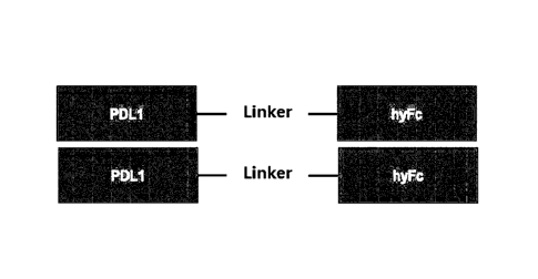
FIG. 1 shows the structure of a fusion protein (PD-L1-hyFc21 fusion protein)
including PD-L1 protein and a modified immunoglobulin Fc region.
FIG. 2a is the results of analyzing the cell concentration of cells expressing
the PD-L1-hyFc21 fusion protein over time, and FIG. 2b is the results of
analyzing the
purity of the target protein through SE-HPLC in the cell culture medium of
cells
zo expressing the PD-Li-hyFc21 fusion protein.
FIG. 3a is the results of analyzing the cell concentration of cells expressing
the PD-L1-hyFc5 fusion protein over time, and FIG. 3b is the results of
analyzing the
purity of the target protein through SE-HPLC in the cell culture medium of
cells
expressing the PD-L1-hyFc5 fusion protein.
5
CA 03164910 2022- 7- 14
FIG. 4 is an SDS-PAGE analysis result of the purified PD-L1-hyFc21 or PD-
L1-hyFc5 fusion protein.
FIG. 5a is an SE-HPLC analysis result of the purified PD-L1-hyFc21 fusion
protein, and FIG. 5b is an SE-HPLC analysis result of the purified PD-L1-hyFc5
protein.
FIG. 6 is a gel I EF analysis result of the purified PD-L1-hyFc21 or PD-L1-
hyFc5 fusion protein.
FIG. 7 is a differential scanning fluorescence (DSF) analysis result of the
purified PD-Li-hyFc21 or PD-Li-hyFc5 fusion protein.
FIG. 8 is the results of comparing the binding affinity of the PD-L1-hyFc21
or PD-L1-hyFc5 fusion protein to PD-1.
FIG. 9 is the results of comparing the inhibition capacities of the mixed
lymphocyte reaction by the PD-Li-hyFc21 or PD-L1-hyFc5 fusion protein.
FIG. 10a is the results of comparing the inhibition capacities of the
proliferation of activated human CD4 T cells by the PD-L1-hyFc21 or PD-L1-
hyFc5
fusion protein, and FIG. 10b is the results of comparing the inhibition
capacities of the
expression of cytokines of the activated human CD4 T cells.
FIG. 11 is the results of comparing the inhibition capacities of the
expression
of cytokines of activated mouse CD4 T cells by the PD-L1-hyFc21 or PD-L1-hyFc5
fusion protein.
FIG. 12a is the results of measuring the ear thicknesses of mice after
subcutaneous administration of the PD-L1-hyFc21 fusion protein to the I M Q-
induced
psoriasis mouse model, and FIG. 12b is the results of measuring the ear
thicknesses of
mice after intravenous administration of the PD-L1-hyFc21 fusion protein to
the I MQ-
induced psoriasis mouse model.
6
CA 03164910 2022- 7- 14
FIG. 13a is the results of confirming the changes in the skin epithelial
tissue
through H&E staining after subcutaneous administration of the PD-L1-hyFc21
fusion
protein to the rtTA-Peli1 psoriasis mouse model, and FIG. 13b is the results
of
measuring the thickness of the skin epithelial layer.
FIG. 14 is a result showing the degree of infiltration of T cells and
macrophages by immunofluorescence analysis in a non-psoriasis control group
(rtTA)
or psoriasis-induced rtTA-Peli1 psoriasis mouse model (left panel: rtTA; and
right
panel: rtTA-Pel i 1).
FIG. 15 is the results of measuring the numbers of T cells and macrophages
infiltrated into skin tissues after subcutaneous administration of the PD-L1-
hyFc21
fusion protein to the rtTA-Peli1 psoriasis mouse model.
FIG. 16 is the results of measuring the number of K14+ keratinocytes in the
skin tissue after subcutaneous administration of the PD-L1-hyFc21 fusion
protein to
the rtTA-Peli1 psoriasis mouse model.
FIG. 17 is the results of analyzing the changes in the skin epithelial layer
as a
score index after intravenous administration of the PD-L1-hyFc21 fusion
protein to
the rtTA-Peli1 psoriasis mouse model.
FIG. 13 is the results of measuring the skin thickness of the abdominal region
after intravenous administration of the PD-L1-hyFc21 fusion protein to the
rtTA-Pel i 1
psoriasis mouse model.
FIG. 19a is the results of confirming the changes in the skin epithelial
tissue
through H&E staining after intravenous administration of the PD-L1-hyFc21
fusion
protein to the rtTA-Peli1 psoriasis mouse model, and FIG. 19b is the results
of
measuring the thickness of the skin epithelial layer.
FIG. 20 is the results of confirming the changes in the mRNA expressions of
7
CA 03164910 2022- 7- 14
Th17 cell-associated genes (IL-17A and IL-22) and innate immune cell-
associated
genes (IL-113 and IL-24) through qRT-PCR after intravenous administration of
the PD-
L1-hyPc21 fusion protein to the rtTA-Peli1 psoriasis mouse model.
[Best Mode]
Hereinafter, the present invention will be described in detail.
The present invention provides a fusion protein including PD-L1 protein and
a modified immunoglobulin Fc region.
The PD-L1 protein may be an extracellular domain of PD-L1 protein or a
fragment thereof. The extracellular domain of the PD-L1 protein may be a
polypeptide including an immunoglobulin V like domain (Ig V like domain) of PD-
Li
and an immunoglobulin C like domain (Ig C like domain) of PD-L1.
Specifically, the extracellular domain of the PD-L1 protein is a protein
region
exposed outside the cell membrane, and may be a polypeptide consisting of the
196 to
2381h amino acids of SEQ ID NO: 1 or a polypeptide consisting of the 19th to
239th
amino acids of SEQ ID NO: 1.
In this case, the extracellular domain of the PD-L1 protein includes an Ig V
like (Ig V, Ig V like) sequence that is a conserved sequence similar to the
amino acid
sequence of an immunoglobulin (Ig, immunoglobulin), and the highly conserved
Ig V
like sequence is the amino acid sequence of the 68th to 114th amino acids of
SEQ ID
NO: 1. In addition, it includes an Ig C like (Ig C, Ig C like) sequence, and
the highly
conserved sequence region is the amino acid sequence of the 153rd to 210th
amino acids
of SEQ ID NO: 1. In addition, the fragment of the extracellular domain of the
PD-
L1 protein may include all or a part of the Ig V like domain including the Ig
V like
8
CA 03164910 2022- 7- 14
sequence of PD-L1.
In addition, the Ig V like domain in the extracellular domain of the PD-Li
protein is a site capable of interacting with PD-1, and may be a polypeptide
(SEQ ID
NO: 3) consisting of the amino acid sequences of the 19th to 239th amino acids
of SEQ
ID NO: 1 or a polypeptide consisting of the amino acid sequence of the 21st to
239th
amino acids of SEQ ID NO: 1. In addition, it may be a polypeptide (SEQ ID NO:
4)
consisting of the amino acid sequence of the 19th to 133rd amino acids of SEQ
ID NO:
1 or a polypeptide consisting of the amino acid sequence of the 215t to 133rd
amino
acids of SEQ ID NO: 1. In addition, it may be a polypeptide consisting of the
amino
acid sequence of the 215t to 114th amino acids of SEQ ID NO: 1 or a
polypeptide
consisting of the amino acid sequence of the 19th to 114th amino acids of SEQ
ID NO:
1. In addition, it may be a polypeptide consisting of the amino acid sequence
of the
21st to 120th amino acids of SEQ ID NO: 1 or a polypeptide consisting of the
amino
acid sequence of the 19th to 120th amino acids of SEQ ID NO: 1. In addition,
it may
be a polypeptide (SEQ ID NO: 5) consisting of the amino acid sequence of the
19th to
12711' amino acids of SEQ ID NO: 1 or a polypeptide (SEQ ID NO: 6) consisting
of
the amino acid sequence of the 21st to 127th amino acids of SEQ ID NO: 1. In
addition, it may be a polypeptide consisting of the amino acid sequence of the
21st to
130th amino acids of SEQ ID NO: 1 or a polypeptide consisting of the amino
acid
sequence of the 19th to 130th amino acids of SEQ ID NO: 1. In addition, it may
be a
polypeptide consisting of the amino acid sequence of the 21st to 1315t amino
acids of
SEQ ID NO: 1 or a polypeptide consisting of the amino acid sequence of the
19th to
1315t amino acids of SEQ ID NO: 1.
In addition, when the fragment of the extracellular domain of the PD-L1
protein includes an Ig V like domain or a fragment thereof, it may further
include an
9
CA 03164910 2022- 7- 14
immunoglobulin C like domain (Ig C like domain) of the extracellular domain of
the
PD-L1 protein. The Ig C like domain may be a polypeptide consisting of the
amino
acid sequence of the 133rd to 225th amino acids of SEQ ID NO: 1 or a
polypeptide
consisting of the amino acid sequence of the 134th to 225th amino acids of SEQ
ID NO:
1.
In addition, when the fragment of the extracellular domain of the PD-L1
protein includes the Ig V like domain or a fragment thereof, it may further
include a
polypeptide or a fragment thereof including the Ig C like domain of the
extracellular
domain of the PD-Li protein. The polypeptide including the Ig C like domain
refers
to the extracellular domain of the PD-L1 protein excluding the Ig V domain,
and it
may be a polypeptide having the 134th to 239th amino acids of SEQ ID NO: 1
(SEQ ID
NO: 7) or a polypeptide having the 134th to 238th amino acids of SEQ ID NO: 1
(SEQ
ID NO: 8).
In addition, the extracellular domain of the PD-L1 protein or a fragment
thereof may be derived from a human or a mouse.
The extracellular domain of the human PD-L1 protein is a polypeptide (SEQ
ID NO: 3) consisting of the amino acid sequence of the 19th to 239th amino
acids of
SEQ ID NO: 1, and the extracellular domain of the mouse PD-L1 protein is a
polypeptide consisting of the amino acid sequence of the 19th to 239th amino
acids of
SEQ ID NO: 2. In addition, the extracellular domain of the PD-L1 protein may
have
about 70%, 75%, 80%, 85%, 90%, 91%, 92%, 93%, 94%, 95%, 96%, 97%, 98%, 99%
or more homology to a polypeptide sequence consisting of the amino acid
sequence of
the 19th to 239th amino acids of SEQ ID NO: 1.
Specifically, the human PD-L1 protein has 290 amino acid residues and
includes the amino acid sequence of SEQ ID NO: 1 (Accession Number: Q9NZQ7).
CA 03164910 2022- 7- 14
In the amino acid sequence of SEQ ID NO: 1, the 1st to 18th amino acid
residues at the
N-terminus are signal sequences, and the mature human PD-L1 protein includes
the
amino acid sequence of the 19th to 290th amino acids of SEQ ID NO: 1. The
extracellular domain of the human PD-L1 protein includes the amino acid
sequence of
the 19th to 238th amino acids of SEQ ID NO: 1 or the 19th to 239th amino acids
of SEQ
ID NO: 1.
The human PD-L1 protein includes an Ig V like domain which is the 19th to
127th amino acids of SEQ ID NO: 1 and an Ig C like domain which is the 134th
to 226th
amino acids of SEQ ID NO: 1.
The mouse PD-L1 protein is reported to contain 290 amino acids, and it
includes the amino acid sequence of SEQ ID NO: 2 (Accession Number: Q9EP73).
The 1st to 18th amino acid residues of SEQ ID NO: 2 are signal sequences, and
the
mature mouse PD-L1 protein includes the amino acid sequence of the 19th to
290th
amino acids of SEQ ID NO: 2. The extracellular domain of the mouse PD-L1
protein
includes the amino acid sequence of the 19th to 239th amino acids of SEQ ID
NO:2.
The mouse PD-L1 protein includes an Ig V like protein having the 19th to 127th
amino
acids of SEQ ID NO: 2 and an Ig C like domain having the 133rd to 224th amino
acids
of SEQ ID NO: 2.
The extracellular domain of the PD-L1 protein may include the entirety of an
Ig V like domain or a fragment thereof. In addition, the fragment of the
extracellular
domain of the PD-L1 protein may further include an Ig C like domain or a
polypeptide
including an Ig C like domain (the extracellular domain of PD-L1 excluding the
Ig V
like domain).
The extracellular domain of the PD-L1 protein or a fragment thereof may
include variously modified proteins or peptides. The modification may be
performed
11
CA 03164910 2022- 7- 14
by substituting, deleting or adding one or more proteins to the wild-type PD-
L1 protein
as long as the function of PD-L1 is not altered. These various proteins or
peptides
may have 70%, 75%, 80%, 85%, 90%, 91%, 92%, 93%, 94%, 95%, 96%, 97%, 98%
or 99% homology to the wild-type protein.
As used herein, the term "extracellular domain of PD-Li protein" is also used
as a concept including "the extracellular domain of PD-L1 protein and a
fragment
thereof."
As used herein, the terms "protein", "polypeptide" and "peptide" may be used
interchangeably unless otherwise specified.
As used herein, the terms "PD-L1 fusion protein" and "PD-L1-modified
immunoglobulin Fc region fusion protein" refer to a fusion protein in which PD-
Li
protein, the extracellular domain of PD-L1 protein or a fragment thereof is
linked to a
modified immunoglobulin Fc region.
In the present invention, the PD-L1 protein may be fused to the N-terminus or
C-terminus of a modified immunoglobulin Fc region, and preferably, the PD-Li
protein may be fused to the N-terminus of a modified immunoglobulin Fc region.
The PD-L1 protein may be linked to the immunoglobulin Fc region by a linker
peptide.
The linker may include GGGSGGS (SEQ ID NO: 10),
AAGSGGGGGSGGGGSGGGGS (SEQ ID NO: 17), GGSGG (SEQ ID NO: 18),
GGSGGSGGS (SEQ ID NO: 19), GGGSGG (SEQ ID NO: 20), (G45)n (n is an integer
from 1 to 10), (GGS)n (n is an integer from 1 to 10), (GS)n (n is an integer
from 1 to
10), (GSSGGS)n (n is an integer from 1 to 10), KESGSVSSEQLAQFRSLD (SEQ ID
NO: 2i), EGKSSGSGSESKST (SEQ ID NO: 22), GSAGSAAGSGEF (SEQ ID NO:
23), (EAAAK)n (n is an integer from 1 to 10), CRRRRRREAEAC (SEQ ID NO: 24),
12
CA 03164910 2022- 7- 14
A(EAAAK)4ALEA(EAAAK)4A, GGGGGGGG (SEQ ID NO: 25), GGGGGG (SEQ
ID NO: 26), AEAAAKEAAAAKA (SEQ ID NO: 27), PAPAP (SEQ ID NO: 28), (Ala-
Pro)n (n is an integer from 1 to 10), VSQTSKLTRAETVFPDV (SEQ ID NO: 29),
PLGLWA (SEQ ID NO: 30), TRHRQPRGWE (SEQ ID NO: 31), AGNRVRRSVG
(SEQ ID NO: 32), RRRRRRRR (SEQ ID NO: 33), GFLG (SEQ ID NO: 34),
GSSGGSGSSGGSGGGDEADGSRGSQKAGVDE (SEQ ID NO: 35) and the like.
Preferably, PD-L1 and the immunoglobulin Fc region may be linked by a linker
peptide consisting of the amino acid sequence of GGGSGGS (SEQ ID NO: 10).
When the PD-Li protein and the immunoglobulin Fc region are linked using the
linker
peptide, the activity, stability and productivity of the fusion protein may be
optimized.
In addition, the fusion protein may exist in a dimer form. The bond between
the fusion proteins constituting the dimer may be formed by a disulfide bond
by a
cysteine present in a linker. The fusion proteins constituting the dimer are
identical.
That is, the dimer may be a homodimer. In this case, the fusion protein may be
soluble, and particularly, it may be dissolved in purified water or
physiological saline.
The Fc region of the modified immunoglobulin may be any one of Fc regions
of IgG1, IgG2, IgG3, IgD and IgG4, or a combination thereof. The Fc region is
modified such that binding to the Fc receptor and/or complement does not
occur. In
particular, the Fc region of the modified immunoglobulin includes a hinge
region, a
CH2 domain and a CH3 domain from an N-terminal to C-terminal direction,
wherein
the hinge region may include a human I gG1 hinge region (SEQ ID NO: 16),
wherein
the CH2 domain may include a portion of the amino acid residues of the CH2
domain
of human IgD and human IgG4, and wherein the CH3 domain may include a portion
of the amino acid residues of the CH3 domain of human IgG4.
As used herein, the terms "Fc region", "Fc fragment" or "Fc" include the heavy
13
CA 03164910 2022- 7- 14
chain constant region 2 (CH2) and heavy chain constant region 3 (CH3) of an
i mmunoglobul in, and refer to proteins that include the variable regions of
the heavy
and light chains of an immunoglobulin and do not include the light chain
constant
region 1 (CL1). It may further include a hinge region of the heavy chain
constant
region. Hybrid Fc or hybrid Fc fragments are also referred to herein as "hFc"
or
"hyFc."
In addition, the Fc fragment of the present invention may be in the form of a
native sugar chain, an increased sugar chain compared to the native form, a
reduced
sugar chain compared to the native form, or a form in which the sugar chain is
removed.
I mmunoglobul in Fc sugar chains may be modified by conventional methods such
as
chemical methods, enzymatic methods and genetic engineering methods using
microorganisms. Removal of sugar chains from the Fc fragment sharply reduces
the
binding affinity of the primary complement components Cl to C1q and results in
a
decrease or loss of ADCC or CDC, thereby not inducing an unnecessary immune
response in vivo. In this regard, the immunoglobul in Fc fragment in a
deglycosylated
or aglycosylated form may be more suitable for the purpose of the present
invention
as a drug carrier. As used herein, the term "deglycosylation" refers to the
enzymatic
removal of sugars from an Fc fragment. In addition, the term "aglycosylation"
means
that the Fc fragment is produced in an unglycosylated form by prokaryotes,
preferably
E. co/i.
In an exemplary embodiment of the present invention, the modified
i mmunoglobul in Fc region may consist of the amino acid sequence of SEQ ID
NO: 11
(hereinafter, "hyFc").
In an exemplary embodiment of the present invention, the fusion protein may
be represented by Structural Formula I below.
14
CA 03164910 2022- 7- 14
N' - X - L - Y - C' (Structural Formula I)
In the above,
N' is the N terminus of the fusion protein, and C' is the C terminus of the
fusion
protein;
the X is PD-L1 protein, an extracellular domain of PD-L1 protein or a
fragment thereof;
L is a linker; and
Y is an immunoglobulin Fc region.
Preferably, the fusion protein may consist of the amino acid sequence of SEQ
ID NO: 12 or SEQ ID NO: 13. In addition, the fusion protein of the present
invention
may have about 70%, 75%, 80%, 85%, 90%, 91%, 92%, 93%, 94%, 95%, 96%, 97%,
98%, 99% or more homology to the amino acid sequence of SEQ ID NO: 12.
In addition, the present invention provides a nucleic acid molecule encoding
the fusion protein.
Preferably, the nucleic acid molecule may be a nucleic acid molecule encoding
a polypeptide consisting of the amino acid sequence of SEQ ID NO: 12 or SEQ ID
NO: 13. In addition, the nucleic acid molecule may additionally include a
signal
sequence (or signal peptide) or a leader sequence.
As used herein, the term "signal sequence (or signal peptide)" refers to a
short
peptide present at the N-terminus of a newly synthesized protein classified as
the
secretory pathway.
Signal sequences useful in the present invention include an
antibody light chain signal sequence, such as antibody 1418 (Gil I ies et
al.,J Immunol
Meth 1989 125: 191-202), an antibody heavy chain signal sequence, such as the
CA 03164910 2022- 7- 14
MOPC141 antibody heavy chain signal sequence (Sakano et al., Nature 1980 286:
676-683), and other signal sequences known in the art (refer to, for example,
Watson
etal., Nucleic Acid Research 1984 12:5145-5164).
The signal peptide is well known in the art for its characterization, and it
is
generally known to include 16 to 30 amino acid residues and may contain more
or
fewer amino acid residues. A typical signal peptide consists of three regions
of a
basic N-terminal region, a central hydrophobic region and a more polar C-
terminal
region.
The central hydrophobic region includes 4 to 12 hydrophobic residues that
anchor the signal sequence through the membrane lipid bilayer during migration
of the
immature polypeptide. After initiation, the signal sequence is cleaved in the
lumen
of the ER by cellular enzymes commonly known as signal peptidases. In this
case,
the signal sequence may be a secretion signal sequence of tissue plasminogen
activation (tPa), HSV gDs or growth hormone. Preferably, the secretion signal
sequence used in higher eukaryotic cells, including mammals and the like, may
be used,
and more preferably, the tPa sequence or the amino acid sequence of the 1" to
181"
amino acids of SEQ ID NO: 1 may be used. In addition, the signal sequence of
the
present invention may be used by substituting with a codon having a high
expression
frequency in the host cell.
In addition, the present invention provides an expression vector including the
nucleic acid molecule.
As used herein, the term "vector" is understood as a nucleic acid means
including a nucleotide sequence that can be introduced into a host cell to be
recombined and inserted into the host cell genome, or can replicate
spontaneously as
16
CA 03164910 2022- 7- 14
an episome. The vector includes linear nucleic acids, plasmids, phagemids,
cosmids,
RNA vectors, viral vectors and analogs thereof. Examples of viral vectors
include,
but are not limited to, retroviruses, adenoviruses, and adeno-associated
viruses.
In the present invention, a useful expression vector may be RcCMV
(Invitrogen, Carlsbad) or a variant thereof. Useful expression vectors include
the
human cytomegalovirus (CMV) promoter to promote continuous transcription of
the
gene of interest in mammalian cells, and the bovine growth hormone
polyadenylation
signal sequence to increase the steady-state level of RNA after transcription.
In an
exemplary embodiment of the present invention, the expression vector is pAD15,
which is a modified vector of RcCMV.
As used herein, the term "host cell" refers to prokaryotic and eukaryotic
cells
into which recombinant expression vectors can be introduced.
In the present invention, an appropriate host cell may be transformed or
transfected with the DNA sequence of the present invention, and may be used
for the
expression and/or secretion of a target protein. Presently preferred host
cells that can
be used in the present invention include immortal hybridoma cells, NS/0
myeloma
cells, 293 cells, Chinese hamster ovary cells (CHO cells), HeLa cells, CapT
cells
(human amniotic fluid derived cells) and COS cells.
As used herein, the terms "transformation" and "transfection" refer to the
introduction of a nucleic acid (e.g., a vector) into a cell by a number of
techniques
known in the art.
In addition, the present invention provides a composition for preventing or
treating immune disease, including a fusion protein including PD-L1 protein
and a
modified immunoglobulin Fc region as an active ingredient.
The immune disease may be a disease selected from the group consisting of
17
CA 03164910 2022- 7- 14
autoimmune diseases, inflammatory diseases and transplantation rejection
diseases of
cells, tissues or organs.
The autoimmune disease may be selected from the group consisting of arthritis
[acute arthritis, chronic rheumatoid arthritis, gouty arthritis, acute gouty
arthritis,
chronic inflammatory arthritis, degenerative arthritis, infectious arthritis,
Lyme
arthritis, proliferative arthritis, psoriatic arthritis, vertebral arthritis
and rheumatoid
arthritis such as juvenile-onset rheumatoid arthritis, osteoarthritis,
arthritis chronica
progrediente, arthritis deformans, polyarthritis chronica primaria, reactive
arthritis and
ankylosing spondyl ids], psoriasis such as inflammatory hyperproliferative
skin
diseases, plaque psoriasis, gutatte psoriasis, pustular psoriasis and
psoriasis of the nails,
dermatitis including contact dermatitis, chronic contact dermatitis, allergic
dermatitis,
allergic contact dermatitis, herpetiformis dermatitis and atopic dermatitis, X-
linked
hyper-I gM syndrome, urticaria such as chronic allergic urticaria and chronic
idiopathic
urticaria, including chronic autoimmune urticaria,
polymyositis/dermatomyositis,
juvenile dermatomyositis, toxic epidermal necrolysis, scleroderma (including
systemic
scleroderma), systemic sclerosis, sclerosis including multiple sclerosis (MS)
such as
spino-optical MS, primary progressive MS (PPMS) and relapsing remitting MS
(RRMS), progressive systemic sclerosis, atherosclerosis, arteriosclerosis,
sclerosis
disseminata and ataxic sclerosis, inflammatory bowel disease (IBD) [e.g.,
Crohn's
disease, autoimmune-mediated gastrointestinal disease, colitis such as
ulcerative
colitis, colitis ulcerosa, microscopic colitis, collagenous colitis, colitis
polyposa,
necrotizing enterocolitis, transmural colitis and autoimmune inflammatory
bowel
disease], pyoderma gangrenosum, erythema nodosum, primary sclerosing
cholangitis
(episcleritis), respiratory distress syndrome including adult or acute
respiratory
distress syndrome (ARDS), meningitis, inflammation of all or part of the uvea,
iritis,
18
CA 03164910 2022- 7- 14
choroiditis, autoimmune hematological disorder, rheumatoid spondylitis, acute
hearing loss, I gE-mediated disease such as anaphylaxis and allergic and
atopic rhinitis,
encephalitis such as Rasmussen's encephalitis and limbic and/or brainstem
encephalitis,
uveitis such as anterior uveitis, acute anterior uveitis, granulomatous
uveitis,
nongranulomatous uveitis, phacoantigenic uveitis, posterior uveitis or
autoimmune
uveitis, glonnerulonephritis (GN) with or without nephrotic syndrome such as
chronic
or acute glomerulonephritis such as primary GN, immune-mediated GN, membranous
GN (membranous nephropathy), idiopathic membranous nephropathy or idiopathic
membranous GN, membrano- or membranous proliferative GN (MPGN) including
Type I and Type II and rapidly progressive GN, allergic diseases, allergic
reaction,
eczema including allergic or atopic eczema, asthma such as asthma bronchiale,
bronchial asthma and autoimmune asthma, disease related with T cell
infiltration and
chronic inflammatory response, chronic pulmonary inflammatory disease,
autoimmune myocarditis, leukocyte adhesion deficiency, systemic lupus
erythematosus (SLE) or systemic lupus erythematosus such as cutaneous SLE,
subacute cutaneous lupus erythematosus, neonatal lupus syndrome (NLE), lupus
erythematosus disseminatus, lupus [including nephritis, cerebritis, pediatric,
non-renal,
extra-renal, discoid and alopecia], juvenile-onset (Type I) diabetes mellitus
including
pediatric insulin-dependent diabetes mellitus (IDOM), adult-onset (Type II)
diabetes
mellitus, autoimmune diabetes mellitus, idiopathic diabetes insipidus, immune
responses associated with acute and delayed hypersensitivity mediated by T-
lymphocytes and cytokines, granulomatosis including tuberculosis, sarcoidosis,
lymphomatoid granulomatosis, Wegener's granulomatosis, agranulocytosis,
vasculitis
[including giant vessel vasculitis (polymyalgia rheumatic and Takayasu's
arteritis],
Kawasaki disease, medium vessel vasculitis including polyarteritis nodosa,
19
CA 03164910 2022- 7- 14
microscopic polyarteritis, CNS arthritis, necrotizing, cutaneous or
hypersensitivity
vasculitis, systemic necrotizing vasculitis, vasculitides including ANCA-
related
vasculitis such as Churg-Strauss vasculitis or syndrome (CSS), temporal
arteritis,
aplastic anemia, autoimmune aplastic anemia, coombs benign anemia, Diamond
Blackfan anemia, immune-hemolytic anemia including hemolytic anemia or
autoimmune hemolytic anemia (Al HA), pernicious anemia (anemia perniciosa),
Addison's disease, pure red cell anemia aplasia (PRCA), factor VHI deficiency,
hemophilia A, autoimmune neutropenia, pancytopenia, leukopenia, leukocyte
diapedesis-related disease, CNS inflammatory disorder, multiple organ injury
syndrome such as those secondary to septicemia, trauma or hemorrhage, antigen-
antibody complex-mediated diseases, anti-glomerular basement membrane disease,
anti-phospholipid antibody syndrome, allergic neuritis, Bechet's or Behcet's
disease,
Castleman's syndrome, Goodpasture's syndrom, Raynaud's syndrome, Sjogren's
syndrome, Stevens-Johnson syndrome, pemphigoid such as bullous pemphigoid and
skin pemphigoid, pemphigus (including pemphigus vulgaris, pemphigus foliaceus,
pemphigus mucous-membrane pemphigoid and pemphigus erythematosus),
autoimmune polyendocrinopathies, Reiter's disease or syndrome, immune complex
nephritis, antibody-mediated nephritis, neuromyelitis optica,
polyneuropathies,
chronic neuropathy such as IgM polyneuropathy or IgM-mediated neuropathy,
thrombocytopenia (e.g., one which develops in myocardial infarction patient),
including thrombotic thrombocytopenic purpura (TTP) and autoimmune or immune-
mediated thrombocytopenia such as idiopathic thrombocytopenic purpura (ITP)
including chronic or acute ITP, autoimmune disorder of the testis and ovary
including
autoimmune orchitis and oophoritis, primary hypothyroidism, hypothyroidism
including thyroiditis such as autoimmune thyroiditis, autoimmune endocrine
disease,
CA 03164910 2022- 7- 14
Hashimoto's disease, chronic thyroiditis (Hashimoto's thyroiditis) or subacute
thyroiditis, autoimmune thyroid disease, idiopathic hypothyroidism, Grave's
disease,
polyglandular syndrome such as autoimmune polyglandular syndrome (or
polyglandular endocrinopathy syndrome), paraneoplastic syndromes including
paraneoplastic neurological syndrome such as Lambert-Eaton myasthenic syndrome
or Eaton-Lambert syndrome, stiff-man or stiff-person syndrome,
encephalomyelitis
such as allergic encephalomyelitis or encephalomyelitis allergica and
experimental
allergic encephalomyelitis (EAE), myasthenia gravis such as thymoma-associated
myasthenia gravis, cerebellar degeneration, neuromyotonia, opsoclonus or
opsoclonus
myoclonus syndrome (OMS) and sensory neuropathy, multifocal motor neuropathy,
Sheehan's syndrome, autoimmune hepatitis, chronic hepatitis, lupoid hepatitis,
giant
cell hepatitis, chronic active hepatitis or autoimmune chronic active
hepatitis,
lymphoid interstitial pneumonitis, bronchiolitis obliterans (non-transplant)
vs. NSIP,
Guillain-Barre syndrome, Berger's disease (IgA nephropathy), idiopathic IgA
nephropathy, linear IgA dermatosis, primary biliary cirrhosis,
pneumonocirrhosis,
autoimmune enteropathy syndrome, Celiac disease, Coeliac disease, celiac sprue
(gluten enteropathy), refractory sprue, idiopathic sprue, cryoglobulinemia,
amyotrophic lateral sclerosis (ALS, Lou Gehrig's disease), coronary artery
disease,
autoimmune ear disease such as autoimmune inner ear disease (AGED), autoimmune
hearing loss, opsoclonus myoclonus syndrome (OMS), polychondritis such as
refractory or relapsing polychondritis, pulmonary alveolar proteinosis,
amyloidosis,
scleritis, non-cancerous lymphocytosis, primary lymphocytosis including
monoclonal
B cell lymphocytosis (e.g., benign monoclonal gammopathy and monoclonal
gammopathy of undetermined significance; MGUS), peripheral neuropathy,
paraneoplastic syndrome, epilepsy, migraine, arrhythmia, muscular disorder,
deafness,
21
CA 03164910 2022- 7- 14
blindness, periodic paralysis, channelopathies such as CNS channelopathies,
autism,
inflammatory myopathy, focal segmental glomerulosclerosis (FSGS), endocrine
ophthalmopathy, uveoretinitis, chorioretinitis, autoimmune hepatological
disorder,
fibromyalgia, multiple endocrine failure, Schmidt's syndrome, adrenalitis,
gastric
atrophy, presenile dementia, demyelinating diseases such as autoimmune
denvelinating diseases, diabetic nephropathy, Dressler's syndrome, alopecia
areata,
CREST syndrome (calcinosis), Raynaud's phenomenon, esophageal dysmotility,
sclerodactyly and telangiectasia, male and female infertility, mixed
connective tissue
disease, Chagas' disease, rheumatic fever, recurrent abortion, farmer's lung,
erythema
multiforme, post-cardiotomy syndrome, Cushing's syndrome, bird-fancier's lung,
allergic granulomatous angiitis, benign lymphocytic angiitis, Alport's
syndrome,
alveolitis such as allergic alveolitis and fibrous periostitis, interstitial
lung disease,
transfusion diseases, leprosy, malaria, leishmaniasis, trypanosomiasis,
schistosomiasis,
ascariasis, aspergillosis, Samter's syndrome, Caplan's syndrome, dengue,
endocarditis,
endomyocardial fibrosis, diffuse interstitial pulmonary fibrosis, interstitial
lung
fibrosis, idiopathic pulmonary fibrosis, cystic fibrosis, endophthalmitis,
erythema
elevatum et diutinum, erythroblastosis fetalis, eosinophilic fasciitis,
Shulman's
syndrome, Felty's syndrome, filariasis, cyclitis such as chronic cyclitis,
heterochromia
chronic cyclitis, iridocyclitis or Fuch's cyclitis, Henoch-Schonlein purpura,
human
immunodeficiency virus (HIV) infection, ECHO virus infection, cardiomyopathy,
Alzheimer's disease, parvovirus infection, rubella virus infection, post-
vaccination
syndromes, congenital rubella infection, Epstein-Barr virus infection, mumps,
Evan's
syndrome, autoimmune gonadal failure, Sydenham's chorea, poststreptococcal
nephritis, thromboangiitis obliterans, thyrotoxicosis, tabes dorsal is,
chorioiditis, giant
cell polymyalgia, endocrine ophthamopathy, chronic hypersensitivity
pneumonitis,
22
CA 03164910 2022- 7- 14
keratoconjunctivitis sicca, epidemic keratoconjunctivitis, idiopathic
nephritic
syndrome, minimal change nephropathy, benign familial and ischemia-reperfusion
injury, retinal autoimmunity, joint inflammation, bronchitis, chronic
obstructive
airway disease, silicosis, aphthae, aphthous stomatitis, arteriosclerotic
disorders,
spermatogenesis, autoimmune hemolysis, Boeck's disease, cryoglobulinemia,
Dupuytren's contracture, endophthalmia phacoanaphylactica, enteritis
allergica,
erythema nodosum leprosum, idiopathic facial paralysis, chronic fatigue
syndrome,
febris rheumatica, Hamman-Rich's disease, sensorineural hearing loss,
haemoglobinuria paroxysmatica, hypogonadism, ileitis regionalis, leucopenia,
mononucleosis infectiosa, transverse myelitis, primary idiopathic myxedema,
nephrosis, ophthalmia symphatica orchitis granulomatosa, pancreatitis,
polyradiculitis
acuta, pyoderma gangrenosum, Quervain's thyroiditis, acquired spenic atrophy,
infertility due to antispermatozoan antobodies, non-malignant thymoma,
vitiligo,
SCID and Epstein-Barr virus-associated diseases, acquired immune deficiency
syndrome (AIDS), parasitic disease such as Leishmania, toxic-shock syndrome,
food
poisoning, disease associated with T cell infiltration, leukocyte adhesion
deficiency,
immune responses associated with acute and delayed hypersensitivity mediated
by
cytokines and T cells, leukocyte diapedesis-related disease, multiple organ
injury
syndrome, antigen-antibody complex mediated diseases, antiglonnerular basement
membrane disease, allergic neuritis, autoimmune polyendocrinopathies,
oophoritis,
primary myxedema, autoimmune atrophic gastritis, sympathetic ophthalmia,
rheumatic diseases, mixed connective tissue disease, nephrotic syndrome,
insulitis,
polyendocrine failure, peripheral neuropathy, autoimmune polyglandular
syndrome
type I, adult-onset idiopathic hypoparathyroidism (A01H), alopecia totalis,
dilated
cardiomyopathy, epidermolysis bullosa acquisita (EBA), hemochromatosis,
23
CA 03164910 2022- 7- 14
myocarditis, nephrotic syndrome, primary sclerosing cholangitis, purulent or
non-
purulent sinusitis, acute or chronic sinusitis, ethmoid, frontal, maxillary or
sphenoid
sinusitis, eosinophilic disorder such as eosinophilia, pulmonary infiltration
eosinophilia, eosinophilia-myalgia syndrome, Lofflers syndrome, chronic
eosinophilic pneumonia, topical pulmonary eosinophilia, bronchopneumonic
aspergillosis, aspergilloma or granulomas including eosinophils, anaphylaxis,
seronegative spondyloarthritides, polyendocrine autoimmune disease, sclerosing
cholangitis, sclera, episclera, chronic mucocutaneous candidiasis, Bruton's
syndrome,
transient hypogammaglobulinemia of infancy, Wiskott-Aldrich syndrome, ataxia
telangiectasia, autoimmune disorders associated with collagen diseases,
rheumatism,
neurological diseases, ischemic reperfusion disorder, reduction in blood
pressure
response, blood vessel malfunction, angiectasis, tissue injury, cardiovascular
ischemia,
hyperalgesia, cerebral ischemia and disease accompanying vascularization,
allergic
hypersensitivity disorder, glomerulonephritides, reperfusion injury,
reperfusion injury
of myocardium or other tissues, dermatoses having acute inflammatory
component,
acute purulent meningitis or other central nervous system inflammatory
disorder,
ocular and orbital inflammatory disorder, granulocyte transfusion-associated
syndromes, cytokine-induced toxicity, acute serious inflammation, chronic
intractable
inflammation, pyelitis, pneunnonocirrhosis, diabetic retinopathy, diabetic
large-artery
disorder, endarterial hyperplasia, peptic ulcer, valvulitis and endometriosis.
Preferably, the autoimmune disease may be selected from the group consisting
of type 1 diabetes, alopecia areata, anti-phospholipid antibody syndrome,
rheumatoid
arthritis, psoriasis or psoriatic arthritis, multiple sclerosis, systemic
lupus
erythematosus, inflammatory bowel disease, Addison's disease, Graves' disease,
Sjogren's syndrome, Guillain-Barre syndrome, Hashimoto's thyroiditis,
myasthenia
24
CA 03164910 2022- 7- 14
gravis, inflammatory myopathy, autoimmune vasculitis, autoimmune hepatitis,
hemolytic anemia, idiopathic thrombocytopenic purpura, primary biliary
cirrhosis,
scleroderma, vitiligo, pernicious anemia and celiac disease.
The inflammatory disease may be selected from the group consisting of
arthritis, ankylosing spondylitis, reactive arthritis, Reiter's syndrome,
crystal
arthropathies, Lyme disease, polymyalgia rheumatica, systemic sclerosis,
polymyositis,
dermatomyositis, polyarteritis nodosa, Wegener's granulomatosis, Churg-Strauss
syndrome, sarcoidosis, atherosclerotic vascular disease, atherosclerosis,
ischemic
heart disease, myocardial infarction, stroke, peripheral vascular disease,
uveitis,
corneal disease, iritis, iridocyclitis and cataracts.
The transplant rejection disease may be a tissue or organ transplant rejection
reaction, and the tissue or organ transplant rejection reaction may be
selected from
rejection reactions of bone marrow transplantation, heart transplantation,
corneal
transplantation, intestinal transplantation, liver transplantation, lung
transplantation,
pancreatic transplantation, kidney transplantation and skin transplantation.
As used herein, the term "inflammatory skin disease" refers to a disease
occurring in the skin by an inflammatory reaction.
Skin keratinocytes are
components that constitute most of the epidermal cells, and they form keratin
and are
involved in various inflammatory and immune responses by producing various
cytokines. When skin keratinocytes are exposed to environmental and
physiological
stress, an inflammatory response occurs, and by this, various types of
inflammatory
cytokines such as tumor necrosis factor-a (TNF-a), interleukin-113 (IL-113),
IL-6 and
chemokine (C-C motif) I igand (CCL) are secreted. These cytokines reduce the
rate
of the proliferation of keratinocytes in the skin and interfere with the
formation of
matrix in the dermal layer, thereby slowing the healing rate of damaged skin.
CA 03164910 2022- 7- 14
Therefore, the proliferation of keratinocytes in inflammatory skin diseases
plays an
important role and is closely related to inflammatory skin diseases such as
skin aging,
atopic dermatitis, psoriasis and the like. Accordingly, the inflammatory skin
disease
may be psoriasis or atopic dermatitis, and preferably, psoriasis.
As used herein, the term "psoriasis" refers to a disease that forms various
sizes
of erythematous papules and plaques with clear boundaries that are covered
with
silvery-white scales on the skin. Histologically, it is one of the chronic
inflammatory
skin diseases characterized by epithelial hyperplasia, showing various
clinical features
and repeating exacerbation and improvement.
The preferred dosage of the pharmaceutical composition varies depending on
the condition and weight of the patient, the degree of disease, the drug form,
the route
and duration of administration, but may be appropriately selected by those
skilled in
the art.
In the pharmaceutical composition for treating or preventing psoriasis
according to the present invention, the active ingredient may be included in
any
amount (effective amount) depending on the use, formulation, purpose of
formulation
and the like, as long as it can exhibit a therapeutic activity for psoriasis,
and it will be
determined within the range of 0.001 wt.% to 20.0 wt.% on the basis of the
total weight
of the composition. Herein, the term "effective amount" refers to the amount
of an
active ingredient capable of inducing a psoriasis treatment effect. Such
effective
amounts may be determined empirically within the ordinary ability of those
skilled in
the art.
As used herein, the term "treatment" may be used to include both therapeutic
treatment and prophylactic treatment. In this case, prevention may be used in
the
sense of alleviating or reducing a pathological condition or disease of a
subject. In
an embodiment, the term "treatment" includes any form of application or
26
CA 03164910 2022- 7- 14
administration for treating a disease in mammals, including humans. In
addition, the
term includes the meanings of inhibiting or slowing a disease or the progress
of a
disease; restoring or repairing damaged or missing function to partially or
completely
relieve the disease; stimulating inefficient processes; or alleviating serious
diseases.
Herein, the term "therapeutically effective amount" or "pharmaceutically
effective amount" refers to the amount of a compound or composition effective
to
prevent or treat a target disease, which is sufficient to treat the disease at
a reasonable
benefit/risk ratio applicable to medical treatment, and it means an amount
that does
not cause side effects. The level of the effective amount may be determined by
the
patient's health status, disease type, severity, drug activity, drug
sensitivity,
administration method, administration time, administration route and excretion
rate,
treatment period, factors including drugs used in combination or concurrently
and
factors well known in the medical field.
In an embodiment, a therapeutically
effective amount refers to the amount of a drug effective to treat psoriasis.
The composition of the present invention may include a pharmaceutically
acceptable carrier, and may additionally include a pharmaceutically acceptable
adjuvant, excipient or diluent in addition to the carrier.
As used herein, the term "pharmaceutically acceptable" refers to a
composition that is physiologically acceptable and does not normally cause
gastrointestinal disorders, allergic reactions such as dizziness or similar
reactions when
administered to humans.
Examples of the carriers, excipients and diluents may
include lactose, dextrose, sucrose, sorbitol, mannitol, xylitol, erythritol,
maltitol, starch,
gum acacia, alginate, gelatin, calcium phosphate, calcium silicate, cellulose,
methyl
cellulose, polyvi nylpyrrol i done,
water, methyl hyd roxybenzoate,
propylhydroxybenzoate, talc, magnesium stearate and mineral oil. In addition,
fillers,
27
CA 03164910 2022- 7- 14
anti-agglomeration agents, lubricants, wetting agents, fragrances, emulsifiers
and
preservatives may be further included.
The pharmaceutical composition of the present invention may be formulated
by using methods known in the art to enable rapid, sustained or delayed
release of the
active ingredient upon administration to mammals. Formulations include
powders,
granules, tablets, emulsions, syrups, aerosols, soft or hard gelatin capsules,
sterile
injectable solutions and sterile powder forms.
The composition of the present invention may be formulated in a suitable form
together with a pharmaceutically acceptable carrier. For example,
pharmaceutically
acceptable carriers include carriers for parenteral administration such as
water, suitable
oils, saline, aqueous glucose, glycol and the like, and may further include
stabilizers
and preservatives. Suitable stabilizers include antioxidants such as sodium
bisulfite,
sodium sulfite or ascorbic acid.
Suitable preservatives include benzalkonium
chloride, methyl- or propyl-paraben and chlorobutanol. In addition, the
composition
according to the present invention may appropriately include a suspending
agent,
solubilizer, stabilizer, isotonic agent, preservative, adsorption inhibitor,
surfactant,
diluent, excipient, pH adjuster, analgesic agent, buffer, antioxidant and the
like if
necessary depending on the administration method or formulation.
Pharmaceutically
acceptable carriers and agents suitable for the present invention, including
those
exemplified above, are described in detail in the literature [Remington's
Pharmaceutical Sciences, latest edition].
The composition of the present invention may be sterilized according to
commonly known sterilization techniques.
The composition may include
pharmaceutically acceptable auxiliary substances and adjuvants, toxicity
control
agents and analogs thereof, which are required to control physiological
conditions such
28
CA 03164910 2022- 7- 14
as pH control, and for example, there are sodium acetate, sodium chloride,
potassium
chloride, calcium chloride, sodium lactate and the like. The concentration of
the
fusion protein in such formulations may vary widely, and for example, it may
be about
0.5% or less depending on the weight or generally at least about 1% to 15% or
up to
20%, and depending on the particular method of administration selected, it may
be
selected preferentially based on body fluid volume, viscosities and the like.
A preferred dosage for the composition of the present invention may be in the
range of 0.01 pg/kg to 10 g/kg per day or in the range of 0.01 mg,/kg to 1
g/kg,
depending on the patient's condition, body weight, gender, age, patient's
severity and
administration route. Administration may be performed once or divided into
several
times a day. Such dosages should not be construed as limiting the scope of the
invention in any respect.
Subjects to which the composition of the present invention can be applied
(prescribed) are mammals and humans, and in particular, preferably, humans.
The
administration route, administration dosage and administration frequency of
the fusion
protein or fusion protein dimer may be administered to a subject by various
methods
and amounts depending on the patient's condition and presence or absence of
side
effects, and the optimal administration method, administration dosage and
administration frequency may be selected by a person skilled in the art within
an
appropriate range.
The composition of the present invention may be administered by any route.
The composition of the present invention may be provided to an animal either
directly
(e.g., topically by injection, implantation or local administration at a
tissue site) or
systemically (e.g., parenterally or orally) by any suitable means.
When the
composition of the present invention is provided parenterally such as
intravenous,
29
CA 03164910 2022- 7- 14
subcutaneous, ophthalmic, intraperitoneal, intramuscular, oral, rectal,
intraorbital,
intracerebral, intracranial, intraspinal, intraventricular, intrathecal,
intracisternal,
intracapsular, intranasal or aerosol administration, the composition is
preferably
aqueous or it is preferable to include portions of physiologically applicable
bodily fluid
suspensions or solutions. Accordingly, since the carrier or vehicle is
physiologically
acceptable, it may be added to the composition and delivered to the patient,
without
adversely affecting the patient's electrolyte and/or volumetric balance.
Therefore, as
a body fluid medium for the preparation, it may generally include a
physiological
saline.
A DNA construct (or gene construct) including a nucleic acid encoding the
fusion protein of the present invention may be used as part of a gene therapy
protocol
carrying a nucleic acid encoding the fusion protein construct.
In the present invention, an expression vector for infecting and expressing
the
fusion protein in vivo in a specific cell type in order to reconstitute or
supplement the
desired function of the fusion protein may be administered together with any
biologically effective carrier, and examples thereof include any formulations
or
compositions capable of efficiently delivering a gene encoding a desired
fusion protein
or a fusion protein thereof to cells in vivo.
For gene therapy using a nucleic acid encoding the fusion protein, the gene of
interest may be inserted into a viral vector including a recombinant
retrovirus, an
adenovirus, an adeno-associated virus and herpes simplex virus-1, or a
recombinant
bacterial plasmid or a recombinant eukaryotic plasmid.
The dosage of the nucleic acid encoding the fusion protein of the present
invention is in the range of 0.1 mg to 100 mg in humans, preferably, 1 mg to
10 mg,
and more preferably, 2 mg to 10 mg. The optimal amount and dosage form may be
CA 03164910 2022- 7- 14
determined by routine experimentation within the level of ordinary skill in
the art.
The unit dose of the fusion protein of the present invention is 0.1 mg/kg to
1,500 mg/kg in humans, preferably, 1 mg/kg to 100 mg/kg, and more preferably,
5
mg/kg to 20 mg/kg. The unit dose may vary depending on the disease to be
treated
and the presence or absence of side effects. However, the optimal dosage may
be
determined using routine experimentation. Administration of the fusion protein
may
be by periodic bolus injections or by continuous intravenous, subcutaneous or
intraperitoneal administration from an external reservoir (e.g., intravenous
bag) or the
inside (e.g., bioerodable implant).
The composition of the present invention may be administered in combination
with other drugs or physiologically active substances having a prophylactic or
therapeutic effect on the disease to be prevented or treated, or may be
formulated in
the form of a combination formulation with such other drugs.
The method for preventing or treating a disease using the fusion protein or
composition of the present invention may include administering another drug or
physiologically active substance having a prophylactic or therapeutic effect
in
combination with the fusion protein or composition of the present invention,
and the
route of concurrent administration, administration timing and administration
dosage
may be determined according to the type of disease, the disease state of the
patient, the
purpose of treatment or prevention and other drugs or physiologically active
substances used in combination.
In addition, the present invention provides the use of a fusion protein
including
PD-L1 protein and a modified immunoglobulin Fc region for producing a
pharmaceutical preparation having an effect of preventing or treating immune
disease.
The present invention also provides a method for preventing or treating
31
CA 03164910 2022- 7- 14
immune disease, including administering a fusion protein including PD-L1
protein and
a modified immunoglobul in Fc region, and a pharmaceutically acceptable
carrier to a
subject.
The therapeutically effective amount is preferably applied differently
depending on the specific composition including the type and extent of the
response
to be achieved and whether other agents are used as necessary, the subject's
age, weight,
general health status, gender and diet, the administration time,
administration route,
and secretion rate of the composition, the treatment period and drugs used
together or
concurrently with a specific composition and similar factors well known in the
pharmaceutical field. Therefore, it is preferable to determine the effective
amount of
the composition suitable for the purpose of the present invention in
consideration of
the aforementioned factors.
The subject is applicable to any mammals, and the mammals include not only
humans and primates, but also domestic animals such as cattle, pigs, sheep,
horses,
dogs and cats.
[Modes of the Invention]
Hereinafter, the present invention will be described in more detail through
examples. These examples are for describing the present invention in more
detail,
and the scope of the present invention is not limited to these examples.
Example 1. Preparation of gene construct for production of fusion protein
including PD-Li protein and modified immunoglobulin Fc region
A gene construct was prepared for producing a fusion protein in which human
32
CA 03164910 2022- 7- 14
programmed cell death-ligand 1 (PD-L1) protein and a modified immunoglobulin
Fc
domain were fused. Specifically, for the human PD-L1 gene, a known amino acid
sequence (Accession Number: Q9NZQ7) was used, and the gene construct including
the extracellular domain (19 to 239 aa) of the PD-L1 protein was prepared by
TOP
Gene Technologies. (Canada, Quebec). Human PD-L1 protein was prepared to be
fused to the N-terminus of the modified immunoglobulin Fc region. The modified
immunoglobulin Fc domain (SEQ ID NO: 11) is a hybrid type of human I gD Fc and
human I gG4 Fc, and is characterized by including an I gG1 hinge region (SEQ
ID NO:
16) composed of 8 amino acids.
The fusion protein of the present invention was prepared such that the PD-Li
protein was linked to the modified immunoglobulin Fc region by a peptide
linker. In
the present invention, in order to maintain flexibility while inducing
dimerization, a
fusion protein (hereinafter, "PD-L1-hyFc21" or "PD-L1-hyFc21 fusion protein",
SEQ
ID NO: 12 or 13) was prepared, which included human PD-L1 protein and a
modified
immunoglobulin Fc region linked by a GS linker (SEQ ID NO: 10) consisting of a
7
amino acid sequence. As a control, a fusion protein including an
immunoglobulin Fc
region including the I gD hinge region of SEQ ID NO: 14 (hereinafter, "PD-L1-
hyFc5"
or "PD-L1-hyFc5 fusion protein", SEQ ID NO: 15) was used.
A recombinant expression vector was prepared using the gene construct
including the nucleotide encoding the fusion protein. The prepared recombinant
expression vector was transformed using a gene transfer method (NeonTM kit, 10
pL,
I nvitrogen Cat. MPK1096) in which DNA was introduced into the cell by
suspending
a DNA solution in the CHO DG44 cell line and passing a pulse of high direct
current.
Afterwards, the amplification step of HT selection (HT supplement, I
nvitrogen, 11067-
030) and MTX (Methotrexate, Sigma, M8407) was performed, and cell passage was
33
CA 03164910 2022- 7- 14
performed such that only cells with a high expression rate were selected.
Cells were
passaged in an amount of 0.4 x 10⁶ cells/mL every 3 days, and the number and
viability
of cells were measured using a cell counting device (Vi-cell, Beckman
coulter). HT
selection is a selection method in which only transformed cells survive by
removing
HT from the media, and MTX amplification is a method of amplifying genes by
placing MTX at a selected concentration in the passage medium. The selected
cell
pool was subjected to single cell cloning using the limiting dilution cloning
method.
Briefly, cells were al iquoted to 1 cell/well in a 96-well plate, and cell
images were
stored on days 0, 7 and 14 using a CSI device (clone selection imager,
Molecular
Devices) to trace back clones derived from one cell. The productivity of the
selected
single cell-derived cell line was confirmed by using Fc ELISA (Human IgG ELI
SA
Quantitation Set, Bethyl, E80-104). The finally selected 5 to 6 clones were
subjected
to batch culture and long-term stability evaluation, and clones whose
stability was
confirmed were prepared as a research cell bank (RCB).
Example 2. Securing of PD-L1-hyFc21 or PD-L1-hyFc5 fusion protein
In order to mass produce the PD-L1-hyFc21 or PD-L1-hyFc5 fusion protein,
the target protein was isolated from the cell culture medium produced from the
PD-
L1-hyFc5 or PD-L1-hyFc21 suspension cell line obtained in Example 1 and
purified.
In order to secure a large amount of the PD-L1-hyFc21 or PD-L1-hyFc5
fusion protein, a cell line expressing the PD-L1-hyFc21 or PD-L1-hyFc5 fusion
protein was cultured for 20 days in Glass bioreactor 15L by the same fed-batch
culture
method to produce the PD-L1-hyFc21 or PD-L1-hyFc5 fusion protein. During fed-
batch culture, the cell viability of the cell line expressing the fusion
protein was
measured, and size exclusion chromatography (SE-HPLC) was performed to confirm
34
CA 03164910 2022- 7- 14
the final expression level and purity of the target protein.
As a result of confirming the maximum cell concentration and the final
expression level of the fusion protein of the expression cell line over time,
the
expression cell line of the PD-L1-hyFc21 fusion protein was confirmed to have
a
maximum cell concentration of 13.9 x 10⁶ cells/mL over time (FIG. 2a), and the
expression level of the finally recovered fusion protein was confirmed to be
5.6 g/L
(FIG. 2b). In comparison, the expression cell line of the PD-L1-hyFc5 fusion
protein
was confirmed to have a maximum cell concentration over time of 16.7 x 10⁶
cells/mL
(FIG. 3a), and the expression level of the finally recovered fusion protein
was
confirmed to be 2.2 g/L (FIG. 3b).
In addition, as a result of confirming the purity of the fusion protein, it
was
confirmed that the purity of the PD-L1-hyFc21 fusion protein was 79.5%, and it
was
confirmed to include 7.7% of high molecular weight impurities (HMW) and 9.8%
of
low molecular weight impurities (LMW) (Table 1). In comparison, the purity of
the
PD-L1-hyFc5 fusion protein was 47.8%, and it was confirmed to include 31.7% of
high molecular weight impurities (HMW) and 20.5% of low molecular weight
impurities (LMW) (Table 1).
[Table 1]
Area (%) PD-L1-hyFc21 PD-L1-hyFc5
HMW (%) 7.7 31.7
Main (%) 79.5 47.8
LMW (%) 9.8 20.5
*HMW: high molecular weight impurity; Main: target protein; and LMW: low
molecular
weight impurity.
Afterwards, in order to obtain high-purity PD-L1-hyFc21 or PD-L1-hyFc5
CA 03164910 2022- 7- 14
fusion protein, the following three-step purification process was performed:
Culture
medium => Protein A affinity chromatography => Anion exchange chromatography 1
=> Anion exchange chromatography 2 => Ultrafiltration/diafiltration.
The obtained target protein was confirmed for purity and yield through size
exclusion chromatography (SE-H PLC) analysis. As a result, it was confirmed
that
the purity of the PD-L1-hyFc21 fusion protein was 97.3% and the yield was
30.2%.
In comparison, it was confirmed that the purity of the PD-L1-hyFc5 fusion
protein was
93.2% and the yield was 5.1% (Table 2).
[Table 2]
Process PD-L1-hyFc21 PD-L1-hyFc5
Name of test Purity Purity
Y ield (%) Y ield
(%)
solution (SE-HPLC) (SE-HPLC)
Culture medium 79.5 100.0 47.8 100.0
Final purification 97.3 30.2 93.2 5.1
From the above results, it was confirmed that the PD-L1-hyFc21 fusion
protein showed a production amount which was about 2.5 times higher than that
of the
PD-L1-hyFc5 fusion protein, and the purity of the protein was also high.
Example 3. Characterization analysis of PD-L1-hyFc21 or PD-L1-hyFc5
fusion protein
3.1. Polyacrylamide gel electrophoresis (sodium dodecyl sulfate-
polyacrylamide gel electrophoresis, SDS-PAGE)
In order to confirm the molecular weight of the purified PD-L1-hyFc21 or PD-
L1-hyFc5 fusion protein, sodium dodecyl sulfate polyacrylamide gel
electrophoresis
(SOS-PAGE) was performed. Briefly, the fusion protein was diluted with
deionized
36
CA 03164910 2022- 7- 14
water, mixed with NuPAGE'LDS Sample Buffer (Thermo Fisher Scientific) and
loaded on 4-12% Bis-Tris gel (Invitrogen) to 3 ig/well to perform
electrophoresis.
After electrophoresis, the gel was stained by the Coomassie staining method.
As a result, both of the purified PD-L1-hyFc21 and PD-L1-hyFc5 fusion
proteins were confirmed at a size marker of 98 kDa under non-reducing
conditions
(FIG. 4). However, the purified PD-L1-hyFc5 fusion protein included a cleaved
form
of low-molecular impurities.
3.2. Size-exclusion chromatography (SE-HPLC)
In order to analyze the main peak and impurity peaks such as dimer, multimer
or truncated abnormal peptides in the purified PD-L1-hyFc21 or PD-L1-hyFc5
fusion
protein, the size-exclusion chromatography (SE-HPLC) was performed. Briefly,
after the fusion protein was diluted and prepared with formulation buffer to
1.0 mg/mL,
the area ratio (% area) of the main peak of the fusion protein among the
separated
peaks was analyzed by using a gel filtration chromatography column (TOSOH TSK-
GEL G3000SWxL column, 7.8 mm x 300 mm).
As a result, it was confirmed that the purity of the PD-L1-hyFc21 fusion
protein was 97.3% (FIG. 5a), whereas the purity of the PD-L1-hyFc5 fusion
protein
was confirmed to be 93.2% (FIG. 5b).
3.3. lsoelectric focusing (IEF)
In order to confirm the charge variants and distribution of the purified PD-L1-
hyFc5 and PD-L1-hyFc21 proteins, isoelectric focusing (I EF) was performed.
I soelectric point electrophoresis is an electrophoresis method that analyzes
separated
proteins using the pl value of the protein, and the pl value refers to the pH
value at
37
CA 03164910 2022- 7- 14
which a charged protein becomes electrically neutral, which is called the
isoelectric
point. In isoelectric point electrophoresis, the protein that has reached the
isoelectric
point does not move anymore and stays on the gel and is separated. Briefly, in
the
present invention, the fusion protein was separated according to the pl value
using a
pH 3 to 10 isoelectric point gel (I nvitrogen), and the gel after
electrophoresis was fixed
with a 12% trichloroacetic acid (TCA) solution and then stained by the
Coomassie
staining method.
As a result, both of the PD-L1-hyFc21 and PD-L1-hyFc5 fusion proteins were
confirmed to have pl values in the range of 5.2 to 6.0 (FIG. 6).
3.4. Differential scanning fluorimetry (DSF)
In order to analyze the heat stability of the purified PD-L1-hyFc21 or PD-L1-
hyFc5 fusion protein, the experiment was performed using the PROTEOSTAT
Thermal Shift Stability Assay Kit. Since the assay kit contains a fluorescent
dye that
detects protein aggregation, it is possible to confirm the temperature at
which a large
amount of protein aggregates under heat stress conditions.
The aggregation
temperature (tagg) is an indicator of protein stability, and it can confirm
the stability
of the protein structure.
As a result, the aggregation temperature of the PD-L1-hyFc21 fusion protein
was confirmed to be 55.8 C, whereas the aggregation temperature of the PD-L1-
hyFc5 fusion protein was confirmed to be 50.7 C (FIG. 7). As a result, it was
confirmed that the heat stability of the PD-L1-hyFc21 fusion protein was
structurally
more excellent compared to PD-L1-hyFc5.
Example 4. In vitro activity analysis of PD-L1-hyFc21 or PD-L1-hyFc5
38
CA 03164910 2022- 7- 14
fusion protein
4.1. Binding affinity analysis for PD-1 using J urkat (SHP-1) cell line
expressing PD-1
The binding affinity of the PD-L1-hyFc21 or PD-L1-hyFc5 fusion protein to
PD-1 was analyzed using a J urkat (SHP-1) cell line expressing PD-1. Briefly,
J urkat
cells (PathHuntere J urkat PD-1 (SHP-1), DiscoverX) were al iquoted in a 96-
well plate
and stabilized in a 37 C 5% CO2 incubator for 2 hours. Afterwards, the PD-L1-
hyFc21 or PD-L1-hyFc5 fusion protein was added so as to become 75 and 600 nM,
respectively, and then reacted in a 37 C 5% CO2 incubator for 1 hour. After
completion of the reaction, a reagent (PathHunter Bioassay Detection Kit,
DiscoverX) for detecting SHP-1 expressed by the PD-1:PD-L1 signaling response
was
added to measure the luminescence of SHP-1.
As a result, the group treated with the PD-L1-hyFc21 fusion protein showed
a higher degree of luminescence of SHP-1 at both concentrations of 75 and 600
nM,
compared to the group treated with the PD-L1-hyFc5 fusion protein (FIG. 8).
Accordingly, it was confirmed that the PD-L1-hyFc21 fusion protein had a
higher
binding affinity to PD-1 than the PD-L1-hyFc5 fusion protein.
4.2. Analysis of inhibitory capacity of mixed lymphocyte reaction
The inhibitory capacity of the mixed lymphocyte response by the PD-L1-
hyFc21 or PD-L1-hyFc5 fusion protein was analyzed. Briefly, donor and
recipient
peripheral blood mononuclear cells (PBMCs) were prepared and subjected to CTV
(CellTracem Violet) and CTR (CellTracr Far Red) staining for intracellular
proteins.
Each of the donor and recipient PBM Cs was placed as response cells and
stimulator
cells, mixed at a ratio of 1:1 and placed in a 96-well U-bottom plate, and
after 0.5 [tM
39
CA 03164910 2022- 7- 14
of the PD-L1-hyFc21 or PD-L1-hyFc5 fusion protein was added, it was reacted in
a
37 C 5% CO2 incubator for 5 days. By analyzing the degree of decrease in the
fluorescence of CTV (CellTraceni Violet) stained on the response cells in the
cells in
which the reaction had been completed, the degrees of inhibition of the mixed
lymphocyte reaction by the PD-L1-hyFc21 or PD-L1-hyFc5 fusion protein were
compared.
As a result, the proliferation of CD4 T cells and CD8 T cells of the activated
response cells by the mixed lymphocyte reaction was reduced in the group
treated with
the PD-L1-hyFc21 or PD-L1-hyFc5 fusion protein, compared to the control group
(Allo, hyFc treated group) (FIG. 9). In particular, it
was confirmed that the
proliferations of CD4 T cells and CD8 T cells of the activated response cells
were
significantly reduced in the group treated with the PD-L1-hyFc21 fusion
protein at the
same concentration condition, compared to the group treated with the PD-L1-
hyFc5
fusion protein (FIG. 9).
4.3. Analysis of proliferation inhibition and cytokine production
inhibition of activated human T cells
The inhibition capacities of the proliferation of human T cells by the PD-L1-
hyFc21 or PD-L1-hyFc5 fusion protein were compared under T cell activation
conditions using human PBM Cs. Briefly, the PD-L1-hyFc21 or PD-L1-hyFc5 fusion
protein together with anti-CD3 antibody was treated in a 96-well plate and
coated at
4 C for one day. After CD4 T cells were isolated from PBM Cs of normal people
using microbeads (MACS), CTV (CellTracem Violet, 2.5 M) staining for
intracellular
proteins was performed on the isolated CD4 T cells. After the coated plate was
washed with PBS, CD4 T cells stained with CTV were placed in a test plate and
CA 03164910 2022- 7- 14
cultured in a 37 C 5% CO2 incubator for 3 days. After 4 days, the cells from
the test
plate were harvested, and the degree of decrease in CTV fluorescence was
analyzed
using flow-cytometry.
As a result, it was confirmed that the proliferation of activated human T
cells
was reduced in the group treated with the PD-L1-hyFc21 or PD-L1-hyFc5 fusion
protein, compared to the control group (no treatment or hyFc treatment group)
(FIG.
10a). In particular, it was confirmed that the proliferation of activated
human T cells
was significantly inhibited by about 2 times in the group treated with the PD-
L1-
hyFc21 fusion protein, compared to the group treated with the PD-Li-hyFc5
fusion
protein (FIG. 10a).
In addition, the inhibition capacities of the cytokine expression of human T
cells by the PD-L1-hyFc21 or PD-L1-hyFc5 fusion protein were compared under T
cell activation conditions using human PBMCs. Briefly, the PD-L1-hyFc21 or PD-
L1-hyFc5 fusion protein together with anti-CD3 antibody was diluted to be
treated at
0.5 [tM and coated in a 48-well plate at 4 C for one day. CD4 T cells were
isolated
from normal human PBM Cs using microbeads (M iltenyi Biotech). CD4T cells were
isolated from normal human PBM Cs using microbeads (MACS). After the coated
plate was washed with PBS, the isolated CD4 T cells were placed in a test
plate and
cultured in a 37 C 5% CO2 incubator for 3 days. Anti-IFN-7 antibody (DuoSet
Human I FN-gamma ELISA set, R&D system) was placed in a test plate and coated
at
room temperature. After washing the coated test plate with PBS (PBST), in
which
0.05% Tween-20 was added, 3 times, PBS in which 1% BSA was added was blocked
in the test plate at room temperature for 2 hours. The culture medium was
transferred
to the CD4 cells on the test plate and reacted at room temperature for 2
hours. After
washing 5 times with PBST, the detection antibody was added and reacted for 2
hours
41
CA 03164910 2022- 7- 14
at room temperature, followed by washing 5 times with PBST. After adding
streptavidin-H RP to the washed test plate, it was reacted at room temperature
for 20
minutes. Afterwards, it was washed 5 times with PBST, and after the TM B
substrate
was added, sulfuric acid (H2SO4) at a concentration of 2N was added to measure
the
absorbance at 450 nm. The ability of the PD-L1-hyFc21 or PD-L1-hyFc5 fusion
protein to inhibit IFNI production by activated CD4 T cells was analyzed using
the
measured absorbance values.
As a result, it was confirmed that the amount of IFN-y produced in activated
human CD4 T cells was significantly reduced in the group treated with the PD-
L1-
hyFc21 or PD-L1-hyFc5 fusion protein, compared to the control group (no
treatment
or hyFc treatment group) (FIG. 10b). In particular, the amount of IFN-y
generated
from activated human CD4 T cells was significantly inhibited in the group
treated with
the PD-L1-hyFc21 fusion protein by about 3 times, compared to the group
treated with
the PD-L1-hyFc5 fusion protein (FIG. 10b).
4.4. Analysis of cytokine production inhibition of activated mouse T cells
The inhibition capacities of the cytokine expression of mouse T cells by the
PD-L1-hyFc21 or PD-L1-hyFc5 fusion protein were compared under the mouse T
cell
activation conditions. Briefly, the PD-L1-hyFc21 or PD-L1-hyFc5 fusion protein
(0.5 M) together with 5 pg/mL of anti-CD3 antibody was placed in a test plate
and
coated at 4 C for one day. CD4 T cells were isolated from lymphocytes
isolated
from the lymph nodes of mice using microbeads (Miltenyi Biotech). After
washing
the coated plate with PBS, the isolated CD4 T cells were added and cultured in
a 37
C 5% CO2 incubator at for 3 days. Anti-IL-2 antibody (Mouse IL-2 ELI SA MAXTM
Deluxe, Biolegend) and anti-IFN-y antibody (Mouse IFNgamma ELISA MAX'
42
CA 03164910 2022- 7- 14
Deluxe, Biolegend) were placed in a new test plate and coated at room
temperature.
The test plate coated the previous day was washed 4 times with washing buffer,
and
then, assay diluent A was added and blocked at room temperature for 1 hour.
After
washing the test plate 4 times with washing buffer again, the CD4 T cell
culture
medium was transferred and reacted at room temperature for 2 hours. After
washing
4 times with washing buffer, the detection antibody was added and reacted with
shaking at room temperature for 1 hour, and washed 4 times with washing buffer
again.
After adding streptavidin-HRP to the washed test plate, it was reacted at room
temperature for 30 minutes. After washing with washing buffer 5 times, the TM
B
substrate was added, and it was reacted for 30 minutes at room temperature to
block
light. Absorbance was measured at 450 nm within 15 minutes by adding a stop
solution. The measured absorbance values were used to calculate the
concentrations
of IL-2 and IFN-7, and the ability of the PD-L 1 -hyFc2 1 or PD-L 1 -hyFc5
fusion protein
to inhibit the production of IL-2 and IFN-y produced by activated CD4 T cells
was
analyzed.
As a result, it was confirmed that the amounts of IL-2 and IFN-y produced in
the activated mouse CD4 T cells were significantly reduced in the group
treated with
the PD-L1-hyFc21 or PD-L1-hyFc5 fusion protein, compared to the control group
(no
treatment or hyFc treatment group) (FIG. 11). In particular, it was confirmed
that the
amount of IL-2 generated from activated human CD4 T cells was significantly
inhibited in the group treated with the PD-L1-hyFc21 fusion protein by about
1.8 times,
compared to the group treated with the PD-L1-hyFc5 fusion protein, and the
amount
of IFN-y was significantly inhibited by about 4 times (FIG. 11).
Example 5. In vivo activity analysis of PD-L1-hyFc21 fusion protein
43
CA 03164910 2022- 7- 14
5.1. Efficacy evaluation of PD-L1-hyFc21 fusion protein in imiquimod
(IMQ)-induced psoriasis mouse model
When psoriasis develops, dendritic cells are activated by toll like receptor
7/8
(TLR7/8), and as a I igand of TLR7 and TLR8, imiquimod (I MQ) is known to
cause an
inflammatory response like psoriasis, when it is repeatedly applied to mouse
skin.
I miquimod (IMQ) is involved not only in dendritic cells but also in T cells,
which are
adaptive immune cells, and among them, gamma delta T cells (y6T17) that
secrete IL-
17 (interleukin-17) are preferentially involved in psoriatic inflammation.
Therefore,
the I M Q-induced psoriasis mouse model has a phenotype similar to that of
psoriasis
patients, and it can be used as a suitable model for the development of
psoriasis
therapeutics.
Briefly, in order to induce psoriasis in a mouse animal model, I M Q cream was
applied to both ears of mice daily at 20 mg/mouse/day for 6 days. The ear
thickness
of the mice in each group was measured using a caliper every day for 7 days
from day
0, when the I MQ cream was applied, to day 6, the end date of the test, and
the changes
in the ear thickness of the mice of each group were compared based on the ear
thickness of mice not applied with I M Q cream (no treatment). The PD-L1-
hyFc21
fusion protein was administered subcutaneously or intravenously on days 0, 1,
3 and
5. Alternatively, the anti-I L12p40 antibody was administered
intravenously (iv) once
on day 2 of the test. The control groups and the experimental groups are shown
in
Table 3 below.
[Table 3]
Group Inducer Dose N Route
Volume
Control 1 Vehicle - 5 Subcutaneous
3 mL/kg
44
CA 03164910 2022- 7- 14
2 I MQ - 5 3
mL/kg
PD-L1-hyFc21 3 I MQ 3 mg/kg 5 Subcutaneous 3
mL/kg
4 I MQ 10 mg/kg 5 3
mL/kg
I MQ 30 mg/kg 5 3 mL/kg
6 I MQ 3 mg/kg 5 Intravenous 5
mL/kg
7 I MQ 10 mg/kg 5 5
mL/kg
8 I MQ 30 mg/kg 5 5
mL/kg
As a result, it was confirmed that the ear thicknesses increased by about 68%
on day 6, the end date of the test, in the I M Q-treated group (I M G-vehicle)
compared
to the control group not treated with I M Q (no treatment-vehicle) (FIG. 12a).
On the
5 other hand, differences were observed in the ear thicknesses of mice from
day 4 in the
groups subcutaneously administered with 3, 10 and 30 mg/kg of the PD-L1-hyFc21
fusion protein (FIG. 12a). In particular, the ear thicknesses were suppressed
by 51%
on day 6, the end date of the test, in the group subcutaneously administered
with the
lowest dose of 3 mg/kg of the PD-L1-hyFc21 fusion protein, compared to the
group
treated with I M Q (I M G-vehicle), and the groups subcutaneously administered
with the
medium and high doses of 10 mg/kg and 30 mg/kg of the PD-L1-hyFc21 fusion
protein
showed 67% and 66% inhibition, respectively, compared to the I M Q-treated
group
(I MG-vehicle) (FIG. 12a). In addition, there was a significant difference
according
to the dose between the groups administered with 10 mg/kg or 30 mg/kg and the
group
administered with a low dose of 3 mg/kg, but there was no significant
difference
between the group administered with 10 mg/kg and the group administered with
30
mg/kg.
In addition, it was confirmed that the ear thickness of the mice was
significantly inhibited by about 87 to 92% until day 6, the end date of the
test, in all of
the groups administered intravenously with 3, 10 and 30 mg/kg of the PD-L1-
hyFc21
CA 03164910 2022- 7- 14
fusion protein, compared to the I M Q-treated group (I M G-vehicle) (FIG.
12b).
5.2. Efficacy evaluation according to subcutaneous administration of PD-
L1-hyFc21 fusion protein in psoriasis mouse model transformed with
doxycycline-induced Pe111 gene
In order to evaluate the efficacy of the PD-L1-hyFc21 fusion protein in a
psoriasis mouse model of chronic inflammation, the inventors of the present
invention
used a psoriasis mouse model (hereinafter, "rtTA-Peli1 psoriasis mouse model")
in
which long-term chronic psoriasis symptoms were induced by transforming the
Pelil
(Pellino homolog 1) gene to be overexpressed according to the administration
of
doxycycline in addition to the imiquimod (I M Q)-i nduced psoriasis mouse
model. As
psoriasis is induced, the rtTA-Peli1 psoriasis mouse model causes the
development of
psoriatic lesions in the epidermis, an increase in inflammatory responses such
as an
increase in inflammatory cytokines, an increase in the epithelial cell layer,
and an
increase in the infiltration of phagocytes, dendritic cells and Th17 cells
into the dermis.
Briefly, in order to induce psoriasis in mice, 4-week-old mice were allowed to
drink drinking water containing 5% sucrose and 2 mg/mL of doxycycline for 6
months.
Saline containing doxycycline was continuously supplied during the test
period.
Mice that were not transformed with the Peli gene (hereinafter, "rtTA") were
used as a
control group. The PD-L1-hyFc21 fusion protein was administered subcutaneously
once a week for 8 weeks.
First, after subcutaneous administration of the PD-L1-hyFc21 fusion protein
to the rtTA-Peli1 psoriasis mouse model, an experiment was performed to
measure
changes in the skin epithelial tissue and the thickness of the skin epithelial
layer
through H&E staining. The results of H&E staining were observed at 200X
46
CA 03164910 2022- 7- 14
magnification, and 6 parts of each picture were arbitrarily designated and
measured.
As a result, the average thickness of the epithelial layer was 8.74 pm in the
non-
psoriasis control group (rtTA), and the average thickness of the epithelial
layer was
48.56 pm in the psoriasis-induced rtTA-Pelil group (rtTA-Peli 1 Vehicle), and
it was
confirmed that the thickness of the epithelial layer in the rtTA-Peli1 group
(rtTA-Pelil
Vehicle) was increased by about 5.6 times compared to the control group (FIGS.
13a
and 13b). On the other hand, the average epithelial layer thicknesses were
38.29 pm,
40.18 pm and 31.74 p,m in the groups subcutaneously administered with 3, 10
and 30
mg/kg of the PD-L1-hyFc21 fusion protein, respectively, and it was confirmed
that
there was an effect of reducing the thickness of the epithelial layer compared
to the
psoriasis-induced rtTA-Peli1 group (rtTA-Peli1 Vehicle) (FIGS. 13a and 13b).
However, no dose-dependent effect was observed.
In addition, after subcutaneous administration of the PD-L1-hyFc21 fusion
protein to the rtTA-Peli1 psoriasis mouse model, an experiment was performed
to
compare the degrees of infiltration of T cells and macrophages into the dermal
layer
through immunofluorescence staining. An anti-CD3 antibody was used for the
detection of T cells, and an anti-F4/80 antibody was used for the detection of
macrophages.
The results were observed at 400X magnification of an
immunofluorescence microscope, and the number of cells in the same area was
calculated.
As a result, it was confirmed that T cells and macrophages hardly infiltrated
into the skin tissue in the control group (rtTA) not induced with psoriasis,
and it was
confirmed that the numbers of T cells and macrophages infiltrated into the
skin tissue
were increased in the psoriasis-induced rtTA-Peli1 group (rtTA-Peli1 Vehicle)
(FIG.
14). In particular, the
number of T cells infiltrated into the skin tissue in the psoriasis-
47
CA 03164910 2022- 7- 14
induced rtTA-Peli1 group (rtTA-Peli1 Vehicle) was about 47 (FIG. 15). On the
other
hand, the numbers of T cells infiltrated into the skin tissue were about 32,
14 and 7 in
the groups subcutaneously administered with 3, 10 and 30 mg/kg of the PD-L1-
hyFc21
fusion protein, respectively, and it was confirmed that the number of T cells
was
significantly reduced in a dose-dependent manner (FIG. 15).
In addition, after subcutaneous administration of the PD-L1-hyFc21 fusion
protein to the rtTA-Peli1 psoriasis mouse model, changes in keratinocytes in
the skin
tissues were analyzed by immunofluorescence staining using an anti-K14
antibody.
The expression of keratin 14 (K14) appears in the basal layer responsible for
proliferation, and an increase in their expression can be interpreted as an
increase in
the number of cells responsible for proliferation among keratinocytes. The
results
were compared by measuring the number of cells in the same area. As a result,
the
number of K14+ keratinocytes was 59 in the non-psoriasis-induced control group
(rtTA), and the number of keratinocytes was 197 in the psoriasis-induced rtTA-
Pelil
group (rtTA-Peli1 Vehicle), and thus, the number of keratinocytes in the rtTA-
Peli1
group increased by about 3.3 times, compared to the control group (FIG. 16).
On the
other hand, the numbers of keratinocytes were about 179, 150 and 98 in the
groups
subcutaneously administered with 3, 10 and 30 mg/kg of the PD-L1-hyFc21 fusion
protein, respectively, and it was confirmed that the number of keratinocytes
decreased
in a dose-dependent manner, and particularly, the number of keratinocytes was
significantly reduced at a dose of 30 mg/kg (FIG. 16).
5.3. Efficacy evaluation according to intravenous administration of PD-
L1-hyFc21 fusion protein in psoriasis mouse model transformed with
48
CA 03164910 2022- 7- 14
doxycycline-induced Pe111 gene
After intravenous administration of the PD-L1-hyFc21 fusion protein to the
rtTA-Peli1 psoriasis mouse model once a week for 5 weeks, changes in the
epithelial
layer of the skin were analyzed as a score index, and an experiment was
performed to
measure the skin thickness of the abdominal region using a caliper. The score
index
was evaluated as follows: 0 = normal skin; 1 = keratoplasia appear; 2 =
keratoplasia
appear on half of back skin, or skin lesions slightly overtop the normal skin;
3 =
thickness appear on whole back skin, or skin lesions significantly overtop the
normal
skin; 4 = skin lesions sclerosis (Biomedicine & Pharmacotherapy, Volume 110,
February 2019, Pages 265-274).
As a result, the score index increased by nearly 3 in the psoriasis-induced
rtTA-Peli1 group (rtTA-Peli1 Vehicle), compared to the control group (rtTA),
whereas
it was confirmed that the score index was reduced in the group intravenously
administered with the PD-L1-hyFc21 fusion protein, compared to the rtTA-Peli1
group
(FIG. 17). In addition, similar to the score index, the skin thickness of the
abdominal
region increased by about 100 [im in the psoriasis-induced rtTA-Pelil group
(rtTA-
Pel i 1 Vehicle), compared to the control group (rtTA), whereas it was
confirmed that
the skin thickness was reduced to a level similar to that of the control group
(rtTA) in
the group intravenously administered with the PD-L1-hyFc21 fusion protein
(FIG. 18).
However, no dose-dependent effect was confirmed in the above results.
In addition, after intravenous administration of the PD-L1-hyFc21 fusion
protein to the rtTA-Peli1 psoriasis mouse model, an experiment was performed
to
measure changes in the skin epithelial tissue and the thickness of the skin
epithelial
layer through H&E staining. The results of H&E staining were observed at 400X
magnification of a microscope, 6 parts of the epithelial layer were
arbitrarily
49
CA 03164910 2022- 7- 14
designated, and the skin epithelial layer thickness was measured using the
Image J
program. As a result, the average thickness of the epithelial layer was 23.8
gm in the
non-psoriasis control group (rtTA), and the average thickness of the
epithelial layer
was 104.5 gm in the psoriasis-induced rtTA-Pelil group (rtTA-Pelil Vehicle),
and thus,
it was confirmed that the thickness of the epithelial layer in the psoriasis-
induced rtTA-
Peli1 group (rtTA-Peli1 Vehicle) was increased by about 4.4 times compared to
the
control group (FIGS. 19a and 19b). On the other hand, the average thicknesses
of
the epithelial layers were about 97.9 gm, 86.3 gm and 73.3 gm in the groups
administered intravenously with 1, 3 and 10 mg/kg of the PD-L1-hyFc21 fusion
protein, respectively, and thus, it was confirmed that there was an effect of
reducing
the thickness of the epithelial layer in a dose-dependent manner, compared to
the
psoriasis-induced rtTA-Peli1 group (rtTA-Peli1 Vehicle) (FIGS. 19a and 19b).
In addition, after intravenous administration of the PD-L1-hyFc21 fusion
protein to the rtTA-Peli1 psoriasis mouse model, qRT-PCR was performed to
confirm
changes in the mRNA expressions of Th17 cell-associated genes such as IL-17A
and
IL-22 and innate immune cell-associated genes such as IL-113 and IL-24. As a
result,
the psoriasis-induced rtTA-Peli1 group (rtTA-Peli1 Vehicle) showed the
increased
mRNA expressions of Th17 cell-associated genes such as IL-17A and I L-22
compared
to the control group (rtTA), whereas it was confirmed that the mRNA
expressions of
the Th17 cell-associated genes were reduced in the group intravenously
administered
with the PD-L1-hyFc21 fusion protein (FIG. 20). In addition, similarly, the
mRNA
expressions of the innate immune cell-associated genes such as IL-1 0 and IL-
24
increased in the psoriasis-induced rtTA-Peli1 group (rtTA-Peli1 Vehicle)
compared to
the control group (rtTA), whereas the mRNA expressions of the Th17 cell-
associated
genes showed a tendency to decrease in the group intravenously administered
with the
CA 03164910 2022- 7- 14
PD-L1-hyFc21 fusion protein (FIG. 20).
In particular, the expression of IL-24
mRNA was significantly reduced in the group intravenously administered with 10
mg/kg of the PD-L1-hyFc21 fusion protein (FIG. 20).
From the above results, it was confirmed that since the PD-L1-hyFc21 fusion
protein inhibits the proliferation of CD4 T cells, which is an important
factor in the
pathogenesis of autoinrimune diseases such as psoriasis, through subcutaneous
administration or intravenous administration, and has an effect of suppressing
the
expression of inflammatory cytokines accompanying the lesion, it can be used
as a
therapeutic agent for immune diseases such as psoriasis.
51
CA 03164910 2022- 7- 14
[ABSTRACT]
The present invention relates to a fusion protein including PD-L1 protein and
a modified immunoglobul in Fc region and use thereof, and since the fusion
protein has
significantly higher purity and production yield compared to the existing
fusion protein,
has a high binding affinity to PD-1, reduces the proliferation of activated T
cells,
inhibits the generation of cytokines generated by activated T cells, and has
an effect of
inhibiting the infiltration of T cells or macrophages into tissues, it can be
effectively
used in the treatment of immune diseases.
CA 03164910 2022- 7- 14