Note: Descriptions are shown in the official language in which they were submitted.
CA 03164919 2022-06-15
WO 2021/126970
PCT/US2020/065299
SEQUENTIAL TARGETING IN CROSSLINKING NANO-THERANOSTICS FOR
TREATING BRAIN TUMORS
CROSS-REFERENCES TO RELATED APPLICATIONS
[0001] This application claims priority to U.S. Provisional Application No.
62/949,284
filed December 17, 2019, which is incorporated herein in its entirety for all
purposes.
STATEMENT AS TO RIGHTS TO INVENTIONS MADE UNDER
FEDERALLY SPONSORED RESEARCH AND DEVELOPMENT
[0002] The present invention was made with Government support under Grant No.
R01CA199668 awarded by the National Institutes of Health/National Cancer
Institute, and
Grant No. R01HD086195 awarded by the National Institutes of Health/ National
Institute of
Child Health and Human Development. The Government has certain rights in the
invention.
BACKGROUND
[0003] The efficacy of therapeutics for brain tumors is seriously hampered by
multiple drug
delivery barriers, including severe destabilizing effects in blood
circulation, the blood-brain
barrier/blood-brain tumor barrier (BBB/BBTB) and limited tumor uptake. Herein
is a
Sequential Targeting In CrosslinKing (STICK) nano-delivery strategy to
circumvent these
important physiological barriers to improve drug delivery to brain tumors.
STICK
nanoparticles (STICK-NPs) could sequentially target BBB/BBTB and brain tumor
cells with
surface maltobionic acid (MA) and 4-carboxyphenylboronic acid (CBA),
respectively, and
simultaneously enhance nanoparticle stability with pH-responsive crosslinkages
formed by
MA and CBA in situ. STICK-NPs exhibited prolonged circulation time (17-fold
higher area-
under-curve) than free agent, allowing increased opportunities to transpass
BBB/BBTB via
glucose transporter-mediated transcytosis by MA. Tumor acidic environment then
triggered
the transformation of STICK-NPs into smaller nanoparticles and revealed
secondary CBA
targeting moiety for deep tumor penetration and enhanced uptake in tumor
cells. STICK-NPs
significantly inhibited tumor growth and prolonged the survival time with
limited toxicity in
mice with aggressive and chemo-resistant diffuse intrinsic pontine glioma.
This formulation
tackles multiple physiological barriers on-demand with a simple and smart
STICK design.
Therefore, these features allow STICK-NPs to unleash the potential of brain
tumor
therapeutics to improve their treatment efficacy.
[0004] Patients with aggressive brain tumors, such as glioblastoma (GBM) or
pediatric
diffuse intrinsic pontine glioma (DIPG), have a dismal prognosis.
Particularly, for DIPG, a
1
CA 03164919 2022-06-15
WO 2021/126970
PCT/US2020/065299
devastating and aggressive pediatric brain tumor arising in the ventral pons,
radiotherapy is
currently the only treatment modality. Children with DIPG have only around 2%
five-year
survival rate. Many chemotherapeutic drugs such as vincristine (VCR) and novel
epigenetic
modulating agents, such as inhibitors for Histone deacetylase (HDAC),
bromodomains of
Bromodomain and Extra-terminal motif (BET), and enhancer of zeste homolog 2
(EZH2)
showed promising results in the pre-clinical models. Unfortunately, all the
clinical trials on
the chemotherapy and epigenetic modulating agents failed to improve the
treatment outcome
compared to radiation alone. The clinical therapeutic effect of these agents
is markedly
hampered by the poor drug delivery to brain tumors due to several
physiological barriers,
including strong destabilizing conditions during the circulation in blood
(Barrier 1), the
blood-brain barrier (BBB)/blood-brain tumor barrier (BBTB) (Barrier 2), poor
specificity for
targeting tumor cells (Barrier 3) and the relatively weak enhanced
permeability and retention
effect displayed by brain tumors (FIG. 1A). There is a clear and urgent need
to develop new
therapeutic strategies against brain tumors.
[0005] A variety of nanocarriers have been reported attempting to circumvent
these
biological barriers by actively targeting the receptors or transporters on the
BBB/BBTB (e.g.
glucose transporter 1 (GLUT1), transferrin receptors, low-density lipoprotein
receptor,
choline transporter, and amino acids transporters)) and tumor cell/tissue
(e.g. sialic acid,
integrin family, tropomyosin receptor kinase (TRK) family proteins, epidermal
growth factor
receptor (EGFR), and folate receptor), respectively. The BBB/BBTB is a highly
regulated
barrier that controls the traversal of blood-borne substances into the
parenchyma of the
central nervous system (CNS) and prevents toxic agents, including
chemotherapeutic drugs
from entering. Several nutrients including glucose are essential for the
brain. The transport of
glucose into the CNS is facilitated by GLUT1, which is specifically localized
on the
BBB/BBTB. Several studies have established that GLUT1 as a validated target
for
transporter-mediated transcytosis of nanoparticles. It is also known that many
types of tumor
cells (including those of brain tumors) show an increased sialic acid
expression on membrane
glycoproteins. The hypersialation of a cell membrane during malignant
transformation not
only contributes to tumor growth and metastasis but also strongly associates
with poor
prognosis in cancer patients. Thus, targeting tumor cells by their aberrant
sialylation has been
an attractive strategy for cancer treatment. GLUT1 and sialic acid, had been
separately
targeted with different nano-carriers, but had never been dually/sequentially
targeted with one
particle design.
2
CA 03164919 2022-06-15
WO 2021/126970
PCT/US2020/065299
[0006] To tackle the challenge in brain tumor delivery, multifunctional
nanoparticles must
be designed with consideration of the whole-process in drug delivery to brain
tumors as well
as the dynamic requirements for each delivery stage. Several dual targeting
strategies were
developed attempting to address the multiple barriers in brain tumor delivery.
For example, a
dual-targeting peptide angiopep-2 was decorated on the nanoparticles to target
both BBB and
GBM cells, and this dual-targeting nanocarrier was demonstrated to exhibit
superior anti-
intracranial GBM effects. Polysorbate 80 (PS 80) was introduced to polymer-
bound
tratuzumab (anti-Her2 Antibody) to target both BBB and Her2+ breast cancer
brain
metastasis. In this system, the first step involved in the PS 80-mediated
recruitment of
circulating apolipoprotein resulting in transcytosis, and the second step was
to target Her2 on
breast cancer cells with tratuzumab after nanoparticle dissociation. While
conceptually
attractive, these conventional dual targeting design is usually achieved by
simply decorating
one or two different targeting moieties on the nanoparticle surface. These
moieties ONLY
serve for targeting purpose without adding various favorable physical features
to the
nanoparticle platform to sophisticatedly address the complicated problems in
brain tumor
delivery.
[0007] Herein, is developed a simple-yet-effective Sequential Targeting In
CrosslinKing
(STICK) nano-delivery approach to improve drug delivery to brain tumors.
Strategically, one
unique pair of targeting molecules was selected, maltobionic acid (MA, a
glucose derivative)
and 4-carboxyphenylboronic acid (CBA) , as dual targeting moieties for BBB and
brain
tumor via GLUT1 and sialic acid, respectively, to build interlocking STICK
nanoparticles
(STICK NPs). Beyond targeting functions, this pair of targeting moieties could
form pH-
sensitive boronate ester bonds to stabilize the nanocarriers with
intermicellar crosslinks,
thereby benefiting NP stability in blood circulation (FIG. 1A, Barrier 1).
Excess MA (a
glucose derivative) on the nanoparticle surface can be recognized by GLUT1 and
then trigger
the GLUT1-mediated BBB/BBTB transcytosis (FIG. 1A, Barrier 2). Upon exposure
to the
acidic extracellular pH in solid tumors, the intrinsic MA-CBA boronate ester
crosslinkages
are cleaved, resulting in the transformation of STICK NPs into small secondary
nanoparticles
with newly unshielded surface CBA (a synthetic mimic of lectin) which allows
deeper tumor
penetration and recognition of tumor surface sialic acid, respectively (FIG.
1A, Barrier 3). In
this study, is provided a step-by-step proof for the dynamic properties
specifically designed to
overcome each barrier with STICK approach, including their sequential
targeting abilities,
pharmacokinetics, and pH-dependent drug release/transformation features.
Lastly, it was
3
CA 03164919 2022-06-15
WO 2021/126970
PCT/US2020/065299
demonstrated their superior anti-cancer targeting abilities using the dual-
modality imaging
and anti-cancer efficacies in two different aggressive orthotopic brain tumor
models.
BRIEF SUMMARY OF THE INVENTION
[0008] In one embodiment, the present invention provides a compound of Formula
I:
(RI)m-DI-LI-PEG-L2-D2-(R2), (I), wherein: each RI is independently a peptide,
1,2-
dihydroxy compound, or boronic acid derivative; each R2 is independently
cholic acid or a
cholic acid derivative; DI and D2 are each independently a dendritic polymer
having a single
focal point group, and a plurality of branched monomer units X; ach branched
monomer unit
X is a diamino carboxylic acid, a dihydroxy carboxylic acid or a hydroxyl
amino carboxylic
acid; LI and L2 are each independently a bond or a linker linked to the focal
point group of
the dendritic polymer; PEG is a polyethylene glycol (PEG) polymer having a
molecular
weight of 1-100 kDa; subscript m is an integer from 2 to 8; and subscript n is
an integer from
2 to 16.
[0009] In another embodiment, the present invention provides a nanoparticle
comprising a
plurality of first and second conjugates, wherein: each first conjugate is a
compound of
Formula I wherein each RI is independently a peptide, 1,2-dihydroxy compound,
sugar
compound glucose, or glucose derivative; each second conjugate is a compound
of Formula I
wherein each RI is independently a boronic acid derivative; and the plurality
of conjugates
self-assemble by forming crosslinking bonds to form a nanoparticle such that
the interior of
the nanoparticle comprises a hydrophilic interior comprising a plurality of
micelles with a
hydrophobic core.
[0010] In another embodiment, the present invention provides a nanoparticle
comprising a
hydrophilic exterior and interior, wherein the nanoparticle interior comprises
a hydrophilic
interior comprising a plurality of micelles having a hydrophobic core and
hydrophilic micelle
exterior, wherein each micelle comprises a plurality of first and second
conjugates, wherein:
each first conjugate is a compound of Formula I wherein each RI is
independently a peptide,
1,2-dihydroxy compound, sugar compound glucose, or glucose derivative; each
second
conjugate is a compound of Formula I wherein each RI is independently a
boronic acid
derivative; and the plurality of first and second conjugates self-assemble by
forming
crosslinking bonds to form the micelle with the hydrophobic core, with the
crosslinking
bonds on the hydrophilic micelle exterior.
4
CA 03164919 2022-06-15
WO 2021/126970
PCT/US2020/065299
[0011] In another embodiment, the present invention provides a method of
delivering a
drug, the method comprising: administering a nanoparticle of the present
invention, wherein
the nanoparticle further comprises a hydrophilic and/or hydrophobic drug and a
plurality of
cross-linked bonds; and cleaving the cross-linked bonds in situ, such that the
drug is released
from the nanoparticle, thereby delivering the drug to a subject in need
thereof.
[0012] In another embodiment, the present invention provides a method of
treating a
disease, the method comprising administering a therapeutically effective
amount of a
nanoparticle of the present invention, wherein the nanoparticle further
comprises a
hydrophilic and/or hydrophobic drug, to a subject in need thereof.
[0013] In another embodiment, the present invention provides a method of
imaging,
comprising: administering an effective amount of a nanoparticle of the present
invention,
wherein the nanoparticle further comprises a hydrophilic and/or hydrophobic
imaging agent
to a subject in need thereof; and imaging the subject.
BRIEF DESCRIPTION OF THE DRAWINGS
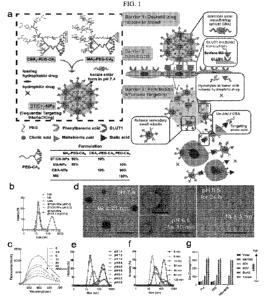
[0014] FIG. lA shows the design of transformable STICK-NPs and detailed multi-
barrier
tackling mechanisms to brain tumors. The pair of targeting moieties selected
to form
Sequential Targeting In CrosslinKing (STICK) were maltobionic acid (MA), a
glucose
derivative, and carboxyphenylboronic acid (CBA), one type of boronic acid, and
were built
into well-characterized self-assembled micelle formulations (PEG-CA8). STICK-
NPs were
assembled by a pair of MA4-PEG-CA8 and CBA4-PEG-CA8 with the molar ratio of
9:1
while inter-micelle boronate crosslinkages, STICK, formed between MA and CBA
resulting
in larger nanoparticle size. Excess MA moieties were on the surface of the
nanoparticles,
while CBA moieties were firstly shielded inside the STICK to avoid non-
specific bindings.
Hydrophobic drugs were loaded in the hydrophobic cores of secondary small
micelles, while
hydrophilic agents were trapped in the hydrophilic space between small
micelles. In the
following studies several control micelle formulations were used including NM
(no
targeting), MA-NPs (single BBB targeting), and CBA-NPs (single sialic acid
tumor
targeting) nanoparticles (inserted table). In detail, STICK-NPs could overcome
Barrier 1
(destabilizing condition in the blood) by intermicellar crosslinking strategy,
Barrier 2
(BBB/BBTB) by active GLUT1 mediated transcytosis through brain endothelial
cells, and
Barrier 3 (penetration & tumor cell uptake) by transformation into secondary
smaller micelles
and reveal of secondary active targeting moiety (CBA) against sialic acid
overexpressed on
5
CA 03164919 2022-06-15
WO 2021/126970
PCT/US2020/065299
tumor cells in response of acidic extracellular pH in solid tumors. FIG. 1B
shows intensity-
weighted distribution of MA-NPs, CBA-NPs, NM, and STICK-NPs at pH 7.4 and 6.5.
FIG.
1C shows boronate ester bond formation verified by a fluorescence assay based
on the
indicator of alizarin red S (ARS) (Ex: 468 nm, 0.1 mg/mL). ARS fluorescence
decreased
.. along with a dose-dependent increase of MA4-PEG-CA8 concentrations from 0
ttM to 40 ttM
(fixed CBA4-PEG-CA8 with 2.5 tM). This demonstrated the formation of boronate
ester
bonds between MA4-PEG-CA8 and CBA4-PEG-CA8.FIG. 1D shows Transmission Electron
Micrograph (TEM) imaging for visualizing the transformation process of STICK-
NPs (92
21m) into secondary small micelles (14 3nm) when changing from pH 7.4 to pH
6.5 at 10
.. mins (intermediate status) and 24 hours. The size of both large and
secondary small micelles
measured by TEM were more compatible with the size measured in number-weighted
distribution with DLS (pH 7.4: 113.6 45.4 nm and pH 6.5: 14 3 nm,
respectively) (FIG.
8F). Of note, the low-contrast nanoparticle outline in the intermediate status
represented the
empty large nanoparticle with associated secondary small micelles outside.
Scale bar, 200 nm
or 100 nm (insert). FIG. 1E shows pH-dependent and FIG. 1F shows time-
dependent
intensity-weighted distribution changes of STICK-NPs under pH 6.5. pH 6.8
appears to be
the cut-off value for triggering micelle transformation. FIG. 1G shows the Z-
average size of
STICK-NPs that was formulated with different solvents (various polarities) and
treated with
sodium dodecyl sulfate (SDS) or not in PBS. ACN: acetonitrile; DCM:
dichloromethane;
.. Et0Ac: ethyl acetate.
[0015] FIGs. 2A and 2B show cumulative release profile for both hydrophilic
(Gd-DTPA)
(FIG. 2A) and hydrophobic (Cy7.5) payloads (FIG. 2B) from STICK-NPs and NM in
the
presence of different pH. A mixture of NM and free Gd was used in (FIG. 2A),
as Gd could
not be loaded into NM. Drug release study was performed initially at pH 7.4
PBS (grey areas)
.. and was then subjected to pH 6.5 after 4 h (pink areas). Samples were
collected at different
time points and were measured by inductively coupled plasma mass spectrometry
(ICP-MS)
for Gd-DTPA level and fluorescence spectrometer for the concentration of
Cy7.5. (n = 3).
FIG. 2C shows in vitro Ti-weighted MRI signal of Gd-DTPA, and STICK-NP@Cy@Gd
under pH7.4 or pH6.5 at different concentrations acquired by a Bruker Biospec
7T MRI
scanner. FIG. 2D shows the Z-average size stability test of STICK-NP@Cy@Gd in
the
presence of PBS, 10 mg/mL SDS or 10% FBS. (n = 3) FIG. 2E show the intensity-
weighted
distribution changes of STICK-NPs in the presence of different concentrations
of glucose
(mmol/L). Of note, normal human serum glucose level ranges from 3.9 to 5.5
mmol/L. FIG.
6
CA 03164919 2022-06-15
WO 2021/126970
PCT/US2020/065299
2F show pharmacokinetic profiles of free Cy7.5, STICK-NP@Cy, and NM@Cy (Cy7.5,
10
mg/kg) in jugular vein catheterized rats (n = 3). Serum was collected at
different time points,
and drug concentrations were measured based on fluorescence signals. The error
bars were
the standard deviation (SD).
[0016] FIGs. 3A-3M show multi-barrier tackling mechanism studies for STICK-NPs
mediated brain tumor drug delivery process in vitro. FIG. 3A shows diagram for
Transwell0
(0.4 ttm pore size) modeling for Barrier 2 (BBB/BBTB), and the STICK-NP@Cy
mediated
transcytosis through brain endothelial cells. Mouse brain endothelial cells
(bEnd.3) were
cultured in the upper chamber. FIG. 3B shows quantitative measurements for the
intracellular
fluorescence intensity of Cy7.5 in bEnd.3 cells. bEnd.3 cells were incubated
with free Cy7.5,
STICK-NP@Cy, MA-NP@Cy, CBA-NP@Cy and NM@Cy (Cy7.5: 0.1 mg/mL) and lysed
at different time points. To inhibit GLUT1 activity, cells were pre-treated
with 40 M WZB-
117 for 1 hour before cellular uptake study in the following (FIGs. 3B-3C). (n
= 3, "p<0.01,
two-way ANOVA). FIG. 3C shows the efficiency of the transcytosis of different
formulations with Cy7.5 in the Transwell system as (FIG. 3A). Mouse bEnd.3
cells were
seeded in the upper chamber to form a tight junction that was confirmed with >
200 1.cm2
trans-endothelial electrical resistance (TEER). Free Cy7.5, MA-NP@Cy, CBA-
NP@Cy,
NM@Cy, and STICK-NP@Cy were loaded in the upper chamber and medium in the
lower
chambers were collected at different time points to measure the fluorescence
intensity of
Cy7.5. FIG. 3D shows the intensity-weighted distribution of the STICK-NP@Cy
presented
in the upper chamber, and lower chamber with medium adjusted to pH 7.4 and
6.5,
respectively. The size was measured by DLS. n = 3. FIG. 3E show representative
confocal
image of the subcellular distribution of STICK-NP@DiD (red) in the bEnd.3
cells after 1
hour of incubation. Lysotracker (green): lysosome; Hochst 33342 (blue) :
nuclear staining;
Scale bar = 20 pm. FIG. 3F show VCR concentrations in normal brain tissue in
Balb/c mice
with intact BBB at 6 hours post-intravenous injection of STICK-NPs@VCR and
other
formulations (2 mg/kg). The whole brains were homogenized. VCR was extracted
and the
concentrations were measured by liquid chromatography-mass spectrometry (LC-
MS). FIG.
3G show the diagram depicting barrier 3 - tumor uptake and pH-dependent
transformation
with newly revealed CBA for sialic acid-mediated tumor targeting. FIG. 3H show
quantitative fluorescence measurement of total intracellular Cy7.5 with the
same treatment at
different time points. The Cy7.5 fluorescence intensity was measured through
the lysed cells.
n = 3, "p<0.01, two-way ANOVA. Scale bar = 20 pm. Representative quantitive
analysis
7
CA 03164919 2022-06-15
WO 2021/126970
PCT/US2020/065299
(FIG. 31) and fluorescence images (FIG. 3J) of U87-MG cellular uptake of free
Cy7.5, MA-
NP@Cy, CBA-NP@Cy, NM@Cy and STICK-NP@Cy (Cy7.5: 0.1 mg/mL) under different
pH (7.4 and 6.5) at 1 hour time point. In one parallel group treated STICK-
NPs, the sialic
acid expression on the tumor cell surface was augmented with 40 M
azidothymidine (AZT).
In another parallel group of treated STICK-NPs, 40 M free CBA were added to
compete
with the surface CBA (secondary targeting moiety) on the secondary STICK-NPs.
n = 3,
**p<0.01, two-way ANOVA. FIG. 3K show the diagram of Transwell (0.4 ttm pore
size) co-
culture system with the bEND3 cells in the upper chamber and U87-MG cells in
the lower
chamber to model Barriers 2+3. Representative fluorescence images (FIG. 3L)
and
quantitive analysis (FIG. 3M) of U87-MG cells at 1 hour after treatment with
free Cy7.5,
MA-NP@Cy, CBA-NP@Cy, NM@Cy and STICK-NP@Cy (Cy7.5: 0.1 mg/mL) in the
upper chamber. After adding in the upper chamber for one hour, the lower
chamber medium
was adjusted to pH 7.4 or 6.5 for another hour and the U87-MG cells at lower
chamber were
incubated for another hour. In a parallel group treated STICK-NPs, GLUT1
activity was pre-
inhibited by WZB-117. Scale bar = 20 pm. The error bars were the standard
deviation (SD).
[0017] FIGs. 4A-4D show transforming-dependent tumor penetration study for
STICK-
NPs. FIG. 4A shows quantitative analysis of the penetration in U87-MG-GFP
neurosphere
with STICK-NP@DiD (pH 7.4 and 6.5) and other formulations (pH 7.4). The Z-
average size
of STICK-NP@DiD (pH 7.4) was around 155 nm, while STICK-NP@DiD (pH6.5) and
other
nanoformulations were around 20 nm. n = 3. t-test, **P < 0.01. FIG. 4B shows
the
representative images and quantitative analysis of the penetration of STICK-
NP@DiD (red)
into DIPG tumor spheroid at 24 hours under pH 7.4 and 6.5. (DiD: 0.05 mg/mL).
n = 3. t-test,
**P < 0.01. Scale bar, 100 ttm. FIG. 4C shows tissue penetration of STICK-
NP@DiD at the
normal brain area and implanted DIPG area from the orthotopic mouse model at
16 hours
post-injection of STICK-NP@DiD and NM@DiD (Red, 5mg/kg). DIPG-XIII-P cells
were
injected into the mouse brainstem to establish the orthotopic model. DIPG
bearing mice were
injected with STICK-NP@DiD and NM@DiD (Red, 5mg/kg) for 16 hours. Before
sacrificing the mice, Dextran-FITC (green, moleclular weight = 70 K) were
injected to
highlight blood vessels. Penetration distance from the blood vessels was
analyzed with Image
J (right). DAPI (blue): nuclear staining. Scale bar = 100 ttm. FIG. 4D shows
tissue
penetration analysis of STICK@DiD and NM@DiD (Red) beyond the blood vessels
(FITC,
green) at both normal brain and DIPG tumor sites corresponding to the cross-
sections (yellow
line) in FIG. 4C.
8
CA 03164919 2022-06-15
WO 2021/126970
PCT/US2020/065299
[0018] FIGs. 5A-5F show dual-modality imaging (MRI & NIRF imaging)-guided
delivery
process of STICK-NPs in orthotopic PDX glioblastoma and PDX DIPG brain tumor
models.
FIG. 5A shows in vivo Ti-weighted MRI and NIRF images ( in vivo and ex vivo)
on
glioblastoma PDX bearing mouse model as indicated time points after iv
injections of
Cy7.5+Gd, MA-NP@Cy+Gd, CBA-NP@Cy+Gd, NM@Cy+Gd or STICK-NP@Cy@Gd
(Gd-DTPA: 25 mg/kg; Cy7.5: 10 mg/kg). Since hydrophilic Gd-DTPA could not be
loaded
in MA-NP, CBA-NP, NM, free Gd-DTPA was given in conjunction with Cy7.5 loaded
nanoparticles as controls. Tumor location was double-verified with T2-weighted
MR
imaging. FIG. 5B shows quantitative analysis of MRI Ti signal intensity
normalized to
normal brain tissue. t-test, **p<0.01. FIG. 5C show the NIRF intensity
analysis of orthotopic
brain tumors based on the whole mouse in vivo imaging at 24 and 48 hours post-
injection. n
= 3, t-test, **p<0.01, *p<0.05. FIG. 5D shows biodistribution analysis based
on the Cy7.5
fluorescence intensity (ex vivo NIRF imaging) in PDX GBM bearing mice at 24
hours pos-
injections of Cy7.5+Gd, MA-NP@Cy+Gd, CBA-NP@Cy+Gd, NM@Cy+Gd, and STICK-
NP@Cy@Gd. n = 3, t-test, **p<0.01. FIG. 5E shows representative confocal
images from
the cryosection of the mouse brain with implanted GBM tumors at 24 hours post-
injection of
Cy7.5+Gd, MA-NP@Cy+Gd, CBA-NP@Cy+Gd, NM@Cy+Gd, and STICK-NP@Cy@Gd.
Blue: DAPI; Green: U87-MG-GFP; Red: Cy7.5. Scale bar = 500 ttm. The error bars
were the
standard deviation (SD). FIG. 5F shows Ti-weighted MRI and confocal
fluorescence
imaging, with quantitative analysis, on orthotopic PDX DIPG brain tumor model
at 24 hours
post-administration of NM@Cy+Gd or STICK-NP@DiD@Gd (Gd-DTPA: 25 mg/kg; DiD: 5
mg/kg as indicated. Before sacrificing the mice, animals were injected with
Dextran-
FITC(green) to highlight blood vessels. Red: DiD; Scale bar = 2 mm.
[0019] FIGs. 6A-6E show anti-cancer efficacy studies of STICK-NPs@VCR in the
orthotopic PDX DIPG mouse model. FIG. 6A shows tumor progression (blue dotted
outline)
of orthotopic DIPG mouse model monitored with Gd-enhanced Ti-weighted MRI of
the
same representative mouse from each group on day 0, 6, 12, 18 and 24 day after
treatment
with PBS, free VCR, NM@VCR, MA-NP@VCR, CBA-NP@VCR, STICK-NP@VCR,
Marqibo (VCR 1.5 mg/kg) free VCR2 and STICK-NM@VCR2 (VCR 2 mg/kg) every six
days (intravenous injection). Scale bar =10 mm. FIG. 6B shows actual tumor
burden was
confirmed with histopathology (blue dotted outline) on day 12 post-injection
from the same
representative mouse with MRI results in FIG. 6A. Scale bar = 5 mm. FIG. 6C
shows
quantitative analysis of the tumor growth curve based on MRI, Kaplan¨Meier
survival curve
9
CA 03164919 2022-06-15
WO 2021/126970
PCT/US2020/065299
is shown in FIG. 6D, and body weight changes is shown in FIG. 6E of the DIPG
bearing
mice after treatment of STICK-NP, Marqibo, and other formulations. n = 6. t-
test for tumor
burden analysis; Log-rank (Mantel-Cox) test for survival time analysis.
**p<0.01, *p < 0.05.
Of note, all the mice in the treatment groups of PBS, free VCR, NM@VCR, MA-
NP@VCR
and CBA-NP@VCR died after day 12, while there were survivors in the STICK-
NP@VCR
groups. Therefore, the tumor growth curve and body weight changes were only
plotted based
on survived mice in STICK-NP@VCR groups beyond day 12.
[0020] FIGs. 7A-7J show characterizations of CBA4-PEG-CA8 and MA4-PEG-CA8
telodendrimers. FIG. 7A shows synthetic process and chemical structure of CBA4-
PEG-CA8
and MA4-PEG-CA8 telodendrimers. FIG. 7B shows MALDI-TOF MS and gel permeation
chromatography (GPC) of NH2-PEG5k-NH2 polymer, CBA4-PEG-CA8 telodendrimer and
MA4-PEG-CA8 telodendrimer. 1H NMR spectra of CBA4-PEG-CA8 in CDC13 is shown in
FIG. 7C and MA4-PEG-CA8 in CDC13 is shown in FIG. 7D. The chemical shift of
PEG
chains (3.5-3.7 ppm), cholic acid (0.5-2.4 ppm) and the linked MA (3.2-4.5
ppm) could be
observed in the 1HNMR spectra of MA4-PEG-CA8 in CDC13 by the characteristic
peaks.
The chemical shift of PEG chains (3.5-3.7 ppm), cholic acid (0.5-2.4 ppm) and
the linked
CBA (7.2-8.4 ppm) could be observed in the 1HNMR spectra of CBA4-PEG-CA8 in
CDC13
by the characteristic peaks. The effects of the ratio of two telodendrimers on
the size is shown
in FIG. 7E and PdI in FIG. 7F, (n = 3). FIG. 7G shows representative
fluorescence images
and quantitative expression for the cell uptake of the ratio of two
telodendrimers on brain
endothelial cell (bEND.3) by loading DiD dye (red). Hoechst (blue): nuclear
staining. FIG.
7H shows size distributions (by number weighted) of MA-NPs, CBA-NPs, NM, and
STICK-
NPs at pH 7.4, and 6.5pH-dependent in FIG. 71, and time-dependent in FIG. 7J
size changes
(by number weighted) of STICK-NPs under pH 6.5. pH 6.8 appears to be the cut-
off value
for triggering micelle transformation. The error bars were the standard
deviation (SD).
[0021] FIGs. 8A-8F show characterizations of STICK-NP@Cy@Gd. TEM image of MA-
NPs (FIG. 8A) and CBA-NPs (FIG. 8B) micelles are shown. The concentration of
the
micelles was kept at 1.0 mg/mL. FIG. 8C shows the fluorescence spectrum of
STICK-
NP@Cy@Gd (Cy7.5: 0.02 mg/mL) in PBS. Ex/Em = 820/848 nm. Relaxation rates (rl)
for
STICK-NP@Cy@Gd at pH 7.4 is shown in FIG. 8D, and pH 6.5 is shown in FIG. 8E.
FIG.
8F shows intensity- (left panel) and number- (right panel) weighted
distribution of STICK-
NP under pH 7.4 (upper panel) and 6.5 (lower panel). Summary table of
nanoparticle size
measured with different methods. Number-weighted distribution emphasized more
on smaller
CA 03164919 2022-06-15
WO 2021/126970
PCT/US2020/065299
nanoparticles and are usually more compatible with the finding in TEM or Cryo-
EM. The
slight SIZE difference between TEM and peak mean +/- SD in the number-weighted
distribution is because TEM measured the dried-down size, while DLS measured
hydrodynamic size.
[0022] FIG. 9 shows WZB-117 (GLUT1 inhibitor, 40 ttM) restrain brain
endothelial cell
surface expression of GLUT1. Immunofluorescence localization (a) and
quantitative
expression (b) of GLUT1 in brain endothelial cell (bEND.3) with WZB-117
(positive control:
no treat; negative control: without GLUT1 antibody). c) Quantitative analysis
of BBB
penetrating efficiency for different VCR formulations after 1-hour incubation
in the
Transwell (0.4 ttm pore size) BBB model system with the bEND.3 cells seeded in
the upper
chamber. The error bars were the standard deviation (SD).
[0023] FIG. 10 shows the efficiency of BBB/BBTB transverse for STICK-NPs.
Brain
endothelial cell (bEND.3) uptake of free Cy, MA-NP@Cy, CBA-NP@Cy, NM@Cy and
STICK-NP@Cy, observed by confocal microscope and quantitative fluorescence
intensity. In
an additional group, bEND.3 cells were pretreated with WZB-117 (GLUT1
inhibitor)
followed by incubation with STICK-NP@Cy. Scale bar = 40 ttm.
[0024] FIG. 11 shows the representative images for the penetration of STICK-
NP@DiD
(red) into U87-MG-GFP (green) tumor spheroid at 24 h under pH 7.4 and 6.5.
(DiD, 0.05
mg/mL). Scale bar = 100 ttm. White dot line: depth of maximum penetration
[0025] FIGs. 12A-12D show dual-model imaging-guided drug delivery of
orthotopic
GBM(PDX) brain tumor-bearing mice for STICK-NPs. FIG. 12A shows in vivo whole-
brain
MR imaging of orthotopic PDX brain tumor-bearing mice received Cy+Gd,
NM@Cy+Gd,
MA-NP@Cy+Gd, CBA-NP@Cy+Gd and STICK-NP@Cy@Gd (Cy7.5: 10 mg/kg, Gd-
DTPA: 25 mg/kg) at different time points post-injection. In vivo (FIG. 12B)
and ex vivo
(FIG. 12C) NIR fluorescence imaging of orthotopic PDX brain tumor bearing mice
received
Cy+Gd, NM@Cy+Gd, MA-NP@Cy+Gd, CBA-NP@Cy+Gd and STICK-NP@Cy@Gd
(Cy7.5: 10 mg/kg, Gd-DTPA: 25/kg) at different time points post-injection are
shown. The ex
vivo imaging was at 24-hour time point. FIG. 12D show magnified representative
confocal
images from the cryo-section of the mouse brain with PDX tumour at 24 h post-
injection of
STICK-NP@Cy@Gd, focused on tumour area. Blue: DAPI; Green: U87-MG-GFP; Red:
Cy7.5. Scale bar = 500 ttm.
11
CA 03164919 2022-06-15
WO 2021/126970
PCT/US2020/065299
[0026] FIG. 13 shows tumor growth data plotted of PBS, free VCR, NM@VCR, MA-
NP@VCR, CBA-NP@VCR, STICK-NP@VCR, Marqibo (VCR 1.5 mg/kg) free VCR2 and
STICK-NP@VCR2 (VCR 2 mg/kg) groups based on MRI.
[0027] FIG. 14 shows body wieght changes data plotted of PBS, free VCR,
NM@VCR,
MA-NP@VCR, CBA-NP@VCR, STICK-NP@VCR, Marqibo (VCR 1.5 mg/kg) free VCR2
and STICK-NP@VCR2 (VCR 2 mg/kg) groups.
[0028] FIG. 15A shows MR imaging for monitoring of the orthotopic U87-MG tumor
(red
arrows) burden on day 0, 6, 12 and 18 after treatment with PBS, free VCR,
NM@VCR, MA-
NP@VCR, CBA-NP@VCR, and STICK-NP@VCR (VCR 2 mg/kg). Scale bar = 10 mm.
FIG. 15B shows quantitative analysis of the tumor growth curve based on MRI. n
= 4, t-test,
**p<0.01. FIG. 15C shows Kaplan¨Meier plots for the survival of orthotopic U87-
MG
bearing mice treated as (FIG. 15G). (n = 4). Log-rank (Mantel-Cox) test, *p <
0.05. FIG.
15D shows histopathologic evaluation of the brain/U87-MG brain tumor (black
arrows)
section on day 12 post-injection. Scale bar = 5 mm. The error bars were the
standard
deviation (SD). FIG. 15E shows body weight changes of U87-MG orthotopic brain
tumor
mice treated with PBS, VCR, NM@VCR, MA-NP@VCR, CBA-NP@VCR and STICK-
NP@VCR on day 1 and 12 as indicated (VCR: 2 mg/kg). (n = 4). FIG. 15F shows
histopathological evaluation of major organs from the orthotopic U87-MG brain
tumor-
bearing mice treated with PBS, VCR, NM@VCR, MA-NP@VCR, CBA-NP@VCR and
STICK-NP@VCR (VCR: 2 mg/kg) at 12 days post initial treatment (Scale bar = 200
ttm,
H&E stain). The error bars were the standard deviation (SD).
DETAILED DESCRIPTION OF THE INVENTION
I. GENERAL
[0029] The present invention provides a dendrimer compound wherein one end
comprises
.. cholic acid or a derivative thereof, and the other end comprises a peptide,
1,2-dihydroxy
compound, or boronic acid derivative, which can form nanocarriers by
crosslinking. The
nanocarriers comprise a plurality of at least two different conjugates which
can crosslink, and
can comprise hydrophilic and hydrophobic drugs in the interior. The
nanocarriers can be used
for drug delivery, treating diseases, and imaging.
12
CA 03164919 2022-06-15
WO 2021/126970
PCT/US2020/065299
DEFINITIONS
[0030] Unless specifically indicated otherwise, all technical and scientific
terms used
herein have the same meaning as commonly understood by those of ordinary skill
in the art to
which this invention belongs. In addition, any method or material similar or
equivalent to a
method or material described herein can be used in the practice of the present
invention. For
purposes of the present invention, the following terms are defined.
[0031] "A," "an," or "the" as used herein not only include aspects with one
member, but
also include aspects with more than one member. For instance, the singular
forms "a," "an,"
and "the" include plural referents unless the context clearly dictates
otherwise. Thus, for
example, reference to "a cell" includes a plurality of such cells and
reference to "the agent"
includes reference to one or more agents known to those skilled in the art,
and so forth.
[0032] "Peptide" refers to a compound comprising two or more amino acids
covalently
linked by peptide bonds. As used herein, the term includes amino acid chains
of any length,
including full-length proteins.
[0033] "1,2-dihydroxy compound" refers to a compound that has at least 2
hydroxyl groups
which are on adjacent carbon atoms. 1,2-dihydroxy compounds include, but are
not limited to
sugars, glucose, glucose derivatives, cellulose, oligosaccharide,
cyclodextrin, maltobionic
acid, glucosamine, sucrose, trehalose, and cellobiose.
[0034] "Boronic acid derivative" refers to compound which have a ¨B(OH)2
functional
group. Examples of boronic acid derivatives include, but are not limited to 3-
carboxy-5-
nitrophenylboronic acid, 4-carboxyphenylboronic acid, 3-carboxyphenylboronic
acid, 2-
carboxyphenylboronic acid, 4-(hydroxymethyl)phenylboronic acid, 5-bromo-3-
carboxyphenylboronic acid, 2-chloro-4-carboxyphenylboronic acid, 2-chloro-5-
carboxyphenylboronic acid, 2-methoxy-5-carboxyphenylboronic acid, 2-carboxy-5-
pyridineboronic acid, 6-carboxy-2-fluoropyridine-3-boronic acid, 5-carboxy-2-
fluoropyridine-3-boronic acid, 4-carboxy-3-fluorophenylboronic acid, and 4-
(bromomethyl)phenylboronic acid.
[0035] "Cholic acid" refers to (R)-4-((3R, 5S, 7R, 8R, 9S, 10S, 12S, 13R, 14S,
17R)-3, 7,
12-trihydroxy- 10, 13-dimethylhexadecahydro- 1 H- cyclopenta[a]phenanthren- 1
7-
yl)pentanoic acid. Cholic acid is also known as 3a,7a, 12a- trihydroxy-513-
cholanoic acid; 3-
a,7-a, 12-a-Trihydroxy-5-cholan-24-oic acid; 17-1341 - methy1-3-
carboxypropyl)etiocholane-
13
CA 03164919 2022-06-15
WO 2021/126970
PCT/US2020/065299
3a,7a, 12a-triol; cholalic acid; and cholalin. Cholic acid derivatives and
analogs, such as but
not limited to, allocholic acid, pythocholic acid, avicholic acid, deoxycholic
acid,
chenodeoxycholic acid, are also useful in the present invention. Cholic acid
derivatives can
be designed to modulate the properties of the nanocarriers resulting from
telodendrimer
assembly, such as micelle stability and membrane activity. For example, the
cholic acid
derivatives can have hydrophilic faces that are modified with one or more
glycerol groups,
aminopropanediol groups, or other groups.
[0036] "Monomer" and "monomer unit" refer to a diamino carboxylic acid, a
dihydroxy
carboxylic acid or a hydroxyl amino carboxylic acid. Examples of diamino
carboxylic acid
groups of the present invention include, but are not limited to, 2,3-diamino
propanoic acid,
2,4-diaminobutanoic acid, 2,5-diaminopentanoic acid (ornithine), 2,6-
diaminohexanoic acid
(lysine), (2-Aminoethyl)-cysteine, 3-amino-2- aminomethyl propanoic acid, 3-
amino-2-
aminomethy1-2-methyl propanoic acid, 4-amino-2- (2-aminoethyl) butyric acid
and 5-amino-
2-(3-aminopropyl) pentanoic acid. Examples of dihydroxy carboxylic acid groups
of the
present invention include, but are not limited to, glyceric acid, 2,4-
dihydroxybutyric acid,
glyceric acid, 2,4-dihydroxybutyric acid, 2,2- Bis(hydroxymethyl)propionic
acid and 2,2-
Bis(hydroxymethyl)butyric acid. Examples of hydroxyl amino carboxylic acids
include, but
are not limited to, serine and homoserine. One of skill in the art will
appreciate that other
monomer units are useful in the present invention.
[0037] "Diamino carboxylic acid" refers to a compound which comprises two
amine
functional groups and at least one carboxyl functional group.
[0038] "Dihydroxy carboxylic acid" refers to a compound which comprises two
hydroxyl
functional groups and at least one carboxyl functional group.
[0039] "Hydroxyl amino carboxylic acid" refers to a compound which comprises
at least
one hydroxyl functional group, at least one amine functional group, and
[0040] "Nanoparticle" or "nanocarrier" refers to a particle or carrier
resulting from
aggregation of the micelles of the present invention. The nanoparticle or
nanocarrier can be
spherical in shape with a diameter ranging from 1 to 500 nanometers or more.
The
nanocarrier of the present invention has a hydrophilic interior comprising
micelles and a
hydrophilic exterior.
14
CA 03164919 2022-06-15
WO 2021/126970
PCT/US2020/065299
[0041] "Micelle" refers to an aggregate of compounds of the present invention.
The
micelles of the present invention has a hydrophobic core and a hydrophilic
exterior, which is
part of the nanoparticle interior environment.
[0042] "Drug" refers to an agent capable of treating and/or ameliorating a
condition or
.. disease. A drug may be a hydrophobic drug, which is any drug that repels
water, or a
hydrophilic drug, which can dissolve in water. Hydrophobic drugs useful in the
present
invention include, but are not limited to, deoxycholic acid, taxanes,
doxorubicin, etoposide,
irinotecan, paclitaxel (PTX), docetaxel, Patupilone (epothelone class),
rapamycin and
platinum drugs. Hydrophilic drugs useful in the present invention include, but
are not limited
to, gemicitabine, doxorubicin hydrochloride (DOX=HC1), and cyclophosphamide.
Other drugs
includes non-steroidal anti-inflammatory drugs, and vinca alkaloids such as
vinblastine and
vincristine. The drugs of the present invention also include prodrug forms.
One of skill in the
art will appreciate that other drugs are useful in the present invention.
[0043] "Imaging" refers to using a device outside of the subject to determine
the location of
an imaging agent, such as a compound of the present invention. Examples of
imaging tools
include, but are not limited to, fluorescence microscopy, positron emission
tomography
(PET), magnetic resonance imaging (MRI), ultrasound, single photon emission
computed
tomography (SPECT) and x-ray computed tomography (CT).
[0044] "Imaging agents" refer to a compound which increases the contrast of
structure
within the location of the cell or body for imaging methods including, but not
limited to
fluorescence microscopy, MRI, PET, SPECT, and CT. Imaging agents can emit
radiation,
fluorescence, magnetic fields or radiowaves. Imaging agents include, but are
not limited to
radiometal chelators, radiometal atoms or ions, and fluorophores.
[0045] "Administering" refers to oral administration, administration as a
suppository,
topical contact, parenteral, intravenous, intraperitoneal, intramuscular,
intralesional,
intranasal or subcutaneous administration, intrathecal administration, or the
implantation of a
slow-release device e.g., a mini-osmotic pump, to the subject.
[0046] "Subject" refers to animals such as mammals, including, but not limited
to, primates
(e.g., humans), cows, sheep, goats, horses, dogs, cats, rabbits, rats, mice
and the like. In
certain embodiments, the subject is a human.
CA 03164919 2022-06-15
WO 2021/126970
PCT/US2020/065299
[0047] "Therapeutically effective amount" or "therapeutically sufficient
amount" or
"effective or sufficient amount" refers to a dose that produces therapeutic
effects for which it
is administered. The exact dose will depend on the purpose of the treatment,
and will be
ascertainable by one skilled in the art using known techniques (see, e.g.,
Lieberman,
Pharmaceutical Dosage Forms (vols. 1-3, 1992); Lloyd, The Art, Science and
Technology of
Pharmaceutical Compounding (1999); Pickar, Dosage Calculations (1999); and
Remington:
The Science and Practice of Pharmacy, 20th Edition, 2003, Gennaro, Ed.,
Lippincott,
Williams & Wilkins). In sensitized cells, the therapeutically effective dose
can often be
lower than the conventional therapeutically effective dose for non-sensitized
cells.
[0048] "Treat", "treating" and "treatment" refers to any indicia of success in
the treatment
or amelioration of an injury, pathology, condition, or symptom (e.g., pain),
including any
objective or subjective parameter such as abatement; remission; diminishing of
symptoms or
making the symptom, injury, pathology or condition more tolerable to the
patient; decreasing
the frequency or duration of the symptom or condition; or, in some situations,
preventing the
onset of the symptom. The treatment or amelioration of symptoms can be based
on any
objective or subjective parameter; including, e.g., the result of a physical
examination.
[0049] "Disease" refers to an abnormal condition that negatively affects the
structure or
function of part or all of an organism, which is not due to any external
injury. Diseases are
often construed as medical conditions that are associated with specific
symptoms and signs.
Diseases may include cancer, immunodeficiency, hypersensitivity, allergies,
and autoimmune
disorders.
III. COMPOUNDS
[0050] In some embodiments, the present invention provides a compound of
Formula I:
(RI)m-DI-LI-PEG-L2-D2-(R2), (I), wherein: each RI is independently a
peptide, 1,2-
dihydroxy compound, or boronic acid derivative; each R2 is independently
cholic acid or a
cholic acid derivative; DI and D2 are each independently a dendritic polymer
having a single
focal point group, and a plurality of branched monomer units X; ach branched
monomer unit
X is a diamino carboxylic acid, a dihydroxy carboxylic acid or a hydroxyl
amino carboxylic
acid; LI and L2 are each independently a bond or a linker linked to the focal
point group of
the dendritic polymer; PEG is a polyethylene glycol (PEG) polymer having a
molecular
weight of 1-100 kDa; subscript m is an integer from 2 to 8; and subscript n is
an integer from
2 to 16.
16
CA 03164919 2022-06-15
WO 2021/126970
PCT/US2020/065299
[0051] Each R1 of the present invention can include any suitable peptide, 1,2-
dihydroxy
compound, or boronic acid derivative known by one of skill in the art.
[0052] In some embodiments, each R1 is a peptide. In some embodiments, the
peptide is an
oligopeptide, cyclic peptide, dipeptide, tripeptide, or tetrapeptide. In some
embodiments, the
peptide is an oligopeptide such as angiopep-2, lixisenatide, plecanatide,
parsabiv, teriparatide,
or abaloparatide. In some embodiments, the peptide is angiopep-2.
[0053] In some embodiments, each R1 is a 1,2-dihydroxy compound. In some
embodiments, the 1,2-dihydroxy compound is levodopa, dopamine, cellulose,
oligosaccharide, cyclodextrin, maltobionic acid, glucosamine, allose, glucose,
mannose,
galactose, fructose, sucrose, trehalose, or cellobiose. In some embodiments,
the 1,2-
dihydroxy compound is levodopa, cellulose, oligosaccharide, cyclodextrin,
maltobionic acid,
glucosamine, sucrose, trehalose, or cellobiose. In some embodiments, the 1,2-
dihydroxy
compound is maltobionic acid.
[0054] In some embodiments, each R1 is independently a peptide, 1,2-dihydroxy
compound, sugar compound, glucose, or glucose derivative. In some embodiments,
each R1
is independently angiopep-2, levodopa, cellulose, oligosaccharide,
cyclodextrin, maltobionic
acid, glucosamine, sucrose, trehalose, or cellobiose. In some embodiments,
each R1 is
independently maltobionic acid.
[0055] In some embodiments, each R1 is independently a boronic acid
derivative. In some
embodiments, the boronic acid derivative is phenylboronic acid, 2-
thienylboronic acid,
methylboronic acid, cis-propenylboronic acid, trans-propenylboronic acid, 3-
carboxy-5-
nitrophenylboronic acid, 4-carboxyphenylboronic acid, 3-carboxyphenylboronic
acid, 2-
carboxyphenylboronic acid, 4-(hydroxymethyl)phenylboronic acid, 5-bromo-3-
carboxyphenylboronic acid, 2-chloro-4-carboxyphenylboronic acid, 2-chloro-5-
carboxyphenylboronic acid, 2-methoxy-5-carboxyphenylboronic acid, 2-carboxy-5-
pyridineboronic acid, 6-carboxy-2-fluoropyridine-3-boronic acid, 5-carboxy-2-
fluoropyridine-3-boronic acid, 4-carboxy-3-fluorophenylboronic acid, or 4-
(bromomethyl)phenylboronic acid.
[0056] In some embodiments, each R1 is independently a 3-carboxy-5-
nitrophenylboronic
acid, 4-carboxyphenylboronic acid, 3-carboxyphenylboronic acid, 2-
carboxyphenylboronic
acid, 4-(hydroxymethyl)phenylboronic acid, 5-bromo-3-carboxyphenylboronic
acid, 2-
chloro-4-carboxyphenylboronic acid, 2-chloro-5-carboxyphenylboronic acid, 2-
methoxy-5-
17
CA 03164919 2022-06-15
WO 2021/126970
PCT/US2020/065299
carboxyphenylboronic acid, 2-carboxy-5-pyridineboronic acid, 6-carboxy-2-
fluoropyridine-3-
boronic acid, 5-carboxy-2-fluoropyridine-3-boronic acid, 4-carboxy-3-
fluorophenylboronic
acid, or 4-(bromomethyl)phenylboronic acid. In some embodiments, each R1 is
independently
4-carboxyphenylboronic acid.
[0057] R2 can be any suitable cholic acid or cholic acid derivative as known
by one of skill
in the art. Cholic acid derivatives and analogs include, but are not limited
to, allocholic acid,
pythocholic acid, avicholic acid, deoxycholic acid, and chenodeoxycholic acid.
Cholic acid
derivatives can be designed to modulate the properties of the nanocarriers
resulting from
telodendrimer assembly, such as micelle stability and membrane activity. For
example, the
cholic acid derivatives can have hydrophilic faces that are modified with one
or more
glycerol groups, aminopropanediol groups, or other groups.
[0058] In some embodiments, each R2 is independently cholic acid, (3a, 513,
7a, 12a)-7,12-
dihydroxy-3-(2,3-dihydroxy-1-propoxy)-cholic acid (CA-40H), (3a, 513, 7a, 12a)-
7-hydroxy-
3,12-di(2,3-dihydroxy-1-propoxy)-cholic acid (CA-50H), or (3a, 513, 7a, 12a)-
7,12-
dihydroxy-3-(3-amino-2-hydroxy-1-propoxy)-cholic acid (CA-30H-NH2). In some
embodiments, each R2 is cholic acid.
[0059] In some embodiments, each branched monomer unit X can be a diamino
carboxylic
acid, a dihydroxy carboxylic acid and a hydroxyl amino carboxylic acid. In
some
embodiments, X is a diamino carboxylic acid. In some embodiments, each diamino
carboxylic acid can be 2,3-diamino propanoic acid, 2,4-diaminobutanoic acid,
2,5-
diaminopentanoic acid (ornithine), 2,6-diaminohexanoic acid (lysine), (2-
Aminoethyl)-
cysteine, 3-amino-2-aminomethyl propanoic acid, 3-amino-2-aminomethy1-2-methyl
propanoic acid, 4-amino-2-(2-aminoethyl) butyric acid or 5-amino-2-(3-
aminopropyl)
pentanoic acid. In some embodiments, each dihydroxy carboxylic acid can be
glyceric acid,
2,4-dihydroxybutyric acid, 2,2-Bis(hydroxymethyl)propionic acid, 2,2-
Bis(hydroxymethyl)butyric acid, serine or threonine. In some embodiments, each
hydroxyl
amino carboxylic acid can be serine or homoserine. In some embodiments, the
diamino
carboxylic acid is an amino acid.
[0060] In some embodiments, each X is independently 2,3-diamino propanoic
acid, 2,4-
diaminobutanoic acid, 2,5-diaminopentanoic acid (ornithine), 2,6-
diaminohexanoic
acid (lysine), (2-Aminoethyl)-cysteine, 3-amino-2-aminomethyl propanoic acid,
3-amino-2-
18
CA 03164919 2022-06-15
WO 2021/126970
PCT/US2020/065299
aminomethy1-2-methyl propanoic acid, 4-amino-2-(2-aminoethyl) butyric acid and
5-amino-
2-(3-aminopropyl) pentanoic acid. In some embodiments, each X is lysine.
[0061] L1 of the present invention is a bond or any suitable linker. In some
embodiments,
L1 is a bond. In some embodiments, L1 is a linker. The linker can be any
suitable linker
known by one of skill in the art. In some embodiments, the linker is a C1-20
alkylene, C2-20
alkenylene, C2-20 alkynylene, a PEG polymer, or peptide. In some embodiments,
the linker is
a Cm alkylene, C2-10 alkenylene, C2-10 alkynylene, or a PEG polymer.
[0062] L2 of the present invention is a bond or any suitable linker. In some
embodiments,
L2 is a bond. In some embodiments, L2 is a linker. The linker can be any
suitable linker
known by one of skill in the art. In some embodiments, the linker is a C1-20
alkylene, C2-20
alkenylene, C2-20 alkynylene, a PEG polymer, or peptide. In some embodiments,
the linker is
a Cm alkylene, C2-10 alkenylene, C2-10 alkynylene, or a PEG polymer.
[0063] Polyethylene glycol (PEG) polymers of any size and architecture are
useful in the
present invention. In some embodiments, PEG has a molecular weight of 1-100
kDa. In some
embodiments, PEG has a molecular weight of 1-50 kDa. In some embodiments, PEG
has a
molecular weight of 1-20 kDa. In some embodiments, PEG has a molecular weight
of 1-10
kDa. In some embodiments, PEG has a molecular weight of about 10 kDa, about 9
kDa,
about 8 kDa, about 7 kDa, about 6 kDa, about 5 kDa, about 4 kDa, about 3 kDa,
about 2 kDa,
or about 1 kDa. In some embodiments, PEG has a molecular weight of about 5
kDa. One of
skill in the art will appreciate that other PEG polymers and other hydrophilic
polymers are
useful in the present invention. PEG can be any suitable length.
[0064] Subscript m and subscript n can be any suitable integer. In some
embodiments,
subscript m is an integer from 2 to 8. In some embodiments, subscript m is an
integer from 3
to 6. In some embodiments, subscript m is 4. In some embodiments, subscript n
is an integer
from 2 to 16. In some embodiments, subscript n is an integer from 4 to 12. In
some
embodiments, subscript n is an integer from 6 to 10. In some embodiments,
subscript n is 8.
In some embodiments, subscript m is 4 and subscript n is 8.
[0065] In some embodiments, the compound has the structure of Formula (Ia):
19
CA 03164919 2022-06-15
WO 2021/126970
PCT/US2020/065299
R2
R2¨X
11 I R2
µ 1
R1-X X-X,
µ / R2
X-L1-PEG¨L2-X
\ 2
R1-X/
/X-X\-R
R1 R2-X R2
µR2 (Ta).
[0066] In some embodiments, the compound has the structure of Formula (Ib):
R2
Fie R2¨X R2
R1-X X-X
/ ,R2
\
, X¨PEG¨X
R1-X \
x-x-R2
R1 i \
R2-X R2
µR2 (Ib).
[0067] In some embodiments, the present invention provides the compound of
Formula (Ib)
wherein: each R1 is maltobionic acid; each R2 is cholic acid; each X is
lysine; and PEG has a
molecular weight of about 5 kDa.
[0068] In some embodiments, the present invention provides the compound of
Formula (Ib)
wherein each R1 is 4-carboxyphenylboronic acid; each R2 is cholic acid; each X
is lysine; and
PEG has a molecular weight of about 5 kDa.
IV. NANOPARTICLES
[0069] In some embodiments, the present invention provides a nanoparticle
comprising a
plurality of first and second conjugates, wherein: each first conjugate is a
compound of
Formula I wherein each R1 is independently a peptide, 1,2-dihydroxy compound,
sugar
compound glucose, or glucose derivative; each second conjugate is a compound
of Formula I
wherein each R1 is independently a boronic acid derivative; and the plurality
of conjugates
self-assemble by forming crosslinking bonds to form a nanoparticle such that
the interior of
the nanoparticle comprises a hydrophilic interior comprising a plurality of
micelles with a
hydrophobic core.
[0070] In some embodiment, the present invention provides a nanoparticle
comprising a
hydrophilic exterior and interior, wherein the nanoparticle interior comprises
a hydrophilic
interior comprising a plurality of micelles having a hydrophobic core and
hydrophilic micelle
exterior, wherein each micelle comprises a plurality of first and second
conjugates, wherein:
CA 03164919 2022-06-15
WO 2021/126970
PCT/US2020/065299
each first conjugate is a compound of Formula I wherein each R1 is
independently a peptide,
1,2-dihydroxy compound, sugar compound glucose, or glucose derivative; each
second
conjugate is a compound of Formula I wherein each R1 is independently a
boronic acid
derivative; and the plurality of first and second conjugates self-assemble by
forming
crosslinking bonds to form the micelle with the hydrophobic core, with the
crosslinking
bonds on the hydrophilic micelle exterior.
[0071] The first and second conjugates can be any suitable compound of the
present
invention. In some embodiments, the first and second conjugate are
independently a
compound of Formula (Ia). In some embodiments, the first and second conjugates
are
independently a compound of Formula (Ia) or Formula (Ib). In some embodiments,
the first
conjugate is a compound of Formula (Ib) wherein R1 is a peptide, 1,2-dihydroxy
compound,
sugar compound, glucose, or glucose derivative. In some embodiments, the first
conjugate is
a compound of Formula (Ib) wherein R1 is angiopep-2, levodopa, cellulose,
oligosaccharide,
cyclodextrin, maltobionic acid, glucosamine, sucrose, trehalose, or
cellobiose. In some
embodiments, the first conjugate is a compound of Formula (Ib) wherein R1 is
maltobionic
acid.
[0072] In some embodiments, the second conjugate is a compound of Formula (Ib)
wherein
R1 is a boronic acid derivative. In some embodiments the second conjugate is a
compound of
Formula (Ib) wherein R1 is 3-carboxy-5-nitrophenylboronic acid, 4-
carboxyphenylboronic
acid, 3-carboxyphenylboronic acid, 2-carboxyphenylboronic acid, 4-
(hydroxymethyl)phenylboronic acid, 5-bromo-3-carboxyphenylboronic acid, 2-
chloro-4-
carboxyphenylboronic acid, 2-chloro-5-carboxyphenylboronic acid, 2-methoxy-5-
carboxyphenylboronic acid, 2-carboxy-5-pyridineboronic acid, 6-carboxy-2-
fluoropyridine-3-
boronic acid, 5-carboxy-2-fluoropyridine-3-boronic acid, 4-carboxy-3-
fluorophenylboronic
acid, or 4-(bromomethyl)phenylboronic acid. In some embodiments, the first
conjugate is a
compound of Formula (Ib) wherein R1 is 4-carboxyphenylboronic acid.
[0073] In some embodiments, the first conjugate is a compound of Formula (Ib)
wherein:
each R1 is maltobionic acid; each R2 is cholic acid; each X is lysine; and PEG
has a molecular
weight of about 5 kDa, and the second conjugate is a compound of Formula (Ib)
wherein
each R1 is 4-carboxyphenylboronic acid; each R2 is cholic acid; each X is
lysine; and PEG
has a molecular weight of about 5 kDa.
21
CA 03164919 2022-06-15
WO 2021/126970
PCT/US2020/065299
[0074] In some embodiments, the nanoparticle further comprises a hydrophilic
drug or
imaging agent. In some embodiments, the hydrophilic drug or imaging agent is
encapsulated
in the hydrophilic nanocarrier interior and the hydrophilic micelle exterior.
[0075] Hydrophilic drugs useful in the present invention can be any suitable
hydrophilic
drug. In some embodiments, the hydrophilic drug is atenolol, penicillin,
ampicillin,
Lisinopril, vancomycin, cisplatin, gemicitabine, doxorubicin hydrochloride
(DOX=HC1), and
cyclophosphamide. In some embodiments, the hydrophilic drug is vancomycin,
cisplatin,
gemicitabine, doxorubicin hydrochloride (DOX=HC1), and cyclophosphamide. In
some
embodiments, the hydrophilic drug is cisplatin, gemicitabine, doxorubicin
hydrochloride
(DOX=HC1), and cyclophosphamide.
[0076] Hydrophilic imaging agents useful in the present invention can be any
suitable
hydrophilic imaging agent. In some embodiments, the hydrophilic imaging agent
is calcein,
Alexa 680, gadopentetic acid (Gd-DTPA), or indocyanine green (ICG). In some
embodiments, the hydrophilic imaging agent is calcein, gadopentetic acid (Gd-
DTPA), or
indocyanine green (ICG). In some embodiments, the hydrophilic imaging agent is
gadopentetic acid (Gd-DTPA), or indocyanine green (ICG).
[0077] In some embodiments, the hydrophilic drug or imaging agent is
gadopentetic acid
(Gd-DTPA), indocyanine green (ICG), cisplatin, gemicitabine, doxorubicin
hydrochloride
(DOX=HC1), or cyclophosphamide.
[0078] In some embodiments, the nanoparticle further comprises a hydrophobic
drug or
imaging agent. In some embodiments, the hydrophobic drug or imaging agent is
encapsulated
in the hydrophobic core of the micelle interior in the interior of the
nanoparticle.
[0079] Hydrophobic drugs useful in the present invention can be any suitable
hydrophobic
drug. In some embodiments, the hydrophobic drug is resiquimod, gardiquimod,
imiquimod,
.. doxorubicin (DOX), vincristine (VCR), everolimus, carmustine, lomustine,
temozolomide,
lenvatinib mesylate, sorafenib tosylate, regorafenib, Irinotecan, paclitaxel
(PTX), Docetaxel,
BET inhibitors, OTX015, BET-d246, ABBV-075, I-BET151, I-BET 762, HDAC
inhibitors,
Valproic acid, Vorinostat, Panobinostat, Entinostat, Ricolinostat, AR-42,
JMJD3 inhibitors,
GSKJ4, EZH2 inhibitors, Tazemetostat, GSK2816126, MC3629, EGFR inhibitors,
Gefitinib,
erlotinib, Lapatinib, Osimertinib, AZD92291, IDH inhibitors, enasidenib,
ivosidernib, Notch
inhibitors, R04929097, CDK4/6 inhibitors, Palbociclib, Ribociclib,
Abemaciclib,
22
CA 03164919 2022-06-15
WO 2021/126970
PCT/US2020/065299
PI3K/Akt/mTOR inhibitors, Rapamycin, Buparlisib, Curcumin, or Etoposide. In
some
embodiments, the hydrophobic drug is doxorubicin (DOX), vincristine (VCR),
everolimus,
carmustine, lomustine, temozolomide, lenvatinib mesylate, sorafenib tosylate,
regorafenib,
Irinotecan, paclitaxel (PTX), Docetaxel, BET inhibitors, 0TX015, BET-d246,
ABBV-075, I-
BET151, I-BET 762, HDAC inhibitors, Valproic acid, Vorinostat, Panobinostat,
Entinostat,
Ricolinostat, AR-42, JMJD3 inhibitors, GSKJ4, EZH2 inhibitors, Tazemetostat,
GSK2816126, MC3629, EGFR inhibitors, Gefitinib, erlotinib, Lapatinib,
Osimertinib,
AZD92291, IDH inhibitors, enasidenib, ivosidernib, Notch inhibitors,
R04929097, CDK4/6
inhibitors, Palbociclib, Ribociclib, Abemaciclib, PI3K/Akt/mTOR inhibitors,
Rapamycin,
Buparlisib, Curcumin, or Etoposide.
[0080] Hydrophobic imaging agents useful in the present invention can be any
suitable
hydrophobic imaging agent. In some embodiments, the hydrophobic imaging agent
is cyanine
5.5 (Cy5.5), cyanine 7.5 (Cy7.5), or 1,1'-Dioctadecy1-3,3,3',3'-
tetramethylindodicarbocyanine 4-chlorobenzenesulfonate (DiD). In some
embodiments, the
hydrophobic imaging agent is cyanine 7.5 (Cy7.5), or 1,1'-Dioctadecy1-
3,3,3',3'-
tetramethylindodicarbocyanine 4-chlorobenzenesulfonate (DiD).
[0081] In some embodiments, the hydrophobic drug or imaging agent is cyanine
7.5
(Cy7.5), 1,1'-Dioctadecy1-3,3,3',3'-tetramethylindodicarbocyanine 4-
chlorobenzenesulfonate
(DiD), doxorubicin (DOX), vincristine (VCR), everolimus, carmustine,
lomustine,
temozolomide, lenvatinib mesylate, sorafenib tosylate, regorafenib,
Irinotecan, paclitaxel
(PTX), Docetaxel, BET inhibitors, OTX015, BET-d246, ABBV-075, I-BET151, I-BET
762,
HDAC inhibitors, Valproic acid, Vorinostat, Panobinostat, Entinostat,
Ricolinostat, AR-42,
JMJD3 inhibitors, GSKJ4, EZH2 inhibitors, Tazemetostat, GSK2816126, MC3629,
EGFR
inhibitors, Gefitinib, erlotinib, Lapatinib, Osimertinib, AZD92291, IDH
inhibitors,
enasidenib, ivosidernib, Notch inhibitors, R04929097, CDK4/6 inhibitors,
Palbociclib,
Ribociclib, Abemaciclib, PI3K/Akt/mTOR inhibitors, Rapamycin, Buparlisib,
Curcumin, or
Etoposide.
[0082] The ratio of the first and second conjugates can be any suitable ratio
known by one
of skill in the art and is reported as a molar ratio. In some embodiments, the
ratio of the first
conjugate to the second conjugate is about 100:1 to 1:10. In some embodiments,
the ratio of
the first conjugate to the second conjugate is about 50:1 to 1:10. In some
embodiments, the
ratio of the first conjugate to the second conjugate is about 25:1 to 1:10. In
some
23
CA 03164919 2022-06-15
WO 2021/126970
PCT/US2020/065299
embodiments, the ratio of the first conjugate to the second conjugate is about
10:1 to 1:10. In
some embodiments, the ratio of the first conjugate to the second conjugate is
about 50:1,
25:1, 10:1, 9:1, 5:1, 1:1, 1:5, or 1:10. In some embodiments, the ratio of the
first conjugate to
the second conjugate is about 10:1, 9:1, 5:1, 1:1, 1:5, or 1:10. In some
embodiments, the ratio
of the first conjugate to the second conjugate is about 10:1, 9:1, and 5:1. In
some
embodiments, the ratio of the first conjugate to the second conjugate is about
9:1.
V. PHARMACEUTICAL COMPOSITION FORMULATIONS
[0083] The compositions of the present invention can be prepared in a wide
variety of oral,
parenteral and topical dosage forms. Oral preparations include tablets, pills,
powder, dragees,
capsules, liquids, lozenges, cachets, gels, syrups, slurries, suspensions,
etc., suitable for
ingestion by the patient. The compositions of the present invention can also
be administered
by injection, that is, intravenously, intramuscularly, intracutaneously,
subcutaneously,
intraduodenally, or intraperitoneally. Also, the compositions described herein
can be
administered by inhalation, for example, intranasally. Additionally, the
compositions of the
present invention can be administered transdermally. The compositions of this
invention can
also be administered by intraocular, intravaginal, and intrarectal routes
including
suppositories, insufflation, powders and aerosol formulations (for examples of
steroid
inhalants, see Rohatagi,i Clin. Phannacol. 35:1187-1193, 1995; Tjwa, Ann.
Allergy Asthma
Immunol. 75:107-111, 1995). Accordingly, the present invention also provides
pharmaceutical compositions including a pharmaceutically acceptable carrier or
excipient and
the compound of the present invention.
[0084] For preparing pharmaceutical compositions from the compounds of the
present
invention, pharmaceutically acceptable carriers can be either solid or liquid.
Solid form
preparations include powders, tablets, pills, capsules, cachets,
suppositories, and dispersible
granules. A solid carrier can be one or more substances, which may also act as
diluents,
flavoring agents, binders, preservatives, tablet disintegrating agents, or an
encapsulating
material. Details on techniques for formulation and administration are well
described in the
scientific and patent literature, see, e.g., the latest edition of Remington's
Pharmaceutical
Sciences, Maack Publishing Co, Easton PA ("Remington's").
[0085] In powders, the carrier is a finely divided solid, which is in a
mixture with the finely
divided active component. In tablets, the active component is mixed with the
carrier having
the necessary binding properties in suitable proportions and compacted in the
shape and size
24
CA 03164919 2022-06-15
WO 2021/126970
PCT/US2020/065299
desired. The powders and tablets preferably contain from 5% or 10% to 70% of
the
compound the present invention.
[0086] Suitable solid excipients include, but are not limited to, magnesium
carbonate;
magnesium stearate; talc; pectin; dextrin; starch; tragacanth; a low melting
wax; cocoa butter;
carbohydrates; sugars including, but not limited to, lactose, sucrose,
mannitol, or sorbitol,
starch from corn, wheat, rice, potato, or other plants; cellulose such as
methyl cellulose,
hydroxypropylmethyl-cellulose, or sodium carboxymethylcellulose; and gums
including
arabic and tragacanth; as well as proteins including, but not limited to,
gelatin and collagen.
If desired, disintegrating or solubilizing agents may be added, such as the
cross-linked
polyvinyl pyrrolidone, agar, alginic acid, or a salt thereof, such as sodium
alginate.
[0087] Dragee cores are provided with suitable coatings such as concentrated
sugar
solutions, which may also contain gum arabic, talc, polyvinylpyrrolidone,
carbopol gel,
polyethylene glycol, and/or titanium dioxide, lacquer solutions, and suitable
organic solvents
or solvent mixtures. Dyestuffs or pigments may be added to the tablets or
dragee coatings for
product identification or to characterize the quantity of active compound
(i.e., dosage).
Pharmaceutical preparations of the invention can also be used orally using,
for example,
push-fit capsules made of gelatin, as well as soft, sealed capsules made of
gelatin and a
coating such as glycerol or sorbitol. Push-fit capsules can contain the
compound of the
present invention mixed with a filler or binders such as lactose or starches,
lubricants such as
talc or magnesium stearate, and, optionally, stabilizers. In soft capsules,
the compound of the
present invention may be dissolved or suspended in suitable liquids, such as
fatty oils, liquid
paraffin, or liquid polyethylene glycol with or without stabilizers.
[0088] For preparing suppositories, a low melting wax, such as a mixture of
fatty acid
glycerides or cocoa butter, is first melted and the compound of the present
invention is
dispersed homogeneously therein, as by stirring. The molten homogeneous
mixture is then
poured into convenient sized molds, allowed to cool, and thereby to solidify.
[0089] Liquid form preparations include solutions, suspensions, and emulsions,
for
example, water or water/propylene glycol solutions. For parenteral injection,
liquid
preparations can be formulated in solution in aqueous polyethylene glycol
solution.
[0090] Aqueous solutions suitable for oral use can be prepared by dissolving
the compound
of the present invention in water and adding suitable colorants, flavors,
stabilizers, and
thickening agents as desired. Aqueous suspensions suitable for oral use can be
made by
CA 03164919 2022-06-15
WO 2021/126970
PCT/US2020/065299
dispersing the finely divided active component in water with viscous material,
such as natural
or synthetic gums, resins, methylcellulose, sodium carboxymethylcellulose,
hydroxypropylmethylcellulose, sodium alginate, polyvinylpyrrolidone, gum
tragacanth and
gum acacia, and dispersing or wetting agents such as a naturally occurring
phosphatide (e.g.,
lecithin), a condensation product of an alkylene oxide with a fatty acid
(e.g., polyoxyethylene
stearate), a condensation product of ethylene oxide with a long chain
aliphatic alcohol (e.g.,
heptadecaethylene oxycetanol), a condensation product of ethylene oxide with a
partial ester
derived from a fatty acid and a hexitol (e.g., polyoxyethylene sorbitol mono-
oleate), or a
condensation product of ethylene oxide with a partial ester derived from fatty
acid and a
hexitol anhydride (e.g., polyoxyethylene sorbitan mono-oleate). The aqueous
suspension can
also contain one or more preservatives such as ethyl or n-propyl p-
hydroxybenzoate, one or
more coloring agents, one or more flavoring agents and one or more sweetening
agents, such
as sucrose, aspartame or saccharin. Formulations can be adjusted for
osmolarity.
[0091] Also included are solid form preparations, which are intended to be
converted,
shortly before use, to liquid form preparations for oral administration. Such
liquid forms
include solutions, suspensions, and emulsions. These preparations may contain,
in addition
to the active component, colorants, flavors, stabilizers, buffers, artificial
and natural
sweeteners, dispersants, thickeners, solubilizing agents, and the like.
[0092] Oil suspensions can be formulated by suspending the compound of the
present
invention in a vegetable oil, such as arachis oil, olive oil, sesame oil or
coconut oil, or in a
mineral oil such as liquid paraffin; or a mixture of these. The oil
suspensions can contain a
thickening agent, such as beeswax, hard paraffin or cetyl alcohol. Sweetening
agents can be
added to provide a palatable oral preparation, such as glycerol, sorbitol or
sucrose. These
formulations can be preserved by the addition of an antioxidant such as
ascorbic acid. As an
example of an injectable oil vehicle, see Minto, J. Pharmacol. Exp. Ther.
281:93-102, 1997.
The pharmaceutical formulations of the invention can also be in the form of
oil-in-water
emulsions. The oily phase can be a vegetable oil or a mineral oil, described
above, or a
mixture of these. Suitable emulsifying agents include naturally-occurring
gums, such as gum
acacia and gum tragacanth, naturally occurring phosphatides, such as soybean
lecithin, esters
or partial esters derived from fatty acids and hexitol anhydrides, such as
sorbitan mono-
oleate, and condensation products of these partial esters with ethylene oxide,
such as
polyoxyethylene sorbitan mono-oleate. The emulsion can also contain sweetening
agents and
26
CA 03164919 2022-06-15
WO 2021/126970
PCT/US2020/065299
flavoring agents, as in the formulation of syrups and elixirs. Such
formulations can also
contain a demulcent, a preservative, or a coloring agent.
[0093] The compositions of the present invention can also be delivered as
microspheres for
slow release in the body. For example, microspheres can be formulated for
administration
via intradermal injection of drug-containing microspheres, which slowly
release
subcutaneously (see Rao, J. Biomater Sci. Polym. Ed. 7:623-645, 1995; as
biodegradable and
injectable gel formulations (see, e.g., Gao Pharm. Res. 12:857-863, 1995); or,
as
microspheres for oral administration (see, e.g., Eyles, I Phann. Pharmacol.
49:669-674,
1997). Both transdermal and intradermal routes afford constant delivery for
weeks or
months.
[0094] In another embodiment, the compositions of the present invention can be
formulated
for parenteral administration, such as intravenous (IV) administration or
administration into a
body cavity or lumen of an organ. The formulations for administration will
commonly
comprise a solution of the compositions of the present invention dissolved in
a
pharmaceutically acceptable carrier. Among the acceptable vehicles and
solvents that can be
employed are water and Ringer's solution, an isotonic sodium chloride. In
addition, sterile
fixed oils can conventionally be employed as a solvent or suspending medium.
For this
purpose any bland fixed oil can be employed including synthetic mono- or
diglycerides. In
addition, fatty acids such as oleic acid can likewise be used in the
preparation of injectables.
These solutions are sterile and generally free of undesirable matter. These
formulations may
be sterilized by conventional, well known sterilization techniques. The
formulations may
contain pharmaceutically acceptable auxiliary substances as required to
approximate
physiological conditions such as pH adjusting and buffering agents, toxicity
adjusting agents,
e.g., sodium acetate, sodium chloride, potassium chloride, calcium chloride,
sodium lactate
and the like. The concentration of the compositions of the present invention
in these
formulations can vary widely, and will be selected primarily based on fluid
volumes,
viscosities, body weight, and the like, in accordance with the particular mode
of
administration selected and the patient's needs. For IV administration, the
formulation can be
a sterile injectable preparation, such as a sterile injectable aqueous or
oleaginous suspension.
This suspension can be formulated according to the known art using those
suitable dispersing
or wetting agents and suspending agents. The sterile injectable preparation
can also be a
sterile injectable solution or suspension in a nontoxic parenterally-
acceptable diluent or
solvent, such as a solution of 1,3-butanediol.
27
CA 03164919 2022-06-15
WO 2021/126970
PCT/US2020/065299
[0095] In another embodiment, the formulations of the compositions of the
present
invention can be delivered by the use of liposomes which fuse with the
cellular membrane or
are endocytosed, i.e., by employing ligands attached to the liposome, or
attached directly to
the oligonucleotide, that bind to surface membrane protein receptors of the
cell resulting in
endocytosis. By using liposomes, particularly where the liposome surface
carries ligands
specific for target cells, or are otherwise preferentially directed to a
specific organ, one can
focus the delivery of the compositions of the present invention into the
target cells in vivo.
(See, e.g., Al-Muhammed, I Microencapsul. 13:293-306, 1996; Chonn, Curr. Opin.
BiotechnoL 6:698-708, 1995; Ostro, Am. J. Hosp. Phann. 46:1576-1587, 1989).
VI. ADMINISTRATION
[0096] The compositions of the present invention can be delivered by any
suitable means,
including oral, parenteral and topical methods. Transdermal administration
methods, by a
topical route, can be formulated as applicator sticks, solutions, suspensions,
emulsions, gels,
creams, ointments, pastes, jellies, paints, powders, and aerosols.
[0097] The pharmaceutical preparation is preferably in unit dosage form. In
such form the
preparation is subdivided into unit doses containing appropriate quantities of
the compounds
of the present invention. The unit dosage form can be a packaged preparation,
the package
containing discrete quantities of preparation, such as packeted tablets,
capsules, and powders
in vials or ampoules. Also, the unit dosage form can be a capsule, tablet,
cachet, or lozenge
itself, or it can be the appropriate number of any of these in packaged form.
[0098] The compound of the present invention can be present in any suitable
amount, and
can depend on various factors including, but not limited to, weight and age of
the subject,
state of the disease, etc. Suitable dosage ranges for the compound of the
present invention
include from about 0.1 mg to about 10,000 mg, or about 1 mg to about 1000 mg,
or about 10
mg to about 750 mg, or about 25 mg to about 500 mg, or about 50 mg to about
250 mg.
Suitable dosages for the compound of the present invention include about 1 mg,
5, 10, 20, 30,
40, 50, 60, 70, 80, 90, 100, 200, 300, 400, 500, 600, 700, 800, 900 or 1000
mg.
[0099] The compounds of the present invention can be administered at any
suitable
frequency, interval and duration. For example, the compound of the present
invention can be
administered once an hour, or two, three or more times an hour, once a day, or
two, three, or
more times per day, or once every 2, 3, 4, 5, 6, or 7 days, so as to provide
the preferred
28
CA 03164919 2022-06-15
WO 2021/126970
PCT/US2020/065299
dosage level. When the compound of the present invention is administered more
than once a
day, representative intervals include 5, 10, 15, 20, 30, 45 and 60 minutes, as
well as 1, 2, 4,
6, 8, 10, 12, 16, 20, and 24 hours. The compound of the present invention can
be
administered once, twice, or three or more times, for an hour, for 1 to 6
hours, for 1 to 12
hours, for 1 to 24 hours, for 6 to 12 hours, for 12 to 24 hours, for a single
day, for 1 to 7
days, for a single week, for 1 to 4 weeks, for a month, for 1 to 12 months,
for a year or more,
or even indefinitely.
[0100] The composition can also contain other compatible therapeutic agents.
The
compounds described herein can be used in combination with one another, with
other active
agents known to be useful in modulating a glucocorticoid receptor, or with
adjunctive agents
that may not be effective alone, but may contribute to the efficacy of the
active agent.
[0101] The compounds of the present invention can be co-administered with
another active
agent. Co-administration includes administering the compound of the present
invention and
active agent within 0.5, 1, 2, 4, 6, 8, 10, 12, 16, 20, or 24 hours of each
other. Co-
administration also includes administering the compound of the present
invention and active
agent simultaneously, approximately simultaneously (e.g., within about 1, 5,
10, 15, 20, or 30
minutes of each other), or sequentially in any order. Moreover, the compound
of the present
invention and the active agent can each be administered once a day, or two,
three, or more
times per day so as to provide the preferred dosage level per day.
[0102] In some embodiments, co-administration can be accomplished by co-
formulation,
i.e., preparing a single pharmaceutical composition including both the
compound of the
present invention and the active agent. In other embodiments, the compound of
the present
invention and the active agent can be formulated separately.
[0103] The compound of the present invention and the active agent can be
present in the
compositions of the present invention in any suitable weight ratio, such as
from about 1:100
to about 100:1 (w/w), or about 1:50 to about 50:1, or about 1:25 to about
25:1, or about 1:10
to about 10:1, or about 1:5 to about 5:1 (w/w). The compound of the present
invention and
the other active agent can be present in any suitable weight ratio, such as
about 1:100 (w/w),
1:50, 1:25, 1:10, 1:5, 1:4, 1:3, 1:2, 1:1, 2:1, 3:1, 4:1, 5:1, 10:1, 25:1,
50:1 or 100:1 (w/w).
.. Other dosages and dosage ratios of the compound of the present invention
and the active
agent are suitable in the compositions and methods of the present invention.
29
CA 03164919 2022-06-15
WO 2021/126970
PCT/US2020/065299
VII. METHODS OF TREATMENT
[0104] In some embodiments, the present invention provides a method of
delivering a drug,
the method comprising: administering a nanoparticle of the present invention,
wherein the
nanoparticle further comprises a hydrophilic and/or hydrophobic drug and a
plurality of
cross-linked bonds; and cleaving the cross-linked bonds in situ, such that the
drug is released
from the nanoparticle, thereby delivering the drug to a subject in need
thereof.
[0105] The nanoparticle of the present invention can comprise a plurality of
cross-linked
bonds which can be cleaved in situ under suitable pH conditions such that the
drug is released
from the nanoparticle. In some embodiments, the pH is 7 or less. In some
embodiments, the
.. pH is about 6.5 or less. In some embodiments, the pH is from 1 to 7. In
some embodiments,
the pH is from 1 to 6.5. In some embodiments, the pH is from 2 to 6.5. In some
embodiments,
the pH I from 4 to 6.5. In some embodiments, the pH is about 4, 4.5, 5, 5,5,
6, or 6.5. In some
embodiments, the pH is about 6.5.
[0106] The hydrophobic drugs useful in the present invention can be any
hydrophobic drug
known by one of skill in the art. Hydrophobic drugs useful in the present
invention include,
but are not limited to, deoxycholic acid, deoxycholate, resiquimod,
gardiquimod, imiquimod,
a taxane (e.g., paclitaxel, docetaxel, cabazitaxel, Baccatin III, 10-
deacetylbaccatin,
Hongdoushan A, Hongdoushan B, or Hongdoushan C), doxorubicin, etoposide,
irinotecan,
SN-38, cyclosporin A, podophyllotoxin, Carmustine, Amphotericin, Ixabepilone,
Patupilone
(epothelone class), rapamycin and platinum drugs. Hydrophilic drugs useful in
the present
invention include, but are not limited to, atenolol, penicillin, ampicillin,
Lisinopril,
vancomycin, cisplatin, gemicitabine, doxorubicin hydrochloride (DOX=HC1), and
cyclophosphamide. Other drugs includes non-steroidal anti-inflammatory drugs,
and vinca
alkaloids such as vinblastine and vincristine.
[0107] Drugs useful in the present invention include chemotherapeutic agents
and
immunomodulcatory agents. For example, the drugs can be, but are not limited
to,
deoxycholic acid, or the salt form deoxycholate, pembrolizumab, nivolumab,
cemiplimab, a
taxane (e.g., paclitaxel, docetaxel, cabazitaxel, Baccatin III, 10-
deacetylbaccatin,
Hongdoushan A, Hongdoushan B, or Hongdoushan C), doxorubicin, etoposide,
irinotecan,
SN-38, cyclosporin A, podophyllotoxin, Carmustine, Amphotericin, Ixabepilone,
Patupilone
(epothelone class), rapamycin and platinum drugs. Other drugs include non-
steroidal anti-
CA 03164919 2022-06-15
WO 2021/126970
PCT/US2020/065299
inflammatory drugs, and vinca alkaloids such as vinblastine and vincristine.
In some
embodiments, the drug is paclitaxel, resiquimod, gardiquimod, or deoxycholate.
[0108] In some embodiments, the hydrophilic and/or hydrophobic drug is
doxorubicin
hydrochloride (DOX=HC1), doxorubicin (DOX), vincristine (VCR), or paclitaxel
(PTX).
[0109] In some embodiments, the present invention provides a method of
treating a disease,
the method comprising administering a therapeutically effective amount of a
nanoparticle of
the present invention, wherein the nanoparticle further comprises a
hydrophilic and/or
hydrophobic drug, to a subject in need thereof.
[0110] The nanocarriers of the present invention can be administered to a
subject for
treatment, of diseases including cancer such as, but not limited to:
carcinomas, gliomas,
mesotheliomas, melanomas, lymphomas, leukemias, adenocarcinomas, breast
cancer, ovarian
cancer, cervical cancer, glioblastoma, leukemia, lymphoma, prostate cancer,
and Burkitt's
lymphoma, head and neck cancer, colon cancer, colorectal cancer, non-small
cell lung cancer,
small cell lung cancer, cancer of the esophagus, stomach cancer, pancreatic
cancer,
hepatobiliary cancer, cancer of the gallbladder, cancer of the small
intestine, rectal cancer,
kidney cancer, bladder cancer, prostate cancer, penile cancer, urethral
cancer, testicular
cancer, cervical cancer, vaginal cancer, uterine cancer, ovarian cancer,
thyroid cancer,
parathyroid cancer, adrenal cancer, pancreatic endocrine cancer, carcinoid
cancer, bone
cancer, skin cancer, retinoblastomas, multiple myelomas, Hodgkin's lymphoma,
and non-
Hodgkin's lymphoma (see, CANCER: PRINCIPLES AND PRACTICE (DeVita, V. T. et al.
eds 2008) for additional cancers).
[0111] Other diseases that can be treated by the nanocarriers of the present
invention
include: (1) inflammatory or allergic diseases such as systemic anaphylaxis or
hypersensitivity responses, drug allergies, insect sting allergies;
inflammatory bowel diseases,
such as Crohn's disease, ulcerative colitis, ileitis and enteritis; vaginitis;
psoriasis and
inflammatory dermatoses such as dermatitis, eczema, atopic dermatitis,
allergic contact
dermatitis, urticaria; vasculitis; spondyloarthropathies; scleroderma;
respiratory allergic
diseases such as asthma, allergic rhinitis, hypersensitivity lung diseases,
and the like,
(2) autoimmune diseases, such as arthritis (rheumatoid and psoriatic),
osteoarthritis, multiple
sclerosis, systemic lupus erythematosus, diabetes mellitus,
glomerulonephritis, and the like,
(3) graft rejection (including allograft rejection and graft-v-host disease),
and (4) other
diseases in which undesired inflammatory responses are to be inhibited (e.g.,
atherosclerosis,
31
CA 03164919 2022-06-15
WO 2021/126970
PCT/US2020/065299
myositis, neurological conditions such as stroke and closed-head injuries,
neurodegenerative
diseases, Alzheimer's disease, encephalitis, meningitis, osteoporosis, gout,
hepatitis, nephritis,
sepsis, sarcoidosis, conjunctivitis, otitis, chronic obstructive pulmonary
disease, sinusitis and
Behcet's syndrome).
[0112] In some embodiments, the disease is cancer. In some embodiments, the
disease is
selected from the group consisting of bladder cancer, brain cancer, brain
metastases, breast
cancer, cervical cancer, cholangiocarcinoma, colorectal cancer, esophageal
cancer, gall
bladder cancer, gastric cancer, glioblastoma, diffuse intrinsic pontine
glioma, intestinal
cancer, head and neck cancer, leukemia, liver cancer, lung cancer, melanoma,
myeloma,
ovarian cancer, pancreatic cancer and uterine cancer. In some embodiments, the
disease is
selected from the group consisting of bladder cancer, breast cancer,
colorectal cancer,
esophageal cancer, glioblastoma, head and neck cancer, leukemia, lung cancer,
myeloma,
ovarian cancer, and pancreatic cancer.
[0113] In some embodiments, the disease is cancer. In some embodiments, the
disease is
glioblastoma, diffuse intrinsic pontine glioma, brain metastases, lung cancer,
breast cancer,
colon cancer, kidney, cancer, or melanoma.
[0114] The hydrophilic and hydrophobic drugs useful in the present invention
are listed
above. In some embodiments, the hydrophilic and/or hydrophobic drug is
doxorubicin
hydrochloride (DOX=HC1), doxorubicin (DOX), vincristine (VCR), or paclitaxel
(PTX).
VIII. METHODS OF IMAGING
[0115] In some embodiments, the present invention provides a method of
imaging,
comprising: administering an effective amount of a nanoparticle of the present
invention,
wherein the nanoparticle further comprises a hydrophilic and/or hydrophobic
imaging agent
to a subject in need thereof; and imaging the subject.
[0116] The imaging techniques useful in the present invention are any suitable
techniques
known by one of skill in the art. In some embodiments, the imaging technique
is positron
emission tomography (PET), magnetic resonance imaging (MRI), ultrasound,
single photon
emission computed tomography (SPECT), x-ray computed tomography (CT),
echocardiography, fluorescence spectroscopy, near-infrared fluorescence (NIRF)
spectroscopy, or a combination thereof. In some embodiments, the imaging
technique is MRI,
32
CA 03164919 2022-06-15
WO 2021/126970
PCT/US2020/065299
fluorescence spectroscopy, NIRF spectroscopy, or a combination thereof. In
some
embodiments, the imaging technique is MRI, NIRF spectroscopy, or a combination
thereof.
[0117] The imaging agents useful in the present invention can be any imaging
agent known
by one of skill in the art. The imaging agents of the present invention can be
either
hydrophobic or hydrophilic imaging agent. Imaging agents include, but are not
limited to,
paramagnetic agents, optical probes, and radionuclides. Paramagnetic agents
are imaging
agents that are magnetic under an externally applied field. Examples of
paramagnetic agents
include, but are not limited to, iron particles including nanoparticles.
Optical probes are
fluorescent compounds that can be detected by excitation at one wavelength of
radiation and
detection at a second, different, wavelength of radiation. Optical probes
useful in the present
invention include, but are not limited to, indocyanine green (ICG), Cy5.5,
Cy7.5, Alexa 680,
Cy5, DiD (1,1'-dioctadecy1-3,3,3',3'-tetramethylindodicarbocyanine
perchlorate) and DiR
(1,1'-dioctadecy1-3,3,3',3'-tetramethylindotricarbocyanine iodide). Other
optical probes
include quantum dots. Radionuclides are elements that undergo radioactive
decay.
Radionuclides useful in the present invention include, but are not limited to,
3H, 11C, 13N, 18F,
19F, 60co, 64cu., 67-Th,
68Ga, 82Rb, "Sr, 90Y, 99TC, 99mTC, 111In, 1231, 1241, 1251, 1291, 131-,
1 137CS,
177LU, 186Re, 188Re, 211At, Rn, Ra, Th, U, Pu and 241Am.
[0118] In some embodiments, the hydrophilic and/or hydrophobic imaging agent
is
gadopentetic acid (Gd-DTPA), indocyanine green (ICG), cyanine 7.5 (Cy7.5), or
1,1'-
Dioctadecy1-3,3,3',3'-tetramethylindodicarbocyanine 4-chlorobenzenesulfonate
(DiD).
IX. EXAMPLES
Example 1. Synthesis of Telodendrimers
[0119] Chemicals. 0-(2-Aminoethyl)-0'-[2-(Boc-amino) ethyl] decaethylene
glycol
(NH2-PEG-Boc, Mw: 5000 Da) and 0-(2-Aminoethyl)polyethylene glycol (NH2-PEG,
Mw:
5000 Da) were purchased from Rapp Polymere (Germany). 4-carboxyphenylboronic
acid
(CBA) and maltobionic acid (MA) were obtained from Combi-Blocks (San Diego,
CA).
(Fmoc)lys(Boc)-OH was purchased from AnaSpec Inc (San Jose, CA). Gadopentetic
acid
(Gd-DTPA) was purchased from Alizarin red S (ARS), cyclohexanone,
phosphorus(V)
oxychloride (P0C13) 1,1,2-trimethylbenz[e]indole 3-iodopropionic acid, sodium
dodecyl
33
CA 03164919 2022-06-15
WO 2021/126970
PCT/US2020/065299
sulfate (SDS), D-fructose, cholic acid, azidothymidine(AZT) and all other
chemicals were
purchased from Sigma-Aldrich (St. Louis). CY7.5 dye was synthesized in lab.
[0120] Syntheses of PEG-CA8, Boc-NH-PEG-CA8, MA4-PEG-CA8 and CBA4-PEG-
CA8 telodendrimers. The PEG5k-CA8 telodendrimer and Boc-NH-PEG-CA8
telodendrimer
were synthesized according to previously reported methods to prepare the non-
cross-linked
micelle (NM) and synthesize the precursor of crosslinked micelle,
respectively, by NH2-PEG
and NH2-PEG-Boc. MA4-PEG-CA8 and CBA4-PEG-CA8 telodendrimers were synthesized
by using Boc-NH-PEG-CA8 as a starting material via solution phase condensation
reactions
as described previously. Briefly, the Boc groups of Boc-NH-PEG-CA8 were
removed by the
treatment with 50% (v/v) trifluoroacetic acid in dimethylformamide (DMF) and
NH2-PEG-
CA8 were precipitated by adding cold ether and then washed with cold ether
twice.
(Fmoc)Lys(Fmoc)-OH (4 eq.) was coupled onto the N terminus of NH2-PEG-CA8
using DIC
and HOBt as coupling reagents until a negative Kaiser test result was
obtained, thereby
indicating completion of the coupling reaction resulting in (Fmoc)Lys(Fmoc)-
PEG-CA8.
.. This polymer was then precipitated by adding cold ether and washed with
cold ether twice.
Then, Fmoc groups were removed by treating the polymer with 20% (v/v) 4-
methylpiperidine
in dimethylformamide (DMF), followed by precipitation and washing steps as
described
above. White powder precipitate was dried under vacuum and two couplings of
(Fmoc)Lys(Fmoc)-OH were carried out respectively to generate the second
generation of
dendritic polylysine terminated with four Fmoc groups on one end of PEG-CA8.
MA and
CBA were coupled to the terminal end of dendritic polylysine after Fmoc
removal, resulting
in MA4-PEG-CA8 telodendrimer and CBA4-PEG-CA8 telodendrimer, respectively. The
two
telodendrimers were then dialyzed and finally lyophilized.
[0121] The mass spectra of the telodendrimers were collected on the ABI 4700
MALDI-
TOF/TOF mass spectrometer (linear mode), using 2,5-dihydroxybenzoic acid as a
matrix.
The molecular weight distribution and polydispersity index (PdI) were
collected by the gel
permeation chromatography (GPC, Waters e2695, mobile phase 0.1 M NH4Ac aqueous
solution). 1H-NMR spectra of the polymers were recorded on a Bruker 800 MHz
Avance
Nuclear Magnetic Resonance Spectrometer using CDC13 as solvents.
Example 2. Nanoparticles
[0122] Preparation of nanoparticles. MA4-PEG-CA8 and CBA4-PEG-CA8 (different
ratio) were first dissolved in certain polar solvent, e.g. chloroform, in a
round bottom flask.
34
CA 03164919 2022-06-15
WO 2021/126970
PCT/US2020/065299
The solvent was evaporated under vacuum to form a thin film. PBS buffer was
added to re-
hydrate the thin film, followed by 30 min of sonication. Boronate ester bonds
formed
between CBA and MA of adjacent telodendrimers, upon self-assembly in PBS,
resulted in the
formation of cross-linked STICK-NPs. The nanoparticle solution was filtered
with 0.22 pm to
sterilize the sample. Similarly, NM micelles, MA-NPs micelles and CBA-NPs
micelles were
prepared by using 10 mg PEG-CA8, 9mg MA4-PEG-CA8 and 1 mg PEG-CA8, and 1 mg
CBA4-PEG-CA8 and 9 mg PEG-CA8, in 1 mL PBS, respectively. No crosslinks were
formed in those three control micelles.
[0123] Characterizations of nanoparticles. The size and size distribution of
the
nanoparticles were measured by dynamic light scattering (DLS) instruments
(Malvern, Nano-
ZS). The telodendrimer concentrations of the nanoparticles were kept at 1.0
mg/mL for DLS
measurements. Each sample was measured three times with an acquisition time at
room
temperature. The data were analyzed by Malvern Zetasizer Software and values
were
reported as the means for each triplicate measurement. The morphology of
nanoparticles was
observed on a TALOS L120C TEM transmission electron microscope (TEM) at pH 7.4
and
6.5 (at 10 min and 24 h). The aqueous nanoparticle solution (1.0 mg/mL) was
deposited onto
copper grids and measured at room temperature. 1H-NMR spectra of the
telodendrimers were
recorded using a Bruker 800 MHz spectrometer in CDC13.
[0124] Investigation of the formation of STICK-NPs. The MA4-PEG-CA8 (0.9 mg)
and
CBA4-PEG-CA8 (0.1 mg) were dissolved in 1 mL water, methanol, acetonitrile
(ACN),
dichloromethane (DCM), ethyl acetate and methylbenzene, respectively, and the
size of these
nanoparticles was tested by DLS. Then, the solvent was evaporated under vacuum
to form a
thin film. PBS buffer (1 mL) was added to re-hydrate the thin film, followed
by 30 min of
sonication. The size and morphology of these nanoparticles were tested by DLS
and TEM. In
addition, 0.1 mL, 20 mg/mL SDS solution was added to these nanoparticles to
test the
formation of boronate cross-linkages by DLS.
CA 03164919 2022-06-15
WO 2021/126970 PCT/US2020/065299
Table 1. The loading rate of hydrophilic and hydrophobic agents by STICK-NPs
(20
mg/mL).
Co-loading or Hydrophobic Loading
hydrophilic agent Loading rate Size (DLS) agent rate Size
(DLS)
Gd-DTPA (2.5 mg) 82.4 % (by
& Cy7.5 (1 mg) ICP) & 90 % 146 nm Cy7.5 (1 mg) 91% 172 nm
ICG (1 mg) 98% 162 nm DiD (0.5 mg) 92.6% 155 nm
D0XIIC1 (2 mg) 81.2 % 171 nm VCR (0.2 mg) 89.1 % 162 nm
PTX (2 mg) 87.5% 149 nm
[0125] The principle of STICK approach is to select two different targeting
moieties which
could also form stimuli-responsive crosslinkages. Considering the barrier 2
and 3 in brain
tumor delivery, MA was chosen, glucose derivative, for GLUT1-mediated
transcytosis
through the BBB/BBTB endothelial cells, and CBA which is a type of boronic
acid that can
target highly expressed sialic acid on brain tumor cells. A pair of the
telodendrimers, MA4-
PEG-CA8 and CBA4- PEG-CA8, (FIG. 1A; FIG. 7A) were synthesized, and the
molecular
weight, polydispersity index (PdI) and chemical structure of two
telodendrimers were
characterized by matrix-assisted laser desorption/ ionization time of flight
mass spectrometry
(MALDI-TOF MS), gel permeation chromatography (GPC) (FIG. 7B) and 1H nuclear
magnetic resonance spectroscopy (1H-NMR) (FIG. 7C-7D), respectively. Similar
to PEG-
CA8, both MA4-PEG-CA8 and CBA4-PEG-CA8 telodendrimers could individually form
well-defined small (Z-average size: ¨24nm) spherical nanoparticles with a
narrow size
distribution (FIG. 1B; FIG. 7E-7F, and FIG. 8A-8B). In order to realize
sequential targeting,
for the first stage brain endothelial cells, a higher ratio of MA
telodendrimer is required to
remain free MA targeting moiety on the nanoparticle surface after forming
boronate ester
bonds with a lower ratio of CBA telodendrimer (FIG. 1C). Thus, different
ratios (1:1, 5:1,
and 9:1) of MA4-PEG-CA8 and CBA4-PEG-CA8 were mixed to form STICK-NPs. The
intensity-weighted distribution, polydispersity index (PdI), and brain
endothelial cell
targeting ability were assessed using dynamic light scatter (DLS) and
fluorescence image,
respectively (FIG. 7E-7G). It was discovered that with the increase of the MA4-
PEG-CA8
ratio, the size of resulting nanoparticles and endothelial cell targeting
ability increased, the
nanoparticle PdI decreased. Considering all the factors mentioned above, the
9:1 ratio of
MA4-PEG-CA8 and CBA4-PEG-CA8 were determined as the optimal ratio as this
36
CA 03164919 2022-06-15
WO 2021/126970
PCT/US2020/065299
formulation gave the most uniform nanoparticle (lowest PdI) among all ratios.
Other ratios
appeared to form both large and small nanoparticles indicating possible
increased
intramicelle crosslinkages (formed inside small micelles). Unlike the small
micelles (around
¨14 nm by TEM) formed based on one species of telodendrimers (FIG. 8A-8B),
STICK-NPs
were relatively large (Z-average size: 144 nm; TEM size : 92 21 nm),
spherical in shape,
and contained numerous smaller secondary micelles with a comparable size to
non-
crosslinked micelles (FIGs. 1B, 1D). With the decrease of the pH (7.4 to 6.5),
boronate ester
bonds degraded and STICK-NPs were dissociated into numerous smaller secondary
micelles
(Z- average size: ¨25nm, FIG. 1B; TEM size: 14 3 nm, FIG. 1D). Of note, Z-
average size
and intensity-weighted distribution were exclusively used in this study to
better describe the
process of the transformation. Nevertheless, number-weighted distributions of
STICK-NP
under both pH 7.4 and 6.5 were also included in the FIG. 8F, to better explain
the TEM
findings (FIG. 1D). The cut-off pH value for pH-dependent transformation of
STICK-NPs is
around 6.8 (FIG. 1E), and the transformation took place as early as 5 min and
completed at
around 1 hour upon exposure to pH 6.5 environment (FIG. 1F).
[0126] Another particular feature of STICK-NPs is their capability to
encapsulate both
hydrophobic and hydrophilic payloads, which offers a significant advantage
over
conventional micelles that generally only load hydrophobic drugs. STICK-NPs
were self-
assembled selectively in low-polarity solvents into core-inversible micelles
driven by
hydrophilic interactions and formed plenty of hydrophilic spaces as reported
in another study.
The formation of inter-micellar crosslinkages preserves the hydrophilic spaces
in the
subsequent assembly procedures in aqueous solution together with the newly
formed
hydrophobic cores. This allows the trapping of hydrophilic agents between
secondary
micelles and hydrophobic agents in the hydrophobic cholic acid core, like
other control
micelles (FIG. 1A). It was demonstrated that both hydrophilic agents (e.g.
indocyanine green
(ICG), gadopentetic acid (Gd-DTPA), doxorubicin hydrochloride (D0XIIC1)) and
hydrophobic agents (e.g. Cyanine7.5 (Cy7.5), 1,1'-Dioctadecy1-3,3,3',3'-
tetramethylindodicarbocyanine 4-chlorobenzenesulfonate (DiD), VCR and
paclitaxel (PTX))
could be encapsulated into STICK-NPs with high loading efficiency (Table 1).
Gd-DTPA
and Cy7.5 could be co-loaded together into STICK-NPs with a diameter of 146 nm
for a
variety of theranostic applications as shown in the subsequent sections.
[0127] STICK-NPs were formulated in diverse solvents with various polarities
(FIG. 1G).
In a nonpolar solvent, the size of the inversible micelles was maintained at
over 116 nm even
37
CA 03164919 2022-06-15
WO 2021/126970
PCT/US2020/065299
with the solvent evaporation and re-hydration in PBS. Even strong detergents,
such as
sodium-dodecyl sulfate (SDS), failed to break down the micelles, as MA4-PEG-
CA8 and
CBA4-PEG-CA8 were able to form stable intermicellar crosslinkages in the
presence of a
nonpolar solvent. In contrast, in polar solvents, MA4-PEG-CA8 and CBA4-PEG-CA8
were
.. not able to form core-inversible micelles and the final nanoparticles
showed a smaller size as
compared to other control micelles. Such smaller micelles could be easily
destroyed in the
presence of SDS (FIG. 1G), which was likely due to the lack of formation of
enough boronate
cross-linkages to stabilize the nanoparticles.
Example 3. Drug delivery
[0128] Loading hydrophobic and hydrophilic agents by STICK-NPs. Hydrophobic
and
hydrophilic agents (Table 1) were loaded into STICK-NPs by the solvent
evaporation and
cross-linked packaging method as described. Briefly, hydrophilic agents, MA4-
PEG-CA8 (9
mg) and CBA4-PEG-CA8 (1 mg) were dissolved in 2 mL ultrapure water, followed
by 3 min
of sonication and the water was evaporated under vacuum to form a thin film in
a round-
bottom flask. Then the thin film was dispersed in 3 mL anhydrous chloroform
with
hydrophobic agents. The chloroform was evaporated under vacuum to form a thin
film again.
PBS buffer (1 mL) was added to re-hydrate the thin film, followed by 5 min of
sonication.
The unloaded free agents were removed by running the nanoparticle solutions
through
centrifugal filter devices (MWCO: 3 kDa, Microcon0). The hydrophobic and
hydrophilic
agents loaded STICK-NPs on the filters were recovered with PBS. The drug
loading rate was
calculated according to the calibration curve and concentrations of drug
standard by the
absorption intensity (such as Cy7.5), HPLC (such as vincristine) or
inductively coupled
plasma mass spectrometry (ICP-MS) (such as Gd-DTPA). The loading efficiency is
defined
as the ratio of agents loaded into nanoparticles to the initial agent content.
[0129] Drug release profile. STICK-NP@Cy@Gd was prepared to evaluate the in
vitro
release profile using dialysis cassettes (Pierce Chemical Inc.) with a 3 kDa
MWCO. To make
an ideal sink condition, 10 g charcoal was added in the release medium. The
cassettes were
dialyzed against PBS (pH7.4) at room temperature. The PBS at pH 7.4 was
replaced with
fresh PBS at pH 6.5 at 4 h. The concentration of CY7.5 and Gd-DTPA remaining
in the
dialysis cassette at various time points was measured by UV-vis spectroscopy
and ICP-MS.
[0130] The distinctive drug loading in different compai talents of STICK-
NPs led to
different drug release profiles of the hydrophilic and hydrophobic payloads in
response to pH
38
CA 03164919 2022-06-15
WO 2021/126970
PCT/US2020/065299
changes. Hydrophilic Gd-DTPA and hydrophobic Cy7.5 dye were used as model
drugs for
co-loading into STICK-NPs, and a drug release study was performed in pH 7.4
medium
initially and then in pH 6.5 medium after 4 hours (FIG. 2A-2B). This
experimental was
purposely designed to model the two-stage in vivo drug release (pH 7.4 in
blood and pH 6.5
in tumor microenvironment). Hydrophilic Gd-DTPA could not be loaded into NMs
efficiently, and thus NM+ free Gd-DTPA was used in this study. FIG. 2A showed
that free
Gd-DTPA was released immediately, while Gd-DTPA was released from STICK-NPs at
a
much lower rate but could be accelerated upon changing to pH 6.5 solution.
This was because
hydrophilic Gd-DTPA was trapped between micelles and could gradually diffuse
but only
rapidly release upon pH-dependent cleavage of intermicellar crosslinkages. The
release rate
of hydrophobic Cy7.5 loaded in the hydrophobic interior of secondary micelles
of STICK-
NPs was dramatically slower than that of Gd-DTPA at pH 7.4, which is likely
due to the
hydrophobic property of Cy7.5 (FIG. 2B). At acidic pH, the release of Cy7.5
from STICK-
NPs was slightly enhanced, probably due to the mild crosslinkage formed within
the
secondary micelles. In contrast, Cy7.5 loaded non-crosslinked non-targeting
micelles
(NM@Cy) showed faster drug release under pH7.4 and had minimal response to pH
changes
as there were no pH-responsive crosslinkages (FIG. 2B). These results
demonstrated that
STICK-NP can rapidly release hydrophilic drugs in a lower-pH responsive manner
and
deliver hydrophobic drugs into tumors through a secondary micelle release
mechanism.
Taking advantage of the co-loaded Cy7.5 and Gd-DTPA, STICK-NPs could
potentially be
applied for dual-modal imaging (magnetic resonance imaging (MRI) and near-
infrared
fluorescence (NIRF) imaging) (FIG. 2C; FIG. 8C-8E). Upon exposure to a lower
pH
environment, STICK-NP@Cy@Gd transformed and released hydrophilic Gd-DTPA,
resulting in a recovered Ti signal comparable to that of free Gd-DTPA. The rl
of STICK-
NP@Cy@Gd increased from 1.061 mM-1*s-1 to 4.447 mM-1*s-1 when the pH was
changed
from 7.4 to 6.5 (FIG. 8E).
[0131] The first biological barrier for brain tumor nanoparticle delivery is
the strong
destabilizing effects in blood circulation that includes: extreme dilution, an
ionic
environment, and interaction with blood proteins and lipoproteins (e.g. HDL,
LDL), resulting
in nanoparticle dissociation and premature drug release. Stabilized by inter-
micellar
crosslinkages, STICK-NP@Cy@Gd retained their size in PBS and even in the
presence of 50
mM SDS and 10% FBS/PBS over a period of 35 days (FIG. 2D). Since STICK was
dependent on the formation of the boronate ester bond between CBA and MA
(glucose
39
CA 03164919 2022-06-15
WO 2021/126970
PCT/US2020/065299
derivative with two cis-diols), there was a concern for the possible
competition from the
serum glucose resulting in the degradation of crosslinkages. Therefore,
additional
experiments were performed and demonstrated that crosslinkage was very stable
at the
physiological levels of glucose and up to a glucose concentration reaching 100
mmol/L (FIG.
.. 2E). Of note, the level of serum glucose for normal human was around 3.9-
5.5 mmol/L (70-
100 mg/dL), and even patients suffering from diabetes are unlikely to reach a
glucose level of
50 mmol/L. Additionally, STICK-NP performed exceptionally in a pharmacokinetic
study in
rats. Compared to conventional NM and free Cy7.5 formulations, STICK-NP@Cy@Gd
increased the area under the curve (AUC(0-Go)) by 5.4 times and 17.6 times,
respectively
(FIG. 2F; Table 2). Besides, STICK-NP@Cy exhibited the highest Cmax (34.98
3.63 mg/L,
or 5 times higher than NM@Cy), and longest t1/2z (34.66 12.13 hours, or 2
times longer
than NM@Cy). These results strongly support that STICK-NPs exhibited superior
stability
during circulation and prevented premature drug release due to inter-micellar
crosslinkages.
Such improvements that significantly increase systemic circulation time offer
a prolonged
drug delivery window to brain tumors.
Table 2. Pharmacokinetic parameters for various formulations.
Parameter Unit STICK-NP@Cy NM@Cy Free Cy
AUC(o_.) pg/mL=h 906.1 143.9 167.9 39.9 51.4 13.4
t1/2z h 34.66 12.13 17.14 9.8 16.92 0.78
Cmax mg/L 34.98 3.63 7.91 0.65 4.19 0.4
Vz L/kg 0.27 0.051 0.7 0.23 2.46 0.51
[0132] As orthotopic brain tumor model may not have intact BBB due to
mechanical
disruption, it was decided to validate the ability of the STICK-NPs for
delivery of the poorly
brain permeable chemotherapeutic drug, VCR for poorly brain permeable, in
vitro and in
normal Balb/c mice. Similarly, STICK-NP@VCR could transpass brain endothelial
cells and
deliver significantly higher VCR to the lower chamber, compared to free and
NM@VCR in
the BBB transwell modeling system (FIG. 9C). In the Balb/c model, at 6 hours
post-injection,
whole brains were harvested and tissue drug concentrations were measured by
LC/MS.
Around double amounts of VCR retained in the normal brain parenchyma after
STICK-
NP@VCR was demonstrated, compared to free VCR, or other non- or single
targeting
CA 03164919 2022-06-15
WO 2021/126970
PCT/US2020/065299
formulations (FIG. 3F). Collectively, these results confirmed that STICK-NPs
could
efficiently transverse the BBB/BBTB via GLUT1 mediated transcytosis.
[0133] Drug accumulation in brain tissue. 4-5 weeks-old female Balb/c mice
(Envigo,
Sacramento, CA) were i.v. injected with free VCR, NM@VCR, MA-NP@VCR, CBA-
NP@VCR, and STICK-NP@VCR (n = 4) at 2 mg/kg. Six hours later, animals were
sacrificed and the whole brain was harvested immediately. Brain tissues were
weighed and
homogenized in PBS. VCR was extracted with methanol by 3 mm sonication. Tissue
VCR
concentrations were determined by the validated LC-MS/MS methods.
[0134] In brief, the triple quadrupole LC-MS/MS system consisted of a 1200
series HPLC
system (Agilent Technologies, USA) and a mass spectrometer (6420 triple Quad
LC/MS,
Agilent Technologies, USA). Chromatographic separation was achieved on a
Waters
XBridge-C18 (2.1 mm x 50 mm, 3.5 pm) column at 40 C with an isocratic mobile
phase A
was 10 mM ammonium acetate 0.1% formic acid aqueous and mobile phase B was
acetonitrile.
[0135] The gradient was 0 mm, 10% B; 0.8 mm, 10% B; 2 mm, 20% B; 3.0 mm, 90%
B;
3.5 mm, 90% B; then back to 10% B in 0.5 mm and equilibrated for 0.8 mm for
the next
injection. The injection volume was 10 pL and the flow rate was 0.2 mL/min.
VCR and
vinblastine (as internal standard) were all ionized by ESI source in positive
ion mode. The
MS parameters were as follows: capillary, 5000 V; gas temperature, 320 C; gas
flow, 8
L/min; and nebulizer, 40 psi. Quantification was performed using multiple
reaction
monitoring (MRM) of the transition of m/z 825¨> 765 with collision energy (CE)
of 40 eV
and fragmentor of 280 V for VCR, and m/z 811¨>355 with CE of 40 eV and
fragmentor of
280 V for vinblastine. The system control and data analysis were performed by
Mass Hunter
Work station Software Qualitative Analysis (Version B.06.00) and Quantitative
Analysis
(Version B.05.02).
[0136] While VCR has well demonstrated anticancer activity, its effectiveness
in brain
tumors is limited due to its inability to penetrate the BBB/BBTB and dose-
limiting
neurotoxicity. Hence, STICK-NPs was employed to deliver VCR and evaluated
their anti-
cancer effects in a very aggressive and infiltrating orthotopic DIPG brain
tumor model.
Pediatric DIPG cells were injected into the pons of the SCID mouse brain to
establish
orthotopic model. After confirming the establishment of the DIPG brain tumors
in mice using
Gd-enhanced Ti weighted MRI (FIG. 6A), mice were randomly assigned into 9
groups: PBS,
41
CA 03164919 2022-06-15
WO 2021/126970
PCT/US2020/065299
1.5 mg/kg free VCR, NM@VCR, MA-NP@VCR, CBA-NP@VCR, STICK-NP@VCR and
Marqibo (liposomal VCR), and two high dose groups, free VCR2 and STICK-NP@VCR2
(VCR 2 mg/mL) (n = 6). Since this is a very aggressive DIPG model, free VCR
(1.5 and 2
mg/kg), NM@VCR, MA-NP@VCR, CBA-NP@VCR, and Marqibo, all had minimal
inhibition effects on tumor growth and failed to extend the survival of the
animals compared
to PBS control (FIG. 6A-6D). Very encouragingly, STICK-NP@VCR exhibited
promising
effects in hindering tumor growth (FIG. 6A-6C; FIG. 13) and almost doubled the
survival
times (21.3 days) compared to Marqibo, CBA-NP@VCR and MA-NP@VCR (survival time
12.5 days, 12 days and 12 days, respectively) (FIG. 6D). Even at the higher
dose (2mg/kg),
VCR had no benefit in the survival time of DIPG bearing mice (FIG. 6A-6C). In
contrast,
STICK-NP@VCR at the equivalent dose level could further prolong the overall
survival
time, and 2 out of 6 mice in this group survived over 50 days. To achieve the
best results, the
remaining animals were continuously treated with 2 mg/kg of STICK-NP@VCR every
6
days. The orthotopic DIPG tumors in these mice were completely eradicated.
During the
treatment period, there were no significant changes in body weight, until the
development of
the neurological syndrome due to increased tumor burden and invasion (FIG. 6E;
FIG. 14).
Additionally, a similar efficacy study was performed in a more vascularized
GBM orthotopic
model in nude mice (FIG. 15). STICK-NP@VCR consistently outperformed other
formulations with only a single dose of 2 mg/kg VCR. STICK@VCR significantly
impeded
tumor progression based on both MRI and histopathology (FIG. 15A, 15D) and
prolonged the
median survival times (34 days), compared to other formulations (all less than
17 days).
Major organs were also harvested on day 12 post-treatment, and no major
pathological
changes were identified in all groups (FIG. 15F). STICK-NPs could efficiently
deliver a high
dose of the chemotherapeutic drug to the tumor site and eradicate brain tumors
with limited
toxicity. The disappointing anti-cancer results by either CBA or MA single
targeting
nanoparticles restates the need to consider the complexity and dynamic
circumstances during
brain tumor delivery.
Example 4. Treating Diseases
[0137] Cell culture. The mouse bEnd.3 cells and human U87-MG cells were
obtained
from ATCC and were maintained in the DMEM, containing 10% fetal bovine serum
(FBS)
and 1% penicillin/streptomycin at 37 C under 5% CO2 environment. U87-MG cells
were
transfected with GFP for imaging studies.
42
CA 03164919 2022-06-15
WO 2021/126970
PCT/US2020/065299
[0138] Transwell culture system. To model the BBB/BBTB, Transwell culture
system
was employed to culture bEnd.3 cells on the upper chamber with or without U87-
MG
cultured in the lower chamber. The pore size of Transwell was 0.4 ttm and each
well was
seeded with 5 x 10⁴ bEnd.3 cells. The integrity of bEnd.3 monolayer in vitro
was evaluated
.. by transendothelial electrical resistance. After 7 days, the
transendothelial electrical
resistance value reached over 200 12.cm2 and was considered as the formation
of tight
junctions. U-87-MG cells were then cultured in the lower chamber overnight.
STICK-
NP@Cy (0.2 mg/mL Cy7.5) and other controls as indicated were placed in the
upper
chamber for 2 h allowing spontaneous transcytosis. The samples in the lower
chamber were
.. collected at different time points to detect Cy fluorescence and particle
size (PBS was used
instead of FBS) using DLS. Transwell was removed, and the pH values of the
lower chamber
medium were adjusted to pH 6.5 by 10 mM HC1 or left at pH 7.4. Nanoparticle-
containing
medium was further left in the lower chamber with U87-MG cells for another
hour allowing
cell uptake. The U87-MG cell uptake in the lower chamber was monitored with a
fluorescence microscope (BZ-X700, Keyence, Japan). Imaging was quantified and
analyzed
by Image J.
[0139] In vitro and in vivo penetration study. The second barrier encountered
by the
STICK-NPs is the BBB/BBTB, tight junctions formed by the brain microvessel
endothelial
cells. Excess ratios of MA (glucose derivative) on STICK-NPs are the first
exposed targeting
moiety for GLUT1 mediated endothelial cell transcytosis, while CBA is shielded
in the
STICK (FIG. 1A). Mouse brain endothelial cells (bEnd.3) cells were cultured in
the top
chamber of a Transwell system and the formation of the tight junctions was
confirmed by
the transendothelial electrical resistance (TEER) > 200 1.cm2 (FIG. 3A). The
evaluation of
the total fluorescence intensities in the bEnd.3 cells (during transcytosis)
(FIG. 3B; FIG 10)
.. and the medium in the lower chamber (post-transcytosis) (FIG. 3C) were
performed at
different time points after loading nanoparticles on the top chamber. FIG. 3B
demonstrated
that STICK-NP@Cy and MA-NP@Cy (also targeting GLUT1 via MA) had the highest
intracellular signals among all groups. Consistent with this finding, STICK-
NP@Cy and MA-
NP@Cy groups had the highest tight-junction transversed amounts into the lower
chambers
.. (FIG. 3C). When GLUT1 was blocked by the GLUT1 inhibitor (WZB-117) (FIG. 9A-
9B),
the transverse of STICK-NP@Cy was diminished. The most intriguing finding was
that the
size of the STICK-NP@Cy remained similar before transcytosis (-164 nm) and
after
transcytosis (-146 nm) through bEnd.3 cells when comparing the size of STICK-
NP@Cy in
43
CA 03164919 2022-06-15
WO 2021/126970
PCT/US2020/065299
the upper and lower chambers (FIG. 3D). When the subcellular distribution of
STICK-
NP@DiD in bEnd.3 cells was assessed, it was discovered that STICK-NP@DiD did
not co-
localize with lysosome with a low Pearson's coefficient index of 0.057.
Presumably, the low
lysosomal pH (5.5) should have destroyed the crosslinkages and initiated the
release of
secondary smaller micelles if a lysosomal-dependent pathway occurred. Those
collective
evidenced support the notion that STICK-NP transpass BBB probably via a
transytosis
pathway and further detailed mechanism studies are undergoing.
[0140] The U87-MG three-dimensional spheroids were cultured according to the
reported
method. Briefly, U87-MG-GFP cells were seeded in U shaped bottom plate at the
density of
.. 1 x 10⁴ cells/well. Four days later, the cells grew into tight spheroids
with the diameter up to
400 ttm. Tumor spheroids were then incubated with STICK-NP@DiD (under pH
7.4/6.5) or
other controls for 24 h. Imaging was acquired by Leica confocal laser scanning
microscopy to
evaluate the degree of the nanoparticle penetration toward the center of the
tumor spheroids.
The imaging was further analyzed by Image J.
[0141] Orthotopic brain tumor models were established by injecting 2.5x 10⁴
DIPG (PDX)
cells into the left side of brainstem of the female SCID mouse. The mice were
injected with
STICK-NP@DiD and NM@DiD (2.5 mg/kg for DiD). After 24 h, the mouse were
sacrificed
and injected with FITC-Dextran (70 K) to mark the blood vessel at 2 min before
the
sacrificing.
.. [0142] Cellular uptake assay. Lastly, after passing through the BBB, STICK-
NPs then
enter the acidic tumor microenvironment (Barrier 3). In response to the lower
extracellular
pH, STICK was broken down resulting in the release of secondary small micelles
(FIG. 3D,
3G). CBA was originally shielded as part of STICK and now to be exposed after
cleavage of
crosslinking as the secondary tumor targeting moiety for brain tumors (FIG. lA
and FIG.
3G). Next, the brain tumor cell targeting and cellular uptake abilities of
secondary STICK-
NPs using fluorescence imaging was investigated. Human U87-MG GBM cells were
treated
with STICK-NP@Cy and other control formulations under both pH 7.4 and pH 6.5
for 4
hours (FIGs. 3H-3I). Results demonstrated that the overall cellular uptake was
relatively
lower at pH 7.4 in all groups, including STICK-NPs with shielded CBA. In
contrast,
pretreatment with pH 6.5 exposed CBA which significantly enhanced brain tumor
cell uptake
of STICK-NP@Cy. Conversely, there was no significant enhancement in free
Cy7.5, MA-
NP@Cy, CBA-NP@Cy, and NM@Cy groups even with pre-treatment at pH 6.5. To
further
44
CA 03164919 2022-06-15
WO 2021/126970
PCT/US2020/065299
explore the potential role of sialic acid expression in the nanoparticle
uptake, cells were
treated with 3-Azidothymidine (AZT) to increase surface sialic acid
expression. Such
treatment further facilitated tumor cell uptake of STICK-NPs (pH 6.5) (FIG. 3H-
3J).
Furthermore, the CBA mediated cellular uptake of STICK-NPs (pH 6.5) could be
radically
blocked by excess free CBA (FIG. 3H-3J). These results proved that STICK-NPs
could be
effectively uptaken by brain tumor cells after transformation, which is likely
due to the newly
revealed CBA to enhance the sialic acid-mediated transcytosis. It was worth
considering that
under pH 6.5, CBA has a much higher affinity toward sialic acid than glucose
(as MA), and
thus would preferably bind to sialic acid on tumor cells.
[0143] To study the cellular uptake of STICK-NP@Cy, the bEnd.3 cells or U87-MG
cells
were seeded on 8-well chamber slides (10000 cells/well) and treated with STICK-
NP@Cy
and other controls (0.1 mg/mL Cy7.5) for 1 hour and washed by PBS three times.
Cells were
then fixed and stained with DAPI. Cell imaging was acquired using a Keyence
fluorescence
microscope. For quantitative study, bEnd.3 cells or U87-MG cells (10000
cells/well) were
seeded in 96 well plate for overnight and then treated the cells with STICK-
NP@Cy and
other controls (0.1 mg/mL Cy7.5). Cells were harvested at 0 h, 1 h, 2 h, 3 h,
and 4 hand
washed with PBS. Total cells were lysed with 100 ttL DMSO and the fluorescence
intensity
was measured by fluorescence spectrophotometer (RF-6000, Shimazu, Japan). To
inhibit
GLUT1 activity, bEnd.3 cells were pretreated with 40 ttM of WZB-117 for 24 h
before
incubation with STICK-NP@Cy. For the tumor uptake study, U87-MG cells were
pretreated
with 40 ttM of AZT for 24 h to alter the expression of surface sialic acid. To
block the
interaction, U87-MG cells were pre-incubated with excess free CBA (80 ttM) for
24 h to
compete for the binding sites with STICK-NP@Cy (pH 6.5) through the CBA
targeting
moiety in the secondary smaller micelles.
[0144] To model the combination of barrier 2 (BBB/BBTB) and barrier 3 (brain
tumor
uptake) in delivery to brain tumors, bEnd.3 cells were cultured in the upper
chamber of
Transwell and U87-MG brain tumor cells in the lower chamber (FIG. 3K). STICK-
NP@Cy
and other control NPs were loaded in the upper chamber for 1 hour and the pH
of the medium
in the lower chamber was adjusted to 7.4 or 6.5 for an additional 1 hour
allowing U87-MG
tumor cell uptake. As expected, FIG. 3L, m shows that STICK-NP@Cy (pH 6.5)
group
achieved the highest uptake in U87-MG cell compared to STICK-NP@Cy (pH7.4), MA-
NP@Cy, CBA-NP@Cy, and NM@Cy (pH7.4 and 6.5) groups or free dye in the lower
chamber. GLUT1 inhibition also impeded the final U87-MG cell uptake
potentially due to
CA 03164919 2022-06-15
WO 2021/126970
PCT/US2020/065299
decreased transcytosis (FIG. 3B-3C). Those results altogether provided a step-
by-step
validation of the mechanisms for the significantly enhanced drug delivery of
STICK-NPs
including BBB/BBTB transcytosis, transformation, and tumor cellular uptake.
Importantly,
single targeting nanoparticles either with CBA or MA may slightly improve the
delivery to
brain tumors but the efficiency was still sub-optimal in comparison.
[0145] After transcytosis and transformation, STICK-NPs released numerous
secondary
micelles with a diameter of around 20 nm, which is more suitable for deep
tissue penetration
in tumors (FIGs.1B, 1D). The three-dimensional multicellular spheroid system
most
resembles in vivo conditions and forms a compact extracellular matrix
environment allowing
.. for testing of drug penetration in vitro. To assess the size-dependent
tissue penetration
effects, the U87-MG neurosphere (-400 ttm) were incubated with STICK-NP@DiD
and
other control formulations under pH 7.4 or 6.5. After 24 hours, confocal
fluorescence
imaging of U87-MG spheroid showed that non-transformed STICK-NP@DiD (pH 7.4)
group
had poor penetration and lower penetration depth (30.1 ttm 5.9 ttm) (FIG.
4A; FIG. 11) due
to its relatively large size (-142 nm) (FIG. 1B). Upon pH-dependent
transformation, STICK-
NP@DiD (pH 6.5) possessed significantly superior penetration ability compared
to STICK-
NP@DiD (pH 7.4) and reached a similar depth compared to other nanoformulations
with a
small size (-20 nm) (FIG. 4A; FIG. 1B; FIG. 11). Similar pH dependent
transformation/penetration effects were further confirmed in the DIPG patient-
derived
xenograft (PDX) neurosphere (-300 ttm in diameter) (FIG. 4B). The pH-
responsive feature
actually equips STICK-NP with tumor selectivity. Accordingly, an orthotopic
DIPG model
was employed to evaluate the degree of the tissue penetration of STICK-NPs at
both normal
brain and acidic tumor sites. FIGS. 4C-4D showed that at 24 hours, STICK-
NP@DiD were
able to penetrate into DIPG tumor tissue around 30 ttm far from the blood
vessels. In
contrast, in the normal brain parenchyma (reported dog brain parenchyma pH was
7.13),
STICK-NP@DiD only penetrated around 5 ttm beyond the blood vessel. Meanwhile,
NM@DiD control had minimal normal brain penetration ability (FIG. 4C). Along
with the in
vitro studies, it was concluded that STICK-NP could be selectively responsive
to the acidic
environment to release secondary nanoparticles with newly revealed CBA
targeting moiety
allowing better tumor tissue penetration and tumor cell uptake. With the pH
selectivity,
STICK-NP would have limited normal tissue penetration and less concern for
neurotoxicity.
[0146] Anti-cancer efficacy study in orthotopic brain tumor models. Orthotopic
brain
tumor models were established by injecting 2.5x 10⁴ DIPG (PDX) cells into the
left side of
46
CA 03164919 2022-06-15
WO 2021/126970
PCT/US2020/065299
brainstem of the female SCID mouse or 5x 10⁴ GBM (U87-MG) cells into the left
side of
brain of the female nude mouse brain as described above. After confirming the
establishment
of brain tumors, mice were randomly assigned into different groups. Tumor size
was
monitored using advanced Ti-weight imaging (TRITE = 300 ms/15 ms). For imaging
study,
mice were injected with 250 mg/kg Gd-DTPA. Tumor size of DIPG model was
calculated
from the aggretation of tumor area in different MRI slices, 1 mm thick. Tumor
size of GBM
model was calculated as the followed equation:
Tumor volume (mm3) = L x 14/2
2
where W is the width of the tumor and the L is the length of the tumor (W <
L). One mouse
per group was sacrificed on day 12 after MRI imaging, and organs and brain
with tumors
were harvested for histopathology evaluation. Animals were continuously
monitored their
appearance, behavior, and body weight. Once the body weight loss >20%, animals
were
considered as reaching the humane endpoint.
[0147] The targeted delivery of STICK-NPs was further investigated in an
orthotopic DIPG
.. PDX model. Gd-enhanced Ti-weighted MRI was first used to locate DIPG. After
the
clearance of the Gd signal, the mice were re-injected with DiD+Gd, NM@DiD+ Gd,
and
STICK-NPs@Gd@DiD and re-imaged at 16 hours post-injection (FIG. 5F). As shown
in
FIG. 5F, STICK-NPs@Gd@DiD selectively and efficiently concentrated at the
tumor sites as
shown in both imaging modalities. The imaging studies served as strong support
that STICK-
NP@Cy@Gd could specifically deliver payloads to the tumor sites allowing
accurate
imaging-guided drug delivery and potential utilization for delineation of
tumor margins
during surgery. In contrast, single target formulations, MA-NPs, and CBA-NPs
which
previously showed their targeting effects in vitro, were not able to deliver
sufficient payload
to orthotopic brain tumors in vivo.
Example 5. Imaging
[0148] ARS based fluorescence assay. ARS is a catechol dye displaying dramatic
changes
in absorption and fluorescence intensity upon binding to boronic acid. ARS
based
fluorescence assay was utilized to confirm the formation of boronate ester
bonds in solution.
Briefly, ARS (0.1 mg/mL) was mixed with the CBA4-PEG-CA8 (2.5 ttM) and
different
concentrations of MA4-PEG-CA8 (0 ¨ 40 tM). The change of fluorescence
intensity (em:
47
CA 03164919 2022-06-15
WO 2021/126970
PCT/US2020/065299
585 nm, ex: 468 nm) of ARS was measured with a fluorescence spectrophotometer
(Shimadzu, RF-6000).
[0149] Establishment of orthotopic brain model and studies for optical and MR
imaging. An orthotopic PDX GBM model was next utilized to evaluate the
biodistribution of
STICK-NPs@Cy@Gd using the dual-modality imaging: NIRF imaging (Cy7.5) and MRI
(Gd-DTPA) (FIG. 5A). At 10 min post-injection, all groups had increased
overall brain MRI
Ti weighted signals (FIG. 5A). At 24 and 48 hours post-injection, STICK-
NP@Cy@Gd
groups had both significantly higher Ti-weighted MRI signal intensity (FIGs.
5A-5B) and
Cy7.5 fluorescence intensity (FIGs. 5A, 5C, 5D) at the tumor sites, compared
to free
Cy7.5+Gd, NM@Cy+Gd, CBA-NP@Cy+Gd, and MA-NP@Cy+Gd groups. It is important
to note that unlike in STICK-NPs, hydrophilic Gd-DTPA could not be loaded in
the NM,
CBA-NPs, and MA-NPs and thus were injected as free Gd-DTPA in those groups
along with
Cy7.5 loaded nanoparticles as control groups. The NIRF or Ti-weight MRI
signals of
STICK-NP@Cy@Gd were maintained in the tumors for the longest time and only
returned to
baseline at 72 hours post-injection (FIG. 12A). Although only used 1/3 of the
clinical dose of
Gd-DTPA was used, it appeared that this particular PDX model exhibited poor
permeability,
evidenced by the minimal Ti signals of Gd-DTPA presented at the tumors sites
at 10 min
(FIG. 5A). Nevertheless, STICK-NPs could still efficiently target, penetrate,
and retain in the
PDX GBM model.
[0150] To further dissect the target delivery efficiency and selectivity into
the brain tumor,
another set of mice were sacrificed at 24 hours post nanoparticle
administration and major
organs/brain with brain tumors were harvested for ex vivo NIRF imaging.
Biodistribution
was assessed based on the Cy7.5 signals in the brain and other major organs.
As shown in
FIGs. 5A, 5D, 12B, and 12C, STICK-NPs could specifically deliver a higher
concentration of
Cy7.5 to the orthotopic PDX GBM tumors compared to other major organs,
excepting the
kidney, which could potentially be the clearance route for Cy7.5 dye. The
STICK-NPs treated
group had a significantly higher accumulation of the Cy7.5 signals at the
brain tumor sites,
comparing to free Cy7.5+Gd and NM@Cy+Gd. NIRF imaging of cryosections from the
orthotopic brain tumors in the STICK-NPs group exhibited a strong correlation
between
tumor cells (green) and Cy7.5 (red) (FIG. 5E; FIG 12D) with a calculated
Pearson's
coefficient index of up to 0.637. Meanwhile, the normal brain had minimal
uptake suggesting
the excellent tumor selectivity of STICK-NPs (FIGS. 5C,5E). The semi-
quantitative imaging
analysis demonstrated that orthotopic glioblastoma PDX tumor had around 1.5
times and 4
48
CA 03164919 2022-06-15
WO 2021/126970
PCT/US2020/065299
times higher signals than adjacent normal brain tissues on MRI and NIRF
imaging,
respectively (FIGs. 5B, 5D).
[0151] Patient derived-xenograft (PDX) glioblastoma was kindly provided by Dr.
C. David
James from UCSF. Cells were previously transfected with GFP. To establish an
orthotropic
brain tumor, 5 ttl of PDX cells (1 x 10⁷/ mL) or U87 (1 x 10⁷/ mL) were
injected into the
right striatum area of the mouse with the aid of a mouse stereotactic
instrument (Stoelting).
Cells were injected within 5 min and mice were allowed to rest another 5 min
under general
anesthesia. Animals received post-surgery for pain management for 3 days. Two
weeks later,
animals were intravenously administrated with STICK-NP@Cy@Gd and other control
.. groups as indicated (Cy7.5: 10 mg/kg; Gd: 25 mg/kg). The in vivo near
infrared red
fluorescence imaging was acquired at different time points as indicated using
Kodak imaging
station (4000 MM). The same mice were also subjected to Ti weighted MR imaging
for the
brain at 0 min, 10 min, 24 h, 48 h, and 72 h. Bruker Biospec 7T MRI scanner
was used to
record imaging through the coronal cross-sectional view. The following
parameters were
used for all Ti weighted MR images recorded: TR = 400 ms; TE = 15 ms; matrix =
256 x
256; FOV = 20 x 20 mm2. After 24 h post imaging, mice were sacrificed, and all
organs were
harvested including tumor containing brain for ex vivo imaging. The whole
brain with the
tumor was fixed in the optimum cutting temperature compound. 10 ttm of cryo-
section was
used for fluorescence microscopy imaging (Keyence), while the nuclei were
stained with
DAPI.
[0152] In summary, the STICK technology provides a simple but smart solution
in tackling
multiple barriers in drug delivery to brain tumors. STICK was designed based
on a unique
pair of two targeting moieties which could also form a stimuli-responsive
bond, such as
glucose derivatives and boronic acid families which could form pH-responsive
boronate
.. crosslinkages. In the current STICK approach, the targeting moieties (CBA
or MA) serve
much more than targeting purpose. They are integrated into the nanoparticle
architecture and
significantly contribute the desirable characteristics (e.g. stability,
stimuli-responsiveness,
transformability and versatile drug loading capability) and overall delivery
performance of
these nanoparticles. Such a unique STICK design clearly distinguished itself
from previously
published dual targeting systems. STICK strategy is introduced into well-
characterized
micelle formulation and showed that STICK-NPs could survive in the bloodstream
and
sequentially STICK into the BBB/BBTB and brain tumor cells, respectively.
STICK-NPs
were demonstrated to overcome the destabilizing environment in blood with the
inter-
49
CA 03164919 2022-06-15
WO 2021/126970
PCT/US2020/065299
micellar crosslinkages formed by MA (exposed) and CBA (shielded) and showed
significantly prolonged circulation time allowing a wider brain tumor
targeting window (FIG.
1). During circulation, surface excess MA on the nanoparticle could facilitate
GLUT1-
mediated transcytosis through BBB/BBTB to "actively" target brain tumors (FIG.
3).
Subsequently, the STICK was cleaved after encountering the intrinsic acidic pH
at the tumor
sites, triggering the transformation into secondary smaller nanoparticles for
deep tumor tissue
penetration (FIG. 4), and revealing the secondary targeting moiety, CBA
against the sialic
acid overexpressed in tumor cells for enhanced cellular uptake (FIG. 5). The
pH-dependent
selectivity further endowed their biosafety features. In the orthotopic
glioblastoma and DIPG
mouse models, STICK-NPs effectively delivered both hydrophobic and hydrophilic
image
agents to tumor sites for the dual-modality imaging. Most excitingly, STICK-
NP@VCR
exhibited superior brain tumor inhibition effect and dramatically prolonged
survival time
even in the most aggressive and VCR-resistant DIPG model in comparison to the
single
targeting formulations (FIG. 6). These promising results highlighted the
unique feature of
STICK at overcoming different complicated barriers and the importance of
considering all
the obstacles during nanoparticle design for successful brain tumor delivery.
Given the
versatile drug loading capability, STICK-NP could provide the immediate second
hope to
deliver the most advanced epigenetic modulating agents, such as HDAC and EZH2
inhibitors, which efficacies were greatly hindered by the BBB/BBTB resulting
in failed
clinical trials. The STICK strategy provides noteworthy opportunities to apply
the approach
to many other nanoformulation designs against dynamic and entanglement
biological barriers
and also have an impact in advancing the drug development/delivery for
aggressive brain
tumors.
[0153] Although the foregoing invention has been described in some detail by
way of
illustration and example for purposes of clarity of understanding, one of
skill in the art will
appreciate that certain changes and modifications may be practiced within the
scope of the
appended claims. In addition, each reference provided herein is incorporated
by reference in
its entirety to the same extent as if each reference was individually
incorporated by reference.
Where a conflict exists between the instant application and a reference
provided herein, the
instant application shall dominate.