Note: Descriptions are shown in the official language in which they were submitted.
WO 2021/158413
PCT/US2021/015362
DESCRIPTION
NOVEL LILRB2 ANTIBODIES AND USES THEREOF
PRIORITY CLAIM
[0001] This application claims benefit of priority to U.S. Provisional
Application Serial
No. 62/970,496, filed February 5, 2020, the entire contents of which are
hereby incorporated
by reference.
SEQUENCE LISTING
[0002] The sequence listing that is contained in the file named
"UTFHP0359WO_ST25", which is 213 KB (as measured in Microsoft Windows) and was
created on January 27, 2021, is filed herewith by electronic submission and is
incorporated by
reference herein.
BACKGROUND
1. Field
[0003] The present disclosure relates generally to the fields of medicine,
oncology,
immunology and immuno-oncology. More particular, the disclosure relates to
antibodies that
bind to LILRBs and can treat cancers, including leukemia and solid tumors.
2. Description of Related Art
[0004] Current immune checkpoint blockade strategies have been successful in
treating
certain types of solid cancer. However, most cancer patients do not respond to
current
checkpoint blockade or they relapse after treatment. Additionally, checkpoint
blockade
monotherapies have not been successful against most hematologic malignancies
including
multiple myeloma and leukemia.
[0005] It is believed that tumor microenvironment (TME) plays a critical role
in
regulating immune responses to tumors. Among the complex factors and
components that
constitute the tumor microenvironment, myeloid derived suppressor cells
(MDSC), tumor
associated macrophages (TAMs) and extracellular matrix all play critical role
in suppressing
the immune responses to tumor.
1
CA 03165532 2022- 7- 20
WO 2021/158413
PCT/US2021/015362
[0006] Recently, it has been shown that inhibitory leukocyte immunoglobulin-
like
receptors (LILRBs) and a related immunoreceptor tyrosine-based inhibitory
motif (ITIM)-
containing receptor, LA1R1, have tumor-promoting functions in various
hematopoietic and
solid cancer cells and in the immunosuppressive tumor microenvironment. ITIM-
containing
receptors are expressed on a wide range of immune cells and transduce signals
by recruitment
of phosphatases SHP-1, SHP-2, or SHIP, leading to negative regulation of
immune cell
activation. Similar to CTLA-4 and PD-1, LILRBs are considered immune
checkpoint factors.
[0007] LILRB2 has been identified as a key regulator of myeloid cell phenotype
in
vitro and in vivo as its activation by various ligands suppresses the pro-
inflammatory activity
of myeloid cells. Because myeloid cells with a suppressive/anti-inflammatory
phenotype can
down-regulate the activation, proliferation and cytotoxic activity of T cells
in the solid tumor
microenvironment, therapeutic blocking of LILRB2 in myeloid-rich solid tumors
has the
potential to reactivate or enhance anti-tumor immune responses in patients
presenting with
disease unresponsive/relapsed to T cell checkpoint inhibitors.
[0008] LILRBs may inhibit activities of a number of immune cell types
facilitating
tumour immune escape. LILRB2 belongs to the subfamily B class of LIR receptors
which
contain two or four extracellular immunoglobulin domains, a transmembrane
domain, and two
to four cytoplasmic immunoreceptor tyrosine-based inhibitory motifs (ITIMs).
The receptor is
expressed on myeloid cells; it binds to multiple types of ligands, including
HLA class I
molecules, ANGPTLs, myelin inhibitors (including Nogo66, MAG, and 0Mgp), and
f3-
amyloid, transducing a negative signal that inhibits stimulation of an immune
response. It is
thought to control inflammatory responses and cytotoxicity to help focus the
immune response
and limit auto reacti vity. Multiple transcript variants encoding different i
soforms have been
found for this gene.
[0009] Conversely, by agonising LfLRB family of receptors, we may be able to
suppress immune response or inflammations found in autoimmune or inflammatory
diseases.
SUMMARY
[0010] Thus, in one aspect, the present disclosure provides an isolated
monoclonal
antibody or an antigen-binding fragment thereof that binds specifically to
LILRB2. In certain
embodiments, the antibody or antigen-binding fragment, when bound to LILRB2,
modulates
the activation of LILRB2. In certain embodiments, the antibody or antigen-
binding fragment,
2
CA 03165532 2022- 7- 20
WO 2021/158413
PCT/US2021/015362
when bound to LILRB2, activates LILRB2. In certain embodiments, the antibody
or antigen-
binding fragment, when bound to LILRB2, suppresses activation of LILRB2. In
certain
embodiments, the antibody or antigen-binding fragment, when bound to LILRB2,
specifically
blocks binding of MHC and other ligands to LILRB2.
[0011] In one aspect, the isolated monoclonal antibody or an antigen-binding
fragment
thereof comprises a heavy chain (HC) variable region (VH) and a light chain
(LC) variable
region (VL) comprising the clone-paired CDR sequences as set forth in Table 2;
and variants
thereof wherein one or more of the LC-CDRs has one, two, or three amino acid
substitutions,
additions, deletions, or combinations thereof. The isolated monoclonal
antibody or an antigen
binding fragment thereof of claim 1, wherein the isolated monoclonal antibody
is a murine, a
rodent, a rabbit, a chimeric, humanized, or human antibody. The isolated
monoclonal antibody
or an antigen-binding fragment thereof may have VH and VL chains with amino
acid sequences
at least 90% or 95% identical to the clone-paired sequences of Appendices II
and IV,
respectively. The isolated monoclonal antibody or an antigen-binding fragment
thereof may
have VH and VL chains encoded by nucleic acid sequences at least 80% or 90%
identical to
the clone-paired sequences of Appendices I and III, respectively. The isolated
monoclonal
antibody or an antigen-binding fragment thereof of may have VH and VL chains
with amino
acid sequences identical to the clone-paired sequences of Appendices II and
IV, respectively.
The isolated monoclonal antibody or an antigen binding fragment thereof may
have VH and
VL chains encoded by nucleic acid sequences identical to the clone-paired
sequences of
Appendices I and III, respectively.
[0012] The variants may be those where one or more of the HC-CDRs or LC-CDRs
has one, two, or three amino acid substitutions, additions, deletions, or
combinations thereof.
In certain embodiments, each CDR is defined in accordance with Kabat
definition, the Chothia
definition, the combination of Kabat definition and Chothia definition, the
AbM definition, or
the contact definition of CDR.
[0013] In another aspect, the present disclosure provides an isolated
monoclonal
antibody or an antigen-binding fragment thereof, which competes for the same
epitope with an
antibody having clone-paired heavy and light chain CDR sequences from Table 2.
In certain
embodiments, the epitope bound by the antibody or antigen-binding fragment is
located within
the linker region between the D1 and D2 domain of human LILRB2.
3
CA 03165532 2022- 7- 20
WO 2021/158413
PCT/US2021/015362
[0014] In certain embodiments, the isolated monoclonal antibody described
herein is a
chimeric, humanized, or human antibody. In certain embodiments, isolated
monoclonal
antibody described herein is of the IgGl, lgG2, IgG3 or IgG4 type. In certain
embodiments,
the antigen-binding fragment described herein is a recombinant ScFv (single
chain fragment
variable) antibody, Fab fragment, F(ab')2 fragment, or Fv fragment.
[0015] In another aspect, there is provided a pharmaceutical composition
comprising
an isolated monoclonal antibody or an antigen-binding fragment thereof as
provided herein,
and at least one pharmaceutically acceptable carrier.
[0016] In another aspect, there is provided an isolated nucleic acid that
encodes the
isolated monoclonal antibody or an antigen-binding fragment thereof as
provided herein.
[0017] In another aspect, there is provided a vector comprising the isolated
nucleic acid
as provided herein.
[0018] In another aspect, there is provided a host cell comprising the vector
as provided
herein. The host cell may be a mammalian cell. The host cell may be a CHO
cell.
[0019] In another aspect, there is provided a hybridoma encoding or producing
the
isolated monoclonal antibody as provided herein.
[0020] In another aspect, there is provided a process of producing an
antibody. The
method may comprise culturing the host cell as provided herein under
conditions suitable for
expressing the antibody and recovering the antibody.
[0021] In another aspect, there is provided a chimeric antigen receptor (CAR)
protein
comprising an antigen-binding fragment as provided herein.
[0022] In another aspect, there is provided an isolated nucleic acid that
encodes a CAR
protein as provided herein.
[0023] In another aspect, there is provided an engineered cell comprising the
isolated
nucleic acid as provided herein. In certain embodiments, the cell is a T cell,
NK cell, or myeloid
cell.
4
CA 03165532 2022- 7- 20
WO 2021/158413
PCT/US2021/015362
[0024] In another, there is provided a method of treating or ameliorating the
effect of a
cancer in a subject, the method comprising administering to the subject a
therapeutically
effective amount of the antibody or an antigen-binding fragment thereof as
defined herein.
[0025] The method may reduce or eradicate the tumor burden in the subject, may
and/or
slow tumor growth rate, may reduce the number of tumor cells, may reduce tumor
size, may
reduce tumor infiltration, may reduce tumor metastasis, may eradicate the
tumor in the subject.
The cancer may be a solid tumor or hematologic malignancy.
[0026] In certain embodiments, the cancer is a solid tumor including adrenal
cancer,
bile duct carcinoma, bone cancer, brain cancer, breast cancer, cervical
cancer,
choriocarcinoma, colon cancer, colorectal cancer, esophageal cancer, eye
cancer, gastric
cancer, glioblastoma, head and neck cancer, kidney cancer, liver cancer, lung
cancer,
mesothelioma, melanoma, merkel cell cancer, nasopharyngeal carcinoma,
neuroblastoma, oral
cancer, ovarian cancer, pancreatic cancer, penile cancer, pinealoma, prostate
cancer, renal cell
cancer, retinoblastoma, sarcoma, skin cancer, testicular cancer, thymic
carcinoma, thyroid
cancer, uterine cancer, and vaginal cancer.
[0027] In some embodiments, the cancer is a metastatic, recurrent or drug-
resistant
cancer.
[0028] In some embodiments, said cancer is hematologic malignancies including
acute
lymphocytic leukemia (ALL), acute myeloid leukemia (AML), B-cell leukemia,
blastic
plasmacytoid dendritic cell neoplasm (BPDCN), chronic lymphoblastic leukemia
(CLL),
chronic myel om on ocyti c leukemia (CMML), chronic m yel ocytic leukemia
(CML), pre-B
acute lymphocytic leukemia (Pre-B ALL), diffuse large B-cell lymphoma (DLBCL),
extranodal NK/T-cell lymphoma, hairy cell leukemia, HHV8-associated primary
effusion
lymphoma, plasmablastic lymphoma, primary CNS lymphoma, primary mediastinal
large B-
cell lymphoma, T-cell/histiocyte-rich B-cell lymphoma, heavy chain disease,
Hodgkin's
lymphoma, non-Hodgkin's lymphoma, Waldenstrom's macroglobulinemia, multiple
myeloma
(MM), myelodysplastic syndromes (MDS), myeloproliferative neoplasms, and
polycythemia
vera.
[0029] The antibody or an antigen-binding fragment thereof may be administered
intravenously, intra-arterially, intra-lumorally, or subcutaneously.
5
CA 03165532 2022- 7- 20
WO 2021/158413
PCT/US2021/015362
[0030] In certain embodiments, the method may further comprise administering
to the
subject one or more drugs selected from the group consisting of administering
to the subject
one or more drugs selected from the group consisting of a topoisomerase
inhibitor, an
anthracycline topoisomerase inhibitor, an anthracycline, a daunorubicin, a
nucleoside
metabolic inhibitor, a cytarabine, a hypomethylating agent, a low dose
cytarabine (LDAC), a
combination of daunorubicin and cytarabine, a daunorubicin and cytarabine
liposome for
injection, Vyxeos0, an azacytidine, Vidaza0, a decitabine, an all-trans-
retinoic acid (ATRA),
an arsenic, an arsenic trioxide, a histamine dihydrochloride, Ceplene0, an
interleukin-2, an
aldesleukin, Proleukin , a gemtuzumab ozogamicin, Mylotarg , an FLT-3
inhibitor, a
midostaurin, Rydapt0, a clofarabine, a farnesyl transferase inhibitor, a
decitabine, an IDH1
inhibitor, an ivosidenib, Tibsovo , an IDH2 inhibitor, an enasidenib, Idhifa ,
a smoothened
(SMO) inhibitor, a glasdegib, an arginase inhibitor, an IDO inhibitor, an
epacadostat, a BCL-2
inihbitor, a venetoclax, Venclexta0, a platinum complex derivative,
oxaliplatin, a kinase
inhibitor, a tyrosine kinase inhibitor, a PI3 kinase inhibitor, a BTK
inhibitor, an ibrutinib,
IMBRUVICA , an acalabrutinib, CALQUENCE , a zanubrutinib, a PD-1 antibody, a
PD-Li
antibody, a CTLA-4 antibody, a LAG3 antibody, an ICOS antibody, a TIGIT
antibody, a TIM3
antibody, a CD40 antibody, a 4-1BB antibody, a CD47 antibody, a SIRPla
antibody or fusions
protein, a CD70 antibody, and CLL1 antibody, a CD123 antibody, an antagonist
of E-selectin,
an antibody binding to a tumor antigen, an antibody binding to a T-cell
surface marker, an
antibody binding to a myeloid cell or NK cell surface marker, an alkylating
agent, a nitrosourea
agent, an antimetabolite, an antitumor antibiotic, an alkaloid derived from a
plant, a hormone
therapy medicine, a hormone antagonist, an aromatase inhibitor, and a P-
glycoprotein inhibitor.
[0031] The isolated monoclonal antibody or an antigen binding fragment thereof
may
comprise an antitumor drug linked thereto. The antitumor drug may be linked to
said antibody
through a photolabile linker. The antitumor drug may be linked to said
antibody through an
enzymatically-cleaved linker. The antitumor drug may a toxin, a radioisotope,
a cytokine, or
an enzyme.
[0032] In another embodiment, there is provided a method of detecting a cancer
cell or
cancer stem cell in a sample or subject comprising (a) contacting a subject or
a sample from
the subject with the antibody or an antigen-binding fragment thereof as
defined herein; and (b)
detecting binding of said antibody to a cancer cell or cancer stem cell in
said subject or sample.
The sample may be a body fluid or biopsy, or blood, bone marrow, sputum,
tears, saliva,
6
CA 03165532 2022- 7- 20
WO 2021/158413
PCT/US2021/015362
mucous, serum, urine or feces. Detection may comprise immunohistochemistry,
flow
cytometry, immunoassays (including ELIS A, RIA etc.) or Western blot. The
method may
further comprise performing steps (a) and (b) a second time and determining a
change in
detection levels as compared to the first time. The isolated monoclonal
antibody or an antigen
binding fragment thereof may further comprise a label, such as a peptide tag,
an enzyme, a
magnetic particle, a chromophore, a fluorescent molecule, a chemo-luminescent
molecule, or
a dye. The isolated monoclonal antibody or an antigen binding fragment thereof
may be
conjugated to a liposome or nanoparticle.
[0033] In still an additional aspect, there is provided a method of treating
or
ameliorating the effect of an autoimmune disease in a subject, the method
comprising
administering to the subject a therapeutically effective amount of the
antibody or an antigen-
binding fragment thereof as defined herein. The antibody or an antigen-binding
fragment
thereof may be administered intravenously, intra-arterially, intra-tumorally,
or subcutaneously.
The method may further comprise administering to the subject one or more drugs
selected from
the group consisting of a steroid or an NSAID. The autoimmune disease may be
Guillain-Barre
syndrome, Chronic inflammatory demyelinating polyneuropathy, ankylosing
spondylitis,
psoriatic arthritis, en teropathic arthritis,
reactive arthritis, undifferentiated
spondyloarthropathy, juvenile spondyloarthropathy, Behcet's disease,
enthesitis, ulcerative
colitis, Crohn's disease, irritable bowel syndrome, inflammatory bowel
disease, fibromyalgia,
chronic fatigue syndrome, pain conditions associated with systemic
inflammatory disease,
systemic lupus erythematosus, Sjogren's syndrome, rheumatoid arthritis,
juvenile rheumatoid
arthritis, juvenile onset diabetes mellitus (also known as Type I diabetes
mellitus), Wegener's
granulomatosis, polymyositis, dermatomyositis, inclusion body myositis,
multiple endocrine
failure, Schmidt's syndrome, autoimmune uveitis, Addison's disease, Grave's
Disease,
Hashimoto's thyroiditis, autoimmune thyroid disease, pernicious anemia,
gastric atrophy,
chronic hepatitis, lupoid hepatitis, atherosclerosis, multiple sclerosis,
amyotrophic lateral
sclerosis, hypoparathyroidism, Dressler's syndrome, myasthenia gravis, Eaton-
Lambert
syndrome, autoimmune thrombocytopenia, idiopathic thrombocytopenic purpura,
hemolytic
anemia, pemphigus vulgaris, pemphigus, dermatitis herpetiformis, alopecia,
scleroderma,
progressive systemic sclerosis, CREST syndrome (calcinosis, Raynaud's
phenomenon,
esophageal dysmotility, sclerodactyly, and telangtasia), adult onset diabetes
mellitus (also
known as Type II diabetes mellitus), mixed connective tissue disease,
polyarteritis nodosa,
systemic necrotizing vasculitis, glomerulonephritis, atopic dermatitis, atopic
rhinitis,
7
CA 03165532 2022- 7- 20
WO 2021/158413
PCT/US2021/015362
Goodpasture's syndrome, Chagas' disease, sarcoidosis, rheumatic fever, asthma,
anti-
phospholipidsyndrome, erythema multiforme, Cushing's syndrome, autoimmune
chronic
active hepatitis, allergic disease, allergic encephalomyelitis, transfusion
reaction, leprosy,
malaria, leshmaniasis, trypanosomiasis, Takayasu's arteritis, polymyalgia
rheumatica,
temporal arteritis, shistosomiasis, giant cell arteritis, eczema, lymphomatoid
granulomatosis,
Kawasaki's disease, endophthalmitis, psoriasis, erythroblastosis fetalis,
eosinophilic faciitis,
Shulman's syndrome, Felty's syndrome, Fuchs cyclitis, IgA nephropathy, Henoch-
Schonlein
purpura, graft versus host disease, transplantation rejection, tularemia,
periodic fever
syndromes, pyogenic arthritis, Familial Mediterranean Fever, TNF-receptor
associated
periodic syndrome (TRAPS), Muckle-Wells syndrome, or hyper-IgD syndrome.
[0034] Also provided is monoclonal antibody that binds to LILRB2 and (a) does
not
bind to LILRA or another LILRB; (b) binds to LILRB2 Domain 1 or 4; (c)
activates or
antagonizes LILRB2; (d) enhances monocyte inflammatory potential; (e) prevents
myeloid-
derived suppressor cell function; and/or (1) inhibits leukemia cell migration
and/or infiltration
in vivo.
[0035] The use of the word "a" or "an" when used in conjunction with the term
"comprising" in the claims and/or the specification may mean "one," but it is
also consistent
with the meaning of "one or more," "at least one," and "one or more than one."
The word
"about" means plus or minus 5% of the stated number.
[0036] It is contemplated that any method or composition described herein can
be
implemented with respect to any other method or composition described herein.
Other objects,
features and advantages of the present disclosure will become apparent from
the following
detailed description. It should be understood, however, that the detailed
description and the
specific examples, while indicating specific embodiments of the invention, are
given by way
of illustration only, since various changes and modifications within the
spirit and scope of the
disclosure will become apparent to those skilled in the art from this detailed
description.
8
CA 03165532 2022- 7- 20
WO 2021/158413
PCT/US2021/015362
BRIEF DESCRIPTION OF THE DRAWINGS
[0037] The following drawings form part of the present specification and are
included
to further demonstrate certain aspects of the present invention. The invention
may be better
understood by reference to one or more of these drawings in combination with
the detailed
description of specific embodiments presented herein.
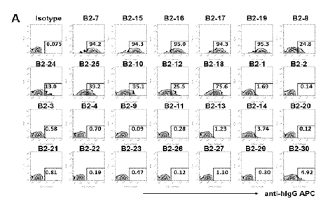
[0038] FIGS. 1A-C. Screening specific monoclonal antibodies for LILRB2. (FIG.
1A) Representative flow cytometric profiles showing that monoclonal antibodies
bind to
LILRB2 reporter cells. Binding of monoclonal antibodies was screened using
flow cytometer
on LILRB2 reporter cells. Bound antibodies were detected with Allophycocyanin
(APC)-
labeled goat anti-human IgG secondary antibody. (FIG. 1B) Quantification of
binding ability
of monoclonal antibodies to LILRB2 reporter cells. B2-7, B2-15, B2-16, B2-17,
B2-19, B2-8,
B2-24, B2-25, B2-10, B2-12 and B2-18 highly bind to LILRB2 reporter cells.
(FIG. 1C)
Quantification of binding ability of monoclonal antibodies B2-7, B2-15, B2-16,
B2-17, B2-19,
B2-8, B2-24, B2-25, B2-10, B2-12 and B2-18 to LILRAs, LILRBs and LAIR1
reporter cells.
[0039] FIGS. 2A-B. Antibodies B2-8, B2-24, B2-10, B2-10 and B2-15 increase GFP
signaling of LILRB2 reporter cells. (FIG. 2A) GFP signaling of LILRB2 reporter
cells
incubated with soluble antibodies. (FIG. 2B) GFP signaling of LILRB2 reporter
cells incubated
with soluble antibodies combined with K562.
[0040] FIGS. 3A-C. Antibodies B2-7, B-15, B2-16, B2-17 and B2-19 block GFP
signaling of LILRB2 reporter cells activated by coated ANGPTL2. (FIG. 3A)
Representative flow cytometric profiles showed that coated ANGPTL2 stimulates
GFP
expression in LILRB2 reporter cells. (FIG. 3B) Representative flow cytometric
profiles showed
that B2-7, B-15, B2-16, B2-17 and B2-19 effectively block GFP expression in
LILRB2 reporter
cells activated by coated ANGPTL2. (FIG. 3C) Dose-dependent inhibitory ability
of B2-7, B-
15, B2-16, B2-17 and B2-19 to GFP expression induced by coated ANGPTL2.
Blocking
potency (IC50) of B2-19, B2-16, B2-7, B2-15 and B2-17 was 48.54 ng/ml, 131.4
ng/ml, 221.1
ng/ml, 341.3 ng/ml and 405.1 ng/ml respectively.
[0041] FIGS. 4A-C. Antibodies B2-7, B-15, B2-16, B2-17 and B2-19 block GFP
signaling of LILRB2 reporter cells activated by coated SEMA4A. (FIG. 4A)
Representative
flow cytometric profiles showed that coated SEMA4A stimulates GFP expression
in LILRB2
reporter cells. (FIG. 4B) Representative flow cytometric profiles showed that
B2-7, B-15, B2-
9
CA 03165532 2022- 7- 20
WO 2021/158413
PCT/US2021/015362
16, B2-17 and B2-19 effectively block GFP expression in LILRB2 reporter cells
activated by
coated SEMA4A. (FIG. 4C) Dose-dependent inhibition of B2-7, B-15, B2-16, B2-17
and B2-
19 to GFP expression induced by coated SEMA4A. Blocking potency (IC50) of B2-
19, B2-16,
B2-7, B2-15 and B2-17 was 167.1 ng/ml, 449 ng/ml, 701 ng/ml, 1001 ng/ml and
1034 ng/ml
respectively.
[0042] FIGS. 5A-C. Antibodies B2-7, B-15, B2-16, B2-17 and B2-19 block GFP
signaling of LILRB2 reporter cells activated by HLA-G overexpressed on K562
cells.
(FIG. 5A) Representative flow cytometric profiles showed that HLA-G
overexpressed on K562
cells stimulates GFP expression in LILRB2 reporter cells. (FIG. 5B)
Representative flow
cytometric profiles showed that B2-7, B-15, B2-16, B2-17 and B2-19 effectively
block GFP
expression in LILRB2 reporter cells activated by HLA-G overexpressed on K562
cells. (FIG.
5C) Quantification of GFP percentage shown in FIG. 5B.
[0043] FIGS. 6A-C. The effect of LILRB2 antibodies on LPS response in primary
human monocytes. (FIGS. 6A-B) Representative flow cytometric profiles showed
that surface
CD86 and intracellular TNFa staining of cells gated on CD33+ monocytes. PBMCs
were
cultured for 48 hours with anti-LILRB2 antibody (10 ng/ml) followed by 6 hours
of LPS
stimulation (50 ng/ml) in the presence of brefeldin A. (FIG. 6C)
Quantification of fold changes
was defined by the ratio of mean fluorescence intensity (MFI) of CD86 and TNFa
in anti-
LILRB2 treated samples relative to their respective MFI in IgG treated samples
shown in FIG.
6A-B. MFI values represent cells gated on CD33+ monocytes.
[0044] FIGS. 7A-J. Antagonistic LILRB2 mAbs inhibit the development of
leukemia cells in C1498-LILRB2 tumor-bearing model. (FIG. 7A) Human LILRB2
expression on mouse C1498 parental cells and human LILRB2 retroviral
transduced C1498
cells. (FIG. 7B) LILRB2 promotes the death of leukemia-bearing mice. Kaplan-
Meier survival
curve of humanized NS G mice which were i.v. injected with LILRB2-
overexpressed or control
(ctrl) C1498 cells (1 x 10⁶ cells per mouse). (FIG. 7C-D) Representative flow
cytometry plots
and summary of leukemia cell infiltration in bone marrow (BM), peripheral
blood (PB), liver
(LV) and spleen (SP) from C57BL/6 which were i.v. injected with LILRB2-
overexpressed or
ctrl C1498 cells (1 x 10⁶ cells per mouse). (FIG. 7E) Kaplan-Meier survival
curve of C57BL/6
mice which were i.v. injected with LILRB2-overexpressed or ctrl C1498 cells (1
x 106 cells
per mouse). (FIG. 7F-G) Representative flow cytometry plots and summary of
myeloid cell
infiltration in peripheral blood (PB), from C57BL/6 which were i.v. injected
with LILRB2-
CA 03165532 2022- 7- 20
WO 2021/158413
PCT/US2021/015362
overexpressed or ctrl C1498 cells (1 x 10⁶ cells per mouse). (FIG. 7H-I)
Representative flow
cytometry plots and summary of leukemia cell infiltration in peripheral blood
(PB) from
C56BL/6 mice with the treatment of LALAPG mutated anti-LILRB2 antibodies or
1gG control
after leukemia cell transplant. LILRB2-overexpressed C1498 cells (1 x 10⁶
cells per mouse)
were injected into C57BL/6 mice followed by treatment with LALAPG Fc mutated
anti-
LILRB2 antibodies or LALAPG Fc mutated IgG. The percentage of leukemia cell
(GFP+)
from peripheral blood (PB) was determined by flow cytometry 20 days after
transplantation.
(FIG. 7J) Summary of myeloid cell infiltration in peripheral blood (PB) from
C56BL/6 mice
treated at indicated time after leukemia cell transplant with LALAPG mutated
anti-LILRB2
antibodies or LALAPG Fc mutated IgG control.
[0045] FIGS. 8A-F. Antagonistic LILRB2 mAbs inhibit the development of
leukemia cells in MLL-AF9 leukemia model. (FIG. 8A) The expression of LILRB2
on PIRB-
KO MLL-AF9 leukemia cells transduced with LILRB2 (LILRB2) or Control (Ctrl)
leukemia
cells. (FIG. 8B-C) Representative flow cytometry plots and summary of leukemia
cell
infiltration in peripheral blood (PB) from C56BL/6 mice transplanted with PirB-
K0 MLL-AF9
leukemia cells transduced with LILRB2 (LILRB2) or control PirB-K0 MLL-AF9
(Ctrl)
leukemia cells. (FIG. 8D) Kaplan-Meier survival curve of leukemia mice which
were
transplanted with LILRB2-overexpressed or Ctrl PirB-K0 MLL-AF9 cells. (FIG. 8E-
F)
Representative flow cytometry plots and summary of leukemia cell infiltration
in peripheral
blood (PB) from C56BL/6 mice transplanted with PirB-K0 LILRB2 leukemia cells
and
followed by the treatment of LALAPG mutated anti-LILRB2 antibodies or IgG
control.
[0046] FIGS. 9A-G. Anti-LILRB2 antibodies inhibit the migration and
infiltration
of AML cells. (FIG. 9A) LILRB2 expression on THP-1 cells was confirmed using a
commercial phycoerythrin (PE)-anti¨LILRB2 antibody. (FIG. 9B) Short-term (20
h)
infiltration of leukemia cells in NSG mice treated with LALAPG Fc mutated anti-
LILRB2
antibodies or LALAPG Fc mutated IgG control after leukemia transplant. THP-1
cells (1 x 10⁷
cells per mouse) were injected into NSG mice followed by treatment with LALAPG
mutated
IgG control or anti-LILRB2 antibodies immediately. The numbers of leukemia
cells (GFP+)
from bone marrow (BM), liver (LV) and spleen (SP) were determined by flow
cytometry 20
hours after transplantation and normalized to number in peripheral blood (PB).
(FIG. 9C)
Percentage of leukemia cells (GFP+) in indicated organs such as liver (LV),
bone marrow
(BM), spleen (SP) and peripheral blood (PB) at day 21 post-transplant in NSG
mice treated
11
CA 03165532 2022- 7- 20
WO 2021/158413
PCT/US2021/015362
with LALAPG mutated anti-LILRB2 antibodies or IgG control after THP-1
injection. (FIG.
9D) Body weight for each mouse shown in Fig. 9B was measured at indicated day
after THP-
1 cells injection. (FIG. 9E) Comparison of the size of livers from NSG mice at
28 days after
THP-1 transplant and treated with IgG or anti-LILRB2 antibodies containing the
LALAPG Fc
mutations. (FIG. 9F) Quantification of liver weight shown in FIG. 9E
normalized to respective
individual mouse body weight. (FIG. 9G) Survival curve of NSG mice treated
with LALAPG
Fc mutated IgG control or anti-LILRB2 antibodies after THP-1 transplantation.
[0047] FIGS. 10A-E. Antagonistic LILRB2 mAbs inhibit the development of
leukemia cells in patient-derived xenograft (PDX) model. (FIG. 10A) LILRB2
expression
pattern on primary leukemia cells from AML M5 patients. (FIG. 10B) Analysis of
correlation
between LILRB2 niRNA levels and the overall survival of patients with AML-M5 =
132,
divided into two groups based on gene expression) in TCGA database
(https://xena.ucsc.edu)
by Kaplan¨Meier long-rank test. (FIG. 10C) Human CD45 CD33 leukemia cells
infiltration
in peripheral blood (PB), bone marrow (BM), spleen (SP) and liver (LV) of NSG
mice
transplanted with primary AML-M5 leukemia samples and treated with anti-LILRB2
antibodies or control IgG. (FIG. 10D) Representative bright-field microcopy
image of primary
AML-M5 leukemia cell cultured in the present of LALAPG mutated anti-LILRB2
antibodies
or IgG control. Anti-LILRB2 treated cells showed more adherent differentiation
morphology.
(FIG. 10E) Intracellular expression of CD68 on primary AML-M5 leukemia cell
cultured in
the present of LALAPG mutated anti-LILRB2 antibodies or IgG control.
[0048] FIGS. 11A-G. Antagonistic LILRB2 mAbs can prevent the T cell
suppressive function of Myeloid-Derived Suppressor Cells (MDSC) in vitro.
(FIG. 11A)
One representative histogram showed that antagonistic LILRB2 mAbs attenuated
the
suppressive function of MDSC towards CDS+ T cells. The MDSC were isolated from
the
PBMC of patients with solid tumor, by depleting HLD_DRb"ght cells and then
enriching the
CD14+ cells, using autoMACS. MDSC were cocultured with T cells from the same
donor (E:
T =1), which was pre-stained with CSBE to monitor the cell proliferation. 10
ug/mL LALAPG
mutated IgG, B2-7, or B2-19 was added into the cell culture. The percentage of
proliferative T
cells, indicated by reduced intensity of CFSE signal, was determined by flow
cytometry 5 days
after treatments. (FIG. 11B) Quantification of the effects of anti-LILRB2 mAbs
on the
inhibitory functions of MDSC towards T cells. The percentage of proliferative
T cells (left
panel: CD8+ T cells, right panel CD4+ T cells), indicated by reduced intensity
of CFSE signal,
12
CA 03165532 2022- 7- 20
WO 2021/158413
PCT/US2021/015362
was determined by flow cytometry 5-7 days after treatments. (FIG. 11C) Anti-
LILRB2 mAbs
attenuated the inhibitory functions of MDSC towards T cells, accessed by
measuring the IFN-
y secretion in the supernatants of T cell cultures. (FIG. 11D) Anti-LILRB2
mAbs decreased the
expression of M2 macrophage markers, while increased the expression of M1
macrophage
markers, on MDSC. The MDSC isolated from peripheral blood of patients with
solid tumors
were cultured with 10 ug/mL anti-LILRB2 antibodies for 7 days. The expression
of CD163,
CD206 and CD86 were analyzed by flow cytometry. (FIG. 11E) Anti-LILRB2 mAbs
decreased
the M2 markers expression, while increased M1 markers expression, on monocyte-
derived
macrophage from a healthy donor. The monocytes isolated from the peripheral
blood of a
health donor were cultured and incubated with 10 ug/mL anti-LILRB2 antibodies
for 7 days.
The expression of CD163, CD206 and CD86 were analyzed by flow cytometry. (FIG.
11F)
Anti-LILRB2 mAbs decreased the M2 markers expression, while increased M1
markers
expression, on the cell surface of tumor associated macrophage/monocyte in
ascites from a
patient with ovarian cancer. The CD14+ cells were isolated from the ascites by
autoMACS and
cultured with anti-LILRB2 mAbs with IgG4 Fc for 7 days. CD163, CD206 and CD86
were
analyzed by flow cytometry. (FIG. 11G) Anti-LILRB2 mAbs increased the M1
macrophage
cytokines and chemokine secreted from MDSC from 4 to 5 patients with solid
tumors.
[0049] FIGS. 12A-B. Antibody sequence analysis of positive phages. (FIG. 12A)
Phylogenic tree of the heavy chain variable region (VH) and light chain
variable region (VI.).
(FIG. 12B) ELISA binding to LILRB2 of the 24 positive phages.
[0050] FIGS. 13A-B. ELISA binding ECso to LILRB2. (FIG. 13A) ELISA binding
curves of LILRB2 antibodies. (FIG. 13B) Calculated EC50 values of LILRB2
antibodies.
[0051] FIGS. 14A-D. Binding specificity of LILRB2 antibodies. (FIG. 14A) ELISA
binding of LILRB2 antibodies to antigens of other members in LILR family.
(FIGS. 14B-D)
Comparison of ELISA binding curves to LILRB2 and LILRA1 of antibody (FIG. 14B)
B2-10,
(FIG. 14C) B212 and (FIG. 14D) B218.
[0052] FIG. 15. Epitope binning of LILRB2 antibodies. The epitope binning was
performed in a sandwich format on Octet RED 96 System. Each antibody was
loaded on protein
A biosensor as Pt antibody. After blocking of biosensor with non-relevant IgG,
LILRB2
antigen was then captured and the biosensors were further incubated with the
rest of other
13
CA 03165532 2022- 7- 20
WO 2021/158413
PCT/US2021/015362
antibodies (2" antibodies). "-F" indicates the 1st antibody blocked the signal
of the 2" antibody.
Antibodies belonging to the same bins were highlighted in different colors.
[0053] FIGS. 16A-D. Binding domains on LILRB2 by the antibodies. (FIG. 16A)
Schematic diagram shown different truncated ECD proteins with Fc fusions.
(FIG. 16B) SDS-
PAGE of purified fusion proteins. (FIG. 16C) ELISA binding of antibodies to
different
truncated proteins. (FIG. 16D) Summary of antibody binding domains.
[0054] FIGS. 17A-F. Mapping of key residues on D1 and D4. (FIGS. 17A-B)
Alignment of the (FIG. 17A (SEQ ID NOS:593-595)) D1 and the (FIG. 17B (SEQ ID
NOS:
596-597)) D4 domain of LILRB2 and LILRB1. The regions that are different
between LILRB2
and LILRB1 and are exposed and locate in the loop regions are boxed. (FIGS.
17C-D) the
sequences of mutations on the (FIG. 17C (SEQ ED NOS: 598-605)) D1 and the
(FIG. 17D
(SEQ ID NOS: 606-613)) D4 domain. (FIGS. 17E-F) Binding loss of antibodies to
mutants of
the (FIG. 17E) D1 and the (FIG. 17F) D4 domain based on ELISA. The percent of
binding
with each mutant relative to the wildtype B2-ECD were plotted as stack bar
graphs.
[0055] FIG. 18. Affinities of blocking antibodies. Antibodies were captured on
protein A biosensors. The sensors were dipped in serially diluted LILRB2
solution for 300secs
to allow association and then dipped into kinetic buffer for 600 secs to allow
dissociation. The
association and dissociation curves are shown in blue solid lines and the two
phases are divided
by red dotted lines.
[0056] FIGS. 19A-13, ELISA binding measurement cif .antibodies to human and
non-human primate (eynomolgtis monkey, cymi) LIER112 recombinantly produced in
HEK293 cells, (FIG. 19A) ELISA binding to human 1.,11..R132. (EEG. 198) ELISA
binding to
cynci-LILRB2. The fusion proteins of the extracellular domain (BCD) of human
LILRB2 or
NUIP-LILRB2 with Fc of mouse IgG2a were used to coat ELBA plates and
antibodies were
titrated in different concentrations as indicated in the graph
[0057] FIGS. 20A-B. Measurements of cell surface LILRB2 binding by
monoclonal antibodies by flow eytometry, (FIG. 20A) Quantification of binding
ability of
monoclonal antibodies to LILRB2 reporter cells. Monoclonal autibodies were
screened using
flow cytorneter on LILRB2 reporter cells labeled with Allophycocyanin (APC)
goat anti-
human IgCi secondary antibody. (FIG. 20-B) Representative flow cytometric
profiles showing
that FICB2-5 and FICB2-10 monoclonal antibodies highly bind to LILRB2 reporter
cells and
14
CA 03165532 2022- 7- 20
WO 2021/158413
PCT/US2021/015362
fiCB2-2 slightly bind to LILRB2 reporter cells. NC: non-labeled reporter
cells. ISO: incubate
the reporter cells only (ISO) with secondary Ab (anti-human Fe specific)
conjugated with APC.
The number indicates mean fluorescence intensity (IVIFI) of APC or AF647.
[0058] FIG. 21. Detection of 11LRB2 antibodies binding to cyno-LILRB2
expressed on cell surface. HER-293 cells stably expressing full length cyno-
LILRB2 with IN-
terminus FLAG tag was used to detect antibody binding by flow cytometry. An
isotype human
IgGI was used as control shown as black line and pink solid peaks indicate the
binding of the
LH¨R.132 antibody to cyno-LILRB2 on cell surface. Mouse-anti-Flag monocknial
antibody was
used for detection by flow cytometry.
[0059] FIG 22. Determination of engagement of GFP reporter signaling by
immobilized LILRB2 antibodies. NC, negative control and IgG for isotype
control.
[0060] FIGS. 23A-B. Determination of soluble antibody engagement of GFP
signaling in LILRB2 reporter cells in the present or absence of K562 cells.
(FIG. 23A)
GFP signaling of LILRB2 reporter cells incubated with soluble antibodies.
(FIG. 23B) GFP
signaling of LILRB2 reporter cells incubated with soluble antibodies combined
with K562.
[0061] FIGS. 24A-B. Specific binding of LILRB2 by LILRB2 monoclonal
antibodies. (P/G. 24A.) Representative flow cytometrie profiles showing that
only positive
control mAbs (POS), but not LILRB2 mAbs bind to LILRAs reporter cells (RC).
(FIG. NB)
Representative flow cytometric profiles showing that [TERM mAbs bind to
LELRB2.-
expressing reporter cells, but not to reporter cells expressing LILRBs and LA
IR I. Binding of
positive control tnAbs (POS) demonstrate expression level of each receptor.
Binding of
LILRB2 monoclonal antibodies was detected using flow cytometry on LILRAs,
LILRBs or
LAIRI. -expressing reporter cells using a Allophycocyanin (APC)-labeled goat
anti-human IgG
Fe specific secondary antibody. Non: non-stained reporter cells. NEG: cells
incubated only
with secondary Ab. POS: cells incubated with commercial antibody conjugated
with APC or
AF647 for the respective LEL or LAIR-I receptors. The number indicates mean
fluorescence
intensity (RIFf) of APC or AF647.
[0062] FIGS. 25A-B. Blocking activity of LILRB2 antibodies assayed in GFP
signaling of LILRB2 reporter cells activated by HLA-G overexpressed on K562
cells.
(FIG. 25A) Representative flow cytometric profiles showed that HLA-G
overexpressed on
K562 cells stimulates GFP induction of LILRB2 reporter cells (FIG. 2513)
Representative flow
CA 03165532 2022- 7- 20
WO 2021/158413
PCT/US2021/015362
cytometric profiles showed that HCB2-5 and IfICB2-10 effectively block GFP
signaling of
LILRB2 reporter cells activated by HI,A-G overexpressing K562 cells. IgG,
isotype control.
[0063] FIGS. 26A-C. Blocking activity of LILRB2 antibodies assayed in GFP
signaling of LILRB2 reporter cells activated by coated ANGPTI.2. (FIG. 26A)
Representative flow cytometric profiles showed that HC132-5 and HCB2-10
effectively block
GFP signaling of LILRB2 reporter cells activated by coated ANGPTL2. (FIG. 26B)
Quantification of CiFP percentage shown in FIG. 26A. (FIG. 26C) Dose-dependent
inhibitory
ability of HCB2-5 and HCB2-10 to GFP signaling induced by coated ANOPT1.2.
[0064] FIGS. 27A-C. Blocking activity of HCB2-5 and HCB240 assayed in GYP
signaling of LILRB2 reporter cells activated by coated SEMA4A. (FIG. 27A)
Representative flow cytometric profiles showed that Ffil.B2-5 and HCB2-10
effectively block
OFF signaling of LILRB2 reporter cells activated by coated SEMA4A.
278)
Quantification of GET percentage. shown in FIG. 27A. (Ha 27C) Dose-dependent
inhibitory
ability of /-ECB2-5 and 4CB2-10 to GFP signaling induced by coated SEMA4A,
[0065] FIGS. 28A-B. Antibody VH and VL phyltigenic trees.
[0066] FIG. 29. Binding of LILRB2 antibodies on reporter cell line expressing
ectodomain of LILR family proteins on cell surface. Bound antibodies were
detected with
anti-human Fc-specific secondary antibody (2nd Ab) conjugated to AF647.
Expression of each
LILR was confirmed using commercially available antibodies directly conjugated
with AF647
or APC (Ctl+ Ab). All incubations were performed for 30 minutes at 4 C. Data
shown is mean
fluorescence intensity after sample acquisition in flow cytometer.
[0067] FIG. 30. Binding of LILRB2 antibodies on leukocytes from human whole
blood harvested from healthy donors. LILRB2 antibodies were directly
conjugated with
AF647. One hundred microliters of whole blood were incubated with antibodies
for cell surface
markers and LILRB2 antibodies, following protocols available in the literature
(Hensley et al.,
J Vis Exp 2012; 67: 4302). Data shown is averaged geometric mean fluorescence
intensity
standard error of the mean (s.e.m., N= 2 donors) after sample acquisition in
flow cytometer
(BD FACS Celesta) and subtracted by background fluorescence of stained samples
in which
LILRB2 antibodies were omitted.
16
CA 03165532 2022- 7- 20
WO 2021/158413
PCT/US2021/015362
[0068] FIG. 31. Binding of LILRB2 antibodies on HEK293 stably expressing full
length human LILRB2. Fifty thousand cells were incubated with a dilution
series (40-0.0006
pg/mL) of LILRB2 antibodies in a final volume of 100 uL. Bound antibodies were
detected
with anti-human Fc-specific secondary antibody conjugated to AF647. All
incubations were
performed for 30 minutes at 4 C. Data shown is averaged geometric mean
fluorescence
intensity standard error of the mean (s.e.m.) after sample acquisition (in
duplicates) in flow
cytometer (BD FACS Celesta) and subtracted by background fluorescence of
samples
incubated with secondary antibody only.
[0069] FIG. 32. Binding of LILRB2 antibodies on CD14+CD16- monocytes isolated
from human PBMC from healthy donors. Fifty thousand cells were incubated for
30 minutes
at 4 C with a dilution series (40-0.0006 ug/mL) of LILRB2 antibodies directly
conjugated to
AF647 in a final volume of 100 uL. Data shown is averaged geometric mean of
fluorescence
intensity from duplicate samples acquired in a flow cytometer (BD FACS
Celesta) from one
donor and is representative of 2 experiments with cells isolated from
different donors.
[0070] FIG. 33. Inhibition of HLA-G-His (5 pg/mL) binding on HEK293 cells
stably expressing full length LILRB2 in the presence of a dilution series (40-
0.0098
lag/mL) of competing LILRB2 antibodies. Bound HLA-G was detected by flow
cytometry
using an anti-His antibody directly conjugated to APC. All incubations were
performed for 30
minutes at 4 C. Data shown is averaged geometric mean of fluorescence
intensity standard
error of the mean (s.e.m.) from duplicate samples acquired in a flow cytometer
(BD FACS
Celesta).
[0071] FIG. 34. Inhibition of SEMA4A-hFc-AF647 (5 ug/mL) binding on
HEK293_LILRB2 cells in the presence of a dilution series (40-0.0098 ug/mL) of
competing LILRB2 antibodies. Incubation was performed for 30 minutes at 4 C.
Data shown
is averaged geometric mean of fluorescence intensity standard error of the
mean (s.e.m.) from
duplicate samples acquired in a flow cytometer (BD FACS Celesta).
[0072] FIG. 35. Effect of LILRB2 blocking antibodies on levels of TNF-u
secreted
by PBMC stimulated with 50 ng/mL LPS. Data shown is from 2 donors and it is
representative from 6 donors (from total of N=6 donors). PBMC isolated from
healthy donors
were incubated (in duplicates) with LPS (Sigma-Aldrich) and various
concentrations of
17
CA 03165532 2022- 7- 20
WO 2021/158413
PCT/US2021/015362
antibodies for 3 days. Cytokines were measured in the culture media
supernatant using a
Human Cytokine Premixed Magnetic Luminex Performance Assay.
[0073] FIG. 36. Effect of LILRB2 blocking antibodies on levels of IFN-y
secreted
by PBMC stimulated with 50 ng/mL LPS. Data shown is from 2 donors and it is
representative from 5 donors (from total of N=6 donors). PBMC isolated from
healthy donors
were incubated (in duplicates) with LPS (Sigma-Aldrich) and various
concentrations of
antibodies for 3 days. Cytokines were measured in the culture media
supernatant using a
Human Cytokine Premixed Magnetic Luminex Performance Assay.
[0074] FIG. 37. Effect of LILRB2 blocking antibodies (40 mg/mL) on levels of
IL-
12p40 secreted by PBMC stimulated with 50 ng/mL LPS (from total of N=3
donors). PBMC
isolated from healthy donors were incubated (in duplicates) with LPS (Sigma-
Aldrich) and 40
itig/mL of B2-19 antibody for 3 days. IL-12p40 concentration was measured in
the culture
media supernatant using a human IL-12 ELISA assay (BD Biosciences)
[0075] FIG. 38. Effect of LILRB2 blocking antibodies on levels of IFN-y
secreted
by PBMC stimulated with 10 ng/mL anti-CD3 activating antibody HIT3a. Data
shown is
from 2 donors and it is representative from 6 donors (from total of N=6
donors). PBMC isolated
from healthy donors were incubated (in duplicates) with HIT3a (Biolegend) and
various
concentrations of antibodies for 3 days. Cytokines were measured in the
culture media
supernatant using a Human Cytokine Premixed Magnetic Luminex Performance
Assay.
[0076] FIG. 39. Effect of LILRB2 blocking antibodies on levels of TNF-u
secreted
by PBMC stimulated with 10 ng/mL anti-CD3 activating antibody HIT3a. Data
shown is
from 2 donors and it is representative from 6 donors (from total of N=6
donors). PBMC isolated
from healthy donors were incubated (in duplicates) with HIT3a (Biolegend) and
various
concentrations of antibodies for 3 days. Cytokines were measured in the
culture media
supernatant using a Human Cytokine Premixed Magnetic Luminex Performance
Assay.
[0077] FIG. 40. Effect of LILRB2 blocking antibodies on levels of GM-CSF
secreted by PBMC stimulated with 10 ng/mL anti-CD3 activating antibody HIT3a.
Data
shown is from 2 donors and it is representative from 6 donors (from total of
N=6 donors).
PBMC isolated from healthy donors were incubated (in duplicates) with HIT3a
(Biolegend)
and various concentrations of antibodies for 3 days. Cytokines were measured
in the culture
media supernatant using a Human Cytokine Premixed Magnetic Luminex Performance
Assay.
18
CA 03165532 2022- 7- 20
WO 2021/158413
PCT/US2021/015362
[0078] FIG. 41. Effect of LILRB2 blocking antibodies on levels of IL-la
secreted
by PBMC stimulated with 10 ng/mL anti-CD3 activating antibody HIT3a. Data
shown is
from 2 donors and it is representative from 6 donors (from total of N=6
donors). PBMC isolated
from healthy donors were incubated (in duplicates) with HIT3a (Biolegend) and
various
concentrations of antibodies for 3 days. Cytokines were measured in the
culture media
supernatant using a Human Cytokine Premixed Magnetic Luminex Performance
Assay.
[0079] FIG. 42. Effect of LILRB2 blocking antibodies on levels of IL-111
secreted
by PBMC stimulated with 10 ng/mL anti-CD3 activating antibody HIT3a. Data
shown is
from 2 donors and it is representative from 6 donors (from total of N=6
donors). PBMC isolated
from healthy donors were incubated (in duplicates) with HIT3a (Biolegend) and
various
concentrations of antibodies for 3 days. Cytokines were measured in the
culture media
supernatant using a Human Cytokine Premixed Magnetic Luminex Performance
Assay.
[0080] FIG. 43. Effect of LILRB2 blocking antibodies on levels of IL-6
secreted by
PBMC stimulated with 10 ng/mL anti-CD3 activating antibody HIT3a. Data shown
is from
2 donors and it is representative from 5 donors (from total of N=6 donors).
PBMC isolated
from healthy donors were incubated (in duplicates) with HIT3a (Biolegend) and
various
concentrations of antibodies for 3 days. Cytokines were measured in the
culture media
supernatant using a Human Cytokine Premixed Magnetic Luminex Performance
Assay.
[0081] FIG. 44. Effect of LILRB2 blocking antibodies on levels of CXCL2
secreted
by PBMC stimulated with 10 ng/mL anti-CD3 activating antibody HIT3a. Data
shown is
from 2 donors and it is representative from 6 donors (from total of N=6
donors). PBMC isolated
from healthy donors were incubated (in duplicates) with HIT3a (Biolegend) and
various
concentrations of antibodies for 3 days. Cytokines were measured in the
culture media
supernatant using a Human Cytokine Premixed Magnetic Luminex Performance
Assay.
[0082] FIG. 45. Effect of 10 litg/mL B2-19 antibody on monocyte-derived
macrophage cell surface markers. CD14 CD16- monocytes isolated from human PBMC
from healthy donors were differentiated into macrophages for 6 days in the
presence of 100
ng/mL of human CSF-1 followed by 24 hours incubation with 100 ng/mL of human
CSF-1, 20
ng/mL human IL-4 and B2-19 antibody or isotype control. Cells were detached
and stained for
flow cytometric analysis (FACS Celesta) using standard protocols. Data shown
is fold change
19
CA 03165532 2022- 7- 20
WO 2021/158413
PCT/US2021/015362
of geometric mean fluorescence intensity (MFI) for cells treated with B2-19
versus cells treated
with i sotype control.
[0083] FIG. 46. Effect of 40 tig/mL B2-19 antibody on cell surface expression
of
CD25 on CD8+ T cells. PBMC isolated from healthy donors were incubated 10
ng/mL with
HIT3a (Biolegend) and 40 ug/mL of B2-19 antibody for 3 days. CD8+ T cells were
analyzed
for cell surface CD25 expression by flow cytometric analysis (FACS Celesta)
using standard
protocols. Data shown is percent change in geometric mean fluorescence
intensity (MF1) for
cells treated with B2-19 versus cells treated with isotype control.
[0084] FIG. 47. B2-19 antibody enhances the production of cytokines and
chemokines in immature DC treated for 2 days with IL-10 (to induce tolerogenic
DC).
Each line represents the result from a different donor.
[0085] FIG. 48. B2-19 antibody enhanced the pro-inflammatory phenotype of DC
trcatcd with LPS, as evidenced by changes in expression of several cell
surface markers.
Each line represents the result from a different donor.
[0086] FIG. 49. B2-19 antibody displays the expected pharmacokinetics profile
(CL and half-life) of a human IgG4 dosed at 5 mg/kg in C57BL/6J wild-type
mice.
[0087] FIGS. 50A-B. (FIG. 50A) B2-19 antibody monotherapy reduces tumor
growth rate in humanized NSG-SGM3 mice xenografted with SK-MEL-5 melanoma cell
line. (FIG. 50B) B2-19 antibody monotherapy causes tumor growth inhibition in
humanized NSG-SGM3 mice xenografted with SK-MEL-5 melanoma cell line.
[0088] FIG. 51. Representative flow cytometric profiles showed that coated
human ANGPTL2 (hANGPLT2) and mouse Angpt12 (mAngpt12) stimulates GFP
expression in LILRB2 reporter cells and B2-19 effectively block GFP expression
in
LILRB2 reporter cells activated by coated hANGPLT2 and mAngpt12.
[0089] FIG. 52. Representative flow cytometric profiles showed that coated
human CD1d (hCD1d) and mouse CD1d (mCD1d) stimulates GFP expression in LILRB2
reporter cells and B2-19 effectively block GFP expression in LILRB2 reporter
cells
activated by coated hCD1d and mCD1d.
CA 03165532 2022- 7- 20
WO 2021/158413
PCT/US2021/015362
DESCRIPTION OF ILLUSTRATIVE EMBODIMENTS
[0090] The inventors determined that LILRB2 plays critical roles in regulation
of both
innate and adaptive immunity. LILRB2 is expressed on several types of immune
cells, such as
normal monocytes, dendritic cells, granulocytes and myeloid derived suppressor
cells
(MDSCs). The inventors have isolated a panel of novel monoclonal antibodies
recognizing
LILRB2 protein, which can be used for the treatment of cancer and autoimmune
diseases.
Within this panel of anti-LILRB2 antibodies, there are examples of antagonists
and agonists of
LILRB2 signalling.
[0091] The following description of the disclosure is merely intended to
illustrate
various embodiments of the disclosure. As such, the specific modifications
discussed are not
to be construed as limitations on the scope of the disclosure. It will be
apparent to one skilled
in the art that various equivalents, changes, and modifications may be made
without departing
from the scope of the disclosure, and it is understood that such equivalent
embodiments are to
be included herein. All references cited herein, including publications,
patents and patent
applications are incorporated herein by reference in their entirety.
I. Definition
[0092] It is to be understood that both the foregoing general description and
the
following detailed description are exemplary and explanatory only and are not
restrictive of the
invention as claimed. In this application, the use of the singular includes
the plural unless
specifically stated otherwise. In this application, the use of "or" means
"and/or" unless stated
otherwise. Furthermore, the use of the term "including", as well as other
forms, such as
"includes" and "included", is not limiting. Also, terms such as -element- or
"component"
encompass both elements and components comprising one unit and elements and
components
that comprise more than one subunit unless specifically stated otherwise.
Also, the use of the
term "portion" can include part of a moiety or the entire moiety.
[0093] As used herein, the singular forms "a", "an" and "the" include plural
references
unless the context clearly dictates otherwise.
[0094] The term "about" as used herein when referring to a measurable value
such as
an amount, a temporal duration, and the like, is meant to encompass variations
of up to - 10%
from the specified value. Unless otherwise indicated, all numbers expressing
quantities of
21
CA 03165532 2022- 7- 20
WO 2021/158413
PCT/US2021/015362
ingredients, properties such as molecular weight, reaction conditions, and so
forth used in the
specification and claims are to be understood as being modified in all
instances by the term
"about." Accordingly, unless indicated to the contrary, the numerical
parameters set forth in
the following specification and attached claims are approximations that may
vary depending
upon the desired properties sought to be obtained by the disclosed subject
matter. At the very
least, and not as an attempt to limit the application of the doctrine of
equivalents to the scope
of the claims, each numerical parameter should at least be construed in light
of the number of
reported significant digits and by applying ordinary rounding techniques.
Notwithstanding that
the numerical ranges and parameters setting forth the broad scope of the
invention are
approximations, the numerical values set forth in the specific examples are
reported as
precisely as possible. Any numerical value, however, inherently contain
certain errors
necessarily resulting from the standard deviation found in their respective
testing
measurements.
[0095] The term "antibody" refers to an intact immunoglobulin of any isotype,
or a
fragment thereof that can compete with the intact antibody for specific
binding to the target
antigen, and includes, for instance, chimeric, humanized, fully human, and
bispecific
antibodies. An "antibody" is a species of an antigen binding protein. An
intact antibody will
generally comprise at least two full-length heavy chains and two full-length
light chains, but in
some instances can include fewer chains such as antibodies naturally occurring
in camelids
which can comprise only heavy chains. Antibodies can be derived solely from a
single source,
or can be "chimeric," that is, different portions of the antibody can be
derived from two
different antibodies as described further below. The antigen binding proteins,
antibodies, or
binding fragments can be produced in hybridomas, by recombinant DNA
techniques, or by
enzymatic or chemical cleavage of intact antibodies. Unless otherwise
indicated, the term
"antibody" includes, in addition to antibodies comprising two full-length
heavy chains and two
full-length light chains, derivatives, variants, fragments, and muteins
thereof, examples of
which are described below. Furthermore, unless explicitly excluded, antibodies
include
monoclonal antibodies, bispecific antibodies, minibodies, domain antibodies,
synthetic
antibodies (sometimes referred to herein as "antibody mimetics"), chimeric
antibodies,
humanized antibodies, human antibodies, antibody fusions (sometimes referred
to herein as
"antibody conjugates"), and fragments thereof, respectively. In some
embodiments, the term
also encompasses peptibodies.
22
CA 03165532 2022- 7- 20
WO 2021/158413
PCT/US2021/015362
[0096] Naturally occurring antibody structural units typically comprise a
tetramer.
Each such tetramer typically is composed of two identical pairs of polypeptide
chains, each
pair having one full-length "light" (in certain embodiments, about 25 kDa) and
one full-length
"heavy" chain (in certain embodiments, about 50-70 kDa). The amino-terminal
portion of each
chain typically includes a variable region of about 100 to 110 or more amino
acids that typically
is responsible for antigen recognition. The carboxy-terminal portion of each
chain typically
defines a constant region that can be responsible for effector function. Human
light chains are
typically classified as kappa and lambda light chains. Heavy chains are
typically classified as
mu, delta, gamma, alpha, or epsilon, and define the antibody's isotype as IgM,
IgD, IgG, IgA,
and IgE, respectively. IgG has several subclasses, including, but not limited
to, IgG1 , IgG2,
IgG3, and IgG4. IgM has subclasses including, but not limited to, IgM1 and
IgM2. IgA is
similarly subdivided into subclasses including, but not limited to, IgA l and
IgA2. Within full-
length light and heavy chains, typically, the variable and constant regions
are joined by a "J"
region of about 12 or more amino acids, with the heavy chain also including a
"D" region of
about 10 more amino acids. See, e.g., Fundamental Immunology, Ch. 7 (Paul, W.,
ed., 2nd ed.
Raven Press, N.Y. (1989)) (incorporated by reference in its entirety for all
purposes). The
variable regions of each light/heavy chain pair typically form the antigen
binding site.
[0097] The term "variable region" or "variable domain- refers to a portion of
the light
and/or heavy chains of an antibody, typically including approximately the
amino-terminal 120
to 130 amino acids in the heavy chain and about 100 to 110 amino terminal
amino acids in the
light chain. In certain embodiments, variable regions of different antibodies
differ extensively
in amino acid sequence even among antibodies of the same species. The variable
region of an
antibody typically determines specificity of a particular antibody for its
target.
[0098] The variable regions typically exhibit the same general structure of
relatively
conserved framework regions (FR) joined by three hyper variable regions, also
called
complementarity determining regions or CDRs. The CDRs from the two chains of
each pair
typically are aligned by the framework regions, which can enable binding to a
specific epitope.
From N-terminal to C-terminal, both light and heavy chain variable regions
typically comprise
the domains FR1, CDR1, FR2, CDR2, FR3, CDR3 and FR4. The assignment of amino
acids
to each domain is typically in accordance with the definitions of Kabat
Sequences of Proteins
of Immunological Interest (National Institutes of Health, Bethesda, Md. (1987
and 1991)),
23
CA 03165532 2022- 7- 20
WO 2021/158413
PCT/US2021/015362
Chothia & Lesk, J. Mol. Biol., 196:901-917 (1987) or Chothia et al., Nature,
342:878-883
(1989).
[0099] In certain embodiments, an antibody heavy chain binds to an antigen in
the
absence of an antibody light chain. In certain embodiments, an antibody light
chain binds to an
antigen in the absence of an antibody heavy chain. In certain embodiments, an
antibody binding
region binds to an antigen in the absence of an antibody light chain. In
certain embodiments,
an antibody binding region binds to an antigen in the absence of an antibody
heavy chain. In
certain embodiments, an individual variable region specifically binds to an
antigen in the
absence of other variable regions.
[00100] In certain
embodiments, definitive delineation of a CDR and
identification of residues comprising the binding site of an antibody is
accomplished by solving
the structure of the antibody and/or solving the structure of the antibody-
ligand complex. In
certain embodiments, that can be accomplished by any of a variety of
techniques known to
those skilled in the art, such as X-ray crystallography. In certain
embodiments, various methods
of analysis can be employed to identify or approximate the CDR regions.
Examples of such
methods include, but are not limited to, the Kabat definition, the Chothia
definition, the AbM
definition and the contact definition.
[00101]
The Kabat definition is a standard for numbering the residues in an
antibody and is typically used to identify CDR regions. See, e.g., Johnson &
Wu, Nucleic Acids
Res., 28: 214-8 (2000). The Chothia definition is similar to the Kabat
definition, but the Chothia
definition takes into account positions of certain structural loop regions.
See, e.g., Chothia et
al., J. Mol. Biol., 196: 901-17 (1986); Chothia et al., Nature, 342: 877-83
(1989). The AbM
definition uses an integrated suite of computer programs produced by Oxford
Molecular Group
that model antibody structure. See, e.g., Martin et al., Proc Natl Acad Sci
(USA), 86:9268-
9272 (1989); "AbMTm, A Computer Program for Modeling Variable Regions of
Antibodies,"
Oxford, UK; Oxford Molecular, Ltd. The AbM definition models the tertiary
structure of an
antibody from primary sequence using a combination of knowledge databases and
ab initio
methods, such as those described by Samudrala et al., "Ab Initio Protein
Structure Prediction
Using a Combined Hierarchical Approach," in PROTEINS. Structure, Function and
Genetics
Suppl., 3:194-198 (1999). The contact definition is based on an analysis of
the available
complex crystal structures. See, e.g., MacCallum et al., J. Mol. Biol., 5:732-
45 (1996).
24
CA 03165532 2022- 7- 20
WO 2021/158413
PCT/US2021/015362
[00102]
By convention, the CDR regions in the heavy chain are typically referred
to as H1, H2, and H3 and are numbered sequentially in the direction from the
amino terminus
to the carboxy terminus. The CDR regions in the light chain are typically
referred to as Li, L2,
and L3 and are numbered sequentially in the direction from the amino terminus
to the carboxy
terminus.
[00103]
The term "light chain" includes a full-length light chain and fragments
thereof having sufficient variable region sequence to confer binding
specificity. A full-length
light chain includes a variable region domain, VL, and a constant region
domain, CL. The
variable region domain of the light chain is at the amino-terminus of the
polypeptide. Light
chains include kappa chains and lambda chains.
[00104]
The term "heavy chain" includes a full-length heavy chain and
fragments thereof having sufficient variable region sequence to confer binding
specificity. A
full-length heavy chain includes a variable region domain, VH, and three
constant region
domains, CHL CH2, and CH3. The VH domain is at the amino-terminus of the
polypeptide,
and the CH domains are at the carboxyl-terminus, with the CH3 being closest to
the carboxy-
terminus of the polypeptide. Heavy chains can be of any isotype, including IgG
(including
IgGi, IgG2, IgG3 and IgG4 subtypes), IgA (including IgAl and IgA2 subtypes),
IgM and IgE.
[00105]
A bispecific or bifunctional antibody typically is an artificial hybrid
antibody having two different heavy/light chain pairs and two different
binding sites. Bispecific
antibodies can be produced by a variety of methods including, but not limited
to, fusion of
hybridomas or linking of Fab' fragments. See, e.g., Songsivilai et at., Clin.
Exp. Immunol., 79:
315-321 (1990); Kostelny et al., J. Immunol., 148:1547-1553 (1992).
[00106]
The term "antigen" refers to a substance capable of inducing adaptive
immune responses. Specifically, an antigen is a substance which serves as a
target for the
receptors of an adaptive immune response. Typically, an antigen is a molecule
that binds to
antigen-specific receptors but cannot induce an immune response in the body by
itsself.
Antigens are usually proteins and polysaccharides, less frequently also
lipids. Suitable antigens
include without limitation parts of bacteria (coats, capsules, cell walls,
flagella, fimbrai, and
toxins), viruses, and other microorganisms. Antigens also include tumor
antigens, e.g., antigens
generated by mutations in tumors. As used herein, antigens also include
immunogens and
haptens.
CA 03165532 2022- 7- 20
WO 2021/158413
PCT/US2021/015362
[00107]
An "antigen binding protein" ("ABP") as used herein means any protein
that binds a specified target antigen. In the instant application, the
specified target antigen is
the L1LRB protein or fragment thereof. "Antigen binding protein" includes but
is not limited
to antibodies and antigen-binding fragment thereof. Peptibodies are another
example of antigen
binding proteins.
[00108]
The term "antigen-binding fragment- as used herein refers to a portion
of a protein which is capable of binding specifically to an antigen. In
certain embodiment, the
antigen-binding fragment is derived from an antibody comprising one or more
CDRs, or any
other antibody fragment that binds to an antigen but does not comprise an
intact native antibody
structure. In certain embodiments, the antigen-binding fragment is not derived
from an
antibody but rather is derived from a receptor. Examples of antigen-binding
fragment include,
without limitation, a diabody, a Fab, a Fab', a F(ab')2, an Fv fragment, a
disulfide stabilized Fv
fragment (dsFv), a (dsFv)2, a bispecific dsFv (dsFv-dsFv'), a disulfide
stabilized diabody (ds
diabody), a single-chain antibody molecule (scFv), an scFv dimer (bivalent
diabody), a
multispecific antibody, a single domain antibody (sdAb), a camelid antibody or
a nanobody, a
domain antibody, and a bivalent domain antibody. In certain embodiments, an
antigen-binding
fragment is capable of binding to the same antigen to which the parent
antibody binds. In
certain embodiments, an antigen-binding fragment may comprise one or more CDRs
from a
particular human antibody grafted to a framework region from one or more
different human
antibodies. In certain embodiments, the antigen-binding fragment is derived
from a receptor
and contains one or more mutations. In certain embodiments, the antigen-
binding fragment
does not bind to the natural ligand of the receptor from which the antigen-
binding fragment is
derived.
[00109]
A "Fab fragment- comprises one light chain and the CH1 and variable
regions of one heavy chain. The heavy chain of a Fab molecule cannot form a
disulfide bond
with another heavy chain molecule.
[00110]
A "Fab' fragment" comprises one light chain and a portion of one heavy
chain that contains the VH domain and the CH1 domain and also the region
between the CH1
and CH2 domains, such that an interchain disulfide bond can be formed between
the two heavy
chains of two Fab' fragments to form an F(ab')2 molecule.
26
CA 03165532 2022- 7- 20
WO 2021/158413
PCT/US2021/015362
[00111]
A "F(ab')2 fragment" contains two light chains and two heavy chains
containing a portion of the constant region between the CH1 and CH2 domains,
such that an
interchain disulfide bond is formed between the two heavy chains. A F(ab').2
fragment thus is
composed of two Fab' fragments that are held together by a disulfide bond
between the two
heavy chains.
[00112]
An "Pc" region comprises two heavy chain fragments comprising the
CH1 and CH2 domains of an antibody. The two heavy chain fragments are held
together by
two or more disulfide bonds and by hydrophobic interactions of the CH3
domains.
[00113]
The "Fv region" comprises the variable regions from both the heavy and
light chains but lacks the constant regions.
[00114]
"Single-chain antibodies" are Fv molecules in which the heavy and light
chain variable regions have been connected by a flexible linker to form a
single polypeptide
chain, which forms an antigen binding region. Single chain antibodies are
discussed in detail
in International Patent Application Publication No. WO 88/01649 and U.S. Pat.
No. 4.946,778
and No. 5,260,203, the disclosures of which are incorporated by reference.
[00115]
A "domain antibody" is an immunologically functional immunoglobulin
fragment containing only the variable region of a heavy chain or the variable
region of a light
chain. In some instances, two or more VH regions are covalently joined with a
peptide linker
to create a bivalent domain antibody. The two VH regions of a bivalent domain
antibody can
target the same or different antigens.
[00116]
A "bivalent antigen binding protein- or "bivalent antibody- comprises
two antigen binding sites. In some instances, the two binding sites have the
same antigen
specificities. Bivalent antigen binding proteins and bivalent antibodies can
be bispecific, see,
infra. A bivalent antibody other than a "multispecific" or "multifunctional"
antibody, in certain
embodiments, typically is understood to have each of its binding sites
identical.
[00117]
A "multispecific antigen binding protein" or "multispecific antibody" is
one that targets more than one antigen or epitope.
[00118]
A "bispeci fic," "dual-specific" or "hi functional" antigen binding
protein
or antibody is a hybrid antigen binding protein or antibody, respectively,
having two different
27
CA 03165532 2022- 7- 20
WO 2021/158413
PCT/US2021/015362
antigen binding sites. Bispecific antigen binding proteins and antibodies are
a species of
multispecific antigen binding protein antibody and can be produced by a
variety of methods
including, but not limited to, fusion of hybridomas or linking of Fab'
fragments. See, e.g.,
Songsivilai and Lachmann, 1990, Clin. Exp. Immunol. 79:315-321; Kostelny et
al., 1992, J.
Immunol. 148:1547-1553. The two binding sites of a bispecific antigen binding
protein or
antibody will bind to two different epitopes, which can reside on the same or
different protein
targets.
[00119]
"Binding affinity- generally refers to the strength of the sum total of
non-covalent interactions between a single binding site of a molecule (e.g.,
an antibody) and
its binding partner (e.g., an antigen). Unless indicated otherwise, as used
herein, "binding
affinity" refers to intrinsic binding affinity that reflects a 1:1 interaction
between members of
a binding pair (e.g., antibody and antigen). The affinity of a molecule X for
its partner Y can
generally be represented by the dissociation constant (Kd). Affinity can be
measured by
common methods known in the art, including those described herein. Low-
affinity antibodies
generally bind antigen slowly and tend to dissociate readily, whereas high-
affinity antibodies
generally bind antigen faster and tend to remain bound longer. A variety of
methods of
measuring binding affinity are known in the art, any of which can be used for
purposes of the
present invention. Specific illustrative and exemplary embodiments for
measuring binding
affinity are described in the following.
[00120] An antibody
that "specifically binds to" or is "specific for" a particular
polypeptide or an epitope on a particular polypeptide is one that binds to
that particular
polypeptide or epitope on a particular polypeptide without substantially
binding to any other
polypeptide or polypeptide epitope. For example, the LILRB2 specific
antibodies of the present
invention are specific to LILRB2. In some embodiments, the antibody that binds
to LILRB2
has a dissociation constant (Kd) of 100 nM, 10 nM, 1 nM, 0.1 nM, ().01 nM, or
0.001
nM (e.g., 10"8M or less, e.g., from 10"8M to 10"13M, e.g., from 10"9M to 10-13
M). The
dissociation constant Kd used herein refers to the ratio of the dissociation
rate to the association
rate (koff/koõ), which may be determined by using any conventional method
known in the art,
including but are not limited to surface plasmon resonance method, microscale
thermophoresis
method, HPLC-MS method and flow cytometry (such as FACS) method. In certain
embodiments, the Kd value can be appropriately determined by using flow
eytometry.
28
CA 03165532 2022- 7- 20
WO 2021/158413
PCT/US2021/015362
[00121]
The term "compete when used in the context of antigen binding
proteins (e.g., antibody or antigen-binding fragment thereof) that compete for
the same epitope
means competition between antigen binding proteins as determined by an assay
in which the
antigen binding protein (e.g., antibody or antigen-binding fragment thereof)
being tested
prevents or inhibits (e.g., reduces) specific binding of a reference antigen
binding protein (e.g.,
a ligand, or a reference antibody) to a common antigen (e.g., LILRB or a
fragment thereof).
Numerous types of competitive binding assays can be used to determine if one
antigen binding
protein competes with another, for example: solid phase direct or indirect
radioimmunoassay
(RIA), solid phase direct or indirect enzyme immunoassay (EIA), sandwich
competition assay
(see, e.g., Stahli et al., 1983, Methods in Enzymology 9:242-253); solid phase
direct biotin-
avidin EIA (see, e.g., Kirkland et al., 1986, J. Immunol. 137:3614-3619) solid
phase direct
labeled assay, solid phase direct labeled sandwich assay (see, e.g., Harlow
and Lane,
1988, Antibodies, A Laboratory Manual, Cold Spring Harbor Press); solid phase
direct label
RIA using 1-125 label (see, e.g., Morel et al., 1988, Molec. Immunol. 25:7-
15); solid phase
direct biotin-avidin EIA (see, e.g., Cheung, et al., 1990, Virology 176:546-
552); and direct
labeled RIA (Moldenhauer et al., 1990, Scand. J. Immunol. 32:77-82).
Typically, such an assay
involves the use of purified antigen bound to a solid surface or cells bearing
either of these, an
unlabelled test antigen binding protein and a labeled reference antigen
binding protein.
Competitive inhibition is measured by determining the amount of label bound to
the solid
surface or cells in the presence of the test antigen binding protein. Usually
the test antigen
binding protein is present in excess. Antigen binding proteins identified by
competition assay
(competing antigen binding proteins) include antigen binding proteins binding
to the same
epitope as the reference antigen binding proteins and antigen binding proteins
binding to an
adjacent epitope sufficiently proximal to the epitope bound by the reference
antigen binding
protein for steric hindrance to occur. Additional details regarding methods
for determining
competitive binding are provided in the examples herein. Usually, when a
competing antigen
binding protein is present in excess, it will inhibit (e.g., reduce) specific
binding of a reference
antigen binding protein to a common antigen by at least 40-45%, 45-50%, 50-
55%, 55-60%,
60-65%, 65-70%, 70-75% or 75% or more. In some instances, binding is inhibited
by at least
80-85%, 85-90%, 90-95%, 95-97%, or 97% or more.
[00122]
The term "epitope" as used herein refers to the specific group of atoms
or amino acids on an antigen to which an antibody binds. The epitope can be
either linear
epitope or a conformational epitope. A linear epitope is formed by a
continuous sequence of
29
CA 03165532 2022- 7- 20
WO 2021/158413
PCT/US2021/015362
amino acids from the antigen and interacts with an antibody based on their
primary structure.
A conformational epitope, on the other hand, is composed of discontinuous
sections of the
antigen's amino acid sequence and interacts with the antibody based on the 3D
structure of the
antigen. In general, an epitope is approximately five or six amino acid in
length. Two antibodies
may bind the same epitope within an antigen if they exhibit competitive
binding for the antigen.
[00123]
A "cell", as used herein, can be prokaryotic or eukaryotic. A prokaryotic
cell includes, for example, bacteria. A eukaryotic cell includes, for example,
a fungus, a plant
cell, and an animal cell. The types of an animal cell (e.g., a mammalian cell
or a human cell)
includes, for example, a cell from circulatory/immune system or organ, e.g., a
B cell, a T cell
(cytotoxic T cell, natural killer T cell, regulatory T cell, T helper cell), a
natural killer cell, a
granulocyte (e.g., basophil granulocyte, an eosinophil granulocyte, a
neutrophil granulocyte
and a hypersegmented neutrophil), a monocyte or macrophage, a red blood cell
(e.g.,
reticulocyte), a mast cell, a thrombocyte or megalcaryocyte, and a dendritic
cell; a cell from an
endocrine system or organ, e.g., a thyroid cell (e.g., thyroid epithelial
cell, parafollicular cell),
a parathyroid cell (e.g., parathyroid chief cell, oxyphil cell), an adrenal
cell (e.g., chromaffin
cell), and a pineal cell (e.g., pinealocyte); a cell from a nervous system or
organ, e.g., a glioblast
(e.g., as trocyte and oligodendrocyte), a microglia, a magnocellular
neurosecretory cell, a
stellate cell, a boettcher cell, and a pituitary cell (e.g., gonadotrope,
corticotrope. thyrotrope,
somatotrope, and lactotroph); a cell from a respiratory system or organ, e.g.,
a pneumocyte (a
type I pneumocyte and a type II pneumocyte), a clara cell, a goblet cell, and
an alveolar
macrophage; a cell from circular system or organ (e.g., myocardiocyte and
pericyte); a cell
from digestive system or organ, e.g., a gastric chief cell, a parietal cell, a
goblet cell, a paneth
cell, a G cell, a D cell, an ECL cell, an I cell, a K cell, an S cell, an
enteroendocrine cell, an
enterochromaffin cell, an APUD cell, and a liver cell (e.g., a hepatocyte and
Kupffer cell); a
cell from integumentary system or organ, e.g., a bone cell (e.g., an
osteoblast, an osteocyte,
and an osteoclast), a teeth cell (e.g., a cementoblast, and an ameloblast), a
cartilage cell (e.g., a
chondroblast and a chondrocyte), a skin/hair cell (e.g., a trichocyte, a
keratinocyte, and a
melanocyte (Nevus cell), a muscle cell (e.g., myocyte), an adipocyte, a
fibroblast, and a tendon
cell; a cell from urinary system or organ (e.g., a podocyte, a juxtaglomerular
cell, an
intraglomerular mesangial cell, an extraglomerular mesangial cell, a kidney
proximal tubule
brush border cell, and a macula densa cell); and a cell from reproductive
system or organ (e.g.,
a spermatozoon, a Sertoli cell, a leydig cell, an ovum, an oocyte). A cell can
be normal, healthy
cell; or a diseased or unhealthy cell (e.g., a cancer cell). A cell further
includes a mammalian
CA 03165532 2022- 7- 20
WO 2021/158413
PCT/US2021/015362
zygote or a stern cell which include an embryonic stern cell, a fetal stern
cell, an induced
pluripotent stem cell, and an adult stem cell. A stem cell is a cell that is
capable of undergoing
cycles of cell division while maintaining an undifferentiated state and
differentiating into
specialized cell types. A stem cell can be an omnipotent stem cell, a
pluripotent stem cell, a
multipotent stem cell, an oligopotent stem cell and a unipotent stem cell, any
of which may be
induced from a somatic cell. A stem cell may also include a cancer stem cell.
A mammalian
cell can be a rodent cell, e.g., a mouse, rat, hamster cell. A mammalian cell
can be a lagomorpha
cell, e.g., a rabbit cell. A mammalian cell can also be a primate cell, e.g.,
a human cell.
[00124]
The term "chimeric antigen receptor" or "CAR" as used herein refers to
an artificially constructed hybrid protein or polypeptide containing an
antigen binding domain
of an antibody (e.g., a single chain variable fragment (scFv)) linked to a
domain or signaling,
e.g., T-cell signaling or T-cell activation domains, that activates an immune
cell, e.g., a T cell
or a NK cell (see, e.g., Kershaw et al., supra, Eshhar et al., Proc. Natl.
Acad. Sci. USA, 90(2):
720-724 (1993), and Sadelain et al., Curr. Opin. Immunol. 21(2): 215-223
(2009)). CARs are
capable of redirecting the immune cell specificity and reactivity toward a
selected target in a
non-MHC-restricted manner, taking advantage of the antigen-binding properties
of monoclonal
antibodies. The non-MHC-restricted antigen recognition confers immune cells
expressing
CARs on the ability to recognize an antigen independent of antigen processing,
thus bypassing
a major mechanism of tumor escape. In addition, when expressed in T-cells,
CARs
advantageously do not dimerize with endogenous T-cell receptor (TCR) alpha and
beta chains.
[00125]
As used herein, "essentially free," in terms of a specified component, is
used herein to mean that none of the specified component has been purposefully
formulated
into a composition and/or is present only as a contaminant or in trace
amounts. The total amount
of the specified component resulting from any unintended contamination of a
composition is
therefore well below 0.05%, preferably below 0.01%. Most preferred is a
composition in which
no amount of the specified component can be detected with standard analytical
methods.
[00126]
The term "host cell" means a cell that has been transformed, or is
capable of being transformed, with a nucleic acid sequence and thereby
expresses a gene of
interest. The term includes the progeny of the parent cell, whether or not the
progeny is identical
in morphology or in genetic make-up to the original parent cell, so long as
the gene of interest
is present.
31
CA 03165532 2022- 7- 20
WO 2021/158413
PCT/US2021/015362
[00127]
The term "identity" refers to a relationship between the sequences of
two or more polypeptide molecules or two or more nucleic acid molecules, as
determined by
aligning and comparing the sequences. "Percent identity- means the percent of
identical
residues between the amino acids or nucleotides in the compared molecules and
is calculated
based on the size of the smallest of the molecules being compared. For these
calculations, gaps
in alignments (if any) are preferably addressed by a particular mathematical
model or computer
program (i.e., an "algorithm-). Methods that can be used to calculate the
identity of the aligned
nucleic acids or polypeptides include those described in Computational
Molecular Biology,
(Lesk, A. M., ed.), 1988, New York: Oxford University Press; Biocomputing
Informatics and
Genome Projects, (Smith, D. W., ed.), 1993, New York: Academic Press; Computer
Analysis
of Sequence Data, Part I, (Griffin, A. M., and Griffin, H. G., eds.), 1994,
New Jersey: Humana
Press; von Heinje, G., 1987, Sequence Analysis in Molecular Biology, New York:
Academic
Press; Sequence Analysis Primer, (Gribskov, M. and Devereux, J., eds.), 1991,
New York: M.
Stockton Press; and Carillo etal., 1988, SIAM J. Applied Math. 48:1073.
[00128] In
calculating percent identity, the sequences being compared are
typically aligned in a way that gives the largest match between the sequences.
One example of
a computer program that can be used to determine percent identity is the GCG
program
package, which includes GAP (Devereux et al., 1984, Nucl. Acid Res. 12:387;
Genetics
Computer Group, University of Wisconsin, Madison, Wis.). The computer
algorithm GAP is
used to align the two polypeptides or polynucleotides for which the percent
sequence identity
is to be determined. The sequences are aligned for optimal matching of their
respective amino
acid or nucleotide (the "matched span", as determined by the algorithm). A gap
opening penalty
(which is calculated as 3x the average diagonal, wherein the "average diagonal-
is the average
of the diagonal of the comparison matrix being used; the "diagonal" is the
score or number
assigned to each perfect amino acid match by the particular comparison matrix)
and a gap
extension penalty (which is usually 1/10 times the gap opening penalty), as
well as a
comparison matrix such as PAM 250 or BLOSUM 62 are used in conjunction with
the
algorithm. In certain embodiments, a standard comparison matrix (see, Dayhoff
et al.,
1978, Atlas of Protein Sequence and Structure 5:345-352 for the PAM 250
comparison matrix;
Henikoff et al., 1992, Proc. Natl. Acad. Sci. U.S.A. 89:10915-10919 for the
BLOSUM 62
comparison matrix) is also used by the algorithm.
32
CA 03165532 2022- 7- 20
WO 2021/158413
PCT/US2021/015362
[00129]
Examples of parameters that can be employed in determining percent
identity for polypeptides or nucleotide sequences using the GAP program can be
found in
Needleman et al., 1970, J. Mol. Biol. 48:443-453.
[00130]
Certain alignment schemes for aligning two amino acid sequences may
result in matching of only a short region of the two sequences, and this small
aligned region
may have very high sequence identity even though there is no significant
relationship between
the two full-length sequences. Accordingly, the selected alignment method (GAP
program) can
be adjusted if so desired to result in an alignment that spans at least 50 or
other number of
contiguous amino acids of the target polypeptide.
[00131] The term
"link- as used herein refers to the association via
intramolecular interaction, e.g., covalent bonds, metallic bonds, and/or ionic
bonding, or inter-
molecular interaction, e.g., hydrogen bond or noncovalent bonds.
[00132]
Leukocyte immunoglobulin-like receptor subfamily B member 2
(LILRB2) is a protein that in humans is encoded by the LILRB2 gene. This gene
is a member
of the leukocyte immunoglobulin-like receptor (LIR) family, which is found in
a gene cluster
at chromosomal region 19q13.4. The encoded protein belongs to the subfamily B
class of LIR
receptors which contain two or four extracellular immunoglobulin domains, a
transmembrane
domain, and two to four cytoplasmic immunoreceptor tyrosine-based inhibitory
motifs
(ITIMs). The receptor is expressed on myeloid cells; it binds to multiple
types of ligands,
including HLA class I molecules, ANGPTLs, myelin inhibitors (including Nogo66,
MAO, and
0Mgp), and and 13-amyloid, transducing a negative signal that inhibits
stimulation of an
immune response. It is thought to control inflammatory responses and
cytotoxicity to help
focus the immune response and limit autoreactivity.
[00133]
The term "operably linked" refers to an arrangement of elements
wherein the components so described are configured so as to perform their
usual function.
Thus, a given signal peptide that is operably linked to a polypeptide directs
the secretion of the
polypeptide from a cell. In the case of a promoter, a promoter that is
operably linked to a coding
sequence will direct the expression of the coding sequence. The promoter or
other control
elements need not be contiguous with the coding sequence, so long as they
function to direct
the expression thereof. For example, intervening untranslated yet transcribed
sequences can be
33
CA 03165532 2022- 7- 20
WO 2021/158413
PCT/US2021/015362
present between the promoter sequence and the coding sequence and the promoter
sequence
can still be considered "operably linked" to the coding sequence.
[00134]
The use of the term "or" in the claims is used to mean "and/or" unless
explicitly indicated to refer to alternatives only or the alternatives are
mutually exclusive,
although the disclosure supports a definition that refers to only alternatives
and "and/or." As
used herein "another" may mean at least a second or more.
[00135]
The term "polynucleotide- or -nucleic acid- includes both single-
stranded and double-stranded nucleotide polymers. The nucleotides comprising
the
polynucleotide can be ribonucleotides or deoxyribonucleotides or a modified
form of either
type of nucleotide. Said modifications include base modifications such as
bromouridine and
inosine derivatives, ribose modifications such as 2',3'-dideoxyribose, and
internucleotide
linkage modifications such as phosphorothioate, phosphorodithioate,
phosphoroselenoate,
phosphorodiselenoate, phosphoroanilothioate, phoshoraniladate and
phosphoroamidate.
[00136]
The terms "polypeptide" or "protein" means a macromolecule having
the amino acid sequence of a native protein, that is, a protein produced by a
naturally-occurring
and non-recombinant cell; or it is produced by a genetically-engineered or
recombinant cell,
and comprise molecules having the amino acid sequence of the native protein,
or molecules
having deletions from, additions to, and/or substitutions of one or more amino
acids of the
native sequence. The term also includes amino acid polymers in which one or
more amino
acids are chemical analogs of a corresponding naturally-occurring amino acid
and polymers.
The terms "polypeptide" and "protein" specifically encompass L1LRB antigen
binding
proteins, antibodies, or sequences that have deletions from, additions to,
and/or substitutions
of one or more amino acid of antigen-binding protein. The term "polypeptide
fragment" refers
to a polypeptide that has an amino-terminal deletion, a carboxyl-terminal
deletion, and/or an
internal deletion as compared with the full-length native protein. Such
fragments can also
contain modified amino acids as compared with the native protein. In certain
embodiments,
fragments are about five to 500 amino acids long. For example, fragments can
be at least 5, 6,
8, 10, 14, 20, 50, 70, 100, 110, 150, 200, 250, 300, 350, 400, or 450 amino
acids long. Useful
polypeptide fragments include immunologically functional fragments of
antibodies, including
binding domains. In the case of a LILRB-binding antibody, useful fragments
include but are
not limited to a CDR region, a variable domain of a heavy and/or light chain,
a portion of an
antibody chain or just its variable region including two CDRs, and the like.
34
CA 03165532 2022- 7- 20
WO 2021/158413
PCT/US2021/015362
[00137]
The pharmaceutically acceptable carriers useful in this invention are
conventional. Remington's Pharmaceutical Sciences, by E. W. Martin, Mack
Publishing Co.,
Easton, PA, 15th Edition (1975), describes compositions and formulations
suitable for
pharmaceutical delivery of the fusion proteins herein disclosed. In general,
the nature of the
carrier will depend on the particular mode of administration being employed.
For instance,
parenteral formulations usually comprise injectable fluids that include
pharmaceutically and
physiologically acceptable fluids such as water, physiological saline,
balanced salt solutions,
aqueous dextrose, glycerol or the like as a vehicle. For solid compositions
(e.g., powder, pill,
tablet, or capsule forms) , conventional non-toxic solid carriers can include,
for example,
pharmaceutical grades of mannitol, lactose, starch or magnesium stearate. In
addition to
biologically- neutral carriers, pharmaceutical compositions to be administered
can contain
minor amounts of non-toxic auxiliary substances, such as wetting or
emulsifying agents,
preservatives, and pH buffering agents and the like, for example sodium
acetate or sorbitan
monolaurate.
[00138] As used
herein, the term "subject" refers to a human or any non-human
animal (e.g., mouse, rat, rabbit, dog, cat, cattle, swine, sheep, horse or
primate). A human
includes pre- and post-natal forms. In many embodiments, a subject is a human
being. A subject
can be a patient, which refers to a human presenting to a medical provider for
diagnosis or
treatment of a disease. The term "subject" is used herein interchangeably with
"individual- or
"patient." A subject can be afflicted with or is susceptible to a disease or
disorder but may or
may not display symptoms of the disease or disorder.
00139]
The term "therapeutically effective amount- or "effective dosage" as
used herein refers to the dosage or concentration of a drug effective to treat
a disease or
condition. For example, with regard to the use of the monoclonal antibodies or
antigen-binding
fragments thereof disclosed herein to treat cancer, a therapeutically
effective amount is the
dosage or concentration of the monoclonal antibody or antigen-binding fragment
thereof
capable of reducing the tumor volume, eradicating all or part of a tumor,
inhibiting or slowing
tumor growth or cancer cell infiltration into other organs, inhibiting growth
or proliferation of
cells mediating a cancerous condition, inhibiting or slowing tumor cell
metastasis, ameliorating
any symptom or marker associated with a tumor or cancerous condition,
preventing or delaying
the development of a tumor or cancerous condition, or some combination
thereof.
CA 03165532 2022- 7- 20
WO 2021/158413
PCT/US2021/015362
[00140]
"Treating or "treatment" of a condition as used herein includes
preventing or alleviating a condition, slowing the onset or rate of
development of a condition,
reducing the risk of developing a condition, preventing or delaying the
development of
symptoms associated with a condition, reducing or ending symptoms associated
with a
condition, generating a complete or partial regression of a condition, curing
a condition, or
some combination thereof.
[00141]
As used herein, a "vector" refers to a nucleic acid molecule as introduced
into a host cell, thereby producing a transformed host cell. A vector may
include nucleic acid
sequences that permit it to replicate in the host cell, such as an origin of
replication. A vector
may also include one or more therapeutic genes and/or selectable marker genes
and other
genetic elements known in the art. A vector can transduce, transform or infect
a cell, thereby
causing the cell to express nucleic acids and/or proteins other than those
native to the cell. A
vector optionally includes materials to aid in achieving entry of the nucleic
acid into the cell,
such as a viral particle, liposome, protein coating or the like.
II. LILRB2 Related Diseases
[00142]
LILRB2 has been identified as a key regulator of myeloid cell
phenotype. The activation of LILRB2 suppresses the pro-inflammatory activity
of myeloid
cells. While myeloid cells with a suppressive/anti-inflammatory phenotype can
down-regulate
the activation, proliferation and cytotoxic activity of T cells, modulation of
LILRB2 has the
potential in therapeutic use in conditions and disorders including cancer,
autoimmune diseases,
and inflammatory diseases.
00143]
While hyperproliferative diseases can be associated with any disease
which causes a cell to begin to reproduce uncontrollably, the prototypical
example is cancer.
[00144]
Examples of cancer can be generally categorized into solid tumors and
hematologic malignancies. Solid tumors include but are not limited to, adrenal
cancer, bile
duct carcinoma, bone cancer, brain cancer (e.g., astrocytoma, brain stem
glioma,
craniopharyngioma, ependymoma, hemangioblastoma, medulloblastoma, meningioma,
oligodendroglioma, spinal axis tumor), breast cancer (including acoustic
neuroma, basal breast
carcinoma, ductal carcinoma and lobular breast carcinoma), cervical cancer,
choriocarcinoma,
colon cancer, colorectal cancer, esophageal cancer, eye cancer, gastric
cancer, glioblastoma,
head and neck cancer, kidney cancer (including Wilms tumor), liver cancer
(including
36
CA 03165532 2022- 7- 20
WO 2021/158413
PCT/US2021/015362
hepatocellular carcinoma (HCC)), lung cancer (including bronchogenic
carcinoma, non-small
cell lung cancer (squamous/non-squamous), bronchioloalveolar cell lung cancer,
papillary
adenocarcinomas), mesothelioma, melanoma, merkel cell cancer, nasopharyngeal
carcinoma,
neuroblastoma, oral cancer, ovarian cancer, pancreatic cancer, penile cancer,
pinealoma,
prostate cancer, renal cell cancer, retinoblastoma, sarcoma (including
chondrosarcoma,
Ewing' s sarcoma, fibrosarcoma, leiomyosarcoma, liposarcoma, myxosarcoma,
osteogenic
sarcoma, rhabdomyosarcoma, synovial sarcoma), skin cancer (including basal
cell carcinoma,
sebaceous gland carcinoma, squamous cell carcinoma), testicular cancer
(including
seminoma), thymic carcinoma, thyroid cancer (e.g., medullary thyroid
carcinoma, papillary
thyroid carcinoma), uterine cancer, and vaginal cancer.
[00145]
Hematologic malignancies include but are not limited to blastic
plasmacytoid dendritic cell neoplasm (BPDCN), heavy chain disease, leukemias
(including but
not limited to acute lymphocytic leukemia (ALL), acute myeloid leukemia (AML)
(including
but not limited to acute promyelocytic leukemia (APL) or M3 AML, acute
myelomonocytic
leukemia or M4 AML, acute monocytic leukemia or M5 AML), B-cell leukemia,
chronic
lymphoblastic leukemia (CLL), chronic myelomonocytic leukemia (CMML), chronic
myelocytic leukemia (CML), pre-B acute lymphocy tic leukemia (Pre-B ALL),
diffuse large B-
cell lymphoma (DLBCL), extranodal NK/T-cell lymphoma, hairy cell leukemia,
HHV8-
associated primary effusion lymphoma, plasmablastic lymphoma, primary CNS
lymphoma,
primary mediastinal large B-cell lymphoma, T-cell/histiocyte-rich B-cell
lymphoma),
lymphomas (including but not limited to Hodgkin's lymphoma, non-Hodgkin's
lymphoma,
Waldenstrom's macroglobulinemia), multiple myeloma (MM), myelodysplastic
syndromes
(MDS), myeloproliferative neoplasms, and polycythemia vera.
[00146]
Immunotherapy holds great promise to achieve long-lasting anti -tumor
effects. Immune checkpoint PD-1 and CTLA-4 blockade therapies have been
successful in
treating some types of cancers but not others. These immunotherapies target
inhibitory
molecules on T cells to reactivate dysfunctional T cells within the tumor
microenvironment
(TME). Other populations of immune cells, including monocytic cells, are
present in the TME
in even larger numbers than T cells. In fact, monocyte-derived macrophages are
the most
abundant immune cell population in tumor tissues. While these innate cells
possess the capacity
to kill tumor cells and to prime or reactivate T cells, they become
dysfunctional in TME and
turn into MDSCs and tumor-associated macrophages (TAMs) that support tumor
development
37
CA 03165532 2022- 7- 20
WO 2021/158413
PCT/US2021/015362
and suppress immune surveillance and attack. MDSCs, including monocytic MDSCs
(M-
MDSCs) and polymorphonuclear MDSCs (PMN-MDSCs), represent a heterogeneous
population of immature myeloid cells that fail to terminally differentiate.
TAMs are a mixed
macrophage population in TME. They are anti-inflammatory and correlated with a
poor
prognosis. Despite their phenotypic plasticity, MDSCs and TAMs are defined by
their
immunosuppressive function. Removing, reprogramming, or blocking trafficking
of these
immune-suppressive monocytic cells is becoming an attractive anti-cancer
therapeutic strategy.
[00147]
LILRB2 is expressed on MDSCs and TAMs in TME. Therapeutic
blocking of LILRB2 in myeloid-rich solid tumors has the potential to
reactivate or enhance
anti-tumor immune responses in patients presenting with disease
unresponsive/relapsed to T
cell checkpoint inhibitors.
[00148]
LILRB2 expression on myeloid cells may regulate systems involved in
autoimmune and inflammatory diseases. Therapeutic activating or agonizing
LILRB2 has the
potential to treat autoimmune or inflammatory diseases.
[00149] Autoimmune
or inflammatory diseases include, but are not limited to,
Acquired Immunodeficiency Syndrome (AIDS, which is a viral disease with an
autoimmune
component), alopecia areata, ankylosing spondylitis, antiphospholipid
syndrome, autoimmune
Addison's disease, autoimmune hemolytic anemia, autoimmune hepatitis,
autoimmune inner
ear disease (AIED), autoimmune lymphoproliferative syndrome (ALPS), autoimmune
thrombocytopenic purpura (ATP), B eh cet' s disease, cardiomyopathy, celiac
sprue-dermatitis
hepetiformis; chronic fatigue immune dysfunction syndrome (CF1DS), chronic
inflammatory
demyelinating polyneuropathy (CIPD), cicatricial pemphigold, cold agglutinin
disease, crest
syndrome, Crohn's disease, Degos' disease, dermatomyositis-juvenile, discoid
lupus, essential
mixed cryoglobulinemia, fibromyalgia-fibromyositis, Graves' disease, Guillain-
Barre
syndrome, Hashimoto's thyroiditis, idiopathic pulmonary fibrosis, idiopathic
thrombocytopenia purpura (ITP), IgA nephropathy, insulin-dependent diabetes
mellitus,
juvenile chronic arthritis (Still's disease), juvenile rheumatoid arthritis,
Meniere's disease,
mixed connective tissue disease, multiple sclerosis, myasthenia gravis,
pemacious anemia,
polyarteritis nodosa, polychondritis, polyglandular syndromes, polymyalgia
rheumatica,
polymyositis and dermatomyositis, primary agammaglobulinemia, primary biliary
cirrhosis,
psoriasis, psoriatic arthritis, Raynaud's phenomena, Reiter's syndrome,
rheumatic fever,
rheumatoid arthritis, sarcoidosis, scleroderma, systemic scleroderma,
progressive systemic
38
CA 03165532 2022- 7- 20
WO 2021/158413
PCT/US2021/015362
sclerosis (PSS), systemic sclerosis (SS), Sjogren's syndrome, stiff-man
syndrome, systemic
lupus eryth em atos us (SLE), Takayasu arteriti s, temporal arteriti s/gi ant
cell arteri ti s ,
inflammatory bowel disease (IBD), ulcerative colitis, Cohn's disease,
intestinal mucosal
inflammation, wasting disease associated with colitis, uveitis, vitiligo and
Wegener's
granulomatosis, Alzheimer's disease, asthma, atopic allergy, allergy,
atherosclerosis, bronchial
asthma, eczema, glomerulonephritis, graft vs. host disease, hemolytic anemias,
osteoarthritis,
sepsis, stroke, transplantation of tissue and organs, vasculitis, diabetic
retinopathy, ventilator
induced lung injury, viral infections, autoimmune diabetes and the like.
Inflammatory disorders
include, for example, chronic and acute inflammatory disorders.
III. Monoclonal Antibodics and Production Thcrcof
[00150]
The monoclonal antibodies described herein can be prepared using
standard methods, followed by screening, characterization and functional
assessment. Variable
regions can be sequenced and then subcloned into a human expression vector to
produce the
chimeric antibody genes, which are then expressed and purified. These chimeric
antibodies can
be tested for antigen binding, signaling blocking, and in xenograft
experiments. The
monoclonal antibodies described herein can also be prepared using phage
display method, in
which a large library of phage displayed human scFv is panned against the
target protein. The
human scFv selected to specifically binding to the target protein can be
sequenced and then
subcloned into a human expression vector to produce the desired human
antibody.
A. General Methods
[00151]
It will be understood that monoclonal antibodies binding to LILRB2 will
have several applications. These include the production of diagnostic kits for
use in detecting
and diagnosing cancer, as well as for cancer therapies. In these contexts, one
may link such
antibodies to diagnostic or therapeutic agents, use them as capture agents or
competitors in
competitive assays, or use them individually without additional agents being
attached thereto.
The antibodies may be mutated or modified, as discussed further below. Methods
for preparing
and characterizing antibodies are well known in the art (see, e.g.,
Antibodies: A Laboratory
Manual, Cold Spring Harbor Laboratory, 1988; U.S. Patent 4,196,265).
[00152]
The classical methods for generating monoclonal antibodies (MAbs)
generally begin along the same lines as those for preparing polyclonal
antibodies. The first step
for both these methods is immunization of an appropriate host. As is well
known in the art, a
given composition for immunization may vary in its immunogenicity. It is often
necessary
39
CA 03165532 2022- 7- 20
WO 2021/158413
PCT/US2021/015362
therefore to boost the host immune system, as may be achieved by coupling a
peptide or
polypeptide immunogen to a carrier_ Exemplary and preferred carriers are
keyhole limpet
hemocyanin (KLH) and bovine serum albumin (BSA). Other albumins such as
ovalbumin,
mouse serum albumin or rabbit serum albumin can also be used as carriers.
Means for
conjugating a polypeptide to a carrier protein are well known in the art and
include
glutaraldehyde, m-maleimidobencoyl-N-hydroxysuccinimide ester, carbodiimyde
and bis-
biazotized benzidine. As also is well known in the art, the immunogenicity of
a particular
immunogen composition can be enhanced by the use of non-specific stimulators
of the immune
response, known as adjuvants. Exemplary and preferred adjuvants include
complete Freund's
adjuvant (a non-specific stimulator of the immune response containing killed
Mycabacteriurn
tuberculosis), incomplete Freund's adjuvants and aluminum hydroxide adjuvant.
[00153]
The amount of immunogen composition used in the production of
polyclonal antibodies varies upon the nature of the immunogen as well as the
animal used for
immunization. A variety of routes can be used to administer the immunogen
(subcutaneous,
intramuscular, intradermal, intravenous and intraperitoneal). The production
of polyclonal
antibodies may be monitored by sampling blood of the immunized animal at
various points
following immunization. A second, booster injection, also may be given. The
process of
boosting and titering is repeated until a suitable titer is achieved. When a
desired level of
immunogenicity is obtained, the immunized animal can be bled and the serum
isolated and
stored, and/or the animal can be used to generate MAbs.
[00154]
Following immunization, somatic cells with the potential for producing
antibodies, specifically B lymphocytes (B cells), are selected for use in the
MAb generating
protocol. These cells may be obtained from biopsied spleens or lymph nodes, or
from
circulating blood. The antibody-producing B lymphocytes from the immunized
animal are then
fused with cells of an immortal myeloma cell, generally one of the same
species as the animal
that was immunized or human or human/mouse chimeric cells. Myeloma cell lines
suited for
use in hybridoma-producing fusion procedures preferably are non-antibody-
producing, have
high fusion efficiency, and enzyme deficiencies that render then incapable of
growing in certain
selective media which support the growth of only the desired fused cells
(hybridomas). Any
one of a number of myeloma cells may be used, as are known to those of skill
in the art (Goding,
pp. 65-66, 1986; Campbell, pp. 75-83, 1984).
CA 03165532 2022- 7- 20
WO 2021/158413
PCT/US2021/015362
[00155]
Methods for generating hybrids of antibody-producing spleen or lymph
node cells and myeloma cells usually comprise mixing somatic cells with
myeloma cells in a
2:1 proportion, though the proportion may vary from about 20:1 to about 1:1,
respectively, in
the presence of an agent or agents (chemical or electrical) that promote the
fusion of cell
membranes. Fusion methods using Sendai virus have been described by Kohler and
Milstein
(1975; 1976), and those using polyethylene glycol (PEG), such as 37% (v/v)
PEG, by Gefter
et al. (1977). The use of electrically induced fusion methods also is
appropriate (Goding, pp.
71-74, 1986). Fusion procedures usually produce viable hybrids at low
frequencies, about 1 x
10-6 to 1 x 10⁻⁸. However, this does not pose a problem, as the viable, fused
hybrids are
differentiated from the parental, infused cells (particularly the infused
myeloma cells that
would normally continue to divide indefinitely) by culturing in a selective
medium. The
selective medium is generally one that contains an agent that blocks the de
nova synthesis of
nucleotides in the tissue culture media. Exemplary and preferred agents are
aminopterin,
methotrexate, and azaserine. Aminopterin and methotrexate block de novo
synthesis of both
purines and pyrimidines, whereas azaserine blocks only purine synthesis. Where
aminopterin
or methotrexate is used, the media is supplemented with hypoxanthine and
thymidine as a
source of nucleotides (HAT medium). Where azaserine is used, the media is
supplemented with
hypoxanthine. Ouabain is added if the B cell source is an Epstein Barr virus
(EBV) transformed
human B cell line, in order to eliminate EBV transformed lines that have not
fused to the
myeloma.
[00156]
The preferred selection medium is HAT or HAT with ouabain. Only
cells capable of operating nucleotide salvage pathways are able to survive in
HAT medium.
The myeloma cells are defective in key enzymes of the salvage pathway, e.g.,
hypoxanthine
phosphoribosyl transferase (HPRT), and they cannot survive. The B cells can
operate this
pathway, but they have a limited life span in culture and generally die within
about two weeks.
Therefore, the only cells that can survive in the selective media are those
hybrids formed from
myeloma and B cells. When the source of B cells used for fusion is a line of
EBV-transformed
B cells, as here, ouabain is also used for drug selection of hybrids as EBV-
transformed B cells
are susceptible to drug killing, whereas the myeloma partner used is chosen to
be ouabain
resistant.
[00157]
Culturing provides a population of hybridomas from which specific
hybridomas are selected. Typically, selection of hybridomas is performed by
culturing the cells
41
CA 03165532 2022- 7- 20
WO 2021/158413
PCT/US2021/015362
by single-clone dilution in microtiter plates, followed by testing the
individual clonal
supernatants (after about two to three weeks) for the desired reactivity. The
assay should be
sensitive, simple and rapid, such as radioimmunoassays, enzyme immunoassays,
cytotoxicity
assays, plaque assays dot immunobinding assays, and the like. The selected
hybridomas are
then serially diluted or single-cell sorted by flow cytometric sorting and
cloned into individual
antibody-producing cell lines, which clones can then be propagated
indefinitely to provide
mAbs. The cell lines may be exploited for MAb production in two basic ways. A
sample of the
hybridoma can be injected (often into the peritoneal cavity) into an animal
(e.g., a mouse).
Optionally, the animals are primed with a hydrocarbon, especially oils such as
pristane
(tetramethylpentadecane) prior to injection. When human hybridomas are used in
this way, it
is optimal to inject immunocompromised mice, such as SCID mice, to prevent
tumor rejection.
The injected animal develops tumors secreting the specific monoclonal antibody
produced by
the fused cell hybrid. The body fluids of the animal, such as serum or ascites
fluid, can then be
tapped to provide MAbs in high concentration. The individual cell lines could
also be cultured
in vitro, where the MAbs are naturally secreted into the culture medium from
which they can
be readily obtained in high concentrations. Alternatively, human hybridoma
cells lines can be
used in vitro to produce immunoglobulins in cell supernatant. The cell lines
can be adapted for
growth in serum-free medium to optimize the ability to recover human
monoclonal
immunoglobulins of high purity.
[00158] MAbs
produced by either means may be further purified, if desired,
using filtration, centrifugation and various chromatographic methods such as
FPLC or affinity
chromatography. Fragments of the monoclonal antibodies of the disclosure can
be obtained
from the purified monoclonal antibodies by methods which include digestion
with enzymes,
such as pepsin or papain, and/or by cleavage of disulfide bonds by chemical
reduction.
Alternatively, monoclonal antibody fragments encompassed by the present
disclosure can be
synthesized using an automated peptide synthesizer.
[00159]
It also is contemplated that a molecular cloning approach may be used
to generate monoclonals. For this, RNA can be isolated from the hybridoma line
and the
antibody genes obtained by RT-PCR and cloned into an immunoglobulin expression
vector.
Alternatively, combinatorial immunoglobulin phagemid libraries are prepared
from RNA
isolated from the cell lines and phagemids expressing appropriate antibodies
are selected by
panning using viral antigens. The advantages of this approach over
conventional hybridoma
42
CA 03165532 2022- 7- 20
WO 2021/158413
PCT/US2021/015362
techniques are that approximately 104 times as many antibodies can be produced
and screened
in a single round, and that new specificities are generated by H and L chain
combination which
further increases the chance of finding appropriate antibodies.
[00160]
Recently, additional methods for generating mAb, such as scFv phage
display, have been developed (see CM Hammers and JR Stanley, Antibody phage
display:
technique and applications, J Invest Dermatol (2014) 134: e17). Generally, a
panel of human
mAbs that bind to a target protein, e.g., human L1LRB2, are generated by
panning a large
diversity of human scFv phage displayed antibody library.
[00161]
To generate the human scFv phage displayed antibody library, RNA is
extracted from the chosen cell source, e.g., peripheral blood mononuclear
cells. The RNA is
then reversed-transcribed into cDNA, which is used for PCR of the VH and VL
chains of the
encoded antibodies. Defined sets of primers specific for the different VH and
VL chain region
gene families allow the amplification of all transcribed rearranged variable
regions within a
given immunoglobulin repertoire, reflecting all antibody specificities in a
particular individual.
[00162] The VH and
VL PCR products that represent the antibody repertoire are
ligated into a phage display vector that is engineered to express the VH and
VL as an scFv
fused to the pIII minor capsid protein of a filamentous bacteriophage of E.
coli that was
originally derived from the M13 bacteriophage. This generates a library of
phages, each of
which expresses on its surface a scFv and harbors the vector with the
respective nucleotide
sequence within.
[00163]
The library is then screened for phage binding to a target antigen through
its expressed surface scFv by a technique called bio-panning. In short, the
target protein is
coated on solid phase for incubation with phage libraries. After washing and
elution, antigen
enriched phages are recovered and used for next rounds of phage panning. After
at least three
rounds of phage panning, single bacterial colonies are picked for phage ELISA
and other
functional/genetic analysis.
[00164]
The positive hits are sequenced for the scFv region and are converted to
full human IgG heavy and light chain constructs, which are used to generate
the mAb of interest
using the methods disclosed supra. For example, the IgG expressing plasmids
are cotransfected
into Expi293 cells using transfection reagent PEI. After 7 days of expression,
supernatants are
harvested, and antibodies are purified by affinity chromatography using
protein A resin.
43
CA 03165532 2022- 7- 20
WO 2021/158413
PCT/US2021/015362
[00165]
Other U.S. patents, each incorporated herein by reference, that teach the
production of antibodies useful in the present disclosure include U.S. Patent
5,565,332, which
describes the production of chimeric antibodies using a combinatorial
approach; U.S. Patent
4,816,567 which describes recombinant immunoglobulin preparations; and U.S.
Patent
4,867,973 which describes antibody-therapeutic agent conjugates.
B. Antibodies of the Present Disclosure
1. Antibodies to LILRB2
[00166]
Antibodies or antigen-binding fragments thereof according to the
present disclosure may be defined, in the first instance, by their binding
specificity, which in
this case is for LILRB2. Those of skill in the art, by assessing the binding
specificity/affinity
of a given antibody using techniques well known to those of skill in the art,
can determine
whether such antibodies fall within the scope of the instant claims.
[00167]
In one aspect, there are provided antibodies and antigen-binding
fragments specifically bind to LILRB2. In some embodiments, when bound to
LILRB2, such
antibodies modulate the activation of LILRB2. In certain embodiments, the
antibody or
antigen-binding fragment, when bound to LILRB2, activates LILRB2. In certain
embodiments,
the antibody or antigen-binding fragment, when bound to LILRB2, suppresses
activation of
LILRB2. In certain embodiments, the antibody or antigen-binding fragment, when
bound to
LILRB2, can specifically interfere with, block or reduce the interaction
between LILRB2 and
its binding partners. In certain embodiments, the antibody or antigen-binding
fragment
provided herein is capable of inhibiting the immunosuppressive activity of
MDSCs and other
solid tumor-infiltrating myeloid cells, such as tumor-associated macrophages
(TAMs) and
tolerogenic dendritic cells (DCs). In certain embodiments, the antibodies or
antigen-binding
fragments provided herein specifically or selectively bind to human LILRB2.
[00168] In some
embodiments, the antibodies or antigen-binding fragments bind
specifically to human LILRB2 and/or substantially inhibits binding of human
LILRB2 to HLA-
G, AN GPTLs, SEMA4A by at least about 20%-40%, 40-60%, 60-80%, 80-85%, or more
(for
example, by an assay as disclosed in the Example). In some embodiments, the
antibody or
antigen-binding fragment has a Kd of less (binding more tightly) than 10-6, 10-
7, 10-8, 10-9,
10-io, 10-11, 10-12, vs-13
V M. In some embodiments, the antibody or antigen-binding fragment
has an IC50 for blocking the binding of HLA-G, ANGPTLs, SEMA4A to LILRB2 of
less than
10 uM, 10 uM to 1 uM, 1000 nM to 100 nM, 100 nM to 10 nM, 10 nM to 1 nM, 1000
pM to
44
CA 03165532 2022- 7- 20
WO 2021/158413
PCT/US2021/015362
500 pM, 500 pM to 200 pM, less than 200 pM, 200 pM to 150 pM, 200 pM to 100
pM, 100
pM to 10 pM, 10 pM to 1 pM.
[00169]
In some embodiments, the antibodies or antigen-binding fragments
provided herein having the clone-paired CDRs illustrated in Table 2.
[00170] In certain
embodiments, the antibodies may be defined by their variable
sequence, which include additional "framework" regions. The antibody is
characterized by
clone-paired heavy chain and light chain amino acid sequences from Appendices
I and III.
Furthermore, the antibodies sequences may vary from these sequences,
particularly in regions
outside the CDRs. For example, the amino acids may vary from those set out
above by a given
percentage, e.g., 80%, 85%, 90%, 91%, 92%, 93%, 94%, 95%, 96%, 97%, 98% or 99%
homology, or the amino acids may vary from those set out above by permitting
conservative
substitutions (discussed below). Each of the foregoing apply to the amino acid
sequences of
Appendices I and III. In another embodiment, the antibody derivatives of the
present disclosure
comprise VL and VH domains having up to 0, 1, 2, 3, 4, 5, 6, 7, 8, 9, 10 or
more conservative
or non-conservative amino acid substitutions, while still exhibiting the
desired binding and
functional properties.
[00171]
While the antibodies of the present disclosure were generated as IgG' s,
it may be useful to modify the constant regions to alter their function. The
constant regions of
the antibodies typically mediate the binding of the antibody to host tissues
or factors, including
various cells of the immune system (e.g., effector cells) and the first
component (Clq) of the
classical complement system. Thus, the term "antibody" includes intact
immunoglobulins of
types IgA, IgG, IgE, IgD, IgM (as well as subtypes thereof), wherein the light
chains of the
immunoglobulin may be of types kappa or lambda. Within light and heavy chains,
the variable
and constant regions are joined by a 35 "J" region of about 12 or more amino
acids, with the
heavy chain also including a "D" region of about 10 more amino acids. See
generally,
Fundamental Immunology Ch. 7 (Paul, W., ed., 2nd ed. Raven Press, N.Y. (1989).
[00172]
The present disclosure further comprises nucleic acids which hybridize
to nucleic acids encoding the antibodies disclosed herein. In general, the
nucleic acids hybridize
under moderate or high stringency conditions to nucleic acids that encode
antibodies disclosed
herein and also encode antibodies that maintain the ability to specifically
bind to an LILRB2.
A first nucleic acid molecule is "hybridizable- to a second nucleic acid
molecule when a single
CA 03165532 2022- 7- 20
WO 2021/158413 PCT/US2021/015362
stranded form of the first nucleic acid molecule can anneal to the second
nucleic acid molecule
under the appropriate conditions of temperature and solution ionic strength
(see Sambrook et
al., MOLECULAR CLONING: A LABORATORY MANUAL, 3rd ed., Cold Spring Harbor
Press, Cold
Spring Harbor, N.Y. 2001). The conditions of temperature and ionic strength
determine the
"stringency- of the hybridization. Typical moderate stringency hybridization
conditions are
40% formamide, with 5X or 6X SSC and 0.1% SDS at 42 C. High stringency
hybridization
conditions are 50% formamide, 5X or 6X SSC (0.15M NaC1 and 0.015M Na-citrate)
at 42 C
or, optionally, at a higher temperature (e.g., 57 C, 59 C, 60 C, 62 C, 63 C,
65 C or 68 C).
Hybridization requires that the two nucleic acids contain complementary
sequences, although,
depending on the stringency of the hybridization, mismatches between bases are
possible. The
appropriate stringency for hybridizing nucleic acids depends on the length of
the nucleic acids
and the degree of complementation, variables well known in the art. The
greater the degree of
similarity or homology between two nucleotide sequences, the higher the
stringency under
which the nucleic acids may hybridize. For hybrids of greater than 100
nucleotides in length,
equations for calculating the melting temperature have been derived (see
Sambrook et al.,
supra). For hybridization with shorter nucleic acids, e.g., oligonucleotides,
the position of
mismatches becomes more important, and the length of the oligonucleotide
determines its
specificity (see Sambrook et al., supra).
2. Exemplary Epitopcs and Competing Antigen Binding Proteins
[00173] In another
aspect, the present disclosure provides epitopes to which anti-
LILRB2 antibodies bind. In some embodiments, epitopes that are bound by the
antibodies
described herein are useful. In certain embodiments, an epitope provided
herein can be utilized
to isolate antibodies or antigen binding proteins that bind to LILRB2. In
certain embodiments,
an epitope provided herein can be utilized to generate antibodies or antigen
binding proteins
which bind to LILRB2. In certain embodiments, an epitope or a sequence
comprising an
epitope provided herein can be utilized as an immunogen to generate antibodies
or antigen
binding proteins that bind to LILRB2. In certain embodiments, an epitope
described herein or
a sequence comprising an epitope described herein can be utilized to interfere
with biological
activity of LILRB2.
[00174] In some
embodiments, antibodies or antigen-binding fragments thereof
that bind to any of the epitopes are particularly useful. In some embodiments,
an epitope
provided herein, when bound by an antibody, modulates the biological activity
of LILRB2. In
46
CA 03165532 2022- 7- 20
WO 2021/158413
PCT/US2021/015362
some embodiments, an epitope provided herein, when bound by an antibody,
activates
LILRB2. In some embodiments, an epitope provided herein, when bound by an
antibody,
suppress the activation of LILRB2. In some embodiments, an epitope provided
herein, when
bound by an antibody, block the interaction between LILRB2 and its binding
partners.
[00175] In some
embodiments, the domain(s)/region(s) containing residues that
are in contact with or are buried by an antibody can be identified by mutating
specific residues
in LILRB2 and determining whether the antibody can bind the mutated LILRB2
protein. By
making a number of individual mutations, residues that play a direct role in
binding or that are
in sufficiently close proximity to the antibody such that a mutation can
affect binding between
the antibody and antigen can be identified. From knowledge of these amino
acids, the
domain(s) or region(s) of the antigen that contain residues in contact with
the antigen binding
protein or covered by the antibody can be elucidated. Such a domain can
include the binding
epitope of an antigen binding protein.
[00176]
In another aspect, the present disclosure provides antigen-binding
proteins that compete with one of the exemplified antibodies or antigen-
binding fragment
binding to the epitope described herein for specific binding to LILRB2. Such
antigen binding
proteins can also bind to the same epitope as one of the herein exemplified
antibodies or the
antigen-binding fragment, or an overlapping epitope. Antigen-binding proteins
that compete
with or bind to the same epitope as the exemplified antibodies are expected to
show similar
functional properties. The exemplified antibodies include those described
above, including
those with the heavy and light chain variable regions and CDRs included in
Table 2, heavy and
light chains as shown in Appendices 1 and III, and heavy and light chain
coding regions as
shown in Appendices II and IV.
C. Engineering of Antibody Sequences
[00177] In various
embodiments, one may choose to engineer sequences of the
identified antibodies for a variety of reasons, such as improved expression,
improved cross-
reactivity or diminished off-target binding. The following is a general
discussion of relevant
techniques for antibody engineering.
[00178]
Hybridomas may be cultured, then cells lysed, and total RNA extracted.
Random hexamers may be used with RT to generate cDNA copies of RNA, and then
PCR
performed using a multiplex mixture of PCR primers expected to amplify all
human variable
47
CA 03165532 2022- 7- 20
WO 2021/158413 PCT/US2021/015362
gene sequences. PCR product can be cloned into pGEM-T Easy vector, then
sequenced by
automated DNA sequencing using standard vector primers. Assay of binding and
neutralization
may be performed using antibodies collected from hybridoma supernatants and
purified by
FPLC, using Protein G columns. Recombinant full-length IgG antibodies may be
generated by
subcloning heavy and light chain Fv DNAs from the cloning vector into an IgG
plasmid vector,
transfected into 293 Freestyle cells or CHO cells, and antibodies collected a
purified from the
293 or CHO cell supernatant.
[00179]
The rapid availability of antibody produced in the same host cell and
cell culture process as the final cGMP manufacturing process has the potential
to reduce the
duration of process development programs. Lonza has developed a generic method
using
pooled transfectants grown in CDACF medium, for the rapid production of small
quantities
(up to 50 g) of antibodies in CHO cells. Although slightly slower than a true
transient system,
the advantages include a higher product concentration and use of the same host
and process as
the production cell line. Example of growth and productivity of GS-CHO pools,
expressing a
model antibody, in a disposable bioreactor: in a disposable bag bioreactor
culture (5 L working
volume) operated in fed-batch mode, a harvest antibody concentration of 2 g/L
was achieved
within 9 weeks of transfection.
[00180]
Antibody molecules will comprise fragments (such as F(ab'), F(ab")-))
that are produced, for example, by the proteolytic cleavage of the mAbs, or
single-chain
immunoglobulins producible, for example, via recombinant means. Such antibody
derivatives
are monovalent. In one embodiment, such fragments can be combined with one
another, or
with other antibody fragments or receptor ligands to form "chimeric" binding
molecules.
Significantly, such chimeric molecules may contain substituents capable of
binding to different
epitopes of the same molecule.
1. Antigen Binding Modifications
[00181]
In related embodiments, the antibody is a derivative of the disclosed
antibodies, e.g., an antibody comprising the CDR sequences identical to those
in the disclosed
antibodies (e.g., a chimeric, or CDR-grafted antibody). Alternatively, one may
wish to make
modifications, such as introducing conservative changes into an antibody
molecule. In making
such changes, the hydropathic index of amino acids may be considered. The
importance of the
hydropathic amino acid index in conferring interactive biologic function on a
protein is
generally understood in the art (Kyte and Doolittle, 1982). It is accepted
that the relative
48
CA 03165532 2022- 7- 20
WO 2021/158413
PCT/US2021/015362
hydropathic character of the amino acid contributes to the secondary structure
of the resultant
protein, which in turn defines the interaction of the protein with other
molecules, for example,
enzymes, substrates, receptors, DNA, antibodies, antigens, and the like.
[00182]
It also is understood in the art that the substitution of like amino acids
can be made effectively on the basis of hydrophilicity. U.S. Patent 4,554,101,
incorporated
herein by reference, states that the greatest local average hydrophilicity of
a protein, as
governed by the hydrophilicity of its adjacent amino acids, correlates with a
biological property
of the protein. As detailed in U.S. Patent 4.554,101, the following
hydrophilicity values have
been assigned to amino acid residues: basic amino acids: arginine (+3.0),
lysine (+3.0), and
histidine (-0.5); acidic amino acids: aspartate (+3.0 1), glutamate (+3.0
1), asparagine
(+0.2), and glutamine (+0.2); hydrophilic, nonionic amino acids: serine
(+0.3), asparagine
(+0.2), glutamine (+0.2), and threonine (-0.4), sulfur containing amino acids:
cysteine (-1.0)
and methionine (-1.3); hydrophobic, nonaromatic amino acids: v aline (-1.5),
leucine (-1.8),
isoleucine (-1.8), proline (-0.5 1), alanine (-0.5), and glycine (0);
hydrophobic, aromatic
amino acids: tryptophan (-3.4), phenylalanine (-2.5), and tyrosine (-2.3).
[00183]
It is understood that an amino acid can be substituted for another having
a similar hydrophilicity and produce a biologically or immunologically
modified protein. In
such changes, the substitution of amino acids whose hydrophilicity values are
within 2 is
preferred, those that are within 1 are particularly preferred, and those
within 0.5 are even
more particularly preferred.
[00184]
As outlined above, amino acid substitutions generally are based on the
relative similarity of the amino acid side-chain substituents, for example,
their hydrophobicity,
hydrophilicity, charge, size, and the like. Exemplary substitutions that take
into consideration
the various foregoing characteristics are well known to those of skill in the
art and include:
arginine and lysine; glutamate and aspartate; senile and threonine; glutamine
and asparagine;
and valine, leucine and isoleucine.
[00185]
The present disclosure also contemplates isotype modification. By
modifying the Fc region to have a different isotype, different functionalities
can be achieved.
For example, changing to IgGi can increase antibody dependent cell
cytotoxicity, switching to
class A can improve tissue distribution, and switching to class M can improve
valency.
49
CA 03165532 2022- 7- 20
WO 2021/158413 PCT/US2021/015362
[00186]
Modified antibodies may be made by any technique known to those of
skill in the art, including expression through standard molecular biological
techniques, or the
chemical synthesis of polypeptides. Methods for recombinant expression are
addressed
elsewhere in this document.
2. Fc Region Modifications
[00187]
The antibodies disclosed herein can also be engineered to include
modifications within the Fc region, typically to alter one or more functional
properties of the
antibody, such as serum half-life, complement fixation, Fc receptor binding,
and/or effector
function (e.g., antigen-dependent cellular cytotoxicity). Furthermore, the
antibodies disclosed
herein can be chemically modified (e.g., one or more chemical moieties can be
attached to the
antibody) or be modified to alter its glycosylation, again to alter one or
more functional
properties of the antibody. Each of these embodiments is described in further
detail below. The
numbering of residues in the Fc region is that of the EU index of Kabat. The
antibodies
disclosed herein also include antibodies with modified (or blocked) Fc regions
to provide
altered effector functions. See, e.g., U.S. Patent 5,624,821; W02003/086310;
W02005/120571; W02006/0057702. Such modification can be used to enhance or
suppress
various reactions of the immune system, with possible beneficial effects in
diagnosis and
therapy. Alterations of the Fc region include amino acid changes
(substitutions, deletions and
insertions), glycosylation or deglycosylation, and adding multiple Fc. Changes
to the Fc can
also alter the half-life of antibodies in therapeutic antibodies, enabling
less frequent dosing and
thus increased convenience and decreased use of material. This mutation has
been reported to
abolish the heterogeneity of inter-heavy chain disulfide bridges in the hinge
region.
[00188]
In one embodiment, the hinge region of CH1 is modified such that the
number of cysteine residues in the hinge region is increased or decreased.
This approach is
described further in U.S. Patent 5,677,425. The number of cysteine residues in
the hinge region
of CH1 is altered, for example, to facilitate assembly of the light and heavy
chains or to increase
or decrease the stability of the antibody. In another embodiment, the antibody
is modified to
increase its biological half-life. Various approaches are possible. For
example, one or more of
the following mutations can be introduced: T252L, T254S, T256F, as described
in U.S. Patent
6,277,375. Alternatively, to increase the biological half-life, the antibody
can be altered within
the CH1 or CL region to contain a salvage receptor binding epitope taken from
two loops of a
CH2 domain of an Fc region of an IgG, as described in U.S. Patents 5,869,046
and 6,121,022.
CA 03165532 2022- 7- 20
WO 2021/158413
PCT/US2021/015362
In yet other embodiments, the Fc region is altered by replacing at least one
amino acid residue
with a different amino acid residue to alter the effector function(s) of the
antibodies. For
example, one or more amino acids selected from amino acid residues 234, 235,
236, 237, 297,
318, 320 and 322 can be replaced with a different amino acid residue such that
the antibody
has an altered affinity for an effector ligand but retains the antigen binding
ability of the parent
antibody. The effector ligand to which affinity is altered can be, for
example, an Fc receptor or
the Cl component of complement. This approach is described in further detail
in U.S. Patents
5,624,821 and 5,648,260.
[00189]
In another example, one or more amino acid residues within amino acid
positions 231 and 239 are altered to thereby alter the ability of the antibody
to fix complement.
This approach is described further in PCT Publication WO 94/29351. In yet
another example,
the Fc region is modified to increase or decrease the ability of the
antibodies to mediate
antibody dependent cellular cytotoxicity (ADCC) and/or to increase or decrease
the affinity of
the antibodies for an Fcy receptor by modifying one or more amino acids at the
following
positions: 238, 239, 243, 248, 249, 252, 254, 255, 256, 258, 264, 265, 267,
268, 269, 270, 272,
276, 278, 280, 283, 285, 286, 289, 290, 292, 293, 294, 295, 296, 298, 301,
303, 305, 307, 309,
312, 315, 320, 322, 324, 326, 327, 329, 330, 331, 333, 334, 335, 337, 338,
340, 360, 373, 376,
378, 382, 388, 389, 398, 414, 416, 419, 430, 434, 435, 437, 438 or 439. This
approach is
described further in PCT Publication WO 00/42072. Moreover, the binding sites
on human
IgG1 for FcyR1, FcyRII, FcyRIII and FcRn have been mapped and variants with
improved
binding have been described. Specific mutations at positions 256, 290, 298,
333, 334 and 339
were shown to improve binding to FcyRIII. Additionally, the following
combination mutants
were shown to improve FcyRIII binding: T256A/S298A, S298A/E333A, S298A/K224A
and
S298A/E333A/K334A.
[00190] In one
embodiment, the Fc region is modified to decrease the ability of
the antibodies to mediate effector function and/or to increase anti-
inflammatory properties by
modifying residues 243 and 264. In one embodiment, the Fc region of the
antibody is modified
by changing the residues at positions 243 and 264 to alanine. In one
embodiment, the Fc region
is modified to decrease the ability of the antibody to mediate effector
function and/or to
increase anti-inflammatory properties by modifying residues 243, 264, 267 and
328.
51
CA 03165532 2022- 7- 20
WO 2021/158413
PCT/US2021/015362
[00191]
In one embodiment, the Fc region is modified to abolish the ability of
the antibodies to mediate effector function by modifying residues 234, 235 and
329 to alanine
or glycine (L234A-L235A-P329G).
[00192]
In still another embodiment, the antibody comprises a particular
glycosylation pattern. For example, an aglycosylated antibody can be made
(i.e., the antibody
lacks glycosylation). The glycosylation pattern of an antibody may be altered
to, for example,
increase the affinity or avidity of the antibody for an antigen. Such
modifications can be
accomplished by, for example, altering one or more of the glycosylation sites
within the
antibody sequence. For example, one or more amino acid substitutions can be
made that result
removal of one or more of the variable region framework glycosylation sites to
thereby
eliminate glycosylation at that site. Such aglycosylation may increase the
affinity or avidity of
the antibody for antigen. See, e.g., U.S. Patents 5,714,350 and 6,350,861.
[00193]
An antibody may also be made in which the glycosylation pattern
includes hypofucosylated or afucosylated glycans, such as a hypofucosylated
antibodies or
afucosylated antibodies have reduced amounts of fucosyl residues on the
glycan. The
antibodies may also include glycans having an increased amount of bisecting
GlcNac
structures. Such altered glycosylation patterns have been demonstrated to
increase the ADCC
ability of antibodies. Such modifications can be accomplished by, for example,
expressing the
antibodies in a host cell in which the glycosylation pathway was been
genetically engineered
to produce glycoproteins with particular glycosylation patterns. These cells
have been
described in the art and can be used as host cells in which to express
recombinant antibodies
of the invention to thereby produce an antibody with altered glycosylation.
For example, the
cell lines Ms704, Ms705, and Ms709 lack the fucosyltransferase gene, FUT8 (a
(1,6)-
fucosyltransferase), such that antibodies expressed in the Ms704, Ms705, and
Ms709 cell lines
lack fucose on their carbohydrates. The Ms704, Ms705, and Ms709 FUT8-/- cell
lines were
created by the targeted disruption of the FUT8 gene in CHO/DG44 cells using
two replacement
vectors (see U.S. Patent Publication No. 20040110704. As another example, EP 1
176 195
describes a cell line with a functionally disrupted FUT8 gene, which encodes a
fucosyl
transferase, such that antibodies expressed in such a cell line exhibit
hypofucosylation by
reducing or eliminating the a-1,6 bond-related enzyme. EP 1 176 195 also
describes cell lines
which have a low enzyme activity for adding fucose to the N-acetylglucosamine
that binds to
the Fc region of the antibody or does not have the enzyme activity, for
example the rat myeloma
52
CA 03165532 2022- 7- 20
WO 2021/158413
PCT/US2021/015362
cell line YB2/0 (ATCC CRL 1662). PCT Publication WO 03/035835 describes a
variant CHO
cell line, Led 1 3 cells, with reduced ability to attach fucose to Asn(297)-
linked carbohydrates,
also resulting in hypofucosylation of antibodies expressed in that host cell.
Antibodies with a
modified glycosylation profile can also be produced in chicken eggs, as
described in PCT
Publication WO 06/089231. Alternatively, antibodies with a modified
glycosylation profile can
be produced in plant cells, such as Lemna (US Patent 7,632,983). Methods for
production of
antibodies in a plant system are disclosed in the U.S. Patents 6,998,267 and
7,388,081. PCT
Publication WO 99/54342 describes cell lines engineered to express
glycoprotein-modifying
glycosyl transferases (e.g., 13(1,4)-N-acetylglucosaminyltransferase III
(GnTIII)) such that
antibodies expressed in the engineered cell lines exhibit increased bisecting
GlcNac structures
which results in increased ADCC activity of the antibodies.
[00194]
Alternatively, the fucose residues of the antibodies can be cleaved off
using a fucosidase enzyme; e.g., the fucosidase ct-L-fucosidase removes
fucosyl residues from
antibodies. Antibodies disclosed herein further include those produced in
lower eukaryote host
cells, in particular fungal host cells such as yeast and filamentous fungi
have been genetically
engineered to produce glycoproteins that have mammalian- or human-like
glycosylation
patterns. A particular advantage of these genetically modified host cells over
currently used
mammalian cell lines is the ability to control the glycosylation profile of
glycoproteins that are
produced in the cells such that compositions of glycoproteins can be produced
wherein a
particular N-glycan structure predominates (see, e.g., U.S. Patents 7,029,872
and 7,449,308).
These genetically modified host cells have been used to produce antibodies
that have
predominantly particular N-glycan structures.
[00195]
In addition, since fungi such as yeast or filamentous fungi lack the
ability
to produce fucosylated glycoproteins, antibodies produced in such cells will
lack fucose unless
the cells are further modified to include the enzymatic pathway for producing
fucosylated
glycoproteins (see for example, PCT Publication W02008112092). In particular
embodiments,
the antibodies disclosed herein further include those produced in lower
eukaryotic host cells
and which comprise fucosylated and nonfucosylated hybrid and complex N-
glycans, including
bisected and multiantennary species, including but not limited to N-glycans
such as GlcNAc(1-
4)Man3G1cNAc2; Gal(1-4)G1cNAc(1-4)Man3G1cNAc2; NANA(1-4)Gal(1-4)G1cNAc(1-
4)Man3G1cNAc2. In particular embodiments, the antibody compositions provided
herein may
comprise antibodies having at least one hybrid N-glycan selected from the
group consisting of
53
CA 03165532 2022- 7- 20
WO 2021/158413
PCT/US2021/015362
G1cNAcMan5G1cNAc2; Ga1G1cNAcMan5G1cNAc2; and NANAGa1G1cNAcMan5G1cNAc2.
In particular aspects, the hybrid N-glycan is the predominant N-glycan species
in the
composition. In further aspects, the hybrid N-glycan is a particular N-glycan
species that
comprises about 30%, 40%, 50%, 60%, 70%, 80%, 90%, 95%, 97%, 98%, 99%, or 100%
of
the hybrid N-glycans in the composition.
[00196]
In particular embodiments, the antibody compositions provided herein
comprise antibodies having at least one complex N-glycan selected from the
group consisting
of GlcNAcMan3G1cNAc2; GalG1cNAcMan3G1cNAc2; NANAGalG1cNAcMan3G1cNAc2;
GlcNAc2Man3G1cNAc2; GalG1cNAc2Man3G1cNAc2; Gal2G1cNAc2Man3G1cNAc2;
NANAGal2G1cNAc2Man3G1cNAc2; and NANA2Ga12G1cNAc2Man3G1cNAc2. In particular
aspects, the complex N-glycan is the predominant N-glycan species in the
composition. In
further aspects, the complex N-glycan is a particular N-glycan species that
comprises about
30%, 40%, 50%, 60%, 70%, 80%, 90%, 95%, 97%, 98%, 99%, or 100% of the complex
N-
glycans in the composition. In particular embodiments, the N-glycan is
fusosylated. In general,
the fucose is in an a1,3-linkage with the G1cNAc at the reducing end of the N-
glycan, an a1,6-
linkage with the GlcNAc at the reducing end of the N-glycan, an a1,2-linkage
with the Gal at
the non-reducing end of the N-glycan, an u1,3-linkage with the GlcNac at the
non-reducing
end of the N-glycan, or an a1,4-linkage with a GlcNAc at the non-reducing end
of the N-glycan.
[00197]
Therefore, in particular aspects of the above the glycoprotein
compositions, the glycoform is in an a1,3-linkage or a1,6-linkage fucose to
produce a
glycoform selected from the group consisting of Man5G1cNAc2(Fuc),
GlcN AcMan5 GlcN A c2 (Fuc), Man3G1cNAc2(Fuc),
GlcNAcMan3G1cNAc2(Fuc),
GlcNAc2Man3G1cNAc2(Fuc),
GalG1cNAc2Man3G1cNAc2(Fuc),
Gal2G1cNAc2Man3G1cNAc2(Fuc), NANAGal2G1cNAc2Man3G1cNAc2(Fuc),
and
NANA2Gal2G1cNAc2Man3G1cNAc2(Fuc); in an a1,3-linkage or al,4-linkage fucose to
produce a glycoform selected from the group consisting of
GlcNAc(Fuc)Man5G1cNAc2,
GlcNAc(Fuc)Man3G1cNAc2, GlcNAc2(Fuc 1-2)Man3G1cNAc2,
GalG1cNAc2(Fuc1-
2)Man3G1cNAc2, Gal2G1cNAc2(Fuc1-2)Man3G1cNAc2, NANAGal2G1cNAc2(Fuc1-
2)Man3G1cNAc2, and NANA2Gal2G1cNAc2(Fucl-2)Man3G1cNAc2; or in an a1,2-linkage
fucose to produce a glycoform selected from the group consisting of
Gal(Fuc)G1cNAc2Man3G1cNAc2, Gal2(Fuc 1 -2)G1cNAc2Man3G1cNAc2, NANAGa12(Fuc1-
2)GlcNAc2Man3G1cNAc2, and NANA2Gal2(Fuc1-2)GlcNAc2Man3G1cNAc2.
54
CA 03165532 2022- 7- 20
WO 2021/158413
PCT/US2021/015362
[00198]
In further aspects, the antibodies comprise high mannose N-glycans,
including but not limited to, Man8G1cNAc2, Man7G1cNAc2, Man6G1cNAc2,
Man5G1cNAc2,
Man4G1cNAc2, or N-glycans that consist of the Man3G1cNAc2 N-glycan structure.
In further
aspects of the above, the complex N-glycans further include fucosylated and
non-fucosylated
bisected and multiantennary species. As used herein, the terms "N-glycan" and
"glycoform"
are used interchangeably and refer to an N-linked oligosaccharide, for
example, one that is
attached by an asparagine-N-acetylglucosamine linkage to an asparagine residue
of a
polypeptide. N-linked glycoproteins contain an N-acetylglucosamine residue
linked to the
amide nitrogen of an asparagine residue in the protein.
D. Single Chain Antibodies
[00199]
A Single Chain Variable Fragment (scFv) is a fusion of the variable
regions of the heavy and light chains of immunoglobulins, linked together with
a short (usually
serine, glycine) linker. This chimeric molecule retains the specificity of the
original
immunoglobulin, despite removal of the constant regions and the introduction
of a linker
peptide. This modification usually leaves the specificity unaltered. These
molecules were
created historically to facilitate phage display where it is highly convenient
to express the
antigen binding domain as a single peptide. Alternatively, scFv can be created
directly from
subcloned heavy and light chains derived from a hybridoma. Single chain
variable fragments
lack the constant Fc region found in complete antibody molecules, and thus,
the common
binding sites (e.g., protein A/G) used to purify antibodies. These fragments
can often be
purified/immobilized using Protein L since Protein L interacts with the
variable region of kappa
light chains.
[00200]
Flexible linkers generally are comprised of helix- and turn-promoting
amino acid residues such as alaine, serine and glycine. However, other
residues can function
as well. Tang et al. (1996) used phage display as a means of rapidly selecting
tailored linkers
for single-chain antibodies (scFvs) from protein linker libraries. A random
linker library was
constructed in which the genes for the heavy and light chain variable domains
were linked by
a segment encoding an 18-amino acid polypeptide of variable composition. The
scFv repertoire
(approx. 5 x 10⁶ different members) was displayed on filamentous phage and
subjected to
affinity selection with hapten. The population of selected variants exhibited
significant
increases in binding activity but retained considerable sequence diversity.
Screening
1054 individual variants subsequently yielded a catalytically active scFv that
was produced
CA 03165532 2022- 7- 20
WO 2021/158413
PCT/US2021/015362
efficiently in soluble form. Sequence analysis revealed a conserved proline in
the linker two
residues after the VH C terminus and an abundance of argi nines and prolines
at other positions
as the only common features of the selected tethers.
[00201]
The recombinant antibodies of the present disclosure may also involve
sequences or moieties that permit dimerization or multimerization of the
receptors. Such
sequences include those derived from IgA, which permit formation of multimers
in conjunction
with the J-chain. Another multimerization domain is the Gal4 dimerization
domain. In other
embodiments, the chains may be modified with agents such as biotin/avidin,
which permit the
combination of two antibodies.
[00202] In a
separate embodiment, a single-chain antibody can be created by
joining receptor light and heavy chains using a non-peptide linker or chemical
unit. Generally,
the light and heavy chains will be produced in distinct cells, purified, and
subsequently linked
together in an appropriate fashion (i.e., the N-terminus of the heavy chain
being attached to the
C-terminus of the light chain via an appropriate chemical bridge).
[00203] Cross-
linking reagents are used to form molecular bridges that tie
functional groups of two different molecules, e.g., a stablizing and
coagulating agent. However,
it is contemplated that dimers or multimers of the same analog or heteromeric
complexes
comprised of different analogs can be created. To link two different compounds
in a step-wise
manner, hetero-bifunctional cross-linkers can be used that eliminate unwanted
homopolymer
formation.
[00204]
An exemplary h etero-bi functional cross-linker contains two reactive
groups: one reacting with primary amine group (e.g., N-hydroxy succinimide)
and the other
reacting with a thiol group (e.g., pyridyl disulfide, maleimides, halogens,
etc.). Through the
primary amine reactive group, the cross-linker may react with the lysine
residue(s) of one
protein (e.g., the selected antibody or fragment) and through the thiol
reactive group, the cross-
linker, already tied up to the first protein, reacts with the cysteine residue
(free sulfhydryl
group) of the other protein (e.g., the selective agent).
[00205]
It is preferred that a cross-linker having reasonable stability in blood
will
be employed. Numerous types of disulfide-bond containing linkers are known
that can be
successfully employed to conjugate targeting and therapeutic/preventative
agents. Linkers that
contain a disulfide bond that is sterically hindered may prove to give greater
stability in vivo,
56
CA 03165532 2022- 7- 20
WO 2021/158413
PCT/US2021/015362
preventing release of the targeting peptide prior to reaching the site of
action. These linkers are
thus one group of linking agents.
[00206]
Another cross-linking reagent is SMPT, which is a bifunctional cross-
linker containing a disulfide bond that is "sterically hindered- by an
adjacent benzene ring and
methyl groups. It is believed that steric hindrance of the disulfide bond
serves a function of
protecting the bond from attack by thiolate anions such as glutathione which
can be present in
tissues and blood, and thereby help in preventing decoupling of the conjugate
prior to the
delivery of the attached agent to the target site.
[00207]
The SMPT cross-linking reagent, as with many other known cross-
linking reagents, lends the ability to cross-link functional groups such as
the SH of cysteine or
primary amines (e.g., the epsilon amino group of lysine). Another possible
type of cross-linker
includes the hetero-bifunctional photoreactive phenylazides containing a
cleavable disulfide
bond such as sulfosuccinimidy1-2-(p-azido salicylamido) ethyl-1,3'-
dithiopropionate. The N-
hydroxy-succinimidyl group reacts with primary amino groups and the
phenylazide (upon
photolysis) reacts non-selectively with any amino acid residue.
[00208]
In addition to hindered cross-linkers, non-hindered linkers also can be
employed in accordance herewith. Other useful cross-linkers, not considered to
contain or
generate a protected disulfide, include SATA, SPDP and 2-iminothiolane
(Wawrzynczak &
Thorpe, 1987). The use of such cross-linkers is well understood in the art.
Another embodiment
involves the use of flexible linkers.
[00209]
U.S. Patent 4,680,338 describes bifunctional linkers useful for
producing conjugates of ligands with amine-containing polymers and/or
proteins, especially
for forming antibody conjugates with chelators, drugs, enzymes, detectable
labels and the like.
U.S. Patents 5,141,648 and 5,563,250 disclose cleavable conjugates containing
a labile bond
that is cleavable under a variety of mild conditions. This linker is
particularly useful in that the
agent of interest may be bonded directly to the linker, with cleavage
resulting in release of the
active agent. Particular uses include adding a free amino or free sulfhydryl
group to a protein,
such as an antibody, or a drug.
[00210]
U.S. Patent 5,856,456 provides peptide linkers for use in connecting
polypeptide constituents to make fusion proteins, e.g., single chain
antibodies. The linker is up
to about 50 amino acids in length, contains at least one occurrence of a
charged amino acid
57
CA 03165532 2022- 7- 20
WO 2021/158413
PCT/US2021/015362
(preferably arginine or lysine) followed by a proline, and is characterized by
greater stability
and reduced aggregation. U.S. Patent 5,880,270 discloses aminooxy-containing
linkers useful
in a variety of immunodiagnostic and separative techniques.
E. Purification
[00211] In certain
embodiments, the antibodies of the present disclosure may be
purified. The term "purified," as used herein, is intended to refer to a
composition, isolatable
from other components, wherein the protein is purified to any degree relative
to its naturally-
obtainable state. A purified protein therefore also refers to a protein, free
from the environment
in which it may naturally occur. Where the term "substantially purified" is
used, this
designation will refer to a composition in which the protein or peptide forms
the major
component of the composition, such as constituting about 50%, about 60%, about
70%, about
80%, about 90%, about 95% or more of the proteins in the composition.
[00212]
Protein purification techniques are well known to those of skill in the
art. These techniques involve, at one level, the crude fractionation of the
cellular milieu to
polypeptide and non-polypeptide fractions. Having separated the polypeptide
from other
proteins, the polypeptide of interest may be further purified using
chromatographic and
electrophoretic techniques to achieve partial or complete purification (or
purification to
homogeneity). Analytical methods particularly suited to the preparation of a
pure peptide are
ion-exchange chromatography, exclusion chromatography; poly acrylamide gel
electrophoresis; isoelectric focusing. Other methods for protein purification
include,
precipitation with ammonium sulfate, PEG, antibodies and the like or by heat
denaturation,
followed by centrifugation; gel filtration, reverse phase, hydroxylapatite and
affinity
chromatography; and combinations of such and other techniques.
[00213]
In purifying an antibody of the present disclosure, it may be desirable to
express the polypeptide in a prokaryotic or eukaryotic expression system and
extract the protein
using denaturing conditions. The polypeptide may be purified from other
cellular components
using an affinity column, which binds to a tagged portion of the polypeptide.
As is generally
known in the art, it is believed that the order of conducting the various
purification steps may
be changed, or that certain steps may be omitted, and still result in a
suitable method for the
preparation of a substantially purified protein or peptide.
58
CA 03165532 2022- 7- 20
WO 2021/158413
PCT/US2021/015362
[00214]
Commonly, complete antibodies are fractionated utilizing agents (i.e.,
protein A) that bind the Fc portion of the antibody. Alternatively, antigens
may be used to
simultaneously purify and select appropriate antibodies. Such methods often
utilize the
selection agent bound to a support, such as a column, filter or bead. The
antibodies is bound to
a support, contaminants removed (e.g., washed away), and the antibodies
released by applying
conditions (salt, heat, etc.).
[00215]
Various methods for quantifying the degree of purification of the protein
or peptide will be known to those of skill in the art in light of the present
disclosure. These
include, for example, determining the specific activity of an active fraction,
or assessing the
amount of polypeptides within a fraction by SDS/PAGE analysis. Another method
for
assessing the purity of a fraction is to calculate the specific activity of
the fraction, to compare
it to the specific activity of the initial extract, and to thus calculate the
degree of purity. The
actual units used to represent the amount of activity will, of course, be
dependent upon the
particular assay technique chosen to follow the purification and whether or
not the expressed
protein or peptide exhibits a detectable activity.
[00216]
It is known that the migration of a polypeptide can vary, sometimes
significantly, with different conditions of SDS/PAGE (Capaldi et al., 1977).
It will therefore
be appreciated that under differing electrophoresis conditions, the apparent
molecular weights
of purified or partially purified expression products may vary.
V. Treatment of Cancer
A. Formulation and Administration
[00217]
The present disclosure provides pharmaceutical compositions
comprising anti-LILRB antibodies and antigens for generating the same. Such
compositions
comprise a prophylactically or therapeutically effective amount of an antibody
or a fragment
thereof, and a pharmaceutically acceptable carrier. In a specific embodiment,
the term
"pharmaceutically acceptable" means approved by a regulatory agency of the
Federal or a state
government or listed in the U.S. Pharmacopeia or other generally recognized
pharmacopeia for
use in animals, and more particularly in humans. The term "carrier" refers to
a diluent,
excipient, or vehicle with which the therapeutic is administered. Such
pharmaceutical carriers
can be sterile liquids, such as water and oils, including those of petroleum,
animal, vegetable
or synthetic origin, such as peanut oil, soybean oil, mineral oil, sesame oil
and the like. Water
is a particular carrier when the pharmaceutical composition is administered
intravenously.
59
CA 03165532 2022- 7- 20
WO 2021/158413
PCT/US2021/015362
Saline solutions and aqueous dextrose and glycerol solutions can also be
employed as liquid
carriers, particularly for injectable solutions. Other suitable pharmaceutical
excipients include
starch, glucose, lactose, sucrose, gelatin, malt, rice, flour, chalk, silica
gel, sodium stearate,
glycerol monostearate, talc, sodium chloride, dried skim milk, glycerol,
propylene, glycol,
water, ethanol and the like.
[00218]
The composition, if desired, can also contain minor amounts of wetting
or emulsifying agents, or pH buffering agents. These compositions can take the
form of
solutions, suspensions, emulsion, tablets, pills, capsules, powders, sustained-
release
formulations and the like. Oral formulations can include standard carriers
such as
pharmaceutical grades of mannitol, lactose, starch, magnesium stearate, sodium
saccharine,
cellulose, magnesium carbonate, etc. Examples of suitable pharmaceutical
agents are described
in "Remington's Pharmaceutical Sciences." Such compositions will contain a
prophylactically
or therapeutically effective amount of the antibody or fragment thereof,
preferably in purified
form, together with a suitable amount of carrier so as to provide the form for
proper
administration to the patient. The formulation should suit the mode of
administration, which
can be oral, intravenous, intraarterial, intrabuccal, intranasal, nebulized,
bronchial inhalation,
or delivered by mechanical ventilation.
[00219]
Antibodies of the present disclosure, as described herein, can be
formulated for parenteral administration, e.g., formulated for injection via
the intradermal,
intravenous, intra-arterial, intramuscular, subcutaneous, intra-tumoral or
even intraperitoneal
routes. The antibodies could alternatively be administered by a topical route
directly to the
mucosa, for example by nasal drops, inhalation, or by nebulizer.
Pharmaceutically acceptable
salts include the acid salts and those which are formed with inorganic acids
such as, for
example, hydrochloric or phosphoric acids, or such organic acids as acetic,
oxalic, tartaric,
mandelic, and the like. Salts formed with the free carboxyl groups may also be
derived from
inorganic bases such as, for example, sodium, potassium, ammonium, calcium, or
ferric
hydroxides, and such organic bases as isopropylamine, trimethylamine, 2-
ethylamino ethanol,
histidine, procaine, and the like.
[00220]
Passive transfer of antibodies, known as artificially acquired passive
immunity, generally will involve the use of intravenous injections. The forms
of antibody can
be human or animal blood plasma or serum, as pooled human immunoglobulin for
intravenous
(IVIG) or intramuscular (IG) use, as high-titer human IVIG or IG from
immunized or from
CA 03165532 2022- 7- 20
WO 2021/158413
PCT/US2021/015362
donors recovering from disease, and as monoclonal antibodies (MAb). Such
immunity
generally lasts for only a short period of time, and there is also a potential
risk for
hypersensitivity reactions, and serum sickness, especially from gamma globulin
of non-human
origin. However, passive immunity provides immediate protection. The
antibodies will be
formulated in a carrier suitable for injection, i.e., sterile and syringeable.
[00221]
Generally, the ingredients of compositions of the disclosure are supplied
either separately or mixed together in unit dosage form, for example, as a dry
lyophilized
powder or water-free concentrate in a hermetically sealed container such as an
ampoule or
sachette indicating the quantity of active agent. Where the composition is to
be administered
by infusion, it can be dispensed with an infusion bottle containing sterile
pharmaceutical grade
water or saline. Where the composition is administered by injection, an
ampoule of sterile water
for injection or saline can be provided so that the ingredients may be mixed
prior to
administration.
[00222]
The compositions of the disclosure can be formulated as neutral or salt
forms. Pharmaceutically acceptable salts include those formed with anions such
as those
derived from hydrochloric, phosphoric, acetic, oxalic, tartaric acids, etc.,
and those formed with
cations such as those derived from sodium, potassium, ammonium, calcium,
ferric hydroxides,
isopropylamine. triethylamine, 2-ethylamino ethanol, histidine, procaine, etc.
B. Cell Therapies
[00223] In another
aspect, the present disclosure provides immune cells which
express a chimeric antigen receptor (CAR). In some embodiment, The CAR
comprises an
antigen-binding fragment provided herein. In an embodiment, the CAR protein
includes from
the N-terminus to the C-terminus: a leader peptide, an anti-LILRB2 heavy chain
variable
domain, a linker domain, an anti-LILRB2 light chain variable domain, a human
IgG1¨CH2-
CH3 domain, a spacer region, a CD28 transmembrane domain, a 4-1BB
intracellular co-
stimulatory signaling and a CD3 intracellular T cell signaling domain.
[00224]
Also provided are methods for immunotherapy comprising
administering an effective amount of the immune cells of the present
disclosure. In one
embodiment, a medical disease or disorder is treated by transfer of an immune
cell population
that elicits an immune response. In certain embodiments of the present
disclosure, cancer or
infection is treated by transfer of an immune cell population that elicits an
immune response.
61
CA 03165532 2022- 7- 20
WO 2021/158413
PCT/US2021/015362
Provided herein are methods for treating or delaying progression of cancer in
an individual
comprising administering to the individual an effective amount of an antigen-
specific cell
therapy.
[00225]
The immune cells may be T cells (e.g., regulatory T cells, CD4+ T cells,
CD8+ T cells, or gamma-delta T cells), NK cells, invariant NK cells, NKT
cells, or
macrophages. Also provided herein are methods of producing and engineering the
immune
cells as well as methods of using and administering the cells for adoptive
cell therapy, in which
case the cells may be autologous or allogeneic. Thus, the immune cells may be
used as
immunotherapy, such as to target cancer cells.
[00226] The immune
cells may be isolated from subjects, particularly human
subjects. The immune cells can be obtained from healthy human subjects,
healthy volunteers,
or healthy donors. The immune cells can be obtained from a subject of
interest, such as a subject
suspected of having a particular disease or condition, a subject suspected of
having a
predisposition to a particular disease or condition, or a subject who is
undergoing therapy for
a particular disease or condition. Immune cells can be collected from any
location in which
they reside in the subject including, but not limited to, blood, cord blood,
spleen, thymus, lymph
nodes, and bone marrow. The isolated immune cells may be used directly, or
they can be stored
for a period of time, such as by freezing.
[00227]
The immune cells may be enriched/purified from any tissue where they
reside including, but not limited to, blood (including blood collected by
blood banks or cord
blood banks), spleen, bone marrow, tissues removed and/or exposed during
surgical
procedures, and tissues obtained via biopsy procedures. Tissues/organs from
which the
immune cells are enriched, isolated, and/or purified may be isolated from both
living and non-
living subjects, wherein the non-living subjects are organ donors. In
particular embodiments,
the immune cells are isolated from blood, such as peripheral blood or cord
blood. In some
aspects, immune cells isolated from cord blood have enhanced immunomodulation
capacity,
such as measured by CD4- or CD8-positive T cell suppression. In specific
aspects, the immune
cells are isolated from pooled blood, particularly pooled cord blood, for
enhanced
immunomodulation capacity. The pooled blood may be from 2 or more sources,
such as 3, 4,
5, 6, 7, 8, 9, 10 or more sources (e.g., donor subjects).
62
CA 03165532 2022- 7- 20
WO 2021/158413
PCT/US2021/015362
[00228]
The population of immune cells can be obtained from a subject in need
of therapy or suffering from a disease associated with reduced immune cell
activity. Thus, the
cells will be autologous to the subject in need of therapy. Alternatively, the
population of
immune cells can be obtained from a donor, preferably a histocompatibility
matched donor.
The immune cell population can be harvested from the peripheral blood, cord
blood, bone
marrow, spleen, or any other organ/tissue in which immune cells reside in said
subject or donor.
The immune cells can be isolated from a pool of subjects and/or donors, such
as from pooled
cord blood.
[00229]
When the population of immune cells is obtained from a donor distinct
from the subject, the donor is preferably allogeneic, provided that the cells
obtained are subject-
compatible in that they can be introduced into the subject. Allogeneic donor
cells may or may
not be human-leukocyte-antigen (HLA)-compatible. To be rendered subject-
compatible,
allogeneic cells can be treated to reduce immunogenicity.
[00230]
The immune cells can be genetically engineered to express antigen
receptors such as engineered TCRs and/or chimeric antigen receptors (CARs).
For example,
the host cells (e.g., autologous or allogeneic T-cells) are modified to
express a T cell receptor
(TCR) having antigenic specificity for a cancer antigen. In particular
embodiments, NK cells
are engineered to express a TCR. The NK cells may be further engineered to
express a CAR.
Multiple CARs and/or TCRs, such as to different antigens, may be added to a
single cell type,
such as T cells or NK cells.
[00231]
Suitable methods of modification are known in the art. See, for instance,
Sambrook et al., supra; and Ausubel et al., CURRENT PROTOCOLS IN MOLECULAR
BIOLOGY,
Greene Publishing Associates and John Wiley & Sons, NY, 1994. For example, the
cells may
be transduced to express a T cell receptor (TCR) having antigenic specificity
for a cancer
antigen using transduction techniques described in Heemskerk et al. (2008) and
Johnson et al.
(2009).
[00232]
In some embodiments, the cells comprise one or more nucleic acids
introduced via genetic engineering that encode one or more antigen receptors,
and genetically
engineered products of such nucleic acids. In some embodiments, the nucleic
acids are
heterologous, i.e., normally not present in a cell or sample obtained from the
cell, such as one
obtained from another organism or cell, which for example, is not ordinarily
found in the cell
63
CA 03165532 2022- 7- 20
WO 2021/158413
PCT/US2021/015362
being engineered and/or an organism from which such cell is derived. In sonic
embodiments,
the nucleic acids are not naturally occurring, such as a nucleic acid not
found in nature (e.g.,
chimeric).
C. Combination Therapies
[00233] It may also
be desirable to provide combination treatments using
antibodies of the present disclosure in conjunction with additional anti-
cancer therapies. These
therapies would be provided in a combined amount effective to achieve a
reduction in one or
more disease parameter. This process may involve contacting the cells/subjects
with the both
agents/therapies at the same time, e.g., using a single composition or
pharmacological
formulation that includes both agents, or by contacting the cell/subject with
two distinct
compositions or formulations, at the same time, wherein one composition
includes the antibody
and the other includes the other agent.
[00234]
Alternatively, the antibody may precede or follow the other treatment
by intervals ranging from minutes to weeks. One would generally ensure that a
significant
period of time did not expire between the time of each delivery, such that the
therapies would
still be able to exert an advantageously combined effect on the cell/subject.
In such instances,
it is contemplated that one would contact the cell with both modalities within
about 12-24 hours
of each other, within about 6-12 hours of each other, or with a delay time of
only about 12
hours. In some situations, it may be desirable to extend the time period for
treatment
significantly; however, where several 10 days (2, 3, 4, 5, 6 or 7) to several
weeks (1, 2, 3, 4, 5,
6, 7 or 8) lapse between the respective administrations.
[00235]
It also is conceivable that more than one administration of either the
anti-
LILRB2 antibody or the other therapy will be desired. Various combinations may
be employed,
where the antibody is "A," and the other therapy is "B," as exemplified below:
A/B/A B/A/B B/B/A A/A/B B/A/A A/B/B B/B/B/A B/B/A/B
A/A/B/B A/B/A/B A/B/B/A B/B/A/A B/A/B/A B/A/A/B B/B/B/A
A/A/A/B B/A/A/A A/B/A/A A/A/B/A A/B/B/B B/A/B/B B/B/A/B
[00236]
Other combinations are contemplated. To kill cells, inhibit cell growth,
inhibit metastasis, inhibit angiogenesis or otherwise reverse or reduce the
malignant phenotype
of tumor cells, using the methods and compositions of the present invention,
one may contact
64
CA 03165532 2022- 7- 20
WO 2021/158413
PCT/US2021/015362
a target cell or site with an antibody and at least one other therapy. These
therapies would be
provided in a combined amount effective to kill or inhibit proliferation of
cancer cells. This
process may involve contacting the cells/site/subject with the
agents/therapies at the same time.
[00237]
Particular agents contemplated for combination therapy with antibodies
of the present disclosure include chemotherapy and hematopoietic stem cell
transplantation.
Chemotherapy may include cytarabine (ara-C) and an anthracycline (most often
daunorubicin),
high-dose cytarabine alone, all-trans-retinoic acid (ATRA) in addition to
induction
chemotherapy, usually an anthracycline, histamine dihydrochloride (Ceplene)
and interleukin
2 (Proleukin) after the completion of consolidation therapy, gemtuzumab
ozogamicin
(Mylotarg) for patients aged more than 60 years with relapsed AML who are not
candidates
for high-dose chemotherapy, clofarabine, as well as targeted therapies, such
as kinase
inhibitors, farnesyl transferase inhibitors, decitabine, and inhibitors of
MDR1 (multidrug-
resistance protein), or arsenic trioxide or relapsed acute promyelocytic
leukemia (APL).
[00238]
In certain embodiments, the agents for combination therapy are one or
more drugs selected from the group consisting of a topoisomerase inhibitor, an
anthracycline
topoisomerase inhibitor, an anthracycline, a daunorubicin, a nucleoside
metabolic inhibitor, a
cytarabine, a hypomethylating agent, a low dose cytarabine (LDAC), a
combination of
daunorubicin and cytarabine, a daunorubicin and cytarabine liposome for
injection, Vyxeos ,
an azacytidine, Vidaza0, a decitabine, an all-trans-retinoic acid (ATRA), an
arsenic, an arsenic
trioxide, a histamine dihydrochloride, Ceplenee, an interleukin-2, an
aldesleukin, Proleukin ,
a gemtuzumab ozogamicin, Mylotarg , an FLT-3 inhibitor, a midostaurin, Rydapt
, a
clofarabine, a farnesyl transferase inhibitor, a decitabine, an 1DH1
inhibitor, an ivosidenib,
Tibsovo0, an IDH2 inhibitor, an enasidenib, Idhifa0, a smoothened (SMO)
inhibitor, a
glasdegib, an arginase inhibitor, an IDO inhibitor, an epacadostat, a BCL-2
inihbitor, a
venetoclax, Venclexta0, a platinum complex derivative, oxaliplatin, a kinase
inhibitor, a
tyrosine kinase inhibitor, a PI3 kinase inhibitor, a BTK inhibitor, an
ibrutinib, IMBRUVICA ,
an acalabrutinib, CALQUENCE , a zanubrutinib, a PD-1 antibody, a PD-Li
antibody, a
CTLA-4 antibody, a LAG3 antibody, an ICOS antibody, a TIGIT antibody, a TIM3
antibody,
a CD40 antibody, a 4-1BB antibody, a CD47 antibody, a SIRPla antibody or
fusions protein,
a CD70 antibody, and CLL1 antibody, a CD123 antibody, an antagonist of E-
selectin, an
antibody binding to a tumor antigen, an antibody binding to a T-cell surface
marker, an
antibody binding to a myeloid cell or NK cell surface marker, an alkylating
agent, a nitrosourea
CA 03165532 2022- 7- 20
WO 2021/158413
PCT/US2021/015362
agent, an antimetabolite, an antitumor antibiotic, an alkaloid derived from a
plant, a hormone
therapy medicine, a hormone antagonist, an aromatase inhibitor, and a P-
glycoprotein inhibitor.
VI. Antibody Conjugates
[00239]
Antibodies of the present disclosure may be linked to at least one agent
to form an antibody conjugate. In order to increase the efficacy of antibody
molecules as
diagnostic or therapeutic agents, it is conventional to link or covalently
bind or complex at least
one desired molecule or moiety. Such a molecule or moiety may be, but is not
limited to, at
least one effector or reporter molecule. Effector molecules comprise molecules
having a
desired activity, e.g., cytotoxic activity. Non-limiting examples of effector
molecules which
have been attached to antibodies include toxins, anti-tumor agents,
therapeutic enzymes,
radionuclides, antiviral agents, chelating agents, cytokines, growth factors,
and oligo- or
polynucleotides. By contrast, a reporter molecule is defined as any moiety
which may be
detected using an assay. Non-limiting examples of reporter molecules which
have been
conjugated to antibodies include enzymes, radiolabels, haptens, fluorescent
labels,
phosphorescent molecules, chemiluminescent molecules, chromophores,
photoaffinity
molecules, colored particles or ligands, such as biotin.
[00240]
Antibody-drug conjugates have emerged as a breakthrough approach to
the development of cancer therapeutics. Antibody¨drug conjugates (ADCs)
comprise
monoclonal antibodies (MAbs) that are covalently linked to cell-killing drugs.
This approach
combines the high specificity of MAbs against their antigen targets with
highly potent cytotoxic
drugs, resulting in "armed" MAbs that deliver the payload (drug) to tumor
cells with enriched
levels of the antigen. Targeted delivery of the drug also minimizes its
exposure in normal
tissues, resulting in decreased toxicity and improved therapeutic index. The
approval of two
ADC drugs, ADCETRISO (brentuximab vedotin) in 2011 and KADCYLA (trastuzumab
emtansine or T-DM1) in 2013 by FDA validated the approach. There are currently
more than
ADC drug candidates in various stages of clinical trials for cancer treatment
(Leal et al.,
2014). As antibody engineering and linker-payload optimization are becoming
more and more
mature, the discovery and development of new ADCs are increasingly dependent
on the
identification and validation of new targets that are suitable to this
approach and the generation
30 of
targeting MAbs. Two criteria for ADC targets are upregulated/high levels of
expression in
tumor cells and robust internalization.
66
CA 03165532 2022- 7- 20
WO 2021/158413
PCT/US2021/015362
[00241]
Antibody conjugates are also preferred for use as diagnostic agents.
Antibody diagnostics generally fall within two classes, those for use in in
vitro diagnostics,
such as in a variety of immunoassays, and those for use in vivo diagnostic
protocols, generally
known as "antibody-directed imaging." Many appropriate imaging agents are
known in the art,
as are methods for their attachment to antibodies (see, for e.g., U.S. Patents
5,021,236,
4,938,948, and 4,472,509). The imaging moieties used can be paramagnetic ions,
radioactive
isotopes, fluorochromes, NMR-detectable substances, and X-ray imaging agents.
[00242]
In the case of paramagnetic ions, one might mention by way of example
ions such as chromium (III), manganese (II), iron (III), iron (II), cobalt
(II), nickel (II), copper
(II), neodymium (III), samarium (III), ytterbium (III), gadolinium (III),
vanadium (II), terbium
(III), dysprosium (III), holmium (III) and/or erbium (III), with gadolinium
being particularly
preferred. Ions useful in other contexts, such as X-ray imaging, include but
are not limited to
lanthanum (III), gold (III), lead (II), and especially bismuth (III).
[00243]
In the case of radioactive isotopes for therapeutic and/or diagnostic
11
application, one might mention astatine2, 14 carbon, 51chromium, 36ch1orine,
57coba1t, 58coba1t,
copper67, 152Eu, gallium67, 3hydrogen, iodine123, iodine125, iodine131,
indium", 59iron,
32phosphorus, rhenium186, rhenium188, 75selenium, 35su1phur, technicium99'
and/or yttrium96.
1251 is often being preferred for use in certain embodiments, and
technicium991 and/or indium"
are also often preferred due to their low energy and suitability for long
range detection.
Radioactively labeled monoclonal antibodies of the present disclosure may be
produced
according to well-known methods in the art. For instance, monoclonal
antibodies can be
iodinated by contact with sodium and/or potassium iodide and a chemical
oxidizing agent such
as sodium hypochlorite, or an enzymatic oxidizing agent, such as
lactoperoxidase. Monoclonal
antibodies according to the disclosure may be labeled with technetium99m by
ligand exchange
process, for example, by reducing pertechnate with stannous solution,
chelating the reduced
technetium onto a Sephadex column and applying the antibody to this column.
Alternatively,
direct labeling techniques may be used, e.g., by incubating pertechnate, a
reducing agent such
as SNC12, a buffer solution such as sodium-potassium phthalate solution, and
the antibody.
Intermediary functional groups which are often used to bind radioisotopes
which exist as
metallic ions to antibody are diethylenetriaminepentaacetic acid (DTPA) or
ethylene
diaminetetracetic acid (EDTA).
67
CA 03165532 2022- 7- 20
WO 2021/158413
PCT/US2021/015362
[00244]
Among the fluorescent labels contemplated for use as conjugates
include Alexa 350, Alexa 430, AMCA, BODIPY 630/650, BODIPY 650/665, BODIPY-FL,
BODIPY-R6G, BODIPY-TMR, BODIPY-TRX, Cascade Blue, Cy3, Cy5,6-FAM, Fluorescein
Isothiocyanate, HEX, 6-JOE, Oregon Green 488, Oregon Green 500, Oregon Green
514,
Pacific Blue, REG, Rhodamine Green, Rhodamine Red, Renographin, ROX, TAMRA,
TET,
Tetramethylrhodamine, and/or Texas Red.
[00245]
Another type of antibody conjugate contemplated in the present
disclosure are those intended primarily for use in vitro, where the antibody
is linked to a
secondary binding ligand and/or to an enzyme (an enzyme tag) that will
generate a colored
product upon contact with a chromogenic substrate. Examples of suitable
enzymes include
urease, alkaline phosphatase, (horseradish) hydrogen peroxidase or glucose
oxidase. Preferred
secondary binding ligands are biotin and avidin and streptavidin compounds.
The use of such
labels is well known to those of skill in the art and are described, for
example, in U.S. Patents
3,817,837, 3,850,752, 3,939,350, 3,996,345, 4,277,437, 4,275,149 and
4,366,241.
[00246] Yet another
known method of site-specific attachment of molecules to
antibodies comprises the reaction of antibodies with hapten-based affinity
labels. Essentially,
hapten-based affinity labels react with amino acids in the antigen binding
site, thereby
destroying this site and blocking specific antigen reaction. However, this may
not be
advantageous since it results in loss of antigen binding by the antibody
conjugate.
[00247] Molecules
containing azido groups may also be used to form covalent
bonds to proteins through reactive nitrene intermediates that are generated by
low intensity
ultraviolet light (Potter and Haley, 1983). In particular, 2- and 8-azido
analogues of purine
nucleotides have been used as site-directed photoprobes to identify nucleotide
binding proteins
in crude cell extracts (Owens & Haley, 1987; Atherton et al., 1985). The 2-
and 8-azido
nucleotides have also been used to map nucleotide binding domains of purified
proteins
(Khatoon et al., 1989; King et al., 1989; Dholakia et al., 1989) and may be
used as antibody
binding agents.
[00248]
Several methods are known in the art for the attachment or conjugation
of an antibody to its conjugate moiety. Some attachment methods involve the
use of a metal
chelate complex employing, for example, an organic chelating agent such a
diethylenetriaminepentaacetic acid anhydride (DTPA);
ethylenetriaminetetraacetic acid; N-
68
CA 03165532 2022- 7- 20
WO 2021/158413
PCT/US2021/015362
chloro-p-toluenesulfonamide; and/or tetrachloro-3a-6a-diphenylglycouri1-3
attached to the
antibody (U.S. Patents 4,472,509 and 4,938,948). Monoclonal antibodies may
also be reacted
with an enzyme in the presence of a coupling agent such as glutaraldehyde or
periodate.
Conjugates with fluorescein markers are prepared in the presence of these
coupling agents or
by reaction with an isothiocyanate. In U.S. Patent 4,938,948, imaging of
breast tumors is
achieved using monoclonal antibodies and the detectable imaging moieties are
bound to the
antibody using linkers such as methyl-p-hydroxybenzimidate or N-succinimidy1-3-
(4-
hydroxyphenyl)propionate.
[00249]
In other embodiments, derivatization of immunoglobulins by selectively
introducing sulfhydryl groups in the Fc region of an immunoglobulin, using
reaction conditions
that do not alter the antibody combining site are contemplated. Antibody
conjugates produced
according to this methodology are disclosed to exhibit improved longevity,
specificity and
sensitivity (U.S. Patent 5,196,066, incorporated herein by reference). Site-
specific attachment
of effector or reporter molecules, wherein the reporter or effector molecule
is conjugated to a
carbohydrate residue in the Fc region have also been disclosed in the
literature (0' Shannessy
et al., 1987). This approach has been reported to produce diagnostically and
therapeutically
promising antibodies which are currently in clinical evaluation.
VII. Immunodetection Methods
[00250]
In still further embodiments, the present disclosure concerns
immunodetection methods for binding, purifying, removing, quantifying and
otherwise
generally detecting LILRB -related cancers. While such methods can be applied
in a traditional
sense, another use will be in quality control and monitoring of vaccine and
other virus stocks,
where antibodies according to the present disclosure can be used to assess the
amount or
integrity (i.e., long term stability) of H1 antigens in viruses.
Alternatively, the methods may be
used to screen various antibodies for appropriate/desired reactivity profiles.
[00251] Some immunodetection methods include enzyme linked
immunosorbent assay (ELISA), radioimmunoas say (RIA), immunoradiometric assay,
fluoroimmunoassay, chemiluminescent assay, bioluminescent assay, and Western
blot to
mention a few. In particular, a competitive assay for the detection and
quantitation of LILRBs
also is provided. The steps of various useful immunodetection methods have
been described in
the scientific literature, such as, e.g., Doolittle and Ben-Zeev (1999),
Gulbis and Galand (1993),
De Jager et al. (1993), and Nakamura et al. (1987). In general, the
immunobinding methods
69
CA 03165532 2022- 7- 20
WO 2021/158413
PCT/US2021/015362
include obtaining a sample suspected of containing LILRB-related cancers and
contacting the
sample with a first antibody in accordance with the present disclosure, as the
case may be,
under conditions effective to allow the formation of immunocomplexes.
[00252]
These methods include methods for detecting or purifying LILRBs or
LILRB-related cancer cells from a sample. The antibody will preferably be
linked to a solid
support, such as in the form of a column matrix, and the sample suspected of
containing the
L1LRB-related cancer cells will be applied to the immobilized antibody. The
unwanted
components will be washed from the column, leaving the LILRB-expressing cells
immunocomplexed to the immobilized antibody, which is then collected by
removing the
organism or antigen from the column.
[00253]
The immunobinding methods also include methods for detecting and
quantifying the amount of LILRB-related cancer cells or related components in
a sample and
the detection and quantification of any immune complexes formed during the
binding process.
Here, one would obtain a sample suspected of containing LILRB-related cancer
cells and
contact the sample with an antibody that binds LILRBs or components thereof,
followed by
detecting and quantifying the amounts of immune complexes formed under the
specific
conditions. In terms of antigen detection, the biological sample analyzed may
be any sample
that is suspected of containing LILRB-related cancers, such as a tissue
section or specimen, a
homogenized tissue extract, a biological fluid, including blood and serum, or
a secretion, such
as feces or urine.
[00254]
Contacting the chosen biological sample with the antibody under
effective conditions and for a period of time sufficient to allow the
formation of immune
complexes (primary immune complexes) is generally a matter of simply adding
the antibody
composition to the sample and incubating the mixture for a period of time long
enough for the
antibodies to form immune complexes with, i.e., to bind to LILRBs. After this
time, the sample-
antibody composition, such as a tissue section, ELISA plate, dot blot or
Western blot, will
generally be washed to remove any non-specifically bound antibody species,
allowing only
those antibodies specifically bound within the primary immune complexes to be
detected.
[00255]
In general, the detection of immunocomplex formation is well known in
the art and may be achieved through the application of numerous approaches.
These methods
are generally based upon the detection of a label or marker, such as any of
those radioactive,
CA 03165532 2022- 7- 20
WO 2021/158413
PCT/US2021/015362
fluorescent, biological and enzymatic tags. Patents concerning the use of such
labels include
U.S. Patents 3,817,837, 3,850,752, 3,939,350, 3,996,345, 4,277,437, 4,275,149
and 4,366,241.
Of course, one may find additional advantages through the use of a secondary
binding ligand
such as a second antibody and/or a biotin/avidin ligand binding arrangement,
as is known in
the art.
[00256]
The antibody employed in the detection may itself be linked to a
detectable label, wherein one would then simply detect this label, thereby
allowing the amount
of the primary immune complexes in the composition to be determined.
Alternatively, the first
antibody that becomes bound within the primary immune complexes may be
detected by means
of a second binding ligand that has binding affinity for the antibody. In
these cases, the second
binding ligand may be linked to a detectable label. The second binding ligand
is itself often an
antibody, which may thus be termed a "secondary" antibody. The primary immune
complexes
are contacted with the labeled, secondary binding ligand, or antibody, under
effective
conditions and for a period of time sufficient to allow the formation of
secondary immune
complexes. The secondary immune complexes are then generally washed to remove
any non-
specifically bound labeled secondary antibodies or ligands, and the remaining
label in the
secondary immune complexes is then detected.
[00257]
Further methods include the detection of primary immune complexes by
a two-step approach. A second binding ligand, such as an antibody that has
binding affinity for
the antibody, is used to form secondary immune complexes, as described above.
After washing,
the secondary immune complexes are contacted with a third binding ligand or
antibody that
has binding affinity for the second antibody, again under effective conditions
and for a period
of time sufficient to allow the formation of immune complexes (tertiary immune
complexes).
The third ligand or antibody is linked to a detectable label, allowing
detection of the tertiary
immune complexes thus formed. This system may provide for signal amplification
if this is
desired.
[00258]
One method of immunodetection uses two different antibodies. A first
biotinylated antibody is used to detect the target antigen, and a second
antibody is then used to
detect the biotin attached to the complexed biotin. In that method, the sample
to be tested is
first incubated in a solution containing the first step antibody. If the
target antigen is present,
some of the antibody binds to the antigen to form a biotinylated
antibody/antigen complex. The
antibody/antigen complex is then amplified by incubation in successive
solutions of
71
CA 03165532 2022- 7- 20
WO 2021/158413
PCT/US2021/015362
streptavidin (or avidin), biotinylated DNA, and/or complementary biotinylated
DNA, with
each step adding additional biotin sites to the antibody/antigen complex. The
amplification
steps are repeated until a suitable level of amplification is achieved, at
which point the sample
is incubated in a solution containing the second step antibody against biotin.
This second step
antibody is labeled, as for example with an enzyme that can be used to detect
the presence of
the antibody/antigen complex by histoenzymology using a chromogen substrate.
With suitable
amplification, a conjugate can be produced which is macroscopically visible.
[00259]
Another known method of immunodetection takes advantage of the
immuno-PCR (Polymerase Chain Reaction) methodology. The PCR method is similar
to the
Cantor method up to the incubation with biotinylated DNA, however, instead of
using multiple
rounds of streptavidin and biotinylated DNA incubation, the
DNA/biotin/streptavidin/antibody
complex is washed out with a low pH or high salt buffer that releases the
antibody. The
resulting wash solution is then used to carry out a PCR reaction with suitable
primers with
appropriate controls. At least in theory, the enormous amplification
capability and specificity
of PCR can be utilized to detect a single antigen molecule.
1. ELISAs
[00260]
Immunoassays, in their most simple and direct sense, are binding assays.
Certain preferred immunoassays are the various types of enzyme linked
immunosorbent assays
(ELISAs) and radioimmunoassays (RIA) known in the art. Immunohistochemical
detection
using tissue sections is also particularly useful. However, it will be readily
appreciated that
detection is not limited to such techniques, and western blotting, dot
blotting, FACS analyses,
and the like may also be used.
[00261]
In one exemplary ELISA, the antibodies of the disclosure are
immobilized onto a selected surface exhibiting protein affinity, such as a
well in a polystyrene
microtiter plate. Then, a test composition suspected of containing the LILRB-
related cancer
cells is added to the wells. After binding and washing to remove non-
specifically bound
immune complexes, the bound antigen may be detected. Detection may be achieved
by the
addition of another anti-LILRB antibody that is linked to a detectable label.
This type of ELISA
is a simple "sandwich ELISA." Detection may also be achieved by the addition
of a second
anti-LILRB2 antibody, followed by the addition of a third antibody that has
binding affinity
for the second antibody, with the third antibody being linked to a detectable
label.
72
CA 03165532 2022- 7- 20
WO 2021/158413
PCT/US2021/015362
[00262]
In another exemplary ELISA, the samples suspected of containing the
LILRB2-related cancer cells are immobilized onto the well surface and then
contacted with the
anti- L1LRB2 antibodies of the disclosure. After binding and washing to remove
non-
specifically bound immune complexes, the bound anti-LILRB2 antibodies are
detected. Where
the initial anti-LILRB2 antibodies are linked to a detectable label, the
immune complexes may
be detected directly. Again, the immune complexes may be detected using a
second antibody
that has binding affinity for the first anti-L1LRB2 antibody, with the second
antibody being
linked to a detectable label.
[00263]
Irrespective of the format employed, ELISAs have certain features in
common, such as coating, incubating and binding, washing to remove non-
specifically bound
species, and detecting the bound immune complexes. These are described below.
[00264]
In coating a plate with either antigen or antibody, one will generally
incubate the wells of the plate with a solution of the antigen or antibody,
either overnight or
for a specified period of hours. The wells of the plate will then be washed to
remove
incompletely adsorbed material. Any remaining available surfaces of the wells
are then
"coated" with a nonspecific protein that is antigenically neutral with regard
to the test antisera.
These include bovine serum albumin (BSA), casein or solutions of milk powder.
The coating
allows for blocking of nonspecific adsorption sites on the immobilizing
surface and thus
reduces the background caused by nonspecific binding of antisera onto the
surface.
[00265] In ELIS As,
it is probably more customary to use a secondary or tertiary
detection means rather than a direct procedure. Thus, after binding of a
protein or antibody to
the well, coating with a non-reactive material to reduce background, and
washing to remove
unbound material, the immobilizing surface is contacted with the biological
sample to be tested
under conditions effective to allow immune complex (antigen/antibody)
formation. Detection
of the immune complex then requires a labeled secondary binding ligand or
antibody, and a
secondary binding ligand or antibody in conjunction with a labeled tertiary
antibody or a third
binding ligand.
[00266]
"Under conditions effective to allow immune complex
(antigen/antibody) formation- means that the conditions preferably include
diluting the
antigens and/or antibodies with solutions such as BSA, bovine gamma globulin
(BGG) or
73
CA 03165532 2022- 7- 20
WO 2021/158413
PCT/US2021/015362
phosphate buffered saline (PBS)/Tween. These added agents also tend to assist
in the reduction
of nonspecific background.
[00267]
The "suitable" conditions also mean that the incubation is at a
temperature or for a period of time sufficient to allow effective binding.
Incubation steps are
typically from about 1 to 2 to 4 hours or so, at temperatures preferably on
the order of 25 C to
27 C or may be overnight at about 4 C or so.
[00268]
Following all incubation steps in an ELISA, the contacted surface is
washed so as to remove non-complexed material. A preferred washing procedure
includes
washing with a solution such as PBS/Tween, or borate buffer. Following the
formation of
specific immune complexes between the test sample and the originally bound
material, and
subsequent washing, the occurrence of even minute amounts of immune complexes
may be
determined.
[00269]
To provide a detecting means, the second or third antibody will have an
associated label to allow detection. Preferably, this will be an enzyme that
will generate color
development upon incubating with an appropriate chromogenic substrate. Thus,
for example,
one will desire to contact or incubate the first and second immune complex
with a urease,
glucose oxidase, alkaline phosphatase or hydrogen peroxidase-conjugated
antibody for a
period of time and under conditions that favor the development of further
immune complex
formation (e.g., incubation for 2 hours at room temperature in a PBS-
containing solution such
as PBS -Tween).
[00270]
After incubation with the labeled antibody, and subsequent to washing
to remove unbound material, the amount of label is quantified, e.g., by
incubation with a
chromogenic substrate such as urea, or bromocresol purple, or 2,2'-azino-di-(3-
ethyl-
benzthiazoline-6-sulfonic acid (ABTS), or H202, in the case of peroxidase as
the enzyme label.
Quantification is then achieved by measuring the degree of color generated,
e.g., using a visible
spectra spectrophotometer.
2. Western Blot
[00271]
The Western blot (alternatively, protein immunoblot) is all analytical
technique used to detect specific proteins in a given sample of tissue
homogenate or extract. It
uses gel electrophoresis to separate native or denatured proteins by the
length of the
polypeptide (denaturing conditions) or by the 3-D structure of the protein
(native/ non-
74
CA 03165532 2022- 7- 20
WO 2021/158413
PCT/US2021/015362
denaturing conditions). The proteins are then transferred to a membrane
(typically
nitrocellulose or PVDF), where they are probed (detected) using antibodies
specific to the
target protein.
[00272]
Samples may be taken from whole tissue or from cell culture. In most
cases, solid tissues are first broken down mechanically using a blender (for
larger sample
volumes), using a homogenizer (smaller volumes), or by sonication. Cells may
also be broken
open by one of the above mechanical methods. However, it should be noted that
bacteria, virus
or environmental samples can be the source of protein and thus Western
blotting is not
restricted to cellular studies only. Assorted detergents, salts, and buffers
may be employed to
encourage lysis of cells and to solubilize proteins. Protease and phosphatase
inhibitors are often
added to prevent the digestion of the sample by its own enzymes. Tissue
preparation is often
done at cold temperatures to avoid protein denaturing.
[00273]
The proteins of the sample are separated using gel electrophoresis.
Separation of proteins may be by isoelectric point (pI), molecular weight,
electric charge, or a
combination of these factors. The nature of the separation depends on the
treatment of the
sample and the nature of the gel. This is a very useful way to determine a
protein. It is also
possible to use a two-dimensional (2-D) gel which spreads the proteins from a
single sample
out in two dimensions. Proteins are separated according to isoelectric point
(pH at which they
have neutral net charge) in the first dimension, and according to their
molecular weight in the
second dimension.
[00274]
In order to make the proteins accessible to antibody detection, they are
moved from within the gel onto a membrane made of nitrocellulose or
polyvinylidene
difluoride (PVDF). The membrane is placed on top of the gel, and a stack of
filter papers placed
on top of that. The entire stack is placed in a buffer solution which moves up
the paper by
capillary action, bringing the proteins with it. Another method for
transferring the proteins is
called electroblotting and uses an electric current to pull proteins from the
gel into the PVDF
or nitrocellulose membrane. The proteins move from within the gel onto the
membrane while
maintaining the organization they had within the gel. As a result of this
blotting process, the
proteins are exposed on a thin surface layer for detection (see below). Both
varieties of
membrane are chosen for their non-specific protein binding properties (i.e.,
binds all proteins
equally well). Protein binding is based upon hydrophobic interactions, as well
as charged
interactions between the membrane and protein. Nitrocellulose membranes are
cheaper than
CA 03165532 2022- 7- 20
WO 2021/158413
PCT/US2021/015362
PVDF but are far more fragile and do not stand up well to repeated probings.
The uniformity
and overall effectiveness of transfer of protein from the gel to the membrane
can be checked
by staining the membrane with Coomassie Brilliant Blue or Ponceau S dyes. Once
transferred,
proteins are detected using labeled primary antibodies, or unlabeled primary
antibodies
followed by indirect detection using labeled protein A or secondary labeled
antibodies binding
to the Fc region of the primary antibodies.
3. Immunohistochemistry
[00275]
The antibodies of the present disclosure may also be used in conjunction
with both fresh-frozen and/or formalin-fixed, paraffin-embedded tissue blocks
prepared for
study by immunohistochemistry (IHC). The method of preparing tissue blocks
from these
particulate specimens has been successfully used in previous IHC studies of
various prognostic
factors and is well known to those of skill in the art (Brown et al., 1990;
Abbondanzo et al.,
1990; Allred et al., 1990).
[00276]
Briefly, frozen-sections may be prepared by rehydrating 50 ng of frozen
"pulverized" tissue at room temperature in phosphate buffered saline (PBS) in
small plastic
capsules; pelleting the particles by centrifugation; resuspending them in a
viscous embedding
medium (OCT); inverting the capsule and/or pelleting again by centrifugation;
snap-freezing
in -70 C isopentane; cutting the plastic capsule and/or removing the frozen
cylinder of tissue;
securing the tissue cylinder on a cryostat microtome chuck; and/or cutting 25-
50 serial sections
from the capsule. Alternatively, whole frozen tissue samples may be used for
serial section
cuttings.
[00277]
Permanent-sections may be prepared by a similar method involving
rehydration of the 50 mg sample in a plastic microfuge tube; pelleting;
resuspending in 10%
fonnalin for 4 hours fixation; washing/pelleting; resuspending in warm 2.5%
agar; pelleting;
cooling in ice water to harden the agar; removing the tissue/agar block from
the tube;
infiltrating and/or embedding the block in paraffin; and/or cutting up to 50
serial permanent
sections. Again, whole tissue samples may be substituted.
4. Immunodetection Kits
[00278]
In still further embodiments, the present disclosure concerns
immunodetection kits for use with the immunodetection methods described above.
As the
antibodies may be used to detect LII_RB-related cancer cells, the antibodies
may be included
76
CA 03165532 2022- 7- 20
WO 2021/158413
PCT/US2021/015362
in the kit. The immunodetection kits will thus comprise, in suitable container
means, a first
antibody that binds to an LILRB, and optionally an immunodetection reagent.
[00279]
In certain embodiments, the antibody may be pre-bound to a solid
support, such as a column matrix and/or well of a microtitre plate. The
immunodetection
reagents of the kit may take any one of a variety of forms, including those
detectable labels that
are associated with or linked to the given antibody. Detectable labels that
are associated with
or attached to a secondary binding ligand are also contemplated. Exemplary
secondary ligands
are those secondary antibodies that have binding affinity for the first
antibody.
[00280]
Further suitable immunodetection reagents for use in the present kits
include the two-component reagent that comprises a secondary antibody that has
binding
affinity for the first antibody, along with a third antibody that has binding
affinity for the second
antibody, the third antibody being linked to a detectable label. As noted
above, a number of
exemplary labels are known in the art and all such labels may be employed in
connection with
the present disclosure.
[00281] The kits may
further comprise a suitably aliquoted composition of
LILRBs, whether labeled or unlabeled, as may be used to prepare a standard
curve for a
detection assay. The kits may contain antibody-label conjugates either in
fully conjugated form,
in the form of intermediates, or as separate moieties to be conjugated by the
user of the kit. The
components of the kits may be packaged either in aqueous media or in
lyophilized form.
[00282] The
container means of the kits will generally include at least one vial,
test tube, flask, bottle, syringe or other container means, into which the
antibody may be placed,
or preferably, suitably aliquoted. The kits of the present disclosure will
also typically include
a means for containing the antibody, antigen, and any other reagent containers
in close
confinement for commercial sale. Such containers may include injection or blow-
molded
plastic containers into which the desired vials are retained.
5. Flow Cytometry and FACS
[00283]
The antibodies of the present disclosure may also be used in flow
cytometry or FACS. Flow cytometry is a laser- or impedance-based technology
employed in
many detection assays, including cell counting, cell sorting, biomarker
detection and protein
engineering. The technology suspends cells in a stream of fluid and passing
them through an
electronic detection apparatus, which allows simultaneous multiparametric
analysis of the
77
CA 03165532 2022- 7- 20
WO 2021/158413
PCT/US2021/015362
physical and chemical characteristics of up to thousands of particles per
second. Flow
cytometry is routinely used in the diagnosis disorders, especially blood
cancers, but has many
other applications in basic research, clinical practice and clinical trials.
[00284]
Fluorescence-activated cell sorting (FACS) is a specialized type of
cytometry. It provides a method for sorting a heterogenous mixture of
biological cells into two
or more containers, one cell at a time, based on the specific light scattering
and fluorescent
characteristics of each cell. In general, the technology involves a cell
suspension entrained in
the center of a narrow, rapidly flowing stream of liquid. The flow is arranged
so that there is a
large separation between cells relative to their diameter. A vibrating
mechanism causes the
stream of cells to break into individual droplets. Just before the stream
breaks into droplets, the
flow passes through a fluorescence measuring station where the fluorescence of
each cell is
measured. An electrical charging ring is placed just at the point where the
stream breaks into
droplets. A charge is placed on the ring based immediately prior to
fluorescence intensity being
measured, and the opposite charge is trapped on the droplet as it breaks form
the stream. The
charged droplets then fall through an electrostatic deflection system that
diverts droplets into
containers based upon their charge.
[00285]
In certain embodiments, to be used in flow cytometry or FACS, the
antibodies of the present disclosure are labeled with fluorophores and then
allowed to bind to
the cells of interest, which are analyzed in a flow cytometer or sorted by a
FACS machine.
VIII. Examples
[00286]
The following examples are included to demonstrate preferred
embodiments of the invention. It should be appreciated by those of skill in
the art that the
techniques disclosed in the examples which follow represent techniques
discovered by the
inventor to function well in the practice of the invention, and thus can be
considered to
constitute preferred modes for its practice. However, those of skill in the
art should, in light of
the present disclosure, appreciate that many changes can be made in the
specific embodiments
which are disclosed and still obtain a like or similar result without
departing from the spirit and
scope of the invention.
Example 1
[00287] Screening
specific monoclonal antibodies for LILRB2. The inventors
employed a stable reporter cell system to detect the binding ability of
selected monoclonal
78
CA 03165532 2022- 7- 20
WO 2021/158413
PCT/US2021/015362
antibodies to LILRB2. In this chimeric receptor reporter system, the
extracellular domain
(ECD) of LILRB2 is fused with transmembrane/intracellular domains of PILRb
that associates
with the adaptor protein DAP12 containing an immunoreceptor tyrosine-based
activation
motif. 27 monoclonal antibodies were screened. The LILRB2 reporter cells were
incubated
with selected monoclonal antibody, then labeled with (APC) goat anti-human IgG
secondary
antibody. Flow cytometer analysis demonstrated that more than 95% LILRB2
reporter cells
were labeled with clones B2-7, B2-15, B2-16, B2-17, B2-18, B2-19. A lower
percentage of
LILRB2 reporter cells were labeled with clones B2-8, B2-10, B2-12, B2-24, B2-
25. Other
clones did not have binding ability to LILRB2 reporter cells (FIGS 1A-B).
Because members
of LILRAs and LILRBs exhibit high sequence homology, the inventors detected
the potential
cross-reactivity of 112-7, B2-15, B2-16, B2-17, B2-18, B2-19, B2-8, B2-10, B2-
12, B2-24 and
B2-25 to LILRBAs and LILRBs. The inventors generated LILRBAs and LILRBs
reporters
stably transduced with each receptor's extracellular domain (ECD). Flow
cytometry analysis
showed that B2-7, B2-15, B2-16, B2-17, B2-18, B2-19, B2-8, B2-24, B2-25 did
not cross-react
with LILRAs and other LILRBs except LILRB2. B2-10, B2-12 and B2-18
nonspecifically
bound to LILRA1 (FIG. 1C and FIG. 29). Therefore, these were eight antibodies
identified as
specific monoclonal antibodies for LILRB2. In addition, the inventors
demonstrated that B2-
7, B2-15, B2-16, B2-17 and B2-19 only bind myeloid cells in samples of human
peripheral
blood (FIG. 30), thereby further confirming binding on cells expressing
endogenous LILRB2.
The inventors also confirmed that B2-7, B2-15, B2-16, B2-17 and 132-19 bind
HEK293 cells
stably expressing full length LILRB2 (HEK293_LILRB2) (FIG. 31) and primary
monocytes
(FIG. 32) in a dose-dependent manner. The binding EC50 values range from 0.101
to 0.277
iLig/mL in HEK293_LILRB2 (FIG. 31) and from 0.056 to >10 iLig/mL in primary
monocytes
(FIG. 32).
[00288] Screening
potential antagonistic and agonistic antibodies for
LILRB2. Typically, receptor blocking antibodies are targeted to the ligand-
binding site and
compete with the ligand and/or block the interaction of receptor with its
ligand. LILRB2
ligands include angiopoietin-like proteins (ANGPTLs), semaphorin 4A (SEMA4A),
TIM-3,
CD-1 molecules, amyloid-I3 (A13) oligomer, classical human leukocyte antigen
(HLA) class I
molecules (HLA-A, HLA-B, HLA-C) and non-classical HLA-class I molecules (HLA-
E,
HLA-F, HLA-G, and HLA-H). The LILRB2 reporter system has provided a powerful
tool to
screen the potential antagonistic antibody for LILRB2. When the chimeric
receptor is activated
by ligand binding to the ECD of LILRB2, ZAP70 or Syk kinase is recruited to
the
79
CA 03165532 2022- 7- 20
WO 2021/158413
PCT/US2021/015362
immunoreceptor tyrosine-based activation motif of the adaptor DAP12 and
activates NFATs
to promote GFP expression driven by the NFAT-responsive promoter. An antibody
that
significantly decreased GFP signaling was consider potential antagonistic
antibody.
[00289]
The functions of LILRB2 mAbs to block the activation of LILRB2
reporter cells stimulated by coated ANGPTL2 (FIGS. 3A-C; FIG. 51) and SEMA4A
(FIGS.
4A-C) were tested. The results showed that B2-7, B2-15, B2-16, B2-17, and B2-
19 could
prevent the activation of LILRB2 reporter cells by the coated ligands in a
dose-dependent
manner. B2-19 antibody was also able to block the activation of LILRB2
reporter cells
stimulated by coated CD1d (FIG. 52). The LILRB2 reporter cells were also
cocultured with
K562 cells overexpressed with HLA-G and then treated with LILRB2 mAbs. The
results also
showed that these LILRB2 mAbs blocked the activation of LILRB2 reporter cells
stimulated
by HLA-G on K562 cells (FIGS. 5A-C). LILRB2 antibodies were also shown to
block binding
of HLA-G and SEMA4A to HEK293 cells expressing full length LILRB2 in a dose-
dependent
manner (FIG. 33 and FIG. 34, respectively). These data proved that B2-7, B2-
15, B2-16, B2-
17, and B2-19 are antagonistic LILRB2 mAbs.
[00290]
Contrastingly, several LILRB2 mAbs (B2-8, B2-24, B2-25, B2-10, B2-
12 and B2-18) can activate the LILRB2 reporter cells when added into cell
culture medium
(FIG. 2A) or the cocultured with K562 (FIG. 2B), demonstrating that these
LILRB2 mAbs are
agonistic antibodies.
[00291] The effect
of anti-LILRB2 antibodies on LPS response in primary
human monocytes. LILRB2 is expressed on hematopoietic stem cells, monocytes,
macrophages, dendritic cells, basophils in some individuals, decidual
macrophages, mast cell
progenitors, endothelial cells and osteoclasts but not on lymphoid cells.
LILRB2 is classified
as an immune inhibitory receptor and associated with down modulation of immune
response.
To determine the function of screened anti-LILRB2 antibodies, peripheral blood
mononuclear
cell (PBMC) based functional assays were conducted to assess whether screened
antibodies
could amplify or inhibit monocyte activation. B2-7, B2-15, B2-16, B2-17 and B2-
19 could
enhance CD86 and TNFa levels in the presence of LPS. B2-8, B2-24 and B2-25
seemed has
no effect on TNFa levels, even though showed an inhibition on CD86 levels with
LPS
stimulation (FIGS. 6A-C). B2-7, B2-16 and B2-19 could enhance IFN-y (FIG. 35)
and TNF-a
(FIG. 36) secretion from PBMC stimulated with LPS. B2-19 could also enhance
secretion of
IL-12p40 from PBMC stimulated with LPS (FIG. 37). The inventors identified B2-
7, B2-15,
CA 03165532 2022- 7- 20
WO 2021/158413
PCT/US2021/015362
B2-16, B2-17 and B2-19 LILRB2-specific antibodies that enhanced monocyte
inflammatory
potential in response to LPS stimulation. All these antibodies are functional
antagonistic
antibodies. B2-8, B2-24 and B2-25 are potential active antibodies for LILRB2.
[00292]
The effect of anti-LILRB2 antibodies on PBMC stimulated by anti-
CD3 activating antibody. The inventors evaluated the indirect effect of LILRB2
blockade on
T cell activation by an activating anti-CD3 antibody. B2-7 and B2-19
antibodies were able to
enhance the secretion of IFN-y (FIG. 38), TNF-a (FIG. 39), GM-CSF (FIG. 40),
IL-la (FIG.
41), IL-111 (FIG. 42), IL-6 (FIG. 43) and CXCL2 (FIG. 44). B2-19 was also able
to enhance
the cell surface expression of CD25 on CD8+ T cells (FIG. 46). The inventors
identified B2-7
and B2-19 LILRB2-specific antibodies that enhanced T cell activation.
[00293]
The effect of anti-LILRB2 antibodies on monocyte derived M2a
macrophages. The inventors evaluated the effect of B2-19 on the phenotype of
M2a
macrophages in vitro. In M2a macrophages differentiated from monocytes of
several donors,
B2-19 decreased the expression of CD64, CD163 and CD14 (FIG. 45). The
inventors identified
B2-19 as a LILRB2-specific antibody capable of modulating the phenotype of M2a
macrophages.
[00294]
Anti-LILRB2 antibodies inhibit the development of leukemia cells
in C1498-LILRB2 tumor-bearing model. The inventors evaluated whether LILRB2
blockade could inhibit tumor progression in a C1498 leukemia model. A C1498
subline
overexpressing human LILRB2 (C1498-LILRB2) was generated by retrovi ral
transduction .
LILRB2 is stably expressed on C1498-LILRB2 (FIG. 7A). LILRB2 could promote
leukemia
development. Overexpressed LILRB2 promoted the leukemia development in NSG
mice (FIG.
7B). LILRB2 overexpression have worse leukemia infiltration (FIG. 7C-D),
survival curve
(FIG. 7E) and myoid cell infiltration (FIG. 7G). Furthermore, LALAPG variants
of B2-7 and
B2-19 containing Fe mutations were generated to eliminate complement binding
and fixation
as well as Fc-y dependent antibody-dependent cell-mediated cytotoxicity (ADCC)
in both
murine IgG2a and human IgGl. Treatment with LALAPG mutant antagonistic
antibody
significantly suppressed myeloid leukemia growth ((FIG. 7H-J).
[00295]
Anti-LILRB2 antibodies inhibit the development of leukemia cells
in MLL-AF9 model. The inventors evaluated whether LILRB2 blockade could
inhibit tumor
progression in an MLL-AF9 leukemia model. A PirB-K0 MLL-AF9 leukemia subline
81
CA 03165532 2022- 7- 20
WO 2021/158413
PCT/US2021/015362
overexpressing human LILRB2 (LILRB2) was generated by retroviral transduction
and mouse
transplantation. Expression of LILRB2 could be detected on Pi rB -K 0 MLL-AF9
LILRB2 or
control (Ctrl) leukemia cells (FIG. 8A). In this MLL-AF9 model, LILRB2
overexpression have
worse leukemia infiltration (FIG. 8B-C), survival curve (FIG. 8D).
Furthermore, treatment with
LALAPG mutant antagonistic antibody significantly suppressed leukemia
infiltration (FIG.
8E-F).
[00296]
LILRB2 blockage inhibits the migration and infiltration of AML
cells. LILRB2 plays various roles in cancer biology as well. Expression of
LILRB2 could be
detected on THP-1, a human leukemia cell line (FIG. 9A). The inventors used
humanized
mouse xenograft model to test LILRB2 function on leukemia. Blockage of LILRB2
reduced
homing and engraftment of leukemia cells to hematopoietic organs (FIGS. 9B -
C), resulting in
delayed body weight loss (FIG. 9D), less severe infiltration of myeloid
leukemia cells into liver
(FIGS. 9E-F), prolonged survival of xenografted mice. Together, blockade of
LILRB2 reduced
leukemia migration and infiltration.
[00297] Antagonistic
LILRB2 mAbs inhibit the development of leukemia
cells in patient-derived xenograft (PDX) model. Flow cytometry analysis showed
that of
LILRB2 expressed on some cases of primary monocytic AML (M5) (FIG. 10A). In
TCGA
database, LILRB2 mRNA level was negatively correlated with the AML patient
survival (FIG.
10B). In PDX model, LALAPG mutant antagonistic antibodies B2-7 and B2-19
significantly
suppressed leukemia infiltration (FIG. 10C). In vitro culture system, LALAPG
mutant
antagonistic antibody promoted the differentiation of AML-M5 leukemia cells
(FIG. 10D-E).
[00298]
Antagonistic LILRB2 mAbs can prevent the T cell suppressive
function of Myeloid-Derived Suppressor Cells (MDSC) in vitro and polarize
CD14+ cells
from cancer-derived ascites towards a pro-inflammatory phenotype. The function
of
antagonistic LILRB2 antibodies in T cell suppressive function of MDSC was
tested in vitro.
Paired MDSC and T cells from the same tumor patient were cocultured and
treated with
antagonistic LILRB2 antibodies. The treatment of B2-7 and B2-19 antibodies
significantly
attenuated the inhibition of T cell proliferation induced by MDSC, possibly by
converting
immune suppressive myeloid cells to proinflammatory myeloid cells (FIGS. 11A-E
and FIG.
11G). B2-7, B215, B216, B2-17 and B219 were also able to decrease anti-
inflammatory
markers CD163 and CD206 and increase pro-inflammatory marker CD86 in primary
CD14+
cells isolated from cancer-derived ascites (FIG. 11F).
82
CA 03165532 2022- 7- 20
WO 2021/158413
PCT/US2021/015362
Example 2
[00299]
Generation and characterization of LILRB2 antibodies. LILRB2
antibodies were generated by panning phage displayed human antibody scFv
library. The
LILRB2-His antigen was coated on solid phase for 3 rounds of panning. In each
round of phage
panning, the LILRB1-His antigen was used for de-selection. After phage
panning, single
bacterial colonies were picked for phage ELISA testing. The positive ones in
phage ELSIA
were sequenced and analyzed of theirs scFv region. As shown in FIG. 12A, 24
unique antibody
sequences were obtained. The phages presenting the scFv showed binding to
LILRB2 antigen
in phage ELISA (FIG. 12B). These 24 scFvs were then converted to full human
IgG1 for further
analysis.
[00300]
ELISA binding ECso of LILRB2 antibodies. The ELISA binding EGo
of these 24 antibodies were measured by indirect ELISA. Briefly, ELISA plates
were coated
with LILRB2-His antigen. Serially diluted antibodies were incubated with
antigen coated pates.
After incubation, HRP-conjugated goat anti-human Fc secondary antibody was
incubated with
the plates. The TMB substrate was added for color development and measurement
of
absorbance at 450nm. As shown in FIGS. 13A-B, these antibodies bind to LILRB2
in a dose-
dependent manner (FIG. 13A) and their EC50 values range from 0.355 to 46.49nM
(FIG. 13B).
[00301]
Binding specificity of LILRB2 antibodies. The binding specificity of
the LILRB2 antibodies were evaluated by determining their ELISA binding
activities with
antigens for the other members of LILR family. As shown in FIG. 14A, of the 24
antibodies,
21 showed very specific binding to LILRB2, 3 antibodies (B2-10, -12, -18)
showed weak cross
binding to LILR Al. A further titration of these 3 antibodies with LILRB2 and
LILR Al antigen
showed that the binding abilities of these three antibodies to LILRB2 are much
more potent (>
100 times) than to LILRA1 (FIG. 14B).
[00302] Epitope
binning of LII,R112 antibodies. Epitope binning was
performed for 22 antibodies on OctetRED96 system by a sandwich format. Each
antibody was
cross-binned with the rest of antibodies to determine whether they could
compete with each
other. As shown in FIG. 15, these antibodies can be divided into 3 bins. In
each bin, the
antibodies compete with each other for LILRB2 binding. Antibodies in bin 1 and
bin 2 showed
overlap for their blocking profile.
83
CA 03165532 2022- 7- 20
WO 2021/158413
PCT/US2021/015362
[00303]
Domain mapping of LILRB2 antibodies. LILRB2 has four Ig-like
domains (D1, D2, D3 and D4) and a juxta membrane domain (JM). To determine the
binding
domain of LILRB2 antibodies, the full extracellular domain (ECD) or the
truncated ECD of
LILRB2 were fused to Fc fragment of mouse IgG2a for recombinant protein
expression (FIGS.
16A-B). The binding abilities of LILRB2 antibodies to these domain proteins
were then
determined. Deletion of D1 domain resulted in loss of binding to antibodies B2-
7, -15, -16, -
17 and -19, but not to other antibodies, indicating these antibodies bind to
the D1 domain of
LILRB2 (FIG. 16C). Similarly, the binding domains of other antibodies were
determined and
summarized (FIG. 16D).
[00304] Kcy residues
for antibody binding. To fine map the key residues for
antibody binding, a series of mutant B2-ECD proteins were produced for
determining antibody
binding. Because most antibodies bind to the D1 or D4 domain, the inventors
designed
mutations only on D1 or D4 domain and the mutations were based on two
criteria: 1) residues
that are different between LILRB2 and LILRB1; (2) residues that are exposed
and locate in the
flexible loop region. As shown in FIGS. 17A-C, four regions on D1 domain of
LILRB2 were
mutated to the correspondent sequences on LILRB1. Similarly, four regions on
D4 domain of
LILRB2 were also muLated (FIGS. 17B-D). The four D1 mutants were used to test
binding
with the 5 antibodies (B2-7, -15, -16, -17 and-19) that bind to the D1 domain,
and the antibody
B2-18, which binds to the D4 region, was used as a control. As shown in FIG.
17E, D1M4 and
D1M2 mutants completely abolished binding by antibodies B2-7, -15 and-17,
indicating that
the two regions are key to the binding activities of these antibodies; D1M4
completely, while
D1M2 partially, abolished binding by antibodies B2-16 and-19, indicating that
the M4 region
is key to the binding activities of the two antibodies. As a control, none of
these mutants
abolished binding by antibody B2-18. By similar method, key regions to the
binding activities
of other antibodies on the D4 domain were determined (FIG. 17F).
[00305]
Affinity measurement of blocking antibodies. Affinities of the 5
blocking antibodies were measured on Octet RED 96 system. As shown in FIG. 18
and Table
1, the affinities range from 1.87 to 35.9 nM.
84
CA 03165532 2022- 7- 20
WO 2021/158413
PCT/US2021/015362
Example 3
[00306]
Classical monocytes isolated from PBMC of apparently healthy donors
were differentiated into immature DC for 6 days using 50 ng/mL GM-CSF and 35
ng/mL IL-
4. On day 6, DC were treated with 40 ng/mL IL-10 (as a tolerogenic stimulus)
and with 10
p.g/mL B2-19 or its isotype control. After 48 hours, the levels of cytokines
and chemokines
were measured in the culture media supernatant, using a Luminex assay (R&D
Systems). B2-
19 triggered enhanced production of the pro-inflammatory cytokine IL-6 and of
chemokines
known to recruit monocytes (CCL2) and neutrophils (CXCL8) (FIG. 47).
[00307]
Classical monocytes isolated from PBMC prepared from apparently
healthy donors were differentiated into immature DC for 6 days using 50 ng/mL
GM-CSF and
35 ng/mL 1L-4. On day 6, DC were treated with 100 ng/mL LPS (as a maturation
stimulus) and
with 10 mg/mL B2-19 or its isotype control. After 48 hours, the expression
levels of several
cell surface markers were measured by flow cytometry. B2-19 increased the
expression of
CD83 (a DC maturation marker), CD86 (a co-stimulatory molecule), HLA-DR
(antigen
presentation). On the other hand, B2-19 decreased the expression levels of the
inhibitory
receptors LILRB4 and CD209. Altogether, these data indicate that B219 further
promotes a
pro-inflammatory phenotype in LPS-stimulated DC (FIG. 48).
[00308]
The pharmacokinetics of B2-19 were evaluated in wild-type mice (no
LILRB2 expression). Nine C57BL/6J mice received a single 5 mg/kg intravenous
dose of B2-
19 and were randomized into groups of 3 for blood sampling at alternated time
points. Sera
were prepared from blood samples and B2-19 concentration determined using
ELISA. The
antibody in sera was captured by plate-coated antigen (LILRB2-ECD, R&D
Systems) and
detected using an HRP-conjugated goat-anti-human IgG Pc antibody (Jackson
ImmunoResearch) and TMB substrate. The results indicate that B2-19 antibody
presents the
expected pharmacokinetics profile (CL and half-life) of human IgG4 in C57BL/6J
wildtype
mice dosed at 5 mg/kg. Since the ELISA format measures active antibody, these
results
demonstrate that B2-19 in vivo exposure (in mice) does not decrease its
binding activity.
[00309]
The anti-tumor efficacy of B2-19 antibody was evaluated in humanized
NSG-SGM3 mice xenografted with SK-MEL-5 melanoma cell line. NSG-SGM3 mice (The
Jackson Laboratory, stock No. 013062) were humanized with 1 x 10⁵ CD34
hematopoietic
cells isolated from umbilical cord blood of individual donors. Six weeks after
humanization,
mice were implanted subcutaneously with 1 x 10⁶ SK-MEL-5 cells. When the mean
tumor size
CA 03165532 2022- 7- 20
WO 2021/158413
PCT/US2021/015362
reached 70-80 Bath' (29 days following tumor implantation), mice were
randomized into 2
treatment groups with 6 mice per group, based on the following criteria: %
human
CD45'CD14+ cells in blood, tumor volume and donor (2 mice from each CD34+
donor per
treatment cohort). Mice were treated intravenously with 20 mg/kg B2-19 or its
isotype control
every 3 days for 3 doses. Compared to its isotype control, B2-19 monotherapy
decreased tumor
growth rate and caused a 29% reduction in tumor size (FIGS. 50A-B). These data
demonstrate
that B2-19 antibody monotherapy displays anti-tumor efficacy in a mouse model
of solid
tumor.
Table 1. Affinities of LILRB2 blocking antibodies
Antibody KD (nM) Kon (1/Ms) Kdis (1/s) R2 ___
B2-7 15.2 0.2 2.00E+5 3.05E-3 0.9803
B2-15 18.9 0.4 2.99E+5 5.65E-3 0.9631
B2-16 7.45 0.02 1.21E+5 9.00E-4 0.9993
B2-17 35.9 0.9 3.38E+5 1.21E-2 0.9675
B2-19 1.84 0.01 2.50E+5 4.60E-4 0.9929
HCB2-5 10.2 0.08 3.17E+5 3.24E-3 0.8083
HCB2-10 8.67 0.41 7.03E+5 6.10E-3 0.9497
86
CA 03165532 2022- 7- 20
n
>
o
u,
"
o
U'
u,
u,
r.,
r.,
o
r,
'.'
-z=I
r,
o Table 2. Amino acid sequences of CDRs for LILRB2 antibodies
0
N
0
N
I-L
--...
SEQ SEQ SEQ
SEQ SEQ SEQ 1--L
Light
P.A
Heavy chain CDR1 ID CDR2 ID CDR3 ID CDR1
ID CDR2 ID CDR3 ID 00
chain
.6.
NO: NO: NO:
NO: NO: NO: 1¨L
04
TTDRYSSSW
B2-19 HC GFTESNAW 97 IKSETDGGTT 98 99 B2-19 LC
SSNIGNNV 100 YDD 101 ATWDDSLNGPV 1C2
YSPAFDI
ARGNQGDTA 4
B2-1_HC GGTESSYA 1 IIPIFGTA 2 < B2-1
DC SSDVGGYNY 4 DVS 5 SSYTSSSTPYV 6
EDT
ARLLLGYYG
B2-2 HC GYTTSDYG 7 ISGYNGNT 8 9 B2-2
DC QS7GNY 10 AAS 11 QQSFSIPPIT 12
MDV
ARDRGIAAG
B2-3 HC GYTESSYG 13 ISGNTGNT 14 15 B2-3
DC SSNIGTNP 16 NNN 17 SSWDDSLSAWV 18
RAFDD
ARDVQYAMD
R2-430 GYPFTSNG 19 ISTNSGNT 20 El B2-4
DC PSNIGSNP 22 SNN 23 AAWDDNVHGV 24
V
AKDLLLRYF
B2-7 HC GFTISSYG 25 ISYDGSNK 26 DWSPKYYYN 27
52-710 NIGSKS 28 DDS 29 QVWDSSSDHPGV 30
GMDV
of: ARVAVADTF
--I B2-8 HC GGTESSYA 31 IIPIFGTA 32 23 B2-8 DC
SSDVGGYNY 34 DVS 35 SSYTSSSTPYV 36
FDY
B2-9 HC GY7FTSYG 37 ISAYNGNT 38 ARDGYGMDV 29
B2-900 HTVNSY 40 AAS 41 QQSYRTFIT 42
AKDHITIFG
B2-10 HC GFTESSYA 43 INDSGGST 44 45 B2-
10 LC SSNIGGNP 46 YND 47 ATWDDSLNGWV 48
VAPFDY
AROESAAD
B2-11 HC GDSVSSNSAA 49 TYYRSKWYN 50 51 B2-
11 LC KSNVGNNF 52 SNL 53 AAWDDSLPGWV 54
DAFD-
ARETEGVWT
B2-12 HC GYTTPDNG 55 INVDTGYT 56 57 B2-
12 LC SSNIGSNP 58 NNN 59 AAWDDSLNGWV 6C
A
ARSDYGDSY
B2-13 HC GGTESSYA 61 IIPIFGTA 62 63 B2-
13 LC SSDVGGENY 64 DVS 65 SSYTSSTAPYV 66
FDY
ARASMVETY
B2-14 HC GGTFSSYA 67 IIPIFGTA 68 69 B2-
14 LC SSDVGGYNY 70 EVS 71 SSYAGGYTFYV 72
FDY
ARDGEYIPM
t
B2-15 HC GY7FTNYG 73 ISGDAGDT 74 FRGFDNYYG 75
B2-151C NANIGSNP 76 SNN 77 EGWDDSLNGYV 78 n
LDV
ASGEDPDNP
cSLVYSDGN
B2-16 HC GGTESSYA 79 IIPIFGTA 80 El
B2-16 LC 82 KVS 83 MQGTHWFVT 84 CA
YGMDV TY
N
0
ASASPNWGA
N
B2-17_HC GVSTSSTHW 85 ILHNGNT 86 87 B2-
17 DC SSNIGSNP 88 SDN 89 STWDDSLNGLV 9C 1¨L
SGAFDA
C---,
ARDRGSGYL
B2-18 HC GY7FTRYG 91 ISGYNGNT 92 93 B2-
181C QG7YSS 94 GTS 95 QQHYNLFIT 96 P.A
EY
w
cn
w
n
>
o
w
U'
v
w
m
m
0
m
m
B2-22H0 GYTETS7G 113 ISANAGNT 104 ARVGSYGDY 105 B2-22T10
QGISNY 106 OAS 107 QKYNSAPLT 108
r.,
o ARAGGDGYN
52-23 HO GY-FTHSY 119 TNPVGGS- 111 11- R2-23
TO QSTSSW 112 KAS 113 QQYNSYT,T 114
PYFDY
ARVGDLDAF
0
B2-24H0 GE-SESD7Y 115 ISSSSSYT 116 117 B2-24
LC SSNIGSNY 118 RNN 119 AAWDDSLSGWV 120 N
DI
N
ARDSTHVDG
1-L
B2-25H0 GE-SESDHY 121 ITTC-GSTM 122 123 B2-25
LC NIGSKS 124 DOS 125 QVWDSSSCHWV 126 ---,
AFDC
1-L
Pli
ARDRGGWFD
00
B2-26H0 GYTETS7G 127 ISANAGNT 128 129 B2-26
LC QSISNF 130 GAS. 131 QQGYSVPLT 132 F.
P
1-L
t=4
ARFSYSSSS
102-27 HC GG-CFSS7A 133 IIPTFGTA 134 135 52-27
LC SSDVGGYNY 136 DVS. 137 SSYTSSSTPYV 138
FDY
ARAKEYOSG
B2-30 HC GE-SESS7D 139 IGTAGDT 140 141 B2-30
LC QSIRTY 142 OAT 143 QQSYSAPVFT 144
GSOQDAFDI
ARDWYYYSS
HCB2 1c HC GYTETD7Y 145 VNANGGD7 146 147
HCB2 lc LC QSISSF 148 GAS. 149 QQSYSIPI0 150
GSFPFDY
ARDWYYYSS
HCB2-1d HC GYTETD7Y 151 VNANGGD7 152 153
HCB2-1d LC QDVSTA 154 SAS. 155 QQHYTTPLT 156
GSFPFDY
ARDPGSAIF
5052-2 50 GYFFNTNG 157 TNVy'VSN- 158 159 HOB2-2
TO QSTSSF 162 GAS. 161 QQSYSTPT- 162
DM
ARGSGYTAF
HCB2-4 HC GDAIKDKY 163 ISNSGST 164 165 HCB2-4
LC QSISSY 166 OA S 167 QQSYSTPPT 168
DI
AKCVDPYSS
HCB2-5 HC GE-SESS7A 169 ISGSGGST 170 171 HCB2-5
LC QGIGSW 172 VAS. 173 QQANSFPLT 174
00 GYFDY
00
ASSGFGSFC
HCB2-10 HC GFTESN7A 175 ISGSSIST 176 177
HCB2-10 LC SSNIGDNS 178 DOD 179 ATWDDSLNGVV 180
Y
AKAKSYSSD
QSLLNSGNQ
HCB2-13 HC GFC-LSS7A 131 INSGGST 182 183
HCB2-13 LO 184 GAS. 185 QNDHSYPE-S 186
LCH KTY
AKDLLGDSG
HCB2-14 HC GE-SESS7A 187 ISGSGGST 188 189
HCB2-14 LC QSISSY 190 AAS 191 QQSYSTPYT 192
SYPAFDT
HOB2-15 HC GFPFADYA 193 IKSKTRDGT= 194 ALGGAAAL
195 HCB2-:5 LC SSNIGAGYG 196 DON 197 QSYDSSLGVGV 198
AISSGYYYN
HCB2-17H0 GGTESS7A 199 IIPTFGTA 200 201
HCB2-17 LC QSISSY 202 OA S 203 QQSYSTPLT 204
PFD7
ARGGYQPLD
HCB2-18H0 GGTESS7A 205 IIPTFGTA 206 207
HCB2-18 LC QSISSY 208 ADS 209 QQSYSTPE-S 210
Y
ARAGGTHYY
HCB2-24H0 GGTESS7A 211 IIPTFGTA 212 213
HCB2 '4 LC QSISSY 214 AAS 215 QQSYSTPLT 216
DSLDY
t
CVTGRRVAK
n
HCB2-27H0 GFCTANAW 217 IKPSSSGGRT 218
219 HCB2 '7 LC QSISSY 220 AAS 221 QQSYSTPRT 222
TFDF
I--,
Cl)
N
0
N
I-,
05
I-,
!A
Co)
cn
w
el
0
en lb
kr, 9c 93.6-
233 cc 9.6b322
,-1 bi599.6fi-_-erb
686 686 P8 PP 088 04PPTEraebbc TI-81-8E 6LZ
B6Z6lbebbfreb 8L8 Popqr.b3qopc LL-e, 3-2.6333999 3H-Z1-6E
933.6pop.69-E.
,-1 Teo-2-233935p
oTeppbefielob pb99532e39P E3p3-29956,
el 6369p36.636
0
el .696
ozelpf, -lob
(.00 b644.5.6o=b
4444:J.54Pb-4Pb qes9P
49.64.62:DOL,
988,
43:16232.61Le S/.8, 9-9-C4P2OBP tE8 9-4PPTe8aebbc TI-T1-88E FE" :Jabeobbol2P5
',i.;,, 95b9bp2polb T/E'
' D612131395 DH 39
PI bi569pn6pob 96oppoolbPp
9-p6p'pnnbppob .633-29329scP
93pnpbb5b
c)
Po bqb
TIc 3293-2.69-19
pc 3051p-1
bi699bb-Lyeb boopobblfaebb
OLZ 696 9P61PP9P9 897, n1PP166P6F),, TI-01-8E 9Z
99 pn6p16515FTI s9?, nhpnbp-m ali-ci-9
9,00bPoPb4P 911911?b0P1=-2
9Q3-ey33.43b-e 63;y53.2.299P 33y33.1.-e66
B569-enpp3b Teolp6v-ppfiob
93-23939 6 pc
9.5b929
Telobpc
7960000PE6PCP Eg, op-128,69cf, 898 0'I-6 -8E
69Z 98ebb1-2Z6BcP9- 098 PoPP4631-2-88 657, 0-6P00.29-19- 36-
6-8E
PP-14boopoPc
9-T5-2.6porPo ca63-25pfiebob p-
193535po9P 33p3-29956,
396
929op -306326499299 PC A.3b929
92q9333333
8E6 o/16 146204&406 98 0929459.69669 'j
4663,1 9,; 33232693b6.Tb t,c.,; 6p329.63.4999 :c.6'
36,23.62:D49 DA-9-41
6-23bpobyen
9533695pobp Pa66952bpbcb 4390309ec3:2 ocpabbp6b
P1P1P0105P
olbc-eb
3166F,6 Fylp1663ppnpl
P p obb929
5331.-go9-251. 95-2.crepq OPIOV4PPECOO
ZSZ 663 36-24.259p.6 088 0'I-L-Z,E 6Z
af, -2-_pplb2e6B-1 Lt?, 93-23&2392 36L-6H5-29bp9.6-29-2
Eepbbllpo-ep 3.6-2M:1.o-25999
pb9-29-2399-2 33p399p56,
556319E63o TeTeboe99-299
CN
cqoqpbeppbob cc
9-TEce66orob qc olbopb PC
qb15922
9,t, 96oppopbqp 666 9:2-2988-29-BP 77,7, 3-IPP9_6-2266c YI-t-
ZTE 61-29604-29-e-20 -,,t3 po2-325535pc It,,, o1/eopP41-1 3-l7 IÃ
B5E4.63.6p3b 92333334.65p 99693526pEo1S ppq02n3vnqp
onnnp9956
1596 99p9pb
9c pc 9b1,929
b6m6o<6pb 9999o596oc6b
Ot'Z 6E6 "I-P.242'1PP 8E,7õ 3-42-2937?.ebbc
0T--Z,E 9,,, Popp9.659opc suõ 3.6-23E:y2999 3-6-6-1
933.6pop.63p P3.6.635E1:29E5
923-2-233935P p396595e39P E3p3-29956,
6569-23-L.639 gboTebefiebob
4oeylpboop 0463266zelb15 pc
9.6E129
P6 PP
156 poo9p9.6p39 E,cj.6 op-1=698E6 o'i-
E[ ', TEZ 98840P1166EE0 0E8 -202P4B5q= 6?,?, 0P-
606241'1 366 61
?'E'7' 66993obebpc
916-2.6poppo 613.699efiebob p-
11.6595e39P 33p3-29956,
396
42-10P 01-PT2544440b PC 43b9e9
4-24qoo-Lovo
KZ 6-2oficenfippn = -16.2096-Teb 988
PTelqabgbbq 3/1-1-z,g c'8, bopq-25655bpo t7,';3 bpo246399qc
u,,, ofyeabpoq 361 OH
963-ablbenbP 9Tebbbbfiebob 9393339e39P c9po6bp56,
31e9-2393bp
fn -296
01:2T254411-0b
99 P3p23sE5 bbqoob
,-1 9333E6-L-236
933339o-29.669
.1. E',E Teb4PbT24 88E EiPPT214ebbc YI
61-Z,E 6,818, nb.põ.6,0.6pqr1 08E h-LbEcTe3qops 61/1
0-2p46-201q 018 51 OH
00 933ficenp.69p
1(6Tect-200q05-e P3a6p-22e99-2 qnpoTve56,
r-I 6569po2rob
P3o9pbeopoop
,-1 :ON :ON :ON :ON
:ON :ON
el TEIPTIo
uTPEo
0 CI DIGO GI a00 GI -LEW
qubT,1 GI EEGO GI DIGO GI 69413AA.Papi
el nEs nas nHs nas
aas nas
0
0
,..,
saipoculue zillylli aoj %ID jo saaumbas vNia T., amei,
A
"
"
0
"
IN
m
m
n
o
,
m
o
<
U
el
ozeqpf,
0
fe) qoe011.6q.60
oPloopE qq4q.ofaefie-e
Povo-2311Lbq opboeq
kr, 99E p000hlbpop 59E popec5108, 79E
31-0E-8E E9E 0.640&21-651bb 89E 99E 06-G18600010- 081-0E-ZE
,-1 E011p0.6yEpc
0,51,0p156001
5.;:i', -106-2.6poprn
1f8eqbqq-elprb ocrnglp58,
,-1
ELbobbfiesob
N
0 0qb
el 1. 1.1.
08013251117=1 -ec -.a15-1-0-1
-124o0o-LoP0
(00 095 65E TEIP04b1PL 8.5-2124;bb4bbl yi¨Le-?,a Ls-
f,0;3b05P4P1 95-. EPJP4b54410 SSE DhPoh-Poll oli¨Le-zE
.50a6pobppo
16080.6462051. aDq.6-262626.8.6 1p100D1231-2 88:3po66p55
PI Plegp=.60
c.) lopol0
=0:026 -ec 1.6500_
Po tsE 00oqh1.6200 EsE 00-120MBB Z8E -11010-0-0.0
YI-98-88[ TSE 01015b4655Pbb OSE -80-T211)34s-00 67E 0-6P002110- 081-9Z-85
Eppqp0bebec
-1016.6.6popv0 BBDTebefiebob p-Iwbo3e0-
1 o0,p0-2.1a68,
6-1_6 Eq
02100o
554Teo-T-26-1 qb.-qerpq 0:0b41440616b
85 LtE 0bp1.250.pb 97E
381¨q8-85 S.E. 71711 26206. 1711o0.6162010- 081¨SZEZE
fL-24Erelbrqp E0r5511r0pr -1-055154p0r=
cplo0-102-1-1-0 ocp09,9p58,
5684.61bbp0 00-1TebeErefob
fEli,
Te 099265-0-1 -ec opq00-1
56q4.5.6-L6p15
Zt'E TtE -1-001:epEB-8, OfrE 8182.21bebbc
381¨f78-85 6E8 l0bT251101808, HE p32q1b215-0q LEE
00.618500010- 9H175 55
loobpopb0
-100pp00.1,05-e p55ogbybp.E0b hp1b0-1852T188 ocpoglp58,
55642060:Tb
aelcpb
-188-100-1
bbqofieg 0-44D-0'44004PP
95E 101:211100T0 HE -1315c5bpp HE
pob2163166q -LH 02000201'1 318-5Z-ZE
t'E' 160-11-elSebpc Y-I¨ "YE E 00eqobbq2bp6b
p-p95-mppn --110nnr,n9E
nnpniql
bbbbobebpq0b
qopo.10b op q -ec
'1865108-1
TELITepc
HE 00aohlfre00 67,5 00'1206000B
8ZE EITITeobbbpc YI¨-.a LZE 0P54b518240BP 9,0,E p02p;b5q18Tec sH
ab-2002-1010_ 081¨ZZEZE 0
0
P1E1_5'2'2'2E0 05.6.615efiebob
Pi;o5o3eo-108 o0,potlabb
00POTE, p-elep51700 .pc
4.65.08-1
goTabe0
81E 00-eq012-000 1.05 E0q5002'IBB 9TE-Y1-
81-85 STE 8-Plqb53-6.21.6b tTE p-0-0-2.1863TpTec ETE ELpoop-101-1 3H-8T-55
E[
pqq-lpobbbp0
TIPOPEOPPO BboTebefiabob -
8E0;E:30320T:0 o0,202.100055
oqb 00.61-gq.-0-1
180 Eb0'01-eol_n-8,
-10.-185.602pb -13.6.66b0fieq0b
op2op2sabq
ZTE 555 1-P2126 '15P OTE 0102211,2.26162.
YI¨L1-85 60E 80E LEO 886206.2002 0H¨LT-ZE
goobpo269p '0.6.6E1)4o-2-2100
pp1,20-81.00-180
Teopp0040ELe o2.001,b168,
h5Fripnpp01 F0)16n6prrihn1,
096025
oaeoqbb aegoopoprPE -ec
.1.358-1
15 266029003
90E 006.61op020 5O5 1-0"1-110EPP 173E
512.69.62.024 J91 1,15 E08 NE hpo29.6399qc ToE of8206-20-1
9H91 55
- -1q-25400-2.-Lp
pifybppobq0 -150400.6yppc Te1,0001vnT0
o2p0bbp58,
-1-126062605
c-lb
-115 0pbbloq66LP-1
qc -20 1.568-1
T2qobLIE026 ae;o22aef901
fr.) 00E 6655 1,224P2'I15P
86Z 0-4P-P9bP-255c yi¨sl-a i.67, 96', -807-51-651-0-60 q68, 0-2P00P110- 018-
0T-85
Tyabeoebqe 2.61).6.6o4-4-4.6=
,-1 -_eoppoo60-e-e
_0 P.61.65o3eol oopl55
.1 5569266re6
qooggleTeTepb
cc
kr,
5bbTg85262bob
,-1
0-1_18,
,-1 -1-8,qo-0
opqop.6111cpq -ec gobg0q
el -12q00003-2
0 1768, -216.6.6obnpo E68, -16yol66pb 8,6Z
P1P14661b5q J171 ,15 56Z lotlY2501b.680P 068 hP0P11,3111-2
68. 06P06200- 5H171 1.15
el -15-1p515e0b-e -
lowobefieEob Tewooqe0T2 o2066p55
C) bleTeo-Loq
oqb
90o-_06 -12qOP :=40-e.6111cpq PC
qobleq
8855 L8 TEyeozErTeb 98Z -
24441-6511,5q YI-51-8E S8Z 0040263-66021- tu hpopqb399qc Eu, a620.620-1 081-
5T-ZE 0
00a6poby20
8.,
-150-eblbe0b-e .'2.6.6o96526ob Tewooqe0T2 oc2066p55 A
pivqpn- obp
c.,
0
c.,
,..,
m
m
n
o
,
m
o
<
U
n
>
o
u,
"
o
U'
u,
u,
r.,
r.,
o
r,
r,
got agagat tg
-z,
r, gggtacac gt caacgct a
gtattactatt caacagagt
0 HCB2- 110132- cagagcatta cttcaccg 367
atggtggtga 368 att cggggagt 369 ggtgcat c
lc LC gcagcttt
370 371 t acagt at a 372
lc HC
c
act att at caca
ttccctttt ga ccgat cacc 0
ct a c
N
gct agagat tg
N
gggtacac gt caacgct a
gtattactatt cagcagcac 1¨L
-,
HCB2 H022 aaagatgtgt
tccgccag 1--L
cttcaccg 373 atggtggtga 374 ctt
cggggagt 375 376 377 tacatcacc 378 !A
1d HC ld LC ccatcgcc
c oo
actattat aa aa
ttccctttt ga cctttaaca .6.
1¨L
ctac
ca
ggtt acat accaacgttt
gogagagaccc caacagagt
HCB2- HCB2- cagagcatta cttcaaca 379
acaatagt aa 380 gggctcggcta 381 ggtgcat c
2 IC gcagottt
382 383 t acagt at a 384
2 HC
c
cat atggt caca
tttttgatatg ccgat cacc
ggtgacgc at ct ctaaca
gcgagaggcag caacagagt
HCB2- 110172-
cagageatta gctgcat c
cat caaag 385 gtggaagcac 386 cggtt
at actg 387 388 389 tacagtacc 393
4 HC 4 IC gcagct at
c
at aagtac a
cttttgatatc ccgcccact
gcgaaagat gt
ggat tcac at t
agtggt a caacaggct
HCB2- cgacccctata 110172-
cagggtattg gttgcatc
ctttagca 391 gtggtggtag 392 393
394 395 aacagttt a 396
HC gtagtggtt at 5 IC gcagctgg
c
gct atgcc caca
ccgct cact
tttgactac
gcaacatgg
ggattcac att agtggt a gcgt cctcggg acct ccaata
11u2- 110132-
ottgatga gatgacagc
ctttagca 397 gtagtattag 398 ttt
cgggtcct 399 tcggagataa 400 401 402
1C HC 10 LC
t ctgaacggt
act atgcc caca ttgactac ttct
z,
gtggtt
1¨,
gcgaaagcgaa cagagt ctgt
ggattcac
attaatagtg cagaatgat
HCB2- gagtt at agca HCB2-
taaacagtgg ggggcat c
cttaagt a 433 gtgqtagcac 404 405 406
401 catagttat 408
13110 gtgaccttgac 13 LC aaatcaaaag
c
gct atgcc a
ccatt cacg
cac acctac
gcgaaagat at
ggattcac att agtggt a tot
cggggat a caacagagt
HCB2 H022 aagagcatta ctttagca 409 gtggtgqtag
410 gtgggagct ac 411 14 LC gcagc:t at gctgcat c 412 413
tacagtacc 414
14110
c
gat a tgca ca ca cct
gctttt ga ccgt a cact
tat a
cagt act at
ggatt cc att aaaagca gccct cggggg acct ccaaca
HCB2- 110112-
ggtgacaa gacagcaga
ctttgcgg 415 agactagaga 416 agcagcagctc 417 tcggggcagg 418 419 423
15110 15 LC
c ctgggggtt
act atgct tgggacaaca ta ttatggt
ggtgtg
it
gagattagt ag
ggaggcac at
catccct a caacagagt n
HCB2- tggttattatt 423 110B2-
cagagcatta gctgcat c
t...1 cttcagca 421 tatttgqtac 422
424 425 tacagtacc 426
17 liC at adeccat tt 17 LC gcagct dt
C
gctatgct agca
cccct cact Cl)
gactac
r..)
ggaggcac at catccct a
gagagaggggg caacagagt o
ts.)
HCB2- 110172-
cagagcatta gctgcat a
ctt cagca 427 tctttggtac 428 at
accagccac 429 430 431 tacagtacc 432 1¨L
1811C 18 LC gcagct at
gct atgct agca
tggattac ccatt cact
P.A
ca
cn
w
u,
gcgagagct gg
ggaggcao at catcc ct a
caacagagt
HCB2- gggaatccatt HCB2- cagagcatta gctgcat c
o cttcagca 433 tctttggtac 434 435 436
437 tacagtacc 438
2411C act atgatagt 24 LC gcagct at
gct atgct agca
cctctgacg
ttagactac
0
tgt gtcacagg
ggattcac attaagccca
caacagagt
HCB2- aagaagggt og HCB2- cagagcatta gctgcat
c
cttcgcga 439 gttctagcgg 440 441 442
443 tacagtacc 444 1¨L
27
liC cgaagactt tt 27 IC gcagct at
atgoctgg ogggagga ca
cctcggacg
gat t t c
oc
1¨L
ts.)
VVC1 2021)158413
PCT/US2021/015362
Table 4: DNA sequences of antibody heavy chains (HC)
>B2-19_HC (SEQ ID NO: 461)
CAGGTGCAGCTGGTGCAGTCTGGGGGAGGCTTGGTAAAGCCTGGGGGGTCCCTTAGACTCTCCTGTGCAGCCTCT
GGATTCACTTTCAGTAACGCCTGGATGAGCTGGGTCCGCCAGGCTCCAGGGAAGGGGCTGGAGTGGGT7GGCCGT
ATTAAAAGCAAAACTGATGGTGGGACAACAGACTACGCTGCACCCGTGAAAGGCAGATTCACCATCTCAAGAGAT
GATTcAAAAAACACGCTGTATcTGCAAATGAAcAGccTGAAAACCGAGGACACAGCCGTGTATTACTGTACCACA
GATCGATATAGGAGGAGCTGGTACTCCCCTGCTTTTGATATCTGGGGCCAAGGGACAACGGICAGCGTCTCCTCA
>E2-1_HC (SEQ ID NO: 445)
GAGGTGCAGCTGGTGCAGTCTGGGGCTGAGGTGAAGAAGCCTGGGTCCTCGGTGAAGGTCTCCTGCAAGGCTTCT
GGAGGCACCTTCAGCAGCTATGCTATCAGCTGGGTGCGACAGGCCCCTGGACAAGGGCTTGAGTGGATGGGAGGG
ATCATCCCTATcTTTGGTACAGCAAACTACGCACAGAAGTTCCAGGGCAGAGTCACGATTACCGCGGACAAATCC
ACGAGCACAGCCTACATGGAGCTGAGCAGCCTGAGATCTGAGGACACGGCCGTGTATTACTGTGCGAGGGGGAAT
CAGGGGGATACGGCTTTTGATATC7GGGGCCAAGGGACCACGGTCATCGTCTCCTCA
>B2-2 HC (SEQ ID NO: 446)
CAGGTGCAGCTGGTGCAGTCTGGAGCTGAGGTGAAGAAGCCTGGGGCCTCAGTGAAGGTCTCCTGCAAGGCTTCT
GGTTACACcTTTAGCGACTATGGTTTCAGCTGGGTGCGACAGGCCCCTGGACAAGGGCTTGAGTGGATGGGATGG
ATCAGTGGTTACAATGGTAACACAAACTATGCACAGAAGTTCCAGGGCAGAGTCACCATGACCATAGACGCATCC
ACGAGCACAGCCTACATGGAGCTGAGGAGCCTGAAATCTGACGACACGGCCGTGTATTACTGTGCGAGATTGCTG
CTGGGTTACTACGGTATGGACGTC7GGGGCCAAGGGACCACGGTCACCGTCTCCTCA
>B2-3 HC (SEQ ID NO: 447)
CAGGTGCAGCTGGTGCAGTCTGGAGCTGAGGTGAAGAAGCCTGGGGCCTCAGTGAAGGTCTCCTGCAGGACTTCT
GGTTACACGTTTAGCAGCTATGGTATCAGCTGGGTGCGACAGGCCCCTGGACAAGGGCTTGAGTGGATGGGATGG
ATCAGTGGIAACACTGGTAACACAAAGTATACACCGAAGCTCCAAGGCAGAGTCACCATGACCACAGACACATCC
ACGAGCACAGCCTACATGGAGCTGAGGAGCCTGAGATCTGACGACACGGCCGTGTATTACTGTGCGAGAGATCGT
GGTATAGCGGCAGGCCGTGCTTTTGATATCTGGGGCCAAGGGACAATGGTCACCGTCTCTTCA
>82-4_HC (SEQ ID NO: 448)
CAGGTGCAGCTGGTGCAGTCTGGACCCACGGTGAGGAAGCCTGGGGCCTCAGTGAGGGTCTCCTGCAAGGCTTCT
GGTTACCCCTTTACCAGCAATGGTATGAGCTGGGTGCGGCAGGCCCCTGGACAAGGACTTGAGTGGATGGGATGG
ATCAGCACTAACAGCGGAAACACAAACTATGCGCAGAAAITTCAGGGCAGAGTCACCTTGACCACAGACACATCC
TCGACCACTACGTACCTGGATCTGAGGAGCCTGACATCTGACGACACGGCCATATATTACTGTGCGAGAGATGTT
CAATATCGTATGGACGTCTGGGGCCAAGGGACCACGGTCACCGTCTCTTCA
>B2-7_HC (SEQ ID NO: 449)
GAGGTGCAGCTGGTGGAGTCTGGGGGAGGCGTGGTCCAGCCCGGGAGGTCCCTGAGACTCTCCTGTGCAGCCTCT
GGATTCACCATCAGGAGTTATGGCATGCACTGGGTCCGCCAGGCTCCAGGCAAGGGGCTGGAGTGGGTGGCAGTT
ATATCATATGATGGAAGTAATAAATACTATGCAGACTCCGTGAAGGGCCGATTCACCATCTCCAGAGACAATTCC
AAGAACACGCTGTATCTGCAAATGAACAGCCTGAGAGTTGAGGACACGGCTGTGTATTACTGTGCGAAAGATCTC
TTATTACGATATTTTGACTGGAGCCCCAAATAcTAcTAcAACGGTATGGACGTCTGGGGCCAAGGGACAATGGTC
ACCGTCTCCTCA
>B2-8_HC (SEQ ID NO: 450)
CAGGTGCAGCTGGTGCAATCTGGGGCTGAGGTGAAGAAGCCTGGGTCCTCGGTGAAGGTCTCCTGCAAGGCTTCT
GGAGGCACCTTCAGCAGCTATGCTATCAGCTGGGTGCGACAGGCCCCTGGACAAGGGCTTGAGTGGATGGGAGGG
ATCATCCCTATcTTTGGTACAGcAAACTACGCACAGAAGTTCCAGGGCAGAGTCACGATTACCGCGGACGAATCC
ACGAGCACAGCCTACATGGAGCTGAGCAGCCTGAGATCTGAGGACACGGCCGTGTATTACTGTGCGAGAGTGGCA
GTGGCTGACACCTTCTTTGACTACTGGGGCCAGGGAACCCTGGTCACCGTCTCCTCA
>132-9_HC (SEQ ID NO: 451)
CAGGTGCAGCTGGTGCAGTCTGGAGCTGAGGTGAAGAAGCCTGGGGCCTCAGTGAAGGTCTCCTGCAAGGCTTCT
GGTTACACcTTTACCAGCTATGGTATCAGCTGGGTGCGACAGGCCCCTGGACAAGGGCTTGAGTGGATGGGATGG
ATCAGCGCTTACAATGGTAACACAAACTATGCACAGAAGCTCCAGGGCAGAGTCACCATGACCACAGACACATCC
ACGAGCACAGCCTACATGGAGCTGAGGAGCCTGAGATCTGACGACACGGCCGTGTATTACTGTGCGAGAGACGGC
TACGGTATGGACGTCTGGGGCCAAGGGACCACGGTCACCGTCTCCTCA
>E2-10 HC (SEQ ID NO: 452)
CAGGTGCAGCTGGTGCAGTCTGGGGGAGGCTTGGTCCAGCCTGGGGGGTCCCTGAGACTCTCCTGTGCAGCCTCT
GGATTCACCTTTAGCAGCTATGCCATGAGCTGGGTCCGCCAGGCTCCAGGGAAGGGGCTGGAGTGGGTCTCAACT
ATTAATGATAGTGGTGGTAGCACATACTACGCAGACTCCGTGAAGGGCCGGTTCACCATCTCCAGAGACAATTCC
AAGAACACGCTGTATCTGCAAATGAACAGCCTGAGAGCCGAGGACACGGCCGTATATTACTGTGCGAAAGATCAT
ATTACGATTTTTGGAGTGGCCCCGTTTGACTACTGGGGCCTGGGAACCCTGGTCACCGTCTCCTCA
>132-11_HC (SEQ ID NO: 453)
CAGGTGCAGCTACAGCAGTCAGGTCCAGGACTGGTGAAGCCCTCGCAGACCCTCTCACTCACCTGTGCCATCTCC
GGGGACAGTGTCTCTAGCAACAGTGCTGCTTGGAACTGGATCAGGCAGTCCCCATCGAGAGGCCTTGAGTGGCTG
GGAAGGACATACTACAGGTCCAAGTGGTATAATGATTATGCAGTATCTGTGAAAAGTCGAATAACCATCAACCCA
93
CA 03165532 2022- 7- 20
W02021/158413
1471W1S2021/015362
GACACATCCAAGAACCAGTTCTCCCTGCAGCTGAACTCTGTGACTCCCGAGGACACGGCTGTGTATTACTGTGCA
AGACAAGATGAATCGGCAGCCGATGATGCTTTTGATATCTGGGGCCAAGGAACCCTGGTCATCGTCTCCTCA
>132-12_HC (SEQ ID NO: 454)
CAGGTGCAGCTGGTGCAGTCTGGAGATGAGGTGAAGAAGGCTGGGGCCTCAGTGAGGGTCTCCTGCAAGGCTTCT
GGTTACACGTTTCCCGACAACGGTATCAGTTGGGTGCGACAGGCCCCTGGACACGGGCTTGAGTGGATGGGTTGG
ATCAACGTTGACACTGGATACACCCACTATGCACAGAGGGTCCAGGACAGAGTCGTCATGACCACAGACACGTCC
ACGAACACACTCCACATGACATTGAGGAGCCTGACAACGGACGACACGGCCGTTTATTATTGTGCGAGAGAAATC
GAGGGAGTGTGGACAGCTTGGGGCCAGGGAACCCTGGTCATCGTCTCCTCA
>92-13_11C (SEQ ID NO: 455)
CAGGTGCAGCTGGTGCAGTCTGGGGCTGAGGTGAAGAAGCCTGGGTCCTCGGTGAAGGTCTCCTGCAAGGCTTCT
GGAGGCACCTTCAGCAGCTATGCTATCAGCTGGGTGCGACAGGCCCCTGGACAAGGGCTTGAGTGGATGGGAGGG
ATCATCCCTATCTTTGGTACAGCAAACTACGCACAGAAGTTCCAGGGCAGAGTCACGATTACCGCGGACGAATCC
ACGAGCACAGCCTACATGGAGCTGAGCAGCCTGAGATCTGAGGACACGGCCGTGTATTACTGTGCGAGGTCGGAC
TACGGTGACTCCTACTTTGACTACTGGGGCCAGGGAACCCTGGTCAGCGTCTCCTCA
>82-14_HC (SEQ ID NO: 456)
CAGGTGCAGCTGGTGCAGTCTGGGGCTGAGGTGAAGAAGCCTGGGTCCTCGGTGAAGGTCTCCTGCAAGGCTTCT
GGAGGCACCTTCAGCAGCTATGCTATCAGCTGGGTGCGACAGGCCCCTGGACAAGGGCTTGAGTGGATGGGAGGG
ATCATCCCTATCTTTGGTACAGCAAACTACGCACAGAAGTTCCAGGGCAGAGTCACGATTACCGCGGACGAATCC
ACGAGCACAGCCTACATGGAGCTGAGCAGCCTGAGATCTGAGGACACGGCCGTGTATTACTGTGCGAGAGCCTCT
ATGGTCGAGACTTACTTTGACTACTGGGGCCAGGGAACCATGGTCACCGTCTCTTCA
>82-15_11C (SEQ ID NO: 457)
GAGGTGCAGCTGGTGCAGTCTGGAGCTGAGGTGAAGAAGTCTGGGGCCTCAGTGAAGGTCTCCTGCAAGTCTTCT
GGTTACACCTTTACCAACTATGGTATCAGTTGGGTGCGACAAGCCCCGGGACAAGGGCTTGAGTGGATGGGCTGG
ATCAGCGGTGACGCTGGTGACACAAAATTTGCACAGAAGITCCAGGGCAGAGTCACCATGACGACAGACACATCC
ACGACTACAACGTACATGGAGCTGAGGAGCCTGAGATCTGACGACACGGCCGTGTATTACTGTGCGAGAGATGGG
GAATATATTCCTATGTTTCGGGGATTTGACAACTACTACGGTCTGGACGTCTGGGGCCAAGGGACCCTGGTCAGC
GTCTCCTCA
>82-16_HC (SEQ ID NO: 458)
CAGGTGCAGCTGGTGCAGTCTGGGGCTGAGGTGAAGAAGCCTGGGTCCTCGGTGAAGGTCTCCTGCAAGGCTTCT
GGAGGCACCTTCAGCAGCTATGCTATCAGCTGGGTGCGACAGGCCCCTGGACAAGGGCTTGAGTGGATGGGAGGG
ATCATCCCTATCTTTGGTACAGCAAACTACGCACAGAAGTTCCAGGGCAGAGTCACGATTACCGCGGACGAATCC
ACGAGCACAGCCTACATGGAGCTGAGCAGCCTGAGATCTGAGGACACGGCCGTGTATTACTGTGCGAGCGGGATT
ATACCTGATAATCCCTACGGTATGGACGTCTGGGGCCAAGGGACAATGGTCAGCGTCTCCTCA
>82-17_HC (SEQ ID NO: 459)
GAGGTGCAGCTGTTGGACTCGGGCCCACGACTGTTGAAGCCTTCGGAGACCCTGTCCCTCACCTGCGG7GTGTCA
GGTGTCTCCACCAGCAGTACTCATTGGTGGAGTTGGGTCCGCCAGACCCCAGGGAAGGGGCTGGAGTGGATTGGT
GAAATCCTTCATAATGGAAACACCAACTCCAATCCGTCCCTCAAGAGTCGAGTCTCCATGTCGATTGACAAGTCG
AGGAACCAATTCTCCCTACAACTGAAGTCTATGACCGCCGCGGACACGGCCGTCTACTACTGTGCGTCAGCGTCG
CCTAACTGGGGAGCTAGCGGGGCTTTTGATGCCTGGGCCCAAGGGACAATGGTCACCGTCTCCTCA
>92-18_11C (SEQ ID NO: 460)
CAGGTGCAGCTGGTGCAGTCTGGAGCTGAGGTGAAGAAGCCTGGGGCCTCAGTGAAGGTCTCCTGCAAGGCTTCT
GGTTACACCTTTACCAGGTATGGT7TCAGCTGGGTGCGACAGGCCCCTGGACAAGGGCTTGAGTGGATGGGATGG
ATCAGCGGTTACAATGGTAATACAAAGTATGCACAGAAGTTCCAGGGCAGAGTCACCATGACTACAGACACATCT
ACGAGCACAGCCTACATGGAGCTGAGGAGCCTAACATCTAACGACACGGCCGTTTATTACTGTGCGAGAGATCGG
GGTAGTGGTTATCTTGAATACTGGGGCCCGGGAACCCTGGTCACCGTCTCTTCA
>132-22_8C (SEQ ID NO: 462)
CAGGTGCAGCTGGTGCAGTCTGGAGCTGAGGTGAAGAAGCCTGGGGCCTCAGTGAAGGTCTCCTGCAAGGCTTCT
GGTTACACCTTTACCAGCTATGGTATCAGCTGGGTGCGACAGGCCCCTGGACAAGGGCTTGAGTGGATGGGATGG
ATCAGCGCTTACAATGGTAACACAAACTATGCACAGAAGCTCCAGGGCAGAGTCACCATGACCACAGACACATCC
ACGAGCACAGCCTACATGGAGCTGAGGAGCCTGAGATCTGACGACACGGCCGTGTATTACTGTGCGAGAGTGGGC
AGCTATGGTGACTACTGGGGCCAGGGAACCCTGGTCACCGTCTCCTCA
>92-23_11C (SEQ ID NO: 463)
CAGGTGCAGCTGGTGCAGTCTGGGGCTGAGGTGAAGAAGCCTGGGGCCTCAGTGAAGGTTTCCTGCAAGGCATCT
GGGTACACCTTCACCCACTCCTATATACACTGGGTGCGCCAGGCCCCTGGACAAGGGCTTGAGTGGATGGGAGTA
ATCAACCCTGTTGGTGGTAGCACAACCTACGCACAGAGGTTCCAGGGCAGAGTCACCATGACCAGGGACACGTCC
ACGAGCACAGTCTACATGGAGCTGAGCAGCCTGAGATCTGAGGACACGGCCGTGTATTACTGTGCTAGAGCGGGG
GGAGATGGCTACAATCCTTACTTTGACTACTGGGGCCAGGGAACCC7GGTCACCGTCTCCTCA
>92-24_11C (SEQ ID NO: 464)
CAGGTGCAGCTGGTGGAGTCTGGGGGAGGCTTGGTCAAGCCTGGAGGGTCCCTGAGACTCTCCTGTGCAGCCTCT
GGATTCACCTTCAGTGACTACTACATGAGCTGGATCCGCCAGGCTCCAGGGAAGGGGCTGGAGTGGGT7TCATAC
ATTAGTAGTAGTAGTAGTTACACAAACTACGCAGACTCTGTGAAGGGCCGATTCACCATCTCCAGAGACAACGCC
94
CA 03165532 2022- 7- 20
VVC1 2021)158413
PCT/US2021/015362
AAGAACTCACTGTATCTGCAAATGAACAGCCTGAGAGCCGAGGACACGGCCGTGTATTACTSTGCGAGAGTCGGA
GATCTTGATGCTTTTGATATCTGGGGCCAAGGGACAATGGTCACCGTCTCTTCA
>B2-25_HC (SEQ ID NO: 465)
CAGGTGCAGCTGGTGGAGTCTGGGGGAGGCTTGGTCAAGCCTGGAGGGTCCCTGAGACTCTCCTGTGCAGCCTCT
GGATTCACCTTCAGTGACCACTACATGAGCTGGATCCGCCAGGCTCCAGGAAAGGGGCTGGAGTGGGT7TCATAC
ATTACTACTACTGGTAGTACCATGTCCTACGCAGACTCTGTGAAGGGCCGCTTCACCATTTCCAGGGACAACTCC
AAGAACTCACTGCATCTGCAATTGAGGAGCCTGAGAGCCGAGGACACGGCCGTATATTACTGTGCGAGAGATTCC
ATACATGTGGATGGTGCTTTTGATATTTGGGGCCAAGGGACAATGGTCACCGTCTCCTCA
>B2-26_HC (SEQ ID NO: 466)
CAGGTGCAGCTGGTGCAGTCTGGAGCTGAGGTGAAGAAGCCTGGGGCCTCAGTGAAGGTCTCCTGCAAGGCTTCT
GGTTACACCTTTACCAGCTATGGTATCAGCTGGGTGCGACAGGCCCCTGGACAAGGGCTTGAGTGGATGGGATGG
ATCAGCGCTTACAATGGTAACACAAACTATGCACAGAAGCTCCAGGGCAGAGTCACCATGACCACAGACACATCC
ACGAGCACAGCCTACATGGAGCTGAGGAGCCTGAGATCTGACGACACGGCCGTGTATTACTGTGCGAGAGATCGG
GGAGGGTGGTTCGACCCCTGGGGCCAGGGAACCCTGGTCATCGTCTCCTCA
>B2-27 HC (SEQ ID NO: 467)
CAGGTGCAGCTGGTGCAGTCTGGGGCTGAGGTGAAGAAGCCTGGGTCCTCGGTGAAGGTCTCCTGCAAGGCTTCT
GGAGGCACCTTCAGGAGCTATGCTATCAGCTGGGTGCGACAGGCCCCTGGACAAGGGCTTGAGTGGATGGGAGGG
ATCATCCCTATCTTTGGTACAGCAAACTACGCACAGAAGITCCAGGGCAGAGTCACGATTACCGCGGACGAATCC
ACGAGCACAGCCTACATGGAGCTGAGCAGCCTGAGATCTGAGGACACGGCCGTGTATTACTGTGCGAGAGAGTCG
TATAGCAGCTCGTCCTTTGACTACTGGGGCCAGGGAACCACGGTCAGCGTCTCCTCA
>B2-30 HC (SEQ ID NO: 468)
CAGGTGCAGCTGGTGGAGTCTGGGGGAGGCTTGGTACAGCCTGGGGGGTCCCTGAGACTCTCCTGTGCAGCCTCT
GGATTCACCTTCAGTAGCTACGACATGCACTGGGTCCGCCAAGCTACAGGAAAAGGTCTGGAGTGGGTCTCAGCT
ATTGGTACTGCTGGTGACACATACTATCCAGGCTCCGTGAAGGGCCGATTCACCATCTCCAGAGAAAATGCCAAG
AACTCCTTGTATCTTCAAATGAACAGCCTGAGAGCCGGGGACACGGCTGTGTATTACTGTGCAAGGGCGAAGGAA
TATTGTAGTGGTGGTAGCTGCCAAGATGCTTTTGATATCTGGGGCCAAGGGACAATGGTCAGCGTCTCCTCA
>HCB2-1c_HC (SEQ ID NO: 469)
GAAGTGCAGCTGGTGCAGTCTGGGGCTGAGGTGAAGAAGCCTGGGGCCTCAGTGAAGGTTGOCTGCAAGGCATCT
GGGTACACCTTCACCGACTATTATATACACTGGGTGCGACAGGCCCCTGGACAAGGGCTTGAGTGGATGGGAATA
GTCAACGCTAATGGTGGTGACACAAACTACGCACAACAGTTCCAGGGCAGAGTCACCATGACCAGGGACACGTCC
ACGAGCACAGTCTATTTGGAGTTGACCAGCCTGAGATCTGACGACACGGCCGTATATTACTGTGCTAGAGATTGG
TATTACTATTCTTCGGGGAGTTTCCCTTTTGACTACTGGGGCCAGGGAACCCTGGTCAGCGTCTCCTCA
>HCB2-1d_HC (SEQ ID NO: 470)
GAAGTGCAGCTGGTGCAGTCTGGGGCTGAGGTGAAGAAGCCTGGGGCCTCAGTGAAGGTTGCCTGCAAGGCATCT
GGGIACACGTTCACCGACTATTATATACACTGGGTGCGACAGGCCCCTGGACAAGGGCTTGAGTGGATGGGAATA
GTCAACGCTAATGGTGGTGACACAAACTACGCACAACAGTTCCAGGGCAGAGTCACCATGACCAGGGACACGTCC
ACGAGCACAGTCTATTTGGAGTTGACCAGCCTGAGATCTGACGACACGGCCGTATATTACTGTGCTAGAGATTGG
TATTACTATTCTTCCGGGAGTTTCCCTTTTGACTACTGGGGCCAGGGAACCCTGGTCAGCGTCTCCTCA
>HCB2-2_HC (SEQ ID NO: 471)
CAGGTGCAGCTGGTGCAGTCTGGAGCTGAGGIGAAGAAGCCTGGGGCCTCAGTGAAGGTCTCCTGCAAGGCTTCT
GGTTACATCTTCAACACCTATGGTATCAGTTGGGTGCGACAGGCCCCTGGACAAGGACTTGAGTGGATGGGATGG
ACCAACGTTTACAATAGTAACACAGACAGTGGACACAAGTTCCAGGGCAGAGTCACCATGACCACAGACACATCC
ACGGACACAGCCTACATGGAACTGAGGAGCCTGAGATCTGACGACACGGCCGTGTATTACTGTGCGAGAGACCCG
GGCTCGGCTATTTTTGATATGTGGGGCCAAGGGACAATGGTCACCGTCTCCTCA
>HCB2-4_HC (SEQ ID NO: 472)
CAGGTGGAGCTGCAGGAGTCGGGCCCAGGACTGGTGAAGCCTTCGGAGACCCTGTCCCTCACTTGCACTGTCTCT
GGTGACGCCATCAAAGATAAGTACTGGAGTTGGGTCCGGCAGCCCCCAGGGAAGGCACTGGAGTGGATTGGCTAC
ATCTCTAACAGTGGAAGCACCAACTACAACCCCTCCCTCAAGAGTCGAGTCAGTCTATCAGTAGACACGTCCAAG
AATCAGTTCTCCCTGAAGGAGACCTCTGTGACCGCTGCGGACACGGCCACATATTACTGTGCGAGAGGCAGCGGT
TATACTGCTTTTGATATCTGGGGCCAAGGGACAATGGTCACCGTCTCCTCA
>HCB2-5_HC (SEQ ID NO: 473)
CAGGTGCAGCTGGTGGAGTCTGGGGGAGGCTTGGTACAGCCTGGGGGGTCCCTGAGACTCTCCTGTGCAGCCTCT
GGATTCACCTTTAGCAGCTATGCCATGAGCTGGGTCCGCCAGGCTCCAGGGAAGGGGCTGGAGTGGGTCTCAGCT
ATTAGTGGTAGTGGTGGTAGCACATACTACGCAGACTCCGTGAAGGGCCGGTTCACCATCTCCAGAGACAATTCC
AAGAACACGCTGTATCTGCAAATGAACAGCCTGAGAGCCGAGGACACGGCCGTATATTACTGTGCGAAAGATGTC
GACCCCTATAGTAGTGGTTATTTTGACTACTGGGGCCAGGGAACCC=GGTCATCGTCTCCTCA
>HCB2-10_HC (SEQ ID NO: 474)
CAGGTGCAGCTGGTGGAGTCTGGGGGAGGCTIGGTACAGCCTGGGGGGTCCCTGAGACTCTCCTGTGCAGCCTCT
GGATTCACCTTTAGCAACTATGCCATGAGCTGGGTCCGCCAGGCTCCAGGGAAGGGGCTGGAGTGGGTCTCAGCC
ATTAGTGGTAGTAGTATTAGCACATACTACGCAGACTCCGTGAAGGGCCGGTTCACCATCTCCAGAGACAATTCC
CA 03165532 2022- 7- 20
VVC1 2021)158413
PCT/US2021/015362
AAGAACACGCTTTATCTGCAAATGAACAGCCTGAGAGCCGAGGACACGGCCGTATATTACTSTGCGTCCTCGGGT
TTCGGGTCCTTTGACTACTGGGGCCAGGGCACCCTGGTCACCGTCTCTTCA
>HCB2-13_HC (SEQ ID NO: 475)
CAGGTGCAGCTGGTGGAGTCTGGGGGAGGCTTGGTACAGCCTGGGGGGTCCCTGAGACTCTCCTGTGCAGCCTCT
GGATTCACCTTAAGTAGCTATGCCATGAGCTGGGTCCGCCAGGCTCCAGGGGAGGGGCTGGAGTGGGTCTCAAGT
ATTAATAGTGGTGGTAGCACATACTACGCAGGCTCCGTGAGGGGCCGGTTCACCATCTCCAGAGACAATTCCAAG
AACACGTTGTATCTGCAAATGAACAGCCTGAGAGCCGAGGACACGGCCGTATATTACTGTGCGAAAGCGAAGAGT
TATAGCAGTGACCTTGACCACTGGGGCCAGGGAACCACGGTCACCGTCTCTTCA
>HCB2-14_HC (SEQ ID NO: 476)
CAGGTGCAGATGGTGGAGTCTGGGGRAGGCTTGGTACAGCCTGGGGGGTCCCTGAGACTCTCCTGTGCAGCCTCT
GGATTCACCTTTAGCAGCTATGCCATGAGCTGGGTCCGCCAGGCTCCAGGGAAGGGGCTGGAGTGGGTCTCAGCT
ATTAGTGGTAGTGGTGGTAGCACATACTACGCAGACTCCGTGAAGGGCCGGTTCACCATCTCCAGAGACAATTCC
AAGAACACGCTGTATCTGCAAATGAACAGCCTGAGAGCCGAGGACACGGCCGTATATTACTGTGCGAAAGATCTT
CTCGGGGATAGTGGGAGCTACCCTGCTTTTGATATCTGGGGCCAAGGGACAATGGTCACCGTCTCTTCA
>HCB2-15 HC (SEQ ID NO: 477)
CAGGTGCAGATGGTGGAGTCTGGGGGAGACTTGGTACAGCCAGGGCAATCCCTGAGACTCTCCTGTGTAACTTCT
GGATTCCCCTTTGCGGACTATGCTATGAGCTGGTTCCGCCAGGCTCCAGGGCAGCGGCCGGAGTGGATAGGTTAC
ATTAAAAGCAAGACTAGAGATGGGACAACAAAATACGCCGAGTCTCTGCGAGGCAGATTCACCATCTCAAGAGAT
GATTCCAAAAGCATCCCCTATCTACAAATGAACAACTTAAAAAGGGAAGACACAGCCGTCTATTACTG7GCCCTC
GGGGGAGCAGCAGCTCTATGGGGCCCGGGAACCCTGGTCAGCGTCTCCTCA
>HCB2-17 HC (SEQ ID NO: 478)
CAGGTGCAGCTGGTGCAGTCTGGGGCTGAGGTGAAGAAGCCTGGGTCCTCGGTGAAGGTCTCCTGCAAGGCTTCT
GGAGGCACCTTCAGCAGCTATGCTATCAGCTGGGTGCGACAGGCCCCTGGACAAGGGCTTGAGTGGATGGGAGGG
ATCATCCCTATCTTTGGTACAGCAAACTACGCACAGAAGITCCAGGGCAGAGTCACGATTACCGCGGACGAATCC
ACGAGCACAGCCTACATGGAGCTGAGCAGCCTGAGATCTGAGGACACGGCCGTGTATTACTGTGCGATTAGTAGT
GGTTATTATTATAACCCATTTGACTACTGGGGCCAGGGAACCCTGGTCACCGTCTCCTCA
>HCB2-18_HC (SEQ ID NO: 479)
CAGGTGCAGCTGGTGCAGTCTGGGGCTGAGGTGAAGAAGCCTGGGTCCTCGGTGAAGGTCTCCTGCAAGGCTTCT
GGAGGCACCTTCAGCAGCTATGCTATCAGCTGGGTGCGACAGGCCCCTGGACAAGGGCTTGAGTGGATGGGAGGG
ATCATCCCTATCTTTGGTACAGCAAACTACGCACAGAAGTTCCAGGGCAGAGTCACGATTACCGCGGACGAATCC
ACGAGCACAGCCTACATGGAGCTGAGCAGCCTGAGATCTGAGGACACGGCCGTGTATTACTGTGCGAGAGGGGGA
TACCAGCCACTGGATTACTGGGGCCAGGGAACCCTGGTCATCGTCTCCTCA
>HCB2-24_HC (SEQ ID NO: 480)
CAAATGCAGCTGGAGCAGTCTGGGGCTGAGGTGAAGAAGCCTGGGTCCTCGGTGAAGGTCTCCTGCAAGGCTTCT
GGAGGCACCTTCAGCAGCTATGCTATCAGCTGGGTGCGACAGGCCCCTGGACAAGGGCTTGAGTGGATGGGAGGG
ATCATCCCTATCTTTGGTACAGCAAACTACGCACAGAAGTTCCAGGGCAGAGTCACGATTACCGCGGACAAATCC
ACGAGCACAGCCTACATGGAGCTGAGCAGCCTGAGATCTGAGGACACGGCCGTGTATTACTGTGCGAGAGCTCGG
GGAATCCATTACTATGATAGTTTAGACTACTGGGGCCAGGGAACCCTGGTCACCGTCTCCTCA
>HCB2-27_HC (SEQ ID NO: 481)
CAGGTGCAGCTGGTGGAGTCTGGGGGAGGCTIGGTAAAGCCGGGGGGGTCTCTAAGACTCTCCTGTGCAGCCTCT
GGATTCACCTTCGCGAATGCCTGGATGGGCTGGGTTCGCCAGGCTCCAGGGAAGGGGCTGCAGTGGGTATCCCAT
ATTAAGCCCAGTTCTAGCGGCGGGAGGACAACAGACTACGATGCACCCGTGAAAGGCCGATTCACCATCTCAAGA
GATGATTCAAACGACACGGTGTATCTGCAAATGAACAGCCTCAAGACCGAAGACACAGGCGTCTATTACTGTGTC
ACAGGAAGAAGGGTCGCGAAGACT7TTGATTTCTGGGGCCAGGGGACAACGGTCAGCGTCTCCTCA
96
CA 03165532 2022- 7- 20
WO 2021/158413
PCT/US2021/015362
Table 5: Amino acid sequences of antibody heavy chains (HC)
>B2-19_11C (SEQ ID NO: 498)
QVQLVUGGGLVF.PGGSLRLSCAASGFTFSNAWMSWVRQAPGKGLEWVGRIKSKTDGGTTDYAAPVKGRFTISRD
DSKNTLYLQMNSLK ........ Y,DTAVYYCT7DRYSSSWYSPAFDTWGQGITVSVSS
>B2-1.11C (SEQ ID NO: 482)
EVQLVAV-,,,SW.K.VSCKASGGIFSSYAISWVRQAPGQGLEWMGGIIFIEGTANYAQKFQGRVTITADKS
ISTAYm'=i.LAVYYCARGNQGDTAFDIWGQGTIVIVSS
>112-2_11C (SEQ ID NO: 483)
QVQLVQSGAEV-KXPGASVFVSCKASGYTFSDYGFSWVRQAPGQGLEWMGWISGYNGNTNYAQKFQGRVTMTIDAS
ISTAYMELRSLKSDDTAVYYCARLLLGYYGMDVWGQGTTVIVSS
>52-3_11C (SEQ ID NO: 484)
QVQLVQ5GAEVKKPGASVKVSCRT5GYTFS5YGI5WVRQAPGOGLEWMGWISGNTGNIKYTPKLOGRV7MTIDTS
TSTAYMELRSLRSDDTAVYYCARDRGIAAGRAFDIWGQGINVTVSS
>432-4_0C (SEQ ID NO: 485)
QVOLVQSGPIVRKPGASVEMSCKASGYPFTSNGMSWVRQAPGQGLEWMGWISTNSGNINY
AQKFQGRVILITDTSSITIYLDLBSLISDDTAIYYCARDVQYARMDVWGWITVIVSS
>132-7_11C (SEQ ID NO: 486)
EVQLVESGGGVYQPGRSLRLSCAASGFTISSYGMHWVRQAPGIGGLEWVAVISYDGSNKYYADSVKGRF2ISRDNS
KNTLYLQMNSLRVEDTAVYYCAKDLLLRYFDWSPKYYYNGMDVWGQGTMVTVSS
>1112-8_EC (SEQ ID NO: 487)
QVQLVQSGAEVKKPGSSVKVSCKASGGTFSSYAISWVRQAPGQGLEWMGGIIPIFGTANYAQKFQGRV7ITADES
ISTAYMELSSLRSEDTAVYYCARVAVADIFFDYWGQGTLVIVSS
>82-9...BC (SEQ ID NO: 488)
QVQLVQSGAEVKKIDGASVKVSCKASGYIFTSYGISWVRQAPGQGLEWMGWISAYNGNINYAQKLQGRVTMTIDIS
TSTAYMELRSLRSDDTAVYYCARDGYGMDVWGOGTTVTVSS
.52-10_11C (SEQ ID NO: 489)
QVQLVQSGGGLVUGGSLRLSCAASGFTFSSYAMSWVPQAPGKGLEWVSTINDSGGSTYVADSVIKGRF7ISRDNS
KNTLYLQMNSLRAEDTAVYYCAKDHITIFGVAPFDYWGLGILVIVSS
>52-11_gc (SEQ ID NO: 490)
OVOLOOSGPGLVKPSOTLSLTCAISGDSVSSNSAAWNWIROSPSRGLEWLGRTYYRSKWYNDYAVSVKSRITINP
DISKNUSLQLNSVTPEDTAVYYCARQDESAADDAFDIWGQGTLVIVSS
>82-12...HC (SEQ ID NO: 491)
QVQLVQSGDEVKKAGASVRVSCKASGYTFPDNGISWVRQAPGHGLEWMGWINVDTGYTHYAQRVQDRVVMTIDTS
INTLHMTLRSLITDDTAVYYCARE=EGVWTAWGQGTLVIVSS
>B2-13_11C (SEQ ID NO: 492)
QVQLVQSGAEVMKPGSSVIKVSCFASGGTEPSSYAISWVRQAPGQGLEWMGGYIPIFGTANYAQKFQGRV7ITADES
ISTAYMELSSLRSEDTAVYYCARSDYGDSYFDYWGQGTLVSVSS
>132-14_gc (SEQ ID NO: 493)
QVQLVQSGAEVKKPGSSVKVSCKASGGTFSSYAISWVRQAPGQGLEWMGGIIFIEGTANYACKFQGRVTITADES
TSTAYMELSSLRSEDTAVYYCARASMVETYFDYWGQGTMVTVSS
>132-15_11C (SEQ ID NO: 494)
EVQLVQSGAEVKKSGASVKVSCKSSGYTFTNYGISWVRQAPGQGLEWMGWISGDAGDTKFAQKFCGRV7MTTDTS
TITTYMELR3LRSDDTAVYYCARDGEYIPMFRGFDNYYGLDVWGQGTLVSVSS
>132-16_11C (SEQ ID NO: 495)
QVQLVQSGAEVKKPGSSVKVSCKA3GGTFSSYAISWVRQAPGQGLEWMGGIIPIFGTANYAQKFOGRV7ITADES
TSTAYMELSSLRSEDTAVYYCASG=IPDNPYGMDVWGQGITIVSVSS
>52-27_0C (SEQ ID NO: 496)
EVOLLDSGFRLLKID3EIL3LTCGVSGV3T55T1IWWSWVRQTPGKGLEWIGEILIINGNIN3WID3L15RVSMSIDKS
RNQFSLQLKSMTAADTAVYYCASASPNWGASGAFDAWAQGTMVIVSS
>B2-18_11C (SEQ ID NO: 497)
QVQLVQSGAEVKKPGASVKVSCKASGYTFTRYGESWVRQAPGQGLEWMGWISGYNGNTKYAQKFQGRV2MTTDTS
ISTAYMELRSLISNDTAVYYCARDRGSGYLEYWGPGILVTVSS
>1112-22_11C (SEQ ID NO: 499)
QVQLVQSGAEVFXPGASVICVSCKASGYTFTSYGISWVRQAPGQGLEWMGWISAYNGNINYAQKLQGRV7MTIDTS
TSTAYMELRSLRSDDTAVYYCARVGSYGDYWGQGTLVTVSS
>132-23_14C (SEQ ID NO: 500)
QVQLVQSGAEVKKPGASVKVSCKASGYTFTHSYIHWVRQAPGQGLEWMGVINPVGGSITYAUFQGRV7MTRDIS
TSIVYMELSSLRSEDIAVYYCARAGGDGYNPYFDYWGOGILVIVSS
97
CA 03165532 2022- 7- 20
WC)20211158413
fq7171182021/015362
>82-24_11C (SEQ ID NO: 501)
QVQLVESGGGINKPGGSLRLSCAASGETFSDYYMSWIRQAPGKGLEWVSYISSSS.SYTNYADSVKGRFI-ISRDNA
KNSLYLQMNSLRAEDUVVYYCARVGDLDAFTIWGQGTMVFVSS
>82-25_HC (SEQ ID NO: 502)
QVOLVESGGGLVKPGGSLRLSCAASGETFSDHYMSWIRCAPGKGLEWVSYTT7TGSTMSYADSVKGREPTISRDNS
KNSLHLQLSSLRAEDIAVYYCARDSIHVDGATDIWGQGTMVTVSS
>82-26_HC (SEQ ID NO: 503)
QVQINQSGAEVHNPGASVKVSCKASGYTFTSYGISWVRQAPGQGLEWMGWISAYNGNTNYAQKLQGRW2MTIDIS
TSTAYMELRSLRSDDTAVYYCARDRGGWFDPWGQGTLVIVSS
>B2-27_HC (SEQ ID NO: 504)
QVQLVQSGAEVKKPGSSVKVSCKASGGTFSSYAISWVRQAPGQGLEWMGGIIPIFGTANYAQKFQGRVTITADES
TSTAYMELSSLRSEDTAVYYCARESYSSSSFDYWGQGTTVSVSS
>82-30HC (SEQ ID NO: 505)
QVQLVESGGGLVQPGGSLRLSCAASGITTFSSYDMHWVRQATGKGLEWVSAIGTAGDTYYPGSVKGRETESRENAK
NSLYLQMNSLRAGDTAVYYCARAXEYCSGGSCQDAFDIWGQGTMVSVSS
>HCB2-1c_HC (SEQ ID NO: 506)
EVQLVQSGAEVKKPGASVKVACKASGYTFTDYYINWVRQAPGQGLEWMGIVNANUGDTNYAQQFQGRV=RDTS
TSTVYLELTSLRSDDTAVYYCARDWYYYSSGSFPFDYWGQGTLVSVSS
>HCB2-1d_HC (SEQ ID NO: 507)
EVQLVQSGAEVKKPGASVKVACKASGYTETDYYTHWVRQAPGQGLEWMGIVNANGGDTNYAQQFQGRVEMTRDTS
TSTVYLELTSLRSDDTAVYYCARDWYYYSSGSFPFDYWGQGTLVSVSS
>HCB2-2_HC (SEQ ID NO: 508)
QVQLVQSGAEVKKFGASVKVSCKASGYIENTYGISWVRQAPGQGLEWMGWTNVYNSNTDSGHKFQGRVEMTTDTS
TDTAYMELRSLRSDDTAVYYCARDPGSAIEDMWGQGTMV1VSS
>HCB2-4_HC (SEQ ID NO: 509)
OVELOESGPGLVKPSETLSLTCTVSGDAIKDKYWSWVROPPGKGLEWIGYISNSGSTNYNPSLKSRVSLSVDTSK
NQFSLKQTSVTAADTATYYCARGSGYTAFDIWGQGTMVTVSS
>HCB2-5_HC (SEQ ID NO: 510)
QVQLVESGGGLVQPGGSLRLSCAASGETESSYAMSWVRQAPGKGLEWVSAISGSGGSTYYADSVKGREFISRDNS
KNTLYLQMNSLRAEDTAVYYCAKDVDPYSSGYFDYWGQGILVIVSS
>HCB2-10 HC (SEQ ID NO: 511)
QVQLVESGGGLVQPGGSLRLSCAASGFTFSNYAMSWVRQAPGKGLEWVSAISGSSISTYYADSVKGRFTISRDNS
KNTLYLOMNSLRAEDTAVYYCASSGEGSFDYWGQGTLVTVSS
>HCB2-13_HC (SEQ ID NO: 512)
QVQLVESGGGLVQPGGSLRLSCAASGFTLSSYAMSWVRQAPGEGLEWVSSINSGGSTYYAGSVRGRFTESRDNSK
NTLYLOMNSLRAEDTAVYYCAKAKSYSSDLDHWGQGTTVTVSS
>HCB2-14_HC (SEQ ID NO: 513)
OVOMVESGGGLVOPGGSLRLSCAASGFTESSYAMSWVRQAPGKCLEWVSAISGSGGSTYYADSVKGRFTISRDNS
KNTLYLQMNSLRAEDTAVYYCAKDLLGDSGSYFAEDIWGQGTMVTVSS
>HCB2-15_HC (SEQ ID NO: 514)
QVQMVESGGDLVQPGQSLRLSCVTSGFPFADYAMSWERQAPGQRPEWIGYIKSKTRDGTTKYAESLRGRFTISRD
DSKSIAYLQMNNLKREDTAVYYCALGGAAALWGPGTLVSVSS
>HCB2-17_HC (SEQ ID NO: 515)
QVQLVQSGAEVKHFGSSVKVSCKASGGTFSSYAISWVRQAPGQGLEWMGGIIFIEGTANYAQKFQGRVEITADES
TSTAYMELSSLRSEDTAVYYCAISSGYYYNPFDYWGQGTLVTVSS
>HCB2-18_HC (SEQ ID NO: 516)
OVOLVQSGAEVKKPGSSVKVSCKASGGITSSYAISWVRQAPGQGLEWMGGIIPIFGTANYAQKFOGAVEITADES
TSTAYMELSSLASEDTAVYYCAAGGYQPLDYWGQGTLVIVSS
>HCB2-24 HC (SEQ ID NO: 517)
QMQLEQSGAEVKKPGSSVKVSCKASGGTESSYAISWVRQAPGQGLEWMGGIIPIEGTANYAQKFQGRVEITADKS
TSTAYMELSSLRSEDTAVYYCARAGGIHYYDSLDYWGQGTLVTVSS
>HCB2-27 HC (SEQ ID NO: 518)
QVQLVESGGGLVKPGGSLRLSCAASGFTFANAWMGWVRQAPGKGLQWVSAIKPSSSGGRTTOYDAPVKGRFTISA
DDSNDTVYLQMNSLKTEDTGVYYCVTGRRVAKTEDEWGQCTTVSVSS
98
CA 03165532 2022- 7- 20
VVC1 2021)158413
PCT/US2021/015362
Table 6: DNA sequences of LILRB2 antibody light chains (LC)
>B2-19_LC (SEQ ID NO: 535)
CAGCCTGTGCTGACTCAGCCACCC7CAGTGTCTGAAGCCCCCAGGCAGAGGGTCACCATCTCCTGTTC7GGAAGC
AGCTCCAACATCGGAAATAATGTTGTAAACTGGTACCAGCAGCTCCCAGGAAAGGCTCCCAAACTCCTCATCTAT
TATGATGATCTGCTGCCCTCAGGGGTCTCTGACCGATTCTCTGGCTCCAAGTCTGGCACCTCAGCCTCCCTGGCC
ATCAGTGGGCTCCAGTCTGAGGATGAGGCTGATTATTACIGTGCAACATGGGATGACAGCCTGAATGGCCCTGTA
TTCGGCGGAGGGACCAAGCTGACCGTCCTA
>B2-1_LC (SEQ ID NO: 519)
CAGTCTGCCCTGAATCAGCCTGCC7CCGTGTCTGGGTCTCCTGGACAGTCGATCACCATCTCCTGCACTGGAACC
AGCAGTGACGTTGGTGGTTATAACTATGTCTCCTGGTACCAACAACACCCAGGCAAAGCCCCCAAACTCATGATT
TATGATGTCAGTAATCGGCCCTCAGGGGTTTCTAATCGCITCTCTGGCTCCAAGTCTGGCAACACGGCCTCCCTG
ACCATCTCTGGGCTCCAGGCTGAGGACGAGGCTGATTATTACTGCAGCTCATATACAAGCAGCAGCACTCCTTAT
GTCTTCGGAACTGGGACCAAGGTCACCGTCCTA
>B2-2 LC (SEQ ID NO: 520)
GACATCGTGATGACCCAGTCTCCATCCTCCCTGTCTGCATCTGTAGGAGACAGAGTCACCCTCACTTGCCGGGCA
AGTCAGAGCATTGGCAACTATTTAAATTGGTATCAACAGAAACCAGGGAAAGCCCCTAACCTCCTGATCTATGCT
GCATCCAGTTTGCAAAGTGGGGTCCCATCAAGGTTCAGTGGCAGTGGATCTGGGACAGATTTCACTCTCACCATC
AGCAGTCTGCAGCCTGAAGATTTTGCAACTTACTACTGTCAACAGAGTTTCAGTATCCCCCCGATAACTTTCGGC
CAAGGGACACGACTGGAGATTAAA
>B2-3 LC (SEQ ID NO: 521)
CAGCCTGTGCTGACTCAGCCACCC7CAGCGTCTGGGACCCCCGGCCAGAGGGTCACCATCTOTTGTTC7GGAAGC
AGCTCCAACATCGGAACTAATCCTGTAAACTGGTACCAGCAAGTCCCAGGAACGGGCCCCAAACTCCTCATCTAT
AATAATAATCAGTGGCCCICAGGGGTCCCTGACCGATTCICTGGCTCCAAGTCTGCCACCTCAGCCTCCCTGGCC
ATCTATGGGCTCCAGTCTGGGGATGAGGCTCATTATTACIGTTCGTCATGGGACGACAGCCIGAGTGCGTGGGTG
TTCGGCGGAGGGACCAAGGTCACCGTCCTA
>B2-4_LC (SEQ ID NO: 522)
CAGTCTGTGCTGACTCAGCCACCC7CAGCGTCTGGGACCCCCGGGCAGAGGGTCACCATCTOTTGTTC7GGAAGC
AGGTCCAACATCGGAAGTAATCCTGTAAACTGGTATCAGCACCTCCCAGGAACGGCCCCCAAACTCCTCGTCTAT
AGTAATAATCGGCGGCCCTCAGGGGTCCCTGACCGATTCTCTGGCTCCAAGTCTGGCACCTCAGCCTCCCTGTCC
ATCAGTGGGCTCCAGTCTGAGGATGAAGGTGTTTATTACTGTGCAGCGTGGGATGACAACGTGCACGGAGTTTTC
GGCGGAGGGACCAAGGTCACCGTCCTA
>B2-7_LC (SEQ ID NO: 523)
CAGTCTGTGCTGACTCAGCCACCC7CGGTGTCAGTGGCCCCAGGAAAGACGGCCAGGATTACCTGTGGGGGAAAC
AACATTGGAAGTAAAAGTGTGCACTGGTACCAGCAGAAGCCAGGCCAGGCCCCTGTGCTGGTCGTCTA:GATGAT
AGCGACCGGCCCTCAGGGATCCCTGAGCGATTCTCTGGCTCCAACTCTGGGAACACGGCCACCCTGACCATCAGC
AGGGTCGAAGCCGGGGATGAGGCCGACTATTACTGTCAGGTGTGGGATAGTAGTAGTGATCATCCGGGGGTCTTC
GGAACTGGGACCAAGGTCACCGTCCTA
>B2-8_LC (SEQ ID NO: 524)
CAGCCTGTGCTGACTCAGCCTGC=CCGTGTCTGGGTCTCCTGGACAGTCGATCACCATCTCCTGCAC?GGAACC
AGCAGTGACGTTGGTGGTTATAACTATGTCTCCTGGTACCAACAACACCCAGGCAAAGCCCCCAAACTCATGATT
TATGATGTCAGTAATCGGCCCTCAGGGGTTTCTAATCGCTTCTCTGGCTCCAAGTCTGGCAACACGGCCTCCCTG
ACCATCTCTGGGCTCCAGGCTGAGGACGAGGCTGATTATTACTGCAGCTCATATACAAGCAGCAGCACCCCTTAT
GTCTTCGGAACTGGGACCAAGGTCACCGTCCTA
>B2-9_LC (SEQ ID NO: 525)
CAGTCTCCATCCTCCCTGTCTGCATCTGTAGGCGACAGAGTCACCATCACTTGCCGGACAAGTCACACCGTTAAC
ACCTATCTAAATTGGTATCAACAAGAACCAGGGAAAGCCCCTAAACTCCTGATCTATGCTGCATCCAATTTGCAA
AGTGGGGTCCCGTCAAGGTTCAGTGGCAGTGGATCTGGGACAGATT7CTCTCTCACCATCCACAATCTGCAACCT
GAAGATTTTGCAACTTACTACTGTCAACAGAGTTACAGAACCCCTC7CACTTTTGGCCAGGGGACCAAAGTGGAT
ATCAAA
>B2-10_LC (SEQ ID NO: 526)
CAGTCTGTGCTGACTCAGCCACCC7CAGCGTCTGGAACCCCCGGGCAGAGGGTCACCATCTCTTGTTC7GGAAGC
AGCTCCAACATCGGAGGTAATCCTGTAAACTGGTACCAGCACCTCCCAGGAACGGCCCCCAAGCTCCTCATCTAT
TATAATGATCAGCGGCCTTCAGGCCTCCCTGACCGATTCTCTGGCTCCAAGTCTGGCACCTCAGCCTCCCTGGCC
ATCAGTGGACTCCAGTCTGAGGATGAGACTGATTATTACTGTGCAACATGGGATGACAGCCTGAATGGTTGGGTG
TTTGGCGGAGGGACCCAGCTGACCGTCCTA
>B2-11_LC (SEQ ID NO: 527)
CAGTCTGTGCTGACTCAGCCACCC7CAGCGTCTGGCACCCCCGGGCAGAGGGTCACCATCTCTTGTTC7GGAAGC
AAGTCCAACGTCGGAAATAATTTTGTACATTGGTACCAGCAACTCCCAGGGACGGCCCCCAAACTCCTCCTCTAT
AGCAATCTTGAGCGGTCCTCAGGGGTCCCTGAGCGATTCTCTGGCTCCAAGTCTGGCACCTCAGCCTACCTGGCC
99
CA 03165532 2022- 7- 20
VVC1 2021)158413
PCT/US2021/015362
ATCAGTGGGCTCCGGTCCGACGATGAGGCTGATTATTACTGTGCAGCATCGGATGACAGCCTGCCCGGTTGGGTG
TTCGGCGGAGGGACCAAGCTGACCGTCCTA
>B2-12_LC (SEQ ID NO: 528)
CAGCCTGTGCTGACTCAGCCACCC7CAGCGTCTGGGACCCCCGGGCAGAGGGTCACCATCTCTTGTTC7GGAAGC
AGCTCCAACATCGGAAGTAATCCTCTAAACTGGTACCAGCAACTCCCAGGAACGGCCCCCAAACTCCTCATCTAT
AATAATAATCAGCGGCCCTCAGGGGTCCCTGACCGATTCTCTGGCTCCAAGTCTGGCACCTCAGCCTCCCTGGCC
ATCAGTGGACTCCAGTCTGAGGATGAGGCTGATTATTACIGTGCGGCATGGGATGACAGCCTGAATGGTTGGGTG
TTCGGCGGAGGGACCCAGCTGACCGTCCTA
>B2-13_LC (SEQ ID NO: 529)
CAGGCTGTGGTGACTCAGCCTGCC7CCGTGTCTGGGTCTCCTGGACAGTCGGTCAGCATCTCCTGCACTGGAACC
AGCAGTGACGTTGGTGGTTTTAACTATGTCTCCTGGTATCAACAACACCCAGGCAAAGCCCCCAAACTCATGATT
TATGATGTCAGTAATCGGCCCTCAGGGGTTTCTAATCGCITCTCTGGCTCCAAGTCTGGCAACACGGCCTCCCTG
ACCATCTCTGGGCTCCAGGCTGAGGACGAGGCTGATTATTACTGCAGCTCATATACAAGCAGCACCGCTCCTTAT
GTCTTCGGAACTGGGACCAAGGTCACCGTCCTA
>B2-14 LC (SEQ ID NO: 530)
CAGTCTGTGCTGACTCAGCCTCGC7CAGTGTCCGGGTCTCCTGGACAGTCAGTCACCATCTCCTGCACTGGAACC
AGCAGTGATGTTGGTGGTTATAACTATGTCTCCTGGTACCAACAACACCCAGGCAAAGCCCCCAAACTCATGATT
TCTGAGGTCAGTAAGCGGCCCTCAGGGGTCCCTGATCGCTTCTCTGGCTCCAAGTCTGGCAACACGGCCTCCCTG
ACCGTCTCTGGGCTCCAGGCTGAGGATGAGGCTGATTATTACTGCTCCTCATATGCAGGCGGGTACACCCCCTAT
GTCTTCGGAACTGGGACCAAGGTCACCGTCCTA
>B2-15 LC (SEQ ID NO: 531)
CAGTCTGTGCTGACTCAGCCACCC7CAGCGTCTGCGACCCCCGGGCAGAGGGTCACCATCTOTTGTTC7GGAAGA
AACGCCAACATCGGAAGTAATCCTGTAAACTGGTACCAGCAGCTCCCAGGGACGGCCCCCAGACTCGTCATGTAT
AGTAATAATCAGCGGCCCTCAGGGGTCCCTGACCGATTCTCTGGCTCCAAGTCTGGCACCTCAGCCTCCCTGGCC
ATCAGTGGGCTCCAGTCTGAGGACGAGGCTGATTATTACIGTGAAGGATGGGAIGACAGCCIGAATGGCTATGTC
TTCGGAACTGGGACCAAGGTCACCGTCCTA
>B2-16_LC (SEQ ID NO: 532)
GATATTGTGATGACTCAGTCTCCACTCTCCCTGCCCGTCACCCTTGGACAGCCGGCCTCCATCTCCTGCACGTCT
AGTCAAAGCCTCGTATACAGTGATGGAAACACCTACTTGAATTGGT7TCACCAGAGGCCAGGCCAGTCTCCAAGG
CGCCTATTTTATAAGGTTTCTAACCGGGCCTCTGGGGTCCCAGACAGATTCAGCGGCAGTGGGTCAGGCACTGAT
TTCACACTGAAAATCAGCAGGGTGGAGGCTGAGGATCTTGGGGTTTATTACTGCATGCAAGGTACACACTGGCCG
GTCACCTTCGGCCAAGGGACACGACTGGAGATTAAA
>B2-17_LC (SEQ ID NO: 533)
CAGTCTGTGCTGACTCAGCCACCCTCAGCGTCTGCGACCCCCGGGCAGAGGGTCACCATCTOTTGTTC7GGAAGC
AGCTCCAACATCGGAAGTAATCCTGTAAACTGGTATCAGCAGGTCCCAGGAATGGCCCCCGAACTCGTCATGTAT
AGTGATAATCAGCGGCCCTCAGGGGTCCCTGACCGATTCTCTGGCTCCAACTCTGGCACCTCAGCCTCCCTGGCC
ATCAGTGGCCTCCAGTCTGAGGATGAGGCTGATTATTACTGTTCAACATGGGATGACAGCCTGAATGG7CTTGTC
TTCGGAACTGGGACCAAGGTCACCGTCCTA
>B2-18_LC (SEQ ID NO: 534)
GACGTCCAGATGACCCAGTCTCCATCCTCCCIGTCTGCATCTGTAGGAGAGAGAGTCACCCICACTTGCCGGGCG
AGTCAGGGCATTTACAGTTCTTTAGCCTGGTATCAGCAAAAACCAGGTAAAGCCCCTAAACTCCTACTATATGGT
ACGTCGCAATTCGAACGTGGGGTCCCATCCAGATTCAGTGGCAGTGGATCTGGGACGGATTACACTCTCACCATC
AGCAGCCTGCAGCCTGAAGATTCTGCAACTTATTATTGTCAACAACATTACAATCTACCCATCACCTTCGGCCAA
GGGACACGACTGGAGATTAAA
>B2-22_LC (SEQ ID NO: 536)
CAGTCTCCATCCTCCCTGTCTGCATCTGTAGGAGACAGAGTCACCATCACTTGCCGGGCGAGTCAGGGCATTAGC
AATTATTTAGCCTGGTATCACCAGAAACCAGGGAAAGTTCCTAAGCTCCTGATCTATGCTGCATCCACTTTGCAA
TCAGGGGTCCCATCTCGGTTCAGTGGCAGTGGATCTGGGACAGATTTCACTCTCACCATCAGCAGCCTGCAGCCT
GAAGATGTTGCAACTTATTACTGTCAAAAGTATAACAGTGCCCCGCTCACTTTCGGCGGAGGGACCAAGCTGGAG
ATCAAA
>B2-23_LC (SEQ ID NO: 537)
CAGTCTCCTTCCACCCTGTCTGCATCTGTAGGAGACAGAGTCACCATCACTTGCCGGGCCAGTCAGAGTATTAGT
AGCTGGTTGGCCTGGTATCAGCAGAAACCAGGGAAAGCCCCTAAGCTCCTGATCTATAAGGCGTCTAGTTTAGAA
AGTGGGGTCCCATCAAGGTTCAGCGGCAGTGGATCTGGGACAGAATTCACTCTCACCATCAGCAGCCTGCAGCCT
GATGATTTTGCAACTTATTACTGCCAACAGTATAATAGTTATCTCACTTTCGGCGGAGGGACCAAAGTGGATATC
AAA
>B2-24_LC (SEQ ID NO: 538)
CAGTCTGTGCTGACTCAGCCACCCTCAGCGTCTGGGACCCCCGGGCAGAGGGTCACCATCTCTTGTTC7GGAAGC
AGCTCCAACATCGGAAGTAATTATGTATACTGGTACCAGCAGCTCCCAGGAACGGCCCCCAAACTCCTCATCTAT
AGGAATAATCAGCGGCCCTCAGGGGTCCCTGACCGATTCTCTGGCTCCAAGTCTGGCACCTCAGCCTCCCTGGCC
100
CA 03165532 2022- 7- 20
VVC1 2021)158413
PCT/US2021/015362
ATCAGTGGGCTCCGGTCCGAGGATGAGGCTGATTATTACTGTGCAGCATGGGATGACAGCCTGAGTGGTIGGGTG
TTCGGCGGAGGGACCAAGGTCACCGTCCTA
>132-25_LC (SEQ ID NO: 539)
TCCTATGAGCTGACTCAGCCACCC7CGGTGTCAGTGGCCCCAGGACAGACGGCCAGGATTACCTGTGGGGGAAAC
AACATTGGAAGTAAAAGTGTGCACTGGTACCAGCAGAAGCCAGGCCAGGCCCCTGTGCTGGTCGTCTATGATGAT
AGCGACCGGCCCTCAGGGATCCCTGAGCGATTCTCTGGCTCCAACTCTGGGAACACGGCCACCCTGACCATCAGC
AGGGTCGAAGCCGGGGATGAGGCCGACTATTACTGTCAGGTGTGGGATAGTAGTAGTGATCATTGGGTGTTCGGC
GGAGGGACCAAGCTGACCGTCCTA
>82-26_LC (SEQ ID NO: 540)
CAGTCTCCATCCTCCCTGTCTGCATCTGTAGGAGACAGAGTCACCATCACTTGCCGGGCAAGTCAGAGCATAAGC
AACTTTGTAAATTGGTATCAGCAGAAACCAGGGGAAGTCCCTAAGCTCCTGATCTACGGTGCATCCAGTITGCAG
AGTGGGGTCCCATCAAGGTTCAGTGGCAGTGGATCTGGGACGGATTTCACTCTCACCATCAGCAGTCTGCAACCT
GAAGATTTTGCGACTTACTACTGTCAACAGGGTTACAGTGTCCCACTCACTTTCGGCGGAGGGACTACAGTGGAT
ATCAAA
>82-27 LC (SEQ ID NO: 541)
CAGCCTGTGCTGACTCAGCCTGCC7CCGTGTCTGGGTCTCCTGGACAGTCGATCACCATCTCCTGCACTGGAACC
AGCAGTGACGTTGGTGGTTATAACTATGTCTCCTGGTACCAACAACACCCAGGCAAAGCCCCCAAACTCATGATT
TATGATGTCAGTAATCGGCCCTCGGGGGTTTCTAATCGCTTCTCTGGCTCCAAGTCTGGCAACTCGGCCTCCCTG
ACCATCTCTGGGCTCCAGGCTGAGGACGAGGCTGATTATTACTGCAGCTCATATACAAGCAGCAGCACTCCCTAT
GTCTTCGGAACTGGGACCAAGGTCACCGTCCTA
>B2-30 LC (SEQ ID NO: 542)
CAGTCTCCATCCTCCTTGTCTGCATCTGTAGGAGACAGAGTCACCATCACTTGCCGGGCAAGTCAGAGCATTAGG
ACCTACTTAAATTGGTATCAGCAGAAACCAGGCAAAGACCCTGAACTCCTGATCTATGCTGCAACCAGTTTGCAA
AGTGGGGTCCCATCAAGGTTCACTGGCAGCGGATCTGGGACAGACTTCACTCTCACCATCAGCAGTCIGCAGCCT
GAAGATTTTGCAACTTATTACTGTCAACAGAGTTACAGTGCCCCCGTGTTCACITTTGGCCAGGGGACCAAGCTG
GAGATCAAA
>ECE2-1c_LC (SEQ ID NO: 543)
GCCATCCAGATGACCCAGTCTCCATCCTCCCTGTCTGCATCTGTAGGAGACAGAGTCTCCATCACTTGCCGGGCA
AGTCAGAGCATTAGCAGCTTTTTAAATTGGTATCAGCAGAGACCAGGGAAAGCCCCTGAGCTCCTGATCTATGGT
GCATCCAGCTIGCAAAGTGGGGTCCCATCAAGGTTCAGTGGCAGTGGATCTGGGACAGATTICACTCTCACCATC
AGTAGTCTGCAACCTGAAGATTTTGCAACTTACTACTGTCAACAGAGTTACAGTATCCCGATCACCTTCGGCCAA
GGGACACAGCTGGAGATCAAA
HCB2-1d_LC (SEQ ID NO: 544)
GACGTCCAGATGACCCAGTCCCCTTCCTCTTTATCCGCTAGCGTGGGCGATCGTGTGTCCATCACTTGTAAGGCC
TCCCAAGATGTGTCCATCGCCGTGGCTTGGTACCAGCAGAAGCCCGGCAAGGCCCCCAAGCIGCTGATCTACTCC
GCCAGCTATCGTTACACCGGCGTGCCCGATCGTTTCTCCGGCTCCGGATCCGGCACCGACTTCACTTTAACCATC
TCCTCTTTACAGCCCGAGGACTTCGCCGTCTACTACTGCCAGCAGCACTACATCACCCCTTTAACATTCGGCGCC
GGCACCAAGCTGGAGATCAAA
>HCB2-2_LC (SEQ ID NO: 545)
GACGTCCAGATGACCCAGTCTCCATCCTCCCIGTCTGCATCTGTAGGAGACAGAGTCTCCATCACTTGCCGGGCA
AGTCAGAGCATTAGCAGCTTTTTAAATTGGTATCAGCAGAGACCAGGGAAAGCCCCTGAGCTCCTGATCTATGGT
GCATCCAGCTTGCAAAGTGGGGTCCCATCAAGGTTCAGTGGCAGTGGATCTGGGACAGATTTCACTCTCACCATC
AGTAGTCTGCAACCTGAAGATTTTGCAACTTACTACTGTCAACAGAGTTACAGTATCCCGATCACCTTCGGCCAA
GGGACACGACTGGAGATCAAA
>HCB2-4_LC (SEQ ID NO: 546)
AACATCCAGATGACCCAGTCTCCATCCTCCCTGTCTGCATCTGTAGGAGACAGAGTCACCATCACTTGCCGGGCA
AGTCAGAGCATTAGCAGCTATTTAAATTGGTATCAGCAGAAACCAGGGAAAGCCCCTAAGCTCCTGATCTATGCT
GCATCCAGTTTGCAAAGTGGGGTCCCATCAAGGTTCAGTGGCAGTGGATCTGGGACAGATTTCACTCTCACCATC
AGCAGTCTGCAACCTGAAGATTTTGCAACTTACTACTGTCAACAGAGTTACAGTACCCCGCCCACTTTCGGCGGA
GGGACTACAGTGGAGATCAAA
>HCB2-5_LC (SEQ ID NO: 547)
GCCATCCAGATGACCCAGTCTCCATCTTCCGTGTCTGCATCTGTAGGAGACAGAGTCACCATCACTTG7CGGGCG
AGTCAGGGTATTGGCAGCTGGTTAGCCTGGTATCAGCAGAAACCAGGGAAAGCCCCTAAGCTCCTGATCTCTGTT
GCATCCAGTTTGCAAAGTGGGGTCCCATCAAGGTTCAGCGGCAGTGGATCTGGGACAGATTTCACTCTCACCATC
AGCAGCCTGCAGCCTGAAGATTTTGCAACTTACTATTGTCAACAGGCTAACAGITTCCCGCTCACTTTTGGCGGA
GGGACTACAGTGGAGATCAAA
>8C132-10_LC (SEQ ID NO: 548)
CAGCCACCCTCGGTGTCTGCAGCCCCCAGGCAGAGGGTCACCATCTCCTGTTCTGGAAGCAGCTCCAA7ATCGGA
GATAATTCTGTTAACTGGTACCAGCAGCTCCCAGGAAAGGCTCCCAAACTCCTCATTTATCTTGATGATCTCCTG
CCCTCAGGGGTCTCTGGCCGATTCTCTGGCTCCAAGTCTGGCACCTCAGCCTCCCTGGTCATCAGTGGCCTCCAG
101
CA 03165532 2022- 7- 20
VVC1 2021)158413
PCT/US2021/015362
TCTGAGGATGAGGCTGATTATTACTGTGCAACATGGGATCACAGCCTGAACGGTGTGGTTTTCGGCGGAGGGACC
AAGGTCACCGTCCTA
>HCB2-13_LC (SEQ ID NO: 549)
GATGTTGTGATGACGCAGTCTCCATCCTCCCTGAGTGTGTCAGCAGGAGAGAAGGTCACTATGAGCTGCAAGTCC
AGTCAGAGTCTGTTAAACAGTGGAAATCAAAAGACCTACTTGGCCTGGTACCAGCAGAAACCAGGGCAGCCTCCT
AAACTGTTGATCTACGGGGCATCCACTAGGGAATCTGGGGTCCCTGATCGCTTCACAGGCAGTGGATCTGGAACC
GATTTCACTCTTACCATCAGCAGTGTGCAGGCTGAAGACCTGGCAGTTTATTACTGTCAGAATGATCATAGTTAT
CCATTCACGTTCGGCTCGGGGACAAAGGTGGAAATCAAA
>HCB2-14_LC (SEQ ID NO: 550)
AACATCCAGATGACCCAGTCTCCATCCTCCCTGTCTGCATCTGTAGGAGACAGAGTCACCATCACTTGCCGGGCA
AGTCAGAGCATTAGCAGCTATTTAAATTGGTATCAGCAGAAACCAGGGAAAGCCCCTAAGCTCCTGATCTATGCT
GCATCCAGTTTGCAAAGTGGGGTCCCATCAAGGTTCAGTGGCAGTGGATCTGGGACAGATTICACTCTCACCATC
AGCAGTCTGCAACCTGAAGATTTTGCAACTTACTACTGTCAACAGAGTTACAGTACCCCGTACACTTTTGGCCAG
GGGACCAAGCTGGAGATCAAA
>HCB2-15 LC (SEQ ID NO: 551)
CAGCCGCCCTCGGTGTCTGGGGCCCCAGGGCAGAGGGTCACCATCTCCTGCGCTGGGAGCAGCTCCAACATCGGG
GCAGGTTATGGTGTGCACTGGTATCAACACCTTCCAGGAACAGCCCCCAAACTCCTCATCTATGGTGACAACAAT
CGGCCCTCAGGGGTCCCTTACCGATTCTCTGGGTCCAAGICTGGCACCTCAGCCTCCCTGGCCATCACTGGACTC
CAGGCTGAGGATGAGGCTGATTATTTCTGCCAGTCCTATGACAGCAGCCTGGGGGTTGGTGTGTTCGGCGGAGGG
ACCAAGGTCACCGTCCTAC
>HCB2-17 LC (SEQ ID NO: 552)
GCCATCCAGTTGACCCAGTCTCCATCCTCCCTGTCTGCATCTGTAGGAGACAGAGTCACCATCACTTGCCGGGCA
AGTCAGAGCATTAGCAGCTATTTAAATTGGTATCAGCAGAAACCAGGGAAAGCCCCTAAGCTCCTGATCTATGCT
GCATCCAGTTTGCAAAGTGGGGTCCCATCAAGGTTCAGTGGCAGTGGATCTGGGACAGATTICACTCTCACCATC
AGCAGTCTGCAACCTGAAGATTTTGCAACTTACTACTGTCAACAGAGTTACAGTACCCCCCICACTTTCGGCGGA
GGGACCAAGCTGGAGATCAAA
>HCB2-18_LC (SEQ ID NO: 553)
GACATCGTGATGACCCAGTCTCCATCCTCCCTGTCTGCATCTGTAGGAGACAGAGTCACCATCACTTGCCGGGCA
AGTCAGAGCATTAGCAGCTATTTAAATTGGTATCAGCAGAAACCAGGGAAAGCCCCTAAGCTCCTGATCTATGCT
GCATCCAGTTTGCAAAGTGGGGTCCCATCAAGGTTCAGTGGCAGTGGATCIGGGACAGATTICACTCTCACCATC
AGCAGTCTGCAACCTGAAGATTTTGCAACTTACTACTGTCAACAGAGTTACAGTACCCCATTCACTTTCGGCCCT
GGGACCAAGCTGGAGATCAAA
>HCB2-24_LC (SEQ ID NO: 554)
GACGTCCAGATGACCCAGTCTCCATCCTCCCTGTCTGCATCTGTAGGAGACAGAGTCACCATCACTTGCCGGGCA
AGTCAGAGCATTAGCAGCTATTTAAATTGGTATCAGCAGAAACCAGGGAAAGCCCCTAAGCICCTGATCTATGCT
GCATCCAGTTTGCAAAGTGGGGTCCCATCAAGGTTCAGTGGCAGTGGATCTGGGACAGATTTCACTCTCACCATC
AGCAGTCTGCAACCTGAAGATTTTGCAACTTACTACTGTCAACAGAGTTACAGTACCCCTCTGACGTTCGGCCAA
GGGACCAAGCTGGAGATCAAA
>HCB2-27_LC (SEQ ID NO: 555)
AACATCCAGATGACCCAGTCTCCATCCTCCCIGTCTGCATCTGTAGGAGACAGAGTCACCATCACTTGCCGGGCA
AGTCAGAGCATTAGCAGCTATTTAAATTGGTATCAGCAGAAACCAGGGAAAGCCCCTAAGCTCCTGATCTATGCT
GCATCCAGTTTGCAAAGTGGGGTCCCATCAAGGTTCAGTGGCAGTGGATCTGGGACAGATTTCACTCTCACCATC
AGCAGTCTGCAACCTGAAGATTTTGCAACTTACTACTGTCAACAGAGTTACAGTACCCCTCGGACGTTCGGCCAA
GGGACTACGGTGGAAATCAAA
102
CA 03165532 2022- 7- 20
VVC1 2021)158413
PCT/US2021/015362
Table 7: Amino acid sequences of LILRB2 antibody light chains (LC)
>82-19_LC (SEQ ID NO: 572)
QPVLTUPSVSEAP::QRVTISCSGSSSNIGNNVVNWYQQLPGKAPKLLIYYDDLLPSGVSDRFSGSKSGTSASLA
ISGLOSEDEADYYCATMDDSINGPVFGGGTKL7VL
>82-1_LC (SEQ ID NO: 556)
QSALNQPASVSGSPGQSITISCTGTSSDVGGYNYVSTRYQQHFGKAPKLMIYDVSNRFSGVSNRFSGSKSGNTASL
TISGLQAEDEADYYCSSYISSSIPYVFGTGTKIITVL
>132-2_LC (SEQ ID NO: 557)
DIVMTQSPSSLSASVGDRVTLTCRkSQSIGNYLNWYQQKPGKAPNLLIYAASSLQSGVPSRFSGSGSG7DFTLTI
SSLUEDFATYYCQQSITSIPPIT7GQGTRLEIK
>82-3.__LC (SEQ ID NO: 558)
QPVLTQFP.SASGTPGQRVTISCSGSSSNIGTNPVNWYQWPGTGPKLLIYNNNQWPSGVPDRFSGSKSATSASLA
IYGLQSGDEAHYYCSSWDDSLSAWVEGGGTKVTVL
>82-4_LC (SEQ ID NO: 559)
QSVLITYPPSASGTPGQPVTISCSGSRSNIGSNPVNWYMILPGTAFKLLVYSNNRRPSGVPDRFSGSKSGTSASLS
ISGLQSEDEGVYYCAAWDDNVIIGVFGGGTKVTVL
>82-7_LC (SEQ ID NO: 560)
QSVLTUPSVSVAPGKTARITCGGNNIGSKSVHWYQQKPGQAPVLVVYDDSDRPSGIPERFSGSNSGNTATLTIS
RVEAGDEADYYCQVWDSSSEHPGVFGTGTKVTVL
>82-8LC (SEQ ID NO: 561)
QPVLTUASVSGSPGQSITISCTGTSSDVGGYNYVSTRYQQHPGKAPKLMIYDVSNRPSGVSNRFSGSKSGNTASL
TISGLQAEDEADYYCSSYTSSSTPYVFGTGTKVTVL
>132-9LC (SEQ ID NO: 562)
QSPSSLSASVGURvi_ICRTSHTVNSYLNWYQQEPGKAPKLLIYAASNLQSGVPSRFSGSGSGTOFSL=HNLQP
EDFATYYCOOSYRT_?LTFGOGIKVDIK
>82-10_LC (SEQ ID NO: 563)
QSVLTQPPSASG7Pc;QRVTISCSGSSSNIGGNPVNWYQHLPGTAPKLLTYYNDUPSGVPDRFSGSKSGTSASLA
ISGLQSEDETDYYCATWDDSLNGWVFGGGTQLTVL
>82-11_LC (SEQ ID NO: 564)
OSVLTOPPSASGTPGORVTISCSGSKSNVGNNFVHWYOOLPGTAPKLLLYSNLERSSGVPERFSGSKSGTSAYLA
ISGLRSDDEADYYCAAWDDSLPGWVFGGGTKLTVL
>132-12_1.0 (SEQ ID NO: 565)
QPVLTUPSASGIPGQRVTISCSGSSSNIGSNPLNWYQQLPGTAPKLLIYNNNQRPSGVPDRFSGSKSGTSASLA
ISGLQSEDEADYI-CAAWDOSLNGWVFGGGTQLTVL
>82-13_LC (SEQ ID NO: 566)
QAVVTUASVSGSPGQSVSTSCTGTSSDVGGFNYVSWYQQHPGKAPKLMIYDVSNR:EDSGVSNRFSGSKSGETASL
TTSELOAEDEADYYCSSYTSSTAPYVFGTGTKVTVS
>82-14_LC (SEQ ID NO: 567)
QSVLIQPRSVSGSP GQSVTIS=G?SSDVGGYNYVSWYQQHPGKAPKLMISEVSKRPSGVPDRFSGSKSGNTASL
TVSGLQAEDEADYYCSSYAGGYITYVFGTGTKVTVL
>82-15_LC (SEQ ID NO: 568)
QSVLTUPSASATPGQITISCSGRNANIGSNPVNTATYQQLPGTAPRLVMYSNNQRPSGVPDRFSGSKSGTSASLA
ISGLQSEDEADOGWDDSLNGYVFGTGTKVTVL
>82-16_LC (SEQ ID NO: 569)
DIVMTOSPLSLPVTLGQPASISCTSSULVYSDGNTYLNWFHQRPGQSPRRLFYKVSNRASGVPDRFSGSGSGTD
FTLKISRVEAEDL(WYYCMQGTHWPVTFGOGTRLEIK
>82-17_LC (SEQ ID NO: 570)
QEWLTQFPSASAIPGQRVTISCSGSSSNIGSNPVNWYOWPGMAPELVMYSDNQRPSGVPDRFOGSMSGTSASLA
ISGLUEDEADYYCSTWIDDSLNGLVFGTGTKVTVL
>82-18_LC (SEQ ID NO: 571)
DVQMTUPSSLSASVGERVTLI-CRASQGIYSSLAWYQQKPGKAPKLLLYGTSQLERGVPSRFSGSGSG7DYTLTI
SSLUEDSATYYCQQHYNLPIT7GQGTRLEIK
>82-22LC (SEQ ID NO: 573)
QSPSSLSASVGDRVTITCRASQGISNYLAWYQQKPGKVPKLLIYAASTLQSGVPSRFSGSGSGTDITTL7ISSLU
EDVATYYCQKYNSA-PLTFGGGTKLEIK
>132-23_1.0 (SEQ ID NO: 574)
QSPSTLSASVGDRVTITCRASQSISSWLAWYQQKPGKAPKLLIYKASSLESGVPSRFSGSGSGTEFTLTTSSLQP
DDFATYYCWYNSYLTEPGGGTKVD:K
103
CA 03165532 2022- 7- 20
VVC1 2021)158413
PCT/US2021/015362
>B2-24LC (SEQ ID NO: 575)
QSVLTUPSASGTPGQRVTISCSGSSSNIGSNYVYWYQQLPGTAPKLLIYRNNQRPSGVPDRFSGSKSGTSASLA
ISGLRSEDEADYYCAAWDDSLSGWVFGGGTKVTVL
>B2-25_LC (SEQ ID NO: 576)
SYELTQPPSVSVAPGQTARTTCGGNNIGSKSVHWYQQKPGGAPVLVVYTODSDRPSGIPERFSGSNSGNTATLTIS
RVEAGDEADYYCQVWDSSSDHWV7GGGTKLTVL
>82-26_1,C (SEQ ID NO: 577)
QSPSSLSASVGDRVT1TCRASQS.15WfVNWYQQKFGEVPKLLIYGASSLQSGVPSRFSGSGSGTDF=ISSLQP
EDFATYYCQQGYSVPLIFGGGTTVDIK
>132-27_LC (SEQ ID NO: 578)
QPVLTUASVSGSPGQSITISCTGTSSDVGGYNYVSWYQQHPGKAPKLMIYDVSNRPSGVSNRFSGSKSGNSASL
TISGLQAEDEADYYCSSYTSSSTPYVFGTGTKVTVL
>B2-30__LC (SEQ ID NO: 579)
QSPSSLSASVGDRVTITCRASQSIRTYLNWYQQKPGKDPELLIYAK:SLQSGVPSRFTGSGSGTDFTL=SSLQP
EDFATYYCQQSYSAIWTTFGQGTKLEIK
>HCB2-1c_LC (SEQ ID NO: 580)
AIQMIQSPSSLSASVGDRVSITCRASQSISSFLNWYQQRPGKAPELLIYGASSLQSGVPSRFSGSGSG=FTLTI
SSLUEDFATYYCQQSYSIPITFGQGTQLEIK
>HCB2-1d_LC (SEQ ID NO: 581)
DVQMIQSPSSLSASVGDRVSITCKASQDVSIAVAWYQQKPGKAPKLLIYSASYRYTGVPDRFSGSGSG7DFILTI
SSLOPEDFAVYYCOOHYITPLIFGAGTKLEIK
>HCB2-2_LC (SEQ ID NO: 582)
DVQMIQSPSSLSASVGDRVSITCRASQSISSFLNWYQQRPGKAPELLIYGASSLQSGVPSRFSGSGSG7DFILTI
SSLQPEDFATYYCQQSYSIPITEGQGIRLEIK
>HCB2-4_LC (SEQ ID NO: 583)
NIOMTOSPSSLSASVGDRVTITCRASOSISSYLNWYQQKPGKAPKLLIYAASSLQSGVPSRFSGSGSGTDETLTI
SSLQPEDFATYYCQQSYSTPPTFGGGITVEIK
>HCB2-5_LC (SEQ ID NO: 584)
AIQMIQSPSSVSASVGDRVTITCRASQGIGSWLAWYQQKPGKAPKLLISVASSLQSGVPSRFSGSGSG7DFILTI
SSLUEDFATYYCQQANSFPLIFGGGIIVEIK
>HCB2-10 LC (SEQ ID NO: 585)
QPPSVSAAPRQRVTISCSGSSSNIGDNSVNWYQQLPGKAPKLLIYLDDLLPSGVSGRFSGSKSGTSASLVISGLQ
SEDEADYYCATWDDSLNGVV7GGGTKVIVL
>HCB2-13_LC (SEQ ID NO: 586)
DVVMIQSPSSLSVSAGEKVIMSCKSSQSLLNSGNQKTYLAWYQQKPGQPPKLLIYGASTRESGVPDRF?GSGSGI
DETLTISSVOAEDLAVYYCONDHSYPETFGSGTKVEIKRTVAA
>HCB2-14_LC (SEQ ID NO: 587)
NIOMTOSPSSLSASVGDRVTITCRASOSISSYLNWYQQKPGKAPKLLIYAASSLQSGVPSRFSGSGSGTDFILTI
SSLUEDFATYYCQQSYSTPYT7GQGTKLEIK
>HCB2-15_LC (SEQ ID NO: 588)
OPPSVSGAPGQRVTISCAGSSSNIGAGYGVHWYOHLPGTAPKLLIYGDNNRPSGVPYRESGSKSGTSASLAITGL
QAEDEADYFCQSYDSSLGVGVFGGGTKVTVL
>HCB2-17_LC (SEQ ID NO: 589)
AIQLTQSFSSLSASVGDRVTITCRASQSISSYLNWYQQKPGKAPKLLIYAASSLQSGVFSRFSGSGSGTDFILTI
SSLQPEDFATYYCQQSYSIPLIFGGGIKLEIK
>HCB2-18_LC (SEQ ID NO: 590)
DIVMTQSPSSLSASVGDRVTITCRASOSISSYLNWYQQKRGKAPKLLIYAASSLQSGVPSRFSGSGSGTDFTLTI
SSLQPEDFATYYCQQSYSTPFTFGPGTKLEIK
>HCB2-24 LC (SEQ ID NO: 591)
DVQMIQSPSSLSASVGDRVTITCRASQSISSYLNWYQQKPGKAPKLLIYAASSLQSGVPSRFSGSGSG7DFILTI
SSLQPEDEATYYCQQSYSTPLTEGQGTKLEIK
>HCB2-27 LC (SEQ ID NO: 592)
NIOMTOSPSSLSASVGDRVTITCRASQSISSYLNWYQQKPGKAPKLLIYAASSLQSGVPSRFSGSGSGTDFILTI
SSLUEDFATYYCQQSYSTFRTFGQGTTVEIK
104
CA 03165532 2022- 7- 20
WO 2021/158413
PCT/US2021/015362
* * * * * * * * * * * * *
[00310] All of the methods disclosed and claimed herein can be made and
executed
without undue experimentation in light of the present disclosure. While the
compositions and
methods of this invention have been described in terms of preferred
embodiments, it will be
apparent to those of skill in the art that variations may be applied to the
methods and in the
steps or in the sequence of steps of the method described herein without
departing from the
concept, spirit and scope of the invention. More specifically, it will be
apparent that certain
agents which are both chemically and physiologically related may be
substituted for the agents
described herein while the same or similar results would be achieved. All such
similar
substitutes and modifications apparent to those skilled in the art are deemed
to be within the
spirit, scope and concept of the invention as defined by the appended claims.
105
CA 03165532 2022- 7- 20
WO 2021/158413
PCT/US2021/015362
REFERENCES
The following references, to the extent that they provide exemplary procedural
or other
details supplementary to those set forth herein, are specifically incorporated
herein by
reference.
Swerdlow, S., Campo, E., Harris, N.L., Jaffe, E.S., Pileri, S.A., Stein, H.,
Thiele, J., Arber, D.,
Hasserjian, R., and Le Beau, M. (2017). WHO classification of tumors of
haematopoietic and lymphoid tissues (WHO).
Schuler, E., Schroeder, M., Neukirchen, J., Strupp, C., Xicoy, B., Kundgen,
A., Hildebrandt,
B., Haas, R., Gattermann, N., and Germing, U. (2014). Refined medullary blast
and
white blood cell count based classification of chronic myelomonocytic
leukemias. Leuk
Res 38, 1413-1419.
106
CA 03165532 2022- 7- 20