Note: Descriptions are shown in the official language in which they were submitted.
WO 2021/150858
PCT/US2021/014561
MINIMALLY-INVASIVE TOOLS AND METHODS FOR ACCESSING THE
MIDDLE AND INNER EAR THROUGH THE TYMPANIC MEMBRANE
CROSS-REFERENCE TO RELATED APPLICATIONS
[0001] This application claims the benefit of priority to U.S.
Provisional Application No.
62/965,481 filed on January 24, 2020, U.S. Provisional Application No.
63/024,183 filed on
May 13, 2020, U.S. Provisional Application No. 63/040,495 filed on June 17,
2020, U.S.
Provisional Application No. 63/051,568, filed on July 14, 2020, U.S.
Provisional Application
No. 63/077,448 filed on September 11, 2020, U.S. Provisional Application No.
63/078,141
filed on September 14, 2020, U.S. Provisional Application No. 63/080,510 filed
on
September 18, 2020, U.S. Provisional Application No. 63/081,015 filed on
September 21,
2020, and U.S. Provisional Application No. 63/082,996 filed on September 24,
2020. The
disclosures of the prior applications are hereby incorporated by reference in
their entirety.
BACKGROUND
[0002] Hearing loss can be a result of a variety of ear
disorders. Conductive Hearing Loss
(CHL) involves the loss of normal mechanical pathways for sound to reach the
hair cells in
the cochlea, for example due to malformation, accumulation of fluid in the
middle ear,
presence of tumors, and/or damage to ossicles. SensoriNeural Hearing Loss
(SNHL) is due
to the absence of, or damage to, hair cells in the cochlea, or to the acoustic
nerve. SNHL is
typically associated with exposure to loud noise, head trauma, aging,
infection, Meniere's
Disease, tumors, ototoxicity, and the like.
[0003] Therapeutic treatments of hearing loss are known. The need
exists for safe, direct,
and effective drug delivery devices and methods capable of providing
therapeutic effect in
treating hearing loss and other maladies of the ear, in particular, the middle
and inner ear.
BRIEF DESCRIPTION OF THE DRAWINGS
[0004] These and other aspects will now be described in detail
with reference to the
following drawings. Generally speaking the figures are not to scale in
absolute terms or
comparatively but are intended to be illustrative. Also, relative placement of
features and
elements may be modified for the purpose of illustrative clarity.
1
CA 03165914 2022- 7- 25
WO 2021/150858
PCT/ITS2021/014561
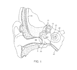
[0005] FIG. 1 illustrates the anatomy of an ear in coronal
section view;
[0006] FIGs. 2 and 3 illustrate implementations of a stabilizer
device positioned across the
tympanic membrane;
[0007] FIG. 4 illustrates the stabilizer device of FIG. 2
positioned on an insertion tool;
[0008] FIGs. 5 and 6 illustrate other implementations of a
stabilizer device positioned
external to the tympanic membrane;
[0009] FIGs. 7A-7B illustrate stabilizer legs used in conjunction
with the stabilizer device
of FIG. 5 in a collapsed and expanded configuration, respectively;
10010] FIGs. 8A-8B illustrate another implementation of a
stabilizer device positioned
external to the tympanic membrane;
10011] FIGs. 9-10 illustrate other implementations of a
stabilizer device positioned
external to the tympanic membrane.
DETAILED DESCRIPTION
[0012] Conductive Hearing Loss (CHL) involves the loss of normal mechanical
pathways
for sound to reach the hair cells in the cochlea, for example due to
malformation,
accumulation of fluid in the middle ear, presence of tumors, and/or damage to
ossicles.
SensoriNeural Hearing Loss (SNHL) is due to the absence of, or damage to, hair
cells in the
cochlea, or to the acoustic nerve. SNHL is typically associated with exposure
to loud noise,
head trauma, aging, infection, Meniere's Disease, tumors, ototoxicity, and
genetic diseases
like Usher's disease, and the like.
[0013] Treatment of SNHL depending on the cause can include drug treatments
for hair
cell and cochlear nerve afferents regeneration, reversal of cochlear oxidative
stress damage,
and apoptosis inhibition and reversal of inflammation. There are several drugs
in the final
stages of clinical development for the treatment of hearing loss including STS
(Fennec
Pharmaceuticals) to protect against cisplatin-induced hearing loss; AM-101
(Auris Medical)
for the treatment of tinnitus; AM-111 (Auris Medical) for otoprotection in
acute inner ear
hearing loss; OTO-104 (Otonomy) for the treatment of Meniere's Disease; SPI-
1005 (Sound
2
CA 03165914 2022- 7- 25
WO 2021/150858
PCT/ITS2021/014561
Pharmaceuticals) for the treatment of mild to moderate acute noise-induced
hearing loss and
for the treatment of Meniere's Disease.
[0014] The inner ear is difficult to treat effectively. For
example, the inner ear accounts
for only 0.004% of the average circulating blood volume and is encapsulated in
one of the
densest bones in the body. These, combined with the presence of the blood-
labyrinth barrier
(BLB), limit access of most therapeutic agents to the inner ear. Oral,
intravenous, and
intramuscular routes of administration are inefficient and require high doses
and the risk of
systemic side effects. Local drug delivery methods are also known. For
example, inner ear
therapeutics (e.g. drugs formulated as biocompatible gels) can be delivered
via intra-
tympanic injections into the middle ear across the tympanic membrane (TM).
Passive
diffusion of agents from the middle ear to the inner ear following intra-
tympanic injection has
variable efficacy due to anatomical variations, such as the presence of
pseudomembrane
covering the round window membrane, failure of the injected formulation to
contact the
round window membrane and limited permeability of the round window and oval
window
membranes. Further, rapid clearance of agents from the perilymph of the inner
ear results in
the need for repeated intra-tympanic injections, which are undesirable for
patients and are
associated with cumulative risk of infection, inflammation, and long-term
damage to the
tympanic membrane, in addition to the risk of lower compliance. Accurate
placement of
formulations in proximity to the round window membrane and assessment and
removal of
pseudo membrane structures would greatly improve effectiveness of therapy, but
cannot be
readily achieved with current intra-tympanic procedures, which are performed
"blindly"
without visualization of middle ear structures.
[0015] Direct delivery of therapeutics into the inner ear can
also be achieved by injecting
agents or drug releasing implants directly into the inner ear either through
the round window
membrane or by drilling a small cochleostomy. This procedure would be
analogous to the
placement of implants for cochlear stimulation. However, such procedures are
currently
performed in a relatively invasive manner, by creating a post auricular
incision and drilling
through the mastoid bone to the middle ear cavity. The degree of invasiveness
of the current
middle and inner ear procedures is too high to justify the precise delivery of
therapeutics into
the inner ear for the purpose of clinical trials and for their subsequent
adoption as valuable
treatments for inner ear disorders. A less invasive approach is needed.
3
CA 03165914 2022- 7- 25
WO 2021/150858
PCT/ITS2021/014561
[0016] The systems described here provide a more effective
administration of inner ear
therapeutics, whether via intra-tympanic administration or intracochl ear
administration, by
providing minimally invasively access to the middle ear through the ear canal
and tympanic
membrane. The systems described herein also improve accessibility for various
otological
surgical procedures, such as cholesteatoma removal, tympanic membrane repair
and ossicular
chain repair, and allow them to be performed in a less invasive manner.
10017] The materials, compounds, compositions, articles, and
methods described herein
may be understood more readily by reference to the following detailed
description of specific
aspects of the disclosed subject matter and the Examples included therein.
Before the present
materials, compounds, compositions, articles, devices, and methods are
disclosed and
described, it is to be understood that the aspects described below are not
limited to specific
methods or specific reagents, as such may vary. It is also to be understood
that the
terminology used herein is for the purpose of describing particular aspects
only and is not
intended to be limiting.
[0018] Unless defined otherwise, all technical and scientific
terms used herein have the
same meaning as is commonly understood by one of skill in the art to which the
invention(s)
belong. All patents, patent applications, published applications and
publications, websites and
other published materials referred to throughout the entire disclosure herein,
unless noted
otherwise, are incorporated by reference in their entirety. In the event that
there are pluralities
of definitions for terms herein, those in this section prevail. Where
reference is made to a
URL or other such identifier or address, it is understood that such
identifiers can change and
particular information on the intemet can come and go, but equivalent
information is known
and can be readily accessed, such as by searching the intemet and/or
appropriate databases.
Reference thereto evidences the availability and public dissemination of such
information.
[0019] As used herein, relative directional terms such as
anterior, posterior, proximal,
distal, lateral, medial, sagittal, coronal, transverse, etc. are used
throughout this disclosure.
Such terminology is for purposes of describing devices and features of the
devices and is not
intended to be limited. For example, as used herein "proximal" generally means
closest to a
user implanting a device and farthest from the target location of
implantation, while -distal"
means farthest from the user implanting a device in a patient and closest to
the target location
of implantation.
4
CA 03165914 2022- 7- 25
WO 2021/150858
PCT/ITS2021/014561
[0020] As used herein, a disease or disorder refers to a
pathological condition in an
organism resulting from, for example, infection or genetic defect, and
characterized by
identifiable symptoms.
[0021] As used herein, treatment means any manner in which the symptoms of a
condition, disorder or disease are ameliorated or otherwise beneficially
altered. Treatment
also encompasses any surgical or pharmaceutical use of the devices described
and provided
herein.
[0022] As used herein, amelioration or alleviation of the
symptoms of a particular
disorder, such as by administration of a particular pharmaceutical
composition, refers to any
lessening, whether permanent or temporary, lasting, or transient that can be
attributed to or
associated with administration of the composition.
[0023] As used herein, an effective amount of a compound for
treating a particular disease
is an amount that is sufficient to ameliorate, or in some manner reduce the
symptoms
associated with the disease. Such an amount can be administered as a single
dosage or can be
administered according to a regimen, whereby it is effective. The amount can
cure the disease
but, typically, is administered in order to ameliorate the symptoms of the
disease. Repeated
administration can be required to achieve the desired amelioration of
symptoms.
Pharmaceutically effective amount, therapeutically effective amount,
biologically effective
amount and therapeutic amount are used interchangeably herein to refer to an
amount of a
therapeutic that is sufficient to achieve a desired result, i.e. Therapeutic
effect, whether
quantitative or qualitative. In particular, a pharmaceutically effective
amount, in vivo, is that
amount that results in the reduction, delay, or elimination of undesirable
effects (such as
pathological, clinical, biochemical and the like) in the subject.
[0024] As used herein, sustained release encompasses release of
effective amounts of an
active ingredient of a therapeutic agent for an extended period of time. The
sustained release
may encompass first order release of the active ingredient, zero order release
of the active
ingredient, or other kinetics of release such as intermediate to zero order
and first order, or
combinations thereof The sustained release may encompass controlled release of
the
therapeutic agent via passive molecular diffusion driven by a concentration
gradient across a
porous structure.
CA 03165914 2022- 7- 25
WO 2021/150858
PCT/ITS2021/014561
[0025] As used herein, a subject includes any animal for whom
diagnosis, screening,
monitoring or treatment is contemplated. Animals include mammals such as
primates and
domesticated animals. An exemplary primate is human. A patient refers to a
subject such as a
mammal, primate, human, or livestock subject afflicted with a disease
condition or for which
a disease condition is to be determined or risk of a disease condition is to
be determined.
[0026] As used herein, a therapeutic agent referred to with a
trade name encompasses one
or more of the formulation of the therapeutic agent commercially available
under the
tradename, the active ingredient of the commercially available formulation,
the generic name
of the active ingredient, or the molecule comprising the active ingredient. As
used herein, a
therapeutic or therapeutic agents are agents that ameliorate the symptoms of a
disease or
disorder or ameliorate the disease or disorder. Therapeutic agent, therapeutic
compound,
therapeutic regimen, or chemotherapeutic include conventional drugs and drug
therapies,
including vaccines, which are known to those skilled in the art and described
elsewhere
herein. Therapeutic agents include, but are not limited to, moieties that are
capable of
controlled, sustained release into the body.
[0027] As used herein, a composition refers to any mixture. It
can be a solution, a
suspension, an emulsion, liquid, powder, a paste, aqueous, non-aqueous or any
combination
of such ingredients.
10028] As used herein, fluid refers to any composition that can
flow. Fluids thus
encompass compositions that are in the form of semi-solids, pastes, solutions,
aqueous
mixtures, gels, lotions, creams and other such compositions.
[0029] As used herein, a kit is a packaged combination,
optionally, including instructions
for use of the combination and/or other reactions and components for such use.
[0030] Referring now to the figures, FIG. 1 shows the anatomy of an ear
showing the
outer ear, the middle ear, and the inner ear as well as a portion of the skull
35 and the
Eustachian canal 45. The outer ear includes an auricle and an ear canal 40.
The tympanic
membrane 5 provides a barrier between the outer ear canal 40 and the middle
ear or tympanic
cavity 30. The inner ear can be divided into the bony labyrinth and the
membranous
labyrinth. The structural cavities within the bony labyrinth of the inner ear
include the
vestibule 10, the semicircular canals 15, and the cochlea 20. Hair cells of
the cochlea 20 are
critical in transducing acoustic signals into nerve impulses. The hair cells
are bathed in
6
CA 03165914 2022- 7- 25
WO 2021/150858
PCT/ITS2021/014561
secreted fluids such as perilymph supplied by cells that line the bony
labyrinth and
endolymph found within the membranous labyrinth, which help discern vibrations
to assist in
the process of hear as well as maintain a sense of balance and equilibrium.
The round
window 25 includes a round window membrane that in combination with the oval
window of
the cochlea 20 allow the fluid in the cochlea 20 to move.
[0031] Described herein are devices configured to directly access
the middle ear cavities
through the tympanic membrane in a sutureless, minimally-invasive manner. For
example,
the devices described herein provide direct access to the middle ear for the
direct delivery of
one or more therapeutic agent(s), implants, reservoirs, purpose-built
instruments such as
endoscopes, cutters, forceps, needles, aspiration devices, lasers, etc. to the
inner ear or middle
ear cavities. The direct access through the tympanic membrane is safer, less
invasive, and
requires no sealing or sutures of the tympanic membrane after removal of the
devices.
[0032] The devices described herein can be a purely mechanical
device or can be an at
least partially powered instrument. In some implementations, as will be
described in more
detail below, the device incorporates one or more features that can provide
stabilization,
guidance, and/or visualization to a user allowing for greater control during
the procedure and
understanding of the relative location of the injection such that informed
choices can be made
on the fly.
10033] Although the following describes tool and methodology in
terms of surgical
procedure through the tympanic membrane, it should be recognized that other
surgical
procedures can adapt the methodology to yield other types of sutureless
surgical procedures
in the ear. Any number of combinations of tools and/or agents can be delivered
using any of
the devices and systems described herein. Additionally, the surgical
procedures include
procedures performed on adults as well as pediatric applications.
[0034] After preparing the ear for the surgical procedure, the
surgical personnel generally
mount the stabilizer device on a tool such as the insertion tool shown in
schematic in FIG. 4.
The stabilizer device can be inserted through the ear canal 40 and implanted
in the tympanic
membrane 5. This insertion procedure is repeated as needed to insert the
number of stabilizer
devices to meet the needs of a given procedure. In some implementations, the
surgical
procedure uses two surgical instruments simultaneously. Two stabilizer devices
can be
inserted to accommodate the two surgical instruments.
7
CA 03165914 2022- 7- 25
WO 2021/150858
PCT/ITS2021/014561
10035] FIGs. 2 and 3 illustrate implementations of a stabilizer
device 100 positioned
within the tympanic membrane 5. The device 100 can include a proximal portion
102, a
distal portion 106, and a transmembrane region 105 positioned between the
proximal portion
102 and the distal portion 106. In some implementations, the transmembrane
region 105 and
distal portion 106 are configured to penetrate the tympanic membrane 5 whereas
the proximal
portion 102 is configured to stay external to the tympanic membrane 5.
10036] The distal portion 106 is sized and shaped to pass through
the tympanic membrane
in a minimally-invasive manner such that it is positioned within the middle
ear distal of the
tympanic membrane 5. The long axis of the distal portion 106 can be oriented
to be
approximately or substantially perpendicular to the external surface of the
tympanic
membrane 5 at the point of insertion. A distal end 108 of the distal portion
106 can be
oriented so that the long axis is at any angle with respect to the tympanic
membrane 5 and/or
the longitudinal axis A of the device 100. The distal portion 106 is sized to
have a width that
is sufficiently small such that the removal of the distal portion 106 from the
tympanic
membrane 5 leaves an incision or fenestration that does not require sutures to
heal. In some
implementations, the largest diameter of the distal portion 106 is no greater
than about 2 mm
such that it can be inserted through a fenestration that is no greater than 3
mm in length.
10037] The distal portion 106 can taper distally from a first
outer diameter to a second,
smaller outer diameter. In some implementations, the distal portion 106 tapers
to the distal
end 108. The distal end 108 can be sharpened to penetrate the tympanic
membrane 5. For
example, a force can be applied to the stabilizer device 100 during insertion
to cause the
distal end 108 to pierce the tympanic membrane 5 and pass through it without a
prior
fenestration being formed. The distal end 108 can form a non-traumatic tip
that minimizes
damage to the tissue being penetrated. The distal end 108 can incorporate any
of a variety of
non-coring bevel shapes of the needle art to facilitate insertion of the
device 100 through the
tympanic membrane 5. In other implementations, the distal portion 106 tapers
to a smaller
outer diameter distal end 108, but the distal end 108 is generally blunt. In
this
implementation, the stabilizer device 100 may be inserted through a pre-formed
fenestration
in the tympanic membrane S. In other implementations, the device 100 can be
preloaded
onto an introducer tool with a sharpened tube or post element, such as a
needle or knife, on
the distal end that extends beyond portion 108 when in the loaded
configuration. This
sharpened element can create the fenestration and can be withdrawn after
device 100 is in
8
CA 03165914 2022- 7- 25
WO 2021/150858
PCT/ITS2021/014561
place, leaving the working channel 110 open in the final implanted
configuration. Whether
the distal end 108 is sharpened like a needle or generally blunt, the taper of
the distal portion
106 allows for the stabilizer device 100 to pass smoothly through the tympanic
membrane 5
to avoid catching on the membrane during distal advancement.
[0038] The proximal portion 102 is configured and sized to
prevent over-insertion of the
stabilizer device 100 through the tympanic membrane 5. For example, the
proximal portion
102 can form a flange having an enlarged diameter compared to the distal
portion 106 and the
intervening transmembrane region 105. The angular arrangement of the proximal
portion
102 relative to the long axis A of the device 100 can aid in preventing
passage of the
proximal portion 102 through the tympanic membrane 5. For example, the
proximal portion
102 can extend approximately at a right angle relative to the long axis A of
the device 100.
The distal portion 106 of the stabilizer device 100 can be inserted through
the tympanic
membrane 5 until the tympanic membrane 5 is received within the transmembrane
region 105
and the larger diameter proximal portion 102 abuts against an external surface
of the
tympanic membrane 5.
[0039] The proximal portion 102 can define a proximal opening 112 into a
working
channel 110 that extends through the stabilizer device 100 to a distal opening
114 at or near
the distal end 108. The working channel 110 of the stabilizer device 100 may
be a fully
enclosed lumen extending from the proximal opening 112 in the proximal portion
102 to the
distal opening 114 at or near the distal end 108. In other implementations,
the working
channel 110 can be a curved guiding surface (e.g., c-shaped) that is not fully
enclosed, but is
configured to receive the curved exterior surfaces of the instruments to guide
the instrument
through the tympanic membrane 5. The shape of the working channel 110 is
configured to
geometrically complement the shape of the surgical instruments being inserted
through the
device 100. The shape of the working channel 110 is generally cylindrical or
arcuate. The
size of the working channel 110 is configured to complement the size of the
surgical
instrument being inserted. In some implementations, the working channel 110
may have a
cross-sectional diameter of about 25 gauge or 0.5 mm up to about 1.0 mm.
10040] It should be appreciated that the overall length of the
stabilizer device 100 can
vary. In some implementations, the stabilizer device 100 is approximately 1.5
mm to about 3
mm long from proximal opening 112 to distal opening 114 and is formed of a
relatively rigid
material. In other implementations, the stabilizer device 100 is approximately
1.5 mm to
9
CA 03165914 2022- 7- 25
WO 2021/150858
PCT/ITS2021/014561
about 5 mm long from proximal opening 112 to distal opening 114 and is formed
of a
relatively, flexible material that is similar to a flexible cannula. With each
implementation, a
smaller diameter transmembrane region extends about 0.1 mm to about 1.5 mm in
length that
is configured to traverse the tympanic membrane 5 and maintain positioning of
the stabilizer
device 100 within the membrane 5.
[0041] Distal portion 106 can dilate the incision in the tympanic
membrane 5 as the device
is inserted so that transmembrane region 105 is captured within the incision.
The length of the
distal portion 106 can be sufficient to allow for extension of the stabilizer
device 100 into the
middle ear such that the distal opening 114 is positioned a distance away from
the internal
surface of the tympanic membrane 5. However, the dimensions of distal portion
106 should
be minimized so as avoid contact between the device and middle ear structures.
10042] The proximal portion 102 can provide sufficient surface
area and thickness to
prevent the stabilizer device 100 from being pushed through the tympanic
membrane 5 and to
provide a sufficiently large surface area for surgical personnel to identify
and locate the
device 100 positioned within the tympanic membrane 5. The dimensions of the
proximal
portion 102 (e.g., outer diameter or thickness) can vary. In some
implementations, the outer
diameter of the proximal portion 102 can be between about 2 mm to about 5 mm.
The
proximal portion 102 can serve as a handle for the device 100 or the proximal
portion 102
can additionally incorporate a grasping feature that is configured to be
manipulated by a user
for insertion and removal of the device from the ear. The grasping feature may
be grasped
with a tool such as a pair or forceps or by an insertion tool specifically
configured to mate
with the grasping feature.
[0043] The transmembrane region 105 can have an outer dimension relative to
the
proximal portion 102 that is sized and shaped to receive the tympanic membrane
5 when the
distal portion 106 is inserted through the tympanic membrane 5. The
transmembrane region
105 can have an outer diameter that is between 0.25 mm and 1.0 mm. The
transmembrane
region 105 can have a length along the long axis A of the stabilizer device
100 that is
between about 0.10 and 0.5 mm. The outer diameter and length of the
transmembrane region
105 is sufficient to receive the thickness of the tympanic membrane 5 while
preventing
buckling, tearing, or other forces from being imparted inadvertently on the
membrane 5 upon
insertion of the device 100. The outer diameter of the transmembrane region
105 can vary
along its length. FIG. 2 illustrates an implementation of the stabilizer
device 100 having a
CA 03165914 2022- 7- 25
WO 2021/150858
PCT/ITS2021/014561
transmembrane region 105 that is substantially cylindrical such that the outer
diameter
remains relatively constant along its length. FIG. 3 illustrates an
implementation of the
stabilizer device 100 having a transmembrane region 105 that has a curved
geometry along
the longitudinal axis A of the device 100. In this implementation, the outer
diameter enlarges
towards the distal end before the distal portion 106 tapers towards the distal
end 108 of the
device 100. The outer diameter of at least a portion of the transmembrane
region 105 can be
larger than an outer diameter of the distal portion 106 at its proximal-most
end (see FIG. 3).
[0044] The transmembrane region 105 can have a shape configured to aid in the
retention
of the device 100 within the tympanic membrane fenestration. The transmembrane
region
105 can form an annulus or toroid. The cross-sectional profile of the
transmembrane region
105 can be circular. The cross-sectional profile of the transmembrane region
105 can be
elongated and sized to correspond to the shape of the fenestration through the
tympanic
membrane upon insertion of the device 100. For example, the fenestration can
be a small
incision that is slit shaped. The elongate cross-sectional profile of the
transmembrane region
105 can improve the fit of the device 100 within this slit-shaped fenestration
through the
tympanic membrane 5. The elongated cross section may include a first dimension
that is
longer than a second dimension forming a dilated slit, dilated slot, lentoid,
oval, ovoid, bi-
convex, or elliptical shape.
10045] The stabilizer device 100 can be formed of a material
having a rigidity and strength
to be inserted and removed from the tympanic membrane 5 while also
withstanding stresses
that may arise during manipulation of surgical instruments inserted
therethrough. In some
implementations, at least a portion of the stabilizer device 100 is formed of
surgical metals
such as stainless steel, titanium, platinum, Nitinol, and/or plastics such as
polyimide, PEEK,
fluoropolymers, silicone, and the like. In some implementations, the inserted
portion of the
device 100 can be formed of polyimide and have a maximum outer diameter of no
more than
about 20 gauge (8 mm). One or more portions of the stabilizer device 100 can
be coated with
or formed of a conformable material. For example, the retention feature 102
can be coated
with or formed by over-molding with a material such as silicone or
polyurethane.
10046] The stabilizer device 100 can be an integral, one-piece
structure such that the
proximal portion 102, the transmembrane region 105 and the distal portion 106
are all part of
the same structure. It should also be appreciated that one or more portions of
the stabilizer
device 100 can be separate components of the device 100 that are arranged to
work with one
11
CA 03165914 2022- 7- 25
WO 2021/150858
PCT/ITS2021/014561
another, but not necessarily rigidly affixed or integrated with one another.
For example, the
proximal portion 102 and the distal portion 106 can be removably coupled to
one another.
[0047] The stabilizer device 100 is configured and sized so that
its removal from the
tympanic membrane 5 does not necessitate the use of sutures to seal the
incision or
fenestration formed in the tympanic membrane during insertion of the
stabilizer device 100.
Generally, a self-sealing fenestration through the tympanic membrane 5 is no
greater than
about 2 mm in length, preferably between about 0.5 mm and 1.5 mm in length.
Although the
tools and methods described herein provide the advantage of sutureless access
to the middle
and/or inner ear, this does not preclude a surgeon from applying one or more
closure
techniques upon removal of the stabilizer device 100. For example, if a
surgeon so desires,
one or more techniques for closure of the fenestration in the tympanic
membrane 5 can be
performed.
[0048] In use, a user may form one, two, or more fenestrations in the tympanic
membrane
5. The fenestrations may be between about 0.25 mm and 1.25 mm in diameter. The
fenestrations can be performed using an appropriate cutting tool such as a
blade, a needle, a
trephine, a laser, or other tool. A stabilizer device 100 may be implanted
into each tympanic
membrane fenestration. In some implementations, the fenestration is a slice
through the
tympanic membrane such as can be made with a needle. In other implementations,
the
fenestration is a hole in the tympanic membrane (e.g., made by a laser or
trephine). The size
of the stabilizer device positioned within the hole can be sized to fit that
hole such that forces
imparted on the instrument are distributed around a perimeter of the hole to
prevent further
tearing.
[0049] In some implementations, the cutting tool to form the
fenestrations in the tympanic
membrane 5 is the distal end 108 of the stabilizer device 100. In other
implementations, the
cutting tool is part of the tool used to insert the stabilizer device 100. For
example, the
stabilizer device 100 can be mounted on an insertion tool 200 (see FIG. 4).
The insertion tool
200 can include a proximal handle 205 configured to be grasped by a user, one
or more
actuators 210 movable relative to the handle 205, a distal delivery shaft 210
projecting from
the distal end of the handle 205, and a stylet 212. The stylet 212 can be
inserted through the
working channel 110 of the stabilizer device 100 such that the distal tip 214
of the stylet 212
extends beyond the distal opening 114 from the stabilizer device 100. The
distal tip 214 of
the stylet 212 can be beveled like a needle so it can be used to form the
fenestration through
12
CA 03165914 2022- 7- 25
WO 2021/150858
PCT/ITS2021/014561
the tympanic membrane 5. The actuator(s) 210 can be actuated to release the
device 100
leaving it in position within the ear.
[0050] The handle 205, depending on whether the insertion tool
200 is intended to be
durable or disposable, may be made of a high performance-engineering
thermoplastic (e.g.
PTFE) or of a metal such as stainless steel or aluminum. The handle 205 can be
unitary,
single-piece, molded construction or can be formed by two or more panels
configured to
couple together. The handle 205 can include threaded or friction fit panels
configured to be
opened to access an interior of the handle 205. The handle 205 may be similar
in form factor
to an otoscope, syringe, speculum, or other hand-held type instrument for use
with the ear.
The handle 205 can include an angular bend to ensure an unobstructed view
through the
operative microscope or one or more gripping features such as indentations or
ergonomic
features for gripping the tool 200.
[0051] As mentioned, the handle 205 can incorporate one or more
actuators 210 such as
one or more plungers, triggers, buttons, switches, keys, sliders, or
combination thereof
mounted on a portion of the handle 205 that are configured to be activated
such as retracted,
extended, pressed, squeezed, slid, or otherwise actuated to perform a certain
function of the
tool 200. The one or more actuators 210 can be incorporated into a portion of
the handle 205
such as a hand-held portion in such a way that is ergonomically comfortable to
a user.
10052] The stabilizer device 100 may be provided as part of a kit
that includes one or more
stabilizer devices 100, an insertion tool 200, with or without the surgical
instruments
configured to be inserted through the stabilizer device 100.
[0053] Once the stabilizer device(s) 100 are positioned within
the tympanic membrane 5,
one or more instruments may be inserted through the working channel 110 of the
device 100.
The working channel 110 can provide a passage for introduction of any of a
variety of
instruments or fluids through the device 100. The instruments can be
repeatedly inserted and
removed through the working channel 110 without causing damage or strain on
the tympanic
membrane 5. Generally, the instruments inserted through the working channel
110 can have
an outer diameter between 0.25 mm and 0.80 mm.
[0054] Any of a variety of instruments may be inserted through the working
channel 110
including cutting instruments, infusing instruments, aspirating instruments,
light transmitting
instruments, energy applying instruments, tissue manipulating instruments,
implant
13
CA 03165914 2022- 7- 25
WO 2021/150858
PCT/ITS2021/014561
delivering instruments can be inserted through the working channel 110 of the
stabilizer
device 100. The instruments inserted through the working channel 110 of the
one or more
stabilizer device 100 can include small gauge endoscopes with, or without, a
light source, and
with, or without, a working channel. The instruments inserted through the
working channel
110 of the one or more stabilizer device 100 can include a -chandelier" fiber-
optic light
source tailored for middle ear illumination. The small gauge chandelier fiber
optic light
source can provide hands-free endo-illumination directly into the middle ear
and can reduce
reflection off the tympanic membrane when viewed with transcanal illumination
thereby
improving visualization of middle ear structures. The instruments inserted
through the
working channel 110 of the one or more stabilizer device 100 can include micro-
cutters /
vertical scissors for pseudo membrane dissection. In some implementations, the
cutting angle
of the vertical scissors (i.e., angle of the blade relative to the shaft) can
be between 45-120
degrees. The instruments inserted through the working channel 110 of the one
or more
stabilizer device 100 can include curved aspirating pick/forceps for pseudo
membrane
removal. The instruments inserted through the working channel 110 of the one
or more
stabilizer device 100 can include can be integrated with fiber optic
components. In some
implementations, a diffuse light source may be placed into the middle ear
through the
working channel 110 allowing for better trans-tympanic membrane visualization
directly.
This can provide endo-illumination of the features of interest and avoid
problems associated
with external illumination such as light reflection. Any of a variety of
surgical interventions
may be performed through the stabilizer devices 100 once implanted.
[0055] Small gauge endoscopes for visualization of the middle ear
can be inserted through
a less invasive tympanic membrane perforation. Endoscopes typically used in
otology are
about 3 mm in diameter. Smaller high-resolution wide-field endoscopes (e.g. 23
G) can be
designed for the ear to enable visualization through small perforations in the
tympanic
membrane without creation of a surgical tympanomeatal flap.
[0056] In some implementations, the instruments inserted through
the working channel
110 are configured to change shape and/or direction once exiting the distal
opening 114 of
the working channel 110. This allows for positioning the instruments, for
example, at the
round window membrane niche, for perforating, depositing material, and/or
removing a false
round window membrane niche. As an example, the instrument can be an
extendable,
articulable and/or curved microcannula for precise injections and/or
placements of a drug
14
CA 03165914 2022- 7- 25
WO 2021/150858
PCT/ITS2021/014561
formulation or implantable devices on, at, or through the RWM. Various other
instruments
are considered herein including a vented or small gauge needle that is curved,
extendable,
and/or articulable, ultrasharp knife for RWM perforation for controlled access
to the inner ear
cavity, diamond-dusted forceps and spatulas for improved gripping and
scraping, as well as
endolasers for RWM permeability enhancement.
[0057] The stabilizer device 100 provides for investigating
middle ear disorders and for
delivering therapeutics to treat inner ear disorders. For example, the
stabilizer device 100 can
be used to precisely place drug product at or near the oval window or RWM,
remove any
pseudo membrane or other mucosal obstruction that might inhibit absorption of
drug product
to the inner ear. The stabilizer device 100 can also provide for better
visualization or and
precise placement of implants or devices at or near the RWM, oval window, or
other access
points for the treatment of inner ear disorders.
[0058] After completion of the surgical procedure or
administration of the therapy, the
stabilizer device 100 is removed and the tympanic membrane 5 left to heal on
its own without
the need for additional intervention.
[0059] The tympanic membrane 5 is a delicate tissue that is prone to damage.
However,
direct contact with the membrane 5 can provide guidance for attaining proper
instrument
depth (e.g., during injections with a needle). FIGs. 5-6 illustrate an
interrelated
implementation of a stabilizer device 1100 that does not penetrate and is
configured to remain
fully external to the tympanic membrane 5 within the ear canal. The stabilizer
device 1100
can include a proximal anchor 1105 that is configured to adjustably anchor
against the ear
canal 40 and that is coupled to a distal cannula 1106 configured to be
positioned adjacent the
external surface of the tympanic membrane 5. A working channel 1110 can extend
through
the device 1100 from a proximal opening 1112 to a distal opening 1114. The
distal opening
1114 can be positioned at a distal end 1108 of the distal cannula 1106 for
insertion of
minimally-invasive instruments through the stabilizer device 1100 and through
the tympanic
membrane 5.
[0060] The proximal anchor 1105 can enlarge from an insertion configuration
having a
small outer diameter to a deployed configuration having a larger outer
diameter configured to
hold the device 1100 in place within the ear canal 40. The proximal anchor
1105 can safely
engage the surrounding canal 40 with sufficient force and/or friction to
inhibit movement of
CA 03165914 2022- 7- 25
WO 2021/150858
PCT/ITS2021/014561
the stabilizer device 1100 or instruments inserted through the stabilizer
device 1100 during
treatment.
[0061] The configuration of the proximal anchor 1105 can vary including one or
more
rings, support legs, foam, balloon, expandable mesh, or other anchor. The
proximal anchor
1105 can be conformal or compressible such that it deforms and takes on the
shape of the ear
canal 40 upon insertion. In some implementations, the proximal anchor 1105 can
include an
inner layer covered by an outer compressible layer. The outer compressible
layer of the
proximal anchor 1105 may include a compressible foam such as a urethane foam.
Alternatively, the proximal anchor 1105 can be formed of a material such as
gum rubber
compounds, urethanes, fluorocarbon elastomer, butyl rubber, EPDM (Ethylene-
Propylene
Rubber), latex rubber, neoprene (polychloroprene), nitrile rubber
(acrylonitrile),
polybutadiene, silicone rubber, SBR (Stryrene-Butadiene Rubber), I-INBR
(Hydrogenated
Nitrile Rubber), fluoroelastomer, fluorosilicone.
[0062] The proximal anchor 1105 may expand resiliently within the
canal 40 or include
soft solid elastomeric or plastically deformable polymers. The proximal anchor
1105 may
also include an actively expanded feature such as a balloon, support rings,
etc. FIG. 5
illustrates an implementation of the stabilizer device 1100 having an
expandable balloon as
the proximal anchor 1105. FIG. 6 illustrates an implementation of the
stabilizer device 1100
having a plurality of support rings or flexible flanges configured to conform
to the ear canal
40 upon insertion towards the tympanic membrane 5.
10063] The proximal anchor 1105 can provide alignment within the ear canal 40
and direct
the distal cannula 1106 toward the desired location of the tympanic membrane
5. The
proximal anchor 1105 can be generally cylindrical having an outer diameter
configured for
smooth and comfortable insertion and engagement with the ear canal 40. The
proximal
anchor 1105 can allow for a slight seal to form between the ear canal wall and
its outer
surface. The length of the proximal anchor 1105 can vary. At least a portion
of the proximal
anchor 1105 can taper towards the distal cannula 1106, which can have a
smaller outer
diameter than a proximal end region of the proximal anchor 1105.
[0064] The working channel 1110 can have a uniform inner diameter as shown in
FIGs. 5
and 6. The working channel 1110 also can have an inner diameter that varies
along its
length. For example, the working channel 1110 can be tapered with the smallest
inner
16
CA 03165914 2022- 7- 25
WO 2021/150858
PCT/ITS2021/014561
diameter near or at the distal opening 1114 and adjacent to the tympanic
membrane 5. Such a
configuration positions a fulcrum point of instruments extending through the
working
channel 1110 close in proximity to the tympanic membrane 5 and mitigates
damage to the
tympanic membrane 5 during manipulation and movements of the instruments.
[0065] In some implementations, the stabilizer device can
incorporate a structure similar
to tympanostomy tubes or a "grommet". As discussed above, the fenestration
through the
tympanic membrane that the stabilizer is placed into can be a hole made by a
laser or trephine
or a slice made by a surgical blade or needle. The size of the stabilizer
device positioned
within the hole can be sized to fit that hole such that forces imparted on the
instrument are
distributed around a perimeter of the hole to prevent further tearing. The
grommet-like
stabilizer device fitted into the hole made in the tympanic membrane can be
left behind and
allow for passage of the instruments in and out of the middle ear during a
surgical procedure
in a manner that distributes the instrument forces on the tympanic membrane
thereby
preventing tearing. In combination with the grommet-like stabilizer device or
as a separate,
stand-alone approach, a scaffold or fixation device (such as the proximal
anchors described
elsewhere herein) can be positioned within the ear canal allowing instrument
forces to be
directed towards the walls of the canal rather than solely by the tympanic
membrane.
[0066] The configuration of this ear canal scaffold can vary as
described herein. FIGs.
8B show an implementation of a stabilizer device 1100 that includes a proximal
anchor 1105
positioned within the ear canal 40 analogous to an otic speculum. The proximal
anchor 1105
can further reduce forces that would otherwise be exerted on the tympanic
membrane 5
during instrument manipulations, rotations, etc. The proximal anchor 1105 can
be in the
shape of a conical, adjustable speculum or cone fitted to the ear canal 40.
The proximal
anchor 1105 can be threaded or otherwise telescope to allow for adjustment in
close
proximity to the tympanic membrane 5. Instruments can be passed the interior
of the
proximal anchor 1105 and through one or more small rings 1116 (e.g., 0.5 mm to
1.0 mm in
diameter) located on a distal face of the proximal anchor 1105 adjacent to the
tympanic
membrane 5 providing the fulcrum around which the instruments would rotate.
The cone
shape of the proximal anchor 1105 can define a larger inner viewing channel
and the rings
1116 located at a distal end of the cone can provide smaller working channels
through which
one or more instruments may be inserted. The rings 1116 can provide
stabilization and
guidance for instrument manipulations as described elsewhere herein.
17
CA 03165914 2022- 7- 25
WO 2021/150858
PCT/ITS2021/014561
[0067] In some implementations, the proximal anchor 1105 can be an expandable
mesh,
braid, stent, basket, cage or other structural element configured to expand
from a smaller
dimension suitable for insertion to a larger dimension configured to fit
within and anchor
against the ear canal (see FIGs. 9-10). In some implementations, the proximal
anchor 1105
can have a conical shape such that a central opening through the anchor 1105
provides access
to the tympanic membrane as described above and as shown in FIG. 9. In other
implementations, the proximal anchor 1105 can be closed at a distal end region
near the
tympanic membrane 5 (see FIG. 10). Openings in the mesh adjacent to the
tympanic
membrane 5 can be sized to allow passage of instruments. Fenestrations in the
tympanic
membrane 5 can be created after placement of the proximal anchor 1105 in order
to ensure
alignment of mesh openings for instrument passage into the middle ear 30. The
proximal
anchor 1105 can have any of a variety of shape. The proximal anchor 1105 can
be basket-
shaped or cup-shaped such that it is open on the proximal end to allow maximum
instrument
rotation around the distal mesh openings. Alternatively, the mesh openings can
be of varying
size, tapering from proximal to distal ends of the device. Following the end
of the procedure,
the proximal anchor 1105 can be collapsed and removed.
[0068] In some implementations, the stabilizer device 1100 can be
similar in shape and
form factor to an ear speculum. For example, the stabilizer device 1100 can
include a sloped
frustoconical shape and a smooth surface that permits insertion into the ear
canal 40 to a
limited depth without injuring the ear.
[0069] The working channel 1110 can extend through both the proximal anchor
1105 and
the distal cannula 1106. The working channel 1110 can be sized to receive any
of a variety
of instruments as described above. The working channel 1110 can be coaxial
with the
longitudinal axis A of the device 1100 or can be offset from the axis A.
[0070] FIGs. 7A-7B show an implementation of a stabilizer device
1100 comprising a
plurality of support legs 1200. The support legs 1200 can be expanded from an
insertion
configuration in which the support legs 1200 extend substantially parallel to
the longitudinal
axis A of the device 1100 to an enlarged configuration in which the support
legs extend
outward at an angle relative to the longitudinal axis A. The support legs 1200
can be coupled
to a central housing 1205. In an implementation, the stabilizer device 1100
includes three
collapsible legs 1200 coupled to a region of the central housing 1205 such
that upon
extension they form a tri-pod of stabilization relative to the distal cannula
1106. The legs
18
CA 03165914 2022- 7- 25
WO 2021/150858
PCT/ITS2021/014561
1200 can be arranged symmetrically around the longitudinal axis A of the
device 1100. The
legs 1200 can each extend outward by an angle relative to the axis A. The
angle and also the
length of the legs 1200 in the extended configuration allow for placement of
the legs 1200
against a patient's ear canal 40. For example, a first leg 1200 can be
positioned anteriorly on
a patient's jaw, a second leg 1200 can be positioned caudally on a patient's
skull near the
neck, and a third leg 1200 can be positioned more cephalad on a patient's
skull near the
crown. Each leg 1200 can incorporate a foot member movable coupled to a distal
end of the
leg 1200 and configured to fold outward when the legs 1200 are in an extended
configuration
and fold inward when the legs 1200 are in a collapsed configuration. The legs
1200 can snap
into the expanded configuration such that they avoid inadvertent collapse. The
degree of
extension of each leg 1200 can be selectable between a plurality of pre-set
angles relative to
the longitudinal axis A. Each foot member can swivel around its attachment
with the leg
1200 between the inward and outward folded configurations to provide a
tailored fit with the
patient to provide better stabilization. In some implementations, the foot
member is coupled
to its leg 1200 by a barrel hinge type coupling having at least 2 degrees of
freedom. In other
implementations, the foot member is coupled to its leg 1200 by a ball and
socket type
coupling providing any degree of freedom. Any of the stabilizer devices
described herein can
be coupled to a plurality of support legs 1200.
[0071] The devices described herein can incorporate one or more
features that aid in the
visualization, aiming, and targeting of one or more instruments to prevent
inadvertent
penetrations and damage to delicate structures in the ear during a procedure.
The devices
described herein can be coupled to a viewing lens such as an otoscope lens or
surgical
microscope for the user to view the tympanic membrane 5 while the device is
advanced
toward the membrane. Endoscopes, video visualization devices, optical
coherence
tomography, ultrasound, and other viewing instruments or techniques, as well
as one or more
illumination elements, such as a LEDs, lenses, light pipes, filters, etc. that
improve the
visibility within the middle ear during use can be incorporated. Techniques to
enhance
viewing through the tympanic membrane directly using the operating microscope
or
otoscope, such as by applying glycerin or saline to the tympanic membrane to
increase
tympanic membrane transparency and reduce refractive index across the membrane
in
conjunction with middle ear illumination and/or wavelength filters can also be
used to
eliminate the need for an additional port for passage of an endoscope.
Increasing membrane
transparency and reducing variations in refractive index across the membrane,
particularly
19
CA 03165914 2022- 7- 25
WO 2021/150858
PCT/ITS2021/014561
when coupled with a middle ear light source, can allow visualization of the
middle ear
directly through the membrane via the operating microscope.
[0072] Direct trans-tympanic visualization can also be provided
by infrared (IR) imaging
or operative ocular coherence tomography (OCT). For example, a camera or probe
directed
at the tympanic membrane through the ear canal can provide visualization of
the middle ear
structures directly through an intact tympanic membrane.
[0073] THERAPEUTICS AND DISEASES
[0074] The treatment devices described herein can be used to
treat and/or prevent a variety
of other conditions, including but not limited to hearing loss, including
hidden hearing loss,
noise-induced hearing loss, age-related hearing loss, drug-induced hearing
loss, such as
chemotherapy-induced hearing loss or aminoglycoside-induced hearing loss,
sudden
sensorineural hearing loss (SNHL), autoimmune inner ear disease, and the like.
Any of a
variety of ear disorders can be treated using the devices described herein.
The treatment
devices described herein can be used to treat other ear disorders such as
tinnitus. The
treatment devices described herein can be used to treat balance disorders
including vertigo,
Meniere's disease, vestibular neuronitis, vestibular schwannoma,
labyrinthitis, and the like.
The treatment devices described herein can be used to treat other ear
disorders such as,
otosclerosis, ossicular chain dislocation, cholesteatoma, middle ear
infections, tympanic
membrane perforations, and the like.
10075] Examples of therapeutic agents that may be delivered from or with the
help of the
treatment devices described herein and/or are described in the applications
incorporated by
reference herein are provided below.
10076] Therapeutics that can be delivered from or with the help
of the treatment devices
described herein include but are not limited to antioxidants, anti-
inflammatories, steroids,
antimicrobials, NMDA receptor antagonists, nootropics, anti-apoptotic agents,
neurotrophins,
neuroprotective agents, neural protective proteins such as CNTF, BDNF, PEDF,
NGF, and
the like, cannabinoids, monoclonal antibodies, other proteins, gene therapy,
iRNA, tyrosine
kinase inhibitors (TKIs), dual leucine zipper kinase (DLK) inhibitors, and
protein therapies
like anti-VEGF.
CA 03165914 2022- 7- 25
WO 2021/150858
PCT/ITS2021/014561
10077] As an example, the therapeutic agent can include, but is
not limited to
antimicrobials such as antibiotics such as tetracycline, chlortetracycline,
bacitracin,
neomycin, polymyxin, gramicidin, cephalexin, oxytetracycline, chloramphenicol
kanamycin,
rifampicin, ciprofloxacin, tobramycin, gentamycin, erythromycin and
penicillin; antifungals
such as amphotericin B and miconazole; anti-bacterials such as sulfonamides,
sulfadiazine,
sulfacetamide, sulfamethizole and sulfisoxazole, nitrofurazone and sodium
propionate;
antivirals such as idoxuridine, trifluorotymidine, acyclovir, ganciclovir and
interferon;
antiallergenics such as sodium cromoglycate, antazoline, methapyriline,
chlorpheniramine,
pyrilamine, cetirizine and prophenpyridamine; anti-inflammatories such as
hydrocortisone,
hydrocortisone acetate, dexamethasone, dexamethasone 21-phosphate,
fluocinolone,
medrysone, prednisolone, prednisolone 21-phosphate, prednisolone acetate,
fluoromethalone,
betamethasone, and triamcinolone; non-steroidal anti-inflammatories such as
salicylate,
indomethacin, ibuprofen, diclofenac, flurbiprofen and piroxicam; decongestants
such as
phenylephrine, naphazoline and tetrahydrozoline; miotics and
anticholinesterases such as
pilocarpine, salicylate, acetylcholine chloride, physostigmine, eserine,
carbachol, diisopropyl
fluorophosphate, phospholine iodide and demecarium bromide; mydriatics such as
atropine
sulfate, cyclopentolate, homatropine, scopolamine, tropicamide, eucatropine
and
hydroxyamphetamine; sypathomimetics such as epinephrine; antineoplastics such
as
carmustine, cisplatin and fluorouracil; immunological drugs such as vaccines
and immune
stimulants; hormonal agents such as estrogens, estradiol, progestational,
progesterone,
insulin, calcitonin, parathyroid hormone and peptide and vasopressin
hypothalamus releasing
factor; beta adrenergic blockers such as timolol maleate, levobunolol HC1 and
betaxolol HCl;
growth factors such as epidermal growth factor, fibroblast growth factor,
platelet derived
growth factor, transforming growth factor beta, somatotropin and fibronectin;
carbonic
anhydrase inhibitors such as dichlorophenamide, acetazolamide and
methazolamide and other
drugs such as prostaglandins, antiprostaglandins and prostaglandin precursors;
antioxidants,
NMDA receptor antagonists, nootropics, anti-apoptotic agents, neurotrophins,
neuroprotective agents, tyrosine kinase inhibitors (TKIs), dual leucine zipper
kinase (DLK)
inhibitors, carmabinoids, monoclonal antibodies, antibody fragments, other
proteins, and gene
therapy. Other therapeutic agents known to those skilled in the art which are
capable of
controlled, sustained release into the ear in the manner described herein are
also suitable for
use in accordance with embodiments of the devices described herein.
21
CA 03165914 2022- 7- 25
WO 2021/150858
PCT/ITS2021/014561
[0078] The therapeutic agent can include, but is not limited to
sodium thiosulfate to
protect against cisplatin-induced hearing loss; NMDA receptor antagonists for
the treatment
of tinnitus (AM-101; Auris Medical); AM-111 containing the synthetic peptide D-
JNKI-1
(D-stereoisomer of c-Jun N-terminal Kinase Inhibitor 1; Auris Medical) for
otoprotection in
acute inner ear hearing loss; dexamethasone for the treatment of Meniere's
Disease; D-
methionine (Southern Illinois University) to protect against Noise-induced
hearing loss;
LY411575 (a selective gamma secretase inhibitor that blocks Notch activation);
and NT-3
neurotrophic factor.
[0079] The therapeutic agent can include, but is not limited to
local anesthetics for
delivery into the ear canal including benzocaine, antipyrine, butamben,
dibucaine, lidocaine,
pnlocaine, oxybuprocaine, pramoxine, proparacaine, proxymetacaine, and
tetracaine.
10080] Various pharmaceutically acceptable carriers for the
therapeutic agents described
herein can include such as, for example, solids such as starch, gelatin,
sugars, natural gums
such as acacia, sodium alginate and carboxymethyl cellulose; polymers such as
silicone
rubber; liquids such as sterile water, saline, dextrose, dextrose in water or
saline;
condensation products of castor oil and ethylene oxide, liquid glyceryl
triester of a lower
molecular weight fatty acid; lower alkanols; oils such as corn oil, peanut
oil; sesame oil,
castor oil, and the like, with emulsifiers such as mono- or di-glyceride of a
fatty acid, or a
phosphatide such as lecithin, polysorbate 80, and the like; glycols and
polyalkylene glycols
including P407 and other combinations of polyethylene glycol and polypropylene
glycol;
aqueous media in the presence of a suspending agent, for example, sodium
carboxymethylcellulose, hyaluronic acid, sodium hyaluronate, sodium alginate,
poly(vinyl
pyrrolidone) and similar compounds, either alone, or with suitable dispensing
agents such as
lecithin, cyclodextrins, polyoxyethylene stearate and the like. The carrier
may also contain
adjuvants such as preserving, stabilizing, wetting, emulsifying agents or
other related
materials.
[0081] While this specification contains many specifics, these
should not be construed as
limitations on the scope of what is claimed or of what may be claimed, but
rather as
descriptions of features specific to particular embodiments. Certain features
that are
described in this specification in the context of separate embodiments can
also be
implemented in combination in a single embodiment. Conversely, various
features that are
described in the context of a single embodiment can also be implemented in
multiple
22
CA 03165914 2022- 7- 25
WO 2021/150858
PCT/ITS2021/014561
embodiments separately or in any suitable sub-combination. Moreover, although
features
may be described above as acting in certain combinations and even initially
claimed as such,
one or more features from a claimed combination can in some cases be excised
from the
combination, and the claimed combination may be directed to a sub-combination
or a
variation of a sub-combination. Similarly, while operations are depicted in
the drawings in a
particular order, this should not be understood as requiring that such
operations be performed
in the particular order shown or in sequential order, or that all illustrated
operations be
performed, to achieve desirable results. Only a few examples and
implementations are
disclosed. Variations, modifications and enhancements to the described
examples and
implementations and other implementations may be made based on what is
disclosed. The
claimed subject matter has been described in conjunction with the detailed
description
thereof, the foregoing description is intended to illustrate and not limit the
scope of the
claimed subject matter of the appended claims.
[0082] In the descriptions above and in the claims, phrases such
as "at least one of' or
"one or more of' may occur followed by a conjunctive list of elements or
features. The term
"and/or- may also occur in a list of two or more elements or features. Unless
otherwise
implicitly or explicitly contradicted by the context in which it is used, such
a phrase is
intended to mean any of the listed elements or features individually or any of
the recited
elements or features in combination with any of the other recited elements or
features. For
example, the phrases -at least one of A and B;" -one or more of A and B;" and -
A and/or B"
are each intended to mean "A alone, B alone, or A and B together." A similar
interpretation
is also intended for lists including three or more items. For example, the
phrases "at least one
of A, B, and C;" -one or more of A, B, and C;- and "A, B, and/or C" are each
intended to
mean -A alone, B alone, C alone, A and B together, A and C together, B and C
together, or A
and B and C together."
[0083] Use of the term -based on," above and in the claims is
intended to mean, -based at
least in part on," such that an unrecited feature or element is also
permissible.
23
CA 03165914 2022- 7- 25