Note: Descriptions are shown in the official language in which they were submitted.
CA 03165950 2022-06-24
METHOD FOR COLLECTING EXTRACELLULAR VESICLES
DERIVED FROM NERVOUS SYSTEM CELLS
Technical Field
[0001]
The present description discloses a method for collecting extracellular
vesicles derived from
nervous system cells, a method for detecting a neuropsychiatric disorder, a
method for collecting
components derived from nervous system cells, a reagent for collecting
extracellular vesicles, and a
kit for detecting extracellular vesicles containing the reagent.
Background Art
[0002]
Patent Literature 1 discloses biomarkers and diagnostic and prognostic methods
for Alzheimer's
disease and other neurodegenerative disorders. Patent Literature 2 discloses
that biomarkers in
vesicles (for example, exosomes) isolated from a biological sample are
detected to be used for
diagnosis and prognostication of Alzheimer's disease and other
neurodegenerative disorders. Patent
Literature 3 discloses methods for quantifying subpopulations of exosomes and
diagnostic and
prognostic methods for neurodegenerative disorders (for example, Alzheimer's
disease). Patent
Literature 4 discloses how to use exosomes and exosomal biomarkers in
diagnostic and prognostic
methods for neurological disorders, immunological disorders, placental
diseases, cancer,
hematological disorders, kidney disease, gastrointestinal diseases, liver
diseases, and musculoskeletal
diseases.
Citation List
Patent Literature
[0003]
Patent Literature 1: W02015/061634
Patent Literature 2: W02017/193115
Patent Literature 3: W02018/094120
Patent Literature 4: W02019/144056
1
Date Recue/Date Received 2022-06-24
CA 03165950 2022-06-24
Summary of Invention
Technical Problem
[0004]
Patent Literatures 2 to 4 disclose use of exosomes for diagnosis and
prognostication of
neurodegenerative disorders. However, for example, NCAM or CD171 used for
collecting exosomes
described in Patent Literature 2 is not a protein specific to nervous system
cells. In order to efficiently
collect extracellular vesicles derived from nervous system cells, it is
necessary to collect extracellular
vesicles derived from nervous system cells using a protein highly specific to
other nerve cells.
[0005]
The present invention addresses a problem of providing a method for collecting
extracellular
vesicles derived from nervous system cells at an improved efficiency.
Solution to Problem
[0006]
As a result of diligent research, the present inventors have found that
extracellular vesicles
derived from nervous system cells can be efficiently collected by performing
immunoprecipitation
targeting APLP1 present in the extracellular vesicles.
[0007]
The present invention includes, for example, the following aspects as an
embodiment.
Item 1. A method for collecting extracellular vesicles derived from nervous
system cells comprising
a step of mixing an anti-APLP1 antibody and a sample containing extracellular
vesicles to form a
complex of the anti-APLP1 antibody and the extracellular vesicle, and a step
of collecting the complex
of the anti-APLP1 antibody and the extracellular vesicle.
Item 2. The method for collecting the extracellular vesicles according to item
1, wherein the sample
containing the extracellular vesicles contains the extracellular vesicles
crudely purified from a
specimen.
Item 3. The method for collecting the extracellular vesicles according to item
1 or 2, wherein the
crude purification is performed by a size exclusion chromatography, an
ultracentrifugation, an affinity
purification, a polymer precipitation, or a combination thereof.
2
Date Recue/Date Received 2022-06-24
CA 03165950 2022-06-24
Item 4. A method for detecting the neuropsychiatric disorder comprising a step
of obtaining measured
values of polypeptides and/or polynucleotides that are a biomarker of the
neuropsychiatric disorder
from the extracellular vesicles collected by the method according to any one
of Claims 1 to 3, and a
step of comparing the measured value with a corresponding reference value to
determine whether the
measured value is within or outside a reference range.
Item 5. The method for detecting the neuropsychiatric disorder according to
item 4, wherein the
neuropsychiatric disorder is selected from neurodegenerative disorder, post-
cerebral spinal trauma
neurologic dysfunction, brain tumor, infection-related cerebrospinal disorder,
multiple sclerosis,
schizophrenia, and bipolar disorder.
Item 6. The method for detecting the neuropsychiatric disorder according to
item 5, wherein the
neurodegenerative disorder is selected from dementia, Parkinson's disease,
amyotrophic lateral
sclerosis, progressive supranuclear palsy, multiple system atrophy, and
triplet repeat disease.
Item 7. A method for collecting components derived from the nervous system
cells comprising a step
of collecting at least one biomolecule selected from the group consisting of
sugar, lipid, polypeptide,
and polynucleotide from the extracellular vesicles collected by the method
according to any one of
items 1 to 3.
Item 8-1. A test reagent used to collect the extracellular vesicles comprising
an anti-APLP1 antibody.
Item 8-2. The test reagent according to item 8-1, wherein the extracellular
vesicles are derived from
nervous system cells.
Item 8-3. A test reagent comprising the anti-APLP 1 antibody, wherein the test
reagent is used to carry
out the method for collecting the extracellular vesicles according to any one
of items 1 to 3, carry out
the method for detecting the neuropsychiatric disorder according to any one of
items 4 to 6, or carry
out the method for collecting the components derived from the nervous system
cells according to item
7.
Item 9-1. A test kit comprising the test reagent comprising the anti-APLP 1
antibody used to collect
the extracellular vesicles.
Item 9-2. The test kit according to item 9-1 wherein the extracellular
vesicles are derived from the
nervous system cells.
Item 9-3. The test kit comprising the test reagent comprising an anti-APLP1
antibody wherein the
test kit used to carry out the method for collecting the extracellular
vesicles according to any one of
3
Date Recue/Date Received 2022-06-24
CA 03165950 2022-06-24
items 1 to 3, carry out the method for detecting the neuropsychiatric disorder
according to any one of
items 4 to 6, or carry out the method for collecting the components derived
from the nervous system
cells according to item 7.
Advantageous Effects of Invention
[0008]
According to the present invention, it is possible to provide an improved
efficient method for
collecting extracellular vesicles derived from nervous system cells.
BriefDescription ofDrawings
[0009]
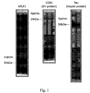
Fig. 1 shows results of Western Blot of extracellular vesicles collected from
plasma.
Fig. 2 shows results of Western Blot of extracellular vesicles collected from
culture supernatant
of nerve cells differentiated from iPS cells.
Description of Embodiments
[0010]
1. Method for collecting extracellular vesicles
This embodiment relates to a method for collecting extracellular vesicles
derived from nervous
system cells. The method for collecting extracellular vesicles comprises a
step of mixing an anti-
APLP1 antibody and a sample containing the extracellular vesicles, and a step
of collecting a complex
of the anti-APLP1 antibody and the extracellular vesicles. In the method for
collecting the
extracellular vesicles, the complex of the anti-APLP1 antibody and the
extracellular vesicles can be
formed by mixing the anti-APLP1 antibody and the sample containing the
extracellular vesicles.
[0011]
The extracellular vesicles are particles having a size of about several tens
to several thousand
nm and covered with a membrane containing phospholipids released from cells as
a main component.
The extracellular vesicles include exosomes, microvesicles, apoptotic bodies,
and the like.
Biomolecules are often present in the extracellular vesicles. For example, the
exosome or the
microvesicle comprises at least one biomolecule selected from the group
consisting of polypeptides
4
Date Recue/Date Received 2022-06-24
CA 03165950 2022-06-24
and polynucleotides (RNA such as mRNA, miRNA, and non-coding RNA, and DNA).
For example,
the apoptotic body includes at least one selected from the group consisting of
fragmented nuclei and
organelles. The extracellular vesicle preferably comprises at least one
biomolecule selected from the
group consisting of polypeptides and polynucleotides. More preferably, the
extracellular vesicle
comprises at least one biomolecule selected from the group consisting of
polypeptides and
polynucleotides. Here, the polypeptide refers to a compound in which a
plurality of amino acids are
bound by a peptide bond, and includes a protein having a relatively large
molecular weight and a
peptide having a relatively small molecular weight.
[0012]
Amyloid beta precursor like protein 1: APLP1 is one of the members of the
amyloid precursor
protein gene family, and the APLP1 protein is expressed in a brain and is a
membrane-bound
glycoprotein cleaved by a secretase similar to the cleavage of amyloid beta A4
precursor protein. An
intracellular cytoplasmic fragment that may act as a transcriptional activator
is released by this
cleavage. Human APLP1 is registered, for example, with Gene ID: 333. NCBI
Reference Sequence
is, for example, NM 001024807.3. Two variants have been reported for human
APLP1, but in the
present embodiment, the type of variant is not limited.
[0013]
The nervous system cells can include nerve cells and glial cells. The glial
cells can include
astrocytes, oligodendrocytes and microglial cells.
[0014]
The sample contains extracellular vesicles crudely purified from a specimen
containing the
extracellular vesicles. The sample may be a dispersion of extracellular
vesicles or a pellet of the
extracellular vesicles.
[0015]
Methods for crudely purifying extracellular vesicles from a specimen are
known, and for
example, extracellular vesicles can be crudely purified by a size exclusion
chromatography, an
ultracentrifugation, an affinity purification, a polymer precipitation, or a
combination thereof. Known
methods can be used as these crude purification methods. The size exclusion
chromatography is not
limited as long as the extracellular vesicles can be fractionated by size. For
example, the size
exclusion chromatography can be performed using an extracellular vesicle
isolation kit qEV (Izon
Date Recue/Date Received 2022-06-24
CA 03165950 2022-06-24
Science) or the like. In the case of ultracentrifugation, extracellular
vesicles can be obtained, for
example, by ultracentrifuging at 100,000 g to 150,000 g for about 2 to 3
hours. Preferably, the
specimen is preferably diluted with PBS, HEPES buffer, cell culture medium or
the like so that the
specific gravity becomes about 1.000 to 1.010 when performing
ultracentrifugation, if necessary. The
affinity purification may include, for example, a phosphatidylserine affinity
purification, a CD63
affinity purification, an anion affinity purification, and the like. The
polymer precipitation is a method
of precipitating extracellular vesicles using a polyether such as a
polyethylene glycol, a polypropylene
glycol, or a polytetramethylene glycol. The crude purification of
extracellular vesicles is preferably
performed by a method capable of obtaining extracellular vesicles having a
size of about 70 nm to
1000 nm.
[0016]
The specimen is taken from a living body of an animal, preferably a mammal
such as a human,
a mouse, a rat, a rabbit, a dog, a cat, a cow, a pig, or a horse, and is not
limited as long as it contains
extracellular vesicles. For example, the specimen includes whole blood, serum,
plasma, lymph, urine,
ascites, pleural effusion, cerebrospinal fluid, intercellular fluid, tears,
nasal discharge, saliva, and the
like.
[0017]
The anti-APLP1 antibody is not limited as long as it can bind at least a
portion of the APLP1
protein. In addition, the anti-APLP1 antibody may be one type or a mixture of
a plurality of types.
As the "antibody", any of a polyclonal antibody, a monoclonal antibody, and a
fragment thereof (for
example, Fab, F(ab'), F(ab)2, and the like) can be used. The class and
subclass of immunoglobulin
are not particularly limited. In addition, the antibody may be one screened
from an antibody library,
such as a chimeric antibody, scFv, or the like. For example, R&D SYSTEMS
AF3129 can be used
as the anti-APLP1 antibody.
[0018]
The antibody does not necessarily have to be purified, and may be an antiserum
containing the
antibody, ascites, an immunoglobulin fraction fractionated from these, or the
like.
[0019]
6
Date Recue/Date Received 2022-06-24
CA 03165950 2022-06-24
In addition, the antibody may be bound to a carrier. Examples of the carrier
include a known
carrier used for immunoprecipitation. For example, the carrier is magnetic
bead, agarose bead,
cellulose bead, microplate, tube or the like.
[0020]
Mixing the anti-APLP1 antibody and the sample containing the extracellular
vesicles is not
limited as long as the mixing is performed under conditions such that the anti-
APLP1 antibody can
bind to APLP1 present in the extracellular vesicles. As an example, Tris-HC1
buffer, phosphoric acid
buffer, HEPES buffer, maleic acid buffer, CHAPS buffer, and the like whose pH
is about 6 to 9 can
be included. It is preferable to add sodium chloride to these buffers in the
same amount as saline. In
addition, a bovine serum albumin, a skim milk or the like may be added as a
blocking reagent. The
reaction temperature is about 4 C to 37 C. Furthermore, the reaction between
the antibody and the
extracellular vesicle is preferably carried out with stifling. The reaction
time depends on the reaction
temperature, but is about 4 hours to 48 hours when the reaction temperature is
about 4 C, and about
0.5 hours to 4 hours when the reaction temperature is about 37 C.
[0021]
By the above reaction, the anti-APLP1 antibody binds to the extracellular
vesicle, and a
complex of the anti-APLP1 antibody and the extracellular vesicle (also
referred to as "anti-APLP1
antibody-extracellular vesicle complex") is formed.
[0022]
Collecting the anti-APLP1 antibody-extracellular vesicle complexes can be
performed by a
known method. When the anti-APLP1 antibodies are bound to carriers in advance,
the collecting
method can be selected according to properties of the carrier. For example,
when the carriers are
magnetic beads, the anti-APLP1 antibody-extracellular vesicle complexes can be
adsorbed on
magnets and collected. When the carriers are non-magnetic beads such as
agarose beads or cellulose
beads, the anti-APLP1 antibody-extracellular vesicle complexes can be
collected by centrifugation.
[0023]
When the anti-APLP1 antibodies are not previously bound to carriers, carriers
that are bound to
substances having an affinity for the antibody used, for example, a secondary
antibody that binds to
the anti-APLP1 antibody, protein A or protein G, may be used to collect the
anti-APLP1 antibody-
extracellular vesicle complexes. The method for collecting the carriers and
the anti-APLP1 antibody-
7
Date Recue/Date Received 2022-06-24
CA 03165950 2022-06-24
extracellular vesicle complexes bound to the carriers is the same as when the
anti-APLP1 antibodies
are previously bound to the carriers.
[0024]
In the process of collecting the anti-APLP1 antibody-extracellular vesicle
complexes, a step of
washing the anti-APLP1 antibody-extracellular vesicle complexes for B
(bound)/F (free) separation
in which the extracellular vesicles unreacted with the anti-APLP1 antibody and
the anti-APLP1
antibodies unreacted with the extracellular vesicles are appropriately removed
may be included.
[0025]
Since APLP1 is a protein specifically expressed in nervous system cells,
extracellular vesicles
derived from nervous system cells can be collected by the above collecting
method.
[0026]
2. Method for detecting neuropsychiatric disorder
In this embodiment, the extracellular vesicles collected in the above 1 are
used to detect
neuropsychiatric disorder.
[0027]
The method for detecting neuropsychiatric disorder includes a step of
obtaining a measured
value of the polypeptide and/or the polynucleotide of the biomarker of
neuropsychiatric disorder from
the extracellular vesicles collected in the above 1 and comparing the measured
values with a
corresponding reference value to determine whether the measured value is
within or outside a
reference range. When the measured value is outside the reference range, it
can be determined that a
subject from whom the specimen was taken suffers from the neuropsychiatric
disorder. Alternatively,
if the measured value is within the reference range, it can be determined that
the subject from whom
the specimen was taken dose not suffer from the neuropsychiatric disorder.
[0028]
In addition, the severity of the neuropsychiatric disorder may be determined
by determining
how much the measured value is dissociated from the reference value.
[0029]
The neuropsychiatric disorder can include psychiatric disorder and nervous
system disease. The
nervous system disease can include neurodegenerative disorder, post-cerebral
spinal trauma
neurologic dysfunction, brain tumor, infection-related cerebrospinal disorder,
multiple sclerosis, and
8
Date Recue/Date Received 2022-06-24
CA 03165950 2022-06-24
the like. The neurodegenerative disorder can include dementia, Parkinson's
disease, amyotrophic
lateral sclerosis, progressive supranuclear palsy, multiple system atrophy,
triplet repeat disease, and
the like. The dementia can include Alzheimer's disease, senile dementia, Lewy
body disease,
frontotemporal dementia, vascular dementia, alcohol-related dementia and
corticobasal degeneration.
The infection-related cerebrospinal disorder can include meningitis, brain
abscess, Creutzfeldt-Jakob
disease, and AIDS dementia. The brain tumor can include astrocytoma.
[0030]
The psychiatric disorder can include schizophrenia, depression, bipolar
disorder, and the like.
[0031]
For example, a tau protein contained in an extracellular vesicle, particularly
a phosphorylated
tau protein, is a biomarker for Alzheimer's disease. When a measured value of
the tau protein or the
phosphorylated tau protein in a specimen taken from a subject is higher than a
reference value, the
subject can be determined to have Alzheimer's disease.
[0032]
As biomarkers reported in each disease, a-synuclein for Parkinson's disease
and dementia with
Lewy body; TAR DNA-binding protein (TDP-43) for amyotrophic lateral sclerosis
and
frontotemporal dementia; abnormal prion protein for Creutzfeldt-Jakob disease;
Neurofilament Light
Chain for post-cerebral spinal trauma neurologic dysfunction, multiple
sclerosis, amyotrophic lateral
sclerosis, progressive supranuclear palsy, multiple system atrophy, triplet
repeat disease, Alzheimer's
disease, Lewy body disease, frontotemporal dementia, vascular dementia,
corticobasal degeneration,
and the like can be included. In addition, insulin-like growth factor (IGF-1)
and brain-derived
neurotrophic factor (BDNF) for depression; microRNA hsa-miR-34a, microRNA hsa-
miR-432 for
schizophrenia; IGF-1, BDNF, and the like for bipolar disorder can be included.
[0033]
The biomarker for the neuropsychiatric disorder contained in an extracellular
vesicle is detected
as a polypeptide or as a polynucleotide. The polynucleotide may be RNA or DNA,
and the RNA may
include microRNA, ncRNA, and the like in addition to mRNA. The polypeptide
and/or the
polynucleotide of the biomarker for the neuropsychiatric disorder may include
their fragment as well
as full length one.
[0034]
9
Date Recue/Date Received 2022-06-24
CA 03165950 2022-06-24
The method for detecting the biomarker for the neuropsychiatric disorder as a
polypeptide may
include known methods such as Western blotting, Enzyme-Linked Immuno Sorbent
Assay (ELISA),
and the like. In addition, the method for detecting the biomarker for the
neuropsychiatric disorder as
RNA may include known methods such as RT-PCR (including quantitative RT-PCR),
microarray,
RNA-Seq, and the like. The method for detecting the biomarker for the
neuropsychiatric disorder as
DNA may include known methods such as PCR (including quantitative PCR),
microarray, sequencing,
and the like.
[0035]
When the biomarker for the neuropsychiatric disorder is detected as a
polypeptide by Western
blotting, ELISA or the like, extracellular vesicles are lysed with a
predetermined lysis buffer as a
pretreatment. A sample lysed with the lysis buffer is used as a test sample.
[0036]
Primary antibody for detecting the biomarker of the neuropsychiatric disorder
by Western
blotting, ELISA, and the like, are not limited as long as the biomarker of the
neuropsychiatric disorder
can be detected.
[0037]
When the biomarker for the neuropsychiatric disorder is detected as RNA by RT-
PCR,
microarray, RNA-Seq, and the like, the RNA is extracted from extracellular
vesicles as a pretreatment.
Further, if necessary, the extracted RNA may be used as a template for reverse
transcription to
synthesize complementary DNA (cDNA). RNA or cDNA can be used to detect the
biomarker.
[0038]
As a primer used for RT-PCR (in the case of quantitative RT-PCR, a probe may
be included),
a commercially available primer can be used. In addition, a commercially
available microarray can
also be used.
[0039]
For the RNA-Seq, a next-generation sequencer (for example, manufactured by
Illumina) or the
like can be used to obtain the number of reads of mRNA of the biomarker for
the neuropsychiatric
disorder.
[0040]
Date Recue/Date Received 2022-06-24
CA 03165950 2022-06-24
When the biomarker for the neuropsychiatric disorder is detected as DNA by
PCR, microarray,
sequencing, or the like, DNA is extracted from extracellular vesicles as a
pretreatment. Further, if
necessary, an amplification reaction may be carried out using the extracted
DNA as a template. The
DNA extracted from the extracellular vesicles or the amplified DNA can be used
to detect the
biomarker.
[0041]
As a primer used for PCR (in the case of quantitative PCR, a probe may be
included), a
commercially available primer can be used. In addition, a commercially
available microarray can also
be used.
[0042]
For the sequencing, the next-generation sequencer (for example, manufactured
by Illumina) or
the like can be used to obtain the number of reads of DNA associated with the
biomarker for the
neuropsychiatric disorder.
[0043]
When the biomarker for the neuropsychiatric disorder is detected by Western
blotting, ELISA,
RT-PCR, PCR, RNA-Seq, sequencing, microarray, or the like, if the biomarker
for the
neuropsychiatric disorder is detected in extracellular vesicles, it may be
determined that "the
biomarker for the neuropsychiatric disorder is detected" or "an expression of
the biomarker for the
neuropsychiatric disorder is positive". Alternatively, when the amount of
polypeptide of the
biomarker for the neuropsychiatric disorder derived from extracellular
vesicles of a subject and the
amount of polypeptide of the biomarker for the neuropsychiatric disorder
derived from extracellular
vesicles of a healthy individual are compared, or the amount of polynucleotide
of the biomarker for
the neuropsychiatric disorder derived from the extracellular vesicles of the
subject and the amount of
polynucleotide of the biomarker for the neuropsychiatric disorder derived from
the extracellular
vesicles of the healthy individual are compared, if the amount of polypeptide
of the biomarker for the
neuropsychiatric disorder or the amount of polynucleotide of the biomarker for
the neuropsychiatric
disorder in the test sample taken from the subject is a higher value than the
amount of polypeptide of
the biomarker for the neuropsychiatric disorder or the amount of
polynucleotide of the biomarker for
the neuropsychiatric disorder in the extracellular vesicles taken from the
healthy individual, it may be
determined that "the biomarker for the neuropsychiatric disorder is detected"
or "an expression of the
11
Date Recue/Date Received 2022-06-24
CA 03165950 2022-06-24
biomarker for the neuropsychiatric disorder is positive". In addition, if the
amount of polypeptide of
the biomarker for the neuropsychiatric disorder or the amount of
polynucleotide of the biomarker for
the neuropsychiatric disorder in the extracellular vesicles taken from the
subject is the same degree as
the amount of polypeptide of the biomarker for the neuropsychiatric disorder
or the amount of
polynucleotide of the biomarker for the neuropsychiatric disorder in the
extracellular vesicles taken
from the healthy individual, it may be determined that "the biomarker for the
neuropsychiatric disorder
is not detected" or "an expression of the biomarker for the neuropsychiatric
disorder is negative".
Here, the expression "is a higher value" can be exemplified as a case where
the value shows a value
1.2 times or more, preferably 1.5 times or more, more preferably 2 times or
more, still more preferably
times or more. The expression "is the same degree" can be exemplified as a
case where the value
shows a value about 0.8 times to less than 1.2 times. In addition, before
comparing the amounts of
polypeptide of the biomarker for the neuropsychiatric disorder in a subject
and that in a healthy
individual or the amounts of polynucleotide of the biomarker for the
neuropsychiatric disorder in the
subject and that in the healthy individual, the number of extracellular
vesicles purified from each
specimen may be normalized by the amount of polypeptide of CD9, CD63, CD81, or
the like that is
a marker for the extracellular vesicle. Further, the normalization of the
number of extracellular
vesicles may be performed by the number of particles measured by Nanoparticle
Tracking Analysis
method or the like. The amount of the polypeptide may be expressed by mass or
concentration, and
may also be expressed by emission intensity of a substrate or the like. The
amount of the
polynucleotide may be the number of copies or the number of reads of the
polynucleotide, and may
be expressed by fluorescence intensity or the like.
[0044]
As another embodiment, a reference value for the amount of polypeptide or RNA
of the
biomarker for the neuropsychiatric disorder is determined in advance, and if
the amount of polypeptide
or RNA of the biomarker for the neuropsychiatric disorder in the extracellular
vesicles derived from
the subject is out of a reference range, it may be determined that "the
biomarker for the
neuropsychiatric disorder is detected" or "an expression of the biomarker for
the neuropsychiatric
disorder is positive". In addition, if the amount of the polypeptide or the
RNA of the biomarker for
the neuropsychiatric disorder in the extracellular vesicles derived from the
subject is within the
reference range, it may be determined that "the biomarker of the
neuropsychiatric disorder is not
12
Date Recue/Date Received 2022-06-24
CA 03165950 2022-06-24
detected" or "the expression of the biomarker for the neuropsychiatric
disorder is negative". The
reference value is not limited as long as it is a value that can determine
whether the amount of the
polypeptide of the biomarker for the neuropsychiatric disorder or the amount
of the polynucleotide of
the biomarker for the neuropsychiatric disorder is detected or not or whether
the expression of the
biomarker for the neuropsychiatric disorder is positive or not, and can be
determined by a known
method. The value that can determine whether the amount of the polypeptide of
the biomarker for the
neuropsychiatric disorder or the amount of the polynucleotide of the biomarker
for the
neuropsychiatric disorder is detected or not or whether the expression of the
biomarker for the
neuropsychiatric disorder is positive or not can also be determined by an ROC
(receiver operating
characteristic curve) curve, a discriminant analysis method, a mode method, a
Kittler method, a 3G
method, a p-tile method, or the like. Further, as a reference value,
sensitivity, specificity, negative
predictive value, positive predictive value, first quartile, and the like can
be exemplified.
[0045]
3. Method for collecting components derivedfrom nervous system cells
In this embodiment, the extracellular vesicles collected in the above I are
used to collect the
components derived from nervous system cells.
[0046]
The method for collecting the components derived from the nervous system cells
comprises a
step of collecting at least one biomolecule selected from the group consisting
of sugar, lipid,
polypeptide, and polynucleotide from the extracellular vesicles collected in
the above I.
[0047]
The sugar may include monosaccharide, disaccharide, oligosaccharide, and
polysaccharide. In
addition, the sugar may be bound to lipid, protein, or the like. Collecting
sugar from the extracellular
vesicles can be performed by a known method using a hydrazide/oxyamine-
carrying polymer, a lectin,
or the like.
[0048]
The lipid may include fatty acid, eicosanoid, triacylglycerol, wax ester,
phospholipid,
sphingolipid, isoprenoid, lipoprotein, and the like. Extracting lipid from the
extracellular vesicles can
be performed by using Folch method, lipid extraction kit (Lipid Extraction
Kit: Cell Biolabs, Inc.), or
the like.
13
Date Recue/Date Received 2022-06-24
CA 03165950 2022-06-24
[0049]
The polypeptide and polynucleotide may also contain the biomarker described in
the above 2
and a component other than the above biomarker. The description of the
biomarker is incorporated
herein by reference.
[0050]
The step of collecting polypeptides and polynucleotides can be performed
according to known
methods. In addition, a commercially available extraction kit may be used.
[0051]
4. Test reagent
In this section, the test reagent for collecting extracellular vesicles is
described. The test reagent
comprises at least an anti-APLP1 antibody. The description of the anti-APLP1
antibody is
incorporated herein by reference to the description in the above 1.
[0052]
The anti-APLP1 antibody comprised in the test reagent may be one kind or two
or more kinds.
[0053]
The anti-APLP1 antibody comprised in the test reagent may be in a dry state or
may be dissolved
in a buffer such as phosphate-buffered saline. Further, the test reagent may
comprise at least one of a
stabilizer such as beta-mercaptoethanol and DTT; a protective agent such as
albumin; a surfactant
such as polyoxyethylene (20) sorbitan monolaurate and polyoxyethylene (10)
octylphenyl ether; a
preservative such as sodium azide, and the like.
[0054]
The anti-APLP1 antibody may be labeled with an enzyme or a fluorescent dye.
The antibody
that binds to adipophilin may be immobilized on a microplate, magnetic beads,
or the like.
[0055]
5. Test kit
In this section, the kit comprising the test reagent for collecting
extracellular vesicles described
in the above 4 is described.
[0056]
The test kit may be provided as a test kit that includes a package insert that
describes the test
reagent and how to use the reagent or that describes a URL of a web page that
describes how to use
14
Date Recue/Date Received 2022-06-24
CA 03165950 2022-06-24
the reagent. In addition, when the anti-APLP1 antibody dose not bind to a
carrier or a solid phase,
secondary antibody-conjugated beads, Protein A beads, Protein G beads, a
secondary antibody
immobilized microplate, a Protein A-immobilized microplate, a Protein G-
immobilized microplate, a
secondary antibody-immobilized tube, and the like for binding the anti-APLP1
antibody to the carrier
or the solid phase may be comprised.
[0057]
Further, the test kit may comprise a column for size exclusion chromatography,
a column for
affinity purification, a polyether such as polyethylene glycol, and the like
for crudely purifying the
extracellular vesicles.
[0058]
Furthermore, it may comprise an antibody, a probe, a primer, or the like for
detecting the
biomarker of the neuropsychiatric disorder present in the extracellular
vesicles.
Examples
[0059]
Hereinafter, the present embodiment will be described in more detail with
reference to
Examples, but the present invention is not construed as being limited to the
Examples.
[0060]
1. Example /: Collecting nervous system cells-derived extracellular vesicles
from plasma by using
anti-APLP1 antibody
(i) Specimen
Blood was collected from a patient using an EDTA-2K tube, and 20 mL of plasma
was separated.
The plasma was stored at -80 C until testing. At the time of use, the plasma
was thawed and
centrifuged at 2,500 g, 4 C for 10 minutes, and supernatant was collected.
[0061]
(ii) Crude purification of extracellular vesicles in plasma
20 mL of size exclusion chromatography (SEC) fraction was collected from 10 mL
of the
plasma by using qEV10 (Meiwafosis Co., Ltd.) according to a protocol attached
to it. The above step
was carried out twice in total, and 40 mL of the SEC fraction was collected.
The SEC fraction was
concentrated to 600 pt by using Amicon Ultra-15 100 kDa MWCO.
Date Recue/Date Received 2022-06-24
CA 03165950 2022-06-24
[0062]
(iii) Preparation of beads for immunoprecipitation
mg of Dynabeads M-270 Epoxy of Dynabeads Antibody Coupling Kit (Thermo
Fisher
Scientific) and 10 pg of anti-APLP1 antibody (R&D SYSTEMS AF3129) were coupled
according to
a protocol attached to the kit to prepare magnetic beads to which the anti-
APLP1 antibody was bound.
[0063]
(iv) Immunoprecipitation of extracellular vesicles derived from nervous system
cells
100 pL of the beads for immunoprecipitation prepared in the above (iii) was
added to 600 pt
of the concentrated SEC fraction in a tube, and incubated at 4 C for 4 hours
while being inverted and
mixed. After spinning down the tube, it was attached to a magnetic stand and
waited for 1 minute and
then, remove the supernatant. Subsequently, in order to wash the magnetic
beads, 800 pt of D-PBS
(-) was added to the tube and mixed by inversing. After spinning down the
tube, it was attached to
the magnetic stand and waited for 1 minute and then, remove the supernatant.
It was spun down again,
attached to the magnetic stand, waited for 1 minute and then the supernatant
was completely removed.
[0064]
The tube with magnetic beads remained in it was added with 17 pL of 2x Laemmli
Buffer, and
after vortexing, incubated at 97 C for 5 minutes. After spinning down the
tube, it was attached to the
magnetic stand and waited for 1 minute and the supernatant was collected and
then used for analysis.
[0065]
(5) Western Blot
The supernatant collected in the previous step was mixed with denaturing
buffer and then heated
to prepare a sample for application. The sample was loaded into a 5-15% Tris-
glycine SDS gel and
run at 200 V for 55 minutes. The SDS gel after electrophoresis was run under
semi-dry conditions at
V for 30 minutes using Towbin Buffer (with 5% methanol) to perform transfer
from the gel to a
PVDF membrane.
[0066]
Blocking was performed at room temperature for 1 hour using 2% ECL Prime
Blocking Agent
dissolved in TBS-T. Primary antibodies were diluted with 2% ECL Prime Blocking
Agent dissolved
in TBS-T and reacted at 4 C overnight. The antibodies used were anti-APLP1
antibody (Calbiochem,
171615), anti-CD81 antibody (Santa Cruz, sc-23962), and anti-Tau antibody
(Merck Millipore,
16
Date Recue/Date Received 2022-06-24
CA 03165950 2022-06-24
5778801), and all of them were diluted 1,000 times to be used. After reacting
with the primary
antibodies, a 5-minute wash was performed 6 times using TBS-T.
[0067]
The secondary antibody was diluted with 2% ECL Prime Blocking Agent dissolved
in TBS-T
and reacted at room temperature for 1 hour. The antibodies used were HRP-
labeled anti-mouse
antibody (Promega, W4021) and HRP-labeled anti-rabbit antibody (Promega,
W4011), all of them
were diluted 10,000 times to be used. A 5-minute wash was performed 6 times
using TBS-T.
[0068]
1,4001uL of ImmunoStar LD A + B solution was added to the membrane, and the
mixture was
reacted at room temperature for 5 minutes. By using myECL Imager (Thermo
Scientific), HRP was
emitted and the signal was imaged.
[0069]
(6) Results
The results of Western Blot are shown in Fig. 1.
[0070]
In addition to the targeted APLP1, CD81 that is an extracellular vesicle (EV)
protein and Tau
that is a neural protein were detected by immunoprecipitation using plasma.
From this, it was found
that APLP1-positive EV could be isolated and neural proteins exist in it, and
consequently,
extracellular vesicles derived from nervous system cells could be collected.
[0071]
2. Example 2: Extracellular vesicles from culture supernatant of nerve cells
differentiated from iPS
cells
(i) Preparation of beads for immunoprecipitation
5mg of Dynabeads M-270 Epoxy of Dynabeads Antibody Coupling Kit (Thermo
Fisher
Scientific) and 10pg of anti-APLP1 antibody (R&D SYSTEMS AF3129) were coupled
according to
a protocol attached to the kit to prepare magnetic beads to which the anti-
APLP1 antibody was bound.
[0072]
(ii) Preparation of extracellular vesicles (EV) in the culture supernatant
17
Date Recue/Date Received 2022-06-24
CA 03165950 2022-06-24
The culture supernatant of the nerve cells differentiated from iPS cells was
centrifuged at 2,500
g for 10 minutes, and the supernatant was put in 4 ultracentrifugation tubes
by 12 mL each. After
ultracentrifugation at 120,000 g for 2.5 hours, the supernatant was removed
and pellet containing
extracellular vesicles was collected. The pellet was added with 12 mL of PBS,
pipetted, and again
ultracentrifuged at 120,000 g for 2.5 hours to collect the pellet. The pellet
was added with 25 pt PBS,
pipetted, and transferred to one 1.5 mL tube.
[0073]
(iii) Immunoprecipitation and Western Blot
100 L of Dynabeads coupled with APLP1 antibody was put in a screw cap tube.
20 [IL of EV
prepared by ultracentrifugation, 80 pt of x10 complete (Roche), and 600 III,
of PBS were added, and
the mixture was incubated at 4 C for 4 hours while being rotated and mixed.
The tube was spun down,
attached to a magnetic stand, and allowed to stand for 1 minute. The
supernatant was transferred to
another tube, 800 pt of PBS was added to the beads, and the tube was shaken up
and down. The tube
was spun down, attached to the magnetic stand, and allowed to stand for 1
minute. After removing
the supernatant, 12 [IL of x2 sample buffer for Western blotting was added to
the tube containing the
beads, and the mixture was heated at 97 C for 5 minutes after vortex. After
centrifugation, it was
attached to the magnetic stand and allowed to stand for 1 minute, and 10 [IL
was electrophoresed on
a 5-20% or 15% SDS-PAGE gel. 20 pL of the supernatant after
immunoprecipitation was taken in
another tube, 4 pL x6 sample buffer was added, and the mixture was heated at
97 C for 5 minutes
after vortex. After centrifugation, 20 pt was electrophoresed on a 5-20% or
15% SDS-PAGE gel at
a voltage of 120 V for 100 minutes.
[0074]
The gel after electrophoresis was transferred to a PVDF membrane by wet
transfer (400 mA, 1
hour). The membrane after blotting was blocked with 2% ECL Prime Blocking
Reagent for 2 hours.
Subsequently, the membranes were added with APLP1 C-terminal antibody
(Calbiochem
171615) diluted 10,000 times, L1CAM antibody (Santa Cruz sc-53386) diluted
1,000 times, CD63
antibody (Santa Cruz sc-5275) diluted 2,000 times, Flotillin-1 antibody (BD
Transduction 610820)
diluted 1,000 times and SNAP25 antibody (abcam ab5666) diluted 1,000 times
with 2% ECL Prime
Blocking Reagent respectively, and incubated overnight at 4 C with shaking.
After incubation, the
membranes were washed with 0.1% TBS-T for 5 minutes 6 times.
18
Date Recue/Date Received 2022-06-24
CA 03165950 2022-06-24
[0075]
The membranes reacted with L1CAM, CD63, and Flotillin-1 respectively were
added with anti-
Mouse IgG-HRP (Promega W402B) diluted 10,000 times with 2% ECL Prime Blocking
Reagent as
a secondary antibody and incubated at room temperature for 1 hour with
shaking. The membranes
reacted with APLP1 and SNAP25 respectively were added with anti-Rabbit IgG-HRP
(Promega
W401B) diluted 10,000 times with 2% ECL Prime Blocking Reagent as a secondary
antibody and
incubated at room temperature for 1 hour with shaking. After incubation, the
membranes were washed
with 0.1% TBS-T for 5 minutes 6 times.
[0076]
Solution A and solution B of ImmunoStar LD (Wako) were mixed in an amount of
800 luL
each, and the membranes were immersed and allowed to stand for 5 minutes. A
signal was detected
by myECL Imager (Thermo Fisher Scientific) (10 to 300 seconds).
[0077]
(iv) Results
The results of Western Blot are shown in Fig. 2.
[0078]
Lane 1 is 1 lug of iPS neuron lysate, lane 2 is EV before immunoprecipitation,
lane 3 is input,
lane 4 is the supernatant after immunoprecipitation of EV, and lane 5 is
antibody-coupled beads after
immunoprecipitation of EV. The amounts of the input and the supernatant
electrophoresed were one-
fortieth of the whole.
[0079]
APLP1, EV marker protein (CD63, Flotillin-1), and nerve-derived protein
(L1CAM, SNAP25)
were detected in antibody beads. From this, it was found that extracellular
vesicles can be collected
by using the anti-APLP1 antibody.
19
Date Recue/Date Received 2022-06-24