Note: Descriptions are shown in the official language in which they were submitted.
WO 2021/202438
PCT/US2021/024761
SYSTEM, METHOD, AND APPARATUS FOR MULTI-SPECTRAL
PHOTOACOUSTIC IMAGING
CROSS-REFERENCE TO RELATED APPLICATION(S)
The present application is related to, and claims the benefit of, U.S.
Provisional Patent
Application Serial No. 63/002,714, filed March 31, 2020, which is herein
incorporated by
reference in its entirety.
STATEMENT REGARDING SPONSORED RESEARCH
The project leading to this application has received funding from the European
Union's
Horizon 2020 research and innovation program under the Marie Sklodowska-Curie
grant
agreement number 811226.
BACKGROUND
The practice of observing tissue chromophores can be a helpful tool in the
early
detection, prediction, and monitoring of various diseases or health
conditions. Molecular
imaging can be one of the methods used to detect and quantify tissue
chromophores. In
particular, multi-spectral photoacoustic (PA) imaging has emerged as a useful,
non-invasive
molecular imaging tool to visualize tissue chromophores. The underlying
principle of PA
imaging is using nanosecond laser pulses to illuminate different wavelengths
into the biological
tissue. Depending on the optical absorption coefficient of the tissue
chromophores, the tissue
can absorb the laser light and produce acoustic waves caused by thermo-elastic
expansion of
the tissue. Similar to conventional ultrasound imaging, these generated
photoacoustic signals
can be detected and the optical absorption of the tissue chromophores can be
reconstructed.
PA imaging is therefore considered a hybrid imaging modality that combines the
optical
absorption contrast of the chromophores and the spatial acoustic resolution
and penetration
depth of ultrasound imaging to provide molecular information.
1
CA 03167234 2022- 8-5
WO 2021/202438
PCT/US2021/024761
Being a hybrid imaging modality of ultrasound and optical, this multi-modal
imaging
technology can provide anatomical, functional, and molecular information
several centimeters
deep in the tissues with a resolution up to tens of micrometers. PA imaging
can be used in
various preclinical applications, such as tumor progression, prediction of
tumor recurrence,
therapy monitoring, imaging of vasculature, and the bio distribution of
contrast agents. In
addition to preclinical applications, PA imaging can be used in clinical
trials or applications.
For example, breast cancer imaging, sentinel lymph node imaging, and/or
examining temporal
arteries in patients with suspect giant-cell arteritis (GCA). Other usages
have included using
PA imaging in low-resource settings for the visualization of superficial
vasculatures and needle
guidance for minimally invasive procedures.
While emphasis has generally been placed on hardware development of PA
imaging,
the data analysis and reconstruction algorithms can play an important role in
increasing the
sensitivity of PA imaging.
SUMMARY
The disclosed subject matter described below provides for a non-limiting
example of
an improved PA imaging system, apparatus, and method. For example, embodiments
of the
disclosed subject matter can provide an unsupervised spectral unmixing process
or algorithm
for processing PA image data. Using the unsupervised spectral unmixing process
or algorithm
can help to improve the detection of tissue chromophores, thereby improving
the monitoring,
diagnosis, or treatment of a disease or medical condition associated with the
detected tissue
chromophores.
An example photoacoustic imaging method can include receiving multi-spectral
photoacoustic image data from a photoacoustic (PA) imaging system. The method
can also
include pre-processing the multi-spectral photoacoustic image data. The pre-
processing can
include determining a number of significant components above a noise floor of
the multi-
2
CA 03167234 2022- 8-5
WO 2021/202438
PCT/US2021/024761
spectral photoacoustic image data. In other words, the noise floor or level
can be used to define
the number of significant components from the multi-spectral photoacoustic
image data. In
addition, the method can include detecting tissue chromophores based on the
number of
significant components from the multi-spectral photoacoustic image data using
an
unsupervised spectral unmixing process or algorithm. The unsupervised spectral
unmixing
process or algorithm can include clustering and windowing of the multi-
spectral photoacoustic
image data. In other words, the windowing and clustering can be used to
determine the
prominent tissue chromophores present in the multi-spectral photoacoustic
image data.
Further, the method can include displaying the detected tissue chromophores in
an abundance
map
In another example, a photoacoustic imaging apparatus can include at least one
memory
comprising computer program code, and at least one processor. The computer
program code
can be configured, when executed by the at least one processor, to cause the
PA imaging
apparatus at least to receive multi-spectral photoacoustic image data, and pre-
process the multi-
spectral photoacoustic image data. The pre-processing can include determining
the number of
significant components above a noise floor of the multi-spectral photoacoustic
image data. The
computer program code can also be configured, when executed by the at least
one processor,
to cause the PA imaging apparatus at least to detect tissue chromophores based
on the number
of significant components from the multi-spectral photoacoustic image data
using an
unsupervised spectral unmixing process or algorithm. The unsupervised spectral
unmixing
process or algorithm can include clustering and windowing of the multi-
spectral photoacoustic
image data. In addition, the computer program code can also be configured,
when executed by
the at least one processor, to cause the PA apparatus at least to display the
detected tissue
chromophores in an abundance map.
According to certain non-limiting embodiments a non-transitory computer-
readable
3
CA 03167234 2022- 8-5
WO 2021/202438
PCT/US2021/024761
medium encodes instructions that, when executed in hardware, perform a
process. The process
can include receiving multi-spectral photoacoustic image data from a PA
imaging system. The
process can also include pre-processing the multi-spectral photoacoustic image
data. The pre-
processing can include determining a number of significant components above a
noise floor
from the multi-spectral photoacoustic image data. In addition, the process can
include
detecting tissue chromophores based on the number of significant components
from the multi-
spectral photoacoustic image data using an unsupervised spectral unmixing
process or
algorithm. The unsupervised spectral unmixing process or algorithm can include
clustering
and windowing of the multi-spectral photoacoustic image data. Further, the
process can
include displaying the detected tissue chromophores in an abundance map
An apparatus, in certain non-limiting embodiments, can include a computer
program
product encoding instructions for performing a process according to a method.
The method
can include receiving multi-spectral photoacoustic image data from a PA
imaging system. The
method can also include pre-processing the multi-spectral photoacoustic image
data. The pre-
processing can include determining a number of significant above a noise floor
from the multi-
spectral photoacoustic image data. In addition, the method can include
detecting tissue
chromophores based on the number of significant components from the multi-
spectral
photoacoustic image data using an unsupervised spectral unmixing process or
algorithm. The
unsupervised spectral unmixing process or algorithm can include clustering and
windowing of
the multi-spectral photoacoustic image data. Further, the method can include
displaying the
detected tissue chromophores in an abundance map.
BRIEF DESCRIPTION OF THE DRAWINGS
FIG. 1 is a diagram illustrating a supervised unmixing system or method
according to
some examples of the disclosed subject matter.
FIG. 2 is a diagram illustrating an unsupervised unmixing system or method
according
4
CA 03167234 2022- 8-5
WO 2021/202438
PCT/US2021/024761
to some examples of the disclosed subject matter.
FIG. 3 is a diagram illustrating embodiments of unsupervised unmixing
algorithms
according to some examples of the disclosed subject matter.
FIG. 4 is a diagram illustrating an unsupervised unmixing system or method
according
to some examples of the disclosed subject matter.
FIG. 5 is a diagram illustrating pre-processing according to some examples of
the
disclosed subject matter.
FIG. 6 is a diagram illustrating abundance maps for supervised and
unsupervised
unmixing algorithms according to some examples of the disclosed subject
matter.
FIG 7 is a diagram illustrating a component spectra according to some examples
of the
disclosed subject matter.
FIG. 8 is a flow diagram of a method or process according to some examples of
the
disclosed subject matter.
FIG. 9 is a diagram illustrating exemplary components of a system or apparatus
according to some examples of the disclosed subject matter.
DETAILED DESCRIPTION
Reference will now be made in detail to the various exemplary embodiments of
the
disclosed subject matter, which embodiments are illustrated in the
accompanying drawings.
The structure and corresponding method of operation of the disclosed subject
matter will be
described in conjunction with the detailed description of the system. The
examples and
embodiments described below are merely exemplary, and should not be taken in
any way as
limiting the scope of the disclosed subject matter.
In certain non-limiting embodiments, PA imaging employs spectral unmixing
algorithms. In particular, since the optical absorption coefficient of the
tissue chromophores
varies over the spectrum, multispectral image processing approaches can be
applied to
5
CA 03167234 2022- 8-5
WO 2021/202438
PCT/US2021/024761
distinguish between chromophores. The pixel intensity of the multi spectral
photoacoustic
images can be proportional to the absorption value of the respective tissues
at a specific
wavelength of the light excitation. By considering the pixel size as
infinitesimal, every
absorption signal throughout the different wavelengths can represent the
signal obtained from
a single molecular component. However, due to the finite dimension of pixels
and the presence
of noise, which can corrupt the acquired signal, each spectrum can result in a
mixed signal. In
some non-limiting embodiments, the mixed signal can be a combination of the
absorption
spectra of different source chromophores and undesired biological and
instrumental noise. A
spectral unmixing algorithm can be used to unmix these signals from different
optical absorbers
and/or to estimate the concentration of a given tissue chromophore
In particular, spectral unmixing can be a data decomposition approach based on
a linear
mixing model. To that end, spectral unmixing algorithms can be used to
differentiate the mixed
pixel spectra into a collection of spectra, also referred to as endmembers,
and a set of fractional
abundance maps. The component spectra or endmembers represent the pure
molecule
absorption spectrum extracted from the mixed pixel spectra or the multi-
spectral photoacoustic
image data. The maps of abundance represent the percentage of each endmember
present in a
given pixel. The spectral unmixing algorithm can therefore be used as part of
the multi spectral
image processing to characterize molecules present in the tissue based on the
spectral
absorption profile of a given molecule. In certain non-limiting embodiments
the spectral
unmixing algorithm can process the received multi-spectral photoacoustic image
data to
estimate or infer the rest of the image(s).
FIG. 1 is a diagram illustrating a supervised unmixing system or method
according to
some examples of the disclosed subject matter. In particular, FIG. 1
illustrates a method for
detecting tissue chromophores with multi-spectral PA imaging using a
differential based
unmixing algorithm with a known spectral signature as a-prior information. As
shown in FIG.
6
CA 03167234 2022- 8-5
WO 2021/202438
PCT/US2021/024761
1, input 110 can be a multi-spectral photoacoustic image data obtained from a
PA imaging
system. The data can be received as images of a two-dimensional region across
the wavelength
near infrared spectroscopy (NIR) range between 680 and 970 nanometers (nm). In
other non-
limiting embodiments, any other wavelength range can be used. The multi-
spectral
photoacoustic image data, for example, can have a step size of 1 nm, 5 nm, or
10 nm. The
lower the step size the higher the resolution, meaning that a step size of 1
nm can have a higher
resolution than a step size of 5 nm or 10 nm. As such, processing a multi-
spectral photoacoustic
image data with a step size of 1 nm can require more resources than data
having a step size of
5 nm or 10 nm. In some other non-limiting embodiments any other step size can
be used. For
a wavelength NW range of 680 ¨ 970 nm with a step size of 5 nm, the data set
can include the
same or similar two-dimensional region imaged at 59 different wavelengths.
The system or method shown in FIG. 1 can be used to detect the tissue
chromophores
using a supervised unmixing method. In particular, FIG. 1 uses a differential
based unmixing
method with a known spectral signature as a-priori information 120. A-priori
information 120
can be a user defined endmember absorption spectra of the tissue chromophores,
such as an
oxyhemoglobin (Oxy Hb) absorption spectrum and deoxyhemoglobin (Deoxy Hb)
absorption
spectrum. In another example a-priori information can be a predetermined or
known region of
interest. Based on a-priori information 120 inputted by the user, input 110
can be processed
and output 130 can be produced. Output 130 can include one or more abundance
maps
showing, for example, the distributions of endogenous tissue chromophores like
oxyhemoglobin, deoxyhemoglobin and an exogenous contrast agent such as
Indocyanine green
(ICG). As discussed above, the supervised spectral unmixing approaches can be
challenging,
as the spectral signatures of the tissues differs with respect to the disease
or medical condition.
Disease or medical conditions can involve complicated processes that can
induce changes in
the characteristics of the theoretical absorption spectra of the tissue
chromophores. In addition,
7
CA 03167234 2022- 8-5
WO 2021/202438
PCT/US2021/024761
the absorption spectra of the exogenous contrast agents can also differ due to
the interaction,
after the injection, within the tissues.
As shown in FIG. 1, some non-limiting embodiments utilize a supervised
spectral
unmixing algorithm, requiring user interaction. Such supervised algorithms
therefore are
dependent on a user being able to define a source spectra or a previously
identified or known
region of interest for prominent chromophores. Because the spectral signature
of the tissue
differs with respect to diseases or medical conditions, being able to define
the source spectra
or region of interest can be difficult or biased by the user. The source
spectra or the previously
identified or known region of interest can be referred to as a-priori
information, meaning that
a supervising user has to pre-define the information before or during
processing In addition,
to obtain an accurate fitting result using a supervised spectral unmixing
algorithm, a higher
number of wavelengths will be required for the PA image acquisition. Using a
higher number
of wavelength can increase resource usage by the PA image acquisition system
or apparatus,
requiring more memory, broadband, network, or processor resources. The
supervised spectral
unmixing algorithm also relies on user interaction to reduce biological and
instrumental noises,
both are which are subject to user biases and preferences.
To help overcome one or more of the above disadvantages, certain non-limiting
embodiments can utilize one or more unsupervised or autonomous spectral
unmixing
algorithms. An unsupervised spectral unmixing process or algorithm, which can
be a type of
unsupervised machine learning algorithm, does not require any user
supervision, thereby
allowing for automatic detection of prominent component spectra. Prominent
component
spectra can simply be referred to as components herein. The unsupervised
spectral unmixing
process or algorithm, for example, can be singular value decomposition (SVD),
principal
component analysis (PCA), sparse filtering (SF), independent component
analysis (ICA),
reconstruction independent component analysis (RICA), non-negative matrix
factorization
8
CA 03167234 2022- 8-5
WO 2021/202438
PCT/US2021/024761
(NNMF), or any other unsupervised spectral unmixing process or algorithm known
in the art.
In certain non-limiting embodiments, the unsupervised spectral unmixing
process or
algorithm can be referred to as a blind source separation algorithm. The blind
source separation
algorithm can be based on iterative optimization methodologies, which includes
approximation
by minimizing a cost function. One or more cost function can be defined or
determined for
every blind approach. Each iteration of the algorithm can be used to minimize
the cost function.
Using this iterative approach, spectral unmixing can help decompose the PA
image data into
quantitative component maps that identify the biodistribution of the recovered
underlying
tissue chromophores that provide spectral contrast. The multi-spectral
photoacoustic image
data inputted into the blind source separation algorithm can be in the form of
a single data
matrix.
The unsupervised spectral unmixing process or algorithm can allow access to
molecular and quantitative information from the PA image data, with high
sensitivity and
specificity. In some non-limiting embodiments the unsupervised spectral
unmixing process or
algorithm can allow a system, apparatus, or processor to learn from multi-
spectral
photoacoustic image data with limited or no human intervention by detecting or
recognizing
representative spectral patterns. As a data drive method, the unsupervised
spectral unmixing
process or algorithm can accurately extract hidden underlying features,
components, or
chromophores.
As discussed above, certain non-limiting embodiments can utilize an
unsupervised
unmixing algorithm based on machine learning that can facilitate the
extraction and
quantification of the intrinsic absorption trend of biomarkers in both
physiological and also in
the pathological condition. Using such a data-driven approach, the prominent
spectra from the
tissue chromophores can be detected from the multi-spectral imaging data set
with limited or
no user interaction. The resulting tissue chromophores can be visualized
and/or quantified with
9
CA 03167234 2022- 8-5
WO 2021/202438
PCT/US2021/024761
superior sensitivity. FIG. 2 is a diagram illustrating an unsupervised
unmixing system or
method according to some examples of the disclosed subject matter. In
particular, FIG. 2
illustrates the processing of PA image data using an unsupervised mixing
algorithm and
outputting an abundance map or component spectra.
As shown in FIG. 2, raw multi spectral PA images 210, also referred to as
multi-spectral
photoacoustic image data, can be inputted to the method, apparatus, or system.
Raw multi
spectral PA images can be received from a PA imaging machine or device. In
some non-
limiting embodiments, the ultrasound and PA imaging machine or device can be
included
within the same machine or device. In certain non-limiting embodiments, raw
multi spectral
PA images 210 can take the form of a two-dimensional region across part or all
of the
wavelength NIR range 680 nm ¨ 970 nm. In other non-limiting embodiments, any
other
wavelength NIR range can be utilized. The step size of the wavelength in the
NIR range can
be 1 nm, 5 nm, 10 nm, or any other step size. One or more of raw multi
spectral PA images
210 can be pre-processed using at least one of background removal 211, data
correction 212,
and/or data reduction 213. Data correction 212 can include a Gaussian filter
to reduce the
biological or instrumental noise. The Gaussian filter can process the data
using a kernel of 5x5
pixels, or any other sized kernel. Data reduction 213, on the other hand, can
include grouping
image pixels according to a squared region of interest of 4x4. In other non-
limiting
embodiments data reduction 213 can include grouping one or more pixels
according to a square
region of interest, or a region of interest having a different shape, and
having one or more
pixels.
In certain non-limiting embodiments, the pre-processing can include
determining a
noise level 215 to define a number of significant components 216 from the
multi-spectral
photoacoustic image data. The component, for example, can be a biomarker. The
unsupervised
spectral unmixing process or algorithm 217 can help to facilitate the
extraction and
CA 03167234 2022- 8-5
WO 2021/202438
PCT/US2021/024761
quantification of the intrinsic absorption of the one or more biomarkers. For
example, the
component or biomarker can be melanin, water, collagen, lipids, oxyhemoglobin,
deoxyhemoglobin, myoglobin, or any other biomarker known in the art. The
detected tissue
chromophores can be based on the determined significant components which are
above the
noise level.
As shown in FIG. 7, for example, significant component spectra which are above
the
noise floor can be displayed. In some non-limiting embodiments, an eigenvalue
algorithm can
be used to determine the noise floor and then the components which are
significant. The
eigenvalue algorithm, for example can be a power iteration, inverse iteration,
Rayleigh quotient
iteration, locally optimal block preconditioned conjugate gradient algorithm,
QR algorithm,
Jacobi eigenvalue algorithm, factorable polynomial equations, or any other
known equation,
method, or algorithm used to determine eigenvalues.
As shown in FIG. 2, after the input and pre-processing step the data can be
mixed using
a linear mixing model, or any other known mixing model. All or part of the
resulting mixed
observations 214 can be loaded or inputted to the unsupervised spectral
unmixing process or
algorithm. Unsupervised spectral unmixing process or algorithm 217 can use an
iterative
approach to separate pure molecules spectra from the multi-spectral
photoacoustic image data.
Unsupervised spectral unmixing process or algorithm 217 can include PCA, ICA,
RICA, SF,
and/or NNMF. For example, NNMF can be used to discriminate the mixed pixel
spectra from
a multi spectral image into a collection of constituent spectra, also referred
to as endmembers
218, and a set of abundance maps 219, which can be referred to fractional
abundance maps.
The endmembers 218 and/or the set of abundance maps 219 can be referred to as
first outcomes.
The NNWIF can therefore result in a component spectra, spatial distribution
maps, and/or
abundance maps of the prominent chromophores in the region of interest.
In certain non-limiting embodiments, the unsupervised spectral unmixing
process can
11
CA 03167234 2022- 8-5
WO 2021/202438
PCT/US2021/024761
include clustering and/or windowing. Clustering, for example, can include
similar spectra 220
and/or easily distinguishable spectra 221. Windowing, on the other hand, can
include dividing
the multi-spectral photoacoustic image data into one or more subsets 222. Once
one or more
subsets 222 are determined, tissue chromophores can be detected for each
subset based on the
number of significant components using an unsupervised spectra unmixing
process. The
endmembers from the subsets 222 and/or the endmembers from the whole data set
221 can then
be combined to determine the significant components spectra 223. Abundance map
224 can
then be derived from significant component spectra 223. In certain embodiments
abundance
map 224 can be a quantitative spatial distribution of all the detected
significant components
223
FIG. 3 is a diagram illustrating embodiments of unsupervised unmixing
algorithms
according to some examples of the disclosed subject matter. In particular,
FIG. 3 illustrates an
ideal or theoretical absorption spectra of oxy-deoxy hemoglobin 300 and the
background tissue
absorption compared to different unsupervised spectral unmixing processes or
algorithms 310-
360 processing test data, such as test multi-spectral photoacoustic image
data. SVD 310 and
PCA 320 are unsupervised dimension reduction approaches in machine learning.
Both SVD
310 and PCA 320 rely on the source components being uncorrelated and
orthogonal. In PCA
320, the goal can be to reduce the correlated observed variables. ICA 330 and
RICA 340, on
the other hand, assume that the observation data can be a superimposition of a
number of
stochastically independent processes. ICA 330 relies on the source components
being
maximally independent and non-Gaussian. The non-Gaussianity of the source data
can make
ICA 330 more powerful than PCA 320 in some non-limiting embodiments. RICA 340
takes
ICA 330 a step further by adding a reconstruction cost to the cost function of
the standard ICA
approach.
In contrast to the previous algorithms 310-340, SF 350 cannot explicitly be
based on a
12
CA 03167234 2022- 8-5
WO 2021/202438
PCT/US2021/024761
model of data distribution. SF 350 can be based on optimization of sparsity of
the features
distribution and/or can be formulated by implementing an uncomplicated code.
In some non-
limiting embodiments, SF 350 can be computationally expensive. In contrast,
NNMF 360 can
be a linear mixture model based on using non-negative matrices. In other
words, NNMF 360
can assume that the source data can be non-negative, making NNMF 360
compatible with PA
imaging in which all pixel values are either zero or positive. Imposing a
positivity condition,
similar to NNMF 360, can help to enhance the convergency of the optimization
algorithm used
for the unmixing problem. In addition, or as an alternative, by imposing the
nonnegativity
constraints NNMF 360 can learn or process a parts-based representation of the
data, allowing
a whole image to be formed as a combination of additive components Processing
multi-
spectral photoacoustic image data with NNMF 360 can result in a non-negative
matrix X of
mixed observation data that can be factorized into a source and mixing
coefficient matrices.
As shown in FIG. 3, NNMF 360 can have a better sensitivity and specificity
than SVD
310, PCA 320, ICA 330, RICA 340, and/or SF 350. NNMF 360 can also be most
similar to
the ideal absorption spectral of oxy-deoxy hemoglobin.
Further, below Table A shows the correlation values evaluated between the
ideal oxy
and deoxy HB spectra and the extracted prominent spectra found by using the
different
unsupervised unmixing algorithms.
SVD PCA ICA RICA SF NNMF
Oxy HB 0.9676 -0.9721 0.1471 0.9811 0.9869
1
Deoxy HB 0.8993 0.9698 -0.1180 -0.9229 0.9775
1
TABLE A
As shown in Table A, NNMF 360 has the highest positive correlation. The high
correlation can demonstrate the ability of unsupervised spectral unmixing
process or algorithm
NNMF 360 to detect both endogenous (oxy, deoxy hemoglobin) and exogenous
characteristics
13
CA 03167234 2022- 8-5
WO 2021/202438
PCT/US2021/024761
(contrast agents) spectra.
Despite the advantages provided by NN1VIF 360, in certain non-limiting
embodiments
any other unsupervised spectral unmixing process or algorithm can be used. For
example,
certain non-limiting embodiments can select an unsupervised spectral unmixing
process or
algorithm without a non-negative constraint. The
non-negative constraint can be
computationally expensive to implement, and each component can be estimated
only up to a
multiple scale factor. To lower the number of computer or processing
resources, some non-
limiting embodiments can use SVD 310, PCA 320, ICA 330, RICA 340, SF 350, or
any other
known unsupervised algorithm.
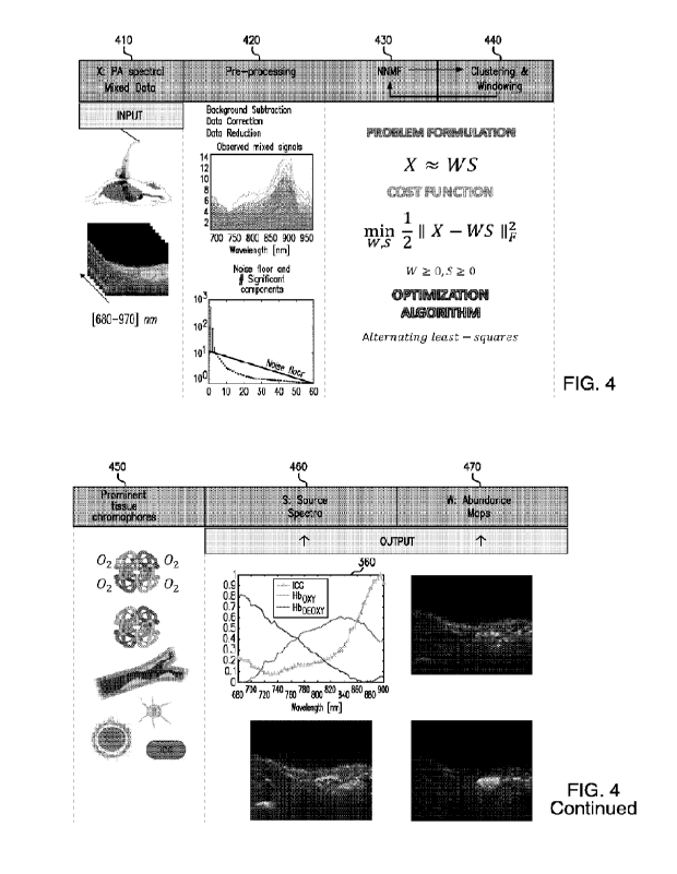
FIG 4 is a diagram illustrating an unsupervised unmixing system or method
according
to some examples of the disclosed subject matter. For input 410, a PA imaging
system 411 can
be used to image two-dimensional region across the entire wavelength NIR range
of 680 ¨ 970
nm with a step size of 5 nm. In other words, 59 two-dimensional images of the
same or similar
region of interest can be captured. The captured images can be referred to as
multi-spectral
photoacoustic image data. For pre-processing 420, the multi-spectral
photoacoustic image data
can undergo data correction and/or data reduction. In certain non-limiting
embodiments pre-
processing 420 can include determining the number of significant components
which are above
the noise level from the multi-spectral photoacoustic image data. The noise
level and the
number of significant components can be determined using one or more
eigenvalue algorithms.
While the method or system shown in FIG. 4 utilizes unsupervised spectral
unmixing process
or algorithm, the pre-processing can be supervised or unsupervised. For
example, if the pre-
processing is supervised a user can intervene by changing or adjusting the
number of significant
components.
NNIVIF 430 can then be used to detect tissue chromophores from the multi-
spectral
photoacoustic image data. The detecting can be based on the number of
significant components
14
CA 03167234 2022- 8-5
WO 2021/202438
PCT/US2021/024761
above the noise floor as determined during pre-processing 420. NNMF 430 can be
represented
using the following equation: X
WS, where X is the matrix containing the mixed multi
spectral observations or the multi-spectral photoacoustic image data. X can be
factorized into
anxr matrix W and ar xm matrix S, where W represents abundance distribution
component
values and S represents the main spectral curves. The number of prominent
component sources
r can be a hyperparameter, which can be smaller than n or m. r can also be
referred to as the
number of significant components. Using NNMF 430 can result in a dimensional
reduction of
the original mixed data, also referred to as multi-spectral photoacoustic
image data, into
endmembers and their respective abundance per each pixel.
As discussed above, NNMF 430 unmixed the multi-spectral photoacoustic image
data
using an optimization iterative approach. Each iteration of the unsupervised
spectral unmixing
process or algorithm can minimize the following cost function: -
inwisn -21 IIX WSii.,W
0,S > 0, where X represents the mixed observations, W represents the abundance
maps, and
S represents the source spectra. W and S are restricted to non-negative
matrices. Matrices W
and S are iteratively obtained using the above cost function to minimize the
root mean squared
residual. As such, the cost function can evaluate the quality of the
approximate factorization.
Since no elements of the above equation are negative, the unsupervised
spectral unmixing
process or algorithm can be a process that generates the original data by
linear combinations
of the prominent components, meaning that the tissue chromophores are detected
based on the
number of significant components from the multi-spectral photoacoustic image
data. At the
end of the iterative minimization, NNMF 430 can provide main spectral curves
in S.
In certain non-limiting embodiments, the unsupervised spectral unmixing
process or
algorithm, such as NNMF 430, can include clustering and windowing 440 to
determine the
prominent tissue chromophores present in the multi-spectral photoacoustic
image data.
Clustering process include grouping of the spectra which are similar and
differentiating the
CA 03167234 2022- 8-5
WO 2021/202438
PCT/US2021/024761
spectra which are easily distinguishable after applying the first iteration of
NNMF algorithm.
The spectra which are similar will have higher correlation and the
distinguishable will have
lower correlation values. Using clustering 440, the unsupervised spectral
unmixing process or
algorithm can find one or more significant components, in a given wavelength
range or step
size, which are having lower correlation values. To further investigate the
groups of highly
correlated spectra and thus differentiate the compounds, a windowing approach
is
implemented. In this approach, multiple subsets of spectra can be created from
the original
data set (X). Employing windowing 440 allows for searching the significant
components 450
in each individual subset, rather than searching the data as a whole. The
resulting subsets of
spectra can reduce the number of wavelength and observations For each subset,
significant
component above the noise floor and the NNMF can be estimated. After each
subset is
searched, the detected tissue chromophores from each subsets and the tissue
chromophores
detected earlier from the clustering can be combined. Combining the detected
tissue
chromophores for example, can include superimposing or overlapping the spectra
or any other
method of combination. Clustering and windowing 440 can help to increase the
sensitivity of
the unsupervised spectral unmixing process or algorithm.
Using pre-processing 420, NNMF 430, and/or clustering and windowing 440,
tissue
chromophores for one or more components 450 can be detected. Components 450
can be
biomarkers, such as melanin, water, collagen, lipids, hemoglobin,
oxyhemoglobin,
deoxyhemoglobin, and myoglobin. At the end of the iterative minimization, the
unsupervised
spectral unmixing process or algorithm, such as NNNIF 430, can provide main
spectral curve
in S and/or abundance distribution component values W reshaped into original
dimension
image masks. Each pixel of the masks can quantify the presence of the
different prominent
components. In FIG. 4 source spectra 460 illustrates the outputted main
spectral curve, while
abundance maps 470 illustrate three different abundance distribution
components.
16
CA 03167234 2022- 8-5
WO 2021/202438
PCT/US2021/024761
FIG. 5 is a diagram illustrating pre-processing according to some examples of
the
disclosed subject matter. In particular, FIG. 5 illustrates the input matrix
processed by the
unsupervised spectral unmixing process or algorithm, such as NNMF 430. As
shown in FIG.
5, multi-spectral PA images 500, which includes original data set 2D-F2, can
be pre-processed,
for example, using data correction 510 and data reduction 520. Data correction
510 can include
a noise removal or reduction step that uses a Gaussian filter having a kernel
of 5x5 pixels. Data
reduction 520 can include a reduction of the number of observations to limit
the computational
cost. By using a squared ROI of 4x4 pixels to average the pixels into the
region of interest
(ROI), the number of pixels per image can be reduced. In some non-limiting
embodiments
data correction 510 can be within a given two-dimensional image, while data
reduction or
removal 520 can be between two or more two-dimensional images.
In some non-limiting embodiments, the mixed spectra can be structured into an
n x m
matrix 530. The n rows of matrix 530 can represent the number of observations
or pixels, also
referred to as the region of interest, while m columns represent the number of
variables per
object or different wavelengths. In other words, the received or acquired data
can be organized
into matrix 530, where each column refers to a vectorized PA image obtained at
a specific
wavelength. Mixed spectra 540 can illustrate a single row of matrix 530,
further stressing the
need for using a mixing algorithm to evaluate the mixed data.
FIG. 6 is a diagram illustrating abundance maps for supervised and
unsupervised
unmixing algorithms according to some examples of the disclosed subject
matter. In particular,
FIG. 6 illustrates abundance maps for oxyhemoglobin 630, deoxyhemoglobin 640,
and ICG
650 using a supervised spectral unmixing algorithm 620 and unsupervised
spectral unmixing
process or algorithm 610. The abundance maps illustrated the detected tissue
chromophores
for oxyhemoglobin 630, deoxyhemoglobin 640, and/or ICG 650. In addition, one
or more of
the abundance maps can be composed of overlapped or superimposed image
clusters. As
17
CA 03167234 2022- 8-5
WO 2021/202438
PCT/US2021/024761
shown in FIG. 6, unsupervised spectral unmixing process or algorithm 620
results in a more
sensitive and specific abundance map compared to supervised spectral unmixing
algorithm
610. Unsupervised spectral unmixing process or algorithm 620 can account for
variations in
the spectral curve when a molecule is in a different environment or condition.
For example,
the spectral characteristics of many dyes, such as ICG, can change in living
tissues. Further,
the theoretical absorption spectra of the tissue chromophores for pathological
conditions,
diseases, or health conditions can also change characteristics. Unsupervised
spectral unmixing
process or algorithm 620 can account for this changed absorption spectra.
FIG. 7 is a diagram illustrating a component spectra according to some
examples of the
disclosed subject matter. In particular, FIG 7 illustrates component spectra
associated with a
determined significant number of components from the multi-spectral
photoacoustic image
data. In some non-limiting embodiments, the significant number of components
710 can be
determined by the user. The user, for example, can select three components
based on a-priori
information, such as a user defined noise floor level. The tissue chromophores
can then be
detected based on the user selected significant number of components. In some
non-limiting
embodiments the significant number of components can be referred to as a
threshold or
prominent number of components. The resulting component spectra 720 only
includes the
three selected components. When compared to theoretical spectra 730, component
spectra 720
does not illustrate many of the potentially significant components.
In some other non-limiting embodiments, therefore, the significant number of
components can be determined using machine learning. For example, the number
of significant
components can be based on the noise floor of the multi-spectral photoacoustic
image data.
The noise floor and/or the number of significant can be determined, in certain
non-limiting
embodiments, using an eigenvalue algorithm or equation. As shown in FIG. 7,
using an
eigenvalue algorithm or equation can determine a higher number of significant
components
18
CA 03167234 2022- 8-5
WO 2021/202438
PCT/US2021/024761
711, with the resulting component spectra 721 illustrating information related
to seven different
components. This can allow a user to observe possible correlations between
components that
were not previously known to the user. For example, the eigenvalue algorithm
or equation can
determine that a lower wavelength range should be processed by the
unsupervised spectral
unmixing process or algorithm, leading to the detection of one or more
significant components.
Even though the user may not previously believe that a component included
within the
wavelength range, such as lipids, should be included as a significant
component, using the
eigenvalue algorithm or equation can allow a user to view the resulting lipids
spectra.
FIG. 8 is a flow diagram of a method or process according to some examples of
the
disclosed subject matter In particular, the method or process can be performed
by any
apparatus that includes a processor, memory, and/or a graphical user
interface. The apparatus
can be a computer, cloud computer, mobile device, server, medical imaging
device, PA
imaging device, ultrasound imaging device, or any other device that includes a
processor,
memory, and/or graphical user interface. In some non-limiting embodiments PA
imaging and
ultrasound imaging can be performed by a single device.
In step 810, the PA imaging method can include receiving the multi-spectral PA
image
data from a photoacoustic imaging system. The multi-spectral PA image data can
be pre-
processed as shown in step 820. The pre-processing can include determining a
number of
significant components above a noise floor of the multi-spectral photoacoustic
image data, as
shown in step 830. At least one of the number of significant components and/or
noise floor
can be determined using an eigenvalue algorithm. In some non-limiting
embodiments, the
significant number of components comprises melanin, oxyhemoglobin,
deoxyhemoglobin,
lipids myoglobin and water. The pre-processing of the multi-spectral PA image
data can also
include at least one of data correction or data reduction. The data correction
can include a
Gaussian filter, and the data reduction can include using a squared region of
interest.
19
CA 03167234 2022- 8-5
WO 2021/202438
PCT/US2021/024761
In step 840, the method can include detecting tissue chromophores based on the
significant number of components from the multi-spectral photoacoustic image
data using an
unsupervised spectral unmixing process or algorithm. The unsupervised spectral
unmixing
process can include clustering and windowing of the multi-spectral
photoacoustic image data.
The unsupervised spectral unmixing process or algorithm, for example, can
include
nonnegative matrix factorization. The nonnegative matrix factorization can be
represented by
min 1
II X WSiI.,W > 0 , S > 0, where X represents the mixed observations, W
represents
w,s 2
the abundance maps, S represents main spectral curves. In other examples, the
unsupervised
spectral unmixing process or algorithm can comprise principal component
analysis,
independent component analysis, reconstruction independent component analysis,
or sparse
filtering.
In step 850, the detected tissue chromophores can be displayed in an abundance
map.
In step 860, a component spectra with the determined number of components can
be displayed
from the multi-spectral photoacoustic image data. The component spectra can
represent a pure
molecule absorption spectrum extracted from the multi-spectral photoacoustic
image data. The
multi-spectral photoacoustic image data, for example, can be received at
wavelengths between
680 and 970 nanometers.
FIG. 9 is an example of an apparatus according to some non-limiting
embodiments of
the disclosed subject matter. In particular, FIG. 9 illustrates an apparatus
910, such as a
computer, mobile device, server, medical imaging device, PA imaging device,
ultrasound
system or device, or any other device that includes a processor 911, memory
912, and/or
graphical user interface 914. In one embodiment the apparatus can be an
ultrasound system,
for example, a portable point-of-care ultrasound, which can be hand held,
portable, or cart-
based. It should be understood that each feature of FIGS. 1-9, and any
combination thereof,
can be implemented by an apparatus or an ultrasound and photoacoustic system,
using various
CA 03167234 2022- 8-5
WO 2021/202438
PCT/US2021/024761
hardware, software, firmware, and/or one or more processors or circuitry, in
connection with
various different embodiments of the disclosed subject matter.
In one embodiment, the apparatus can include at least one processor 911 or
control unit.
At least one memory can also be provided in each apparatus, indicated as 912.
Memory 912
can include computer program instructions or computer code contained therein,
which
instructions or code can be executed by the processor. The system can also
include networked
components communicating over a local network, a wide area network, wirelessly
and/or
wired, or by any other coupling that allows communication of data from one
system component
to another.
In certain non-limiting embodiments one or more transceivers 913 can be
provided
The one or more transceivers 913 can receive signals from transducer probe
916, also referred
to as transducer, which transmits and/or receives sound waves to and from the
subject or body
being examined. Transducer probe 916 can transmit the signal to apparatus 910
via a wireless
or wired communication.
Transducer probe 916 can be able to transmit sound waves of various
frequencies and
receive echo signals. The sound waves, for example, can range from a low
bandwidth
frequency of 3 Megahertz (MHz) to as high frequency of 71 MHz. Other non-
limiting
embodiments can use any other soundwave frequency. Higher frequencies can
allow for the
imaging of superficial structures, while lower frequencies can allow for the
deeper tissue
imaging with each typically providing different resolutions. Transducer probe
916 can in some
non-limiting embodiments also include a beamformer.
In some non-limiting embodiments, transducer probe 916 can be a single element
or a
multi-element transducer that is moved to sweep the transducer over a range of
beam angles.
Transducer probe 916 can use either wired or wireless communication to send
and/or receive
information to apparatus 910. The transmitted information can be saved in
memory 912, or in
21
CA 03167234 2022- 8-5
WO 2021/202438
PCT/US2021/024761
any other external memory or database.
The ultrasound system can also include any other component not shown in FIG.
10,
such as an analog front-end that includes, for example, a low noise amplifier
(LNA), a voltage
controlled attenuator (VCAT), an analog to digital converter, and/or a
beamformer receiver.
Once the analog sound signal is received by the probe, it can be amplified on
the front end of
the ultrasound system and converted into a digital format using any known
analog to digital
converter. Once converted into digital form, the signal can be transmitted to
apparatus 910.
Apparatus 910 can include or be connected to display 914, which can display
the received
digital information.
In certain non-limiting embodiments, display 914 can be located in a separate
apparatus
from apparatus or ultrasound machine 910. In yet another example, instead of a
display the
apparatus can include a projector capable of projecting the image onto an
external display or
screen, or can include active eyeglasses or headset that can be worn by the
operator of the
ultrasound system in order to view the displayed data.
In some non-limiting embodiments, apparatus 910 can be a medical imaging
device,
such as an ultrasound system, configured to carry out the embodiments
described above in
relation to FIGS. 1-8. In certain non-limiting embodiments, at least one
memory including
computer program code can be configured to, when executed by the at least one
processor,
cause the apparatus to perform any or all of the processes described herein.
Processor 911 can
be embodied by any computational or data processing device, such as a central
processing unit
(CPU), digital signal processor (DSP), application specific integrated circuit
(ASIC),
programmable logic devices (PLDs), field programmable gate arrays (FP GA s),
input/output
(1/0) circuitry, digitally enhanced circuits, or comparable device, or any
combination thereof.
In one example, the ASIC described in U.S. Patent No. 8,213,467 can be used.
U.S. Patent No.
8,213,467 is hereby incorporated by reference in its entirety. The processors
can be
22
CA 03167234 2022- 8-5
WO 2021/202438
PCT/US2021/024761
implemented as a single controller, or a plurality of controllers or
processors.
The ultrasound system can also include a system control panel 915. System
control
panel 915, such as the tactile gain control used in, for example, can include
the user interface,
touchpad, or touchscreen used to adjust the near, middle, and far middle gain
control. The
system control panel, can alternatively or in addition to, include other
controls for adjusting or
changing various settings of the ultrasound system.
For firmware or software, the implementation can include modules or a unit of
at least
one chip set (for example, including procedures and/or functions). Memory 912
can
independently be any suitable storage device, such as a non-transitory
computer-readable
medium, a hard disk drive (FIDD), random access memory (RAM), flash memory, or
other
suitable memory. The memories can be combined on a single integrated circuit
with a
processor, or can be separate therefrom. Furthermore, the computer program
instructions can
be stored in the memory and be processed by the processors, and can be any
suitable form of
computer program code, for example, a compiled or interpreted computer program
written in
any suitable programming language. For example, in certain non-limiting
embodiments, a non-
transitory computer-readable medium can be encoded with computer instructions
or one or
more computer programs (such as added or updated software routine, applet or
macro) that,
when executed in hardware, can perform a process such as one of the processes
described
herein. Computer programs can be coded by a programming language, which can be
a high-
level programming language, such as objective-C, C, C++, C#, Java, etc., or a
low-level
programming language, such as a machine language, or assembler. Alternatively,
certain non-
limiting embodiments can be performed entirely in hardware.
In certain non-limiting embodiments FIG. 9 can include a laser 917. Laser 917
can be
used as part of the PA imaging system or apparatus. In particular, laser 917
can deliver non-
ionizing pulses into biological tissue. Laser 917 can be absorbed by the
tissue, causing
23
CA 03167234 2022- 8-5
WO 2021/202438
PCT/US2021/024761
expansion and the emission of ultrasonic waves detected by transducer 916. In
some non-
limiting embodiments the laser can be a nanosecond pulsed laser capable of
emitting 680 ¨
2000 nanometer wavelengths.
The above embodiments provide significant technical improvements and
advantages to
the apparatus itself and for PA imaging in general. The use of an unsupervised
spectral
unmixing process or algorithm in PA imaging can provide improved tissue
chromophores
detection. The use of clustering and windowing as part of the unsupervised
spectral unmixing
process or algorithm, as well as the determining of the significant
components, provide
additional significant technical improvements. The abundance maps shown in
FIG. 6 illustrate
the technical improvements in PA imaging provided for by the unsupervised
spectral unmixing
process or algorithm, as opposed to traditional supervised spectral unmixing
algorithms.
In addition to the above significant technical improvements in PA imaging, the
disclosed embodiments also provide advantages to the apparatus itself. For
example, removing
all user interaction can reduce the number of processor, memory, and/or
network resources
needed to process the multi-spectral photoacoustic image data. Outputting
improved
abundance maps and component spectra can also help with the accurate
determining of disease
or medical conditions, thereby limiting the need for further processing of
multi-spectral
photoacoustic image data.
The features, structures, or characteristics of certain embodiments described
throughout
this specification can be combined in any suitable manner in one or more
embodiments. For
example, the usage of the phrases "certain embodiments," "some embodiments,"
"other
embodiments," or other similar language, throughout this specification refers
to the fact that a
particular feature, structure, or characteristic described in connection with
the embodiment can
be included in at least one embodiment of the disclosed subject matter. Thus,
appearance of
the phrases "in certain embodiments," "in some embodiments," "in other
embodiments," or
24
CA 03167234 2022- 8-5
WO 2021/202438
PCT/US2021/024761
other similar language, throughout this specification does not necessarily
refer to the same
group of embodiments, and the described features, structures, or
characteristics can be
combined in any suitable manner in one or more embodiments.
One having ordinary skill in the art will readily understand that the
disclosed subject
matter as discussed above can be practiced with procedures in a different
order, and/or with
hardware elements in configurations which are different from those disclosed
Therefore,
although the disclosed subject matter has been described based upon these
embodiments, it
would be apparent to those of skill in the art that certain modifications,
variations, and
alternative constructions would be apparent, while remaining within the spirit
and scope of the
disclosed subject matter
CA 03167234 2022- 8-5